Universidade Federal de Pernambuco Programa de Pós-graduação em ... · O objetivo do presente...
95
10 Universidade Federal de Pernambuco Centro de Ciências da Saúde Programa de Pós-graduação em Neuropsiquiatria e Ciências do Comportamento Inibição Neonatal da Recaptação de Serotonina: Repercussões Sobre o Desenvolvimento Morfológico Neuromuscular do Esôfago em Ratos Nutridos ou Não Recife 2009
Transcript of Universidade Federal de Pernambuco Programa de Pós-graduação em ... · O objetivo do presente...

10
Universidade Federal de Pernambuco
Centro de Ciências da Saúde
Programa de Pós-graduação em Neuropsiquiatria e Ciências do
Comportamento
Inibição Neonatal da Recaptação de Serotonina:
Repercussões Sobre o Desenvolvimento Morfológico
Neuromuscular do Esôfago em Ratos Nutridos ou Não
Recife
2009

11
Kelli Nogueira Ferraz Pereira
Inibição Neonatal da Recaptação de Serotonina: Repercussões
Sobre o Desenvolvimento Morfológico Neuromuscular do
Esôfago em Ratos Nutridos ou Não
Orientador: Prof. Dr. Raul Manhães de Castro Co-Orientador: Prof. Dr. Hilton Justino da Silva
Recife
2009
Dissertação Apresentada ao Programa de Pós-Graduação em Neuropsiquiatria e Ciências do Comportamento do Centro de Ciências da Saúde da Universidade Federal de Pernambuco, como Parte dos Requisitos para Obtenção do Título de Mestre em Neurociências.

12
Pereira, Kelli Nogueira Ferraz
Inibição neonatal da recaptação de serotonina: repercussões sobre o desenvolvimento morfológico neuromuscular do esôfago em ratos nutridos ou não / Kelli Nogueira Ferraz Pereira. – Recife: O Autor, 2009.
93 folhas: il., fig., tab.
Dissertação (mestrado) – Universidade Federal de Pernambuco. CCS. Neuropsiquiatria e Ciências do Comportamento, 2009.
Inclui bibliografia e anexos.
1. Desenvolvimento neuromuscular do esôfago. 2. Desnutrição. 3. Sertralina. I. Título.
616.32 CDU (2.ed.) UFPE
616.32 CDD (22.ed.) CCS2009-123

13
Kelli Nogueira Ferraz Pereira
Inibição Neonatal da Recaptação de Serotonina: Repercussões
Sobre o Desenvolvimento Morfológico Neuromuscular do
Esôfago em Ratos Nutridos ou Não
Dissertação aprovada em 26 de Fevereiro de 2009.
Recife
2009
14
Dedico esta dissertação a minha avó Constância. Uma mulher forte e
batalhadora, que sempre foi sinônimo para mim e minha família de amor, carinho e
dedicação. Infelizmente, não está mais ao nosso lado, mas sempre estará em nossos
corações e nas nossas lembranças.

15
Agradecimentos
Gostaria de agradecer a Deus pelas conquistas da minha vida. Agradecer pela
minha família, pelos amigos do passado e os conquistados, e pelas providências que toma
nos momentos de maior desespero.
À minha mãe Zeza, uma mulher forte e de muita garra, que, apesar de todos os
problemas e dificuldades, sempre está sorrindo, é uma lição de vida para mim. E, ao meu
pai Tadeu, que, me apoiou e ajudou de todas as formas possíveis.
Aos meus irmãos, Keilla e Pablo, especialmente a minha irmã, que sempre esteve
ao meu lado, me ajudando no que fosse possível, minha melhor amiga.
À Tiago, que foi essencial nesta fase da minha vida, sempre me dando força e
apoio, suportando alguns momentos dificéis.
Às minhas tias, especialmente à Silvana, Elba, Cleonice e Catarina, que foram
mais do que tias, e sim mães. Sempre me deram muito apoio e, no que pudessem ajudar,
estavam disponíveis.
À Dr. Edgar, meu segundo pai, que me ajuda desde pequenininha. Sem ele, com
certeza, isto não estaria acontecendo.
À Ana Claudia, minha ex-professora e agora uma amiga. Foi ela quem me
incentivou à procurar por prof. Raul e tentar o mestrado em neurociências. Desde a
faculdade, além de me passar muitos conhecimentos, sempre foi sinônimo para mim de
simplicidade e luta.
A prof. Raul, que acreditou em mim desde o início e que me fez crescer muito, não
só como pesquisadora, mas como pessoa. Com as nossas conversas, sempre procurei
absover todos os seus conselhos e ensinamentos.

16
À Ana Elisa, que desde o começo me ajudou muito, foi a minha segunda co-
orientadora, na grande parte do tempo.
A Hilton , que também foi imprescíndivel para a realização deste trabalho. Me
passou muito conhecimento.
À Raquel, uma amiga que fiz desde o início do mestrado e que foi de fundamental
importância para o meu projeto, me ajudando, especialmente no início, quando eu estava
um pouco perdida. Obrigada também pelos nossos almoços de muitas conversas e
orientações.
Às minhas estagiárias, Ísis e Patrícia. Ísis comigo desde o início, com a sua calma
na manipulação dos animais, além da sua disposição, eu podia contar com ela a qualquer
hora e qualquer momento. E Patrícia, chegou quando nós já tínhamos começado, mas foi
de extrema importância para a realização do nosso trabalho.
Ao grupo de pesquisa NNI, muito obrigada pela acolhida. É muito bom fazer parte
de um grupo tão forte e dedicado, que procura crescer e trabalhar junto.
À profª. Sônia, que ao final do projeto foi imprescindível, me dando, sempre que
nos encontravámos, aulas de histologia.
À Fátima e Silvânia, que foram essenciais na preparação das lâminas. Aprendi
muito com vocês.
A Dr. França, que sempre me ajudou muito com o animais, tirando as minha
dúvidas.
A Seu Paulino, pela elaboração da dieta.
À Lúcia, pelas dúvidas com a estatística e sua disponibilidade a qualquer momento.
À profª. Sílvia Regina, que no início foi imprescindível, me orientando e tirando
todas as minhas dúvidas.
17
“Há tempo de nascer, e tempo de morrer; tempo de plantar, e tempo de arrancar o que se plantou; Tempo de matar, e tempo de curar; tempo de derrubar, e tempo de edificar; Tempo de chorar, e tempo de rir; tempo de prantear, e tempo de dançar; Tempo de espalhar pedras, e tempo de ajuntar pedras; tempo de abraçar, e tempo de afastar-se de abraçar; Tempo de buscar, e tempo de perder; tempo de guardar, e tempo de lançar fora; Tempo de rasgar, e tempo de coser; tempo de estar calado, e tempo de falar; Tempo de amar, e tempo de odiar; tempo de guerra, e tempo de paz. Que proveito tem o trabalhador naquilo em que trabalha? Tenho visto o trabalho que Deus deu aos filhos dos homens, para com ele os exercitar. Tudo fez formoso em seu tempo; também pôs o mundo no coração do homem, sem que este possa descobrir a obra que Deus fez desde o princípio até ao fim. Já tenho entendido que não há coisa melhor para eles do que alegrar-se e fazer bem na sua vida; E também que todo o homem coma e beba, e goze do bem de todo o seu trabalho; isto é um dom de Deus. Eu sei que tudo quanto Deus faz durará eternamente; nada se lhe deve acrescentar, e nada se lhe deve tirar; e isto faz Deus para que haja temor diante dele. O que é, já foi; e o que há de ser, também já foi; e Deus pede conta do que passou. Vi mais debaixo do sol que no lugar do juízo havia impiedade, e no lugar da justiça havia iniqüidade. Eu disse no meu coração: Deus julgará o justo e o ímpio; porque há um tempo para todo o propósito e para toda a obra. Disse eu no meu coração, quanto a condição dos filhos dos homens, que Deus os provaria, para que assim pudessem ver que são em si mesmos como os animais. Porque o que sucede aos filhos dos homens, isso mesmo também sucede aos animais, e lhes sucede a mesma coisa; como morre um, assim morre o outro; e todos têm o mesmo fôlego, e a vantagem dos homens sobre os animais não é nenhuma, porque todos são vaidade. Todos vão para um lugar; todos foram feitos do pó, e todos voltarão ao pó. Quem sabe que o fôlego do homem vai para cima, e que o fôlego dos animais vai para baixo da terra? Assim que tenho visto que não há coisa melhor do que alegrar-se o homem nas suas obras, porque essa é a sua porção; pois quem o fará voltar para ver o que será depois dele?”
Eclesiaste 3

18
Resumo
Existem períodos críticos do desenvolvimento do sistema nervoso que são vulneráveis a agressões do ambiente. Nessa fase da vida em mamíferos, acontece também o desenvolvimento do trato gastrintestinal, ressaltam-se suas estruturas neurais responsáveis pelo controle motor do sistema digestório. Durante essas etapas, estímulos precoces ou insultos podem resultar em mudanças persistentes na estrutura e função do organismo. Esse fenômeno, conhecido como programação, pode ser responsável pelo estabelecimento de doenças na vida adulta. O objetivo do presente estudo foi investigar as possíveis repercussões da desnutrição e/ou da inibição neonatal da recaptação de serotonina no desenvolvimento estrutural neuromuscular do esôfago em ratos. A amostra consistiu de 97 ratos machos da linhagem Wistar. Os animais foram divididos em dois grupos: nutrido (n=39) e desnutrido (n=58). A manipulação farmacológica com inibidor seletivo de recaptação de serotonina foi realizada durante o período de lactação. Os animais de cada grupo foram divididos em dois subgrupos: 1o tratados com soro fisiológico (NS, n=21; DS, n=38); 2o, com sertralina (10 mg/Kg pc, s.c.) (NSert, n=18; DSert, n=20). Ratos de cada subgrupo foram pesados e sacrificados para retirada de esôfago e posterior análise dos parâmetros macroscópicos e miscroscópicos aos 22 ou 90 dias de idade. O esôfago de cada animal era removido e medido o comprimento total; em seguida, dividido por dissecção em duas porções de igual comprimento. Após aferição do peso da porção proximal, só essa era então utiliza, fixada, desidratada e seccionada para coloração. Inicialmente, foi utilizada a Hematoxilina/Eosina para localização dos neurônios do plexo mioentérico. Em seguida, para confirmação e eventuais medidas, foi empregado o método de impregnação neuronal por nitrato de prata. A desnutrição e/ou a ação neonatal da sertralina parecem ter sido responsáveis pela redução do peso corporal na fase de lactação. Nos desnutridos, houve redução ponderal que persistiu mesmo após o período de recuperação nutricional. A restrição protéica precoce e o uso neonatal de sertralina tiveram impactos relevantes no tamanho e no peso do esôfago nos animais com 22 dias de vida. A manipulação nutricional gerou mudanças persistentes, em ambas as idade estudadas, na área e no perímetro das fibras musculares longitudinais. Ambos os insultos (nutricional ou farmacológico) ocasionaram diminuição do número de neurônios mioentéricos. No entanto, esses efeitos só foram observados nos animais desnutridos no 22o dia de idade. Curiosamente, nos animais tratados com sertralina houve diminuição de neurônios, somente ao 90o dia de idade. Quanto à relação entre o número de neurônios do plexo mioentérico e a área das células musculares longitudinais, os animais desnutridos apresentaram maior proporção a curto e a longo prazo. Enquanto que na relação entre o número de neurônios e o perímetro das fibras, os efeitos dieta e droga demonstraram, respectivamente, maior e menor proporções a longo prazo. A desnutrição e o uso de inibidor seletivo de recaptação de serotonina parecem programar alterações neuromusculares na morfologia do esôfago. Essas observações indicam que as mudanças estruturais associadas com a programação estão relacionadas à função regulatória da serotonina no desenvolvimento neuromotor do trato gastrintestinal.
Palavras-chave: desnutrição; serotonina; sertralina; fibras musculares; plexo mioentérico; esôfago.

19
Abstract
There are critical periods of development of the nervous system that are vulnerable to environmental insults. At that stage of life in mammals, is also the development of the gastrointestinal tract, stress is neural structures responsible for motor control of the digestive system. During these stages, stimuli or insults early may result in persistent changes in the structure and function of the body. This phenomenon, known as programming, may be responsible for the establishment of disease in adulthood. The purpose of this study was to investigate the possible effects of malnutrition and/or neonatal inhibition of serotonin reuptake in neuromuscular structural development of the esophagus in rats. The sample consisted of 97 male rats of Wistar strain. The animals were divided into two groups: nourished (n=39) and malnourished (n=58). The pharmacological manipulation with serotonin selective reuptake inhibitor was performed during the period of lactation. The animals of each group were divided into two subgroups: 1st treated with saline (NS, n=21, DS, n=38); 2nd, with sertraline (10 mg / kg bw, sc) (NSert, n=18; DSert, n=20). Rats of each group were weighed and sacrificed for removal of the esophagus and subsequent analysis of the macroscopic and microscopic parameters to 22 or 90 days of age. The esophagus of each animal was removed and measured the total length, then divided by dissection into two portions of equal length. After measuring the weight of the proximal portion, but that was then used, fixed, dehydrated and sectioned for staining. Initially, we used the Hematoxylin/Eosin for location of neurons in the myenteric plexus. Then, for confirmation and possible measures, was used the method of neuronal impregnation by silver nitrate. Malnutrition and/or the action of neonatal sertraline appear to have been responsible for reducing body weight during the lactation. In malnourished, decreased weight that persisted even after the period of nutritional recovery. The early protein restriction and the neonatal use of sertraline had relevant impacts on the size and weight of the esophagus in animals with 22 days of life. The nutritional manipulation caused persistent changes in both age studied in the area and the perimeter of the longitudinal muscle fibers. Both insults (nutritional or pharmacological) caused decrease in the number of myenteric neurons. However, these effects were only observed in malnourished animals in the 22nd day of age. Interestingly, animals treated with sertraline decrease of neurons, only the 90th day of age. Concerning the relationship between the number of neurons in the myenteric plexus and longitudinal muscle cells of the area, the malnourished animals showed a higher proportion of short and long term. While the relationship between the number of neurons and the perimeter of the fiber, the diet and drug effects demonstrated, respectively, higher and lower proportions in the long term. Malnutrition and the use of serotonin selective reuptake inhibitor seem plan neuromuscular changes in the morphology of the esophagus. These observations indicate that the structural changes associated with the programming are related to the regulatory role of serotonin in the neuromotor development of the gastrointestinal tract. Keywords: malnutrition; serotonin, sertraline, muscle fibers; myenteric plexus, esophagus.

20
Sumário
1. Introdução 11
1.1. Apresentação 11
1.2. Objetivos 14
1.2.1. Objetivo Geral 14
1.2.2. Objetivos Específicos 14
1.2. Hipóteses 14
1.4. Revisão da Literatura (Artigo de Revisão) 15
2. Métodos 35
2.1. Animais 35
2.2. Manipulação Nutricional 35
2.3. Manipulação Farmacológica 37
2.4. Delineamento Experimental 38
2.4.1. Evolução Ponderal 38
2.4.2. Estudo do Desenvolvimento 38
3. Resultados (Artigo Original) 45
4. Considerações Finais 70
Referências Bibliográficas 71
ANEXO A 80
ANEXO B 85
ANEXO C 93

10
INTRODUÇÃO

11
1. Introdução
1.1. Apresentação
Há um dos períodos críticos do desenvolvimento do Sistema Nervoso (SN) do rato
que corresponde às três primeiras semanas de vida pós-natal (período de aleitamento).
Nessa fase, ocorre a evolução dos componentes neurais responsáveis pelo controle motor
do sistema digestório, assim como das estruturas pertencentes ao trato gastrintestinal.
Durante esses estágios críticos, o SN fica mais propenso a injúrias ambientais em razão dos
processos implicados no seu desenvolvimento ocorrerem com muita rapidez.
Insultos em idades precoces do desenvolvimento a exemplo da desnutrição e da
manipulação farmacológica com Inibidores Seletivos da Recaptação de Serotonina (ISRS)
parecem aumentar os níveis de serotonina (5-HT) no cérebro. A 5-HT parece exercer papel
programador em diferentes sistemas do organismo. Estudos experimentais sugerem a
participação do sistema serotoninérgico na regulação do comportamento alimentar e do
peso corporal, além do controle das funções sensório-motoras gastrintestinais. A restrição
protéica e a manipulação farmacológica do sistema serotoninérgico em períodos críticos do
desenvolvimento parecem suscitar mudanças persistentes nas estruturas e funções do
organismo. A desnutrição e a administração de ISRS no período de aleitamento causam
retardo do crescimento somático com déficit de crescimento corporal, diminuição das
medidas craniais e redução do peso e das medidas do encéfalo; ademais, atraso na
maturação de reflexos. Essas agressões parecem também afetar o desenvolvimento
morfológico e fisiológico da musculatura lisa e dos plexos ganglionares do trato
gastrintestinal.

12
Uma diversidade de estudos trata das manipulações nutricionais ou farmacológicas
de neurotransmissores em fases criticas da vida (em especial o período de aleitamento),
onde acontece o desenvolvimento de muitas estruturas do organismo, inclusive abordando
questões concernentes ao trato gastrintestinal. Há, portanto, um grande interesse de
discernir o papel da nutrição e de neurotransmissores, como a serotonina, no
desenvolvimento de estruturas nervosas e musculares que regulam a função de sistemas, a
exemplo do digestório.
São ainda pouco claros os efeitos precoces e tardios da desnutrição e/ou do aumento
induzido de serotonina no desenvolvimento neuromuscular do trato gastrintestinal, em
particular do esôfago. Esse órgão é extremamente importante no processo da deglutição.
Essa função é sempre alvo de alterações em uma série importante de disfunções na primeira
infância, e de doenças neurodegenerativas. Além disso, é importante a relação dos insultos
nutricionais e/ou farmacológicos, incidentes no período neonatal do desenvolvimento, com
a programação, sobretudo aqueles que podem atingir a morfogênese neuromuscular
esofagiana. E por fim, mas não menos importante, o papel de controle da serotonina nesses
eventos relacionados ao desenvolvimento. Diante desses eixos que fundamentam o
interesse científico, o objetivo geral da presente dissertação foi estudar as eventuais
repercussões da manipulação neonatal do sistema serotoninérgico, induzida por meio da
desnutrição e/ou da administração de inibidores seletivos da recaptação de serotonina, sobre
a maturação neuromotora do esôfago em ratos. A princípio, a pergunta de condução da
pesquisa foi: Seria a manipulação do sistema serotoninérgico, durante o período de
lactação, causa de alterações precoces e tardias no desenvolvimento morfológico das fibras
musculares lisas e da população neuronal do plexo mioentérico no esôfago? Nossos
resultados, como poderá ser verificado nesse trabalho nos deram respostas e nos trouxeram

13
outras questões. Esse conjunto de evidências e hipóteses provenientes do labor
experimental permitiu a elaboração de um artigo de revisão, intitulado “Repercussões da
desnutrição e/ou da inibição neonatal da recaptação de serotonina no desenvolvimento
neuromuscular do trato gastrintestinal: revisão da literatura”, que foi submetido à revista
Neurobiologia (ANEXO A); e um artigo original intitulado “Do serotonergic system
manipulation program changes in the neuromuscular development of the esophagus?”, o
qual foi submetido à revista indexada Autonomic Neuroscience: Basic and Clinical
(ANEXO B).
Para testar as hipóteses dessa pesquisa, foram utilizados métodos simples, mas
elucidativos, de processamento histológico de rotina, como a coloração de
Hematoxilina/Eosina para localização dos neurônios do plexo mioentérico. Em seguida, a
fim de confirmar e evidenciar os mencionados neurônios, foi realizado o método de
impregnação por nitrato de prata. Os métodos empregados permitiram avaliar a
morfometria das fibras musculares longitudinais e da população neuronal do plexo
mioentérico do esôfago.
14
1.2.Objetivos:
1.2.1.Objetivo Geral:
Estudar, em ratos, as eventuais repercussões da manipulação neonatal do sistema
serotoninérgico, induzida por meio da desnutrição e/ou da inibição da recaptação de
serotonina, sobre a maturação neuromotora do esôfago.
1.2.2.Objetivos Específicos:
Avaliar, durante o período de aleitamento (do 1º ao 21º dias de vida pós-natal) e aos
30, 40, 50, 60, 70, 80 e 90 dias de idade, a curva de peso corporal;
Analisar aos 22 ou 90 dias (post morten) os parâmetros macroscópicos (tamanho e
peso) e microscópicos (área e perímetro das fibras musculares longitudinais e número total
de neurônios do plexo mioentérico) da porção superior do esôfago.
1.3.Hipóteses:
A manipulação neonatal do sistema serotoninérgico, durante o período crítico do
desenvolvimento cerebral, causa:
Redução, a curto e a longo prazo, do tamanho e do peso da porção proximal do esôfago;
Diminuição, precoce e tardia, da área e do perímetro das fibras musculares
longitudinais;
Redução, a curto e a longo prazo, do número total de neurônios dos plexos mioentéricos
do esôfago.

15
1.4.Revisão da Literatura
Artigo de Revisão (Anexo 1)
Repercussões da desnutrição e/ou da inibição neonatal da recaptação de serotonina no
desenvolvimento neuromuscular do trato gastrintestinal: revisão da literatura
Effects of malnutrition and/or neonatal inhibition of serotonin reuptake in neuromuscular
development of the gastrointestinal tract: review of literature
Kelli Nogueira Ferraz Pereira – Mestranda do Programa de Pós-graduação em
Neuropsiquiatria e Ciências do Comportamento da Universidade Federal de Pernambuco.
Ísis Lorelly Serrano Vitoriano – Graduação em Fisioterapia pela Universidade Federal de
Pernambuco.
Maria Patrícia Pereira Melo – Graduanda em Fisioterapia pela Universidade Federal de
Pernambuco.
Raquel da Silva Aragão – Mestranda do Programa de Pós-graduação em Neuropsiquiatria
e Ciências do Comportamento da Universidade Federal de Pernambuco.
Ana Elisa Toscano – Doutorado em Nutrição pela Universidade Federal de Pernambuco
com co-tutela em Universite de Technologie de Compiegne – França.
Hilton Justino da Silva – Doutorado em Nutrição pela Universidade Federal de
Pernambuco.
Raul Manhães de Castro – Doutorado em Ciências da Vida pela Université de Paris VI
(Pierre et Marie Curie).
Número total de páginas: 19.

16
Resumo Há períodos críticos do desenvolvimento do sistema nervoso mais vulneráveis a insultos ambientais pelo fato dos eventos sucederem com muita rapidez. Nessa fase da vida, acontece também o desenvolvimento do trato gastrintestinal, salientam-se suas estruturas neurais responsáveis pelo controle motor do sistema digestório. Agressões ambientais precoces incidindo nos períodos críticos podem acarretar efeitos permanentes sobre estruturas e funções de sistemas orgânicos com repercussão na vida adulta. No presente estudo, foi investigado, por meio da revisão da literatura, as possíveis repercussões da desnutrição e/ou da inibição da recaptação de serotonina no desenvolvimento neuromuscular do trato gastrintestinal. Uma busca sistemática na literatura foi realizada no mês de outubro de 2008, nas bases de dados eletrônicas Pubmed e SciELO (Scientific Electronic Library Online), assim como no banco de teses da Capes. Esta busca priorizou estudos do ano de 1962 a 2008. Com base nos dados da literatura, sugere-se que a desnutrição e/ou o aumento da disponibilidade de serotonina, por meio do bloqueio da recaptação do mencionado neurotransmissor, durante o perído crítico do sistema nervoso, promovem alterações histológicas no desenvolvimento das estruturas neuromusculares do trato gastrintestinal. Esses achados indicam que as mudanças morfológicas estão relacionadas à função regulatória da serotonina no controle motor do sistema digestório. Palavras-chave: desnutrição; serotonina; fibras musculares; plexo mioentérico; trato gastrintestinal. Abstract
There are critical periods of development of the nervous system more vulnerable to environmental insults because of the events was very fast. At that stage of life, is also the development of the gastrointestinal tract, highlight those neural structures responsible for motor control of the digestive system. Environmental insults focusing on early critical periods may have permanent effects on structure and function of organ systems with repercussions in adulthood. In this study, was investigated by means of literature review, the possible effects of malnutrition and/or inhibiting the reuptake of serotonin on neuromuscular development of the gastrointestinal tract. A systematic literature search was conducted in October 2008, in the electronic databases Pubmed and SciELO (Scientific Electronic Library Online) and in the bank views the Capes. This search focused studies of the year 1962 to 2008. Based on data from the literature, it is suggested that malnutrition and/or increase the availability of serotonin, by blocking the reuptake of the neurotransmitter mentioned, during the critical period of development of the nervous system, promote histological changes in the neuromuscular structures of the gastrointestinal tract. These findings indicate that the morphological changes are related to the regulatory role of serotonin in the digestive system of motor control. Keywords: malnutrition; serotonin; muscle fibers; myenteric plexus, gastrointestinal tract.

17
Introdução
Existem períodos críticos do desenvolvimento do sistema nervoso que ocorrem a
evolução dos componentes neurais responsáveis pelo controle motor do sistema digestório1,
assim como das estruturas pertencentes ao trato gastrintestinal31,32,33,61. Durante esses
estágios críticos, o SN fica mais propenso a injúrias ambientais em razão dos processos
implicados no seu desenvolvimento ocorrerem com muita rapidez52,74.
Insultos a exemplo da desnutrição perinatal63 e da manipulação farmacológica com
Inibidores Seletivos de Recaptação de Serotonina (ISRS)35,75 são capazes de aumentar os
níveis de serotonina na fenda sináptica. A serotonina atua na regulação do comportamento
alimentar e no peso corporal15,45,50,66 além de controlar as funções sensório-motoras
gastrintestinais13. A restrição protéica e a manipulação farmacológica do sistema
serotoninérgico na vida precoce parecem gerar mudanças permanentes nas estruturas e
funções do organismo28,40,43,44. Essas agressões parecem afetar também o desenvolvimento
morfológico e fisiológico da musculatura lisa e dos plexos ganglionares do trato
gastrintestinal7,9,26,37,46.
Uma variedade de estudos trata das manipulações nutricionais ou farmacológicas de
neurotransmissores em etapas criticas da vida, onde acontece o desenvolvimento de muitas
estruturas do organismo, inclusive expondo questões concernentes ao trato
gastrintestinal7,9,26,37,46,62. Há, portanto, um grande interesse de discernir o papel da nutrição
e de neurotransmissores, como a serotonina, no desenvolvimento de estruturas nervosas e
musculares que regulam a função de sistemas, a exemplo do digestório.
São ainda pouco claros os efeitos precoces e tardios da manipulação do sistema
serotoninérgico, por meio da desnutrição e da inibição neonatal da recaptação da referida
bioamina, no desenvolvimento neuromuscular do trato gastrintestinal. Além disso, é
18
importante a relação dos insultos nutricionais incidentes no período neonatal do
desenvolvimento com a programação. E por fim, mas não menos importante, o papel de
controle da serotonina nesses eventos sobretudo relacionados ao desenvolvimento. Diante
desses eixos, o objetivo do presente artigo foi investigar, por meio da revisão da literatura,
as eventuais repercussões da desnutrição e/ou da manipulação farmacológica do sistema
serotoninérgica, ocorridas no período crítico do desenvolvimento cerebral, sobre a
maturação muscular e neuronal do trato gastrintestinal em ratos.
Desenvolvimento neuromuscular do trato gastrintestinal e programação
No rato, o período crítico do desenvolvimento do sistema nervoso inicia na
embriogênese até o desmame (cerca de 21 dias de vida pós-natal)51. Nesse momento, o SN
é mais vulnerável às agressões do ambiente porque as etapas implicadas no seu
desenvolvimento ocorrem com muita rapidez52,74.
Em humanos, os períodos pré e pós-natal são críticos também para o
desenvolvimento do trato gastrintestinal, iniciado na embriogênese e finalizado nos
primeiros dois a quatro anos de vida pós-natal31,32,33,61. A função do esôfago começa por
volta do 4º mês de gestação, por meio da ingestão de fluido aminiótico61. Depois do
nascimento, as características anatômicas e fisiológicas começam a se diferenciar31,33. Com
um mês de idade, a velocidade das ondas peristálticas e a pressão esofagiana mudam
ligeiramente32, acrescido pela maturação do controle motor e da função esfinctérica31.
Durante esse período, danos na musculatura lisa esofagiana contribuem para
mudanças na função motora do esôfago67. Em razão disso, pode-se concluir que as
características ativas do esôfago (distensão longitudinal e circunferencial) dependem da sua
morfologia e das suas propriedades passivas (comprimento circunferencial interno e

19
externo, espessura e área das camadas mucosa e muscular)27. Ademais, também são
influenciadas pelo desenvolvimento do SNE que possui sua fase de maior crescimento e
desenvolvimento nessa etapa da vida25.
Estudos epidemiológicos e em animais têm mostrado conseqüências deletérias na
vida adulta como resultado de agressões ambientais durante os períodos fetal, neonatal ou
infância28,40,53,57. Essas alterações incidindo em período crítico do desenvolvimento podem
assim ter efeitos permanentes na estrutura e na função dos órgãos41.O mecanismo associado
com esses eventos é chamado de programação41.
A fim de explicar os processos intrínsecos à programação, varias hipóteses de
trabalho e estudos foram elaborados28,36,54, destacando-se a hipótese do fenótipo protetor de
Hales, Baker (1992)28. Segundo esses autores, uma condição nutricional pobre na vida
precoce programa um fenótipo na vida adulta, de maneira que é benéfica para sobreviver
sobre condições nutricionais pobres, em contrapartida, é prejudicial, quando a nutrição é
abundante28. Três décadas anteriormente Neel (1962)54 defendia a hipótese do genótipo
protetor, e sugeria ocorrer mudanças genéticas benignas para sobrevivência sobre
condições de fome. No entanto, no caso de situação nutricional favorável, essas mudanças
genéticas se tornam maléficas54. Reforçando as idéias de Hales e Barker28, em 2005, Lee et
al.36 sugeriram um mecanismo epigenético, conhecido como imprinting genômico, para
explicar o efeito a longo-prazo da desnutrição precoce.
Insultos incidentes nas fases críticas da vida, como a desnutrição e a administração
de inibidores seletivos da recaptação de serotonina (ISRS), podem levar a danos
permanentes na morfologia e fisiologia dos órgãos e tecidos do sistema nervoso central28,
40,43,44 e do sistema nervoso entérico37,46. Tais repercussões no SNE são capazes de
comprometer o funcionamento das estruturas pertencentes ao trato gastrintestinal,

20
interferindo no transporte do bolo alimentar ou líquidos, essenciais no mecanismo de
absorção e digestão de nutrientes17.
Desnutrição e/ou inibição neonatal da recaptação de serotonina: repercussões no
desenvolvimento neuromuscular do sistema digestório
Nas etapas críticas do desenvolvimento do sistema nervoso, a nutrição atua como
importante fator de programação ambiental43, com capacidade de gerar conseqüências
significativas na vida adulta42. Portanto, a desnutrição perinatal acarreta retardo no
desenvolvimento crânio-facial e induz atrofia muscular1,6. Ademais, no sistema muscular, a
desnutrição neonatal danifica irreversivelmente a estrutura do músculo5 e reduz a
multiplicação celular, diminuindo o número das fibras e dos núcleos musculares nos
descendentes3,5,58. São verificadas também mudanças nas proporções dos tipos de fibras
musculares após desnutrição precoce65. Por conseguinte, as propriedades biomecânicas
contráteis e elásticas musculares são comprometidas65.
No sistema digestório, a escassez de proteína nos períodos críticos da vida ocasiona
um atraso no desenvolvimento dos tecidos e neurônios mioentéricos de ratos jovens7,9,26,37.
No entanto, as características morfológicas e morfométricas dos neurônios entéricos
restabelecem-se após a recuperação nutricional9,26,47,49. Esses possíveis efeitos da
desnutrição protéica nas estruturas neurais do plexo mioentérico são indicativos de que o
controle motor da motilidade gastrintestinal será afetado de forma negativa49.
A desnutrição durante os períodos pré e pós-natal afeta sistemas de
neurotransmissores, dentre eles o sistema serotoninérgico29. Dados experimentais mostram
que a desnutrição pós-natal promove aumento nas concentrações de serotonina no cérebro
do rato29. Essas repercussões causadas pela desnutrição podem se estender por longos

21
períodos mesmo após a recuperação nutricional11.
Substâncias conhecidas como Inibidoras Seletivas da Recaptação de Serotonina
(ISRS) agem como bloqueadoras da inativação da serotonina, o que se dá no âmbito da
molécula transportadora de 5-HT (SERT)75. A inibição do SERT gera o aumento da
concentração sináptica da serotonina, permitindo uma maior disponibilidade para a ligação
desse neurotransmissor com os seus receptores35. Dessa forma, o uso de ISRS pode causar
efeitos no funcionamento do cérebro75 e do trato gastrintestinal23,71 devido a modificações
na função dos receptores 5-HT69.
A administração crônica dos ISRS no início da vida ocasiona retardamento da
ontogênese somática do crescimento, bem como atraso na maturação de alguns reflexos14.
Além disso, podem provocar déficit de crescimento corporal, diminuição das medidas
craniais e redução do peso e das medidas do encéfalo, na fase pós-natal em ratos14,44.
A manipulação do sistema serotoninérgico por meio dos Inibidores seletivos da
recaptação de serotonina, no período neonatal, interfere no desenvolvimento morfológico e
fisiológico do esôfago62, assim como ocasiona redução no número de neurônios e menor
tamanho das células nervosas mioentéricas no segmento do intestino delgado distal, ao
desmame e na adolescência46. Efeitos gastrintestinais são verificados em cerca de 20±30%
dos pacientes tratados com fluoxetina48. Essas repercussões se justificam pelo acúmulo
endógeno de 5-HT aos receptores localizados no TGI, gerando mudanças no funcionamento
dos presentes receptores69. Estudos mostram que os ISRS possuem a capacidade de
antagonizar os receptores 5-HT4, o que pode interferir na motilidade gastrintestinal68,69.

22
Papel da serotonina no trato gastrintestinal
A atividade neuromuscular do trato gastrintestinal requer o desenvolvimento
coordenado dos neurônios entéricos e células da glia, das camadas musculares lisa e das
células intersticiais de cajal72. Os nervos do sistema nervoso entérico (SNE) efetuam o
controle motor das fibras musculares lisas do sistema digestório56. Qualquer interferência
nesse mecanismo neuromuscular pode afetar o processo de transporte do bolo alimentar,
que é imprescindível para a sobrevivência e o crescimento dos mamíferos17.
O SNE corresponde a uma rede neuronal complexa, que exerce forte influência na
homeostase, agindo, dessa forma, no controle da tonicidade dos vasos sanguíneos do tubo
digestório; na motilidade e no transporte de líquidos; e na secreção das células endócrinas
intrínsecas70. O referido sistema é formado por plexos ganglionares que se estendem do
esfíncter esofágico superior ao esfincter anal interno19. Os plexos ganglionares do SNE,
plexos mioentérico e submucoso, contêm componentes neurais reflexos (neurônios
sensoriais, interneurônios e neurônios motores), responsáveis pelo controle do sistema
motor gastrintestinal18,39. O plexo mioentérico (Auerbach) é uma rede neuronal disposta
entre as camadas musculares longitudinal e circular do trato gastrintestinal39. Sua função
primordial é a iniciação e o controle dos movimentos peristálticos, realizados pela
contração das fibras musculares lisas do sistema digestório59. Enquanto que o plexo
submucoso (Meissner) é encontrado na submucosa, entre a camada muscular lisa circular e
a mucosa, atuando na coordenação dos reflexos de secreção e absorção, bem como no
controle motor da musculatura lisa39,59.
Como no sistema nervoso central, diversos neurotransmissores e neuropeptídeos são
encontrados no SNE, dentre eles a serotonina (5-HT)20,21. O mencionado neurotransmissor
é uma bioamina que possui ações fisiológicas variadas em diferentes sistemas do

23
organismo10. No desenvolvimento do SN, a serotonina influencia o final da divisão celular,
bem como a diferenciação de tecidos-alvo das projeções serotoninérgicas8,34. A mediação
da 5-HT sobre esses eventos celulares ocorre tanto em tecidos neurais38,73 quanto em outros
tecidos, a exemplo da musculatura lisa da artéria aorta55 e do trato gastrintestinal22.
A serotonina possui cerca de 95% dos corpos serotoninérgicos distribuídos ao longo
do TGI16. Desse percentual, aproximadamente 90% encontram-se nas células
enterocromafins e 10% nos neurônios entéricos16. O 5-HT age como um neurotransmissor
das funções sensório-motoras gastrintestinais13.
A serotonina é sintetizada no TGI pela ação de duas enzimas: a triptofano
hidroxilase 1 e a triptofano hidroxilase 230. Já a inativação desse mensageiro molecular se
dá por meio do mecanismo de recaptação exercido pelo transportador de serotonina
(SERT)30. O SERT é uma proteína da membrana plasmática que inativa e degrada a
serotonina, sendo responsável pela remoção dessa amina biogênica do espaço intersticial24.
Esse ciclo de liberação e recaptação da serotonina deve permanecer intacto pela sua
influência no controle dos reflexos motores, sensoriais e secretórios13. Com isso, sugere-se
que modificações na sinalização de 5-HT, que afetam a disponibilidade desse
neurotransmissor, podem contribuir para distúrbios na função gastrintestinal10,12.
As funções exercidas pelo 5-HT no sistema digestório dependerão de uma
diversidade de tipos de receptores encontrados nos neurônios entéricos, nas células
enterocromafins e na musculatura lisa gastrintestinal64. Dentre os tipos e subtipos de
receptores pode-se citar os receptores 5-HT1, inibidor da liberação da acetilcolina (ACh) no
TGI; o 5-HT2, situado nas células musculares lisa, efetua a contração do músculo liso; 5-
HT3, responsável pela contração mediada pela estimulação dos neurônios colinérgicos64; e
o receptor 5-HT4, que exerce várias funções, dependendo da espécie e da região

24
anatômica64. No esôfago de ratos, o 5-HT4 está localizado nas células musculares lisas
longitudinais, levando ao relaxamento da musculatura lisa esofagiana2,60.
Propõe-se que as modificações na sinalização de 5-HT resultantes das manipulações
nutricionais e farmacológicas, que afetam a disponibilidade da serotonina, contribuem para
a ocorrência de distúrbios na função gastrintestinal10,12. No esôfago, os distúrbios motores
geram sintomas como disfagia, dor torácica, regurgitação e pirose17. Na disfagia esofágica,
observa-se a dificuldade na passagem do bolo alimentar pelo corpo esofagiano17. Esse
sintoma, dependendo de sua gravidade e frequência, pode levar os pacientes à
desnutrição17, causando um impacto significativo na qualidade de vida dos indivíduos4.
Diante do exposto, há interferência da nutrição e do sistema serotoninérgico no
desenvolvimento das estruturas pertencentes ao trato gastrintestinal, assim como dos
componentes neuronais responsáveis pelo seu controle motor. É provável, portanto, que
agressões ambientais incidindo no período neonatal possam interferir na maturação de
estruturas neuromusculares, fundamentais para o funcionamento do sistema digestório,
inclusive na vida adulta.
Conclusão
A desnutrição e/ou a inibição neonatal da recaptação de serotonina durante o perído
crítico da vida promovem alterações histológicas no desenvolvimento das estruturas
neuromusculares do trato gastrintestinal. Esses achados sugerem que as mudanças
morfológicas estão relacionadas à função regulatória da serotonina no controle motor do
sistema digestório.
25
Referências
1. Alippi RM, Meta MD, Olivera MI, Bozzini C, Shneider P, Meta I, Bozzini C. Effect of
protein-energy malnutrition in early life on the dimensions and bone quality of the adult rat
mandible. Arch Oral Biol 2002; 47:47-53.
2. Baxter GS, Craig DA, Clarke DE. 5-Hydroxy-tryptamine4 receptors mediate relaxation
of the rat oesophageal tunica muscularis mucosae. Naunyn-Schmied Arch Pharmacol 1991;
343: 439-446.
3. Bayol S, Jones D, Goldspink G, Stickland NC. The influence of undernutrition during
gestation on skeletal muscle cellularity and on the expression of genes that control muscle
growth. Br J Nutr 2004; 91: 331–339.
4. Beattie DT, Smith JAM, Marquess D, Vickery RG, Armstrong SR, Pulido-Rios T,
McCullough JL, Sandlund C, Richardson C, Mai N, Humphrey PPA. The 5-HT4 receptor
agonist, tegaserod, is a potent 5-HT2B receptor antagonist in vitro and in vivo. Br J
Pharmacol 2004; 143:549–560.
5. Bedi KS, Birzgalis AR, Mahon M, Smart JL, Wareham AC. Early life undernutrition in
rats. Quantitative histology of skeletal muscles from underfed young and refed adult
animals. Br J Nutr 1982;47(3):417-31.
6. Bozzini C, Barcelo AC, Alippi RM, Leal TL, Bozzini CE.The concentration of dietary
casein required for normal mandibular growth in the rat. J Dent Res 1989; 68:840-842.
7. Brandão MCS, Angelis RC de, De-Souza RR, Fróes LB, Liberti EA. Effects of pre- and
postnatal protein energy deprivation on the myenteric plexus of the small intestine: a
morphometric study in weanling rats. Nutr Res 2003; 23:215–223.

26
8. Buznicov GA, Lambert HW, Lauder JM. Serotonin and serotonin-like substances as
regulators of early embryogenesis and morphogenesis. Cell Tissue Res 2001; 305: 177–
186.
9. Castelucci P, Souza RR de, et al. Effects of pre- and postnatal protein deprivation and
postnatal refeeding on myenteric neurons of the rat large intestine: a quantitative
morphological study. Cell Tissue Res 2002; 310(1):1-7.
10. Cash BD, Chey WD. Review article: the role of serotonergic agents in the treatment of
patients with primary chronic constipation. Aliment Pharmacol Ther 2005; 22: 1047–1060.
11. Chen JC, Tonkiss J, Galler JR, Volicer L. Effect of prenatal malnutrition on release of
monoamines from hippocampal slices. Life Sci 1995; 57:1467-1475.
12. Coates MD, Mahoney CR, Linden DR et al. Molecular defects in mucosal serotonin
content and decreased serotonin reuptake transporter in ulcerative colitis and irritable bowel
syndrome. Gastroenterology 2004; 126:1657–64.
13. Coates MD, Johnson AC, Meerveld BGV, Mawe GM. Effects of serotonin transporter
inhibition on gastrointestinal motility and colonic sensitivity in the mouse.
Neurogastroenterol Motil 2006; 18:464–471.
14. Deiró TCBJ, Manhães de Castro R, Cabral Filho JE, Souza SL, Freitas-Silva SR,
Ferreira LMP, Guedes RCA, Câmara VRV, Barros KMFT. Neonatal administration of
citalopram delays somatic maturation in rats. Braz J Med Biol Res 2004; 37(10):1503-
1509.
15. Deiró TC, Manhães-de-Castro R, Cabral-filho JE, Barreto-Medeiros JM, Souza SL,
Marinho SM, Castro FM, Toscano AE, Jesus-deiró RA, Barros KM. Sertraline delays the
somatic growth and reflex ontogeny in neonate rats. Physiol Behav 2006; 87:338-344.

27
16. Doe-Young Kim MD, Michael Camilleri MD. Serotonin: A Mediator of the Brain–Gut
Connection. Am J Gastroenterol 2000; 95(10).
17. Domingues GR, Lemme EMO. Diagnóstico diferencial dos distúrbios motores
esofagianos pelas características da disfagia. Arq Gastroenterol 2001;38(1).
18. Duncan M, Davison JS, Sharkey KA. Review article: endocannabinoids and their
receptors in the enteric nervous system. Aliment Pharmacol Ther 2005; 22: 667–683.
19. Furness JB, Costa M. Types of nerves in the enteric nervous system. Neuroscience
1980; 5:1-20.
20. Furness JB. Types of neurons in the enteric nervous system. J Auton Nerv Syst 2000;
81(1–3):87–96.
21. Furness JB, Sanger GJ. Gastrointestinal neuropharmacology: identification of
therapeutic targets. Curr Opin Pharmacol 2002; 2(6): 609–11.
22. Galligan JJ. Electrophysiological studies of 5-hydroxytryptamine receptors on enteric
neurons. Behav Brain Res 1996; 73: 199-201.
23. Gershon MD, Jonakait GM. Uptake and release of 5-hydroxytryptamine by enteric 5-
hydroxytryptaminergic neurons: Effects of fuoxetine and chlorimipramine. Br. J.
Pharmacol 1979; 66:7-9.
24. Gershon MD. Serotonin and its implication for the management of irritable bowel
syndrome. Rev Gastroenterol Disord 2003; 3 (Suppl. 2): S25–34.
25. Gershon MD, Liu MT. Serotonin and neuroprotection in functional bowel disorders.
Neurogastroenterol Motil 2007; 19 (Suppl. 2):19–24.
26. Gomes OA, Castelucci P, Fontes RBV, Liberti EA. Effects of pre- and postnatal protein
deprivation and postnatal refeeding on myenteric neurons of the rat small intestine: A
quantitative morphological study. Auton Neurosci 2006; 126-127:277-284.

28
27. Gregersen H, Lu X, Zhao J. Physiological growth is associated with esophageal
morphometric and biomechanical changes in rats. Neurogastroenterol Motil 2004; 16:403–
412.
28. Hales CN, Barker DJ. Type 2 (non-insulin-dependent) diabetes mellitus: the thrifty
phenotype hypothesis. Diabetologia 1992; 35(7):595-601.
29. Hisatomi K, Niiyama Y. Effects of postnatal undernutrition on the cathecolamine and
serotonin contents of suckling rat brain. J Nutr Sci Vitaminol 1980; 26: 279-292.
30. Hoffman B, Mezey E, Brownstein M. Cloning of a serotonin transporter affected by
antidepressants. Science 1991; 254:579–80.
31. Hollwarth M, Uray E. Physiology and pathophsiology of the esophagus in childhood.
Prog Pediatr Surg 1985; 18:1–13.
32. Jolley SG, Baron HI. Disorders of esophageal function. In: O’Neill JA, Rowe MI,
Grosfeld JL, Fonkalsrud EW, Coran AG, editors. Pediatric surgery; 1998. p. 997–1005.
33. Koch A, Ruggeberg J. Function of the lower esophageal sphincter in infants. Chir
Forum Exp Klin Forsch 1978;1978:53–7.
34. Lauder JM. Hormonal and humoral influences on brain development.
Psychoneuroendocrinology 1983; 8(2):121-155.
35. Laurie LE. Neurociência: fundamentos para a reabilitação. Rio de Janeiro: Elsevier,
2004.
36. Lee YY, Park KS, Pak YK, Lee HK. The role of mitochondrial DNA in the
development of type 2 diabetes caused by fetal malnutrition. J Nutr Biochem 2005; 16:195-
204.

29
37. Liberti EA, Fontes RB, et al. Effects of combined pre- and post-natal protein
deprivation on the myenteric plexus of the esophagus of weanling rats: a histochemical,
quantitative and ultrastructural study. World J Gastroenterol 2007; 13(26):3598-604.
38. Liu J, Lauder JM. Serotonin promotes region-specific glial influences on cultures
serotonin and dopamine neurons. Glia 1992; 5(4): 306-317.
39. Lomax AE, Furness JB. Neurochemical classification of enteric neurons in the guinea-
pig distal colon. Cell Tissue Res 2000; 302:59-72.
40. Lopes-de-Souza S, Orozco-Solis R, Grit I, Manhães-de-Castro R, Bolaños-Jiménez F.
Perinatal protein restriction reduces the inhibitory action of serotonin on food intake. Eur J
Neurosci 2008.
41. Lucas A. 1991. Programming by early nutrition in man. Ciba Foundation Symposium
156, 38–50, discussion 50–35.
42. Lucas A. Programming by early nutrition: an experimental approach. In: Symposium:
The effects of childhood diet on adult health and disease. J Nutr 1998; 128:401S-406S.
43. Lucas A, Fewtrell MS, Cole TJ. Fetal origins of adult disease—the hypothesis revisited.
Bmj 1999; 319.
44. Magalhães CP, Lima LO de, et al. Neonatal treatment effect with selective inhibitor of
5-HT recapture on [corrected] the cranium-encephalic anatomic development. Arq
Neuropsiquiatr 2006; 64(4):990-3.
45. Manjarrez G, Manuel AL, Mercado CR, Hernandez RJ. Serotonergic receptors in the
brain of in utero undernourished rats. Int J Dev Neurosci 2003; 21(5):283–289.
46. Marinho SMOC. Efeito da manipulação neonatal do sistema serotoninérgico sobre o
desenvolvimento do intestino delgado em ratos. Recife: Dissertação (Mestrado),
Departamento de Nutrição, Universidade Federal de Pernambuco, 62p, 2004.

30
47. Meilus M, Natali MRM, Miranda Neto MH. Study of the myenteric plexus of the ileum
of rats subjected to proteic undernutrition. Rev Chil Anat 1998; 16(1).
48. Messiha FS. Fluoxetine-Adverse effects and drug-drug interactions. Clin Toxicol 1993;
31: 603 ± 630.
49. Miranda-Neto MH de, Molinari SL, Stabille SR, Sant´ana DMG, Natali MRM. Arq
Neuropsiquiatr 1999; 57(2-B): 387-391.
50. Mokler DJ, Galler JR, Morgane PJ. Modulation of 5-HT release in the hippocampus of
30-day-old rats exposed in utero to protein malnutrition. Brain Res Dev Brain Res 2003;
142(2):203–8.
51. Morgane PJ, Miller M, Kemper T, Stern W, Forbes W, Hall R, Bronzino J, Kissane J,
Hawrylewick RO. The effects of protein malnutrition on the developing central nervous
system in the rat. Neurosci Biobehav Rev 1978; 2: 137-230.
52. Morgane PJ, Austin-Lafrance R, et al. Prenatal malnutrition and development of the
brain. Neurosci Biobehav Rev 1993; 17(1):91-128.
53. Moura EG de, Lisboa PC, Custódio CM, Nunes MT, Souza K de P, Passos MCF.
Malnutrition during lactation changes growth hormone mRNA expression in offspring at
weaning and in adulthood. J Nutr Biochem 2007; 18: 134– 139.
54. Neel JV. Diabetes mellitus: a ‘thrifty’ genotype rendered detrimental by ‘progress’? Am
J Hum Genet 1962; 14:353–362.
55. Nemecek GM, Coughlin SR, Handley DA, Moskovitz MA. Stimulation of aortic
smooth muscle cell mitogenesis by serotonin. Proc Natl Acad Sci U S A 1986; 83:674-678.
56. Oliveira RB de. Implicações fisiológicas das funções motoras do músculo liso
gastrintestinal. REPM 2008; 2(1): 3-13.
31
57. Ozanne SE, Martensz ND, Petry CJ, Loizou CL, Hales CN. Maternal low protein diet in
rats programmes fatty acid desaturase activities in the offspring. Diabetologia 1998; 41,
1337–1342.
58. Park KS, Kim SK, Kim MS, Cho EY, Lee JH, Lee KU, Pak YK, Lee HK. Fetal and
early postnatal protein malnutrition cause long-term changes in rat liver and muscle
mitochondria. J Nutr 2003; 133(10): 3085-90.
59. Phillips RJ, Powley TL. Innervation of the gastrointestinal tract: Patterns of aging.
Auton Neurosci 2007; 136:1–19.
60. Reeves JJ, Bunce KT, Humphrey PPA. Investigation into the 5-hydroxytryptamine
receptor mediating smooth muscle relaxation in the rat oesophagus. Br J Pharmacol 1991;
103: 1067-1072.
61. Ross MG, Nijland MJM. Development of ingestive behavior. Am J Physiol Regulatory
Integrative Comp Physiol 1998; 274:879-893.
62. Silva HJ. Manipulação neonatal e da recaptação de serotonina em ratos: 1.
Estabelecimento de protocolo para estudo murinométrico e validação; 2. Repercussões
sobre a morfologia e morfometria da túnicas musculares do esôfagoe sobre o consumo
alimentar. Tese (Doutorado), Departamento de Nutrição, Universidade Federal de
Pernambuco, 2006.
63. Sobotka TJ, Cook MP, Brodie RE. Neonatal malnutrition: neurochemical, hormonal
and behavioral manifestations. Brain Res 1974; 65(3): 443–457.
64. Taniyama K, Makimoto N, Furuichi A, Sakurai-Yamashita Y, Nagase Y, Kaibara M,
Kanematsu T. Functions of peripheral 5-hydroxytryptamine receptors, especially 5-
hydroxytryptamine4 receptor, in gastrointestinal motility. J Gastroenterol 2000; 35:575-
582.
32
65. Toscano AE, Manhães-de-Castro R, Canon F. Effect of a low-protein diet during
pregnancy on skeletal muscle mechanical properties of offspring rats. Nutrition 2008;
24:270–278.
66. Toscano AE, Amorim MAF, Carvalho Filho EV de, Aragão R da S, Cabral-Filho JE,
Moraes SRA de, Manhaes-de-Castro R. Do malnutrition and fluoxetine neonatal treatment
program alterations in heart morphology? Life Sci 2008;82:1131–1136.
67. Tugay M, Yvldvz F, Utkan T, et al. Esophagitis impairs esophageal smooth muscle
reactivity in the rat model: an in vitro study. Dig Dis Sci 2003;48:2147- 52.
68. Tuladhar BR, Costall B, Naylor RJ. Pharmacological characterization of the 5-
hydroxytryptamine receptor mediating relaxation in the rat isolated ileum. Br J Pharmacol
1996; 119:303-310.
69. Tuladhar BR, Costall B, Naylor RJ. Modulation of 5-HT4 receptor function in the rat
isolated ileum by Fuoxetine: the involvement of endogenous 5-hydroxytryptamine. Brit J
Pharmacol 2002; 136:150-156.
70. Zanin ST de M, Molinari SL, Sant´Ana D de MG, Miranda Neto MH de. Arq
Neuropsiquiatr 2003; 61(3-A):650-653.
71. Wade PR, Chen J, Jaffe B, Kassem IS, Blakely RD, Gershon MD. Localization and
function of a 5-HT transporter in crypt epthelia of the gastrointestinal tract. J Neurosci
1996; 16:2352-2364.
72. Wallace AS, Burns AJ. Development of the enteric nervous system, smooth muscle and
interstitial cells of Cajal in the human gastrointestinal tract. Cell Tissue Res 2005; 319:
367–382.

33
73. Whitaker-Azmitia PM. Role of serotonin and other neurotransmitter receptors in brain
development: basis for developmental pharmacology. Pharmacol Rev 1991; 43 (4): 553-
561.
74. Winick M, Rosso P, et al. Malnutrition and cellular growth in the brain. Bibl Nutr Diet
1972; 17:60-8.
75. Wong DT, Bymaster FP, Horng JS, Molloy BB. A new selective inhibitor for uptake of
serotonin into synapto-somes of rat brain: 3-(p-tri-fluoromethylphenoxy)-N-methyl-3-
phenylpropylamine. J Pharmacol Exp Therp 1975; 193:804-811.

34
MATERIAIS E MÉTODOS

35
2.Materiais e Métodos
2.1.Animais:
Foram utilizados ratos albinos da linhagem Wistar, provenientes da colônia de
criação do Departamento de Nutrição da Universidade Federal de Pernambuco. Os animais
foram mantidos em um ambiente com temperatura de 23 ± 1°C, num ciclo de luz (6:00 às
18:00 h) e escuridão (18:00 às 6:00 h) constante. Os ratos tiveram livre acesso à água
filtrada e ração padrão do biotério (LABINA - Purina do Brasil, contendo 23% de proteína).
Para obtenção dos animais experimentais, foram acasalados ratos com idade entre
90 e 120 dias na proporção de um macho para 3 fêmeas. O diagnóstico da prenhez foi feito
através da aferição do peso corporal a cada três dias. Confirmado o estado de gestação, as
ratas foram separadas dos machos e alojadas individualmente em gaiolas-maternidade.
Um dia após o nascimento dos filhotes foram constituídas ninhadas de seis neonatos
machos por mãe. Foram escolhidos os machos com peso entre 6,5 a 8,5g. Os animais foram
então distribuídos entre as mães de maneira que não houvesse filhotes de uma mesma
ninhada nos grupos experimentais.
Após o desmame os filhotes foram transferidos para gaiolas coletivas (até 06
animais por gaiola) e passaram a receber a dieta padrão do biotério. Segundo o tratamento
imposto no período de aleitamento, os animais foram divididos em distintos grupos
experimentais.
2.2.Manipulação Nutricional:
De acordo com a manipulação nutricional imposta às mães, os animais foram
divididos em dois grupos:

36
Grupo Nutrido (N, n=39) – Composto por filhotes amamentados por nutrizes que
receberam a dieta padrão utilizada no biotério, denominada “LABINA” (Agribrands Purina
do Brasil, LTDA). Essa dieta é normoprotéica e contém 23% de proteína, conforme
descrito na Tabela 1.
Grupo Desnutrido (D, n=58) – Formado por filhotes amamentados por nutrizes que
receberam dieta hipoprotéica, contento 8% de proteína a Dieta Básica Regional – DBR,
conforme Teodósio et. al.,1990. A DBR (Coutinho, 1976; Teodósio et al., 1990) foi
elaborada com base em informações obtidas em inquéritos alimentares realizados pelo setor
de Nutrição Humana do Departamento de Nutrição da Universidade Federal de
Pernambuco. Os inquéritos foram feitos em populações de baixo poder aquisitivo da zona
da Mata de Pernambuco. A DBR é uma dieta multideficitária. Ressalta-se a deficiência em
proteína, em função da quantidade (cerca de 8%) e da qualidade (predomínio de proteína
vegetal). Conforme descrito na Tabela 2. A utilização crônica da DBR no rato produz um
quadro de desnutrição protéico-calórica severa semelhante à desnutrição infantil (Teodósio
et al., 1990).
Tabela 1. Composição centesimal da Labinaa,c
Composição Centesimal (g%)
Kcal%
Proteínas Carboidratos Lipídeos Cinzas Fibras 23,27 56,81 4,24 6,60 8,00b 358,48
a: itens de enriquecimento por Kg de ração: ácido fólico (14,00 mg), antioxidante (150,00 mg), biotina (0,20 mg), cobalto (2,00 mg), cobre (30,00 mg), colina (2.800,00 mg), ferro (180,00 mg), iodo (2,00 mg), manganês (110,00 mg), niacina (242,00 mg), selênio (0,20 mg), pantotenato de cálcio (100,00 mg), piridoxina (12,00 mg), tiamina (12,00 mg), vitamina A (28.000,00 UI), vitamina B12 (44,00 mg), vitamina B2 (28,00 mg), vitamina D3 (4.400,00 UI), vitamina E (90,00 UI), vitamina K (7,00 mg), zinco (110,00 mg). b: segundo a Agribrands do Brasil. c: Fonte: Laboratório de Experimentação e Análise de Alimentos, DN/UFPE.

37
Tabela 2 - Composição centesimal da Dieta Básica Regional (DBR)
Composição Centesimal Ingredientes
G% Proteína Carboidrato Lipídeos Cinzas Fibras Kcal%
Feijão cozido e seco 18.34 3.99 10.66 0.24 0.57 1.09 60.76 Farinha de mandioca 64.81 0.84 48.59 0.12 0.43 5.64 198.80 Carne seca salgada 3.74 2.74 - 0.06 0.06 - 11.50 Gordura da carne
salgada e seca 0.35 - - 0.35 - - 3.15
Batata doce 12.76 0.30 9.99 0.03 0.20 0.48 41.43 TOTAL 100.00 7.87 69.24 0.80 1.26 7.21 315.64
Teodósio et al., 1990.
2.3.Manipulação Farmacológica:
Os animais submetidos a manipulação nutricional foram divididos em diferentes
subgrupos conforme a manipulação farmacológica estabelecida aos filhotes:
Grupo Nutrido/Salina (NS, n=21) - Composto por filhotes amamentados por
nutrizes que receberam a dieta padrão utilizada no biotério, denominada “LABINA”
(contém 23% de proteína) e tratados com solução fisiológica de cloreto de sódio a 0,9%,
1ml/100g de peso corporal, via subcutânea;
Grupo Nutrido/Sertralina (NSert, n=18) - Composto por filhotes amamentados por
nutrizes que receberam a dieta padrão utilizada no biotério, denominada “LABINA”
(contém 23% de proteína) e tratados com Sertralina com doses de 10 mg/Kg pc, 1ml/100g
de peso corporal, via subcutânea;
Grupo Desnutrido/Salina (DS, n=38) - Formado por filhotes amamentados por
nutrizes que receberam uma dieta hipoprotéica, contento 8% de proteína (Dieta Básica
Regional – DBR) e tratados com solução fisiológica de cloreto de sódio a 0,9%, 1ml/100g
de peso corporal, via subcutânea;
Grupo Desnutrido/Sertralina (DSert, n=20) - Formado por filhotes amamentados por
nutrizes que receberam uma dieta hipoprotéica, contento 8% de proteína (Dieta Básica

38
Regional – DBR) e tratados com Sertralina com doses de 10 mg/Kg pc, 1ml/100g de peso
corporal, via subcutânea;
Os tratamentos foram aplicados diariamente entre as 12 e 14 horas do 1º ao 21º dia
de vida.
2.4. Delineamento Experimental:
Durante o período de aleitamento foi obtida a evolução ponderal. E, nos animais que
foram sacrificados aos 22 ou 90 dias de vida, foi realizado o estudo do desenvolvimento,
onde foram obtidas as seguintes medidas: tamanho e peso da porção proximal do esôfago;
área e perímetro das fibras musculares longitudinais; número total de neurônios do plexo
mioentérico; relação entre o número de neurônios do plexo mioentérico e a área das fibras
musculares longitudinais; e, a relação entre o número de neurônios do plexo mioentérico e
o perímetro das fibras musculares longitudinais.
2.4.1. Evolução Ponderal:
A aferição do peso corporal foi realizada diariamente, do 1o ao 21o dia, e no 30º, 40º,
50º, 60º, 70º, 80º e 90º dias pós-natal, entre 12:00 e 14:00 horas. Foi utilizada balança
eletrônica digital, marca Marte AS 1000C, classe II, capacidade máxima 1000g (menor
divisão 0,01g).
2.4.2. Estudo do desenvolvimento:
Aos 22 ou 90 dias de idade, os animais foram pesados (Fig. 1A) e anestesiados com
solução de uretana a 12,5% e cloralose a 0,5% através de injeção intraperitonial (0,01 ml/g

39
de peso corporal) (Fig. 1B). Sob efeito do anestésico, era medido o eixo longitudinal do
corpo do animal (comprimento do focinho até a base da cauda) (Fig. 1C). Em seguida,
realizada a abertura da cavidade torácica e posterior coleta do esôfago da junção faringo-
esofágica até a junção gastroesofágica. Para isso, cada animal foi colocado em superfície
plana de parafina, em decúbito dorsal, tendo suas patas fixadas através de agulhas. Foi
realizada uma incisão na cavidade torácica e removido o esôfago completamente, da junção
faringo-esofágica até a junção gastroesofágica, onde foi realizada a mensuração do
comprimento total e da porção proximal do esôfago (EES à porção média) (Fig. 1D),
através de paquímetro de aço inoxidável (Starret), bem como obtido o peso dessa parte
superior do órgão em balança de precisão (Fig 1E).
Ao ser medida e pesada, a porção proximal do esôfago foi imersa, para sua fixação,
em folmol tamponado a 10%. Depois de fixado, foi desidratado em uma bateria crescente
de álcoois (70º a 100º), diafanizado em xilol e incluído em parafina. Foram realizados
cortes transversais do esôfago com 5 micrômetros de espessura, e, posteriormente, corados
com Hematoxilina e Eosina (HE) e montados entre lâmina e lamínula com resina sintética
(Entellan-Merck). A coloração de HE foi realizada inicialmente para localização dos
neurônios do plexo mioentérico.
Após a coloração referida acima, foi realizada a impregnação com Nitrato de Prata
contra-corada com HE para evidenciação e confirmação dos neurônios mioentéricos. A
presente técnica possibilitou a análise morfométrica das fibras musculares longitudinais do
esôfago e da população de neurônios do plexo mioentérico.

40
Fig 1. Procedimentos - Estudo do desenvolvimento. (A) Aferição do peso corporal. (B) Aplicação do anestésico. (C) Medida do eixo longitudinal do corpo - ELC. (D) Esôfago coletado da junção gastroesofágica até a junção faringoesofágica. (E) Aferição do peso da porção proximal do esôfago.
O método de impregnação com Nitrato de prata utilizado consistiu na colocação do
material por 10 minutos na estufa para desparafinização. Depois de desparafinizado, o
material histológico foi submerso por 5 minutos em xilol, seguido por 1 minuto em uma
bateria decrescente de álcoois (100%, 90% e 70%). Posteriormente, foi imerso em ácido
periódico por 10 minutos, seguido por água destilada, com posterior colocação no nitrato de
prata em banho maria. Após a reação desejada com nitrato de prata, o material retornou
para a água destilada (três banhos) e depois seguiu para o tiossulfato por 2 minutos. Em
seguida, foi imerso em água corrente por 5 minutos seguido pela contra-coloração com HE.
Depois de contra-corado, passou por uma bateria crescente de alcóois (1 banho), seguido
pela imersão no xilol e posterior montagem entre lâmina e lamínula com resina sintética
A
C B
E
D

41
(Entellan-Merck).
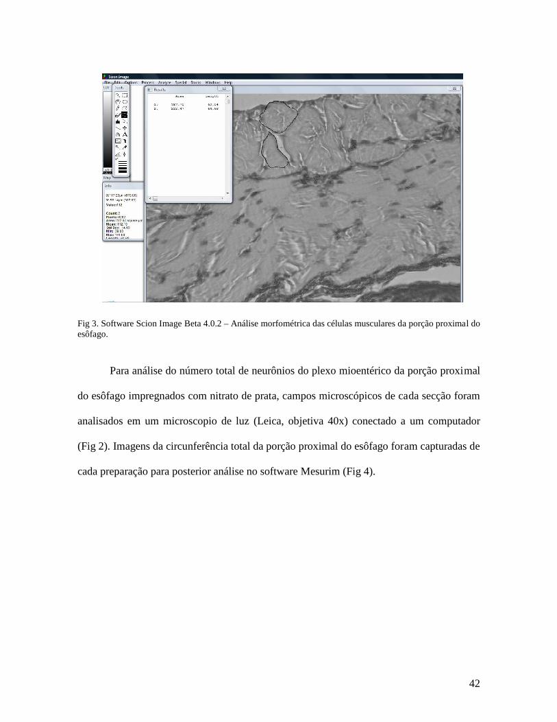
Para avaliar a área e o perímetro das células musculares, campos microscópicos de
cada secção foram analisados em um microscopio de luz (Leica, objetiva 40x) conectado a
um computador (Fig 2). Imagens de 30 células musculares foram capturadas de cada
preparação para posterior análise no software Scion Image Beta 4.0.2 (Fig 3).
Fig 2. Análise morfométrica - microscopio de luz conectado a um computador.
42
Fig 3. Software Scion Image Beta 4.0.2 – Análise morfométrica das células musculares da porção proximal do esôfago.
Para análise do número total de neurônios do plexo mioentérico da porção proximal
do esôfago impregnados com nitrato de prata, campos microscópicos de cada secção foram
analisados em um microscopio de luz (Leica, objetiva 40x) conectado a um computador
(Fig 2). Imagens da circunferência total da porção proximal do esôfago foram capturadas de
cada preparação para posterior análise no software Mesurim (Fig 4).

43
Fig 4. Software Mesurim – Análise morfométrica dos neurônios mioentéricos da porção proximal do esôfago.
O ANOVA Two-Way medidas repetidas foi realizado para o peso corporal. Para
análise dos parâmetros de tamanho e peso do esôfago; área e perímetro das fibras
musculares longitudinais; número de neurônios mioentérico da porção superior do esôfago;
e as possíveis relações alométricas, foi efetuado o ANOVA Two-Way. Como teste post-hoc
foi utilizado o Tukey. A significância estatística foi considerada, admitindo-se um nível
crítico de 5% em todos os casos. Todos os dados são apresentados como média±EPM.
O protocolo experimental deste estudo (processo nº 018130/2007-03) (ANEXO C)
foi aprovado pelo comitê de ética de experimentação animal (CEEA) da Universidade
Federal de Pernambuco, de acordo com o Instituto Nacional de guia de saúde para cuidado
e uso de animais de laboratório.

44
RESULTADOS
45
3.Resultados (Artigo Original)
Do serotonergic system manipulation program changes in the neuromuscular
development of the esophagus?
Ferraz-Pereira, KNa; Vitoriano, ILSa; Melo, MPP a; Leite, SP b; Silva, Sb; Toscano, AE a;
Silva, HJc; Manhães-de-Castro, R a
Abstract Stimuli or insults that occur during critical periods of development of the nervous system can result in persistent changes in the structure and function of the body. In this study, the effects of malnutrition and/or inhibition of serotonin reuptake during the postnatal period on the neuromuscular development of the esophagus were investigated in rats. The animals were divided into two groups, nourished or malnourished. Pharmacological manipulation was performed during the period of lactation. The animals were divided into two subgroups (saline or sertraline). Rats were weighed and sacrificed for removal of the esophagus on days 22 or 90. The total length of the esophagus was measured, and it was then divided into two portions of equal length. The proximal portion was sectioned for staining (Hematoxylin/eosin and impregnation by silver nitrate). Malnutrition and/or the action of neonatal sertraline appear to have been responsible for reducing body weight during lactation and after the period of nutritional recovery. The early protein restriction and the use of neonatal sertraline had impacts on the size and weight of the esophagus in animals on day 22. The nutritional manipulation caused persistent changes in both age groups that were studied in the area and the perimeter of the muscle fibers. Both insults caused a decrease in the number of myenteric neurons. Malnutrition and/or the use of selective serotonin reuptake inhibitors seem to result in neuromuscular changes in the morphology of the esophagus. Keywords: malnutrition; serotonin, sertraline, muscle fibers; myenteric plexus, esophagus.

46
Introduction
There are critical periods in the development of the nervous system that are
vulnerable to environmental insults (Winick, 1972; Morgane, 1978; Morgane, 1993).
During these stages, early stimuli or insults may result in persistent changes in the structure
and function of the body (Lee et al., 2005). This phenomenon, which is known as
programming, may be responsible for the establishment of diseases in adulthood (Harding,
2001).
In rats, pre- and postnatal care are essential to the development of the
gastrointestinal tract, which begins in embryogenesis and extends up to the period of
weaning, about 21 days postnatally (Koch, Ruggeberg, 1978; Hollwarth, Uray, 1985,
Jolley, Baron, 1998, Ross, Nijland, 1998). During such critical phases of life, damage to
the smooth muscle contributes to changes in esophageal motor function (Tugay et al.,
2003). It is therefore suggested that the characteristics of an active esophagus, such as
longitudinal and circumferential strain, depend on its morphology and passive properties,
such as the internal and external circumferential length and the thickness and area of
mucosa and muscle layers (Gregersen, Lu, Zhao, 2004), as well as on the development of
the ENS, which also undergoes further growth and development in this stage of life
(Gershon, Liu, 2007).
Malnutrition and/or the administration of selective serotonin reuptake inhibitors
(SSRIs) in the early stages of development lead to permanent damage to the structures and
functions of various systems of the body (Lucas, Fewtrell, Cole, 1999, Hales, Baker, 1992;
Marinho, 2004; Magalhães et al., 2006; Liberti et al., 2007; Lopes-de-Souza et al., 2008).
In the digestive system, these changes may compromise the functioning of the
neuromuscular structures that belong to the gastrointestinal tract, possibly interfering in the

47
process of transporting the food bolus, which is essential to acquire nutrients (Coates et al.,
2004; Cash, Chey, 2005).
This article aims to investigate whether malnutrition and/or inhibition of serotonin
reuptake can lead to structural changes in the neuromuscular development of the
esophagus in rats. This study will test the hypothesis that malnutrition and/or the
administration of a serotonin reuptake inhibitor during the period of lactation will cause
adaptive changes in the structural development of the longitudinal smooth muscle and
neurons in the myenteric plexus of the esophagus.
Materials and Methods:
Albino rats of the Wistar strain that were established from a colony from the
Department of Nutrition, Federal University of Pernambuco (Brazil) were mated to obtain
the experimental animals. Within 24 hours of their birth, litters were made up of six male
newborns by mothers that were chosen at random. The animals were kept in a controlled
environment with a temperature of 23±1°C in a constant cycle of light (6:00 a.m. to 6:00
p.m.) and darkness (6:00 p.m. to 6:00 a.m.). The animals had free access to filtered water
and standard animal feed (Labini - Purina of Brazil, containing 23% protein).
The rats were divided into two groups, which were nourished (N, n=39) and
undernourished (D, n=58). The D animals were malnourished during lactation by suckling
from mothers who received a hypoproteic diet with only 8% protein, which was the
Regional Basic Diet (RBD) (Teodosio et. al., 1990). The N animals suckled from mothers
who received a normal diet with 23% protein, which was the standard diet used in the
vivarium (Labina - Agribrands Purina of Brazil, LTDA). After weaning, which was at 21
48
days, all pups were fed a normal diet, which had a level of 23% protein, until the final days
of the experiment.
The pharmacological manipulation included treatment with Sertraline once a day
from the first day to the twenty-first day after birth. Members of the different nutritional
groups were divided into two subgroups. The sub-nourished sertraline (NSert, n=18) and
malnourished-sertraline (DSert, n=20) groups were treated with sertraline (10 mg/kg body
weight, subcutaneously). Sertraline, which was obtained in salt form, was dissolved in
saline and injected subcutaneously at a volume of 1 ml/100 g body weight. The sub-
nourished saline (NS, n=21) and malnourished-saline (DS, n=38) groups received an
equivalent volume of saline, which was in the form of a 0.9% solution of sodium chloride.
The weight of each offspring was assessed to day after birth (day 1) until the period of
weaning (day 21), and again at the ages of 30, 40, 50, 60, 70, 80 and 90 days. Animals from
each group were weighed and sacrificed at 22 or 90 days of age. The following parameters
were measured: size and weight of the proximal portion of the esophagus, the area and
perimeter of the longitudinal muscle fibers, the total number of neurons in the myenteric
plexus, the relation between number of neurons and area of the muscle cells and the
relationship between the number of neurons in the myenteric plexus and the perimeter of
the muscle fibers.
The animals were weighed on a digital electronic scale and anesthetized with a
solution of 12.5% urethane and 0.5% cloralose by intraperitoneal injection (0.01 ml/g body
weight). Under the effects of anesthesia, the longitudinal axis of the body, which was the
length of the snout to base of the tail, was measured. After the longitudinal axis was
measured, an opening was made in the chest cavity, and the region of the esophagus from
the pharynx-esophageal junction to the gastroesophageal junction was subsequently

49
collected. In order to perform this extraction, each animal was placed on flat surface of
paraffin, in the supine position, with their feet fixed by needles. An incision was made in
the thoracic cavity, and the esophagus was completely removed. This allowed the length
and the proximal portion of the esophagus (ESS to the middle portion) to be measured via a
stainless steel caliper . In addition, the weight of the upper body in precise balance was
obtained.
The proximal portion of the esophagus was immersed in buffered folmol in order
for it to be measured and weighed. After it was set, the esophagus was dehydrated in a
gradient of alcohol (70% to 100%) and placed immediately into xylol and paraffin.
Transverse sections were made of the esophagus that were 5 microns thick, and they were
subsequently stained with hematoxylin and eosin (HE) and mounted between slides with
synthetic resin (Entellan - Merck). The HE staining was initially performed in order to
locate the neurons of the myenteric plexus. After the neurons were located, the slides were
impregnated with silver nitrate and counter-stained with HE in order to illustrate and
confirm of these neurons. Morphometric analysis was performed on the longitudinal
muscle fibers and neurons in the myenteric plexus of the proximal portion of the
esophagus.
For analysis of the total number of neurons in the myenteric plexus of the proximal
portion of the esophagus that was impregnated with silver nitrate, thirty microscope fields
from each section were analyzed under a light microscope (Leica, 40x objective) that was
connected to a computer. Images of the total circumference of the proximal portion of the
esophagus were taken from each preparation for later analysis by the software Mesurim.
To evaluate the area and perimeter of the muscle cells, thirty microscope fields
from each section were analyzed under a light microscope (Leica, 40x objective) that was

50
connected to a computer. Images of thirty muscle cells were taken from each preparation
for later analysis in the software Scion Image Beta 4.0.2.
The repeated measures two-way ANOVA was performed for body weight. The
two-way ANOVA included the parameters of size and weight of the esophagus, the area
and perimeter of the longitudinal muscle fibers, the number of myenteric neurons of the
upper portion of the esophagus and the possible allometric relationships. In addition, post-
hoc Tukey was used. Statistical significance was considered to be at a critical level of 5%
in all cases.
The experimental protocol of this article (Case 018130/2007-03) was approved by
the ethics committee for animal experimentation (EAEC) at the Federal University of
Pernambuco, according to the National Institutes of Health guide for care and use of
laboratory animals.
Results
Manipulation of nutritional and/or pharmacological factors (F(3.781)=107.45,
p<0.001), age (F(11.781)=3758.52, p<0001) and the manipulation and interaction with age
(F(33.781)=25.60, p<0001) all showed differences in the body weight of the animals. The rats
in DS (q=5.607, p<0001) and DSert (q=4.301, p=0.013) groups had lower body weight
starting from the 14th day of life when they were compared with the rats in NS and Nsert
groups, respectively. The DSert (q=5.969, p<0.001) and NSert groups (q=3.755, p=0.040)
had lower body weights when compared with the groups that were treated with the saline
solution on both days 50 and 70 (Fig 1).

51
Fig 1. Effect of malnutrition and/or pharmacologic manipulation of the serotonergic system on the curve of body weight. The rats were subjected to nutritional manipulation (nourished or malnourished) and/or drug (sertraline 10 mg/kg bw or sodium chloride at 0.9%, 1ml/100g bw) during the period of lactation (two-way repeated measures ANOVA). The data are represented in mean±SEM. Multiple comparisons (Tukey test) - p<0.05: a-NS vs DS b-NSert vs DSert c-DS vs DSert d-NS vs NSert.
Tables 1 and 2 present the parameters that were evaluated for the esophagus on
days 22 and 90, respectively. For the weight of the upper portion of the esophagus on day
22, the diet effect (F(1.51)=51.598, p<0001) led to differences in body weight, but neither
the drug effect (F(1. 51)=0.0760, p= 0784) nor the interaction between the two factors (diet
vs. drugs) (F(1.51)=1.204, p=0.278) led to any differences in body weight. On day 90, the
diet effect (F(1.40)=0.574, p=0.453), the drug effect (F(1.40)=3.063, p=0.088) and the
interaction of diet and drug (F(1.40)=0.000951, p=0.976) showed no difference in the weight
of the upper portion of the esophagus. On day 22, the rats in groups DS and DSert had a
lower weight of the proximal portion of the esophagus when compared to groups NS and
050
100150200250300350400
1 3 7 14 21 30 40 50 60 70 80 90
Age (days)
Bo
dy
wei
gh
t (g
)
NS NSERT DS DSERT
ab ab
ab
abc
abc
abcd
abcd
abcd
ab
52
NSert, respectively. On day 90, there were no differences among any of the groups for the
weight of the proximal portion of the esophagus.
With regard to the size of the upper portion of the esophagus on day 22, the diet
effect (F(1.51)=118.594, p<0001) and the drug effect (F(11.51)=11.641, p=0.001) showed
differences, but there was no diet vs. drug interaction (F(1.51)=0.735, p=0.395). The DS
group on day 22 showed a reduction in the size of the esophagus when compared to the NS
group. The DSert group on day 22 also had a smaller size when compared with both the
NSert and DS groups. On day 90, the diet effect (F(1.40)=0.0101, p=0.921), the drug effect
(F(1.40)=0.575, p=0.453) and the interaction of diet vs. drug (F(1.40)=0.0722,p=0.790)
showed no differences in the size of the esophagus.
With regard to the area and perimeter of the longitudinal muscle fibers on day 22, a
difference was observed for the diet effect (area - F(1.17)=36.666, p<0.001/ perimeter -
F(1.17)=33.406, p<0001) but not for the drug effect (area - F(1.17)=0.0150, p=0.904/
perimeter - F(1.17)=0.000638, p=0.980) nor the interaction of diet vs. drug (area -
F(1.17)=2.073, p=0.172/ perimeter - F(1.17)=2.701, p=0.123). On day 90, a difference was still
observed for the diet effect (area - F(1.18)=26.161, p<0,001/ perimeter - F(1.18)=26.753,
p<0001) and still not for the drug effect (area - F(1.18)=0.534, p=0.476/ perimeter -
F(1.18)=0.534, p=0.476) nor the interaction of diet vs. drugs (area - F(1.18)=0.149, p=0.705/
perimeter - F(1.18)=0.00360, p=0.953). The muscle cells of the DS and DSert groups had
lower area and perimeter when compared to the NS and NSert groups, respectively, at both
the ages that were studied.
With regard to the total number of myenteric neurons in the proximal portion of the
esophagus that were impregnated with silver nitrate on day 22, the diet effect showed
differences (F(1.19)=5.261, p=0.036), but the drug effect (F(1.19)=0.358, p=0.558) and the

53
interaction of diet vs. drug (F(1.19)=0.0311, p=0.862) did not show any differences. On day
90, the drug effect showed a difference (F(1.19)=13.906, p=0.002), but the diet effect
(F(1.19)=0.0204, p=0.888) and interaction of diet vs. drug (F(1.19)=0.550, p=0.469) did not
show a difference (Fig 3). On day 22, a lower number of neurons in the DS (Fig 3C) and
DSert groups (Fig 3D) was observed when compared to the NS (Fig 3A) and NSert groups,
respectively (Fig 3B). On day 90, there was a smaller population of neurons in the DSert
(Fig 3H) and NSert groups when compared to the DS (Fig 3G) and NS groups,
respectively.
Table 1. Effect of malnutrition and/or manipulation of the serotoninergic system on the parameters macroscopes and microscopes of the upper portion of the esophagus in animals sacrificed on days 22. The rats were subjected to nutritional manipulation (nourished or malnourished) and/or drug (sertraline 10 mg/kg bw or sodium chloride at 0.9%, 1ml/100g bw) during the period of lactation (two-way ANOVA). The data are represented in mean ± SEM.
Experimental Groups
ANOVA
Parameters NS NSert DS Dsert F P Weight of the
esophagus (g)
0,014±0,002 (n=9)
0,013±0,001 (n=10)
0,006±0,0004a
(n=21) 0,007±0,001b
(n=12) Diet=51,598 Drug=0,0760
DietxDrug=1,204
<0,001 0,784 0,278
Size of the esophagus
(mm)
21,8±0,7 (n=10)
19,9±0,5a
(n=10) 16,7±0,2a
(n=21) 15,6±0,4b,c
(n=12) Diet=118,594 Drug=11,641
DietxDrug=0,735
<0,001 0,001 0,395
Area of the muscle fibers (µµµµm)
45,3±6,0 (n=4)
50,7±4,3 (n=4)
29,3±1,5a
(n=5) 24,8±1,6b
(n=5) Diet=36,666 Drug=0,0150
DietxDrug=2,07
<0,001 0,904 0,172
Perimeter of the muscle fibers
(µµµµm)
26,7±1,6 (n=4)
28,6±1,6 (n=4)
21,9±0,6a
(n=5) 20,0±0,7b
(n=5) Diet=33,406
Drug=0,000638 DietxDrug=2,70
<0,001 0,980 0,123
Total number of myenteric neurons
211,6±21,7 (n=4)
220,2±30,9 (n=4)
161,2±10,8a
(n=5) 177,0±11,1b
(n=5) Diet=5,261 Drug=0,358
DietxDrug=0,0311
0,036 0,558
0,862 Multiple comparisons (Tukey test) - p<0.05: a-Comparison with the NS group, b-Comparison with the NSert group c-comparison with the DS group.

54
Table 2. Effect of malnutrition and/or the manipulation of the serotoninergic system on the parameters macroscopes and microscopes of the upper portion of the esophagus in animals sacrificed on days 90. The rats were subjected to nutritional manipulation (nourished or malnourished) and/or drug (sertraline 10 mg / kg bw or sodium chloride at 0.9%, 1ml/100g bw) during the period of lactation (two-way ANOVA). The data are represented in mean ± SEM.
Experimental Groups
ANOVA
Parameters NS NSert DS DSert F P Weight of the
esophagus (g)
0,08±0,005 (n=11)
0,07±0,004 (n=8)
0,07±0,002 (n=15)
0,07±0,004 (n=7)
Diet=0,574 Drug=3,063
DietxDrug=0,000951
0,453 0,088 0,976
Size of the esophagus
(mm)
36,6±1,0 (n=11)
35,4±0,9 (n=8)
36,2±0,9 (n=15)
35,6±1,4 (n=7)
Diet=0,0101 Drug=0,575
DietxDrug=0,0722
0,921 0,453 0,790
Area of the muscle fibers
(µµµµm)
302,1±30,4 (n=5)
331,6±34,8 (n=4)
154,2±15,6a
(n=5) 134,8±5,0b
(n=5) Diet=26,161 Drug=0,534
DietxDrug=0,149
<0,001 0,476 0,705
Perimeter of the muscle fibers
(µµµµm)
71,9±3,2 (n=5)
75,6±2,6
(n=4) 50,3±2,6a
(n=5) 48,6±0,5b
(n=5) Diet=26,753 Drug=0,534
DietxDrug=0,00360
<0,001 0,476 0,953
Total number of myenteric neurons
442,2±24,2 (n=5)
356,4±35,1a
(n=4) 459,4±14,9
(n=5) 331±35,4c
(n=5) Diet=0,0204 Drug=13,906
DietxDrug=0,550
0,888 0,002 0,469
Multiple comparisons (Tukey test) - p<0.05: a-Comparison with the NS group, b-Comparison with the NSert group c-comparison with the DS group.
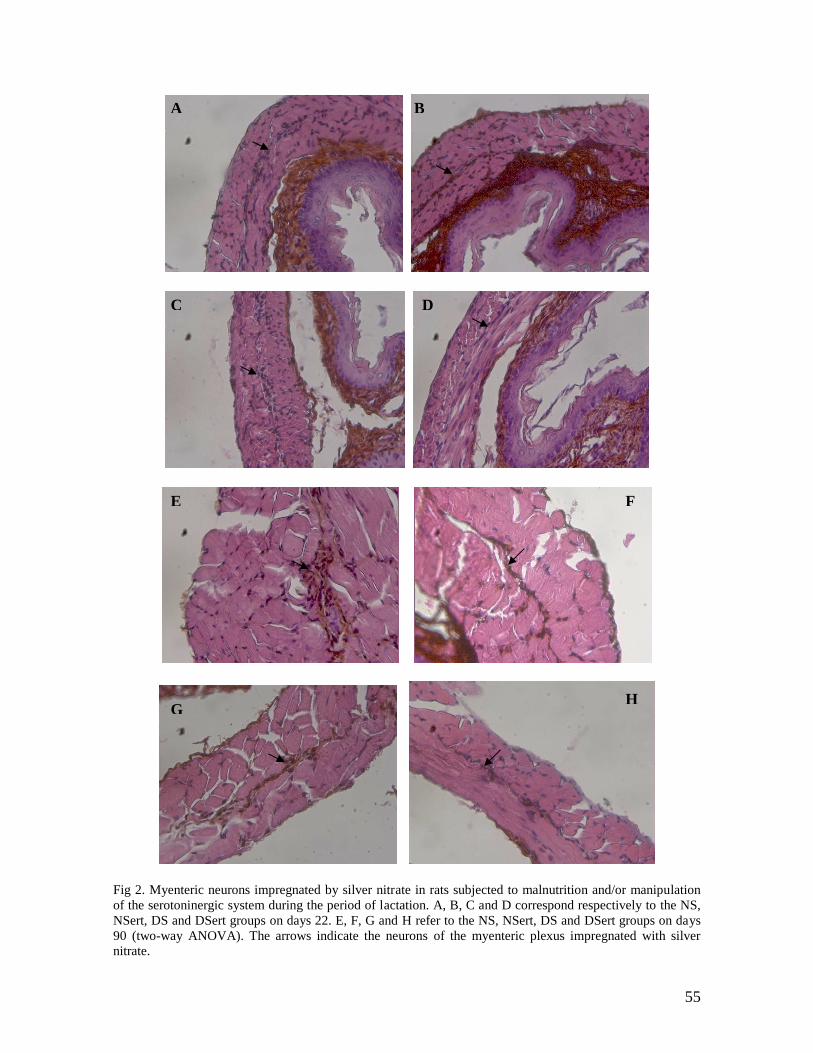
55
Fig 2. Myenteric neurons impregnated by silver nitrate in rats subjected to malnutrition and/or manipulation of the serotoninergic system during the period of lactation. A, B, C and D correspond respectively to the NS, NSert, DS and DSert groups on days 22. E, F, G and H refer to the NS, NSert, DS and DSert groups on days 90 (two-way ANOVA). The arrows indicate the neurons of the myenteric plexus impregnated with silver nitrate.
H G
F E
D C
B A

56
Figure 3 shown the relationship between the total number of neurons in the
myenteric plexus and the area of the esophageal longitudinal muscle fibers on days 22 and
90. With regard to this relationship on day 22, the diet effect showed differences
(F(1.17)=4.912, p=0.044), but the drug effect (F(1.17)=1.063, p=0.320) and the interaction of
diet vs. drug (F(1.17)=1.506, p=0.240) did not show any differences. On day 90, the diet
effect again showed a difference (F(1.18)=27.921, p<0001), and the drug effect
(F(1.18)=3.798, p=0.070) and the interaction of diet vs. drug (F(1.18)=0.398, p=0.538) also did
not show any differences. The ratio was higher in the DS group when compared to the NS
group on days 22 and 90. The ratio was also higher in the DSert group when compared to
the NSert group on days 22 and 90.
0,0
1,0
2,0
3,0
4,0
5,0
6,0
7,0
8,0
9,0
22 90
Age (days)
To
tal n
um
ber
of
neu
ron
s/ar
ea o
f th
e m
usc
le f
iber
s
NS
NSERT
DS
DSERT
a
b
a
b
Fig 3. Effect of malnutrition and/or manipulation of the serotoninergic system on the relationship between the number of neurons in the myenteric plexus and the area of longitudinal muscle fibers in animals sacrificed on days 22 or 90. The rats were subjected to nutritional manipulation (nourished or malnourished) and/or drug (sertraline 10 mg/kg bw or sodium chloride at 0.9%, 1ml/100g bw) during the period of lactation (two-way ANOVA). The data are represented in mean±SEM. Multiple comparisons (Tukey test) - p<0.05: a-Comparison with the NS group, b-Comparison with the NSert group c-comparison with the DS group, d-Comparison with the DSert group.
Figure 4 present the relationship between the total number of neurons in the
myenteric plexus and the perimeter of the esophageal longitudinal muscle fibers on days

57
22 and 90. On day 22, the diet effect (F(1.17)=0.00165, p=0.968), the drug effect
(F(1.17)=0.686, p=0.422) and the interaction of diet vs. drug (F(1.17)=0.498, p=0.492) showed
no difference. On day 90, the diet (F(1.18)=15.570, p=0.001) and drug effects (F(1.18)=8.967,
p=0.009) showed differences, but there was no interaction of diet vs. drug (F(1.18)=1.029,
p=0.326). There was a higher ratio in the DS and DSert groups when compared to the N
and NSert groups, respectively. The NS and DS groups showed a higher ratio when
compared to the NSert and DSert groups, respectively.
0,0
2,0
4,0
6,0
8,0
10,0
12,0
22 90
Age (days)
To
tal n
um
ber
of
neu
ron
s/p
erim
eter
of
the
mu
scle
fib
ers
NS
NSERT
DS
DSERT
ad
b
Fig 4. Effect of malnutrition and/or manipulation of the serotoninergic system on the relationship between the number of neurons in the myenteric plexus and the perímeter of longitudinal muscle fibers in animals sacrificed on days 22 or 90. The rats were subjected to nutritional manipulation (nourished or malnourished) and/or drug (sertraline 10 mg / kg bw or sodium chloride at 0.9%, 1ml/100g bw) during the period of lactation (two-way ANOVA). The data are represented in mean ±SEM. Multiple comparisons (Tukey test) - p <0.05: a-Comparison with the NS group, b-Comparison with the NSert group c-comparison with the DS group, d-Comparison with the DSert group. Discussion
In this study, it was demonstrated that the manipulations that were imposed during
the lactation period promoted changes in body weight and the parameters that were
analyzed in the proximal portion of the esophagus, which included size, weight, area and
perimeter of the longitudinal muscle fibers and total number of neurons in the myenteric

58
plexus. Malnutrition during this critical period of development was responsible for
reducing body weight during the time in which the experimental treatments were
performed, and the malnutrition persisted even after the period of nutritional recovery.
When the manipulation of the serotonergic system through the administration of SSRIs
was associated with malnutrition, a greater difference was observed in the weight of the
animals.
Malnutrition and Sertraline, which was the serotonin reuptake inhibitor, had a
considerable impact on the size and weight of the proximal portion of the esophagus in
animals on day 22. On day 90, however, there were no changes in these parameters.
These findings are in agreement with the observations of Toscano et al. (2008).
These authors observed that malnutrition during lactation reduced body weight and the
weight of the heart over the short term (Toscano et al., 2008). In addition, they observed
that the administration of fluoxetine, which is an SSRI, caused a decrease in growth of the
heart on day 30, with a subsequent recovery (Toscano et al., 2008).
Silva (2006) observed that malnutrition that was associated with the chronic use of
selective serotonin reuptake inhibitors during this critical period of development can
interfere with the development of the esophagus. However, the manipulation of the
serotonergic system through the administration of fluoxetine (SSRI) interferes in the
development of this structure only in the late phase (Silva, 2006).
Programming insults such as malnutrition and the administration of SSRIs during
the early stages of life can lead to permanent damage in the morphology and physiology of
the organs and tissues of the body (Lucas, Fewtrell, Cole, 1999; Hales, Baker, 1992;
Magalhães et al., 2006; Lopes-de-Souza et al., 2008, Marinho, 2004; Liberti et al., 2007).
These changes may be the result of the stimulation of the serotonergic system by means of

59
the aforementioned exogenous factors (Halford, Blundell, 1996). This serotonergic
manipulation may affect somatic growth and development (Toscano et al., 2008). The role
of 5-HT in the regulation of feeding behavior and body weight is already clear (Toscano et
al., 2008). Studies show that protein malnutrition (Manjarrez et al., 2003; Mokler et al.,
2003) and administration of an SSRI (Deiró et al., 2006) affect food intake and body
weight gain as a result of increased availability of serotonin in the synaptic cleft.
We observed that nutritional manipulation caused persistent changes at both ages
that were studied (days 22 and 90) in the area and the perimeter of the longitudinal muscle
fibers of the upper portion of the esophagus. The important factor is the early nutrition
programming environment (Lucas et al., 1999), which can induce considerable
consequences in adulthood (Lucas, 1998). This perinatal malnutrition can induce muscle
atrophy (Alippi et al., 2002; Bozzini et al., 1989). In addition, the neonatal malnutrition
may irreversibly damage the morphology of the muscle (Bedi et al., 1982). It can also
reduce cell proliferation, which reduces the number of muscle fibers and nuclei in the
offspring (Bedi et al., 1982, Park et al., 2003; Bayol et al., 2004). Changes in the
proportions of fiber types after early malnutrition have also been observed (Toscano et al.,
2008). Therefore, malnutrition compromises the biomechanical and elastic contractile
properties of the muscle (Toscano et al., 2008).
Changes in the smooth muscle contribute to changes in esophageal motor function
(Tugay et al., 2003). Therefore, it is proposed that the active characteristics of the
esophagus, such as the longitudinal and circumferential strain, are dependent on its
morphology and passive properties, such as internal and external circumferential length,
the thickness and area of the mucosal layer and the thickness and area of the muscular
layers (Gregersen , Lu, Zhao, 2004).

60
Potential morphological and physiological changes in the neonatal smooth muscle
of the digestive system as a result of malnutrition may also be caused by inactivation of the
carrier molecule of serotonin (SERT) (Coates et al., 2006). This plasma membrane protein
that is responsible for the degradation and inactivation of serotonin that is found within the
interstitial space (Gershon, 2003). This cycle of the release and reuptake of serotonin
should remain untouched by its influence on the control of motor, sensory and secretory
reflexes (Coates et al., 2006). Therefore, it has been suggested that changes in 5-HT
signaling, which affects the availability of the neurotransmitter, may contribute to
disturbances in gastrointestinal function (Coates et al., 2004; Cash; Chey, 2005).
In this study, we observed that both applied environmental insults caused a
reduction in the number of myenteric neurons in the proximal portion of the esophagus.
However, the diet factor only had an effect after weaning on day 22, but there were no
permanent changes in the young adults, 90. Long-term effects were observed for the
administration of the serotonin reuptake inhibitor, although it did not have any noticeable
effects right after weaning.
With regard to the relationship between the number of neurons in the myenteric
plexus and the area of longitudinal muscle cells of the proximal portion of the esophagus,
diet had both short-term and long-term effects. Administration of the serotonin reuptake
inhibitor only had an effect in the long term.
Protein deficiency during this critical period of life causes a delay in the
development of tissues and myenteric neurons in young rats (Castelucci et al., 2002,
Brandão et al, 2003, Gomes et al., 2006; Liberti et al., 2007). However, the morphological
and morphometric characteristics of enteric neurons recover after the rats were returned to
a normal diet (Meilus et al., 1998; Miranda Neto et al., 1999; Castelucci et al., 2002,

61
Gomes et al., 2006). These possible effects of protein malnutrition in the myenteric plexus
neural structures indicate that the control of gastrointestinal motility may be affected in a
negative way (Miranda Neto, 1999).
In the enteric nervous system, the manipulation of the serotoninergic system that is
induced by the use of SSRIs also results in a delay in the development of neurons in the
myenteric plexus of the gastrointestinal tract (Marinho, 2004). The administration of
fluoxetine during the lactation period causes a reduction in the number of neurons and in
the size of myenteric nerve cells in the segment of distal small intestine that can be seen on
day 75 (Marinho, 2004). These SSRIs can act as blockers and inactivate this bioamine,
which functions as the carrier molecule for serotonin (Wong et al., 1975). SERT inhibition
produces an increased synaptic concentration of serotonin, which allows for a greater
availability for this neurotransmitter to connect with its receptor (Laurie, 2004). Thus,
SSRIs may affect brain function (Wong et al., 1975) and gastrointestinal tract function
(Gershon, Jonakait, 1979; Wade et al., 1996) as a result of changes in the function of 5-HT
receptors (Tuladhar, Costall, Naylor, 2002).
Furness (2000) indicated that there was a strong relationship between the
ganglionic myenteric plexus and the smooth muscles of the digestive system. Given that, it
is suggested that changes in neural structures of the myenteric plexus that are associated
with morphological impairment of the muscle fibers of the digestive system are indicative
that damage to gastrointestinal motility may occur (Miranda Neto, 1999).
It has been proposed that the changes in 5-HT signaling as a result of nutritional
and pharmacological manipulations affect the availability of serotonin and, therefore, may
interfere with gastrointestinal function (Coates et al., 2004; Cash, Chey, 2005). One of the

62
symptoms that can result from motor dysfunction in the esophagus is dysphagia
(Domingues, Lemme, 2001), which is the difficulty of the passage of food bolus by the
esophageal body (Domingues, Lemme, 2001). This symptom, which results from a
decrease in esophageal motility, may be responsible for the occurrence of
pharyngoesophageal reflux (Marchesan, Furkim, Santini, 1999) and the increased risk of
laryngotracheal penetration or aspiration after swallowing (Matsuo, Palmer, 2008).
Esophageal dysphagia, depending on its severity and frequency, may lead to malnutrition
(Domingues, Lemme, 2001), which can generate a negative impact on an individual’s
quality of life (Beattie et al. 2004).
Conclusion
Malnutrition and/or serotonin reuptake inhibition during the critical period of life
prior to weaning promotes adaptive changes in the development of the neuromuscular
structures of the upper portion of the esophagus. These findings indicate that the structural
changes that are associated with metabolic programming are related to the regulatory role
of serotonin in the neuromotor development of the gastrointestinal tract.

63
References
1. Alippi, R.M., Meta, M.D., Olivera, M.I., Bozzini, C., Shneider, P., Meta, I., Bozzini, C.,
2002. Effect of protein-energy malnutrition in early life on the dimensions and bone quality
of the adult rat mandible. Arch. Oral Biol. 47, 47-53.
2. Bayol, S., Jones, D., Goldspink, G., Stickland, N.C., 2004. The influence of
undernutrition during gestation on skeletal muscle cellularity and on the expression of
genes that control muscle growth. Brit J Nutr 91, 331–339.
3. Beattie, D.T., Smith, J.A.M., Marquess, D., Vickery, R.G., Armstrong, S.R., Pulido-
Rios, T., McCullough, J.L., Sandlund, C., Richardson, C., Mai, N., Humphrey, P.P.A.,
2004. The 5-HT4 receptor agonist, tegaserod, is a potent 5-HT2B receptor antagonist in
vitro and in vivo. Brit J Pharmacol. 143, 549–560.
4. Bedi, K.S., Birzgalis, A.R., Mahon, M., Smart, J.L., Wareham, A.C., 1982. Early life
undernutrition in rats. Quantitative histology of skeletal muscles from underfed young and
refed adult animals. Br J Nutr. 47(3), 417-31.
5. Bozzini, C., Barcelo, A.C., Alippi, R.M., Leal, T.L., Bozzini, C.E., 1989. The
concentration of dietary casein required for normal mandibular growth in the rat. J Dent
Res. 68, 840-842.
6. Brandão, M.C.S., Angelis, R.C. de, De-Souza, R.R., Fróes, L.B., Liberti, E.A., 2003.
Effects of pre- and postnatal protein energy deprivation on the myenteric plexus of the
small intestine: a morphometric study in weanling rats. Nutr Res. 23, 215–223.
7. Cash, B.D., Chey, W.D., 2005. Review article: the role of serotonergic agents in the
treatment of patients with primary chronic constipation. Aliment Pharmacol Ther. 22,
1047–1060.
64
8. Castelucci, P., Souza, R.R. de, et al., 2002. Effects of pre- and postnatal protein
deprivation and postnatal refeeding on myenteric neurons of the rat large intestine: a
quantitative morphological study. Cell Tissue Res. 310(1):1-7.
9. Coates MD, Mahoney CR, Linden DR et al., 2004. Molecular defects in mucosal
serotonin content and decreased serotonin reuptake transporter in ulcerative colitis and
irritable bowel syndrome. Gastroenterology. 126:1657–64.
10. Coates, M.D., Johnson, A.C., Meerveld, B.G.V., Mawe, G.M., 2006. Effects of
serotonin transporter inhibition on gastrointestinal motility and colonic sensitivity in the
mouse. Neurogastroenterol Motil. 18, 464–471.
11. Deiró, T.C., Manhães-de-Castro, R., Cabral-Filho, J.E., Barreto-Medeiros, J.M., Souza,
S.L., Marinho, S.M., Castro, F.M., Toscano, A.E., Jesus-Deiró, R.A., Barros, K.M., 2006.
Sertraline delays the somatic growth and reflex ontogeny in neonate rats. Physiol Behav.
87, 338–344.
12. Domingues, G.R., Lemme, E.M.O., 2001. Diagnóstico diferencial dos distúrbios
motores esofagianos pelas características da disfagia. Arq. Gastroenterol. 38(1).
13. Furness, J.B., 2000. Types of neurons in the enteric nervous system. J. Auton. Nerv.
Syst. 81(1–3), 87–96.
14. Gershon, M.D., Jonakait, G.M., 1979. Uptake and release of 5-hydroxytryptamine by
enteric 5-hydroxytryptaminergic neurons: Effects of fuoxetine and chlorimipramine. Br. J.
Pharmacol. 66, 7-9.
15. Gershon, M.D., 2003. Serotonin and its implication for the management of irritable
bowel syndrome. Rev. Gastroenterol. Disord. 3 (Suppl. 2), 25–34.
16. Gershon, M.D., Liu, M.T., 2007. Serotonin and neuroprotection in functional bowel
disorders. Neurogastroenterol. Motil. 19 (Suppl. 2), 19–24.

65
17. Gregersen, H., Lu, X., Zhao, J., 2004. Physiological growth is associated with
esophageal morphometric and biomechanical changes in rats. Neurogastroenterol. Motil.
16, 403–412.
18. Gomes, O.A., Castelucci, P., Fontes, R.B.V., Liberti, E.A., 2006. Effects of pre- and
postnatal protein deprivation and postnatal refeeding on myenteric neurons of the rat small
intestine: A quantitative morphological study. Auton Neurosci. 126-127, 277-284.
19. Hales, C.N., Barker, D.J., 1992. Type 2 (non-insulin-dependent) diabetes mellitus: the
thrifty phenotype hypothesis. Diabetologia. 35(7), 595-601.
20. Harding, J.E., 2001. The nutritional basis of the fetal origins of adult disease. Int J
Epidemiol. 30, 15-23.
21. Halford, J.C.G., Blundell, J.E., 1996. Metergoline antagonizes fluoxetine-induced
suppression of food intake but not changes in the behavioural satiety sequence. Pharmacol
Biochem Behav. 54, 745–751.
22. Hollwarth, M., Uray, E., 1985. Physiology and pathophsiology of the esophagus in
childhood. Prog. Pediatr. Surg. 18, 1–13.
23. Jolley, S.G., Baron, H.I., 1998. Disorders of esophageal function. In: O’Neill, J.A.,
Rowe, M.I., Grosfeld, J.L., Fonkalsrud, E.W., Coran, A.G., editors. Pediatric surgery. 997–
1005.
24. Koch, A., Ruggeberg, J., 1978. Function of the lower esophageal sphincter in infants.
Chir. Forum Exp. Klin. Forsch. 1978, 53–7.
25. Laurie, L.E., 2004. Neurociência: fundamentos para a reabilitação. Rio de Janeiro:
Elsevier.

66
26. Lee, Y.Y., Park, K.S., Pak, Y.K., Lee, H.K., 2005. The role of mitochondrial DNA in
the development of type 2 diabetes caused by fetal malnutrition. J Nutr Biochem. 16, 195-
204.
27. Liberti, E.A., Fontes, R.B., et al., 2007. Effects of combined pre- and post-natal protein
deprivation on the myenteric plexus of the esophagus of weanling rats: a histochemical,
quantitative and ultrastructural study. World J Gastroenterol. 13(26):3598-604.
28. Lopes-de-Souza, S., Orozco-Solis, R., Grit, I., Manhães-de-Castro, R., Bolaños-
Jiménez, F., 2008. Perinatal protein restriction reduces the inhibitory action of serotonin on
food intake. Eur J Neurosci.
29. Lucas, A., 1998. Programming by early nutrition: an experimental approach. In:
Symposium: The effects of childhood diet on adult health and disease. J Nutr. 128, 401S-
406S.
30. Lucas, A., Fewtrell, M.S., Cole, T.J., 1999. Fetal origins of adult disease—the
hypothesis revisited. Bmj. 319.
31. Marchesan, I.Q., Furkim, A.M., Santini, C.S., 1999. Disfagias Orofaríngeas. São Paulo:
Profono.
32. Marinho, S.M.O.C., 2004. Efeito da manipulação neonatal do sistema serotoninérgico
sobre o desenvolvimento do intestino delgado em ratos. Recife: Dissertação (Mestrado),
Departamento de Nutrição, Universidade Federal de Pernambuco, 62p.
33. Magalhães, C.P., Lima, L.O. de, et al., 2006. Neonatal treatment effect with selective
inhibitor of 5-HT recapture on [corrected] the cranium-encephalic anatomic development.
Arq Neuropsiquiatr. 64(4):990-3.
34. Manjarrez, G., Manuel, A.L., Mercado, C.R., Hernandez, R.J., 2003. Serotonergic
receptors in the brain of in utero undernourished rats. Int J Dev Neurosci. 21(5), 283–289.

67
35. Matsuo, K., Palmer, J.B., 2008. Anatomy and Physiology of Feeding and Swallowing:
Normal and Abnormal. Phys. Med. Rehabil. Clin. N. Am. 19, 691–707.
36. Meilus, M., Natali, M.R.M., Miranda-Neto, M.H., 1998. Study of the myenteric plexus
of the ileum of rats subjected to proteic undernutrition. Rev. Chil. Anat. 16(1).
37. Miranda-Neto, M.H. de, Molinari, S.L., Stabille, S.R., Sant´Ana, D.M.G., Natali,
M.R.M., 1999. Arq. Neuropsiquiatr. 57(2-B), 387-391.
38. Mokler, D.J., Galler, J.R., Morgane, P.J., 2003. Modulation of 5-HT release in the
hippocampus of 30-day-old rats exposed in utero to protein malnutrition. Brain Res Dev
Brain Res. 142(2), 203–8.
39. Morgane, P.J., Miller, M., Kemper, T., Stern, W., Forbes, W., Hall, R., Bronzino, J.,
Kissane, J., Hawrylewick, R.O., 1978. The effects of protein malnutrition on the developing
central nervous system in the rat. Neurosci Biobehav Rev. 2, 137-230.
40. Morgane, P.J., Austin-Lafrance, R, et al, 1993. Prenatal malnutrition and development
of the brain. Neurosci Biobehav Rev. 17(1):91-128.
41. Park, K.S., Kim, S.K., Kim, M.S., Cho, E.Y., Lee, J.H., Lee, K.U., Pak, Y.K., Lee,
H.K., 2003. Fetal and early postnatal protein malnutrition cause long-term changes in rat
liver and muscle mitochondria. J. Nutr. 133(10), 3085-90.
42. Ross, M.G., Nijland, M.J.M., 1998. Development of ingestive behavior. Am. J. Physiol.
Regulatory Integrative Comp. Physiol. 274, 879-893.
43. Silva, H.J., 2006. Manipulação neonatal e da recaptação de serotonina em ratos: 1.
Estabelecimento de protocolo para estudo murinométrico e validação; 2. Repercussões
sobre a morfologia e morfometria da túnicas musculares do esôfagoe sobre o consumo
alimentar. Tese (Doutorado), Departamento de Nutrição, Universidade Federal de
Pernambuco.

68
44. Toscano, A.E., Amorim, M.A.F., Carvalho Filho, E.V. de, Aragão, R. da S., Cabral-
Filho, J.E., Moraes, S.R.A. de, Manhães-de-Castro, R., 2008. Do malnutrition and
fluoxetine neonatal treatment program alterations in heart morphology? Life Sci. 82, 1131–
1136.
45. Toscano, A.E., Manhães-de-Castro, R., Canon, F., 2008. Effect of a low-protein diet
during pregnancy on skeletal muscle mechanical properties of offspring rats. Nutrition. 24,
270–278.
46. Tuladhar, B.R., Costall, B., Naylor, R.J., 2002. Modulation of 5-HT4 receptor function
in the rat isolated ileum by Fuoxetine: the involvement of endogenous 5-
hydroxytryptamine. Brit J Pharmacol. 136, 150 ± 156.
47. Tugay M, Yvldvz F, Utkan T, et al., 2003. Esophagitis impairs esophageal smooth
muscle reactivity in the rat model: an in vitro study. Dig Dis Sci. 48:2147- 52.
48. Wade, P.R., Chen, J., Jaffe, B., Kassem, I.S., Blakely, R.D., Gershon, M.D., 1996.
Localization and function of a 5-HT transporter in crypt epthelia of the gastrointestinal
tract. J. Neurosci. 16, 2352-2364.
49. Winick M, Rosso P, et al., 1972. Malnutrition and cellular growth in the brain. Bibl
Nutr Dieta; 17:60-8.
50. Wong, D.T., Bymaster, F.P., Horng, J.S., Molloy, B.B., 1975. A new selective inhibitor
for uptake of serotonin into synapto-somes of rat brain: 3-(p-trifuoromethylphenoxy)-N-
methyl-3-phenylpropylamine. J. Pharmacol. Exp. Therp. 193, 804-811.

69
CONSIDERAÇÕES FINAIS

70
4.Considerações finais
Este estudo possibilitou desmonstrar:
• Os efeitos precoces e tardios da desnutrição e da manipulação neonatal do sistema
serotoninérgico sobre o desenvolvimento neuromuscular do trato gastrintestinal.
• A forte relação dos insultos nutricionais e/ou farmacológicos do sistema
serotoninérgico, incidentes nas fases críticas do desenvolvimento, com a
programação, sobretudo os fatores exógenos que atingem a morfogênese
neuromuscular esofagiana.
• O papel programador da serotonina no desenvolvimento morfológico do trato
gastrintestinal, em particular do esôfago.

71
Referências
1. Alippi RM, Meta MD, Olivera MI, Bozzini C, Shneider P, Meta I, Bozzini C., 2002.
Effect of protein-energy malnutrition in early life on the dimensions and bone quality of the
adult rat mandible. Arch Oral Biol. 47:47-53.
2. Baxter GS, Craig DA, Clarke DE, 1991. 5-Hydroxy-tryptamine4 receptors mediate
relaxation of the rat oesophageal tunica muscularis mucosae. Naunyn-Schmied Arch
Pharmacol. 343: 439-446.
3. Bayol S, Jones D, Goldspink G, Stickland NC., 2004. The influence of undernutrition
during gestation on skeletal muscle cellularity and on the expression of genes that control
muscle growth. Br J Nutr. 91: 331–339.
4. Beattie DT, Smith JAM, Marquess D, Vickery RG, Armstrong SR, Pulido-Rios T,
McCullough JL, Sandlund C, Richardson C, Mai N, Humphrey PPA, 2004. The 5-HT4
receptor agonist, tegaserod, is a potent 5-HT2B receptor antagonist in vitro and in vivo. Br
J Pharmacol. 143:549–560.
5. Bedi KS, Birzgalis AR, Mahon M, Smart JL, Wareham AC, 1982. Early life
undernutrition in rats. Quantitative histology of skeletal muscles from underfed young and
refed adult animals. Br J Nutr. 47(3):417-31.
6. Bozzini C, Barcelo AC, Alippi RM, Leal TL, Bozzini CE, 1989. The concentration of
dietary casein required for normal mandibular growth in the rat. J Dent Res. 68:840-842.
7. Brandão MCS, Angelis RC de, De-Souza RR, Fróes LB, Liberti EA, 2003. Effects of
pre- and postnatal protein energy deprivation on the myenteric plexus of the small intestine:
a morphometric study in weanling rats. Nutr Res. 23:215–223.
8. Buznicov GA, Lambert HW, Lauder JM, 2001. Serotonin and serotonin-like substances
as regulators of early embryogenesis and morphogenesis. Cell Tissue Res. 305: 177–186.
72
9. Castelucci P, Souza RR de, et al., 2002. Effects of pre- and postnatal protein deprivation
and postnatal refeeding on myenteric neurons of the rat large intestine: a quantitative
morphological study. Cell Tissue Res. 310(1):1-7.
10. Cash, B.D., Chey, W.D., 2005. Review article: the role of serotonergic agents in the
treatment of patients with primary chronic constipation. Aliment Pharmacol Ther. 22,
1047–1060.
11. Chen JC, Tonkiss J, Galler JR, Volicer L, 1995. Effect of prenatal malnutrition on
release of monoamines from hippocampal slices. Life Sci. 57:1467-1475.
12. Coates MD, Mahoney CR, Linden DR et al., 2004. Molecular defects in mucosal
serotonin content and decreased serotonin reuptake transporter in ulcerative colitis and
irritable bowel syndrome. Gastroenterology. 126:1657–64.
13. Coates MD, Johnson AC, Meerveld BGV, Mawe GM, 2006. Effects of serotonin
transporter inhibition on gastrointestinal motility and colonic sensitivity in the mouse.
Neurogastroenterol Motil. 18:464–471.
14. Deiró TCBJ, Manhães de Castro R, Cabral Filho JE, Souza SL, Freitas-Silva SR,
Ferreira LMP, Guedes RCA, Câmara VRV, Barros KMFT, 2004. Neonatal administration
of citalopram delays somatic maturation in rats. Braz J Med Biol Res. 37(10):1503-1509.
15. Deiró TC, Manhães-de-Castro R, Cabral-filho JE, Barreto-Medeiros JM, Souza SL,
Marinho SM, Castro FM, Toscano AE, Jesus-deiró RA, Barros KM, 2006. Sertraline delays
the somatic growth and reflex ontogeny in neonate rats. Physiol Behav. 87:338-344.
16. Doe-Young Kim MD, Michael Camilleri MD, 2000. Serotonin: A Mediator of the
Brain–Gut Connection. Am J Gastroenterol. 95(10).

73
17. Domingues GR, Lemme EMO, 2001. Diagnóstico diferencial dos distúrbios motores
esofagianos pelas características da disfagia. Arq Gastroenterol. 38(1).
18. Duncan M, Davison JS, Sharkey KA, 2005. Review article: endocannabinoids and their
receptors in the enteric nervous system. Aliment Pharmacol Ther. 22: 667–683.
19. Furness JB, Costa M, 1980. Types of nerves in the enteric nervous system.
Neuroscience. 5:1-20.
20. Furness JB, 2000. Types of neurons in the enteric nervous system. J Auton Nerv Syst.
81(1–3):87–96.
21. Furness JB, Sanger GJ, 2002. Gastrointestinal neuropharmacology: identification of
therapeutic targets. Curr Opin Pharmacol. 2(6): 609–11.
22. Galligan JJ, 1996. Electrophysiological studies of 5-hydroxytryptamine receptors on
enteric neurons. Behav Brain Res. 73: 199-201.
23. Gershon MD, Jonakait GM, 1979. Uptake and release of 5-hydroxytryptamine by
enteric 5-hydroxytryptaminergic neurons: Effects of fuoxetine and chlorimipramine. Br. J.
Pharmacol. 66:7-9.
24. Gershon MD, 2003. Serotonin and its implication for the management of irritable bowel
syndrome. Rev Gastroenterol Disord. 3 (Suppl. 2): S25–34.
25. Gershon MD, Liu MT, 2007. Serotonin and neuroprotection in functional bowel
disorders. Neurogastroenterol Motil. 19 (Suppl. 2):19–24.
26. Gomes OA, Castelucci P, Fontes RBV, Liberti EA, 2006. Effects of pre- and postnatal
protein deprivation and postnatal refeeding on myenteric neurons of the rat small intestine:
A quantitative morphological study. Auton Neurosci. 126-127:277-284.
27. Gregersen H, Lu X, Zhao J, 2004. Physiological growth is associated with esophageal
morphometric and biomechanical changes in rats. Neurogastroenterol Motil. 16:403–412.

74
28. Hales CN, Barker DJ, 1992. Type 2 (non-insulin-dependent) diabetes mellitus: the
thrifty phenotype hypothesis. Diabetologia. 35(7):595-601.
29. Halford, J.C.G., Blundell, J.E., 1996. Metergoline antagonizes fluoxetine-induced
suppression of food intake but not changes in the behavioural satiety sequence. Pharmacol
Biochem Behav. 54, 745–751.
30. Harding, J.E., 2001. The nutritional basis of the fetal origins of adult disease. Int J
Epidemiol. 30, 15-23.
31. Hisatomi K, Niiyama Y, 1980. Effects of postnatal undernutrition on the cathecolamine
and serotonin contents of suckling rat brain. J Nutr Sci Vitaminol. 26: 279-292.
32. Hoffman B, Mezey E, Brownstein M, 1991. Cloning of a serotonin transporter affected
by antidepressants. Science. 254:579–80.
33. Hollwarth M, Uray E, 1985. Physiology and pathophsiology of the esophagus in
childhood. Prog Pediatr Surg. 18:1–13.
34. Jolley SG, Baron HI. Disorders of esophageal function. In: O’Neill JA, Rowe MI,
Grosfeld JL, Fonkalsrud EW, Coran AG, editors. Pediatric surgery; 1998. p. 997–1005.
35. Koch A, Ruggeberg J, 1978. Function of the lower esophageal sphincter in infants. Chir
Forum Exp Klin Forsch. 53–7.
36. Lauder JM, 1983. Hormonal and humoral influences on brain development.
Psychoneuroendocrinology. 8(2):121-155.
37. Laurie LE. Neurociência: fundamentos para a reabilitação. Rio de Janeiro: Elsevier,
2004.

75
38. Lee YY, Park KS, Pak YK, Lee HK, 2005. The role of mitochondrial DNA in the
development of type 2 diabetes caused by fetal malnutrition. J Nutr Biochem. 16:195-204.
39. Liberti EA, Fontes RB, et al., 2007. Effects of combined pre- and post-natal protein
deprivation on the myenteric plexus of the esophagus of weanling rats: a histochemical,
quantitative and ultrastructural study. World J Gastroenterol. 13(26):3598-604.
40. Liu J, Lauder JM, 1992. Serotonin promotes region-specific glial influences on cultures
serotonin and dopamine neurons. Glia. 5(4): 306-317.
41. Lomax AE, Furness JB, 2000. Neurochemical classification of enteric neurons in the
guinea-pig distal colon. Cell Tissue Res. 302:59-72.
42. Lopes-de-Souza S, Orozco-Solis R, Grit I, Manhães-de-Castro R, Bolaños-Jiménez F,
2008. Perinatal protein restriction reduces the inhibitory action of serotonin on food intake.
Eur J Neurosci.
43. Lucas A. 1991. Programming by early nutrition in man. Ciba Foundation Symposium
156, 38–50, discussion 50–35.
44. Lucas A, 1998. Programming by early nutrition: an experimental approach. In:
Symposium: The effects of childhood diet on adult health and disease. J Nutr. 128:401S-
406S.
45. Lucas A, Fewtrell MS, Cole TJ, 1999. Fetal origins of adult disease—the hypothesis
revisited. BMJ. 319.
46. Magalhães CP, Lima LO de, et al., 2006. Neonatal treatment effect with selective
inhibitor of 5-HT recapture on [corrected] the cranium-encephalic anatomic development.
Arq Neuropsiquiatr. 64(4):990-3.

76
47. Manjarrez G, Manuel AL, Mercado CR, Hernandez RJ, 2003. Serotonergic receptors in
the brain of in utero undernourished rats. Int J Dev Neurosci. 21(5):283–289.
48. Marinho SMOC. Efeito da manipulação neonatal do sistema serotoninérgico sobre o
desenvolvimento do intestino delgado em ratos. Recife: Dissertação (Mestrado),
Departamento de Nutrição, Universidade Federal de Pernambuco, 62p, 2004.
49. Marchesan, I.Q., Furkim, A.M., Santini, C.S., 1999. Disfagias Orofaríngeas. São Paulo:
Profono.
50. Matsuo, K., Palmer, J.B., 2008. Anatomy and Physiology of Feeding and Swallowing:
Normal and Abnormal. Phys. Med. Rehabil. Clin. N. Am. 19, 691–707.
51. Meilus M, Natali MRM, Miranda Neto MH, 1998. Study of the myenteric plexus of the
ileum of rats subjected to proteic undernutrition. Rev Chil Anat. 16(1).
52. Messiha FS, 1993. Fluoxetine-Adverse effects and drug-drug interactions. Clin Toxicol.
31: 603 ± 630.
53. Miranda-Neto MH de, Molinari SL, Stabille SR, Sant´ana DMG, Natali MRM, 1999.
Arq Neuropsiquiatr. 57(2-B): 387-391.
54. Mokler DJ, Galler JR, Morgane PJ, 2003. Modulation of 5-HT release in the
hippocampus of 30-day-old rats exposed in utero to protein malnutrition. Brain Res Dev
Brain Res. 142(2):203–8.
55. Morgane PJ, Miller M, Kemper T, Stern W, Forbes W, Hall R, Bronzino J, Kissane J,
Hawrylewick RO, 1978. The effects of protein malnutrition on the developing central
nervous system in the rat. Neurosci Biobehav Rev. 2: 137-230.
56. Morgane PJ, Austin-Lafrance R, et al., 1993. Prenatal malnutrition and development of
the brain. Neurosci Biobehav Rev. 17(1):91-128.

77
57. Moura EG de, Lisboa PC, Custódio CM, Nunes MT, Souza K de P, Passos MCF, 2007.
Malnutrition during lactation changes growth hormone mRNA expression in offspring at
weaning and in adulthood. J Nutr Biochem. 18: 134– 139.
58. Neel JV, 1962. Diabetes mellitus: a ‘thrifty’ genotype rendered detrimental by
‘progress’? Am J Hum Genet. 14:353–362.
59. Nemecek GM, Coughlin SR, Handley DA, Moskovitz MA, 1986. Stimulation of aortic
smooth muscle cell mitogenesis by serotonin. Proc Natl Acad Sci U S A. 83:674-678.
60. Oliveira RB de, 2008. Implicações fisiológicas das funções motoras do músculo liso
gastrintestinal. REPM. 2(1): 3-13.
61. Ozanne SE, Martensz ND, Petry CJ, Loizou CL, Hales CN, 1998. Maternal low protein
diet in rats programmes fatty acid desaturase activities in the offspring. Diabetologia. 41,
1337–1342.
62. Park KS, Kim SK, Kim MS, Cho EY, Lee JH, Lee KU, Pak YK, Lee HK, 2003. Fetal
and early postnatal protein malnutrition cause long-term changes in rat liver and muscle
mitochondria. J Nutr. 133(10): 3085-90.
63. Phillips RJ, Powley TL, 2007. Innervation of the gastrointestinal tract: Patterns of
aging. Auton Neurosci. 136:1–19.
64. Reeves JJ, Bunce KT, Humphrey PPA, 1991. Investigation into the 5-
hydroxytryptamine receptor mediating smooth muscle relaxation in the rat oesophagus. Br J
Pharmacol. 103: 1067-1072.
65. Ross MG, Nijland MJM, 1998. Development of ingestive behavior. Am J Physiol
Regulatory Integrative Comp Physiol. 274:879-893.
66. Silva HJ. Manipulação neonatal e da recaptação de serotonina em ratos: 1.
Estabelecimento de protocolo para estudo murinométrico e validação; 2. Repercussões

78
sobre a morfologia e morfometria da túnicas musculares do esôfagoe sobre o consumo
alimentar. Tese (Doutorado), Departamento de Nutrição, Universidade Federal de
Pernambuco, 2006.
67. Sobotka TJ, Cook MP, Brodie RE, 1974. Neonatal malnutrition: neurochemical,
hormonal and behavioral manifestations. Brain Res. 65(3): 443–457.
68. Taniyama K, Makimoto N, Furuichi A, Sakurai-Yamashita Y, Nagase Y, Kaibara M,
Kanematsu T, 2000. Functions of peripheral 5-hydroxytryptamine receptors, especially 5-
hydroxytryptamine4 receptor, in gastrointestinal motility. J Gastroenterol. 35:575-582.
69. Toscano AE, Manhães-de-Castro R, Canon F, 2008. Effect of a low-protein diet during
pregnancy on skeletal muscle mechanical properties of offspring rats. Nutrition. 24:270–
278.
70. Toscano AE, Amorim MAF, Carvalho Filho EV de, Aragão R da S, Cabral-Filho JE,
Moraes SRA de, Manhaes-de-Castro R, 2008. Do malnutrition and fluoxetine neonatal
treatment program alterations in heart morphology? Life Sci. 82:1131–1136.
71. Tugay M, Yvldvz F, Utkan T, et al., 2003. Esophagitis impairs esophageal smooth
muscle reactivity in the rat model: an in vitro study. Dig Dis Sci.48:2147- 52.
72. Tuladhar BR, Costall B, Naylor RJ, 1996. Pharmacological characterization of the 5-
hydroxytryptamine receptor mediating relaxation in the rat isolated ileum. Br J Pharmacol.
119:303-310.
73. Tuladhar BR, Costall B, Naylor RJ, 2002. Modulation of 5-HT4 receptor function in the
rat isolated ileum by Fuoxetine: the involvement of endogenous 5-hydroxytryptamine. Brit
J Pharmacol. 136:150-156.
74. Zanin ST de M, Molinari SL, Sant´Ana D de MG, Miranda Neto MH de, 2003. Arq
Neuropsiquiatr. 61(3-A):650-653.
79
75. Wade PR, Chen J, Jaffe B, Kassem IS, Blakely RD, Gershon MD, 1996. Localization
and function of a 5-HT transporter in crypt epthelia of the gastrointestinal tract. J Neurosci.
16:2352-2364.
76. Wallace AS, Burns AJ, 2005. Development of the enteric nervous system, smooth
muscle and interstitial cells of Cajal in the human gastrointestinal tract. Cell Tissue Res.
319: 367–382.
77. Whitaker-Azmitia PM, 1991. Role of serotonin and other neurotransmitter receptors in
brain development: basis for developmental pharmacology. Pharmacol Rev. 43 (4): 553-
561.
78. Winick M, Rosso P, et al., 1972. Malnutrition and cellular growth in the brain. Bibl
Nutr Diet. 17:60-8.
79. Wong DT, Bymaster FP, Horng JS, Molloy BB, 1975. A new selective inhibitor for
uptake of serotonin into synapto-somes of rat brain: 3-(p-tri-fluoromethylphenoxy)-N-
methyl-3-phenylpropylamine. J Pharmacol Exp Therp. 193:804-811.
80
ANEXO A
Instruções aos Autores – Disposições Gerais
O periódico Neurobiologia publica artigos inéditos, revisões, notas didáticas, Casos Clínicos, Análise de Artigos e Revisão de Livros, Cartas ao Editor e Noticiário, em português e inglês, de autores de quaisquer centros de pesquisa do Brasil e do exterior. Os requisitos para apresentação de originais foram estabelecidos em conformidade com “Uniform requirements for manuscripts submitted to biomedical journals” do International Commitee of Medical Journals Editors – Grupo Vancouver – publicado no Ann Intern Med 1997:126:36-47, disponível em versão digital em http://www.acponline.org.
Artigos e correspondências devem ser encaminhados para:
REVISTA NEUROBIOLOGIA
Mestrado e Doutorado em Neuropsiquiatria e Ciências do Comportamento da UFPE
Centro de Ciências da Saúde – CCS – UFPE
Av. Prof. Moraes Rego, s/n – Cidade Universitária
50.670-420 Recife/PE Brasil
Tele-fax: 0055-81-2126-8539
E-mail: [email protected]
Home-page: http://www.neurobiologia.org
Aceito para publicação, torna-se o trabalho propriedade permanente da Revista Neurobiologia, que reserva todos os direitos autorais do artigo publicado, no Brasil e no exterior, permitindo, entretanto, sua posterior reprodução como transcrição, com a devida citação da fonte, mediante autorização prévia por escrito.
- Visando substancialmente proteger direitos dos autores, é obrigatório o envio da carta de autorização para publicação assinada por todos os autores. No caso de estudos envolvendo seres humanos, os autores devem mencionar que o estudo foi conduzido conforme os princípios da declaração de Helsinki e com o consentimento informado da cada participante ou de seus responsáveis legais. Não é permitida a apresentação do trabalho em outro periódico.
- Modelo para carta de autorização:

81
“Os autores abaixo assinados transferem com exclusividade todos os direitos para publicação, em qualquer forma ou meio, do artigo....................................................................................................., garantem que o mesmo é inédito e não está sendo avaliado por outro periódico e que o estudo foi conduzido conforme os princípios da declaração de Helsinki (incluir nome completo, endereço postal, telefone, fax, e-mail e assinatura de todos os autores).
Objetivando manter o padrão da Neurobiologia, o respeito às normas para publicação é condição obrigatória para o recebimento do trabalho. Após a análise técnica pelo Editor, quanto ao cumprimento das normas, o trabalho será encaminhado aos Editores de Área que dispõem de plena autoridade para decidir sobre sua aceitação, podendo reapresentá-lo aos autores, no prazo máximo de 30 dias, para que sejam feitas as alterações sugeridas.
De cada artigo será enviado um exemplar da revista. Os trabalhos devem ser enviados em formato eletrônico, acompanhados de duas cópias impressas na última versão, e não serão devolvidos em nenhuma hipótese.
■ Estrutura do artigo
- Todas as páginas devem estar numeradas indicando na primeira o total de páginas.
- A primeira página deve conter: título do trabalho, nome completo dos autores, com entrada direta e sobrenome completo, e filiação científica.
- Os resumos devem ser apresentados em português e inglês, inclusive títulos, com no máximo 200 palavras. Recomenda-se que os resumos sejam previamente encaminhados pelo autor a um revisor especialista no idioma.
- Os unitermos, entre 3 a 10, devem ser apresentados nos dois idiomas. Devem ser utilizados termos da lista denominada Medical Subject Headings do Index Medicus ou da lista de Descritores em Ciências da Saúde, publicada pela BIREME, para trabalhos em língua portuguesa.
- A citação de autores no corpo do texto deve ser numerada em sobrescrito pela ordem de apresentação.
- Correções ortográficas serão feitas, visando manter a homogeneidade e a qualidade da publicação, respeitando, porém, o estilo do autor. Recomenda-se que o texto seja previamente encaminhado a um revisor técnico especialista no idioma.
- Tabelas e ilustrações devem estar numeradas e impressas em folhas separadas, com as respectivas legendas, em formato que permita sua reprodução e incluídas no disquete. Os locais para inserção deverão ser indicados no texto, com destaque.
-Ilustrações não serão aceitas em negativo e a impressão de fotos a cores será cobrada dos autores.

82
- Agradecimentos deverão ser mencionados antes das referências.
-As referências devem ser apresentadas ao final, numeradas e em ordem alfabética. No texto, citá-las apenas pelo número. Dever ser usado o estilo que se seguem:
■ Artigos de revistas
1. Artigo padrão
Vega KJ, Pina I, Krevsky B. Heart transplantation is associated with an increased risk for pancreatobiliary disease. Am Intern Med 1996 jun 1; 124:980-3. (a NLM lista até 25 autores. Caso haja mais que 25, liste os primeiros 24 seguidos da expressão et al.) (Caso o periódico tenha paginação contínua ao longo do volume, o mês e a edição devem ser omitidos.)
2. Uma organização como autor
The Cardiac Society of Australia and New Zealand. Clinical exercise stress testing. Safety and performance guidelines. Med J Aust 1996;164:282-4.
3. Ausência de autor
Cancer in South Africa �editorial�. S Alfr Med J 1994;84:15.
4. Volume com suplemento
Shen HM, Zhang QF. Risk assessment of nickel carcinogenicity and occupational lung cancer. Environ Health Perspect 1994;102 Suppl 1:275-82.
5. Número com suplemento
Payne DK, Sullivan MK, Massie MJ. Women´s psychological reactions to breast cancer. Semin Oncol 1996;23 (1 Suppl 2):89-97.
6. Volume em partes
Ozben T, Nacitarhan S, Tuncer N. Plasma and urine sialic acid in non-insulin dependent diabetes mellitus. Ann Clin Biochem 1995;32 (Pt 3):303-6.
7. Número em partes
Poole GH, Mills SM. One hundred consecutive cases of flap lacerations of the leg in ageing patients. NZ Med J 1994;107 (986 Pt 1):377-8.
8. Número sem volume

83
Turan I, Wredmark T, Fellander-Tsai L. Arthroscopic ankle arthrodesis in rheumatoid arthritis. Clin Orthop 1995;(320):110-4.
9. Sem número nem volume
Browell DA, Lennard TW. Immunologic status of the cancer patient and the effects of blood transfusion on antitumor responses. Curr Opin Gen Surg 1993:325-33.
■ Livros e outras obras monográficas
1. Autor(es) pessoal(is)
Ringsven MK, Bond D. Genrontology and leadership skills for nurses. 2nd ed. Albany (NY):Delmar publishers; 1996.
2. Editor(es), compilador(es) como autor(es)
Norman IJ, Redfern SJ, editors. Mental health care for elderly people. New York: Churchill Linvingstone; 1996.
3. Organização como autor e editor
Institute of Medicine(US). Looking at the future of medical program. Washington: The institute; 1992.
4. Volume com complemento
Phillips SJ, Whisnant JP. Hypertension and stroke. In: Laragh JH, Brenner BM, editors. Hypertension: pathophysiology, diagnosis and management. 2nd ed. New York: Raven Press: 1995, p. 465-78.
5. Anais de congressos
Kimura J, Shibasaki H, editors. Recent advances in clinical neurophysiology. Proceedings of the 10th International Congress of EMG and Clinical Neurophysiology: 1995 Oct 15-19; Kyoto, Japan. Amsterdam: Elsevier; 1996.
6. Trabalho de congresso
Bengtsson S, Solheim BG. Enforcement of data protection, privacy and security in medical informatics. In: Lun KC, Degoulet P, Piemme TE, Rienhoft O, editors, MEDINFO92. Proceedings of the 7th World Congress on Medical Informatics; 1992, p. 1561-5.
7. Relatório científico ou técnico
84
Smith P, Golladay K. Payment for durable medical equipment billed during skilled nursing facility stays. Final report. Dallas (TX): Dept. of Health and Human Services (US), Office of Evaluation and Inspections; 1994 Oct. Report n0.: HHSIGOEI37485300870.
8. Dissertação
Kaplan SJ. Post-hospital home health care: the elderly´s acess and utilization [dissertation]. St. Louis (MO): Washington Univ.; 1995.
■ Outros materiais publicados
1. Artigo em jornal
Lee G. Hospitalizations tied to ozone pollution: study estimates 50.000 admissions annally. The Washington Post 1996 jun 21; Sect. A:3 (col.5).
2. Material audiovisual
HIV+AIDS: the facts and the future [videocassete]. St. Louis (MO): Mosby-Year Book; 1995.
■ Outros materiais publicados
1. No prelo
Lesner AI. Molecular mechanisms of cocaine addiction. N Engl J Med. In press 1996.
■ Material eletrônico
1. Artigo de revista em formato eletrônico
Morse SS. Factors in the emergence of infectious disesases. Emerg Infect Dis [serial online] 1995 jan-mar [cited 1996 jun 5];(1);[24 screens]. Available from: URL: http://www.cde.gov/ncidod/IED/eid.htm
2. Monografia em formato eletrônico
CDI, clinical dermatology illustrated [monograph on CD-ROM], Reeves JRT, Maibach H. CMEA Multimedia Group, producers. 2nd ed. Version 2.0. San Diego: CMEA; 1995.
3. Arquivo de computador
Hemodynamics III: the ups and downs of hemodynamics [computer program]. Version 2.2. Orlando (FL): Computerized Educational System;1993.
85
ANEXO B
Autonomic Neuroscience: Basic and Clinical Adopted as the official publication of The International Society for Autonomic
Neuroscience - http://www.isanweb.org/
Guide for Authors Types of Article Full-length articles of original research; Short Communications; Review articles; Clinical Reports and Book Reviews. For Full-length articles, please note you will be required to select a major and minor classification when submitting to the online system. The Editor-in-Chief should be consulted whenever a review article is under consideration. Rapid Communications will be accepted if extremely concise, fully documented and dealing with a novel and important observation in a research area in rapid expansion. These will receive priority handling both in the reviewing and publishing processes such that their publication will be advanced. Ethics Human Experiments Papers describing experimental work on human subjects which carry a risk of harm must include a statement (a) that the experiments were conducted with the understanding and the consent of each subject, and (b) a statement that the responsible Ethical Committee has approved the experiments. Animal Experiments Papers describing experiments on living animals should provide (a) a full description of any anaesthetic and surgical procedure used, and (b) evidence that adequate steps were taken to ensure that animals did not suffer unnecessarily at any stage of the experiment, and comply with the existing national and international guidelines on the conduct of animal experiments. Copyright Submission Copyright submission of a paper to Autonomic Neuroscience: Basic & Clinical will imply that it represents original research not previously published (except in the form of an abstract or a preliminary report) and that it is not being considered for publication elsewhere. It also implies the transfer of the Copyright from the author to the publisher. Manuscripts submitted under multiple authorship are reviewed on the assumption that all listed authors concur with the submission and that a copy of the final manuscript has been approved by all authors and tacitly or explicitly by the responsible authorities in the laboratories where the work was carried out. If accepted, the manuscript shall not be

86
published elsewhere in the same form, in either the same or another language, without the consent of the Editors and Publisher. If illustrations or other small parts of articles or books already published elsewhere are used in papers submitted to Autonomic Neuroscience: Basic & Clinical, the written permission of author and Publisher concerned must be included with the manuscript. In these cases, the original source must be indicated in the legend of the illustration. Special regulations for readers and authors in the USA regarding copying and copyright are to be found in the preliminary pages of each issue. Manuscript Preparation All manuscripts must be submitted through the online system at http://ees.elsevier.com/autneu/ Manuscripts mailed to the editorial office will not be processed. Registration is required if using the system for the first time. After accessing the web site click on the "Register" link located towards the middle of the top of the screen. Detailed instructions are provided by clicking on the "Tutorial for Authors" link in the grey Author Information box in the upper right corner of the screen. The cover letter must contain a statement assuring that the material has not been published or is under active consideration by another journal. The authors must also indicate in the cover letter that the research was conducted in accordance with the Declaration of Helsinki and/or with the Guide for the Care and Use of Laboratory Animals as adopted and promulgated by the United States National Institutes of Health. The author should select a set of classifications from a list, and a category designation for their manuscript (original article, letter to the editor, short communication, etc.). Details of five reviewers that are required with a submission can be provided in the comments box or at a later stage when uploading the files for submission. Authors may send queries concerning the submission process, manuscript status, or journal procedures to the Editorial Office ([email protected]). Once the uploading is done, the system automatically generates an electronic (PDF) proof, which is then used for reviewing. All correspondence, including the Editor's decision and request for revisions, will be by e-mail. The Title of the paper should be as concise, clear and as informative as possible, it should not contain abbreviations and should not exceed 120 letters and spaces; it should be free of unusual typographical characters so that it will not be too difficult for other authors to type it and retrieve it. The Abstract should summarize the results obtained and the major conclusions in such a way that a reader not familiar with the particular area of work can understand the implications of the work. The Abstract should not exceed one twentieth of the length of the manuscript. Full-length papers should normally be divided into the following headings: Introduction, Materials and Methods, Results, Discussion (and Conclusions), (Acknowledgements) and References. Abbreviations should be used sparingly and should be avoided in the Abstract.

87
Rapid communications Reports on exciting new results within the scope of the journal can be submitted for publication in the rapid communications section. A rapid communication should not exceed 700 words and should contain at most one simple table or figure. A maximum of 8 references may be used. The manuscript should be arranged in the following order: title (not exceeding 100 characters including spaces between words); surname(s) of author(s), preceded by one name spelled out in full; name and address of the establishment where the work was done (all on 1 page); abstract (max. 75 words) and keywords (indexing terms, max. 3 items); text without subheadings; acknowledgement(s); references; figure legend and figure or table. Name, full postal address, telephone, fax numbers and e-mail address of the author to whom correspondence is to be sent should be mentioned on the title page. Rapid communications have priority at the editorial office and publisher. Short communications Short communications should be prepared as rapid communications but should not exceed four pages in print (approx. 2000-3000 words including abstract, captions and references). A maximum of 2 illustrations (figures and tables) is allowed. An abstract of not more than 100 words should be provided and 3-6 keywords should be listed immediately below the abstract. Clinical reports Clinical reports should be prepared as Short communications. Literature References Citations in the text should be given in parentheses at the appropriate place by author(s) name(s) followed by the year in chronological order according to the Harvard system (Paintal, 1973; Birdsall et al., 1980). With more than two authors, name only the first followed by "et al." (Birdsall et al., 1980). When two or more papers by the same author(s) appear in one year, distinguish them by a, b, etc. after the date. The Reference List The reference list should be submitted in double spacing. It should be arranged in alphabetical order of the first author's name. If the first author's name appears more than once, the order is as follows: (1) single author: chronological sequence; (2) author and co-author: alphabetically according to co-author; (3) author and more than one co-author: chronological sequence (as in the text these will be referred to as "et al."). Reference must be complete including, in this order: author's name, initials, year of publication, title of article, title of the journal, volume, first and last page number of the article cited. Title abbreviations should conform to those adopted by List of Serial Title Word Abbreviations (available from International Serial data System, 20 Rue Bachaumont, 75002 Paris, France, ISBN 2-904938-02-8). Examples: Paintal, A.S., 1973. Vagal sensory receptors and their reflex effects. Physiol. Rev. 53, 159-227. Birdsall, N.J.M., Hulme, B.C., Hamner, R., Stockton, J.R., 1980. Subclasses of muscarinic receptors. In: Yamamura, H.I., Olsen, R.W., Usdin, E. (Eds.), Psychopharmacology and Biochemistry of Transmitters and Receptors. Elsevier, Amsterdam, pp. 97-100. Leiblich, I., 1982. Genetics of the Brain. Elsevier, Amsterdam,

88
492 pp. Unpublished experiments may be mentioned only in the text. They must not be included in the list of References. Papers which have been accepted for publication but which have not appeared may be quoted in the reference list as "in press". Personal communications may be used only when written authorization from the investigator is submitted with the manuscript. They must not be included in the list of references. All references listed should be referred to in the text and vice versa. Figures Figures of good quality should be submitted online as a separate file. The lettering should be large enough to permit photographic reduction. Legends should be typed together on a separate page in the electronic manuscript. If a figure cannot be submitted online, a hardcopy may be sent; please contact the Editorial Office ([email protected]) for further instructions. Colour illustrations must be approved by the editors and the extra costs of colour reproduction will be charged to the author(s). If, together with your accepted article, you submit usable colour figures then Elsevier will ensure, at no additional charge, that these figures will appear in colour on the web (e.g. ScienceDirect and other sites) regardless of whether or not these illustrations are reproduced in colour in the printed version. For colour reproduction in print, a limited number of colour figures may be printed in the journal without cost, at the discretion of the Editor, who will make the judgement based on the academic necessity of the colour illustrations. For further information on the preparation of the electronic artwork, please see
http://www.elsevier.com/locate/authorartwork. Each illustration should be numbered in Arabic numerals (Fig. 1, Fig. 2, etc.) An illustration, together with its legend, should be understandable with minimal reference to the text. a. All illustrations should be designed to fit either a single column (7 cm) or the full text width (16 cm). b. Line drawings should normally be about twice the final size. Symbols should be used sparingly and direct labelling with an explicative term or abbreviation is preferred. All symbols and lettering should be large enough to permit reduction. c. Micrographs. These should be submitted in a form suitable for direct reproduction without reduction. The maximum space available for micrographs is 16x19 cm. Micrographs should by carefully cropped, to leave out areas of low information content, and they should be grouped and arranged to optimize the available space. They should be separated by gutters of 2-3 mm and be directly labelled. Micrographs must have a calibration bar. Illustrations and legends should not be placed sideways. Tables Tables should be submitted online as a separate file and should bear a short descriptive title. Legends for each table should appear on the same page as the table. All tables must be numbered consecutively in Arabic numerals and cited in the text. Titles should be brief but descriptive. Tables should not have vertical lines, and horizontal lines must be kept to a

89
minimum. Tables should be prepared for use in a single column (8.4 cm wide) or be of page width (17.6 cm). (a) Each table should have a brief explanatory heading and sufficient experimental detail (following the table body as a footnote) so as to be intelligible without reference to the text. (b) Tables should not duplicate material in text or illustrations (c) Short or abbreviated column headings should be used and if necessary, explained in footnotes, and indicated as a,b,c, etc. (d) Statistical measures of variation, S.D., S.E., M., etc. should be identified. Funding body agreements and policies Elsevier has established agreements and developed policies to allow authors whose articles appear in journals published by Elsevier, to comply with potential manuscript archiving requirements as specified as conditions of their grant awards. To learn more about existing agreements and policies please visit http://www.elsevier.com/fundingbodies Preparation of Supplementary Material Elsevier now accepts electronic supplementary material to support and enhance your scientific research. Supplementary files offer the author additional possibilities to publish supporting applications, movies, animation sequences, high-resolution images, background datasets, sound clips and more. Supplementary files supplied will be published online alongside the electronic version of your article in Elsevier web products, including ScienceDirect: http://www.sciencedirect.com/. In order to ensure that your submitted material is directly usable, please ensure that data is provided in one of our recommended file formats. Authors should submit the material in electronic format together with the article and supply a concise and descriptive caption for each file. For more detailed instructions please visit http://www.elsevier.com/authors. Proofs One set of page proofs in PDF format will be sent by e-mail to the corresponding Author (if we do not have an e-mail address then paper proofs will be sent by post). Elsevier now sends PDF proofs which can be annotated; for this you will need to download Adobe Reader version 7 available free from http://www.adobe.com/products/acrobat/readstep2.html. Instructions on how to annotate PDF files will accompany the proofs. If you do not wish to use the PDF annotations function, you may list the corrections (including replies to the Query Form) and return to Elsevier in an e-mail. Please list your corrections quoting line number. If, for any reason, this is not possible, then mark the corrections and any other comments (including replies to the Query Form) on a printout of your proof and return by fax, or scan the pages and e-mail, or by post. Please use this proof only for checking the typesetting, editing, completeness and correctness of the text, tables and figures. Significant changes to the article as accepted for publication will only be considered at this stage with permission from the Editor. We will do everything possible to get your article published quickly and accurately. Therefore, it is important to ensure that all of your corrections are sent back to us in one communication within 48 hours: please check carefully before replying, as inclusion of any subsequent corrections cannot be guaranteed. Proofreading is solely your responsibility. Note that Elsevier may proceed with

90
the publication of your article if no response is received. Offprints The corresponding author, at no cost, will be provided with a PDF file of the article via e-mail. The PDF file is a watermarked version of the published article and includes a cover sheet with the journal cover image and a disclaimer outlining the terms and conditions of use. Page charge There will be no page charge. For complete up-to-date addresses of Editors please check the link to Editorial Board at the beginning of these instructions."0") have been used properly, and format your article (tabs, indents, etc.) consistently. Characters not available on your word processor (Greek letters, mathematical symbols, etc.) should not be left open but indicated by a unique code (e.g., Gralpha, #, etc., for the Greek letter α). Such codes should be used consistently throughout the entire text. Please make a list of such codes and provide a key. Do not allow your word processor to introduce word splits and do not use a justified layout. Please adhere strictly to the general instructions on style/arrangement and, in particular, the reference style of the journal. Further information may be obtained from the Publisher. Literature References Citations in the text should be given in parentheses at the appropriate place by author(s) name(s) followed by the year in chronological order according to the Harvard system (Paintal, 1973; Birdsall et al., 1980). With more than two authors, name only the first followed by "et al." (Birdsall et al., 1980). When two or more papers by the same author(s) appear in one year, distinguish them by a, b, etc. after the date. The Reference List should be typed in double spacing. It should be arranged in alphabetical order of the first author's name. If the first author's name appears more than once, the order is as follows: (1) single author: chronological sequence; (2) author and co-author: alphabetically according to co-author; (3) author and more than one co-author: chronological sequence (as in the text these will be referred to as "et al."). Reference must be complete including, in this order: author's name, initials, year of publication, title of article, title of the journal, volume, first and last page number of the article cited. Title abbreviations should conform to those adopted by List of Serial Title Word Abbreviations (available from International Serial data System, 20 Rue Bachaumont, 75002 Paris, France, ISBN 2-904938-02-8). Examples: Paintal, A.S., 1973. Vagal sensory receptors and their reflex effects. Physiol. Rev. 53, 159-227. Birdsall, N.J.M., Hulme, B.C., Hamner, R., Stockton, J.R., 1980. Subclasses of muscarinic receptors. In: Yamamura, H.I., Olsen, R.W., Usdin, E. (Eds.), Psychopharmacology and Biochemistry of Transmitters and Receptors. Elsevier, Amsterdam, pp. 97-100. Leiblich, I., 1982. Genetics of the Brain. Elsevier, Amsterdam, 492 pp.

91
Unpublished experiments may be mentioned only in the text. They must not be included in the list of References. Papers which have been accepted for publication but which have not appeared may be quoted in the reference list as "in press". Personal communications may be used only when written authorization from the investigator is submitted with the manuscript. They must not be included in the list of references. All references listed should be referred to in the text and vice versa. Illustrations Each illustration should bear the author's name and be numbered in Arabic numerals (Fig. 1, Fig. 2, etc.), must be referred to in the text and should be accompanied by a legend (typed with double spacing on separate pages). An illustration, together with its legend, should be understandable with minimal reference to the text. a. All illustrations should be designed to fit either a single column (7 cm) or the full text width (16 cm). b. Line drawings: these should be drawn in Indian ink on white card, drawing or tracing paper or be quality black and white prints. Line drawings should normally be about twice the final size. Symbols should be used sparingly and direct labelling with an explicative term or abbreviation is preferred. All symbols and lettering should be large enough to permit reduction. c. Micrographs. These should be mounted on thin cardboard and submitted in a form suitable for direct reproduction without reduction. The maximum space available for micrographs is 16×19 cm. Micrographs should by carefully cropped, to leave out areas of low information content, and they should be grouped and arranged to optimize the available space. They should be separated by gutters of 2-3 mm and be directly labelled by the author with Letraset or similar lettering aids. Micrographs must have a calibration bar. Illustrations and legends should not be placed sideways. The original manuscript should be accompanied by a set of illustrations marked "For Printer". In the 4 copies of the paper, the illustrations should be original photographs or good quality photocopies. (Xerox copies are not acceptable. d. Specific requests for reproduction of illustrations for a particular size (e.g. ×100%) should be mentioned on the reverse side of the figure. e. Colour illustrations must be approved by the editors and the extra costs of colour reproduction will be charged to the author(s). If, together with your accepted article, you submit usable colour figures then Elsevier will ensure, at no additional charge, that these figures will appear in colour on the web (e.g. ScienceDirect and other sites) regardless of whether or not these illustrations are reproduced in colour in the printed version. For colour reproduction in print, a limited number of colour figures may be printed in the journal without cost, at the discretion of the Editor, who will make the judgement based on the academic necessity of the colour illustrations. For further information on the preparation of the electronic artwork, please see http://www.elsevier.com/locate/authorartwork Tables Tables of numerical data should be typed/printed out (double spacing) on a separate page, numbered in sequence in Arabic numerals (Table 1, 2, etc.), provided with a heading and

92
referred to in the text as Table 1, 2, etc. Preparation of Supplementary Material Elsevier now accepts electronic supplementary material to support and enhance your scientific research. Supplementary files offer the author additional possibilities to publish supporting applications, movies, animation sequences, high-resolution images, background datasets, sound clips and more. Supplementary files supplied will be published online alongside the electronic version of your article in Elsevier web products, including ScienceDirect: http://www.sciencedirect.com. In order to ensure that your submitted material is directly usable, please ensure that data is provided in one of our recommended file formats. Authors should submit the material in electronic format together with the article and supply a concise and descriptive caption for each file. For more detailed instructions please visit our Author Gateway at http://authors.elsevier.com Supplementary files can be submitted on disk; these files can be stored on 3.5 inch diskette, ZIP-disk, or CD (either MS-Windows or Macintosh). Proofs Authors should keep a copy of their manuscript as proofs will be sent to them without the manuscript. Proofs will be drawn on lower-quality paper. Only printer's errors may be corrected (clearly marked in the text with red pen and clarified in the margin), no changes in, or additions to, the edited manuscript will be allowed at this stage. For Rapid Communications, in the interest of speed no proofs will be sent to the authors; proofreading will be undertaken by the Publisher. E-Offprints Autonomic Neuroscience: Basic and Clinical offers e-offprints only. The author will receive an acknowledgement letter with an offprint Form highlighting this change. Free paper offprints are no longer sent to the author. Page charge There will be no page charge. For complete up-to-date addresses of Editors please check the link to Editorial Board at the beginning of these instructions. http://authors.elsevier.com

93
ANEXO C