Marcelo Mendes Götze - Repositório Institucional da...
54
UNIVERSIDADE FEDERAL DE PELOTAS Programa de Pós-Graduação em Biotecnologia Dissertação Avaliação de um ELISA competitivo para detecção de anticorpos contra Babesia bovis Marcelo Mendes Götze Pelotas, 2010
Transcript of Marcelo Mendes Götze - Repositório Institucional da...
UNIVERSIDADE FEDERAL DE PELOTAS Programa de Pós-Graduação em Biotecnologia
Dissertação
Avaliação de um ELISA competitivo para detecção de
anticorpos contra Babesia bovis
Marcelo Mendes Götze
Pelotas, 2010
MARCELO MENDES GÖTZE
Avaliação de um ELISA competitivo para detecção de anticorpos
contra Babesia bovis
Dissertação apresentada ao Programa de Pós-Graduação em Biotecnologia da Universidade Federal de Pelotas, como requisito parcial à obtenção do título de Mestre em Ciências (área do conhecimento: Biologia Molecular e Imunologia Aplicada).
Orientador: Odir Antonio Dellagostin
Pelotas, 2010
Dados de catalogação na fonte:
Maria Beatriz Vaghetti Vieira – CRB-10/1032 Biblioteca de Ciência & Tecnologia - UFPel
G683a Götze, Marcelo Mendes
Avaliação de um ELISA competitivo para detecção de anticorpos contra Babesia bovis / Marcelo Mendes Götze. –53f. : il. – Dissertação (Mestrado). Programa de Pós-Graduação em Biotecnologia. Universidade Federal de Pelotas. Centro de Desenvolvimento Tecnológico. Pelotas, 2010. – Orientador Odir Antônio Dellagostin.
1.Biotecnologia. 2.Babesiose bovina. 3.Imunofluorescência indireta. 4.cELISA. 5.Diagnóstico. 6.nested PCR. 7. Babesia bovis. I.Dellagostin, Odir Antônio. II.Título.
CDD: 636.2
Banca examinadora: Dr. Itabajara da Silva Vaz Jr., Universidade Federal do Rio Grande do Sul Dr. Leandro Quintana Nizoli, Universidade Federal de Pelotas. Dra. Cláudia Hartleben, Universidade Federal de Pelotas. Dr. Odir Antônio Dellagostin, Universidade Federal de Pelotas (Orientador)

AGRADECIMENTOS
Aos meus pais, Eugenio e Marisa, e aos meus irmãos, André, Felipe e
Fernanda pelo amor, confiança e apoio em todas as etapas de minha vida, grandes
responsáveis pela minha formação pessoal.
À minha noiva, Daniela, por todo amor, carinho e apoio em todas as minhas
decisões.
Ao meu orientador, Prof. Odir Dellagostin, e aos Professores Leandro Nizoli e
Sérgio Silva pela orientação e suporte em minha formação profissional.
Aos amigos e colegas do Cenbiot, pela amizade, auxílio, e por tornarem o
ambiente de trabalho agradável.
E a todos que de alguma forma contribuíram para a realização deste trabalho.
Muito Obrigado.
RESUMO
GÖTZE, Marcelo Mendes. Avaliação de um ELISA competitivo para detecção de anticorpos contra Babesia bovis. 2010. 53f. Dissertação (Mestrado) - Programa de Pós-Graduação em Biotecnologia. Universidade Federal de Pelotas, Pelotas.
A babesiose bovina, causada por Babesia bovis e Babesia bigemina, é a doença
mais importante transmitida por carrapatos Rhipicephalus (Boophilus) microplus em áreas
tropicais e subtropicais da América do Sul. O diagnóstico definitivo pode ser feito
através da detecção de eritrócitos infectados em esfregaços sanguineos, porém a
parasitemia em sangue periférico é frequentemente muito baixa para que esse
método seja utilizado de forma confiável para fins de diagnóstico. Por esse motivo,
vários testes sorológicos, incluindo a fixação de complemento, hemaglutinação
indireta e imunofluorescência indireta (IIF) têm sido usados para detectar anticorpos
em bovinos infectados. Embora estes testes permitam a detecção de animais
persistentemente infectados, eles têm limitações na especificidade e/ou
sensibilidade. O IIF tem sido o mais sensível, mas a reatividade cruzada entre as
espécies, interpretação subjetiva, e baixa produção tem limitado a sua utilidade. Os
ELISA têm encontrado ampla aplicação no diagnóstico de doenças infecciosas. O
formato cELISA (competitivo) pode fornecer um nível adicional de especificidade,
pois o anticorpo é dirigido para um epitopo único específico para o organismo a ser
detectado. Por estas razões, este estudo teve como objetivo avaliar a sensibilidade e
especificidade do cELISA comparado com IIF e nested PCR (nPCR) para
diagnóstico de babesiose causada por B. bovis. Para tanto, amostras de sangue
bovino foram coletadas no Brasil e na Argentina e processadas para o diagnóstico
de B. bovis. Utilizou-se o nPCR como teste padrão para a validação do cELISA, e a
IIF como teste comparativo. O cELISA para diagnóstico de B. bovis apresentou-se
de fácil processamento, com altos níveis sensibilidade e especificidade, além da
rapidez no processamento de amostras em larga escala, sendo de grande utilidade
para casos de surtos de babesiose bovina.
Palavras-chave: Babesia bovis, diagnóstico, ELISA competitivo, nested PCR, imunofluorescência indireta.
ABSTRACT
GÖTZE, Marcelo Mendes. Evaluation of diagnostic tests for Babesia bovis. 2010.
53f. Dissertação (Mestrado) - Programa de Pós-Graduação em Biotecnologia. Universidade Federal de Pelotas, Pelotas.
Bovine babesiosis caused by Babesia bovis and Babesia bigemina, is the most important
disease transmitted by Rhipicephalus (Boophilus) microplus in tropical and subtropical
areas in South America. Definitive diagnosis can be made by detecting infected
erythrocytes in blood smears. However, the parasitemia in peripheral blood is often
too low for this method to be used for diagnostic purposes. For this reason, several
serological tests, including complement fixation, indirect hemagglutination and
indirect immunofluorescence (IIF) have been used to detect antibodies in infected
cattle. Although these tests allow the detection of persistently infected animals, they
have limitations in specificity and/or sensitivity. The IIF has been the most sensitive,
but cross-reactivity between species, subjective interpretation, and low production
has limited its usefulness. The enzyme linked immunosorbent assay (ELISA) have
found wide application in the diagnosis of infectious diseases. The cELISA format
(competitive) can provide an additional level of specificity, because the antibody is
directed to a single epitope, specific for the organism to be detected. For these
reasons, this study aimed to evaluate the sensitivity and specificity of cELISA
compared to IIF and nested PCR (nPCR) for diagnosis babesiosis caused by B. bovis.
Therefore, blood samples were collected from cattle in Brazil and Argentina, and
processed for the diagnosis of B. bovis. The nPCR was used as the gold standard to
validate the cELISA and the IIF as a comparative test. The cELISA for the diagnosis
of B. bovis presented is easily processed with high levels of sensitivity and specificity.
It is easily performed in a high number of samples, making it useful in cases of
outbreaks of bovine babesiosis.
Key-words: Babesia bovis, diagnostic, competitive ELISA, nested PCR, indirect imunofluorescence.
SUMÁRIO
1. INTRODUÇÃO .................................................................................................................. 10
2. ARTIGOS.......................................................................................................................... 12
2.1. ARTIGO I ........................................................................................................................ 12
BOVINE BABESIOSIS: EPIDEMIOLOGICAL ASPECTS, DIAGNOSIS AND VACCINATION...................................................................................................................... 13
ABSTRACT ....................................................................................................................... 14 INTRODUCTION............................................................................................................... 15 EPIDEMIOLOGICAL ASPECTS .................................................................................... 16 DIAGNOSIS ....................................................................................................................... 18 CHEMOPROPHYLAXIS AND VACCINATION ........................................................... 21 CONCLUSIONS ................................................................................................................ 24 ACKNOWLEDGMENTS .................................................................................................. 24 REFERENCES .................................................................................................................. 25
2.2 . ARTIGO II...................................................................................................................... 37
A FIELD VALIDATION OF A COMPETITIVE ENZYME-LINKED IMMUNOSORBENT ASSAY FOR DETECTION OF ANTIBODIES AGAINST Babesia bovis ...................................................................................................................... 37
ABSTRACT ........................................................................................................................ 39 INTRODUCTION............................................................................................................... 40 MATERIALS AND METHODS ........................................................................................ 41 RESULTS ........................................................................................................................... 43 DISCUSSION .................................................................................................................... 44 ACKNOWLEDGMENTS .................................................................................................. 45 REFERENCES .................................................................................................................. 45
3. CONCLUSÕES GERAIS ................................................................................................ 52
4. REFERÊNCIAS ................................................................................................................ 53
10
1. INTRODUÇÃO 1 2
A babesiose bovina, causada por Babesia bovis e Babesia bigemina, é a doença 3
mais importante transmitida por carrapatos Rhipicephalus (Boophilus) microplus em áreas 4
tropicais e subtropicais da América do Sul (Gluglielmone, 1995). A doença é 5
responsável pela causa de grandes perdas econômicas na pecuária bovina. As 6
perdas diretas estão relacionadas com a morbidade e a mortalidade de bovinos; as 7
indiretas, com o custo do tratamento e do controle. 8
O diagnóstico clínico deve ser sempre confirmado pelo laboratorial específico, 9
em virtude da diferença de sensibilidade aos medicamentos e da possibilidade de 10
diagnóstico errôneo devido a outras doenças com sinais clínicos semelhantes. O 11
diagnóstico definitivo da doença pode ser feito através da detecção de eritrócitos 12
infectados em esfregaços sanguineos, porém a parasitemia em sangue periférico é 13
frequentemente muito baixa para que este método seja utilizado de forma confiável 14
para fins de diagnóstico (Böse, et al., 1995; Goff, et al. 2006). Por este motivo, vários 15
testes sorológicos, incluindo a fixação de complemento (Mahoney, 1962), 16
hemaglutinação indireta (Goodger, 1971), e indiretos como imunofluorescência (Goff 17
et al., 1982) têm sido usados para detectar anticorpos em bovinos infectados. 18
Embora esses testes permitam a detecção de animais persistentemente infectados, 19
eles têm limitações na especificidade e/ou sensibilidade. 20
A utilização da reação em cadeia da polimerase (PCR) para a detecção de B. 21
bovis têm o potencial de proporcionar resultados qualitativos com alta sensibilidade. 22
Entre as ferramentas moleculares, a PCR é o teste mais comumente utilizados, 23
incluindo PCR outros instrumentos derivados, tais como o multiplex e nested PCR 24
(nPCR) (Fahrimal et al., 1992; Figueroa et al., 1992, 1993). A alta sensibilidade e 25
especificidade da técnica de nPCR torna útil para validar os resultados de outras 26
técnicas de diagnóstico e para estudos epidemiológicos (Figueroa et al., 1993; Goff 27
et al., 2006, 2008, Silva et al., 2009). 28
Os testes ELISA têm encontrado ampla aplicação no diagnóstico de doenças 29
infecciosas. O teste pode ser automatizado para grande escala, e tem o potencial de 30
especificidade melhorada em função dos esforços desenvolvidos na preparação e 31
caracterização do antígeno (Goff et al., 2003). O formato cELISA (competitivo) pode 32
fornecer um nível adicional de especificidade, já que utiliza um anticorpo dirigido 33
11
para um epitopo único sabidamente específico para o agente a ser detectado. Por 1
estas razões, foi produzido um cELISA (Goff et al., 2003) com base na capacidade 2
de anticorpos séricos inibirem um anticorpo monoclonal (MAb) diretamente contra 3
um epitopo localizado na RAP-1 (rhoptry-associated protein 1) de Babesia bovis 4
(Suarez et al., 1991, 1993, 1994; Hotzel et al., 1996, 1997; Boonchit et al., 2006). 5
Esse cELISA foi avaliado em condições laboratoriais com 135 amostras positivas e 6
141 amostras negativas. As amostras positivas eram de animais infectados 7
experimentalmente (119 amostras) ou de regiões endêmicas de Porto Rico (16 8
amostras). Amostras negativas eram de regiões livres de R. B. microplus (141 9
amostras). ROC analise revelou um teste com especificidade e valores preditivos 10
positivos de 100 % e sensibilidade de 91,1%. O cELISA foi distribuído em 4 11
diferentes laboratórios e processou-se 100 amostras definidas, incluindo positivas e 12
negativas. A concordância entre laboratórios variou de 94% a 88%, não 13
apresentando variação significante entre os laboratórios. Esse experimento 14
demonstrou os atributos do cELISA para aplicações internacionais (Goff et al., 2006). 15
Devido aos prejuízos causados pela babesiose bovina no Rio Grande do Sul 16
e em países de fronteira, como Argentina e Uruguai, torna-se desejável um teste que 17
possibilite de forma rápida e segura o diagnóstico dessa enfermidade. Dessa forma, 18
este estudo teve como objetivo comparar o testes de cELISA com IIF e nPCR para 19
diagnóstico de B. bovis. 20
21
12
2. ARTIGOS 1
2
2.1. ARTIGO I 3
4
5
BOVINE BABESIOSIS: EPIDEMIOLOGICAL ASPECTS, DIAGNOSIS AND 6
VACCINATION 7
8
9
10
Artigo científico formatado segundo as normas da revista (Pesquisa Veterinária 11
Brasileira) 12
13
14
15
Este artigo constitui-se de uma revisão sobre babesiose bovina, dando um enfoque 16
para aspectos epidemiológicos, diagnóstico e vacinação. A redação da revisão teve 17
contribuição dos pesquisadores Ignácio E. Echaide e Susana de Torioni Echaide, do 18
Instituto Nacional de Tecnología Agropecuaria, Estación Experimental Agropecuaria 19
Rafaela, Santa Fe, Argentina, pois foi elaborado durante uma visita de 2 meses 20
àquela instituição. O pesquisador Leandro Quintana Nizoli, da Universidade Federal 21
de Pelotas, também teve participação na redação da revisão. 22
23
24
25
26
27
28
29
30
31
32
33
34
13
1
BOVINE BABESIOSIS: EPIDEMIOLOGICAL ASPECTS, DIAGNOSIS 2 AND VACCINATION 3
4
5
6
Marcelo M. Götzea, Ignácio E. Echaideb, Susana de Torioni Echaideb, Leandro Q. 7
Nizolic, Odir A. Dellagostina* 8
9
10
11
12
13 aLaboratório de Biologia Molecular, Centro de Biotecnologia, Universidade Federal 14
de Pelotas, Campus Universitário, Caixa Postal 354, CEP 96010-900 Pelotas, RS, 15
Brasil 16 bInstituto Nacional de Tecnología Agropecuaria, Estación Experimental Agropecuaria 17
Rafaela, Ruta 34 km 227, CP 2300 Rafaela, Santa Fe, Argentina 18 cLaboratório de Doenças Parasitárias, Departamento de Veterinária Preventiva, 19
Universidade Federal de Pelotas, Campus Universitário, Caixa Postal 354, CEP 20
96010-900 Pelotas, RS, Brasil 21
22
23
24
25
26
* Corresponding author 27
Odir Antônio Dellagostin 28
Laboratório de Biologia Molecular, Centro de Biotecnologia, UFPel. Campus 29
universitário s/nº; Cx Postal 354; 96010-900; Pelotas-RS; Brasil. [email protected] 30
31
32
33
14
ABSTRACT 1
2
Bovine babesiosis is a tick-borne, hemoprotozoan disease responsible for 3
substantial morbidity and morta lity in cattle throughout the world. In South America, 4
the disease is caused by Babesia bovis and Babesia bigemina. Definitive diagnosis 5
of bovine babesiosis can be made by detecting infected erythrocytes in stained blood 6
films. However, the parasitemia in peripheral blood is often too low to allow this 7
method to be used reliably for diagnostic purposes. Serological testing is used as a 8
tool to determine the babesial infection status of individuals and herds in control 9
programs and, in the absence of treatment, as an indicator or persistently infected 10
hosts. Live vaccines based on attenuated strains are used to control bovine 11
babesiosis in several endemic countries, including Argentina and Brazil. Although 12
these vaccines generally confer a long lasting protective immunity, they have a 13
number of drawbacks that could be circumvented by a subunit vaccine. The 14
identification and characterization of such subunit vaccine candidates is an active 15
field of research. However, no practical candidates are yet forthcoming . The focus of 16
this review will mainly be on the epidemiologic aspects, diagnosis, as well treatment 17
and vaccination of bovine babesiosis. 18
19
Key words: Babesia bovis, Babesia bigemina, bovine babesiosis 20
21
22
23
24
25
26
27
28
29
30
31
32
33
34
15
INTRODUCTION 1
2
Bovine babesiosis is a tick-borne, hemoprotozoan disease responsible for 3
substantial morbidity and mortality in cattle throughout the world (McCOSKER, 1981). 4
Although it has been estimated that most of the world’s cattle population (currently 5
1.371 billion (FAOSTAT Data, 2004) are exposed to babesiosis, this figure probably 6
do not represent a true reflection of the number of animals at risk. 7
Three-quarters of the world’s cattle population are reared in regions where 8
babesiosis is enzootic and in many subtropical and tropical countries the disease 9
results in significant economic losses (UILENBERG, 1995). Costs due to babesiosis 10
are incurred not only from mortality, ill-thrift, abortions, loss of milk/meat production 11
and draft power and from control measures (such as acaricide treatments, purchase 12
of vaccines and therapeutics), but also through its impact on international cattle trade 13
(BOCK et al., 2004). 14
In South America, the two prevalent species are Babesia bovis and Babesia 15
bigemina. B. bovis is classically known as a small Babesia, while B. bigemina is 16
larger and can extend to the full diameter of an erythrocyte (POTGIETER, 1977). 17
Both species show considerable morphological variation, making it difficult to identify 18
one from the other on morphological grounds alone (CALLOW, 1984; DE VOS & 19
POTGIETER, 1994). 20
The world distribution of bovine babesiosis due to B. bovis and B. bigemina is 21
between 32ºS and 40ºN (McCOSKER, 1981) where it is transmitted mainly by 22
Rhipicephalus (Boophilus) microplus tick larvae (MAHONEY & MIRRE, 1979). This 23
tick specie is the only vector of bovine babesiosis in South America 24
(GUGLIELMONE, 1995). Bos indicus x Bos taurus cattle are generally more resistant 25
to R. (B.) microplus infestations than B. taurus breeds, (LEMOS et al., 1985; 26
SUTHERST et al., 1988; GUGLIELMONE et al., 1992). The symptoms of the acute 27
form of the disease include anaemia, fever, haemoglobinuria, ataxia, and sometimes 28
death (BOCK et al., 2004). After acute or clinical infections, recovered animals 29
frequently sustain subclinical infections which are microscopically undetectable. 30
These carrier animals, which are not clinically ill, may continue to infect the tick vector 31
(CALDER et al., 1996). 32
Definitive diagnosis can be made by detecting infected erythrocytes in stained 33
blood films, but the parasitemia in peripheral blood is often too low to use this method 34

16
reliably for diagnostic purposes. Serological testing is used as a tool to determine the 1
babesial infection statuses of individuals and herds in control programs and, in the 2
absence of treatment, is the best indicator or persistently infected hosts. 3
Live vaccines based on attenuated strains are used to control bovine 4
babesiosis in several affected countries, including Argentina (WILKOWSKY et al., 5
2008). Although these vaccines generally confer a long lasting protective immunity, 6
they have a number of drawbacks that could be circumvented by a subunit vaccine 7
(UILENBERG, 1995; BROWN & PALMER, 1999; JENKINS, 2001). Several 8
recombinant B. bovis and B. bigemina antigens have been reported to confer at least 9
partial protective immunity (BROWN AND PALMER, 1999; JENKINS, 2001). The 10
focus of this review will mainly be on the epidemiologic aspects, diagnostic, as well 11
treatment and vaccination of bovine babesiosis. 12
13
EPIDEMIOLOGICAL ASPECTS 14
15
In the Brazilian territory, both species of Babesia are thought to be highly 16
prevalent in most of this region. However, some microregions of the Northeastern 17
and South regions are exceptions (areas South of parallel 32ºS are free of R. (B.) 18
microplus ticks). In Argentina, these protozoans are prevalent North of parallel 30ºS, 19
where their natural vector exists (BONO et al., 2008). The limiting factors in the 20
distribution of tick vectors are humidity and temperature. 21
An important factor in disease outbreaks of babesiosis are the increasing 22
development of acaricide resistance as well as the spread of (infected) ticks under 23
favorable climatic conditions into areas normally free of babesiosis. Climate changes, 24
due to global warming, may also result in the extension of the distribution areas of 25
these ticks and thus of babesiosis (DE WAAL & COMBRIK, 2006). 26
An outbreak of babesiosis due to B. bovis was reported by SCHILD et al. 27
(2008) in Santa Vitória do Palmar municipality, considered a R. (B.) microplus free 28
area, in Southern Brazil. Animals from endemic areas were introduced to the herd 29
and 40 out 393 animals died. It was suggested that the mean temperatures between 30
23.2ºC and 23.6ºC and the relative humidity between 75.3% and 77.5% observed in 31
the period allowed the occurrence of one tick generation, which caused infection by 32
B. bovis in cattle without immunity. 33
17
The Friesian breed is one of the major components of the dairy cattle 1
population in the world, and such cattle suffer heavy losses from this disease when 2
situated in babesiosis enzootic areas (GUGLIELMONE et al., 1992). Calves less than 3
2 months old born to previously unexposed cows are susceptible to infection and 4
effects of the disease while offspring of immune cows are resistant (CALLOW, 1984; 5
DE VOS et al., 1987). 6
After the age of 2 months a natural innate resistance or non-specific immunity, 7
that is not dependent on the immune status of the cow, provides additional 8
protection. This immunity generally persists for at least a further 4-6 months 9
(GUGLIELMONE et al., 1992; GOFF et al., 2001). Infection during this period induces 10
a long-lasting immunity, whereas primary infection later in life can produce severe 11
illness (MAHONEY et al., 1973, 1979). Surviving animals develop persistent 12
infections and serve as reservoirs for continued transmission. 13
Therefore, calves exposed to babesiosis during the first 6-9 months of age, 14
rarely show clinical symptoms and develop a solid long-lasting immunity 15
(DALGLIESH, 1993), leading to the development of an endemic stable disease 16
situation (CALLOW, 1977). It is also during this period that the risk of vaccine 17
reactions is very small and is therefore utilized as a window for vaccination. 18
However, when some animals fail to become infected for a considerable 19
period after birth, an endemic unstable situation arises, and they may develop 20
severe, life threatening disease if exposed later in life (DE WAAL & COMBRIK, 21
2006). According to BERENS et al. (2007) recovered bovines can remain infected for 22
years and experimental evidence showed tha t a single animal can be co-infected with 23
genetically and antigenically distinct strains. 24
Bos indicus cattle and their crosses are more resistant to the clinical effects of 25
infection with B. bovis and B. bigemina than Bos taurus cattle. Thus, because B. 26
indicus cattle and their crosses the inoculation rate of Babesia spp. is lowered 27
because fewer ticks are likely to attach per day, and because a smaller proportion of 28
ticks that feed on infected cattle will in turn be infected (GUGLIELMONE et al., 1989; 29
JONSSON et al., 2008). Therefore, it was postulated that, in some situations the 30
introduction of B. indicus blood would reduce the level of inoculation of Babesia 31
species to the point of converting an enzootic stability situation into instability 32
(GUGLIELMONE, 1995; UILEMBERG, 1995), increasing the risk of babesiosis 33
outbreak. 34
18
According to JONSSON (2006) purebred B. indicus cattle will carry 10% to 1
20% as many ticks as pure B. taurus cattle, and crossbred cattle will carry 2
somewhere in between, more or less proportional to their B. indicus content, when 3
exposed to the same environmental conditions of infestation. Although the genes 4
involved in this expression have not been identified, it is widely known that these 5
phenotypic characteristics are hereditary. In addition, GUGLIELMONE (1994) 6
reported that the outbreaks in dairy cattle are associated with excessive tick control, 7
and the use of annual pasture and rotational grazing, whereas in beef cattle herds 8
outbreaks were associated with the use of acaricides with long residual periods. 9
R. (B.) microplus infection occurs in all 26 Brazilian states and its eradication 10
is not feasible. The cattle population at risk numbers over 144 million, distributed over 11
an area of approximately 8 million km2, with a highly varied climate. This area 12
includes several regions, each with its own characteristic vegetation and patterns of 13
rainfall and temperature (LIMA et al., 2000). In such situation, successful 14
management of bovine babesiosis will be based on the knowledge of the interactions 15
between Babesia parasites, their vector, and cattle host. 16
17
DIAGNOSIS 18
19
Several direct and indirect detection methods, including blood smears, in vitro 20
cultures, DNA probes and serology have been used for the diagnosis of B. bovis and 21
B. bigemina infections. Definitive diagnosis can be made by detecting infected 22
erythrocytes in stained blood films. The technique is inexpensive and reasonably 23
portable. The sensitivity of thin film examination therefore ranges between 10-5 to 10-24 6, one parasite per 105-106 erythrocytes. Blood smear examination is a practice under 25
field conditions. However, the accuracy of diagnosis relies on the training and skill of 26
the microscopist. In addition, the parasitemia in peripheral blood is often too low for 27
this method to be used reliably for diagnostic purposes (GOFF, et al., 2006). 28
Polymerase chain reaction (PCR) assays for the diagnostic detection of Babesia 29
parasites have the potential to provide rapidly qualitative results with high sensitivity. 30
Among the molecular tools, PCR is the most commonly used, including other PCR-31
derived tools, such as the multiplex and nested PCR (FAHRIMAL et al., 1992; 32
FIGUEROA et al., 1992, 1993; ABOULAILA et al., 2009). Recently, a PCR and 33
reverse line blot hybridization assay for detection of B. bigemina based on the rap-1a 34
19
and 18S gene sequences, respectively, has been reported to give increased 1
sensitivity. The one-step PCR assay is highly specific, with an estimated analytical 2
sensitivity corresponding to 0.000002% parasitemia (PETRIGH et al., 2008). More 3
recently, was described the successful development of a nested PCR method for the 4
detection of B. bovis. The sensitivity of nPCR primers of the SBP2 gene was 10 times 5
higher than that of B. bovis RAP-1 and permitted the detection of 2.7 x 10⁻² infected 6
RBC (10-8% parasitemia) (ABOULAILA et al., 2009). 7
According to BÖSE et al. (1995) the high sensitivity and specificity of the PCR 8
technique makes it useful for validating results from other diagnostic techniques and 9
for applications such as export certification of livestock. However, these molecular 10
tools require thermo cyclers and several other expensive operations, which make 11
them unsuitable for routine diagnosis, especially in resource-poor countries. 12
For this reason, several serologic assays, including complement fixation 13
(MAHONEY, 1962), indirect hemagglutination (GOODGER, 1971), rapid card 14
agglutination (TODOROVIC & KUTTLER, 1974), latex bead agglutination (LOPEZ & 15
TODOROVIC, 1978), and indirect immunofluorescence (IFI) (GOFF et al.,1982) have 16
been used to detect antibody in infected cattle. Although these assays allow for the 17
detection of persistently infected cattle, they have limitations in specificity and/or 18
sensitivity. The IFI, OIE recommended test for bovine babesiosis, has been the most 19
sensitive assay, but cross-reactivity among babesial species, subjective 20
interpretation, and low throughput has limited its usefulness (GOFF et al., 2003). 21
Serological testing is used as a tool to determine the babesial infection 22
statuses of individuals and herds in control programs, and in the absence of 23
treatment, is the best indicator of persistently infected hosts (COMBRINK et al., 24
2010). Although a number of serologic assays have been developed and used for 25
several years, they suffer to some extent in sensitivity, specificity, and objectivity. 26
Furthermore, testing large numbers of samples is a cumbersome process. 27
In contrast, enzyme linked-immunosorbent assay (ELISA) is quite sensitive 28
and may be easily used to test large numbers of samples. ELISA has previously 29
been evaluated for the detection of antibodies to B. bovis (WALTISBUHL et al., 1987) 30
and B. bigemina (ECHAIDE et al., 1993) by use of a native antigen. Its potential 31
ability has been demonstrated to be a powerful tool for serological surveys 32
(WALTISBUHL et al., 1987; BÖSE et al., 1990; DE ECHAIDE et al 1995; MACHADO 33
20
et al., 1997), but the poor quality of antigens and the cross-reaction have impeded its 1
application. 2
According to GOFF et al. (2003) ELISAs have found wide application in 3
infectious disease diagnosis. The assay is nonsubjective, can be automated for high 4
throughput, and has the potential for improved specificity depending on the efforts 5
made in antigen preparation and characterization. 6
A large number of ELISAs have been designed for the diagnosis of Babesia 7
infection (ARAUJO et al., 1998; BARRY et al., 1982; MACHADO et al., 1997; 8
MOLLOY et al., 1998; SILVA et al., 2009). Althought these studies report an 9
improvement in sensitivity over other assays, problems with specificity remain due to 10
the fact that antigen preparations are at best only partially purified. The competitive 11
ELISA (cELISA) format can provide an additional level of specificity since antibody 12
against a single epitope known to be organism specific is detected. Several cELISA 13
assays have been developed for diagnosis of hemoparasites, including Theileria 14
(Babesia) equi (KNOWLES et al., 1991) and Babesia caballi (KAPPMEYER et al., 15
1999) in horses and Anaplasma marginale in cattle (DE ECHAIDE et al., 1998). 16
For these reasons a cELISA based on the ability of serum antibody to inhibit a 17
monoclonal antibody (MAb) directed against B. bovis or B. bigemina-specific 18
repetitive epitope within rhoptry-associated protein 1 (RAP-1) (SUAREZ et al., 1991, 19
1993, 1994; HÖTZEL et al., 1996, 1997; GOFF et al., 2003; BOONCHIT et al., 2006; 20
GOFF et al., 2006, 2008) have been developed. The cELISA for detection of B. bovis 21
infection was shown to posses high sensitivity (91.1%) and specificity (100%) when 22
compared to IFAT (GOFF et al. 2006). In addition, this assay detected serological 23
response in experimentally infected animals between 13 and 17 days post infection 24
with B. bovis (GOFF et al. 2003). For detection of B. bigemina infection the cELISA 25
based on a broadly conserved, species-specific, B-cell epitope within the C terminus 26
of rhoptry-associated protein 1a was shown to posses 100% of specificity and 87.2% 27
of sensitivity having the attributes to international application (GOFF et al., 2008). 28
One goal in developing improved diagnostics is to be able to identify the 29
carrier animals. Another goal is differential diagnosis, a concern in countries where 30
several hemoparasitic agents, particularly different babesial species, occur 31
concomitantly. Since differences in pathogenicity, epidemiology, and treatment exist 32
for each species, accurate diagnosis is important (GOFF et al., 2003). Hence, it is 33
essential to be able ancertain the serological status of cattle with regard to Babesia 34
21
species and have a rapid, inexpensive, and reliable test for the detection of the anti-1
specie-specific antibody. Such a test would have great benefits in large-scale 2
epidemiological surveys and lead to eradication. 3
4
CHEMOPROPHYLAXIS AND VACCINATION 5
6
Chemoprophylaxis as a method of short-term protection against babesiosis is 7
often used under particular instances such as the temporary residence of susceptible 8
animals in an infected area (for example agricultural shows), when pregnant cows 9
are at risk and during disease outbreaks (DE WAAL & COMBRIK, 2006). For 10
treatment Imidocarb dipropionate is the only chemotherapeutic agent that provides 11
any significant long-term protection from clinical disease. This drug at a dose rate of 12
3 mg/kg provides protection from clinical disease for 4 weeks in the case of B. bovis, 13
8 weeks in the case of B. bigemina (TAYLOR & MCHARDY, 1979). 14
A major problem associated with this approach is the concern about drug 15
residues in meat and milk, which has led to the withdrawal of imidocarb (ZINTL et al., 16
2003). However, it is well established that protection against babesiosis can be 17
achieved by vaccination of cattle, which thus prevents losses caused by the disease. 18
Live vaccines based on attenuated strains are used to control bovine babesiosis in 19
several affected countries, including Argentina and Brazil (ECHAIDE et al., 1993; 20
GUGLIELMONE, 1994; SACCO et al., 2001; WILKOWSKY et al., 2008). In Argentina 21
300,000 doses of culture-derived vaccines against B. bigemina and B. bovis are 22
produced annually, and successfully applied in the field. 23
There is an increasing demand for highly valued cattle from the temperate 24
region of Argentina in tropical and sub-tropical areas within Argentina and 25
neighboring countries like Brazil and Uruguay. Such cattle are vaccinated before sale 26
and veterinary practioners demand positive antibody diagnosis to confirm the efficacy 27
of vaccination against these diseases. To reduce sampling costs antibody evaluation 28
is made at about 60 days after vaccination (GUGLIELMONE et al., 1997). 29
Vaccination of the cattle with attenuated Babesia merozoites derived from 30
either infected cattle or in vitro cultures results in protective immunity against 31
challenge infection with both homologous and heterologous strains (DALRYMPLE, 32
1992). Although these vaccines generally confer a long lasting protective immunity, 33
they have a number of drawbacks that could be circumvented by a subunit vaccine 34
22
(BROWN & PALMER, 1999; JENKINS, 2001; NORIMINE et al., 2003). The main 1
requirements for this type of vaccines are the inclusion of surface-exposed and 2
functionally relevant antigens expressing B- and T-cell epitopes conserved among 3
different strains, with the ability to generate a protective immune response in cattle 4
(SANTANGELO et al., 2007; WILKOWSKY et al., 2008). 5
The feasibility of developing a successful subunit babesial vaccine is based on 6
the observation that immunization with nonliving parasite extracts or highly purified 7
proteins can induce partial protection against subsequent challenge (BROWN & 8
RICE-FICHT, 1994; WRIGHT et al., 1992). The identification and characterization of 9
such subunit vaccine candidates is an active field of research, and several 10
recombinant B. bovis and B. bigemina antigens have been reported to confer at least 11
partial protective immunity (BROWN & PALMER, 1999; JENKINS, 2001). In addition, 12
conserved merozoite surface antigens, widely recognized by sera of Babesia-infected 13
cattle, could be also used to develop sensitive and specific serological techniques 14
such as ELISA. 15
Several attempts have been made to develop recombinant or subunit 16
vaccines, against babesiosis. However, results to date suggest that vaccines based 17
on neither single nor multiple antigen combinations can confer the cross-protection 18
provided by attenuated vaccines (NORIMINE et al., 2003; WRIGHT et al., 1992). The 19
search for vaccine candidate antigens has focused mainly on merozoite surface 20
antigens that are functionally relevant and immunodominant in naturally immune 21
cattle, as well as conserved among strains. Candidate antigens identified include B. 22
bovis merozoite surface antigen 1 (MSA-1) (HINES et al. 1995), B. bovis merozoite 23
surface antigen 2c (MSA-2c) (WILKOWSKY et al. 2003), and B. bovis and B. 24
bigemina RAP-1 (WRIGHT et al. 1992; SUAREZ et al., 1991, 1993; NORIMINE et al. 25
2002). 26
Among the few B. bovis antigens that have been characterized, the rhoptry-27
associated protein, RAP1, is considered a good candidate for vaccine development. 28
RAP1 of B. bovis is a 60-kDa protein localized to the apical surface and within the 29
rhoptries of merozoites (GOFF et al., 1988; SUAREZ et al., 1991). RAP1 has T-30
helper-lymphocyte epitopes in the aminoterminal region and B-cell epitopes in both 31
the amino and carboxyterminal regions. Immunization of cattle with recombinant 32
RAP1 was shown to significantly reduce parasitemia upon subsequent challenge 33
(WRIGHT et al., 1992; BROWN et al., 1996). 34
23
The use of live recombinant bacteria, including BCG (SANTANGELO et al., 1
2007), as a platform for vaccine development against parasitic diseases is highly 2
attractive given the proven ability of this strategy to induce robust immune responses 3
to numerous antigens from a variety of parasites (OHANA et al., 2001; VARALDO et 4
al., 2004). Recently, this antigen was expressed in the BCG strain of mycobacteria 5
and the recombinant strain elicited both humoral and cellular responses against 6
RAP1 in mice, demonstrating the promising aspect of this antigen (SANTANGELO et 7
al., 2007). 8
The members of the variable merozoite surface antigen (VMSA) gene family of 9
B. bovis were previously identified as candidates for the development of subunit 10
vaccines and new serological tests for improved control of bovine babesiosis (HINES 11
et al., 1989). According to WILKOWSKY et al. (2003) MSA-2c is a novel potential 12
vaccine candidate and diagnostic antigen. Immunization of cattle with recombinant 13
MSA-1 induced antibodies that were capable of neutralizing merozoite invasion of 14
erythrocytes, however the immunized cattle were not protected against virulent 15
challenge (HINES et al. 1995). MSA-2c has been identified as highly conserved 16
among B. bovis strains and bovine antibodies to recombinant MSA-2c were able to 17
neutralize the invasion of erythrocytes by merozoites indicating a functional role for 18
this antigen (WILKOWSKY et al. 2008). However, this antigen has not yet been 19
tested in cattle to determine its protective efficacy. Despite considerable effort 20
expended in the development of non-replicating babesiosis vaccines, no practical 21
candidates are yet forthcoming (BROWN et al., 2006; DE WAAL AND COMBRICK, 22
2006). 23
24
25
26
27
28
29
30
31
32

24
CONCLUSIONS 1
2
Bovine babesiosis is responsible for substantial morbidity and mortality in 3
cattle throughout South America. However, the current epidemiologic situation 4
persisting around de 32ºS parallel are not known, because of lack of information 5
concerning the potential for R. (B.) microplus ticks to become establish. Thus, it is 6
crucial to investigate the epidemiologic parameters in this region. Serological testing 7
is used as a tool to determine the babesial infection statuses of individuals and herds 8
in control programs. In the absence of treatment it is the best indicator of persistently 9
infected hosts. 10
However, there is a need for a standardized diagnostic assay that can be used 11
globally to detect B. bovis and B. bigemina-infected cattle. The cELISA reviewed by 12
Goff et al. (2006; 2008) has the attributes necessary for worldwide application for the 13
detection of specific antibody, including the use of a dried antigen plate format. 14
However, in some cases, supplemental diagnostics such as PCR could be used for 15
samples with inconclusive results. Furthermore, PCR has the potential to provide 16
rapidly qualitative results with high sensitivity and make it useful for validating results 17
from other diagnostic techniques and for applications such as export certification of 18
livestock. 19
The protection against babesiosis can be achieved by vaccination of cattle, 20
with live vaccines based on attenuated strains. Although these vaccines generally 21
confer a long lasting protective immunity, they have a number of drawbacks that 22
could be circumvented by a subunit vaccine. Despite considerable effort expended in 23
the development of non-replicating babesiosis vaccines, no practical candidates are 24
yet forthcoming. Further studies will be necessary to investigate such antigens. 25
Finally, with the imminent completion of the B. bovis genome-sequencing, strategies 26
using combined genomic and proteomic approaches to identify novel vaccine 27
candidates will be necessary. 28
29
ACKNOWLEDGMENTS 30
This study was supported in part by the Coordenação de Aperfeiçoamento de 31
Pessoal de Nível Superior (CAPES). 32
33
34
25
REFERENCES 1
2
ABOULAILA, M.; YOKOYAMA, N.; IGARASHI, I.. 2009. Development and evaluation 3
of a nested PCR based on spherical body protein 2 gene for the diagnosis of Babesia 4
bovis infection. Veterinary Parasitology, n. 6. doi:10.1016/j.vetpar.2009.12.013 5
6
ARAUJO, F.R. ; MADRUGA, C. R.; LEAL, C. R.; SCHENK, M. A.; KESSLER, R. H.; 7
MARQUES, A. P.; LEMAIRE, D. C. 1998. Comparison between enzyme-linked 8
immunosorbent assay, indirect fluorescent antibody and rapid conglutination test in 9
detecting antibodies against Babesia bovis. Veterinary Parasitology, v. 74, p.101–10
108. 11
12
BARRY, D. N.; RODWELL, B. J.; TIMMS, P.; MCGREGOR, W. 1982. A microplate 13
enzyme immunoassay for detecting and measuring antibodies to Babesia bovis in 14
cattle serum. Australian Veterinary Journal, v.59, p.136–140. 15
16
BERENS, S. J.; BRAYTON, K. A.; McELWAIN, T. F. 2007. Coinfection with 17
Antigenically and genetically distinct virulent strains of Babesia bovis is maintained 18
through all phases of the parasite life cycle. Infection Immunology, v. 75 (12), p. 19
5769–5776. 20
21
BOCK, R.; JACKSON, L.; DE VOS, A.; JORGENSEN, W. 2004. Babesiosis of cattle. 22
Parasitology, v.129, p. S247-S269. 23
24
BONO, M. F.; MANGOLD, A. J.; BARAVALLE, M. E.; VALENTINE, B. S.; 25
THOMPSON, C. S.; WILKOWSKY, S. E.; ECHAIDE, I. E.; FARBER, M. D.; 26
ECHAIDE, S. M. T. 2008. Efficiency of a recombinant MSA-2c-based ELISA to 27
establish the persistence of antibodies in cattle vaccinated with Babesia bovis. 28
Veterinary Parasitology, v. 157, p. 203–210. 29
30
BOONCHIT, S.; ALHASSAN, A.; CHAN, B.; XUAN, X.; YOKOYAMA, N.; OOSHIRO, 31
M.; GOFF, W. L.; WAGHELA, S. D.; WAGNER, G.; IGARASHI, I. 2006. Expression o 32
C-terminal truncated and full-length Babesia bigemina rhoptry-associated protein 1 33
26
and their potential use in enzyme-linked immunosorbent assay. Veterinary 1
Parasitology, v. 137, p.28-35. 2
3
BÖSE, R.; JACOBSON, R.H.; GALE, K.R.; WALTISBUHL, D.J; WRIGHT, I.G. 1990. 4
An improved ELISA for the detection of antibodies against Babesia bovis using either 5
a native or a recombinant B. bovis antigen. Parasitology Research, v.76, p.648-652. 6
7
BÖSE, R.; JORGENSEN, W.K.; DALGLIESH, R.J.; FRIEDHOFF, K.T.; DE VOS, A.J. 8
1995. Current state and future trends in the diagnosis of babesiosis. Veterinary 9
Parasitology, v.57, p.61–74. 10
11
BROWN, W. C.; RICE-FICHT, A. C. 1994. Use of helper T cells to identify potentially 12
protective antigens of Babesia bovis. Parasitology Today, v.10, p.145–149. 13
14
BROWN, W. C.; MCELWAIN, T. F.; RUEF, B. J.; SUAREZ, C. E.; SHKAP, V. 15
CHITKO-MCKOWN, C. G.; TUO, W.; RICE-FICHT, C.; PALMER, G. H. 1996. 16
Babesia bovis rhoptry-associated protein 1 is immunodominant for T helper cells of 17
immune cattle and contains T cell epitopes conserved among geographically distant 18
B. bovis strains. Infection and Immunology, v.64, p.3341–3350. 19
20
BROWN, W. C.; PALMER, G. H. 1999. Designing blood stage vaccines against 21
Babesia bovis and B. bigemina. Parasitology Today , v.15, p. 275–281. 22
23
BROWN, W. C.; NORIMINE, J.; KNOWLES, D. P.; GOFF, W. L. 2006. Immune 24
control of Babesia bovis infection. Veterinary Parasitology, v. 138, p.75-87. 25
26
CALDER, J.; REDDY, R.; CHIEVES, L.; COURTNEY, C.; LITTLE, R.; LIVENGOOD, 27
R.; NORVAL, S.; DAME, J. 1996. Monitoring Babesia bovis infections in cattle by 28
using PCR-based tests. Journal of Clinical Microbiology, v.34, p.2748-2755. 29
30
CALLOW, L. L. 1977. Vaccination against bovine babesiosis. In Immunity to Blood 31
Parasites of Man and Animals (ed. Miller, L. H., Pino, J. A. & McKelvey Jr., J. J.), pp. 32
121–149. New York, Plenum Press. 33
34
27
CALLOW, L. L. 1984. Piroplasms. In Animal Health in Australia, Protozoal and 1
Rickettsial Diseases, vol. 5. Animal Health in Australia, pp. 121–160. Canberra, 2
Australian Bureau of Animal Health, AGPS. 3
4
COMBRINK, M.P.; TROSKIE, P.C.; PLESSIS, F.D.; LATIF, A.A. 2008. Serological 5
responses to Babesia bovis vaccination in cattle previously infected with Babesia 6
bigemina, Veterinary Parasitology (2008), doi:10.1016/j.vetpar.2010.02.008 7
8
DALGLIESH, R. J. Babesiosis. Immunology and molecular biology of parasitic 9
infections. Oxford: Blackwell; 1993. p. 352–83. 10
11
DALRYMPLE, B. P. 1992. Diversity and selection in Babesia bovis and their impact 12
on vaccine use. Parasitology Today v.8, p.21–23. 13
14
DE ECHAIDE, S. T.; ECHAIDE, I. E.; GAIDO, A. B.; MANGOLD, A. J.; LUGARESI C. 15
I.; VANZINI, V. R.; GUGLIELMONE, A. A. 1995. Evaluation of an enzyme-linked 16
immunosorbent assay kit to detect Babesia bovis antibodies in cattle. Preventive 17
Veterinary Medicine, v.24, p. 277-283. 18
19
DE ECHAIDE, ST.; KNOWLES, D. P.; MCGUIRE, T. C.; PALMER, G. H.; SUAREZ, 20
C. E.; MCELWAIN, T. F. 1998. Detection of cattle naturally infected with Anaplasma 21
marginale in a region of endemicity by nested PCR and a competitive enzyme-linked 22
immunosorbent assay using recombinant major surface protein 5. Journal of 23
Clininical Microbiology, v.36, p.777–782. 24
25
DE WAAL, D.T.; COMBRINK, M.P. 2006. Live vaccines against bovine babesiosis. 26
Veterinary Parasitology 138:88–96. 27
28
DE VOS, A., 1979. Epidemiology and control of bovine babesiosis in South Africa. In: 29
Soulsy, E. (Ed.), Immune Responses in Parasitic Infections: Immunology, 30
Immunopathology, and Immunoprophylaxis. III. Protozoa. CRC Press, Boca Raton, 31
FL, pp. 183–222. 32
33
28
DE VOS, A.J.; DALGLIESH, R.J.; CALLOW, L.L. 1987. Babesia.In: Immune 1
Response in Parasitic Infections: Immununology, Immunopathology and 2
Immunoprophylaxis, E.J.L. Soulsby (Editor), Vol. 3. Protozoa. CRC Press, Boca 3
Raton, Fl, USA. pp. 183-222. 4
5
DE VOS, A. J.; POTGIETER, F. T. 1994. Bovine babesiosis. In Infectious Diseases 6
of Livestock (ed. Coetzer, J. A. W., Thomson, G. R. & Tustin, R. C.), pp. 278–294. 7
Capetown, Oxford University Press. 8
9
ECHAIDE, I.; DE ECHAIDE, S.; GUGLIELMONE, A., 1993. Live and soluble antigens 10
for cattle protection to Babesia bigemina. Veterinary Parasitology, v.51, p.35–40. 11
12
FAO - Food and Agriculture Organization, 1984. Ticks and Tick-borne Disease 13
Control. A Practical Field Manual. Vol. II. Tick-borne Disease Control. FAO, Rome, 14
pp. 301-621. 15
16
FAOSTAT Data, 2004. Accessed January 20th 2010. 17
http://apps.fao.org/Faostat/Servlet/Xteservlet3?Areas=862andItems=866andEleMent18
s=11andYears=2003andFormat=TableandXaxis=YearsandYaxis=CountriesandAggr19
egate=andCalculate=andDomain=SuaandItemtypes=Production.Livestock.Stocksand20
Language= 21
22
GOODGER, B. V. 1971. Preparation and preliminary assessment of purified antigens 23
in the passive haemagglutination test for bovine babesiosis. Australian Veterinary 24
Journal, v.47, p.251-256. 25
26
GOFF, W. L.; WAGNER, G. G.; CRAIG, T. M.; LONG, R. F. 1982. The bovine 27
immune response to tick-derived Babesia bovis infection: serological studies of 28
isolated immunoglobulins. Veterinary Parasitology, v.11, p.109–120. 29
30
GOFF, W.L.; DAVIS, W.C.; PALMER, G. H.; MCELWAIN, T. F.; JOHNSON, W. C.; 31
BAILEY, J. F.; McGUIRE, T. C. 1988. Identification of Babesia bovis merozoite 32
surface antigens by using immune bovine sera and monoclonal antibodies. Infection 33
and Immunity, v.56, p.2363–2368. 34
29
GOFF, W. L.; JOHNSON, W. C.; PARISH, S. M.; BARRINGTON, G. M.; TUO, W; 1
VALDEZ, R. A. 2001. The age-related immunity in cattle to Babesia bovis infection 2
involves the rapid induction of interleukin-12, interferon-? and inducible nitric oxide 3
synthase mRNA expression in the spleen. Parasitology Immunology, v. 23, p.463-4
471. 5
6
GOFF, W. L.; MCELWIAN, T. F.; SUAREZ, C. E.; JOHNSON, W. C.; BROWN, W. 7
C.; NORIMINE, J.; KNOWLES, D. P. 2003. Competitive Enzyme-Linked 8
Immunosorbent Assay Based on a Rhoptry-Associated Protein 1 Epitope Specifically 9
Identifies Babesia bovis-Infected Cattle. Clinical and Diagnostic Laboratory 10
Immunology, v.10, p.38-43. 11
12
GOFF, W. L.; MOLLOY, J. B.; JOHNSON, W. C.; SUAREZ, C. E.; PINO, I.; 13
RHALEM, A.; SAHIBI, H.; CECI, L.; CARELLI, G.; ADAMS, D. S.; MCGUIRE, T. C.; 14
KNOWLES, D. P.; MCELWAIN, T. F. 2006. Validation of a Competitive Enzyme-15
Linked Immunosorbent Assay for Detection of Antibodies against Babesia Bovis. 16
Clinical and Vaccine Immunology, v. 13, p.1212-1226. 17
18
GOFF, W.L.; JOHNSON, W.C.; MOLLOY, J.B.; JORGENSES, W.K.; WALDRON, 19
S.J.; FIGUEROA, J. V.; MATTHEE, O.; ADAMS, D. S., McGUIRE, T.C.; PINO, I.; 20
MOSQUEDA, J.; PALMER, G. H.; SUAREZ, C. E.; KNOWLES, D. P.; McELWAIN, 21
T.F. 2008. Validation of a Competitive Enzyme-Linked Immunosorbent Assay for 22
Detection of Babesia bigemina Antibodies in Cattle. Clinical And Vaccine 23
Immunology, v.15, p. 1316–1321. 24
25
GUGLIELMONE, A. A.; MANGOLD, A. J.; AGUIRRE, D. H.; GAIDO, A. B.; DE 26
OLSEN, A. A. 1989. The effect of infection by Babesia sp. on some biological 27
parameters of engorged females of Boophilus microplus. Folia Parasitology (Praha), 28
v.36(1), p.1-6. 29
30
GUGLIELMONE, A.A.; AGUIRRE, D.H.; SPATH, E.J.A.; GAIDO, A.B.; MANGOLD, 31
A.J.; DE RIOS, L.G. 1992. Long term study of incidence and financial loss due to 32
cattle babesiosis in an Argentinean dairy farm, Preventive Veterinary Medicine, v.12, 33
p.307-312. 34
30
1
GUGLIELMONE, A.A., 1994. Epidemiologia y control de los hemopantsitos (Babesia 2
y Anaplasma) en la Argentina. In: A. Nail and C. Fiel (Editors), Enfermedades 3
Parasitarias de Importancia Economica en Bovinos. Bases Epidemiologicas para su 4
Prevencion y Control en Argentina y Uruguay. Hemisferio Sur, Montevideo, Uruguay, 5
pp. 461-479. 6
7
GUGLIELMONE, A.A., 1995. Epidemiology of babesiosis and anaplasmosis in South 8
and Central America. Veterinary Parasitology, v.57, p.109-119. 9
10
GUGLIELMONE, A. A.; LUGARESI, C.I.; VOLPOGNI, M. M.; ANZIANI, O. S.; 11
VANZINI, V. R. 1997. Babesial antibody dynamics after cattle immunisation with live 12
vaccines, measured with an indirect immunofluorescence test. Veterinary 13
Parasitology, v.70, p. 33-39 14
15
HINES, S.A.; McELWAIN, T.F.; BUENING, G.M; PALMER, G.H. 1989. Molecular 16
characterisation of Babesia bovis merozoite surface proteins bearing epitopes 17
immunodominant in protected cattle. Molecular Biochemical Parasitology, v. 37: p.1- 18
10. 19
20
HINES, S.A.; PALMER, G.H.; JASMER, D.P.; GOFF, W.L.; MCELWAIN, T.F. 1995. 21
Immunization of Cattle with Recombinant Babesia bovis Merozoite Surface Antigen-22
1. Infection And Immunity, v. 63, p. 349–352 23
24
HÖTZEL, I. ; BROWN, W. C.; MCELWAIN, T. F.; RODRIGUEZ, S. D.; PALMER, G. 25
H. 1996. Dimorphic sequences of rap-1 genes encode B and CD4+ T helper 26
lymphocyte epitopoes in the Babesia bigemina rhoptry associated protein-1. 27
Molecular and Biochemical Parasitology, v.81, p:89-99. 28
29
HÖTZEL, I.; SUAREZ, C. E.; MCELWAIN, T. F.; PALMER, G. H. 1997. Genetic 30
variation in the dimorphic regions of RAP-1 genes and rap-1 loci of Babesia 31
bigemina. Molecular and Biochemical Parasitology, v. 90, p. 479-489. 32
33
31
IBGE – INSTITUTO BRASILEIRO DE GEOGRAFIA E ESTATÍSTICA. 2006. 1
Capturado em 15 Dez. 2009. Online. Disponível na Internet http://www.ibge.gov.br 2
3
JENKINS, M.C. 2001. Advances and prospects for subunit vaccines against protozoa 4
of veterinary importance. Veterinary Parasitology, v.101, p.291–310. 5
6
JONSSON, N.N. 2006. The productivity effects of cattle tick (Boophilus microplus) 7
infestation on cattle, with particular reference to Bos indicus cattle and their crosses. 8
Veterinary Parasitology, v. 137, p. 1–10 9
10
JONSSON, N.N.; BOCK, R.E.; JORGENSEN, W.K. 2008. Productivity and health 11
effects of anaplasmosis and babesiosis on Bos indicus cattle and their crosses, and 12
the effects of differing intensity of tick control in Australia. Veterinary Parasitology, v. 13
155, p.1–9. 14
15
KAPPMEYER, L.S.; PERRYMAN, L.E.; HINES, S.A.; BASZLER, T.V.; KATZ, J.B.; 16
HENNAGER, S.G.; KNOWLES, D. P. 1999. Detection of equine antibodies to 17
Babesia caballi by recombinant B. caballi rhoptry-associated protein 1 in a 18
competitive-inhibition enzyme-linked immunosorbent assay. Journal of Clinical 19
Microbiology, v.37, p.2285–2290. 20
21
KNOWLES, D. P.; PERRYMAN, L. E.; KAPPMEYER, L. S.; HENNAGER, S. G. 1991 22
Detection of equine antibody to Babesia equi merozoite proteins by a monoclonal 23
antibody-based competitive inhibition enzyme-linked immunosorbent assay. Journal 24
of Clinical Microbiology, v. 29, p.2056-2058. 25
26
LEMOS, A.M.; TEODORO, R.L.; OLIVEIRA, G.P.; MADALENA, E.F. 1985. 27
Comparative performance of six Holstein-Friesian x Guzera grades in Brazil, 3. 28
Burdens of Boophilus microplus under field conditions . Animal Production, v. 41, 29
p.185-191. 30
31
LIMA, W.S.; RIBEIRO, M.F.; GUIMARAES, M.P. 2000. Seasonal Variation of 32
Boophilus microplus (Canestrini, 1887) (Acari: Ixodidae) in Cattle in Minas Gerais 33
State, Brazil. Tropical Animal Health and Production, v.32, p. 375-380 34
32
1
LOPEZ, V. G.; TODOROVIC , R. A. 1978. Rapid latex agglutination (RLA) test for the 2
diagnosis of Babesia argentina. Veterinary Parasitology, v. 4, p.1–9. 3
4
MACHADO, R. Z.; MONTASSIER, H.J.; PINTO, A. A.; LEMOS, E. G.; MACHADO, 5
M. R.; VALADÃO, I. F.; BARCI, L. G.; MALHEIROS, E. B. 1997. An enzyme-linked 6
immunosorbent assay (ELISA) for the detection of antibodies against Babesia bovis 7
in cattle. Veterinary Parasitology, v. 71, p. 17-26. 8
9
MAHONEY, D. F. 1962. Bovine babesiosis: diagnosis of infection by a complement 10
fixation test. Australian Veterinary Journal, v. 38, p.48-52. 11
12
MAHONEY, D.F.; WRIGHT, I.G.; MIRRE, G.B. 1973. Bovine babesiosis: The 13
persistence of immunity to Babesia argentina and B. bigemina in calves (Bos taurus) 14
after naturally acquired infection, Annals of tropical medicine and parasitology, v.67, 15
p.197-203. 16
17
MAHONEY, D. F.; MIRRE, G. B. 1979. A note on the transmission of Babesia bovis 18
(syn. B. argentina) by the one-host tick, Boophilus microplus. Research in Veterinary 19
Science, v.26, p.253–254. 20
21
MAHONEY, D. F.; WRIGHT, I.G.; GOODGER, B. V. 1979. Immunity in cattle to 22
Babesia bovis after single infections with parasites of various origin, Aust. Vet. J. 23
55:10-12. FAO, Ticks and tick-borne diseases control. A practical field manual Vol. II. 24
Tick- borne disease control. FAO, Rome, (1984) pp. 301-621. 25
26
McCOSKER, P. J. 1981. The global importance of babesiosis, p. 1–24. In M. Ristic 27
and J. P. Kreier (ed.), Babesiosis. Academic Press, New York, N.Y. 28
29
MOLLOY, J. B.; BOWLES, P. M.; BOCK, R. E.; TURTON, J. A.; KATSANDE, T. C.; 30
KATENDE, J. M.; MABIKACHECHE, L. G.; WALDRON, S. J.; BLIGHT, G. W.; 31
DALGLIESH, R. J.. 1998. Evaluation of an ELISA for detection of antibodies to 32
Babesia bovis in cattle in Australia and Zimbabwe. Preventive Veterinary Medicine, 33
v.33, p.59–67. 34
33
1
NORIMINE, J.; MOSQUEDA, J.; SUAREZ, C.; PALMER, G.H.; MCELWAIN, T.F.; 2
MBASSA, G.; BROWN, W.C. 2003. Stimulation of T-helper cell gamma interferon 3
and immunoglobulin G responses specific for Babesia bovis rhoptry associated 4
protein 1 (RAP-1) or a RAP-1 protein lacking the carboxyterminal repeat region is 5
insufficient to provide protective immunity against virulent B. bovis challenge. 6
Infection and Immunity, v.71, p.5021–5032. 7
8
OHARA, N.; YAMADA, T. 2001. Recombinant BCG vaccines. Vaccine, v.19, p.4089–9
4098. 10
11
PETRIGH, R.; RUYBAL, P.; THOMPSON, C.; NEUMANN, R.; MORETTA, R.; 12
WILKOWSKY, S.; DRAGHI, G.; ECHAIDE, I.; DE ECHAIDE, S. T.; FARBER, M. 13
2008. Improved Molecular Tools for Detection of Babesia bigemina. Annals of the 14
New York Academy of Sciences, v.7, p. 1149:1155. 15
16
PIPANO, E.; SHKAP, V.; KRIEGEL, Y.; LEIBOVITZ, B.; SAVITSKY, I.; FISH, L. 17
2002. Babesia bovis and B. bigemina: Persistence of infection in Friesian cows 18
following vaccination with live antibabesial vaccines. Veterinary Journal, v. 164, p. 19
64–68. 20
21
POTGIETER, F. T. 1977. The life cycle of Babesia bovis and Babesia bigemina in 22
ticks and in cattle in South Africa. PhD thesis, Rand Afrikaans University. 23
24
SACCO, A.M.S.; KESSLER, R.H.; MADRUGA, C.R. 2001. Cepas atenuadas de 25
Babesia bovis e Babesia bigemina e de Anaplasma centrale como imunógenos no 26
controle da tristeza parasitária bovina. Ciência Rural,v. 31, p. 849–855. 27
28
SANTANGELO, M.P.; McINTOSH, D.; BIGI, F.; ARMOA, G.R.G.; CAMPOS, A.S.D.; 29
RUYBAL, P.; DELLAGOSTIN, O.A.; McFADDEN, J.; MENDUM, T.; GICQUEL, B.; 30
WINTER, N.; FARBER, M.; CATALDI, A. 2007. Mycobacterium bovis BCG as a 31
delivery system for the RAP-1 antigen from Babesia bovis. Vaccine, v.25, p. 1104–32
1113. 33
34
34
SCHILD, A. L.; RUAS, J. L.; FARIAS, N. A.; GRECCO, F. B.; SOARES, M. P. 2008. 1
Aspectos epidemiológicos de um surto de babesiose cerebral em bovinos em zona 2
livre de carrapato. Ciencia Rural, v.38, p.2646-2649. 3
4
SILVA, M. G.; HENRIQUES, G.; SANCHEZ, C.; MARQUES P. X.; SUAREZ, C. E.; 5
OLIVA, A. 2009. First survey for Babesia bovis and Babesia bigemina infection in 6
cattle from Central and Southern regions of Portugal using serological and DNA 7
detection methods. Veterinary Parasitology, v. 166, p. 66–72. 8
9
SUAREZ, C. E.; PALMER, G. H.; JASMER, D. P.; HINES, S. A.; PERRYMAN, L. E.; 10
MCELWAIN, T.F. 1991. Characterization of the gene encoding a 60-kDa Babesia 11
bovis merozoite protein with conserved and surface exposed epitopes. Molecular 12
Biochemistry Parasitology, v.46, p.45–52. 13
14
SUAREZ, C.E. ; PALMER, G.H. ; HINES, A. ; MCELWAIN, T.F., 1993. Immunogenic 15
B-cell epitopes of Babesia bovis rhoptry-associated protein 1 are distinct from 16
sequences conserved between species. Infection and Immunity, v. 61, p.3511–3517. 17
18
SUAREZ, C.E.; MCELWAIN, T.F.; ECHAIDE, I. ; DE ECHAIDE, S.T.; PALMER, G.H. 19
1994. Interstrain conservation of babesial RAP-1 surface exposed B-cell epitopes 20
despite rap-1 genomic polymorphism. Infection and Immunity, v. 62, p. 3576–3579. 21
22
SUTHERST, R.W.; MAYWALD, G.F.; BOURNE, A.S.; SUTHERLAND, J.D.; 23
STEGEMAN, D.A. 1988. Ecology of the cattle tick (Boophilus microplus) in Australia. 24
II. Resistance of different breeds of cattle, Australian Journal of Agriculture Research, 25
v. 39, p.299-308. 26
27
TAYLOR, R. J.; McHARDY, N. 1979. Preliminary observations on the combined use 28
of imidocarb and Babesia blood vaccine in cattle. The Journal of the South African 29
Veterinary Association, v.50, p. 326-329. 30
31
TODOROVIC, R. A.; KUTTLER, K. L. 1974. A babesiosis card agglutination test. 32
American Journal of Veterinary Research, v. 35, p. 1347–1350. 33
34
35
TRUEMAN, K. F.; BLIGHT, G.W. 1978. The effect of age on resistance of cattle to 1
Babesia bovis. Australian Veterinary Journal, v. 54, p. 301–305. 2
3
UILENBERG G. 1995. International collaborative research: significance of tickborne 4
hemoparasitic diseases to world animal health. Veterinary Parasitology, v.57, p.19–5
41. 6
7
USDA – UNITED STATES DEPARTMENT OF AGRICULTURE. 2007. Capturado em 8
novembro 2009. Online. Disponivel na Internet http://www.usda.gov 9
10
WALTISBUHL, D.J.; GOODGER, B.V.; WRIGHT, I.G.; COMMINS, M.A.; MAHONEY, 11
D.F. 1987. An enzyme-linked immunosorbent assay to diagnose Babesia bovis 12
infection in cattle. Parasitology Research, v. 73, p. 126-131. 13
14
WILKOWSKY, S.E.; FARBER, M.; ECHAIDE, I.; DE ECHAIDE, S.T; ZAMORANO, 15
P.I.; DOMINGUEZ, M.; SUAREZ, C.E.; FLORIN-CHRISTENSEN, M. 2003; Babesia 16
bovis merozoite surface protein-2c (MSA-2c) contains highly immunogenic, 17
conserved B-cell epitopes that elicit neutralization-sensitive antibodies in cattle. 18
Molecular & Biochemical Parasitology, v. 127, p. 133–141. 19
20
WRIGHT, I. G.; CASU, R. .; COMMINS, M. A.; DALRYMPLE, B. P.; GALE, K. R.; 21
GOODGER, B. V.; RIDDLES, P. W.; WALTISBUHL, D. J.; ABETZ, I.; BERRIE, D. A.; 22
BOWLES, Y.; DIMMOCK, C.; HAYES, T.; KALNINS, H.; LEATCH, G.; MCCRAE, R.; 23
MONTAGUE, P. E.; NISBET, I. T.; PARRODI, F.; PETERS, J. M.; SCHEIWE, P. C.; 24
SMITH, W.; RODE-BRAMANIS, K.; WHITE, M. A. 1992. The development of a 25
recombinant Babesia vaccine. Veterinary Parasitology, v.44, p.3–13. 26
27
VARALDO, P.B.; LEITE, L.C.; DIAS, W.O.; MIYAJI, E.N.;TORRES, F.I.; GEBARA 28
V.C.; ARMOA, G. R. G.; CAMPOS, A. S.; MATOS, D. C. S.; WINTER, N.; GICQUEL, 29
B.; VILAR, M. M.; MCFADDEN, J.; ALMEIDA, M. S.; TENDLER, M., MCINTOSH, D. 30
2004. Recombinant Mycobacterium bovis BCG Expressing the Sm14 Antigen of 31
Schistosoma mansoni protects mice from cercarial challenge. Infection and Immunity, 32
v. 72, p.3336–3343. 33
34
36
ZINTL, A.; MULCAHY, G.; SKERRETT, H.E.; TAYLOR, S.M.; GRAY, J.S. 2003. 1
Babesia divergens, a bovine blood parasite of veterinary and zoonotic importance. 2
Clinical Microbiology Reviews, v. 16, p. 622–636. 3
4
5
6
7
8
9
10
11
12
13
14
37
2.2 . ARTIGO II 1
2
A FIELD VALIDATION OF A COMPETITIVE ENZYME-LINKED 3 IMMUNOSORBENT ASSAY FOR DETECTION OF ANTIBODIES 4
AGAINST Babesia bovis 5 6
7
8
Artigo científico formatado segundo as normas da revista Veterinary 9
Parasitology 10
11
12
13
Este artigo constitui-se de uma validação a campo de um cELISA para diagnóstico 14
da babesiose bovina causada por Babesia bovis. Esse trabalho foi realizado em 15
parte no Instituto Nacional de Tecnología Agropecuaria, Estación Experimental 16
Agropecuaria Rafaela, Santa Fe, Argentina, e com a contribuição dos pesquisadores 17
Will Goff, Carlos Suarez e Donald Knowles, da Washington State University. 18
19
20
21
22
23
24
25
26
27
28
29
30
38
A FIELD EVALUATION OF A COMPETITIVE ENZYME-LINKED 1
IMMUNOSORBENT ASSAY FOR DETECTION OF ANTIBODIES 2
AGAINST Babesia bovis 3
4
5
6
7
Marcelo M. Götzea*, Ignácio E. Echaideb, Susana de Torioni Echaideb, Carlos E. 8
Suarezc, Will L. Goffc, Donald P. Knowlesc, Odir A. Dellagostina 9
10 11 12 13 aLaboratório de Biologia Molecular, Centro de Biotecnologia, Universidade Federal 14
de Pelotas, Campus Universitário, Caixa Postal 354, CEP 96010-900 Pelotas, RS, 15
Brazil 16 bInstituto Nacional de Tecnología Agropecuaria, Estación Experimental Agropecuaria 17
Rafaela, Ruta 34 km 227, CP 2300 Rafaela, Santa Fe, Argentina 18 cAnimal Disease Research Unit, Agricultural Research Service, U.S. Department of 19
Agriculture, Pullman, Washington 99164-6630 20
21
22
23
24
25
26
27
28
* Corresponding author 29
Odir Antonio Dellagostin 30
Laboratório de Biologia Molecular, Centro de Biotecnologia, UFPel. Campus 31
universitário s/nº; Cx Postal 354; 96010-900; Pelotas-RS; Brasil. [email protected] 32
33
39
1
ABSTRACT 2
3
A competitive enzyme-linked immunosorbent assay (cELISA) was field validated for 4
international use as a diagnostic test for Babesia bovis. True positives were identified 5
using two previously established assays, a nested PCR (nPCR) and an indirect 6
immunofluorescent test (IIF). Receiver operating characteristic (ROC) analysis 7
revealed 18% inhibition as the threshold for a negative result, with an associated 8
specificity of 98.8% and sensitivity of 94.8%. Increasing the threshold to 20% 9
increased the specificity to 100% but modestly decreased the sensitivity to 91.6%. 10
The increase in the inhibition threshold from 18% to 20% resulted in a reduction of 11
the negative predictive value in an area with a prevalence of 25% from 98.2% to 12
97.2% but increased the positive predictive value from 96.3% to 100%. At 20% 13
inhibition, the negative predictive value varied from 99.0% in an area with a 14
prevalence of 10% to 38.5% in an area with a prevalence of 95%. The positive 15
predictive values were 100% under all prevalence conditions. Based on these results, 16
the cELISA has the attributes necessary for its use in Argentina and Brazil for 17
diagnosis of bovine babesiosis caused by B. bovis. 18
19
20
21
22
23
24
25
26
27
28
29
30
31
32
33
40
INTRODUCTION 1
Babesiosis caused by Babesia bovis and Babesia bigemina is the most important 2
cattle disease transmitted by Rhipicephalus (Boophilus) microplus ticks in tropical 3
and subtropical areas of South America (Gluglielmone, 1995). The cattle industry is 4
economically important in Southern Brazil and Argentina where this activity is 5
generally not integrated with agriculture practices. Bos indicus x Bos taurus crosses 6
are generally more resistant to R. microplus infestations than B. taurus breeds, 7
(Lemos et al., 1985; Sutherst et al., 1988; Guglielmone et al., 1992) which are 8
predominantly raised in these regions. 9
In the Brazilian territory, both species of Babesia and Anaplasma marginale the 10
rickettsial tick-borne disease agent are thought to be highly prevalent in most of this 11
region. However, some micro-regions of the Northeastern and Southern most regions 12
are exceptions. R.B. microplus ticks do not exist below parallel 30ºS in Argentina and 13
32ºS in Brazil. 14
Calves are protected by non-specific immunity until about seven months of age 15
(De Voz et al., 1987; Guglielmone et al., 1992; Goff et al., 2001). Infection during this 16
period induces a long-lasting immunity, whereas primary infection later in life can 17
produce severe illness (Mahoney et al., 1973, 1979). Surviving animals develop 18
persistent infections and serve as reservoirs for continued transmission. Therefore, 19
the likelihood of babesiosis outbreaks can be indirectly measured by detecting the 20
proportion of infected calves via determining babesial antibody prevalence (FAO, 21
1984). 22
Definitive diagnosis can be made by detecting infected erythrocytes in stained 23
blood smears; however the parasitemia in peripheral blood is often too low for this 24
method to be used reliably for diagnostic purposes (Böse, et al., 1995; Goff, et al., 25
2006). For this reason, several serologic assays, including complement fixation 26
(Mahoney, 1962), indirect hemagglutination (Goodger, 1971), rapid card 27
agglutination (Todorovic et al., 1974), latex bead agglutination (Lopez et al., 1978) 28
and indirect immunofluorescence (IIF) (Goff et al., 1982) have been used to detect 29
antibody in infected cattle. Although these assays allow for the detection of 30
persistently infected cattle, they have limitations in specificity and/or sensitivity. The 31
IIF has been the most sensitive assay, but cross-reactivity among babesial species, 32
41
subjective interpretation, and low throughput has limited its usefulness (Goff et al., 1
2003). 2
Serological testing is used as a tool to determine the babesial infection 3
statuses of individuals and herds in control programs, and in the absence of 4
treatment, is the best indicator of persistently infected hosts. Although a number of 5
serologic assays have been developed and used for several years, they suffer to 6
some extent in sensitivity and specificity. ELISAs have found wide application in 7
infectious disease diagnosis. The assay is nonsubjective, can be automated for high 8
throughput, and has the potential for improved specificity depending on the efforts 9
made in antigen preparation and characterization (Goff et al., 2003). The cELISA 10
format can provide an additional level of specificity since antibody directed toward a 11
single epitope known to be organism specific is detected. For these reasons a 12
cELISA (Goff et al., 2006) based on the ability of serum antibody to inhibit a 13
monoclonal antibody (MAb) directed against Babesia bovis or Babesia bigemina-14
specific repetitive epitope within rhoptry-associated protein 1 (RAP-1) (Suarez et al., 15
1991, 1993, 1994; Hötzel et al., 1996, 1997; Boonchit et al., 2006) has been 16
developed. 17
Finally, we do not know the current epidemiologic situation persisting around 18
the 30ºS and 32ºS parallel, because of lack of information concerning the potential 19
for R.B. microplus ticks to become establish. Therefore, it is crucial to investigate the 20
epidemiologic parameters in this region. In this study we compared the cELISA (Goff 21
et al., 2006) with IIF and nPCR to demonstrate the sensitivity and specificity of the 22
assays, under field conditions and to provide evidence for its utility in Argentina and 23
Brazil. 24
25
MATERIALS AND METHODS 26
27
Sera. For the field validation, 461 known-positive and 481 known-negative sera were 28
evaluated. The known-positive sera were from animals that were from regions of 29
endemicity in Argentina (228 samples) and Brazil (233 samples) and were 30
determined to be positive by a B. bovis-specific nested PCR. Known-negative 31
samples were from below parallel 30ºS in Argentina (252 samples) and 32ºS in Brazil 32

42
(229 samples), where neither B. bovis and B. bigemina nor R. B. microplus tick 1
vectors exist. 2
3
IIF. The indirect immunofluorescence (IIF) assay was performed as previously 4
described (GOFF et al., 1982) using 10 µl of a 1/100 dilution of serum. A positive 5
result was defined as fluorescence equal to or greater than that of a weak positive 6
control sample. 7
8
PCR samples and reaction conditions. External and nested primers were 9
previously described (FIGUEROA et al., 1994) and produced a nested amplification 10
product of approximately 297 base pairs from within the gene coding for RAP-1. For 11
the nPCR, the external-reaction primers were forward, 5’-12
CACGAGGAAGGAACTACCGATGTTGA-3’-, and reverse, 5’-13
CCAAGGAGCTTCAACGTACGAGGTCA-3’-, and the nested primers were forward, 14
5’-TCAACAAGGTACTCTATATGGCTACC-3’-, and reverse, 5’-15
CTACCGAGCAGAACCTTCTTCACCAT-3’-. The external reaction was performed in 16
a 50 µl volume containing 2.0 µl of sample gDNA in Tris buffer, 1.5 µl of 50 mM 17
MgCl2, 5.0 µl 10X reaction buffer, 1.0 µl of 10 mM deoxynucleoside triphosphate mix, 18
1.0 µl of external primers (50 pmol/µl of both forward and reverse primers), 0.4 µl Taq 19
polymerase (5 units/µl), and 39.1 µl of ultrapure water. The primary amplification was 20
carried out with 25 cycles of 95 °C for 1 min, 60 °C for 1 min, and 72 °C for 1.5 min, 21
with a final extension time of 7 min at 72 °C. For the nested reaction, 1 or 2 µl from 22
the primary reaction was used under the same conditions but increased to 35 cycles. 23
The nPCR products were examined following 2% agarose gel electrophoresis. 24
25
cELISA. The format of the cELISA was as previously described using the same C 26
terminus of RAP-1 antigen expressed as a histidine-tagged thioredoxin fusion protein 27
purified on a ProBond resin column (Invitrogen, Carlsbad, Calif.) and dried on the 28
wells of Immulon II plates (GOFF et al., 2003). Optimal concentrations of antigen and 29
MAb were determined by block titration as previously described (GOFF et al., 2003). 30
Prior to use, 200 µl of a blocking buffer (phosphate-buffered saline containing 0.2% 31
Tween 20 and 20% nonfat dry milk) was added to each well, and the plates were 32
incubated at room temperature for 1 h on a rotating platform. After the blocking buffer 33
was aspirated off, 100 µl of undiluted serum was added to each well (principal test 34

43
sera and control sera), and the plates were incubated at room temperature for 30 1
min. After the serum from each well was aspirated off, 100 µl (50 ng/well) of 2
BABB75A4 MAb (GOFF et al., 1988) was added, and the plates were incubated at 3
room temperature for 15 min. Each plate was then washed three times with 200 µl of 4
wash buffer (blocking buffer minus the nonfat dry milk), followed by the addition of 5
100 µl of wash buffer containing an appropriate concentration of conjugate 6
(horseradish peroxidase-labeled goat anti-mouse immunoglobulin G; KPL, 7
Gaithersburg, MD). After incubation at room temperature for 15 min, each plate was 8
washed three times, as before, and then allowed to set for 30 to 60 s in wash buffer 9
before a final three washes. After equal volumes of 3,3’-,5,5’-tetramethylbenzidine 10
and H2O2 were combined according to the manufacturer’s instructions (KPL), 100 µl 11
of the substrate was added to each well and the plates were incubated at room 12
temperature in the dark for 15 min, followed by the addition of 50 µl of stop buffer (2 13
N H2SO4). The mean optical density (OD) at 450 nm was determined for all test wells 14
and for duplicate wells of a positive control serum and negative control sera pooled 15
from five known-negative animals using a microtiter plate reader (Multiskan 16
MCC/340, Titertek Instruments). The percent inhibition for each test sample was 17
determined using the mean of each duplicate well compared to the mean of duplicate 18
control wells using the following formula: percent inhibition = [1 - (OD of sample – OD 19
of buffer/OD of negative control – OD of buffer)] X 100. 20
21
Statistical analysis. To accurately assess the assay for diagnostic specificity, 22
sensitivity, and predictive values, the results from the 461 known-positive and 481 23
known-negative samples were subjected to receiver operating characteristic (ROC) 24
analysis performed using MedCalc statistical software (version 11.2.0.0), and a 25
frequency distribution graph was generated. 26
27
RESULTS 28
Specificity, sensitivity, and predictive value. ROC analysis identified the threshold 29
inhibition value for the definition of a negative result to be 18%. With this value, the 30
assay had a specificity of 98.8% and a sensitivity of 94.8% (Fig. 1A). The area under 31
the ROC curve was 0.981, indicating that the assay had an excellent ability to 32
discriminate between positive and negative samples (Fig. 1B). To increase the 33
44
specificity to 100% without dramatically decreasing the sensitivity, we raised the 1
threshold inhibition value to 20%. As a result, the sensitivity was reduced from 94.8% 2
to 91.6%. Figure 1C shows the frequency distribution graph for the cELISA obtained 3
with 461 known positive and 481 known negative samples and the associated 4
threshold levels of 18% and 20%. The increase in the inhibition threshold from 18% 5
to 20% resulted in a reduction of the negative predictive value in an area with a 6
prevalence of 25% from 98.2% to 97.2% but increased the positive predictive value 7
from 96.3% to 100%. At 20% inhibition, the negative predictive value varied from 8
99.0% in an area with a prevalence of 10% to 38.5% in an area with a prevalence of 9
95% (Fig. 2). The positive predictive values were 100% under all prevalence 10
conditions. 11
12
DISCUSSION 13
A standardized diagnostic assay that can be used globally to detect B. bovis-14
infected cattle would be valuable. Other serological methods that have been 15
developed and utilized in the past include an IIF assay (Goff et al., 1982) and a 16
number of ELISAs (Araujo et al., 1998; Barry et al., 1982; Machado et al., 1997; 17
Molloy et al., 1998; Waltisbuhl et al., 1987). Although these assays allow the 18
detection of infected cattle, they all have limitations to some degree in specificity, 19
sensitivity, reliability, or, in the case of IIF, subjective interpretation and low 20
throughput. 21
Results indicate that the cELISA evaluated in this work is a valid and reliable 22
test for diagnosing B. bovis infection. The test was evaluated under field conditions, 23
where continuous natural B. bovis booster occurs and where herds are found with 24
cattle infected solely by B. bovis or B. Bigemina. 25
The current cELISA protocol was used to test known-negative samples 26
randomly selected for this report, along with the known positives ones. At a cutoff of 27
20% inhibition, the assay has a specificity of 100% and a sensitivity of 91.6%. This 28
translates into a positive predictive value of 100% under any conditions. However, at 29
20% inhibition, the negative predictive value decreases as the prevalence increases 30
and drops off dramatically as the prevalence increases from 80% to 90%. To our 31
knowledge, 90% prevalence has not been reported for any region surveyed (data not 32
shown). Based on this the cELISA has the attributes necessary for its use in 33
45
Argentina and Brazil for diagnostic application. The assay include purified 1
recombinant antigen dried onto microtiter wells for ease of handling, distribution, and 2
stability. This competitive ELISA exhibited performance comparable to other 3
previously described assays using purified antigens. 4
5
ACKNOWLEDGMENTS 6
This study was supported by CAPES - Brazilian Ministry of Education and 7
CNPq – Brazilian Ministry of Science and Technology. 8
9
REFERENCES 10
ARAUJO, F.R. ; MADRUGA, C. R.; LEAL, C. R.; SCHENK, M. A.; KESSLER, R. H.; 11
MARQUES, A. P.; LEMAIRE, D. C. 1998. Comparison between enzyme-linked 12
immunosorbent assay, indirect fluorescent antibody and rapid conglutination test in 13
detecting antibodies against Babesia bovis. Veterinary Parasitology, v. 74, p.101–14
108. 15
16
BARRY, D. N.; RODWELL, B. J.; TIMMS, P.; MCGREGOR, W. 1982. A microplate 17
enzyme immunoassay for detecting and measuring antibodies to Babesia bovis in 18
cattle serum. Australian Veterinary Journal, v.59, p.136–140. 19
20
BOONCHIT, S.; ALHASSAN, A.; CHAN, B.; XUAN, X.; YOKOYAMA, N.; OOSHIRO, 21
M.; GOFF, W. L.; WAGHELA, S. D.; WAGNER, G.; IGARASHI, I. 2006. Expression o 22
C-terminal truncated and full-length Babesia bigemina rhoptry-associated protein 1 23
and their potential use in enzyme-linked immunosorbent assay. Veterinary 24
Parasitology, v. 137, p.28-35. 25
26
BÖSE, R.; JORGENSEN, W.K.; DALGLIESH, R.J.; FRIEDHOFF, K.T.; DE VOS, A.J. 27
1995. Current state and future trends in the diagnosis of babesiosis. Veterinary 28
Parasitology, v.57, p.61–74. 29
30
DE VOS, A.J.; DALGLIESH, R.J.; CALLOW, L.L. 1987. Babesia.In: Immune 31
Response in Parasitic Infections: Immununology, Immunopathology and 32
46
Immunoprophylaxis, E.J.L. Soulsby (Editor), Vol. 3. Protozoa. CRC Press, Boca 1
Raton, Fl, USA. pp. 183-222. 2
3
FAO - Food and Agriculture Organization, 1984. Ticks and Tick-borne Disease 4
Control. A Practical Field Manual. Vol. II. Tick-borne Disease Control. FAO, Rome, 5
pp. 301-621. 6
7
FIGUEROA, J. V.; CHIEVES, L. P.; JOHNSON, G. S.; GOFF, W. L.; BUENING, G. 8
M. 1994. Polymerase chain reaction-based diagnostic assay to detect cattle 9
chronically infected with Babesia bovis. Revista Latino-Americana de Microbiologia, 10
v. 36, p.47–55. 11
12
GOFF, W. L.; WAGNER, G. G.; CRAIG, T. M.; LONG, R. F. 1982. The bovine 13
immune response to tick-derived Babesia bovis infection: serological studies of 14
isolated immunoglobulins. Veterinary Parasitology, v.11, p.109–120. 15
16
GOFF, W.L.; DAVIS, W.C.; PALMER, G. H.; MCELWAIN, T. F.; JOHNSON, W. C.; 17
BAILEY, J. F.; McGUIRE, T. C. 1988. Identification of Babesia bovis merozoite 18
surface antigens by using immune bovine sera and monoclonal antibodies. Infection 19
and Immunity, v.56, p.2363–2368. 20
21
GOFF, W. L.; JOHNSON, W. C.; PARISH, S. M.; BARRINGTON, G. M.; TUO, W; 22
VALDEZ, R. A. 2001. The age-related immunity in cattle to Babesia bovis infection 23
involves the rapid induction of interleukin-12, interferon-? and inducible nitric oxide 24
synthase mRNA expression in the spleen. Parasitology Immunology, v. 23, p.463-25
471. 26
27
GOFF, W. L.; MCELWIAN, T. F.; SUAREZ, C. E.; JOHNSON, W. C.; BROWN, W. 28
C.; NORIMINE, J.; KNOWLES, D. P. 2003. Competitive Enzyme-Linked 29
Immunosorbent Assay Based on a Rhoptry-Associated Protein 1 Epitope Specifically 30
Identifies Babesia bovis-Infected Cattle. Clinical and Diagnostic Laboratory 31
Immunology, v.10, p.38-43. 32
GOFF, W. L.; MOLLOY, J. B.; JOHNSON, W. C.; SUAREZ, C. E.; PINO, I.; 33
RHALEM, A.; SAHIBI, H.; CECI, L.; CARELLI, G.; ADAMS, D. S.; MCGUIRE, T. C.; 34

47
KNOWLES, D. P.; MCELWAIN, T. F. 2006. Validation of a Competitive Enzyme-1
Linked Immunosorbent Assay for Detection of Antibodies against Babesia Bovis. 2
Clinical and Vaccine Immunology, v. 13, p.1212-1226. 3
4
GOODGER, B. V. 1971. Preparation and preliminary assessment of purified antigens 5
in the passive haemagglutination test for bovine babesiosis. Australian Veterinary 6
Journal, v. 47, p.251-256. 7
8
GUGLIELMONE, A.A.; AGUIRRE, D.H.; SPATH, E.J.A.; GAIDO, A.B.; MANGOLD, 9
A.J.; DE RIOS, L.G. 1992. Long term study of incidence and financial loss due to 10
cattle babesiosis in an Argentinean dairy farm, Preventive Veterinary Medicine, v.12, 11
p.307-312. 12
13
GUGLIELMONE, A.A., 1995. Epidemiology of babesiosis and anaplasmosis in South 14
and Central America. Veterinary Parasitology, v.57, p.109-119. 15
16
HÖTZEL, I.; BROWN, W. C.; MCELWAIN, T. F.; RODRIGUEZ, S. D.; PALMER, G. 17
H. 1996. Dimorphic sequences of rap-1 genes encode B and CD4+ T helper 18
lymphocyte epitopoes in the Babesia bigemina rhoptry associated protein-1. 19
Molecular and Biochemical Parasitology, v.81, p:89-99. 20
21
HÖTZEL, I.; SUAREZ, C. E.; MCELWAIN, T. F.; PALMER, G. H. 1997. Genetic 22
variation in the dimorphic regions of RAP-1 genes and rap-1 loci of Babesia 23
bigemina. Molecular and Biochemical Parasitology, v. 90, p. 479-489. 24
25
LEMOS, A.M.; TEODORO, R.L.; OLIVEIRA, G.P.; MADALENA, E.F. 1985. 26
Comparative performance of six Holstein-Friesian x Guzera grades in Brazil, 3. 27
Burdens of Boophilus microplus under field conditions. Animal Production, v. 41, 28
p.185-191. 29
30
LOPEZ, V. G.; TODOROVIC, R. A. 1978. Rapid latex agglutination (RLA) test for the 31
diagnosis of Babesia argentina. Veterinary Parasitology, v. 4, p.1–9. 32
MACHADO, R. Z.; MONTASSIER, H.J.; PINTO, A. A.; LEMOS, E. G.; MACHADO, 33
M. R.; VALADÃO, I. F.; BARCI, L. G.; MALHEIROS, E. B. 1997. An enzyme-linked 34
48
immunosorbent assay (ELISA) for the detection of antibodies against Babesia bovis 1
in cattle. Veterinary Parasitology, v. 71, p. 17-26. 2
MAHONEY, D. F. 1962. Bovine babesiosis: diagnosis of infection by a complement 3
fixation test. Australian Veterinary Journal, v. 38, p.48-52. 4
5
MAHONEY, D.F.; WRIGHT, I.G.; MIRRE, G.B. 1973. Bovine babesiosis: The 6
persistence of immunity to Babesia argentina and B. bigemina in calves (Bos taurus) 7
after naturally acquired infection, Annals of tropical medicine and parasitology, v.67, 8
p.197-203. 9
10
MAHONEY, D. F.; MIRRE, G. B. 1979. A note on the transmission of Babesia bovis 11
(syn. B. argentina) by the one-host tick, Boophilus microplus. Research in Veterinary 12
Science, v.26, p.253–254. 13
14
MOLLOY, J. B.; BOWLES, P. M.; BOCK, R. E.; TURTON, J. A.; KATSANDE, T. C.; 15
KATENDE, J. M.; MABIKACHECHE, L. G.; WALDRON, S. J.; BLIGHT, G. W.; 16
DALGLIESH, R. J.. 1998. Evaluation of an ELISA for detection of antibodies to 17
Babesia bovis in cattle in Australia and Zimbabwe. Preventive Veterinary Medicine, 18
v.33, p.59–67. 19
20
SUAREZ, C. E.; PALMER, G. H.; JASMER, D. P.; HINES, S. A.; PERRYMAN, L. E.; 21
MCELWAIN, T.F. 1991. Characterization of the gene encoding a 60-kDa Babesia 22
bovis merozoite protein with conserved and surface exposed epitopes. Molecular 23
Biochemistry Parasitology, v.46, p.45–52. 24
25
SUAREZ, C.E. ; PALMER, G.H. ; HINES, A. ; MCELWAIN, T.F., 1993. Immunogenic 26
B-cell epitopes of Babesia bovis rhoptry-associated protein 1 are distinct from 27
sequences conserved between species. Infection and Immunity, v. 61, p.3511–3517. 28
29
SUAREZ, C.E.; MCELWAIN, T.F.; ECHAIDE, I.; DE ECHAIDE, S.T.; PALMER, G.H. 30
1994. Interstrain conservation of babesial RAP-1 surface exposed B-cell epitopes 31
despite rap-1 genomic polymorphism. Infection and Immunity, v. 62, p. 3576–3579. 32
SUTHERST, R.W.; MAYWALD, G.F.; BOURNE, A.S.; SUTHERLAND, J.D.; 33
STEGEMAN, D.A. 1988. Ecology of the cattle tick (Boophilus microplus) in Australia. 34
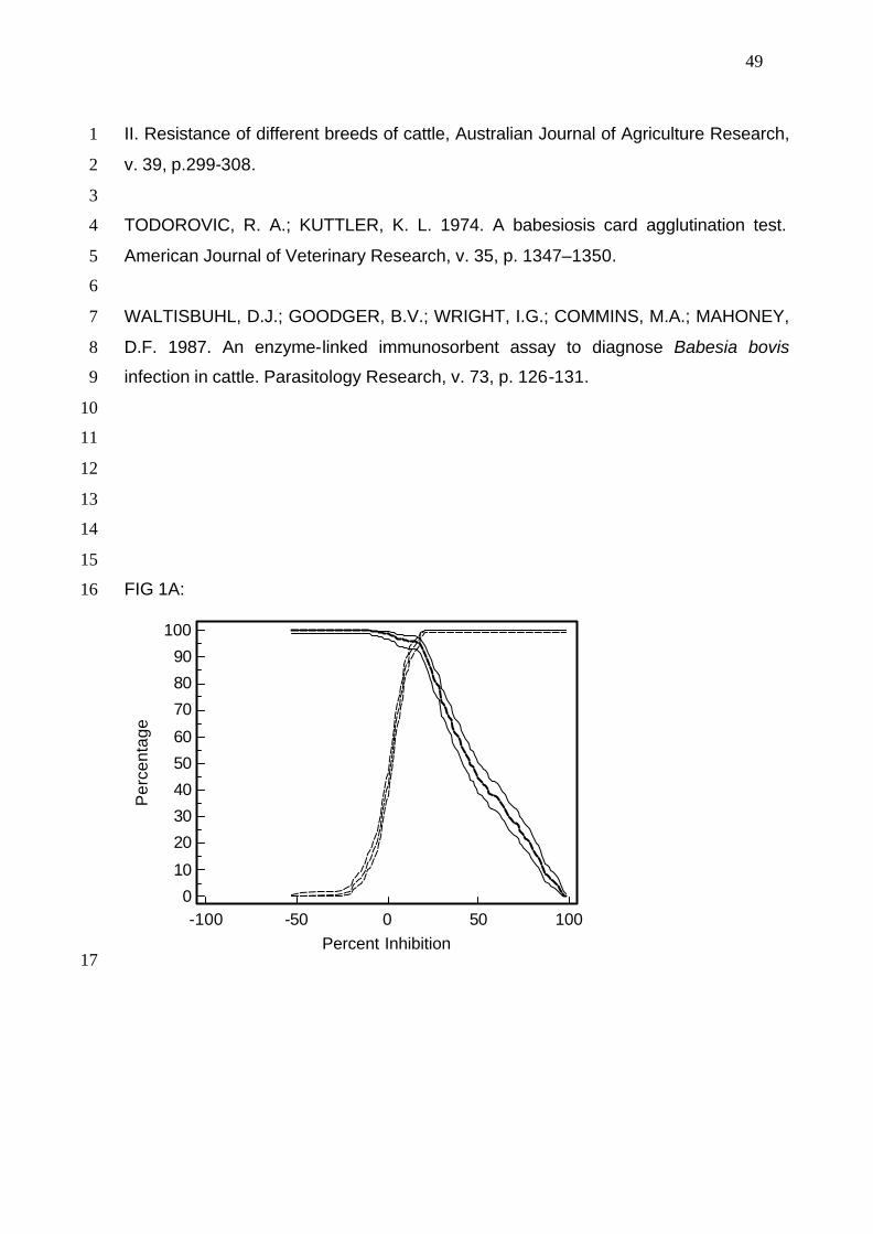
49
II. Resistance of different breeds of cattle, Australian Journal of Agriculture Research, 1
v. 39, p.299-308. 2
3
TODOROVIC, R. A.; KUTTLER, K. L. 1974. A babesiosis card agglutination test. 4
American Journal of Veterinary Research, v. 35, p. 1347–1350. 5
6
WALTISBUHL, D.J.; GOODGER, B.V.; WRIGHT, I.G.; COMMINS, M.A.; MAHONEY, 7
D.F. 1987. An enzyme-linked immunosorbent assay to diagnose Babesia bovis 8
infection in cattle. Parasitology Research, v. 73, p. 126-131. 9
10
11
12
13
14
15
FIG 1A: 16
-100 -50 0 50 100
100
90
80
70
60
50
40
30
20
10
0
Percent Inhibition
Per
cent
age
17
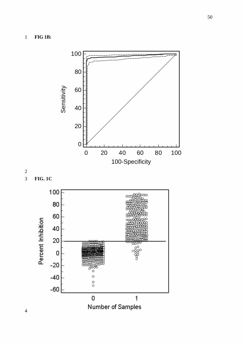
50
FIG 1B: 1
0 20 40 60 80 100
100
80
60
40
20
0
100-Specificity
Sen
sitiv
ity
2 FIG. 1C 3
4
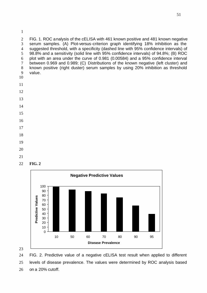
51
1
FIG. 1. ROC analysis of the cELISA with 461 known positive and 481 known negative 2 serum samples. (A) Plot-versus-criterion graph identifying 18% inhibition as the 3 suggested threshold, with a specificity (dashed line with 95% confidence intervals) of 4 98.8% and a sensitivity (solid line with 95% confidence intervals) of 94.8%; (B) ROC 5 plot with an area under the curve of 0.981 (0.00584) and a 95% confidence interval 6 between 0.969 and 0.989; (C) Distributions of the known negative (left cluster) and 7 known positive (right cluster) serum samples by using 20% inhibition as threshold 8 value. 9 10
11
12
13
14
15
16
17
18
19
20
21
FIG. 2 22
Negative Predictive Values
0102030405060708090
100
10 50 60 70 80 90 95
Disease Prevalence
Pre
dic
tive
Val
ues
23 FIG. 2. Predictive value of a negative cELISA test result when applied to different 24
levels of disease prevalence. The values were determined by ROC analysis based 25
on a 20% cutoff. 26
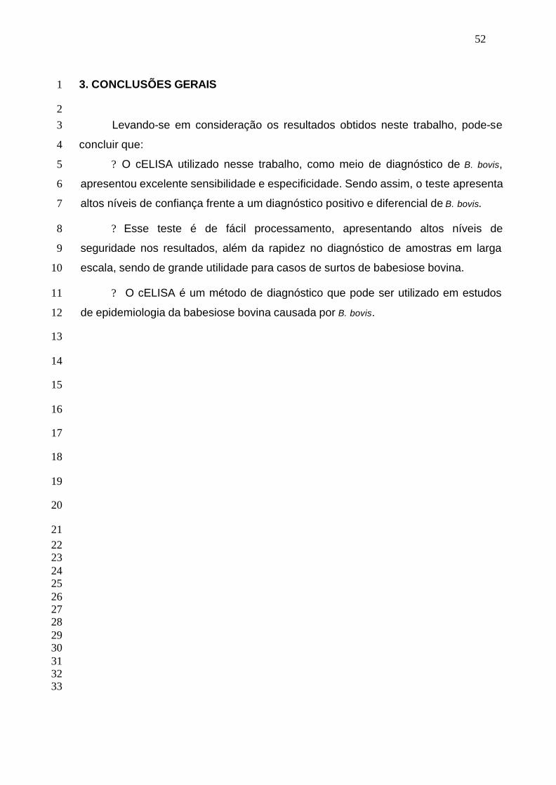
52
3. CONCLUSÕES GERAIS 1
2 Levando-se em consideração os resultados obtidos neste trabalho, pode-se 3
concluir que: 4
? O cELISA utilizado nesse trabalho, como meio de diagnóstico de B. bovis, 5
apresentou excelente sensibilidade e especificidade. Sendo assim, o teste apresenta 6
altos níveis de confiança frente a um diagnóstico positivo e diferencial de B. bovis. 7
? Esse teste é de fácil processamento, apresentando altos níveis de 8
seguridade nos resultados, além da rapidez no diagnóstico de amostras em larga 9
escala, sendo de grande utilidade para casos de surtos de babesiose bovina. 10
? O cELISA é um método de diagnóstico que pode ser utilizado em estudos 11
de epidemiologia da babesiose bovina causada por B. bovis. 12
13
14
15
16
17
18
19
20
21 22 23 24 25 26 27 28 29 30 31 32
33
53
4. REFERÊNCIAS 1 2
BOONCHIT, S.; ALHASSAN, A.; CHAN, B.; XUAN, X.; YOKOYAMA, N.; OOSHIRO, 3
M.; GOFF, W. L.; WAGHELA, S. D.; WAGNER, G.; IGARASHI, I. 2006. Expression o 4
C-terminal truncated and full-length Babesia bigemina rhoptry-associated protein 1 5
and their potential use in enzyme-linked immunosorbent assay. Veterinary 6
Parasitology, v. 137, p. 28-35. 7
8
BÖSE, R.; JORGENSEN, W.K.; DALGLIESH, R.J.; FRIEDHOFF, K.T.; DE VOS, A.J. 9
1995. Current state and future trends in the diagnosis of babesiosis. Veterinary 10
Parasitology, v. 57, p. 61–74. 11
12
FIGUEROA, J. V.; CHIEVES, L. P.; JOHNSON, G. S.; BUENING, G. M. 1992. 13
Detection of Babesia bigemina-infected carriers by polymerase chain reaction 14
amplification. Journal of Clinical Microbiology, v. 30, p. 2576–2582. 15
16
GOODGER, B. V. 1971. Preparation and preliminary assessment of purified antigens 17
in the passive haemagglutination test for bovine babesiosis. Australian Veterinary 18
Journal. 47:251-256. 19
20
GOFF, W. L.; WAGNER, G. G.; CRAIG, T. M.; LONG, R. F. 1982. The bovine 21
immune response to tick-derived Babesia bovis infection: serological studies of 22
isolated immunoglobulins. Veterinary Parasitology, v.11, p.109–120. 23
24
GOFF, W. L.; McELWIAN, T. F.; SUAREZ, C. E.; JOHNSON, W. C.; BROWN, W. C.; 25
NORIMINE, J.; KNOWLES, D. P. 2003. Competitive Enzyme-Linked Immunosorbent 26
Assay Based on a Rhoptry-Associated Protein 1 Epitope Specifically Identifies 27
Babesia bovis-Infected Cattle. Clinical and Diagnostic Laboratory Immunology, v.10, 28
p. 38-43. 29
30
GOFF, W. L.; MOLLOY, J. B.; JOHNSON, W. C.; SUAREZ, C. E.; PINO, I.; 31
RHALEM, A.; SAHIBI, H.; CECI, L.; CARELLI, G.; ADAMS, D. S.; McGUIRE, T. C.; 32
KNOWLES, D. P.; McELWAIN, T. F. 2006. Validation of a Competitive Enzyme-33
54
Linked Immunosorbent Assay for Detection of Antibodies against Babesia Bovis. 1
Clinical and Vaccine Immunology, v.13, p. 1212-1226. 2
3
GOFF, W.L.; JOHNSON, W.C; MOLLOY, J.B.; JORGENSES, W.K.; WALDRON, 4
S.J.; FIGUEROA, J.V.; MATTHEE, O; ADAMS, D. S.; McGUIRE, T.C.; PINO, I. 5
MOSQUEDA, J.; PALMER, G.H.; SUAREZ, C.E.; KNOWLES, D.P.; McELWAIN, 6
T.F.;. 2008. Validation of a Competitive Enzyme-Linked Immunosorbent Assay for 7
Detection of Babesia bigemina Antibodies in Cattle. Clinical And Vaccine 8
Immunology, v. 15, p. 1316–1321. 9
10
GUGLIELMONE, A.A. 1995. Epidemiology of babesiosis and anaplasmosis in South 11
and Central America. Veterinary Parasitology, v.57, p. 109-119. 12
13
HÖTZEL, I. ; BROWN, W. C.; MCELWAIN, T. F.; RODRIGUEZ, S. D.; PALMER, G. 14
H. 1996. Dimorphic sequences of rap-1 genes encode B and CD4+ T helper 15
lymphocyte epitopoes in the Babesia bigemina rhoptry associated protein-1. 16
Molecular and Biochemical Parasitology, v. 81, p. 89-99. 17
18
HÖTZEL, I.; SUAREZ, C. E.; MCELWAIN, T. F.; PALMER, G. H. 1997. Genetic 19
variation in the dimorphic regions of RAP-1 genes and rap-1 loci of Babesia 20
bigemina. Molecular and Biochemical Parasitology, v.90, p. 479-89. 21
22
MAHONEY, D. F. 1962. Bovine babesiosis: diagnosis of infection by a complement 23
fixation test. Australian Veterinary Journal, v. 38, p.48-52. 24
25
SILVA, M. G.; HENRIQUES, G.; SANCHEZ, C.; MARQUES P. X.; SUAREZ, C. E.; 26
OLIVA, A. 2009. First survey for Babesia bovis and Babesia bigemina infection in 27
cattle from Central and Southern regions of Portugal using serological and DNA 28
detection methods. Veterinary Parasitology, v. 166, p. 66–72. 29
30
SUAREZ, C. E. ; PALMER, G. H. ; JASMER, D. P. ; HINES, S. A. ; PERRYMAN, L. 31
E. ; MCELWAIN, T. F. 1991. Characterization of the gene encoding a 60-kDa 32
Babesia bovis merozoite protein with conserved and surface exposed epitopes. 33
Molecular Biochemical Parasitology, v. 46, p. 45–52. 34
55
1
SUAREZ, C.E.; PALMER, G.H.; HINES, A.; McELWAIN, T.F. 1993. Immunogenic B-2
cell epitopes of Babesia bovis rhoptry-associated protein 1 are distinct from 3
sequences conserved between species. Infection and Immunity, v. 61, p. 3511–3517. 4
5
SUAREZ, C.E.; McELWAIN, T.F.; ECHAIDE, I.; DE ECHAIDE, S.T.; PALMER, G.H. 6
1994. Interstrain conservation of babesial RAP-1 surface exposed B-cell epitopes 7
despite rap-1 genomic polymorphism. Infection and Immunity, v. 62, p. 3576–3579. 8
9
10