UNIVERSIDADE FEDERAL DO CEARÁ FACULDADE DE … · no banner Catalogação na Publicação ......
57
0 UNIVERSIDADE FEDERAL DO CEARÁ FACULDADE DE FARMÁCIA, ODONTOLOGIA E ENFERMAGEM PROGRAMA DE PÓS-GRADUAÇÃO EM ODONTOLOGIA MESTRADO EM ODONTOLOGIA CRISTINA MARIA FERNANDES DE QUEIROZ ÁCIDO ANACÁRDICO COMO AGENTE DE LIMPEZA CAVITÁRIA EM ODONTOLOGIA ADESIVA FORTALEZA-CE 2015
Transcript of UNIVERSIDADE FEDERAL DO CEARÁ FACULDADE DE … · no banner Catalogação na Publicação ......

0
UNIVERSIDADE FEDERAL DO CEARÁ
FACULDADE DE FARMÁCIA, ODONTOLOGIA E ENFERMAGEM
PROGRAMA DE PÓS-GRADUAÇÃO EM ODONTOLOGIA
MESTRADO EM ODONTOLOGIA
CRISTINA MARIA FERNANDES DE QUEIROZ
ÁCIDO ANACÁRDICO COMO AGENTE DE LIMPEZA CAVITÁRIA EM
ODONTOLOGIA ADESIVA
FORTALEZA-CE
2015

1
CRISTINA MARIA FERNANDES DE QUEIROZ
ÁCIDO ANACÁRDICO COMO AGENTE DE LIMPEZA CAVITÁRIA EM
ODONTOLOGIA ADESIVA
Dissertação de Mestrado apresentada ao Programa
de Pós-Graduação em Odontologia da Faculdade de
Farmácia, Odontologia e Enfermagem da
Universidade Federal do Ceará, como requisito
parcial para obtenção do título de Mestre em
Odontologia. Área de concentração: Clínica
Odontológica.
Orientadora: Profª. Drª. Monica Yamauti.
Coorientadores: Prof. Dr. Francisco Fábio Oliveira
de Sousa e Prof. Dr. Sérgio Lima Santiago.
FORTALEZA-CE
2015

2
Página reservada para ficha catalográfica que deve ser confeccionada após apresentação e
alterações sugeridas pela banca examinadora.
Para solicitar a ficha catalográfica de seu trabalho, acesse o site: www.biblioteca.ufc.br, clique
no banner Catalogação na Publicação (Solicitação de ficha catalográfica).
----------------------------------------------------------------------------------------------------------

3
CRISTINA MARIA FERNANDES DE QUEIROZ
ÁCIDO ANACÁRDICO COMO AGENTE DE LIMPEZA CAVITÁRIA EM
ODONTOLOGIA ADESIVA
Dissertação de Mestrado apresentada ao Programa
de Pós-graduação em Odontologia da Faculdade de
Farmácia, Odontologia e Enfermagem da
Universidade Federal do Ceará, como requisito
parcial para obtenção do título de Mestre em
Odontologia. Área de concentração: Clínica
Odontológica.
Aprovada em: ___/___/______
BANCA EXAMINADORA
___________________________________________
Profª. Drª. Monica Yamauti (Orientadora)
Universidade Federal de Minas Gerais (UFMG)
___________________________________________
Prof. Dr. Juliano Sartori Mendonça
Universidade Federal do Ceará (UFC)
___________________________________________
Profª. Drª. Vanara Florêncio Passos
Universidade de Fortaleza (UNIFOR)

4
A Deus, por ter me fortalecido. Aos meus pais, por
todo o apoio. Ao meu esposo Sérgio e meu filho
Vitor, que me compreenderam e me deram força,
com muita paciência, amor e carinho.

5
AGRADECIMENTOS
À Universidade Federal do Ceará, na pessoa do seu Magnífico Reitor Prof. Dr.
Jesualdo Pereira Farias.
À Faculdade de Farmácia, Odontologia e Enfermagem, em nome da Diretora
Profª. Drª. Maria Goretti Rodrigues de Queiroz.
Ao curso de Odontologia da Universidade Federal do Ceará, na pessoa do
Coordenador Prof. Dr. Fabrício Bitu Sousa.
Ao programa de Pós-Graduação em Odontologia, em nome da coordenadora
Profª. Drª. Lidiany Karla Azevedo Rodrigues.
Aos meus pais Pedro e Aldenora, por toda a dedicação, compreensão e apoio.
Amo vocês!
Ao meu esposo Sérgio, pela paciência, companheirismo, amor e carinho. Te amo!
Ao meu filho Vitor, que com seu lindo sorriso me deu força e coragem. Filho, a
mamãe te ama muito!
Aos meus irmãos Pedro Jr. e Rafael, pelo incentivo e torcida! Amo vocês!
À minha cunhada Rachel, sempre presente quando precisei.
Aos meus sogros Sérgio e Meyre Eliane, pela torcida, incentivo e apoio.
À minha orientadora Profª. Drª. Monica Yamauti, por toda a orientação, paciência
e incentivo. Muito obrigada por tudo!
Aos coorientadores Prof. Dr. Francisco Fábio Oliveira e Prof. Dr. Sérgio Lima
Santiago, por todo o apoio, disponibilidade e contribuição com o estudo.
Ao Prof. Dr. Juliano Sartori Mendonça, por contribuir com o estudo.
À bolsista Mirelle Nobre Ferreira Bringel, pela ajuda e disponibilidade nas
atividades laboratoriais.
À mestranda Vanessa Fontenele Marques, pela ajuda e disponibilidade nas
atividades laboratoriais.
Aos colegas de turma, pela amizade e companheirismo.
Ao técnico do laboratório David Queiroz de Freitas, por toda a ajuda no
laboratório. Obrigada por tudo!
Às doutorandas Cecília Atem e Jaqueline Nojosa, por tudo o que me ensinaram.
À amiga Denise Lins, por toda a ajuda.
À Central Analítica da UFC, pela oportunidade de utilizar seus equipamentos,
enriquecendo nosso estudo.

6
Ao Prof. Alejandro Pedro Ayala, pela gentileza e por disponibilizar o acesso ao
Laboratório de Cristalografia Estrutural do Departamento de Física (UFC).
À Profa. Drª. Nágila Maria Ricardo, por disponibilizar matéria-prima para o
estudo.
À técnica do Laboratório de Cristalografia Estrutural do Departamento de Física
(UFC) Silmara Alves, pelo acompanhamento durante os experimentos.
À doutoranda Yara Santiago de Oliveira, do Departamento de Física (UFC), pelo
acompanhamento durante os experimentos no Laboratório de Cristalografia Estrutural.
Às funcionárias da coordenação do Programa de Pós-Graduação, Janaine Marques
Leal e Lúcia Ribeiro Marques Lustosa, pela eficiência na resolução das questões burocráticas
referentes à pós-graduação.

7
RESUMO
O objetivo deste estudo in vitro foi avaliar o desempenho do ácido anacárdico como
agente de limpeza cavitária em restaurações adesivas. Foram utilizados três agentes de
limpeza: água destilada (AD), solução de digluconato de clorexidina a 2% (CHX) e solução
de ácido anacárdico (AA). Cada agente de limpeza foi utilizado em duas estratégias adesivas:
após o condicionamento com ácido fosfórico, na técnica adesiva de condicionamento total, e
previamente à aplicação do primer na técnica adesiva autocondicionante. Realizou-se uma
análise através de microscopia eletrônica de varredura (MEV), no intuito de avaliar a
morfologia do substrato quando exposto ao ácido fosfórico ou ao AA, e quando exposto aos
agentes de limpeza, nas duas estratégias adesivas propostas. Adicionalmente, foi realizada
uma análise através de espectrofotometria (FTIR) para se avaliar a ocorrência de interações
químicas entre os agentes de limpeza CHX e AA com a dentina. A resistência de união à
dentina foi avaliada pelo teste de microtração, num total de seis grupos experimentais (duas
estratégias adesivas x três agentes de limpeza). Superfícies dentinárias planas foram
preparadas em dentes humanos extraídos e, após o uso de cada agente e o respectivo
procedimento adesivo, os dentes foram restaurados com resina composta, de forma
incremental até atingir uma altura de material restaurador de 5 mm. Os espécimes foram
armazenados em água destilada a 37ºC por 24 h, e foram seccionados longitudinalmente em
sentidos perpendiculares entre si para que se obtivessem espécimes em forma de palitos com
área de secção transversal de 1 mm². Cada espécime foi tracionado a uma velocidade média
de 0,5 mm ̸ min em uma máquina de ensaios universal. O modo de fratura foi observado sob
uso de lupa com 60x de aumento e 10% dos espécimes tiveram suas superfícies recobertas
com ouro e avaliadas em MEV. Os valores de resistência de união foram estatisticamente
avaliados pelo ANOVA dois critérios, com nível de significância de 5%. Na análise
morfológica, foi observado que o AA não é capaz de desmineralizar o substrato dentinário e
não altera sua morfologia superficial. Encontrou-se um padrão morfológico resultante na
dentina diferente daquele provocado pelo ácido fosfórico. A análise por FTIR mostrou que
houve interação química entre o AA e a dentina, assim como foi encontrada interação entre
DG e o substrato dentinário. Em relação aos resultados de microtração, não houve diferença
estatisticamente significante entre os grupos (p>0.05). O maior percentual de falhas foi de
fratura mista em cada grupo. O ácido anacárdico não afetou a resistência de união após 24 h
de armazenagem e as análises indicam que ele pode ser usado como agente de limpeza
cavitária.
Palavras-chave: Ácido anacárdico; agente de limpeza cavitária; resistência de união;
sistemas adesivos.

8
ABSTRACT
The aim of this in vitro study was to evaluate the performance of anacardic acid as a
cavity-cleaning agent in adhesive restorations. Three cleaning agents were used: distilled
water (DW), chlorhexidine digluconate solution at 2% (CHX) and anacardic acid (AA). Each
cleaning agent was used in two strategies adhesive: after acid etching with phosphoric acid
with etch&rinse adhesive or prior to the primer application in self-etch adhesive. Scanning
electron microscopy (SEM) analysis was performed to evaluate the morphology of dentin
substrate when it was exposed to phosphoric acid or anacardic acid, and when exposed to
DW, CHX or AA with the two adhesive strategies. Additionally, spectrophotometry analysis
was performed (FTIR) to assess the occurrence of chemical interactions between AA and
CHX cleaning agents with dentine. The bond strength to dentin was evaluated by microtensile
test, in a total of six experimental groups (2 adhesive strategies x 3 cleaning agents). Flat
dentin surfaces were prepared in extracted human teeth. After the use of each cleaning agent
and the respective adhesive procedure, teeth were incrementally restored with resin composite
up to of 5 mm built-up. The specimens were stored in 37ºC distilled water for 24 h, and were
sectioned longitudinally in either direction to obtain the specimens in the form of beams with
cross-section area of 1 mm2. Each specimen was tensioned until fracture to with 0.5 mm ̸ min
speed in a universal testing machine. The fracture mode was observed using a stereoscope at
60x and 10% of the specimens had their surfaces covered with gold and evaluated under
SEM. The bond strength values were statistically evaluated using two-way ANOVA with a
5% significance level. The morphological analysis showed that AA is not able to demineralize
dentin and does not alter the substrate surface morphology. The resulting dentin
morphological pattern was than that caused by phosphoric acid. FTIR analysis showed that
there was a chemical interaction between the AA and dentin as well as it was found an
interaction between DG and dentin. Concerning the microtensile bond strength results, there
was no statistically significant difference between groups (p> 0.05). The highest percentage of
failures was mixed fracture in each group. The anacardic acid did not affect the bond strength
after 24 h of storage and analysis showing that it can be used as cavity-cleaning agent.
Keywords: Anacardic acid; antibacterial agent; cavity cleaning; dentin bonding.

9
SUMÁRIO
1 INTRODUÇÃO GERAL ....................................................................................... 10
2 PROPOSIÇÃO ....................................................................................................... 14
2.1 Objetivo geral .......................................................................................................... 14
2.2 Objetivos específicos ............................................................................................... 14
3 CAPÍTULO ............................................................................................................. 15
3.1 Capítulo 1 ................................................................................................................ 16
4 CONCLUSÃO GERAL ......................................................................................... 48
REFERÊNCIAS ..................................................................................................... 49
APÊNDICE.................................................................................................... 51
ANEXO...........................................................................................................52

10
1 INTRODUCÃO GERAL
Anacardium occidentale é uma planta conhecida popularmente por cajueiro,
pertence à família Anacardiaceae e apresenta grande importância econômica no Brasil. O
fruto do cajueiro é a castanha de caju, que é produzida em larga escala no Brasil e,
particularmente, no Ceará, maior produtor e exportador nacional (MAZZETTO;
LOMONACO; MELE, 2009). A partir da casca da castanha do caju, pode ser extraído um
líquido escuro, quase preto, denominado líquido da castanha do caju (LCC) (MAZZETTO;
LOMONACO; MELE, 2009). Quimicamente, o LCC é constituído de vários compostos
orgânicos, estreitamente relacionados (HEMSHEKHAR et al., 2011), nos quais, em maior
quantidade, apresentam-se os ácidos anacárdicos que compõem cerca de 70% do líquido e, em
menor quantidade, encontram-se os componentes fenólicos cardol e cardanol (AGOSTINI-
COSTA et al., 2003; AGOSTINI-COSTA et al., 2005). Os ácidos anacárdicos são divididos
em quatro subtipos e, estruturalmente, esses elementos apresentam um anel fenólico com 15
carbonos em uma cadeia lateral, que podem conter zero, uma, duas ou três duplas ligações
(TREVISAN et al., 2006).
As atividades biológicas dos ácidos anacárdicos vêm sendo amplamente estudadas
na área da saúde, entre elas, sua capacidade antioxidante (KUBO et al., 2006; TREVISAN et
al., 2006) e atividade antitumoral (HEMSHEKHAR et al., 2011; KUBO et al., 1993). Na
Odontologia, despertou interesse por suas características antimicrobianas (SOUSA, 2014;
MUROI; KUBO, 1993; HIMEJIMA; KUBO, 1991). Os ácidos anacárdicos apresentaram boa
ação contra bactérias gram-positivas como o Streptococcus mutans, principal bactéria
relacionada à doença cárie dentária (SOUSA, 2014; GREEN et al., 2008; LIMA; PASTORE;
LIMA, 2000; MUROI; KUBO, 1993; HIMEJIMA; KUBO, 1991). A ação antimicrobiana do
ácido anacárdico apresenta relação direta com o número de duplas ligações da cadeia lateral;
quanto mais duplas ligações, maior o potencial antimicrobiano (MUROI; KUBO, 1993;

11
HIMEJIMA; KUBO, 1991). Os ácidos anacárdicos também apresentaram resultados
promissores na redução do processo de erosão dentinária, quando comparados ao controle
negativo e aos grupos tratados com epigalocatequina-3-galato (EGCG) e fluoreto de sódio
(NaF) (SILVEIRA et al., 2014) .
O LCC pode ser obtido por diferentes processos de extração. A extração feita em
grandes empresas por processos totalmente mecanizados alcança temperaturas elevadas que
provocam uma reação de descarboxilação no ácido anacárdico, convertendo-o em cardanol. A
extração a frio, por processo semi-mecanizado, faz com que o ácido anacárdico permaneça
estável, consequentemente, o LCC terá uma grande quantidade de ácidos anacárdicos
(MAZZETO; LOMONACO; MELE, 2009; AGOSTINI-COSTA et al., 2005).
Nesse contexto, os ácidos anacárdicos podem apresentar-se como uma alternativa
natural a outros agentes antimicrobianos, de uso na Odontologia, como o digluconato de
clorexidina. Apesar de as soluções de clorexidina possuírem conhecida eficácia contra cepas
de Streptococcus mutans (WADE; ADDY, 1989), elas podem causar efeitos colaterais como
manchamento dentário, alterações de paladar e irritação da mucosa bucal (QUIRYNEN et al.,
2001;WATTS; ADDY, 2001).
A união dos materiais restauradores aos tecidos dentários duros ocorre por meio
de desmineralização e consiste em um processo de troca dos minerais perdidos pelo sistema
restaurador adesivo (CARDOSO et al., 2011). Essa união pode ser obtida através de sistemas
adesivos de condicionamento total ou autocondicionantes. A diferença entre esses
mecanismos de união pode ser observada por meio da caracterização química e morfológica
das interações interfaciais do substrato dentinário com o material restaurador (VAN
MEERBEEK et al., 2003).
A técnica de condicionamento total é realizada com ácido fosfórico em
concentrações de 30 a 40%, seguindo-se pela aplicação do primer e do agente de união
12
(adesivo), apresentados em frascos separados ou no mesmo frasco. O condicionamento ácido
dentinário expõe uma microporosa rede de fibras colágenas, na qual o adesivo vai difundir-se,
e a união (substrato-adesivo) dependerá da infiltração do adesivo dentro da camada de fibrilas
colágenas expostas (PASHLEY et al., 2011; VAN MEERBEEK et al., 2003;
NAKABAYASHI; ASHIZAWA; NAKAMURA, 1992).
Os sistemas adesivos autocondicionantes são aplicados diretamente na dentina
sem tratamento ácido prévio, portanto, a smear layer permanece incorporada ao substrato
dentinário (TAY E PASHLEY, 2001). Podem apresentar-se em frasco único ou primer e
adesivo em frascos separados (VAN MEERBEEK et al., 2011). Esse tipo de sistema adesivo
apresenta uma menor sensibilidade técnica e dispende um menor tempo clínico (VAN
MEERBEEK et al., 2003).
Apesar de toda essa evolução, a Odontologia estética restauradora ainda encontra
limitações no que se refere a um bom selamento marginal entre estrutura dentária e material
restaurador (VAN MEERBEEK et al., 2010; VAN MEERBEEK et al., 2003; HASHIMOTO
et al., 2002). A busca por um material que promova selamento ideal é tema de diversas
pesquisas, pois o vedamento da interface adesiva é uma das características que produzem
influência no sucesso clínico de uma restauração (VAN MEERBEEK et al., 2010; DE
MUNCK et al., 2005; PEUMANS et al., 2005). Estudos revelam a persistência de
remanescentes bacterianos nas paredes de preparos cavitários, mesmo após a remoção total do
tecido cariado, que pode interferir na qualidade de união entre materiais adesivos e a estrutura
dentária e, também, pode aumentar a infiltração marginal (BENGTSON et al., 2008;
SOARES et al., 2008). Em função dessa possibilidade, tem sido recomendado o uso de
agentes antimicrobianos na desinfecção das cavidades contaminadas, no intuito de eliminar ou
minimizar os depósitos microbianos aderidos ao preparo cavitário (CAMILOTTI et al., 2013;
BENGTSON et al., 2008; SAY et al., 2004).

13
Torna-se, então, pertinente a busca por um agente de limpeza cavitária oriundo de
produtos naturais da flora brasileira, que tenham características antimicrobianas contra
bactérias relacionadas ao processo carioso e comparar com soluções já utilizadas para essa
finalidade como a de clorexidina. Não há relatos na literatura sobre o emprego do ácido
anacárdico como agente de limpeza cavitária, sendo necessários estudos que caracterizem a
interação dos ácidos anacárdicos com o substrato dentinário. Dessa forma, o objetivo deste
estudo é avaliar o desempenho do ácido anacárdico como agente de limpeza cavitária em
restaurações adesivas com resina composta.
14
2 PROPOSIÇÃO
2.1 Objetivo Geral:
- Avaliar o desempenho do ácido anacárdico como agente de limpeza cavitária em
restaurações adesivas com resina composta.
2.2 Objetivos Específicos:
- Analisar e comparar a estrutura morfológica dentinária após a ação dos ácidos fosfórico e
anácardico;
- Avaliar a interação química entre o ácido anacárdico e a dentina após limpeza cavitária com
solução de ácido anacárdico;
- Avaliar a resistência de união à dentina de um sistema adesivo após condicionamento com
ácido fosfórico e limpeza cavitária com solução de ácido anacárdico, de um sistema adesivo
autocondicionante quando houver aplicação de solução de ácido anacárdico prévia ao uso do
primer.

15
3 CAPÍTULO
Esta dissertação está baseada no Artigo 46 do Regimento Interno do Programa de
Pós-Graduação em Odontologia da Universidade Federal do Ceará, que regulamenta o
formato alternativo para dissertações de Mestrado e teses de Doutorado e permite a inserção
de artigos científicos de autoria ou coautoria do candidato. Por se tratar de um estudo
envolvendo dentes humanos, o projeto de pesquisa deste trabalho foi submetido à apreciação
do Comitê de Ética em pesquisa da Universidade Federal do Ceará, tendo sido aprovado
conforme o parecer consubstanciado nº 810.992, de 4 de Setembro de 2014 (ANEXO).
Assim sendo, esta dissertação é composta de um capítulo contendo um artigo
científico que será submetido ao periódico Operative Dentistry conforme descrito na
sequência:
The use of anarcadic acid solution as cleaning agent prior to adhesive restorations
C M F Queiroz, F F O Sousa, N M P S Ricardo, S L Santiago, M Yamauti.

16
3.1 Capítulo 1
Title: The use of anarcadic acid solution as cleaning agent prior to adhesive restorations
Running Title: Anarcardic acid as cavity cleaning agent
Clinical significance:
Anacardic acid is a product of natural origin that has antibacterial action against
Streptococcus mutans and, based on the results of this study, it does not alter the morphology
of dentin, can chemically interact with dentin and presented no adverse effect on the bond
strength to dentin.

17
Authors: Cristina Maria Fernandes de Queiroz1, Francisco Fabio Oliveira de Sousa
2, Nágila
Maria Pontes Silva Ricardo3, Sérgio Lima Santiago
4, Monica Yamauti
5*
Affiliations: 1DDS, Graduate Student, Faculty of Pharmacy, Dentistry and Nursing, Federal
University of Ceará, Rua Capitao Francisco Pedro, S/N - Rodolfo Teofilo, 60430-170,
Fortaleza-CE-Brazil.
2BPharm, MS, PhD, Adjunct Professor, Department of Biological & Health Sciences, Federal
University of Amapá, Rod. Juscelino Kubitschek, Km 02, 68902-280, Macapa-AP-Brazil.
3Associate Professor Departament of Inorganic and Organic Chemistry, Federal University of
Ceará, 60455-760, Fortaleza-CE-Brazil.
4DDS, MS, PhD, Associate Professor, Department of Restorative Dentistry, Faculty of
Pharmacy, Dentistry and Nursing, Federal University of Ceará, Rua Capitao Francisco Pedro,
S/N - Rodolfo Teofilo, 60430-170, Fortaleza-CE-Brazil.
5DDS, MS, PhD, Adjunct Professor, Department of Restorative Dentistry, Federal University
of Minas Gerais, Av. Antonio Carlos, 6627 - Pampulha, 31270-901, Belo Horizonte-MG-
Brazil.
*Corresponding author:
Monica Yamauti
Adjunct Professor, Department of Restorative Dentistry
Faculty of Dentistry, Federal University of Minas Gerais
Av. Antonio Carlos, 6627 - Pampulha, 31270-901, Belo Horizonte-MG-Brazil.
Phone: +55-31-34092440
E-mail: [email protected]

18
SUMMARY
Aim: This study aimed to evaluate the ability of anacardic acid solution to interact and ̸ or
modify dentin morphology and to analyze the performance of anacardic acid solution as a
cavity-cleaning agent on bond strength of two adhesive strategies.
Methods and materials: The interaction between anacardic acid and chlorhexidine
digluconate solutions with dentin was analyzed using Fourier Transform Infrared
Spectrometry. The morphological analysis of dentin was performed using scanning electron
microscopy (SEM). The bond strength was evaluated by microtensile test with 6 experimental
groups (3 cavity-cleaning agents and 2 adhesive strategies). Distilled water was used as the
cavity-cleaning control agent. Flat dentin surfaces were prepared on extracted teeth and
adhesives systems were applied after the use of the cleaning agents. Teeth were incrementally
restored with resin composite, stored in distilled water at 37ºC for 24 h and were
longitudinally sectioned to obtain sticks with a cross-sectional area of 1.0 mm². Dentin-resin
sticks were submitted to tensile strength until failure occurred. The failure modes were
evaluated at 60X and ten percent of the specimens had their fracture mode examined by SEM.
The bond strength values were statistically evaluated using two-way ANOVA with a 5%
significance level.
Results: There was chemical interaction between chlorhexidine digluconate and anacardic
acid solutions with dentin. Morphological analysis showed anacardic acid solution did not
promote dentin demineralization. There was no statistically significant difference among the
bond strength values of all experimental groups (p>0.05).
Conclusion: The morphological, chemical and bond strength analyses demonstrated that the
anacardic acid did not affect dentin bonding, regardless the adhesive strategy.
Keywords: Anacardic acid, antibacterial agent, cavity cleaning, dentin bonding

19
INTRODUCTION
The success of adhesive restorations is related to the bonding stability between
composite resin and tooth structure.1,2,3
Studies show the persistence of bacterial remaining in
the cavity walls, even after the complete removal of caries.4,5
This contamination can affect
the quality of bonding between adhesive materials and the tooth, and can also increase the
microleakage of adhesive restorations.4,5
The progression rate of secondary caries is an
important factor that determines the restorations longevity.6 So, it is recommended to use
antimicrobial agents to disinfect the contaminated walls and to eliminate or minimize
microbial deposits adhered to the cavity wall.4,7,8
Some antimicrobial agents have been used in Dentistry, such as chlorhexidine that has
efficacy against Streptococcus mutans.9 However, this agent may cause side effects such as
tooth staining, taste disturbance and irritation of the oral tissues.10,11
Thus it is relevant to
search for natural and less harmful antimicrobial agents. There are many medicinal plants
with analgesic, anti-inflammatory, antimicrobial, antiseptic effects to be explored. Brazil is a
territory rich in natural resources, which favors discoveries in the area of herbal medicines.12
It is pertinent to search for a cavity-cleaning agents derived from natural Brazilian products
that present antimicrobial properties against bacterium related to dental diseases.
Cashew nut shell liquid (CNSL) of cashew apple has emerged as a promising
compound due to its variety of biological properties, and many of these have been attributed
to anacardic acid, active component extracted from CNSL.13,14
It is a natural product easy to
obtain for being derived from the cashew nut that is produced on a large scale mainly in
northeastern Brazil.15
Among the biological properties of anacardic acids are their antioxidant
capacity,14,16
antitumour activity 13,17
and antimicrobial characteristic.18-20
In dentistry, the
antimicrobial property has shown active against gram-positive bacteria such as Streptococcus

20
mutans, the main bacteria related to dental caries.18-22
The anacardic acid also has shown
promising results against the dentin erosion process.23
In this context, there is the prospect use of the anacardic acid as a cavity-cleaning
agent. The anacardic acid could be used as cavity-cleaning agent prior to etch-and-rinse or
self-etch adhesive systems. Therefore, studies are needed to characterize the interaction of
anacardic acids with dentin. The purpose of this study was to evaluate the performance of
anacardic acid solution as a cavity-cleaning agent in resin composite restorations. The null
hypothesis tested was that there would be no significant difference between the bond strength
to dentin of two adhesive systems when anarcadic acid solution was used as cavity cleansing
compared to the use of distilled water.
MATERIALS AND METHODS
This is an in vitro qualitative-quantitative research. The materials and chemical used in
the study are described in Table 1.
Anacardic acid
The anacardic acid was obtained at the Laboratory of Polymer and Material Innovation
of Organic and Inorganic Chemistry Department, Federal University of Ceará. The anacardic
acid was produced from cashew nut shell liquid (CNSL) cold extraction. The shells of cashew
nuts were submitted to extraction with hexane solvent (95,0%, Fisher Scientific,
Loughborough, Leicestershire, UK). Then, the obtained crude extract was subjected to solvent
removal through rotoevaporation, resulting in the natural CNSL. The extracted CNSL was
employed to obtain the anacardic acid. CNSL was dissolved in aqueous methanol and
subjected to acid-base reaction with calcium hydroxide (Ca(OH)2, 96,0%, Fluka, Sigma
Aldrich, St Louis, MO, USA), producing calcium anacardate. The obtained calcium

21
anacardate was converted into the acidic form again, by acidifying the reaction medium in the
presence of HCl 11 M. Immediately, a liquid-liquid extraction with ethyl acetate (EtOAc,
99,8%, Sigma Aldrich, St Louis, MO, USA) was performed. The anacardic acid was
converted into the organic phase and concentrated by rotoevaporation. The final result was the
mixture of anacardic acid with different alkyl chains (triene, diene e monoene). The identity
of the mixture was confirmed by high performance liquid chromatography (HPLC).24
From
that anacardic acid a solution of anacardic acids was prepared. A volume of 39 ml of
anacardic acid was mixed slowly with 145 ml of ethanol (99%, Sigma Aldrich, St Louis,MO,
USA ) under constant stirring for 60 s. Then, 25 ml of Tween was added, and finally 1041 ml
of distilled water was slowly poured under stirring. The final concentration of the anarcadic
acid solution was 1.324 µg ̸ ml.
Teeth selection
Forty-four extracted non-carious human third molars were acquired after the patients’
informed consent was obtained under a protocol reviewed and approved by the local Research
and Ethics Committee. The selected teeth were cleaned with periodontal curettes (Hu-Friedy,
Rio de Janeiro, RJ Brazil), stored in 0.1% thymol solution and used within three months after
extraction.
Chemical interactions between anacardic acids and 2% chlorhexidine digluconate to
dentin powder
To determine whether there was any interaction between anacardic acid and 2 %
chlorhexidine digluconate with dentin, samples were prepared and analyzed using Fourier
Transform Infrared Spectrometry (Vertex 70 FT-IR Spectrometer, Billerica, MA, USA)

22
equipped with a horizontal zinc selenide crystal (ZnSe) element of an Attenuated Total
Reflectance (ATR) attachment (Miracle ATR, PIKE Technologies, Madison, WI, EUA).25
Preparation of dentin powder
Six non-carious third molars were ground using diamond burs in a high-speed
handpiece with air-water spray to remove the enamel. The roots were removed at the cement-
enamel junction using a diamond disk (M1D13, Struers, Copenhagen, Denmark) mounted in
low-speed cutting machine (Minitom, Struers, Copenhagen, Denmark) under water-cooling.
The pulpal soft tissue was removed with an excavator and the predentin was removed with a
diamond bur. The resulting crown segments were cut into small fragments (4 x 4 x 3 mm) that
were placed in 10 ml stainless steel screw-top jars containing two 12 mm stainless steel
spheres, submerged in nitrogen for 10 min and triturated at 30 Hz for 9 min in a ball-mill
(MM 400, Retsch, Haan, Germany). This treatment turned the dentin fragments into a fine
powder (mean particle size ˂ 50 µm).25
The samples of anacardic acid solution, dentin, anacardic acid solution mixed with
dentin, 2 % chlorhexidine digluconate solution and 2 % chlorhexidine digluconate solution
mixed with dentin were deposited directly into the crystal of ZnSe. Infrared spectra were
obtained over the range of 4000 - 600 cm-1, with resolution of 8 cm-1 and mirror
displacement speed of 2.8 mm/s. The analysis was performed at controlled room temperature
and relative humidity.
Analysis of etching pattern using Scanning Electron Microscopy (SEM)
Two non-carious extracted teeth were used. One dentin disc (2.0 mm thick) was
obtained from the mid-coronal portion of each tooth using a diamond disk (M1D13, Struers,
Denmark) mounted in a low-speed cutting machine (Minitom, Struers, Copenhagen,

23
Denmark) under water-cooling. Polishing for 60 s with wet 600-grit SiC paper (silicon
carbide) mounted to an electric polishing machine (Aropol 2V – Arotec, Cotia, SP, Brazil)
under water-cooling to create a standardized smear layer. The dentin surfaces were verified
for the absence of pulp chamber exposition using a stereomicroscope (StereoZoom® Leica S8
APO, Leica Microsystems, Wetzlar, Hesse, Germany) with 60X magnification. Each dentin
disc was divided into four slices. The slices were distributed into 8 groups for morphological
analysis as follows:
a) Dentin surfaces from different slices treated either with distilled water for 60 s (Normal
mineralized dentin), 37% phosphoric acid gel for 15 s, Clearfil SE Bond primer was applied
for 20 s or anacardic acid solution for 60 s.
b) Two dentin surfaces were conditioned with 37% phosphoric acid gel for 15 s, rinsed for 30
s with distilled water, dried with oil-/water-free air for 10 s. Then, the etched dentin surfaces
were treated either with 2% chlorhexidine digluconate solution or anacardic acid solution for
60 s.
c) Two other dentin surfaces were treated either with 2% chlorhexidine digluconate solution
or anacardic acid solution for 60 s. Subsequently, the Clearfil SE Bond primer was applied for
20 s under agitation and the solvent was evaporated for 20 s.
The specimens were individually separated in eppendorf tubes, fixed for 24 h in
solution of 2.5% glutaraldehyde in 0.1 M of sodium cacodylate buffer and rinsed three times
in 0.1 M sodium cacodylate buffer. The specimens were then rinsed in distilled water and
dehydrated in an ascending ethanol series 30%, 50%, 70%, 80% (for 20 min each), 95% (two
times for 15 min) and 100% ethanol (two times for 30 min). Afterwards, the specimens were
dried with filter paper and then placed for 10 minutes in Hexamethyldisilazane (HMDS).
Subsequently HMDS was removed and the specimens were placed in contact with filter paper
at room temperature for 24 h. They were then positioned on metal stubs with carbon tape,

24
sputter-coated with gold (Q 150T ES, Quorum Tchnologies Ltd, Ashford, Kent, England) and
analyzed in a SEM (INSPECT S50, FEI, Brno, Czech Republic).
Microtensile bond strength evaluation
Preparation of the dentin surfaces
Thirty-six teeth were sectioned transversely below the dentin–enamel junction using a
diamond disk (M1D13, Struers, Copenhagen, Denmark) mounted in low-speed cutting
machine (Minitom, Struers, Copenhagen, Denmark) under water-cooling to expose flat
dentin. Dentin surfaces were polished with wet 600-grit SiC paper (Carborundum, Saint-
Gobain Abrasivos LTDA, Guarulhos, SP, Brazil) for 60 s under water-cooling in an electric
polishing machine (Aropol 2V – Arotec, Cotia, Brazil) to create a standardized smear layer.
The dentin surfaces were verified for the absence of pulp chamber exposition using a
stereomicroscope (StereoZoom Leica S8 APO, Leica Microsystems, Wetzlar, Hesse
Germany) with 60X magnification.
Six groups were prepared for the microtensile bond strength test (Table 2) and each
group contained 6 teeth. After the bonding procedure, five 1-mm-thick increments of
composite resin were built-up (3M ESPE, Filtek, Z250 XT, São José do Rio Preto, SP, Brazil)
and each increment was light-cured for 20 s. Resin-bonded specimens were then stored in
deionized water at 37°C for 24 h.
After storage, the bonded teeth were longitudinally sectioned across the bonded
interface in both the “x” and “y” directions using a diamond disk (M1D13, Struers,
Copenhagen, Denmark) mounted in low-speed cutting machine (Minitom, Struers,
Copenhagen, Denmark) under water cooling in order to obtain bonded sticks with a cross-
sectional area of approximately 1.0 mm². The cross-sectional area of each stick was measured
25
with a digital caliper (Mitutoyo Absolute, Mitutoyo Sul Americana Ltda, São Paulo, SP,
Brazil) to the nearest 0.01 mm and recorded for subsequent calculation of the bond strength.
Microtensile test
Half of the sectioned sticks of each tooth were randomly selected and tested after 24 h
of storage. The remaining sticks were stored in distilled water at 37°C to be tested after six
months of storage.
To evaluate the bond strength between the groups tested, dentin-resin sticks were
individually fixed by the ends with a cyanoacrylate adhesive gel (Loctite Super Bonder gel
control Henkel Ltda., Itapevi, SP, Brazil) and activated by an activator of cyanoacrylate (Zip
Zicker, PT 50, Pacer Technology, Rancho Cucamonga, Califórnia, USA) to Geraldelli’s
device. The metallic device was adapted by the edges to the universal testing machine (EMIC
DL2000, São José dos Pinhais, PR, Brazil) in order to position the adhesive interface in
perpendicular orientation to tensile strength, at a speed of 0.5 mm/min, using a 50 N load cell,
until failure occurred.
Evaluation of fracture modes
The failure modes were evaluated at 60X magnification (StereoZoom Leica S8 APO,
Leica Microsystems; Wetzlar, Germany) and categorized into four groups. The fracture
modes were classified as cohesive in dentin (CD) when they were located exclusively within
dentin, cohesive in resin (CR) when they were located exclusively within resin, adhesive (A)
when failure occurred at the dentin/adhesive interface, or mixed (M) when modes of failure
occurred simultaneously. Ten percent of the specimens had their fracture mode examined by
SEM (INSPECT S50, FEI, Brno, Czech Republic) as previously described.
26
Statistical analyses
A Shapiro-Wilk test was applied to all groups to analyze the normal distribution of the
bond strength results. As normal distribution was confirmed, data were analyzed using a Two-
Way ANOVA (cleaning agents and adhesive systems factors). Statistical procedures were
performed with the STATA SE 9.1 for Windows statistical program software (Stata, College
Station, TX, USA). The level of significance was set at 5%. Tooth was used as the statistical
unit.
RESULTS
Spectrophotometer Fourier Transform Infrared (FTIR)
The FTIR absorbance spectra of dentin powder showed the highest absorbance at 1014
cm -¹ (PO4), sharp amide bands 1650 cm-¹ (amide I, C=O stretch), slight 1538 cm-¹ band
(amide II, secondary N-H bend and C-N stretch) and broad peaks between 2700-3700 cm-¹
(OH stretching mode of absorbed water).
The FTIR absorbance spectra of 2% chlorhexidine digluconate solution showed sharp
amide bands at 1650 cm-¹ (amide I, C=O stretch) and at 1538 cm-¹ (amide II, secondary N-H
bend and C-N stretch), and broad peaks between 2700-3700 cm-¹ (OH stretching mode of
absorbed water).
Concerning the absorbance spectra of dentin powder mixed with 2% chlorhexidine
digluconate it was observed that the peak 1014 cm-¹ decreased, this can be confirmed when
doing regulate the relation between the peaks a1 and a2 (phosphate) with peaks b1 and b2 (a1
/ b1 = 1.85 and a2 / b2 = 3.75) (Figure 1). In addition, an increasing absorbance in amides
bands of dentin powder incubated with 2% chlorhexidine digluconate was seen. Broad peaks
between 2700-3700 cm-¹ of CHX (OH stretching mode of absorbed water) had superimposed
those from dentin and increased absorbance was observed (Figure 1).

27
In the absorbance spectra of anacardic acid, peaks of amide bands were seen at 1650
cm-¹ (amide I, C=O stretch) and 1538 cm-¹ (amide II, secondary N-H bend and C-N stretch),
and peaks of C=O 1698 cm-¹. The absorbance spectra of dentin powder mixed with anacardic
acid depicted that the phosphate peak decreased considerably, and amide bands I and II were
superimposed on anacardic acid bands. The peaks of C=O 1698 cm-¹ of the anacardic acid
sharply decreased (Figure 2).
Analysis of etching pattern (Scanning Electron Microscopy)
In dentin specimens treated with distilled water it was observed the presence of smear
layer blocking the tubules (Figure 3-A). In the specimens where dentin was treated with
phosphoric acid there was a clear demineralization pattern, and opened tubules and exposed
collagen could be seen (Figure 3-B). In the specimens where dentin was treated with the
primer of Clearfil SE Bond there was a clear demineralization pattern, with opened dentin
tubules (Figure 3-C). In the dentin specimens treated with anacardic acid it was observed
presence of smear layer blocking the tubules, it was possible to note that the anacardic acid
solution did not promote dentin demineralization (Figure 3-D).
When the cavity-cleaning agents chlorhexidine digluconate 2% or anacardic acid were
applied after either the phosphoric acid etching or before the primer of Clearfil SE Bond,
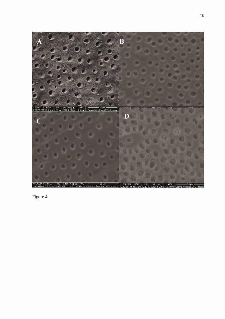
dentin surfaces showed similar morphology for both treatments (Figure 4).
Microtensile test
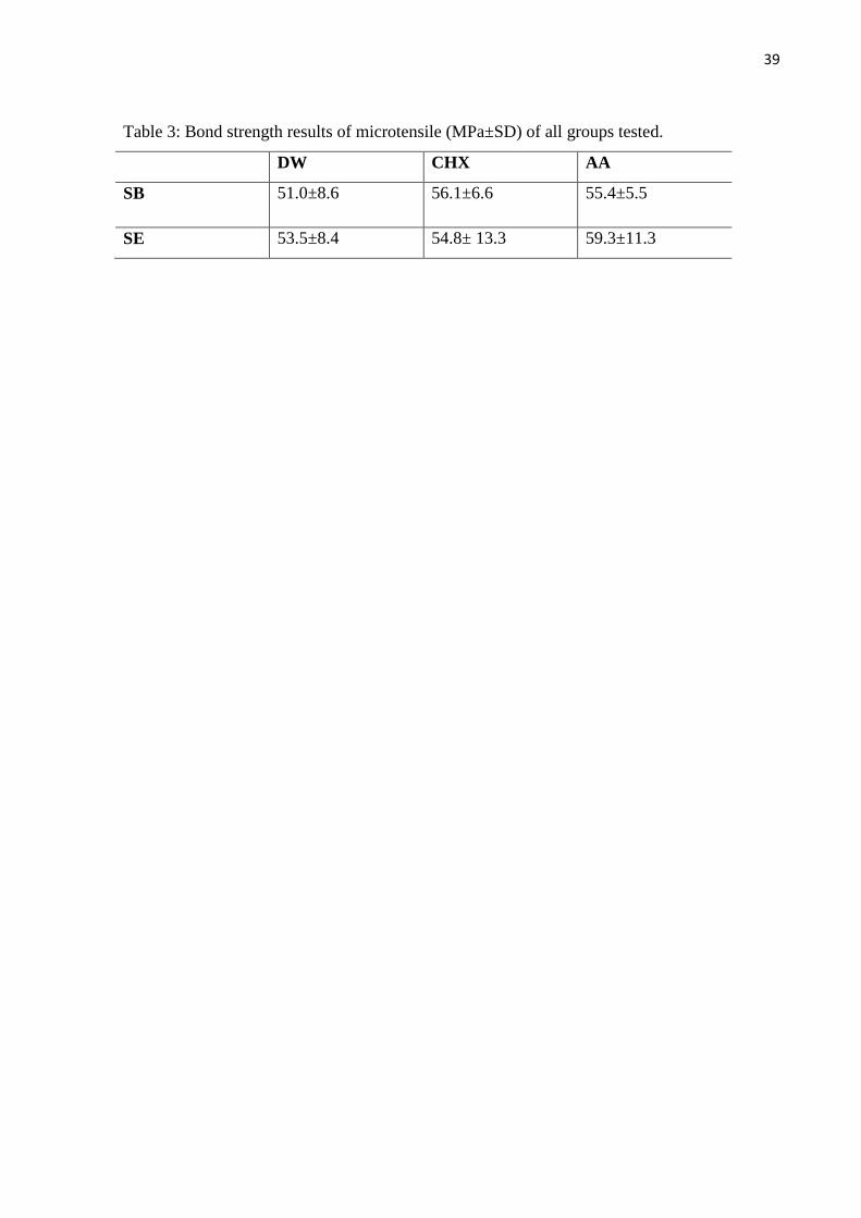
The observed results were not influenced by adhesive systems (p=0.5949; F=0.2990)
or cavity cleaning solutions (p=0.5876; F=0.9191). Interactions among de factors were not
significant (p=0.7870; F=0.2450). After 24 h of storage, there was no statistically significant
difference among the bond strength values of all experimental groups (p > 0.05) (Table 3).

28
Figure 5 summarizes the percentage failure modes of the debonded specimens
according to the adhesive type and cleaning agents used. Mixed fracture modes were
frequently identified in all tested groups, after 24 h of storage.
Scanning Electron Microscopy (SEM) analysis of the debonded dentin surfaces after
24 h of storage showed that Single Bond frequently failed at the bottom of the hybrid layer
(Figure 6), where exposed non-protected collagen can be noted. When the self-etching
adhesive system Clearfil SE Bond was used, dentin tubules remained sealed with restorative
material after fracture in groups where chlorhexidine digluconate 2% and anacardic acid
solutions were used (Figure 7).
DISCUSSION
The anacardic acids have attracted interest in the field of dentistry for its antimicrobial
characteristics against the main causative agent of caries, Streptococcus mutans.19,21
It could
be used to remove the bacterial remnants from dental cavities after caries removal.
The mixture of 2% chlorhexidine digluconate solution with dentin powder attenuated
the IR absorbance of the PO4 band at 1014 cm−1 more than it did in the amide bands. This
result suggests that chlorhexidine digluconate could bind to mineralized dentin, especially to
the inorganic phase of dentin. Probably, the digluconate chlorhexidine binds electrostatically
to phosphate groups in hydroxyapatite crystallites. These results are in agreement with those
of Kim et al. (2010).25
The FTIR absorbance spectra of dentin powder mixed with anacardic acid showed that
the peak of phosphate (1014 cm -¹) was greatly reduced, indicating their interaction with
positively charged groups, possibly the H+ derivatives from carboxylic acid of the anacardic
acid. The peaks of C=O (1698 cm -¹) of the anacardic acid were reduced sharply, indicating
the formation of links between COO- from the acid and amine I groups from the dentin

29
collagen chains. These amines I from collagen were superimposed by OH-, H2O and amides
(1650 and 1538 cm -1) from dentin. The interaction between the cavity cleaning agents and
dentin can be favorable bond strength in the long term, but more studies are needed especially
with the anacardic acid.
The SEM images showed that phosphoric acid treatment promoted dentin
demineralization as expected with open dentinal tubules and exposed collagen; these results
are in agreement with previous literature.26,27
It was observed that anacardic acid solution did
not promote dentin demineralization as smear layer can still be observed on the surface.
Therefore, anarcadic acid should not be used as an etchant agent but is suitable to be
employed as cavity-cleaning solution as it does not alter dentin surface. As it was also
observed a chemical interaction between anacardic acid and dentin, it is speculated that the
substantivity of this acid could be longer and more effective than that of presented by
chlorhexidine. Further studies are necessary to elucidate the substantivity of anacardic acid.
As no statistically significant differences were found in microtensile results among the
groups after 24 h storage, the use of either digluconate chlorhexidine or anacardic acid
solutions did not present any adverse effect on the imediate bond strength. These results are
positive because they allow the use of a cavity-cleaning agent of natural origin, the anacardic
acid solution, without interfering the adhesion with dentin. Regarding the use of
chlorhexidine, the bond strength results are in accordance with other studies.5,28,29
In these
studies no differences were found in the bond strength results between the groups that
received pre-treatment with chlorhexidine and those that did not receive pre-treatment after 24
h. Furthermore, in other study, pre-treatment with different antioxidants, including
chlorhexidine, prevented bond strength loss of an etch-and rise adhesive over time and bond
strength of a self-etching adhesive was not significantly reduced with 9 months of storage.30
Additionally, it was also observed that in both adhesive systems there was an increase in the

30
enzymatic degradation of the hybrid layer. The degradation of hybrid layer is attributed to the
action of the matrix metalloproteinases (MMPs).30,31
Studies suggest that anacardic acid might
be an inhibitor of MMP-2 e MMP-9.23,32
Nevertheless, anacardic acid could protect the
adhesive interface over time and further studies are necessary.
SEM analysis of debonded dentin surfaces showed similar failure pattern at the bottom
of the hybrid layer when both cleaning agents were used with Single Bond. This failure is
attributed to the incomplete monomer infiltration of collagen fibrils, regardless the cleaning
agent, indicating that the cleaning agents used not influenced the bonding to dentin. On the
other hand, SEM analysis of the debonded dentin surfaces from the groups of Clearfil SE
Bond showed that, when the cavity cleaning-agents were applied prior to the primer, dentinal
tubules remained more sealed with restorative material than the group using distilled water
only. It could be suggested a protective effect of the cleaning agents.
The findings from this study indicate a promising application for the anarcardic acid
solution as a cavity-cleaning agent and maybe as an antioxidant to prevent collagen
degradation due to the action of dentin proteases. Further studies regarding the longevity of
dentin bonds and the interaction between collagen and anacardic acid are necessary.
CONCLUSION
The anarcadic acid is suitable to be used as a cavity-cleaning agent. The
morphological, chemical and bond strength analyses demonstrated that the anacardic acid did
not affect dentin bonding, regardless the adhesive strategy.
ACKNOWLEDGMENTS
The authors are thankful to Prof. Pedro Alejandro Ayala and the Department of Physics,
Federal University of Ceara, for their assistance on the use of the FTIR spectroscope. The

31
SEM analysis was conducted at Central Analítica –UFC ̸ CT- INFRA ̸ MCTI – SISNANO ̸
Pró-equipamentos CAPES.
REFERENCES
1.Van Meerbeek B, Peumans M, Poitevin A, Mine A, Van Ende A, Neves A, De Munck J
(2010) Relationship between bond-strength tests and clinical outcomes Dental Materials 26
100-121.
2.De Munck J, Van Landuyt K, Peumans M, Poitevin A, Lambrechts P, Braem M, Van
Meerbeek B (2005) A critical review of the durability of adhesion to tooth tissue: methods
and results Journal of Dental Research 84(2) 118-132.
3.Peumans M, Kanumilli P, De Munck J, Van Landuyt K, Lambrechts P, Van Meerbeek B
(2005) Clinical effectiveness of contemporary adhesives: A systematic review of current
clinical trials Dental Materials 21 864–881.
4.Bengtson CRG, Bengtson AL, Bengtson NG, Turbino ML (2008) Efeito da clorexidina 2%
na resistência de união de dois sistemas adesivos à dentina humana Pesquisa Brasileira em
Odontopediatria e Clínica Integrada 8(1) 51-56.
5.Soares CJ, Pereira CA, Pereira JC, Santana FR, Prado CJ (2008) Effect of chlorhexidine
application on microtensile bond strength to dentin Operative Dentistry 33(2) 183-188.

32
6.Shinohara MS, Yamauti M, Inoue G, Nikaido T, Tagami J, Giannini M, De Goes MF
(2006) Evaluation of antibacterial and fluoride-releasing adhesive system on dentin –
microtensile bond strength and acid-base challenge Dental Materials Journal 25(3) 545-552.
7.Camilotti V, Ioris
MD, Busato
PMR, Ueda
JK, Mendonça MJ (2013) Avaliação da
influência da solução de irrigação na resistência adesiva de um cimento resinoso Revista de
Odontologia da UNESP 42(2) 83-88.
8.Say EC, Koray F, Tarim B, Soyman M, Gülmez T (2004) In vitro effect of cavity
disinfectants on the bond strength of dentin bonding systems Quintessence International
35(1) 56-60.
9.Wade WG, Addy M (1989) In vitro activity of a chlorhexidine-containing mouthwash
against subgingival bacteria Journal of Periodontology 60(9) 521-525.
10.Quirynen M, Avontroodt P, Peeters W, Pauwels M, Coucke W, van Steenberghe D (2001)
Effect of different chlorhexidine formulations in mouthrinses on de novo plaque formation
Journal of Clinical Periodontology 28(12) 1127–1136.
11.Watts A, Addy M (2001) Tooth discolouration and staining: a review of the literature.
British Dental Journal 190(6) 309 -316.
12.Perazzo FF, Silva RS, Carvalho JCT, Groppo FC (2004) Utilización sustancias naturales
en Odontologia Jornal Brasileiro de Fitomedicina 2(1 ̸ 4) 9-15.

33
13.Hemshekhar M, Santhosh MS, Kemparaju K, Girish KS (2011) Emerging roles of
anacardic acid and its derivatives: A pharmacological overview Basic & Clinical
Pharmacology & Toxicology 110(2) 122-132.
14.Trevisan MTS, Pfundstein B, Haubner R, Wurtele G, Spiegelhalder B, Bartsch H, Owen
RW (2006) Characterization of alkyl phenols in cashew (Anacardium occidentale) products
and assay of their antioxidante capacity Food and Chemical Toxicology 44 188-197.
15.Mazzetto SE, Lomonaco D, Mele G (2009) Óleo da castanha de caju: oportunidades e
desafios no contexto do desenvolvimento e sustentabilidade industrial Química Nova 32(3)
732-741.
16.Kubo I, Masuoka N, Ha TJ, Tsujimoto K (2006) Antioxidant activity of anacardic acids
Food Chemistry 99 555-562.
17.Kubo I, Ochi M, Vieira PC, Komatsu S (1993) Antitumor agents from the cashew
(Anacardium occidentale) apple juice Journal of Agricultural and Food Chemistry 41 1012-
1015.
18.Sousa DL Efeito antibacteriano do ácido anacárdico em culturas planctônicas e
biofilmes de Streptococcus mutans. 2014.Tese de Doutorado, Programa de Pós-graduação
em Odontologia, Universidade Federal do Ceará, Fortaleza, 2014.
19.Muroi H, Kubo I (1993) Bactericidal Activity of anacardic acids against Streptococcus
mutans and their potentiation Journal of Agrjcultural and Food Chemistry 41 1780-1783.

34
20.Himejima M, Kubo I (1991) Antibacterial agents from the cashew Anacardium occidentale
(Anacardiaceae) nut shell oil Journal of Agrjcultural and Food Chemistry 39 418-421.
21.Green IR, Tocoli FE, Lee SH, Nihei KI, Kubo I (2008) Design and evaluation of
anacardic acid derivatives as anticavity agents European Journal of Medicinal Chemistry 43
1315-1320.
22.Lima CAA, Pastore GM, Lima EDPA (2000) Estudo da atividade antimicrobiana dos
ácidos anacárdicos do óleo da casca da castanha de caju (cnsl) dos clones de cajueiro-anão-
precoce ccp-76 e ccp-09 em cinco estágios de maturação sobre microrganismos da cavidade
bucal Ciência e Tecnologia Alimentar 20(3) 358-362.
23.Silveira C, Oliveira F, Santos ML, Freitas T, Imparato JC, Magalhães AC (2014)
Anacardic acid from Brazilian cashew nut trees reduces dentine erosion Caries Research 48
549-556.
24.Oliveira SN Síntese, caracterização e aplicação de nanocápsulas de poliuréia a partir
do ácido anacárdico, via miniemulsão inversa. 2014.Tese de Doutorado, Programa de Pós-
Graduação em Química, Universidade Federal do Ceará, Fortaleza, 2014.
25.Kim J, Uchiyama T, Carrilho M, Agee KA, Mazzoni A, Breschi L, Carvalho RM,
Tjaderhane L, Looney S, Wimmer C, Tezvergil-Mutluay A, Tay FR, Pashley DH (2010)
Chlorhexidine binding to mineralized versus demineralized dentin poder Dental Materials
26(8) 771-778.
35
26.Van Meerbeek B, De Munck J, Yoshida Y, Inoue S, Vargas M, Vijay P, Van Landuyt K,
Lambrechts P, Vanherle G (2003) Adhesion to enamel and dentin: Current status and future
challenges Operative Dentistry 28(3) 215-235.
27.Pashley DH, Tay FR, Breschi L, Tjaderhane L, Carvalho RM, Carrilho M, Tezvergil-
Mutluay A (2011) State of the art etch-and-rinse adhesives Dental Materials 27(1) 1-16.
28.Breschi L, Mazzoni A, Nato F, Carrilho M, Visintini E, Tjaderhane L, Ruggeri A, Tay FR,
Dorigo ED, Pashley DH (2010) Chlorhexidine stabilizes the adhesive interface: A 2-year in
vitro study Dental Materials 26(4) 320-325.
29.Carrilho MRO, Carvalho RM, de Goes MF, di Hipólito V, Geraldeli S, Tay FR, Pashley
DH, Tjäderhane L (2007) Chlorhexidine Preserves Dentin Bond in vitro Journal of Dental
Research 86(1) 90-94.
30.Zheng P, Zaruba M, Attin T, Wiegand A (2015) Effect of different matrix
metalloproteinase inhibitors on microtensile bond strength of an etch-and-rinse and a self-
etching adhesive to dentin Operative Dentistry 40(1) 80-86.
31.Mazzoni A, Scaffa P, Carrilho M, Tjäderhane L, Di Lenarda R, Polimeni A, Tezvergil-
Mutluay A, Tay FR, Pashley DH, Breschi L (2013) Effects of etch-and-rinse and self-etch
adhesives on dentin MMP-2 and MMP-9 Journal Dental Research 92(1) 82-86.

36
32.Omanakuttan A, Nambiar J, Harris RM, Bose C, Pandurangan N, Varghese RK, Kumar
GB, Tainer JA, Banerji A, Perry JJP, Nair BG (2012) Anacardic acid inhibits the catalytic
activity of matrix metalloproteinase-2 and matrix metalloproteinase-9 Molecular
Pharmacology 82(4) 614-622.

37
Table 1 - Materials and chemicals used in the study.
MATERIAL ̸
MANUFACTURER
BASIC COMPOSITION MANUFACTURER’S
INSTRUCTIONS
Condac 37 (FGM,
Joinville, SC, Brazil -
FGM)
Phosphoric Acid 37%, dye,
thickener and deionized
water
Apply the gel for 15 s in
dentin and rinse abundantly
with water for 30 s.
Adper Single Bond 2
(3M ESPE, São José do
Rio Preto, SP, Brazil)
Bis-GMA, GDMA, HEMA,
UDMA, nanofillers, water,
ethanol, methacrylate
functional copolymer of
polyacrylic and polytaconic
acids
Apply two consecutive coats
of adhesive to etched humid
dentin, with the saturated
brush, gently stir on surface
for 15 s. Gently drying and
light cure for 10 s.
Clearfil SE Bond
(Kuraray Noritake
Dental Inc., Kurashiki,
Okayama, Japan)
MDP, HEMA, Hydrophilic
dimethacrylate, water,
camphorquinone,
Bis-GMA.
Apply Primer for 20 s, dry
with mild air stream for 5 s.
Apply Bond for 15 s, gentle
air stream, light cure for 10 s.
Composite resin (3M
ESPE, Filteck, Z250
XT, São José do Rio
Preto, SP, Brazil)
Bis-GMA, Bis-EMA,
UDMA, TEGDMA, Zr-
silica.
Place 3M Filtek Z250
restorative in increments less
than 2.5 mm and light cure
each increment for 20 s.
Anacardic acid
(Laboratory of
polymers and materials
innovation of the
Organic and inorganic
Chemistry Department
- UFC)
Anacardic acid
2% chlorhexidine
digluconate solution
(FGM, Joinville, SC,
Brazil)
Chlorhexidine digluconate
to 2%, deionized water,
volatile Surfactant

38
Table 2 – Groups for the microtensile test.
GROUP CAVITY CLEANING
SOLUTION
ADHESIVE
SYSTEM
BONDING PROCEDURE
DW ̸ SB Distilled Water Single Bond
(3M ESPE,
St Louis,
MO, USA)
Acid-etch dentin with
phosphoric acid, rinse and dry
without dehydrating. Apply the
respective cleaning agent cavity
for 60 s under agitation, remove
excess with absorbent paper and
apply the adhesive.
CHX ̸
SB
2% chlorhexidine
digluconate solution
AA ̸ SB Anacardic acid solution
DW ̸ SE Distilled Water
Clearfil SE
Bond
(Kuraray
Noritake
Dental Inc.,
Kurashiki,
Okayama,
Japan)
Apply each cavity-cleaning
agent for 60 s under stirring,
remove excess with absorbent
paper and apply the adhesive
system.
CHX ̸ SE 2% chlorhexidine
digluconate solution
AA ̸ SE Anacardic acid solution
39
Table 3: Bond strength results of microtensile (MPa±SD) of all groups tested.
DW CHX AA
SB 51.0±8.6 56.1±6.6 55.4±5.5
SE 53.5±8.4 54.8± 13.3 59.3±11.3

40
Figure 1

41
Figure 2

42
Figure 3
43
Figure 4

44
Figure 5
0%
10%
20%
30%
40%
50%
60%
70%
80%
90%
100%
DW-SB CHX-SB AA-SB DW-SE CHX-SE AA-SE
CR
CD
A
M

45
Figure 6
46
Figure 7

47
Figure 1. FTIR spectrum for dentin powder, 2% chlorhexidine digluconate, and dentin
powder incubated with chlorhexidine.
Figure 2. FTIR spectrum for dentin powder, anacardic acid solution, and dentin powder
incubated with anacardic acid.
Figure 3. SEM micrographs of dentin. (A) Normal mineralized dentin; (B) Phosphoric-acid
etched dentin; (C) Dentin treated with primer of Clearfil SE Bond; (D) Dentin treated with
anarcadic acid solution.
Figure 4. SEM images of treated dentin. (A) Phosphoric acid-etched dentin + 2%
chlorhexidine digluconate; (B) Phosphoric acid-etched dentin + anacardic acid solution; (C)
2% chlorhexidine digluconate + dentin treated with primer of Clearfil SE Bond; (D)
Anacardic acid solution + dentin treated with primer of Clearfil SE Bond.
Figure 5. Graph shows the failure mode after microtensile test.
Figure 6. SEM images of the fractured surface along the dentin side of specimens treated with
cleaning agents DW, CHX or AA and bonded with Single Bond (24 h storage). Mixed failure
can be observed in all groups. (A) and (D) Dentin treated with DW; (B) and (E) Dentins
treated with CHX; (C) and (F) Dentin treated with AA. Images on top (A, B and C): 200x;
Images on bottom (D, E and F): 10.000x.
Figure 7. SEM micrographs of the fractured surface along the dentin side of specimens treated
with cleaning agents DW, CHX or AA and bonded with Clearfil SE Bond (24 h storage).

48
Mixed failures were observed in all groups. (A) and (D) Dentin treated with DW – few dentin
tubules remained occluded by resin tags; (B) and (E) Dentin treated with CHX; (C) and (F)
Dentin treated with AA. Images on top (A, B and C): 200x; Images on bottom (D, E and F):
10.000x.

49
4 CONCLUSÃO GERAL
O ácido anacárdico mostrou-se viável como agente de limpeza cavitária em
restaurações adesivas com resina composta.
O ácido fosfórico removeu a smear layer e desmineralizou a dentina, expondo as
fibrilas colágenas, enquanto que o ácido anacárdico não desmineralizou o substrato
dentinário.
O ácido anacárdico interagiu quimicamente com a dentina mineralizada.
O uso da solução de ácido anacárdico como agente de limpeza cavitária não
influenciou na resistência de união do adesivo de condicionamento total e também não
interferiu na resistência de união do sistema adesivo autocondicionante após 24 h de
armazenamento.

50
REFERÊNCIAS
AGOSTINI-COSTA T. S. et al. Determinação de ácidos anacárdicos em pedúnculos de caju
(Anacárdium occidentale L.). Rev. Bras. PL. Med., v. 5, n. 2, p. 77-81, 2003.
AGOSTINI-COSTA T. S. et al. Determinação espectrofotométrica de ácido anacárdico em
amêndoas de castanha de caju. Comunicado técnico 122. ISSN 9192-0099. 2005.
BENGTSON C. R. G. et al. Efeito da clorexidina 2% na resistência de união de dois sistemas
adesivos à dentina humana. Pesquisa Brasileira em Odontopediatria e Clínica Integrada
v. 8, n. 1, p. 51-56, 2008.
CAMILOTTI V. et al. Avaliação da influência da solução de irrigação na resistência adesiva
de um cimento resinoso. Rev Odontol UNESP. v. 42(2), p. 83-88, 2013.
CARDOSO M. V. et al. Current Aspects on Bonding Effectiveness and Stability in Adhesive
Dentistry. Aust Dent J., v. 56, p. 31-44, 2011.
DE MUNCK J. et al. A critical review of the durability of adhesion to tooth tissue: methods
and results. J Dent Res., v. 84(2), p. 118-132, 2005.
GREEN I. R. et al. Design and evaluation of anacardic acid derivatives as anticavity agentes.
Eur J Med Chem., v. 43, p. 1315-1320, 2008.
HASHIMOTO M. et al. Over-etching on micro-tensile bond strength and failure patterns for
two dentin bonding systems. J Dent., v. 30, p. 99-105,2002.
HEMSHEKHAR M. et al. Emerging Roles of Anacardic Acid and Its Derivatives: A
Pharmacological Overview. Basic Clin Pharmacol Toxicol., v. 110, Issue 2, p. 122-132,
2011.
HIMEJIMA M.; KUBO I. Antibacterial Agents from the Cashew Anacardium occidentale
(Anacardiaceae) Nut Shell Oil. J Agric Food Chem., v.39, p.418-421, 1991.
KUBO I. et al. Antitumor agentes from the cashew (Anacardium occidentale) apple juice. J
Agric Food Chem., v. 41, p. 1012-1015, 1993.
KUBO I. et al. Antioxidant activity of anacardic acids. Food Chem., v. 99, p. 555-562, 2006.
LIMA C. A. A.; PASTORE G. M.; LIMA E. D. P. A. Estudo da atividade antimicrobiana dos
ácidos anacárdicos do óleo da casca da castanha de caju (cnsl) dos clones de cajueiro-anão-
precoce ccp-76 e ccp-09 em cinco estágios de maturação sobre microrganismos da cavidade
bucal. Ciência e Tecnologia Alimentar, v. 20, n.3, p. 358, 2000.
MAZZETO S. E.; LOMONACO D.; MELE G. Óleo da castanha de caju: oportunidades e
desafios no contexto do desenvolvimento e sustentabilidade industrial. Quim Nova, v. 32, p.
732-741, 2009.
MUROI H.; KUBO I. Bactericidal Activity of Anacardic Acids against Streptococcus mutans
and Their Potentiation. J. Agrjc. Food Chem, v. 41, p. 1780-1783, 1993.

51
NAKABAYASHI N.; ASHIZAWA M.; NAKAMURA M. Identification of a resin-dentin
hybrid layer in vital human dentin created in vivo: durable bonding to vital dentin.
Quintessence Int., v. 23 (2), p. 135-141, 1992.
PASHLEY D. H. et al. State of the art etch-and-rinse adhesives. Dent Mater., v. 27, p. 1-16,
2011.
PEUMANS M. et al. Clinical effectiveness of contemporary adhesives: A systematic review
of current clinical trials. Dent Mater., v. 21, p. 864–881, 2005.
QUIRYNEN M. et al. Effect of different chlorhexidine formulations in mouthrinses on de
novo plaque formation. J Clin Periodontol., v. 28, p. 1127–1136, 2001.
SAY E. C. et al. In vitro effect of cavity disinfectants on the bond strength of dentin bonding
systems. Quintessence Int., v. 35(1), p. 56-60, 2004.
SILVEIRA C. et al. Anacardic acid from Brazilian cashew nut trees reduces dentine erosion.
Caries Res., v.48, p.549-556, 2014.
SOARES C. J. et al. Effect of chlorhexidine application on microtensile bond strength to
dentin. Oper Dent., v.33(2), p.183-188, 2008.
SOUSA D. L. Efeito antibacteriano do ácido anacárdico em culturas planctônicas e
biofilmes de Streptococcus mutans. 2014.Tese de Doutorado, Programa de Pós-graduação
em Odontologia, Universidade Federal do Ceará, Fortaleza, 2014.
TAY F. R..; PASHLEY D. H. Aggressiveness of contemporary self-etching systems. I: Depth
of penetration beyond dentin smear layers. Dent Mater., v. 17, p. 296-308, 2001.
TREVISAN M. T. S. et al. Characterization of alkyl phenols in cashew (Anacardium
occidentale) products and assay of their antioxidante capacity. Food Chem Toxicol., v. 44, p.
188-197, 2006.
VAN MEERBEEK B. et al. Buonocore Memorial Lecture Adhesion to Enamel and Dentin:
Current Status and Future Challenges. Oper Dent., v. 28 (3), p. 215-235, 2003.
VAN MEERBEEK B. et al. Relationship between bond-strength tests and clinical outcomes.
Dent Mater. v. 26, p. 100-121, 2010.
VAN MEERBEEK B. et al. State of the art of self-etch adhesives. Dent Mater., v.27 (1), p.
17-28, 2011.
WADE W. G.; ADDY M. In vitro activity of a Chlorhexidine-containing mouthwash against
subgingival bactéria. J Periodontol., v. 60, p. 521-525, 1989.
WATTS A.; ADDY M. Tooth discolouration and staining: a review of the literature. Br Dent
J., v. 190 (6), p. 309 -316, 2001.

52
APÊNDICE
TERMO DE DOAÇÃO DE DENTES
Pelo presente instrumento que atende às exigências legais, o Sr(a)
_________________________________________________________, após ter tomado
conhecimento do protocolo da pesquisa “ÁCIDO ANACÁRDICO COMO AGENTE DE
LIMPEZA CAVITÁRIA EM ODONTOLOGIA ADESIVA.” que tem como objetivo
avaliar o desempenho do ácido anacárdico como agente de limpeza cavitária dentinária em
restaurações adesivas com resina composta, vem na melhor forma de direito DOAR à
Cirurgiã Dentista Cristina Maria Fernandes de Queiroz ___ dentes (terceiros molares),
declarando, sob as penas da lei, que os dentes objeto da presente doação foram extraídos por
indicação terapêutica, cujos históricos circunstanciados fazem parte dos prontuários dos
pacientes de quem se originam.
Data:__ ̸__ ̸ __
Assinatura:_____________________________________________________________
RG:___________________________________________________________________

53
ANEXO
PARECER CONSUBSTANCIADO DO CEP

54

55

56