Tese Erika Takahagi - UNESP: Câmpus de Botucatu - Instituto de … · 2010-03-23 · compreensão...
72
1 Introdução Paracoccidioides brasiliensis é um fungo dimórfico, agente etiológico da paracoccidioidomicose (PCM), que se apresenta endêmica na América Latina (1) . As manifestações clínicas da micose são de doença granulomatosa crônica, comprometendo especialmente tecidos pulmonares, mucosas e o sistema fagocítico mononuclear, com disseminação para fígado, baço, adrenais e outros órgãos (2,3) . Na PCM como em outras infecções granulomatosas crônicas, a resposta imune celular é o principal mecanismo de defesa. O granuloma na PCM representa uma resposta tecidual específica do hospedeiro contra o fungo, na tentativa de destruir e circunscrever o parasita, evitando sua multiplicação e disseminação (4,5) . Estudos experimentais de PCM em camundongos, demonstraram associação entre resposta dominante Th2 e gravidade da infecção (6) . Na doença humana, alguns autores observaram o predomínio de resposta imune Th2 sobre a resposta Th1, com produção de Interleucina (IL)-4, IL-5 e IL-10 e níveis elevados de IgG4, IgA e IgE (7-9) . Esses parâmetros associados a baixos níveis de IFN-γ foram correlacionados com as formas graves da micose (9) . Células mononucleares de sangue periférico de pacientes com as formas aguda e crônica da micose, apresentam baixa resposta proliferativa e produção de citocinas de padrão Th1, interferon-gamma (IFN-γ) e IL-2 em resposta a antígenos de P. brasiliensis, enquanto os níveis IL-10 se encontram elevados (7,10) . Assim, a imunossupressão na PCM poderia ser entendida como decorrente da perturbação do equilíbrio das células do sistema imune, causada pelo P. brasiliensis, levando ao aparecimento dos diferentes fenômenos envolvidos na modulação da resposta imune. Os trabalhos citados acima, mostram claramente que uma resposta protetora contra o P. brasiliensis depende de um padrão de resposta do tipo Th1, envolvendo como citocinas principais, IFN-γ, fator de necrose tumoral-alfa (TNF-α) e IL-12 (11,12) . O desenvolvimento dessa resposta seria representado, principalmente pelas células fagocitárias, os monócitos e macrófagos, que necessitam de ativação fornecida por essas citocinas para atuarem na resposta inflamatória e na atividade fungicida. Na PCM humana e experimental, os macrófagos estão presentes em grande número nas lesões granulomatosas (4,13,14) e representam a principal célula de defesa contra P. brasiliensis (15,16) . Considerando que P.brasiliensis é um microrganismo intracelular facultativo (17) , os macrófagos podem ter papel essencial na patogênese da doença. Portanto, a investigação de mecanismos relacionados com a fagocitose do
Transcript of Tese Erika Takahagi - UNESP: Câmpus de Botucatu - Instituto de … · 2010-03-23 · compreensão...

1
Introdução
Paracoccidioides brasiliensis é um fungo dimórfico, agente etiológico da
paracoccidioidomicose (PCM), que se apresenta endêmica na América Latina (1). As
manifestações clínicas da micose são de doença granulomatosa crônica, comprometendo
especialmente tecidos pulmonares, mucosas e o sistema fagocítico mononuclear, com
disseminação para fígado, baço, adrenais e outros órgãos (2,3). Na PCM como em outras
infecções granulomatosas crônicas, a resposta imune celular é o principal mecanismo de
defesa. O granuloma na PCM representa uma resposta tecidual específica do hospedeiro
contra o fungo, na tentativa de destruir e circunscrever o parasita, evitando sua
multiplicação e disseminação (4,5).
Estudos experimentais de PCM em camundongos, demonstraram associação
entre resposta dominante Th2 e gravidade da infecção (6). Na doença humana, alguns
autores observaram o predomínio de resposta imune Th2 sobre a resposta Th1, com
produção de Interleucina (IL)-4, IL-5 e IL-10 e níveis elevados de IgG4, IgA e IgE (7-9).
Esses parâmetros associados a baixos níveis de IFN-γ foram correlacionados com as
formas graves da micose (9). Células mononucleares de sangue periférico de pacientes
com as formas aguda e crônica da micose, apresentam baixa resposta proliferativa e
produção de citocinas de padrão Th1, interferon-gamma (IFN-γ) e IL-2 em resposta a
antígenos de P. brasiliensis, enquanto os níveis IL-10 se encontram elevados (7,10).
Assim, a imunossupressão na PCM poderia ser entendida como decorrente da
perturbação do equilíbrio das células do sistema imune, causada pelo P. brasiliensis,
levando ao aparecimento dos diferentes fenômenos envolvidos na modulação da
resposta imune. Os trabalhos citados acima, mostram claramente que uma resposta
protetora contra o P. brasiliensis depende de um padrão de resposta do tipo Th1,
envolvendo como citocinas principais, IFN-γ, fator de necrose tumoral-alfa (TNF-α) e
IL-12 (11,12). O desenvolvimento dessa resposta seria representado, principalmente pelas
células fagocitárias, os monócitos e macrófagos, que necessitam de ativação fornecida
por essas citocinas para atuarem na resposta inflamatória e na atividade fungicida.
Na PCM humana e experimental, os macrófagos estão presentes em grande
número nas lesões granulomatosas (4,13,14) e representam a principal célula de defesa
contra P. brasiliensis (15,16). Considerando que P.brasiliensis é um microrganismo
intracelular facultativo (17), os macrófagos podem ter papel essencial na patogênese da
doença. Portanto, a investigação de mecanismos relacionados com a fagocitose do
2
fungo por macrófagos e sua atividade funcional, torna-se importante para a
compreensão da relação hospedeiro-parasita na PCM.
O contato inicial de P. brasiliensis com o hospedeiro se faz através das células
fagociticas, que constituem um importante compartimento da defesa inata do
organismo. O fungo é fagocitado por macrófagos in vivo (15) e in vitro é internalizado e
se multiplica em monócitos e macrófagos não ativados (18). Apenas monócitos e
macrófagos ativados in vitro com IFN-γ e TNF-α apresentam atividade fungicida contra
P. brasiliensis (19,20).
P. brasiliensis apresenta em sua constituição uma multiplicidade de
componentes antigênicos, que podem ser extraídos da parede do fungo, obtido do
conteúdo citoplasmático (intracelulares) ou do filtrado de cultura (metabólicos ou
exocelulares) (21). Componentes do próprio fungo, como a glicoproteina de 43 kDa
(gp43), considerada o principal antígeno secretado por P. brasiliensis, pode promover a
adesão inicial e a internalização da levedura por células fagocíticas (22). Assim, a gp43
parece estar envolvida na adesão de P.brasiliensis aos macrófagos peritoneais murinos,
fenômeno que leva à fagocitose e pode favorecer a morte do fungo. Esse efeito é inibido
por anticorpos anti-gp43. Ensaios de inibição indicam o envolvimento de resíduos de
fucose e manose, na fagocitose de P. brasiliensis (22).
POPI et al. (23) avaliaram o efeito da gp43 da interação entre macrófagos
peritoneais murinos e P. brasiliensis. A fagocitose de células leveduriformes do fungo
pelas células peritoneais de camundongos das linhagens B10.A e A/Sn, considerados,
respectivamente, suscetíveis e resistentes à infecção, foi inibida pela adição de
diferentes concentrações de gp43 ao meio de cultura. Os autores sugerem que a
produção de gp43 pelo fungo, pode ser considerada um mecanismo de evasão do P.
brasiliensis na instalação da infecção em hospedeiros suscetíveis.
FERREIRA & ALMEIDA (24) estudaram o efeito da imunização prévia com
células dendríticas estimuladas com gp43 sobre a infecção experimental de
camundongos com cepa virulenta de P. brasiliensis. Verificaram que a gp43 apresenta
efeito regulador negativo sobre a expressão de moléculas MHC-classe II e de adesão
CD80, CD54 e CD40 e sobre a produção de IL-12 por essas células. Segundo os
autores, a baixa expressão de moléculas co-estimulatórias e de IL-12 por células
dendríticas, ambas induzidas por gp43, poderia estar envolvida na depressão de resposta
Th1 in vivo, levando ao desenvolvimento de infecção mais grave nos animais.

3
Por outro lado, outros autores relataram que gp43 é capaz de induzir a produção
de níveis elevados de IL-2, IFN-γ e IL-10 por células mononucleares de sangue
periférico de indivíduos saudáveis, previamente infectados com P.brasiliensis e IL-10
por monócitos de pacientes com a forma ativa da doença (7).
A imunidade inata se constitui na primeira linha de defesa contra infecções,
reconhecendo um largo espectro de patógenos, sem necessidade de sensibilização
prévia. Foi inicialmente considerada como mecanismo inespecífico da resposta imune,
caracterizado por fagocitose e digestão de microrganismos e substâncias estranhas por
macrófagos e leucócitos. Entretanto, atualmente sabe-se que os mecanismos da
imunidade inata reconhecem e reagem contra os microrganismos, discriminando entre
os patógenos e o self (25,26). Além disso, a ativação da resposta imune inata desencadeia
uma rede de citocinas, resultando em resposta inflamatória e podendo ser um pré-
requisito para o desencadeamento da imunidade adquirida (27,28).
O reconhecimento inicial de microrganismos é mediado por receptores celulares
expressos em células da imunidade inata. Segundo TRICKER & CHENG (29) a
fagocitose é iniciada por fagócitos incluindo macrófagos e células polimorfonucleares
através do reconhecimento de partículas tais como patógenos ou células apoptóticas.
Esses componentes da imunidade natural reconhecem estruturas que são características
dos patógenos microbianos e não estão presentes nas células dos mamíferos. O
reconhecimento ocorre através dos receptores da superfície celular, cujos ligantes estão
presentes na superfície do patógeno. As substâncias dos microrganismos que estimulam
a imunidade natural são chamadas de padrões moleculares associados aos patógenos
(PAMPs) e os receptores que se ligam a essas estruturas preservadas são chamados de
receptores de reconhecimento de padrões (PRRs). Diferentes classes de microrganismos
como vírus, bactérias e fungos expressam diferentes PAMPs, estruturas que incluem
ácidos nucléicos específicos de microrganismos, proteínas típicas de bactérias e
complexos de lipídeos e carboidratos sintetizados pelos microrganismos, tais como
lipopolissacarídeos (LPS), ácidos teicóicos e oligossacarídeos ricos em manose
encontrados em glicoproteínas microbianas, mas não nos tecidos de mamíferos (30).
Assim, a interação entre essas moléculas de superfície do microrganismo e receptores
homólogos, presentes na membrana celular de macrófagos, modulam a fagocitose e a
ativação da célula (31,32). Portanto, monócitos e macrófagos são células da imunidade
inata que expressam receptores de superfície para manose, CD14, componentes do
4
sistema complemento, porção Fc de moléculas de imunoglobulinas e receptores
semelhantes a Toll (TLR, Toll-like receptor) capazes de reconhecer produtos
microbianos, levando à estimulação da fagocitose, atividade microbicida e produção de
citocinas (30,33).
Receptores semelhantes ao Toll são uma família de proteínas de transmembrana,
evolutivamente conservadas entre insetos e humanos (34) que foram primeiramente
identificados como moléculas determinantes para o padrão embriogênico em
Drosophila e, posteriomente como receptores essenciais na imunidade antifúngica (35).
Essas proteínas servem como receptores de reconhecimento padrão para uma variedade
de moléculas derivadas de microrganismos e estimulam a resposta imune inata. Uma
família de receptores Toll, designada TLRs foi descrita em mamíferos (25). Até agora,
pelo menos 11 TLRs foram identificados em humanos e 13 em camundongos, sendo
fundamentais no reconhecimento de PAMPs (36). Esses receptores podem ser divididos
de acordo com a sua localização na célula: TLR-1/2/4/6/10 são expressos na superfície
celular, enquanto que TLR-3/7/8/9 são expressos em compartimentos endossomais
intracelulares. Assim, a expressão e ativação de TLRs contribuem para a defesa do
hospedeiro contra infecções em Drosophila, camundongos e humanos (35,37,38). A
ativação de TLRs pode regular, não apenas a fagocitose e atividade microbicida, mas
também a liberação de citocinas e diferenciação de células dendríticas imaturas em
maduras, capacitando o sistema imune inato a induzir a resposta imune adaptativa (25,39).
Todos os receptores TLR contêm repetições ricas em leucina, flanqueadas por
motivos ricos em cisteína em suas regiões extracelulares e um domínio de homologia ao
receptor Toll/IL-1R (TIR) em suas regiões citoplasmáticas, o que é essencial para a
sinalização (30). Todo TLR sinaliza através de uma proteína adaptadora MyD88, que
também contém um domínio Toll/IL-1R, resultando na translocação do fator de
transcrição NF-κB e subseqüente transcrição de genes para citocinas pro-inflamatórias (40). Além dessa via, uma via dependente de TRIF pode também ser ativada, que
interage com TRAF 6 e RIP 1, mediando assim a ativação de NFkB (25,41).
A expressão de TLRs na superfície celular pode ser detectada por anticorpos
monoclonais principalmente em monócitos e células dendríticas imaturas (42).
Entretanto, a expressão de TLR é observada em outras células, incluindo neutrófilos,
células endoteliais vasculares, adipócitos, miócitos cardíacos e células epiteliais
intestinais. A expressão de vários TLR é também modulada em resposta a diferentes
estímulos (27).
5
Alguns TLRs podem reconhecer uma variedade de ligantes. Em muitos casos,
dois diferentes TLRs colaboram entre si ou com outro co-receptor para envio de sinais,
após interação com o ligante microbiano (43). TLR4 e seu co-receptor MD-2,
reconhecem LPS de bactérias Gram-negativas bem como polissacáride de Cryptococcus
neoformans (44). Por outro lado, TLR2 medeia resposta celular a peptidoglicanos de
bactérias, lipoproteinas e zimosan, em cooperação com TLR1 ou TLR6 (43). A resposta
imune inata a uma espécie de microrganismo pode refletir a integração das respostas de
vários TLRs para diferentes moléculas produzidas pelo microrganismo (30).
Muitos componentes da parede celular de fungos podem atuar como PAMPs,
que são reconhecidos por TLRs expressos por fagócitos e células dendríticas. Os
principais TLRs envolvidos no reconhecimento de diferentes formas dos fungos como
conídios, hifas e leveduras são TLR1, TLR2, TLR4 e TLR9 (45). Estudos in vitro
envolvendo células fúngicas têm mostrado que C. neoformans, Candida albicans e
Aspergillus fumigatus, podem interagir com TLRs, particularmente TLR2, TLR4 e
TLR9 presentes em células da imunidade inata, (44,46-49). Tem sido descrito que a
resposta de macrófagos a A. fumigatus é dependente da interação com TLR-2, TLR4 e
MyD88 (50), enquanto a resposta a C. neoformans é principalmente mediada por TLR4 (44).
Estudos realizados com macrófagos peritoneais murinos demonstraram que
esférulas de Coccidioides posadasii estimulam a produção de TNF-α, via interação com
TLR2, MyD88 e Dectin-1 (51). VAN DER GRAAF et al. (52) investigaram o
reconhecimento diferencial de blastoconidios e de hifas de C. albicans por TLR,
presentes em células mononucleares de sangue periférico humano e em macrófagos de
camundongos. Os autores verificaram que TLR4 é capaz de mediar a indução de
citocinas pró-inflamatórias após estimulação com o fungo, enquanto o reconhecimento
desse microrganismo por TLR2 conduz principalmente à liberação de citocinas anti-
inflamatórias. Assim, é provável que diferenças nos componentes de superfície desses
fungos, sejam responsáveis pela ligação a diferentes TLRs e por diferentes padrões de
citocinas, produzidas no confronto fungo-célula hospedeira.
A interação de uma única espécie fúngica com diferentes TLRs pode resultar em
diferentes atividades biológicas. Estudos com componentes purificados da parede
celular revelam o principal PRR e via de sinalização utilizada pelas células do
hospedeiro para reconhecer PAMPs, ao contrário de quando se usa o patógeno inteiro
para infectar hospedeiros normais ou deficientes de PRR. A ativação final, embora

6
influenciada pelo receptor ausente, é mediada por PRRs remanescentes que podem
compensar ou não ao receptor deficiente (53). Por outro lado, constituintes purificados da
parede celular fúngica podem ativar mais de uma via de sinalização dependente de
TLR; por exemplo, a produção de TNF-α por macrófagos in vitro, em resposta a
fosfolipomanana de C. albicans é dependente de TLR2, TLR4 e TLR6 (46).
O principal componente da cápsula de C. neoformans, a glicuronoxilomanana
liga-se a vários receptores da superfície de macrófagos, como CD14, CD18 e TLR-4,
afetando algumas funções biológicas dessas células (54). Além disso, manoproteinas
secretadas pelo fungo são reconhecidas por receptores para manose (44,55). CROSS &
BANCROFT (56) demonstraram que a ingestão de formas não-capsuladas de C.
neoformans é mediada por receptores para manose e β-glucana na superfície do
macrófago e, que esse processo induz a produção das citocinas, TNF-α e fator
estimulador de colônias de granulócitos e macrófagos (GM-CSF), resultando na
ativação das células e fagocitose das formas encapsuladas do fungo. Portanto, durante a
infecção fúngica, a geração de uma resposta inflamatória, a morte do fungo e a
sobrevida do hospedeiro envolvem múltiplas vias dependentes ou não de TLR (45,57,58).
Além dos receptores TLR, outro receptor de superfície de macrófagos bem
caracterizado é o receptor para manose, capaz de mediar a fagocitose e a morte
intracelular de microrganismos patogênicos (59). Esse receptor está envolvido na
imunidade antifúngica e seu papel não tem sido totalmente esclarecido (60).
O receptor de manose (MR, CD206) é uma proteína transmembrânica que possui
oito domínios de lectina do tipo C, um domínio com repetições de fibronectina do tipo
II, um domínio rico em cisteína e uma pequena porção citoplasmática (60). O receptor
para manose é uma lectina do macrófago que interage com resíduos terminais de
manose e fucose de glicoproteinas e glicolipídeos. Esses açúcares são moléculas
tipicamente observadas na parede celular de microrganismos, que são assim,
reconhecidas pelas células do hospedeiro (30). A expressão do receptor de manose tem
sido demonstrada em macrófagos peritoneais (61) e alveolares (62), bem como fagócitos
mononucleares humanos (63). Têm sido sugerido que o principal papel do MR é o
clearance endocítico de glicoproteínas derivadas do hospedeiro (64) podendo mediar a
fagocitose de microrganismo não-opsonizado, interagindo com polissacarídeos da
parede celular, bem como com manana fúngica, cápsula bacteriana, lipopolissacáride e
lipoarabonomanana (65). O receptor de manose é essencial na produção de citocinas
7
tanto pró-inflamatórias quanto anti-inflamatórias, sendo capaz de interagir com outros
PRRs, para mediar a sinalização intracelular (66). Esse receptor é capaz de interagir com
uma ampla variedade de microrganismos, como bactérias Gram-positivas, Gram-
negativas, fungos, protozoários e micobactérias (59,62,67-69). Macrófagos derivados de
monócitos humanos fagocitam leveduras de C.albicans não opsonizadas, via receptor
para manose (63,70-73). Além da interação com C. albicans esse receptor também está
envolvido na ligação de outros microrganismos como Pseudomonas (74), Pneumocystis
carinii (75), Leishmania donovani
(76), Mycobacterium avium (77,78) e Paracoccidioides
brasiliensis (22).
Portanto, a produção de citocinas pelas células da imunidade inata parece ser um
evento importante que ocorre após a interação com os patógenos, levando à ativação
celular e resultando na destruição do microrganismo ou na instalação da doença. A
produção de citocinas como TNF-α, IL-1, IL-6, IL-8, IL-10 e IL-12 é observada em
estudos in vitro, após estímulo de macrófagos e monócitos humanos com diferentes
fungos, como C. immitis, (79), C. neoformans (56,80), C. albicans (81,82), Malassezia furfur
(83,84) e P. brasiliensis (85,86), demonstrando que essas células podem ser fontes
importantes de citocinas, após interação com esses microrganismos.
Embora a importância da imunidade inata na resistência a infecções fúngicas já
seja bem reconhecida (45), os PRRs que reconhecem P. brasiliensis e os mecanismos
moleculares envolvidos não estão ainda bem caracterizados (53,87). É possível que TLR2
e TLR4 estejam envolvidos, uma vez que vários fungos como C. albicans, A. niger, A.
fumigatus, e Sacharomyces cerevisiae são reconhecidos por esses receptores (88). A
interação de P.brasiliensis com macrófagos peritoneais é aumentada por opsonização
das células leveduriformes com iC3b (89) e, a fagocitose de conidios do fungo por
linhagens de macrófagos murinos ocorre via CR3 e receptor de manose (MR) (90).
Estudos recentes sobre o papel dos receptores TLR na PCM experimental murina
sugerem que células leveduriformes de P.brasiliensis podem interagir tanto com TLR2
como TLR4 para entrar e infectar os macrófagos, resultando em aumento da atividade
fagocitica, secreção de NO e infecção dos macrófagos. Na infecção in vivo, a
deficiência de TLR resultou em diminuição da carga fúngica nos animais e sobrevida
semelhante à de animais normais, novamente sugerindo que TLRs são usados pelo
fungo para infectar o hospedeiro. Assim, a interação com TLR pode ser considerada um
mecanismo de patogenicidade do P.brasiliensis, que usa os receptores da imunidade
inata (TLR2 e TLR4) para infectar as células e garantir sua própria multiplicação (53).
8
BONFIM et al. (91) avaliaram a expressão de TLR1, TLR2, TLR4 e dectina-1 em
monócitos e neutrófilos de indivíduos saudáveis após estimulação de células
leveduriformes de P. brasiliensis, com alta ou baixa viruência e sugeriram a
participação de TLR2, TLR4 edectina-1 no reconhecimento, internalização e
conseqüente ativação da resposta imune contra o fungo.
Em trabalho recente, avaliamos a modulação da expressão de TLR2 e TLR4 na
superfície de monócitos humanos estimulados in vitro com células leveduriformes de
P.brasiliensis ou com gp43, seu principal antígeno e, a produção de TNF-α e IL-10 por
citometria de fluxo e Elisa, respectivamente. Os resultados mostram que tanto o fungo
como a gp43 são capazes de modular a expressão de TLR2 e TLR4 em relação às
células controle não estimuladas. Entretanto, os resultados obtidos com estímulo de
gp43 foram mais evidentes. Baixa expressão de TLR2 e alta de TLR4 por monócitos
foram induzidas por gp43 em 4h de cultura, associadas com elevada produção de TNF-
α. Entretanto, esse perfil inverteu-se após 18 h de cultura, observando-se maior
expressão de TLR2 e menor de TLR4, associadas com maior produção de IL-10. Assim,
a persistência de gp43 na cultura de monócitos por 18h parece aumentar a expressão de
TLR2 e a produção de IL-10 por essas células, sugerindo um mecanismo de escape do
fungo na célula hospedeira. A produção da citocina antiinflamatória poderia levar a um
estado de supressão da resposta do hospedeiro, permitindo a instalação do fungo nos
tecidos. Por outro lado, considerando o envolvimento de níveis elevados de TNF-α na
patogênese da PCM (92), é possível que a produção sustentada de IL-10, induzida por
gp43, poderia controlar a resposta inflamatória excessiva induzida por citocinas pró-
inflamatórias, que resultaria na lesão tecidual observada na PCM (93).
Esses resultados de modulação TLR2 associada a aumento da síntese de IL-10
por monócitos induzidos por gp43, associados aos resultados de alta produção de IL-10
por monócitos de pacientes com PCM (92) e aumento do número de células T
reguladoras (CD4+CD25+ FoxP3) nesses pacientes (94) poderiam explicar a
imunossupressão observada na PCM e mostram que a imunidade inata pode direcionar
ou interferir nos resultados da imunidade adaptativa.
Assim, tanto células leveduriformes de P. brasiliensis como a gp43 purificada
são capazes de induzir a produção de citocinas por monócitos humanos. O ambiente de
citocinas presente na interação inicial fagócito-P. brasiliensis, poderia ser eficiente para
eliminação do fungo ou permitir sua implantação e multiplicação nos tecidos do
9
hospedeiro, causando doença progressiva. Dessa forma, o perfil e a quantidade de
diferentes citocinas produzidas, com atividade supressora ou estimulatória sobre a
resposta imune, pode determinar diferenças na evolução da PCM. A compreensão dos
mecanismos envolvidos durante o contato inicial entre o fungo e monócitos, pode
auxiliar na compreensão da patogênese desta micose. Portanto, o estudo da interação da
gp43 de P. brasiliensis com receptores TLRs e MR da superfície de monócitos
humanos, permitirá a melhor compreensão das vias de ativação dessas células, que
levam à produção de citocinas pró e anti-inflamatórias durante o confronto fungo-
monócito.
10
Referências bibliográficas 1. Wanke B, Londero AT. Epidemiology and paracoccidioidomycosis infection. In: Franco
M, Lacaz CS, Restrepo-Moreno A, Del Negro G editors. Paracoccidioidomycosis. Boca Raton, Florida: CRC Press; 1994. p.109-20.
2. Franco M, Mendes RP, Moscardi-Bacchi M, Rezkallah-Iwasso MT, Montenegro MR. Paracoccidioidomycosis. Bailliere's Clin Trop Med Comm Dis 1989, 4:185-220.
3. Franco M, Peraçoli MTS, Soares AMVC, Montenegro MR, Mendes RP, Meira DA. Host-
parasite relationship in paracoccidioidomycosis. Curr Top Med Mycol 1993; 5:115-49.
4. De Brito T, Franco MF. Granulomatous infection. Rev Inst Med Trop S Paulo 1994; 36:
185-92.
5. Franco M, Montenegro MR, Mendes RP, Marques SA, Dillon NL, Mota NGS. Paracoccidioidomycosis: A recently proposed classification of its clinical forms. Rev Soc Bras Med Trop 1987; 20:129-32.
6. Calich VLG, Kashino SS. Cytokines produced by suscestible and resistant mice in the course of Paracoccidioides brasiliensis infection. Braz J Med Biol Res 1998; 31: 615-23.
7. Benard G, Romano CC, Cacere CR, Juvenale M, Mendes-Giannini MJ, Duarte AJS. Imbalance of IL-2, IFN-γ and IL-10 secretion in the immunosupression associated with human paracoccidioidomycosis. Cytokine 2001; 13: 248-52.
8. Mamoni RL, Nouér AS, Oliveira SJ, Musatti CC, Rossi CL, Camargo ZP, et al. Enhanced production of specific IgG4, IgE, IgA and TGF-β in sera from patients with juvenile form of paracoccidioidomycosis. Med Mycol 2002; 40: 153- 9.
9. Oliveira SJ, Mamoni RL, Musatti CC, Papaiordanou PMO, Blotta MHSL Cytokines and lymphocyte proliferation in juvenile and adult forms of paracoccidioidomycosis: comparision with infected and non-infected controls. Microb Infect 2002; 4: 139-44.
10. Benard G, Mendes-Giannini MJ, Juvenale M, Miranda ET, Duarte AJ. Immunosupression in paracoccidioidomycosis: T cell hyporesponsiveness to two Paracoccidioides
brasiliensis glycoproteins that elicit strong humoral immune response. J Infect Dis 1997; 175: 1263-7.
11. Arruda C, Franco MF, Kashino SS, Nascimento FR, Fazioli Rdos A, Vaz CA, Russo M, Calich VL. Interleukin-12 protects mice against disseminated infection caused by Paracoccidioides brasiliensis but enhances pulmonary inflammation. Clin Immunol 2002; 103:185-95.
12. Romano CC, Mendes-Giannini MJS, Duarte AJS, Benard G. IL-12 and neutralization of endogenous IL-10 revert the in vitro antigen-specific cellular immunosuppression of paracoccidioidomycosis patients. Cytokine 2002; 18: 149-57.
13. Neworal EPM, Altemani A, Mamoni RL, Noronha IL, Biotta MH. Immunocytochemical
localization of cytokines and inducible nitric oxide sinthase (iNOS) in oral mucosa and lymph nodes of patients with paracoccidioidomycosis. Cytokine 2003; 21: 234-41.
14. Parise-Fortes MR, Marques SA, Soares AMVC, Kurokawa CS, Marques MEA, Peraçoli MTS. Cytokines released from blood monocytes and expressed in mucocutaneous lesions of patients with paracoccidioidomycosis evaluated before and during trimethoprim-

11
sulfametoxazole treatment. Brit J Dermatol 2006; 154: 643-50.
15. Brummer E, Hanson LH, Restrepo A, Stevens DA. Intracellular multiplication of Paracoccidioides brasiliensis in macrophages: killing and restriction of multiplication by activated macrophages. Infect Immun 1989; 57: 2289-94.
16. Gonzalez A, Gregori W, Velez D, Restrepo A, Cano LE. Nitric oxide participation in the fungicidal mechanism of gamma interferon-activated murine macrophages against P.
brasiliensis conidia. Infect Immun 2000; 68: 2546-52.
17. Singer-Vermes LM, Burger E, Calich VLG, Modesto-Xavier LH, Sakamoto TN, Sugizaki MF, Meira DA, Mendes RP. Pathogenicity and immunogenicity of Paracoccidioides brasiliensis isolates in the human disease and in an experimental murine model. Clin exp Immunol 1994; 97: 113-19.
18. Brummer E, Hanson LH, Stevens DA. In vitro and in vivo activation of pulmonary macrophages by IFN-γ for enhanced killing of Paracoccidioides brasiliensis and Blastomyces dermatitides. J. Immunol 1988; 140: 2786-9.
19. Moscardi-Bacchi M, Brummer E, Stevens DA, Support of Paracoccidioides brasiliensis
multiplication by human monocytes or macrophages: inhibition by activated phagocytes. J Med Microbiol 1994; 40: 159-64.
20. Calvi SA, Peraçoli MTS, Mendes RP, Marcondes-Machado J, Fecchio D, Marques SA, Soares AMVC. Effect of cytokines on the in vitro fungicidal activity of monocytes from paracoccidioidomycosis patients. Microbes Infect 2003; 5: 107-13.
21. Restrepo A, Cano LE, Tabares AM. A comparison of mycelial filtrate - and yeast lysate - paracoccidioidin in patients with paracoccidioidomycosis. Mycopathologia 1983; 84: 49-54.
22. Almeida SR, Unterkircher CS, Camargo ZP. Involvement of the major glycoprotein (gp43) of Paracoccidioides brasiliensis in attachment to macrophages. Med Mycol 1998; 36: 405-11.
23. Popi AF, Lopes JD, Mariano M. Gp43 from Paracoccidioides brasiliensis inhibits macrophage functions. An evasion mechanism of the fungus. Cell Immunol 2002; 218: 87-94.
24. Ferreira KS, Almeida Immunization of susceptible mice with gp43-pulsed dendritic cells induce an increase of pulmonary Paracoccidioidomycosis. SR.Immunol Lett. 2006; 103:121-6.
25. Medzhitov R, Janeway CA Jr. Innate immunity: the virtues of a nonclonal system of
recognition. Cell 1997; 91: 295-8.
26. Aderem A, Ulevitch RJ. Toll-like receptors in the induction of the innate immune response. Nature 2000; 406: 782-7.
27. Akira S, Takeda K, Kaisho T. Toll-like receptors: critical proteins linking innate and acquired immunity. Nature Immunol 2001; 2: 675-80.
28. Kokkinopoulos I Jordan WJ, Ritter MA. Toll-like receptor mRNA expression patterns in human dendritic cells and monocytes. Molecular Immunol 2005; 42: 957-68.

12
29. Tricker E, Cheng G. With a little help from my friends: modulation of phagocytosis
through TLR activation. Cell Res 2008; 18: 711-2.
30. Abbas AK, Lichtman AH, Pillai S. Imunidade Natural. 6º ed. Elsevier editora LTDA. Rio de Janeiro, RJ, Brasil. Imunologia Cellular e Molecular, 2008; cap 2, 19-46.
31. Gordon S, Perry VH, Rabinowitz S, Chung LP, Rosen H. Plasma membrane receptors of the
mononuclear phagocyte system. J Cell Sci 1988; 9: 1-26.
32. Ohman L, Maluszynska G, Magnusson KE, Stendahl O. Surface interactions between bacteria and phagocytic cells. Prog Drug Res 1988; 32: 131-47.
33. Underhill DM, Ozinsky A. Phagocytosis of microbes: complexity in action. Annu Rev Immunol 2002; 20: 825-52.
34. Anderson KV. Toll signaling pathwaysin the innate immune response. Curr Opin Immunol 2000; 12: 13-9.
35. Lemaitre B, Nicolas E, Michaut L, Reichhart JM, Hoffmann JA. The dorsoventral regulatory gene cassette spatzle/Toll/cactus controls thepotent antifungal response in Drosophila adults. Cell 1996; 86: 973-83
36. Hurst J, von Landenberg P. Toll-like receptors and autoimmunity. Autoimmun Rev 2008; 7:204-8.
37. Takeuchi O, Hoshino K, Akira S. Cutting edge: TLR2-deficient mice andMyD88-deficient mice are highly susceptive to Staphylococcus aureus infection. J Immunol 2000; 165: 5392-6.
38. Krutzik SR, Ochoa MT, Sieling PA, Uematsu S, Ng YW, Legaspi A et al. Activation and regulation of Toll-like receptors 2 and 1 in human leprosy. Nat Med 2003; 9: 525-32.
39. Krutzik SR, Tan B, Li H, Ochoa MT, Liu PT, Sharfstein SE, Graeber TG, et al. TLR activation triggers the rapid differentiation of monocytes into macrophages and dendritic cells. Nature Med 2005; 11: 653-8.
40. Takeda K, Akira S. TLR signaling pathways. Semin Immunol 2004; 16: 3-9.
41. Rock FL, Hardiman G, Timans JC, Kastelein RA, Bazan JF. A family of human receptors structurally related to Drosophila Toll. Proc Natl Acad Sci U S A 1998; 95: 588-93.
42. Visintin A, Mazzoni A, Spitzer JH, Wyllie DH, Dower SK, Segal DM. Regulation of Toll-like receptors in human monocytes and dendritic cells. J Immunol 2001; 166: 249-55.
43. Ozinsky A, Underhill DM, Fontenot JD, Hajjar AM, Smith KD, Wilson CB et al. The repertoire for pattern recognition of pathogens by the innate immune system is defined by cooperation between toll-like receptors. Proc Natl Acad Sci USA 2000; 97: 13766-71.
44. Shoham S, Huang C, Chen JM, Golenbock DT, Levitz SM. Toll-like receptor 4 mediates intracellular signaling without TNF-α release in response to Cryptococcus neoformans polysaccharide capsule. J Immunol 2001; 166: 4620-6.
13
45. Romani L. Immunity to fungal infections. Nat Rev Immunol 2004; 4: 1-23.
46. Jouault T, Ibata-Ombetta S, Takeuchi O, Trinel PA, Sacchetti P, Lefebvre P, Akira S, Poulain D. Candida albicans phospholipomannan is sensed through toll-like receptors. J Infect Dis 2003; 188: 165-72.
47. Meier A, Kirschning CJ, Nikolaus T, Wagner H, Heesemann J, Ebel F. Toll-like receptor TLR2 and TLR4 are essential for Aspergillus-induced activation of murine macrophages. Cell Microbiol 2003; 5: 561-7.
48. Netea MG, Warris A, Van Der Meer JW, Fenton MJ, Vernon-Janssen TJ, Jacobs LE. Aspergillus fumigatus evades immune recognition during germination through loss of Toll-like receptor-4-mediated signal transduction. J Infect Dis 2003; 188: 320-6.
49. Bellocchio S, Montagnoli C, Bozza S, Gaziano R, Rossi G, Mambula SS et al. The contribution of the Toll-like receptor/IL-1 receptor superfamily to innate and adaptive immunity to fungal pathogens in vivo. J Immunol 2004; 172: 3059-69.
50. Mambula SS, Sau K, Henneke P, Golenbock DT, Levitz SM. Toll-like receptor (TLR) signaling in response to Aspergillus fumigatus. J Biol Chem 2002; 277: 39320-6.
51. Viriyakosol S, Fierer J, Brown GD, Kirkland TN. Innate immunity to the pathogenic fungus Coccidioides posadasii is dependent on Toll-like receptor 2 and Dectin-1. Infect Immun 2005; 73: 1553-60.
52. Van der Graaf CA, Netea MG, Verschuerem I, Van der Meer JW, Kullberg BJ. Differential cytokine production and Toll-like receptor signaling pathways by Candida
albicans blastoconidia and hyphae. Infect Immun; 2005; 73: 7458-64.
53. Calich VL, Pina A, Felonato M, Bernardino S, Costa TA, Loures FV. Toll-like receptors and fungal infections: the role of TLR2, TLR4 and MyD88 in paracoccidioidomycosis. FEMS Immunol Med Microbiol 2008; 53:1-7.
54. Monari C, Bistoni F, Casadevall A, Pericolini E, Pietrella D, Kozel TR, Vecchiarelli A. Glucuronoxylomannan, a microbial compound, regulates expresión of costimulatory molecules and production of cytokines in macrophages. J Infect Dis 2005; 191: 127-37.
55. Levitz SM. Receptor-mediated recognition of Cryptococcus neoformans. Nippon Ishinkin Gakkai Zasshi 2002; 43: 133-6.
56. Cross CE, Bancroft GJ. Ingestion of acapsular Cryptococcus neoformans occurs via mannose and β-glucan receptors, resulting in cytokine production and increased phagocytosis of the encapsulated form. Infect Immun 1995; 63: 2604-11.
57. Levitz SM. Interactions of toll-like receptors with fungi. Microbes Infect 2004; 6: 1351-
55.
58. Brown GD. Dectin: a signaling non-TLR pattern-recognition receptor. Nat Rev Immunol 2006; 6: 33-46.
59. Linehan SA, Martínez-Pomares l, Gordon S. Macrophage lectins in host defence. Microbes Infect 2000; 2: 279-88.
60. Willment JA, Brown GD. C-type lectin receptors in antifungal immunity.Trends
14
Microbiol 2008; 16: 27-32.
61. Stahl P, Gordon S. Expression of a mannosyl-fucosyl receptor for endocytosis on cultured primary macrophages and their hybrids. J Cell Biol 1982; 93: 49-56.
62. Stahl P, Ezekowitz RAB. The mannose receptor is a pattern recognition receptor involved in host defense. Curr Opin Immunol 1998; 10: 50-5.
63. Shepherd VL, Campbell EJ, Senior RM, Stahl PD. Characterization of the mannose/fucose receptor on human mononuclear phagocytes. J Reticuloendothel Soc 1982; 32: 423-31.
64. Smedsrod B, Einarsson M, Pertoft H. Tissue plasminogen activator is endocytosed by mannose and galactose receptors of rat liver cells. Thromb Haemost. 1988; 16;59:480-4.
65. Ofek I, Goldhar J, Keisari Y, Sharon N. Nonopsonic phagocytosis of microorganisms. Annu Rev Microbiol 1995; 49: 239-76.
66. Gazi U, Martinez-Pomares L. Influence of the mannose receptor in host immune responses. Immunobiology 2009; doi: 10.1016/j.imbio.2008.11.004
67. Gaynor CD, Mccormack FX, Voelker DR, Mcgowan SE, Schlesinger LS. Pulmonary surfactant protein A mediates enhanced phagocytosis of Mycobacterium tuberculosis by a direct interaction with human macrophages. J Immunol. 1995; 155: 5343-51.
68. Kahn S, Wleklinski M, Aruffo A, Farr A, Coder D, Kahn M. Trypanosoma cruzi
amastigote adhesion to macrophages is facilitated by the mannose receptor. J Exp Med 1995; 182: 1243-58.
69. O,riordan DM, Standing JE, Limper AH. Pneumocystis carinii glycoprotein A binds macrophage mannose receptors. Infect Immun 1995; 63: 779-84.
70. Kagaya, K, Fukazawa Y. Murine defense mechanism against Candida albicans infection.
II. Opsonization, phagocytosis, and intracellular killing of C. albicans. Microbiol Immunol 1981; 25: 807-18.
71. Kozel TR, Highison B, Stratton CJ. Localization on encapsulated Cryptococcus
neoformans of serum components opsonic for phagocytosis by macrophages and neutrophils. Infect. Immun 1984. 43:574-79.
72. Maródi l, Korchak HM, Johnston Jr RB. Mechanisms of host defense against Candida species . I. Phagocytosis by monocytes and monocyte- derived macrophages. J Immunol 1991; 146: 2783-9.
73. Maródi L, Schreiber S, Anderson D, Macdermott R P, Korchak H M, Johnston R B JR. Enhancement of macrophage candidacidal activity by IFN-γ: increased phagocytosis, killing, and calcium signal mediated by a decreased number of mannose receptors. J Clin Invest 1993; 91: 2596–601.
74. Speert DP, Wright SD, SilversteiN SC, Mah B. Functional characterization of
macrophage receptors for in vitro phagocytosis of unopsonized Pseudomonas aeruginosa. J Clin Invest 1988; 82: 872-9.
15
75. Ezekowitz RAB, Williams DJ, Koziel H, Armstrong MYK, Warner A, Richards FF, Rose RM. Uptake of Pneumocystis carinii mediated by the macrophage mannose receptor. Nature 1991; 351: 155-8.
76. Wilson ME, Pearson RD. Evidence that Leishmania donovani utilizes a mannose receptor on human mononuclear phagocytes to establish intracellular parasitism. J Immunol 1986; 136: 4681-8.
77. Bermudez L.E, Young LS, Enkel H. Interaction of Mycobacterium avium complex with human macrophages: roles of membrane receptors and serum proteins. Infect Immun 1991; 59: 1697-1702.
78. Roecklein JA, Swartz RP, Yeager H Jr. Nonopsonic uptake of Mycobacterium avium complex by human monocytes and alveolar macrophages. J Lab Clin. Med 1991; 119: 772-81.
79. Dooley DP, Cox RA, Hestilow KL, Dolan MJ, Magee DM. Cytokine induction in human coccidioidomycosis. Infect Immun 1994; 62: 3980-3.
80. Vecchiarelli A, Retini C, Monari C, Tascini C, Bistoni F, Kozel TR. Purified capsular
polysaccharide of Cryptococcus neoformans induces interleukin-10 secretion by human monocytes. Infect Immun 1996; 64: 2846-9.
81. Jouault T, Bernigaud A, Lepage G, Trinel PA, Poulain D. The Candida albicans phospholipomannan induces in vitro production of tumor necrosis fator alpha from human and murine macrophages. Immunology 1994; 83: 268-73.
82. Xiong J, Kang K, Liu L, Yoshida Y, Cooper KD, Ghannoum MA. Candida albicans and Candida krusei differentially induce human blood mononuclear cell Interleukin-12 and Gamma Interferon production. Infect Immun 2000; 68: 2464-9.
83. Kesavan S, Walters CE, Holland KT, Ingham E. The effects of Malassezia on pro-inflammatory
cytokine production by human peripheral blood mononuclear cells in vitro. Med Mycol 1998; 36: 97-106.
84. Suzuki T, Tsuzuki A, Ohno N, Ohshima Y, Yadomae T. Enhancement of IL-8 production from human monocytic and granulocytic cell lines, THP-1 and HL-60, stimulated with Malassezia furfur. FEMS Immunol. Med. Microbiol. 2000; 28: 157-62.
85. Parise-Fortes MR, Pereira da Silva MF, Sugizaki MF, Defaveri J, Montenegro MR, Soares AMVC, Peraçoli MTS. Experimental paracoccidioidomycosis of the Syrian hamster: fungicidal activity and production of inflammatory cytokines by macrophages. Med Mycol 2000; 38: 51-60.
86. Kurokawa CS, Araujo JP Jr, Soares AM, Sugizaki MF, Peraçoli MT. Pro- and anti-inflammatory cytokines produced by human monocytes challenged in vitro with Paracoccidioides brasiliensis. Microbiol Immunol. 2007;51: 421-8.
87. Calich VLG, Blotta MHSL. Pulmonary paracoccidioidomycosis In: Fidel PL, Huffnagle GB, editors. Fungal immunology: from an organ perspective, New York, NY: Springer; 2005. p. 201-27.
88. Shoram S, Levitz SM. The immune response to fungal infections. 2005; Br J Haematol 129: 569-82.
16
89. Calich VL, Kipnis TL, Mariano M, Neto CF, DA Silva WD. The activation of the complement system by Paracoccidioides brasiliensis in vitro: its opsonic effect and possible significance for an in vivo model of infection. Clin Immunol Immunopathol 1979; 12: 21–30.
90. Jimenez MP, Restrepo A, Radzioch D, Cano LE, Garcia LF. Importance of complement 3 and mannose receptors in phagocytosis of Paracoccidioides brasiliensis conidia by Nramp1 congenic macrophages lines. Fems Immunol Med Microbiol 2006; 47: 56-66.
91. Bonfim CV, Mamoni RL, Blotta MH. TLR-2, TLR-4 and dectin-1 expression in human monocytes and neutrophils stimulated by Paracoccidioides brasiliensis. Med Mycol 2009; 47:722-33.
92. Peraçoli MT, Kurokawa CS, Calvi SA, Mendes RP, Pereira PC, Marques SA, Soares AM. Production of pro- and anti-inflammatory cytokines by monocytes from patients with paracoccidioidomycosis. Microbes Infect 2003; 5:413-18.
93. Nakaira-Takahagi E, Puccia R, Bannwart CF, Acorsi-Valério MJ, Golim MA, Peraçoli MTS. The glicoprotein (gp43) of Paracoccidioides brasiliensis modulates TLR2 and TLR4 expression and cytokine production by human monocytes. Med Microbiol Immunol 2010. In Press.
94. Cavassani KA, Campanelli AP, Moreira AP, Vancim JO, Vitali LH, Mamede RC, Martinez R, Silva JS. Systemic and local characterization of regulatory T cells in a chronic fungal infection in humans. J Immunol 2006; 177:5811-18.
17
The glicoprotein (gp43) of Paracoccidioides brasiliensis modulates
TLR2 and TLR4 expression and cytokine production by human
monocytes
Erika Nakaira-Takahagi 1, Rosana Puccia
2, Camila Ferreira Bannwart
1, Michele
Janegitz Acorsi-Valerio 1, Marjorie Assis Golim
3, Maria Terezinha Serrão
Peraçoli 1 *
1 Department of Microbiology and Immunology, Institute of Biociences, São Paulo
State University, UNESP, Botucatu, SP, Brazil 2 Department of Microbiology, Immunology and Parasitology, Federal University of
São Paulo, UNIFESP, São Paulo, SP, Brazil 3 Botucatu Blood Center, School of Medicine, São Paulo State University, UNESP,
Botucatu, SP, Brazil
* Corresponding author: Maria Terezinha Serrão Peraçoli, Institute of Biosciences,
Dept. of Microbiology and Immunology, UNESP, CEP 18618-970, Botucatu, São
Paulo, Brazil. Tel: + 55 015 14 3811 6058; Fax: + 55 015 14 3815-3744. E mail:
peraç[email protected]
Trabalho submetido a Medical Microbiology and Immunology, Springer
18
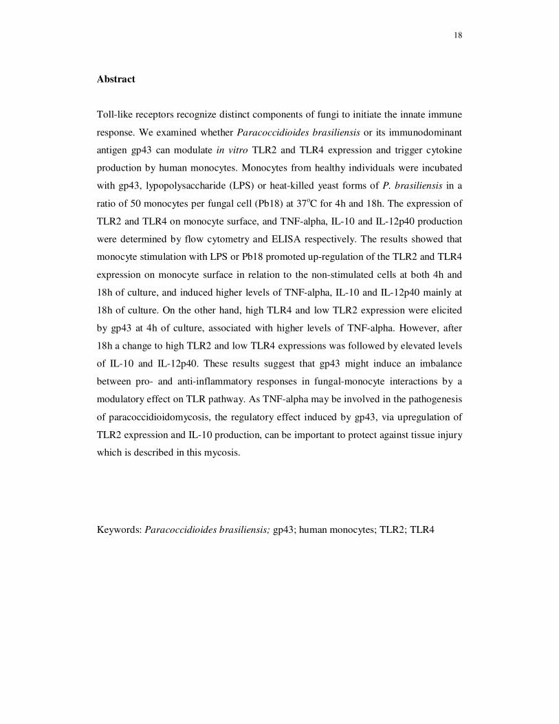
Abstract Toll-like receptors recognize distinct components of fungi to initiate the innate immune
response. We examined whether Paracoccidioides brasiliensis or its immunodominant
antigen gp43 can modulate in vitro TLR2 and TLR4 expression and trigger cytokine
production by human monocytes. Monocytes from healthy individuals were incubated
with gp43, lypopolysaccharide (LPS) or heat-killed yeast forms of P. brasiliensis in a
ratio of 50 monocytes per fungal cell (Pb18) at 37oC for 4h and 18h. The expression of
TLR2 and TLR4 on monocyte surface, and TNF-alpha, IL-10 and IL-12p40 production
were determined by flow cytometry and ELISA respectively. The results showed that
monocyte stimulation with LPS or Pb18 promoted up-regulation of the TLR2 and TLR4
expression on monocyte surface in relation to the non-stimulated cells at both 4h and
18h of culture, and induced higher levels of TNF-alpha, IL-10 and IL-12p40 mainly at
18h of culture. On the other hand, high TLR4 and low TLR2 expression were elicited
by gp43 at 4h of culture, associated with higher levels of TNF-alpha. However, after
18h a change to high TLR2 and low TLR4 expressions was followed by elevated levels
of IL-10 and IL-12p40. These results suggest that gp43 might induce an imbalance
between pro- and anti-inflammatory responses in fungal-monocyte interactions by a
modulatory effect on TLR pathway. As TNF-alpha may be involved in the pathogenesis
of paracoccidioidomycosis, the regulatory effect induced by gp43, via upregulation of
TLR2 expression and IL-10 production, can be important to protect against tissue injury
which is described in this mycosis.
Keywords: Paracoccidioides brasiliensis; gp43; human monocytes; TLR2; TLR4

19
Introduction
Paracoccidioidomycosis is the most prevalent systemic human mycosis in Latin
America, and is caused by the thermally dimorphic fungus Paracoccidioides
brasiliensis [1]. Infection can be acquired by inhalation of fungal mycelial propagules
which are transformed into infective yeast cells in the lung [2,3]. The disease presents as
an entity with a systemic course or as a chronic localized mycosis, depending on several
factors, including host immunocompetence, parasite strain and the environment [4].
Monocytes from patients with active forms of paracoccidioidomycosis are an
important source of pro-inflammatory cytokines such as TNF-α, IL-1, and IL-6 that
may be associated with pathogenesis of the mycosis [5]. In previous work we
demonstrated that in vitro infection of monocytes from healthy individuals with a
virulent strain of P. brasiliensis, induced higher levels of both pro- and anti-
inflammatory cytokines in comparison with the low virulence strain of the fungus [6].
Thus, the cytokine environment during the early fungus-monocyte interactions may be
important to fungal clearance or may promote their growth and implantation in host
tissues, leading to progressive disease.
Innate immunity, mainly involving monocytes and macrophages, is considered
an important defense mechanism against Gram-positive and Gram-negative bacteria,
viruses and pathogenic fungi. Macrophages and dendritic cells from this immune
system directly kill the microorganism through phagocytosis and inactivation of
invading organisms, cytokine production, and interactions with other adaptive immunity
cells such as T and B lymphocytes [7-9].
Macrophage activation is one of the first events in innate resistance to
intracellular fungal infection by recognition of microorganism surface components. The
specific detection of microorganisms by innate cells is mediated by a limited repertoire
20
of germ-line-encoded proteins, the pattern recognition receptors (PRRs), that recognize
microbial structures referred as pathogen-associated molecular patterns (PAMPs),
considered to be essencial for the pathogen survival [8-10]. PAMPs recognition by
PRRs allows self-nonself discrimination, because PAMPs are not produced by host cells
[11]. A variety of receptors that mediate PAMP recognition from phagocytic cells signal
to the host the presence of infection, include mannose receptor (MR), β-glucan receptor
(βGR), dectin-1, and Toll-like receptors (TLRs) [12-14]. TLRs not only mediate
recognition of microbial structures, such as those of fungi, but also trigger the release of
cytokines and the differentiation of immature to mature dendritic cells, enabling the
innate immune system to instruct the adaptative immune response [10,15,16].
The involvement of TLRs on fungi recognition and resistance of mammalian
hosts has been described for Candida albicans, Aspergillus fumigatus and Cryptococcus
neoformans [17-20], and more recently for P. brasiliensis. Bonfim et al. [21] evaluating
the expression of TLR1, TLR2, TLR4 and dectin-1 in monocytes and neutrophils from
healthy individuals after stimulation with high and low virulence yeast of P.
brasiliensis, suggested the participation of TLR2, TLR4 and dectin-1 in fungus
recognition, internalization and consequent activation of immune response against the
fungus. A study on the role of TLRs in a murine model of paracoccidioidomycosis
employing normal and TLR2- or TLR4--gene knockout mice (KO) in a C57BL/6
background suggested that P. brasiliensis yeasts use TLR2 and TLR4 to gain entry into
macrophages and infect mammalian hosts, and allowing the fungus own multiplication
[14]. Experimental model of chronic pulmonary infection with the fungus in TLR2-
deficient mice shows that TLR2 deficiency results in increased Th17 immunity
associated with diminished expansion of regulatory T cells and increased lung
pathology due to unrestrained inflammatory reactions [22].
21
It has been reported that a single fungal species can use different TLRs resulting
in a diverse biological activities. Recognition of C. albicans at the level of cell
membrane by TLR induces different types of cytokines. TLR4 induces mainly pro-
inflammatory signals in monocytes, macrophages and dendritic cells, while TLR2
stimulates the production of moderate amounts of pro-inflammatory cytokines, and
strong IL-10 and TGF-β responses [23]. Different research groups have demonstrated
divergent roles for TLR2 and TLR4, and their importance in the control of C. albicans
and C. neoformans infections is still unclear [12,20]. Thus, studies with purified
components of fungal cell walls may reveal the major PRR and signaling pathways used
by host cells to recognize fungal PAMPs [14].
The main antigenic component of P. brasiliensis is a 43-kDa secreted as high-
mannose glycoprotein (gp43) [24,25] employed for diagnosis and prognosis of
paracoccidiodomycosis [26,27]. Gp43 may participate in the installation mechanisms of
primary infection by inhibiting phagocytosis, nitric oxide (NO) and hydrogen peroxide
(H2O2) production, and fungal intracellular killing [28,29], thus playing different roles
in the pathogenesis of the mycosis [30].
The molecular mechanisms of innate recognition and the receptors involved in
paracoccidioidomycosis are poorly understood. In this study we analyzed whether P.
brasiliensis or its immunodominant antigen gp43 can modulate in vitro TLR2 and
TLR4 expression and trigger cytokine production by human monocytes.

22
Materials and Methods
Reagents and media
All reagents were obtained from Sigma-Aldrich, Inc., (St Louis, MO. USA), unless
stated otherwise. Phycoerythrin-Cy7 (PE/Cy7)-labeled mouse monoclonal antibodies
(MAbs) to human CD14 (M5E2), Phycoerythrin (PE)-labeled MAbs to human TLR4
(HTA125), Fluorescein (FITC)-labeled MAbs to human TLR2 (TLR2.1), and PE/Cy7-,
PE- and FITC-labeled control isotype MAbs were purchased from Biolegend (San
Diego, CA, USA). Quantikine ELISA kit and respective standards of TNF-alpha, IL-10
and IL-12p40 were acquired from R&D Systems (Minneapolis, MN, USA) and used
according to the manufacturer’s instructions.
Healthy individuals
Twenty healthy blood donors were recruited from the University Hospital, Botucatu
Medical School, São Paulo State University, Brazil, age range 20–50 years (mean age
32.5 ± 10.2 years). The study was approved by Botucatu Medical School Ethics
Committee, and informed consent was obtained from all the blood donors.
Fungus
Paracoccidioides brasiliensis strain 18 (Pb18) was maintained in yeast-like form cells
at 35 oC on 2% glucose, 1% peptone, 0.5% yeast extract, and 2% agar medium (GPY
medium) all from Gibco Laboratories, Grand Island, NY, USA) and used on the sixth
day of growth culture. Yeast cells were washed and suspended in 0.15 M phosphate-
buffered saline (PBS-pH 7.2). In order to obtain individual cells, the fungal suspension
was homogenized with glass beads in a Vortex homogenizer (three cycles of 10
seconds) [31]. Fungal suspensions were washed twice with PBS at 400 g for 10 min,
23
and submitted to autoclavation at 121 oC for 15 min to obtain heat killed yeast forms of
P. brasiliensis. The dead yeast cell concentration was adjusted to 1 x 10⁶ cells/mL in
RPMI 1640 medium.
Gp43 from Paracoccidioides brasiliensis
Gp43 was kindly provided by Professor Rosana Puccia of Federal University of Sao
Paulo (UNIFESP) Brazil. A soluble recombinant gp43 (gp43r) was expressed in the
yeast Pichia pastoris, and purified in affinity columns of Affi-Gel 10 bound to Mab17c,
a monoclonal antibody anti-gp43, according to Carvalho et al. [27].
Isolation of peripheral blood mononuclear cells
Peripheral blood mononuclear cells (PBMC) were isolated from heparinized (50U/mL
heparin) venous blood by Histopaque [density (d) = 1.077] (Sigma-Aldrich) density-
gradient centrifugation. Briefly, 5 mL of heparinized blood was mixed with an equal
volume of RPMI-1640 tissue culture medium (Sigma-Aldrich) containing 2 mM L-
glutamine, 10% heat-inactivated fetal calf serum, 20 mM HEPES, and 40 ug/mL
gentamicin (complete medium). Samples were layered over 5-mL Histopaque in a 15-
mL conical plastic centrifuge tube. After centrifuging at 400 g for 30 min at room
temperature, the interface layer of PBMC was carefully aspirated and washed twice
with PBS containing 0.05 mM ethylenediaminetetraacetic acid (PBS-EDTA) and once
with complete medium at 300 g for 10 min. Cell viability, as determined by 0.2%
Trypan Blue dye exclusion, was > 95% in all experiments. Monocytes were counted
using neutral red (0.02%), and were suspended at a concentration of 1 x 10⁶
monocytes/mL in complete medium.
24
Production of monocyte culture supernatants
The monocyte suspension (1 x 10⁶/mL) was distributed (1 mL/well) in 24-well flat-
bottomed plates (Nunc, Life Tech. Inc., Maryland, USA). After incubation for 2 hr at
37 oC in a humidified 5% CO2 atmosphere, non-adherent cells were removed by
aspiration and each well was rinsed twice with complete medium. Monocyte
preparations routinely contained > 90% monocytes as determined by morphologic
examination and staining for nonspecific esterase [32]. In the experiments for evaluating
cytokine production, monocytes were incubated with or without monoclonal antibodies
(MAbs) anti-TLR2 or anti-TLR4 (all from Biolegend) at 0.5 µg/mL for 60 min at 37º C
in 5% CO2. After incubation, the monocytes were washed and treated with complete
medium, in the presence or absence of gp43 (10ng/mL) or lipopolysaccharide (LPS) of
Escherichia coli O55B5 (Sigma-Aldrich) (10 ug/mL) or heat-killed yeast forms of Pb18
(1:50 fungus/monocytes ratio) for 4h and 18h at 37º C in 5% CO2. Culture supernatants
were harvested and stored at -80 oC until assayed.
Determination of cytokines
Cytokine concentrations were determined in cell-free supernatants obtained after 4h or
18h monocyte cultures with gp43, LPS or heat- killed yeast forms of Pb18 (1:50
fungus/monocytes ratio) by enzyme-linked immunosorbent assay (ELISA), as described
above, using Quantikine ELISA kits (R&D Systems) for TNF-α, IL-10, IL-12p40 and
IL-12p70 according to the manufacturer’s instructions. Assay sensitivity limit was 10
pg/mL for TNF-α, 7.5 pg/mL for IL-10 and 15 pg/mL for IL-12p40 and IL-12p70.
25
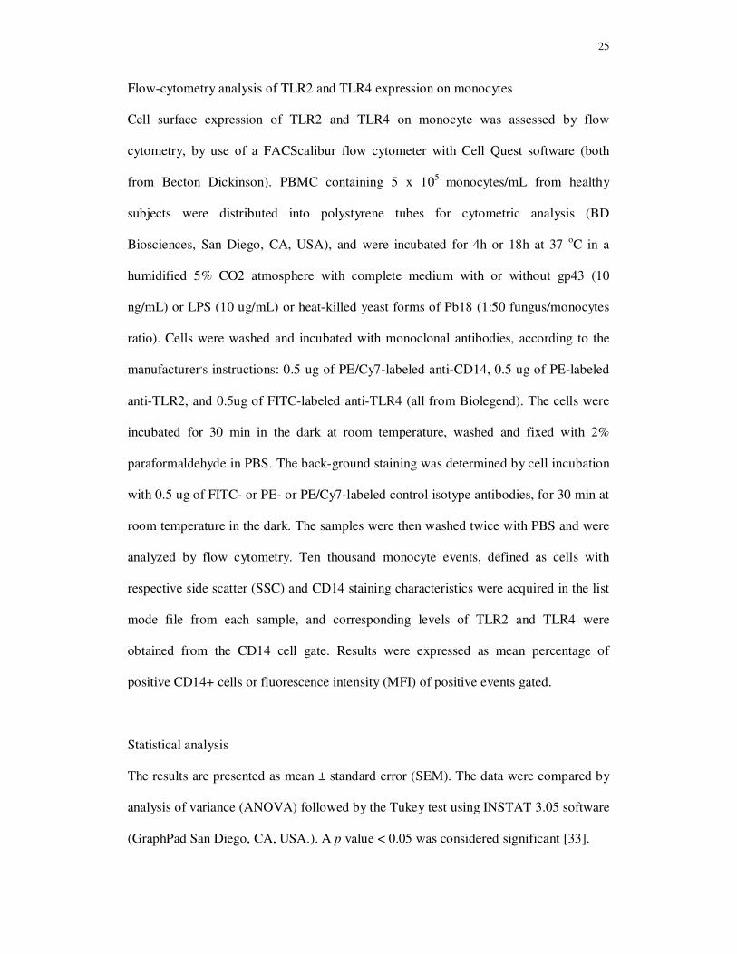
Flow-cytometry analysis of TLR2 and TLR4 expression on monocytes
Cell surface expression of TLR2 and TLR4 on monocyte was assessed by flow
cytometry, by use of a FACScalibur flow cytometer with Cell Quest software (both
from Becton Dickinson). PBMC containing 5 x 10⁵ monocytes/mL from healthy
subjects were distributed into polystyrene tubes for cytometric analysis (BD
Biosciences, San Diego, CA, USA), and were incubated for 4h or 18h at 37 oC in a
humidified 5% CO2 atmosphere with complete medium with or without gp43 (10
ng/mL) or LPS (10 ug/mL) or heat-killed yeast forms of Pb18 (1:50 fungus/monocytes
ratio). Cells were washed and incubated with monoclonal antibodies, according to the
manufacturer,s instructions: 0.5 ug of PE/Cy7-labeled anti-CD14, 0.5 ug of PE-labeled
anti-TLR2, and 0.5ug of FITC-labeled anti-TLR4 (all from Biolegend). The cells were
incubated for 30 min in the dark at room temperature, washed and fixed with 2%
paraformaldehyde in PBS. The back-ground staining was determined by cell incubation
with 0.5 ug of FITC- or PE- or PE/Cy7-labeled control isotype antibodies, for 30 min at
room temperature in the dark. The samples were then washed twice with PBS and were
analyzed by flow cytometry. Ten thousand monocyte events, defined as cells with
respective side scatter (SSC) and CD14 staining characteristics were acquired in the list
mode file from each sample, and corresponding levels of TLR2 and TLR4 were
obtained from the CD14 cell gate. Results were expressed as mean percentage of
positive CD14+ cells or fluorescence intensity (MFI) of positive events gated.
Statistical analysis
The results are presented as mean ± standard error (SEM). The data were compared by
analysis of variance (ANOVA) followed by the Tukey test using INSTAT 3.05 software
(GraphPad San Diego, CA, USA.). A p value < 0.05 was considered significant [33].
26
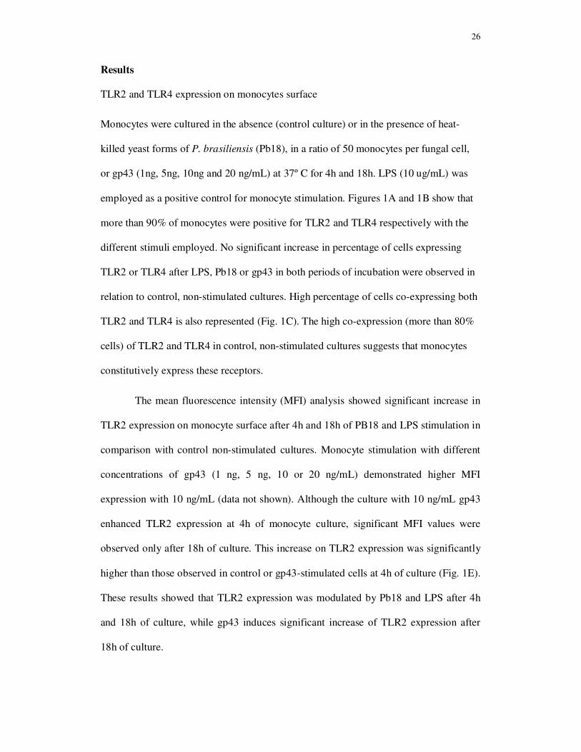
Results
TLR2 and TLR4 expression on monocytes surface
Monocytes were cultured in the absence (control culture) or in the presence of heat-
killed yeast forms of P. brasiliensis (Pb18), in a ratio of 50 monocytes per fungal cell,
or gp43 (1ng, 5ng, 10ng and 20 ng/mL) at 37º C for 4h and 18h. LPS (10 ug/mL) was
employed as a positive control for monocyte stimulation. Figures 1A and 1B show that
more than 90% of monocytes were positive for TLR2 and TLR4 respectively with the
different stimuli employed. No significant increase in percentage of cells expressing
TLR2 or TLR4 after LPS, Pb18 or gp43 in both periods of incubation were observed in
relation to control, non-stimulated cultures. High percentage of cells co-expressing both
TLR2 and TLR4 is also represented (Fig. 1C). The high co-expression (more than 80%
cells) of TLR2 and TLR4 in control, non-stimulated cultures suggests that monocytes
constitutively express these receptors.
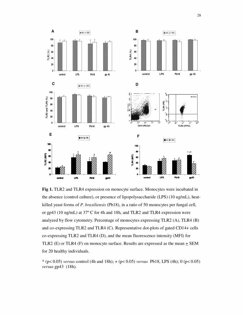
The mean fluorescence intensity (MFI) analysis showed significant increase in
TLR2 expression on monocyte surface after 4h and 18h of PB18 and LPS stimulation in
comparison with control non-stimulated cultures. Monocyte stimulation with different
concentrations of gp43 (1 ng, 5 ng, 10 or 20 ng/mL) demonstrated higher MFI
expression with 10 ng/mL (data not shown). Although the culture with 10 ng/mL gp43
enhanced TLR2 expression at 4h of monocyte culture, significant MFI values were
observed only after 18h of culture. This increase on TLR2 expression was significantly
higher than those observed in control or gp43-stimulated cells at 4h of culture (Fig. 1E).
These results showed that TLR2 expression was modulated by Pb18 and LPS after 4h
and 18h of culture, while gp43 induces significant increase of TLR2 expression after
18h of culture.

27
The MFI analysis of TLR4 expression (Fig 1F) after 4h of culture showed that
this receptor expression on monocyte surface is higher after the three stimuli employed
than in control cultures. No significant difference between 4h and 18h of TLR4
expression were detected in monocyte non-stimulated cultures or cultures stimulated
with Pb18 or LPS. On the other hand, TLR4 expression induced by gp43 at 4h of
culture was significant higher than in control, LPS and Pb18-stimulated cultures.
However, TLR4 expression after 18h of monocyte cultures stimulated with gp43 was
significantly lower than at 4h cultures and did not show statistical difference in
comparison with control cultures. These results showed that monocyte cultured with
gp43 exhibit higher TLR4 expression at 4h and higher TLR2 expression after 18h of
stimulation.
28
Fig 1. TLR2 and TLR4 expression on monocyte surface. Monocytes were incubated in
the absence (control culture), or presence of lipopolysaccharide (LPS) (10 ug/mL), heat-
killed yeast forms of P. brasiliensis (Pb18), in a ratio of 50 monocytes per fungal cell,
or gp43 (10 ng/mL) at 37º C for 4h and 18h, and TLR2 and TLR4 expression were
analyzed by flow cytometry. Percentage of monocytes expressing TLR2 (A), TLR4 (B)
and co-expressing TLR2 and TLR4 (C). Representative dot-plots of gated CD14+ cells
co-expressing TLR2 and TLR4 (D), and the mean fluorescence intensity (MFI) for
TLR2 (E) or TLR4 (F) on monocyte surface. Results are expressed as the mean + SEM
for 20 healthy individuals.
* (p< 0.05) versus control (4h and 18h); + (p< 0.05) versus Pb18, LPS (4h); 0 (p< 0.05) versus gp43 (18h).
0
20
40
60
80
100
control LPS Pb18 gp 43
TL
R2
an
d T
LR
4 (
%)
4h 18hCC
0
20
40
60
80
100
control LPS Pb18 gp 43
TL
R2
(%
)
4h 18h
0
20
40
60
80
100
control LPS Pb18 gp 43
TL
R4
(%
)
4h 18h
0
20
40
60
80
100
control LPS Pb18 gp43
TL
R2
(M
FI)
4h 18h
**
*
*+
0
20
40
60
80
100
control LPS Pb18 gp43
TL
R2
(M
FI)
4h 18h
**
*
*+
A B
**
* *
* + 0
0
20
40
60
80
100
control LPS Pb18 gp43
TL
R4
(M
FI)
4h 18h
**
* *
* + 0
0
20
40
60
80
100
control LPS Pb18 gp43
TL
R4
(M
FI)
4h 18h
ED
FE
DC
R5
CD14 (PE/Cy7)
R5
CD14 (PE/Cy7) TLR2 (FITC)
TL
R4 (
PE
)
98,52%
TLR2 (FITC)
TL
R4 (
PE
)
98,52%
29
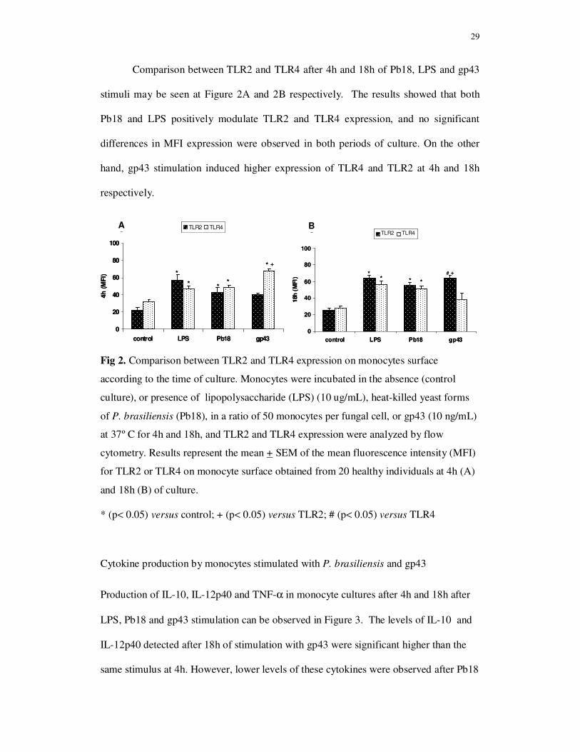
Comparison between TLR2 and TLR4 after 4h and 18h of Pb18, LPS and gp43
stimuli may be seen at Figure 2A and 2B respectively. The results showed that both
Pb18 and LPS positively modulate TLR2 and TLR4 expression, and no significant
differences in MFI expression were observed in both periods of culture. On the other
hand, gp43 stimulation induced higher expression of TLR4 and TLR2 at 4h and 18h
respectively.
Fig 2. Comparison between TLR2 and TLR4 expression on monocytes surface
according to the time of culture. Monocytes were incubated in the absence (control
culture), or presence of lipopolysaccharide (LPS) (10 ug/mL), heat-killed yeast forms
of P. brasiliensis (Pb18), in a ratio of 50 monocytes per fungal cell, or gp43 (10 ng/mL)
at 37º C for 4h and 18h, and TLR2 and TLR4 expression were analyzed by flow
cytometry. Results represent the mean + SEM of the mean fluorescence intensity (MFI)
for TLR2 or TLR4 on monocyte surface obtained from 20 healthy individuals at 4h (A)
and 18h (B) of culture.
* (p< 0.05) versus control; + (p< 0.05) versus TLR2; # (p< 0.05) versus TLR4
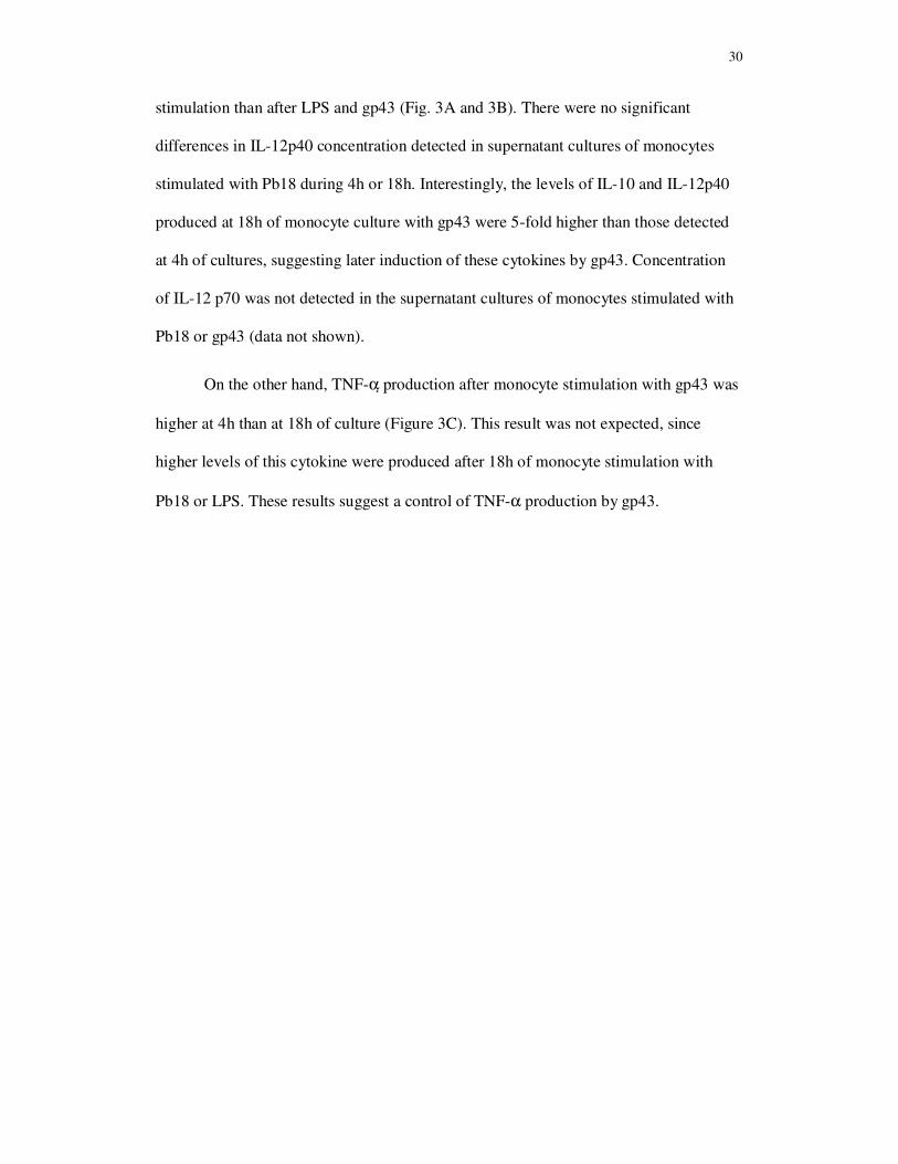
Cytokine production by monocytes stimulated with P. brasiliensis and gp43
Production of IL-10, IL-12p40 and TNF-α in monocyte cultures after 4h and 18h after
LPS, Pb18 and gp43 stimulation can be observed in Figure 3. The levels of IL-10 and
IL-12p40 detected after 18h of stimulation with gp43 were significant higher than the
same stimulus at 4h. However, lower levels of these cytokines were observed after Pb18
0
20
40
60
80
100
control LPS Pb18 gp43
4h
(M
FI)
TLR2 TLR4
*
* **
* +
0
20
40
60
80
100
control LPS Pb18 gp43
4h
(M
FI)
TLR2 TLR4
*
* **
* +
0
20
40
60
80
100
control LPS Pb18 gp43
18h
(M
FI)
TLR2 TLR4
** * *
+#
0
20
40
60
80
100
control LPS Pb18 gp43
18h
(M
FI)
TLR2 TLR4
** * *
+#
AC
BC
30
stimulation than after LPS and gp43 (Fig. 3A and 3B). There were no significant
differences in IL-12p40 concentration detected in supernatant cultures of monocytes
stimulated with Pb18 during 4h or 18h. Interestingly, the levels of IL-10 and IL-12p40
produced at 18h of monocyte culture with gp43 were 5-fold higher than those detected
at 4h of cultures, suggesting later induction of these cytokines by gp43. Concentration
of IL-12 p70 was not detected in the supernatant cultures of monocytes stimulated with
Pb18 or gp43 (data not shown).
On the other hand, TNF-α production after monocyte stimulation with gp43 was
higher at 4h than at 18h of culture (Figure 3C). This result was not expected, since
higher levels of this cytokine were produced after 18h of monocyte stimulation with
Pb18 or LPS. These results suggest a control of TNF-α production by gp43.
31
Fig 3. Cytokine production by human monocytes stimulated without (control culture),
or with lipopolysaccharide (LPS) (10 ug/mL), with heat-killed yeast forms of P.
brasiliensis (Pb18), in a ratio of 50 monocytes per fungal cell, or gp43 (10 ng/mL) at
37º C for 4h and 18h. Results represent the mean + SEM of the cytokine levels
produced by monocytes obtained from 20 healthy individuals.
* (p< 0.05) versus control; + (p < 0.05) versus 4h; 0 (p< 0.05) versus Pb18; # (p< 0.05) versus 18h
0
200
400
600
800
1000
1200
control LPS Pb18 gp 43
TN
F-a
lph
a �� ��(p
g/m
L)
4h 18h
**
*0+
*
+#
0
+
0
200
400
600
800
1000
1200
control LPS Pb18 gp 43
TN
F-a
lph
a �� ��(p
g/m
L)
4h 18h
**
*0+
*
+#
0
+
0
200
400
600
800
control LPS Pb18 gp 43
IL-1
0 (
pg
/mL
)
4h 18h
*
*
* +
+0
0
0
0
200
400
600
800
control LPS Pb18 gp 43
IL-1
0 (
pg
/mL
)
4h 18h
*
*
* +
+0
0
0
0
20
40
60
80
100
120
140
control LPS Pb18 gp 43
IL-1
2 p
40 (
pg
/mL
)
4h 18h
* *
*
* *
+0 0+
0
20
40
60
80
100
120
140
control LPS Pb18 gp 43
IL-1
2 p
40 (
pg
/mL
)
4h 18h
* *
*
* *
+0 0+
AC
BC
CC
32
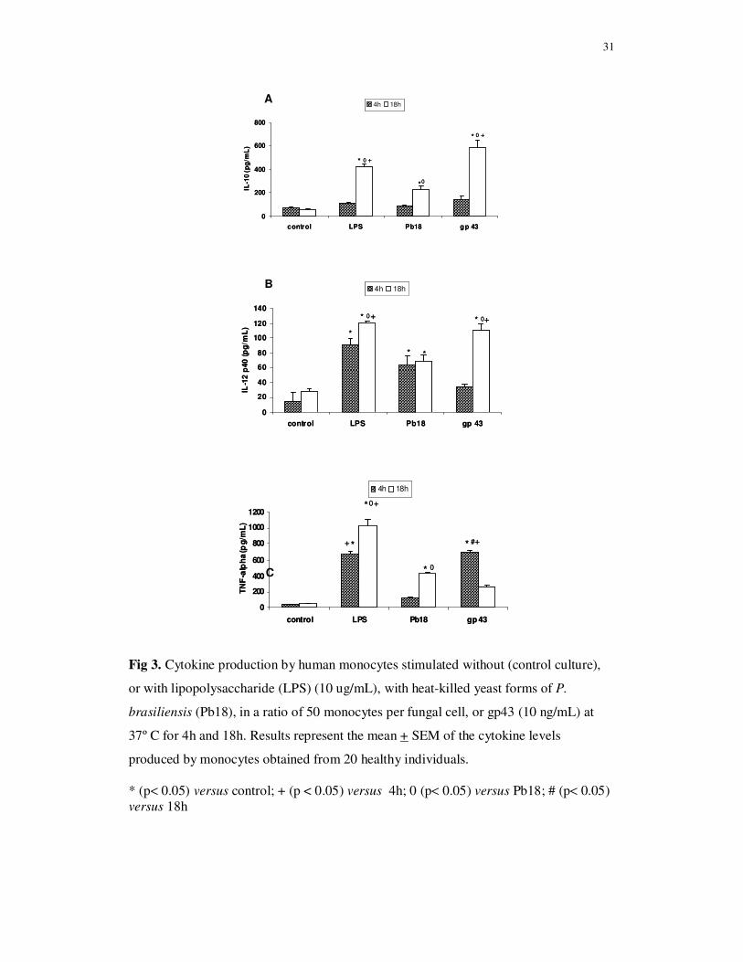
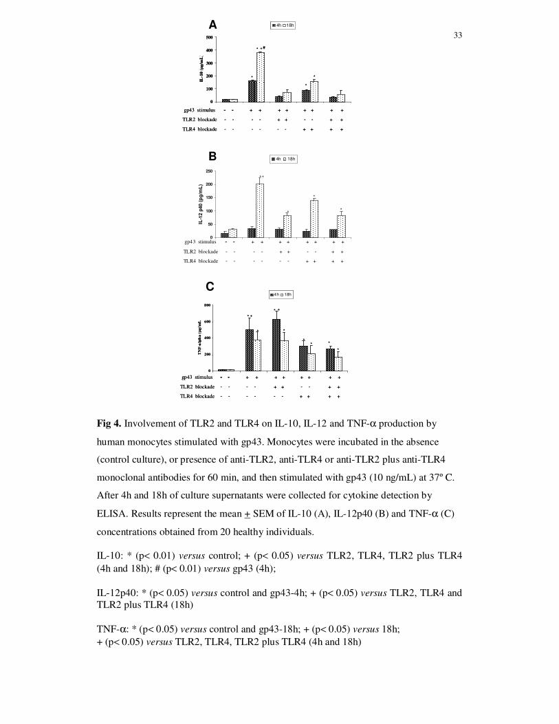
Involvement of TLR2 and TLR4 on cytokine production
The role of TLR2 and TLR4 on IL-10, IL-12p40 and TNF-α production by human
monocytes stimulated with gp43 (10 ug/mL) was evaluated by blockade of these
receptors with specific MAbs, before cell culture with the antigen for 4h and 18h. The
blockade of TLR2 or TLR4 individually, as well as both receptors before gp43
stimulation led to lower levels of IL-10 and IL-12p40 in comparison with monocytes
cultures not submitted to TLR2 and TLR4 blockade. These effects were more evident
after 18h of monocyte stimulation with gp43 (Fig. 4A and 4B). Although there is a
tendency to lower levels of IL-10 and IL-12p40 after TLR2 blockage in relation to
TLR4 blockage, no significant differences were observed when the cytokine levels were
compared. These results suggest that IL-10 and IL-12p40 production may be dependent
on gp43 interaction with TLR2 and TLR4.
The concentration of TNF-α in supernatant culture of monocytes submitted or
not to TLR2, TLR4 or TLR2 plus TLR4 blockade with MAbs, and stimulated with gp43
(10 ng/mL) for 4h was significantly higher than the observed at 18h of culture. The
blockade of TLR4 before gp43 stimulation led to lower levels of TNF-α produced by
monocytes both at 4h and 18h of culture than in cultures with TLR2 blockade or
cultures not submitted to TLR blockade. On the other hand, TLR2 blockade did not
show inhibitory effect on TNF-a production. The concentration of this cytokine was
similar to the values obtained in non-blockade monocyte cultures stimulated with gp43.
The lower levels of TNF-α.detected in cultures with TLR4 blockade before gp43
stimulation suggest that TLR4 play a role in the interaction with gp43 for TNF-α
production (Fig. 4C).
33
Fig 4. Involvement of TLR2 and TLR4 on IL-10, IL-12 and TNF-α production by
human monocytes stimulated with gp43. Monocytes were incubated in the absence
(control culture), or presence of anti-TLR2, anti-TLR4 or anti-TLR2 plus anti-TLR4
monoclonal antibodies for 60 min, and then stimulated with gp43 (10 ng/mL) at 37º C.
After 4h and 18h of culture supernatants were collected for cytokine detection by
ELISA. Results represent the mean + SEM of IL-10 (A), IL-12p40 (B) and TNF-α (C)
concentrations obtained from 20 healthy individuals.
IL-10: * (p< 0.01) versus control; + (p< 0.05) versus TLR2, TLR4, TLR2 plus TLR4 (4h and 18h); # (p< 0.01) versus gp43 (4h); IL-12p40: * (p< 0.05) versus control and gp43-4h; + (p< 0.05) versus TLR2, TLR4 and TLR2 plus TLR4 (18h) TNF-α: * (p< 0.05) versus control and gp43-18h; + (p< 0.05) versus 18h; + (p< 0.05) versus TLR2, TLR4, TLR2 plus TLR4 (4h and 18h)
0
100
200
300
400
500
IL-1
0 (p
g/m
L)
4h 18h
+* #
* *
*
gp43 stimulus - - + + + + + + + +
TLR2 blockade - - - - + + - - + +
TLR4 blockade - - - - - - + + + +
0
100
200
300
400
500
IL-1
0 (p
g/m
L)
4h 18h
+* #
* *
*
gp43 stimulus - - + + + + + + + +
TLR2 blockade - - - - + + - - + +
TLR4 blockade - - - - - - + + + +
0
50
100
150
200
250
IL-1
2 p
40 (
pg
/mL
)
4h 18h
*+
*
*
*
gp43 stimulus - - + + + + + + + +
TLR2 blockade - - - - + + - - + +
TLR4 blockade - - - - - - + + + +
A
B
C
0
200
400
600
800
TN
F-a
lph
a (
pg
/mL
)
4h 18h
*
*
*
*
*
*
+
+
*
gp43 stimulus - - + + + + + + + +
TLR2 blockade - - - - + + - - + +
TLR4 blockade - - - - - - + + + +
*
0
200
400
600
800
TN
F-a
lph
a (
pg
/mL
)
4h 18h
*
*
*
*
*
*
+
+
*
gp43 stimulus - - + + + + + + + +
TLR2 blockade - - - - + + - - + +
TLR4 blockade - - - - - - + + + +
*
34
Discussion
The central role of TLRs in innate immune recognition of fungal pathogens has been
proposed [12,19], but so far, only one study evaluated expression of these receptors in
monocytes and neutrophils stimulated by yeast cells of P. brasiliensis [21] The results
of the present study demonstrated that P. brasiliensis and its main antigen gp43 promote
monocyte activation, by cytokine production and exerting a modulatory effect on TLR
expression by these cells. Gp43 is the best-studied P. brasiliensis component employed
for diagnosis and prognosis of paracoccidioidomycosis described in the literature
[24,26,27]. However so far, studies on modulatory effect of gp43 over TLR expression
by monocytes have not yet been performed.
First, we observed that monocytes from healthy individuals constitutively
express TLR2 and TLR4, since no significant increase in percentage of cells expressing
TLR2 or TLR4 were detected 4h and 18h after LPS, Pb18 or gp43 stimulation in
relation to control, non-stimulated cultures. High percentage of monocytes (80 – 90%)
co-expressed both TLR2 and TLR4. According to Netea et al. [23] monocytes and
neutrophils are the major cells of the innate immune response that recognize invader
pathogens in blood, and express high levels of TLR on their cell membranes. During
differentiation into macrophages, monocytes retain expression of TLR and increase
their expression of lectin receptors. This information might explain the high percentage
of monocytes expressing TLR2 and TLR4 detected in control, non-stimulated cells
observed in the present study.
However, the mean fluorescence intensity (MFI) analysis showed significant
increase in TLR2 expression on monocyte surface after 4h and 18h of Pb18 and LPS
stimulation, while TLR4 expression was higher after 4h of culture with the three stimuli
employed than in control, non-stimulated cultures. The comparison between the MFI
35
values and percentage of monocytes expressing TLR2 and TLR4 after stimulation with
Pb18, LPS and gp43 showed that the MFI parameter allowed the better discrimination
of the modulatory effect induced by the three stimuli employed. Thus, we utilized the
MFI parameter to analyze the modulatory effect of P.brasiliensis and its antigen gp43
on monocyte TLR expression.
Whole yeast cells of Pb18 induced high expression of TLR2 and TLR4 on
monocyte surface both at 4h and 18h of culture, whereas the stimulus with gp43
significantly enhanced TLR2 expression after 18h of culture. On the other hand, TLR4
expression induced by gp43 in 4h was significant higher than LPS and Pb18 stimuli in
this period, suggesting a greater ability to modulate earlier TLR4 expression. After 18h
of monocyte stimulation with gp43, TLR4 expression was significantly lower than at 4h
cultures. These results showed that gp43 upregulates TLR4 expression at 4h, and TLR2
after 18h of stimulation, whereas these differences are not so evident with monocyte
stimulation with Pb18. Our results showing that Pb18 and gp43 positively modulate
TLR2 and TLR4 expression differ from those recently reported by Bonfim et al. [21]
demonstrating a decrease of TLR1, TLR2, TLR4 and dectin-1 expression on
monocytes, as soon as 30 to 60 min after P. brasiliensis yeast cells stimulation, and
suggesting the participation of these receptors in P. brasiliensis recognition,
internalization and consequent activation of the immune response against the fungus.
Since the authors evaluated the expression of these receptors after few minutes of
fungus stimulation, our results might be explained by the longer period (4h and 18h) of
monocyte stimulation with Pb18 and gp43. It is possible that after internalization TLRs
receptors can be re-synthesized and appear at monocyte membrane, explaining the
higher MFI of TLR2 and TLR4 observed after 4h and 18h of stimulation with the
fungus or gp43.
36
Results concerning TLR expression and cytokine production by monocytes
stimulated by Pb18 were not always in accordance with those obtained after cell
stimulation with gp43, in relation to the period of 4h or 18h of culture. Thus, Pb18
stimulated the expression of TLR2 and TLR4 in both periods of culture, and induced
higher levels of TNF-α and IL-10 only after 18h of culture. On the other hand, gp43
preferentially upregulates TLR4 and TNF-α production at 4h, with moderate levels of
IL-10, whereas high TLR2 expression and higher levels of IL-10 and IL-12p40 were
detected at 18h of culture. Therefore, these results were confirmed by experiments of
TLR4 or TLR2 blockade with specific MAbs, and suggest that gp43 stimulates earlier
higher levels of TNF-α, and elevated production of IL-10 latter. The persistence of gp43
in monocyte cultures, for 18h, may induce downregulation of the inflammatory
response, by high production of IL-10 in association with high TLR2 and low TLR4
expression, and followed by low TNF-α production in this period. This antigen
maintenance for a long period of monocyte culture might cause chronic stimulation of
TLR2 with higher IL-10 production which could autocrinally inhibit TNF-α production
by monocytes.
The discrepancy among the results employing Pb18 and gp43 might be
explained by the presence of different antigenic components in the cell wall of the yeast
form of Pb18, capable to interact with different TLR or non-TLR receptors, present in
the cell surface of monocytes, in comparison with gp43. It is possible that the purified
antigen of P. brasiliensis might be better available to interact and stimulate monocyte
cultures than whole fungal cells. The gp43 employed in the present study was obtained
as a soluble recombinant gp43 (gp43r) isoform, a N-mannosylated protein expressed in
the yeast Pichia pastoris and purified in affinity columns containing anti-gp43
antibodies by Carvalho et al. [27]. According to the authors gp43r may replace the
37
native gp43 (gp43n) in assays for diagnosis of paracoccidioidomycosis, because gp43n
expression in supernatant fluid of P. brasiliensis cultures can be unstable, and varies
with the isolate. The advantage of gp43r from P. pastoris relies mainly in
reproducibility for the production of large amounts of a known sequence of gp43, which
is expressed in culture supernatants under inducible conditions in non-pathogenic, fast-
growing yeast. Some authors suggest that the employement of purified cell wall
components of pathogenic fungi could point out the main PRR receptor and the
signaling pathway of the host cell to recognize PAMPs associated with fungi [14,23].
In the present study, monocyte stimulation with gp43 after TLR4 blockade
employing MAbs induced lower levels of TNF-α, while no inhibitory effect on this
cytokine production was observed after TLR2 blockade. Thus, TLR4 may play a role in
gp43 recognition for TNF-α production, as observed in other studies showing
association between TNF-α production and TLR4 recognition of fungal components by
human monocytes [34,35]. On the other hand, TLR2 and TLR4 blockade before
stimulation with gp43 led to low production of IL-10 and IL-12p40, suggesting that
these cytokines release may be dependent on gp43 interaction with TLR2 and TLR4.
Association between TLR2 expression and IL-10 production against pathogenic fungi,
such as A. fumigatus and C. albicans has been described in the literature [36,37]. The
deleterious effect of TLR2 signaling during C. albicans infection is associated with
increased production of IL-10 and development of T regulatory (CD4+CD25+) cells,
resulting in deficient cellular immune response and lower ability to the fungus
elimination [37]. The same association was observed in other infection models with
Schistosoma mansoni [38] and Borrelia burgdorferi [39], suggesting an important role
of TLR2 in inducing the expansion and control of regulatory T cells [40]. In mice
infected with P. brasiliensis, pulmonary dendritic cells showed increased TLR2 gene
38
expression, associated with higher production of IL-10, and contributing to higher
susceptibility to infection [41]. High concentrations of IL-10 were detected in serum,
and as IL-10 mRNA expression and protein release in peripheral blood cell culture
supernatants of patients with paracoccidioidomycosis [42-45]. Monocytes from patients
spontaneously release high levels of IL-10 [5] which decrease after antifungal treatment
and clinical cure [46]. These results corroborates the participation of T regulatory cells
in paracoccidioidomycosis, since it was reported elevated number of these cells in
patients with active disease both in peripheral blood and lesions [47]. However,
experimental model of paracoccidioidomycosis demonstrated that TLR2 deficiency
results in increased Th17 immunity associated with diminished expansion of
CD4+CD25+Foxp3+ T cells and increased lung pathology due to unrestrained
inflammatory response [22]. Therefore, activation of TLR2 and IL-10 production may
have a protective role against the pathogenesis of the mycosis.
The similar profile of IL-10 and IL-12p40 production by monocytes submitted
to TLR2 blockade and gp43 stimulation suggest that P. brasiliensis antigen may interact
with this receptor, leading to both cytokine production. The high production of IL-10
after 18h of monocytes stimulation with gp43 suggest its role in downregulation of
inflammatory response against P. brasiliensis. It has been suggested that IL-12p40
subunit can play anti-inflammatory role by controlling IL-12p70 production, which is
involved in excessive inflammation observed in severe sepses [48]. Release of high
concentration of IL-12p40 has been also reported in mononuclear phagocytes
stimulation with intracellular microorganisms such as Leishmania, whereas low levels
of IL-12 p70 were produced [49]. Thus, in the present study, high levels of IL-10 and
IL-12p40 produced after gp43 monocyte stimulation might be involved in regulation of
inflammatory cytokines produced during excessive monocyte activation. Corroborating
39
this hypothesis, our results showed that monocytes stimulated with Pb18 and gp43
produced higher levels of IL-12p40, whereas production of IL-12 p70 was not detected
in these cultures (data not shown).
Although our data suggest the involvement of TLR2 and TLR4 in gp43
recognition, leading to preferentially IL-10 and TNF-α production respectively, we
cannot discard the possibility that other TLR and non-TLRs may participate in these
cytokine release. Gp43 has been reported to interact with mannose receptor and play a
role in the installation mechanisms of primary infection by inhibiting both phagocytosis
and fungal intracellular killing [28,29]. Recent study demonstrated that peptides P4 and
P23 from gp43 inhibit macrophage function and show non-specific and specific anti-
inflammatory properties [30].
In summary, our results showed that gp43 considered the immunodominant
antigen of P. brasiliensis modulates TLR2 and TLR4 expression during human
monocytes interaction. Association among high TLR4 expression and elevated
production of TNF-α and moderate levels of IL-10 at 4h, and between high TLR2
expression and high production of IL-10 and IL-12p40 at 18h of culture suggest that
this component of the fungus might induce an imbalance between pro-inflammatory and
anti-inflammatory response in host/fungal interactions. Considering that high
production of TNF-α has been described as involved in pathogenesis of
paracoccidioidomycosis [5] it is possible that gp43, by trigger sustained production of
IL-10, might control excessive inflammatory response induced by pro-inflammatory
cytokines which result in tissue injury observed in this mycosis. The TLR blockade
experiments suggest the interaction of gp43 with TLR2 and TLR4 signaling the
cytokine profile obtained. However, experiments showing TLRs blockade and gp43
40
uptake are in progress in our laboratory and will be important to confirm this
hypothesis.
Acknowledgements
This wortk was supported by Fundação de Amparo à Pesquisa do Estado de São Paulo
(FAPESP – grants no 2006/53366-9), Brazil.
Conflict of interest
There is no conflict of interest.
References
1. Franco M, Mendes RP, Moscardi-Bacchi M, Rezkallah-Iwasso MT, Montenegro
MR (1989) Paracoccidioidomycosis. Bailliere’s Clin Trop Med Commun Dis 4:185–
220.
2. McEwen JG, Bedoya V, Patiño MM, Salazar ME, Restrepo A (1987).Experimental
murine paracoccidiodomycosis induced by the inhalation of conidia. J Med Vet Mycol
25:165-175.
3. Restrepo A, Tobón AM (2005) Paracoccidioides brasiliensis. in: Mandell GE,
Bennettt JE, Dollin R (ed) Principles and practice of infectious diseases. 6th edn.
Elsevier, Philadelphia, pp 3062-3068.
4. Franco M, Peraçoli MTS, Soares AMVC, Montenegro MR, Mendes R P, Meira DA
(1993) Host-parasite relationship in paracoccidioidomycosis. Curr Trop Med Mycol
5:115-149.
5. Peraçoli MT, Kurokawa CS, Calvi SA, Mendes RP, Pereira PC, Marques SA, Soares
AM (2003) Production of pro- and anti-inflammatory cytokines by monocytes from
patients with paracoccidioidomycosis. Microbes Infect 5:413-418.
6. Kurokawa CS, Araujo JP Jr, Soares AM, Sugizaki MF, Peraçoli MT (2007) Pro- and
anti-inflammatory cytokines produced by human monocytes challenged in vitro with
Paracoccidioides brasiliensis. Microbiol Immunol 51:421-428.
7. Gordon S (1998) The role of the macrophage in the immune regulation. Res
41
Immunol 149:685-688.
8. Akira S, Uematsu S, Takeuchi O (2006) Pathogen recognition and innate immunity.
Cell 124:783-801.
9. Medzhitov R (2007) Recognition of microorganisms and activation of the immune
response. Nature 449:819-826.
10. Beutler BA (2004) Inferences, questions and possibilities in Toll-like receptor
signaling. Nature 430:257-263.
11. Janeway Jr CA, Medzhitov R (2002) Innate immune recognition. Annu Rev
Immunol 20:197-216.
12. Netea MG, Van der Meer JW, Kullberg BJ (2006) Role of the dual interaction of
fungal pathogens with pattern recognition receptors in the activation and modulation of
host defence. Clin Microbiol Infect 12:404-409.
13. Dennehy KM, Brown GD (2007) The role of the beta-glucan receptor Dectin-1 in
control of fungal infection. J Leukoc Biol 82:253-258.
14. Calich VL, Pina A, Felonato M, Bernardino S, Costa TA, Loures FV (2008) Toll-
like receptors and fungal infections: the role of TLR2, TLR4 and MyD88 in
paracoccidioidomycosis. FEMS Immunol Med Microbiol 53:1-7.
15. Hertz CJ, Kiertscher SM, Godowski PJ, Bouis DA, Norgard MV, Roth MD,
Modlin RL (2001) Microbial lipopeptides stimulate dendritic cell maturation via Toll-
like receptor 2. J Immunol.166:2444-2450.
16. Van de Veerdonk FL, Kullberg BJ, van der Meer JW, Gow NA, Netea MG (2008)
Host-microbe interactions: innate pattern recognition of fungal pathogens. Curr Opin
Microbiol 11:305-312.
17. Netea MG, Van Der Graaf CA, Vonk AG, Verschueren I, Van Der Meer JW,
Kullberg BJ (2002) The role of toll-like receptor (TLR) 2 and TLR4 in the host defense
against disseminated candidiasis. J Infect Dis 185:1483-1489.
18. Braedel S, Radsak M, Einsele H, Latgé JP, Michan A, Loeffler J, Haddad Z,
Grigoleit U, Schild H, Hebart H (2004) Aspergillus fumigatus antigens activate innate
immune cells via toll-like receptors 2 and 4. Br J Haematol 125:392-399.
19. Romani L (2004) Immunity to fungal infections. Nat Rev Immunol 4:1-13.
20. Nakamura K, Miyagi K, Koguchi Y, Kinjo Y, Uezu K, Kinjo T, Akamine M, Fujita
J, Kawamura I, Mitsuyama M, Adachi Y, Ohno N, Takeda K, Akira S, Miyazato A,
Kaku M, Kawakami K (2006) Limited contribution of Toll-like receptor 2 and 4 to the
42
host response to a fungal infectious pathogen, Cryptococcus neoformans. FEMS
Immunol Med Microbiol 47:148-154.
21. Bonfim CV, Mamoni RL, Blotta MH (2009) TLR-2, TLR-4 and dectin-1
expression in human monocytes and neutrophils stimulated by Paracoccidioides
brasiliensis. Med Mycol 47:722-733.
22. Loures FV, Pina A, Felonato M, Calich VL (2009) TLR2 is a negative regulator of
Th17 cells and tissue pathology in a pulmonary model of fungal infection. J Immunol
183:1279-1290.
23. Netea MG, Brown GD, Kullberg BJ, Gow NA (2008) An integrated model of the
recognition of Candida albicans by the innate immune system. Nat Rev Microbiol
6:67-78.
24. Puccia R, Schenkman S, Gorin PA, Travassos LR (1986) Exocellular components
of Paracoccidioides brasiliensis: identification of a specific antigen. Infect Immun
53:199-206.
25. Travassos LR, Taborda CP, Iwai LK, Cunha-Neto E, Ruccia R (2004) The gp43
from Paracoccidioides brasiliensis: a major diagnostic antigen and vaccine candidate.
In: J.E. Domer JE, Kobayashi GS (ed), Mycota XII, human fungal pathogens. Springer-
Verlag, Berlin. Germany, pp 279-296.
26. Mendes-Giannini MJS, Bueno JP, Shikanai-Yasuda MA (1990) Antibody response
to 43 kDa glycoprotein of Paracoccidioides brasiliensis as a marker for the evaluation
of patients under treatment. Am J Trop Med Hyg 43:200-206.
27. Carvalho KC, Vallejo MC, Camargo ZP, Puccia R (2008) Use of recombinant gp43
isoforms expressed in Pichia pastoris for diagnosis of paracoccidioidomycosis. Clin
Vaccine Immunol 15:622-629.
28. Almeida SR, Unterkircher CS, Camargo ZP (1998) Involvement of the major
glycoprotein (gp43) of Paracoccidioides brasiliensis in attachment to macrophages. Med Mycol 36:405-411.
29. Popi AF, Lopes JD, Mariano M (2002) GP43 from Paracoccidioides brasiliensis
inhibits macrophage functions. An evasion mechanism of the fungus. Cell Immunol
218:87-94.
30. Konno AY, Maricato JT, Konno FT, Mariano M, Lopes JD (2009) Peptides from
Paracoccidioides brasiliensis GP43 inhibit macrophage functions and inflammatory
response. Microbes Infect 11:92-99.
43
31. Peraçoli MT, Sugizaki MF, Mendes RP, Naiff R, Montenegro MR (1999)
Paracoccidioides brasiliensis isolated from armadillos is virulent to Syrian hamsters.
Mycopathologia 148:123-130.
32. Li CY, Lam KW, Yam LT. (1973) Esterases in human leukocytes. J Histochem
Cytochem 21:1-12.
33. Godfrey K (1985) Statistics in practice. Comparing the means of several groups. N
Engl J Med 313:1450-1456.
34. Wang JE, Warris A, Ellingsen EA, Jørgensen PF, Flo TH, Espevik T, Solberg R,
Verweij PE, Aasen AO (2001) Involvement of CD14 and toll-like receptors in
activation of human monocytes by Aspergillus fumigatus hyphae. Infect Immun
69:2402-2406.
35. Tada H, Nemoto E, Shimauchi H, Watanabe T, Mikami T, Matsumoto T, Ohno N,
Tamura H, Shibata K, Akashi S, Miyake K, Sugawara S, Takada H (2002)
Saccharomyces cerevisiae- and Candida albicans-derived mannan induced production
of tumor necrosis factor alpha by human monocytes in a CD14- and Toll-like receptor
4-dependent manner. Microbiol Immunol. 46:503-512.
36. Netea MG, Warris A, Van der Meer JW, Fenton MJ, Verver-Janssen TJ, Jacobs
LE, Andresen T, Verweij PE, Kullberg BJ (2003) Aspergillus fumigatus evades
immune recognition during germination through loss of toll-like receptor-4-mediated
signal transduction.J Infect Dis 188:320-326.
37. Netea MG, Sutmuller R, Hermann C, Van der Graaf CA, Van der Meer JW, van
Krieken JH, Hartung T, Adema G, Kullberg BJ (2004) Toll-like receptor 2 suppresses
immunity against Candida albicans through induction of IL-10 and regulatory T cells.
J Immunol 172:3712-3718.
38. Van der Kleij D, Latz E, Brouwers JF, Kruize YC, Schmitz M, Kurt-Jones EA,
Espevik T, de Jong EC, Kapsenberg ML, Golenbock DT, Tielens AG, Yazdanbakhsh
M (2002) A novel host-parasite lipid cross-talk. Schistosomal lyso-phosphatidylserine
activates toll-like receptor 2 and affects immune polarization. J Biol Chem 277:48122-
48129.
39. Diterich I, Rauter C, Kirschning CJ, Hartung T (2003) Borrelia burgdorferi-
induced tolerance as a model of persistence via immunosuppression. Infect Immun
71:3979-3987.
40. Sutmuller RP, den Brok MH, Kramer M, Bennink EJ, Toonen LW, Kullberg BJ,

44
Joosten LA, Akira S, Netea MG, Adema GJ (2006) Toll-like receptor 2 controls
expansion and function of regulatory T cells. J Clin Invest 116:485-494.
41. Ferreira KS, Bastos KR, Russo M, Almeida SR (2007) Interaction between
Paracoccidioides brasiliensis and pulmonary dendritic cells induces interleukin-10
production and toll-like receptor-2 expression: possible mechanisms of susceptibility. J
Infect Dis 196:1108-1115.
42. Benard G, Romano CC, Cacere CR, Juvenale M, Mendes-Giannini MJ, Duarte AJ
(2001) Imbalance of IL-2, IFN-gamma and IL-10 secretion in the immunosupression
associated with human paracoccidioidomycosis. Cytokine 13:248-252.
43. Fornari MC, Bava AJ, Guereño MT, Berardi VE, Silaf MR, Negroni R, Diez RA
(2001) High serum interleukin-10 and tumor necrosis factor alpha levels in chronic
paracoccidioidomycosis. Clin Diagn Lab Immunol 8:1036-1038.
44. Oliveira SJ, Mamoni RL, Mustti CC, Papaiordanou PMO, Blotta MH (2002)
Cytokines and lymphocyte proliferation in juvenile and adult forms of
paracoccidioidomycosis: comparision with infected and non-infected controls.
Microbes Infect 4:139-144.
45. Mamoni RL, Blotta MH (2005) Kinetics of cytokines and chemokines gene
expression distinguishes Paracoccidioides brasiliensis infection from disease.
Cytokine 32:20-29.
46. Parise-Fortes MR, Marques SA, Soares AM, Kurokawa CS, Marques ME, Peraçoli
MT (2006) Cytokines released from blood monocytes and expressed in mucocutaneous
lesions of patients with paracoccidioidomycosis evaluated before and during
trimethoprim-sulfametoxazole treatment. Brit J Dermatol 154:643-650.
47. Cavassani KA, Campanelli AP, Moreira AP, Vancim JO, Vitali LH, Mamede RC,
Martinez R, Silva JS (2006) Systemic and local characterization of regulatory T cells in
a chronic fungal infection in humans. J Immunol 177:5811-5818.
48. Ethuin F, Delarche C, Gougerot-Pocidalo MA, Eurin B, Jacob L, Chollet-Martin S
(2003) Regulation of interleukin 12 p40 and p70 production by blood and alveolar
phagocytes during severe sepsis. Lab Invest. 83:1353-1360.
49. Oliveira MA, Tadokoro CE, Lima GM, Mosca T, Vieira LQ, Leenen PJ,
Abrahamsohn IA (2005) Macrophages at intermediate stage of maturation produce
high levels of IL-12p40 upon stimulation with Leishmania. Microbes Infect 7:213-223.
45
Interaction of TLR2, TLR4 and mannose receptor with gp43 of Paracoccidioides
brasiliensis induces cytokine production by human monocytes
Erika Nakaira-Takahagi a, Marjorie A. Golim
b, Camila F. Bannwart
a, Rosana
Puccia c, Maria T. S. Peraçoli
a *
a Department of Microbiology and Immunology, Institute of Biociences, São Paulo
State University, UNESP, 18618-970, Botucatu, SP, Brazil b Botucatu Blood Center, School of Medicine, São Paulo State University, UNESP,
Botucatu, SP, Brazil c Department of Microbiology, Immunology and Parasitology, Federal University of
São Paulo, UNIFESP, São Paulo, SP, Brazil
* Corresponding author.
Maria Terezinha S. Peraçoli, Institute of Biosciences, Dept. of Microbiology and
Immunology, UNESP, CEP 18618-970, Botucatu, São Paulo, Brazil. Tel: + 55 015 14
3811 6058; Fax: + 55 015 14 3815-3744.
E-mail address: [email protected]
Trabalho a ser submetido a Microbes and Infection, Elsevier
46
Abstract
The glycoprotein gp43 is the immunodominant antigen secreted by Paracoccidioides
brasiliensis that naturally infects human hosts. We investigated whether gp43 can
interact with MR, TLR2 and TLR4 on human monocyte surface, modulates the
expression of these receptors and triggers pro- and anti-inflammatory cytokines
production. Monocytes from 20 healthy individuals were incubated in the absence or
presence of monoclonal antibodies (MAbs) anti-TLR2, anti-TLR4 or anti-MR, or
association of these MAbs and the gp43 binding to monocyte surface was analyzed by
flow cytometry. The expression of TLR2, TLR4 and MR, and TNF-alpha and IL-10
production by monocytes submitted or not to these receptors blockade and gp43
stimulation were determined by flow cytometry and ELISA respectively. The results,
confirmed by receptors blockade, showed that gp43 binds to TLR2, TLR4 and MR,
being MR and TLR2 the most involved receptors in gp43 uptake. All receptors seem to
play a role in IL-10 release, while TNF-alpha production was associated with TLR4 and
MR. Modulatory effect of gp43 was evidenced by high TLR4 and low TLR2
expression after 4h of culture, associated with higher levels of TNF-alpha, whereas high
TLR2 and low TLR4 expressions followed by elevated levels of IL-10 was detected at
18h. Gp43 uptake by the monocyte receptors studied might affect cell activation and
this to determine fungal establishment or its elimination during host-fungus interaction.
Keywords: Paracoccidioides brasiliensis; gp43; human monocytes; Toll-like receptor;
mannose receptor; cytokine production

47
1. Introduction
Paracoccidioides brasiliensis is a thermal dimorphic fungus that causes
paracoccidioidomycosis, the most prevalent systemic mycosis in Latin America [1,2].
The fungus enter the host probably through inhalation of the conidia produced by its
mycelial form, which reach lung alveoli, and establish infection after transformation
into yeast, the infective form [2,3]. The clinical manifestations of the disease are those
of a chronic granulomatous disease with involvement of the lung, reticuloendothelial
system, mucocutaneous areas and other organs [4]. In paracoccidioidomycosis as in
other systemic mycosis, immunity efficiency depends on the interplay between innate
and adaptive host defenses and fungal pathogenic mechanisms. Although the
importance of innate immunity in resistance to fungal infection is well recognized [5],
the molecular mechanisms underlying recognition of P.brasiliensis by innate immune
cells remains poorly defined [6].
Monocyte and macrophage activation is one of the first events in innate
resistance to fungal infections resultant from recognition of microorganism surface
components. Pathogen recognition is mediated by a series of germline encoded pattern
recognition receptors (PRRs) that are either soluble or membrane-bound. These PRRs
recognize conserved microbial structures, such as bacterial lipopolysaccharide, or
fungal β-glucan which are known as pathogen associated molecular patterns (PAMPs)
[7,8,9]. Fungi contain many components that can be considered non-self for the host,
the majority of which are cell-wall-based carbohydrates [10]. These PAMPs can
decorate the surface of the fungus, and include β-glucans, chitin, and mannoproteins
[8,11]. Over recent years, a growing number of opsonic and non-opsonic PRRs that
recognize fungal PAMPs have been identified; however, of particular interest are those
48
of the Toll-like receptor (TLR) and C-type lectin families, which appear to have a
central roles in antifungal immunity [10].
The involvement of TLRs on fungi recognition and resistance of mammalian
hosts has been described for Candida albicans, Aspergillus fumigatus and Cryptococcus
neoformans [5,12-14], and more recently for P. brasiliensis. Bonfim et al. [15]
evaluating the expression of TLR1, TLR2, TLR4 and dectin-1 in monocytes and
neutrophils from healthy individuals after stimulation with high and low virulence yeast
of P. brasiliensis, suggested the participation of TLR2, TLR4 and dectin-1 in fungus
recognition, internalization and consequent activation of the immune response against
the fungus.
The MR (CD206) is a group VI C-type lectin receptor (CLR), possessing eight
extracellular C-type lectin-like domains (CTLDs), a fibronectin type II repeat domain, a
cysteine-rich domain and a short cytoplasmic tail. A soluble form of the MR is shed into
the serum, which is generated through proteolytic cleavage of the membrane-bound
receptor [10]. The MR is present widely on peritoneal macrophages [11] and alveolar
macrophages [16], as well as human monocytes, and recognizes oligosaccharides that
terminate in mannose, fucose and N-acetylglucosamine [17].
MR receptor has been implicated in endocytosis and phagocytosis, and the
engagement with fungi leads to pro-inflammatory and anti-inflammatory cytokine
production [18,19]. MR is able to interact with other canonical pattern recognition
receptors in order to mediate intracellular signaling [20]. Stimulation via the TLR
family leads to initiation of signaling cascades that culminate in activation of nuclear
factor kB (NFkB) and mitogen-activated protein kinases. This process facilities the
transcription of genes that regulate the adaptative immune response, including those for
many cytokines and chemokines [21].
49
The gp43 glycoprotein is the main antigenic component secreted by P.
brasiliensis which is recognized by most sera of patients with active
paracoccidioidomycosis [22,23] and may be used for monitoring patients under
treatment with antifungals [24]. Additionally, gp43 may be a virulence factor due to its
adhesive properties [25,26], modulating P.brasiliensis infection in a hamster
intratesticular model [27]. It has been reported that gp43 plays a role in the installation
mechanisms of primary infection by inhibiting phagocytosis, nitric oxide (NO) and
hydrogen peroxide (H2O2) production, and fungal intracellular killing by macrophages
[28,29]. According to Konno et al. [30] inhibition of macrophage function may facilitate
the homing of the fungus in host tissue, at least in the initial phase of infection, since
phagocytosis of P. brasiliensis is mediated by mannose receptors on macrophage
surface [28,29]. On the other hand, one peptide named P10, derived from gp43 was
shown to mediate specific T-cell activation, leading to protection against
paracoccidioidomycosis in BALB/c mice [31]. Thus, so far, gp43 of P. brasiliensis has
been well recognized as an important fungal component which plays mainly pathogenic
[30], than protective [31] roles for the host. Since studies on its binding to human
monocytes has not yet performed, the present study was undertaken in order to
determine whether the immunodominant antigen gp43 of P. brasiliensis can interact
with MR, TLR2 and TLR4 on human monocyte surface, modulates the expression of
these receptors and trigger pro- and anti-inflammatory cytokine production by these
cells.
2. Materials and methods
2.1. Healthy individuals
Twenty healthy blood donors were recruited from the University Hospital, Botucatu
Medical School, São Paulo State University, Brazil, age range 20–50 years (mean age
50
30.8 ± 8.9 years). The study was approved by Botucatu Medical School Ethics
Committee, and informed consent was obtained from all the blood donors.
2.2. Gp43 from Paracoccidioides brasiliensis
Gp43 was kindly provided by Professor Rosana Puccia of Federal University of Sao
Paulo (UNIFESP) Brazil. A soluble recombinant gp43 (gp43r) was expressed in the
yeast Pichia pastoris, and purified in affinity columns of Affi-Gel 10 bound to Mab17c,
a monoclonal antibody anti-gp43, according to Carvalho et al. [32].
2.3. Isolation of peripheral blood mononuclear cells
Peripheral blood mononuclear cells (PBMC) were isolated from heparinized (50U/mL
heparin) venous blood by Histopaque [density (d) = 1.077] (Sigma-Aldrich, Inc., (St
Louis, MO. USA) density-gradient centrifugation. Briefly, 5 ml of heparinized blood
was mixed with an equal volume of RPMI-1640 tissue culture medium (Sigma-Aldrich)
containing 2 mM L-glutamine, 10% heat-inactivated fetal calf serum (FCS), 20 mM
HEPES, and 40 ug/ml gentamicin (complete medium). Samples were layered over 5-ml
Histopaque in a 15-ml conical plastic centrifuge tube. After centrifuging at 400 g for 30
min at room temperature, the interface layer of PBMC was carefully aspirated and
washed twice with phosphate buffer saline 0.1M pH 7.2 (PBS) containing 0.05 mM
ethylenediaminetetraacetic acid (PBS-EDTA) and once with complete medium at 300 g
for 10 min. Cell viability, as determined by 0.2% Trypan Blue dye exclusion, was >
95% in all experiments. Monocytes were counted using neutral red (0.02%), and were
suspended at a concentration of 1 x 10⁶ monocytes/ml in complete medium.
2.4. Production of monocyte culture supernatants
51
The monocyte suspension (1 x 10⁶/mL) was distributed (1 mL/well) in 24-well flat-
bottomed plates (Nunc, Life Tech. Inc., Maryland, USA) and incubated for 2 hr at 37 oC
in a humidified 5% CO2 atmosphere. Non-adherent cells were removed by aspiration
and each well was rinsed twice with complete medium. Monocyte preparations
routinely contained > 90% monocytes as determined by morphologic examination and
staining for nonspecific esterase (Li et al., 1973). In the experiments for evaluating
cytokine production, monocytes were incubated with or without monoclonal antibodies
(MAbs) at 0.5 µg/mL anti-TLR2 (TLR2.1) or anti-TLR4 (HTA125), or anti-MR (15-2),
all from Biolegend (San Diego, CA, USA) for 60 min at 37º C in 5% CO2. After
incubation, the monocytes were washed and treated with complete medium, in the
presence or absence of gp43 (10ng/mL) or lipopolysaccharide (LPS) of Escherichia coli
O55B5 (Sigma-Aldrich) (10 ug/mL) for 4h and 18h at 37oC with 5% CO2. Culture
supernatants were harvested and stored at -80oC until assayed.
2.5. Determination of cytokines
Cytokine concentrations were determined in cell-free supernatants obtained after 4h or
18 h monocyte cultures with gp43 or LPS by enzyme- linked immunosorbent assay
(ELISA), using Quantikine ELISA kits (R&D Systems, Minneapolis, MN, USA) for
TNF-α and IL-10 according to the manufacturer’s instructions. Assay sensitivity limit
was 10 pg/ml for TNF-α and 7.5 pg/ml for IL-10.
2.6. Preparation of biotin-labeled gp43r
Recombinant gp43 (100 ng/ml) in 1 ml PBS were incubated for 2h on ice with 20ul EZ-
link sulpho-NHS-biotin reagent solution, prepared according to the manufacturer,s
52
instructions (Pierce, Rockford, USA). Afterwards, non-labelled biotin was removed
with a dialysis chamber (Pierce) in PBS overnight.
2.7. Flow-cytometric analysis of uptake of gp43r by monocytes
In the experiments for evaluating gp43r binding to TLR2, TLR4 and MR, 5 x 10⁵
monocytes/ml in complete medium were distributed into poliesterene tubes for
cytometric analysis (BD Biosciences, San Diego, CA, USA) and were incubated in the
absence or presence of 0.5 ug/ml of the following MAbs: anti-TLR2 (TLR2.1) or anti-
TLR4 (HTA125), or anti-MR (15-2) all from Biolegend (San Diego, CA, USA), or
association of these MAbs for 60 min at room temperature for receptors blockade. Cells
were washed in PBS plus 1% FCS and incubated with 10 ng biotin-labeled gp43r for 30
min on ice. After another washing in PBS plus 1% FCS monocytes were subsequently
incubated with streptavidin-FITC (SoutherBiotech, Birmingham, AL, USA) and with
0.5 ug of Pe/Cy7-labeled anti-CD14 (M5E2) for another 30 min on ice, washed once
again in PBS with 1% FCS, fixed with 4% paraformaldehyde and analyzed by use a
FACScalibur flow cytometer with Cell Quest software (both from Becton Dickinson).
The back-ground staining was determined by incubation of the cells, in 0.5 ug of FITC-
labeled control isotype antibodies, for 30 min at room temperature in the dark. Ten
thousand monocyte events, defined as cells with respective side scatter (SSC) and CD14
staining characteristics were acquired in the list mode file from each sample, and
corresponding levels of monocyte positive for gp43r were obtained from the CD14+
cell gate. Results were expressed as fluorescence intensity (MFI) of positive events
gated.
53
2.8. Flow-cytometry analysis of MR, TLR2, TLR4 expression on monocytes
Cell surface expression of MR, TLR2, TLR4 on monocyte were assessed by flow
cytometry, by use of a FACScalibur flow cytometer with Cell Quest software (both
from Becton Dickinson). Peripheral blood mononuclear cells containing 5 x 10⁵
monocytes/ml from healthy subjects were distributed into poliesterene tubes for
cytometric analysis (BD Biosciences), and were incubated in complete medium with or
without gp43 (10 ng/ml) or LPS (10 ug/mL) for 4h or 18h at 37oC and 5% CO2
atmosphere. Cells were washed and incubated with MAbs, according to the
manufacturer,s instructions: 0.5 ug of Pe/Cy7-labeled anti-CD14 (M5E2), 0.5 ug of
FITC-labeled anti-MR (15-2), 0.5 ug of PE-labeled anti-TLR2 (TL2.1), 0.5ug of FITC-
labeled anti-TLR4 (HTA125) (all from Biolegend). The cells were incubated for 30 min
in the dark at room temperature, washed and fixed with 2% paraformaldehyde in PBS.
The back-ground staining was determined by incubation of the cells, in 0.5 ug of FITC-
or PE- or Pe/Cy7-labeled control isotype antibodies, for 30 min at room temperature in
the dark. The samples were then washed twice with PBS and were analyzed by flow
cytometry. Ten thousand monocyte events, defined as cells with respective side scatter
(SSC) and CD14 staining characteristics were acquired in the list mode file from each
sample, and corresponding levels of MR, TLR2 and TLR4 were obtained from the
CD14+ cell gate. Results were expressed as mean percentage of positive cells or
fluorescence intensity (MFI) of positive events gated.
54
2.9. Statistical analysis
The results are presented as mean ± standard deviation. The data were analyzed by
analysis of variance (ANOVA) followed by the Tukey test using INSTAT 3.05 software
(GraphPad San Diego, Calif., U.S.A.). A p value < 0.05 was considered significant [33].
55
3. Results
3.1. Binding of gp43r to TLR2, TLR4 and MR on monocytes
The extent of monocyte binding with gp43r was evaluated by mean intensity
fluorescence (MFI) of the total CD14+ cell population. Monocytes treatment with
different concentrations of gp43 biotin-labeled (1, 5, 10 or 20 ng) and other times of
incubation (10 min, 30 min and 60 min) showed that the highest MFI expression was
obtained with gp43r (10ng/ml) and 30 min of incubation (data not shown). The viability
of monocytes cultured in the presence or absence of gp43r was > 95% in all
determinations performed, indicating that the lack of uptake was not due to cell death.
Results obtained from cells cultured with gp43r without TLR2, TLR4 and MR receptors
blockade showed an elevated loading of gp43r on monocyte surface after 30 min of
incubation. Blockade of TLR2, TLR4 and MR with specific MAbs, individually or in
association, led to a significant decrease in MFI compared with cultures not submitted
to receptors blockade (Fig. 1), suggesting that all the evaluated receptors may be
involved in gp43r uptake. The highest inhibition of MFI was detected in monocytes
after MR blockade (71.1%) or its association with TLR2 and TLR4 blockade (76.7%),
showing values significant higher than after only TLR4 blockade (29.6%). The results
suggest that MR and TLR2 are the most important receptors involved in gp43 uptake.

56
Fig 1. Binding of gp43 on human monocyte surface. Monocytes (5 x 10⁵/ml) were
incubated in the absence (without blockade), or presence of anti-TLR2, anti-TLR4, anti-
MR, anti-TLR2 plus anti-TLR4, anti-TLR2 plus anti-MR, anti-TLR4 plus anti-MR or
anti-TL2 plus anti-TLR4 plus anti-MR for 60 min at at room temperature . After
incubation, the cells were treated with biotin-labeled gp43r (10 ng/ml) for 30 min on
ice, and analyzed by flow cytometry. A) Representative dot-plots of one experiment
showing gated CD14+ cells expressing uptake of gp43; B) MFI of gp43-positive CD14+
cells expressed as mean + SEM of experiments from 20 healthy individuals. Percentage
inhibition of gp43 binding after receptors blockade is also represented. C)
B
A
0
50
100
150
200
250
300
gp4
3-p
osit
ive C
D14
+ c
ells
(M
FI)
TLR2 blockade - + - - + + - +
TLR4 blockade - - + - + - + +
MR blockade - - - + - + + +
% inhibition - 48.9 29.6 71.1 44.4 66.3 56.7 76.7
+
+
+
*
0
50
100
150
200
250
300
gp4
3-p
osit
ive C
D14
+ c
ells
(M
FI)
TLR2 blockade - + - - + + - +
TLR4 blockade - - + - + - + +
MR blockade - - - + - + + +
% inhibition - 48.9 29.6 71.1 44.4 66.3 56.7 76.7
+
+
+
*
gp43r (FITC)
Without blockade
MFI=265
gp43r (FITC)
Without blockade
gp43r (FITC)gp43r (FITC)
Without blockade
MFI=265
gp43r (FITC)
MR blockade
MFI=72
gp43r (FITC)
MR blockade
gp43r (FITC)gp43r (FITC)
MR blockade
MFI=72
C
R12
CD14 (PE/Cy7)
R12
CD14 (PE/Cy7)
CD
14 (
PE
/Cy
7)
gp43r (FITC)
CD
14 (
PE
/Cy
7)
gp43r (FITC)
57
Representative histograms of one experiment showing gp43-positive CD14+ cells before
and after MR blockade.
(* p < 0.05) when compared with TLR2, TLR4, MR, TLR2 plus TLR4, TLR2 plus MR,
TLR4 plus MR and TLR2 plus TLR4 plus MR blockade; (+ p , 0.05) when compared
with TLR4 blockade.
3.2. Involvement of MR, TLR2 and TLR4 on IL-10 and TNF-alpha production
The role of MR, TLR2 and TLR4 played on IL-10 and TNF-α production by human
monocytes stimulated with gp43 (10 ng/ml) was evaluated by blockade of these
receptors with specific MAbs, for 60 min before cell culture with the antigen for 4h and
18h. IL-10 production after 18h of gp43 stimulation was significantly higher than the
observed at 4h in cultures only stimulated with the antigen. Blockade of MR, TLR2 and
TLR4, individually or in association, by specific MAbs followed by gp43 stimulation,
inhibited IL-10 production, leading to cytokine levels significantly lower than in
monocyte cultures non-submitted to these receptors blockade (Fig. 2A). These results
suggest that IL-10 production may be dependent on gp43r binding to TLR2, TLR4 and
MR
The concentration of TNF-α in supernatant of monocytes cultures was
significantly higher after 4h of stimulation with gp43 (10 ng/ml) than after 18h and than
in non-stimulated cultures. The blockade of TLR4 before gp43 stimulation led to lower
levels of TNF-α produced by monocytes both at 4h and 18h of culture than in cultures
with TLR2 blockade or cultures not submitted to TLR blockade. Similar results were
observed after MR blockade. On the other hand, TLR2 blockade did not show inhibitory
effect on TNF-α production. The concentration of this cytokine was similar to the
values obtained in non-blockade monocyte cultures stimulated with gp43.
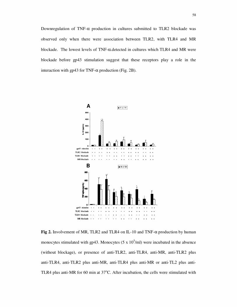
58
Downregulation of TNF-α production in cultures submitted to TLR2 blockade was
observed only when there were association between TLR2, with TLR4 and MR
blockade. The lowest levels of TNF-α.detected in cultures which TLR4 and MR were
blockade before gp43 stimulation suggest that these receptors play a role in the
interaction with gp43 for TNF-α production (Fig. 2B).
Fig 2. Involvement of MR, TLR2 and TLR4 on IL-10 and TNF-α production by human
monocytes stimulated with gp43. Monocytes (5 x 10⁵/ml) were incubated in the absence
(without blockage), or presence of anti-TLR2, anti-TLR4, anti-MR, anti-TLR2 plus
anti-TLR4, anti-TLR2 plus anti-MR, anti-TLR4 plus anti-MR or anti-TL2 plus anti-
TLR4 plus anti-MR for 60 min at 37oC. After incubation, the cells were stimulated with
gp43 stimulus - - + + + + + + + + + + + + + + + +
TLR2 blockade - - - - + + - - - - + + + + - - + +
TLR4 blockade - - - - - - + + - - + + - - + + + +
MR blockade - - - - - - - - + + - - + + + + + +
0
200
400
600
800
TNF-a
lpha
(pg/
mL)
4h 18h
+
+
*
*
*
**
*
**
*
**
*
*
*
*
+ * +
gp43 stimulus - - + + + + + + + + + + + + + + + +
TLR2 blockade - - - - + + - - - - + + + + - - + +
TLR4 blockade - - - - - - + + - - + + - - + + + +
MR blockade - - - - - - - - + + - - + + + + + +
0
200
400
600
800
TNF-a
lpha
(pg/
mL)
4h 18h
+
+
*
*
*
**
*
**
*
**
*
*
*
*
+ * +
0
10 0
20 0
30 0
40 0
50 0
IL-1
0 (
pg/m
L)
4h 18h
gp43 stimulus - - + + + + + + + + + + + + + + + +
TLR2 blockade - - - - + + - - - - + + + + - - + +
TLR4 blockade - - - - - - + + - - + + - - + + + +
MR blockade - - - - - - - - + + - - + + + + + +
*
+*
0
10 0
20 0
30 0
40 0
50 0
IL-1
0 (
pg/m
L)
4h 18h
gp43 stimulus - - + + + + + + + + + + + + + + + +
TLR2 blockade - - - - + + - - - - + + + + - - + +
TLR4 blockade - - - - - - + + - - + + - - + + + +
MR blockade - - - - - - - - + + - - + + + + + +
*
+*
A
B
59
gp43 (10 ng/ml) for 4h and 18h at 37oC. Supernatants of cultures were collected for
cytokine detection by ELISA. Results represent the mean + SEM of IL-10 (A) and
TNF-α (B) concentrations obtained from 20 healthy individuals.
IL-10 production: * (p< 0,01) when compared with control, TLR2, TLR4, MR, TLR2
plus TLR4, TLR2 plus MR, TLR4 plus MR, TLR2 plus TLR4 plus MR (4h and 18h); +
(p< 0,01) vs gp43 (4h)
TNF-α production: * (p< 0,05) when compared with control and 4h; + (p < 0.05) when
compared with TLR4, MR, TLR2 plus TLR4, TLR2 plus MR, TLR4 plus MR, TLR2
plus TLR4 plus MR (4h and 18h)
3.3. Modulatory effect of gp43r on TLR2, TLR4 and MR expression on monocytes
Monocytes were cultured in the absence (control culture) or in the presence of gp43r
(10ng/ml) for 4h and 18h at 37oC. LPS (10 ug/mL) was employed as a positive control
for monocyte stimulation. Figures 3A, 3B, 3C and 3D show the percentage of
monocytes positive for MR, TLR2 and TLR4. High percentage of MR-positive
monocytes were detected in cultures stimulated with gp43 or LPS, while no significant
increase in percentage of cells expressing TLR2 or TLR4 after LPS or gp43 in both
periods of incubation were observed in relation to control, non-stimulated cultures.
Comparison among the percentage of TLR2-, TLR4- and MR-positive cells showed that
few monocytes (less than 2%) express constitutively MR receptor before stimulation
with LPS and gp43, while TLR2 and TLR4 were expressed in more than 90% CD14+
cells (Fig. 3B). The percentage of MR-positive monocytes increased three- and five-fold
after LPS and gp43 stimulation respectively.

60
Fig 3. MR, TLR2 and TLR4 expression on human monocytes surface. Monocytes
(5x10⁵/ml) were incubated in the absence (control culture), or presence of
lipopolysaccharide (LPS) (10 ug/ml), or gp43 (10 ng/ml) at 37º C for 4h and 18h, and
MR, TLR2 and TLR4 expression were analyzed by flow cytometry. A) Representative
dot-plots of one experiment showing gated CD14+ cells expressing MR. Results
represent the mean + SEM percentage of monocytes expressing MR (B), TLR2 (C) or
TLR4 (D) obtained from 20 healthy individuals.
* (p< 0,05) when compared with control.
0
1
2
3
4
5
6
7
control LPS gp43
MR
(%
)
4h 18h
**
**
0
1
2
3
4
5
6
7
control LPS gp43
MR
(%
)
4h 18h
**
**
0
20
40
60
80
100
control LPS gp 43
TL
R4
(%
)
4h 18h
0
20
40
60
80
100
control LPS gp 43
TL
R2
(%
)
4h 18h
C
A
R5
CD14 (PE/Cy7)
R5R5
CD14 (PE/Cy7) C
D14
PE
/Cy
7MR (FITC)
6,41%
CD
14
PE
/Cy
7MR (FITC)
6,41%
D
B
61
The mean fluorescence intensity (MFI) analysis showed that the treatment of
monocytes with LPS or gp43r did not increase MR expression both at 4h and 18h of
periods of stimulation (Fig. 4A). On the other hand, significant increase on TLR2
expression was detected on monocyte surface after 4h and 18h of LPS stimulation in
comparison with control non-stimulated cultures. Although the culture with gp43r
enhanced TLR2 expression at 4h of monocyte culture, significant MFI values were only
observed after 18h of culture. This increase in TLR2 expression was significantly higher
than those observed in control or gp43r-stimulated cells at 4h of culture (Fig. 4B).
TLR4 expression induced by gp43r at 4h of culture was significant higher than in
control and LPS-stimulated cultures. However, TLR4 expression after 18 h of monocyte
cultures stimulated with gp43 was significantly lower than at 4h cultures and did not
show statistical difference in comparison with control cultures. These results showed
that gp43 upregulates TLR4 expression at 4 h and TLR2 expression after 18 h of
stimulation (Fig. 4C), but did not interfere with MR expression (Fig. 4A).

62
Fig 4. MR, TLR2 and TLR4 expression on human monocytes surface. Monocytes
(5x10⁵/ml) were incubated in the absence (control culture), or presence of
lipopolysaccharide (LPS) (10 ug/ml), or gp43 (10 ng/ml) at 37º C for 4h and 18h, and
MR, TLR2 and TLR4 expression were analyzed by flow cytometry. Results represent
the mean + SEM of the mean fluorescence intensity (MFI) for MR (A), TLR2 (B) or
TLR4 (C) on monocyte surface obtained from 20 healthy individuals.
0
20
40
60
80
100
control LPS gp43
TL
R4 (
MF
I)
4h 18h
**
* 0+
0
20
40
60
80
100
control LPS gp43
TL
R4 (
MF
I)
4h 18h
**
* 0+
0
20
40
60
80
100
control LPS gp43
MR
(M
FI)
4h 18h
0
20
40
60
80
100
control LPS gp43
TL
R2 (
MF
I)
4h 18h
**
+*
0
20
40
60
80
100
control LPS gp43
TL
R2 (
MF
I)
4h 18h
**
+*
A
B
C
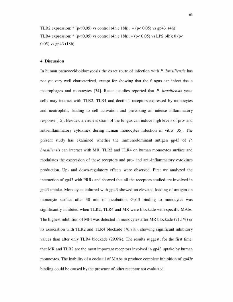
63
TLR2 expression: * (p< 0,05) vs control (4h e 18h); + (p< 0,05) vs gp43 (4h)
TLR4 expression: * (p< 0,05) vs control (4h e 18h); + (p< 0,05) vs LPS (4h); 0 (p<
0,05) vs gp43 (18h)
4. Discussion
In human paracoccidioidomycosis the exact route of infection with P. brasiliensis has
not yet very well characterized, except for showing that the fungus can infect tissue
macrophages and monocytes [34]. Recent studies reported that P. brasiliensis yeast
cells may interact with TLR2, TLR4 and dectin-1 receptors expressed by monocytes
and neutrophils, leading to cell activation and provoking an intense inflammatory
response [15]. Besides, a virulent strain of the fungus can induce high levels of pro- and
anti-inflammatory cytokines during human monocytes infection in vitro [35]. The
present study has examined whether the immunodominant antigen gp43 of P.
brasiliensis can interact with MR, TLR2 and TLR4 on human monocytes surface and
modulates the expression of these receptors and pro- and anti-inflammatory cytokines
production. Up- and down-regulatory effects were observed. First we analyzed the
interaction of gp43 with PRRs and showed that all the receptors studied are involved in
gp43 uptake. Monocytes cultured with gp43 showed an elevated loading of antigen on
monocyte surface after 30 min of incubation. Gp43 binding to monocytes was
significantly inhibited when TLR2, TLR4 and MR were blockade with specific MAbs.
The highest inhibition of MFI was detected in monocytes after MR blockade (71.1%) or
its association with TLR2 and TLR4 blockade (76.7%), showing significant inhibitory
values than after only TLR4 blockade (29.6%). The results suggest, for the first time,
that MR and TLR2 are the most important receptors involved in gp43 uptake by human
monocytes. The inability of a cocktail of MAbs to produce complete inhibition of gp43r
binding could be caused by the presence of other receptor not evaluated.
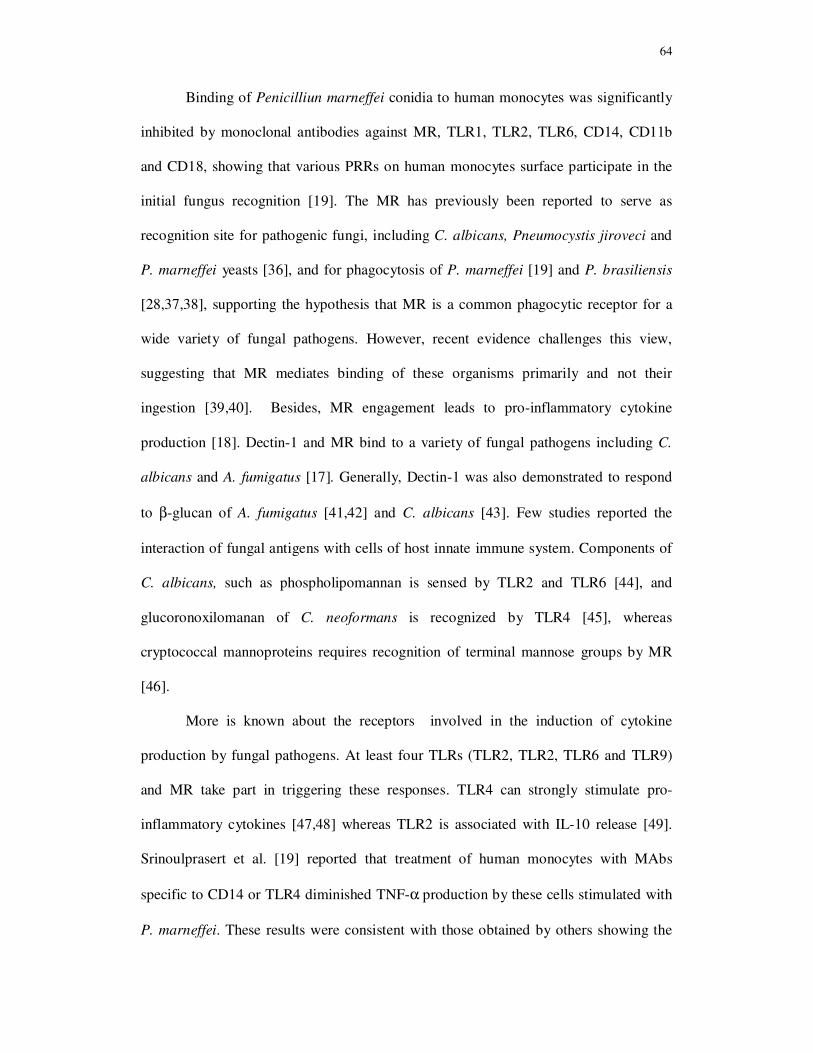
64
Binding of Penicilliun marneffei conidia to human monocytes was significantly
inhibited by monoclonal antibodies against MR, TLR1, TLR2, TLR6, CD14, CD11b
and CD18, showing that various PRRs on human monocytes surface participate in the
initial fungus recognition [19]. The MR has previously been reported to serve as
recognition site for pathogenic fungi, including C. albicans, Pneumocystis jiroveci and
P. marneffei yeasts [36], and for phagocytosis of P. marneffei [19] and P. brasiliensis
[28,37,38], supporting the hypothesis that MR is a common phagocytic receptor for a
wide variety of fungal pathogens. However, recent evidence challenges this view,
suggesting that MR mediates binding of these organisms primarily and not their
ingestion [39,40]. Besides, MR engagement leads to pro-inflammatory cytokine
production [18]. Dectin-1 and MR bind to a variety of fungal pathogens including C.
albicans and A. fumigatus [17]. Generally, Dectin-1 was also demonstrated to respond
to β-glucan of A. fumigatus [41,42] and C. albicans [43]. Few studies reported the
interaction of fungal antigens with cells of host innate immune system. Components of
C. albicans, such as phospholipomannan is sensed by TLR2 and TLR6 [44], and
glucoronoxilomanan of C. neoformans is recognized by TLR4 [45], whereas
cryptococcal mannoproteins requires recognition of terminal mannose groups by MR
[46].
More is known about the receptors involved in the induction of cytokine
production by fungal pathogens. At least four TLRs (TLR2, TLR2, TLR6 and TLR9)
and MR take part in triggering these responses. TLR4 can strongly stimulate pro-
inflammatory cytokines [47,48] whereas TLR2 is associated with IL-10 release [49].
Srinoulprasert et al. [19] reported that treatment of human monocytes with MAbs
specific to CD14 or TLR4 diminished TNF-α production by these cells stimulated with
P. marneffei. These results were consistent with those obtained by others showing the

65
involvement of TLR4 and CD14 on TNF−α production by monocytes and macrophages
activated by A. fumigatus [50-52]. Association between TLR2 expression and IL-10
production against pathogenic fungi, such as A. fumigatus and C. albicans has been
described in the literature [49,53]. The deleterious effect of TLR2 signaling during C.
albicans infection is associated with increased production of IL-10 and development of
T regulatory (CD4+CD25+) cells, resulting in deficient cellular immune response and
lower ability to the fungus elimination [49]. According to Bonfim et al. [15] while the
high virulence strain of P. brasiliensis (Pb18) predominantly induced TNF-α, the low
virulence strain (Pb265) was preferentially recognized by TLR2 and dectin-1, resulting
in the production of adequate concentration of IL-10, which might induce a controlled
immune response beneficial to the host. Differently, our results concerning to MR,
TLR2 and TLR4 involvement in cytokine production after gp43 recognition by human
monocytes, and confirmed by TLRs and MR blockade, showed that all receptors studied
may be involved in IL-10 release, whereas TLR4 and MR may be key receptors for
TNF-α production after gp43 binding. Thus, it is possible that a cross-talk between
TLRs and MR after gp43 binding is necessary to mediate intracellular signaling for
cytokine production. After bind with Pneumocystis carini, MR forms a functional
complex with TLR2 on cell surface and facilitate signal transduction to cytokine
production [54]. The discrepancy between our results and those reported by Bonfim et
al [15] may be explained by the employment of gp43, a purified antigen of P.
brasiliensis for binding studies to PRRs on monocyte surface while the authors used
whole yeast cells to stimulate monocytes. According to Calich et al. [6] studies with
purified components of fungal cells may reveal the major PRR and signaling pathways
used by the host cells to recognize fungal PAMPs.
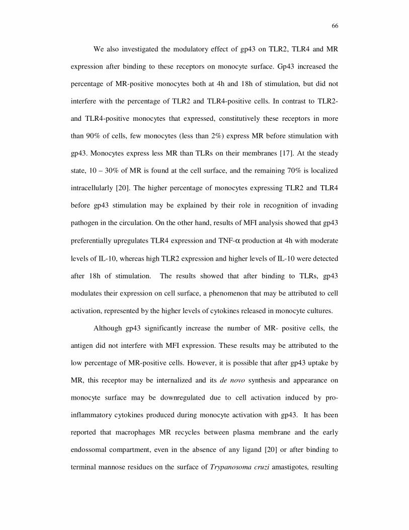
66
We also investigated the modulatory effect of gp43 on TLR2, TLR4 and MR
expression after binding to these receptors on monocyte surface. Gp43 increased the
percentage of MR-positive monocytes both at 4h and 18h of stimulation, but did not
interfere with the percentage of TLR2 and TLR4-positive cells. In contrast to TLR2-
and TLR4-positive monocytes that expressed, constitutively these receptors in more
than 90% of cells, few monocytes (less than 2%) express MR before stimulation with
gp43. Monocytes express less MR than TLRs on their membranes [17]. At the steady
state, 10 – 30% of MR is found at the cell surface, and the remaining 70% is localized
intracellularly [20]. The higher percentage of monocytes expressing TLR2 and TLR4
before gp43 stimulation may be explained by their role in recognition of invading
pathogen in the circulation. On the other hand, results of MFI analysis showed that gp43
preferentially upregulates TLR4 expression and TNF-α production at 4h with moderate
levels of IL-10, whereas high TLR2 expression and higher levels of IL-10 were detected
after 18h of stimulation. The results showed that after binding to TLRs, gp43
modulates their expression on cell surface, a phenomenon that may be attributed to cell
activation, represented by the higher levels of cytokines released in monocyte cultures.
Although gp43 significantly increase the number of MR- positive cells, the
antigen did not interfere with MFI expression. These results may be attributed to the
low percentage of MR-positive cells. However, it is possible that after gp43 uptake by
MR, this receptor may be internalized and its de novo synthesis and appearance on
monocyte surface may be downregulated due to cell activation induced by pro-
inflammatory cytokines produced during monocyte activation with gp43. It has been
reported that macrophages MR recycles between plasma membrane and the early
endossomal compartment, even in the absence of any ligand [20] or after binding to
terminal mannose residues on the surface of Trypanosoma cruzi amastigotes, resulting
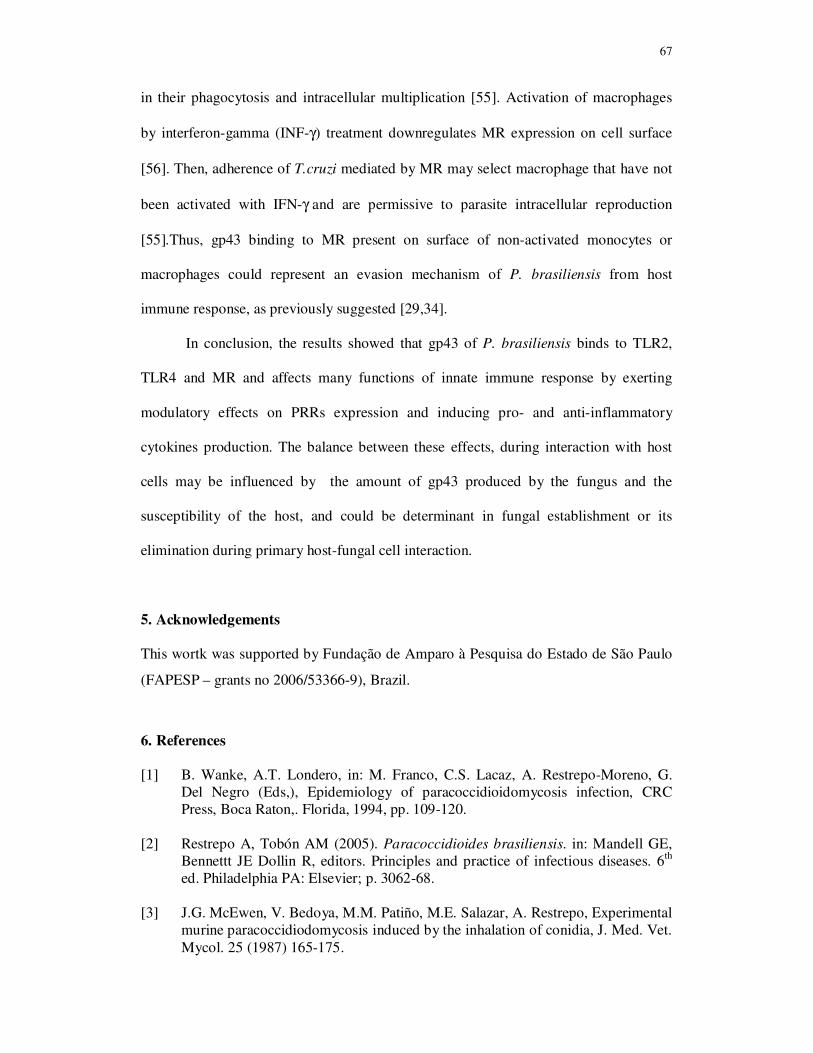
67
in their phagocytosis and intracellular multiplication [55]. Activation of macrophages
by interferon-gamma (INF-γ) treatment downregulates MR expression on cell surface
[56]. Then, adherence of T.cruzi mediated by MR may select macrophage that have not
been activated with IFN-γ and are permissive to parasite intracellular reproduction
[55].Thus, gp43 binding to MR present on surface of non-activated monocytes or
macrophages could represent an evasion mechanism of P. brasiliensis from host
immune response, as previously suggested [29,34].
In conclusion, the results showed that gp43 of P. brasiliensis binds to TLR2,
TLR4 and MR and affects many functions of innate immune response by exerting
modulatory effects on PRRs expression and inducing pro- and anti-inflammatory
cytokines production. The balance between these effects, during interaction with host
cells may be influenced by the amount of gp43 produced by the fungus and the
susceptibility of the host, and could be determinant in fungal establishment or its
elimination during primary host-fungal cell interaction.
5. Acknowledgements
This wortk was supported by Fundação de Amparo à Pesquisa do Estado de São Paulo
(FAPESP – grants no 2006/53366-9), Brazil.
6. References
[1] B. Wanke, A.T. Londero, in: M. Franco, C.S. Lacaz, A. Restrepo-Moreno, G. Del Negro (Eds,), Epidemiology of paracoccidioidomycosis infection, CRC Press, Boca Raton,. Florida, 1994, pp. 109-120.
[2] Restrepo A, Tobón AM (2005). Paracoccidioides brasiliensis. in: Mandell GE, Bennettt JE Dollin R, editors. Principles and practice of infectious diseases. 6th ed. Philadelphia PA: Elsevier; p. 3062-68.
[3] J.G. McEwen, V. Bedoya, M.M. Patiño, M.E. Salazar, A. Restrepo, Experimental murine paracoccidiodomycosis induced by the inhalation of conidia, J. Med. Vet. Mycol. 25 (1987) 165-175.
68
[4] M. Franco, R.P.Mendes, M. Moscardi-Bacchi, M.T. Rezkallah-Iwasso, M.R.
Montenegro, Paracoccidioidomycosis. Bailliere’s Clin. Trop. Med. Commun. Dis. 4 (1989) 185–220.
[5] L. Romani, Immunity to fungal infections, Nat. Rev. Immunol. 4 (2004) 1-13.
[6] V.L.Calich, T.A.da Costa, M. Felonato, C. Arruda, S. Bernardino, F.V. Loures, L.R. Ribeiro, R.de Cássia Valente-Ferreira, A. Pina, Innate immunity to
Paracoccidioides brasiliensis infection, Mycopathologia. 165 (2008) 223-236.
[7] C.A. Janeway Jr, R. Medzhitov, Innate immune recognition, Annu. Rev. Immunol. 20 (2002) 197-216.
[8] S.M. Levitz, Interactions of Toll-like receptors with fungi. Microbes. Infect. 6 (2004) 1351-1355.
[9] R. Medzhitov, Recognition of microorganisms and activation of the immune response. Nature 449 (2007) 819-826.
[10] J.A. Willment, G.B. Brown, C-type lectin receptors in antifungal immunity, Trends. Microbiol. 16 (2007) 27-32.
[11] P.D. Stahl, R.A. Ezekowitz. The mannose receptor is a pattern recognition receptor involved in host defense, Curr. Opin. Immunol. 10 (1998) 50-55.
[12] M.G. Netea, C.A. Van Der Graaf, A.G. Vonk, I. Verschueren, J.W. Van Der Meer, B.J. Kullberg, The role of toll-like receptor (TLR) 2 and TLR4 in the host defense against disseminated candidiasis, J. Infect. Dis. 185 (2002) 1483-1489.
[13] S. Braedel, M. Radsak, H. Einsele, J.P. Latgé, A. Michan, J. Loeffler, Z. Haddad, U. Grigoleit, H. Schild, H. Hebart, Aspergillus fumigatus antigens activate innate immune cells via toll-like receptors 2 and 4. Br. J. Haematol. 125 (2004) 392-399.
[14] K. Nakamura, K. Miyagi, Y. Koguchi, Y. Kinjo, K. Uezu, T. Kinjo, M. Akamine, J. Fujita, I. Kawamura, M. Mitsuyama, Y. Adachi, N. Ohno, K. Takeda, S. Akira, A. Miyazato, M. Kaku, K. Kawakami, Limited contribution of Toll-like receptor 2 and 4 to the host response to a fungal infectious pathogen, Cryptococcus
neoformans. FEMS. Immunol. Med. Microbiol. 47 (2006) 148-154.
[15] C.V. Bonfim, R.L. Mamoni, M.H.Blotta, TLR-2, TLR-4 and dectin-1 expression in human monocytes and neutrophils stimulated by Paracoccidioides
brasiliensis, Med. Mycol. 47 (2009) 722-733.
[16] P. Stahl, S. Gordon, Expression of a mannosyl-fucosyl receptor for endocytosis on cultured primary macrophages and their hybrids, J. Cell. Biol. 93 (1982) 49-56.
[17] M.G.Netea, G.D. Brown, B.J. Kullberg, N.A. Gow, An integrated model of the
69
recognition of Candida albicans by the innate immune system. Nat Rev Microbiol. 6 (2008) 67-78.
[18] Y. Yamamoto, T.W. Klein, H. Friedman. Involvement of mannose receptor in cytokine interleukin-1beta (IL-1beta), IL-6, and granulocyte-macrophage colony-stimulating factor responses, but not in chemokine macrophage inflammatory protein 1beta (MIP-1beta), MIP-2, and KC responses, caused by attachment of Candida albicans to macrophages. Infect. Immun. 65(1997) 1077-1082.
[19] Y. Srinoulprasert, P. Pongtanalert, R. Chawengkirttikul, S.C. Chaiyaroj,
Engagement of Penicillium marneffei conidia with multiple pattern recognition receptors on human monocytes. Microbiol. Immunol. 53(2009)162-172.
[20] U. Gazi, L. Martinez-Pomares, Influence of the mannose receptor in host immune responses. Immunobiology (2009) doi: 10.1016/j.imbio.2008.11.004
[21] S.S. Mambula, K. Sau, P. Henneke, D.T. Golenbock, S.M. Levitz, Toll-like receptor (TLR) signaling in response to Aspergillus fumigatus, J. Biol. Chem. 277(2002):39320-39326.
[22] R. Puccia, S. Schenkman, P.A. Gorin, L.R. Travassos, Exocellular components of Paracoccidioides brasiliensis: identification of a specific antigen. Infect. Immun. 53 (1986) 199-206.
[23] Z.P. Camargo, C.P. Taborda, E.G. Rodrigues, L.R.Travassos, The use of cell-free antigens of Paracoccidioides brasiliensis in serological tests, J. Med. Vet. Mycol. 29 (1991) 31-38.
[24] S.H. Marques da Silva, F. Queiroz-Telles, A.L. Colombo, M.H. Blotta, J.D. Lopes, Z. Pires De Camargo, Monitoring gp43 antigenemia in paracoccidioidomycosis patients during therapy, J. Clin. Microbiol. 42 (2004) 2419-2424.
[25] A.P. Vicentini, J.L Gesztesi, M.F. Franco, W. de Souza, J.Z. de Moraes, L.R. Travassos, J.D. Lopes, Binding of Paracoccidioides brasiliensis to laminin through surface glycoprotein gp43 leads to enhancement of fungal pathogenesis, Infect. Immun. 62 (1994) 1465-1469.
[26] S.A. Hanna, J.L. Monteiro da Silva, M.J. Giannini, Adherence and intracellular parasitism of Paracoccidioides brasiliensis in Vero cells, Microbes Infect. 2(2000) 877-884.
[27] J.L. Gesztesi, R. Puccia, L.R. Travassos, A.P. Vicentini, J.Z. de Moraes, M.F. Franco, J.D. Lopes, Monoclonal antibodies against the 43,000 Da glycoprotein from Paracoccidioides brasiliensis modulate laminin-mediated fungal adhesion to epithelial cells and pathogenesis. Hybridoma. 15 (1996) 415-422.
[28] S.R. Almeida, C.S. Unterkircher, Z.P. Camargo, Involvement of the major

70
glycoprotein (gp43) of Paracoccidioides brasiliensis in attachment to macrophages, Med. Mycol. 36 (1998) 405-411.
[29] A.F. Popi, J.D. Lopes, M. Mariano. GP43 from Paracoccidioides brasiliensis inhibits macrophage functions. An evasion mechanism of the fungus, Cell. Immunol. 218 (2002) 87-94.
[30] A.Y. Konno, J.T. Maricato, F.T. Konno, M. Mariano, J.D. Lopes, Peptides from Paracoccidioides brasiliensis GP43 inhibit macrophage functions and inflammatory response, Microbes Infect. 11 (2009) 92-99.
[31] C.P. Taborda, M.A. Juliano, R. Puccia, M. Franco, L.R. Travassos, Mapping of the T-cell epitope in the major 43-kilodalton glycoprotein of Paracoccidioides
brasiliensis which induces a Th-1 response protective against fungal infection in BALB/c mice, Infect Immun. 66 (1998) 786-793.
[32] K.C. Carvalho, M.C. Vallejo, Z.P. Camargo, R. Puccia, Use of recombinant gp43 isoforms expressed in Pichia pastoris for diagnosis of paracoccidioidomycosis. Clin. Vaccine. Immunol. 15 (2008) 622-629.
[33] K. Godfrey, Statistics in practice. Comparing the means of several groups, N. Engl. J. Med. 313 (1985) 1450-1456.
[34] E. Brummer, L.H. Hanson, A. Restrepo, D.A. Stevens. Intracellular multiplication of Paracoccidioides brasiliensis in macrophages: killing and restriction of multiplication by activated macrophages, Infect. Immun. 57 (1989) 2289-2294.
[35] C.S. Kurokawa, J.P. Araujo Jr, A.M. Soares, M.F. Sugizaki, M.T. Peraçoli, Pro- and anti-inflammatory cytokines produced by human monocytes challenged in vitro with Paracoccidioides brasiliensis, Microbiol. Immunol. 51 (2007) 421-428.
[36] L. East, C.M. Isacke, The mannose receptor family, Biochim. Biophys. Acta. 1572 (2002) 364-386.
[37] K.S. Ferreira, J.D. Lopes, S.R. Almeida, Down-regulation of dendritic cell activation induced by Paracoccidioides brasiliensis. Immunol. Lett, 94 (2004) 107-114.
[38] M. del P. Jiménez, A. Restrepo, D. Radzioch, L.E. Cano, L.F. García, Importance of complement 3 and mannose receptors in phagocytosis of Paracoccidioides
brasiliensis conidia by Nramp1 congenic macrophages lines, FEMS. Immunol. Med. Microbiol. 47(2006) 56-66.
[39] P.R. Taylor, S. Gordon, L. Martinez-Pomares, The mannose receptor: linking homeostasis and immunity through sugar recognition. Trends Immunol. 26(2005) 104-110.
[40] V. Le Cabec, L.J. Emorine, I. Toesca, C. Cougoule, I. Maridonneau-Parini, The
71
human macrophage mannose receptor is not a professional phagocytic receptor, J. Leukoc. Biol. 77(2005) 934-943.
[41] T.M. Hohl, H.L. Van Epps, A. Rivera, L.A. Morgan, P.L. Chen, M. Feldmesser, E.G. Pamer. Aspergillus fumigatus triggers inflammatory responses by stage-specific beta-glucan display. PLoS. Pathog. 1(2005):e30.
[42] C. Steele, R.R. Rapaka, A. Metz, S.M. Pop, D.L. Williams, S. Gordon, J.K. Kolls, G.D. Brown, The beta-glucan receptor dectin-1 recognizes specific morphologies of Aspergillus fumigatus, PLoS. Pathog. 1(2005):e42.
[43] G.D. Brown, S. Gordon, Fungal beta-glucans and mammalian immunity, Immunity. (2003) 311-315.
[44] T. Jouault, S. Ibata-Ombetta, O. Takeuchi, P.A. Trinel, P. Sacchetti, P. Lefebvre, S. Akira, D. Poulain, Candida albicans phospholipomannan is sensed through toll-like receptors, J. Infect. Dis. 188 (2003) 165-172.
[45] C. Monari, F. Bistoni, A. Casadevall, E. Pericolini, D. Pietrella, T.R. Kozel, A. Vecchiarelli, Glucuronoxylomannan, a microbial compound, regulates expression of costimulatory molecules and production of cytokines in macrophages, J. Infect. Dis. 191(2005) 127-137.
[46] M.K. Mansour, L.S. Schlesinger, S.M. Levitz, Optimal T cell responses to Cryptococcus neoformans mannoprotein are dependent on recognition of conjugated carbohydrates by mannose receptors, J. Immunol. 168 (2002):2872-2879
[47] S. Bellocchio, C. Montagnoli, S. Bozza, R. Gaziano, G. Rossi, S.S. Mambula, A. Vecchi, A. Mantovani, S.M. Levitz, L. Romani, The contribution of the Toll-like/IL-1 receptor superfamily to innate and adaptive immunity to fungal pathogens in vivo, J. Immunol. 172(2004) 3059-3069.
[48] C.A. van der Graaf, M.G. Netea, I. Verschueren, J.W. van der Meer, B.J. Kullberg. Differential cytokine production and Toll-like receptor signaling pathways by Candida albicans blastoconidia and hyphae, Infect. Immun. 73 (2005) 7458-7464.
[49] M.G. Netea, R. Sutmuller, C. Hermann, C.A. Van der Graaf, J.W. Van der Meer, J.H. van Krieken, T. Hartung, G. Adema, B.J. Kullberg, Toll-like receptor 2 suppresses immunity against Candida albicans through induction of IL-10 and regulatory T cells, J. Immunol. 172 (2004) 3712-3718.
[50] J.E. Wang, A. Warris, E.A. Ellingsen, P.F. Jorgensen, T.H. Flo, T. Espevik, R. Solberg, P.E. Verweij, A.O. Aasen, Involvement of CD14 and toll-like receptors in activation of human monocytes by Aspergillus fumigatus hyphae, Infect. Immun. 69 (2001) 2402-2406.
[51] S.S. Mambula, K. Sau, P. Henneke, D.T. Golenbock, S.M. Levitz, Toll-like receptor (TLR) signaling in response to Aspergillus fumigatus, J. Biol. Chem. 277 (2002) 39320-39326.

72
[52] A. Meier, C.J. Kirschning, T. Nikolaus, H. Wagner, J. Heesemann, F. Ebel, Toll-
like receptor (TLR) 2 and TLR4 are essential for Aspergillus-induced activation of murine macrophages, Cell. Microbiol. 5 (2003) 561-570.
[53] M.G. Netea, A. Warris, J.W. Van der Meer, M.J. Fenton, T.J. Verver-Janssen, L.E. Jacobs, T. Andresen, P.E. Verweij, B.J.Kullberg, Aspergillus fumigatus
evades immune recognition during germination through loss of toll-like receptor-4-mediated signal transduction, J. Infect. Dis. 188 (2003) 320-326.
[54] S.D. Tachado, J. Zhang, J. Zhu, N. Patel, M. Cushion, H. Koziel, Pneumocystis-mediated IL-8 release by macrophages requires coexpression of mannose receptors and TLR2, J. Leukoc. Biol. 81 (2007) 205-211.
[55] S. Kahn, M. Wleklinski, A. Aruffo, A. Farr, D. Coder, M. Kahn, Trypanosoma
cruzi amastigote adhesion to macrophages is facilitated by the mannose receptor, J. Exp. Med. 182 (1995) 1243-1258.
[56] S.E. Pontow, V. Kery V, P.D. Stahl, Mannose receptor, Int. Rev. Cytol. 137B (1992) 221-244.