Sociedade Brasileira de Cardiologia • ISSN 2318-8219...
40
1 Sociedade Brasileira de Cardiologia • ISSN 2318-8219 • Volume 30, Nº 4, Suplemento 1, Outubro 2017 TRABALHOS APRESENTADOS NO
Transcript of Sociedade Brasileira de Cardiologia • ISSN 2318-8219...

1
Sociedade Brasileira de Cardiologia • ISSN 2318-8219 • Volume 30, Nº 4, Suplemento 1, Outubro 2017
Trabalhos apresenTados no

Diretora PresidenteDra. Samira Saady Morhy - SP
Diretor Vice-Presidente de EcocardiografiaDr. Marcelo Luiz Campos Vieira - SP
Diretor Vice-Presidente Cardiologia NuclearDr. Gabriel Leo Blacher Grossman - RS
Diretora Vice-Presidente Ecografia VascularDra. Simone Nascimento dos Santos - DF
Diretor Vice-Presidente de Ressonância MagnéticaDr. Clério Francisco de Azevedo Filho - RJ
Diretor Vice-Presidente Tomografia ComputadorizadaDr. Tiago Senra Garcia dos Santos - SP
Diretor AdministrativoDr. Silvio Henrique Barberato - PR
Diretor FinanceiroDr. Henry Abensur - SP
Diretor dos Arquivos Brasileiros de Cardiologia - Imagem CardiovascularDr. José Maria Del Castillo - PE
Presidente PassadoDr. Arnaldo Rabischoffsky - RJ
Conselho DeliberativoPresidente:Dr. Fabio Villaça Guimarães Filho - SP
Membros:Dr. Carlos Eduardo Suaide Silva - SPDr. Jorge Eduardo Assef - SPDr. Leonardo Sara da Silva - GODr. Marcelo Zapparoli - PRDr. Orlando Campos Filho - SPDr. Romeu Sergio Meneghelo - SPDra. Simone Nascimento Dos Santos - DF
Comissão CientíficaMembros:Dr. Marcelo Luiz Campos Vieira - SPDr. Gabriel Leo Blacher Grossman - RSDra. Simone Nascimento dos Santos - DFDr. Clério Francisco de Azevedo Filho - RJDr. Tiago Senra Garcia dos Santos - SP
Comissão de HabilitaçãoCoordenadora:Dra. Claudia Gianini Monaco - SP
Membros:Dr. Alessandro Cavalcanti Lianza - SPDra. Gláucia Maria Penha Tavares - SPDra. Ingrid Kowatsch - SPDr. Rafael Bonafim Piveta - SPDra. Renata de Sá Cassar - SPDr. Wercules Antonio Alves de Oliveira - SP
Comissão de Informação e InternetCoordenadores:Dr. José Carlos Moreira dos Santos - RJDr. Edgar Bezerra Lira Filho - SP
Comissão de Honorários e Defesa dos ProfissionaisCoordenadores:Dr. Marcos Valério Coimbra de Rezende - SPDr. Wagner Pires de Oliveira Junior - DF
Comissão do Programa de Educação ContinuadaCoordenador:Dr. José Lazaro de Andrade - SP
Membros:Dra. Ana Clara Tude Rodrigues - SPDr. Edgar Bezerra Lira Filho - SP
Comissão de Ensino e Acreditação Coordenador: Dr. Edgar Bezerra Lira Filho - SP
Departamento de Imagem Cardiovascular
Membros: Dra. Ana Clara Tude Rodrigues - SPDr. David Costa de Souza Le Bihan - SPDr. José Lázaro de Andrade - SP
Comissão de Intercâmbio com outras Especialidades que realizam EcocardiografiaCoordenador:Dr. Cláudio Henrique Fischer - SPDr. David Costa de Souza Le Bihan - SPCorpo EditorialEditor-Chefe:Dr. José Maria Del Castillo - PE
Editora Anterior:Dra. Ana Clara Tude Rodrigues - SP
Editores de Área:Dr. Afonso Akio Shiozaki - PR (Tomografia)Dr. Alessandro Cavalcanti Lianza - SP (Ecocardiografia Pediátrica)Dr. André Luiz Cerqueira de Almeida - BA (Técnicas Avançadas em Ecocardiografia)Dr. Claudio Tinoco Mesquita - RJ (Medicina Nuclear)Dr. José Sebastiao de Abreu - CE (Ecocardiografia Adulto)Dr. Leonardo Sara - GO (Ressonância Magnética) Dra. Simone Nascimento dos Santos - DF (Vascular)
PresidenteMarcus Vinícius Bolívar Malachias
Vice-PresidenteEduardo Nagib Gaui
Presidente-EleitoOscar Pereira Dutra
Diretor AdministrativoDenilson Campos de Albuquerque
Diretora FinanceiraGláucia Maria Moraes de Oliveira
Diretor de Relações GovernamentaisRenault Mattos Ribeiro Junior
Diretor de ComunicaçãoCelso Amodeo
Diretor de Qualidade AssistencialWalter José Gomes
Diretor CientíficoRaul Dias dos Santos Filho
Diretor de Promoção de SaúdeCardiovascular - SBC/Funcor
Weimar Kunz Sebba Barroso de Souza
Diretoria SBCDiretor de Relações Estaduais e Regionais
José Luis Aziz
Diretor de Departamentos EspecializadosJoão David de Souza
Diretor de Tecnologia da InformaçãoOsni Moreira Filho
Diretor de PesquisaLeandro Zimerman
Editor-Chefe Arquivos Brasileirosde Cardiologia
Luiz Felipe P. Moreira

Adelino Parro Junior (SP)Adenalva Lima de Souza Beck (DF)Adriana Pereira Glavam (RJ)Afonso Akio Shiozaki (PR)Afonso Yoshikiro Matsumoto (RJ)Alessandro Cavalcanti Lianza (SP)Ana Camarozano (PR)Ana Clara Tude Rodrigues (SP)Ana Cláudia Gomes Pereira Petisco (SP)Ana Cristina Camarozano Wermelinger (PR)Ana Gardenia Liberato Ponte Farias (CE)Ana Lúcia Martins Arruda (SP)André Luiz Cerqueira de Almeida (BA)Andrea de Andrade Vilela (SP)Andrea Falcao (SP)Andressa Mussi Soares (ES)Aristarco Gonçalves de Siqueira Filho (RJ)Armando Luis Cantisano (RJ)Benedito Carlos Maciel (SP)Brivaldo Markman Filho (PE)Caio Cesar Jorge Medeiros (SP)Carlos Eduardo Rochitte (SP)Carlos Eduardo Suaide Silva (SP)Carlos Eduardo Tizziani Oliveira Lima (SP)Claudia Gianini Monaco (SP)Cláudio Henrique Fischer (SP)Cláudio Leinig Pereira da Cunha (PR)Claudio Tinoco Mesquita (RJ)Clerio Francisco de Azevedo Filho (RJ)David Costa de Souza Le Bihan (SP)Djair Brindeiro Filho (PE)
Edgar Bezerra Lira Filho (SP)Eliza de Almeida Gripp (RJ)Eliza Kaori (SP)Estela Suzana Kleiman Horowitz (RS)Gabriel Leo Blacher Grossman (RS)Gabriela Nunes Leal (SP)Gláucia Maria Penha Tavares (SP)Henry Abensur (SP)Ibraim Masciarelli Francisco Pinto (SP)Ilan Gottlieb (RJ)Iran de Castro (RS)Isabel Cristina Britto Guimaraes (BA)Ivan Romero Rivera (AL)Jaime Santos Portugal (RJ)Jeane Mike Tsutsui (SP)José Lázaro de Andrade (SP)José Luiz Barros Pena (MG)José Maria Del Castillo (PE)José Olimpio Dias Júnior (MG)José Sebastião de Abreu (CE)Joselina Luzia Menezes Oliveira (SE)Laise Antonia Bonfim Guimaraes (SP)Leonardo Sara da Silva (GO)Lilian Maria Lopes (SP)Luciano Aguiar Filho (SP)Luciano Herman Juaçaba Belém (RJ)Luiz Darcy Cortez Ferreira (SP)Luiz Felipe P. Moreira (SP)Manuel Adán Gil (SP)Marcelo Luiz Campos Vieira (SP)Marcelo Souza Hadlich (RJ)
Marcia de Melo Barbosa (MG)Márcio Vinícius Lins de Barros (MG)Maria do Carmo Pereira Nunes (MG)Maria Eduarda Menezes de Siqueira (SP)Marly Uellendahl (SP)Nathan Herszkowicz (SP)Orlando Campos Filho (SP)Oscar Francisco Sanchez Osella (DF)Oswaldo Cesar de Almeida Filho (SP)Paulo Zielinsky (RS)Reginaldo de Almeida Barros (SP)Roberto Caldeira Cury (SP)Roberto Pereira (PE)Rodrigo Alves Barreto (SP)Samira Saady Morhy (SP)Sandra da Silva Mattos (PE)Sandra Nivea dos Reis Saraiva Falcão (CE)Sérgio Cunha Pontes Júnior (SP)Silvio Henrique Barberato (PR)Simone Nascimento dos Santos (DF)Simone Rolim F. Fontes Pedra (SP)Tamara Cortez Martins (SP)Valdir Ambrósio Moisés (SP)Valeria De Melo Moreira (SP)Vera Márcia Lopes Gimenes (SP)Vera Maria Cury Salemi (SP)Viviane Hotta (SP)Washington Barbosa de Araújo (RJ)Wercules Oliveira (SP)William Azem Chalela (SP)Wilson Mathias Júnior (SP)
Conselho Editorial Nacional
Conselho Editorial InternacionalAnton E. Becker – HolandaDaniel Piñeiro – ArgentinaEduardo Escudero – ArgentinaEduardo Guevara – ArgentinaFernando Bosch – VenezuelaGustavo Restrepo – Colombia
Harry Acquatella – VenezuelaJoão A.C.Lima – Estados UnidosJorge Lowenstein – ArgentinaJoseph Kisslo – Estados UnidosLeopoldo Pérez De Isla – EspanhaMani A. Vannan – Estados Unidos
Natesa Pandian – Estados UnidosNavin C. Nanda – Estados UnidosRaffaele De Simone – AlemanhaRicardo Ronderos – ArgentinaVera Rigolin – Estados Unidos

Arquivos Brasileiros de Cardiologia - Imagem Cardiovascular
Os Arquivos Brasileiros de Cardiologia - Imagem Cardiovascular é o órgão oficial do Departamento de Imagem Cardiovascular da Sociedade Brasileira de Cardiologia.
Os artigos aqui publicados somente poderão ser reproduzidos com a expressa autorização dos autores. Publicacões pagas não serão aceitas. As separatas dos artigos deverão ser requisitadas diretamente à Secretaria Editorial e terão custo
equivalente ao total de cópias pedidas.
SBC/Departamento de Imagem CardiovascularRua Barata Ribeiro nº 380 cj.54
01308-000 - São Paulo - SP - BrasilFone/Fax: +55 (11) 3259-2988
Fones: +55 (11) 3120-3363+55 (11) 3259-2988 / +55 (11) 2589-4168
Secretaria Editorial - SBCAv. Marechal Câmara, 160 - 3º andar - Sala 330 20020-907 • Centro • Rio de Janeiro, RJ • Brasil
Tel.: (21) 3478-2716E-mail: [email protected]
http://departamentos.cardiol.br/dic/publicacoes/revistadic/
Produção Editorial:SBC - Tecnologia da Informação e Comunicação
Núcleo Interno de Publicações
Produção Gráfica e Diagramação:Alodê Produções Artísticas & Eventos
Volume 30, Nº 4, Suplemento 1, Outubro 2017
Indexação: LILAC - Literatura Latino-Americana e do Caribe em Ciências da Saúde - www.bireme.br, LATINDEX - Sistema Regional de Información en Línea para Revistas Científicas de América Latina, El Caribe, España y Portugal - www.latindex.unam.mx
6
Mensagem da Presidente do Departamento, Presidente do Congresso e do Coordenador da Comissão de Temas Livres
Prezados colegas,É com grande satisfação que os Arquivos Brasileiros de Cardiologia - Imagem Cardiovascular publicam os resumos dos
pôsteres que serão apresentados durante o 7º Congresso Brasileiro de Imagem Cardiovascular em conjunto com o 4th World Summit on Echocardiography, na bela cidade de Rio de Janeiro.
Estes trabalhos representam o que há de mais avançado e atualizado nas técnicas de Imagem Cardiovascular e serão apresentados na forma de pôster eletrônico.
Os pôsteres serão avaliados para selecionar os que receberão as premiações. O tradicional Prêmio Jonas Talberg será outorgado ao melhor Tema Livre apresentado no Congresso. Haverá, ainda, premiação para os melhores trabalhos de cada área (eco adulto, eco pediátrico e vascular/imagem cardiovascular).
Esperamos que os trabalhos apresentados durante o Congresso DIC e o Summit sejam encaminhados para publicação na Revista do DIC (ABC Imagem Cardiovascular) e desejamos sucesso a todos os apresentadores e congressistas!
José Maria Del Castillo
Editor-Chefe dos ABC – Imagem Cardiovascular
Coordenador da Comissão de Temas Livres
Valdir Ambrosio Moisés
Presidente do 7º Congresso Brasileiro de Imagem Cardiovascular e do 4th World Summit
on Echocardiography
Samira Saady Morhy
Presidente do Departamento de Imagem Cardiovascular da SBC

PÔSTERES

8
Pôsteres
Arq Bras Cardiol: Imagem cardiovasc. 2017;30(4 supl.1):6-41
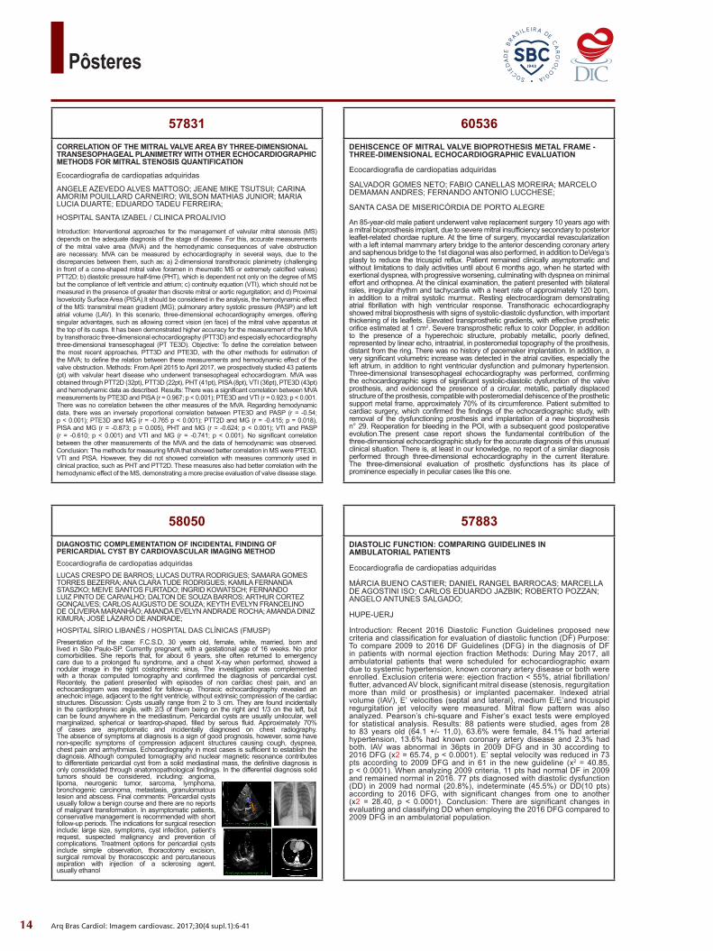
57999A RARE CASE OF AN EPITHELIOID CARDIAC SARCOMA IN THE LEFT VENTRICLEEcocardiografia de cardiopatias adquiridas
NATALIA REGINA METELLO ALECIO DIEHL; NATHALIA SUZAN CAMARÃO SILVA MARTINS; THAIS CARVALHO E SILVA; ALI KASSEN OMAIS; JANICE LANZARIN; CAMILA MARTINES MELLO; ALETHEIA CARPINE FAVINI; GILMAR ANTONIO COELHO DAMIN; JULIO CESAR DE OLIVEIRA; GIBRAN RODER FEGURI; ANNA CAROLINA FRANCO; PAULO RUIZ LUCIO DE LIMA; OLIVER GUILHERME DA SILVA; DANIEL BOUCHABKI DE ALMEIDA DIEHL;
UNIVESIDADE DE CUIABADescription: Male, 39-years-old, with dyspnea and left-sided weakness for the last 3 weeks. During hospitalization, the echocardiogram revealed a highly echogenic mass (4,6x4,1 cm), attached to the interventricular septum protruding to the left ventricular outflow, maximum/mean gradient of 13,4/7,19 mmHg, septal and inferior akinesia and important global systolic left ventricular dysfunction. Cardiac resonance showed large subvalvar aortic mass attached to the left-sided interventricular septum. Histopathology exam -focal myocardium sclerosis and lymphocytic infiltration, epithelioid malignant neoplasia in the left ventricle (neoplastic thrombus). Immunohistochemical exam revealed an epithelioid neoplasia, necrotic, positive for vimentin, citoceratine favoring for not classified cardiac sarcoma. The patient was submitted to tumor resection and evolved to death after twenty days of surgery with heart failure and generalized infection. Discussion: Primary cardiac tumors are rare with an incidence of 0,001% to 0,03% in autopsy series. Seventy-five percent of them are benign mostly myxomas and 25% are malignant predominantly consisting of sarcomas. In a reported series angiosarcoma was the commonest (37%) followed by malignant fibrous histiocytoma 24%, leiomyosarcoma 9%, rhabdomyosarcoma 7%, unclassified 7%, others 16%. Cardiac tumors may be found incidentally or cause symptoms in consequence of systemic or pulmonic embolization, obstruction, heart failure, arrhythmias, pericardial effusion. Epithelioid cardiac sarcomas are yellowish-white and may have extensive areas of necrosis, hemorrhage, and extensively infiltrate the myocardium. An immunoreaction to vimentin and citoceratin are considered a pleomorphic undifferentiated sarcoma with “focal epitheloid habitus”. The stroke could be explained for the tumoral embolism tendency, and the hypercoagulability state. Echocardiography and cardiac resonance were useful to assess the ventricular tumor, without identifying the presence of metastases at the time of diagnosis. The mean age of presentation is around 40 years with no sex predilection and there is a poor survival rate. Conclusion: This report describes an exceptionally rare primary cardiac epithelioid sarcoma in a 40-year-old man. The echocardiogram is of low cost and was fundamental for the diagnosis. In addition to the many modalities of clinical imaging performed, a large panel of immunohistochemical stains, was required to identify its epithelioid nature.
58022A 3D ECHOCARDIOGRAPHIC ASSESSMENT OF COMPLEXITY SCORE IN DEGENERATIVE MITRAL VALVE REGURGITATIONEcocardiografia de cardiopatias adquiridas
MONICA LUIZA DE ALCANTARA; RODOLFO DE PAULA LUSTOSA; FILIPE GOLDBERG; ALEX DOS SANTOS FELIX; ANA PAULA DOS REIS VELLOSO SICILIANO; DEISE PEIXOTO GUIMARÃES; MARCELLA DE AGOSTINI ISO; LEONARDO BALDEZ; CLAUDIA MARIA SANTOS; ORLANDO GLORIA VELOSO; CLAUDIA DE CASSIA FIRMIDA; RODRIGO REGO; SALOMON ISRAEL DO AMARAL; SERGIO SALLES XAVIER;
HOSPITAL SAMARITANO / AMERICAS. MEDICAL CITYIntroduction: In recent publication Anyanwu et al, presented a complexity scoring system (CSS) for degenerative mitral valve regurgitation (DMVR) repair based on surgical inspection and surrogates of techniques to be applied. This CSS intends to separate the lower, from the intermediate or higher complexity group that require greater ability in employing different surgical techniques and should therefore be referred to centers with great expertise in complex mitral repair. This evaluation should ideally be performed before the procedure to ensure a better planning and outcome. The author himself recommends the use of this score in preoperative echocardiography. Purpose: The aim of this study was to apply a modified CSS with 3D transesophageal echocardiography (3D TEE) analysis in patients(pts) with DMVR candidates for surgical intervention and evaluate its capability in discriminating the diferent complexity groups. Methods: from january 2015 to may 2017, we evaluated 42pts patients with significant DMVR previously or intraoperative right before intervention. A 3DTEE full volume dataset with adequate spatial and temporal resolution was obtained and analyzed offline. Mitral valve was divided into 8 segments and each prolapsed segment with or without flail was weighted. A modified CSS was applied where each posterior leaflet segment or cleft was assigned a score of 1, each anterior leaflet segment and commissural scallop a score of 2 and annulus calcification or previous repair a score of 3. Low complexity score (LCS) was defined as 1-2, intermediate (ICS) as 3-5 and high complexity (HCS) as > 5 points. Results: the mean age was 70+/-14 years (17 female). Prevalence of the different groups was as follows: LCS 18 (42.9%), ICS 8 (19.0%) and HCS 16 (38.1%). There was no statistical difference for age among groups but women had a significantly more complex score (p = 0.019). By comparing the CSS with the spectrum of DMVR that means fibroelastic defficiency (FED), fruste Barlow (FB) and Barlow disease (BD), we found as expected a significant correlation between the CSS and the type of the spectrum (p < 0.0001). A cleft was found in 47% of cases being more prevalent in BD (77%) than in the rest of the group (p = 0.042). Conclusion: CSS through 3D TEE analysis is feasible, can be performed in a dynamic peroperative fashion and can discriminate those with lower, intermediate or higher complexity score amenable to valve repair.
Cleft in a patient with Barlow disease ventricular persepctive.
Same patient view from the atrial perspective
58031A 3D ECHOCARDIOGRAPHIC QUANTITATIVE ASSESSMENT MITRAL VALVE DEGENERATIVE DISEASE. ARE THERE DIFFERENCES IN THE SPECTRUM?Ecocardiografia de cardiopatias adquiridas
MONICA LUIZA DE ALCANTARA; ANA PAULA DOS REIS VELOSO SICILIANO; ALEX DOS SANTOS FELIX; FILIPE GOLDBERG; DANIELA PINHEIRO FERNANDES; JUCIARA DA SILVA MATOS; DEISE PEIXOTO GUIMARÃES; LEONARDO BALDEZ; MAXIMILIANO OTERLO LACOSTE; RODOLFO DE PAULA LUSTOSA; SERGIO SALLES XAVIER;
HOSPITAL SAMARITANO / AMERICAS. MEDICAL CITYIntroduction: Mitral Valve Degenerative Disease (MVDD) is defined by a spectrum of lesions starting with fibroelastic defficiency (FED) characterized by a single segment prolapse and eventual chordal rupture in valves with normal annulus size (AS) and no mixomatous degeneration (MD). The other extreme of the spectrum is the Barlow disease (BD) with a large AS diffuse myxomatous changes and multisegment prolapse. In the middle of the spectrum are the valves with a certain degree of MD, more than 1 segment prolapse and intermediate AS. These valves are defined as a fruste Barlow form (FB). Three Dimensional (3D) Echocardiography has evolved from a subjective to a quantitative analysis of these findings with commercial available softwares that measure different mitral valve apparatus parameters (MVAP). Purpose: The aim of this study was to perform a quantitative assessment of some MVAP described as predictive of complexity and extension of MVDD. Methods: From january 2015 to may 2017, we evaluated 40 patients with significant MVDD previously or intraoperative right before intervention. A 3D Transesophageal full volume dataset with adequate spatial and temporal resolution was obtained and analised offline with specific quantification tool. MVDD spectrum was defined as FED, FB and BD. Presence or absence of chordal rupture (flail) was defined through 3D dataset navigation. Results: The mean age was 69+/-14years (16 female). Among the variables tested, age, 3D annulus size, anteroposterior diameter, intercomissural diameter, anterior leaflet area (ALA) and posterior leaflet area (PLA) were signicantly different in each type of the spectrum with a p value < 0.0001 for all except age (p = 0.004) and PLA (p = 0.003). Nonplanar angle, sphericity index, annulus height and mitro-aortic angle did not vary between groups. A flail of one or more segments was present in 23 patients (57%). Presence of flail correlated with FED (p = 0.014), but not with gender or age. The only quantitative variable with a positive correlation with flail was ALA (p = 0.01). Conclusion: Quantitative analysis of MVAP can discriminate different types of MVDD spectrum. Futher analysis is needed to confirm the potential of the method to select those with greater risk of chordal rupture.
Spectrum of mitral valve degenerative disease. From left to righ. Fibroelastic Defficiency, Fruste Barlow form and Barlow Disease. Upper Panel - Quantitative Software analysis with 3D annulus size. Lower panel: 3D rendered images
Prevalence of prolapse and flail among different segments
598403D ECHOCARDIOGRAPHIC IMAGE OF MYXOMA IN RIGHT VENTRICULAR OUTFLOWEcocardiografia de cardiopatias adquiridas
IRVING GABRIEL ARAÚJO BISPO; DEIVIDE RIBEIRO SILVEIRA; JUSSARA REGINA SOUSA RODRIGUES; TAINARA SÁ FREIRE DE ALMEIDA; LIVIA FERRAZ ACCORSI; MURILO SALANI GIL; JULIANA B MATSUMOTO; CINTHIA NIEGE SANTOS SILVA; VERA MARCIA LOPES GIMENES; MERCEDES MALDONADO ANDRADE; IRVING GABRIEL ARAÚJO BISPO; LÍVIA FERRAZ ACCORSI; MERCEDES MALDONADO ANDRADE;
HOSPITAL DO CORAÇÃO DE SÃO PAULO
Case presentation Technological development provided early diagnosis and treatment of primary or metastatic heart tumors. One of the most important technological advances was echocardiography, a low cost method and a high precision in the diagnosis of this disease. We present a case of patient who was referred to the service for better definition of the anatomy using 3D echocardiography. Patient MSS, 44 years old, from São Paulo, was asymptomatic when he sought a cardiologist to check up. A transthoracic echocardiogram was used that showed a cardiac mass adhered to the Right Ventricle. Patient was referred for transesophageal echocardiography using 3D technique to better analyze tumor mass. Transesophageal echocardiography revealed a right ventricle diameter of 36 mm and a mobile lobulated homogeneous image adhered to the anterolateral wall of the right ventricle by a thick pedicle in its medial segment measuring 57.7 mm x 37.2 mm suggestive of tumor mass (myxoma) evidenced by Echocardiogram. The mass travels towards the right ventricular outflow tract caused by slight obstruction of the same with a VSVD-TP gradient of 13 mmHg and a sign of moderate to severe pulmonary hypertension with a systolic RV pressure estimated at 70 mmHg due to tricuspid insufficiency. Discussion: Myxomas make up the majority of cardiac tumors, accounting for 50% of cases in certain studies. A large part is inserted into the fossa ovalis (left atrial face) of the interatrial septum (from 64 to 85% of patients) and 90% are solitary, whereas those originating from the valves are uncommon. A case of RV myxoma with intermittent obstruction to the RV outflow tract was reported by Karagounis and Sarsam; another case of RV myxoma with partial obstruction to the RV outflow tract was reported by Van der Heusen et al. And these suggested intraoperative monitoring with transesophageal echocardiography. Final comments: In this case, we have shown that transesophageal echocardiography, together with the 3D technique, was important for better anatomical definition and provides information for the appropriate surgical procedure.

9
Pôsteres
Arq Bras Cardiol: Imagem cardiovasc. 2017;30(4 supl.1):6-41
58069AN ATYPICAL RIGHT ATRIAL MASS ON 3D ECHOCARDIOGRAM, ASSOCIATED WITH EBSTEIN‘S ANOMALY
Ecocardiografia de cardiopatias adquiridas
LAISSE MARINS DEFANTI GONZAGA; ANGELO ANTUNES SALGADO; LAURA DE ABREU ALVES; RAFAEL DE OLIVEIRA CARDOSO; MILTON RICARDO POFFO; BRUNO ROBERTO ITABORAHY ALABRÍN;
UNIVERSIDADE ESTADUAL DO RIO DE JANEIRO
Case report: A 48 year-old male with Ebstein‘s anomaly, reported 6 months‘ worsening fatigue upon moderate exertion, palpitations and weight loss of 4kg in two months. Investigation findings: Electrocardiogram: Sinus rhythm, right bundle branch block, signs of right atrial overload. Initial transthoracic echocardiogram (April 2014): Severe dilatation of right chambers, preserved biventricular function, moderate tricuspid regurgitation, right ventricular atrialization and low tricuspid valve implantation. No evidence of intracardiac masses. Subsequent transthoracic echocardiogram: Left ventricular function preserved. Increased right cavity diameters and right ventricular dysfunction. Atrial delamination of the septal tricuspid leaflet, compatible with Ebstein‘s anomaly, with faulty leaflet coaptation. Severe tricuspid regurgitation, with systolic pulmonary artery pressure conservatively estimated at 44 mmHg. Observation of a poorly delineated, pediculated right atrial mass adherent to the interatrial septum. 3D transoesophageal echocardiogram: Evidence of a hyperechogenic structure of irregular shape and of soft tissue density in the right atrium, adherent via principal and lesser pedicles near the inferior vena cava and superior vena cava, respectively. Abdominal computerized tomography for a etiological investigation: Presence of a large, irregular contrast-enhancing hepatic lesion measuring 8.12 x 7.3cm and multiple scattered nodular images, possibly representing secondary deposits. Discussion: Cardiac tumours are rare and divided into primary and metastatic tumours. In an autopsy series, the latter were 100 times more common than the former. Among primary cardiac tumors, 90% are benign, myxomas being the most common (50% - 80%). Final considerations: We report a case of Ebstein‘s anomaly with an atypical right atrial mass of probable neoplastic etiology. This case shows the importance of echocardiography in the investigation of cardiac tumours, and that 3D echocardiography is indispensable for evaluation of the insertion points of masses. When diagnosing an intracardiac mass, it is necessary to differentiate between primary and metastatic tumours, thrombi and congenital anomalies. When surgery is necessary, histopathology is of paramount importance in establishing definitive conduct.
60444ABLATION OF ATRIAL FIBRILLATION BY RADIOFREQUENCY CATHETER WITH CONTINUOUS MONITORING BY TRANSESOPHAGEAL 3D
Ecocardiografia de cardiopatias adquiridas
JOAO CARLOS TRESS; BRUNO RUSTUM ANDREA; EDUARDO MACHADO ANDREA; ROSAURA DE CARVALHO VICTER; TEREZA CRISTINA DUQUE ESTRADA; PABLO DE MOURA LOPES; MARCIA GRACINDO;
COMPLEXO HOSPITALAR DE NITEROI
Atrial fibrillation (AF) is a major public health concern, AF is the most common sustained arrhythmia seen in clinical practice and accounts for approximately one-third of hospitalizations for cardiac dysrhythmias. Atrial fibrillation is characterized by uncoordinated atrial activation with resulting deterioration of atrial mechanical function. AF is associated with significant mortality, morbidity, and health care costs. Catheter ablation for the treatment of AF is increasingly being performed on symptomatic patients as an alternative to medical management or when medical management has been ineffective or not tolerated. Catheter ablation was reported to be effective in approximately 80 percent of patients with a risk of 4.5 percent for major complications in literature. Our objective was to study 36 patients during atrial fibrillation by radiofrequency catheter with 3D transesophageal for the assistance of procedure in relation to trans septal puncture, the 3D assessment of positioning catheters in the ostium of the pulmonary veins, at the three-dimensional assessment of the left appendage, the pulmonary vehicle flow analysis during the whole procedure and the ejection fraction difference with fibrillation and after procedure. All documented complications were reviewed. Major complications were defined as those that results in permanent injury or death, requires intervention for treatment, or prolongs or requires hospitalization for > 48 h. All major complications documented during the procedure or within the 3-month follow-up using departmental tracking process were considered for the analysis. The success to return to sinus rhythm was 87%. The total time of procedure with the participation of 3D transesophageal echo was less than 10% in relation to literature, probably for help during the septal puncture, the use of contrast for the atrial ablation by catheter was decreased in this study, with 10 ml in comparative to 25 ml or more observer in the literature. We also evaluated the flow of the pulmonary veins during fibrillation and after ablation and observed a change in the flow pattern with a real significance in relation to the drop in left atrial pressure, with S wave elevation and D wave reduction, the ejection fraction improves around 20-30% when the Sinus rhythm was reached. We do not have rate of cardiac tamponade/hemopericardium in this study, so, we believe that the transesophageal 3D had strong influence.We believe to be the transesophageal 3D fundamental tool to improve the outcome of atrial fibrillation by radiofrequency ablation throughout the procedure.
60715ABSCESS INVOLVING NATIVE TRICUSPID VALVE LEAFLETS – A RARE CONDITION AND EVOLUTIONEcocardiografia de cardiopatias adquiridas
MAYSA RAMOS VILELA; ALINE SANTOS AZEVEDO; ALINE CAMPOS DE LEO; SERGIO SALLES XAVIER; PLÍNIO RESENDE;
UNIVERSIDADE FEDERAL DO RIO DE JANEIRO
Case Report: An 85-year-old man was admitted in our hospital with fever and right ankle pain. His past medical history includes hypertension and diabetes. There was no history of alcoholism, use of intravenous drugs, central venous catheter or hemodialysis. On admission, the patient was in regular estate, tachypneic with no resorting effort. His vital signs were stable. There were edema, erythema and heat in lateral perimalleolar region of the right ankle, but no skin lesion associated. After detecting 4 positive blood culture for methicillin-sensive Staphylococcus aureus (MSSA) cefazolin antibiotic was iniciated. An ankle computed tomography and scintigraphy demonstrated talus and navicular osteomyelitis. Transthoracic echocardiography did not show suggestive images of infective endocarditis (IE). We proceed a transesophageal echocardiography (TOE) due to high suspicion of IE. It showed an additive image, with heterogeneous density, very mobile, measuring 10 x 8 mm, at the septal leaflet of tricuspid valve. The mass had large echolucent areas, with no flow inside, compatible with abscess. There was mild tricuspid regurgitation associated. The patient remained without signs of heart failure (HF), his blood cultures were negative after 3 days of antibiotics. A pulmonary perfusion ventilation scintigraphy showed no signs of pulmonary embolism. After 4 weeks of treatment a repeat TOE showed an abscess involution. He was given a total of six weeks of antibiotics. Discussion: Tricuspide valve abscess is extremely rare. Right-sided IE is usually associated with well-established risk factors such as intravenous drug use or intravenous devides and account for 5 – 10% of IE cases. We report a case of native tricuspid valve abscess in a patient without such risk factors but that started the symptoms with pyogenic osteomyelitis. This infection can complicate or be complicated by IE. The most common place of osteomyelitis associated IE is vertebral, it occurs in 4.6 – 19% of IE pacients. The most common microorganism involved is Staphylococcus aureus. Other complication is right HF caused by pulmonary hipertension secondary to pulmonary embolism or tricuspid regurgitation. Although the patient had a tricuspid abscess, this only caused mild regurgitation. He did not present pulmonary embolism or other abscess complications such as fistula, pseudoaneurysm or perforation. Right-sided IE treatment must be individualized, with surgery performed only in cases of persistent bacteraemia, severe right-sided HF, repetitive lung embolisms. Tricuspid valve abscess is a relative indication, but with no other complication associated can be clinically managed as in this case. The control TOE performed after 4 weeks antibiotic shown complete treatment response. Conclusion: We present a rare case of tricuspid leaflet abscess associated with MSSA osteomyelitis that had a complete response with antibiotic treatment.
57957ABILITY OF 3D ECHOCARDIOGRAPHY IN DETECTING AND SIZING THE ENTRY TEAR IN TYPE A AORTIC DISSECTION. INITIAL SINGLE CENTER EXPERIENCEEcocardiografia de cardiopatias adquiridas
MONICA LUIZA DE ALCANTARA; JUCIARA DA SILVA MATOS; ALEX DOS SANTOS FELIX; ANA PAULA DOS REIS VELLOSO SICILIANO; GUSTAVO ARUME GUENKA; RODOLFO DE PAULA LUSTOSA; MARCOS PAULO LACERDA BERNARDO; SALOMON ISRAEL AMARAL; MAXIMILIANO OTERO LACOSTE; FILIPE GOLDBERG; RAFAEL CASTRO DA SILVA; SERGIO SALLES XAVIER;
HOSPITAL SAMARITANO / AMERICAS. MEDICAL CITY
Resumo: Introduction: Detection of the primary entry tear (PET) site in acute type A aortic dissection ( ATAAD) is of capital importance for surgical planning, as up to 20% of them may be retrograde originating from the distal ascending portion or aortic arch. Defining the location is not always easy by computed tomography (CT) due to pulsation artifacts in the aorta thus seldom described. 2D transesophageal echo (TEE) has a high sensitivity in detecting the PET site but not the ability to evaluate the real size which might have an impact on progression and risk of aneurysm rupture. 3D TEE through its unique capability of dynamic image aquisition (high temporal resolution), dataset alignment and “en face” view, is theoretically the ideal method in quantifying PET. Purpose: The aim of this study was to evaluate the ability of 3D TEE in detecting and sizing the PET in ATAAD. Methods: Thirty patients admitted to our hospital with the diagnosis of acute aortic syndrome between february 2015 to april 2017 were retrospectively analysed. Sixteen of them with ATAAD. Of these, 12 had 3D TEE available for analysis performed on and offline after postprocessing the aquired 3D dataset. Results: in 7 patients the PET could be demonstrated by 3D TEE. Six of them were located at valsalva sinus and 1 at the the terminal portion of the aortic arch. In one patient the ascending portion of the aorta had an hematoma with dissection starting at the arch. Other 2 patients had the PET at the aortic arch detected during surgical inspection and in other 2 patients the PET could be visualized on 2D but not on 3D TEE due to poor quality of the dataset. In all PET demonstrated, an approximate size could be obtained either on 3D rendered or on 3D flexi-slice derived 2D images. The largest diameter varied from 0.7 to 4.2 cm and the area varied from 0.8 cm2 to 9.0 cm2. Geometry of the PET varied form a slit tear, to an irregular ellipsoid up to a circular shape. Conclusion: 3D TEE localization, sizing and geometry definition of PET in ATAAD is feasible and might select patients with greater risk of progression or aneurysm rupture.
Figura 1 – 3D rendered image of an type A aortic dissection entry tear circular shape
Figura 2 – 3D rendered image of an type A aortic dissection entry tear slit shape
Figura 3 – 3D rendered image of an type A aortic dissection entry tear ellipsoid shape

10
Pôsteres
Arq Bras Cardiol: Imagem cardiovasc. 2017;30(4 supl.1):6-41
57768ASSOCIATION OF ECHOCARDIOGRAPHIC PARAMETERS WITH MORTALITY IN SEPTIC SHOCK
Ecocardiografia de cardiopatias adquiridas
BRUNO FERRAZ DE OLIVEIRA GOMES; LORENA PEREIRA BRAGA ÁVILA; GIOVANNI POSSAMAI DUTRA; CATARINA SCHIAVO GRUBERT; BARBARA FERREIRA DA SILVA MENDES; SUZANA ANDRESSA MORAIS DE PAULA; FERNANDA HENRIQUES PINTO; JOÃO MATHEUS EMÍLIO MOTA MACIEL; MARCELO FERREIRA PALOMO VALLE; LUISA BENFICA GUIMARÃES PINTO COELHO; GUSTAVO HENRIQUE DE OLIVEIRA AMORIM; CINTHIA ARAKAKI WATANABE; VICTOR DA COSTA D‘ELIA; CLARISSA MAGALHÃES BARBOSA; JOÃO LUIZ FERNANDES PETRIZ;
HOSPITAL BARRA D‘OR
Introduction: Myocardial dysfunction of sepsis is common and has impact on prognosis. The echocardiographic evaluation in the initial phase of sepsis provides valuable information on systolic and diastolic function as well as the patient‘s volume status. However, there are few studies using echocardiographic parameters and its association with death in these patients. Objective: To evaluate the association of echocardiographic parameters with hospital death in patients with septic shock Methods: We included all patients from July 2011 to July 2016 admitted with septic shock (definite infectious disease and need for amines for at least one hour) and performed echocardiogram in the first 48 hours after admission. The following echocardiographic parameters were evaluated: LV systolic function, ejection fraction (EF), pulmonary artery systolic pressure (PASP), alterations in segmental contratility, diastolic function, left atrium (LA) enlargement, RV systolic function, mitral and aortic regurgitation, caliber of the inferior vena cava (IVC) and presence of IVC variability. All parameters were compared between deaths and survivors. Statistical analysis performed using the chi-square test for categorical variables and student’s t-test for continuous variables. Results: 289 patients, 75.5 ± 14.8 years, 46.8% men, mean SAPS3 score = 61.1 ± 13.67. There were 161 deaths (61.2%). In the comparison between survivors and deaths, we found, respectively: LV systolic dysfunction of any degree (26.5% x 27.7%, p = 0.479); segmental alteration (25.9% x 34.1%, p = 0.087); normal diastole (18.1% x 10.4%, p = 0.045); LA enlargement (37.1% vs. 41.6%, p = 0.258); RV systolic dysfunction (5.6% x 5.9%, p = 0.583); normal/mild mitral regurgitation (91,4% x 88,4%, p = 0.275); normal/mild aortic regurgitation (94,8% x 91,9%, p = 0.238); reduced VCI caliper (18.0 x 23.0, p = 0.198); presence of IVC variability (56.0 x 45.9%, p = 0.073); FE (65.6% x 61.8%, p = 0.008); PASP (38.7 mmHg x 41.7 mmHg, p = 0.024). Conclusions: In this population of septic shock patients with a high mortality rate, the survivors showed a higher prevalence of normal diastolic function, higher ejection fraction and lower levels of PASP.
57836ASSESSMENT OF LEFT VENTRICULAR DEFORMATION, ROTATION AND TWISTING, USING TWO-DIMENSIONAL STRAIN
Ecocardiografia de cardiopatias adquiridas
BRENO SIQUEIRA FERNANDES; MARCUS FREIRE VINHAS; DEUSDETH TEIXEIRA SOARES SEGUNDO; JOSÉ MARIA DEL CASTILLO; CARLOS ANTONIO DA MOTA SILVEIRA; EUGENIO SOARES DE ALBUQUERQUE; OSCAR FRANCISCO SANCHEZ OSELLA; ANTONIA DULCINEIDE MEDEIROS SENA;
PRONTO SOCORRO CARDIOLÓGICO DE PERNAMBUCO PROF LUIZ TAVARES - UPE
Introduction: The cardiac muscle has an architecture associated with physical properties that provide variations of parietal deformation, which can be decomposed in several directions: longitudinal, radial and circumferential, all perpendiculars between them. Apical approach is used to determine longitudinal strain. Short axis views at the base of the left ventricle, at papillary muscles level and apical region level are used to measure circumferential strain, radial strain and rotational movement. Objective: The aim of this work is to assess the myocardial deformation and rotation in individuals with no echocardiographic cardiac disease. Methods: Were selected 258 individuals with no echocardiographic evidence of heart disease. In classical echocardiographic approach we studied the longitudinal deformation of 17 myocardial segments, circunferential deformation, the radial deformation and the basal and apical rotation. One Way Analysis of Variance performed statistical analysis, complemented with Student-Newman-Keuls test for significance between individual segments. Inter-observer variability and reproducibility was analyzed by correlation coefficient. Results: In the apical approach of the left ventricle, the regional deformation values were -20.97 ± 2.83% in the basal segments, -22.52 ± 3.42% in the median segments and -23.42 ± 2.94% in the apical segments. The global longitudinal strain was -21.91 ± 2.53%. In short axis view, the regional values of circumferential strain were -21.09 ± 3.95% in basal segments, -23.23 ± 4.18% in medial segments and -23.51 ± 5.63% in apical segments. The regional values of radial strain were 54.07 ± 11.06% in the basal segments, 46.23 ± 9.79% at the level of the papillary muscles and 38.24% ± 7.84% in the apical segments. Values of rotation were -5.90° ± 3.70° at mitral valve segments and 10.39° ± 4.03° at apical segments. Angular difference between basal and apical rotation, called twisting, was 16.29° ± 5.19°. Conclusion: The impact of new imaging methods on the understanding of cardiac mechanics is very important, since knowledge implies understanding several pathological mechanisms. The Speckle tracking, as any new methodology, needs acquisition of benchmarks consisting of normal and pathological studies.
57841ASSESSMENT OF THE LEFT VENTRICULAR DIASTOLIC FUNCTION WITH TWO-DIMENSIONAL STRAIN AND STRAIN RATE
Ecocardiografia de cardiopatias adquiridas
CARLOS MAZZAROLLO; ALEX BARROS DOS SANTOS; CARLOS ALBERTO DE SOUZA MARTINS; LUIS WELLINGTON BARRETO VIEIRA; JOSÉ MARIA DEL CASTILLO; EUGENIO SOARES DE ALBUQUERQUE; CARLOS ANTONIO DA MOTA SILVEIRA; OSCAR FRANCISCO SANCHEZ OSELLA; ANTONIA DULCINEIDE MEDEIROS SENA;
PRONTO SOCORRO CARDIOLÓGICO DE PERNAMBUCO PROF LUIZ TAVARES - UPE
Introduction: The evaluation of left ventricular (LV) diastolic dysfunction presents a significant number of indeterminate dysfunctions, especially when the ejection fraction (EF) is preserved. Overall longitudinal strain (GLS), and systolic (SRs) and early diastolic (SRd) strain rates, may be useful to reclassify these patients. Objective: to evaluate, with GLS, SRs and SRd, patients with diastolic dysfunction, to compare with healthy subjects and verify the additive value of the method. Methods: studied 149 patients (age 62.2 ± 10.6 years) with diastolic dysfunction (49.7% grade 1, 15.4% grade 2, 18.1% grade 3 and 16.8% undetermined) and 189 healthy subjects (age 44.5 ± 13.3 years). Measured dimensions and function of the LV and LA, mitral and tissue Doppler velocities and their relationships, GLS, SRs and SRd. Data was evaluated by the Kolmogorov-Smirnoff test, Kruskal-Wallis test, multiple regression analysis and area under the ROC curve. Significant data when p < 0.05. Results: diastolic dysfunction increased the size and thickness of the left ventricle, reducing the EF, changed mitral and tissue Doppler velocities and increased LA volume and tricuspid regurgitation velocity. The GLS and SRs decreased in grade 2 and 3 diastolic dysfunction and the SRd also decreased in dysfunction grade 1 being the best parameter correlated with diastolic dysfunction. The area under the ROC curve showed cutoff value of 1.0 s-1 for the SRd. Conclusion: Diastolic dysfunction supplemented with myocardial strain rate seems to add sensitivity and specificity in cases where diastolic function is indeterminate and can be used to reclassify these patients.
57971APPLICABILITY OF LONGITUDINAL STRAIN OF LEFT VENTRICLE IN UNSTABLE ANGINA
Ecocardiografia de cardiopatias adquiridas
NATASHA SOARES SIMÕES DOS SANTOS; MARIANA REZENDE OLIVEIRA; MURILO CASTRO FERREIRA; ANDREA DE ANDRADE VILELA; MARCELA PAGANELLI DO VALE; RODRIGO BELLIO DE MATTOS BARRETTO; NELSON HENRIQUE GOES SCORSIONI; ALEXANDRE JOSÉ AGUIAR ANDRADE; OLÍVIA XIMENES DE QUEIROGA;
INSTITUTO DANTE PAZZANESE DE CARDIOLOGIA
This was a descriptive, cross-sectional, observational study with a 60-day observation period to determine the applicability prevalence of left ventricular two-dimensional longitudinal strain (S2DL) for identification of myocardial ischemia in patients with unstable angina (UA). The sample consisted of 78 patients, of whom fifteen (19.2%) were eligible for longitudinal strain analysis. In the group of ineligible patients, a lower proportion of women, a higher prevalence of diabetes, larger cavitary diameters and a higher rate of use of ASA, statins and beta-blockers was observed. The main causes of non-applicability were the presence of previous infarction (56.4%), previous coronary angioplasty (TCA) (22.1%), previous surgical revascularization (MR) (11.5%) or both (16.7%) and presence of specific electrocardiographic alterations (12.8%). The evaluation S2DL revealed a decrease strain value in those with severe lesion in some epicardial coronary arteries (16.25 ± 2.26 versus 20.54 ± 3.43, with p = 0.014). Segmental strain assessment showed an association between severe circumflex injury and reduction of basal lateral longitudinal strain (13.75 ± 2.63 versus 20.82 ± 6.32, with p = 0.04), in addition to severe right coronary lesion and inferior baseline longitudinal strain reduction (13.5 ± 3.12 versus 19.36 ± 4.76, with p = 0.026). In spite of a lower applicability, we can observe that the evaluation of the S2DL showed a correlation with the presence of anatomically severe coronary lesion, and could be included in the diagnostic arsenal of patients with UA in the future.
11
Pôsteres
Arq Bras Cardiol: Imagem cardiovasc. 2017;30(4 supl.1):6-41
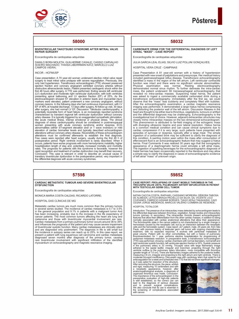
59852CASE REPORT: PROLAPSING OF GIANT MOBILE THROMBUS IN THE TRICUSPID VALVE UNTIL PULMONARY ARTERY BIFURCATION IN PATIENT WITH TESTICULAR GERM CELL TUMOREcocardiografia de cardiopatias adquiridas
NATAN CACCIA COSTA; RAPHAEL CASSIANO MOREIRA; DÉBORA TABOSA DE ALMEIDA; LETICIA BRAGA PACIELLO DA SILVA; GIL VICENTE LICO CIVIDANES; FABRICIO ASSAMI BORGES; TIAGO KENJI TAKAHASHI; CAIO CESAR JORGE MEDEIROS; MARCOS VALÉRIO COIMBRA DE RESENDE;
HOSPITAL TOTALCORIntroduction: The presence of an intracavitary mass detected by echocardiogram leads to the differential diagnosis between thrombus, vegetation, foreign bodies and intracavitary tumors (primary or secondary). The intracardiac thrombi present echocardiographic features that allow their differentiation from the other cardiac masses, in addition to being normally associated with cardiac anatomical alterations that allow their appearance. The thrombophilic state in the cancer patient is not characterized by a single change in hemostasis caused by the neoplastic cell, but by an interaction between the neoplastic cells and the hemostatic system. Case report: JLF patient, male, 29 years old, from São Paulo, with previous history of testicular germ cell tumor with ongoing chemotherapy treatment, with no expected response. It evolves with peritoneal metastasis of great volume. Patient with no other comorbidities, but with a history of pulmonary thromboembolism for 1 year, performs elective hospitalization for programming of peritoneal metastasis resection. In preoperative tests, a transthoracic echocardiogram (TTE) was performed, showing: cardiac chambers with normal diameters, normal left and right ventricular systolic function, left ventricular ejection fraction is 55%. Systolic pressure in the pulmonary artery is 33 mmHg. Tricuspid valve with large filamentary image adhered to the septal leaflet, irregular and branched, projecting through the right ventricle outflow to the pulmonary artery bifurcation, more compatible with thrombus (greater length of 7.6 cm). There are filamentary images adhered to the posterior leaflet, measuring 2.8 cm, irregular and projecting to the right atrium and right ventricle. There is moderate tricuspid insufficiency. Discussed case with cardiology team that opted for full anticoagulation for a week and scheduled new TTE that did not show significant changes, so it was opted for resection of the structure. After removing the structure, the piece, whitish and rigid, measuring 15 centimeters and with a neoplastic appearance, however, after anatomopathological analysis, a diagnosis of organized thrombus was made. Conclusion: The importance of the echocardiogram in the preoperative evaluation of high-risk patients is well established, so the correct use can lead to the diagnosis of serious diseases and to prevent surgical complications. We also conclude that its use is essential in the screening of complications associated with cancer.
58032CARBONATE DRINK FOR THE DIFFERENTIAL DIAGNOSIS OF LEFT ATRIAL “MASS" - CASE REPORT
Ecocardiografia de cardiopatias adquiridas
JULIA GARCIA LEAL ELIAS; SILVIO LUIZ POLLINI GONÇALVES;
HOSPITAL VERA CRUZ - CAMPINAS
Case Presentation A 81 year-old woman with a history of hypertension presented with new onset of palpitations and presyncope. Her medical history included gastroesophageal reflux disease. Transthoracic echocardiography identified a mass in the region of the left atrium. Left ventricular contractile function was intact and there were no significant valvular abnormalities. Physical examination was normal. Resting electrocardiography demonstrated normal sinus rhythm. To further delineate the intra-cardiac mass, the patient underwent 3D transesophageal echocardiography that revealed no intra-cardiac masses. Suspecting hiatal hernia, the patient was asked to ingest a commercially available carbonated drink during a transthoracic echocardiography; immediately after the first sip, we could observe that the “mass” was suddenly and completely filled with bubbles. After the echocardiographic examination, a cardiac magnetic resonance imaging was performed. It demonstrated a large hiatus hernia compressing and distorting the posterior wall of the left atrium. Discussion Masses in the heart are thought to be either due to thrombus, vegetations, tumours or artifact and the differential diagnosis may be challenging. Echocardiography is the investigational tool of choice. However, adjacent extracardiac structures may closely mimic intracardiac masses on the two dimensional echocardiogram. This phenomenon is attributed to far-field imaging of the echogenic mass with diverging ultrasonic beams and poor lateral resolution. Hiatus hernia is a common medical condition and can cause clinical symptoms due to cardiac compression if it is very large; such patients have presented with episodes of syncope or dyspnea, typically after a large meal. The simple ingestion of a carbonated drink may be sufficient to clarify the diagnosis of this condition. A swirling motion seen within the mass after consumption of effervescent fluid is an echocardiographic feature that may suggest a hiatus hernia. Final Comments It was realized 30 years ago that the sonographic appearance of a diaphragmatic hernia could simulate a left atrial mass. The ingestion of sparkling beverages for the echocardiographic diagnosis of hiatal hernias has been occasionally reported in the literature and may allow a simple way to clarify the diagnosis in cases of echocardiographic evidence of left atrial “mass” of unknown origin.
57598CARDIAC METASTATIC TUMOUR AND SEVERE BIVENTRICULAR DYSFUNCTION
Ecocardiografia de cardiopatias adquiridas
MONICA MARIA COSTA CALDAS; RICARDO LATORRE;
HOSPITAL DAS CLÍNICAS DE MG
Metastatic cardiac tumors are much more common than the primary tumors in several series studied. The incidence of cardiac metastasis is 0.7 to 3.5% in the general population and 9.1% in patients with a malignant tumor And has been increasing, probably due to the increase in the life expectancy of cancer patients. The most common tumors affecting the heart are lung and melanoma and those with biventricular myocardial involvement are rare. Cardiac metastasis from a primary pulmonary tumor occurs around 36 to 39% and influences the prognosis of the patient and may cause severe impairment of biventricular systolic function. Many cardiac metastases are clinically silent and are diagnosed only postmortem. The diagnosis in life is still small but the incidence in autopsy series can reach up to 25%. In this case report, we present a patient with lung squamous cell carcinoma and cardiac metastasis diagnosed seven months after diagnosis of the primary tumor, causing rare biventricular involvement with significant infiltration of the identified myocardium on echocardiography and magnetic resonance imaging.
58000BIVENTRICULAR TAKOTSUBO SYNDROME AFTER MITRAL VALVE REPAIR SURGERY
Ecocardiografia de cardiopatias adquiridas
DANILO BORA MOLETA; GUILHERME CASALE; CASSIO CARVALHO SOEIRO MACHADO; THAISA LIBERRMAN KATZ; MARCELO LUIZ CAMPOS VIEIRA;
INCOR - HCFMUSP
Case presentation: A 70 year-old woman underwent elective mitral valve repair surgery to treat mitral valve prolapse with severe regurgitation. Previously, she only had hypertension. Transthoracic echocardiogram (TTE) showed preserved ejection fraction and coronary angiogram exhibited coronary arteries without obstructive atherosclerotic lesion. Patient presented cardiogenic shock within the first 48 hours after surgery. A TTE was performed, finding severe left ventricular (LV) dysfunction and moderate right ventricular dysfunction, with both ventricles presenting apical ballooning and LV ejection fraction (EF) of 25%. As the electrocardiogram showed ST elevation in anterior leads and myocardial injury markers were elevated, patient underwent a new coronary angiogram, without coronary lesions. In the following days she had continuous improvement, with LV EF of 49% at hospital discharge, 13 days after surgery. At follow-up visit, 30 days after surgery, she had normal LV EF. Discussion: Takotsubo cardiomyopathy, a potentially reversible form of heart failure, is an increasingly reported syndrome characterized by transient regional left ventricular dysfunction without coronary artery disease. It is typically triggered by an exaggerated sympathetic stimulation, like acute medical illness, intense emotional or physical stress. The clinical diagnosis of stress cardiomyopathy should be considered in postmenopausal women presenting with acute coronary syndrome and/or dyspnea, ECG abnormalities (either ST-segment elevation and/ or T wave inversion) or mild elevation of cardiac biomarker levels and typically described echocardiogram alterations without coronary artery disease. Reversibility of these echocardiogram alterations must be confirmed afterwards to establish the final diagnosis. Few cases were reported after heart surgery, usually during the first 48 h of postoperative period. Right ventricle involvement is rarely described. When it occurs, patients have worse prognosis with more hemodynamic instability, higher hospitalization length of stay and, potentially, increased mortality and morbidity rates. The prognosis for patients with this syndrome is favorable, however the rapid recognition and treatment of cardiac dysfunction may benefit their recovery. Conclusion: This case is relevant in order to describe an unusual cause of transitory biventricular dysfunction in the postoperative period, very important in the differential diagnosis with acute coronary syndromes.

12
Pôsteres
Arq Bras Cardiol: Imagem cardiovasc. 2017;30(4 supl.1):6-41
57830COMPARISON OF EARLY AND LATE ECHOCARDIOGRAM DIAGNOSIS OF AORTIC PROSTHESIS MISMATCH AND IMPACT ON LEFT VENTRICLE REMODELING
Ecocardiografia de cardiopatias adquiridas
MARIA ESTEFÂNIA BOSCO OTTO; BIANCA CORRÊA ROCHA DE MELLO; LUIZ CARLOS MADRUGA RIBEIRO; LARISSA LUCAS SCHLOICKA; ANA CAROLINA PEREIRA MATOS DOMINGUES; MARCELO DO NASCIMENTO MOREIRA; JOYCE GOMES ELIAS LIMA; REYNA PINHEIRO CALZADA; MAIARA SANCHEZ RIBEIRO; ARMINDO JREIGE JÚNIOR; FERNANDO ANTIBAS ATIK;
ICDF/DF E CLÍNICA UCI
Background: Aortic prosthesis mismatch, should have an early transthoracic echocardiogram diagnosis 2-4 weeks (early TTE) after surgery, confirmed by a late echocardiography 6-12 months (late TTE) to predict outcomes. Purpose: Compare early and late diagnosis of mismatch in a series of patients from a tertiary Brazilian Center focus on Public Health Care submitted to aortic valve replacement (AVR) and observe the impact on left ventricle remodeling. Methods: From 316 patients submitted to AVR with TTE performed within the first 30 days after surgery studied from January 2011- July 2016 we had 65 patients with late TTE in 404 ± 306 days. Mismatch was classified according to aortic prosthesis effective orifice area (PEOA) in 3 groups: severe SM (PEOA < 0,65 cm2/m2), mild to moderate MM (PEOA = 0,65 cm2/m2 to 0,85 cm2/m2) or no mismatch (NL) (PEOA > 0,85 cm2/m2). To study ventricular remodeling, we compared left ventricular mass index (LVMi), left ventricular systolic (LVSD) and diastolic diameters (LVDD). Results: The mean age was 55 ± 17 y, 63% males. There was a high incidence of mismatch: SM 36%; MM 34% and NL29% at early TTE. At late TTE the incidence was similar SM 35,5%; MM 35,5% and NL 29%. However, 64% of patients were classified at the same group of mismatch and 36% changed groups with 17% increasing mismatch grade and 19% improving mismatch (p = 0,22). For SM only 2 patients were considered NL at late TTE (8%). There was no difference in systolic blood pressure (117 ± 18; 124 ± 26 for late TTE; p = NS); diastolic blood pressure (69 ± 13; 69 ± 13 for late TTE; p = NS) and heart rate (84 ± 18; 78 ± 15 for late TTE; p=NS). LVMi regression was similar and significant (p < 0,05) in all groups from early TTE (SM 121± 43 g/m2; MM 127 ± 38 g/m2; NL 123 ± 49g/m2) to late TTE (SM 94 ± 21 g/m2; MM 110 ± 45 g/m2; NL 102 ± 28 g/m2). LVDD regression was similar and significant (p < 0,05) from early TTE (SM 51 ± 12 mm; MM 50 ± 9 mm; NL 53± 10 mm) to late TTE (SM 48 ± 5 mm; MM 48 ± 8 mm; NL 50 ± 7 mm). LVSD regression was similar and significant (p < 0,05) from early TTE (SM 35 ± 11 mm; MM 36 ± 11 mm; NL 36 ± 12 mm) to late TTE (SM 30 ± 5 mm; MM 32 ± 9 mm; NL 33 ± 7 mm). Conclusions: Early TTE is reliable for mismatch diagnosis, but late TTE change classification in 36% of patients. The incidence of SM is high in a tertiary Brazilian Center although it does not modify significantly LV remodeling. Nevertheless, long-term follow up is necessary to look for changes in ventricular remodeling and symptoms.
57618CLINICAL AND ECHOCARDIOGRAPHIC DETERMINANTS OF AORTIC PROSTHESIS MISMATCH IN ONE BRAZILIAN TERTIARY SURGICAL CENTEREcocardiografia de cardiopatias adquiridas
MARIA ESTEFÂNIA BOSCO OTTO; MARCELO DO NASCIMENTO MOREIRA; LARISSA LUCAS SCHOICKA; MAIARA SANCHEZ RIBEIRO; LUIZ CARLOS MADRUGA RIBEIRO; REYNA PINHEIRO CALZADA; ARMINDO JREIGE JR; BIANCA CORRÊA ROCHA DE MELLO; JOYCE GOMES ELIAS LIMA; ANA CAROLINA PEREIRA MATOS DOMINGUES; FERNANDO ANTIBAS ATIK;
ICDF/DF E CLÍNICA UCI
Background: Aortic prosthesis mismatch, a possible cause of impaired late left ventricle remodeling, is reported in 30-70% of patients submitted to aortic valve replacement (AVR). Purpose: To identify the main determinants of aortic prosthesis mismatch in a surgical tertiary Brazilian center series of patients with emphasis on the Brazilian Public Health System Methods: 316 patients submitted to AVR with echocardiograms performed within the first 30 days after surgery were studied from January 2011- July 2016. In 176 the aortic prosthesis effective orifice area (PEOA) was obtained by continuity equation and patients were classified in 2 groups for analysis: severe mismatch SM (PEOA < 0,65 cm2/m2) and mild to moderate mismatch or no mismatch NM (PEOA > 0,65 cm/m2). The presence of SM was compared with clinical and echocardiographic variables such as: cause of AVR, etiology, associated diseases, body mass Index (BMI), LVOT diameter (LVOTD), gender, age, type and number of valve implanted, LV systolic and diastolic diameters, ejection fraction and left atrial index volume (LAVi). Results: The mean age was 56 ± 16, 61% of males, 57% had hypertension and 12,5% had DM. There was a high incidence of SM 34% and 66% NM. The cause for AVR was 84% Aortic Stenosis; 12,4% Aortic regurgitation; 3,4% Balance lesions and 0,2% prosthesis mismatch. The etiology was 48% degenerative, 21% rheumatic; 19% congenital; 10% aortic root dilation and 2% endocarditis; 86% received biological and 14% mechanical prosthesis. There was only 19% of aortic root enlargement surgery. The independent variables associated with SM by logistic regression were BMI, LAVi, age and LVOTD. An increase in one unit of BMI raises the risk for SM in 17,9% (p = 0,001); for LAVi, an increase in one unit raises the risk in 2,7% (p = 0,02); Age, an increase in one unit lowers the risk in 4,3% (p = 0,002) and for LVOTD an increase in one unit, lowers the risk for SM in 97,7% (p < 0,0001) creating a mathematical model, demonstrated in the attached file. Conclusions: Severe mismatch in AVR in a tertiary Brazilian Center is associated with higher BMI and LAVi, smaller LVOTD, and younger age. The mathematical model for identifying SM could be applied as a prevention in the pre-operative evaluation for the size and type of prosthesis implanted to prevent SM. In addition, this model can be helpful to select patients where aortic root enlargement surgery will be necessary.
58028CLINICAL AND ECHOCARDIOGRAPHIC DETERMINANTS OF ATRIAL FIBRILLATION IMMEDIATELY AFTER SURGICAL CORRECTION OF ORGANIC MITRAL REGURGITATION IN A BRAZILIAN POPULATION
Ecocardiografia de cardiopatias adquiridas
ADENALVA LIMA DE SOUZA BECK; TERESA CRISTINA ALVES DUARTE; JOSÉ MOREIRA KFOURI FILHO; DANIEL GODOY DEFAVARI; RONNY THOMAS ONIBENE OLIVEIRA; FERNANDO ANTIBAS ATIK; LUIS CLÁUDIO LEMOS CORREIA; MARIA ESTEFANIA BOSCO OTTO;
INSTITUTO DE CARDIOLOGIA DO DF/FUC-ICDF
Introduction: The optimal timing of mitral valve surgery (MVs) in patients with organic severe mitral regurgitation (MR) remains controversial. Ideally it should occur when left ventricular (LV) contractile function is still preserved. The contractile dysfunction may be present even when the left ventricular ejection fraction (LVEF) is still preserved and causes a higher incidence of postoperative mortality, heart failure and arrhythmias. Purpose: To identify predictors of atrial fibrillation in the early postoperative period of MVs (in the intensive care unit (ICU)) and thus help in the early indication of surgery for MR in a Brazilian population. Methods: One hundred and thirty-one patients (mean age 47.79 ± 17.11 years, 91 women, 85 rheumatic) who underwent MVs between january 2011 and december 2016 were retrospectively analyzed. Results: Atrial fibrillation (AF) occurred in 26 patients, 18 of whom were of the group with 10% decrease in the postoperative LVEF while 6 patients were of the group with < 10% decrease in the LVEF (p = 0.0001). Age was significantly higher in the group with reduction of postoperative LVEF (52,36 ± 17,38 vs 44,37 ± 16,19 years, p = 0.01). Left atrial volume indexed to the body surface area (LAVi) was increased (73,43 ± 34,18 ml/m) and similar in both groups preoperatively but only reduced in the group without postoperative ventricular dysfunction. On multivariate analysis, LAVi (odds ratio 1.02, p = 0.004) and age (odds ratio 1.05, p = 0.001) were independent predictors of AF in the ICU. Conclusion: In a Brazilian population where rheumatic fever is the predominant etiology, age and baseline LAVi are determinants of AF immediately after surgical correction of mitral regurgitation. These parameters may be useful for decision making regarding the optimal timing of MVs.
60686CASE REPORT: TAKOTSUBO CARDIOMYOPATHY OF RIGHT VENTRICLE IN A PATIENT WITH LUNG CANCER UNDERGOING CHEMOTHERAPYEcocardiografia de cardiopatias adquiridasRAPHAEL CASSIANO MOREIRA; NATAN CACCIA COSTA; DÉBORA TABOSA DE ALMEIDA; LETICIA BRAGA PACIELLO DA SILVA; AUGUSTO ALBERTO DA COSTA JR; CAIO CESAR JORGE MEDEIROS; MARCOS VALÉRIO COIMBRA DE RESENDE;HOSPITAL TOTALCORIntroduction: Takotsubo‘s cardiomyopathy is characterized by transient dysfunction of the apical and / or middle segments of the left ventricle and / or right ventricle that simulates myocardial infarction, but without significant obstructive coronary disease. Although the exact mechanism is still not fully understood, it is believed to be caused by an influx of catecholamines in the myocardial cells, generating calcium overload, leading to systolic dysfunction. The prevalence and incidence of Takotsubo are not yet perfectly established. However, biventricular dysfunction occurs in 26.0% to 30.0% of cases. Treatment to date is symptomatic, like other cardiomyopathies, is determined by the complications that occur during the acute phase. Case report: Patient M.H.C.F, 73, from São Paulo, with previous diagnosis of non-small cell lung carcinoma and breast metastasis. In treatment with chemotherapy and follow up with oncology sector. He was admitted to the emergency room with complaints of dyspnea on medium exertion (functional class III) and precordial pain. Electrocardiogram was requested: sinus rhythm, regular, without changes in the ST segment and transthoracic echocardiography: severe left ventricular systolic impairment due to akinesia of the mid and apical regions of all the walls with hyperkinetic basal regions; Moderate right ventricular systolic impairment due to akinesia of the apical middle region of the free wall and the anterior wall. We discussed a case with a multidisciplinary team and opted to perform coronary angiography that showed no critical coronary lesions. Patient was kept in intensive care with clinical improvement after two weeks. A new transthoracic echocardiogram was performed after 40 days, which showed improvement of left and right ventricular systolic functions without segmental deficit. Conclusion: The echocardiogram is an exam of choice for the diagnosis of Takotsubo cardiopathy because it is a non-invasive and rapid test. It helps to identify qualitatively and quantitatively left ventricular systolic dysfunction and/or right ventricular dysfunction. We also conclude that the possibility of chemotherapy triggering Takotsubo can not be entirely excluded, although studies are lacking to conclude this hypothesis.

13
Pôsteres
Arq Bras Cardiol: Imagem cardiovasc. 2017;30(4 supl.1):6-41
58076CORRELATION BETWEEN LEFT ATRIAL VOLUME AND FUNCTION IN HYPERTROPHIC CARDIOMYOPATHY
Ecocardiografia de cardiopatias adquiridas
THEREZA CRISTINA PEREIRA GIL; MARCIA BUENO CASTIER; ALYNE FREITAS PEREIRA GONDAR; ANA LUIZA FEREIRA SALES; RICARDO MOURILHE ROCHA;
INTO
Introduction: Atrial fibrillation is the most common arrhythmia in patients with hypertrophic cardiomyopathy (HCM), occurring in about one fifth of all HCM patients, which is four times the frequency expected in the general population. Atrial fibrillation is associated to left atrial (LA) enlargement, often leading to acute or progressive heart failure, as well as an increased risk of stroke. Left atrial enlargement is related to ventricular hypertrophy, ventricular filling pressures, and left ventricular outflow gradient, reflecting the hemodynamic condition. It has been demonstrated that LA increase is an independent factor of adverse outcome in this disease. Therefore, the early detection of patients at risk of atrial fibrillation has an important clinical implication regarding the monitoring and treatment of this disease. Purpose: Analyze the correlation between volume and LA function in patients with HCM. Methods: In a sectional study, 45 HCM patients were consecutively recruited from Pedro Ernesto University Hospital / State University of Rio de Janeiro (22 males, mean age 45 + 14 years). The echocardiographic studies were performed (iE-33 Matrix, Philips), using QLAB software. The left atrial volume was measured by 2D and 3D-echocardiography and compared with LA active emptying fraction. The statistical software package R (version 3.2.4) was used for correlation test analysis (Pearson’s test). Results: Left atrial volume was increased in 57.8% to 2D echo and 46.7% had maximum volume increased to 3D echo (> 34 ml/m2). Reduction of LA active emptying fraction (< 43%) was observed in 22% of the patients. Statistical analysis showed a strong correlation between LA volume increase and reduction of active emptying fraction both to the 2D echo (r - 0.82, p < 0,05) and to the 3D echo (r -0.75, p < 0,05). Conclusion: Patients with HCM who have increased LA volume and reduced LA active emptying fraction are possibly at a higher risk of developing atrial fibrillation. Prospective studies are needed for better evaluation.
60618CONTRIBUTION OF THREE-DIMENSIONAL ECHOCARDIOGRAPHY IN THE TRANSOPERATIVE EVALUATION OF PERIPROSTHETIC LEAK
Ecocardiografia de cardiopatias adquiridas
SALVADOR GOMES NETO; FABIO CANELLAS MOREIRA; MARCELO DEMAMAN ANDRES; FERNANDO ANTONIO LUCCHESE;
SANTA CASA DE MISERICÓRDIA DE PORTO ALEGRE
A 56-year-old male patient with a history of mitral rheumatic valve disease and a bicuspid aortic valve with severe regurgitation, underwent implantation of aortic and mitral valve bioprostheses about a year ago. Patient evolved with postoperative infective endocarditis, with thickening and residual transprosthetic regurgitation of both prostheses. About two months ago, the patient developed progressive functional class worsening with dyspnea on minimal exertion, in addition to the detection of a diastolic murmur in the aortic approach, and underwent a transthoracic echocardiographic evaluation that demonstrated dysfunction of both valvar prostheses, with severe regurgitation and significant thickening of leaflets, besides a significant aortic paravalvular leak. Indicated surgical valve replacement, with implantation of bioprostheses in the mitral and aortic positions, monitored by transoperative three-dimensional echocardiography. In the withdrawal of the extracorporeal circulation, an important mitral paravalvular leak was detected, promptly corrected by the surgical team. The case described demonstrates the utility of three-dimensional transesophageal echocardiography in the optimization of the transoperative location of paravalvar leaks. The additional information provided by the three-dimensional image allows the surgical team to locate the presence of periprosthetic leaks with much greater precision than the two-dimensional method, allowing real-time localization of suture defects.
57994CONVENTIONAL ECHOCARDIOGRAPHIC PROFILE AND BY MEANS OF STRAIN IN PATIENTS WITH MUCOPOLYSACHARIDOSIS IN BAHIA-BRAZIL
Ecocardiografia de cardiopatias adquiridas
MIRELA FREDERICO DE ALMEIDA ANDRADE; ANGELINA XAVIER ACOSTA; EMÍLIA KATIANE EMBIRUÇU LEÃO; MOISÉS IMBASSAHY GUIMARÃES MOREIRA; TAIS ALVES; CARLOS MAURÍCIO CARDEAL MENDES; ISABEL CRISTINA BRITTO GUIMARÃES;
UFBA
Background: Mucopolysaccharidosis (MPS) is a genetic disease, a lisossomal storage of glycosaminoglycans. It affects various organs, with frequent cardiovascular compromise, characterized mainly by left valvular lesions and left ventricle (LV) hypertrophy. The cardiovascular signs and symptoms are underestimated due to the simultaneous involvement of the disease in other organs. Enzyme replacement therapy (ERT) can be used in MPS I, II, IV and VI. In face of this scenario, the knowledge of the application of new tools of conventional and advanced echocardiography is relevant in order to improve cardiac care. Methods: This is an outpatient sectional descriptive study, from the genetic services of the Federal University of Bahia-Brazil. The patients underwent conventional echocardiography and strain by speckle-tracking from January to June, 2016. Results: 16 patients have been evaluated with median age of 14.2 years (deviation: 5.2 years); 12 (75%) were male. The most common type: MPS VI (8 patients - 50%), MPS II (4 patients - 25%), MPS I (3 patients - 18.8%) and MPS III (1 patient - 6.2%). Left valve lesion was found in 15 patients (93.3%) with a higher prevalence of mitral lesions - 13 patients (81.2%) all of which had insufficiency. Twelve (75%) showed concentric LV remodeling. All patients had LV systolic function preserved (Simpson and Teichholz). Three (18.8%) patients had abnormal LV MPI (myocardial performance index). Nine (56.2%) patients had change in LV global longitudinal strain (SGL). The study showed a positive association between mass and change the LV MPI and LV SGL and start time of ERT and LV MPI and LV SGL. Conclusions: Echocardiographic changes in patients with MPS are frequent, especially the left valve changes, change in LV geometry and subclinical LV dysfunction. The use of the new tools of conventional and advanced echocardiography can improve this follow-up.
59792COMPARISON OF TRANSESOPHAGEAL AND TRANSTHORACIC ECHOCARDIOGRAM WITH TRANSCRANIAL DOPPLER FOR THE DIAGNOSIS OF PATENT FORAMEN OVALE IN PATIENTS AFTER CEREBROVASCULAR ACCIDENTEcocardiografia de cardiopatias adquiridas
MARIA LUCIANA HANNOUCHE TRINDADE; ANA CLARA TUDE RODRIGUES; MARCELO DE LIMA OLIVEIRA; EDSON BOR-SENG SHU; GISELE SAMPAIO SILVA; CLAUDIA GIANINNI MONACO; EDGAR LIRA FILHO; MARCELO LUIZ DE CAMPOS VIEIRA; CLAUDIO H FISCHER; SAMIRA S MORHY;
HOSPITAL ISRAELITA ALBERT EINSTEIN
Introduction: A patent foramen ovale (PFO) occurs in 10 to 15% of the general population, and up to 45% of patients with cryptogenic ischemic stroke (iCVA). Diagnosis is either with transcranial Doppler (TCD), transthoracic echocardiogram (TTE) or transesophageal (TEE); TEE sensitivity is historically superior to TCD due to excellence of image quality, but requires an adequate Valsalva maneuver, not always effective during sedation. To assess concordance between these examinations (TEE + TTE vs TCD)regarding the diagnosis of a PFO, we studied patients within one week of iCVA. Methods: We assessed patients of both sex, > 18 years old with confirmed diagnosis of iCVA (magnetic resonance and/or computed tomography). TTE was immediately followed by TEE, both with peripheral intravenous injection of agitated saline to detect left-sided MB, with and without Valsalva maneuver. The exam was positive when MB were detected in the left cavities within the first 3 cardiac cycles after saline ejection. The TCD, also with and without Valsalva, was considered positive in the presence of high intensity signals up to 20s of the injection of agitated saline. Concordance between TCD versus TTE/TEE regarding the detection of left-sided MB was tested with kappa statistics, and TCD used as the reference exam. Sensitivity, specificity, positive predictive value (PPV) and negative predictive value (NPV) were also calculated. Results: We studied 50 patients, 28 male, mean age of 60± 13 years, from June 2013 to May 2016. Three patients with a positive TCD did not show PFO for TEE/TTE, while two patients with negative TCD presented with PFO during TEE/TTE, resulting in an excellent agreement (kappa = 0.79). In the TEE/TTE negative patients, the transthoracic window was limited (1 patient), and two did not perform Valsalva adequately. For TCD, it was assumed that Valsalva maneuver was not synchronized with contrast injection. Conclusion: The TTE association with the TEE and MB allows for the diagnosis of PFO accurately, showing excellent concordance with the TCD.
TCD Microbubbles
PFO TTE/TEE
Neg. Pos. Total Kappa Sens. Spec. PPV NPV
n % n % n % (IC 95%) (IC 95%) (IC 95%) (IC 95%) (IC 95%)
Neg. Pos. - - - - -
Total
14
Pôsteres
Arq Bras Cardiol: Imagem cardiovasc. 2017;30(4 supl.1):6-41
57883DIASTOLIC FUNCTION: COMPARING GUIDELINES IN AMBULATORIAL PATIENTS
Ecocardiografia de cardiopatias adquiridas
MÁRCIA BUENO CASTIER; DANIEL RANGEL BARROCAS; MARCELLA DE AGOSTINI ISO; CARLOS EDUARDO JAZBIK; ROBERTO POZZAN; ANGELO ANTUNES SALGADO;
HUPE-UERJ
Introduction: Recent 2016 Diastolic Function Guidelines proposed new criteria and classification for evaluation of diastolic function (DF) Purpose: To compare 2009 to 2016 DF Guidelines (DFG) in the diagnosis of DF in patients with normal ejection fraction Methods: During May 2017, all ambulatorial patients that were scheduled for echocardiographic exam due to systemic hypertension, known coronary artery disease or both were enrolled. Exclusion criteria were: ejection fraction < 55%, atrial fibrillation/flutter, advanced AV block, significant mitral disease (stenosis, regurgitation more than mild or prosthesis) or implanted pacemaker. Indexed atrial volume (IAV), E’ velocities (septal and lateral), medium E/E’and tricuspid regurgitation jet velocity were measured. Mitral flow pattern was also analyzed. Pearson’s chi-square and Fisher’s exact tests were employed for statistical analysis. Results: 88 patients were studied, ages from 28 to 83 years old (64.1 +/- 11,0), 63.6% were female, 84.1% had arterial hypertension, 13.6% had known coronary artery disease and 2.3% had both. IAV was abnormal in 36pts in 2009 DFG and in 30 according to 2016 DFG (x2 = 65.74, p < 0.0001). E’ septal velocity was reduced in 73 pts according to 2009 DFG and in 61 in the new guideline (x2 = 40.85, p < 0.0001). When analyzing 2009 criteria, 11 pts had normal DF in 2009 and remained normal in 2016. 77 pts diagnosed with diastolic dysfunction (DD) in 2009 had normal (20.8%), indeterminate (45.5%) or DD(10 pts) according to 2016 DFG, with significant changes from one to another (x2 = 28.40, p < 0.0001). Conclusion: There are significant changes in evaluating and classifying DD when employing the 2016 DFG compared to 2009 DFG in an ambulatorial population.
60536DEHISCENCE OF MITRAL VALVE BIOPROTHESIS METAL FRAME - THREE-DIMENSIONAL ECHOCARDIOGRAPHIC EVALUATION
Ecocardiografia de cardiopatias adquiridas
SALVADOR GOMES NETO; FABIO CANELLAS MOREIRA; MARCELO DEMAMAN ANDRES; FERNANDO ANTONIO LUCCHESE;
SANTA CASA DE MISERICÓRDIA DE PORTO ALEGRE
An 85-year-old male patient underwent valve replacement surgery 10 years ago with a mitral bioprosthesis implant, due to severe mitral insufficiency secondary to posterior leaflet-related chordae rupture. At the time of surgery, myocardial revascularization with a left internal mammary artery bridge to the anterior descending coronary artery and saphenous bridge to the 1st diagonal was also performed, in addition to DeVega‘s plasty to reduce the tricuspid reflux. Patient remained clinically asymptomatic and without limitations to daily activities until about 6 months ago, when he started with exertional dyspnea, with progressive worsening, culminating with dyspnea on minimal effort and orthopnea. At the clinical examination, the patient presented with bilateral rales, irregular rhythm and tachycardia with a heart rate of approximately 120 bpm, in addition to a mitral systolic murmur.. Resting electrocardiogram demonstrating atrial fibrillation with high ventricular response. Transthoracic echocardiography showed mitral bioprosthesis with signs of systolic-diastolic dysfunction, with important thickening of its leaflets. Elevated transprosthetic gradients, with effective prosthetic orifice estimated at 1 cm2. Severe transprosthetic reflux to color Doppler, in addition to the presence of a hyperechoic structure, probably metallic, poorly defined, represented by linear echo, intraatrial, in posteromedial topography of the prosthesis, distant from the ring. There was no history of pacemaker implantation. In addition, a very significant volumetric increase was detected in the atrial cavities, especially the left atrium, in addition to right ventricular dysfunction and pulmonary hypertension. Three-dimensional transesophageal echocardiography was performed, confirming the echocardiographic signs of significant systolic-diastolic dysfunction of the valve prosthesis, and evidenced the presence of a circular, metallic, partially displaced structure of the prosthesis, compatible with posteromedial dehiscence of the prosthetic support metal frame, approximately 70% of its circumference. Patient submitted to cardiac surgery, which confirmed the findings of the echocardiographic study, with removal of the dysfunctioning prosthesis and implantation of a new bioprosthesis n° 29. Reoperation for bleeding in the POI, with a subsequent good postoperative evolution.The present case report shows the fundamental contribution of the three-dimensional echocardiographic study for the accurate diagnosis of this unusual clinical situation. There is, at least in our knowledge, no report of a similar diagnosis performed through three-dimensional echocardiography in the current literature. The three-dimensional evaluation of prosthetic dysfunctions has its place of prominence especially in peculiar cases like this one.
58050DIAGNOSTIC COMPLEMENTATION OF INCIDENTAL FINDING OF PERICARDIAL CYST BY CARDIOVASCULAR IMAGING METHODEcocardiografia de cardiopatias adquiridas
LUCAS CRESPO DE BARROS; LUCAS DUTRA RODRIGUES; SAMARA GOMES TORRES BEZERRA; ANA CLARA TUDE RODRIGUES; KAMILA FERNANDA STASZKO; MEIVE SANTOS FURTADO; INGRID KOWATSCH; FERNANDO LUIZ PINTO DE CARVALHO; DALTON DE SOUZA BARROS; ARTHUR CORTEZ GONÇALVES; CARLOS AUGUSTO DE SOUZA; KEYTH EVELYN FRANCELINO DE OLIVEIRA MARANHÃO; AMANDA EVELYN ANDRADE ROCHA; AMANDA DINIZ KIMURA; JOSÉ LÁZARO DE ANDRADE;
HOSPITAL SÍRIO LIBANÊS / HOSPITAL DAS CLÍNICAS (FMUSP)Presentation of the case: F.C.S.D, 30 years old, female, white, married, born and lived in São Paulo-SP. Currently pregnant, with a gestational age of 16 weeks. No prior comorbidities. She reports that, for about 6 years, she often returned to emergency care due to a prolonged flu syndrome, and a chest X-ray when performed, showed a nodular image in the right costophrenic sinus. The investigation was complemented with a thorax computed tomography and confirmed the diagnosis of pericardial cyst. Recentely, the patient presented with episodes of non cardiac chest pain, and an echocardiogram was requested for follow-up. Thoracic echocardiography revealed an anechoic image, adjacent to the right ventricle, without extrinsic compression of the cardiac structures. Discussion: Cysts usually range from 2 to 3 cm. They are found incidentally in the cardiorphrenic angle, with 2/3 of them being on the right and 1/3 on the left, but can be found anywhere in the mediastinum. Pericardial cysts are usually unilocular, well marginalized, spherical or teardrop-shaped, filled by serous fluid. Approximately 70% of cases are asymptomatic and incidentally diagnosed on chest radiography. The absence of symptoms at diagnosis is a sign of good prognosis, however, some have non-specific symptoms of compression adjacent structures causing cough, dyspnea, chest pain and arrhythmias. Echocardiography in most cases is sufficient to establish the diagnosis. Although computed tomography and nuclear magnetic resonance contributes to differentiate pericardial cyst from a solid mediastinal mass, the definitive diagnosis is only consolidated through anatomopathological findings. In the differential diagnosis solid tumors should be considered, including: angioma, lipoma, neurogenic tumor, sarcoma, lymphoma, bronchogenic carcinoma, metastasis, granulomatous lesion and abscess. Final comments: Pericardial cysts usually follow a benign course and there are no reports of malignant transformation. In asymptomatic patients, conservative management is recommended with short follow-up periods. The indications for surgical resection include: large size, symptoms, cyst infection, patient‘s request, suspected malignancy and prevention of complications. Treatment options for pericardial cysts include simple observation, thoracotomy excision, surgical removal by thoracoscopic and percutaneous aspiration with injection of a sclerosing agent, usually ethanol
57831CORRELATION OF THE MITRAL VALVE AREA BY THREE-DIMENSIONAL TRANSESOPHAGEAL PLANIMETRY WITH OTHER ECHOCARDIOGRAPHIC METHODS FOR MITRAL STENOSIS QUANTIFICATION
Ecocardiografia de cardiopatias adquiridas
ANGELE AZEVEDO ALVES MATTOSO; JEANE MIKE TSUTSUI; CARINA AMORIM POUILLARD CARNEIRO; WILSON MATHIAS JUNIOR; MARIA LUCIA DUARTE; EDUARDO TADEU FERREIRA;
HOSPITAL SANTA IZABEL / CLINICA PROALIVIO
Introduction: Interventional approaches for the management of valvular mitral stenosis (MS) depends on the adequate diagnosis of the stage of disease. For this, accurate measurements of the mitral valve area (MVA) and the hemodynamic consequences of valve obstruction are necessary. MVA can be measured by echocardiography in several ways, due to the discrepancies between them, such as: a) 2-dimensional transthoracic planimetry (challenging in front of a cone-shaped mitral valve foramen in rheumatic MS or extremely calcified valves) PTT2D; b) diastolic pressure half-time (PHT), which is dependent not only on the degree of MS but the compliance of left ventricle and atrium; c) continuity equation (VTI), which should not be measured in the presence of greater than discrete mitral or aortic regurgitation; and d) Proximal Isovelocity Surface Area (PISA).It should be considered in the analysis, the hemodynamic effect of the MS: transmitral mean gradient (MG); pulmonary artery systolic pressure (PASP) and left atrial volume (LAV). In this scenario, three-dimensional echocardiography emerges, offering singular advantages, such as allowing correct vision (en face) of the mitral valve apparatus at the top of its cusps. It has been demonstrated higher accuracy for the measurement of the MVA by transthoracic three-dimensional echocardiography (PTT3D) and especially echocardiography three-dimensional transesophageal (PT TE3D). Objective: To define the correlation between the most recent approaches, PTT3D and PTE3D, with the other methods for estimation of the MVA; to define the relation between these measurements and hemodynamic effect of the valve obstruction. Methods: From April 2015 to April 2017, we prospectively studied 43 patients (pt) with valvular heart disease who underwent transesophageal echocardiogram. MVA was obtained through PTT2D (32pt), PTT3D (22pt), PHT (41pt), PISA (8pt), VTI (36pt), PTE3D (43pt) and hemodynamic data as described. Results: There was a significant correlation between MVA measurements by PTE3D and PISA (r = 0.967; p < 0.001); PTE3D and VTI (r = 0.923; p < 0.001. There was no correlation between the other measures of the MVA. Regarding hemodynamic data, there was an inversely proportional correlation between PTE3D and PASP (r = -0.54; p < 0.001); PTE3D and MG (r = -0.765 p < 0.001); PTT2D and MG (r = -0.415; p = 0.018), PISA and MG (r = -0.873; p = 0.005), PHT and MG (r = -0.624; p < 0.001); VTI and PASP (r = -0.610; p < 0.001) and VTI and MG (r = -0.741; p < 0.001). No significant correlation between the other measurements of the MVA and the data of hemodynamic was observed. Conclusion: The methods for measuring MVA that showed better correlation in MS were PTE3D, VTI and PISA. However, they did not showed correlation with measures commonly used in clinical practice, such as PHT and PTT2D. These measures also had better correlation with the hemodynamic effect of the MS, demonstrating a more precise evaluation of valve disease stage.

15
Pôsteres
Arq Bras Cardiol: Imagem cardiovasc. 2017;30(4 supl.1):6-41
57903ECHOCARDIOGRAPHIC ANALYSIS OF VENTRICULAR MECHANICS IN FEMALE ATHLETES
Ecocardiografia de cardiopatias adquiridas
FÁBIO ALMEIDA MACIEL SOBRINHO; DANILO MIRANDA ALMEIDA ARAÚJO; GABRIEL FREIRE CORDEIRO SAMPAIO; ISAAC NEWTON GUIMARÃES ANDRADE; JOSÉ MARIA DEL CASTILLO; EUGENIO SOARES DE ALBUQUERQUE; CARLOS ANTONIO DA MOTA SILVEIRA; OSCAR FRANCISCO SANCHEZ OSELLA; ANTONIA DULCINEIDE MEDEIROS SENA;
PRONTO SOCORRO CARDIOLÓGICO DE PERNAMBUCO PROF LUIZ TAVARES - UPE
Introduction: Athletic activities can be classified by the static (anaerobic) and dynamic (aerobic) components. Soccer is characterized by low static component (< 20% muscle contraction) and high dynamic component (> 70% oxygen consumption), which causes myocardial adaptation phenomena. Objective: to evaluate professional female soccers with conventional echocardiography and speckle tracking, determining longitudinal strain and strain rate, circumferential and radial strain, apical and basal rotation and myocardial twisting. The data were compared with sedentary individuals of the same age group. Material: studied 27 female athletes, mean age 19 ± 2 years, determining the dimensions and function of the LV and RV, LV mass index, diastolic function, LA and LV volumes, longitudinal, circumferential and radial strain of the LV, longitudinal strain of the RV, basal and apical rotation and LV twisting. The data were compared with the results of sedentary individuals of the same age group obtained in meta-analyzes. Results: There were no significant differences in the dimensions and systolic and diastolic function of the LV and RV, except in heart rate (60.8 ± 7.2 bpm vs 75.1 ± 8.9 bpm, p < 0.0001). Mass index and indexed volume of LA also showed no significant difference. The longitudinal, circumferential and radial strain of the LV and the RV showed no significant difference. The basal and apical rotation and LV twisting were significantly decreased (p < 0.0001) in the athletes (-4.1° ± 1.7° vs -6.1° ± 3.6° for the basal rotation, 6.9° ± 3.2° vs 13.1° ± 6.0° for The apical rotation and 11.1° ± 3.7° vs 19.1° ± 6.4° for the twisting). Conclusion: female athletes with low anaerobic activity and high oxygen consumption did not present alterations in cardiac dimensions, mass index, LV and LA volumes, and systolic and diastolic function parameters, both estimated by conventional echocardiography and myocardial deformation. Significant resting decrease in basal and apical rotation and LV twisting were observed, probably due a mechanism to store contractile reserve to supply the need during periods of intense aerobic activity.
58029EARLY POSTOPERATIVE OUTCOME AFTER SURGICAL CORRECTION OF ORGANIC MITRAL REGURGITATION WITH THE WATCHFUL WAITING STRATEGYEcocardiografia de cardiopatias adquiridas
ADENALVA LIMA DE SOUZA BECK; TERESA CRISTINA ALVES DUARTE; JOSÉ MOREIRA KFFURI FILHO; DANIEL GODOY DEFAVARI; RONNY THOMAS ONIBENE OLIVEIRA; FERNANDO ANTIBAS ATIK; LUIS CLAUDIO LEMOS CORREIA; MARIA ESTEFANIA BOSCO OTTO;
INSTITUTO DE CARDIOLOGIA DO DF/FUC-ICDF
Introduction: The optimal time for mitral valve surgery (MVs) in patients with severe organic mitral regurgitation (MR) remains controversial. Early intervention before reduction of left ventricular (LV) ejection fraction (LVEF), LV chamber remodeling, increased pulmonary artery systolic pressure (PASP) or appearance of heart failure (HF) may prevent LV dysfunction. However, if mitral valve repair is not possible, implanting a prosthesis may also worsen the prognosis, and in this case the “watchful waiting” strategy may be advisable. Purpose: Evaluate early postoperative outcome after surgical correction of MR in a population where rheumatic fever is the predominant etiology. In this population repair is often not possible, and therefore the “watchful waiting” strategy predominated. Methods: One hundred and thirty one patients (mean age 47.79 ± 17.11 years, 91 women, 85 rheumatic) who underwent MVs were retrospectively analyzed. Pre and postoperative clinical, surgical and echocardiographic parameters were compared in group A (< 10% decrease in LVEF) and in group B (≥ 10% decrease in LVEF). Results: Before surgery, most patients had LVEF > 60% (84%) and some degree of HF (91%). Mitral valvar prosthesis was implanted in 112 patients. Decrease in LVEF (from 68.15 ± 8 to 54.53 ± 10.63 p < .0001) was noted in 56 patients. Still, the majority of the patients improved the HF in both groups (p < 0.0001). Diastolic diameter of the left ventricle, left atrial volume (LAV) and PASP decreased immediately after MVs in both groups (p < 0.0001). Systolic diameter of the left ventricle also decreased in group A (from 35.87 ± 6.1 to 31.75 ± 5.4 p < 0.0001) but increased in group B (from 34.16 ± 6.95 to 36.23 ± 8.23 p = 0.004). During intensive care unit admission, there were 4 deaths and 4 strokes (2 in each group). Atrial fibrillation occurred in 26 patients, 18 from group B (p = 0.0001). The first 6 months after discharge presented 4 deaths, with no significant difference between groups. Conclusion: In a population with MR predominantly rheumatic who underwent MVs with the “watchful waiting” strategy, worsening of LVEF was noted in 42% of the studied population, who had more AF in the ICU. Nevertheless, during the first 6 months there was improvement of HF and reverse LV remodeling in the entire group. Death and stroke were similar in both groups.
010203040506070
Pré Pós
CFNYHA - Geral
I
II
III
IV
58005ECHOCARDIOGRAPHIC PARAMETERS ASSOCIATED WITH 6-MIN WALK TEST (6MWT) IN DILATED CARDIOMYOPATHY (DM)Ecocardiografia de cardiopatias adquiridas
ULISSES PEREIRA MENDONÇA; DIANE MICHELA NERY HENRIQUE; ANA PAULA FERREIRA; JÚNIA FLÁVIA APARECIDA PEREIRA CANAAN; MARSELHA MARQUES BARRAL;
HOSPITAL E MATERNIDADE THEREZINHA DE JESUSIntroduction: Heart failure is still a global burden which carries substantial risk of morbidity and mortality. Thus, appropriate approach of diagnosis and layering the prognosis of heart failure are of great importance. Doppler echocardiography represents one of the most important methods in the evaluation of these patients. The 6-min walk test (6MWT) provides information regarding functional capacity and prognosis across a range of chronic cardiopulmonary conditions. Objective: The aim of this study was to compare echocardiography parameters (ECO) with the 6MWT distance in patients with DM. Methods: nineteen consecutive patients with DM were enrolled. Patients were included in the study if they had diameter/body surface area (BSA) > 31mm and left ventricular (LV) ejection fraction < 50% in Simpson method. Medical therapy was individually adjusted according to a standardized treatment regimen. 6MWT and ECO were obtained on the same occasion in all patients. All recordings were performed by one investigator, who was blinded to the clinical evaluation of the patients. The ECO techniques were performed according to the recommendations of the American Society of Echocardiography. Patients who had atrial fibrillation, pulmonary or orthopedic diseases were excluded. Data were expressed as the mean value ± standard deviation for continuous variables and absolute or relative frequencies for categorical variables. Correlations between 6MWT and ECO variables were assessed using GraphPad software. The permanence of all variables in the final model was established when the significance level was < 0.05. Results: The mean age was 53 years and 48% were women. The mean distance performed in 6WMT was 533,1 meters and mean fraction ejection was 41%. According to the established classification of diastolic dysfunction, four (21%) patients had normal diastolic function, 9 (48%) showed grade I of diastolic dysfunction, 2 (10%) grade II and four (21%) grade III. In multivariate analysis, the ECO variables that remained correlated with the 6MWT distance were left atrial volume index (ml) (r = 0,56; p = 0,01) (graphic 1) and E/E‘ right ventricle (r = -0,50; p = 0,02). Conclusions: The left atrial volume index has been shown to be associated with a pulmonary capillary wedge pressure in patients with depressed ejection fractions and correlates with 6MWT.E/E‘ right ventricle ratio > 6 reflects right ventricle dysfunction and highs levels associated with a lower distance performed in 6MWT.
57862DIFFERENTIATED CASE OF HAMARTOMA AND ITS CORRELATION WITH LITERATUREEcocardiografia de cardiopatias adquiridas
VICTOR RODRIGUES RIBEIRO FERREIRA; ELIANA MIGLIORINI MUSTAFA; SOFIA BRAILE SABINO; MARIA CHRISTIANE VALÉRIA BRAGA BRAILE-STERNIERI; BETHINA CANAROLI SBARDELLINI; GIOVANNI BRAILE STERNIERI; LÚCIA ANGÉLICA BUFFULIN DE FARIA; IDIBERTO JOSÉ ZOTARELLI FILHO; DOMINGO MARCOLINO BRAILE; VICTOR RODRIGUES RIBEIRO FERREIRA; MARIA CHRISTIANE VALÉRIA BRAGA BRAILE-STERNIERI; IDIBERTO JOSÉ ZOTARELLI FILHO;
INSTITUTO DOMINGO BRAILE
Case report: A 63-year-old female patient with a history of chest pain and dyspnea on exertion with progressive worsening six months ago. The echocardiography presented apical akinesia with an intramyocardial fibro-calcified mass, presenting an intense local coronary vascularization, with a dilated epicardial coronary artery at the tip with 5.0 mm, where the intramyocardial vessels that irrigate the tumor of the apical region originate. The superficial myocardial bridge was visualized by computed tomography angiography (CTA) in the middle segment of aDA. In the CTA and magnetic resonance imaging (MRI) study, myocardial thickness increased in the apical region and in the anteroposterior-medial LV segment around 2.2 cm, with an irregular appearance and extensive areas of calcification and microaneurysms. This mass also shows a cleavage plane with the structures adjacent to the myocardium and heterogeneous signal. The myocardial perfusion had a partial defect in the calcified areas, presence of areas of fibrosis and apical necrosis and in the papillary muscle. Coronary angiotomography showed important obstructive coronary artery disease. The patient underwent endomyocardial biopsy with a suggestive result of Hamartoma of mature cardiac myocytes. Subsequently submitted to the withdrawal of the tumor mass in apical region associated with the myocardial revascularization, presenting good postoperative evolution. Microscopic analysis of the anatomopathological showed that Hamartoma presented dense collagen, adipose tissue, hypertrophic and with disarticulated cardiomyocytes. Discussion: The present paper describes a case report of a very rare apical cardiac HA, with akinesia of the apical region with presence of intramyocardial fibro-calcified mass, presenting intense local coronary vascularization, with dilated epicardial coronary artery at the tip with 5.0 mm, where Intramyocardial vessels that irrigate the tumor of the apical region. The MRI results showed an increase in myocardial thickness in the apical region and in the anteroposterior-medial segment of the LV, around 2.2 cm, with irregular appearance and presence of extensive areas of calcification and microaneurysms. This directed strongly to the diagnosis of apical HA. Final comments: Despite the diverse range of cardiac neoplasms, heart tumors are very rare, even more so than the apical Hamartoma and confused with fibroma presented in the present work.

16
Pôsteres
Arq Bras Cardiol: Imagem cardiovasc. 2017;30(4 supl.1):6-41
60730FEIGENBAUM‘S WALL MOTION SCORE INDEX EVALUATING MYOCARDIAL PERFORMANCE TO ASSESS LEFT VENTRICULAR FUNCTION APPROPRIATE TO BE USED IN HANDHELD MACHINES AT BEDSIDE
Ecocardiografia de cardiopatias adquiridas
MARA LUCIA NEVES DOS SANTOS MOSQUEIRA GOMES; ANGELA DE AMEIDA LEITE; CESAR AUGUSTO DA SILVA NASCIMENTO; BERNARDO RANGEL TURA; RAPHAEL NEVES DOS SANTOS MOSQUEIRA GOMES; LAURA DE ABREU ALVES; LAISSE MARINS DEFANTI GONZAGA; EMANUELE FERREIRA ADRIANO; GABRIELLA ANDRADE DE SÁ; RAFAEL DE OLIVEIRA CARDOSO; BRUNO ROBERTO ITABORAHY ALABRIN;
INC
Background: Despite the availability of several, up-to-date and fast methods to obtain Left Ventricular Ejection Fraction (LVEF) values, the subjective impression is still the most accessible one to the physicians needing a helping number in evaluating their patients. In this way, a cardiac formula using the Wall Motion Score (WS) to evaluate the LVEF was previously proposed by our group and displayed a significant correlation with LVEF obtained by Simpson’s rule(SR). Now, we seek to obtain an even better correlation between LVEF from Feigenbaum’s WS classification(FWS) and LVEF from SR. Purpose: Evaluate a process to offer to the intensive care specialist an archetype number of left ventricular function near to real, appropriate to be used in handheld machines at bedside. Methods: The sample population consisted of 18 patients (mean age 62.28, from 42 to 88 years old, 77,78% male) with ischemic heart disease with and without failure. The WS index was studied using 17 segments and each segment was scored according to FWS and according to the Traditional classification (TWS). By regression analysis, it was possible to obtain a formula that yielded LVEF calculation using WS closely resembling the LVEF obtained by SR. The regression equation was Index = 90.5 – 27.8 x WS, simplified to Index = 69/WS. Then, we obtained the LVEF using the simplified formula against SR. GE E9 echo machine was used for all the analysis, with a 3.5 MHz transducer. Images were storaged and analyzed by two different observers. Bland and Altman plot was used for statistical analysis. Results: The LVEF values obtained through: SR varied from 19% to 69% (mean 41.82%); TWS varied from 26% to 69% (mean 38.84%); FWS varied from 25% to 69%(mean 38.31%). Bland and Altman plot analysis showed the following results: FWS LVEF x SR LVEF yielded mean bias 3.09(95% CI, -8.18 to 14.35); TWS LVEF x SR LVEF yielded mean bias 2.38(95% CI, -12.03 to 16.78); FWS LVEF xTWS LVEF yielded mean bias -0.26 (95% CI, -7.98 to 7.46). These findings show that confidence intervals for FWS LVEF are superior to those for TWS LVEF. Conclusions: Calculation of LVEF using FWS is feasible and yields even more reliable values than those obtained through TWS. Thus, it can be used in everyday bedside clinical practice with handheld machines without up-to-date software, as it is cheaper and easier to execute. Another advantage of this method is that we only use the segments which are adequately seen.
57840ECHOCARDIOGRAPHIC EVALUATION OF PRESSURE AND VOLUME OVERLOAD OF THE RIGHT VENTRICLE
Ecocardiografia de cardiopatias adquiridas
JONNY VITOR DINIZ; MARCIO SOUZA NASCIMENTO; ANDRÉIA LOPES DE FARIAS; JOSÉ MARIA DEL CASTILLO; EUGENIO SOARES DE ALBUQUERQUE; CARLOS ANTONIO DA MOTA SILVEIRA; OSCAR FRANCISCO SANCHEZ OSELLA; ANTONIA DULCINEIDE MEDEIROS SENA;
PRONTO SOCORRO CARDIOLÓGICO DE PERNAMBUCO PROF LUIZ TAVARES - UPE
Introduction: The function of the right ventricle (RV) in patients with pressure overload (RVPO) and volume overload (RVVO) was analyzed. Objective: To analyze echocardiographic abnormalities and RV longitudinal and transverse strain in patients with RVPO, RVVO and healthy individuals (CTRL). Methods: 50 CTRL individuals (mean age 47 ± 18 years, 31 female); fifty RVPO patients (mean age 48 ± 12 years, 32 female) and thirty RVVO patients (mean age 43 ± 14 years, 19 female). Were analyzed the dimensions of the left ventricle (LV), left atrium (LA), right ventricle and right atrium (RA), LV and RV function, two-dimensional longitudinal strain of the LV and RV and RV transverse strain. The means and standard deviation were compared by analysis of variance, with a significance of p < 0.05. Results: Demographic data showed no significant differences between groups, as well as the LV ejection fraction and indexed LA volume. RV diameter and wall thickness, tricuspid regurgitated gradient and indexed volume of RA were higher in RVPO patients. The parameters of RV function were decreased in RVPO and were normal in RVVO patients. The transverse strain of the RV lateral wall was increased both in RVPO and RVVO. Conclusion: Echocardiographic parameters and two-dimensional strain showed signs of RV remodeling, both in RVPO and RVVO, and decreased systolic function in RVPO. Increased transversal RV strain is probably due to the chamber remodeling, and decreased RV systolic longitudinal strain to chamber dysfunction. Decreased LV longitudinal strain may indicate early biventricular dysfunction.
58026ECHOCARDIOGRAPHIC EVALUATION OF THE DIASTOLIC DYSFUNCTION IN PATIENTS WITH HYPERTROPHIC CARDIOMYOPATHY: A RETROSPECTIVE STUDY
Ecocardiografia de cardiopatias adquiridas
JAMIL ALLI MURAD JUNIOR; CRISTIANE DE CARVALHO SINGULANE; MAURICIO DE NASSAU MACHADO; PAULO ROBERTO PAVARINO; JOAO FOLCHINE TRINDADE; MARINA BRAGA DE OLIVEIRA; ANGELICA NORIEGA FONSECA; LUIZ FERNANDO DAL COL; HÉLIO AUGUSTO DOS REIS CORBUCCI;
FUNDAÇÃO FACULDADE REGIONAL DE MEDICINA DE SÃO JOSÉ DO RIO PRETO (FAMERP)
The presence of diastolic dysfunction in patients with hypertrophic cardiomyopathy (HCM) is a prognostic marker, determining a higher mortality in 5 years even when the dysfunction is present in grade I. Objective: To evaluate the presence of diastolic dysfunction in patients who underwent echocardiographic examination In the last 10 years in a tertiary referral hospital in the treatment of cardiomyopathies. Methods: A total of 112930 echocardiography examinations performed between 2006 and 2016 were retrospectively analyzed, including 119 patients (53 years, 41-65 years) with a diagnosis of HCM with parietal thickness greater than or equal to 15 mm and variables related to the morphology and function of Left ventricle and atrium were analyzed between two distinct groups stratified according to the degree of diastolic dysfunction. Results: The prevalence of diastolic dysfunction greater than or equal to grade 2 was 12.1%, and grade III was present in only 1.5% of the patients. Comparative analysis between groups identified a statistically significant difference between left ventricular parietal thickness (p = 0.012). The degree of diastolic dysfunction and the thickness of the interventricular septum presented a positive correlation after simple linear regression (r2 square = 0.29 with P = 0.049). Conclusion: Moderate or significant diastolic dysfunction is an uncommon finding in patients with HCM and its degree correlates with a greater thickness of the LV septal wall.
57909ECHOCARDIOGRAPHIC ASSESSMENT IN PATIENTS WITH CHAGAS DISEASE: EVALUATION OF DIMENSION, FUNCTION AND DEFORMATION OF CARDIAC CHAMBERS IN THE INDETERMINED FORM
Ecocardiografia de cardiopatias adquiridas
KLEIBER MARCIANO LIMA BONFIM; ANA KAROLINA QUEIROZ DE SOUZA; ARNALDO XAVIER OLIVEIRA JUNIOR; SARA MARIA CRISÓSTOMO DE OLIVEIRA; FLÁVIA LIMA DE HOLANDA; JOSÉ MARIA DEL CASTILLO; EUGENIO SOARES DE ALBUQUERQUE; CARLOS ANTÔNIO DA MOTA SILVEIRA; OSCAR FRANCISCO SÁNCHEZ OSELLA; ANTONIA DULCINEIDE MEDEIROS SENA;
PRONTO SOCORRO CARDIOLÓGICO DE PERNAMBUCO PROF LUIZ TAVARES - UPE
Introduction: It is denominated indeterminate form of Chagas disease, which presents only positive serology, without other symptoms or clinical signs. As some patients may progress to symptomatic forms, early detection of signs indicative of this condition is important. Objective: To evaluate, by conventional echocardiography and myocardial deformation methods, the parameters may change earlier in the indeterminate form of Chagas‘ disease, comparing the data with healthy individuals of the same age group. Methods: We studied 30 patients with positive serology for Chagas disease who met the criteria for indeterminate form, mean age 51 ± 9 years. We determined demographic parameters and studied echocardiography dimensions, mass and systolic and diastolic function of the LV and RV and atrial volumes. With speckle tracking we analyzed the longitudinal deformation of the LV and RV, the mechanical dispersion, the circumferential and radial strain, the LV twisting LV and the LA and RA longitudinal strain. These data were compared, paired, with those of 30 healthy subjects, mean age 49 ± 10 years. Results: Height and body surface area were lower in the indeterminate group. Among the cardiac dimensions LV diameters and mass index were higher in the indeterminate group. Volumes, systolic function and diastolic parameters showed no difference, except the s‘ and e ‘ tissue Doppler waves were smaller in the indeterminate group and the higher E/e‘ ratio. In RV, area change was lower in the indeterminate group, but TAPSE showed no difference. The tricuspid regurgitation velocity and the AD volume were higher in the indeterminate group. Among the parameters of myocardial deformation the mechanical dispersion of the LV and the RV were larger in the indeterminate group with non-significant difference of the other parameters. Conclusion: Patients with undetermined form of Chagas‘ disease presented a larger dimension of the cardiac chambers and decreased mitral annular velocities, but with values still within the range of normal, which does not allow a clear separation with the normal individuals. Among the parameters that measure myocardial deformation, the significant increase in the mechanical dispersion of both ventricles was noted, and may be indicative of changes in the conduction of the electrical stimulus, which is one of the first changes observed in patients with cardiac form.
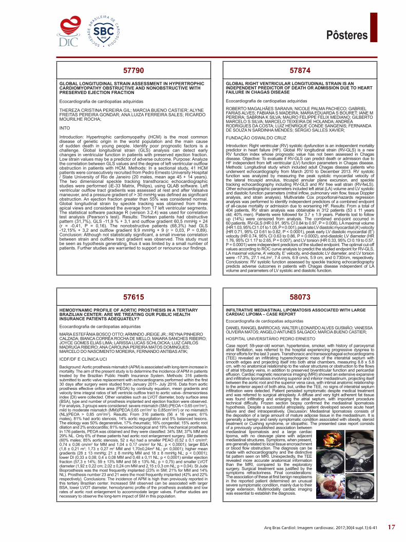
17
Pôsteres
Arq Bras Cardiol: Imagem cardiovasc. 2017;30(4 supl.1):6-41
58073INFILTRATIVE MEDIASTINAL LIPOMATOSIS ASSOCIATED WITH LARGE CARDIAC LIPOMA – CASE REPORTEcocardiografia de cardiopatias adquiridas
DANIEL RANGEL BARROCAS; WALTER LEONARDO ALVES GUSMÃO; VANESSA OLIVEIRA MATOS; ANGELO ANTUNES SALGADO; MARCIA BUENO CASTIER;
HOSPITAL UNIVERSITÁRIO PEDRO ERNESTO
Case report: 59-year-old woman, hypertensive, smoker, with history of paroxysmal atrial fibrillation, was referred to the hospital experiencing progressive dyspnea to minor efforts for the last 3 years. Transthoracic and transesophageal echocardiograms (TEE) revealed an infiltrating hyperechogenic mass of the interatrial septum with smooth edges and projecting itself into both atrial chambers, measuring 8.8 x 5.8 cm, with no anatomical relationship to the valvar structures or obstruction to the flows of atrial tributary veins, in addition to preserved biventricular function and pericardial effusion. Cardiac magnetic resonance imaging (MRI) showed an extensive expansive and infiltrative lipomatosis involving superior and inferior mediastinum, projecting itself between the aortic root and the superior vena cava, with intimal anatomic relationship to the anterior aspect of both atria, but, unlike the TEE, no signs of interatrial septum infiltration were detected. Patient persisted symptomatic despite medical treatment and was referred to surgical atrioplasty. A diffuse and very tight adherent fat tissue was found infiltrating and enlarging the atrial septum, with important procedure technical difficulty. Frozen section biopsy confirmed the mediastinal lipomatosis hypothesis. Despite a successful atrioplasty, patient developed severe acute heart failure and died intraoperatively. Discussion: Mediastinal lipomatosis consists of the deposition of a large amount of mature adipose tissue in the mediastinum. It is generally a benign and rarely symptomatic condition associated with obesity, steroid treatment or Cushing syndrome, or idiopathic. The presented case report consists of a previously unpublished association between mediastinal lipomatosis and a large cardiac lipoma, with no cleavage plane with adjacent mediastinal structures. Symptoms, when present, are generally related to local tissue encroachment or blood flow obstruction. The diagnosis can be made with echocardiography and the distinctive fat pattern seen on MRI. Unexpectedly, the TEE revealed more accurate anatomical information than the MRI, compared to the exploratory surgery. Surgical treatment was justified by the symptoms refractoriness. Final considerations: The association of these at first benign neoplasms in the reported patient determined an unusual severe symptomatic condition, mainly due to their large extension. Multimodality cardiac imaging was essential to establish the diagnosis.
57874GLOBAL RIGHT VENTRICULAR LONGITUDINAL STRAIN IS AN INDEPENDENT PREDICTOR OF DEATH OR ADMISSION DUE TO HEART FAILURE IN CHAGAS DISEASE
Ecocardiografia de cardiopatias adquiridas
ROBERTO MAGALHÃES SARAIVA; NICOLE PALMA PACHECO; GABRIEL FARIAS ALVES; FABIANA S MADEIRA; MARIA EDUARDA S BOURET; IANE M PEREIRA; SABRINA K SILVA; MAURO FELIPPE FELIX MEDIANO; GILBERTO MARCELO S SILVA; MARCELO TEIXEIRA DE HOLANDA; ANDRÉA RODRIGUES DA COSTA; LUIZ HENRIQUE CONDE SANGENIS; FERNANDA DE SOUZA N SARDINHA MENDES; SÉRGIO SALLES XAVIER;
FUNDAÇÃO OSWALDO CRUZ
Introduction: Right ventricular (RV) systolic dysfunction is an independent mortality predictor in heart failure (HF). Global RV longitudinal strain (RV-GLS) is a new RV function index whose prognostic value has not been assessed in Chagas disease. Objective: To evaluate if RV-GLS can predict death or admission due to HF independent from left ventricular (LV) function parameters in Chagas disease. Methods: Longitudinal study which included adult Chagas disease patients who underwent echocardiography from March 2010 to December 2013. RV systolic function was analyzed by measuring the peak systolic myocardial velocity of the lateral tricuspid annulus, tricuspid annular plane excursion, and speckle tracking echocardiography including RV-GLS and RV free wall strain (RV-fwLS). Other echocardiographic parameters included left atrial (LA) volume and LV systolic and diastolic function parameters (mitral inflow, pulmonary vein flow, tissue Doppler analysis, and strain analysis). Multivariate Cox proportional-hazards regression analysis was performed to identify independent predictors of a combined endpoint of all-cause mortality or admission due to worsening HF. Results: From a total of 404 patients, RV strain analysis was obtainable in 312 patients (53 ± 11 years old; 40% men). Patients were followed for 3.7 ± 1.9 years. Patients lost to follow up (14%) were censored from analysis. The combined end-point occurred in 60 patients. RV-GLS (HR 0.91, 95% CI 0.84 to 0.97, P = 0.008), LA maximal volume (HR 1.03, 95% CI 1.01 to 1.05, P = 0.001), peak late LV diastolic myocardial (A’) velocity (HR 0.71, 95% CI 0.61 to 0.82, P < 0.0001), peak early LV diastolic myocardial (E’) velocity (HR 0.74, 95% CI 0.63 to 0.86, P = 0.0002), end-diastolic LV diameter (HR 1.76, 95% CI 1.17 to 2.65, P = 0.007), and LV torsion (HR 0.33, 95% CI 0.19 to 0.57, P = 0.0001) were independent predictors of the studied endpoint. The optimal cut-off values according to ROC curve analysis to predict the studied endpoint for RV-GLS, LA maximal volume, A’ velocity, E’ velocity, end-diastolic LV diameter, and LV torsion were -17.3%, 27.1 mL/m2, 7.4 cm/s, 6.9 cm/s, 5.9 cm, and 0.730/cm, respectively. Conclusions: RV systolic function assessed by speckle tracking echocardiography predicts adverse outcomes in patients with Chagas disease independent of LA volume and parameters of LV systolic and diastolic function.
57615HEMODYNAMIC PROFILE OF AORTIC PROSTHESIS IN A TERTIARY BRAZILIAN CENTER: ARE WE TREATING OUR PUBLIC HEALTH INSURANCE PATIENTS ADEQUATELY?
Ecocardiografia de cardiopatias adquiridas
MARIA ESTEFÂNIA BOSCO OTTO; ARMINDO JREIGE JR.; REYNA PINHEIRO CALZADA; BIANCA CORRÊA ROCHA DE MELLO; MAIARA SANCHES RIBEIRO; JOYCE GOMES ELIAS LIMA; LARISSA LUCAS SCHLOICKA; LUIZ CARLOS MADRUGA RIBEIRO; ANA CAROLINA PEREIRA MATOS DOMINGUES; MARCELO DO NASCIMENTO MOREIRA; FERNANDO ANTIBAS ATIK;
ICDF/DF E CLÍNICA UCI
Background: Aortic prosthesis mismatch (APM) is associated with long-term increase in mortality. The aim of the present study is to determine the incidence of APM in patients treated by the Brazilian Public Health Insurance System. Methods: 316 patients submitted to aortic valve replacement with echocardiograms performed within the first 30 days after surgery were studied from January 2011- July 2016. Data from aortic prosthesis effective orifice area (PEOA) by continuity equation, mean gradients and velocity time integral ratios of left ventricle outflow tract to aortic valve- dimensionless index (DI) were collected. Other variables such as LVOT diameter, body surface area (BSA), type and number of prosthesis implanted and ejection fraction were observed. For analysis, 3 groups were considered: severe mismatch (SM) (PEOA < 0,65 cm2/m2), mild to moderate mismatch (MM)(PEOA:0,65 cm2/m2 to 0,85cm2/m2) or no mismatch (NL)(PEOA > 0,85 cm2/m2). Results: From 316 patients (56 ± 16 years; 61% males), 81% had aortic stenosis, 14% aortic regurgitation and 5% balanced lesions. The etiology was 50% degenerative, 17% rheumatic; 16% congenital; 15% aortic root dilation and 2% endocarditis; 81% received biological and 19% mechanical prosthesis. In 176 patients, PEOA was obtained and they were classified: 34% SM; 37% MM and 29% NL. Only 6% of these patients had aortic root enlargement surgery. SM patients (60% males; 85% aortic stenosis, 52 ± 4y) had a smaller PEAO (0,52 ± 0,1 cm/m²; 0,74 ± 0,06 cm/m2 for MM and 1,04 ± 0,17 cm/m2 for NL; p < 0,0001); larger BSA (1,8 ± 0,21 m2; 1,73 ± 0,27 m2 MM and 1,70±0,24m² NL; p< 0,0001); higher mean gradients (28 ± 13 mmHg; 21 ± 8 mmHg MM and 18 ± 8 mmHg NL; p < 0,0001); lower DI (0,33 ± 0,08; 0,4 ± 0,08 MM and 0,48 ± 0,11 NL; p < 0,0001) similar ejection fraction (57,3 ± 14%; 59 ± 13% MM and 58 ± 13% NL; p = 0,75) and smaller LVOT diameter (1,92 ± 0,22 cm; 2,02 ± 0,24 cm MM and 2,15 ± 0,3 cm NL; p = 0,04). St Jude Bioprosthesis was the most frequently implanted (23% in SM; 21% for MM and 14% NL). Prosthesis number 23 and 21 were the most frequently implanted (42% and 22% respectively). Conclusions: The incidence of APM is high than previously reported in this tertiary Brazilian center. Increased SM observed can be associated with larger BSA, lower LVOT diameter, hemodynamic profile of the prosthesis available and low rates of aortic root enlargement to accommodate larger valves. Further studies are necessary to observe the long-term impact of SM in this population.
57790GLOBAL LONGITUDINAL STRAIN ASSESSMENT IN HYPERTROPHIC CARDIOMYOPATHY OBSTRUCTIVE AND NONOBSTRUCTIVE WITH PRESERVED EJECTION FRACTION
Ecocardiografia de cardiopatias adquiridas
THEREZA CRISTINA PEREIRA GIL; MARCIA BUENO CASTIER; ALYNE FREITAS PEREIRA GONDAR; ANA LUIZA FERREIRA SALES; RICARDO MOURILHE ROCHA;
INTO
Introduction: Hypertrophic cardiomyopathy (HCM) is the most common disease of genetic origin in the world population and the main cause of sudden death in young people. Identify poor prognostic factors is a challenge. Global longitudinal strain (GLS) analysis can detect early changes in ventricular function in patients with preserved ejection fraction. Low strain values may be a predictor of adverse outcome. Purpose: Analyze the correlation between GLS values and the degree of left ventricular outflow obstruction in patients with HCM. Methods: In a sectional study, 41 HCM patients were consecutively recruited from Pedro Ernesto University Hospital / State University of Rio de Janeiro (20 males, mean age 45 + 14 years). The two dimensional speckle tracking transthoracic echocardiographic studies were performed (iE-33 Matrix, Philips), using QLAB software. Left ventricular outflow tract gradients was assessed at rest and after Valsalva maneuver, and a pressure gradient of > 30 mmHg was defined as significant obstruction. An ejection fraction greater than 55% was considered normal. Global longitudinal strain by speckle tracking was obtained from three apical views and considered the average from 17 left ventricular segments. The statistical software package R (version 3.2.4) was used for correlation test analysis (Pearson’s test). Results: Thirteen patients had obstructive pattern (31,7%), GLS -11,9 % + 3,1 and outflow gradient 60,5 mmHg + 24 (r = -0,41, P = 0,16). The nonobstructive patients (68,3%) had GLS -12,15% + 3,2 and outflow gradient 9,9 mmHg + 9 (r = 0,03, P = 0,89). Conclusion: Although not statistically significant, a small inverse correlation between strain and outflow tract gradient was observed. This study must be seen as hypothesis generating, thus it was limited by a small number of patients. Further studies are warranted to support or renounce our findings.

18
Pôsteres
Arq Bras Cardiol: Imagem cardiovasc. 2017;30(4 supl.1):6-41
60712LEFT VENTRICULAR PSEUDOANEURYSM - A RARE COMPLICATION OF INFECTIVE ENDOCARDITIS IN CHILDHOOD
Ecocardiografia de cardiopatias adquiridas
ELIANE LUCAS; FERNANDA MARIA CORREIA FERREIRA LEMOS; CARLOS CESAR ASSEF; MARIA DE MARILACC ROISEMAN; FERNANDA DEMIDOLF; THAMIRES VIEIRA; LARISSA MAXIMO; ADRIANA OLIVEIRA; LIDUINA ISABELE REBOUÇAS ALMEIDA;
HOSPITAL FEDERAL DE BONSUCESSO
Introduction: Left ventricular pseudoaneurysm is a rare entity, especially in childhood, characterized by aneurysmal formation with walls composed of pericardial tissue and complete absence of myocardial fibers (present in the true aneurysm). In some cases, the pseudoaneurysm can progress to rupture and death, making its early diagnosis essential. Case report: A 9-year-old female student with chronic renal failure was admitted to the pediatric ICU for acute pulmonary edema responsive to hemodialysis. The echocardiogram revealed a large cavity image in the postero lateral region of the left ventricle, just mitral and communicating with the ventricle. Its dimensions were 3.9 x 2.3 cm and had a pseudoaneurysm aspect. History of previous hospitalization for 4 months, in the same unit, for infective endocarditis in the mitral valve with good clinical response. Magnetic ressonance imaging (MRI) confirmed the image on the ventricular free wall. In the surgery, the aneurysmal cavity was filled with bioglue®, closed the cervix communicating with bovine pericardium and mitral valve repair. Discussion: The echocardiogram can demonstrate the characteristics of pseudoaneurysm, but it is important to differentiate with true aneurysm. This definition is difficult in some cases, especially when it is located in the posterior region of the heart. Cardiac MRI contributes much to the diagnosis, facilitating the definition of these formations, especially those located later, and is therefore indicated in this pathology, despite being a high cost and low accessibility examination. Conclusion: The authors emphasize the importance of early diagnosis of left ventricular pseudoaneurysm through imaging tests such as two - dimensional color/Doppler echocardiography and cardiac magnetic resonance imaging
57766IS THERE A CORRELATION BETWEEN DIASTOLIC DYSFUNCTION AND OBSTRUCTIVE CORONARY ARTERY DISEASE?
Ecocardiografia de cardiopatias adquiridas
BRUNO FERRAZ DE OLIVEIRA GOMES; CATARINA SCHIAVO GRUBERT; GIOVANNI POSSAMAI DUTRA; LORENA PEREIRA BRAGA ÁVILA; SUZANA ANDRESSA MORAIS DE PAULA; BARBARA FERREIRA DA SILVA MENDES; FERNANDA HENRIQUES PINTO; CLARISSA MAGALHÃES BARBOSA; LUISA BENFICA GUIMARÃES PINTO COELHO; GUSTAVO HENRIQUE DE OLIVEIRA AMORIM; WALACE MAGALHÃES BARBOSA; CINTHIA ARAKAKI WATANABE; VICTOR DA COSTA D‘ELIA; FELIPE PERES NAZARIO; DANIELA TEOTONIO DE ARAÚJO CARTAXO QUEIROGA; JOÃO LUIZ FERNANDES PETRIZ;
HOSPITAL BARRA D‘OR
Introduction: Coronary artery disease limits myocardial flow and may lead to changes in relaxation, compromising diastolic function. The echocardiogram plays a fundamental role in the initial evaluation of the patient with acute coronary syndrome, bringing information that helps in the selection of patients with an increased risk of obstructive coronary disease (OCD). However, the role of the diastolic function analysis in these patients is not well defined. Objective: To evaluate the relationship between diastolic function and the presence and extent of coronary artery disease Methods: Patients undergoing cardiac catheterization from July 2011 to December 2016 and who underwent an echocardiogram at the same hospital stay were selected. Patients with systolic ventricular dysfunction of any degree, prior myocardiopathies of any origin and undetermined diastolic function were excluded. Diastolic dysfunction was assessed by echocardiogram according to ASE guidelines. The presence of OCD in each degree of diastolic dysfunction as well as the extent of the disease will be evaluated, determined by the number of vessels affected by obstruction ≥ 70%. Statistical analysis performed using the chi-square test. Results: 358 patients, 71.2% men, 61 ± 12 years were included. 259 (72.3%) had OCD. As for diastolic function, 27.4% had normal function, 62.0% grade I dysfunction, 10.3% grade II dysfunction and 0.3% grade III-IV dysfunction. In the comparison of patients without and with OCD, we found, respectively: normal diastolic function (32.3% x 25.5%, p = 0.122); grade I diastolic dysfunction (58.6% x 63.3%, p = 0.24); grade II diastolic dysfunction (9.1% x 10.8%, p = 0.377) and grade III-IV diastolic dysfunction (0% x 0.4%, p = 0.723). In the analysis of disease extension, there was a progressive reduction of cases of normal diastolic function with an increase in the number of vessels affected (0 vessels: 32.2%, 1 vessel 29.6%, 2 vessels 22.2%, ≥ 3 vessels: 19.6%, p = 0.032) Conclusion: In this study, no degree of diastolic dysfunction was related to the presence of obstructive CAD. However, a progressive increase in the prevalence of diastolic dysfunction was observed according to the extent of obstructive coronary disease.
57965LEFT ATRIAL STRAIN IN THE RESERVOIR PHASE PREDICTS SHORT-TERM OUTCOME IN PATIENTS WITH ACUTE DECOMPENSATED HEART FAILURE. STRAIN-DHF STUDY (SPECKLE TRACKING ADDS INFORMATION IN DECOMPENSATED HEART FAILURE)
Ecocardiografia de cardiopatias adquiridas
CASSIO CS MACHADO; DANILO BORA MOLETA; DIOGO FC AZEVEDO; RENATO P AZEVEDO; WILSON MATHIAS JUNIOR; MARCO STEPHAN LOFRANO ALVES;
INSTITUTO DO CORAÇÃO DA FACULDADE DE MEDICINA DA USP
Introduction: Risk stratification in patients with acute decompensated heart failure (ADHF) is crucial in the medical decision-making process in the emergency department. Speckle tracking echocardiography (STE) is a recently introduced technology that allows quantifying left atrium (LA) strain during its reservoir, conduit and contractile phases. Purpose: To determine whether quantification of left atrial deformation using speckle tracking echocardiography (STE) adds prognostic value to routine echocardiographic assessment in patients admitted with ADHF at the emergency department. Methods: Consecutive patients with ADHF were included. They underwent a STE study in the first 48 hours of admission in the emergency department. Patients were followed during the hospitalization period and up to six months after hospital discharge or until occurrence of the primary endpoint (composite of cardiac death, heart transplantation, circulatory assist device use or readmission). All patients signed informed consent. The study was approved by local Ethics Committee. Data are expressed as mean and standard deviation; statistical significance were admitted when p < 0.05. Results: Fifty-one patients were included (41% female, age 58 ± 12 years, LVEF = 31 ± 10%, BNP = 1309 ± 755 pg/mL). Twenty-eight patients (54%) reached the primary endpoint with a mean follow-up of 100 ± 62 days. The mean global left atrial strain in the reservoir phase (GLAs-Res) was 9.7±5.5%. GLAs-Res showed significant correlation with BNP (r = -0.51, 95%CI -0.72 to -0.20, p = 0.002). GLAs-Res of patients who reached the primary endpoint was significantly lower than for those who did not reach the endpoint (8.5 ± 5.6% vs 17.4 ± 13.9%, respectively; p = 0.006). In a Cox multivariate model, GLAs-Res (HR 0.91, 95%CI 0.84-0.99, p = 0.03) independently predicted events even when corrected by age or gender. According to ROC analysis, a GLAs-Res lower than 8.6% had 77% sensitivity and 83% specificity for predicting the primary endpoint (AUC 0.78, 95%CI 0.63-0.89, p < 0001) whereas left-ventricle ejection fraction and global longitudinal strain did not discriminate the occurrence of events. Conclusion: Our data show that GLAs-Res obtained with STE is the strongest echocardiographic predictor for short-term adverse events in patients with ADHF. The prognostic evaluation should include this measurement in the emergency department for these patients.
58072INTRACARDIAC INVASION AND TUMORAL PULMONARY EMBOLISM BY HIGH GRADE LEIOMYOSARCOMA
Ecocardiografia de cardiopatias adquiridas
STERFFESON LAMARE LUCENA DE ABREU; JOANA D‘ ARC MATOS FRANÇA DE ABREU;
HOSPITAL UNIVERSITÁRIO PRESIDENTE DUTRA
A 55-year-old female patient admitted in the emergency room of a cancer hospital with history of abdominal distension and dyspnea. She had reported hysterectomy (Wertheim-Meigs) with bilateral salpingectomy 3 months ago for a uterine tumor. A CT angiography was performed and was indicative of pulmonary thromboembolism. CT scan of the abdomen showed a large mass in the topography of the pelvic floor and suspicion of thrombosis of the inferior vena cava and left renal vein. There was elevation of urea and creatinine. Anticoagulation was initiated and an echocardiogram was requested. The echocardiogram showed an extensive and continuos gelatinous tumor mass invading from the inferior vena cava to the right heart chambers, including overpassing the tricuspid valve plane associated with dilation of the pulmonary artery. Result of biopsy of hysterectomy showed a high grade leiomyosarcoma. Leiomyosarcoma (Greek, fleshy smooth muscle tumor) is an unpredictable and rare malignant tumor, originating from some smooth muscle. They usually arise in the uterus, blood vessels or digestive tract. They represent 5 to 10% of soft tissue sarcomas (such as connective tissue or muscle) and only 0.7% of cancers. They are a little more common in women, more frequent in the 50‘s and 70‘s, and usually appear in the bottom of the abdominal cavity (retroperitoneum). The inferior vena cava leiomyosarcoma is a rare neoplasm. Up to now, approximately 300 cases have been reported since Virchow‘s first autopsy was diagnosed and described by Perl in 1871. The best treatment, with an impact on survival, is the complete surgical resection of the tumor without compromising the surgical margins. There is still no evidence of the efficacy of adjuvant or neoadjuvant treatments with chemotherapy and radiotherapy.

19
Pôsteres
Arq Bras Cardiol: Imagem cardiovasc. 2017;30(4 supl.1):6-41
58143MITRAL VALVE DESTRUCTION AND LEFT ATRIAL MURAL ENDOCARDITIS: A RARE AND POTENTIALLY LIFE-THREATENING PRESENTATIONEcocardiografia de cardiopatias adquiridas
ALEX DOS SANTOS FELIX; JUCIARA DA SILVA MATOS; ANA PAULA DOS REIS VELLOSO SICILIANO; GUSTAVO ARUME GUENKA; RAFAEL CASTRO E SILVA; MONICA LUIZA DE ALCANTARA;
INC / DASA-RJ / AMERICAS MEDICAL CITYCase Presentation: We present a 37-year-old male patient referred to our hospital with heart failure. He had a history of intravenous drug use (IDU), fever, weight loss and progressive dyspnea in the last 4 months and treated empirically elsewhere for pulmonary tuberculosis with no satisfactory clinical response. A transthoracic echocardiography performed on admission showed left chambers volume overload with preserved left ventricular function, vegetations on both leaflets of the mitral valve (MV) and subvalvular apparatus, flail of the anterior leaflet, with severe mitral regurgitation (MR), signs of right ventricular (RV) failure, severe RV dysfunction, severe tricuspid regurgitation, pulmonary hypertension and signs of peripheral congestion. We performed a 3D transesophageal echocardiogram (TEE) that depicted anatomic details of the MV, showing a large number of vegetations attached to both leaflets, with flail of the anterior leaflet (A1 and A2 segments) and multiple vegetations attached to the posterior wall of the left atrium (LA), along the path of the eccentric MR jet, the largest measuring 1.2x1.0x0.6cm. Empiric antibiotic treatment (AT) for infective endocarditis (IE) with ampicillin and gentamicin was initiated as hemocultures were all negative, and surgical intervention was planned for a week after. Peripheral embolization (PE) to brain, kidney and spleen were found on computed tomography. In the 7th day of AT the patient evolved with cardiopulmonary arrest, being reanimated with good neurologic recovery, and underwent MV replacement 2 days after. Surgical inspection confirmed the aspect of the lesions as previously depicted by 3DTEE, with rupture of chordae related to A1 and A2, with vegetations attached. The patient evolved with sepsis after the surgery, being submitted to splenectomy for splenic abscess, and died on the immediate post-operative period of surgical complications. Conclusion: IDU is a well-known predisposing condition for IE, and determines a higher morbidity and mortality. LA mural IE is a rare presentation, and results from high-velocity regurgitant jets causing endothelial injury and microorganism invasion on the cardiac wall, and by itself defines a higher risk for PE. Echocardiography is the main imaging modality for diagnosing it, and the use of 3DTEE as an adjunctive method for 2DTEE can provide better spatial orientation and detailed anatomic images, providing additional information that may improve diagnostic accuracy and help surgical planning.
Transthoracic echocardiogram - parasternal longaxis view showing vegetations on both leaflets of mitral valve, with A2 flail.
Transthoracic echocardiogram - parasternal longaxis view color - showing severe mitral regurgitation with eccentric jet (directed to the posterior wall of left atrium) with "coanda effect".
Transthoracic echocardiogram - parasternal short-axis view color - showing severe mitral regurgitation with two main jets originating from the position of lateral (A1) segment and middle (A2) segment of the anterior mitral leaflet.
Transthoracic 3D echocardiogram - Full volume multi-beat acquisition rendered images. Ventricular perspective showing the mitral subvalvular apparatus with vegetation attached to a chordae.
Transesophageal 3D echocardiogram - Atrial perspective showing the extensive involvement of the mitral tissue, with multiple vegetations to both leaflets, and flail of lateral (A1) segment and middle (A2) segment of the anterior mitral leaflet (marked with an asterisk).
Transesophageal 3D echocardiogram - Atrial perspective focusing the left atrial mural vegetations, on the postero-lateral wall, along the path of the regurgitant jet
Transesophageal 3D echocardiogram - Atrial perspective focusing the eccentric mitral regurgitant jet, directed to the postero-lateral wall of the left atrium, site of the left atrial mural vegetations.
Transesophageal 3D echocardiogram - Sagital view from medial to lateral perspective showing vegetations to both mitral valve leaflets, with a large vegetation attached to the anterior leaflet and flail of middle anterior segment (A2).
58087LOW TO MODERATE AVERAGE ALCOHOL CONSUMPTION AND MYOCARDIAL ISCHEMIA THROUGH EXERCISE STRESS ECHOCARDIOGRAPHY
Ecocardiografia de cardiopatias adquiridas
VÍTOR JOAQUIM BARRETO FONTES; FLAVIO MATEUS DO SACRAMENTO CONCEIÇÃO; MARIA JÚLIA SILVEIRA SOUTO; CAIO JOSÉ COUTINHO LEAL TELINO; PAULO VICTOR DE JESUS SILVA; LORENA ALMEIDA SANT‘ANA; ANA CAROLINA SOUZA DOS SANTOS; JÉSSICA APARECIDA DE SANTANA DÓRIA; JÚLIO CÉSAR OLIVEIRA COSTA TELES; GEANNE MARIA HOLANDA DE MENEZES BARROSO; URSULA MARIA MOREIRA COSTA BURGOS; ENALDO VIEIRA DE MELO; ANTONIO CARLOS SOBRAL SOUSA; JOSELINA LUZIA MENEZES OLIVEIRA;
UNIVERSIDADE FEDERAL DE SERGIPE
Background: The impact of alcohol consumption on coronary artery disease (CAD) remains uncertain. Studies diverge whether there is a cardioprotective association or a risk factor for low to moderate average alcohol consumption on CAD. Objective: To study the relationship between low to moderate average alcohol consumption and myocardial ischemia through exercise stress echocardiography (ESE). Methods: Cross-sectional study, in which we studied 6632 patients with known or suspected CAD undergoing ESE, between January/2000 and December/2015. The patients were divided into two groups: G1, composed of 2130 (32.1%) patients whose report showed consumption of 1 or less drink per day on average for women or 2 or less drinks per day for men, and G2, in which individuals self-reported absence of any alcohol consumption. Results: G1 group had higher frequency of men (77.1%; p < 0,001), lower mean age (54.8 ± 10.3 years old; p < 0.001) and higher frequency of myocardial ischemia through ESE (p = 0.014). Only age, male sex, dyslipidemia, systemic arterial hypertension, diabetes mellitus, smoking and family history were found independently associated with myocardial ischemia through ESE. We did not observe independent association between low to moderate alcohol consumption and myocardial ischemia through ESE (OR 0.96; [CI] 95% 0.83 to 1.11). However, there was association between age, male sex, smoking and dyslipidemia. Conclusion: Low to moderate alcohol consumption was not independently associated with myocardial ischemia through ESE. Nevertheless, we observed a predominance of dyslipidemic men and smokers in the group of alcohol consumers, which are important predictors of myocardial ischemia.
60725MEDIASTINAL ANTERIOR MASS FIRST DETECTED ON TRANSTHORACIC ECHOCARDIOGRAM – CASE REPORTEcocardiografia de cardiopatias adquiridas
NATHALIA SUZAN CAMARÃO SILVA; ALI KASSEN OMAIS; JULIO CESAR DE OLIVEIRA; RICARDO FRANCO PEREIRA; NATALIA REGINA METELLO ALECIO DIEHL; DANIEL BOUCHABKI DE ALMEIDA DIEHL; THAIS CARVALHO DA SILVA; JANICE LANZARIN; JOSE ALFREDO SEJOPOLES; CAMILA MARTINES MELO; ALETHEIA CARPINE FAVINI; MONICA BOEHLER IGLESIAS AZEVEDO FERREIRA; VALDIRO JOSE CARDOSO JUNIOR;
UNIVESIDADE DE CUIABADescription Male, 65 years old, farmer, complains of atypical chest pain without any other symptoms. Previous history of hypertension and a brother who died of aortic dissection. The patient was submitted to transthoracic echocardiogram that revealed a circular echogenic mass measuring 6,3x9,8 cm being difficult to visualize the heart on parasternal longitudinal axis. Chest X Ray showed mediastinum enlargement with obscuration of the left heart border. The chest computer tomography demonstrated a space-occupying mass in the antero- superior mediastinum with 10 cm of diameter, close to the ascending aorta and pulmonary artery. Coronary computed tomography angiography revealed a mass of 91x89x77 mm localized in the anterior mediastinum without coronary artery compression The histopathology exam was suggestive of benign neoplasic thymoma and the immunohistochemical exam favored lymphocytic subtype thymoma. The tumor was removed by sternal incision and after clinical stabilization, he underwent radiotherapy. Discussion: Mediastinal masses are relatively uncommon and invasive thymoma and thymic carcinomas are rare tumors that represent approximately 0.2%–1.5% of all malignancies. Overall incidence of thymoma is 0.15 cases per 100,000, and is the most common tumor of the anterior mediastinum, with the highest incidence in middle aged patients. Other tumors of the anterior mediastinum include lymphoma, teratomas and seminomas. When a thymoma is suspected, the mainstay of diagnosis is computed tomography scan, and it is performed to estimate the size, extent of the tumor, compression and invasion of mediastinal structures. A mediastinal mass can be seen incidentally by chest radiography or be found on tomography in patients with cough, stridor, dyspnea, pain. Echocardiogram is not the mainstay for mediastinal detection but Mancuso et al demonstrated that two-dimension echocardiography was found to have similar specifity but higher sensitivity comparing to chest x-ray exam, in 50 patients with mediastinal masses when a concomitant pleural or pericardial effusion was present. Conclusion: Echocardiogram is not the eligible method of image to detect mediastinal masses, but can be an effective and important tool in incidental exams and in investigative clinical patients with suspected mediastinal masses specially in careful and experienced hands.
58047LONG-TERM LEFT VENTRICULAR PSEUDOANEURYSM AFTER ACUTE MYOCARDIAL INFARCTION – CASE REPORT OF AN UNUSUAL EVOLUTIONEcocardiografia de cardiopatias adquiridas
DANIEL RANGEL BARROCAS; MARCELO NOGUEIRA DA MOTA; RAPHAELA MANNARINO THEODORO CARREIRA; CARLOS EDUARDO FERREIRA JAZBIK; ANGELO ANTUNES SALGADO; MARCIA BUENO CASTIER;
HOSPITAL UNIVERSITÁRIO PEDRO ERNESTOCase report: 53-year-old male, with no previously known comorbidities, presented to the hospital with an anterior ST segment elevation myocardial infarction (MI) in 2010, not treated with any reperfusion therapy, followed by an ischemic stroke few weeks later. On that occasion, the patient was diagnosed with hypertension and permanent atrial fibrillation, and had begun treatment with chronic warfarin therapy. In 2015, patient began to experience episodes of severe, non-exercise-related, stabbing chest pain. The transthoracic echocardiography detected a voluminous pseudoaneurysm, with a maximum diameter of 55 mm, composed of two cavities separated by a septum, at apical and inferior-apical segments. The techniques of microbubbles contrasted and three-dimensional (3D) echocardiography were used to better define the cavity size, the size of the neck and the blood flow inside. The coronariography revealed a proximal left anterior descending artery occlusion. Surgical exploration demonstrated a very accurate correlation with the echocardiographic findings. Correction was performed through the implant of a pericardial patch, with no perioperative complications. Discussion: Left ventricular pseudoaneurysm is a rare but potentially lethal complication of acute MI (55% of all cases), particularly of the inferior wall. It is formed when a cardiac rupture is contained by adherent pericardium or fibrous tissue. The most frequent symptoms reported in the literature are chest pain, which was our patient’s principal symptom, and dyspnea, but 48 % of patients may be asymptomatic in some series. Unlike a true aneurysm, a pseudoaneurysm has a narrower neck (generally < 40% of the maximum diameter) and an abrupt interruption of the myocardial wall, in addition to regional dyskinesia. Echocardiography offers reliable information compared with ventriculography. In this case, the 3D enhanced technique was essential to better delineate and localize the wall defect, while the contrast was important to precisely determine the size and the presence of blood flow inside the entire cavity. Early surgical repair is the recommended treatment, because the risk of rupture is very high (30 to 45%). Final considerations: The recent technologies in echocardiography has proven to be helpful in several anatomic heart conditions, allowing early diagnosis and better surgical planning.

20
Pôsteres
Arq Bras Cardiol: Imagem cardiovasc. 2017;30(4 supl.1):6-41
57990PREDICTORS OF WORSENING OF LEFT VENTRICULAR FUNCTION AFTER SURGICAL CORRECTION OF ORGANIC MITRAL REGURGITATION IN A BRAZILIAN POPULATION
Ecocardiografia de cardiopatias adquiridas
ADENALVA LIMA DE SOUZA BECK; TERESA CRISTINA ALVES DUARTE; JOSÉ MOREIRA KFOURI FILHO; DANIEL GODOY DEFAVARI; RONNY THOMAS ONIBENE OLIVEIRA; FERNANDO ANTIBAS ATIK; LUIS CLAUDIO LEMOS CORREIA; FERNANDO MELO NETTO; MARIA ESTEFANIA BOSCO OTTO;
INSTITUTO DE CARDIOLOGIA DO DF/FUC-ICDF
Introduction: The development of left ventricular dysfunction after surgical correction of organic mitral regurgitation (MR) worsens the prognosis. Purpose: Verify if clinical, surgical and / or echocardiographic indexes can predict worsening of postoperative left ventricular ejection fraction (LVEF) and thus help in the early indication of surgery for MR in a Brazilian population where the main etiology it is still rheumatic fever. Methods: One hundred and thirty-one patients (mean age 47.79 ± 17.11 years, 91 women, 85 rheumatic) who underwent to mitral valve surgery between january 2011 and december 2016 were retrospectively analyzed. Pre and postoperative clinical, surgical and traditional echocardiographic parameters were compared in group with < 10% decrease in the postoperative LVEF and in group with > 10% decrease in the postoperative LVEF. Results: Worsening of postoperative LVEF (from 68.15 ± 8 to 54.53 ± 10.63 p <0.0001) was noted in 56 patients (42% of the studied population). In this group, age (52.36 ± 17.38 vs 44.37 ± 16.19 years, p 0.01), extracorporeal circulation time (114.76 ± 45.72 vs 88.21 ± 31.62 min p = 0.02), clamp time (93.86 ± 35.84 vs 69.82 ± 26.14 min p= 0.01) and LVEF (68.15 ± 8 vs. 64.17 ± 7.46 p 0.0045) were higher and were predictors of worsening of postoperative LVEF on univariate analysis. On multivariate analysis, age (odds ratio, 1.02, p = 0.0149) and LVEF (odds ratio, 1.07, p = 0.0086) remained as independent predictors of postoperative LVEF decrease. Heart failure was predictor only in the univariate analysis (p = 0.03). Conclusion: In a Brazilian population, age and LVEF are associated to worsening of left ventricular function after surgical correction of MR with predominantly rheumatic etiology. Hyperdynamic baseline LVEF may be an indicator of adaptive changes in LV function in response to the volumetric overload imposed to more severe MR, or left ventricular subclinical dysfunction.
57935NONINVASIVE IMAGING METHODS IN THE DIAGNOSTIC AND PROGNOSTIC EVALUATION OF ACUTE AORTIC SYNDROME. SINGLE CENTER EXPERIENCE OF 29 PATIENTSEcocardiografia de cardiopatias adquiridas
JUCIARA DA SILVA MATOS; MONICA LUIZA DE ALCANTARA; ALEX DOS SANTOS FELIX; ANA PAULA DOS REIS VELLOSO SICILIANO; GUSTAVO ARUME GUENKA; RODOLFO DE PAULA LUSTOSA; FLAVIO DE SOUZA AFONSO; VICTOR DE SOUZA CRAVO; BRUNO MACEDO FERNANDES; VITOR SARDENBERG; RICARDO MIGUEL; MARIO AMAR; MAURO PAES LEME DE SÁ; GAUDÊNCIO ESPINOSA; SERGIO SALLES XAVIER;
HOSPITAL SAMARITANO / AMERICAS. MEDICAL CITYIntroduction: Acute Aortic Syndromes (AAS) define a spectrum of life-threatening aortic patholgies that encompass aortic dissection (AD) type A and B according to Stanford Classification, Intramural Hematoma (IMH), penetrating ulcer and ruptured aneurysm. Prompt diagnosis and identification of signs that can preview major complications like aneurysm rupture, cardiac tamponade and organ ischemia are of major importance in the diagnostic work-up, treatment and outcome of these patients (pts). Non invasive maging methods (NIIM) such as computed tomographic aortography (CTA), transtoracic echocardiography (TTE) and transesophageal echocardiography (TEE) play a pivotal role in this setting. Purpose: The aim of this study was to present the added value of these NIIM in the diagnostic workup of AAS. Methods: We retrospectively evaluated 29 consecutive pts from february 2015 to april 2017, admitted to our hospital with AAS. All underwent at least one NIIM. Results: The mean age was 62 +/- 14 years (17 women). Sixteen pts had type A AD (55%), 10 type B AD (34%) and 3 IMH (20%). CTA was the first diagnostic NIIM in 48% of cases followed by TTE 27,5% and TEE 21%. In one case, diagnosis was made during coronary angiography due to a misdiagnosis of coronary artery disease. Other findings in the overall population were: involvement of renal artery in 27%, visceral arteries 24% and iliac arteries 38% of the cases. In pts with type A AD extension of flap to supraaortic trunks (SAT) ocurred in 56% of the cases, flap interference on aortic valve with severe regurgitation in 50%, pericardial effusion 19%, pleural effusion 7% and segmental wall motion abnormalities in 7%. Three pts had massive stroke and were not considered for surgical intervention. Sixteen pts underwent open heart surgery and 10 pts endovascular procedure. Two pts died in the operating room. Median hospital stay was 15 days with an interquartile interval of 5-25 days. Overall mortality was 31% being 43% for type A AD, 10% for type B AD and 33% for IMH. Although older age was related to mortality, this wasn‘t statistically significant. Among the variables tested, the presence of major complications (p = 0,001) and extension of the dissection to SAT(p = 0,028) were predictors of mortality. Conclusion: NIIM should be extensively employed in the setting of AAS to provide the heart team with the most complete scenario as possible for decision making and strategy to be employed.
Figura 1 – Type A Aortic Dissection with flap interfering with coaptation of an aortic bicuspid valve
Figura 2 – Type A Aortic Dissection with extension to carotid artery
58015PERFORMANCE OF A RAMP PROTOCOL FOR EXERCISE ECHOCARDIOGRAPHY WITH CYCLO ERGOMETEREcocardiografia de cardiopatias adquiridas
JOSÉ LUIS DE CASTRO E SILVA PRETTO; MARCELA ORTIGA FERREIRA; EMANUELA TODESCHINI MENEGOTTO; FERNANDO BALESTRERI; RAQUEL MELCHIOR ROMAN;
HOSPITAL SÃO VICENTE DE PAULO
Introduction: Exercise stress echocardiography (ESE) can be performed with treadmill (TE) or cycle ergometer (CE). In comparison, CE has the advantage of continuous image monitoring. Accordingly, ischemia can be evaluated during its development and at its peak rather than at the time when ischemia is resolving, allowing the detection of ischemia more frequent and more extensive. The most used protocols with CE are staggered with fixed increments of load at each stage. Although ramp protocols are more physiological, they have been underused in clinical practice to perform ESE with CE. The estimation of maximum VO2 in CE ramp protocol consider the maximum achieved load in Watts and the patient‘s weight, allowing a more adequate individualized physiological adaptation. Objective: Evaluating the performance of a ramp protocol with CE individualized for the patient‘s weight, applicable to ESE. Methodology: A quasi-experimental study was performed with volunteers aged between 45 and 65 years, asymptomatic and without significant comorbidities. All underwent both TE and CE on alternate days, limited to symptoms. The standard Bruce protocol was used for TE and the study ramp protocol was used for CE. The CE ramp protocol was performed with a semi-supine bicycle which starts from rest, no initial load (energy consumption equivalent to 1 MET). A constant and linear increase of load (in Watts) was estimated to obtain an increase equivalent to one MET per minute of exercise. The final load (W12min), individualized for participant‘s weight, was calculated to achieve the equivalent to 13 METs in the 12th minute according to the formula: W12min = (45.5 x weight - 300) / 12, automatically calculated by the software. Participants were instructed to pedal at a constant speed of 50-60 rpm until exhaustion. Results: Twenty-eight individuals were evaluated, mean age 52 ± 4 years, 75% females, BMI 26.2 ± 3 Kg/m2. The participants‘ performance on the different protocols is summarized in table 1. Conclusion: The CE ramp protocol, individualized for patient weight, provides a test with double product similar to TE Bruce protocol, although shorter exercise duration and lower heart rate, and is therefore suitable for use in ESE.
58086MITRAL VALVE REPAIR WITH PTFE NEOCHORDAE BY MINIMALLY INVASIVE CARDIAC SURGERY VIDEO-ASSIST: ROLE OF 2D AND 3D ECHOCARDIOGRAPHY IN THE PREOPERATIVE PLANNING AND INTRAOPERATIVE GUIDANCE
Ecocardiografia de cardiopatias adquiridas
THIAGO FERREIRA; OLÍVIO ALVES DE SOUZA NETO; PATRICK PERTEL CAPATTO; ADEMIR MASSARICO BRAZ; WILLIAM CAMARGO DE OLIVEIRA;
PREVENT SENIOR
With the technological advancement and the increasing trend of minimally invasive procedures, transesophageal echocardiography has become an important method for preoperative planning through a better understanding of the mitral valve anatomy and regurgitation mechanism, besides being an indispensable tool during the intraoperative period. The objective of this work is to introduce the experience of our service with this recent technique of mitral valve repair, and how the echocardiography can be useful in the selection of patients and in the guidance during the various surgical procedure steps. The combination of a mitral valve repair using neochordae of polytetrafluoroethylene (PTFE) and the loop technique, that preserves maximal valve tissue, coupled with a minimally invasive surgical approach video-assist, has proven to be a safe and effective alternative, with a lower mortality rate, fewer days of hospitalization, and greater satisfaction of the patient.
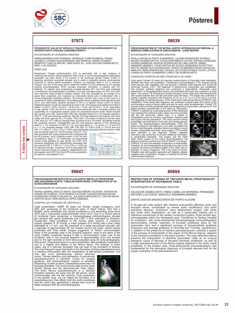
21
Pôsteres
Arq Bras Cardiol: Imagem cardiovasc. 2017;30(4 supl.1):6-41
60684RESTRICTION OF OPENING OF TRICUSPID METAL PROSTHESIS BY INTERPOSITION OF PACEMAKER CABLE
Ecocardiografia de cardiopatias adquiridas
SALVADOR GOMES NETO; FABIO CANELLAS MOREIRA; FERNANDO ANTONIO LUCCHESE; MARCELO DEMAMAN ANDRES;
SANTA CASA DE MISERICÓRDIA DE PORTO ALEGRE
A 44-year-old male patient with infective endocarditis affecting aortic and tricuspid valves, complicated by severe aortic insufficiency and aortic perivalvar abscess, underwent valve replacement surgery with aortic and mitral stent implantation, as well as a pacemaker implant due to infectious involvement of the cardiac conduction system, three months ago. Anticoagulated within the therapeutic goal. Transferred to Tertiary Hospital for evaluation, with three-dimensional transesophageal echocardiography demonstrating mobility restriction of tricuspid prosthesis discs by the pacemaker wire, and a significant increase of transprosthetic gradients (maximum and average gradients of 14mmHg and 11mmHg, respectively), in addition to the presence of massive pseudoaneurysm, possibly a sequel of the process of endocarditis in the region of the fibrous trigone, adjacent to the mechanic prosthesis in the aortic position. The case described above presents the interposition of definitive pacemaker cable as an unusual iatrogenic cause of stenosis of tricuspid mechanic prosthesis, as well as a huge pseudoaneurysm of the fibrous trigone adjacent to the aortic metal prosthesis. In the present case, three-dimensional echocardiography was fundamental for the etiological diagnosis of tricuspid stenosis and for the proper evaluation of the pseudoaneurism.
58039PSEUDOANEURYSM OF THE MITRAL-AORTIC INTERVALVULAR FIBROSA: A SERIOUS COMPLICATION OF ENDOCARDITIS – CASE REPORTEcocardiografia de cardiopatias adquiridas
PAULA CARVALHO PINTO GUIMARÃES; JULIANA RODRIGUES SOARES; MAURO SOARES MOTTA; FLÁVIA NASCIMENTO DUTRA; RAFAELA ANSELMO SOARES BARBOSA; MOACIR RODRIGUES DE LIMA JÚNIOR; DANIEL ANDRADE MENDES; THAIS FREITAS DE SOUZA; BERNARDO SCHETTINO MOTTA; ANDRÉ SILVA RODRIGUES; THIAGO GUIMARÃES ROSA CARVALHO; MÁRLON VIEIRA RODRIGUES; RAQUEL DE SOUZA NOVAES COSTA; LETÍCIA CARVALHO PINTO GUIMARÃES; DARCY DE ALMEIDA NETO;
FUNDAÇÃO HOSPITALAR SÃO FRANCISCO DE ASSISCase report: Female, 43 years old, previous medical history of rheumatic mitral valvopathy, admitted with fever and prostration. Transthoracic echocardiogram (TTE) showed severe aortic stenosis with vegetation and ring abscess, moderate mitral stenosis and normal left ventricular function (LVF). The diagnosis of enterococcal endocarditis was established. She received antibiotic treatment and underwent a bioprosthetic mitral-aortic valve replacement. In the postoperative period, she presented chest pain and symptoms of heart failure. The control TTE showed an important aneurysm of the mitral-aortic intervalvar fibrosis with systolic-diastolic flow and left para-atrial cavity formation. This image was confirmed in cardiac magnetic resonance with measures of 5.4 cm x 9.7 cm x 6.3 cm and a 1,9 neck. The diagnosis of pseudoaneurysm of the mitral-aortic intervalvular fibrosa (P-MAIVF) was established. Three weeks after diagnosis, she underwent surgical repair with closure of the communication using a Dacron patch and also an aortic valve re-replacement. Control TTE showed mitral and aortic prostheses with normal appearance and function, with good LVF. The patient remains asymptomatic in the follow-up. Discussion: P-MAIVF is defined as a pseudoaneurysm of the interanular zone between the mitral and aortic valves and communicates with the left ventricular outflow tract. It is related to complications such as coronary compression, fistulous path and rupture into the pericardium. The most common clinical presentation are heart failure, chest pain and cerebrovascular events. The transesophageal echocardiogram (TEE) has better sensitivity for the diagnosis. In our case, the patient has the two most frequently causative factors according to recent reviews: endocarditis and aortic valve surgery. TTE provided good resolution in the diagnosis. Cardiac magnetic resonance was performed to provide additional information in preoperative evaluation. The surgery was the Heart Team choice based on the recent recommendations, the patient’s symptoms and the size of the P-MAIVF. Conclusion: P-MAIVF is a rare and dreaded complication of endocarditis. Therefore, an echocardiographic examination focused on the subaortic structures and anterior leaflet of the mitral valve is essential. In case of suspicion, TEE is the exam of choice. Pulsatile mass with systolic expansion and diastolic collapse suggests such complication. Surgery is currently the first line recommended treatment.
Figure 1 – The control TTE showed an important aneurysm of the mitral-aortic intervalvar fibrosis with systolic-diastolic flow and left para-atrial cavity formation.
Figure2 – The P-MAIVF was confirmed in cardiac magnetic resonance with measures of 5.4cm x 9.7cm x 6.3cm and a 1,9 neck.
59847PSEUDOANEURYSM WITH FISTULA IN AORTIC METALLIC PROSTHESIS AND ASCENDING AORTIC TUBULAR PROSTHESIS: CONTRIBUTION OF 3D ECHOCARDIOGRAMEcocardiografia de cardiopatias adquiridas
IRVING GABRIEL ARAÚJO BISPO; DEIVIDE RIBEIRO SILVEIRA; TAINARA SÁ FREIRE DE ALMEIDA; LÍVIA FERRAZ ACCORSI; JUSSARA REGINA SOUSA RODRIGUES; MURILO SALANI GIL; JULIANA B MATSUMOTO; CINTHIA NIEGE SANTOS SILVA; VERA MARCIA LOPES GIMENES;
HOSPITAL DO CORAÇÃO DE SÃO PAULO
Case presentation: LMMF, 58 years old, female, sought emergency room (PS) with worsening of the functional class of Heart Failure. She had a Metallic aortic prosthesis and tubular prosthesis in the ascending aorta since 2015 and a myocardial revascularization since 2013. Due to a clinical picture of functional class worsening, a transesophageal echocardiogram showed left ventricle with apical dyskinesia and LVEF = 52% estimated by Simpson; Double-disc metal prosthesis in the normal aortic position, a maximum systolic gradient of 18 mmHg, a mean gradient of 9 mmHg with a flow orifice estimated at 1.3 cm², and the presence of a large aneurysmal saccular with a diameter of approximately 62 mm located around the aortic valved tubular prosthesis with Flow inside. Images suggestive of “fistula” communication holes of the prosthetic tube in the proximal segment, close to the plane of the aortic metallic prosthesis feeding it with 2 communication holes: one of the prosthetic tube in the proximal segment of approximately 4mm and another one for the right ventricle of 3mm being evaluated using 3D echocardiography. Discussion: Pseudoaneurysm is a rare complication after prosthetic implantation due to a fragility and dilation of the fibrous tissue. The findings, in these cases, are of a saccular formation that can lead to the formation of fistulas. The most frequent cause that leads to the formation of these pseudoaneurysms is infective endocarditis after the implantation of left-handed prostheses, and may also occur spontaneously, without infectious cause. Precise detection and delineation of perivalvular pseudoaneurysms is therefore crucial for surgical guidance, and echocardiogram support is essential. Depending on their location, pseudoaneurysms may not be easily identified. The most common sites are the aortic mitrotic fibrosis and the atrioventricular node region. The aortic fibrous pseudoaneurysm is a saccular formation between the aorta and the left atrium, which communicates with the left ventricle. Final comments: In the specific case, we can detect the formation of a pseudoaneurysm that affects both the aortic prosthesis and the valve tube, generating 2 fistulas that could be better analyzed with 3D echocardiography.
57873PROGNOSTIC VALUE OF SPECKLE TRACKING ECHOCARDIOGRAPHY IN PATIENTS WITH CHAGAS CARDIOMYOPATHYEcocardiografia de cardiopatias adquiridas
MINNA MOREIRA DIAS ROMANO; HENRIQUE TURIN MOREIRA; FAWAZ ALENEZI; LUCIANO ALBUQUERQUE LIMA SARAIVA; ANDRE SCHMIDT; BENEDITO CARLOS MACIEL; ANIS RASSI JR.; JOSE ANTONIO MARIN-NETO; ERIC J VELAZQUEZ;
FMRP-USPBackground: Chagas cardiomyopathy (CC) is associated with a high incidence of cardiovascular events. Global longitudinal strain (GLS), a novel echocardiographic deformation parameter, has been shown to be superior to left ventricular ejection fraction (LVEF) in detecting subclinical myocardial damage, and is useful in predicting adverse cardiovascular outcomes in various patient population. However, its prognostic value in CC is unknown Purpose: The aim of this study was to determine whether LV GLS measured by 2-D speckle tracking echocardiography (STE) provides prognostic information in patients with CC. Methods: CC patients were prospectively included between 2011 and 2014, and underwent a transthoracic echocardiography at baseline. GLS was calculated using 2-D STE, and LVEF was measured using Simpson`s biplane method. GLS was calculated as an average of LV apical three projections and measured as endocardial global peak of deformation with a vendor-independent dedicated software. The primary outcome was a time to first event of a composite of: death, hospitalization, resuscitated cardiac arrest, sustained ventricular tachycardia (SVT), new heart failure, absolute decrease of 10% in LV ejection fraction (LVEF) or stroke. Statistical analysis: results are expressed as mean ± SD. GLS groups were defined according to tertiles: (1) GLS ≤ -18.9%, (2) GLS > -18.9% and < -10.3%, and (3) GLS ≥ -10.3%. Association of age, gender, LVEF and GLS to outcomes were performed with a univariate analysis and sequentially on a multivariate analysis using Cox proportional hazard model. Model included age, gender and LVEF. Hazard Ratio (HR) was calculated based on a 5% absolute change in GLS. P < 0.05 was defined as significant. Results: GLS was obtained in 85 subjects, 52% were males and mean age was 55 ± 15 years. There were 7.3% losses of follow-up and the mean LVEF was 52 ± 14 %. After an average follow up period of 28 ± 19 months, 39 patients reached the composite outcome. Hospitalization was the most frequent outcome (n=15), followed by death (n = 12), decrease of LVEF (n = 8), new heart failure (n = 3), stroke (n = 3) and STV (n = 1). On univariate analysis, LVEF (HR: 0.95; 95% CI: 0.95 to 0.99; p = 0.013) and GLS (HR: 1.11; 95% CI: 1.05 to 1.18; p < 0.001) were significantly associated with the outcome, while age and gender were not. The GLS group 3 (GLS ≥ -10.3) was associated with worse prognosis when compared with the GLS groups 1 and 2 (logrank-p-value = 0.001 for both comparisons), figure 1. On multivariate Cox proportional hazard model adjusting for age, gender and LVEF, GLS remained significantly and independently associated with the outcome (HR: 2.7; 95% CI: 1.31 to 5.54; p = 0.007 per 5% absolute change). Conclusion: GLS is associated with worse outcomes among CC subjects, independently from demographic and LVEF. GLS from STE has the potential to be used as a risk predictor in CC.
A ta
bela
veio
corta
da

22
Pôsteres
Arq Bras Cardiol: Imagem cardiovasc. 2017;30(4 supl.1):6-41
60718SERIAL ECHOCARDIOGRAM VALUE ON MANAGEMENT OF MYOCARDIAL INJURY AFTER SCORPION ENVENOMATIONEcocardiografia de cardiopatias adquiridas
CRISTIANE DE CARVALHO SINGULANE; FAISSAL IBRAHIM SOUMAILLE; BRUNNA PRISCYLLA AMÉRICO CARVALHO; PAULO DIOGO SOARES MOTA; JOÃO UBER FOLCHINE TRINDADE; MAURÍCIO DE NASSAU MACHADO; JAMIL ALLI MURAD JÚNIOR;
FUNDAÇÃO FACULDADE REGIONAL DE MEDICINA DE SÃO JOSÉ DO RIO PRETO (FAMERP)Introduction: Scorpion stings in Brazil are important not only because of their incidence but also for their potential ability to induce severe, and often fatal, clinical situations. Severe poisoning is characterized by cardiocirculatory failure which can lead to myocarditis, acute pulmonary edema and cardiogenic shock, the main causes of death. Cardiac involvement has been attributed to adrenergic discharge and a possible toxic effect of venom on the myocardium. The early diagnosis and management of this complications is crucial for the patients’ prognosis. Hemodynamic monitoring using transthoracic echocardiogram (ETT) in the critically ill can be a real asset. It’s a non-invasive exam that enables a direct real-time estimation of pressures, cardiac output and cardiac contractility allowing the adoption of appropriate invasive therapy such as volume resuscitation, use of inotropic agents and mechanical ventilation. Case Report: A 9 years old girl was victim of scorpion sting in the 5th pododactyl 3 hours before admission. In addition to local pain, she had been presenting sweating, vomiting, hypertensive crisis (blood pressure of 200x110 mmHg) and delirium. She had a regular heart rate (75 bpm) with a third heart beat (B3). Despite initial management, including anti-hypertensives and anti-scorpion serum, she evolved with convulsive crisis, acute pulmonary edema and acute respiratory failure requiring orotracheal intubation. The electrocardiogram showed sinus rhythm with left ventricular overload pattern and the chest X-ray showed cardiomegaly and pulmonary congestion. The ultrasensitive troponin T levels (reference value < 14 pg/mL) where 62 (admission) and 518 (after 3 hours). ETT was performed daily and showing a progressive mild left ventricular contractile dysfunction (due to septal hypokinesia) with lowest ejection fraction of 44.6%. She remained in the Intensive Care Unit for 5 days and had been used dobutamine and prazosin. She was discharged after seven days with favorable clinical evolution and complete recovery of left ventricular function. Comment: We report a victim of scorpion sting that had developed acute myocardial injury evolving with cardiogenic shock. The patient had presented increased myocardial injury marker and left ventricular dysfunction assessed by serial ETT, which helped the clinical management. She was discharged from hospital after seven days with improved ETT parameters after optimized clinical treatment.
58011RIGHT VENTRICULAR LONGITUDINAL STRAIN: DIAGNOSTIC AND PROGNOSTIC IMPACT IN PATIENTS WITH PULMONARY ARTERIAL HYPERTENSION AND PRESERVED FUNCTIONAL RESERVE; AN ECHOCARDIOGRAPHIC AND MAGNETIC RESONANCE STUDY
Ecocardiografia de cardiopatias adquiridas
AUGUSTO ALBERTO DA COSTA JUNIOR; JAQUELINA SONOE OTA-ARAKAKI; ROBERTA PULCHERI RAMOS; MARLY UELLENDAHL; FREDERICO JOSE NEVES MANCUSO; MANUEL ADAN GIL; CLAUDIO HENRIQUE FISCHER; VALDIR AMBROSIO MOISES; ANTONIO CARLOS DE CAMARGO CARVALHO; ORLANDO CAMPOS FILHO;
ESCOLA PAULISTA DE MEDICINA
Abstract: Introduction: right ventricular (RV) dysfunction signals adverse prognosis in pulmonary arterial hypertension (PAH). Quantitative RV systolic function evaluation by two-dimensional echocardiography (2DE) is jeopardized by geometric constraints; RV strain might be a useful tool in this clinical setting. Objectives: we aimed to assess the diagnostic and prognostic impact of conventional 2DE systolic indices and 2DE speckle-tracking RV longitudinal strain in a group of PAH patients without severe impairment of functional capacity, chronic pulmonary thromboembolism or left ventricular dysfunction (group I of pulmonary hypertension classification). Methods: sixty-six group I PAH patients, most of them (67 %) in NYHA functional class I or II, none in class IV were studied by 2DE: fractional area change, tricuspid annular plane systolic excursion, RV myocardial performance index, tissue Doppler tricuspid annulus systolic velocity, global, free wall (RVFreeWSt) and septal RV longitudinal systolic strain were obtained. RV ejection fraction was also assessed by cardiac magnetic resonance (CMR-RVEF). The patients were followed up to 3.9 years (mean 3.3 years). Results: the best correlation between CMR-RVEF and 2DE indices was observed with RVFreeWSt (r: 0,83; p < 0,005). RVFreeWSt > 18% was a predictor of CMR-RVEF > 0,45 (p < 0,05 sensitivity 92% specificity 81% AUC 0,931 [0,858-1,00]); RVFreeWSt < 15% correlated with CMR-RVEF < 0,35 (p < 0,05 sensitivity 88,5% specificity 88,2% AUC 0,930 [0,853 - 1,00]). The combined endpoints of hospitalization for worsening PAH or cardiovascular death occurred in 15 (22.7%) patients (6 hospitalizations and 9 deaths). Multivariate analysis identified RVFreeWSt ≤ 14% as the only 2DE independent variable associated with combined endpoints [HR 4.66 (1.25-17.37); p < 0.05]. Conclusions: RVFreeWSt could be relied upon as non-geometric 2DE surrogate of CMR-RVEF in PAH patients and a powerful independent predictor of long-term outcome in this subgroup with relatively preserved functional capacity.
57630RIGHT VENTRICULAR MYOCARDIAL INVOLVEMENT IN GLYCOGEN STORAGE CARDIOMYOPATHY (PRKAG2): ANALYSIS BY ESTABLISHED AND ADVANCED ECHOCARDIOGRAPHIC TECHNIQUES
Ecocardiografia de cardiopatias adquiridas
JOSÉ LUIZ BARROS PENA; WANDER COSTA SANTOS; ISAAC H SAMPAIO; MARIA HELENA ALBERNAZ SIQUEIRA; ALINE BRAGA RODRIGUES; EDUARDO BACK STERNICK;
HOSPITAL FELICIO ROCHO
Background: PRKAG2 is a rare, early-onset, autosomal-dominant inherited disease, characterized by cardiac hypertrophy, ventricular pre-excitation and atrial tachyarrhythmia. Chronotropic incompetence and advanced heart blocks frequently accompany this condition, leading to pacemaker (PM) implantation. The syndrome can show different manifestations, ranging from complete asymptomatic condition to sudden cardiac death. To our knowledge, right ventricular (RV) involvement in this pathology has yet to be studied systematically. We evaluated RV echocardiographic findings in a cohort of patients (Pts) with PRKAG2 cardiomyopathy. Methods: Twenty-one Pts with genetically proven PRKAG2 (R302Q), 13 (65%) male, mean age 36 + 10.7 years old, underwent echocardiographic (ECO) examination, including M-mode, 2D measurements, 2D speckle tracking of global longitudinal strain (GLS). 3D RV volume was measured using TomTec software 1.1. RV-focused, 4-chamber view was acquired for measurement. 2D RV free wall speckle tracking was measured, both excluding and including interventricular septum (IVS). All data were reviewed offline. Intra- and interobserver reproducibility was good for RV measurements. Cardiac magnetic resonance was performed on all Pts without PM. Results: PM had been implanted in 10 (50%) of Pts. Thirteen Pts had pre-excitation (65%). Only 3 Pts had no pre-excitation or PM (15%). RV hypertrophy was found in varying degrees in all Pts (RV free wall end diastolic thickness obtained by subcostal view was 8.45 ± 3.24). Six Pts showed RV enlargement. Tricuspid annular plane systolic excursion (TAPSE) was reduced in 4 Pts. 2D RV free wall GLS excluding IVS was 21.98 + 5.57 and including IVS was 19.30 + 4.76. Compared to Pts with PM, measurements were lower (20.9 + 5.4 x 17.6 + 3.6). 3D RV end-diastolic volume (EDV), end-systolic volume (ESV), and RV ejection fraction (EF) were 108 + 48.6 ml, 45.1 + 21.8ml, and 42.6 + 11.4%, respectively. Pts with PM had lower EF, though not statiscally significant. Positive correlation coefficients of 2D RV free wall GLS and 3D EF were observed, especially in Pts without PM (r = 0.82). Cardiac magnetic resonance confirmed the echocardiographic findings in 10 Pts. Conclusions: RV involvement in PRKAG2 is frequent and occurs in different degrees. Echocardiography is a valuable tool in detecting RV diffuse and focal myocardial abnormalities in PRKAG2 cardiomyopathy. Deformation indices and 3D EF are especially revealing because they may be helpful in identifying RV structure and function. 2D RV free wall GLS excluding IVS and 3D EF are good indicators of RV systolic function in this condition. Additional longitudinal studies are warranted to further understand the natural history of RV involvement and determine its impact on Pt outcomes.
59599RIGHT SIDE FOREIGN BODIES: IMAGES HELPING IN DECISION-MAKINGEcocardiografia de cardiopatias adquiridas
MARIA ESTEFÂNIA BOSCO OTTO; FERNANDO MELO NETTO; ALINE DE OLIVEIRA MARTINS CAMPOS; JOYCE GOMES ELIAS LIMA; ANA CAROLINA PEREIRA MATOS DOMINGUES; MAIARA SANCHEZ RIBEIRO; ADENALVA LIMA DE SOUZA BECK;
ICDF/DF E CLÍNICA UCI
Case Report: Two different cases of right-heart side foreign bodies (FB) will be reported in order to discuss diagnosis and therapeutics depending on the location, size, symptoms and associated lesions Case 1: A 42-year-old man, asymptomatic, hypertensive, came for a routine transthoracic echocardiography (TTE) in our laboratory. During the exam was noticed a hyperechoic linear image in the right ventricle (RV) and filamentous images on the tricuspid valve suggesting thrombus or vegetation. The patient was sent to the emergency department (ED) and subsequently submitted to a chest computed tomography (CT) which confirmed a needle inside the RV and demonstrated other metallic fragments in the epigastric and right hypochondrium region indicative of self-inflicted injuries. This hypothesis was further confirmed by psychological investigation. Two days later the patient was submitted to cardiac surgery and the needle entrance was identified on the epicardium, allowing the extraction without cardiopulmonary bypass. Intraoperative transesophageal echocardiography (TEE) was performed and revealed no tricuspid valve vegetation. On the fifth day, the patient was discharged from hospital asymptomatic. Case 2: A 27-year-old man, with a history of upper abdomen injury by a barbecue wood skewer, underwent exploratory laparotomy in other hospital. Six days after the injury, he came to our ED with shortness of breath, fever and pulsus paradoxus. Chest X-ray showed increased cardiac size, but no foreign body. TTE showed foreign body in RV and right atrium (RA), mobile mass in the RA near to a dilated inferior vena cava (IVC) and severe pericardial effusion with restriction. TEE confirmed TTE findings. Chest CT found the fistulous path from the abdomen to the thorax. The patient was submitted in a few hours to surgery with cardiopulmonary bypass due to multiple lesions and was discharged from hospital 15 days after surgery. Discussion: Diagnosis of FB may be suspect from the type of injury or incidentally during investigation. The clinical manifestation varies according to the size, location and other structures that might be involved. TTE and TEE are excellent for identifying FB, mostly if they are radiopaque. CT confirm their location and injury of other structures. Comments: Surgical options depend on the location of the FB and need for cardiopulmonary bypass. TTE, TEE and CT are essential for clinical decision and surgical approach.
Right Ventricle Needle
Right Ventricle and Atrium / Barbecue Wood Skewer

23
Pôsteres
Arq Bras Cardiol: Imagem cardiovasc. 2017;30(4 supl.1):6-41
58037THE ROLE OF AIR POLLUTION ON CARDIAC REMODELING ON THE ACUTE PHASE IN AN EXPERIMENTAL MODEL OF CHAGAS CARDIOMYOPATHYEcocardiografia de cardiopatias adquiridasVIVIANE TIEMI HOTTA; KEILA CARDOSO BARBOSA FONSECA; FERNANDA G. PESSOA; ORLANDO NASCIMENTO RIBEIRO; BARBARA MARIA IANNI; FABIO FERNANDES; ROGERIO SILVA DO NASCIMENTO; PAULO HILARIO NASCIMENTO SALDIVA; CHARLES MADY; FELIX JOSE ALVAREZ RAMIRES;INCOR/FMUSP AND FLEURY MEDICINA E SAÚDEBackground: Chagas´ Disease (CD) is a neglected tropical disease and an important cause of cardiomyopathy and heart failure in Latin America. CD is associated with extensive myocardial fibrosis, which relates directly to a complex inflammatory process, oxidative stress and apoptosis. This geometric and structural cardiac remodeling is responsible for cardiac dysfunction and it relate s to CD´s prognosis. Atmospheric pollution is a significant environmental problem in big cities with elevated population density. Air pollution leads to an intense activation of the same mechanisms that amplify the response against an infection. AIM: The aim of this study was to evaluate the role of air pollution in cardiac biventricular morphology, systolic and diastolic function and in the interstitial collagen volume in the acute phase (60 days) of an experimental model of CD. Methods: Seventy female Hamsters Sirius were evaluated in 4 groups: healthy control group (Ct)(N = 10), healthy exposed to air pollution (Ct + Pol)(N = 10), CD group (Inf)(N = 25) and 25 in the CD exposed to air pollution (Inf + Pol)(N = -25). Animals in the corresponding groups were infected with 105 Y strains of Trypanossoma cruzi and exposed to pollution by the inhale of particulate material produced by the diesel burning, three times a week. Healthy and CD groups inhaled only purified air. Transthoracic echocardiogram including M Mode, two dimensional and pulsed wave Doppler were performed with Vivid E9, GE Medical Systems, and with 9 to 12 mm and 9 to 12 MHz transducer. The following parameters were evaluated: left atrial diameter (LAD), diastolic (LVDD) and systolic (LVSD) left ventricle diameters, left ventricle ejection fraction (LVEF), isovolumic relaxation time (IVRT) and E and A waves´ velocities and E/A ratio. The volume fraction of interstitial collagen of the left ventricle (LV VFIC) and right ventricle (RV VFIC) were evaluated with histological samples colored Picrosirius Red and evaluated with the software QWIN image precessing and analysis software (LEICA). Numerical values were presented in mean ± standard deviations. Parametrical and non-parametrical tests were performed according to the results of normality tests. Survival rates were analysed with Kaplan-Meier. Results: Results are shown in Table 1. Infected groups (Inf and Inf + Pol) evidenced larger LV diastolic and systolic diameters and lower LVEF in comparison do controls groups (Ct and Ct + Pol) 60 days after infection. Ct+ Pol, Inf and Inf + Pol groups evidenced larger volumes fractions of interstitial collagen of the left ventricle in comparison to Ct group. Survival rates evidenced high mortality rates in infected groups (Inf and Inf + Pol) in comparison to controls groups (Ct and Ct + Pol)(P = 0,006) and irrespective of air pollution. Conclusions: In this study, air pollution in the acute phase of an experimental model of CD did not have a significant impact on cardiac biventricular morphology, function, interstitial collagen volume or mortality.
LVSD. *p<0.05 (CT vs Inf) (CT= control, CT + Pol = control + pollution, Inf = Infected, Inf + Pol = Infected + Pollution).
LVDD. *p<0,01 (CT vs Inf) and p<0,01 (CTPol vs Inf). (CT= control, CT + Pol = control + pollution, Inf = Infected, Inf + Pol = Infected + Pollution).
LVEF. *p < 0,01 (CT vs Inf) and p <0,01 (CT+Pol vs Inf). (CT= control, CT + Pol = control + pollution, Inf = Infected, Inf + Pol = Infected + Pollution).
VFIC (%), volume fraction of interstitial collagen. LV, left ventricle; RV, rigth ventricle. *p<0,036 (CT vs CT+Pol). (CT= control, CT + Pol = control + pollution, Inf = Infected, Inf + Pol = Infected + Pollution).
Survival rates (60 days ). p= 0,0006 (CT= control, CT + Pol = control + pollution, Inf = Infected, Inf + Pol = Infected + Pollution).
E/A ration. p= 0,034 (CT vs Inf + Pol) (CT= control, CT + Pol = control + pollution, Inf = Infected, Inf + Pol = Infected + Pollution).
IVRT p= 0,092. (CT= control, CT + Pol = control + pollution, Inf = Infected, Inf + Pol = Infected + Pollution).
Table 1. LAD: left atrial diameter, LVDD: left ventricular diastolic diameter, LVSD: left ventricular systolic diameter, LVEF: left ventricular ejection fraction, IVRT: isovolumic relaxation time, E/A: E and A waves´ velocities ratio, LV VFIC: volume fraction of interstitial collagen of the left ventricle, RV VFIC: volume fraction of interstitial collagen of the right ventricle.1
LEFT VENTRICULAR DYASTOLIC DIAMETER (LVDD)
LEFT VENTRICULAR SISTOLIC DIAMETER (LVSD) E/A Ratio ISOVOLUMIC RELAXATION TIME (IVRT)
INTERSTITIAL COLLAGEN LEFT VENTRICULAR EJECTION FRACTION
(LVEF)
SURVIVAL RATES
SUR
VIVA
L (%
)
DAYS
VFI
C (%
)
LV
RV
Ct Ct + Pol Inf Inf + Pol p LAD (mm) 4,4 ± 0,3 4,4 ± 0,33 4,2 ± 0,5 4,5 ± 0,3 0,53 LVDD (mm) 5,7 ± 0,7* 5,9 ± 0,4 6,3 ± 0,2* 6,2 ± 0,5 0,038 LVSD (mm) 4,0 ± 0,3* 4,0 ± 0,3§ 4,6 ± 0,1*§ 4,4 ± 0,7 0,002 LVEF (%) 0,65 ± 0,03* 0,66 ± 0,04§ 0,60 ± 0,02*§ 0,60 ± 0,010 0,002 IVRT 30 ± 4 31 ± 7 30 ± 5 39 ± 13 0,092 E/A 7,0 ± 2,0* 7,6 ± 2,0 7,7 ± 1,8 9,9 ± 3,5* 0,034 LV VFIC (%) 1,3 ± 0,3* 1,8 ± 0,2 1,6 ± 0,2* 1,7 ± 0,4 0,036 RV VFIC (%) 2,0 ± 0,5 2,0 ± 0,4 2,2 ± 0,7 2,3 ± 0,4 0,598
LAD, left atrium diameter. p = 0,53. (CT= control, CT + Pol = control + pollution, Inf = Infected, Inf + Pol = Infected + Pollution).
57946STRUCTURAL COMPLICATIONS OF INFECTIVE ENDOCARDITIS: THE ADDED VALUE OF THREE-DIMENSIONAL TRANSESOPHAGEAL ECHOCARDIOGRAPHYEcocardiografia de cardiopatias adquiridas
ANA PAULA DOS REIS VELLOSO SICILIANO; MARCELLA DE AGOSTINI ISO; ALEX DOS SANTOS FÉLIX; MONICA LUISA DE ALCANTARA; RODRIGO REGO; SALOMON ISRAEL DO AMARAL; DANIELA PINHEIRO FERNANDES; CLAUDIA MARIA DOS SANTOS; ORLANDO GLÓRIA VELOSO; CLÁUDIA DE CÁSSIA FIRMIDA; SÉRGIO SALLES XAVIER; CLAUDIA DE CASSIA FIRMIDA; SERGIO SALLES XAVIER;
HOSPITAL SAMARITANO / AMERICAS. MEDICAL CITY
Introduction: Two-dimensional (2D) transesophageal echocardiography (TEE) plays a central role in diagnosing, identifying complications and guiding management of Infective Endocarditis (IE). Three-dimensional (3D) TEE is emerging as an adjunctive tool and thus can improve diagnostic accuracy and help surgical planning. Purpose: The aim of this study was to evaluate the added value of 3D TEE over 2D TEE in the assessment of structural complications (SC), as periannular abscess and perforation or valve destruction. Methods: From january 2014 to april 2017 39 patients in two tertiary hospitals with definite diagnosis of infective endocarditis (based on modified Duke criteria) who underwent TEE were evaluated for SC. Of these, 8 patients didn’t have 3D datasets and were excluded. 3D TEE was considered the gold standard (images acquired with adequate temporal and spatial resolution). Another experienced observer (blinded to 3D results) retrospectively analyzed 2D images, so both methods could be compared. Results: Mean age of the studied population (31 pts) was 66 + 17 years (13 females) and 58% (18 out of 31) had SC. 3D TEE identified 4 aortic periannular abscesses, 9 mitral perforations (2 prosthesis and 7 native valves) and 6 aortic perforations. Accuracy, sensitivity and specificity of 2D TEE were respectively for overall SC: 80%, 83% and 77%, for abscess: 96%, 100% and 96%, for aortic perforation/destruction: 87%, 71% and 91% and for mitral perforation: 87%, 66% and 95%. Conclusion: 3D TEE is a complement to 2D TEE, in providing additional information that improves diagnostic accuracy. The improved spatial orientation of 3D enables more precise and complete examination of intracardiac anatomy. It is superior in identifying its complications, in particular in locating and evaluating anatomical spatial relationship of perivalvular abscesses and valvular perforation, which might enable better risk stratification, surgical planning and decision-making.
3D rendered image of aortic valve destruction, jet lesion of the anterior mitral leaßet and perforation of A2 segment.
3D rendered image aortic perspective of the aortic valve , showing a perforation in detail
Figura 1 – 3D rendered image of aortic valve destruction, jet lesion of the anterior mitral leaflet and perforation of A2 segment.
Figura 2 – 3D rendered image aortic perspective of the aortic valve , showing a perforation in detail.
58068THE GLOBAL LONGITUDINAL STRAIN AND THE LONGITUDINAL SYSTOLIC STRAIN RATE, OBTAINED AFTER A LAST DOSE OF DOXORUBICIN, ARE PREDICTORS OF CARDIOTOXICITY AT THE END OF THE 1ST YEAR OF TREATMENT WITH ANTHRACYCLIC
Ecocardiografia de cardiopatias adquiridas
ANDRÉ LUIZ CERQUEIRA DE ALMEIDA; THYAGO MONTEIRO DO ESPÍRITO SANTO; CAROLINE DE SOUZA ALMEIDA; MATHEUS PAMPONET FREITAS; CECÍLIA LOPES VIANA SANTOS; SAMUEL OLIVEIRA AFONSECA; ISRAEL REIS; SUZANE PEREIRA DE SOUZA; MAURÍCIO GOMES DA SILVA SERRA; UESLEI MENEZES DE ARAÚJO PEREIRA; GABRIEL SANTOS DE JESUS; MARLON MOURA DOS SANTOS; BRANDA CAVALCANTE DOURADO; ANA CAROLINA SILVA ASSUNÇÃO; EDVAL GOMES DOS SANTOS JÚNIOR;
SANTA CASA DE MISERICÓRDIA DE FEIRA DE SANTANA
Rationale: Doxorubicin (DOX) is an effective chemotherapeutic agent in the treatment of cancer. Its beneficial effects, however, are mitigated by the possibility of cardiotoxicity (CTX) associated with its use. Objective: To investigate whether the global longitudinal strain (GLS), measured in the course and soon after treatment with DOX, can predict cardiotoxicity at the end of the first year of treatment. Material and methods: Seventy-six patients (pcts) with cancer and with indication for use of DOX, were prospectively included. Of these, 73 had breast cancer and 3 lymphoma. The pcts performed echocardiogram in four moments: Baseline (T1), after the first dose of DOX (T2), after the last dose of DOX (T3) and after one year of anthracyclic infusion (T4). CTX was defined as a reduction in LVEF > 10% to a value < 53% at T4. Results: Nine pcts (11.8%) developed CTX. The mean cumulative dose of DOX was 241mg/m², with no difference between the group that developed CTX (Group 1) and those who did not develop CTX (Group 2) (p = 0.364). Age, SBP, DBP, BMI, LVEF, S` wave, E/E‘ ratio, GLS and systolic Strain Rate (SRs) were similar between groups in T1. In T2, the E/E´ ratio (9.7 ± 3.7 vs. 7.8 ± 2.2) and SRs (-0.93 ± 0.18s-1 vs -1.05 ± 0.16s-1) were higher in G1 (p = 0.034, p = 0.044 and p = 0.032, respectively). In T3, GLS was lower (-16.8 ± 1.7% vs -18.7 ± 2.5%, p = 0.045) and SRs were higher (-0.83 ± 0.13s-1 vs -0.96 ± 0.15s-1, p = 0.016) in pcts that developed CTX at T4. For the other variables analyzed, there was no difference between groups. Among the variables that presented the statistical significance described above, GLS and SRs obtained at T3 were predictors of CTX at T4, with area under the ROC curve of 0.73 (95%CI:0.57-0.89, p = 0.038) for GLS and 0.73 (95%CI: 0.57-0,90, p = 0.035) for SRs. GLS of -17.48%, measured at T3, showed sensitivity of 78% and specificity of 76% for detection of CTX at T4. Conclusion: Based on the results of the C statistic, SLG and SRs obtained after the infusion of 241mg/m² of DOX (final), are predictors of future anthracyclic-induced CTX.
57963SPECKLE TRACKING ANALYSIS OF THE RIGHT VENTRICLE PREDICTS SHORT-TERM ADVERSE EVENTS IN PATIENTS WITH ACUTE DECOMPENSATED HEART FAILURE. STRAIN-DHF STUDY (SPECKLE TRACKING ADDS INFORMATION IN DECOMPENSATED HEART FAILURE)
Ecocardiografia de cardiopatias adquiridas
DANILO BORA MOLETA; CASSIO C S MACHADO; DIOGO FC AZEVEDO; RENATO P AZEVEDO; WILSON MATHIAS JUNIOR; MARCO STEPHAN LOFRANO ALVES;
INSTITUTO DO CORAÇÃO DA FACULDADE DE MEDICINA DA USP
Background: Risk stratification in patients with acute decompensated heart failure (ADHF) is crucial in the medical decision-making process in the emergency department. Speckle tracking echocardiography (STE) is a recently introduced technology that allows the evaluation of myocardial deformation during the cardiac cycle. Purpose: To determine whether quantification of right ventricle deformation using speckle tracking echocardiography (STE) adds prognostic value to routine echocardiographic assessment in patients admitted with ADHF at the emergency department. Methods: Consecutive patients with new or worsening HF symptoms and need of intravenous therapy were included. They underwent a comprehensive echocardiographic study including STE in the first 48 hours of admission. Patients were followed during the hospitalization period and up to six months after hospital discharge or until occurrence of the primary endpoint (composite of cardiac death, heart transplantation, circulatory assist device use or readmission). All patients signed informed consent. The study was approved by local Ethics Committee. Data are expressed as mean and standard deviation; statistical significance were admitted when p < 0.05. Results: Fifty-one patients were included (41% female, age 58±12 years, LVEF = 31 ± 10%, BNP = 1309 ± 755 pg/mL). Twenty-eight patients (54%) reached the primary endpoint with a mean follow-up of 100 ± 62 days. From speckle tracking-derived parameters, global longitudinal strain rate of the right ventricle (RV-GLSR) showed significant correlation with serum BNP levels (r = -0.57, 95%CI 0.30 to 0.76, p < 0.001). According to ROC analysis, a RV-GLSR higher than -0.66s-1 had 63% sensitivity and 63% specificity for predicting the primary endpoint (AUC 0.64, 95%CI 0.49-0.77, p < 0.043). From conventional echocardiography parameters of RV function, only fractional area change discriminated the occurrence of adverse events (AUC 0.69, 95%CI 0.54-0.81, p = 0.012). Neither conventional echocardiographic or STE-derived parameters of the left ventricle showed significant association with events. Conclusion: Functional analysis of right ventricle is crucial for prognostic evaluation of patients with ADHF in the emergency department. Our data also show that RV-GLSR obtained with STE is suitable for predicting short-term adverse events in patients with ADHF. The prognostic evaluation should consider RV-GLSR measurement in these patients.
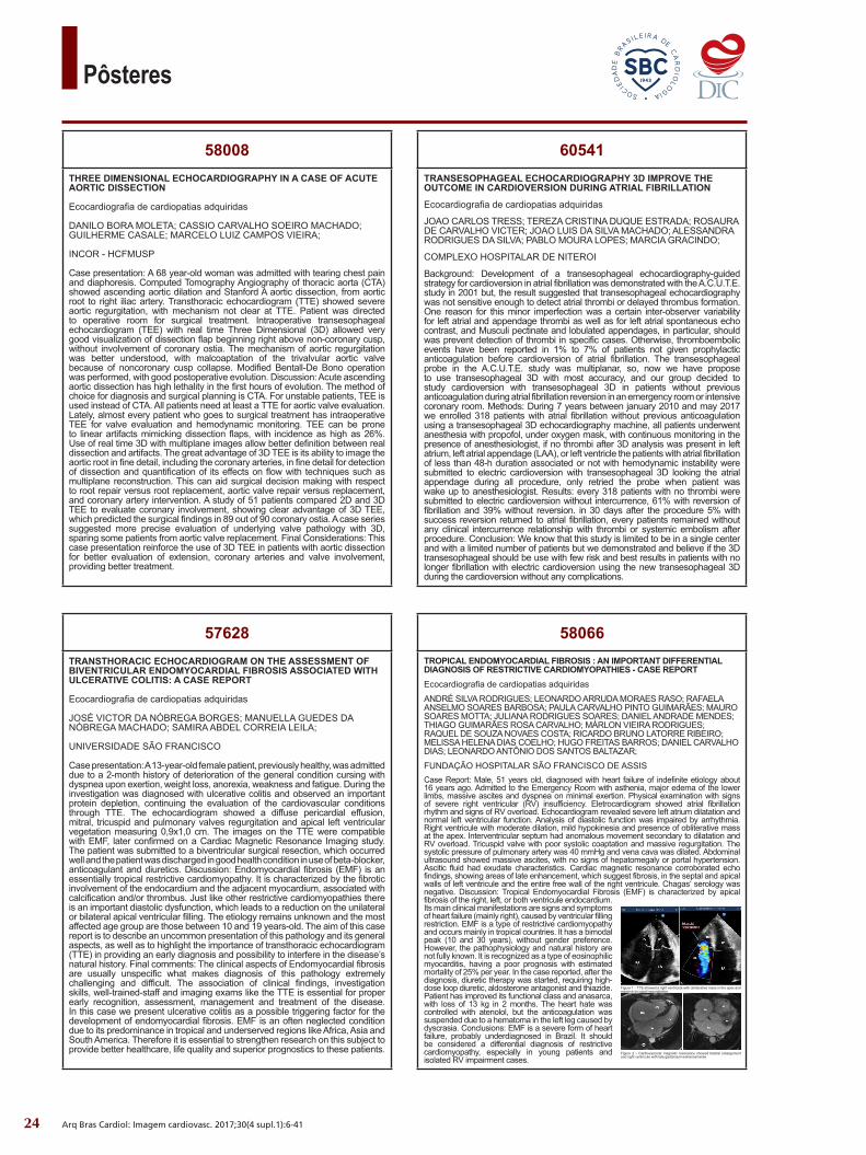
24
Pôsteres
Arq Bras Cardiol: Imagem cardiovasc. 2017;30(4 supl.1):6-41
58066TROPICAL ENDOMYOCARDIAL FIBROSIS : AN IMPORTANT DIFFERENTIAL DIAGNOSIS OF RESTRICTIVE CARDIOMYOPATHIES - CASE REPORTEcocardiografia de cardiopatias adquiridas
ANDRÉ SILVA RODRIGUES; LEONARDO ARRUDA MORAES RASO; RAFAELA ANSELMO SOARES BARBOSA; PAULA CARVALHO PINTO GUIMARÃES; MAURO SOARES MOTTA; JULIANA RODRIGUES SOARES; DANIEL ANDRADE MENDES; THIAGO GUIMARÃES ROSA CARVALHO; MÁRLON VIEIRA RODRIGUES; RAQUEL DE SOUZA NOVAES COSTA; RICARDO BRUNO LATORRE RIBEIRO; MELISSA HELENA DIAS COELHO; HUGO FREITAS BARROS; DANIEL CARVALHO DIAS; LEONARDO ANTÔNIO DOS SANTOS BALTAZAR;
FUNDAÇÃO HOSPITALAR SÃO FRANCISCO DE ASSISCase Report: Male, 51 years old, diagnosed with heart failure of indefinite etiology about 16 years ago. Admitted to the Emergency Room with asthenia, major edema of the lower limbs, massive ascites and dyspnea on minimal exertion. Physical examination with signs of severe right ventricular (RV) insufficiency. Eletrocardiogram showed atrial fibrillation rhythm and signs of RV overload. Echocardiogram revealed severe left atrium dilatation and normal left ventricular function. Analysis of diastolic function was impaired by arrhythmia. Right ventricule with moderate dilation, mild hypokinesia and presence of obliterative mass at the apex. Interventricular septum had anomalous movement secondary to dilatation and RV overload. Tricuspid valve with poor systolic coaptation and massive regurgitation. The systolic pressure of pulmonary artery was 40 mmHg and vena cava was dilated. Abdominal ultrasound showed massive ascites, with no signs of hepatomegaly or portal hypertension. Ascitic fluid had exudate characteristics. Cardiac magnetic resonance corroborated echo findings, showing areas of late enhancement, which suggest fibrosis, in the septal and apical walls of left ventricule and the entire free wall of the right ventricule. Chagas‘ serology was negative. Discussion: Tropical Endomyocardial Fibrosis (EMF) is characterized by apical fibrosis of the right, left, or both ventricule endocardium. Its main clinical manifestations are signs and symptoms of heart failure (mainly right), caused by ventricular filling restriction. EMF is a type of restrictive cardiomyopathy and occurs mainly in tropical countries. It has a bimodal peak (10 and 30 years), without gender preference. However, the pathophysiology and natural history are not fully known. It is recognized as a type of eosinophilic myocarditis, having a poor prognosis with estimated mortality of 25% per year. In the case reported, after the diagnosis, diuretic therapy was started, requiring high-dose loop diuretic, aldosterone antagonist and thiazide. Patient has improved its functional class and anasarca, with loss of 13 kg in 2 months. The heart hate was controlled with atenolol, but the anticoagulation was suspended due to a hematoma in the left leg caused by dyscrasia. Conclusions: EMF is a severe form of heart failure, probably underdiagnosed in Brazil. It should be considered a differential diagnosis of restrictive cardiomyopathy, especially in young patients and isolated RV impairment cases.
Figure 1 – TTE showed a right ventricule with obliterative mass in the apex and massive tricuspid regurgitation.
Figure 2 – Cardiovascular magnetic resonance showed biatrial enlargement and right ventricule with late gadolinium-enhancemente
60541TRANSESOPHAGEAL ECHOCARDIOGRAPHY 3D IMPROVE THE OUTCOME IN CARDIOVERSION DURING ATRIAL FIBRILLATION
Ecocardiografia de cardiopatias adquiridas
JOAO CARLOS TRESS; TEREZA CRISTINA DUQUE ESTRADA; ROSAURA DE CARVALHO VICTER; JOAO LUIS DA SILVA MACHADO; ALESSANDRA RODRIGUES DA SILVA; PABLO MOURA LOPES; MARCIA GRACINDO;
COMPLEXO HOSPITALAR DE NITEROI
Background: Development of a transesophageal echocardiography-guided strategy for cardioversion in atrial fibrillation was demonstrated with the A.C.U.T.E. study in 2001 but, the result suggested that transesophageal echocardiography was not sensitive enough to detect atrial thrombi or delayed thrombus formation. One reason for this minor imperfection was a certain inter-observer variability for left atrial and appendage thrombi as well as for left atrial spontaneous echo contrast, and Musculi pectinate and lobulated appendages, in particular, should was prevent detection of thrombi in specific cases. Otherwise, thromboembolic events have been reported in 1% to 7% of patients not given prophylactic anticoagulation before cardioversion of atrial fibrillation. The transesophageal probe in the A.C.U.T.E. study was multiplanar, so, now we have propose to use transesophageal 3D with most accuracy, and our group decided to study cardioversion with transesophageal 3D in patients without previous anticoagulation during atrial fibrillation reversion in an emergency room or intensive coronary room. Methods: During 7 years between january 2010 and may 2017 we enrolled 318 patients with atrial fibrillation without previous anticoagulation using a transesophageal 3D echocardiography machine, all patients underwent anesthesia with propofol, under oxygen mask, with continuous monitoring in the presence of anesthesiologist, if no thrombi after 3D analysis was present in left atrium, left atrial appendage (LAA), or left ventricle the patients with atrial fibrillation of less than 48-h duration associated or not with hemodynamic instability were submitted to electric cardioversion with transesophageal 3D looking the atrial appendage during all procedure, only retried the probe when patient was wake up to anesthesiologist. Results: every 318 patients with no thrombi were submitted to electric cardioversion without intercurrence, 61% with reversion of fibrillation and 39% without reversion. in 30 days after the procedure 5% with success reversion returned to atrial fibrillation, every patients remained without any clinical intercurrence relationship with thrombi or systemic embolism after procedure. Conclusion: We know that this study is limited to be in a single center and with a limited number of patients but we demonstrated and believe if the 3D transesophageal should be use with few risk and best results in patients with no longer fibrillation with electric cardioversion using the new transesophageal 3D during the cardioversion without any complications.
57628TRANSTHORACIC ECHOCARDIOGRAM ON THE ASSESSMENT OF BIVENTRICULAR ENDOMYOCARDIAL FIBROSIS ASSOCIATED WITH ULCERATIVE COLITIS: A CASE REPORT
Ecocardiografia de cardiopatias adquiridas
JOSÉ VICTOR DA NÓBREGA BORGES; MANUELLA GUEDES DA NÓBREGA MACHADO; SAMIRA ABDEL CORREIA LEILA;
UNIVERSIDADE SÃO FRANCISCO
Case presentation: A 13-year-old female patient, previously healthy, was admitted due to a 2-month history of deterioration of the general condition cursing with dyspnea upon exertion, weight loss, anorexia, weakness and fatigue. During the investigation was diagnosed with ulcerative colitis and observed an important protein depletion, continuing the evaluation of the cardiovascular conditions through TTE. The echocardiogram showed a diffuse pericardial effusion, mitral, tricuspid and pulmonary valves regurgitation and apical left ventricular vegetation measuring 0,9x1,0 cm. The images on the TTE were compatible with EMF, later confirmed on a Cardiac Magnetic Resonance Imaging study. The patient was submitted to a biventricular surgical resection, which occurred well and the patient was discharged in good health condition in use of beta-blocker, anticoagulant and diuretics. Discussion: Endomyocardial fibrosis (EMF) is an essentially tropical restrictive cardiomyopathy. It is characterized by the fibrotic involvement of the endocardium and the adjacent myocardium, associated with calcification and/or thrombus. Just like other restrictive cardiomyopathies there is an important diastolic dysfunction, which leads to a reduction on the unilateral or bilateral apical ventricular filling. The etiology remains unknown and the most affected age group are those between 10 and 19 years-old. The aim of this case report is to describe an uncommon presentation of this pathology and its general aspects, as well as to highlight the importance of transthoracic echocardiogram (TTE) in providing an early diagnosis and possibility to interfere in the disease’s natural history. Final comments: The clinical aspects of Endomyocardial fibrosis are usually unspecific what makes diagnosis of this pathology extremely challenging and difficult. The association of clinical findings, investigation skills, well-trained-staff and imaging exams like the TTE is essential for proper early recognition, assessment, management and treatment of the disease. In this case we present ulcerative colitis as a possible triggering factor for the development of endomyocardial fibrosis. EMF is an often neglected condition due to its predominance in tropical and underserved regions like Africa, Asia and South America. Therefore it is essential to strengthen research on this subject to provide better healthcare, life quality and superior prognostics to these patients.
58008THREE DIMENSIONAL ECHOCARDIOGRAPHY IN A CASE OF ACUTE AORTIC DISSECTION
Ecocardiografia de cardiopatias adquiridas
DANILO BORA MOLETA; CASSIO CARVALHO SOEIRO MACHADO; GUILHERME CASALE; MARCELO LUIZ CAMPOS VIEIRA;
INCOR - HCFMUSP
Case presentation: A 68 year-old woman was admitted with tearing chest pain and diaphoresis. Computed Tomography Angiography of thoracic aorta (CTA) showed ascending aortic dilation and Stanford A aortic dissection, from aortic root to right iliac artery. Transthoracic echocardiogram (TTE) showed severe aortic regurgitation, with mechanism not clear at TTE. Patient was directed to operative room for surgical treatment. Intraoperative transesophageal echocardiogram (TEE) with real time Three Dimensional (3D) allowed very good visualization of dissection flap beginning right above non-coronary cusp, without involvement of coronary ostia. The mechanism of aortic regurgitation was better understood, with malcoaptation of the trivalvular aortic valve because of noncoronary cusp collapse. Modified Bentall-De Bono operation was performed, with good postoperative evolution. Discussion: Acute ascending aortic dissection has high lethality in the first hours of evolution. The method of choice for diagnosis and surgical planning is CTA. For unstable patients, TEE is used instead of CTA. All patients need at least a TTE for aortic valve evaluation. Lately, almost every patient who goes to surgical treatment has intraoperative TEE for valve evaluation and hemodynamic monitoring. TEE can be prone to linear artifacts mimicking dissection flaps, with incidence as high as 26%. Use of real time 3D with multiplane images allow better definition between real dissection and artifacts. The great advantage of 3D TEE is its ability to image the aortic root in fine detail, including the coronary arteries, in fine detail for detection of dissection and quantification of its effects on flow with techniques such as multiplane reconstruction. This can aid surgical decision making with respect to root repair versus root replacement, aortic valve repair versus replacement, and coronary artery intervention. A study of 51 patients compared 2D and 3D TEE to evaluate coronary involvement, showing clear advantage of 3D TEE, which predicted the surgical findings in 89 out of 90 coronary ostia. A case series suggested more precise evaluation of underlying valve pathology with 3D, sparing some patients from aortic valve replacement. Final Considerations: This case presentation reinforce the use of 3D TEE in patients with aortic dissection for better evaluation of extension, coronary arteries and valve involvement, providing better treatment.
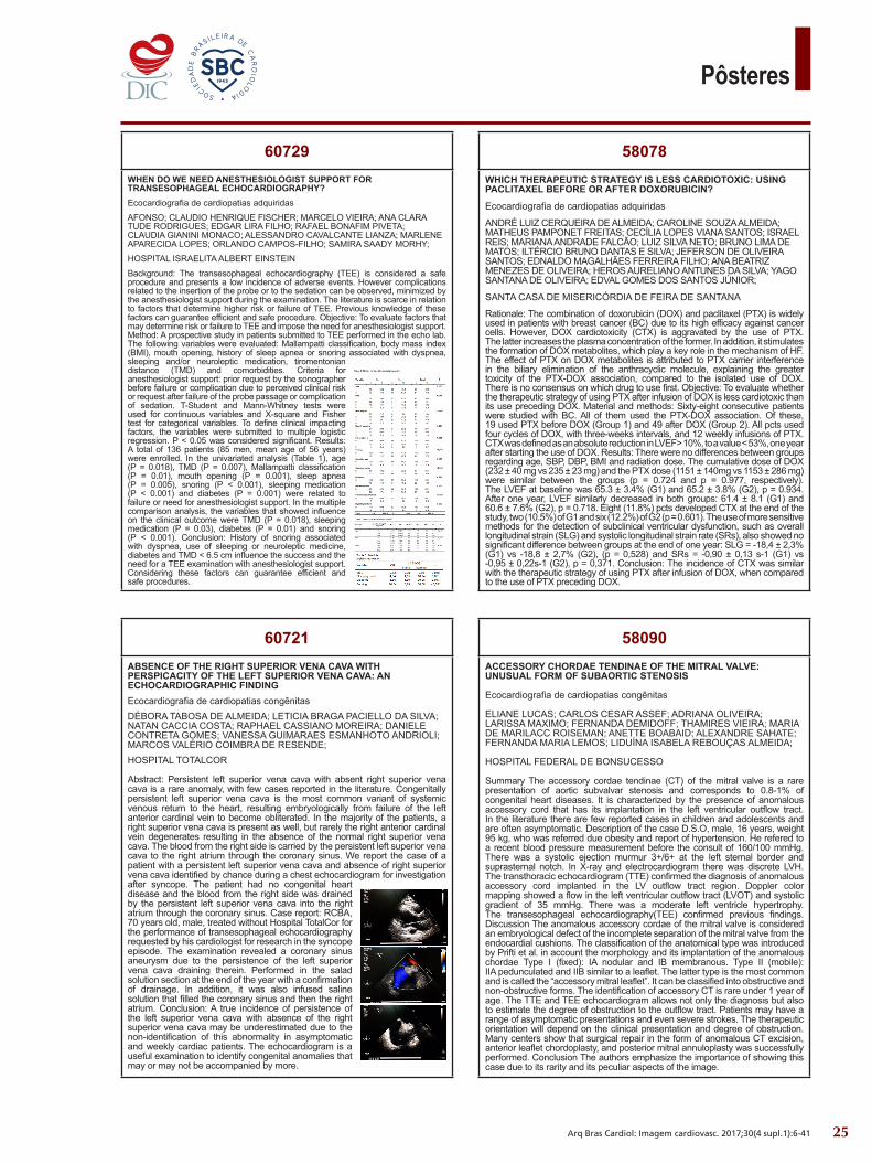
25
Pôsteres
Arq Bras Cardiol: Imagem cardiovasc. 2017;30(4 supl.1):6-41
58090ACCESSORY CHORDAE TENDINAE OF THE MITRAL VALVE: UNUSUAL FORM OF SUBAORTIC STENOSIS
Ecocardiografia de cardiopatias congênitas
ELIANE LUCAS; CARLOS CESAR ASSEF; ADRIANA OLIVEIRA; LARISSA MAXIMO; FERNANDA DEMIDOFF; THAMIRES VIEIRA; MARIA DE MARILACC ROISEMAN; ANETTE BOABAID; ALEXANDRE SAHATE; FERNANDA MARIA LEMOS; LIDUÍNA ISABELA REBOUÇAS ALMEIDA;
HOSPITAL FEDERAL DE BONSUCESSO
Summary The accessory cordae tendinae (CT) of the mitral valve is a rare presentation of aortic subvalvar stenosis and corresponds to 0.8-1% of congenital heart diseases. It is characterized by the presence of anomalous accessory cord that has its implantation in the left ventricular outflow tract. In the literature there are few reported cases in children and adolescents and are often asymptomatic. Description of the case D.S.O, male, 16 years, weight 95 kg, who was referred due obesity and report of hypertension. He refered to a recent blood pressure measurement before the consult of 160/100 mmHg. There was a systolic ejection murmur 3+/6+ at the left sternal border and suprasternal notch. In X-ray and electrocardiogram there was discrete LVH. The transthoracic echocardiogram (TTE) confirmed the diagnosis of anomalous accessory cord implanted in the LV outflow tract region. Doppler color mapping showed a flow in the left ventricular outflow tract (LVOT) and systolic gradient of 35 mmHg. There was a moderate left ventricle hypertrophy. The transesophageal echocardiography(TEE) confirmed previous findings. Discussion The anomalous accessory cordae of the mitral valve is considered an embryological defect of the incomplete separation of the mitral valve from the endocardial cushions. The classification of the anatomical type was introduced by Prifti et al. in account the morphology and its implantation of the anomalous chordae Type I (fixed): IA nodular and IB membranous. Type II (mobile): IIA pedunculated and IIB similar to a leaflet. The latter type is the most common and is called the “accessory mitral leaflet”. It can be classified into obstructive and non-obstructive forms. The identification of accessory CT is rare under 1 year of age. The TTE and TEE echocardiogram allows not only the diagnosis but also to estimate the degree of obstruction to the outflow tract. Patients may have a range of asymptomatic presentations and even severe strokes. The therapeutic orientation will depend on the clinical presentation and degree of obstruction. Many centers show that surgical repair in the form of anomalous CT excision, anterior leaflet chordoplasty, and posterior mitral annuloplasty was successfully performed. Conclusion The authors emphasize the importance of showing this case due to its rarity and its peculiar aspects of the image.
58078WHICH THERAPEUTIC STRATEGY IS LESS CARDIOTOXIC: USING PACLITAXEL BEFORE OR AFTER DOXORUBICIN?
Ecocardiografia de cardiopatias adquiridas
ANDRÉ LUIZ CERQUEIRA DE ALMEIDA; CAROLINE SOUZA ALMEIDA; MATHEUS PAMPONET FREITAS; CECÍLIA LOPES VIANA SANTOS; ISRAEL REIS; MARIANA ANDRADE FALCÃO; LUIZ SILVA NETO; BRUNO LIMA DE MATOS; ILTÉRCIO BRUNO DANTAS E SILVA; JEFERSON DE OLIVEIRA SANTOS; EDNALDO MAGALHÃES FERREIRA FILHO; ANA BEATRIZ MENEZES DE OLIVEIRA; HEROS AURELIANO ANTUNES DA SILVA; YAGO SANTANA DE OLIVEIRA; EDVAL GOMES DOS SANTOS JÚNIOR;
SANTA CASA DE MISERICÓRDIA DE FEIRA DE SANTANA
Rationale: The combination of doxorubicin (DOX) and paclitaxel (PTX) is widely used in patients with breast cancer (BC) due to its high efficacy against cancer cells. However, DOX cardiotoxicity (CTX) is aggravated by the use of PTX. The latter increases the plasma concentration of the former. In addition, it stimulates the formation of DOX metabolites, which play a key role in the mechanism of HF. The effect of PTX on DOX metabolites is attributed to PTX carrier interference in the biliary elimination of the anthracyclic molecule, explaining the greater toxicity of the PTX-DOX association, compared to the isolated use of DOX. There is no consensus on which drug to use first. Objective: To evaluate whether the therapeutic strategy of using PTX after infusion of DOX is less cardiotoxic than its use preceding DOX. Material and methods: Sixty-eight consecutive patients were studied with BC. All of them used the PTX-DOX association. Of these, 19 used PTX before DOX (Group 1) and 49 after DOX (Group 2). All pcts used four cycles of DOX, with three-weeks intervals, and 12 weekly infusions of PTX. CTX was defined as an absolute reduction in LVEF> 10%, to a value < 53%, one year after starting the use of DOX. Results: There were no differences between groups regarding age, SBP, DBP, BMI and radiation dose. The cumulative dose of DOX (232 ± 40 mg vs 235 ± 23 mg) and the PTX dose (1151 ± 140mg vs 1153 ± 286 mg) were similar between the groups (p = 0.724 and p = 0.977, respectively). The LVEF at baseline was 65.3 ± 3.4% (G1) and 65.2 ± 3.8% (G2), p = 0.934. After one year, LVEF similarly decreased in both groups: 61.4 ± 8.1 (G1) and 60.6 ± 7.6% (G2), p = 0.718. Eight (11.8%) pcts developed CTX at the end of the study, two (10.5%) of G1 and six (12.2%) of G2 (p = 0.601). The use of more sensitive methods for the detection of subclinical ventricular dysfunction, such as overall longitudinal strain (SLG) and systolic longitudinal strain rate (SRs), also showed no significant difference between groups at the end of one year: SLG = -18,4 ± 2,3% (G1) vs -18,8 ± 2,7% (G2), (p = 0,528) and SRs = -0,90 ± 0,13 s-1 (G1) vs -0,95 ± 0,22s-1 (G2), p = 0,371. Conclusion: The incidence of CTX was similar with the therapeutic strategy of using PTX after infusion of DOX, when compared to the use of PTX preceding DOX.
60721ABSENCE OF THE RIGHT SUPERIOR VENA CAVA WITH PERSPICACITY OF THE LEFT SUPERIOR VENA CAVA: AN ECHOCARDIOGRAPHIC FINDINGEcocardiografia de cardiopatias congênitasDÉBORA TABOSA DE ALMEIDA; LETICIA BRAGA PACIELLO DA SILVA; NATAN CACCIA COSTA; RAPHAEL CASSIANO MOREIRA; DANIELE CONTRETA GOMES; VANESSA GUIMARAES ESMANHOTO ANDRIOLI; MARCOS VALÉRIO COIMBRA DE RESENDE;HOSPITAL TOTALCOR
Abstract: Persistent left superior vena cava with absent right superior vena cava is a rare anomaly, with few cases reported in the literature. Congenitally persistent left superior vena cava is the most common variant of systemic venous return to the heart, resulting embryologically from failure of the left anterior cardinal vein to become obliterated. In the majority of the patients, a right superior vena cava is present as well, but rarely the right anterior cardinal vein degenerates resulting in the absence of the normal right superior vena cava. The blood from the right side is carried by the persistent left superior vena cava to the right atrium through the coronary sinus. We report the case of a patient with a persistent left superior vena cava and absence of right superior vena cava identified by chance during a chest echocardiogram for investigation after syncope. The patient had no congenital heart disease and the blood from the right side was drained by the persistent left superior vena cava into the right atrium through the coronary sinus. Case report: RCBA, 70 years old, male, treated without Hospital TotalCor for the performance of transesophageal echocardiography requested by his cardiologist for research in the syncope episode. The examination revealed a coronary sinus aneurysm due to the persistence of the left superior vena cava draining therein. Performed in the salad solution section at the end of the year with a confirmation of drainage. In addition, it was also infused saline solution that filled the coronary sinus and then the right atrium. Conclusion: A true incidence of persistence of the left superior vena cava with absence of the right superior vena cava may be underestimated due to the non-identification of this abnormality in asymptomatic and weekly cardiac patients. The echocardiogram is a useful examination to identify congenital anomalies that may or may not be accompanied by more.
60729WHEN DO WE NEED ANESTHESIOLOGIST SUPPORT FOR TRANSESOPHAGEAL ECHOCARDIOGRAPHY?Ecocardiografia de cardiopatias adquiridas
AFONSO; CLAUDIO HENRIQUE FISCHER; MARCELO VIEIRA; ANA CLARA TUDE RODRIGUES; EDGAR LIRA FILHO; RAFAEL BONAFIM PIVETA; CLAUDIA GIANINI MONACO; ALESSANDRO CAVALCANTE LIANZA; MARLENE APARECIDA LOPES; ORLANDO CAMPOS-FILHO; SAMIRA SAADY MORHY;
HOSPITAL ISRAELITA ALBERT EINSTEIN
Background: The transesophageal echocardiography (TEE) is considered a safe procedure and presents a low incidence of adverse events. However complications related to the insertion of the probe or to the sedation can be observed, minimized by the anesthesiologist support during the examination. The literature is scarce in relation to factors that determine higher risk or failure of TEE. Previous knowledge of these factors can guarantee efficient and safe procedure. Objective: To evaluate factors that may determine risk or failure to TEE and impose the need for anesthesiologist support. Method: A prospective study in patients submitted to TEE performed in the echo lab. The following variables were evaluated: Mallampatti classification, body mass index (BMI), mouth opening, history of sleep apnea or snoring associated with dyspnea, sleeping and/or neuroleptic medication, tiromentonian distance (TMD) and comorbidities. Criteria for anesthesiologist support: prior request by the sonographer before failure or complication due to perceived clinical risk or request after failure of the probe passage or complication of sedation. T-Student and Mann-Whitney tests were used for continuous variables and X-square and Fisher test for categorical variables. To define clinical impacting factors, the variables were submitted to multiple logistic regression. P < 0.05 was considered significant. Results: A total of 136 patients (85 men, mean age of 56 years) were enrolled. In the univariated analysis (Table 1), age (P = 0.018), TMD (P = 0.007), Mallampatti classification (P = 0.01), mouth opening (P = 0.001), sleep apnea (P = 0.005), snoring (P < 0.001), sleeping medication (P < 0.001) and diabetes (P = 0.001) were related to failure or need for anesthesiologist support. In the multiple comparison analysis, the variables that showed influence on the clinical outcome were TMD (P = 0.018), sleeping medication (P = 0.03), diabetes (P = 0.01) and snoring (P < 0.001). Conclusion: History of snoring associated with dyspnea, use of sleeping or neuroleptic medicine, diabetes and TMD < 6.5 cm influence the success and the need for a TEE examination with anesthesiologist support. Considering these factors can guarantee efficient and safe procedures.

26
Pôsteres
Arq Bras Cardiol: Imagem cardiovasc. 2017;30(4 supl.1):6-41
58023ANOMALOUS ORIGIN OF LEFT CORONARY ARTERY FROM PULMONARY ARTERY: A RARE CAUSE OF LEFT VENTRICULAR DYSFUNCTION IN CHILDREN
Ecocardiografia de cardiopatias congênitas
LIDIANE DIAS RIBEIRO; PATRICIA GUEDES DE SOUZA; MIRELA FREDERICO DE ALMEIDA ANDRADE; KARLA LUIZA MATOS PEDROSA; NADJA CECILIA DE CASTRO KRAYCHETE; ISABEL CRISTINA BRITTO GUIMARÃES;
HOSPITAL ANA NERI
Case description: A seven month old boy presented to the pediatric department of a tertiary hospital with dyspnea and failure to thrive. The patient was born by cesarean delivery, at 42 weeks, without reports of prenatal or neonatal care intercurrences. The mother referred onset of symptoms at 3 months, which motivated search for medical care. After several visits, being treated as respiratory tract infections, a chest radiography revealed cardiomegaly and patient was referred to specialist. At admission was observed tachypnea, increased antero-posterior chest diameter, with no cardiac murmurs. Electrocardiogram showed left ventricular hypertrophy and ST segment depression of DII, DII, aVF, V5 and V6, without Q waves. Troponin was negative. On first day patient had subtle cardiac arrest after intense crying, and was successfully resuscitated. Echocardiography was performed with enlargement of left chambers, moderate secondary mitral regurgitation, severe left ventricular dysfunction (ejection fraction: 18%), hyperechogenic papillary muscles. An abnormal retrograde left coronary flow was observed from posterior sinus of pulmonary artery (PA), suggesting anomalous origin of left coronary artery from pulmonary artery (ALCAPA). A dilated right coronary artery and septal arteries were also visualizated representing indirect signs of disease. Coronary angiography confirmed diagnosis. Patient underwent Takeuchi procedure successfully. Discussion: ALCAPA is a rare congenital heart disease with an estimated incidence of 0.25 to 0.5% of all congenital cardiac anomalies. There is no predilection for gender or race. Without appropriate surgical correction, the condition has a high mortality rate in the first year of life. Echocardiography is crucial and shows important clues for diagnosis as increased echogenicity of pappilary muscle, excessive dilation of right coronary artery. ALCAPA normally arises from the anterior-facing sinus of PA. In this case, echocardiography suspected of origin from posterior sinus, but it was not confirmed by operatory findings and reimplantation was not possible. Final comments: Coronary artery origins should be carefully evaluated in patients with left ventricular dysfunction and dilated cardiomyopathy, leading to adequate diagnosis and treatment of this potentially fatal disease.
57995ANEURYSMAL DILATATION OF PULMONARY VEINS IN PATIENTS WITH ANOMALOUS IMPLANTATION OF THE SEPTUM PRIMUM AND FLOW RESTRICTION IN HYPOPLASTIC LEFT HEART SYNDROME (HLHS)
Ecocardiografia de cardiopatias congênitas
CAROLINA BIRON PIERANTI; CELIA TOSHIE NAGAMATSU; GUSTAVO ANTONIO GUIMARÃES FAVARO; JULIANO DA SILVA CORDEIRO; DANIELA CARIOLI SANCHEZ; AMANDA REGINA CAMBOIM RIBEIRO; BEATRIZ FURLANETTO; FABRICIO MARCONDES CAMARGO; KAREN MONTEIRO MISAWA GAMBOA;
HOSPITAL BENEFICÊNCIA PORTUGUESA
Case Presentation One-month-old infant arrived late in a reference center with diagnosis of HLHS. Echocardiographic findings were aortic and mitral atresia, hypoplastic ascending aorta, restrictive atrial septal defect (mean gradient of 15 mmHg and velocity of 2.3 m/s), wide ductus arteriosus, moderate tricuspid regurgitation, preserved ventricular function and indirect signs of pulmonary hypertension. The restrictive nature of atrial septal defect due to anomalous implantation of the septum primum leading to pulmonary aneurysm veins (up to 19 mm) and significant dilation of the left atrium. In the first postoperative day of Atriosseptectomy associated with Pulmonary Artery Bandage, in prostin‘s hands, he presented a wide atrial septal defect and a significant reduction in the caliber of the pulmonary veins (9 mm). After one week of hospitalization, he was submitted to the Norwood-Sano surgical technique, with good results. Discussion Although extensively studied, some morphologic features of HLHS remain controversial. The case addresses the anomalous implantation of the septum primum with aneurysmal dilatation of the pulmonary veins, which, after an effective echocardiographic diagnosis, ensured hemodynamic stability and an important reduction in the caliber of the pulmonary veins after atriosseptectomy. In the 1980s, the atrial septal variants were grouped into intact atrial septum, restrictive foramen oval, ostium secundum atrial septal defect, ostium primum type (associated with unbalanced atrioventricular canal defects) and deviation of the atrial septum (the flap valve) relative to the infolded atrial roof (septum secundum). In the 90‘s, a study with 102 specimens obtained at Brompton Hospital, University of Padova and Heart Institute - HCFMUSP found 30% of septal malalignment and in 48% of these patients, mitral atresia. In 2006, Cardiol Young reported a relationship between interatrial septum types, fetal development, neonatal hemodynamic and clinical repercussion, and interstage of palliative surgeries. In septal malalignment, there is a gap of at least 4 mm, faking a wide atrial septal defect, even in the presence of incompatible clinical and turbulent Doppler flow. Final comments The case shows the best conduction of this leftward displacement of the septum primum when the fetal diagnosis allows the birth of these patients in specialized centers, since there is a need for surgical enlargement of this atrial septum.
60688ANOMALOUS LEFT CORONARY ARTERY IN ADULT – UNUSUAL DIAGNOSISEcocardiografia de cardiopatias congênitas
ANA ROSA VACCARI; ANA LÚCIA GIUSTI SCARSI; MÔNICA SUSO DOS SANTOS LEMOS; JOSÉ CARLOS DE ARAÚJO HAERTEL; VOLNEI VALDOMIRO DE MARCO; NELSON ANDRE BRAUN; JOÃO VITOR DARDE RODRIGUES; PAULO AFFONSO SALGADO FILHO; EDUARDO MASCARENHAS AZEVEDO; SARAH BERNARDON DE OLIVEIRA; CAROLINA SANDER REISER; MARCELO HAERTEL MIGLIORANZA;
INSTITUTO DE CARDIOLOGIA DO RIO GRANDE DO SUL
Case Description: 27 years old woman sought for medical evaluation due to newly-developed chest pain on mild efforts. During her childhood, she had the diagnosis of a dilated cardiomyopathy which was treating without a properly etiologic evaluation. Along this period, she remained asymptomatic. In the emergency department, the ECG revealed left chamber hypertrophy with septal and lateral inactive zones. The cardiac necrosis markers were normal. An echocardiogram was performed, reveling left ventricular enlargement with diffuse hypocinesia and severe systolic impairment. Several diastolic flows were identified by color Doppler through the right ventricular free wall and interventricular septum, corresponding to dilated vessels. Annomalous diastolic flow was also seen on pulmonary artery color Doppler. A huge right coronary was noted originating from the right coronary sinus while the left main coronary ostium was not seen. The coronary angiography reveled a long right coronary, with ectasia, filling the anterior descending and circumflex coronaries, and an anomalous left main coronary originating from the pulmonary artery (ALCAPA) was noted. The ALCAPA diagnosis was confirmed by a coronary computed tomography angiography. Discussion: Although ALCAPA is the most frequent coronary anomaly in children, it is a rare congenital heart malformation. After birth, the pressure and resistance of the pulmonary artery decrease, creating the phenomenon of coronary steal. This result in a myocardium being perfused by desaturated blood, leading to myocardial ischemia. Collateral circulation system is created and the right coronary becomes with ectasia. Around 85% of the patients present heart failure symptoms within the first two months of life and 80% of them die in the first months of life. Rarely, stabilization of ischemia could occur and the symptoms will appear just in the adulthood. Early diagnosis is crucial to allow on time treatment and prevent the development of irreversible myocardial ischemia and heart failure. Treatment is surgical with left coronary reimplantation in the aortic root. The present case demonstrates the importance of a multimodality imaging evaluation for a complete diagnosis. Although the final diagnosis is performed only by coronary angiography or computed tomography angiography, the echocardiographic evaluation may raise the diagnosis suspicion when signs of coronary anomalies are found.
58012ANATOMICALLY CORRECTED MALPOSITION OF GREAT ARTERIES ASSOCIATED WITH TOTAL ANOMALOUS PULMONARY VENOUS CONNECTION TO THE CORONARY SINUS WITH PREVIOUS DIAGNOSIS OF TRANSPOSITION OF THE GREAT ARTERIES
Ecocardiografia de cardiopatias congênitas
JULIANO DA SILVA CORDEIRO; CELIA TOSHIE NAGAMATSU; GUSTAVO ANTONIO GUIMARÃES FAVARO; CAROLINA BIRON PIERANTI; DANIELA CARIOLI SANCHEZ; AMANDA REGINA CAMBOIM RIBEIRO; EDUARDO MIRANDA TEIXEIRA; PAULA TIEMI NISHITANI; ANA LAURA BASTOS DA COSTA KAWASAKA; ERIKA YUMI ISHICAVA TAKAHASHI; BEATRIZ FURLANETTO;
BP - HOSPITAL BENEFICÊNCIA PORTUGUESA
Case presentation A 20-days-of-life newborn with previous diagnosis of transposition of the great arteries was transferred to a reference hospital in congenital heart surgery for surgical correction. On admission to the reference hospital, the first transthoracic echocardiogram showed situs solitus, levocardia, normal axis for the heart, normal systemic venous return, total anomalous pulmonary venous connection (TAPVC) to the coronary sinus, atrioventricular concordance, ventriculoarterial concordance and atrial septal defect (ASD). Although the two great vessels were origin from respective ventricles, they ran parallel and side by side to each other with the aorta to the left of the pulmonary artery. During hospitalization, the patient underwent a chest angiotomography that confirmed the echocardiographic diagnosis and then the surgical correction of TAPVC occurred. The patient was discharged after a few weeks. Discussion Anatomically corrected malposition of great arteries is a rare and benign condition when isolated. The first description of this entity was performed by Theremin around 1895 and was characterized by Van Praagh et al in 1975. The malposition has been classified into four types based on the atrial situs, ventricular loop and the relationship of the great arteries. Anderson et al has defined that this corresponds to any situation in which the aorta arises from the left ventricle but to the left of the pulmonary artery. This occurs when there is ventriculoarterial concordance and the vessels arise in parallel instead of occur the torsion. It is a condition that shows the difference between malposition and transposition of great arteries. The most common associated anomalies are ventricular septal defect (VSD), right ventricular outflow tract (RVOT) obstruction, subaortic obstruction, juxtaposition of atrial appendages and right aortic arch. The presence of TAPVC to the coronary sinus makes this case even rarer because of this unusual association. Final comments This unusual case was reported by rarity and to reinforce the need for a systematic and rigorous approach in the proper diagnostic evaluation and differentiation of other more common conditions such as misdiagnosis of transposition of the great arteries.

27
Pôsteres
Arq Bras Cardiol: Imagem cardiovasc. 2017;30(4 supl.1):6-41
58761BICUSPID AORTIC VALVE: HOW IS THE NATURAL HISTORY IN ARGENTINA?
Ecocardiografia de cardiopatias congênitas
MARIA CELESTE CARRERO; GERARDO MASSON JUAREZ; TERESA GARCIA BOTTA; GONZALO DIAZ BABIO; GUSTAVO; MARIA MEZZADRA; PABLO STUTZBACH;
INSTITUTO CARDIOVASCULAR SAN ISIDRO- SANATORIO LAS LOMAS
Background: Bicuspid aortic valve (BAV) is the most common congenital heart defect, however, there is no data about clinical and echocardiographic characteristics and cardiovascular outcomes in a contemporary population of adults with BAV in our country. Objective: To determine the basal characteristics, the incidence of major complications and predictors in a local cohort of patients with BAV. Methods: We included all consecutive patients referred for echocardiography with diagnosis of definite BAV (2009-2017). We collected clinical, echocardiographic and chemical variables. We analyzed the incidence of a combined end-point of intervention on aortic valve or ascending aorta and death, and a combined secondary end-point of moderate/severe aortic valvular stenosis and/or regurgitation, aortic aneurysm (aorta ≥ 45 mm) and endocarditis. Moreover, we assessed determinants and evolution of complications. We conducted logistic regression analysis to determine predictors of outcome. Results The cohort included 215 consecutive patients (43 ± 15.2 y/o) with definite BAV diagnosed by echocardiography. Mean duration of follow-up was 4.1 ± 2.3 y. At baseline 62 pts (40,5%) were ≤ 30 y/o, 74% males with a 2.9:1 proportion male to female. The mean age at diagnosis was 37 ± 15.6 y/o (median 38; range: 15-49 y/o) and 20% were hypertensive. Indications for echocardiogram were abnormal auscultatory findings (click, systolic or diastolic murmur) only in 50 cases (24%). In the majority (71%) the diagnosis was unsuspected previously to perform the echocardiogram. 7% of patients referred having first degree relatives with BAV. Baseline mean left ventricular ejection fraction was 64.2 ± 7,2%. 169 patients (79%) presented Type I valvular fenotipe (fusion of coronary cusps) and 71% presented a raphe. We detected any degree of aortic regurgitation in 69% (46% with mild regurgitation) and any degree of aortic stenosis in 26%. There were 18 events (combined primary end-point) in 17 patients (8.3%), including 1 cardiovascular death and interventions on aortic valve or ascending aorta in the remaining. The most frequent procedure (58.8% of all interventions) was aortic valve replacement, followed by Bentall de Bono procedure (23.5%) and severe aortic stenosis was the indication for surgery in 41%. The 16.8% of patients presented the secondary end-point during the follow-up. The independent predictors of primary cardiac events were age older than 50 y/o (OR: 6.57 (1,71-25,2), p = 0,003) and severe aortic valve stenosis or regurgitation (OR: 43.2 (10,7-174), p = 0.00000001) at baseline. Conclusions: In this study population of young adults with BAV, the incidence of intervention on aortic valve or ascending aorta and death over a mean of 4.06 years of follow-up was high, mostly owed to valvular interventions. Age and severity of valvular dysfunction were independently associated with primary cardiac events.
60491APICALLY DISPLACED PAPILLARY MUSCLES MIMICKING LEFT VENTRICULAR APICAL MASS: THE IMPORTANCE OF INVASIVE ECHOCARDIOGRAMEcocardiografia de cardiopatias congênitas
CRISTIANE DE CARVALHO SINGULANE; DIEGO NOVELLI; LUIZ FERNANDO DAL COL; FAISSAL IBRAHIM SOUMAILLE; MAURÍCIO DE NASSAU MACHADO; HÉLIO AUGUSTO DOS REIS CORBUCCI; JAMIL ALLI MURAD JÚNIOR;
FUNDAÇÃO FACULDADE REGIONAL DE MEDICINA DE SÃO JOSÉ DO RIO PRETO (FAMERP)
Introduction: The integrity of the mitral valve depends on the proper functioning of the papillary muscles. The most reported morphological abnormalities of the papillary muscles are their anomalous insertions and hypertrophy. This anomaly may remain asymptomatic in life-time or present with mitral valve stenosis or regurgitation or both. It might have clinical implications such as a dynamic left ventricular (LV) outflow tract obstruction even in the absence of hypertrophy. A careful evaluation of the left ventricular apex for proper diagnosis is extremely important since there are cases of apically displaced papillary muscles (ADPM) mimicking apical hypertrophic cardiomyopathy and even LV apical mass that may possibly be a thrombus, tumor or vegetation, entities that have very different treatments. Case Report: A 57-year-old male with hypertension was admitted to the emergency room with chest pain characterized as atypical angina. The electrocardiogram showed sinus rhythm, dubious electrically inactive area of the inferior wall and up to 1.5 mm J-point elevation in V2 to V4 Leads (suggestive of subendocardial ischemia). Due to the possibility of acute coronary syndrome, the patient was driven to early invasive risk stratification with coronary angiography. The left anterior descending coronary artery had a 10% stenosis in the mid portion. Contrast ventriculography showed an unconstrained hypotransparent mass on apical LV wall. The initial diagnostic hypothesis was intracavitary thrombus or tumor. For further investigation, a two-dimensional echocardiogram (ETT) has been performed, showing normal movement and thickness of the septum and LV walls, preserved systolic function, mild mitral and aortic regurgitation. However, due to significant impairment of the ETT acoustic window, an additional clarification of the mass was not possible. These findings were corroborated by transesophageal echocardiogram (EET). This method allowed the visualization of apical implantation of the postero-lateral papillary muscle of the LV confirming the absence of thrombi. Comments: We present a case of a patient with chest pain and electrocardiogram findings suggesting myocardial ischemia. Contrast ventriculography and ETT showed a mass in the LV apex, although it was not possible to have a diagnostic clarification. The EET identified this mass as ADPM. Therefore, in the presence of an apical mass, if the ETT is equivocal, a EET is mandatory to correct the diagnosis.
58014ARTERIOVENOUS FISTULA CONGENITAL CAROTIDA-JUGULAR. DIFFERENTIAL DIAGNOSIS IN CERVICAL REGION
Ecocardiografia de cardiopatias congênitas
FLAVIA GURGEL; ADRIANA INNOCENZI; MONICA CELENTE; VIVIANE XAVIER; LUIS CARLOS SIMOES; SALEM FRAGA; JULIANA RESI;
UNIVERSIDADE GAMA FILHO
Introduction: Congenital arteriovenous fistula(FAVC) in cervical region is a rare entity, with few cases described in the literature.Like other high-throughput fistulas, the connection between the carotid artery and the jugular vein can cause complications such as left heart failure and embolization. In this report, the case described is from an infant who looked for cardiac care for pulsate mass, with blow and thrill in the left cervical region. Relate : 5 months, N.G.L., male with a mass behind the pavilion left ear since birth. Normal cardiac auscultation. Transfontanela Ultrasound within normal range. Denies surgery, trauma, infections. Echocardiography with doppler showed VE overload, stream with increased speed in nameless. Cervical Doppler showed presence of FAVC carotida-jugular with high debt, confirmed by angiotomography. Discussion: Most of the arteriovenous communications is secondary to cervical post traumatic injuries or post surgical procedures, and the occurrence of congenital malformation rather rare, mostly in Pediatrics. The diagnosis of cervical mass is often due to high incidence of lymphoproliferative diseases within higher etaria. Persistence of embryonic communications during the development of arteries and veins from the common capillary plexus in the human fetus might be the basis for FAVC. The clinical diagnosis of an arteriovenous fistula of the neck carotid arteries involving the and the jugular veins is usually made without difficulty, when the compression of the carotid artery abolish the bruit, but the vessels involved should be precisely identified with angiography. Ultrasonograph Doppler of the neck is valuable especially in the young for screening but is not as precise as the angiotomography in delineating the anatomy.The precise evolution of the anatomy is fundamental to the decision of the fistula approach because good control of the afferent artery is essential, especially because of the rich collateral‘s. Optimum treatment includes ligation and or excision of all tributaries.
58049ANOMALY OF THE COMMON CAROTID ARTERY RIGHT ORIGINATING FROM THE PULMONARY TRUNK
Ecocardiografia de cardiopatias congênitas
DANIELA CARIOLI SÁNCHEZ; GUSTAVO ANTÔNIO GUIMARÃES FAVARO; CELIA TOSHIE NAGAMATSU; CAROLINA BIRON PIERANTI; AMANDA REGINA CAMBOIM RIBEIRO; JULIANO DA SILVA CORDEIRO; FERNANDA CAMPANHA DE MENDONÇA CRUZ; FABRÍCIO MARCONDES CAMARGO; ANA LAURA BASTOS DA COSTA KAWASAKA; CINTIA COSTA MELO;
BENEFICIÊNCIA PORTUGUESA
Case presentation Newborn of 15 days of life with prenatal diagnosis of aortic atresia, ascending aortic hypoplasia, atrial septal defect and ventricular septal defect. After the postnatal diagnosis with the echocardiographic team was confirmed and the patient underwent pulmonary artery banding, and stent implantation in the ductus arteriosus. In the transthoracic echocardiography of post-surgical control, another congenital heart malformation was diagnosed, the right common carotid artery originated from the pulmonary trunk. As echocardiography presents limitations of images in the extracardiac structures, it was suggested to complement the diagnosis with cardiac and vessel angiotomography, which confirmed that the right common carotid artery originated at the proximal end of the pulmonary trunk and diagnosed that the right subclavian artery originated in the portion of the descending aorta with a retroesophageal pathway. At 3 months old, he underwent Norwood-classic surgery and redirection of the right common carotid artery to the neoaorta. At 6 months of life he underwent Rastelli surgery and occluded the central Shunt. Discussion We describe a rare congenital anomaly, complex and difficult to diagnose and without reports in the literature. Congenital cardiac echocardiography played an important role, providing benefits both for clinical management and for surgical management of the patient. The complementation of the diagnosis with angiotomography allowed to visualize the images of the vascular anatomy of the base in the three-dimensional form. Final comments Detailed echocardiographic investigation, cardiac anatomy, and the experience of the congenital echocardiographer allow for many rare diagnoses of pediatric cardiology. Providing agility and benefits in clinical management and surgical programming.
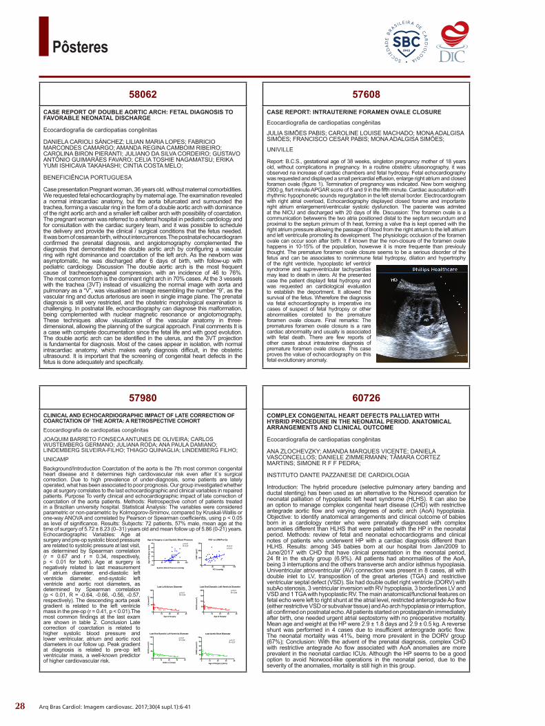
28
Pôsteres
Arq Bras Cardiol: Imagem cardiovasc. 2017;30(4 supl.1):6-41
60726COMPLEX CONGENITAL HEART DEFECTS PALLIATED WITH HYBRID PROCEDURE IN THE NEONATAL PERIOD. ANATOMICAL ARRANGEMENTS AND CLINICAL OUTCOME
Ecocardiografia de cardiopatias congênitas
ANA ZLOCHEVZKY; AMANDA MARQUES VICENTE; DANIELA VASCONCELLOS; DANIELE ZIMMERMANN; TÂMARA CORTEZ MARTINS; SIMONE R F F PEDRA;
INSTITUTO DANTE PAZZANESE DE CARDIOLOGIA
Introduction: The hybrid procedure (selective pulmonary artery banding and ductal stenting) has been used as an alternative to the Norwood operation for neonatal palliation of hypoplastic left heart syndrome (HLHS). It can also be an option to manage complex congenital heart disease (CHD) with restrictive antegrade aortic flow and varying degrees of aortic arch (AoA) hypoplasia. Objective: to identify anatomical arrangements and clinical outcome of babies born in a cardiology center who were prenatally diagnosed with complex anomalies different than HLHS that were palliated with the HP in the neonatal period. Methods: review of fetal and neonatal echocardiograms and clinical notes of patients who underwent HP with a cardiac diagnosis different than HLHS. Results: among 345 babies born at our hospital from Jan/2009 to June/2017 with CHD that have clinical presentation in the neonatal period, 24 fit in the study group (6.9%). All patients had abnormalities of the AoA, being 3 interruptions and the others transverse arch and/or isthmus hypoplasia. Univentricular atrioventricular (AV) connection was present in 8 cases, all with double inlet to LV, transposition of the great arteries (TGA) and restrictive ventricular septal defect (VSD). Six had double outlet right ventricle (DORV) with subAo stenosis, 3 ventricular inversion with RV hypoplasia, 3 borderlines LV and VSD and 1 TGA with hypoplastic RV. The main anatomical/functional features on fetal echo were left to right shunt at the atrial level, restricted anterograde Ao flow (either restrictive VSD or subvalvar tissue) and Ao arch hypoplasia or interruption, all confirmed on postnatal echo. All patients started on prostaglandin immediately after birth, one needed urgent atrial septostomy with no preoperative mortality. Mean age and weight at the HP were 2.9 ± 1.8 days and 2.9 ± 0.5 kg. A reverse shunt was performed in 4 cases due to insufficient anterograde aortic flow. The neonatal mortality was 41%, being more prevalent in the DORV group (67%); Conclusion: With the advent of the prenatal diagnosis, complex CHD with restrictive antegrade Ao flow associated with AoA anomalies are more prevalent in the neonatal cardiac ICUs. Although the HP seems to be a good option to avoid Norwood-like operations in the neonatal period, due to the severity of the anomalies, mortality is still high in this group.
57608CASE REPORT: INTRAUTERINE FORAMEN OVALE CLOSURE
Ecocardiografia de cardiopatias congênitas
JULIA SIMÕES PABIS; CAROLINE LOUISE MACHADO; MONA ADALGISA SIMÕES; FRANCISCO CESAR PABIS; MONA ADALGISA SIMÕES;
UNIVILLE
Report: B.C.S., gestational age of 38 weeks, singleton pregnancy mother of 18 years old, without complications in pregnancy. In a routine obstetric ultasonography, it was observed na increase of cardiac chambers and fetal hydropsy. Fetal echocardiography was requested and displayed a small pericardial effusion, enlarge right atrium and closed foramen ovale (figure 1). Termination of pregnancy was indicated. New born weighing 2900 g, fisrt minute APGAR score of 8 and 9 in the fifth minute. Cardiac auscultation with rhythmic hypophonetic sounds regurgitation in the left sternal border. Electrocardiogram with right atrial overload, Echocardiography displayed closed forame and importante right atrium enlargement/ventricular systolic dysfunction. The paciente was admited at the NICU and discharged with 20 days of life. Discussion: The foramen ovale is a communication betwwens the two atria positioned distal to the septum secundum and proximal to the septum primum of th heat, forming a valve tha is kept opened with the right atrium pressure allowing the passage of blood from the right atrium to the left atrium and left ventriculle promoting its development. The physiologic occlusion of the foramen ovale can occur soon after birth. It if known thar the non-closure of the foramen ovale happens in 10-15% of the population, howevwe it is more frequente than previouly thought. The premature foramen ovale closure seems to be a serious disorder of the fetus and can be associates to nonimmune fetal hydropsy, dilation and hypertrophy of the right ventricle, hypoplastic lef ventriclr syndrome and supreventricular tachycardias may lead to death in útero. At the presented case the patient displayd fetal hydropsy and was requested an cardiological evaluation to establish the deportment. It allowed the survival of the fetus. Wherefore the diagnosis via fetal echocardiography is imperative ins cases of suspect of fetal hydropsy or other abnormalities corelated to the premature foramen ovale closure. Final remarks: The prematures foramen ovale closure is a rare cardiac abnormality and usually is associated with fetal death. There are few reports of other cases about intrauterine diagnosis of premature foramen ovale closure. This case proves the value of echocardiography on this fetal evolutionary anomaly.
57980CLINICAL AND ECHOCARDIOGRAPHIC IMPACT OF LATE CORRECTION OF COARCTATION OF THE AORTA: A RETROSPECTIVE COHORTEcocardiografia de cardiopatias congênitas
JOAQUIM BARRETO FONSECA ANTUNES DE OLIVEIRA; CARLOS WUSTEMBERG GERMANO; JULIANA RODA; ANA PAULA DAMIANO; LINDEMBERG SILVEIRA-FILHO; THIAGO QUINAGLIA; LINDEMBERG FILHO;
UNICAMP
Background/Introduction Coarctation of the aorta is the 7th most common congenital heart disease and it determines high cardiovascular risk even after it`s surgical correction. Due to high prevalence of under-diagnosis, some patients are lately operated, what has been associated to poor prognosis. Our group investigated whether age at surgery correlates to the last echocardiographic and clinical variables in repaired patients. Purpose To verify clinical and echocardiographic impact of late correction of coarctation of the aorta patients. Methods: Retrospective cohort of patients treated in a Brazilian university hospital. Statistical Analysis: The variables were considered parametric or non-parametric by Kolmogorov-Smirnov, compared by Kruskal-Wallis or one-way ANOVA and correlated by Pearson or Spearman coefficients, using p < 0.05 as level of significance. Results: Subjects: 72 patients, 57% male, mean age at the time of surgery of 5.72 ± 8.23 (0–31) years old and mean follow up of 5.86 (0-21) years. Echocardiographic Variables: Age at surgery and pre-op systolic blood pressure are related to systolic pressure at last visit, as determined by Spearman correlation (r = 0.67 and r = 0.34, respectively, p < 0.01 for both). Age at surgery is negatively related to last measurement of atrium diameter, end-diastolic left ventricle diameter, end-systolic left ventricle and aortic root diameters, as determined by Spearman correlation (p < 0.01, R = -0.64, -0.66, -0.56, -0.57, respectively). The descending aorta peak gradient is related to the left ventricle mass in the pre-op (r = 0.41, p < 0.01).The most common findings at the last exam are shown in table 2. Conclusion Late correction of coarctation is related to higher systolic blood pressure and lower ventricular, atrium and aortic root diameters in our follow up. Peak gradient at diagnosis is related to pre-op left ventricular mass, a well-known predictor of higher cardiovascular risk.
50 100 150 200-10
0
10
20
30
40
Age at Surgery x Last Systolic Blood Pressure
Age
at S
urge
ry (y
ears
)
Systolic Blood Pressure (mmHg)
p<0,01R=0,67
0 10 20 30 400
50
100
150
Last Left Atrium Diameter
Age at Surgery
LA (m
m/m
2 )
p<0,01R=-0,64
0 10 20 30 400
50
100
150
Last End Systolic Left Ventricle Diameter
Idade a Cirurgia
SLVD
(mm
/m2 )
p<0,01R=-0,56
0 50 100 1500
100
200
300
PG1 vs LVM Pre-Op
PG1 (mmHg)
LVM
1 (g
/m2 )
R=0.41p<0.01
0 10 20 30 400
50
100
150
200
Last End Diastolic Left Ventricle Diameter
Age at Surgery
p<0,01R=-0,66
DLV
D (m
m/m
2 )
0 10 20 30 400
20
40
60
80
100
Last Aortic Root Diameter
Age at Surgery (years)
AO
RD
(mm
/m2 )
p<0,01R=-0,57
58062CASE REPORT OF DOUBLE AORTIC ARCH: FETAL DIAGNOSIS TO FAVORABLE NEONATAL DISCHARGE
Ecocardiografia de cardiopatias congênitas
DANIELA CARIOLI SÁNCHEZ; LILIAN MARIA LOPES; FABRICIO MARCONDES CAMARGO; AMANDA REGINA CAMBOIM RIBEIRO; CAROLINA BIRON PIERANTI; JULIANO DA SILVA CORDEIRO; GUSTAVO ANTÔNIO GUIMARÃES FAVARO; CELIA TOSHIE NAGAMATSU; ERIKA YUMI ISHICAVA TAKAHASHI; CINTIA COSTA MELO;
BENEFICIÊNCIA PORTUGUESA
Case presentation Pregnant woman, 36 years old, without maternal comorbidities. We requested fetal echocardiography by maternal age. The examination revealed a normal intracardiac anatomy, but the aorta bifurcated and surrounded the trachea, forming a vascular ring in the form of a double aortic arch with dominance of the right aortic arch and a smaller left caliber arch with possibility of coarctation. The pregnant woman was referred to a referral hospital in pediatric cardiology and for consultation with the cardiac surgery team, and it was possible to schedule the delivery and provide the clinical / surgical conditions that the fetus needed. It was born of cesarean birth, without intercurrences.The postnatal echocardiogram confirmed the prenatal diagnosis, and angiotomography complemented the diagnosis that demonstrated the double aortic arch by configuring a vascular ring with right dominance and coarctation of the left arch. As the newborn was asymptomatic, he was discharged after 6 days of birth, with follow-up with pediatric cardiology. Discussion The double aortic arch is the most frequent cause of tracheoesophageal compression, with an incidence of 46 to 76%. The most common form is the dominant right arch in 70% cases. At the 3 vessels with the trachea (3VT) instead of visualizing the normal image with aorta and pulmonary as a “V”, was visualised an image resembling the number “9”, as the vascular ring and ductus arteriosus are seen in single image plane. The prenatal diagnosis is still very restricted, and the obstetric morphological examination is challenging. In postnatal life, echocardiography can diagnose this malformation, being complemented with nuclear magnetic resonance or angiotomography. These techniques allow visualization of the vascular anatomy in three-dimensional, allowing the planning of the surgical approach. Final comments It is a case with complete documentation since the fetal life and with good evolution. The double aortic arch can be identified in the uterus, and the 3VT projection is fundamental for diagnosis. Most of the cases appear in isolation, with normal intracardiac anatomy, which makes early diagnosis difficult, in the obstetric ultrasound. It is important that the screening of congenital heart defects in the fetus is done adequately and specifically.

29
Pôsteres
Arq Bras Cardiol: Imagem cardiovasc. 2017;30(4 supl.1):6-41
60222DIAGNOSTIC CHALLENGE IN LATE POSTOPERATIVE CORRECTION OF ATRIAL SEPTAL DEFECT: OBSTRUCTION OF SYSTEMIC VEINS
Ecocardiografia de cardiopatias congênitas
RENATA DE SÁ CASSAR; FLÁVIA ENGERS SALLES; FLORA MACIEL PENACHIO; NATHÁLIA MARTINS TELLES; GABRIEL FRANCISCO PENA VILA LIMA; BIANCA SARAIVA SANTORO; BEATRIZ RESENDE MARIANO; RENATA PERAZZO BARBOSA MENDES; CLAUDIA REGINA PINHEIRO CASTRO GRAU; GLÁUCIA MARIA PENHA TAVARES; WILSON MATHIAS JUNIOR;
INCOR-HC-FMUSP-SP
Case report: We report two cases of patients with ostium secundum atrial septal defect (ASD) who underwent surgical correction in childhood, and developed uncommon complications, resulting in diagnostic challenge. (1) G.P.S.L., 39 years old, female, submitted to atrioseptoplasty at 4 years old, developing with right heart failure, hepatic congestion and Budd Chiari syndrome. (2) L.M.B.R.S., 49 years old, female, submitted to atrioseptoplasty at age 11, reoperated by residual shunt, evolving with signs of pulmonary arterial hypertension (cyanosis and flow inversion at Doppler echocardiography. In both cases the transthoracic echocardiogram showed no significant changes. It was decided to perform the transesophageal echocardiogram(TEE), which revealed severe stenosis of the Superior and Inferior vena cava in the first patient; in the second patient showed obstruction inside the right atrium, secondary to “surgical pacth protrusion”, in addition to residual shunt with flow directed to the left atrium. Both cases underwent balloon catheter angioplasty with significant clinical improvement. Discussion: ASD has an estimated incidence of 1 in 1,500 live births and corresponds to 6-10% of all congenital heart diseases. Ostium secundum ASD corresponds to 75-80% of the cases and the gold standard diagnostic test is TEE. The surgical or percutaneous repair is indicated when there is overload of the right chambers without irreversible pulmonary hypertension. Late complications following surgical ASD repair are vary rare, with a rate of 7% of recurrent shunt and less than 2% of reoperation rates for correction of residual left-to-right shunt or recurrent ASD. Conclusion: In the reported cases, there was dissociation between the clinical and the initial findings of the ETT. The use of TEE was decisive in these patients, allowing a correct diagnosis and management of late complications following surgical ASD repair.
57986DEEP CLEFT-LIKE MITRAL INDENTATIONS ASSOCIATED WITH BARLOW’S DISEASE AND RUPTURED CHORDAE – REPORT OF 2 CASES: IMPACT OF THREE-DIMENSIONAL ECHOCARDIOGRAPHYEcocardiografia de cardiopatias congênitas
ANGELE AZEVEDO ALVES MATTOSO; MARTA NERI LACERDA; RICARDO JOSÉ COSTA MATTOSO; MARIA LÚCIA DUARTE; EDUARDO TADEU FERREIRA;
HOSPITAL SANTA IZABEL / CLINICA PROALIVIO
Presentation: Case 01. A 32-year-old man with no prior medical problems. Transthoracic echocardiogram showed moderate mitral regurgitation. The patient underwent a transesophageal echocardiography to further evaluation of the mitral valve. Two-dimensional transesophageal echocardiogram showed diffuse thickening of the mitral leaflets and prolapse of the posterior leaflet. Real-time “zoom” three-dimensional transesophageal echocardiography showed a large amount of excess myxomatous tissue of the both leaflet (Barlow`s Disease) and did reveal a deep cleft-like indentation between P2 and P3 (red arrow) of the posterior leaflet (figure 1). Case 02. A 53-year-old man complaining of progressive dyspnea. Transthoracic echocardiogram showed eccentric and severe mitral regurgitation consistent with posterior leaflet disease of the mitral valve. Two-dimensional transesophageal echocardiogram showed the prolapsing scallop (P2) with ruptured chordae (red arrow) of the posterior leaflet. Real-time “zoom” three-dimensional transesophageal echocardiography did also reveal a deep cleft-like indentation between P2 and P3 (yellow arrow) of the posterior leaflet (Figure 2). Discussion: Accurate characterization of mitral valve disease is critically important to predict feasibility of mitral valve repair. Deep cleft-like indentation can occur in myxomatous mitral valve disease and can be associated with mitral regurgitation and failure of mitral repair. Following the advent of three-dimensional transesophageal echocardiography, that offers singular advantages, such as correct vision (en face) of the mitral valve, it appears to be more common than previously thought. In these cases, we demonstrated two patients with different spectrum of myxomatous mitral valve disease, being possible the diagnosis of the deep cleft-like indentation of the posterior leaflet only by diagnostic enhancement of three-dimensional transesophageal echocardiography of the mitral valve. Final comments: The enhance diagnostic of the three-dimensional transesophageal echocardiography is critically important to accurate characterization of the diversity of myxomatous mitral valve disease.
Figura 1
Figura 2
60710DIAGNOSIS OF ISOLATED FETAL CORONARY-CAMERAL FISTULA MIMETIZING VENTRICULAR SEPTAL DEFECT: CASE REPORT
Ecocardiografia de cardiopatias congênitas
MARA RUFINO DE ANDRADE; FABRICIO MARCONDES CAMARGO; MARIANE DE FÁTIMA YUKIE MAEDA; WALTHER YOSHIHARU ISHIKAWA; GUSTAVO FORONDA; NANA MIURA IKARI; GLÁUCIA MARIA PENHA TAVARES; MARCO ANTÔNIO BORGES LOPES; ROSSANA PULCINELI VIEIRA FRANCISCO;
INCOR
Introduction: Isolated coronary-cameral fistula is a rare congenital malformation. It may originate in both coronaries, although it is often related to the right coronary. The diagnosis in a prenatal scan can be done but in this period it can mimetize very well other heart malformations like ventricular septal defect. In the neonatal period, patients may remain asymptomatic or require an immediate intervention. Case Description: A 22 years of age patient, 23 weeks of gestation of her third pregnancy, admitted at the Division of Fetal Medicine of our institution, diagnosed with fetal ventricular septal defect. After a fetal echocardiography, it was identified a moderate fetal coronary-cameral fistula of the left coronary, which would flow into the ventricular septum at high velocity, mimetizing a great ventricular septal defect. Serial fetal echocardiogram was performed and no signal of hemodynamic decompensation was detected as the cardiac chambers were normal, there was no fetal hydrops and fetal wellbeing tests were normal. The infant, 3820g, female, was born at 39 weeks of gestation by C-section. Postnatal echocardiogram showed a long and twisted isolated left coronary fistula to the ventricular septum (its maximum diameter is 10 mm) with a 9mm opening to the left ventricle, with systolic-diastolic flow. Right coronary artery was dilated, but without other abnormalities. The computed tomography angiogram confirmed the diagnosis. Because the patient was asymptomatic in the neonatal period, no intervention was requiring. The patient is taking aspirin, under clinical observation, and up to this moment continues asymptomatic. Comments: Coronary-cameral fistulas are rare, and can be diagnosed during the fetal stage. Color Doppler is extremely useful to identify their origin to ending point and the total assessment of their extension. Early diagnosis and follow-up during the gestation period can predict the cases that might require early treatment.
60713CROSSED PULMONARY ARTERIES: ANALYSIS OF A SERIE OF CASES DURING A FIVE YEARS PERIOD IN A SINGLE CENTEREcocardiografia de cardiopatias congênitas
BARBARA NEIVA TANAKA; VALÉRIA DE MELO MOREIRA; WALTHER YOSHIHARU ISHIKAWA; VERA DEMARCHI AIELLO; RENATA NOVIS ROSSI; WILSON MATHIAS JUNIOR; MARCIO MIRANDA BRITO; LEONARDO AUGUSTO MIANA; FLÁVIA ENGERS SALLES; LUCIANA DA VEIGA KINOSHITA; SANTIAGO RAUL ARRIETA; NANA MIURA; GUSTAVO FORONDA; RENATA PERAZZO BARBOSA MENDES; GLAUCIA MARIA PENHA TAVARES;
INSTITUTO DO CORACAO - HCFMUSPBackground: Crossed pulmonary arteries (CPA) are rare congenital anomalies characterized by an abnormal ostium of the left pulmonary artery that lies to the right of and above the right pulmonary artery. The branch pulmonary arteries then crisscross, extrapericardially, as they course to each respective lung. They are often associated with other congenital heart diseases and extracardiac anomalies. Objectives: The aim of this study is to analyze 22 cases of CPA from the hospital-based prevalence, clinical information, outcome of surgery and imaging findings. Methodology: From January 2012 to December 2016 we conducted a retrospective search from our departmental database. We recorded and summarized the echocardiograms, angiographies, computed tomographies, clinical information, surgery description, outcome and necropsy of the patients with CPA and expressed the results as the number and percentage of the patients. Results: The 22 patients (13 females and 9 males) ranged in age from 1 day to 3 years at the time of the initial diagnosis. The cardiovascular abnormalities associated with CPA are summarized in Table 1. The majority of the patients had complex cardiac pathologies such as truncus arteriosus, hypoplastic left ventricle and criss-cross heart. Aortic arch abnormalities such as interrupted aortic arch and coarctation of the aorta were detected in six patients (27%). The diagnosing methods of CPA are described in Table 1. Echocardiography detected CPA in 21 cases (95%). In 50 % of the cases, an associated genetic syndrome or an extracardiac anomaly was present. Among the 22 cases of CPA, 13 patients underwent successful operations and 6 patients died during this period. At this writing, the remaining cases are clinically stable and being followed. Conclusions: CPA is an uncommon condition and probably subdiagnosed. The difficulty in diagnosing CPA is overcome by the advent of color Doppler imaging and three-dimensional reconstruction. Although CPA are established as a benign congenital anomaly, it can co-exist with clinically important cardiac lesions. We believe that CPA may interfere in hemodynamic conditions of some of these situations. Preoperative diagnosis can facilitate surgical management of this unusual condition. Since it is frequent the association of CPA and multiple syndromes, genetic evaluation should be considered in all patients with this anomaly. According to our search of the literature, fewer than 70 cases had been reported up to 2016.
CASE SEX AGE CARDIAC ANOMALIES DIAGNOSING METHOD
EXTRACARDIAC ANOMALIES / GENETIC SYNDROMES
1 M < 1 month Truncus arteriosus Echo + Surgery 2 F < 1 month Truncus arteriosus Echo + CT 3 M < 1 month Tetralogy of Fallot Echo Intelectual disabillity 4 M < 1 month Ectopia cordis, ASD, VSD, superior-inferior
ventricles, CoA, ARSCA Echo Cantrell Pentalogy
5 F < 1 month IAA, VSD, ARSCA Echo + CT 6 F < 1 month Truncus arteriosus Echo Di George 7 F 1 month Truncus arteriosus Echo 8 F 1dv IAA, ASD, VSD, BAV Echo + CT Di George 9 F 3 years ALCAPA Echo + CT
10 M < 1 month Truncus arteriosus Echo + CT Di George 11 M 1 month Truncus arteriosus Echo 12 M 5 months ASD Echo VACTERL 13 M 1 month PAIVS Echo 14 M 8 months ASD, VSD, hypoplastic RPA, RAA, ALSCA Echo + CT 15 F 1 month VSD Echo + CT Edwards 16 F < 1 month Criss cross, MS, CoA, hypoplastic LPA Surgery 17 F 8 months ASD, hypoplastic RPA Echo + CT Encephalocele 18 M < 1 month Double aortic arch Echo + Angiography 19 F < 1 month HLV Echo + Necropsy Duodenal atresia 20 F 2 years ASD Echo Polycystic kidney disease 21 F < 1 month HLV Echo 22 F 2 years ASD Echo Blackfan Diamond anemia
M male F female ASD atrial septal defect VSD ventricular septal defect CoA coarctation of the aorta ARSCA aberrant right subclavian artery IAA interrupted aortic arch BAV bicuspid aortic valve PAIVS pulmonary atresia with intact ventricular septum ALCAPA anomalous left coronary artery from pulmonary artery RPA right pulmonary artery LPA left pulmonary artery RAA right aortic arch ALSCA aberrant left subclavian artery MS mitral stenosis HLV hypoplastic left heart Echo echocardiopgraphy CT computed tomography
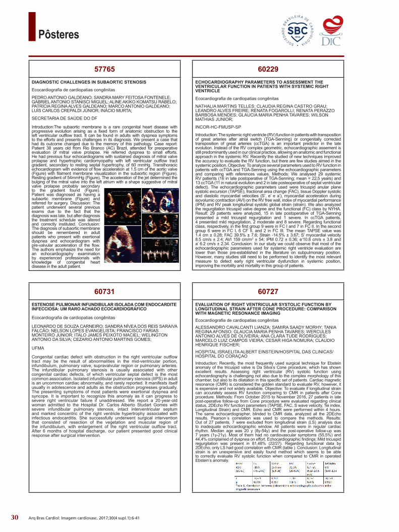
30
Pôsteres
Arq Bras Cardiol: Imagem cardiovasc. 2017;30(4 supl.1):6-41
60727EVALUATION OF RIGHT VENTRICULAR SYSTOLIC FUNCTION BY LONGITUDINAL STRAIN AFTER CONE PROCEDURE: COMPARISON WITH MAGNETIC RESONANCE IMAGINGEcocardiografia de cardiopatias congênitasALESSANDRO CAVALCANTI LIANZA; SAMIRA SAADY MORHY; TANIA REGINA AFONSO; GLAUCIA MARIA PENHA TAVARES; WERCULES ANTONIO ALVES DE OLIVEIRA; ANA CLARA TUDE RODRIGUES; MARCELO LUIZ CAMPOS VIEIRA; CESAR HIGA NOMURA; CLAUDIO HENRIQUE FISCHER;HOSPITAL ISRAELITA ALBERT EINSTEIN/HOSPITAL DAS CLINICAS/HOSPITAL DO CORAÇAOIntroduction: Recently, the most frequently used surgical technique for Ebstein anomaly of the tricuspid valve is Da Silva’s Cone procedure, which has shown excellent results. Assessing right ventricular (RV) systolic function using echocardiography is challenging not also due to the complex morphology of this chamber, but also to its dilatation in this specific set of patients. Cardiac magnetic resonance (CMR) is considered the golden standard to evaluate RV, however, it is expensive and not widely available. Objective: To evaluate if longitudinal strain can accurately assess RV function comparing to CMR in patients after Cone procedure. Methods: From October 2015 to November 2016, 27 patients in late post-operative follow-up from Cone procedure were evaluated regarding clinical status, 2DEcho RV function parameters (TAPSE, FAC, S wave velocity, Tei index, Longitudinal Strain) and CMR. Echo and CMR were performed within 4 hours. The same echocardiographer, blinded to CMR data, analyzed all the 2DEcho results. Pearson´s correlation was used to compare the methods. Results: Out of 27 patients, 7 were excluded from longitudinal strain (LS) analysis due to inadequate echocardiographic window. All patients were in regular cardiac rhythm. Median age was 20 y (8y-54y) and the post-operative follow-up was 7 years (1y-21y). Most of them had no cardiovascular symptoms (55,5%) and 44,4% complained of dyspnea on effort. Echocardiographic findings: Mild tricuspid regurgitation was present in 81,48% (22/27). Regarding functional data by 2DEcho, only LS had good correlation with CMR (table ). Conclusion: Longitudinal strain is an unexpensive and easily found method which seems to be able to correctly evaluate RV systolic function when compared to CMR in operated Ebstein’s anomaly.
60229ECHOCARDIOGRAPHY PARAMETERS TO ASSESSMENT THE VENTRICULAR FUNCTION IN PATIENTS WITH SYSTEMIC RIGHT VENTRICLE
Ecocardiografia de cardiopatias congênitas
NATHALIA MARTINS TELLES; CLAUDIA REGINA CASTRO GRAU; LEANDRO ALVES FREIRE; RENATA FOGAROLLI; RENATA PERAZZO BARBOSA MENDES; GLAUCIA MARIA PENHA TAVARES; WILSON MATHIAS JUNIOR;
INCOR-HC-FMUSP-SP
Introduction: The systemic right ventricle (RV) function in patients with transposition of great arteries after atrial switch (TGA-Senning) or congenitally corrected transposition of great arteries (ccTGA) is an important predictor in the late evolution. Instead of the RV complex geometric, echocardiographic assement is still predominantly used in our clinical practice, allowing an anatomic and functional approach in the systemic RV. Recently the studied of new techniques improved the accuracy to evaluate the RV function, but there are few studies aimed in the systemic position. Objective: To analyze several parameters used to RV function in patients with ccTGA and TGA-Senning using the echocardiographic parameters and comparing with references values. Methods: We analysed 29 systemic RV patients (16 in late postoperative of TGA-Senning; mean = 22,5 years) and 13 ccTGA (11 in natural evolution and 2 in late posteoperative of septal ventricular defect). The echocardiographic parameters used were tricuspid anular plane systolic excursion (TAPSE), fractional area change (FAC), tissue Doppler systolic and diastolic myocardial velocities (S’, e’ e a’), myocardial acceleration during isovolumic contraction (AVI) on the RV free wall, index of myocardial performance (IPM) and RV peak longitudinal systolic global strain (strain). We also analyzed the regurgitation tricuspid valve degree and the functional (FC) class by NYHA. Result: 29 patients were analyzed, 15 in late postoperative of TGA-Senning presented a mild tricuspid regurgitation and 1 severe. In ccTGA patients, 4 presented mild regurgitation, 2 moderate and 8 severe. Regarding functional class, respectively, in the first group 9 were in FC I and 7 in FC II. In the second group 6 were in FC I, 6 CF II, and 2 in FC III. The mean TAPSE value was 1.4 cm ± 0.28; FAC 39.5% ± 7.6; Strain -14.5% ± 3.67; S’ myocardial velocity 8.5 cm/s ± 2.4; AVI 159 cm/m2 ± 54; IPM 0.72 ± 0.36; e‘10.8 cm/s ± 3.8 and a’ 6.2 cm/s ± 2.34. Conclusion: In our study we could observe that most of the echocardiographic parameters used for systemic right ventricle evaluation are lower than those pre-established in the literature on subpulmonary position.However, many studies still need to be performed to identify the most relevant measure to detect early right ventricular dysfunction in systemic position, improving the morbility and mortality in this group of patients.
60731ESTENOSE PULMONAR INFUNDIBULAR ISOLADA COM ENDOCARDITE INFECCIOSA: UM RARO ACHADO ECOCARDIOGRÁFICO
Ecocardiografia de cardiopatias congênitas
LEONARDO DE SOUZA CARNEIRO; SANDRA NÍVEA DOS REIS SARAIVA FALCÃO; NELSON LOPES EVANGELISTA; FRANCISCO FARIAS MONTEIRO JUNIOR; ITALO JAMES PEIXOTO MACIEL; WELINGTON ANTONIO DA SILVA; CEZARIO ANTONIO MARTINS GOMES;
UFMA
Congenital cardiac defect with obstruction in the right ventricular outflow tract may be the result of abnormalities in the mid-ventricular portion, infundibulum, pulmonary valve, supravalvular region or pulmonary arteries. The infundibular pulmonary stenosis is usually associated with other congenital cardiac defects, of which ventricular septal defect is the most common association. Isolated infundibular pulmonary stenosis (IIPS) in adult is an uncommon cardiac abnormality, and rarely reported. It manifests itself usually in adolescence and adults as the obstruction progresses gradually. The presenting symptoms of most patients were exertional dyspnea and syncope. It is important to recognize this anomaly as it can progress to severe right ventricular failure if unaddressed. We report a 20‑year‑old woman admitted to the Hospital Dr. Carlos Alberto Studart Gomes with severe infundibular pulmonary stenosis, intact interventricular septum and marked concentric of the right ventricle hypertrophy associated with infectious endocarditis. She successfully underwent surgical intervention that consisted of resection of the vegetation and muscular region of the infundibulum, with enlargement of the right ventricular outflow tract. After 6 months of hospital discharge, our patient presented good clinical response after surgical intervention.
57765DIAGNOSTIC CHALLENGES IN SUBAORTIC STENOSIS
Ecocardiografia de cardiopatias congênitas
PEDRO ANTONIO GALDEANO; SANDRA MARY FEITOSA FONTENELE; GABRIEL ANTONIO STANISCI MIGUEL; ALINE AKIKO KOMATSU RABELO; PATRÍCIA REGINA ALVES GALDEANO; MARCO ANTONIO GALDEANO; LUÍS CARLOS CREPALDI JÚNIOR; INÁCIO MURTA;
SECRETARIA DE SAÚDE DO DF
Introduction:The subaortic membrane is a rare congenital heart disease with progressive evolution arising as a fixed form of anatomic obstruction to the left ventricular outflow tract. It can be found in adults with dyspnea symptoms to the efforts and presents challenges in its diagnosis. We present a case that had its outcome changed due to the memory of this pathology. Case report: Patient 38 years old from Rio Branco (AC) Brazil, attended for preoperative evaluation of mitral valve prolapse. He referred dyspnea to small efforts. He had previous four echocardiograms with sustained diagnosis of mitral valve prolapse and hypertrophic cardiomyopathy with left ventricular outflow tract gradient, secondary to resting septal hypertrophy, of 60 mmHg. Transthoracic echocardiogram with evidence of flow acceleration at 1.5 cm of the aortic valve (Figure) with filament membrane visualization in the subaortic region (Figure). Resting gradient of 54mmHg (Figure). The acceleration of the jet determined the bulging of the mitral valve into the left atrium with a shape suggestive of mitral valve prolapse probably secondary to the gradient found (Figure). Patient was diagnosed as having a subaortic membrane (Figure) and referred for surgery. Discussion: The patient underwent several previous exams due to the fact that the diagnosis was late, but after diagnosis the treatment schedule was altered and correctly instituted. Conclusion: The diagnosis of subaortic membrane should be remembered in adult patients who present with exertional dyspnea and echocardiogram with pre-valvular acceleration of the flow. The authors emphasize the need for an echocardiography examination by experienced professionals with knowledge of congenital heart disease in the adult patient.
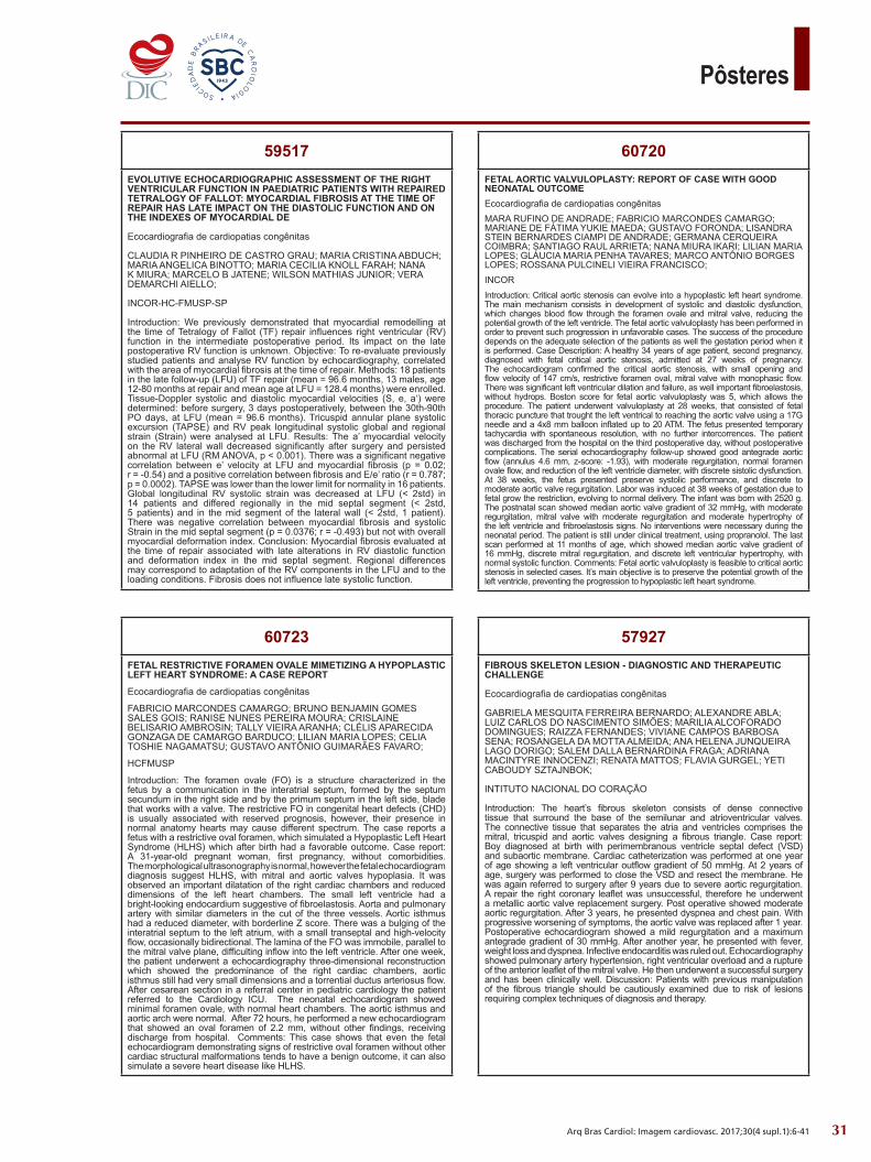
31
Pôsteres
Arq Bras Cardiol: Imagem cardiovasc. 2017;30(4 supl.1):6-41
57927FIBROUS SKELETON LESION - DIAGNOSTIC AND THERAPEUTIC CHALLENGE
Ecocardiografia de cardiopatias congênitas
GABRIELA MESQUITA FERREIRA BERNARDO; ALEXANDRE ABLA; LUIZ CARLOS DO NASCIMENTO SIMÕES; MARILIA ALCOFORADO DOMINGUES; RAIZZA FERNANDES; VIVIANE CAMPOS BARBOSA SENA; ROSANGELA DA MOTTA ALMEIDA; ANA HELENA JUNQUEIRA LAGO DORIGO; SALEM DALLA BERNARDINA FRAGA; ADRIANA MACINTYRE INNOCENZI; RENATA MATTOS; FLAVIA GURGEL; YETI CABOUDY SZTAJNBOK;
INTITUTO NACIONAL DO CORAÇÃO
Introduction: The heart’s fibrous skeleton consists of dense connective tissue that surround the base of the semilunar and atrioventricular valves. The connective tissue that separates the atria and ventricles comprises the mitral, tricuspid and aortic valves designing a fibrous triangle. Case report: Boy diagnosed at birth with perimembranous ventricle septal defect (VSD) and subaortic membrane. Cardiac catheterization was performed at one year of age showing a left ventricular outflow gradient of 50 mmHg. At 2 years of age, surgery was performed to close the VSD and resect the membrane. He was again referred to surgery after 9 years due to severe aortic regurgitation. A repair the right coronary leaflet was unsuccessful, therefore he underwent a metallic aortic valve replacement surgery. Post operative showed moderate aortic regurgitation. After 3 years, he presented dyspnea and chest pain. With progressive worsening of symptoms, the aortic valve was replaced after 1 year. Postoperative echocardiogram showed a mild regurgitation and a maximum antegrade gradient of 30 mmHg. After another year, he presented with fever, weight loss and dyspnea. Infective endocarditis was ruled out. Echocardiography showed pulmonary artery hypertension, right ventricular overload and a rupture of the anterior leaflet of the mitral valve. He then underwent a successful surgery and has been clinically well. Discussion: Patients with previous manipulation of the fibrous triangle should be cautiously examined due to risk of lesions requiring complex techniques of diagnosis and therapy.
60720FETAL AORTIC VALVULOPLASTY: REPORT OF CASE WITH GOOD NEONATAL OUTCOMEEcocardiografia de cardiopatias congênitasMARA RUFINO DE ANDRADE; FABRICIO MARCONDES CAMARGO; MARIANE DE FÁTIMA YUKIE MAEDA; GUSTAVO FORONDA; LISANDRA STEIN BERNARDES CIAMPI DE ANDRADE; GERMANA CERQUEIRA COIMBRA; SANTIAGO RAUL ARRIETA; NANA MIURA IKARI; LILIAN MARIA LOPES; GLÁUCIA MARIA PENHA TAVARES; MARCO ANTÔNIO BORGES LOPES; ROSSANA PULCINELI VIEIRA FRANCISCO;INCORIntroduction: Critical aortic stenosis can evolve into a hypoplastic left heart syndrome. The main mechanism consists in development of systolic and diastolic dysfunction, which changes blood flow through the foramen ovale and mitral valve, reducing the potential growth of the left ventricle. The fetal aortic valvuloplasty has been performed in order to prevent such progression in unfavorable cases. The success of the procedure depends on the adequate selection of the patients as well the gestation period when it is performed. Case Description: A healthy 34 years of age patient, second pregnancy, diagnosed with fetal critical aortic stenosis, admitted at 27 weeks of pregnancy. The echocardiogram confirmed the critical aortic stenosis, with small opening and flow velocity of 147 cm/s, restrictive foramen oval, mitral valve with monophasic flow. There was significant left ventricular dilation and failure, as well important fibroelastosis, without hydrops. Boston score for fetal aortic valvuloplasty was 5, which allows the procedure. The patient underwent valvuloplasty at 28 weeks, that consisted of fetal thoracic puncture that trought the left ventrical to reaching the aortic valve using a 17G needle and a 4x8 mm balloon inflated up to 20 ATM. The fetus presented temporary tachycardia with spontaneous resolution, with no further intercorrences. The patient was discharged from the hospital on the third postoperative day, without postoperative complications. The serial echocardiography follow-up showed good antegrade aortic flow (annulus 4.6 mm, z-score: -1.93), with moderate regurgitation, normal foramen ovale flow, and reduction of the left ventricle diameter, with discrete sistolic dysfunction. At 38 weeks, the fetus presented preserve systolic performance, and discrete to moderate aortic valve regurgitation. Labor was induced at 38 weeks of gestation due to fetal grow the restriction, evolving to normal delivery. The infant was born with 2520 g. The postnatal scan showed median aortic valve gradient of 32 mmHg, with moderate regurgitation, mitral valve with moderate regurgitation and moderate hypertrophy of the left ventricle and fribroelastosis signs. No interventions were necessary during the neonatal period. The patient is still under clinical treatment, using propranolol. The last scan performed at 11 months of age, which showed median aortic valve gradient of 16 mmHg, discrete mitral regurgitation, and discrete left ventricular hypertrophy, with normal systolic function. Comments: Fetal aortic valvuloplasty is feasible to critical aortic stenosis in selected cases. It’s main objective is to preserve the potential growth of the left ventricle, preventing the progression to hypoplastic left heart syndrome.
60723FETAL RESTRICTIVE FORAMEN OVALE MIMETIZING A HYPOPLASTIC LEFT HEART SYNDROME: A CASE REPORT
Ecocardiografia de cardiopatias congênitas
FABRICIO MARCONDES CAMARGO; BRUNO BENJAMIN GOMES SALES GOIS; RANISE NUNES PEREIRA MOURA; CRISLAINE BELISARIO AMBROSIN; TALLY VIEIRA ARANHA; CLÉLIS APARECIDA GONZAGA DE CAMARGO BARDUCO; LILIAN MARIA LOPES; CELIA TOSHIE NAGAMATSU; GUSTAVO ANTÔNIO GUIMARÃES FAVARO;
HCFMUSP
Introduction: The foramen ovale (FO) is a structure characterized in the fetus by a communication in the interatrial septum, formed by the septum secundum in the right side and by the primum septum in the left side, blade that works with a valve. The restrictive FO in congenital heart defects (CHD) is usually associated with reserved prognosis, however, their presence in normal anatomy hearts may cause different spectrum. The case reports a fetus with a restrictive oval foramen, which simulated a Hypoplastic Left Heart Syndrome (HLHS) which after birth had a favorable outcome. Case report: A 31-year-old pregnant woman, first pregnancy, without comorbidities. The morphological ultrasonography is normal, however the fetal echocardiogram diagnosis suggest HLHS, with mitral and aortic valves hypoplasia. It was observed an important dilatation of the right cardiac chambers and reduced dimensions of the left heart chambers. The small left ventricle had a bright-looking endocardium suggestive of fibroelastosis. Aorta and pulmonary artery with similar diameters in the cut of the three vessels. Aortic isthmus had a reduced diameter, with borderline Z score. There was a bulging of the interatrial septum to the left atrium, with a small transeptal and high-velocity flow, occasionally bidirectional. The lamina of the FO was immobile, parallel to the mitral valve plane, difficulting inflow into the left ventricle. After one week, the patient underwent a echocardiography three-dimensional reconstruction which showed the predominance of the right cardiac chambers, aortic isthmus still had very small dimensions and a torrential ductus arteriosus flow. After cesarean section in a referral center in pediatric cardiology the patient referred to the Cardiology ICU. The neonatal echocardiogram showed minimal foramen ovale, with normal heart chambers. The aortic isthmus and aortic arch were normal.After 72 hours, he performed a new echocardiogram that showed an oval foramen of 2.2 mm, without other findings, receiving discharge from hospital. Comments: This case shows that even the fetal echocardiogram demonstrating signs of restrictive oval foramen without other cardiac structural malformations tends to have a benign outcome, it can also simulate a severe heart disease like HLHS.
59517EVOLUTIVE ECHOCARDIOGRAPHIC ASSESSMENT OF THE RIGHT VENTRICULAR FUNCTION IN PAEDIATRIC PATIENTS WITH REPAIRED TETRALOGY OF FALLOT: MYOCARDIAL FIBROSIS AT THE TIME OF REPAIR HAS LATE IMPACT ON THE DIASTOLIC FUNCTION AND ON THE INDEXES OF MYOCARDIAL DE
Ecocardiografia de cardiopatias congênitas
CLAUDIA R PINHEIRO DE CASTRO GRAU; MARIA CRISTINA ABDUCH; MARIA ANGELICA BINOTTO; MARIA CECILIA KNOLL FARAH; NANA K MIURA; MARCELO B JATENE; WILSON MATHIAS JUNIOR; VERA DEMARCHI AIELLO;
INCOR-HC-FMUSP-SP
Introduction: We previously demonstrated that myocardial remodelling at the time of Tetralogy of Fallot (TF) repair influences right ventricular (RV) function in the intermediate postoperative period. Its impact on the late postoperative RV function is unknown. Objective: To re-evaluate previously studied patients and analyse RV function by echocardiography, correlated with the area of myocardial fibrosis at the time of repair. Methods: 18 patients in the late follow-up (LFU) of TF repair (mean = 96.6 months, 13 males, age 12-80 months at repair and mean age at LFU = 128.4 months) were enrolled. Tissue-Doppler systolic and diastolic myocardial velocities (S, e, a‘) were determined: before surgery, 3 days postoperatively, between the 30th-90th PO days, at LFU (mean = 96.6 months). Tricuspid annular plane systolic excursion (TAPSE) and RV peak longitudinal systolic global and regional strain (Strain) were analysed at LFU. Results: The a’ myocardial velocity on the RV lateral wall decreased significantly after surgery and persisted abnormal at LFU (RM ANOVA, p < 0.001). There was a significant negative correlation between e’ velocity at LFU and myocardial fibrosis (p = 0.02; r = -0.54) and a positive correlation between fibrosis and E/e’ ratio (r = 0.787; p = 0.0002). TAPSE was lower than the lower limit for normality in 16 patients. Global longitudinal RV systolic strain was decreased at LFU (< 2std) in 14 patients and differed regionally in the mid septal segment (< 2std, 5 patients) and in the mid segment of the lateral wall (< 2std, 1 patient). There was negative correlation between myocardial fibrosis and systolic Strain in the mid septal segment (p = 0.0376; r = -0.493) but not with overall myocardial deformation index. Conclusion: Myocardial fibrosis evaluated at the time of repair associated with late alterations in RV diastolic function and deformation index in the mid septal segment. Regional differences may correspond to adaptation of the RV components in the LFU and to the loading conditions. Fibrosis does not influence late systolic function.
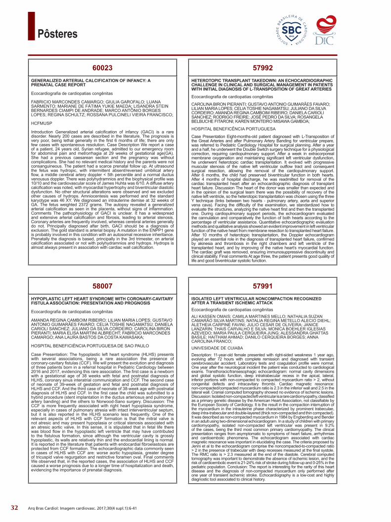
32
Pôsteres
Arq Bras Cardiol: Imagem cardiovasc. 2017;30(4 supl.1):6-41
57991ISOLATED LEFT VENTRICULAR NONCOMPACTION RECOGNIZED AFTER A TRANSIENT ISCHEMIC ATTACKEcocardiografia de cardiopatias congênitasALI KASSEN OMAIS; CAMILA MARTINES MELLO; NATHALIA SUZAN CAMARÃO SILVA MARTINS; NATALIA REGINA METELLO ALECIO DIEHL; ALETHEIA CARPINÉ FAVINI; JULIO CESAR DE OLIVEIRA; JANICE LANZARIN; THAIS CARVALHO E SILVA; MONICA BOEHLER IGLESIAS AZEVEDO; MARIA PAULA CERQUEIRA JUNG; ALESSANDRA GHATTAS BASILE; HAITHAM AHMAD; DANILO CERQUEIRA BORGES; ANNA CAROLINA FRANCO;UNIVESIDADE DE CUIABADescription: 11-year-old female presented with right-sided weakness 1 year ago, evolving after 72 hours with complete remission and diagnosed with transient cerebrovascular accident. Laboratory tests and coagulation profile were normal. One year after the neurological incident the patient was conducted to cardiological exams. Transthoracic/transesophagic echocardiogram: normal cavity dimensions and global systolic function, deep intratrabecular recess in the apical, lateral, inferior portions with non-compacted to compacted myocardium ratio > 2, without congenital defects and intracavitary thrombi. Cardiac magnetic resonance: non-compacted/compacted myocardium ratio is 2.3 in the inferior wall and 2.5 in the lateral wall. Cranial computed tomography showed no evidence of ischemic lesions. Discussion: Isolated non-compacted left ventricular is a rare cardiomyopathy, classified as a primary genetic disease by the American Heart Association, not classifiable by the European Society of Cardiology. It is the result in the compaction interruption of the myocardium in the intrauterine phase characterized by prominent trabeculae, deep intra-trabecular and double-layered (thick non-compacted and thin compacted). It was described as non-compacted myocardium in 1984 by Engberding and Bender in an adult using two-dimensional echocardiogram. In a study of children with primary cardiomyopathy, isolated non-compacted left ventricular was present in 9.2% of the cases, being the third most common primary cardiomyopathy. The clinical presentation ranges from asymptomatic to symptoms of heart failure, arrhythmias and cardioembolic phenomena. The echocardiogram associated with cardiac magnetic resonance was important in elucidating the case. The criteria proposed by Jenni et al to the echocardiogram comprise the noncompacted-to-compacted ratio > 2 in the presence of trabecular with deep recesses measured at the final systole. The RMC ratio is > 2.3 measured at the end of the diastole. Cerebral computed tomography was important to demonstrate the absence of ischemic lesion, and the risk of cardioembolic event is 21-24% risk of stroke during follow-up and 0-25% in the pediatric population. Conclusion: The report is interesting for the rarity of this heart disease and the diagnosis of non-compacted myocardium only performed after one year of transient ischemic stroke. Echocardiography is a low-cost and highly diagnostic tool associated to clinical history.
57992HETEROTOPIC TRANSPLANT TAKEDOWN: AN ECHOCARDIOGRAPHIC CHALLENGE IN CLINICAL AND SURGICAL MANAGEMENT IN PATIENTS WITH INITIAL DIAGNOSIS OF L-TRANSPOSITION OF GREAT ARTERIES
Ecocardiografia de cardiopatias congênitas
CAROLINA BIRON PIERANTI; GUSTAVO ANTONIO GUIMARÃES FAVARO; LILIAN MARIA LOPES; CELIA TOSHIE NAGAMATSU; JULIANO DA SILVA CORDEIRO; AMANDA REGINA CAMBOIM RIBEIRO; DANIELA CARIOLI SANCHEZ; RODRIGO FREIRE; JOSÉ PEDRO DA SILVA; ROSANGELA BELBUCHE FITARONI; KAREN MONTEIRO MISAWA GAMBOA;
HOSPITAL BENEFICÊNCIA PORTUGUESA
Case Presentation Eight-months-old patient diagnosed with L-Transposition of the Great Arteries and after Pulmonary Artery Banding for ventricular prepare, was referred to Pediatric Cardiology Hospital for surgical planning. After a year and a half, he underwent the Double Switch surgery technique for a physiological correction, requiring cardiopulmonary support. After a week in extracorporeal membrane oxygenation and maintaining significant left ventricular dysfunction, he underwent heterotopic cardiac transplantation. It evolved with progressive muscular stenosis of the native left ventricular outflow tract and consequent surgical resection, allowing the removal of the cardiopulmonary support. After 6 months, the child had preserved biventricular function in both hearts. After 4 months of hospital discharge, he was readmitted for removal of the cardiac transplanted heart after an echocardiographic diagnosis of transplant heart failure. Discussion The heart of the donor was smaller than expected and in the opinion of the surgical team there was the possibility of recovery of the native left ventricle, so the heterotopic transplantation was chosen using the three Y technique (links between two hearts - pulmonary artery, aorta and superior vena cava). Facing the difficulty of the examination, we standardized how to evaluate the structures, analyzing the native heart first and then the transplanted one. During cardiopulmonary support periods, the echocardiogram evaluated the cannulation and comparatively the function of both hearts according to the percentage of ventricular assistance. Quantitative echocardiographic evaluation methods and qualitative analysis showed an evident improvement in left ventricular function of the native heart from membrane resection to transplanted heart failure. After 10 months of heterotopic transplantation, the Doppler echocardiogram played an essential role in the diagnosis of transplanted heart failure, confirmed by akinesia and thrombosis in the right chambers and left ventricle of the transplanted heart, and by improving of the native heart’s myocardial function. The cardiac graft was removed, ensuring immunosuppressive discontinuity and clinical stability. Final comments At age three, the patient presents good quality of life and good biventricular systolic function.
58007HYPOPLASTIC LEFT HEART SYNDROME WITH CORONARY-CAVITARY FISTULA ASSOCIATION: PRESENTATION AND PROGNOSIS
Ecocardiografia de cardiopatias congênitas
AMANDA REGINA CAMBOIM RIBEIRO; LILIAN MARIA LOPES; GUSTAVO ANTONIO GUIMARÃES FAVARO; CELIA TOSHIE NAGAMATSU; DANIELA CARIOLI SANCHEZ; JULIANO DA SILVA CORDEIRO; CAROLINA BIRON PIERANTI; MARIA ELISA MARTINI ALBRECHT; FABRICIO MARCONDES CAMARGO; ANA LAURA BASTOS DA COSTA KAWASAKA;
HOSPITAL BENEFICIËNCIA PORTUGUESA DE SAO PAULO
Case Presentation: The hypoplastic left heart syndrome (HLHS) presents with several associations, being a rare association the presence of coronary-cavitary fistulas (CCF). We will present the evolution and diagnosis of three patients born in a referral hospital in Pediatric Cardiology between 2016 and 2017, evidencing this rare association. The first case is a newborn with a gestational age of 38 weeks with fetal and postnatal diagnosis of HLHS, coronary sinus interatrial communication and CCF. The second case of neonate of 39-week of gestation and fetal and postnatal diagnosis of HLHS and CCF. And the third case of neonate of 38-week age with postnatal diagnosis of HLHS and CCF. In the first case the child was submitted to a hybrid procedure (stent implantation in the ductus arteriosus and pulmonary artery banding) and the others to Norwood-Sano surgery. Discussion: The CCF is more frequently associated with right heart hypoplasia syndrome, especially in cases of pulmonary atresia with intact interventricular septum, but it is also reported in the HLHS scenario less frequently. One of the relevant aspects of HLHS with CCF formation is that the mitral valve is not atresic and may present hypoplasia or critical stenosis associated with an atresic aortic valve. In this sense, it is stipulated that in fetal life there was blood flow in the hypoplastic left ventricle that may have contributed to the fistulous formation, since although the ventricular cavity is grossly hypoplastic, its walls are relatively thin and the endocardial lining is normal. It is reported in the literature that patients with endocardial fibroelastosis are protected from CCF formation. The echocardiographic data commonly seen in cases of HLHS with CCF are: worse aortic hypoplasia, greater degree of tricuspid valve regurgation and restrictive foramen oval. Final comments We observed that, in the reported cases, the association of HLHS and CCF caused a worse prognosis due to a longer time of hospitalization and death, evidencing the importance of prenatal diagnosis.
60023GENERALIZED ARTERIAL CALCIFICATION OF INFANCY: A PRENATAL CASE REPORT
Ecocardiografia de cardiopatias congênitas
FABRICIO MARCONDES CAMARGO; GIULIA GAROFALO; LUANA SARMENTO; MARIANE DE FÁTIMA YUKIE MAEDA; LISANDRA STEIN BERNARDES CIAMPI DE ANDRADE; MARCO ANTÔNIO BORGES LOPES; REGINA SCHULTZ; ROSSANA PULCINELI VIEIRA FRANCISCO;
HCFMUSP
Introduction Generalized arterial calcification of infancy (GACI) is a rare disorder. Nearly 200 cases are described in the literature. The prognosis is very poor, being lethal generally in the first 6 months of life; there are only few cases with spontaneous resolution. Case Description We report a case of a patient, 24 years old, Syrian refugee, admitted to our emergency room for abdominal pain and metrorrhagia at 29 weeks of gestational age(GA). She had a previous caesarean section and the pregnancy was without complications. She had no relevant medical history and the parents were not consanguineous. The patient had a scarce prenatal follow up. At ultrasound the fetus was hydropic, with intermittent absent/reversed umbilical artery flow, a middle cerebral artery doppler < 5th percentile and a normal ductus venousus doppler. There was polyhydramnios and the biophysical profile was 10/10 and the cardiovascular score of James Huhta was 6/10. A diffuse artery calcification was noted, with myocardial hypertrophy and biventricular diastolic dysfunction. No other structural alterations were observed and we excluded other causes of hydrops. Amniocentesis was performed and the standard karyotype was 46 XY. We diagnosed an intrauterine demise at 32 weeks of GA. The fetus weighted 2372 grams. The autopsy revealed a generalized arterial calcification as seen in the placenta, without signs of inflammation. Comments The pathophysiology of GACI is unclear. It has a widespread and extensive arterial calcification and fibrosis, leading to arterial stenosis. Coronary arteries are frequently involved, whereas cerebral arteries generally do not. Principally diagnosed after birth, GACI should be a diagnosis of exclusion. The gold standard is arterial biopsy. A mutation in the ENPP1 gene is probably involved in this condition with an autosomal recessive inheritance. Prenatally the diagnosis is based, principally in the 3rd trimester, on arterial calcification associated or not with polyhydramnios and hydrops. Hydrops is almost always present in association with cardiac wall calcification.

33
Pôsteres
Arq Bras Cardiol: Imagem cardiovasc. 2017;30(4 supl.1):6-41
58048SERIES OF RARE CASES OF QUADRICUSPID AORTIC VALVESEcocardiografia de cardiopatias congênitas
SANDRA MARY FEITOSA FONTENELE; ALINE AKIKO KOMATSU RABELO; PEDRO ANTÔNIO GALDEANO; WANESSA CARTANO CUNHA; JULIANA RODRIGUES BEAL; GABRIEL ANTONIO STANISCI MIGUEL; ALINE AKIKO KOMATSU RABELO; PEDRO ANTONIO GALDEANO; PATRICIA REGINA ALVES GALDEANO;
SECRETARIA DE SAÚDE DO DFIntroduction: Diagnosis of quadricuspid aortic valve (QAV) is a rare finding, especially in pediatric age, when most patients are asymptomatic and have valves without any functional changes. Most of the time, QAV occurs without clinical manifestations, its incidence varying between 0.008 and 0.043%. There’s a slight predominance in males, estimated in 1.6 : 1, when compared to the incidence in females. Such diagnosis is more commonly made after the fifth decade of life, when some individuals may manifest symptoms, predominantly related to aortic insufficiency. Clinical cases: Case 1: A 77-year-old male, hypertensive and smoker. Admitted to the Coronary ICU of the Hospital de Base de Brasília, with ST-segment elevation acute coronary syndrome. The echocardiographic evaluation showed presence of a quadricuspid aortic valve with mild sclerosis and insufficiency, besides segmental alteration. It was possible in this case the visualization of the coronary outflow tract. (Figure 1) Case 2: A 40-year-old female, with complaints of paroxysmal palpitations. No history of any cardiovascular history or symptoms was informed. On routine ambulatory echocardiography, the presence of a quadricuspid aortic valve with discrete insufficiency without other alterations was observed. (Figure 2) Case 3: A 44-year-old female patient, investigating a heart a murmur during a cholecystectomy preoperative consultation. She denied comorbidities. During routine ambulatory echocardiographic examination, the presence of a quadricuspid aortic valve with a turbulent flow with a peak gradient of 20 mmHg and a discrete aortic insufficiency without other changes was evidenced. (Figure 3) Discussion: The cases presented were echocardiographic findings of asymptomatic patients from the valve standpoint. Except for Case 1 (one) that had a poor final outcome (death), secondary to a coronary disease, the other patients evolved in asatisfactory manner. Conclusion: QAV is, for the most part, a rare and isolated anomaly, diagnosed as an incidental finding or as a result of symptoms secondary to aortic insufficiency in adulthood. However, it can sometimes be associated with other cardiac malformations. There are reports of cases of QAF in association with hypertrophic cardiomyopathy, interatrial and interventricular communication, pulmonary valve stenosis, hypoplasia of the aortic ring, mitral valve disease, transposition of the great arteries, and tetralogy of Fallot. Among them, the most common association is with the anomalous origin of coronary artery. This association is found in 10% of the cases of QAV described in the medical literature. The main anomalies of the described coronaries were single coronary ostium and anomalous location of the coronary ostia
57904REMARKABLE DOUBLE CLEFT MITRAL VALVE SIMULATING A RUPTURED CHORDAE: IMPACT OF THREE-DIMENSIONAL TRANSESOPHAGEAL ECHOCARDIOGRAPHY
Ecocardiografia de cardiopatias congênitas
ANGELE AZEVEDO ALVES MATTOSO; MARTA NERI LACERDA; MAIRA ROCHA ALVES; MARIA LUCIA DUARTE; EDUARDO TADEU FERREIRA;
HOSPITAL SANTA IZABEL / CLINICA PROALIVIO
Presentation: A 64-year-old woman complaining of progressive dyspnea 1 year ago. Transthoracic echocardiogram showed eccentric and severe mitral regurgitation consistent with posterior leaflet disease and severe dilatation of the left atrium (LA). The patient underwent a transesophageal echocardiography to further evaluation of the mitral valve. Two-dimensional transesophageal echocardiogram showed diffuse thickening of the mitral leaflets (myxomatous) as well flail due to ruptured chordae of the scallop P3 of the posterior leaflet (Figure 1). Real-time “zoom” three-dimensional transesophageal echocardiography did not reveal ruptured chordae, instead, a remarkable, isolated and double cleft mitral valve of the posterior leaflet (Figure 2), simulating a ruptured chordae. There were no other congenital heart lesions. Discussion: Isolated cleft mitral valve may occur alone or in association with other congenital heart lesions. Following the advent of three-dimensional transesophageal echocardiography, it appears to be more common than previously thought. This is the case of an unusual and remarkable form of presentation of the isolated cleft mitral valve that was demonstrated only by enhance diagnostic of three-dimensional transesophageal echocardiography that offers singular advantages, such as correct vision (en face) of the mitral valve. Final comments: Three-dimensional transesophageal echocardiography is critically important to accurate characterization of mitral valve lesion’s complexity.
Figura 1
Figura 2
59856RIGHT ATRIAL MYXOMA IN EXTREME PREMATURE - DIAGNOSTIC AND THERAPEUTIC APPROACHEcocardiografia de cardiopatias congênitas
ANDRESSA MUSSI SOARES; PAULO JOSÉ FERREIRA SOARES; BERNARDO MUSSI SOARES; FATIMA FERNANDA LESSA; MARCELO RODRIGUES CRESPO; JOSÉ COIMBRA DE REZENDE NETO; RESI APOLINÁRIO; PATRÍCIA LOPES MOTA; RENATA DE BACKER PACÍFICO; LUIZ FERNANDO LUNZ; LIVIA ALBINO DA SILVA; GILBANIA RAFAEL LANDI; ODILON SILVA HENRIQUE JUNIOR; LUIZ DANIEL DA FRAGA TORRES;
HOSPITAL EVANGELICO DE CACHOEIRO DE ITAPEMIRIMCase Report: Extreme premature (PMT), 28 weeks gestational age, birth weight: 1135 g, male. He evolved with early respiratory distress syndrome requiring prolonged mechanical ventilation and several antibiotics due to septicemia. The PMT had normal neurological examination despite dilation of lateral ventricles to the transcranial ultrasound, as well as renal left hydronephrosis. He presented patent ductus arteriosus and occlusion of ductus arteriosus with ibuprofen. After about 60 days of life there was clinical worsening with bacteremia, positive blood culture for Klebsiella and enterococcus being made a new antibiotic. When echocardiogram was repeated, a large intracardiac mass was detected in right atrium (RA) measuring about 17x9 mm in diameter. The mass had a globular apperarance, heterogeneous, contained echolucent and echo-bright areas and prolapsed into the tricuspide valve during ventricular diastole. Full heparinization was instituted with low molecular weight heparin. There was clinical improvement with negative blood cultures, but patiently maintained with tachypnea pattern and low weight. As the intracardiac mass maintained with unchanged characteristics with risk of embolization and reinfection, indicated surgical removal of the mass being confirmed the origin in the IVC. The anatomopathological examination was suggestive of myxoma confirmed by the immunohistochemical study. The child was discharged after 20 days of cardiac surgery and was clinically well in outpatient follow-up. Discussion: Intracardiac masses generally represent diagnostic challenges that need to be based on a clinical context associated with imaging findings. The diagnosis of these intracardiac masses in premature infants is even more complex due to its presentation and rareness. Final comments: Myxoma is a tumor of rare incidence in children. Right atrial myxoma originating from IVC is even rarer. Its location implies greater technical-surgical care, especially in underweight premature. Despite the rareness, benign characteristics and low chance of return, the surgical treatment of the myxoma should be as aggressive as possible and therefore, his diagnosis must be remembered in the intracardiac masses. immunohistochemical study. The child was discharged after 20 days of cardiac surgery and was clinically well in outpatient follow-up. Discussion: Intracardiac masses generally represent diagnostic challenges that need to be based on a clinical context associated with imaging findings. The diagnosis of these intracardiac masses in premature infants is even more complex due to its presentation and rareness. Final comments: Myxoma is a tumor of rare incidence in children. Right atrial myxoma originating from IVC is even rarer. Its location implies greater technical-surgical care, especially in underweight premature. Despite the rareness, benign characteristics and low chance of return, the surgical treatment of the myxoma should be as aggressive as possible and therefore, his diagnosis must be remembered in the intracardiac masses.
60711PRENATAL DIAGNOSIS OF DOUBLE OUTLET OF RIGHT VENTRICLE WITH AGENESIS OF PULMONARY VALVE: CASE REPORT
Ecocardiografia de cardiopatias congênitas
MARA RUFINO DE ANDRADE; FABRICIO MARCONDES CAMARGO; MARIANE DE FÁTIMA YUKIE MAEDA; VALÉRIA DE MELO MOREIRA; GUSTAVO FORONDA; NANA MIURA IKARI; FILOMENA REGINA BARBOSA GOMES GALAS; GLÁUCIA MARIA PENHA TAVARES; MARCO ANTÔNIO BORGES LOPES; ROSSANA PULCINELI VIEIRA FRANCISCO;
INCOR
Introduction: Double outlet of the right ventricle (DORV) is a complex congenital malformation that occurs with a wide spectrum of presentation, depending of the relation and size of the arteries, and their relation with and size of ventricular septal defect. Whereas cases of pulmonary valve agenesis are exceedingly rare, Tetralogy of Fallot is one of the most common congenital heart disorders where it occurs. Severe fetal pulmonary regurgitation can lead to the dilatation of the right ventricle as well as the aneurysmal dilatation of the pulmonary trunk and its branches. Such alterations may interfere negatively in lung development causing an extrinsic compression of the airway, and consequently postnatal breathing problems. Case Description: A 27 years of age patient, third pregnancy, with suspicion of fetal left atrial aneurysm. A fetal echocardiogram identified a double outlet of the right ventricle with subpulmonary ventricular septal defect; pulmonary valve agenesis; persistent left superior vena cava; aneurysmal dilatation of the pulmonary trunk and its branches. During the pregnancy, serial fetal echocardiogram was performed, monitoring the signals of hemodynamic decompensation, but no hydrops was noted. The delivery happened by a Cesarean section at 39 weeks and six days, due to antepartum meconium, with no complications. The baby’s birth weight was 2880 grams. An echocardiogram showed DORV with anterior aorta and posterior pulmonary trunk; large in ventricular septal defect (8.7 mm); pulmonary valve agenesis; dilatation of the trunk and pulmonary arteries; hypoplasia of the mitral valve and left ventricle; hypoplastic aortic isthmus; without coarctation. Computed tomography angiogram confirmed the initial findings and revealed coronary anomaly; aberrant right retro-esophageal subclavian artery; more extrinsic airway compression on the left side. Due to the complexity of the case, the patient underwent a central shunt, and had the pulmonary trunk removed. Whenever discharged of the intensive care unit, and evolved into multiple respiratory infections and extubation failure, which required a tracheostomy, but died at 9 months of age. Discussion: DORV with pulmonary valve agenesis and hypoplastic left ventricle is very uncommon. It can be diagnosed accurately during fetal life, which is important to planning the treatment and follow-up. It is known that such patients have worse prognosis, and breathing difficulties tend to be the cause of death.

34
Pôsteres
Arq Bras Cardiol: Imagem cardiovasc. 2017;30(4 supl.1):6-41
XXXTHE USE OF CARDIAC IMAGING IN THERAPEUTIC DECISION MAKING - A REPORT OF TWO CASES AT NATIONAL CARDIOLOGY INSTITUTE
Ecocardiografia de cardiopatias congênitas
RAIZZA FERNANDES DA COSTA; MARITZA XAVIER ANZANELLO; GABRIEL CAMARGO; RENATA MATTOS; LUIZ CARLOS DO NASCIMENTO SIMÕES; LINA MIURA; ADRIANA INNOCENZI; MARIA DO SOCORRO PEDROSO TAGHARI; VICTOR HUGO DE OLIVEIRA; GABRIELA DE MESQUITA FERREIRA BERNARDO; MONICA CELENTE; SALEM BERNARDINO DALLAS; MARILIA ALCOFORADO; ALEXANDRE ABLA; ALINE BERETA FARIA;
INSTITUTO NACIONAL DE CARDIOLOGIA
Multimodality and advanced imaging methods are frequently needed for diagnosis and adequate choice between surgical or percutaneous treatment of ventricular septal defects (VSD). Case 1 – 19-year-old female with a history of surgical VSD closure presented with dyspnea on moderate efforts and was diagnosed with an aparently small residual VSD on a transthoracic echocardiogram, associated with mild aortic insufficiency, left ventricle enlargement and elevation of systolic pulmonary pressure. Since case presentation suggested a more significant residual shunt a transesophageal echo was perfomed to better assess the defect size, however patch calcifications prevented its full assement. With high suspition of a larger VSD cardiac catheterization was performed for a better evaluation, followed by percutaneous closure if needed. After ventriculography, a large residual VSD was identified. Percutaneous occlusion was successfully acomplished with a symmetric occluder size 16mm. Case report 2 – One year-old-child, diagnosed by transthoracic echocardiogram with a large VSD, left chambers enlargement and shortness of breath at great efforts was referred to surgical treatment. Pre-operative 3D transesophageal echocardiography identified a large saccular multifenestrated VSD with several outlet jets. With the use of multiplanar reconstrutions a suitable landing site for a percutaneous occluder was identified and surgery was deferred. The percutaneous procedure was successfull and the defect was occluded with a symmetric 16 mm device. These cases illustrate that the use of advanced imaging techniques and a combination of imaging modalities may better depict disease morphology, allowing for less invasive therapies.
XXXSIMPLE TRANSPOSITION OF THE GREAT ARTERIES WITH INTACT VENTRICULAR SEPTUM: THE IMPORTANCE OF PRE-NATAL DIAGNOSIS
Ecocardiografia de cardiopatias congênitas
DANIELA VASCONCELOS; GABRIELA FUENMAYOR; TAMARA CORTEZ MARTINS; ANA ZLOCHEVSKY; CARLOS R FERREIRO; MARCELO JATENE; CARLOS A C PEDRA; SIMONE R F FONTES PEDRA;
INSTITUTO DANTE PAZZANESE DE CARDIOLOGIA
Simple transposition of the great arteries (S-TGA) can present with severe hypoxemia immediately after birth and need urgent balloon atrial septostomy (BAS) to improve oxygenation. For this reason, it is of critical importance to deliver babies with this disease in a hospital that can provide specialized neonatal cardiac care. In this scenario, fetal diagnosis of TGA with potentially restrictive FO is crucial. Objective: review the outcome of babies with prenatal diagnosis of simple TGA delivered in a pediatric cardiology center with specific emphasis to the need of BAS in the first hours of life due to restrictive FO. Patients and methods: retrospective review of records of 38 babies with prenatal diagnosis of S-TGA born at a single pediatric cardiology institution from Jan/2009 to June/2017 results: During this period, 345 babies with prenatal diagnosis of severe congenital heart disease (CHD) were born at our Hospital. Thirty-eight (11%) had S-TGA. Gestational age at birth was 38.3 ± 0.96 weeks, weight 3 ± 0.37 kg, and 1st and 5th minute APGAR score 7.6 ± 1.8 and 8,6 ± 1 respectively. At birth, median FO diameter was 2.5mm (1 to 8.3 mm). Twenty-six babies (68%) needed BAS in the first 10 hours of life. Oxygen saturation was 72±13% and increased to 88 ± 5% after the procedure, all performed by echo guidance in the ICU. There was no mortality related to hypoxemia. Fetal echo was performed by different examiners, including outside referring doctors. Sensibility, specificity, positive and negative predictive values of fetal echo in predicting the need of BAS in the first hours of life were 56, 50, 70 and 36%. Arterial Switch Operation was performed 1 to 22 (m = 5) days after birth, and was not delayed because of the need of BAS: 7.5 ± 5.5 group BAS vs 6 ± 5.5 group without BAS (p < 0,05). Postoperative survival was 82% with no difference between the 2 groups. Conclusion: This study shows that S-TGA represents only 10% of critical CHD referred for delivery in specialized center, possibly due to difficulties related to the obstetric screening. FO is significantly restrictive in close to 70% of the babies leading to urgent BAS in the first hours of life. In our community fetal echo has low Sensibility, Specificity, Positive and Negative Predictive values in anticipating the need of urgent BAS. In order to improve neonatal outcomes, it seems rational to establish a delivery unit attached to the CHD center where urgent BAS is available immediately after birth.
60722SUBMITRAL STENOSIS DUE TO HUGE ANOMALOUS TENDON ORIGINATING FROM THE POSTEROMEDIAL PAPILLARY MUSCLE
Ecocardiografia de cardiopatias congênitas
STERFFESON LAMARE LUCENA DE ABREU; JOANA D‘ ARC MATOS FRANÇA DE ABREU;
HOSPITAL UNIVERSITÁRIO PRESIDENTE DUTRA
A 21-year-old male patient with a history of mitral valve replacement by bioprosthesis three years ago. A routine transthoracic echocardiogram showed an anomalous tendon originating from a hypertrophied posteromedial papillary muscle and causing division of the mitral flow bypassing the anomalous tendon and generating an estimated mitral valve mean gradient of 12.58 mmHg. The opening of the bioprosthesis was preserved and the mitral subvalvar obstruction was clear. The incidence of anomalous tendons in the left ventricle is controversial in the literature. Autopsy studies reveal some type of anomalous tendon in the left ventricle in up to 45% of the autopsies. Studies using M and 2D mode echocardiography find much more modest numbers, with incidence between 0.5 and 3%. Anomalous Left Ventricle tendons usually do not cause hemodynamic repercussion. The most frequently reported relationship is with arrhythmias. They rarely cause obstruction in the left ventricle, and the few cases reported are about obstruction of the left ventricular outflow tract. We did not find any case reports associating anomalous tendons and submitral obstruction.
57932SEVERE COMPLICATIONS OF INFECTIVE ENDOCARDITIS (IE): REPORT OF TWO CASES
Ecocardiografia de cardiopatias congênitas
SALEM DALLA BERNARDINA FRAGA; ALEXANDRE MARQUES ABLA; MARILIA ALCOFORADO DOMINGUES; RAIZZA FERNANDES; GABRIELA DE MESQUITA FERREIRA BERNARDO; VIVIANE CAMPOS BERBOSA SENA; ADRIANA MACINTYRE INNOCENZI;
INTITUTO NACIONAL DO CORAÇÃO
Background: IE has nonspecific symptoms, so early diagnosis is often difficult. It presents a high morbidity and mortality. Complications such as abscesses and aneurysms are rare but potentially lethal. Both had poor dental hygiene. Case 1: Nine-year-old boy with bicuspid aortic valve previously submitted to valve replacement. He was admitted with a history of fever for two weeks, weakness and conjunctival petechiae. Echocardiogram showed vegetation on the atrial side of the anterior leaflet of the mitral leaflet, which was also perforated and supravalvar aortic vegetation. Blood culture was positive for S. viridans and he was started on antibiotics. Computed tomography (CT) showed sings of hepatic, splenic and cerebral embolism. He was submitted to an emergency surgery, when an abscess was found in the fibrous skeleton of the heart and ascending aorta, as well as vegetations in the mitral and aortic valves. Successful Konno-Bentell surgery was performed. The histopathological study demonstrated active IE. He had six weeks of venous antibiotic treatment after surgery, with good clinical outcome. Case 2: Six-year-old boy, with previous diagnosis of ventricular septal defect (VSD), right ventricular muscle band and aortic regurgitation, with fever for four weeks and hyporexia. Echocardiogram showed a peduncular vegetation in the mitroaortic region and an aneurysm that extended from the base of the anterior mitral leaflet to the aortic root, measuring around 12 mm. He was submitted to surgery, with removal of vegetations, closure of the aneurysm ostium, aortic valve repair and ventriculo-septoplasty. Antibiotic therapy was continued for four weeks after surgery, with good clinical outcome. Discussion: IE is a disease that should always be considered as a differential diagnosis of persistent fever, especially in children with structural disease and poor dental hygiene. The two reported cases of complicated IE had a favorable outcome, despite the severity of the lesions.
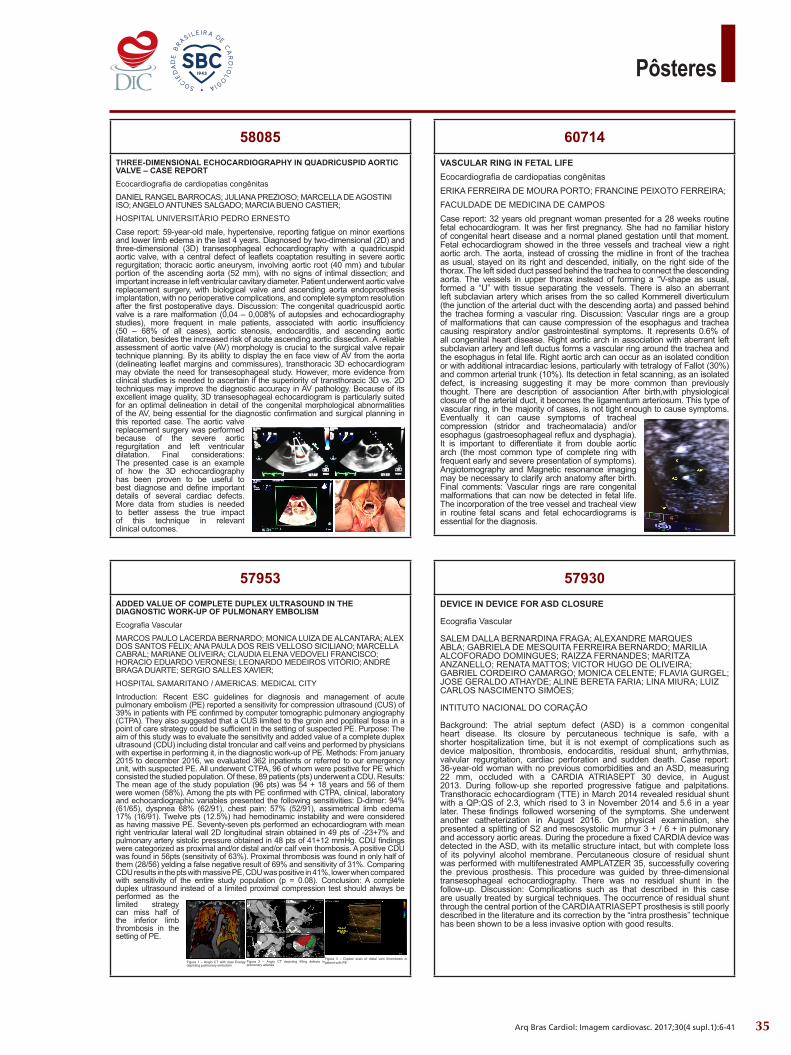
35
Pôsteres
Arq Bras Cardiol: Imagem cardiovasc. 2017;30(4 supl.1):6-41
57930DEVICE IN DEVICE FOR ASD CLOSURE
Ecografia Vascular
SALEM DALLA BERNARDINA FRAGA; ALEXANDRE MARQUES ABLA; GABRIELA DE MESQUITA FERREIRA BERNARDO; MARILIA ALCOFORADO DOMINGUES; RAIZZA FERNANDES; MARITZA ANZANELLO; RENATA MATTOS; VICTOR HUGO DE OLIVEIRA; GABRIEL CORDEIRO CAMARGO; MONICA CELENTE; FLAVIA GURGEL; JOSE GERALDO ATHAYDE; ALINE BERETA FARIA; LINA MIURA; LUIZ CARLOS NASCIMENTO SIMÕES;
INTITUTO NACIONAL DO CORAÇÃO
Background: The atrial septum defect (ASD) is a common congenital heart disease. Its closure by percutaneous technique is safe, with a shorter hospitalization time, but it is not exempt of complications such as device malposition, thrombosis, endocarditis, residual shunt, arrhythmias, valvular regurgitation, cardiac perforation and sudden death. Case report: 36-year-old woman with no previous comorbidities and an ASD, measuring 22 mm, occluded with a CARDIA ATRIASEPT 30 device, in August 2013. During follow-up she reported progressive fatigue and palpitations. Transthoracic echocardiogram (TTE) in March 2014 revealed residual shunt with a QP:QS of 2.3, which rised to 3 in November 2014 and 5.6 in a year later. These findings followed worsening of the symptoms. She underwent another catheterization in August 2016. On physical examination, she presented a splitting of S2 and mesosystolic murmur 3 + / 6 + in pulmonary and accessory aortic areas. During the procedure a fixed CARDIA device was detected in the ASD, with its metallic structure intact, but with complete loss of its polyvinyl alcohol membrane. Percutaneous closure of residual shunt was performed with multifenestrated AMPLATZER 35, successfully covering the previous prosthesis. This procedure was guided by three-dimensional transesophageal echocardiography. There was no residual shunt in the follow-up. Discussion: Complications such as that described in this case are usually treated by surgical techniques. The occurrence of residual shunt through the central portion of the CARDIA ATRIASEPT prosthesis is still poorly described in the literature and its correction by the “intra prosthesis” technique has been shown to be a less invasive option with good results.
60714VASCULAR RING IN FETAL LIFEEcocardiografia de cardiopatias congênitasERIKA FERREIRA DE MOURA PORTO; FRANCINE PEIXOTO FERREIRA;FACULDADE DE MEDICINA DE CAMPOSCase report: 32 years old pregnant woman presented for a 28 weeks routine fetal echocardiogram. It was her first pregnancy. She had no familiar history of congenital heart disease and a normal planed gestation until that moment. Fetal echocardiogram showed in the three vessels and tracheal view a right aortic arch. The aorta, instead of crossing the midline in front of the trachea as usual, stayed on its right and descended, initially, on the right side of the thorax. The left sided duct passed behind the trachea to connect the descending aorta. The vessels in upper thorax instead of forming a “V-shape as usual, formed a “U” with tissue separating the vessels. There is also an aberrant left subclavian artery which arises from the so called Kommerell diverticulum (the junction of the arterial duct with the descending aorta) and passed behind the trachea forming a vascular ring. Discussion: Vascular rings are a group of malformations that can cause compression of the esophagus and trachea causing respiratory and/or gastrointestinal symptoms. It represents 0.6% of all congenital heart disease. Right aortic arch in association with aberrant left subclavian artery and left ductus forms a vascular ring around the trachea and the esophagus in fetal life. Right aortic arch can occur as an isolated condition or with additional intracardiac lesions, particularly with tetralogy of Fallot (30%) and common arterial trunk (10%). Its detection in fetal scanning, as an isolated defect, is increasing suggesting it may be more common than previously thought. There are description of associantion After birth,with physiological closure of the arterial duct, it becomes the ligamentum arteriosum. This type of vascular ring, in the majority of cases, is not tight enough to cause symptoms. Eventually it can cause symptoms of tracheal compression (stridor and tracheomalacia) and/or esophagus (gastroesophageal reflux and dysphagia). It is important to differentiate it from double aortic arch (the most common type of complete ring with frequent early and severe presentation of symptoms). Angiotomography and Magnetic resonance imaging may be necessary to clarify arch anatomy after birth. Final comments: Vascular rings are rare congenital malformations that can now be detected in fetal life. The incorporation of the tree vessel and tracheal view in routine fetal scans and fetal echocardiograms is essential for the diagnosis.
57953ADDED VALUE OF COMPLETE DUPLEX ULTRASOUND IN THE DIAGNOSTIC WORK-UP OF PULMONARY EMBOLISMEcografia Vascular
MARCOS PAULO LACERDA BERNARDO; MONICA LUIZA DE ALCANTARA; ALEX DOS SANTOS FÉLIX; ANA PAULA DOS REIS VELLOSO SICILIANO; MARCELLA CABRAL; MARIANE OLIVEIRA; CLAUDIA ELENA VEDOVELI FRANCISCO; HORACIO EDUARDO VERONESI; LEONARDO MEDEIROS VITÓRIO; ANDRÉ BRAGA DUARTE; SERGIO SALLES XAVIER;
HOSPITAL SAMARITANO / AMERICAS. MEDICAL CITY
Introduction: Recent ESC guidelines for diagnosis and management of acute pulmonary embolism (PE) reported a sensitivity for compression ultrasound (CUS) of 39% in patients with PE confirmed by computer tomographic pulmonary angiography (CTPA). They also suggested that a CUS limited to the groin and popliteal fossa in a point of care strategy could be sufficient in the setting of suspected PE. Purpose: The aim of this study was to evaluate the sensitivity and added value of a complete duplex ultrasound (CDU) including distal troncular and calf veins and performed by physicians with expertise in performing it, in the diagnostic work-up of PE. Methods: From january 2015 to december 2016, we evaluated 362 inpatients or referred to our emergency unit, with suspected PE. All underwent CTPA, 96 of whom were positive for PE which consisted the studied population. Of these, 89 patients (pts) underwent a CDU. Results: The mean age of the study population (96 pts) was 54 + 18 years and 56 of them were women (58%). Among the pts with PE confirmed with CTPA, clinical, laboratory and echocardiographic variables presented the following sensitivities: D-dimer: 94% (61/65), dyspnea 68% (62/91), chest pain: 57% (52/91), assimetrical limb edema 17% (16/91). Twelve pts (12.5%) had hemodinamic instability and were considered as having massive PE. Seventy-seven pts performed an echocardiogram with mean right ventricular lateral wall 2D longitudinal strain obtained in 49 pts of -23+7% and pulmonary artery sistolic pressure obtained in 48 pts of 41+12 mmHg. CDU findings were categorized as proximal and/or distal and/or calf vein thombosis. A positive CDU was found in 56pts (sensitivity of 63%). Proximal thrombosis was found in only half of them (28/56) yelding a false negative result of 69% and sensitivity of 31%. Comparing CDU results in the pts with massive PE, CDU was positive in 41%, lower when compared with sensitivity of the entire study population (p = 0.08). Conclusion: A complete duplex ultrasound instead of a limited proximal compression test should always be performed as the limited strategy can miss half of the inferior limb thrombosis in the setting of PE.
Figura 1 – Angio CT with dual Energy depicting pulmonary embolism
Figura 3 – Duplex scan of distal vein thrombosis in patient with PE
Angio CT with dual Energy depicting pulmonary embolism
Angio CT depicting Þlling defects in pulmonary arteries
Duplex scan of distal vein thrombosis in patient with PE
Figura 2 – Angio CT depicting filling defects in pulmonary arteries
58085THREE-DIMENSIONAL ECHOCARDIOGRAPHY IN QUADRICUSPID AORTIC VALVE – CASE REPORTEcocardiografia de cardiopatias congênitas
DANIEL RANGEL BARROCAS; JULIANA PREZIOSO; MARCELLA DE AGOSTINI ISO; ANGELO ANTUNES SALGADO; MARCIA BUENO CASTIER;
HOSPITAL UNIVERSITÁRIO PEDRO ERNESTO
Case report: 59-year-old male, hypertensive, reporting fatigue on minor exertions and lower limb edema in the last 4 years. Diagnosed by two-dimensional (2D) and three-dimensional (3D) transesophageal echocardiography with a quadricuspid aortic valve, with a central defect of leaflets coaptation resulting in severe aortic regurgitation; thoracic aortic aneurysm, involving aortic root (40 mm) and tubular portion of the ascending aorta (52 mm), with no signs of intimal dissection; and important increase in left ventricular cavitary diameter. Patient underwent aortic valve replacement surgery, with biological valve and ascending aorta endoprosthesis implantation, with no perioperative complications, and complete symptom resolution after the first postoperative days. Discussion: The congenital quadricuspid aortic valve is a rare malformation (0,04 – 0,008% of autopsies and echocardiography studies), more frequent in male patients, associated with aortic insufficiency (50 – 68% of all cases), aortic stenosis, endocarditis, and ascending aortic dilatation, besides the increased risk of acute ascending aortic dissection. A reliable assessment of aortic valve (AV) morphology is crucial to the surgical valve repair technique planning. By its ability to display the en face view of AV from the aorta (delineating leaflet margins and commissures), transthoracic 3D echocardiogram may obviate the need for transesophageal study. However, more evidence from clinical studies is needed to ascertain if the superiority of transthoracic 3D vs. 2D techniques may improve the diagnostic accuracy in AV pathology. Because of its excellent image quality, 3D transesophageal echocardiogram is particularly suited for an optimal delineation in detail of the congenital morphological abnormalities of the AV, being essential for the diagnostic confirmation and surgical planning in this reported case. The aortic valve replacement surgery was performed because of the severe aortic regurgitation and left ventricular dilatation. Final considerations: The presented case is an example of how the 3D echocardiography has been proven to be useful to best diagnose and define important details of several cardiac defects. More data from studies is needed to better assess the true impact of this technique in relevant clinical outcomes.

36
Pôsteres
Arq Bras Cardiol: Imagem cardiovasc. 2017;30(4 supl.1):6-41
60724ATHEROSCLEROTIC PLAQUE TO CORONARY ANGIOTOMOGRAPHY IN PATIENTS WITH A ZERO CALCIUM SCORE
Imagem cardiovascular
FABIOLA SANTOS GABRIEL; CARLOS JOSÉ OLIVEIRA DE MATOS; CAIO JOSÉ COUTINHO LEAL TELINO; MIRELLA SOBRAL SILVEIRA; JÚLIO CÉSAR OLIVEIRA COSTA TELES; PAULO VICTOR DE JESUS SILVA; IGOR LARCHERT MOTA; ISABELLA MARIA DA SILVA CARDOSO; LORENA ALMEIDA SANT`ANA; ANA CAROLINA SOUZA DOS SANTOS; LUIZ FLÁVIO GALVÃO; ENALDO VIEIRA DE MELO; IBRAIM MASCIARELLI PINTO; ANTONIO CARLOS SOBRAL SOUSA; JOSELINA LUZIA MENEZES OLIVEIRA;
UNIVERSIDADE FEDERAL DE SERGIPE
Background: In view of the high mortality due to Cardiovascular Diseases in the world, a stratification of the main risk factors and correct choice of the diagnostic modality is necessary. Several studies have shown that the calcium score (CS) has a low risk of cardiovascular events. However, the frequency of individuals with coronary atherosclerotic plaques and zero CS is conflicting in the specialized medical literature. Objective: To evaluate the frequency of coronary atherosclerotic plaques and associated factors in patients with CS. Methods: This was a prospective, cross-sectional study with 367 volunteers with zero CS through the realization CS test and coronary computed tomography angiography (CCTA), from 2011 to 2016, in four imaging centers. The sample was obtained for convenience and consecutively. Results: The frequency of atherosclerotic plaque in the coronary arteries of the 367 patients with zero CS was 9.3% (34 individuals); in this subgroup the mean age was 52 ± 10 years, 18 (52.9%) were women and 16 (47%) had significant coronary obstructions (> 50%), some in more than one segment. The frequency of non-obese individuals (90.6% vs 9.4%, p = 0.037) and those of alcoholics (55.9% vs 44.1%, p = 0.015) was significantly higher in patients with atherosclerotic plaques, Variable, odds ratio of 3.4 for the development of these plaques. Conclusions: The frequency of atherosclerotic plaque with zero CS was considerable, evidencing that the absence of calcification, through the CCTA, does not exclude plaque and nor obstructive lesion, mainly in the non-obese and the alcoholic.
58013ALCAPA IN ASYMPTOMATIC ADULT WITH PRESERVED VENTRICULAR FUNCTION
Imagem cardiovascular
SERGIO RODRIGO BERALDO; FABRICIO RODRIGUES DOS ANJOS; BENEDICTO JOSE PIERINI; PAULA APARECIDA DELFRARO; LUCIENE ELAINE PEREIRA; MATHEUS HENRIQUE FERNANDES; PEDRO COUTINHO MEYER FERNANDES; FLAVIO SIQUEIRA JUNQUEIRA;
CONSULTÓRIO
The Anomalous origin of the Left Coronary Artery from the Pulmonary Artery (ALCAPA) is a rare congenital coronary anomaly with an incidence of 1 for every 300,000 live births. About 80% to 85% of affected patients die before the first year of life. Patients who reach adulthood, about 15% to 20% of cases, develop a rich network of right-side to left-side collateral, but they may still have acute myocardial infarction, mitral insufficiency, congestive heart failure, Ventricular arrhythmias and sudden death. This report is about the case of a 55-year-old woman who sought medical care for routine asymptomatic follow-up. The patient has systemic arterial hypertension and diabetes mellitus type 2 and makes use of medication to control these diseases; No previous or family history of coronary artery disease. The ECG presented sinus rhythm with alteration of lateral wall ventricular repolarization. An initial echocardiogram (ECO) was within normal limits. An ergometric test (ET) was performed, which evidenced myocardial ischemia with a 2 mm rectified ST depression, with low load, without symptoms of coronary disease, and myocardial scintigraphy (SPECT) with physical stress that did not show myocardial ischemia. Remains in followed up with satisfactory blood pressure and glycemic control. Later, the patient underwent coronary Coronary Computadorized Tomography Angiography (CCTA) for a anatomical study, which revealed Left Coronary Artery originating from the Pulmonary Artery, with ectasic and tortuous coronary circulation, Right Coronary with discrete calcified mural plate without luminal reduction and Calcium Score 46. Current ECO showed a left atrial enlargement of 43 mm, LV diastolic dysfunction with pseudo-normal pattern, preserved ejection fraction, and presence of systolic-diastolic flow, with diastolic predominance, in the right ventricular outflow tract. In asymptomatic adults, recent literature suggests that if only moderate chronic ischemia and small area of necrosis are present, survival without surgical correction is possible. In this context, Magnetic Resonance (MRI) in assessing myocardial function and viability is an excellent tool in the algorithm for therapeutic decision. In the case in question, we opted for clinical treatment and follow-up periodic functional tests
58025AN UNUSUAL FINDING OF INTERVENTRICULAR SEPTAL PERFORATION BY TEMPORARY PACING WIRE ON ECHOCARDIOGRAM – ROLE OF IMAGE MULTIMODALITY
Imagem cardiovascular
REYNA PINHEIRO CALZADA; LUIZ CARLOS MADRUGA RIBEIRO; ANA CAROLINA PEREIRA MATOS DOMINGUES; MARCELO DO NASCIMENTO MOREIRA; JOYCE GOMES ELIAS LIMA; MAIARA SANCHEZ RIBEIRO; ADENALVA LIMA DE SOUZA BECK;
INSTITUTO DE CARDIOLOGIA DO DF/FUC-ICDF
Case report: A 78-years-old woman performed an electrophysiological study for investigation of pre-syncope episodes, which detected second degree cardiac block. At that moment, she was submitted to temporary transvenous pacemaker implant, apparently without complications. After 13 days, she was transferred to intensive care unit to our hospital for permanent pacemaker implant and presented with hypotension at admission. Emergency echocardiogram was performed at the bedside and demonstrated an isolated hyperrefringent image at the apex of the left ventricle (LV). There was great difficulty in visualizing the pacing wire route into right chambers. Only in the subcostal window, the tip of the pacing lead was noted into right ventricle transfixing the distal interventricular septum (SIV) and apex, raising the hypothesis of septal perforation. Ultrasonography of the cervical vessels (UCV) confirmed the pacing lead coursing through the right internal jugular vein. The electrocardiogram showed pacing capture with right bundle branch block (RBBB) pattern. Chest computed tomography (CT) confirmed the septal perforation. The pacing wire was removed without complications and the permanent pacemaker was implanted. Discussion: The temporary transvenous pacemaker implant is frequent in the emergency room. Pneumothorax, accidentally artery punction and local infection are the most frequent complications. Septal perforation is rare (less than 0,4%) and can be associated with the stiffness of the pacing wire, mainly with its long term use. It must be suspected when electrocardiogram shows RBBB pattern. Lateral chest X-ray can be useful for the diagnosis, however it is a limited method in patients at critical conditions. In this case, echocardiogram was the first exam performed, but UCV and chest CT were necessary to confirm the diagnosis. Conclusion: Interventricular septal perforation by pacemaker is a rare complication, usually asymptomatic and accidentally identified by electrocardiogram or chest X-ray. Transthoracic echocardiogram is the first image exam to confirm the right position of the pacing wire. UCV, transesophageal echocardiogram or chest computed tomography can be useful in the diagnosis, especially if there are limitations of the transthoracic echocardiogram view or unusual coursing of the pacing wire.
57997ACUTE MYOCARDIAL INFARCTION IN A YOUNG PATIENT AFTER BLUNT CHEST TRAUMA: ROLE OF NONINVASIVE DIAGNOSTIC METHODS IN DIAGNOSIS AND STRATIFICATIONImagem cardiovascular
RAFAEL CASTRO DA SILVA; ILAN GOTTLIEB; GABRIELLA ANDRADE DE SA; ALEX DOS SANTOS FELIX; DANIELLA BASTOS RAWET; MARIAH RODRIGUES PAULINO; EMANUELLE FERREIRA ADRIANO; MILTON RICARDO POFFO; ANDRÉ LUIZ DA FONSECA FEIJÓ;
INSTITUTO NACIONAL DE CARDIOLOGIA
Case presentation: We present a case of a 39-year old male, previously healthy, who fell from a roof two months ago, suffering a dorsal thoracic injury from a blunt chest trauma, being hospitalized for orthopedic treatment and discharged without any major complications. One day after the discharge, presented acute severe precordial pain after physical exertion being admitted to the emergency room of a general hospital. A 12-lead electrocardiogram at admission (ECG) showed a right bundle branch block and serum dosage of myocardial necrosis markers (total creatine phosphokinase, myocardial band fraction and troponin) were elevated. The transthoracic echocardiogram showed aneurism and marked thinning of the middle septum. Cardiac magnetic resonance showed late enhancement by gadolinium with transmural distribution in the middle anteroseptal segment with a pattern suggestive of ischemic etiology with no myocardial viability in this segment. Cineangiocoronariography demonstrated a large first septal branch of the anterior descending artery, with important stenosis. The patient evolved asymptomatically and remained in clinical follow-up. Discussion: Acute myocardial infarction after blunt chest trauma is a rare, potentially lethal, and often undiagnosed complication due to the fact that chest pain can be attributed to trauma itself. This is a case report of acute myocardial infarction with evidence of significant transmural infarction in a 39-year old patient with no risk factor for coronary artery disease after blunt chest trauma. Final comments: Although there is no clear consensus in the literature to guide the identification of post traumatic myocardial infarction, it should always be part of the differential diagnosis in patients who are victims of thoracic trauma, as it causes a shear force at the arterial endothelium level and may lead to infarction due to the formation of subintimal hematoma with evolution to coronary dissection, formation of intraluminal thrombus or coronary spasm. Diagnostic accuracy and immediate treatment become essential. As a general rule, an individualized approach should be performed and involving multiple modalities of cardiovascular imaging.

37
Pôsteres
Arq Bras Cardiol: Imagem cardiovasc. 2017;30(4 supl.1):6-41
60692CARDIAC MAGNETIC RESONANCE IMAGING IN PATIENTS WITH FABRY DISEASE
Imagem cardiovascular
SERGEY MOISEEV; ELENA MERSHINA; VALENTIN SINITSIN; PAVEL NOVIKOV; ALEXEY MOISEEV; VICTOR FOMIN; ALEXEY MOISEEV;
SECHENOV FIRST MOSCOW STATE MEDICAL UNIVERSITY
Introduction: Fabry disease is a genetic lysosomal storage disorder with heart involvement characterized by progressive left ventricular hypertrophy and myocardial fibrosis. Magnetic resonance imaging (CMR) is able to accurately assess left ventricular mass (LVM), while the late gadolinium enhancement (LGE) represents myocardial fibrosis in Fabry disease. Objective: To evaluate heart involvement in patients with Fabry disease using cardiac MRI with LGE. Methology: Forty patients with Fabry disease (28 males and 12 females, median age 33 years) underwent cardiac MRI with LGE. Criteria of myocardial hypertrophy included left ventricular wall and/or interventricular septum thickening (≥ 12 mm) and increased left ventricular mass index (LVMI) > 85 g/m2 in males and > 81 g/m2 in females. LGE was studied in 17 segments of left ventricle. Results: LVMI increased with age both in males and females. However, these changes did not reach statistical significance. Myocardial hypertrophy was found in 16 of 40 patients (40.0%), including 13 of 28 males (46.4%) and 3 of 12 females (25.0%). Patients with increased left ventricular mass were older than patients with normal LVMI, while a percentage of males in these groups was similar. Median LVMI was similar in males and females with myocardial hypertrophy (115.0 g/m2 and 114.0 g/m2, respectively). Myocardial hypertrophy was mild to moderate in 12 of 16 patients (75.0%) and symmetrical in 10 patients (62.5%). LGE indicating areas of intramyocardial fibrosis was detected in 7 of 16 patients (43.8%). Median LVMI in patients with left ventricular hypertrophy and intramyocardial fibrosis was significantly higher than in patients with left ventricular hypertrophy but without LGE (133.5 vs 95.5 g/m2). Heart disease was frequently asymptomatic. Clinical signs of heart involvement included chest pain in 7 patients, atrial fibrillation in 3 patients and chronic heart failure in only 1 patient. Conclusion: Cardiac MRI showed heart involvement in 40% of patients with Fabry disease. However, myocardial hypertrophy was mild to moderate in the majority of patients and frequently asymptomatic. Fabry disease should be ruled out in all patients with left ventricular hypertrophy of unknown origin, even asymmetric.
60695ATRIAL MYXOMA OF LOCATION PERI VALVAR MITRAL: CASE REPORTImagem cardiovascularHUGO LEONARDI BALDISSEROTTO; VANIELI PEREIRA CAMARGO; EVANDRO DE CAMPOS ALBINO;UNISULIntroduction: Primary cardiac neoplasms have a low incidence (< 0.02%). Among them, cardiac myxoma (CM) is the most common according to Kuroczyński (Cardiol J, 2009; 16: 447-454), corresponding to 50% of the cases. The location of the highest incidence of CM is in the oval fossa, and rarely in the heart valves. In the rare cases in which it affects the valves, the anterior leaflet of the mitral valve is the most affected place. Objective: We report an atypical case of MC of mitral perivalve localization, which, due to the rarity of the case, becomes important for analysis and discussion. Case Report: A 55-year-old Caucasian woman, housewife, underwent preoperative outpatient exams for bariatric surgery when she had a transthoracic echocardiogram (TTE) showing an atrial tumor. Interned, asymptomatic, for investigation. She had systemic arterial hypertension, grade III obesity and dyslipidemia. He denied family history for heart disease. On physical examination, he did not present alterations in cardiac and pulmonary auscultation, as well as peripheral pulses. Blood pressure at admission was 160/90 mmHg. The abdomen without changes and the lower limbs showed no edema. The electrocardiogram showed regular sinus rhythm without ST changes. TTE showed a tumor mass 2.2 x 2.4 cm in diameter, adhered to the anterior leaflet of the mitral valve, with no obstructive gradient, with preserved ejection fraction of the left ventricle. Cardiac magnetic resonance was performed for confirmation, showing characteristics compatible with the diagnosis of mitral valve CM. On the 30th day of hospitalization, the patient underwent cardiac surgery for resection of the tumor. The mass was excised with maintenance of the mitral valve. The Anatomopathological found that the mass was compatible with myxoma. In the postoperative period there were no significant intercurrences. The patient was discharged on the 10th day and is currently undergoing outpatient follow-up. Conclusion: This clinical case becomes singular due to its perivalvar anatomical location and the fact that a patient is asymptomatic, since less than 10% of the patients are asymptomatic. It is important to perform imaging exams soon for the surgical treatment performed without delay, since mitral valve MCs are related to cardiovascular and cerebral symptoms. This can also reduce the length of hospital stay and the costs associated with prolonged hospitalizations.
58075ATRIAL POUCH: A NEW ANATOMIC VARIANT OF THE INTERATRIAL SEPTUM
Imagem cardiovascular
ANA CAROLINA DE FREITAS PORTELA; MARIANA GOUVEIA MAGALHAES; RAFAEL DOSSIN DE CASTILHOS; JOAO FELIPE ZANCONATO; DANIEL RABISCHOFFSKY; ELIZA DE ALMEIDA GRIPP; RAFAEL RABISCHOFFSKY; FLAVIA CANDOLO; ARNALDO RABISCHOFFSKY;
HOSPITAL PRÓ CARDÍACO
Case report: A 62 year-old man with alcoholic dilated cardiomyopathy presented a transient ischemic attack (TIA). The patient underwent transthoracic Doppler echocardiography in order to investigate TIA`s etiology, which demonstrated an image suggestive of thrombus in the left atrium. He was referred to three-dimensional transesophageal echocardiography for additional investigation. The exam revealed severe biventricular dysfunction and an interatrial septum thrombus, with slight mobility, measuring 1.4 x 1.0 cm. Besides this, an image compatible with atrial pouch was seen, communicating with left atrium, and thrombus in its interior. Left atrial appendage showed signs of slow velocity blood flow and no thrombus. Discussion: This new anatomical entity, atrial pouch, forms due to incomplete fusion of septum primum and septum secundum, in caudal portion of the overlap zone. This condition was described in 2010 by Krishna and Salazar and is more common in the left atrium. Unlike atrial appendage, it has no contractility. As It`s formed almost exclusively by fibrous tissue, in situations of blood stasis, this favors thrombus formation. Despite this, association with cardioembolic events is not yet defined in literature and its prevalence is unknown. Final considerations: In literature, atrial pouch may be a possible site of thrombus formation and, as long as it communicates with the systemic circulation, it might be cause of embolization. However, in this case, it is important to notice that the patient presented an ischemic cerebrovascular event, probably due to cardioembolic origin, with two possible sources: first from the atrial pouch or from the atrial septum.
59611ATHLETE‘S HEART. THE FORGOTTEN PAPILLARY MUSCLESImagem cardiovascularGONZALO DIAZ BABIO; GUSTAVO VERA JANAVEL; CELESTE CARRERO; GERARDO MASSON; MARIA MEZZADRA; IVAN CONSTANTIN; SOLEDAD VIGUIE; TERESA GARCIA BOTTA; RAUL PEREZ ETCHEPARE; PABLO STUTZBACH;ICSI-SANATORIO LAS LOMASBackground: The papillary muscles (PM) have been previously studied in untrained healthy individuals. They are part of the wall of the left ventricle, so it is therefore logical to propose that the isometric training would increase the dimensions of these structures as one of the adaptations observed in the athlete’s heart. However, at the moment there is scarce information about PM structure and dimensions in highly trained rugby athletes. Purpose We studied international-elite rugby players (RP) and untrained control patients (UT) to asses and compare the structure and dimensions of PM. Methods Healthy RP and UT volunteers with normal electrocardiogram were prospectively included for comprehensive TTE assessment of PM structure and dimensions as previously described. Briefly, anterolateral PM (APM), posteromedial PM (PPM) and bifid MP diameters were measured at end-diastole to calculate maximum diameter (Md) and areas, using the ellipse equation. PM areas were summed to obtain PM total area (PMA). The correlation between PMA or, Md and body surface area (BSA) was evaluated (Pearson). Group data was compared with T-test or ANOVA and associations were tested with Chi-square test for independence with Yates’ correction. Normal cut-off value for Md was 12 mm and for PMA was 151 mm2. Results are reported as mean ± SEM, or as percent proportions and odds ratio (OR, with 95% confidence intervals). Normal cut-off values were obtained from 95th percentiles. Significance was set at p < 0.05. Results RP (n = 45) were younger (RP: 24.5 ± 3.5, UT: 37.9 ± 13.5 y/o, p < 0.001) and had larger BSA (RP: 2.3 ± 0.2, UT: 1.8±0.2, m2 p < 0.001) than UT (n = 82). Similarly, athletes had greater Md (RP: 11.3 ± 1.4, P95 = 13 mm. UT: 9 ± 1.2, P95 = 11 mm. P < 0.001)and PMA (RP: 177 ± 48.5, P95 = 243.5 mm2. UT: 108 ± 25.8, P95 = 150.8 mm2. P < 0.001). There was no correlation between APM and BSA nor between Md and BSA, indicating that patient BSA does not influence PM dimensions. Bifid PM were more frequent in athletes (RP: 31.1% vs UT: 7.3%. OR=5,7 (2-16,2), p < 0.0002). Using cut-off values previously described in the literature for papillary muscle hypertrophy, out-of-range patients were significantly higher in RP, both for Md (RP: 42% vs UT: 2.4%. OR = 29.2 (6.4-134), p < 0.00001) and for PMA (RP: 64.4% vs UT: 4.9%. OR = 35.3 (10.9-114.5), p < 0.00001). Conclusion PM size is greater in RP than UT, and athletes also showed more frequently bifid PM. Moreover, about half of the RP have dimensions above normal ranges previously described for healthy population.This finding is similar to the increase observed in the septal thickness of the athletes‘ heart. Our results suggest a fundamental role of PM in the cardiovascular modifications that occur in the heart of highly trained athletes.

38
Pôsteres
Arq Bras Cardiol: Imagem cardiovasc. 2017;30(4 supl.1):6-41
59963EARLY EXERCISE AT THREE MONTHS POST MYOCARDITIS IS WELL TOLERATED WITHOUT INCREASED
Imagem cardiovascular
CHRISTIAN SCHMIED; NORA BALTENSPERGER; FELIX TANNER; THOMAS F LÜSCHER; JAN BERG; LESLIE COOPER; BETTINA HEIDECKER; CHRISTINE KISSEL; JAN KOTTWITZ; MARINA LOVRINOVIC; ROBERT MANKA; FRANK SCHERFF;
UNIVERSITY OF ZURICH
Background: Expert consensus states that in the absence of arrhythmias or persistent inflammation aerobic exercise can be resumed 3 to 6 months after myocarditis. However, no prospective data on the safety of this strategy exists. Methods: Myocarditis was diagnosed in 42 subjects based on their acute clinical presentation, elevated troponin, typical features on cardiac magnetic resonance imaging (CMR) and angiographic exclusion of coronary artery disease. Patients with ventricular arrhythmias at the time of enrolment (n=4) were excluded from this study. Subjects were counseled to avoid competitive exercise for 3 months at which time adherence to exercise abstinence was assessed by questionnaire. Subjects received standard ACC/ESC guideline based medical therapy. At 3 months, subjects had a cardiovascular examination, exercise stress testing with ramp protocol, 48-hour-Holter,ECG, echocardiography and CMR. Subsequently, patients resumed exercise and were followed for adverse events at 6 months. Results: The average age was 30 (± 14) years and 77% were male. Compliance with exercise abstinence was 66%. At 3 months after enrolment, participants performed ramp protocol with an average of 191 Watts and 10 METS exercising for 8 minutes. No arrhythmias other than rare ectopic beats were detected during stress test and 48-hour Holter monitoring (APC = 0.0007%, PVCs = 0.00002%). Troponins normalized in all subjects. At 6 months, none of the patients developed arrhythmias, recurrent myocarditis or worsening left ventricular ejection fraction (LVEF) based on CMR and echocardiography. Conclusions: In subjects who recover from acute myocarditis, the risk of ventricular arrhythmias, decrease in LVEF, and sudden death is low in the subsequent 3 months after resumption of aerobic exercise.
58091CORONARY ARTERY DISEASE IN PATIENTS WITH CHRONIC OBSTRUCTIVE PULMONARY DISEASE
Imagem cardiovascular
IGOR LARCHERT MOTA; PAULO VICTOR DE JESUS SILVA; CAIO JOSÉ COUTINHO LEAL TELINO; DEIVISON SOUTO DINIZIO; MIRELLA SOBRAL SILVEIRA; MARIA JÚLIA SILVEIRA SOUTO; JÉSSICA APARECIDA DE SANTANA DÓRIA; IGOR RABELO DE FRANÇA; ISABELLA MARIA DA SILVA CARDOSO; FABIOLA SANTOS GABRIEL; MARCOS GABRIEL DO NASCIMENTO JUNIOR; MARIA LUIZA DORIA ALMEIDA; ENALDO VIEIRA DE MELO; ANTONIO CARLOS SOBRAL SOUSA; JOSELINA LUZIA MENEZES OLIVEIRA;
UNIVERSIDADE FEDERAL DE SERGIPE
Background: The chronic obstructive pulmonary disease (COPD) is an independent risk factor to coronary artery disease (CAD) related to the disease severity and the state of systemic inflammation, without necessarily having to do with other risk factors to atherosclerosis1. In patients with stable COPD, CAD often remains without diagnosis and treatment2. Patients referred for cardiac evaluation with suspected CAD, present underdiagnoses of COPD3. In this context, we aimed to evaluate the frequency of CAD in patients with stable COPD. Metods: Cross-sectional study in Aracaju/SE - Brazil between March 2014 and December 2016, involving smokers, ex-smokers and non-smokers with suspected CAD and indication of coronary cineangiography (CINE), submitted to the COPD questionnaires. The 106 patients underwent an initial clinical evaluation, transthoracic echocardiogram, spirometry and CINE, divided into two groups: with spirometric diagnosis of COPD and no diagnosis of COPD. The comparison of quantitative variables was made by Student’s t test or Mann-Whitney’s test; for the categorical variables, the chi-square test or exactly Fisher’s test were used. Results: The average age was 62,22 (± 8,76) years, with 71,7% men. The COPD was diagnosed in 52 (49,0%) individuals, which presented higher frequency of injuries > 50% to CINE (88,5% vs 24,1%) and myocardical ischemia (MI) (48,1% vs 24,1%, p = 0,001) and lower frequency of injuries 30-50% (3,8% vs 35,2%) and the absence of injuries (7,7% vs 40,7%, p < 0,001). Concerning to the risk factors of CAD, the G2 presented higher frequency of family history of CAD (74,1% vs 28,8% p < 0,001). Although, there were not differences between the groups in relation to age [63,44 (± 9,57) vs 61,04 (± 7,82); p = 0,159], Systemic arterial hypertension (79,6% vs 78,8%; p = 0,55), Diabete Mellitus (35,2% vs 69,2%; p = 0,83), Dyslipidemia (77,8% vs 69,2% p = 0,38), Etilism (57,4% vs 51,9%; p = 0,69), Sedentarism (57,4% vs 53,8%; p = 0,76), Body Mass Index [27,99 (± 3,77) vs 26,56 (± 3,96); p = 0,06]. Conclusion: The diagnosed patients with COPD presented higher frequency of MI, as well as the presence of obstructive CAD.
57198DIFFERENTIATION OF PATIENTS WITH SEVERE LEFT VENTRICULAR DIASTOLIC DYSFUNCTION FROM OTHERS BY NEW SIMPLE DOPPLEROGRAPHIC RATIO
Imagem cardiovascular
NIKOLAI NELASSOV; EMIL ARZUMANJAN; OLGA EROSHENKO; ANTONINA MOROZOVA; IRINA KOROLEVA; ANDREI PALENUI; ANJELIKA EROFEEVA;
ROSTOV STATE MEDICAL UNIVERSITY
Introduction: We have shown earlier that reflected high intensity motion signals (RIMS), obtained by conventional pulsed wave Doppler, can be effectively used for detection of left ventricular diastolic dysfunction (LV DD). Objectives: The aim of this study is to analyze if the ratio E/el (where E is a peak velocity of transmitral flow and el is an early diastolic component of RIMS) can be utilized for differentiation of patients with severe LV DD (2-3 grade) from others. Methodology: 72 patients with coronary heart disease (mean age 55.7 ± 9.2, male 48) underwent Doppler echocardiography. DD degree was identified according to ASE/EACVI guidelines (2016). In 27 subjects LV filling was normal (1-st group), in 20 patients there were signs of DD 1-st degree (2-d group) and in 25 - of 2-3 degree (3-d group). For registration of RIMS cardiac probe was placed in apical position and the sample volume was moved 3 cm outwards from lateral border of mitral annulus. Mean values of E/el ratio in subjects of three groups were compared. Sensitivity and specificity of E/el ratio in detection of DD 2-3 grade was determined. Results: Mean value of E/el ratio in subjects of 1-st group was 2.93 ± 0.57 and did not differ significantly from E/e ratio in 2-d group (3.27 ± 0.80; р1-2 = 0.10). Mean value of E/el in 3-d group (4.84 ± 1.69) was significantly higher than in 1-st and 2-d groups (р1-3 < 0.0001, р2-3 = 0.0004) (Figure 1). The cutoff value of E/el > 3.7 differentiated subjects with severe DD from others with sensitivity 86.3% and specificity 86.7%. Conclusion: New simple Doppler E/el ratio can effectively differentiate patients with severe left ventricular diastolic dysfunction from other ones (cutoff value > 3.7).
58074CONGENITAL OUTPOUCHING OF THE LEFT VENTRICLE: ANEURYSM OR DIVERTICULUM? A CHALLENGING DIFFERENTIAL DIAGNOSIS
Imagem cardiovascular
GUILHERME GOMES DUARTE; ADRIANA FURLETTI MACHADO GUIMARÃES; ZILDA MARIA ALVES MEIRA; FÁTIMA DERLENE ROCHA ARAÚJO; MARLY CONCEIÇÃO SILVA;
HOSPITAL DAS CLÍNICAS DA UFMG
Introduction: Left ventricle (LV) aneurysms and diverticuli are rare myocardial anomalies constituted by an outpouching of the left ventricle (LVO). Widespread use of cardiac imaging modalities such as echocardiography (ECHO) and cardiac magnetic resonance (CMR) improved their recognition in recent years. Classical diagnostic criteria employed to discriminate between aneurysms and diverticuli are somewhat ambiguous and not mutually exclusive, and considerable overlap exists between morphological features regarding these structures. Case Report: A twenty-five day old newborn was referred due to a cardiac murmur. ECHO revealed an outpouching at the apex of the LV with features of both an aneurysm (wide neck) and a diverticulum (synchronically contracting wall). Cardiac catheterism performed at one year confirmed these findings and ruled out any coronary circulation abnormalities. Subsequent ECHO assessment demonstrated no progression in the size of the lesion and normal LV systo-dyastolic function. Speckle tracking ECHO exhibited altered contractility at the site of the LVO with normal global longitudinal strain. At CMR the LVO also presented with features compatible with both a diverticulum (all three myocardial layers, synchronical contractility) and an aneurysm (wall thinning with akynesis and late gadolineum enhanced images suggestive of myocardial fibrosis). The latter were confined exclusively to the apex of the lesion. Patient was prescribed aspirin and is currently seven years old and asymptomatic. Discussion: LVOs are most commonly found at either apical or subvalvular regions of the LV and occur in isolation or associated with other malformations, such as in Cantrell´s Pentalogy. Diagnosis in childhood is generally incidental with a benign evolution, although there have been reports of thromboembolic complications, heart failure and sudden death due to rupture or ventricular arrhythmia. Rad et cols. proposed a novel classification of LVOs according to its wall thickness and contractility, establishing a prognostic value, without categorization into aneurysms or diverticuli. Type I lesions show normal wall thickness and motion. Type IIa: reduced wall thickness. Type IIb: diminished regional wall motion. Type IIc: reduced wall thickness and motion. Prognosis is better for Type I lesions and worse for Type IIc. In the presented case the LVO could not be properly classified into an aneurysm or a diverticulum and neither as to its prognosis.
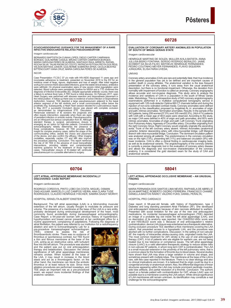
39
Pôsteres
Arq Bras Cardiol: Imagem cardiovasc. 2017;30(4 supl.1):6-41
58041LEFT ATRIAL APPENDAGE OCCLUSIVE MEMBRANE – AN UNUSUAL FINDING
Imagem cardiovascular
MARIA FERNANDA DOS SANTOS LIMA NEVES; RAPHAELA DE ABREU E SILVA MARTINEZ; ROBERTO OSÓRIO FERREIRA; FRANCISCO CHAMIÉ; DANIELLA BASTOS RAWET; VALÉRIO FUKS; DANIEL PERALTA;
HOSPITAL PRO CARDIACO
Case report: A 58-year-old female, with history of Hypertension, type 2 Diabetes and long standing persistent Atrial Fibrilation (AF). She developed oral anticoagulant intolerance because of skin necrosis attributed to warfarin. Presented with social limitation for the use of the new oral anticoagulant medications. An incidental transesophageal echocardiogram (TEE) depicted an image of a probable big clot inside the left atrial appendage (LAA), and no description of its anatomy was reported. Her CHA2DS2-VASc score was 4 and HAS-BLED score was 3. Because of oral anticoagulation´s relative contraindication, device LAA closure was entertained as a therapeutic alternative. During occlusion procedure TEE identified a thick membrane covering the LAA ostium, that prevented access to a hypoplastic LAA, and the procedure was aborted with no further complications. Discussion: In the setting of non-valvular AF, the majority of intracardiac stroke-causing clots, originated inside the LAA. The most common treatment for (AF) stroke is the use of oral anticoagulant therapy (OAT). A lot of patients who are eligible for warfarin use, are not being treated due to low tolerance or compliance issues. The left atrial appendage closure (LAAC) is a valid alternative therapeutic strategy to reduce stroke risks in non-valvular AF patients, in whom long term OAT is contraindicated. The LAA is a small muscular extension in the proximity of the left pulmonary veins, that is a common place for the formation of thrombi duo to blood stasis. Several studies have shown that LAA may show a large variation in volume and shape, and sometimes present with multiple lobes. The membrane at the base of the LAA is rare, with few case reported in the literature. There is no clear etiology and also no clinical implications are known. It is believed that its origin could be due to a congenital anomaly or anatomical variant. Also, the differential diagnosis of linear structures appearing within the LAA may include prominent pectinate muscles, side lobe artifacts, and partial resolution of a thrombi. Conclusion: The authors report on a female patient with contraindication for OAT, whose LAAC was not possible due to a membrane covering the LAA ostium. While clinical significance of LAA membranes still remain unknown, its identification may constitute a real challenge for the echocardiographer.
60728EVALUATION OF CORONARY ARTERY ANOMALIES IN POPULATION OF SOUTH OF MINAS GERAIS STATE
Imagem cardiovascular
HENRIQUE MARTINS DE SOUZA; MULLER AVILA BATISTA; ANNA JULLISA BISÃO FONTANA; SERGIO RODRIGO BERALDO; JAIME SCHMIDT DA SILVA FILHO; MATHEUS HENRIQUE FERNANDES; PEDRO COUTINHO MEYER FERNANDES; FLAVIO SIQUEIRA JUNQUIRA; ELISA FARIA UVEDA;
UNIVAS
Coronary artery anomalies (CAA) are rare and potentially fatal, their true incidence in the general population has yet to be defined and are important causes of sudden death in young athletes. The anatomical variation is the less frequent presentation of the coronary artery, it runs away from the usual anatomical description, but there is no functional impairment. Otherwise, the deviation from normality with impairment of function is called an anomaly. Coronary angiography allows accurate and non-invasive diagnosis. This study aims to analyze the incidence and variations of CAA in a population in the south of Minas Gerais state. Metodology: This is a retrospective study of 543 coronary angiotomography examinations performed in a multislice computerized tomography service in equipment with 128 multi-detector Optima 660 CT channels before and during the injection of non-ionic iodinated contrast media. Coronary anomalies were defined according to the classification proposed by Angeliniet AL in: anomalies of origin and path; intrinsic anomalies; Termination anomalies. Results: In the sample of the 543 patients, being 208 women(37.5%) and 335 men(62.5%), 24(4.41%) patients with CAA with a mean age of 48.8 years were observed. According to the study, as major CAA were defined in 40% of origin and path anomalies; and 60% were intrinsic anomalies. Anomalies of origin and path: Left Coronary Trunk originating from Pulmonary Artery; Agenesis of Circumflex artery; Circumflex originating from the right Coronary Sinus; Right Coronary would originally originate the ascending aorta; Trunk of the left coronary artery absent. Intrinsic Anomalies (anatomical variants): Anterior descending artery with intra-myocardial bridge; Left Marginal Branch with intra-myocardial bridge. Conclusion: The dominant circulation pattern was analyzed among all patients. The predominance in the coronary circulation was on the right (73%), presenting left (11%) and balanced circulation (9%) with lower incidence. CAA is often a clinical finding in cardiovascular imaging tests, as well as its anatomical variants. The angiotomography of the coronary arteries is currently a precise diagnostic tool in the evaluation of coronary artery disease and allows the diagnosis and non-invasive characterization of the coronary anatomy. It is considered the gold standard exam for the study of CAA and anatomical variants.
60704LEFT ATRIAL APPENDAGE MEMBRANE INCIDENTALLY DISCOVERED: CASE REPORT
Imagem cardiovascular
RODRIGO CORDOVIL PINTO LOBO DA COSTA; MIGUEL OSMAN DIAS AGUIAR; MARCELO LUIZ CAMPOS VIEIRA; ANA CLARA TUDE RODRIGUES; CLAUDIO HENRIQUE FISCHER; SAMIRA SAADY MORHY;
HOSPITAL ISRAELITA ALBERT EINSTEIN
Background: The left atrial appendage (LAA) is a blind-ending muscular extension of the left atrium, usually thought to modulate its pressure and volume. The presence of a membrane at the base of the LAA is a rare entity, probably developed during embryological development of the heart, most commonly found accidentally during transesophageal echocardiography. Case Report: a 64-year-old woman with previous history of hypertension, hypothyroidism and breast cancer presented at her cardiologist office for a routine appointment, complaining of palpitations. Following the diagnosis of atrial fibrillation, the patient was referred to our institution for a radiofrequency ablation and sent to Echocardiography Lab for a pre-procedure transesophageal echocardiogram (TEE), ordered to assess the risk of thromboembolic stroke. There was no evidence of thrombus or spontaneous contrast, but TEE found a thin and fibrous membrane at the base of the LAA, acting as an obstructive valve, with turbulent flow into the left atrium. The procedure was aborted and the patient was put on oral anticoagulation therapy and betablockers. Discussion: the clinical significance of left atrial appendage membranes remains unclear. When located at the base of the LAA, it may result in increase in the blood stasis and act as a thrombogenic factor; on the other hand, the membrane can block the embolic thrombus or at least reduce its size. Since more ablation procedures are performed nowadays, and TEE plays an important role as a pre-procedural exam, we expect more incidental findings of that anatomic variation.
60732ECHOCARDIOGRAPHIC GUIDANCE FOR THE MANAGEMENT OF A RARE INFECTIVE ENDOCARDITIS RELATED PSEUDOANEURYSMImagem cardiovascular
BERNARDO BAPTISTA DA CUNHA LOPES; DIEGO CARTER CAMPANHA BORGES; GUILHERME CASALE; BRUNO CARTER CAMPANHA BORGES; MARIA CAROLINA FERES DE ALMEIDA; SANTIAGO RAUL ARRIETA; RAFAEL ROCHA SILVA; TAUIN RAONI DO COUTO; PATRICIA FEITOSA FROTA DOS REIS; WILSON MATHIAS JÚNIOR; GUILHERME SOBREIRA SPINA; CECILIA BEATRIZ BITTENCOURT VIANA CRUZ; MARIA CAROLINA FERES DE ALMEIDA;
INCOR
Case Presentation: F.C.B.C 37 y/o male with HIV-AIDS diagnosed 11 years ago and incomplete adherence to treatment, presented on November 2016 to the ED for an insidious onset of fever, rigours, diaphoresis and loss of weight. After initial negative workup for common AIDS-associated infections, given the stable clinical status, antibiotics were withheld. On physical examination signs of new severe mitral regurgitation were detected. Blood cultures were persistently positive for MSSA and a TTE confirmed the diagnosis of Infective Endocarditis (IE). Despite appropriate antibiotic course, there was a failure to achieve fever lysis. A TEE found a mitral abscess. On February 2017, open Heart Surgery was performed with abscess resection and bioprosthesis placement in the mitral position. In follow-up there was no evidence of active infection or bioprosthesis dysfunction, however, TEE depicted a large pseudoaneurysm adjacent to the basal anterior segment of the left ventricle and a small communicating orifice below the prosthetic ring. Subsequently, a Heart Team session decided for a percutaneous repair. In May 2017 a successful Occlutech device was placed with complete occlusion as demonstrated by intraprocedural ETE. Discussion: Periannular extension of IE worsens the prognosis and often require intervention, especially when there are signs of persistent infection or embolic events. Pseudoaneurysms are an infrequent but serious complication from IE. Although standard therapy is surgical, percutaneous closure is emerging as an option for high-risk patients. Conventional echocardiography reliably determines the presence of those complications, however, 3D TEE provides better insight for complex anatomy cases, define the shape of the communicating orifice, assists the choice and placement of the device, and also confirm the efficacy. Other imaging modalities, especially cardiac CT and MR also improve the procedure planning. Conclusion: This report reinforces the role of 3D TEE in the advance of novel transcatheter interventions, providing reliable and comprehensible real-time guidance for the treatment of those high-risk cases. Transcatheter closure of IE-related left ventricular pseudoaneurysm is a possible alternative to surgery.

40
Pôsteres
Arq Bras Cardiol: Imagem cardiovasc. 2017;30(4 supl.1):6-41
60709RIGHT VENTRICULAR FUNCTION ANALYSIS WITH LONGITUDINAL STRAIN IN ACUTE CORONARY SYNDROMES WITHOUT ST ELEVATIONImagem cardiovascular
ANDREA DE ANDRADE VILELA; MARCELA PAGANELLI DO VALE; MARIANA OLIVEIRA REZENDE; NATASHA SOARES SIMÔES DOS SANTOS; ANA CAROLINA CAIXETA BOVENDORP; RODRIGO BELLIO DE MATTOS BARRETTO; DAVID COSTA DE SOUZA LE BIHAN; JORGE EDUARDO ASSEF; ELIZABETE SILVA DOS SANTOS; AMANDA GUERRA DE MORAES REGO SOUSA;
INSTITUTO DANTE PAZZANESE DE CARDIOLOGIA
INTRODUCTION: RIGHT VENTRICULAR DYSFUNTION (RVD) IS RELATED TO INCREASED MORTALITY AND WORSE PROGNOSIS AFTER ACUTE CORONARY SYNDROMES (ACS). DESPITE THE EMERGENCE OF NOVEL ECHOCARDIOGRAPHIC TOOLS, THE COMPLEX GEOMETRY OF THE RIGHT VENTRICLE (RV) AND ITS LOCATION STILL MAKE EVALUATIONS DIFFICULT. RV IS MOSTLY SUPPLIED BY THE RIGHT CORONARY ARTERY (RCA). HOWEVER, CIRCUNFLEX CORONARY ARTERY (CCA) CAN ALSO RESPOND FOR RV BLOOD FLOW WHEN IT´S DOMINANT OR PROVIDES COLLATERALS IN SCENARIOS OF RCA CHRONIC OCCLUSION. FEW STUDIES INVOLVE RV FUNCTION ANALYSIS IN ACS AND ONLY ONE OF THEM WAS CONDUCTED IN PATIENTS WITH NON ST ELEVATION ACS (NSTACS). OBJECTIVE: TO EVALUATE RIGHT VENTRICULAR FUNCTION USING NOVEL TECHNOLOGIES AND CONVENTIONAL METHODS TO IDENTIFY THE PRESENCE OF RVD IN PATIENTS WITH SIGNIFICANT STENOSIS IN RCA AND/OR CCA IN NSTACS.METHODOLOGY: 97 PATIENTS WITH NSTACS WHO ATTENDED A TERTIARY CENTER WERE SUBMITTED TO ECHO AND CORONARY ANGIOGRAPHY OR CORONARY ANGIOTOMOGRAPHY. EXCLUSION CRITERIA: LEFT VENTRICULAR HYPERTROFY, MORE THAN MODERATE VALVULAR DYSFUNCTION, TIME BETWEEN LAST PAIN AND ECHO EXAMINATION ≥ 48 HOURS, ATRIAL FIBRILLATION, LEFT BUNDLE BRANCH BLOCK, PACEMAKER, PREVIOUS MYOCARDIAL INFARCTION, INAPROPIATE ACCOUSTIC WINDOW AND CHANGE OF DIAGNOSIS AFTER THE EXAM. ECHO VARIABLES FOR RV FUNCTION ANALYSIS WERE: TRICUSPID ANNULAR PLANE SYSTOLIC EXCURSION (TAPSE); FRACTIONAL AREA CHANGE (FAC); TISSUE DOPPLER S´ WAVE (TDS`), TEI INDEX, AND 2D LONGITUDINAL STRAIN -RV FREE WALL (03 SEGMENTS), RV FREE WALL AND SEPTUM (RVFWS STRAIN - 06 SEGMENTS). RESULTS: THE VARIABLES STATISTICALLY SIGNIFICANT WERE: FAC; TAPSE; TDS` AND RVFWS STRAIN (TABLE). THEY ALLOWED THE IDENTIFICATION OF PATIENTS WITH SEVERE CORONARY STENOSIS OF RCA AND CCA. HOWEVER, THE MEAN VALUES OF FAC (0.44 ± 0.09 VS 0.48 ± 0.09, P=0.009) AND TAPSE (17.19 ± 1.95 VS 18.22 ± 2.58, P=0.045) WERE NORMAL ACCORDING TO CURRENT GUIDELINES. ONLY DTS` (S´ 9.58 ± 1.77 VS 10.36 ± 1.82; P=0.05) AND RVFWS STRAIN (19 ± 3.7 VS -20.8 ± 4.04, P=0.025) EXHIBITED ABNORMALLY LOW MEAN VALUES IN GROUP A. CONCLUSION: BOTH METHODS, CONVENTIONAL (FAC AND TAPSE) AND NEW TECHNOLOGIES (TDS` AND RVWFS STRAIN), ALLOWED THE IDENTIFICATION OF PATIENTS WITH SEVERE CORONARY STENOSIS. NEW TECHNOLOGIES ARE MORE USEFUL IN CLINICAL PRACTICE, SINCE THEY PRESENTED ABNORMALLY LOW VALUES IN GROUP A. STUDIES WITH A LARGER CASUISTIC AND THAT ANALYSE RV FUNCTION WITH ECHO AND CARDIAC MAGNETIC RESSONANCE WILL PROVIDE MORE ROBUST RESULTS.
TABLE: RIGHT VENTRICLE FUNCTION ANALYSIS X CORONARY STENOSIS
GROUP A (N=46)
GROUP B (N=51)
P-VALUE
TAPSE (MM) 17.19 ± 1.95 18.22 ± 2.58 0.045 FAC (%) 0.44 ± 0.090 0.48 ± 0.09 0.009 TISSUE DOPPLER S` WAVE 9.58 ± 1.77 10.36 ± 1.82 0.05 TEI INDEX 0.35 ± 0.09 0.32 ± 0.06 0.24 RV FREE WALL STRAIN -22 ± 4.72 -24 ± 5.75 0.29 RV FREE WALL AND SEPTUM STRAIN
-19 ± 3.7 - 20.8 ± 4.04 0.025
TAPSE: TRICUSPID ANNULAR PLANE SYSTOLIC EXCURSION; FAC: FRACTIONAL AREA CHANGE; RV: RIGHT VENTRICLE GROUP A – RCA AND/OR CA STENOSIS ≥ 70% GROUP B – RCA AND/OR CA STENOSIS <70%
57767RARE ASSOCIATION OF LEFT VENTRICULAR NON-COMPACTION AND HYPERTROPHIC CARDIOMYOPATHY MORE PROMINENT AT INFEROLATERAL WALLImagem cardiovascularNATHALIA MARIA SEGOVIA MONGE; VIVIANE TIEMI HOTTA; MARIA CRISTINA DONADIO ABDUCH; CARLOS EDUARDO ROCHITTE; EDMUNDO ARTEAGA FERNANDEZ; LUCIANO NASTARI; CHARLES MADY;INCOR/FMUSP AND FLEURY MEDICINA E SAÚDECase Report: A 39 year old man was referred to our hospital with syncope. His past medical history was not significant for palpitations, chest pain or dyspnea. The patient denied smoking, alcoholism, drug use, or any regular medications. Family history of sudden death: paternal aunt, unknown etiology of death, without investigation of Left ventricular non compaction (LVNC) or Hypertrophic cardiomyopathy (HCM), and his father died of presumed acute coronary syndrome at age 49. Physical examination was unremarkable. Electrocardiogram showed sinus rhythm with left chambers overload and isolated ventricular extrasystoles. Transthoracic Echocardiogram (TTE) evidenced significant left ventricular (LV) hypertrophy predominantly in the apical region and inferolateral wall, in the absence of a significant intraventricular gradient at rest and after provocative maneuvers, with preserved LV function, suggesting non-obstructive HCM. Cardiac magnetic resonance imaging (MRI) showed LV hypertrophy predominantly in the lateral wall, with a maximum thickness of 35 mm, no signs of left ventricular outflow tract (LVOT) obstruction and prominent apical trabeculation, compatible with the association of LVNC and HCM. A genetic test was performed on this patient with inconclusive results. Discussion: LVNC and HCM are cardiomyopathies with distinct clinical presentation that may present common genetic mutations in genes encoding sarcomeric proteins. Some studies suggest a common genetic basis between HCM and LVNC and the possibility of a phenotypic association of these two cardiomyopathies in the same patient. However, the association of diagnostic criteria in cardiac imaging tests (TTE and cardiac MRI) compatible with MNC and HCM in the same patient is uncommon. In addition, this patient shows an infrequent location of the most prominent hypertrophy in the inferolateral wall. Conclusions: This report illustrates the case of a young and oligosymptomatic patient, in which, clinical investigation revealed findings compatible with LVNC associated with HCM. This case highlights the importance of cardiac imaging methods in the diagnosis of the association of two different and rare cardiomyopathies.
A B
C D SYSTOLE
SYSTOLE DIASTOLE
DIASTOLE
A
DIASTOLE SYSTOLE E F
DIASTOLE
DIASTOLE
SYSTOLE
SYSTOLE
G H
I J
K L
M N
O P
Q R
S T
U V
59511REVERSIBLE CARDIOMYOPATHY IN A PATIENT WITH JUVENILE HEMOCHROMATOSIS AFTER IRON-CHELATING AGENTS FOR THE TREATMENT OF IRON OVERLOAD
Imagem cardiovascular
LORENA SQUASSANTECAPELINE; CIBELE GONTIJO LOPES; DAVI A ZAGONEL; JOSÉ EDUARDO BARBOSA; MARLY UELLENDAHL;
SOCIEDADE BRASILEIRA DE CARDIOLOGIA
Juvenile hemochromatosis is a rare form of iron overload that frequently causes cardiomyopathy. The mechanism by which disordered iron metabolism induces heart failure is not entirely understood, but myocardial dysfunction appears to be intimately related to the deposition of iron in myocytes. Cardiac function characteristically worsens or improves in proportion to the degree of iron accumulation in cardiac myocytes. The authors report the case of a 44-year-old Brazilian woman who had diagnosed juvenile hemochromatosis (HAMP mutation on 5’-UTR region) that developed symptoms of congestive heart failure 8 months ago and was found to have dilated cardiomyopathy in the cardiac magnetic resonance (CMR) with left ventricular ejection fraction (LVEF) of 42%. Her laboratory tests were: hemoglobin (Hb) 11.7 g/dL, transferrin saturation (TS) 100%, serum ferritin (SF) 7,350 ng/mL and CMR using T2* evaluation showed liver iron concentration (LIC) of 30.67 mg Fe/g dry (NV < 2.0 mg/g) and myocardium iron concentration (MIC) of 3.40 mg/g (NV < 1.1 mg/g). Five years after treatment with iron-chelants, CMR showed a dramatic improvement of her cardiac function with LVEF of 63% and of her symptoms of congestive heart failure and no detection of iron in myocardium (MIC of 0.78 ng/mL). This case indicates that the cardiac function in juvenile hemochromatosis could be reversed once iron overload from treatment with iron-chelating agents. Figura 1 – Ventrículo esquerdo superior em 2013 diástole e
sístole e inferior em 2017.
57555QUANTITATIVE MYOCARDIAL PERFUSION AND GATED LEFT VENTRICULAR MEASUREMENTS FROM 82RB PET/CT ASSESSED BY DIFFERENT COMMERCIAL SOFTWARE PACKAGESImagem cardiovascular
JOAQUIM BARRETO; YEW MIN SEW; KSHAMA WECHALEKAR;
UNICAMPIntroduction: Rest-stress 82Rubidium cardiac PET/CT is used widely in USA and it is becoming increasingly available in Europe in the last few years. It not only provides information about ischaemia and LV function but also absolute myocardial blood flow (MBF) and myocardial perfusion reserve (MPR) since dynamic imaging can be performed during the first pass of the tracer. Various software are commercially available to analyse the qualitative and quantitative data. We aim to compare 3 such software packages to assess the reproducibility and variations among them. Purpose: This study compared the agreement between A. Corridor4DM v2017 alpha (INVIA), its predecessor B. Syngo Myocardial Blood Flow package (Siemens Medical Solutions) + Corridor4DM v2015, and C. Quantitative PET (QPET, Cedars-Sinai Cardiac Suite 2015). Methods: Rest-stress 82Rb cardiac PET/CT data from 55 patients (70% male) with mean age 66 years (range 34–85), scanned at our centre between August and December 2016 were processed using 3 software with minimal manual intervention by a single observer blinded to clinical information. The MBF, MPR (corrected with resting rate pressure product), summed scores and gated volumes were assessed using each method. Bland-Altman analysis was used to determine the level of agreement with 95% limits plotted as the mean ± coefficient of repeatability. ANOVA determined whether the mean values were statistically different, considering p < 0.05. Results: There were no significant differences between method A and B in terms of EF, global MPR and MBF (p > 0.05). However method C derived global MPR and stress MBF values differed significantly from method A and B. Pairwise differences of MPR values were no more than 0.37 and 0.42 mL/min/g for stress MBF and MPR. Method C derived EF values were also different compared to A and B (p < 0.01) and EF mean difference was under 6.34% among the 3 methods. The mean summed scores from all methods were not different as determined by one-way ANOVA (p = 0.64, p = 0.95 and p = 0.49, for SSS, SRS and SDS respectively). Bland-Altman analysis showed very high level of agreement (91-98%) among the 3 methods for all variables. Conclusion: The mean values of MPR, stress MBF and EF derived from QPET differed significantly from those obtained from both versions of Corridor 4DM and Syngo MBF. Users should therefore be cautious when using different software interchangeably as systematic differences amongst them may introduce wider quantitative variation which could be clinically significant.

41
Pôsteres
Arq Bras Cardiol: Imagem cardiovasc. 2017;30(4 supl.1):6-41
58089THE ROLE OF 3D ECHO IN THE RARE DIAGNOSIS OF AORTOCAMERAL FISTULA
Imagem cardiovascular
LAURA DE ABREU ALVES; JUCIARA DA SILVA MATOS; RAFAEL DE OLIVEIRA CARDOSO; MILTON RICARDO POFFO; LAISSE MARINS DEFANTI GONZAGA; BRUNO ROBERTO ITABORAHY ALABRÍN;
INSTITUTO NACIONAL DE CARDIOLOGIA
Case report: A 57 year-old woman with hypothyroidism and hypertension reported 3 years worsening fatigue, important lower limb edema, orthopnoea and palpitations. Electrocardiogram showed signs of left ventricular overload. Initial transthoracic echocardiography showed a subaortic ventriculoseptal defect with left to right shunt, preserved biventricular function and moderate aortic stenosis. Follow-up echocardiography showed increased size of all cavities, right ventricular dysfunction, a mean left ventricle to aortic gradient of 31 mmHg and a shunt from the aorta to the right atrium with systolic and diastolic flow. 3-dimensional (3D) transesophageal echocardiography (TOE) suggested the right coronary sinus as the origin of this fistula, and demonstrated a left-right systolic-diastolic shunt. Cardiac catheterization provided evidence of rupture of a right coronary sinus aneurysm and shunting between the aorta and right atrium. Discussion: Fistula between the aorta and right atrium belongs to the group of abnormalities of the vascular connections of the aorta called aortocameral fistulas. They may originate in any of the three sinuses of Valsalva, although rarely from the non-coronary sinus. The acquired form is more frequent than the congenital form, and may be associated with prosthetic endocarditis or transcatheter closure of septal defects. Final considerations: We report a case posing diagnostic ambiguity between subaortic ventriculoseptal defect and aortoatrial fistula. The undertaking of 3D TOE in this patient permitted more accurate characterization of the coronary defect. 3D echocardiography can be an excellent modality to determine the morphology and size of a fistulous tract, helping to choose the best closure method. Selective catheterization enhances the delineation of a fistula’s course and the position of coronary ostia.
60466STUDY OF VENTRICULAR FUNCTION BY THE SPECKLE TRACKING STRAIN TECHNIQUE IN CRITICALLY ILL SEPSIS PATIENTS: POTENTIAL PROGNOSTIC IMPLICATIONSImagem cardiovascular
MARIANA DE BRAGA LIMA CARVALHO CANESSO; ISABELA NASCIMENTO BORGES; THIAGO ADRIANO DE DEUS QUEIROZ; LARISSA HELENA SENHORINE PENA; MARCIO VINICIUS LINS DE BARROS; VANDACK ALENCAR NOBRE JÚNIOR; MARIA DO CARMO PEREIRA NUNES;
UNIVERSIDADE FEDERAL DE MINAS GERAIS
In the intensive care setting, cases sepsis are the main causes of morbidity and mortality. Preliminary data concerning the utility of speckle tracking echocardiography (STE) as a marker of myocardial dysfunction in patients with sepsis are promising. We hypothesize that the STE technique can predict response to therapy and in-hospital outcome. The aim of this study was to evaluate STE in patients with severe sepsis and septic shock admitted to an intensive care unit (ICU), correlating values of myocardial strain with clinical and laboratory prognostic markers. We conducted a cohort prospective observational study in the ICU of the University Hospital of the Federal University of Minas Gerais (HC-UFMG). Our study included all adult patients (≥ 18 years of age) with sepsis or septic shock, initiated within 48 hours of hospital admission, without cardiomyopathy or insufficient image quality for STE. We collected clinical and laboratory data and performed echocardiographic studies on days 1 and 7 post study inclusion. These patients were accompanied until hospital discharge or death. We initially selected 56 patients and 26 patients were eligible for the study. Mean age was 57.0 [35.8/64.8] years, and 54% were female. Septic shock was diagnosed in 88%. The baseline APACHE II and SOFA score were 15 [12.8/21.5] and 8 [5.8/11.3], respectively. All study patients had left ventricle (LV) ejection fraction within reference values, but 73% presented reduced myocardial contractility at hospital admission, assessed by STE. During hospital stay, 7 patients died. At the end of the first week of treatment, we observed increase in longitudinal strain in both ventricles (LV longitudinal STE -18.7 ± 3.5 % at D1 versus -20.3 ± 3.0 % at D7; P: 0.024; and right ventricle longitudinal STE -20.0 ± 5.6% at D1 versus -23.0 ± 5.9 % at D7; P: 0.012). We further analyzed the factors associated with strain values in the first 48 hours of the diagnosis. The respiratory SOFA score, presence of septic shock at admission and ICU length of stay were associated with RV strain, while diabetes mellitus, respiratory SOFA score, baseline lactate and CRP levels were associated with LV strain. In conclusion, these results demonstrated the association between improvement in biventricular longitudinal strain with treatment for sepsis. The prognostic markers were associated with baseline strain values of both ventricles, pointing to potential prognostic value of strain in the context of sepsis.