fusão de membranas como alvo para inativação viral e ...
164
Universidade Federal do Rio de Janeiro Instituto de Bioquímica Médica Fausto Stauffer Junqueira de Souza FUSÃO DE MEMBRANAS COMO ALVO PARA INATIVAÇÃO VIRAL E DESENVOLVIMENTO DE VACINAS Rio de Janeiro 2007
Transcript of fusão de membranas como alvo para inativação viral e ...

Universidade Federal do Rio de Janeiro
Instituto de Bioquímica Médica
Fausto Stauffer Junqueira de Souza FUSÃO DE MEMBRANAS COMO ALVO
PARA INATIVAÇÃO VIRAL E
DESENVOLVIMENTO DE VACINAS
Rio de Janeiro
2007

Fausto Stauffer Junqueira de Souza
FUSÃO DE MEMBRANAS COMO ALVO
PARA INATIVAÇÃO VIRAL E
DESENVOLVIMENTO DE VACINAS
Tese de doutorado apresentada ao Programa de
Pós-graduação em Química Biológica, Instituto
de Bioquímica Médica, Universidade Federal
do Rio de Janeiro, como parte dos requisitos
necessários à obtenção do título de Doutor em
Química Biológica.
Orientadora: Andrea Thompson Da Poian
Rio de Janeiro
2007

FICHA CATALOGRÁFICA
Stauffer, Fausto. Fusão de membranas como alvo para inativação viral e
desenvolvimento de vacinas / Fausto Stauffer Junqueira de Souza. – Rio de Janeiro, 2007.
162 f.: il. Dissertação (Doutorado em Química Biológica) –
Universidade Federal do Rio de Janeiro, Instituto de Bioquímica Médica, 2007.
Orientadora: Andrea Thompson Da Poian 1. Fusão de Membranas. 2. Inativação Viral. 3.
Dietilpirocarbonato. 4. Vacina. I. Da Poian, Andrea T. (Orient.). II. Universidade Federal do Rio de Janeiro. Instituto de Bioquímica Médica. III. Título.
AGRADECIMENTOS
À Andrea, minha orientadora, pelo apoio durante todos esses anos. Muito obrigado pelo voto de confiança no momento que mais precisei. Tenho certeza que nossos laços de amizade irão perdurar apesar do término do doutorado. À Fabiana, pela amizade e orientação durante minha iniciação científica até os dias atuais. Ao prof. Miguel, meu orientador em Portugal, pela hospitalidade e amizade. Aos meus alunos de iniciação científica: Carol, Fernando, Marcos, Nathalia, Sabrina e Vitor. Ao Joari, pelo valioso apoio no início do doutorado. Ao prof. Ronaldo e à Ada, meus colaboradores, pela ajuda no desenvolvimento desta tese. Aos meus outros colaboradores: prof. Gilberto Weissmüller, profa. Gisele Fabrino, profa. Lucia Bianconi e prof. Luiz Juliano. Ao prof. Paulo Mourão, por revisar esta tese e “batalhar” pelo sucesso do programa MD-PhD. Ao pessoal do meu laboratório: Ana Paula, Aninha, Beth, Carla, Eliesier, Fernanda, Flávia, Iranaia, Leandro, Luíza, Mariana, Marina, Marisa, Rogério, Rosângela, Tatiana e Thaís. Ao pessoal do laboratório e amigos de Portugal: David, Guida, Isabel, Manuel, Salomé, Silvia, Sónia. Ao pessoal do laboratório do Ronaldo: Chico, Diego, Emerson e Marcela. Aos membros da banca, pela disposição em aceitar o convite. À minha grande amiga Carol e aos meus amigos Alberto, Flávio, Karina e Lê. À Mel, minha namorada, por tudo. Aos pais da Mel, Fernando e Adelaide. Aos meus pais, por todo o apoio e compreensão que foram imprescindíveis durante o doutorado.

RESUMO
STAUFFER, Fausto Junqueira de Souza. Fusão de membranas como alvo para inativação viral e desenvolvimento de vacinas. Rio de Janeiro, 2007. Dissertação (Doutorado em Química Biológica) – Instituto de Bioquímica Médica, Centro de Ciências da Saúde, Universidade Federal do Rio de Janeiro, Rio de Janeiro, 2007. A fusão de membranas é uma etapa essencial para a entrada dos vírus envelopados na célula alvo. Este processo é catalisado pelas glicoproteínas do envelope viral, que sofrem uma mudança conformacional desencadeada pela interação do vírus com seu receptor celular específico ou exposição ao pH ácido do meio endossomal. Nesta reestruturação da proteína de superfície viral, ocorre a exposição do peptídeo de fusão, que desestabiliza a membrana, iniciando a reação de fusão. A elucidação dos mecanismos moleculares envolvidos na reação de fusão de membranas pode auxiliar no desenvolvimento de novos compostos de inativação viral. Com este objetivo, foi investigada a interação dos vírus da estomatite vesicular e da dengue com vesículas lipídicas, estudando as mudanças conformacionais das glicoproteínas virais e o processo de fusão. Os resultados obtidos com o vírus da dengue indicam que o peptídeo de fusão da glicoproteína E possui alta afinidade por vesículas compostas de lipídeos aniônicos e que a interação é de natureza eletrostática. Tanto o coeficiente de partição quanto a reação de fusão possuem maior intensidade na presença de fosfolipídios carregados negativamente e da oligomerização do peptídeo. No caso do vírus da estomatite vesicular (VSV), estudos prévios haviam demonstrado que a fusão de membranas ocorre numa faixa estreita de pH, entre 6.2 e 5.8, sugerindo que a protonação de resíduos de histidina seria necessária para este processo. A fim de investigar o papel desses aminoácidos na fusão mediada pelo VSV, modificamos quimicamente estes resíduos com dietilpirocarbonato (DEPC). Demonstramos que a fusão de membrana mediada pelo VSV era inibida pela modificação dos resíduos de histidina. Medidas de fluorescência mostraram que a modificação do vírus com DEPC abolia as mudanças conformacionais da proteína G, sugerindo que a protonação de resíduos de histidina dirige a interação entre a glicoproteína viral e a membrana alvo no pH ácido do meio endossomal. Baseado nestes resultados, decidimos avaliar se o tratamento com DEPC era capaz de inativar o vírus e o potencial uso deste composto como inativador viral para o desenvolvimento de vacinas. A infectividade do VSV em células BHK21 e a patogenicidade em camundongos Balb/c foram abolidas através do tratamento viral com DEPC 0.5 mM. Além disso, a modificação com DEPC não alterou a integridade conformacional das proteínas de superfície do VSV inativado como observado por microscopia eletrônica e ELISA de competição. Os anticorpos produzidos nos camundongos após imunização intraperitoneal com VSV inativado pelo DEPC misturado com adjuvantes foram capazes de reconhecer e neutralizar o vírus nativo, além de proteger de forma eficiente os animais do desafio com doses letais de VSV. Esses resultados em conjunto sugerem que a inativação viral com DEPC baseada na inibição da fusão de membranas é um método adequado para o desenvolvimento de vacinas. Palavras chave: fusão de membranas / inativação viral / dietilpirocarbonato

ABSTRACT STAUFFER, Fausto Junqueira de Souza. Fusão de membranas como alvo para inativação viral e desenvolvimento de vacinas. Rio de Janeiro, 2007. Dissertação (Doutorado em Química Biológica) – Instituto de Bioquímica Médica, Centro de Ciências da Saúde, Universidade Federal do Rio de Janeiro, Rio de Janeiro, 2007. Membrane fusion is an essential step in the entry of enveloped viruses into their host cells. This process is catalyzed by viral surface glycoproteins that undergo a conformational changes triggered by interaction with specific cellular receptors or by the exposition to low pH of endossomal medium. The structural reorganization of the viral glycoproteins leads to the exposure of the fusion peptide, a specific segment of these proteins, which destabilizes the lipid bilayers, initiating the fusion reaction. Understanding the virus induced membrane fusion at the molecular level should provide means to develop new viral inactivating compounds. For this purpose, we evaluated the interaction of vesicular stomatitis and dengue viruses with lipid vesicles, studying the conformational changes in viral glycoproteins and the membrane fusion process. In dengue virus studies, our results indicate that E glycoprotein fusion peptide has a high affinity to vesicles composed of anionic lipids and that the interaction is mainly electrostatic. Both partition coefficient and fusion index are enhanced by negatively charged phospholipids and peptide oligomerization. In the case of vesicular stomatitis virus (VSV), previous studies have shown that membrane fusion occurs at a very narrow pH range, between 6.2 and 5.8, suggesting that His protonation is required for this process. To investigate the role of His in VSV fusion, we chemically modified these residues using diethypyrocarbonate (DEPC). We found that membrane fusion mediated by VSV was inhibited by His modification. Fluorescence measurements showed that VSV modification abolished pH-induced conformational changes in G protein, suggesting that His protonation drives G protein interaction with the target membrane at acidic pH. Based on these results, we decided to assess whether treatment with DEPC was able to inactivate the virus and its potential use as a viral inactivating chemical agent for the development of useful vaccines. VSV infectivity in BHK21 cells and pathogenicity in Balb/c mice were abolished by viral treatment with 0.5mM DEPC. In addition, DEPC treatment did not alter the conformational integrity of surface proteins of inactivated VSV as demonstrated by transmission electron microscopy and competitive ELISA. Antibodies elicited in mice by intraperitoneal immunization with DEPC-inactivated VSV mixed with adjuvants were able to recognize and neutralize the native virus and efficiently protected animals against the challenge with lethal doses of VSV. These results together suggest that viral inactivation with DEPC based on membrane fusion inhibition seems to be a suitable method for the development of vaccines. Keywords: membrane fusion / viral inactivation / diethypyrocarbonate

LISTA DE FIGURAS
Figura 1: Mecanismos de entrada dos vírus envelopados nas células hospedeiras Figura 2: Representação esquemática das estruturas das proteínas de fusão de classes I, II e III no estado conformacional pós-fusogênico Figura 3: Mecanismos propostos para o processo de fusão catalisado pelas glicoproteínas virais Figura 4: Representação esquemática do VSV Figura 5: Ciclo de replicação do VSV Figura 6: Seqüência de aminoácidos da proteína G do VSV Figura 7: Estrutura cristalográfica da proteína G do VSV nas conformações pré e pós fusogênica Figura 8: Mudanças estruturais da proteína G do VSV ocorridas na transição das conformações pré e pós-fusogênicas Figura 9: Representação esquemática do curso temporal de aparecimento dos sintomas e sinais clínicos da dengue Figura 10: Representação esquemática da estrutura do vírus da dengue e do genoma viral Figura 11: Ciclo de replicação do vírus da dengue Figura 12: Estrutura cristalográfica da proteína E do vírus da dengue Figura 13: Rearranjo dos domínios da proteína E durante a transição para trímero Figura 14: Alinhamento da região do peptídeo de fusão da glicoproteína E de alguns flavivírus Figura 15: Diagrama esquemático do mecanismo proposto para mudança conformacional induzida pelo pH ácido Figura 16: Estrutura cristalográfica do domínio de fusão da proteína G do VSV nas conformação pós fusogênica Figura 17: Dependência do pH para a aquisição da conformação fusogênica de diferentes vírus envelopados
12 14 17 19 20 21 22 24 26 27 29 30 31 32 109 111 116

LISTA DE TABELAS
Tabela 1: Comparação entre as proteínas de fusão de classes I e II 15
LISTA DE ABREVIATURAS
BHK do inglês: baby hamster kidney
C-terminal Carboxi-terminal
DEN Vírus da dengue
DEPC Dietilpirocarbonato
ELISA do inglês: enzyme-linked immunosorbent assay
GAG Glicosaminoglicano
GP Glicoproteína
HIV Vírus da imunodefiência humana
HS Heparan sulfato
IgG Imunoglobulinas do tipo G
IgM Imunoglobulinas do tipo M
INPI Instituto Nacional da Propriedade Industrial
N-terminal Amino-terminal
PS Fosfatidilserina
RNA do inglês: ribonucleic acid
TBE do inglês: tick born encephalitis
UV Ultra-violeta
VSHV Vírus da septicemia hemorrágica viral
VSV Vírus da estomatite vesicular
VSV-NJ Vírus da estomatite vesicular New Jersey
VSV-IN Vírus da estomatite vesicular Indiana

LISTA DE AMINOÁCIDOS
Ala Cys Asp Glu Phe Gly His Ile Lys Leu Met Asn Pro Gln Arg Ser Thr Val Trp Tyr
A C D E F G H I K L M N P Q R S T V W Y
Alanina Cisteína
Ácido Aspártico Ácido Glutâmico
Fenilalanina Glicina
Histidina Isoleucina
Lisina Leucina
Metionina Asparagina
Prolina Glutamina Arginina Serina
Treonina Valina
Triptofano Tirosina

SUMÁRIO
1 Introdução..........................................................................................................
1.1 Aspectos gerais da infecção viral.......................................................................... 1.2 Fusão de membranas.............................................................................................
1.2.1 Glicoproteínas Virais................................................................................... 1.3 Modelo de estudo I: o vírus da estomatite vesicular.............................................
1.3.1 A Estrutura dos Rabdovírus......................................................................... 1.3.2 Ciclo de Replicação dos Rabdovírus........................................................... 1.3.3 Fusão de membranas mediada pelo VSV....................................................
1.4 Modelo de estudo II: o vírus da dengue................................................................ 1.4.1 A Estrutura dos Flavivírus........................................................................... 1.4.2 Ciclo de replicação dos Flavivírus............................................................... 1.4.3 Proteína E e seu peptídeo de fusão...............................................................
1.5 Mecanismos de inativação viral............................................................................ 1.5.1 Inativação do VSV....................................................................................... 1.5.2 Dietilpirocarbonato......................................................................................
1.6 Artigo 1.................................................................................................................. 2 Resultados...........................................................................................................
2.1 Parte I: Elucidação dos mecanismos da fusão de membranas mediada pelo VSV e pelo vírus da dengue........................................................................................
2.1.1 Artigo 2..................................................... .................................................. 2.1.2 Artigo 3........................................................................................................
2.2 Parte II: Descoberta de um novo composto inativador viral baseado na inibição de fusão de membranas e sua aplicação na formulação de uma vacina....................
2.2.1 Patente 1.............................................................................................. 2.2.2 Artigo 4................................................................................................ 2.2.3 Artigo 5..................................................... ..........................................
3 Discussão.............................................................................................................
3.1 Parte I..................................................... ..................................................... 3.1.1 VSV..................................................................................................... 3.1.2 Vírus da Dengue...................................................................................
3.2 Parte II.......................................................................................................... Referências............................................................................................................ Anexos....................................................................................................................
10 11 11 13 18 19 19 20 25 27 28 29 32 33 34 35 42 43 45 52 64 65 88 98 107 108 108 111 114 118 129

INTRODUÇÃO

1 Introdução
1.1 Aspectos gerais da infecção viral
Os vírus são os menores organismos que podem se replicar e são classicamente
conhecidos pela sua habilidade de atravessar filtros que retêm até as menores bactérias. Na
sua forma mais básica, as partículas virais são compostas por um material genético, que é
revestido por uma capa protéica, conhecida como capsídeo. Em alguns casos, a partícula viral
pode ainda ser envolvida por uma membrana lipídica, conhecida como envelope, que contém
as glicoproteínas virais. Apesar desta simplicidade, os vírus apresentam grande variação
quanto ao tamanho, forma e composição e podem infectar uma gama de organismos na
natureza.
A infecção da célula hospedeira é essencial para a replicação viral, já que os vírus não
possuem metabolismo próprio e dependem da maquinaria enzimática celular para este
processo. Em uma primeira etapa da infecção viral, os vírus aderem às células por meio de
regiões específicas das proteínas de sua superfície interagindo com moléculas presentes na
superfície celular que passam a funcionar como receptores. Estas moléculas podem ser
proteínas, carboidratos ou lipídeos. Após aderirem à superfície celular, os vírus iniciam sua
entrada nas células utilizando diferentes mecanismos, levando à liberação do genoma viral no
citoplasma ou no núcleo da célula hospedeira.
O genoma viral pode ser liberado no citoplasma tanto como moléculas de ácido
nucléico livre, como acontece para alguns vírus RNA, quanto como complexos de ácido
nucléico e proteínas. Coincidindo com, ou imediatamente após a entrada na célula, começa
um processo de desmontagem dos componentes virais, levando à replicação do genoma viral,
com a formação de novas partículas virais.
A liberação destas partículas recém formadas da célula hospedeira pode ocorrer de
diferentes formas. Os vírus envelopados podem sair da célula por brotamento através da
membrana plasmática ou pela fusão de vesículas secretoras contendo partículas virais com a
membrana plasmática. Os vírus não envelopados são geralmente liberados por lise da
membrana celular.
1.2 Fusão de membranas
Tanto os vírus envelopados quanto os não envelopados compartilham as principais
etapas do processo de entrada nas células hospedeiras, que se inicia pela ligação na célula
hospedeira e termina com a liberação do material genético viral no citoplasma (Dimitrov,
11

2004). A principal diferença está na etapa de penetração do nucleocapsídeo no citoplasma:
para os vírus envelopados, ocorre a fusão de membranas, enquanto que para os não
envelopados, ocorre a formação de poros ou lise de membranas (Marsh e Helenius, 2006).
A fusão de membranas, um processo celular essencial para fagocitose, pinocitose e
tráfego de vesículas, é o mecanismo básico de entrada dos vírus envelopados nas células
hospedeiras. É um método “elegante e efetivo” para liberar o genoma viral no citoplasma e
iniciar a infecção (Smith e Helenius, 2004). A reação de fusão do envelope viral pode ser
direta com a membrana plasmática da célula ou com a membrana do compartimento
endossomal após a internalização da partícula viral por endocitose mediada por receptor (Earp
et al., 2005; Harrison, 2005; Kielian e Rey, 2006) (fig.1). As vias de endocitose utilizadas
podem ser dependente de clatrina (mais comum), dependente de caveolina ou independente de
clatrina e caveolina (Sieczkarski e Whittaker, 2002; Marsh e Helenius, 2006). Dentre os vírus
que são capazes de fundir-se diretamente com a membrana plasmática podemos citar como
exemplo os paramixovírus, os retrovírus e os herpesvírus. Já os alfavírus, os flavivírus, os
rabdovírus e o vírus influenza são exemplos de vírus que penetram na célula hospedeira
através de endocitose mediada por receptor, seguida pela fusão da membrana viral com a
Figura 1: Mecanismos de entrada dos
endossomal (Dimitrov, 2004).
vírus envelopados nas células hospedeiras: (A) fusão direta das
A fusão de membranas induzida pelos vírus envelopados é mediada pelas proteínas de
superfície (Kielian e Rey, 2006), que sofrem uma dramática mudança conformacional
A
B
A
B
membranas viral e plasmática; (B) endocitose mediada por receptor, com posterior acidificação do endossoma e
fusão de membranas. Extraído de Da Poian et al. (2005).
12

desencadeada pela interação com o receptor na membrana alvo em pH neutro ou pela
exposição ao pH ácido do meio endossomal (Earp et al., 2005; Sieczkarski e Whittaker,
2005). Outras formas de ativação do processo de fusão têm sido propostas, como a
combinação de interação da glicoproteína viral com seu receptor associada à exposição ao pH
ácido (Mothes et al., 2000; Matsuyama et al., 2004) e a clivagem proteolítica dessas
glicoproteínas por proteases do endossoma ativadas pelo pH ácido (Chandran et al., 2005).
1.2.1 Glicoproteínas Virais
As glicoproteínas dos vírus envelopados, tipicamente proteínas integrais de membrana
aterial genético viral e sintetizadas utilizando-se a maquinaria de
ntese
nom
l, (b) o N-terminal está sempre
asses de proteínas
tipo I, são codificadas pelo m
sí protéica da célula infectada. Após o processamento destas proteínas, elas são
ancoradas em membranas da célula hospedeira e incorporadas às partículas virais durante a
montagem e brotamento dos novos vírus. Na superfície viral, podemos observar que estas
proteínas formam oligômeros altamente organizados e são glicosiladas (Eckert e Kim, 2001).
A fusão de membranas, etapa essencial para infecção dos vírus envelopados, é
mediada por estas proteínas transmembrana (Kielian e Rey, 2006), que por esta razão são
de inadas proteínas de fusão. Estas glicoproteínas contêm uma seqüência de aminoácidos
com grande número de resíduos hidrofóbicos e de glicinas, capaz de interagir com a
membrana alvo, conhecida como peptídeo de fusão (Eckert e Kim, 2001). São sintetizadas
numa conformação metaestável de alta energia na superfície viral, na qual seu peptídeo de
fusão encontra-se “escondido” no interior do oligômero formado pelas glicoproteínas
(Hernandez et al., 1996). No entanto, é proposto que a ligação ao receptor ou a exposição ao
pH ácido acarretaria na transição da glicoproteína viral para a conformação fusogênica de
baixa energia, expondo o peptídeo de fusão e liberando a energia necessária para o processo
de fusão (Carr et al., 1997; Epand, 2003; Dimitrov, 2004).
As características em comum das proteínas de fusão são: (a) a maior parte de sua
massa é composta da porção externa à membrana vira
localizado no domínio externo, (c) os domínios transmembrana e C-terminal são
relativamente pequenos, (d) contêm carboidratos N-ligados, (d) formam oligômeros, e (e)
estão presentes em alta densidade na membrana viral (Eckert e Kim, 2001).
Embora possuam estas semelhanças, com base em critérios estruturais, principalmente
no estado conformacional pós-fusão, foram definidas até o momento duas cl
de fusão (fig. 2) (Heinz e Allison, 2001; Lescar et al., 2001; Weissenhorn et al., 2007).
Entretanto, as proteínas de fusão de alguns vírus possuem características que não as
13

enquadram em classe I ou II e provavelmente representam uma nova classe de proteínas de
fusão, que recentemente vem sendo referida como classe III, como é o caso dos rabdovírus
(fig. 2) (Da Poian et al., 2005; Heldwein et al., 2006; Roche et al., 2006; Weissenhorn et al.,
2007).
Figura 2: Representação esquemática das estruturas das proteínas de fusão de classes I, II e III no estado
conformacional pós-fusogênico. (A) Proteína gp41 do HIV-1 (classe I); (B) Proteína E dos flavivírus (classe
Wilson et al., 1981; Bullough et al., 1994), retrovírus (Fass et al., 1996), coronavirus (Xu et
l., 200
II); (C) Glicoproteína G do VSV (classe III). As posições da porção transmembrana (seta vermelha) e do
peptídeo de fusão (seta preta) estão indicadas. Adaptado de (Weissenhorn et al., 2007).
As proteínas de classe I são exemplificadas pelas proteínas de fusão dos ortomixovírus
(
a 4a; Xu et al., 2004b), filovírus (Weissenhorn et al., 1998) e paramixovírus (Chen et
al., 2001; Yin et al., 2005; Yin et al., 2006). Elas formam espículas triméricas no envelope
viral, com estrutura predominantemente em �-hélices. São sintetizadas como uma proteína
precursora, que após uma clivagem proteolítica gera duas subunidades que permanecem
14

ligadas entre si, seja por pontes dissulfeto ou ligações não covalentes. Uma das subunidades é
responsável pela interação inicial com o receptor na membrana alvo. Já a outra, que se
encontra ancorada no envelope viral, contém na extremidade N-terminal uma seqüência de
aminoácidos hidrofóbicos, que está diretamente relacionada com a fusão de membranas. Após
a ligação ao receptor celular ou a exposição ao baixo pH, a proteína de fusão muda
parcialmente de conformação, mantendo-se em trímeros, e o peptídeo de fusão é então
inserido na membrana alvo, catalisando a reação de fusão. A conformação pós-fusão
apresenta uma estrutura bastante típica, conhecida como hairpin, ou grampo de cabelo (fig.
2).
As proteínas de fusão de classe II são exemplificadas pelos alfavírus (Lescar et al.,
2001; Gibbons et al., 2004) e flavivírus (Rey et al., 1995; Modis et al., 2003; Modis et al.,
omparação entre as proteínas de fusão de classes I e II
Dímero metaestável para trímero ara estávelda proteína de fusãoM
Classe IIClasse ICaracterística
2004). Elas possuem três domínios, que estão principalmente organizados em folhas-�, que
também formam hairpins. Estas proteínas formam homo ou heterodímeros, que se encontram
paralelos ao envelope viral. O peptídeo de fusão está localizado em um loop entre duas fitas
�, que se localiza na interface interna do dímero. Ao contrário das proteínas de classe I, as
proteínas de classe II não sofrem clivagem proteolítica durante sua maturação. No entanto, é
necessária a clivagem de proteínas de membrana que se encontram associadas a estas
proteínas de fusão. Após a exposição ao pH ácido, a proteína se reorganiza em trímeros
perpendiculares à membrana viral, expondo o peptídeo de fusão, catalisando a fusão de
membranas.
As principais características dessas duas classes de proteínas de fusão estão listadas na
tabela 1.
Tabela 1: C
Loop hidrofófico no interior do dímero
Peptídeo localizado na porção N-terminal no interior do trímeroLocalização do peptídeo de fusão
Clivagem proteolítica da proteínaacessória
Clivagem proteolítica da proteínade fusãoMaturação para o conformação pré fusão
Trímero de hairpins compostos de estrutura beta
Trímero de hairpins com umaregião central em alfa héliceEstrutura pós fusão
folha betaalfa héliceEstrutura secundária predominante
estável da proteína de fusãoTrímero metaestável pudança conformacional durante a fusão
Loop hidrofófico no interior do dímero
Peptídeo localizado na porção N-terminal no interior do trímeroLocalização do peptídeo de fusão
Clivagem proteolítica da proteínaacessória
Clivagem proteolítica da proteínade fusãoMaturação para o conformação pré fusão
Trímero de hairpins compostos de estrutura beta
Trímero de hairpins com umaregião central em alfa héliceEstrutura pós fusão
folha betaalfa héliceEstrutura secundária predominante
Dímero metaestável para trímero estável da proteína de fusão
ara estávelda proteína de fusãoM
Classe IIClasse ICaracterística
Trímero metaestável pudança conformacional durante a fusão
15

As proteínas de classe III também apresentam trímeros de hairpins, mas combinam
elementos estruturais de ambas as classes de proteínas de fusão. Semelhante às proteínas de
classe I, apresentam uma região central em �-hélice. No entanto, cada domínio de fusão está
localizado na ponta de folhas-� alongadas, característica marcante das proteínas de classe II.
Além disso, a glicoproteína dos rabdovírus não sofre clivagem proteolítica e nem é sintetizada
associada a alguma outra proteína durante seu processo de maturação, características
presentes nas proteínas de classes I e II, respectivamente. Por último, as mudanças
conformacionais induzidas por baixo pH nesta proteína são reversíveis, enquanto que nas
demais proteínas de fusão são irreversíveis.
Apesar das proteínas de fusão de classes I e II apresentarem importantes diferenças
estruturais, os mecanismos de fusão propostos são bastante similares (fig. 3) (Weissenhorn et
al., 1999; Skehel e Wiley, 2000; Bressanelli et al., 2004; Modis et al., 2004; Weissenhorn et
al., 2007) e as mudanças conformacionais induzidas durante a ativação da forma não
fusogênica para a forma fusogênica são irreversíveis (Carr et al., 1997; Lescar et al., 2001;
Stiasny et al., 2001).
A reação de fusão induzida pelas proteínas de classe I (fig. 3A) é iniciada pela ligação
do vírus ao seu receptor específico na membrana alvo ou pela exposição ao pH ácido no meio
endossomal, sendo desencadeadas mudanças conformacionais que resultam na projeção e
inserção do peptídeo de fusão na membrana alvo. Assim, a proteína de fusão fica ancorada
nas membranas celular e viral. Subsequentemente, ocorre uma reestruturação da proteína, que
se “dobra”, forçando a aproximação da porção N-terminal, que contém o peptídeo de fusão
inserido na membrana alvo, com a porção C-terminal transmembrana, que está ancorada no
envelope viral, acarretando a aproximação dessas duas membranas (Harrison, 2005).
Já o processo de fusão induzido pelas proteínas de fusão de classe II (fig. 3B) inicia-se
com a reorganização da proteína em trímeros desencadeada pela exposição ao pH ácido do
meio endossomal. Durante este processo, ocorre um rearranjo da orientação relativa dos
domínios II e III da glicoproteína, que acarreta na exposição do peptídeo de fusão, que se
insere na membrana alvo, com o ancoramento da proteína nas membranas celular e viral,
como já havia sido descrito para as proteínas de classe I. Posteriormente, ocorre uma
reestruturação do domínio III, que traz o peptídeo de fusão e o domínio transmembrana C-
terminal para posições justapostas, ocasionando a aproximação das membranas viral e celular
(Harrison, 2005).
16

A
B
A
B
Figura 3: Mecanismos propostos para o processo de fusão catalisado pelas glicoproteínas virais. (A)
Proteína de classe I. O trímero da proteína de fusão na sua forma metaestável é representado ancorado no envelope viral,
com seu peptídeo de fusão em rosa escuro (a). Após a ligação ao receptor celular ou exposição ao pH ácido do meio
endossomal, a proteína de fusão adquire uma conformação estendida e o peptídeo de fusão é inserido na membrana alvo (b).
Vários trímeros estão envolvidos no processo (c). A reestruturação da proteína continua, com o “dobramento” da molécula,
que aproxima as membranas viral e celular (d). É formada a haste de hemifusão (e) e finalmente o poro de fusão (f) após o
total rearranjo da proteína de fusão, que adquiriu sua conformação mais estável, onde o peptídeo de fusão e o domínio
transmembrana se encontram justapostos. (B) Proteína de classe II. O dímero é representado na superfície viral, com o
seu peptídeo de fusão interno em verde, domínio I em vermelho, II em amarelo e III em azul (a). A proteína se liga ao
receptor específico e é internalizada por endocitose. Após exposição ao pH ácido do meio endossomal, ocorre um rearranjo
na orientação relativa dos domínios II e III, expondo o peptídeo de fusão (b) e permitindo a formação de trímeros e a inserção
do peptídeo de fusão na membrana alvo (c). A reestruturação da proteína continua, com o “dobramento” do domínio III, que
aproxima as membranas viral e endossomal (c). É formada a haste de hemifusão (d) e finalmente o poro de fusão (f) após o
total rearranjo trimérico da proteína de fusão, onde o peptídeo de fusão e a porção transmembrana se encontram próximos.
Extraído de Mukhopadhyay et al. (2005).
17

A desestabilização da membrana alvo pelo peptídeo de fusão e a aproximação com o
envelope viral são os processos essenciais para formação da haste de hemifusão
(intermediário do processo de fusão, onde as monocamadas externas encontram-se
fusionadas, enquanto que as internas não) (Jahn et al., 2003). Este intermediário se forma
durante a aproximação das membranas viral e celular. No final da reestruturação das proteínas
de fusão de classes I e II, ocorre a formação do poro de fusão que permite a liberação do
nucleocapsídeo viral no citoplasma (Harrison, 2005).
1.3 Modelo de estudo I: o vírus da estomatite vesicular
O vírus da estomatite vesicular (VSV) pertence ao gênero Vesiculovirus, da família
Rhabdoviridae, um grupo de vírus com grande abrangência de hospedeiros (plantas, animais
invertebrados e vertebrados) (Rose e Whitt, 2001). Dentre os rabdovírus, o VSV é o mais
estudado e é o agente responsável pela estomatite vesicular. Esta doença afeta bovinos,
eqüinos e suínos, e suas manifestações clínicas incluem vesiculação e/ou ulceração grave na
língua e nos tecidos orais, podendo, às vezes, provocar lesões nos pés e nas tetas dos animais
acometidos, o que resulta em uma substancial perda de produtividade (Letchworth et al.,
1999). Os sintomas desta doença são indistinguíveis daqueles observados na febre aftosa,
exceto por afetar também eqüinos. A infecção em humanos tem sido observada em pessoas
expostas a animais infectados ou inadvertidamente expostas em laboratório (Johnson et al.,
1966; Fields e Hawkins, 1967; Quiroz et al., 1988). Em humanos, a infecção pode ser
assintomática ou se manifestar com sinais e sintomas de um resfriado comum.
A estomatite vesicular causada pelo VSV é endêmica no continente americano,
ocorrendo desde o sudeste dos Estados Unidos até o norte da América do Sul, passando pelo
México e por toda a América Central (Wilks, 1994; Letchworth et al., 1999; Rodriguez,
2002). Nestas regiões, os sorotipos predominantes são VSV New Jersey (VSV-NJ) e VSV
Indiana (VSV-IN) (Cotton, 1926; Cotton, 1927). No Brasil, é endêmico o sorotipo Indiana 3,
ou Alagoas (Federer et al., 1967; Tesh et al., 1987). Outros vesiculovírus também já foram
encontrados na Índia e na África (Hanson, 1968). Nos Estados Unidos, os dois surtos mais
recentes ocorreram em 1997 e 1998, afetando principalmente eqüinos (Mccluskey et al.,
1999). Em 1995, um grande surto atingiu rebanhos bovinos causando um impacto
significativo na indústria de carne do Colorado (Bridges et al., 1997).
O diagnóstico de infecção por VSV pode ser confirmado através de sorologia por
ELISA nas fases sintomática e de convalescença (Allende e Germano, 1993). Tem sido
18

descrito também o uso de RT-PCR para detecção do VSV em amostras clínicas (Rodriguez et
al., 1993; Hofner et al., 1994). Atualmente, não existe vacina disponível contra a estomatite
vesicular.
1.3.1 A Estrutura dos Rabdovírus
Os rabdovírus são formados por um capsídeo ribonucléico helicoidal envolto por uma
membrana lipídica (Wagner, 1987). O nucleocapsídeo é composto pelo genoma viral, uma fita
simples RNA, polaridade negativa, fortemente associado à proteína N e às proteínas L e P,
que juntas constituem a RNA polimerase viral. Envolvendo este conjunto, temos o envelope
lipídico, que está associado a duas proteínas: a glicoproteína G, integral à membrana, cujos
trímeros formam as espículas virais, e a proteína M, que interage com a face interna da
membrana e com o capsídeo ribonucléico (fig. 4).
Genomaviral
A
C
B
Genomaviral
A
C
B
Figura 4: Representação esquemática do VSV. (A) Neste esquema observamos os dois maiores componentes
estruturais do VSV: o nucleocapsídeo, contendo o RNA envolto, principalmente, pela proteína N, e pelas
proteínas L e P; e a membrana lipídica que contém a glicoproteína transmembrana (G) e a proteína periférica de
matriz (M), que adere na superfície interna da membrana e liga-se ao nucleocapsídeo. (B) Micrografia eletrônica
do vírus. (C) Seqüência do genoma viral com os símbolos representando as proteínas expressas. Adaptado de
Rose e Whitt (2001).
1.3.2 Ciclo de Replicação dos Rabdovírus
Após a interação com receptores celulares específicos, ocorre a endocitose da partícula
viral (Matlin et al., 1982). Durante o processo de acidificação do meio endossomal, é
desencadeada a reação de fusão do envelope viral com a membrana do endossoma, com
conseqüente liberação do nucleocapsídeo no citoplasma (fig. 5). Em seguida, ocorre a
19

dissociação do capsídeo viral, liberando o genoma viral que é então transcrito, traduzido e
replicado. Após a síntese de novas proteínas virais, inicia-se o processo de montagem viral.
As proteínas N, L e P são agrupadas com o RNA genômico formando o nucleocapsídeo. Este
se associa à proteína M, que então interage com o domínio citoplasmático da proteína G,
levando ao brotamento dos novos vírus (Rose e Whitt, 2001).
Adsorção
Ribossomos
Núcleo
Endocitose Proteína M Associação com membrana plasmática
Formação do Nucleocapsídeo
RNA - RNA + RNA -replicado
Síntese e glicosilação
da proteína G
Replicação
Transcrição
Brotamento Partícula viral
Fusão de Membranas
Retículo Endoplasmático
Complexo de Golgi
Incorporação de proteína G na membrana
plasmática
Complexo N:P
Figura 5: Ciclo de replicação do VSV. Adaptado de Rose e Whitt (2001).
1.3.3 Fusão de membranas mediada pelo VSV
A membrana do VSV possui aproximadamente 1.200 moléculas da proteína G, a
glicoproteína de superfície deste vírus, sendo essa proteína envolvida em pelo menos dois
passos importantes do processo de infecção do VSV (White et al., 1983): o reconhecimento
da célula com conseqüente adsorção à superfície desta, e o processo de fusão de membranas,
fundamental à liberação do genoma viral no citoplasma.
A fusão de membranas mediada pela proteína G depende da diminuição do pH (White
et al., 1981; Eidelman et al., 1984; Puri et al., 1988). Durante a entrada na célula hospedeira,
similar a outras proteínas de fusão, a glicoproteína do VSV sofre mudanças conformacionais,
que são essenciais para a aquisição da conformação fusogênica e têm sido motivo de vários
estudos. Pelo menos três estados conformacionais podem ser adotados dependendo do pH do
meio no qual a proteína se encontra (Pak et al., 1997; Carneiro et al., 2001): estado pré-
fusogênico, em pH neutro; estado fusogênico (ativo), em pH ácido, que inicia a reação de
fusão; e estado pós-fusogênico, também em pH ácido, que é inativo. Após a endocitose da
20
partícula viral, ocorre a acidificação do meio endossomal. À medida que o pH diminui no
interior do endossoma, uma dramática mudança conformacional na proteína ocorre.
Primeiramente, há a exposição de um domínio hidrofóbico que interage com a membrana
alvo, e em seguida ocorre perda de estruturas secundária e terciária, que, em pHs mais baixos
ainda, se reorganizam em uma nova estrutura (Carneiro et al., 2001). Estes três estados da
proteína G se encontram em equilíbrio, que é deslocado para a conformação pós-fusogênica
após a exposição ao pH ácido, como observado em estudo realizado com a glicoproteína do
vírus da raiva (Roche e Gaudin, 2002).
Recentemente, as estruturas tridimensionais do ectodomínio da proteína G (seqüência
de aminoácidos de 17 a 426, indicada na figura 6) na sua conformação pré-fusão (pH neutro)
e pós fusão (pH ácido) foram determinadas (Roche et al., 2006; Roche et al., 2007) e podem
ser observadas na figura 7. A conformação pós-fusogênica mostra a clássica conformação de
hairpin observada em outras proteínas de fusão. Como uma proteína de classe I, exibe �-
hélices centrais com o domínio de fusão no N-terminal e o domínio transmembrana no C-
terminal. Cada domínio de fusão contém dois loops de fusão localizados na ponta de uma
folha-�, característica similar a uma proteína de classe II. Sendo assim, foi postulado que
estas similaridades estruturais são resultados de uma evolução convergente (Roche et al.,
2006).
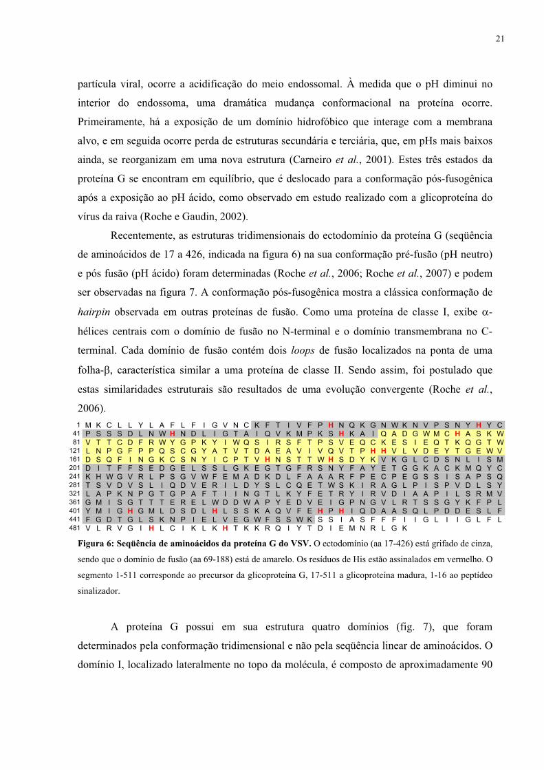
Figura 6: Seqüência de aminoácidos da proteína G do VSV. O ectodomínio (aa 17-426) está grifado de cinza,
sendo que o domínio de fusão (aa 69-188) está de amarelo. Os resíduos de His estão assinalados em vermelho. O
segmento 1-511 corresponde ao precursor da glicoproteína G, 17-511 a glicoproteína madura, 1-16 ao peptídeo
sinalizador.
A proteína G possui em sua estrutura quatro domínios (fig. 7), que foram
determinados pela conformação tridimensional e não pela seqüência linear de aminoácidos. O
domínio I, localizado lateralmente no topo da molécula, é composto de aproximadamente 90
1 M K C L L Y L A F L F I G V N C K F T I V F P H N Q K G N W K N V P S N Y H Y C41 P S S S D L N W H N D L I G T A I Q V K M P K S H K A I Q A D G W M C H A S K W81 V T T C D F R W Y G P K Y I W Q S I R S F T P S V E Q C K E S I E Q T K Q G T W
121 L N P G F P P Q S C G Y A T V T D A E A V I V Q V T P H H V L V D E Y T G E W V161 D S Q F I N G K C S N Y I C P T V H N S T T W H S D Y K V K G L C D S N L I S M201 D I T F F S E D G E L S S L G K E G T G F R S N Y F A Y E T G G K A C K M Q Y C241 K H W G V R L P S G V W F E M A D K D L F A A A R F P E C P E G S S I S A P S Q281 T S V D V S L I Q D V E R I L D Y S L C Q E T W S K I R A G L P I S P V D L S Y321 L A P K N P G T G P A F T I I N G T L K Y F E T R Y I R V D I A A P I L S R M V361 G M I S G T T T E R E L W D D W A P Y E D V E I G P N G V L R T S S G Y K F P L401 Y M I G H G M L D S D L H L S S K A Q V F E H P H I Q D A A S Q L P D D E S L F441 F G D T G L S K N P I E L V E G W F S S W K S S I A S F F F I I G L I I G L F L481 V L R V G I L C I K L K T K K R Q I Y T D I E M N R L G K H H
21

resíduos em dois segmentos (resíduos 17-33 e 326-398)1, principalmente organizados em
folhas-�. O domínio II, localizado na região central no topo da molécula, é composto de três
segmentos (resíduos 34-51, 275-325 e 399-421), principalmente estruturado em �-hélices.
Este domínio está envolvido na formação do trímero. O domínio III é composto de dois
segmentos (resíduos 52-62 e 197-274), que estão inseridos no domínio II. Possui duas �-
hélices e duas folhas-�. O domínio IV (resíduos 69-188) está inserido num loop do domínio
III. É uma estrutura composta de folhas-�, contendo dois loops onde são encontrados resíduos
aromáticos (W88, Y89, Y132, A133).
Pré-fusão Pós-fusão Pós-fusão
Pré-fusão
A B
C
Pré-fusão Pós-fusão Pós-fusão
Pré-fusão
Pré-fusão Pós-fusão Pós-fusão
Pré-fusão
A B
C
Figura 7: Estrutura cristalográfica da proteína G do VSV nas conformações pré e pós fusogênica. (A)
Visão do monômero. (B) Visão do trímero. (C) Diagrama linear mostrando os resíduos de aminoácidos
correspondentes a cada domínio. Loops de fusão em verde escuro, domínio I em vermelho, domínio II em azul,
domínio III em laranja, domínio IV em amarelo e porção C-terminal em rosa. Os números 1 e 2 representam os
sítios de glicosilação. Cter indica a porção C-terminal e Nter a porção N-terminal. Extraído de Roche et al.
(2007).
O domínio IV é denominado domínio de fusão, visto que a presença de uma grande
quantidade de resíduos aromáticos por trímero é provavelmente capaz de desestabilizar a
membrana, iniciando o processo de fusão (Roche et al., 2006). Esta idéia é reforçada por
antigos experimentos de substituições de aminoácidos em regiões do ectodomínio da proteína
1 A numeração usada nesta tese inclui os 16 resíduos que contituem o peptídeo sinal, ao contrário da numeração usada nos artigos de Roche et al, 2006 e 2007, que começou a numeração no resíduo 17.
22

G que eram utilizados como estratégia de localização do peptídeo de fusão do VSV. Mutações
em resíduos presentes na região 117-137, que são altamente conservadas nos vesiculovírus,
aboliram a fusão de membranas mediada pela glicoproteína, sugerindo que esta região
continha o domínio de fusão (Zhang e Ghosh, 1994; Fredericksen e Whitt, 1995). Além disso,
já havia sido demonstrado que o segmento contendo os aminoácidos 59-221 tornava-se capaz
de interagir com membranas durante as transições conformacionais decorrentes da
acidificação (Durrer et al., 1995).
Outras regiões já foram previamente relacionadas com a fusão de membranas
do está
ulas de
catalisadas pelo VSV, estando indiretamente envolvidas com o processo. Este é o caso do
segmento contendo os aminoácidos 395-418 (Shokralla et al., 1998) e do domínio
transmembrana (Odell et al., 1997; Cleverley e Lenard, 1998; Jeetendra et al., 2003).
A reorganização da proteína G que ocorre após a exposição ao pH áci
representada na figura 8 (Roche et al., 2007). Resumidamente, ocorre inicialmente a projeção
do domínio de fusão para o topo da molécula pela conjugação de dois movimentos: rotação do
domínio de fusão (IV) em relação ao domínio III e reposicionamento do domínio III no topo
do domínio II, permitindo a interação inicial dos loops de fusão com a membrana alvo (fig.
8C). Em seguida, ocorre a reestruturação do domínio II, fazendo com que ocorra uma
movimentação da porção C-terminal para a mesma extremidade em que se encontram os
loops de fusão, acarretando na aproximação das membranas viral e endossomal. Sendo assim,
apesar de haver uma grande mudança conformacional da proteína G, somente o domínio II
sofre uma grande reestruturação (fig. 8B), pois os domínios I, III e IV mantêm sua estrutura
inicial, sofrendo apenas uma reorientação através de movimentos de rotação (fig. 8A).
As características e a termodinâmica da interação entre a proteína G e vesíc
diferentes composições lipídicas foram estudadas previamente por nós através do uso da
microscopia de força atômica, calorimetria e espectroscopia de fluorescência (Carneiro et al.,
2002) (anexo A). Mostramos que, em diferentes etapas do ciclo de infecção do vírus, as
interações entre a proteína viral e membranas são interações de natureza eletrostática
envolvendo regiões da proteína carregadas positivamente e fosfolipídios carregados
negativamente, mais especificamente a fosfatidilserina (PS) (Carneiro et al., 2002; Da Poian
et al., 2005; Carneiro et al., 2006) (anexos A, B e C). Estudos prévios chegaram a especular
que este fosfolipídio poderia ser o receptor celular para o VSV (Schlegel et al., 1983). No
entanto, já foi demonstrado que o VSV é capaz infectar células independente da presença de
PS, mostrando que este fosfolipídio não é o receptor do VSV, podendo talvez funcionar como
23

co-receptor (Coil e Miller, 2004). Até o presente momento, o receptor específico para o VSV
na membrana celular ainda não foi definido.
Reorientação dos domínios III e IV
Pré-fusão Pós-fusão Pré-fusão Pós-fusão
Reestruturação do domínio IIA B
C
Reorientação dos domínios III e IV
Pré-fusão Pós-fusão Pré-fusão Pós-fusão
Reestruturação do domínio IIReorientação dos domínios III e IV
Pré-fusão Pós-fusão Pré-fusão Pós-fusão
Reestruturação do domínio IIA B
C
Figura 8: Mudanças estruturais da proteína G do VSV ocorridas na transição das conformações pré e
pós-fusogênicas. (A) Rearranjo da orientação dos domínios III e IV. (B) Reestruturação do domínio II. (C)
Representação esquemática da mudança conformacional. Domínio I em vermelho, domínio II em azul escuro e
diferentes tonalidades de verde, domínio III em laranja, domínio IV em amarelo, loops de fusão em verde,
porção C-terminal em rosa. Adaptado de Roche et al. (2007).
Vários estudos a respeito do efeito do pH sobre a proteína G do rabdovírus mostraram
que as mudanças estruturais sofridas por esta glicoproteína durante a acidificação assim como
a aquisição da capacidade de interagir com membranas negativamente carregadas ocorrem em
uma faixa de pH bastante estreita, entre 6,2 e 5,8 (White et al., 1981; Gaudin, 2000; Carneiro
et al., 2001). Esta é exatamente a faixa de pH na qual ocorre a protonação da cadeia lateral do
aminoácido histidina (pK ~ 6,04). Isso nos sugeriu que a protonação de resíduos de histidina
presentes na proteína G seria necessária para o desencadeamento do processo de fusão. Por
esse motivo, a substância dietilpirocarbonato (DEPC), cujo mecanismo de modificação
24

específica de histidinas vem sendo estudado há anos, foi utilizada nos estudos apresentados
nesta tese para descoberta de possíveis alvos de inativação viral durante a entrada do VSV nas
células hospedeiras.
1.4 Modelo de estudo II: o vírus da dengue
O vírus da dengue é um membro da família Flaviviridae, na qual também se incluem
os vírus da febre amarela, Saint Louis, Oeste do Nilo, Rio Bravo e outros vírus responsáveis
por encefalites (Burke e Monath, 2001). Quatro sorotipos geneticamente distintos do vírus da
dengue são conhecidos: DEN1, DEN2, DEN3 e DEN4, sendo DEN2 o de maior prevalência.
A infecção por este vírus possui um espectro de apresentação que varia desde a forma
assintomática até quadros de hemorragia e choque, podendo evoluir, inclusive para o óbito
(fig. 9) (Who, 1997; Figueiredo e Fonseca, 2002; Guzman e Kouri, 2002; Whitehead et al.,
2007). A dengue é uma doença febril aguda, com duração de 5 a 7 dias. A dengue clássica
apresenta quadro clínico muito variável, geralmente com cefaléia, mialgia (dores no corpo),
seguido de febre alta (39° a 40°) de início abrupto, acompanhada de prostração, artralgia,
anorexia, astenia, dor retro-orbitária, náuseas, vômitos e rash cutâneo. Associada à síndrome
febril, em alguns casos pode ocorrer hepatomegalia dolorosa e, principalmente, nas crianças,
dor abdominal generalizada. Com o desaparecimento da febre, há regressão dos sinais e
sintomas, podendo ainda persistir a fadiga (Figueiredo e Fonseca, 2002; Guzman e Kouri,
2002). Já nos casos da dengue hemorrágica, apesar dos sintomas iniciais serem semelhantes
aos da dengue clássica, ocorre um rápido aparecimento de manifestações hemorrágicas, como
petéquias, equimoses, epistaxe, gengivorragia, sangramento gastrintestinal, hematúria e
metrorragia. Os casos típicos da dengue hemorrágica são caracterizados por febre alta,
fenômenos hemorrágicos e hepatomegalia. Nos casos graves, conhecidos como síndrome do
choque hemorrágico da dengue, ocorre insuficiência circulatória (choque) imediatamente após
o desaparecimento da febre. Sua duração é curta, podendo levar ao óbito em 12 a 24 horas ou
à recuperação rápida após terapia apropriada. Um achado laboratorial importante da dengue
hemorrágica é a trombocitopenia com hemoconcentração concomitante. A gravidade deste
quadro está relacionada à efusão do plasma, caracterizada por valores crescentes do
hematócrito (Figueiredo e Fonseca, 2002; Guzman e Kouri, 2002).
O vírus da dengue é transmitido em um ciclo envolvendo humanos e mosquitos, sendo
Aedes aegypti o vetor mais importante. Este se encontra principalmente em áreas temperadas
e tropicais, em geral durante o verão (Wilder-Smith e Schwartz, 2005). Cerca de dois terços
da população mundial vive em áreas onde a dengue é endêmica ou epidêmica, sendo estimado
25

que cerca de 50-100 milhões de pessoas sejam infectadas com o vírus todo o ano, 250-500 mil
desenvolvam dengue hemorrágica e 12 mil morram em decorrência desta infecção (Rigau-
Perez et al., 1998; Gubler, 2002).
Dias após a infecção
Sina
is e
sin
tom
as c
línic
os
Viremia
Dores no corpo e cefaléia
Febre
Rash cutâneo
Trombocitopenia
Choque
Petéquias e equimoses
Dias após a infecção
Sina
is e
sin
tom
as c
línic
os
Viremia
Dores no corpo e cefaléia
Febre
Rash cutâneo
Trombocitopenia
Choque
Petéquias e equimoses
Figura 9: Representação esquemática do curso temporal de aparecimento dos sintomas e sinais clínicos da
dengue. DF - dengue clássica, DHF - dengue hemorrágica, DSS - síndrome do choque da dengue hemorrágica.
Adaptado de Whitehead et al. (2007).
No Brasil, a dengue encontra-se hoje presente em todos os 27 estados da federação,
sendo responsável por cerca de 60-70% das notificações nas Américas (Siqueira et al., 2005;
Camara et al., 2007). Em 1981, os sorotipos DEN1 e DEN4 foram os primeiros a serem
isolados em uma epidemia de dengue ocorrida Boa Vista, Estado de Roraima (Osanai et al.,
1983). Após um silêncio epidemiológico, o sorotipo DEN1 invadiu o sudeste (Rio de Janeiro
e Minas Gerais) e o nordeste (Alagoas, Ceará, Pernambuco, Bahia) em 1986-1987
(Schatzmayr et al., 1986), espalhando-se pelo país desde então, com as entradas dos sorotipos
DEN2 em 1990-1991 (Nogueira et al., 1991), e o DEN3 em 2001-2002 (Nogueira et al.,
2001; Nogueira et al., 2005).
Dois fatores estão diretamente relacionados com a incidência de dengue no Brasil e no
mundo: distribuição ampla do vetor e taxa de transmissão rápida do vírus (Pinheiro e Corber,
1997; Figueroa e Ramos, 2000; Guzman e Kouri, 2002). O controle do vetor, que a princípio
parecia ser a melhor solução, mostrou-se ineficiente no Brasil nos últimos anos, tanto pela
interrupção dos programas de combate ao mosquito, quanto pelo aparecimento de mosquitos e
larvas resistentes a diversos inseticidas e larvicidas. Além disso, tais programas para serem
efetivos exigem financiamento contínuo ao longo dos anos, principalmente em um país de
dimensões como a do Brasil (Teixeira et al., 2005).
26
Sendo assim, a formulação de uma vacina tetravalente que seja eficaz contra os quatro
sorotipos e produza uma imunidade duradoura é extremamente necessária. Já existem vacinas
de vírus atenuado, inativado (partícula inteira e subunidade) e vacina de DNA que estão sendo
desenvolvidas, algumas delas já em testes clínicos (Whitehead et al., 2007).
1.4.1 A Estrutura dos Flavivírus
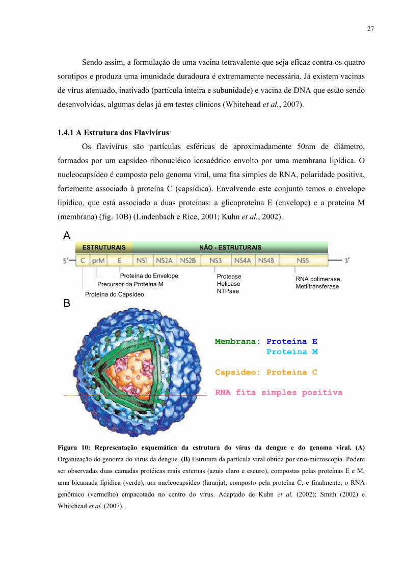
Os flavivírus são partículas esféricas de aproximadamente 50nm de diâmetro,
formados por um capsídeo ribonucléico icosaédrico envolto por uma membrana lipídica. O
nucleocapsídeo é composto pelo genoma viral, uma fita simples de RNA, polaridade positiva,
fortemente associado à proteína C (capsídica). Envolvendo este conjunto temos o envelope
lipídico, que está associado a duas proteínas: a glicoproteína E (envelope) e a proteína M
(membrana) (fig. 10B) (Lindenbach e Rice, 2001; Kuhn et al., 2002).
Membrana: Proteína EProteína M
Capsídeo: Proteína CRNA fita simples positiva
Proteína do Capsídeo
Precursor da Proteína MProteína do Envelope Protease
HelicaseNTPase
RNA polimeraseMetiltransferase
ESTRUTURAIS NÃO - ESTRUTURAISA
B
Membrana: Proteína EProteína M
Capsídeo: Proteína CRNA fita simples positiva
Proteína do Capsídeo
Precursor da Proteína MProteína do Envelope Protease
HelicaseNTPase
RNA polimeraseMetiltransferase
ESTRUTURAIS NÃO - ESTRUTURAIS
Membrana: Proteína EProteína M
Capsídeo: Proteína CRNA fita simples positiva
Proteína do Capsídeo
Precursor da Proteína MProteína do Envelope Protease
HelicaseNTPase
RNA polimeraseMetiltransferase
ESTRUTURAIS NÃO - ESTRUTURAISA
B
Figura 10: Representação esquemática da estrutura do vírus da dengue e do genoma viral. (A)
Organização do genoma do vírus da dengue. (B) Estrutura da partícula viral obtida por crio-microscopia. Podem
ser observadas duas camadas protéicas mais externas (azuis claro e escuro), compostas pelas proteínas E e M,
uma bicamada lipídica (verde), um nucleocapsídeo (laranja), composto pela proteína C, e finalmente, o RNA
genômico (vermelho) empacotado no centro do vírus. Adaptado de Kuhn et al. (2002); Smith (2002) e
Whitehead et al. (2007).
27

1.4.2 Ciclo de replicação dos Flavivírus
Para que ocorra uma replicação viral bem sucedida, os flavivírus precisam liberar seu
nucleocapsídeo no citoplasma (fig. 11). Para isso, o vírus da dengue se liga aos receptores
presente na superfície celular através da proteína E. Existem estudos que sugerem o
envolvimento direto do heparan sulfato (HS), um glicosaminoglicano (GAG) carregado
negativamente, na ligação do vírus à membrana alvo (Chen et al., 1997; Hung et al., 1999;
Hilgard e Stockert, 2000; Germi et al., 2002). No entanto, outros autores acreditam que o HS
é apenas um fator presente na superfície celular que agrega as partículas virais, facilitando sua
interação com seus co-receptores protéicos (Martinez-Barragan e Del Angel, 2001; Thepparit
e Smith, 2004). Além disso, dependendo do tipo celular estudado, o HS nem é necessário para
infecção pelo vírus da dengue (Bielefeldt-Ohmann et al., 2001). Na realidade, não se conhece
o verdadeiro receptor do vírus da dengue e várias outras moléculas já foram envolvidas até o
presente momento, como as “heat shock proteins” 90 e 70 (Reyes-Del Valle et al., 2005),
GRP78/BiP (glucose-regulating protein 78) (Jindadamrongwech et al., 2004), CD-14 (Chen et
al., 1999), receptor de laminina (Thepparit e Smith, 2004) e, mais recentemente, o DC-SIGN
(dendritic-cell-specific ICAM-grabbing non-integrin) (Navarro-Sanchez et al., 2003;
Tassaneetrithep et al., 2003; Lozach et al., 2005; Pokidysheva et al., 2006).
Após a ligação ao receptor específico, ocorre a endocitose da partícula viral
(Mukhopadhyay et al., 2005). A acidificação do compartimento endossomal ocasiona a fusão
do envelope viral com a membrana endossomal, liberando o capsídeo no citoplasma. A
proteína do capsídeo se dissocia, iniciando o processo de tradução e replicação do genoma
viral. O genoma do vírus da dengue (fig. 10A) codifica uma poliproteína processada pela
combinação de serino-proteases virais e enzimas celulares. Como resultado desta clivagem,
são obtidas três proteínas estruturais (C, prM, and E) e sete não-estruturais (NS1, NS2A,
NS2B, NS3, NS4A, NS4B, NS5) (Lindenbach e Rice, 2001; Lindenbach e Rice, 2003). Assim
são formadas as proteínas virais necessárias para a montagem viral.
O processo de montagem dos vírus acontece inicialmente no lúmen do retículo
endoplasmático, onde primeiro são gerados vírus imaturos, compostos de nucleocapsídeo,
envelope e proteínas E e pré-M formando um estável complexo de heterodímeros, que não é
capaz de induzir fusão de membranas (Guirakhoo et al., 1991). Acredita-se que a proteína
pré-M protege a proteína E de adquirir precocemente uma conformação fusogênica durante o
processo de secreção e também funcione como uma chaperona para a organização desta
proteína (Heinz e Allison, 2003). Após a clivagem da proteína pré-M pela furina celular no
28

complexo de Golgi, as partículas virais se tornam maduras e infectantes (Stadler et al., 1997;
Elshuber et al., 2003). Os vírus formados são liberados da célula infectada por exocitose.
Endocitose
Fusão de Membranas
Tradução da poliproteína com processamento
norRetículo endoplasmático
Replicação do genoma viral
Montagem viral Complexo de Golgi
Trans-Golgi
Maturação viral
Exocitosede novas partículas
Endocitose
Fusão de Membranas
Tradução da poliproteína com processamento
norRetículo endoplasmático
Replicação do genoma viral
Montagem viral Complexo de Golgi
Trans-Golgi
Maturação viral
Exocitosede novas partículas
Figura 11: Ciclo de replicação do vírus da dengue. Estão representadas as principais etapas do ciclo de
replicação: ligação do vírus à membrana celular através da ligação da proteína E do vírus com os receptores da
superfície celular, endocitose das partículas virais mediada pelo receptor da célula do hospedeiro, acidificação do
meio endossomal induzindo mudança conformacional irreversível da proteína do envelope viral e promovendo a
fusão com a membrana endossomal, liberação do capsídeo no citoplasma da célula, tradução do RNA genômico
e processamento da poliproteína, replicação do genoma viral originando fitas de RNA intermediárias negativas,
morfogênese dos virions em vesículas intracelulares, transporte dos virions e maturação da glicoproteína do
envelope, fusão da vesícula contendo os virions com a membrana da célula e liberação destes no meio
extracelular. Adaptado de Lindenbach e Rice (2001) e Mukhopadhyay et al. (2005).
1.4.3 Proteína E e seu peptídeo de fusão
A proteína E é a maior proteína estrutural do vírus da dengue, sendo responsável pela
ligação do vírus à célula hospedeira e pela fusão de membranas, etapa essencial para entrada
do vírus na célula hospedeira (Mukhopadhyay et al., 2005; Clyde et al., 2006). Esta
glicoproteína é composta de três “barris de folhas-�” (fig. 12): o domínio I contém a
extremidade N-terminal; o domínio II contém uma região de dimerização e o peptídeo de
29

fusão em sua extremidade distal; e o domínio III contém os sítios de ligação ao receptor
celular (Modis et al., 2003). A proteína E encontra-se na forma de homodímeros dispostos
paralelamente à superfície viral em pH neutro, com seu peptídeo de fusão inacessível ao meio
externo (fig. 12A) (Modis et al., 2003).
A proteína E é classificada como uma proteína de fusão de classe II, devido a sua
estrutura ser formada principalmente por folhas-� e de seu peptídeo de fusão estar localizado
no meio da seqüência da proteína (Modis et al., 2004).
A
C
D
BA
C
D
B
Figura 12: Estrutura cristalográfica da proteína E do vírus da dengue. (A) Conformação pré-fusogênica -
Visão do homodímero. (B) Conformação pós fusogênica - Visão do trímero. (C) Diagrama linear mostrando os
resíduos de aminoácidos correspondentes a cada domínio. (D) Maior detalhe da ponta do trímero, onde está
localizado o loop de fusão, mostrando a presença dos aminoácidos hidrofóbicos. Domínio I em vermelho,
domínio II em amarelo e domínio III em azul. O peptídeo de fusão (aa 98-110) está marcado pela letra C. A letra
N marca o sítio de glicosilação. O triângulo marca o loop de ligação ao receptor cellular. Adaptado de Modis et
al. (2004).
A reorganização estrutural necessária para aquisição da conformação fusogênica é
desencadeada pela exposição da proteína E ao pH ácido e se inicia pelo rearranjo das
orientações relativas dos domínios da proteína E (fig. 13) (Modis et al., 2004). O domínio II
gira aproximadamente 30o em relação ao domínio I. Já o domínio III sofre um maior
deslocamento na transição de dímero para trímero, pois gira cerca de 70o e desloca seu centro
30

de massa 36 Å à frente do domínio II, fazendo com que ele não se encontre mais estendido
linearmente junto com os domínios I e II. Estas reorientações são responsáveis pela formação
dos trímeros, exposição do peptídeo de fusão e formação da haste de hemifusão (explicada
anteriormente). O processo continua com a reestruturação do domínio III, que aproxima o
peptídeo de fusão e a porção transmembrana, ou seja, as membranas viral e endossomal. Com
isso é formado o poro de fusão, permitindo a liberação do nucleocapsídeo no citoplasma.
Resumidamente, quando o pH é acidificado, ocorre a conversão irreversível dos homodímeros
para homotrímeros (Allison et al., 1995; Stiasny et al., 1996; Kuhn et al., 2002), com
exposição do peptídeo de fusão no topo do trímero e conseqüente início da reação de fusão
(Mukhopadhyay et al., 2005). O peptídeo de fusão durante toda a reestruturação da proteína E
se mantém com a mesma conformação (Modis et al., 2004).
A BA B
Figura 13: Rearranjo dos domínios da proteína E durante a transição para trímero. (A) Estrutura do
monômero na conformação pré-fusogênica, encontrada nos dímeros de proteína E em pH neutro. (B) Estrutura
do monômero na conformação pós-fusogênica, encontrada nos trímeros após exposição ao pH ácido. Ocorre um
rearranjo dos domínios II e III (demonstrado pelas setas em A), que mudam sua orientação relativa, fazendo com
que o C-terminal se aproxime do peptideo de fusão, ficando 39 Å mais perto. O peptídeo de fusão permanece
essencialmente com a mesma conformação antes e depois da fusão. Adaptado de Modis et al. (2004).
Observa-se no alinhamento das seqüências de aminoácidos das proteínas E dos
diferentes flavivírus uma homologia de 62 a 77% entre os quatro sorotipos de vírus da dengue
e de 40 a 45% entre os diferentes flavivírus. A região entre os resíduos 98 e 110 é idêntica em
todos os flavivírus, exceto por uma única substituição no vírus tick born encephalitis (TBE)
(fig. 14). Esta região é considerada o peptídeo de fusão, visto que possui grande homologia
entre os flavivírus e encontra-se exposta nos homotrímeros de proteína E (fig. 12C e D). Além
disso, substituições de aminoácidos nesta região foram capazes de abolir a fusão de
membranas mediada pela proteína E (Allison et al., 2001).
31

DENGUE1S
DE NGUE2NDE NGUE4WEST NILEJ AP. ENCEPHAL ITISYE LL OW FEVERTICK-BORNE ENCEPH.
TTTTSSKK
|70
TTTTTTIV
DDDARVNA
SSSTAADA
RRRRARKR
CCCCCCCC
PPPPPPPP
TTTTTTST
QQQQMTTM
GGGGGGGG
EEEEEEEP
|80
AAPPAAAA
TISYHHHT
LLLLNNLL
VPNKEEAA
EEEEKKEE
EEEERREE
QQQQAANH
DDDDDDEQ
AQKQPSGG
NNRQASDG
|90
FYFYFYNT
VVVIVVAV
CCCCCCCC
RKKRKKKK
RHHRQQRR
TTSDGGTD
FYMVVFYQ
VVVVVTSS
DDDDDDDD
RRRRRRRR
|100
GGGGGGGG
WWWWWWWW
GGGGGGGG
NNNNNNNN
GGGGGGGH
CCCCCCCC
GGGGGGGG
LLLLLLLL
FFFFFFFF
GGGGGGGG
|110
KKKKKKKK
GGGGGGGG
SSGGSSSS
LLIVIIII
LVVVDDVV
TTTTTTAA
CCCCCCCC
AAAAAAAV
KKMKKKKK
FFFFFFFA
|120
KQTSASTA
CCCCCCCC
VLKSTTAE
TEKGTSKA
KSNKKKSK
.
.
.
.
.
.
.K
.
.
.
.
.
.
.K
LIMIAAMA
EEKTTIST
GGGGGGLG
|130
KKKNWRFH
IVVLITEV
VVVVIIVY
QQQRQQDD
YHPIKPQA
EEEEEETN
NNNNNNKK
DE NGUE3
Figura 14: Alinhamento da região do peptídeo de fusão da glicoproteína E de alguns flavivírus. Dengue
tipo 1 (cepa Singapura S275/90); dengue tipo 3; dengue tipo 2 (Nova Guiné C); dengue tipo 4; vírus do Nilo do
Oeste; encefalite japonesa (cepa Nakayama); febre amarela (cepa 17D) e encefalite transmitida por carrapato
(cepa Neudoerfl).
Através do uso de metodologias baseadas em espectroscopia de fluorescência, nesta
tese foi realizado o primeiro estudo da interação do peptídeo de fusão do vírus da dengue com
membranas.
1.5 Mecanismos de inativação viral
O desenvolvimento de novas estratégias para inativação viral representa uma
importante linha de pesquisa na área da virologia, visto que a disponibilidade de
medicamentos antivirais com eficácia clínica comprovada é reduzida (De Clercq, 2004) e que
existe carência de vacinas seguras e eficazes contra diversas viroses.
A identificação de uma série de proteínas e enzimas virais essenciais para a replicação
dos vírus e suficientemente diferentes das proteínas celulares viabilizou o desenvolvimento de
drogas direcionadas exclusivamente a alvos virais que seriam, em princípio, inofensivas às
proteínas celulares. A maioria destas drogas inibe enzimas envolvidas na replicação viral,
como é o caso, por exemplo, do aciclovir, que após processamento intracelular se torna um
potente inibidor da DNA polimerase do vírus do herpes (Crumpacker et al., 1979); ou dos
inibidores nucleosídicos e não-nucleosídicos da transcriptase reversa dos retrovírus (Autran et
al., 1997).
Embora a inibição das enzimas envolvidas na replicação viral seja uma abordagem
muito eficaz para o desenvolvimento de drogas antivirais, esta estratégia não tem utilidade
para a formulação de vacinas. Com este intuito, foram desenvolvidas técnicas de inativação
viral que modificavam a partícula viral, bloqueando sua entrada nas células hospedeiras na
etapa da adsorção. Os compostos mais utilizados eram a formalina (Bachmann et al., 1993) e
detergentes (Seitz et al., 2002), sendo também usada a aplicação de radiação ultra-violeta
(UV) (Bay e Reichmann, 1979). No entanto, estes procedimentos de inativação acarretam na
desnaturação das proteínas de superfície viral, comprometendo a imunogenicidade da maioria
32

das partículas virais, ou seja, a indução de imunoglobulinas do tipo G (IgG) neutralizantes
(Bachmann et al., 1994). Por isso, nos últimos 50 anos foram realizados diversos estudos de
inativação viral para formulação de vacinas, com a descoberta de novos compostos e
procedimentos. Uma revisão mais detalhada destas abordagens foi recentemente publicada
por nós e se encontra anexada ao final desta seção (artigo 1).
Dentre os compostos descobertos nos últimos anos, destacam-se aqueles inativadores
virais que impedem a entrada dos vírus envelopados nas células hospedeiras pela inibição da
fusão de membranas: cianovirina (Dey et al., 2000), hipericina e rosa de bengala (Lenard et
al., 1993; Lenard e Vanderoef, 1993). O mecanismo de ação dos dois últimos compostos
químicos está relacionado com a formação de cross-linking das proteínas virais de superfície.
Já o mecanismo molecular de ação da cianovirina foi muito estudado para o HIV e envolve
interações físicas com a proteína de membrana gp120 (Boyd et al., 1997). Estudos
demonstraram que esta molécula se liga aos oligossacarídeos com alta concentração de
manose presentes na gp120 (Bewley e Otero-Quintero, 2001; Bolmstedt et al., 2001; Shenoy
et al., 2001). Outros vírus envelopados que contém oligosacarídeos similares também foram
inativados pela cianovirina (Dey et al., 2000; Barrientos et al., 2003; O'keefe et al., 2003;
Helle et al., 2006). As propriedades antigênicas e imunogênicas das partículas virais
inativadas por estes compostos ainda não foram avaliadas.
1.5.1 Inativação do VSV
O VSV pode ser inativado por tratamento com formalina, com �-propiolactona ou
com luz UV (Bachmann et al., 1993; Bachmann et al., 1994). Nestes casos, o vírus inativado
não foi capaz de induzir a produção de IgG em animais imunizados, embora a resposta de
IgM tenha se mantido inalterada. Em relação à reposta citotóxica, apenas o vírus inativado por
luz UV foi capaz suscitar resposta de linfócitos T citotóxicos (Bachmann et al., 1994).
Além desses métodos usuais, já foram utilizados detergentes (Seitz et al., 2002), bis-
ANS (Bonafe et al., 2000), hipericina e rosa de bengala (Lenard et al., 1993). Também podem
ser citados os métodos físicos, como pressão hidrostática (Silva et al., 1992). A fotoinativação
ainda é utilizada frequentemente associada com diversos compostos químicos (Hirayama et
al., 1997; Kasermann e Kempf, 1997; Kasermann e Kempf, 1998; Hirayama et al., 1999; Lim
et al., 2002). No entanto, nestes estudos de inativação do VSV não foram avaliadas as
capacidades antigênica e imunogênica das partículas virais inativadas, visto que muitas destas
metodologias são utilizadas principalmente para inativação em produtos sanguíneos e
processos de desinfecção, não visando a obtenção de antígenos para formulação de vacinas.
33
Técnicas mais sofisticadas como o rearranjo genético do VSV já foram apontadas
como uma forma eficiente de atenuação viral (Wertz et al., 1998; Flanagan et al., 2001). O
afastamento do gene da proteína N da região promotora reduziu os níveis de transcrição e da
síntese da proteína N com conseqüente atenuação da replicação do vírus e de sua letalidade
para camundongos, preservando sua capacidade imunogênica (Wertz et al., 1998).
1.5.2 Dietilpirocarbonato
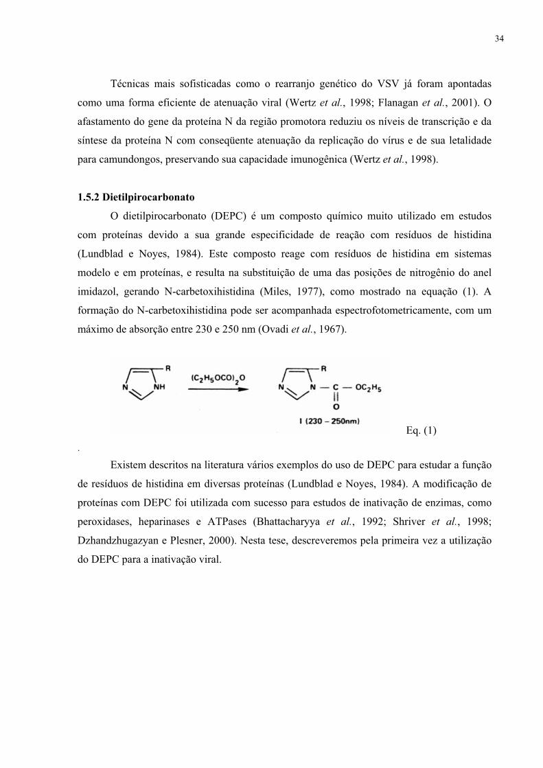
O dietilpirocarbonato (DEPC) é um composto químico muito utilizado em estudos
com proteínas devido a sua grande especificidade de reação com resíduos de histidina
(Lundblad e Noyes, 1984). Este composto reage com resíduos de histidina em sistemas
modelo e em proteínas, e resulta na substituição de uma das posições de nitrogênio do anel
imidazol, gerando N-carbetoxihistidina (Miles, 1977), como mostrado na equação (1). A
formação do N-carbetoxihistidina pode ser acompanhada espectrofotometricamente, com um
máximo de absorção entre 230 e 250 nm (Ovadi et al., 1967).
Eq. (1) .
Existem descritos na literatura vários exemplos do uso de DEPC para estudar a função
de resíduos de histidina em diversas proteínas (Lundblad e Noyes, 1984). A modificação de
proteínas com DEPC foi utilizada com sucesso para estudos de inativação de enzimas, como
peroxidases, heparinases e ATPases (Bhattacharyya et al., 1992; Shriver et al., 1998;
Dzhandzhugazyan e Plesner, 2000). Nesta tese, descreveremos pela primeira vez a utilização
do DEPC para a inativação viral.
34

1.6 Artigo 1
Advances in the Development of Inactivated Virus
Vaccines Fausto Stauffer, Tatiana El-Bacha e Andrea T. Da Poian
Publicado em 2006 no periódico
Recent Patents on Anti-Infective Drug Discovery

Recent Patents on Anti-Infective Drug Discovery, 2006, 1, 000-000 1
1574-891X/06 $100.00+.00 © 2006 Bentham Science Publishers Ltd.
Advances in the Development of Inactivated Virus Vaccines
Fausto Stauffer, Tatiana El-Bacha and Andrea T. Da Poian*
Instituto de Bioquímica Médica, Programa de Biologia Molecular e Biotecnologia, Universidade Federal do Rio deJaneiro, Rio de Janeiro, RJ 21941-590, Brazil
Received: ???? ??, 2005; Accepted: ???? ??, 2006; Revised: March 24, 2006
Abstract: Vaccine discovery stands out as one of the public health interventions that has achieved the greatest impact inworld’s health. Vaccination is the most effective means of disease prevention, especially for viral infections. Starting withthe use of smallpox vaccine by Jenner in the late 1700s, the technology for vaccine development has seen numerousadvances. Currently, vaccines available for human viral illness are based on live attenuated (e.g. measles, mumps, andrubella), inactivated (e.g. hepatitis A) and recombinant (e.g. hepatitis B) viruses. Among these, inactivated vaccines areknown for their easy production and safety. The present article reviews the literature and patents related to themechanisms used for viral inactivation, mainly chemical and physical procedures, including the novel strategies that arecurrently being explored and that have been recently patent protected.
Keywords: Virus, viral inactivation, chemical methods, physical methods, vaccines.
INTRODUCTION
Vaccination is a valuable public health tool, being a safeand cost-effective strategy for controlling infectious diseases[1]. Progress in development and use of vaccines has led tothe decline and, in some cases, eradication of importantinfectious diseases, such as smallpox [2]. Traditionally,vaccines against viruses are classified as attenuated orinactivated. Live attenuated vaccines are based on theattenuation of the pathogen until its virulence is greatlydecreased but its immunogenicity is retained. Alternatively,inactivated vaccines consist of either whole killed virus orspecific viral proteins. In the case of whole-virus inactivatedvaccines, the inactivation treatment through differentprocesses (for review, see [3]) must ensure that all virusparticles are inactivated in order for the vaccine to be safe.Because such vaccines, also called dead vaccines, maycontain certain viral proteins or cellular components whichcould lead to undesirable immune responses in the host, theproduction of subunit vaccines, which contain specific viralproteins and retain immunogenicity properties, was of greatimportance in this field. Because only individual antigens areused in subunit vaccines, their immunogenicity is oftenreduced and the use of split vaccines, which contain acombination of viral proteins with preserved integrity, maybe more effective. More recently, new strategies for vaccinedevelopment have emerged, such as recombinant viralproteins [4] and DNA vaccines [5].
Despite the advances in immunology, molecular biologyand genetics, viral inactivation remains an importantprocedure in basic research, since it is an easy and relativelycheap approach to producing new and safe vaccines.Moreover, this procedure can also be used for otherpurposes, such as treatment of viral infections and clearance
*Address correspondence to this author at the Instituto de BioquímicaMédica, Programa de Biologia Molecular e Biotecnologia, UniversidadeFederal do Rio de Janeiro, Rio de Janeiro, RJ 21941-590, Brazil; Tel: +55-21-22706264; Fax: +55-21-22708647; E-mail: [email protected]
of viruses from plasma and plasma products, equipment andbiotechnology products. Since no inactivation method iseffective for all viruses and all purposes, a number ofdifferent inactivation treatments are being studied anddeveloped. In this review, we will focus on the proceduresavailable and their effectiveness in vaccine developmentagainst viruses.
There are several inactivation techniques that usechemical or physical procedures and have been cited inpatents (Table 1). However, only six whole-virus inactivatedvaccines are currently available for use in humans (Table 2)[3]. In these cases, the viruses are inactivated by chemicalmethods, using formalin or β-propiolactone. In addition tothe chemical treatments used for these established vaccines,other compounds are used for virus inactivation, includinganilinonaphthalene sulfonate compounds, urea, detergent,psoralens, aziridines, sodium periodate and diethylpyro-carbonate. The physical methods used for virus inactivation,such as UV irradiation, heat and high pressure, will also bereviewed.
CHEMICAL INACTIVATION OF VIRUSES
Concerning the chemical processes for virus inactivation,several compounds are used. Although aldehydes, such asformaldehyde, are the most common, there is a growing listof patented compounds and patented combinations of treat-ments which have proved to be effective in virus inactivationand thus may be suitable for vaccine development.
Inactivation using formalin (a saturated solution offormaldehyde) is usually performed by placing the virussuspension in contact with a formalin solution, at 37ºC, theconcentration depending on the type of virus. Theinactivation of viruses with formaldehyde involves a numberof reactions with amino, imino, amido, sulphydryl andhydroxy groups and with peptide linkages within proteins[6]. One of its principal advantages is that, as a result ofcross-linking, the gross three-dimensional architecture of
36

2 Recent Patents on Anti-Infective Drug Discovery, 2006, Vol. 1, No. 3 Da Poian et al.
Table 2. Inactivated Virus Vaccines Currently AvailableAgainst Human Diseases
Disease Inactivation Method
Flu β-propiolactone or formalin
Hepatitis A Formaldehyde/formalin
Japanese Encephalitis Formaldehyde/formalin
Poliomyelitis Formaldehyde/formalin
Rabies β-propiolactone or phenol
Tick-Borne Encephalitis Formaldehyde/formalin
proteins is conserved. This chemical compound has provedeffective for production of vaccines against hepatitis A [7,8], polio [9, 10], influenza [11], rabies [12], measles [13],Japanese encephalitis [14, 15] and immunodeficiency [16,17] viruses. A practical disadvantage of this method is thelong period of incubation required for successful virusinactivation. One solution to this problem is to increase theconcentration of formalin in the solution to reduceincubation time, although it may increase the toxicity of thevaccine. Another problem concerning this compound is thatformalin has proven to be reactogenic and it may beresponsible for the side effects experienced by some peopleafter vaccination. With respect to the preservation ofimmunogenicity, there is a clear association between theconcentration needed to inactivate the viruses and thecomplexity of the process of virus inactivation. Some viruses(e.g. SARS coronavirus and human immunodeficiency virustype 1, HIV-1) need lower concentrations of formaldehyde tobe inactivated, preserving their immunogenicity [18] andtheir neutralizing epitopes [19]. Conversely, it has beenshown that formalin strongly affects antigenic structure inconcentrations necessary to inactivate HIV-1 [20], poliovirus[21] and foot-and-mouth disease virus [22], making thevaccine not only hazardous but ineffective in protectingagainst infection, since dead vaccines are only effective ifthe structure of viral proteins is intact.
Another inactivating agent widely used in the productionof both human and animal virus vaccines is β-propiolactone(BPL). This chemical compound acts on the nucleic acid ofthe virus, with little or no effect on the immunogenic
properties of the viral protein coat [23]. One of the majoradvantages of this alkylating agent is that it undergoescomplete hydrolysis in an aqueous medium, forming non-toxic products [24]. It was shown that low concentrations ofBPL completely inactivate influenza [25] and rabies [12, 24,26] viruses without destroying their antigenic properties. Thecurrently available human vaccines using this compoundprotect against flu [27] and rabies [28] (Table 2). Thedisadvantages of this inactivating method are the elevatedcost of BPL and the risk of adverse reactions. These reac-tions have been associated with the presence of _-propio-lactone-altered human albumin in vaccine preparations (e.g.human diploid cell rabies vaccine) and the development ofimmunoglobulin E (IgE) antibodies to this allergen [29, 30].
Ethylenimine and related derivatives, such as binaryethylenimine (BEI) and acethylethylenimine, are also usedfor virus inactivation and have proven to be efficient for thedevelopment of vaccines against foot-and-mouth disease[31-33], polio [34], influenza [34] and rabies [35] viruses.Moreover, these compounds have also been shown toefficiently inactivate several RNA and DNA viruses [36].The aziridine BEI inactivates viruses by a mechanism similarto BPL, reacting with nucleic acids [31]. As with BPL, thereis no evidence that BEI reacts with viral proteins, and thus itpreserves the antigenic regions of the viruses [37, 38]. Incontrast to BPL, BEI has the advantage of low cost, and it isless hazardous to manipulate [31]. Moreover, the vaccineinactivated with BEI is very stable [35].
Although higher concentrations may decrease the timerequired for complete inactivation, this choice of conditionscan also compromise the safety of the invention.Additionally, aggressive treatment may also affect virusstructure and decrease vaccine efficiency. On the other hand,inactivation under mild conditions may favor vaccine safety.In this case, treatment of the viruses with psoralens, such as4’-aminomethyl 4,5’-8-trimethylpsoralen, in a non-oxidizingatmosphere (such as argon gas) along with UV irradiation,has proven effective in inactivating some viruses, such asbluetongue virus [39], HIV-1 and other immunodeficiencyviruses [40-43], rotavirus [44] and other single and double-stranded DNA or RNA viruses [45, 46]. The antigenicstructure of viral proteins is preserved after inactivation andthis compound leaves no harmful residue [47]. Themechanism of action is based on intercalation of the psoralenbetween the base pairs of double-stranded nucleic acids,forming covalent adducts to pyrimidine bases upon
Table 1. Inactivation Methods Used for Producing Virus Inactivated Vaccines
Chemical Physical Combined
Ascorbic AcidBeta Propiolactone
Beta-aminophenylketoneNon Ionic DetergentsDiethylpyrocarbonate
EthylenimineFormalin / Formaldehyde
Phenol
HeatPressure
UV Light
Ascorbic Acid + UV LightBeta Propiolactone + UV Light
Formalin + HeatFormalin + UV
Heat + Low PressurePressure + Heat or Cold
Psoralen + UV
37

Advances in the Development of Inactivated Virus Vaccines Recent Patents on Anti-Infective Drug Discovery, 2006, Vol. 1, No. 3 3
absorption of long-wave ultraviolet light [48, 49]. Thecovalently bonded psoralens act as inhibitors of DNAreplication and thus have the potential to stop the viralreplication process. Although this method seems suitable forvaccine preparation, it is still only used in viral inactivationof plasma products [50-52]. A great disadvantage of thismethodology is the requirement for special equipment wherethe inactivation process is performed.
Experiments with vaccinia virus revealed that ascorbicacid is a viral-inactivating agent when it undergoes auto-oxidation catalyzed by Cu++ [53]. Other studies have shownthat its main target may be nucleic acids, since RNAinfectivity is lost after ascorbic acid treatment [54]. Virusesthat have been inactivated by this compound includeparainfluenza-3 virus, transmissible gastroenteritis virus[55], poliovirus, coxsackie, echovirus [54], herpes simplexvirus types 1 and 2, cytomegalovirus, parainfluenza-2 virus[56], HIV-1 [57] and rabies virus [58]. This agent is lesshazardous than other commonly used chemicals, and it isinexpensive and effective for viral inactivation, allowing thevirus particles to retain their antigenicity [56].
Compounds such as β-aminophenylketones has beenshown to be effective in inactivating enveloped viruses [59].When several phenylketone compounds were tested over awide range of concentrations, dyclonine was most effective.As with formalin treatment, viral inactivation was achievedby placing the viruses in contact with the compound at 37ºC.Herpes simplex virus types 1 and 2 were readily inactivatedby this kind of treatment although picornaviruses andvaccinia viruses were more resistant in cell culture testing. Invivo protection from herpes simplex virus type 1 was alsoobserved.
A new approach to viral inactivation is the use ofdiethylpyrocarbonate (DEPC). This compound is a widelyused tool in chemical modification of proteins because of thehigh selectivity of the reagent for histidyl residues [60].Modification with DEPC was successfully used ininactivation studies of various groups of enzymes, e.g.peroxidases, heparinases and ATPases [61-63]. It has beenused for inactivation of measles, smallpox, polio and type Ainfluenza viruses [64]. More recently, our groupdemonstrated inactivation of vesicular stomatitis virus byDEPC, suggesting that this procedure can be used for otherenveloped viruses [65]. We were able to show that histidineresidues play a critical role in membrane fusion catalyzed byviral glycoproteins, and since DEPC modifies His residues,its mechanism of action seems to be based on inhibition ofthe fusion event, an essential step for the entry of envelopedviruses into host cells [66]. Besides providing an easyapproach to inactivation of enveloped viruses, DEPC-inactivated viruses retain their immunogenicity [65].
PHYSICAL INACTIVATION OF VIRUSES
Virus inactivation by heat must almost always be com-bined with another treatment, usually chemical, for success-ful results [8, 9, 20, 59, 67]. The use of a chemical comp-ound, although ensuring complete virus inactivation, mayincrease the toxicity of the vaccine. The temperature used forheat inactivation is around 37ºC. Higher temperatures havealso been used to eliminate the need for the combined use of
heat and an aggressive chemical treat-ment. When influenzavirus was submitted to temperatures ranging from 45 to 59ºCfor 25-180 minutes, it became at least 99% inactive [68]. Theresidual infectivity was inacti-vated with a small dose of anon-carcinogenic compound such as thiomersal incombination with ethyl ester. These two chemicals can alsobe combined with the virus during heat inactivation. Suchtreatment was said to be safe because it abolishes the use offormalin or other aggressive compounds and still evokeshumoral responses. One advantage of this invention incomparison with formalin treatment is the reducedincubation period required for inactivation. In addition, therewas also an increase in interferon levels in mice, which mayindicate the increased immunogenicity induced by suchtreatment. This vaccine proved to be effective for theimmunization of mice when administered intranasally orintraperitoneally.
Another commonly used procedure is ultra-violet (UV)irradiation. UV action is considered a physico-chemicalprocess, wherein covalent bonds of the cyclic molecules ofthe purine and pyrimidine bases are disrupted by theexcitation energy of the UV radiation, damaging the nucleicacids of viruses and other microorganisms. When the virus isexposed to effective UVC (100 to 280 nm) radiation, theinactivation process occurs within seconds [69]. However,this method changes protein structure by affecting disulfidebonds, thus destroying antigenic regions. Therefore, it isusually used in combination with other methods, aspreviously mentioned for heat treatment. Successfultreatments were achieved in association with psoralens [40-42, 45, 46], formalin [69] and β-propiolactone [70]. Onestudy with hepatitis A virus showed that UV treatment alonewas able to inactivate the virus, maintaining the antigenicregions, since the action of UV was predominantly on thenucleic acid [71]. According to the authors, the protocol usedwas simple, rapid, inexpensive and safe.
Another method used for virus inactivation is to treat thevirus with a mixture of a solvent (e.g. tri-n-butyl phosphate,TNBP) and a non-ionic detergent, such as sodiumdesoxycholate, Tween™ 80 or Triton-X-100, for hours attemperatures between 6 and 37ºC. This solvent/detergenttreatment [72] is widely used for virus inactivation duringthe manufacture of plasma-derived medicinal products. Theprocedure has been used for inactivation of HIV, hepatitis Band C viruses [73], influenza virus [74] and other envelopedviruses [75]. Moreover, it is effective in producing influenzasplit vaccines [76]. Besides the ability of non-ionicdetergents to disintegrate viral particles, it has been shownthat they solubilize membrane proteins under mildconditions, preserving the native structure of the protein andcreating a lipid-like environment that stabilizes thesolubilized proteins. The use of a detergent from the groupof polysorbates, such as Tween™ at concentrations of 10-20%, promoted the inactivation of several enveloped viruses,such as HIV-1 and HIV-2, tick-borne encephalitis, hepatitisC, influenza and herpes [20] viruses. A complete inactivationof HIV-1 was achieved in only 10 minutes of incubation at20-40ºC. Much longer incubation times (10 hours) werenecessary to inactivate only 0.03-0.1% of the same viruswith formalin, indicating the special advantage of the
38

4 Recent Patents on Anti-Infective Drug Discovery, 2006, Vol. 1, No. 3 Da Poian et al.
combined treatment with detergent and relatively lowtemperature.
The latest physical approach used for viral inactivation ishydrostatic pressure (HP). A study with vesicular stomatitisvirus was the first demonstration that HP is able to inactivatea membrane-enveloped virus [77]. Following this study,other viruses were shown to be inactivated by HP, such asherpes simplex virus, human cytomegalovirus [78], foot-and-mouth disease virus [79], parvovirus [80], influenza virus[81], HIV-1 [82], simian immunodeficiency virus [83] androtavirus [84]. Pressurization induces the dissociation ofviral protein subunits, followed by complete or partial re-association on decompression, without substantial loss ofprotein function or structure [77, 80]. The reassembledparticle is non-infectious and immunogenic [77]. Onedisadvantage of this method is the long time necessary toinactivate viruses and the requirement for a high-pressurebomb. Another concern is the temperature of inactivationwhich can interfere with the inactivation process [80].
CONCLUSION
In this work, we reviewed the patents related toprocedures of whole-virus inactivation for development ofvaccines. The technique most used for viral inactivation isthe mild treatment of viruses with formalin or β-propiolactone. However, these methods have disadvantagessuch as long periods of incubation for successful virus inacti-vation, toxicity of the compound to the host and interferencewith virus structure that modifies the immunogenic regions.Moreover, there have been reports of failure to inactivateassociated with both of these methodologies even in therecent past, predominantly with formaldehyde [85-88]. Thus,after 50 years of research, new procedures and substanceshave been discovered, including psoralens, ethylenimine andnon-ionic detergents. Unfortunately, all of them showdisadvantages similar to those of formalin and beta-propiolactone.
Nowadays, the main goal in the viral inactivation field isto improve the existing methods. On reviewing the patentsand work from the last five years, it is clear that most ofthem tend to make alterations in procedure during theinactivation process, since the sequence of events whileinactivating a virus is extremely important for thepreparation of a safe and effective vaccine. For example, ithas been shown that influenza virus obtained from a serum-and protein-free cell culture and inactivated before thepurification step exhibits significantly greater antigen purity.Because virus antigens lack specific proteins that couldtrigger an allergic reaction, this kind of invention may behelpful for vaccines used for the prophylaxis of influenzavirus infection [11].
The combination of different procedures is anotherstrategy used for virus inactivation. Usually, a physicalmethod is combined with a chemical compound. It wasshown that formalin with UV [69] or heat [8] was asuccessful combination. Since formalin induces cross-linkingof viral proteins [89], it protects the structure from beingdestroyed by the physical methods. Another significantadvance was the application of pressure in different
temperatures [80], which substantially reduces both thepressure and the time necessary for inactivation.
In conclusion, both physical and chemical treatments areused for the production of effective inactivated-virusvaccines. Moreover, whole killed vaccine is safer than liveattenuated vaccines and they are easier and cheaper toproduce than subunit and split vaccines. Despite theadvances in this field, mild treatment of the most resistantviruses still induces changes in the antigenic structure ofviral particles, thus compromising the safety of the proce-dure and delaying the production of successful vaccines.Consequently, the vaccines currently in use for humans arestill inactivated by aggressive chemical compounds, such asformalin and β-propiolactone. Judging from the growing listof patents for new compounds, one can expect that in thenear future novel technologies will allow the production ofinactivated vaccines against other viral diseases.
ACKNOWLEDGMENTS
We thank Dr. Martha Sorenson for the critical reading ofthe manuscript. This work was supported by grants fromConselho Nacional de Desenvolvimento Científico eTecnológico (CNPq), Coordenação de Aperfeiçoamento dePessoal de Nível Superior (CAPES) and Fundação CarlosChagas Filho de Amparo à Pesquisa do Estado do Rio deJaneiro (FAPERJ).
REFERENCES[1] Ada GL, Ramsay AJ. In: Vaccines, vaccination, and the immune
response. Philadelphia, Lippincott-Raven. 1997.[2] Fenner F. Smallpox and its eradication. Geneve, WHO. 1988.[3] Ada G. Vaccines and vaccination. N Engl J Med 2001; 345(14):
1042-53.[4] Arnon R, Ben-Yedidia T. Old and new vaccine approaches. Int
Immunopharmacol 2003; 3(8): 1195-204.[5] Fynan EF, Webster RG, Fuller DH, Haynes JR, Santoro JC,
Robinson HL. DNA vaccines: a novel approach to immunization.Int J Immunopharmacol 1995; 17(2): 79-83.
[6] Gard S. Theoretical considerations in the inactivation of viruses bychemical means. Ann N Y Acad Sci 1960; 83: 638-48.
[7] Schmidtke P, Habermehl P, Knuf M, Meyer CU, Sanger R, ZeppF. Cell mediated and antibody immune response to inactivatedhepatitis A vaccine. Vaccine 2005; 23(44): 5127-32.
*[8] Chitamber, S.D., Kalanidhi, A.P.: WO2005070454 (2005).[9] Montagnon, B.J., Fanget, B.J.C.: US4525349 (1985).[10] Behringwerke, A.G.: GB949684 (1964).
*[11] Barrett, N., Kistner, O., Gerencer, M., Dorner, F.: US2005196413(2005).
[12] Merieux, I.: GB1212363 (1970).[13] Behringwerke, A.G.: GB1161414 (1969).[14] Sugawara K, Nishiyama K, Ishikawa Y, Abe M, Sonoda K,
Komatsu K, et al. Development of Vero cell-derived inactivatedJapanese encephalitis vaccine. Biologicals 2002; 30(4): 303-14.
[15] Appaiahgari MB, Vrati S. Immunogenicity and protective efficacyin mice of a formaldehyde-inactivated Indian strain of Japaneseencephalitis virus grown in Vero cells. Vaccine 2004; 22(27-28):3669-75.
[16] Rossio JL, Esser MT, Suryanarayana K, Schneider DK, Bess JW,Jr., Vasquez GM, et al . Inactivation of human immunodeficiencyvirus type 1 infectivity with preservation of conformational andfunctional integrity of virion surface proteins. J Virol 1998;72(10): 7992-8001.
[17] Murphey-Corb M, Martin LN, Davison-Fairburn B, MontelaroRC, Miller M, West M, et al . A formalin-inactivated whole SIVvaccine confers protection in macaques. Science 1989; 246(4935):1293-7.
39
Advances in the Development of Inactivated Virus Vaccines Recent Patents on Anti-Infective Drug Discovery, 2006, Vol. 1, No. 3 5
[18] Xiong S, Wang YF, Zhang MY, Liu XJ, Zhang CH, Liu SS, et al.Immunogenicity of SARS inactivated vaccine in BALB/c mice.Immunol Lett 2004; 95(2): 139-43.
[19] Sattentau QJ. Conservation of HIV-1 gp120 neutralizing epitopesafter formalin inactivation. Aids 1995; 9(12): 1383-5.
[20] Barrett, N., Kistner, O., Dorner, F.: US6136321 (2000).[21] Ferguson M, Wood DJ, Minor PD. Antigenic structure of
poliovirus in inactivated vaccines. J Gen Virol 1993; 74(Pt 4):685-90.
[22] Nair SP, Sen AK. A study on the immune response of sheep tofoot and mouth disease virus vaccine type 'O' prepared withdifferent inactivants and adjuvants. Acta Virol 1992; 36(5): 473-8.
[23] Logrippo GA, Hartman FW. Antigenicity of beta-propiolactone-inactivated virus vaccines. J Immunol 1955; 75(2): 123-8.
[24] Wiktor TJ, Sokol F, Kuwert E, Koprowski H. Immunogenicity ofconcentrated and purified rabies vaccine of tissue culture origin.Proc Soc Exp Biol Med 1969; 131(3): 799-805.
[25] Philips, N.V.: GB811099 (1959).[26] Frazatti-Gallina NM, Mourao-Fuches RM, Paoli RL, Silva ML,
Miyaki C, Valentini EJ, et al . Vero-cell rabies vaccine producedusing serum-free medium. Vaccine 2004; 23(4): 511-7.
[27] Cox RJ, Brokstad KA, Ogra P. Influenza virus: immunity andvaccination strategies. Comparison of the immune response toinactivated and live, attenuated influenza vaccines. Scand JImmunol 2004; 59(1): 1-15.
[28] Perez O, Paolazzi CC. Production methods for rabies vaccine. JInd Microbiol Biotechnol 1997; 18(5): 340-7.
[29] Swanson MC, Rosanoff E, Gurwith M, Deitch M,Schnurrenberger P, Reed CE. IgE and IgG antibodies to beta-propiolactone and human serum albumin associated with urticarialreactions to rabies vaccine. J Infect Dis 1987; 155(5): 909-13.
[30] Anderson MC, Baer H, Frazier DJ, Quinnan GV. The role ofspecific IgE and beta-propiolactone in reactions resulting frombooster doses of human diploid cell rabies vaccine. J Allergy ClinImmunol 1987; 80(6): 861-8.
[31] Bahnemann HG. Binary ethylenimine as an inactivant for foot-and-mouth disease virus and its application for vaccine production.Arch Virol 1975; 47(1): 47-56.
[32] Cunliffe HR. Inactivation of foot-and-mouth disease virus withethylenimine. Appl Microbiol 1973; 26(5): 747-50.
[33] Bauer, K., Mussgay, M., Wittman, G.: US3636196 (1972).[34] Melvin, P.A., Pryce, J.M.G.: US3318775 (1967).[35] Larghi OP, Nebel AE. Rabies virus inactivation by binary
ethylenimine: new method for inactivated vaccine production. JClin Microbiol 1980; 11(2): 120-2.
[36] Bahnemann HG. Inactivation of viral antigens for vaccinepreparation with particular reference to the application of binaryethylenimine. Vaccine 1990; 8(4): 299-303.
[37] Brown F, Meyer RF, Law M, Kramer E, Newman JF. A universalvirus inactivant for decontaminating blood and biopharmaceuticalproducts. Biologicals 1998; 26(1): 39-47.
[38] Burrage T, Kramer E, Brown F. Structural differences betweenfoot-and-mouth disease and poliomyelitis viruses influence theirinactivation by aziridines. Vaccine 2000; 18(22): 2454-61.
[39] Giles, R.E., Stevens, D.R., Wiesehahn, G.P.: US4545987 (1985).[40] Rios, A.: US6383806 (2002).[41] Rios, A.: US6503753 (2003).[42] Karp, N.M.: WO2005040353 (2005).[43] Carlson JR, McGraw TP, Keddie E, Yee JL, Rosenthal A,
Langlois AJ, et al. Vaccine protection of rhesus macaques againstsimian immunodeficiency virus infection. AIDS Res HumRetroviruses 1990; 6(11): 1239-46.
[44] Groene WS, Shaw RD. Psoralen preparation of antigenically intactnoninfectious rotavirus particles. J Virol Methods 1992; 38(1): 93-102.
[45] Wiesehahn, G.P., Creagan, R.P., Stevens, D.R., Giles, R.:US5106619 (1992).
[46] Wiesehahn, G.P., Creagan, R.P., Stevens, D.R., Giles, R.:US4693981 (1987).
[47] Hanson CV. Photochemical inactivation of viruses with psoralens:an overview. Blood Cells 1992; 18(1): 7-25.
[48] Cimino GD, Gamper HB, Isaacs ST, Hearst JE. Psoralens asphotoactive probes of nucleic acid structure and function: organicchemistry, photochemistry, and biochemistry. Annu Rev Biochem1985; 54: 1151-93.
[49] Hearst JE, Isaacs ST, Kanne D, Rapoport H, Straub K. Thereaction of the psoralens with deoxyribonucleic acid. Q RevBiophys 1984; 17(1): 1-44.
[50] Park, S.C., Goodrich, R.P.J., Yerram, N.S., Sowemimo-Coker,S.O., Platz, M.S., Aquila, B.M. US5516629. (1996).
[51] Alter HJ, Creagan RP, Morel PA, Wiesehahn GP, Dorman BP,Corash L, et al. Photochemical decontamination of bloodcomponents containing hepatitis B and non-A, non-B virus. Lancet1988; 2(8626-8627): 1446-50.
[52] Snyder EL, Dodd RY. Reducing the risk of blood transfusion.Hematology (Am Soc Hematol Educ Program) 2001: 433-42.
[53] Turner GS. Inactivation of Vaccinia Virus by Ascorbic Acid. JGen Microbiol 1964; 35: 75-80.
[54] Salo RJ, Cliver DO. Inactivation of enteroviruses by ascorbic acidand sodium bisulfite. Appl Environ Microbiol 1978; 36(1): 68-75.
[55] Bordt, D., Draayer, H. EP0310317. (1989).[56] White LA, Freeman CY, Forrester BD, Chappell WA. In vitro
effect of ascorbic acid on infectivity of herpesviruses andparamyxoviruses. J Clin Microbiol 1986; 24(4): 527-31.
[57] Rawal BD, Bartolini F, Vyas GN. In vitro inactivation of humanimmunodeficiency virus by ascorbic acid. Biologicals 1995; 23(1):75-81.
[58] Madhusudana SN, Shamsundar R, Seetharaman S. In vitroinactivation of the rabies virus by ascorbic acid. Int J Infect Dis2004; 8(1): 21-5.
[59] Baratz, B.S., Phillips, R.A., Steward, D.L.: US4333941 (1982).[60] Miles EW. Modification of histidyl residues in proteins by
diethylpyrocarbonate. Methods Enzymol 1977; 47: 431-42.[61] Bhattacharyya DK, Bandyopadhyay U, Banerjee RK. Chemical
and kinetic evidence for an essential histidine in horseradishperoxidase for iodide oxidation. J Biol Chem 1992; 267(14): 9800-4.
[62] Dzhandzhugazyan KN, Plesner L. Diethyl pyrocarbonateinactivates CD39/ecto-ATPDase by modifying His-59. BiochimBiophys Acta 2000; 1466(1-2): 267-77.
[63] Shriver Z, Hu Y, Sasisekharan R. Heparinase II fromFlavobacterium heparinum. Role of histidine residues inenzymatic activity as probed by chemical modification and site-directed mutagenesis. J Biol Chem 1998; 273(17): 10160-7.
[64] Pfizer. GB986046. (1965).*[65] Stauffer, F., Miranda, J.M., Carneiro, F.A., Machado, G., Da
Poian, A.T. BR0401466. (2005).[66] Carneiro FA, Stauffer F, Lima CS, Juliano MA, Juliano L, Da
Poian AT. Membrane fusion induced by vesicular stomatitis virusdepends on histidine protonation. J Biol Chem 2003; 278(16):13789-94.
[67] Mellencamp, M.W.: US2005084502 (2005).[68] Zakay-Rones, Z., Levy, R.: WO9714434 (1997).
*[69] Reiter, M., Mundt, W., Barrett, N., Kistner, O.: WO2005118000(2005).
[70] Lo Grippo, G.A., Timm, E.A., Taylor, A.R.: US3076748 (1963).[71] Wang CH, Tschen SY, Flehmig B. Antigenicity of hepatitis A
virus after ultra-violet inactivation. Vaccine 1995; 13(9): 835-40.[72] Horowitz B, Wiebe ME, Lippin A, Vandersande J, Stryker MH.
Inactivation of viruses in labile blood derivatives. II. Physicalmethods. Transfusion 1985; 25(6): 523-7.
[73] Piet MP, Chin S, Prince AM, Brotman B, Cundell AM, HorowitzB. The use of tri(n-butyl)phosphate detergent mixtures toinactivate hepatitis viruses and human immunodeficiency virus inplasma and plasma's subsequent fractionation. Transfusion 1990;30(7): 591-8.
[74] Danihelkova H, Zavadova H. Disruption of influenza virus A bydiethylether-Tween and tri-N-butyl phosphate-Tween mixtures.Acta Virol 1984; 28(1): 26-32.
[75] Seitz H, Blumel J, Schmidt I, Willkommen H, Lower J.Comparable virus inactivation by bovine or vegetable derivedTween 80 during solvent/detergent treatment. Biologicals 2002;30(3): 197-205.
[76] Gross PA, Ennis FA, Gaerlan PF, Denning CR, Setia U, Davis WJ,et al. Comparison of new triton X-100- and tween-ether-treatedsplit-treated vaccines in children. J Clin Microbiol 1981; 14(5):534-8.
[77] Silva JL, Luan P, Glaser M, Voss EW, Weber G. Effects ofhydrostatic pressure on a membrane-enveloped virus: highimmunogenicity of the pressure-inactivated virus. J Virol 1992;66(4): 2111-7.
40

6 Recent Patents on Anti-Infective Drug Discovery, 2006, Vol. 1, No. 3 Da Poian et al.
[78] Nakagami T, Shigehisa T, Ohmori T, Taji S, Hase A, Kimura T, etal. Inactivation of herpes viruses by high hydrostatic pressure. JVirol Methods 1992; 38(2): 255-61.
[79] Ishimaru D, Sa-Carvalho D, Silva JL. Pressure-inactivated FMDV:a potential vaccine. Vaccine 2004; 22(17-18): 2334-9.
*[80] Hess, R.A., Manak, M.M., Dusing, S.K.: WO2002056824 (2002).[81] Gou, S., Gou, H., Wang, C.: CN1269245 (2000).[82] Otake T, Kawahata T, Mori H, Kojima Y, Hayakawa K. Novel
method of inactivation of human immunodeficiency virus type 1by the freeze pressure generation method. Appl MicrobiolBiotechnol 2005; 67(6): 746-51.
[83] Jurkiewicz E, Villas-Boas M, Silva JL, Weber G, Hunsmann G,Clegg RM. Inactivation of simian immunodeficiency virus byhydrostatic pressure. Proc Natl Acad Sci U S A 1995; 92(15):6935-7.
[84] Pontes L, Cordeiro Y, Giongo V, Villas-Boas M, Barreto A,Araujo JR, et al. Pressure-induced formation of inactive triple-shelled rotavirus particles is associated with changes in the spikeprotein Vp4. J Mol Biol 2001; 307(5): 1171-9.
[85] Smith DG, Mamay HK, Marshall RG, Wagner JC. Venezuleanequine encephalomyelitis; laboratory aspects of fourteen humancases following vaccination and attempts to isolate the virus fromthe vaccine. Am J Hyg 1956; 63(2): 150-64.
[86] Nathanson N, Langmuir AD. The Cutter Incident. PoliomyelitisFollowing Formaldehyde- Inactivated Poliovirus Vaccination inthe United States During the Spring of 1955. Ii. Relationship ofPoliomyelitis to Cutter Vaccine. Am J Hyg 1963; 78: 29-60.
[87] Beck E, Strohmaier K. Subtyping of European foot-and-mouthdisease virus strains by nucleotide sequence determination. J Virol1987; 61(5): 1621-9.
[88] Brown F. Review of accidents caused by incomplete inactivationof viruses. Dev Biol Stand 1993; 81: 103-7.
[89] Bachmann MF, Bast C, Hengartner H, Zinkernagel RM.Immunogenicity of a viral model vaccine after differentinactivation procedures. Med Microbiol Immunol (Berl) 1994;183(2): 95-104.
41

RESULTADOS

2 Resultados
2.1 Parte I
Elucidação dos mecanismos da fusão de membranas mediada pelo VSV e pelo vírus da
dengue
A entrada dos vírus envelopados na célula hospedeira sempre envolve uma etapa de
fusão com membranas celulares (plasmática ou endossomal). Este processo é catalisado pelas
glicoproteínas do envelope viral, que sofrem uma mudança conformacional desencadeada
pela interação do vírus com seu receptor celular específico ou exposição ao pH ácido do meio
endossomal. Nesta reestruturação da proteína de superfície viral, ocorre a exposição do
peptídeo de fusão, que desestabiliza a membrana alvo, iniciando a reação de fusão.
Na parte I desta tese, tivemos como objetivo desvendar os mecanismos moleculares da
fusão de membranas mediada pelo VSV e pelo vírus da dengue, permitindo o reconhecimento
de possíveis alvos para inibição da entrada dos vírus nas células. Para isso, realizamos estudos
de interação tanto da partícula viral inteira quanto de seu peptídeo de fusão com vesículas
lipídicas, simulando as membranas biológicas. O resumo dos resultados obtidos encontra-se a
seguir, e os resultados completos serão apresentados na forma de artigos científicos já
publicados (artigos 2 e 3).
A fusão de membranas mediada pelo VSV é catalisada pela proteína G, que sofre
mudanças conformacionais no pH ácido do meio endossomal. A faixa de pH em que o vírus
adquire a capacidade fusogênica é muito estreita, entre 6.2 e 5.8, sugerindo que a protonação
de resíduos de histidina é requerida para este processo. Para investigar tal hipótese, nós
modificamos quimicamente os resíduos de histidina da proteína G com DEPC, um composto
que reage covalentemente com um dos nitrogênios do anel imidazol da histidina, impedindo
sua protonação. Observamos através de espectrofluorimetria que o tratamento viral com
DEPC acarretou na inibição tanto da fusão de membranas quanto da reestruturação da
glicoproteína do VSV, indicando que a protonação de histidinas está envolvida na aquisição
da conformação fusogênica. Análise de espectrometria de massas dos fragmentos trípticos da
proteína G modificada permitiram a identificação dos resíduos de histidina quimicamente
modificados pelo DEPC. Utilizando peptídeos sintéticos, demonstramos que o segmento
localizado entre os resíduos 145 e 168 da glicoproteína viral, conhecido como peptídeo p2-
like, era capaz de mediar a fusão de forma tão eficiente quanto a partícula viral inteira e que
sua atividade fusogênica dependia da protonação das histidinas 148 e 149. Estes resultados
compõem o artigo 2 desta tese, que foi publicado em 2003 no periódico Journal of Biological
43

Chemistry, sendo o primeiro artigo na literatura científica a evidenciar que a protonação de
resíduos de histidina está envolvida com a aquisição da conformação fusogênica de
glicoproteínas virais.
Já no caso do vírus da dengue, a proteína E é a responsável pelo processo de fusão
entre o envelope viral e a membrana endossomal. O alinhamento da seqüência de
aminoácidos da glicoproteína E de diferentes flavivírus evidenciou que havia grande
homologia no segmento localizado entre os resíduos 98 e 110. Estudos prévios de
substituições de aminoácidos nesta região foram capazes de abolir a fusão de membranas
mediada pela proteína E de outros flavivírus (ex. TBE vírus), sugerindo que talvez esta
seqüência representasse o peptídeo de fusão. A análise da estrutura tridimensional da proteína
E do vírus da dengue reforçou tal hipótese, mostrando que este segmento corresponde a um
loop, no meio de folhas beta, e se encontra exposto no topo da molécula durante a interação
com membranas. No entanto, nenhum destes estudos apresentou evidência direta de que este
segmento interagia com membranas. Com este intuito, sintetizamos esta seqüência com parte
das folhas beta adjacentes e realizamos um estudo de interação com vesículas lipídicas através
de técnicas de espectrofluorimetria. Os dados obtidos mostraram que o peptídeo interage com
membranas e é capaz de induzir fusão de lipossomas, indicando que realmente esta seqüência
pode estar catalisando a fusão de membranas mediada pelo vírus da dengue. Tanto a partição
quanto a reação de fusão foram mais intensas na presença de fosfolipídios negativos e são
dependentes do pH e da força iônica do meio, demonstrando a importância de interações
eletrostáticas entre o peptídeo e componentes carregados negativamente das membranas alvo.
Também observamos que na presença destes componentes negativos provavelmente ocorre
oligomerização do peptídeo, sendo esta etapa importante para o processo de fusão catalisado
pela proteína E. Estes resultados foram obtidos durante meu doutorado sandwich realizado na
Universidade de Lisboa e compõem o artigo 3 desta tese, que foi recentemente aceito para a
publicação no periódico Molecular Membrane Biology.
44

2.1.1 Artigo 2
Membrane Fusion Induced by Vesicular Stomatitis Virus
Depends on Histidine Protonation
Fabiana A. Carneiro*, Fausto Stauffer*, Carla S. Lima, Maria A. Juliano, Luiz
Juliano e Andrea T. Da Poian
Publicado em 2003 no periódico
Journal of Biological Chemistry
* Contribuíram igualmente para este trabalho
Membrane Fusion Induced by Vesicular Stomatitis VirusDepends on Histidine Protonation*
Received for publication, October 17, 2002, and in revised form, January 27, 2003Published, JBC Papers in Press, February 4, 2003, DOI 10.1074/jbc.M210615200
Fabiana A. Carneiro‡§, Fausto Stauffer‡§, Carla S. Lima‡, Maria A. Juliano¶, Luiz Juliano¶,and Andrea T. Da Poian‡�
From the ‡Departamento de Bioquımica Medica, Instituto de Ciencias Biomedicas, Universidade Federal do Rio deJaneiro, Rio de Janeiro RJ 21941-590 and ¶Departamento de Biofısica, Escola Paulista de Medicina, UniversidadFederal de Sao Paulo, Rua Tres de Maio, 100, Sao Paulo 04044-020, Brazil
Entry of enveloped animal viruses into their host cellsalways depends on a step of membrane fusion triggeredby conformational changes in viral envelope glycopro-teins. Vesicular stomatitis virus (VSV) infection is medi-ated by virus spike glycoprotein G, which induces mem-brane fusion at the acidic environment of the endosomalcompartment. VSV-induced membrane fusion occurs ata very narrow pH range, between 6.2 and 5.8, suggestingthat His protonation is required for this process. Toinvestigate the role of His in VSV fusion, we chemicallymodified these residues using diethylpyrocarbonate(DEPC). We found that DEPC treatment inhibited mem-brane fusion mediated by VSV in a concentration-de-pendent manner and that the complete inhibition offusion was fully reversed by incubation of modified vi-rus with hydroxylamine. Fluorescence measurementsshowed that VSV modification with DEPC abolished pH-induced conformational changes in G protein, suggest-ing that His protonation drives G protein interactionwith the target membrane at acidic pH. Mass spectrom-etry analysis of tryptic fragments of modified G proteinallowed the identification of the putative active His res-idues. Using synthetic peptides, we showed that themodification of His-148 and His-149 by DEPC, as well asthe substitution of these residues by Ala, completelyinhibited peptide-induced fusion, suggesting the directparticipation of these His in VSV fusion.
Membrane fusion is an essential step in the entry of envel-oped viruses into their host cells (1–3). Virus-induced fusion isalways mediated by viral surface glycoprotein and may occurthrough two different general mechanisms: (i) surface fusionbetween viral envelope and host cell plasma membrane aftervirus interaction with its cellular receptor, and (ii) fusion ofendosomal membrane with viral envelope after virus particleinternalization by receptor-mediated endocytosis. In the lattercase, fusion is triggered by conformational changes in viralglycoproteins induced by the decrease in the pH of the endoso-mal medium.
Vesicular stomatitis virus (VSV)1 is a member of Rhabdoviri-dae family, genus Vesiculovirus. Rhabdoviruses contain heli-cally wound ribonucleocapisid surrounded by a lipid bilayerthrough which spikes project. These spikes are formed by tri-mers of a single type of glycoprotein, named G protein. VSVenters into the cell by endocytosis followed by low pH-inducedmembrane fusion in the endosome (4, 5), which is catalyzed byVSV G protein (6). A common feature of viral fusion proteins isthat they bear a highly conserved hydrophobic fusion domain,which is most often located at the N terminus of the polypep-tide chain (7). However, VSV G protein does not contain anapolar amino acid sequence similar to the fusion peptides foundin other viruses, suggesting alternative mechanisms involvedin VSV-induced membrane fusion.We have shown recently (8) that VSV-induced fusion de-
pends on a dramatic structure reorganization of G protein,which occurs within a very narrow pH range, close to 6.0. Inaddition, we have found that VSV binding to membranes, aswell as the fusion reaction, were highly dependent on electro-static interactions between negative charges on membrane sur-face and positively charged amino acids in G protein at thefusion pH (9). These results suggest the involvement of histidylresidue(s) in G protein conformational changes required forfusion, because the protonation of imidazole ring occurs at thefusion pH range (pK � 6.0).Hydrophobic photolabeling experiments allowed the identi-
fication of a G protein segment comprising amino acids 59 to221, which interacts with membranes at low pH (10). Further-more, studies using site-directed mutagenesis in the regionspanning amino acids 117 to 137 have shown a reduction of Gprotein-induced fusion efficiency (11–13). However, there is noconclusive evidence that this sequence participates directly inthe fusion reaction. Another region of rhabdovirus G proteinhas been implicated in its interaction with anionic phospholip-ids. This segment was better characterized for viral hemor-rhagic septicemia virus, a rhabdovirus of salmonids, and it wasnamed p2 peptide (14, 15). Viral hemorrhagic septicemia virusp2 peptide mediates phospholipid vesicle fusion, lipid mixing,and leakage of liposome contents and inserts itself into lipo-some membranes by adopting a �–sheet conformation (16).p2-like peptide was found among all rhabdoviruses and con-tains two histidyl residues in VSV G protein (17).To evaluate the role of G protein His residues in VSV-in-
* This work was supported by grants from Conselho Nacional deDesenvolvimento Cientıfico e Tecnologico, Fundacao Carlos ChagasFilho de Amparo a Pesquisa do Estado do Rio de Janeiro, Fundacao deAmparo Pesquisa do Estado de Sao Paulo, and Human Frontiers forScience Progress (RG 00043/2000-M). The costs of publication of thisarticle were defrayed in part by the payment of page charges. Thisarticle must therefore be hereby marked “advertisement” in accordancewith 18 U.S.C. Section 1734 solely to indicate this fact.§ Contributed equally to this work.� To whom correspondence should be addressed. Tel.: 55-21-
22706264; Fax: 55-21-22708647; E-mail: [email protected].
1 The abbreviations used are: VSV, vesicular stomatitis virus; DEPC,diethylpyrocarbonate; PS, phosphatidylserine; PC, phosphotidylcho-line; 10-PyPC, 1-hexadecanoyl-2-(1-pyrenedecanoyl)-sn-glycero-3-phos-phocholine; MALDI-TOF, matrix-assisted laser desorption ionizationtime-of-flight; HIV-1, human immunodeficiency virus, type 1; MES,4-morpholineethanesulfonic acid; HPLC, high pressure liquidchromatography.
THE JOURNAL OF BIOLOGICAL CHEMISTRY Vol. 278, No. 16, Issue of April 18, pp. 13789–13794, 2003© 2003 by The American Society for Biochemistry and Molecular Biology, Inc. Printed in U.S.A.
This paper is available on line at http://www.jbc.org 13789
46

duced membrane fusion we modified these residues using di-ethylpyrocarbonate (DEPC). We showed that His protonationwas essential both for low pH-induced conformational changesof VSV G protein and for the fusion reaction itself. Mass spec-trometry analysis of G protein fragments obtained by limitedproteolysis allowed the identification of the putative active Hisresidues. Using synthetic peptides, we found that VSV p2-likepeptide (sequence between amino acids 145 and 168) was asefficient as the virus in catalyzing membrane fusion at pH 6.0and that the modification of His-148 and His-149 by DEPCcompletely abolished fusion activity. Substitution of the His byAla residues inhibits peptide-mediated fusion, confirming therequirement of His protonation in VSV-induced membranefusion.
MATERIALS AND METHODS
Chemicals—DEPC, phosphatidylserine (PS) and phosphotidylcho-line (PC) from bovine brain, trypsin from bovine pancreas, and phen-ylmethylsulfonyl fluoride were purchased from Sigma. 1-Hexadecanoyl-2-(1-pyrenedecanoyl)-sn-glycero-3-phosphocholine (10-PyPC) waspurchased from Molecular Probes Inc., Eugene, OR. All other reagentswere of analytical grade.
Virus Propagation and Purification—VSV Indiana was propagatedin monolayer cultures of BHK-21 cells. The cells were grown at 37 °C inroller bottles containing 150 ml of Dulbecco’s modified Eagle’s mediumsupplemented with 10% fetal bovine serum, 100 �g/ml ampicillin, 5�g/ml gentamicin. When the cells reached confluence, the medium wasremoved, and the cell monolayer was infected with VSV at a multiplic-ity of 0.1 plaque-forming unit/ml. The cultures were kept at 37 °C for16–20 h, and the virus were harvested and purified by differentialcentrifugation followed by equilibrium sedimentation in a sucrose gra-dient as described elsewhere (18). Purified virions were stored at�70 °C.
Preparation of Liposomes—Phospholipids were dissolved in chloro-form and evaporated under nitrogen. The lipid film formed was resus-pended in 20 mM MES, 30 mM Tris buffer, pH 7.5 or 6.0, at a finalconcentration of 1 mM. The suspension was vortexed vigorously for 5min. Small unilamellar vesicles were obtained by sonicating the turbidsuspension using a Branson Sonifier (Sonic Power Company, Danbury,CT) equipped with a titaniummicrotip probe. Sonication was performedin an ice bath, alternating cycles of 30 s at 20% full power with 60-sresting intervals until a transparent solution was obtained (�10 cycles).The vesicles used in this study were composed of PC and PS at a 1:3ratio. For fusion assays, 1% 10-PyPC was incorporated in PC:PS vesi-cles by vortexing for 10 min.
Sample Modification with DEPC—DEPC solutions were freshly pre-pared by dilution of the reagent in cold ethanol. The concentration ofstock DEPC solution was determined by reaction with 10 mM imidazole(19). For modification with DEPC, VSV was diluted in 20 mM MES, 30mM Tris buffer, pH 7.5. At fixed time intervals, aliquots of DEPC wereadded to the mixture, and the reaction was monitored by the increase ofabsorbance at 240 nm because of the formation of N-carbethoxyhisti-dine using a Hitachi U-2001 spectrophotometer. The final concentrationof DEPC ranged from 0.005 to 0.05 mM.To study the kinetics of modification, VSV was diluted in 20 mM
MES, 30 mM Tris buffer, pH 7.5, and the reaction was initiated by theaddition of 0.02 mM DEPC at 25 °C. The time course of the reaction wasmonitored by an increase of absorbance at 240 nm.
Reversal of DEPC Inactivation—VSV was reacted with 0.02 mM
DEPC at 25 °C. After 3 min, the mixture was incubated with 400 mM
hydroxylamine (from a 3 M stock solution of hydroxylamine in 20mMMES, 30 mM Tris buffer, adjusted to pH 7.5) for 15 min at 25 °C. Fordemodification of peptides, the experiment was carried out at the sameconditions except that the concentrations of DEPC and hydroxylamineused were 0.2 and 500 mM, respectively. For the control, a solution thatcontained the same concentration of hydroxylamine without DEPCwas used.
Liposome Fusion Assay—A suspension of liposomes of different phos-pholipid composition containing equal amounts of unlabeled vesiclesand vesicles labeled with 10-PyPC were prepared in 20 mMMES, 30 mM
Tris buffer, pH 6.0 or 7.5, with a final phospholipid concentration of 0.1mM. The emission spectrum of pyrene-labeled vesicles exhibited a broadexcimer fluorescence peak with maximal intensity at 480 nm and twosharp peaks at 376 and 385 nm because of monomer fluorescenceemission (not shown). The fusion reaction was initiated by addition of
purified VSV preincubated with different concentrations of DEPC for 3min at 25 °C, ranging from 0.005 to 0.02 mM. Fusion was followed by thedecrease in the 10-PyPC excimer/monomer fluorescence intensity ratio,which was measured by exciting the sample at 340 nm and collectingthe fluorescence intensities of excimer and monomer at 480 and 376 nm,respectively. A control experiment using equivalent volumes of ethanol(without DEPC) was performed under comparable conditions. For pep-tide-induced fusion, the concentration of DEPC used was 0.02 and0.2 mM.
Intrinsic Fluorescence Measurements—G protein conformationalchanges during VSV interaction with membranes of different phospho-lipid composition were monitored by the changes in virus intrinsicfluorescence. VSV (final protein concentration of 15 �g/ml) was incu-bated with a liposome suspension containing 1 mM phospholipid in 20mM MES, 30 mM Tris buffer, pH 6.0. Intrinsic fluorescence data wererecorded using a Hitachi F-4500 fluorescence spectrometer, exciting thesamples at 280 nm, and collecting emission between 300 and 420 nm.
MALDI-TOF Mass Spectrometry of Modified VSV G Protein—VSV(0.3 mg/ml) was reacted with 0.02 mM DEPC for 15 min at 25 °C. Aftermodification, G protein was denatured by virus incubation with 8 M
urea for 1 h. Then, the sample was diluted 4-fold in 10 mM Tris buffer,pH 7.4, and incubated with trypsin (final concentration of 11 mg/ml) for4 h at 37 °C. The reaction was stopped by addition of 0.1 mM phenyl-methylsulfonyl fluoride. The tryptic peptides were separated from theremaining virus by filtration. For mass spectrometry analysis, aliquotsof 1 �l of the digested sample mixed with 1 �l of the matrix solution (asaturated solution of �-cyano-4-hydroxycinnamic acid in 50% acetoni-trile, 0.1% trifluoroacetic acid) were applied on the plate and allowed todry at room temperature. Mass profiles of digested G protein wereobtained on a Voyager-DE PRO (Applied Biosystems) equipped with anitrogen laser (� � 337 nm). Fifty laser shots were summed per samplespectrum, and an average of five spectra was used. The masses obtainedwere searched against a protein data base containing the sequence ofbovine trypsin and VSV G protein using the ExPASy Molecular BiologyServer (www.expasy.org). Searches were also done with the DEPCmodification option turned on.
Peptides Synthesis—All peptides were synthesized by solid phaseusing the Fmoc (N-(9-fluorenyl)methoxycarbonyl) methodology, and allprotected amino acids were purchased from Calbiochem-Novabiochemor from Neosystem (Strasbourg, France). The syntheses were done in anautomated bench-top simultaneous multiple solid-phase peptide syn-thesizer (PSSM 8 system from Shimadzu). The final deprotected pep-tides were purified by semipreparative HPLC using an Econosil C-18column (10 �m, 22.5 � 250 mm) and a two-solvent system, Solvent A(trifluoroacetic acid/H2O) (1:1000) (v/v) and Solvent B (trifluoroaceticacid/acetonitrile/H2O) (1:900:100) (v/v/v). The column was eluted at aflow rate of 5 ml�min�1 with a 10 or 30 to 50 or 60% gradient of SolventB over 30 or 45 min. Analytical HPLC was performed using a binaryHPLC system from Shimadzu with a SPD-10AV Shimadzu UV-visibledetector, coupled to an Ultrasphere C-18 column (5 �m, 4.6 � 150 mm),which was eluted with a two-solvent system, Solvent A1 (H3PO4/H2O)(1:1000) (v/v) and Solvent B1 (acetonitrile/H2O/H3PO4) (900:100:1) (v/v/v) at a flow rate of 1.7 ml�min�1 and a 10–80% gradient of B1 over 15min. The HPLC column-eluted materials were monitored by their ab-sorbance at 220 nm. The molecular mass and purity of synthesizedpeptides were checked by MALDI-TOF mass spectrometry (TofSpec-E;Micromass) and/or peptide sequencing using a protein sequencerPPSQ-23 (Shimadzu, Tokyo, Japan).
RESULTS
Role of G Protein His Residues in VSV-induced MembraneFusion—VSV was incubated with increasing concentrations ofDEPC, which reacts with His-forming N-carbethoxyhistidylderivatives (19), and the virus-mediated membrane fusion wasquantified by measuring the decrease in pyrene phospholipidexcimer/monomer fluorescence ratio (9, 20) (Fig. 1). Incubationof 0.02 mM DEPC with VSV (15 �g/ml) completely abolishedvirus ability to mediate membrane fusion, whereas lower con-centrations of DEPC partially inhibits it. The formation ofN-carbethoxyhistidine was followed spectrophotometrically bythe absorbance increase in 240 nm (19). The major changesobserved in absorbance occurred when the virus was incubatedwith DEPC in final concentrations up to 0.03 mM (Fig. 2A).Kinetics of VSV modification with 0.02 mM DEPC revealed thatthe reaction was completed after 3 min (Fig. 2B). To further
Role of His on VSV-induced Membrane Fusion13790
47

test whether modification of His residues was responsible forinhibition of virus fusion activity, hydroxylamine, which re-moves the carbethoxy group from imidazole group (19), wasadded 3 min after VSV incubation with 0.02 mM DEPC. Virusincubation with hydroxylamine after modification with 0.02mM DEPC completely restored its ability to catalyze membranefusion (Fig. 3). This set of results indicates that His protonationis required for membrane fusion catalyzed by VSV, suggestinga central role of His in pH-induced conformational changes inVSV G protein.
His Protonation Is Involved in pH-induced ConformationalChanges on G Protein—We have shown recently (8) that Gprotein interaction with liposomes at pH 6.0 resulted in dra-matic protein conformational changes, which can be followedby intrinsic fluorescence. In the presence of vesicles composedof PC and PS, a great increase in tryptophan fluorescence of Gprotein occurred upon acidification of the medium, whereas pHdecrease led to intrinsic fluorescence quenching in the absenceof liposomes (8). VSV incubation with DEPC inhibited intrinsicfluorescence quenching during acidification, suggesting the in-volvement of His protonation in G protein conformationalchanges (Fig. 4A). Time course of fluorescence increase afterVSV incubation with liposomes, at pH 6.0, is shown in Fig. 4B.The increase in fluorescence was completely inhibited when the
virus was incubated with 0.02 mM DEPC. These results indi-cate that the G protein conformational changes that take placeduring protein-lipid interaction are mediated by His protona-tion at pH 6.0.
Mass Spectrometry Analysis of Modified G Protein—VSV Gprotein contains a total of 16 His residues. Previous investiga-tions have revealed that a specific domain spanning residues59 to 221, which contains 6 His residues, interacted with thetarget membrane at low pH (10). To determine whether DEPCtreatment modified the His residues within this sequence, thepeptides obtained after limited proteolysis of modified G pro-tein were analyzed by MALDI-TOF mass spectrometry. Sevenfragments could be identified as VSV G protein peptides (TableI). These peptides cover 64% of G protein (329/511 amino acids).We also analyzed the data considering the increase in massbecause of DEPC modification, and four modified peptidescould be identified (Table II). Two of these peptides are in-
FIG. 1. His modification by DEPC impairs VSV-induced mem-brane fusion. Equal amounts of unlabeled vesicles and vesicles la-beled with 10-PyPC were incubated with purified VSV (●) or VSVpre-incubated with 0.005 (‚), 0.01 (�), and 0.02 (E) mM DEPC. Thevesicles were composed of PC:PS (1:3) and were prepared in 20 mM
MES, 30 mM Tris buffer, pH 6.0, in a final phospholipid concentration of0.1 mM. VSV-induced membrane fusion was measured by the decreasein the 10-PyPC excimer/monomer fluorescence ratio. 10-PyPC was ex-cited at 340 nm, and the intensities were collected at 376 and 480 nm formonomer and excimer, respectively. The final protein concentrationwas 15 �g/ml.
FIG. 2. VSV modification with DEPC. A, purified VSV was dilutedin 20 mM MES, 30 mM Tris buffer, pH 7.5, and incubated with differentconcentrations of DEPC. The formation of carbethoxyhistidyl residueswas followed by the increase in the absorbance at 240 nm. The finalprotein concentration was 35 �g/ml. B, kinetics of VSV His modificationwith 0.02 mM DEPC. The final protein concentration was 140 �g/ml.
FIG. 3. Reversal of DEPC-induced modification in VSV G pro-tein His residues with hydroxylamine. Equal amounts of unlabeledvesicles and vesicles labeled with 10-PyPC were incubated with purifiedVSV (●), VSV pre-incubated with DEPC 0.02 mM (E), or VSV pre-incubated with DEPC 0.02 mM for 3 min and then incubated withhydroxylamine 400 mM for 15 min (Œ). The final protein concentrationwas 15 �g/ml. Other experimental conditions were as in Fig. 1.
FIG. 4. pH-induced conformational changes on VSV G proteininvolve His protonation. A, purified VSV (●) or VSV pre-incubatedwith 0.002 mM DEPC (�) or 0.02 mM DEPC (E) were diluted in 20 mM
MES, 30 mM Tris buffer, pH 7.5, to a final protein concentration of 25�g/ml. Tryptophan fluorescence emission at 334 nm was recordedwhereas pH was gradually acidified by addition of HCl. The excitationwavelength was 280 nm. B, kinetics of G protein interaction withliposomes at low pH was measured by intrinsic fluorescence of purifiedVSV (●) or VSV pre-incubated with 0.02 mM DEPC (E). Vesicles com-posed of PC:PS (1:3) were prepared in 20 mM MES, 30 mM Tris buffer,pH 6.0, in a final phospholipid concentration of 0.1 mM. The excitationwavelength was 280 nm, and the emission was collected at 334 nm. Thefinal protein concentration was 25 �g/ml.
Role of His on VSV-induced Membrane Fusion 13791
48

cluded in the membrane-interacting domain (32–87 and 110–168), suggesting that the active His are located within thissegment.
Role of p2-like Peptide in VSV-induced Membrane Fusion—The putative fusion peptide (region 117–137) and the p2-likepeptide (region 145–168) are located within one of the modifiedsegments of G protein identified by mass spectrometry. Toevaluate the ability of both the p2-like peptide and the putativefusion peptide in catalyzing fusion in vitro, we synthesized anumber of peptides (Fig. 5). Besides the putative fusion peptideand the p2-like peptide, we synthesized three other His-con-taining sequences to be used as controls. The peptides corre-sponding to the sequences between amino acids 65–85 and170–190 contain two His residues and are located within thesequence that was identified as the membrane-interacting seg-ment by photolabeling experiments (10). The peptide betweenamino acids 395–418 was also chosen, because it was found tobe modified by DEPC treatment by mass spectrometry analysis(Table II).Fig. 6 shows that the p2-like peptide was as efficient as the
whole virus to catalyze fusion of PC:PS vesicles. Using p2-likepeptide in a 50-fold lower concentration, which gives a peptidemolar concentration similar to G protein concentration used invirus-induced fusion, we obtained a very similar profile (Fig.6B). In addition, peptide-induced fusion presented the samerequirements of VSV-mediated fusion. It occurs at pH 6.0 butnot at pH 7.5 and depends on the presence of PS on the targetmembrane (Fig. 6B). These data suggest a direct participationof p2-like peptide in VSV-induced membrane fusion. On theother hand, when the synthetic peptide corresponding to theVSV putative fusion peptide was assayed for liposome fusion, itfailed to induce a decrease in pyrene excimer/monomer fluores-cence ratio (Fig. 6C). This result shows that this sequence aloneis not able to catalyze fusion reaction and reinforces the in-volvement of p2-like peptide in VSV fusion.The pH dependence of membrane fusion mediated by p2-like
peptide suggests the participation of His in the process. Toevaluate whether His protonation was also necessary for pep-tide-induced fusion, as observed for the virus, the effect ofpeptide incubation with DEPC on the membrane fusion was
analyzed. As shown on the Fig. 7A, His modification by DEPCabolished peptide activity, suggesting that His residues arecrucial for membrane recognition and fusion. Hydroxylaminetreatment reversed fusion inhibition by DEPC modification(Fig. 7A). In addition, substitution of both His-148 and His-149for Ala residues on the peptide sequence completely abolishedfusion, whereas removing one of the His residues led to a lessefficient fusion (Fig. 7B). All other G protein amino acid se-quences containing two His residues used as controls did notpresent fusion activity (Fig. 7C). These results together suggestthat VSV p2-like peptide directly participates in membranefusion mediated by G protein and that protonation of His isnecessary for peptide fusion activity.
DISCUSSION
In this work, we describe two main findings concerning VSV-induced membrane fusion. First, we showed that fusion isdriven by His protonation at the pH range of endosomal me-dium. Although several residues have already been implicatedin G protein fusion ability, to our knowledge His has never beenconsidered. Second, we found that VSV p2-like peptide was asefficient as the whole virus in catalyzing fusion, whereas the
FIG. 5. Amino acid sequences of the peptides used in thisstudy. The putative VSV fusion peptide corresponds to the G proteinsequence between residues 117 and 137. VSV p2-like peptides used inthis study correspond to VSV G protein residues between 145 and 168.His-148 or His-149 or both were substituted for Ala residues. Dotsrepresent wild-type residues. Peptides corresponding to other G proteinsequences between residues 65 and 85, 170 and 190, and 395 and 418were used as control peptides containing two histidines.
TABLE IMALDI-TOF mass spectrometry analysis of peptides from VSV G protein
Fragment Expectedmass
Measuredmass � mass Sequence
3–66 7375.704 7375.351 0.353 CLLYLAFLFIGVNCKFTIVFPHNQKGNWKNVPSNYHYCPSSSDLNWHNDLIGTALQVKMPKSHK
18–87 8084.867 8085.424 �0.557 FTIVFPHNQKGNWKNVPSNYHYCPSSSDLNWHNDLIGTALQVKMPKSHKAIQADGWMCHASKWVTTCDFR
217–236 2185.997 2185.850 0.147 EGTGFRSNYFAYETGGKACK294–370 8486.476 8486.161 0.314 ILDYSLCQETWSKIRAGLPISPVDLSYLAPKNPGTGPAFTIINGTLKYFETRYIRVDIAAPI
LSRMVGMISGTTTER341–417 8766.324 8766.349 �0.024 YFETRYIRVDIAAPILSRMVGMISGTTTERELWDDWAPYEDVEIGPNGVLRTSSGYKFPL
YMIGHGMLDSDLHLSSK449–493 5103.903 5103.627 0.275 NPIELVEGWFSSWKSSIASFFFIIGLIIGLFLVLRVGIHLCIKLK463–483 2326.394 2326.737 �0.343 SSIASFFFIIGLIIGLFLVLR
TABLE IIMALDI-TOF mass spectrometry analysis of DEPC-modified peptides from VSV G protein
Fragment Expectedmass
Measuredmass � mass Number of
modified His Sequence
3–63 7239.582 7239.194 0.387 3 CLLYLAFLFIGVNCKFTIVFPHNQKGNWKNVPSNYHYCPSSSDLNWHNDLIGTALQVKMPK
32–87 6460.004 6459.685 0.319 1 NVPSNYHYCPSSSDLNWHNDLIGTALQVKMPKSHKAIQADGWMCHASKWVTTCDFR
110–168 6588.162 6588.522 �0.360 1 ESIEQTKQGTWLNPGFPPQSCGYATVTDAEAVIVQVTPHHVLVDEYTGEWVDSQFINGK
392–417 3028.443 3028.409 0.033 2 TSSGYKFPLYMIGHGMLDSDLHLSSKa Underlined letters indicate possible sites of modifications.
Role of His on VSV-induced Membrane Fusion13792
49

putative fusion peptide failed to induce fusion. VSV p2-likepeptide contains two His residues, whose protonation are re-quired for its fusion activity.The identification of the amino acid residues essential for
membrane fusion mediated by viral glycoproteins might con-tribute to the elucidation of the molecular mechanisms under-lying the fusion event. In the case of VSV, mutational analysishave shown that substitution of conserved Gly, Pro, or Asppresent in the region between amino acids 117 and 137 eitherabolished fusion ability of G protein or shifted the optimum pHof fusion (11–13). Based on these results, the authors proposedthat this segment was the putative fusion domain of VSV Gprotein. However, direct evidence that this particular regioninteracts with the target membrane is still lacking. VSV-in-duced membrane fusion occurs in a very narrow pH range,between 5.8 and 6.2 (4, 8). This indicates that the protonationof a small number of ionizable groups is required for G proteinstructural changes. His is the only amino acid whose ionizationpKa is in the range of VSV fusion, suggesting that fusion isdriven by His protonation. Using DEPC, we showed that Hismodification abolished pH-induced conformational changes onG protein and the fusion reaction catalyzed by the virus. VSVputative fusion peptide contains no His, and thus it cannot bemodified by DEPC. In addition, we found that a syntheticpeptide corresponding to the VSV putative fusion sequencefailed to induced phospholipid vesicle fusion, although severalstudies have reported that synthetic fusion peptides of differ-ent viruses promote fusion independent of the remainder pro-
tein (21–25). Further investigation will be necessary to answerwhether the segment between amino acids 117 and 137 of Gprotein directly participates in VSV fusion or whether thesubstitution of its conserved amino acids affects the conforma-tion or the exposure of other membrane-interacting sequencesin G protein.Another question to be answered is how general is the re-
quirement of His protonation for pH-dependent viral mem-brane fusion. In the case of influenza virus, for example, theparticipation of hemagglutinin N-terminal peptide in fusion isvery well established, although this peptide does not containHis residues. In this case, however, the fusion occurs at pH 5.0,in which protonation of acidic amino acids could take place.Another possibility that could not be discarded so far is that theprotonation of His residues in other regions of the fusion pro-tein could affect the overall protein structure leading to theexposure of the fusion peptide.We have shown recently (9) that G protein-membrane inter-
action is highly dependent on the presence of PS, a negativelycharged phospholipid, in the target membrane. In addition, wehave found that G protein conformational changes, as well asVSV-mediated fusion, are driven by electrostatic interactions.Based on the results showed here, we believe that the proto-nation of His residues could generate positive charges on Gprotein, which might contribute to the electrostatic interac-tions required for protein insertion in membrane during fusion.Heptad repeats play an important role in many viral mem-
brane fusion processes. Three-dimensional structures of frag-
FIG. 6. Role of p2-like peptide in VSV-induced membrane fusion. Equal amounts of unlabeled vesicles and vesicles labeled with 10-PyPCwere incubated with purified VSV (A), VSV p2-like peptide (B), or VSV peptide 117–137 (C). The vesicles were prepared in 20 mMMES, 30 mM Trisbuffer in a final phospholipid concentration of 0.1 mM. Membrane fusion was measured by the decrease in the 10-PyPC excimer/monomerfluorescence ratio. 10-PyPC was excited at 340 nm, and the intensities were collected at 376 and 480 nm for monomer and excimer, respectively.The vesicles used were composed of PC:PS (1:3) at pH 6.0 (●), PC:PS (1:3) at pH 7.5(E), and PC only at pH 6.0 (Œ). The final viral proteinconcentration was 15 �g/ml, and peptide concentration was 10 �g/ml (●, E, Œ) or 0.2 �g/ml (f).
FIG. 7. His-148 and His-149 are important for peptide-induced membrane fusion. A, membrane fusion induced by p2-like peptide (●),peptide pre-incubated with 0.02 mM DEPC (�) or 0.2 mM DEPC (E), or peptide pre-incubated with 0.2 mM DEPC for 3 min and then incubated with500 mM hydroxylamine for 15 min (Œ). B, effect of His substitution on p2-like peptide-induced fusion. Membrane fusion activity was evaluated forwild-type p2-like peptide (●), H148A (Œ), H149A (f), and H148A,H149A double-mutant peptide (�). C, membrane fusion activity of VSV peptide65–85 (E), 170–190 (�), and 395–418 (Œ). The final peptide concentrations was 10 �g/ml. Other experimental conditions were as in Fig. 1.
Role of His on VSV-induced Membrane Fusion 13793
50

ments from several viral fusion proteins, including influenzahemagglutinin, Moloney leukemia virus transmembrane (TM)subunit, HIV-1 glycoprotein 41, Ebola virus GP2, and simianimmunodeficiency virus glycoprotein 41, have been determined(26–30). The results obtained revealed that these proteinsadopt a post-fusion hairpin structure formed by the interactionof N-terminal and C-terminal heptad-repeat segments, whichgenerate a trimeric coiled-coil (31). For Sendai virus, heptadrepeats were shown to bind phospholipid membranes with highaffinity, probably assisting in bringing viral and cellular mem-branes closer (32, 33). Indeed, studies using synthetic peptidessupported a direct role of the N-terminal heptad repeat inSendai virus fusion event (34). The G protein from all rhab-doviruses also presents heptad repeats (14), which weremapped as the PS binding domain of this protein (17). Weshowed here that a synthetic peptide corresponding to VSV Gprotein heptad repeat, the p2-like peptide, was very efficient inmediating pH-dependent fusion of PS-containing vesicles,which, as found for the whole virus, was inhibited by treatmentof the peptide with DEPC. p2-like peptide from viral hemor-rhagic septicemia virus, another rhabdovirus, was also able toinduce membrane fusion (16). These results together suggestthat p2-like peptides play an active role in the rhabdoviralfusogenic process. Whether they can be considered the actualrhabdovirus fusion peptides depends on further investigation.A common feature of several viral fusion glycoproteins is that
they are synthesized as a fusion-incompetent precursor that iscleaved to generate the fusogenic protein. The fusion machin-ery from rhabdovirus is completely different. The fusion occursthrough reversible conformational changes that do not requireactivation by proteolytic cleavage (35, 36). Our previous resultsshowed that VSV G protein underwent a dramatic loss ofsecondary structure at the fusogenic pH, which was shown tobe necessary for fusion (8). The loss of secondary structureduring fusion seems to be another particular feature of rhab-dovirus fusion, because most of viral fusion peptides adopt an�-helical structure when inserted in the lipid bilayer, which isnecessary for their fusogenic activity (21–24, 37). In the case ofHIV-1, however, it is hypothesized that the fusion peptideunderwent conformational transitions from �-helix to �-struc-tures when bound to the target membrane (38–40), suggestingthat fusion may require conformational flexibility of the fusionpeptide itself. The results described here suggests that, at leastin the case of VSV, the structural transitions that drive fusionreaction depend on His protonation.
Acknowledgments—We thank Dr. Gonzalo de Prat-Gay for encour-agement, Dr. Ronaldo Mohana-Borges for critical reading of the manu-script, Dr. Russolina B. Zingali for helpful suggestions, and Simone
C. L. Leao and Denis L. S. Dutra for technical assistance. We also thankDr. Adalberto R. Vieyra for use of the fluorometer and the “RedeProteomica do Estado do Rio de Janeiro” for use of the mass spectrom-etry facility.
REFERENCES
1. Gaudin, Y., Ruigrok, R. W. H., and Brunner, J. (1995) J. Gen. Virol. 76,1541–1556
2. Hernandez, L. D., Hoffman, L. R., Wolfsberg, T. G., and White, J. M. (1996)Annu. Rev. Cell Dev. Biol. 12, 627–661
3. Eckert, D. M., and Kim, P. S. (2001) Annu. Rev. Biochem. 70, 777–8104. White, J., Matlin, K., and Helenius, A. (1981) J. Cell Biol. 89, 674–6795. Da Poian, A. T., Gomes, A. M. O., and Coelho-Sampaio, T. (1998) J. Virol.
Meth. 70, 45–586. Florkiewicz, R. Z., and Rose, J. K. (1984) Science 225, 721–7237. Durell, S. R., Martin, I., Ruisschaert, J.-M., Shai, Y., and Blumenthal, R.
(1997) Mol. Membr. Biol. 14, 97–1128. Carneiro, F. A., Ferradosa, A. S., and Da Poian, A. T. (2001) J. Biol. Chem. 276,
62–679. Carneiro, F. A., Bianconi, M. L., Weissmuller, G., Stauffer, F., and Da Poian,
A. T. (2002) J. Virol. 76, 3756–376410. Durrer, P., Gaudin, Y., Ruigrok, W. H., Graf, R., and Brunner, J. (1995) J. Biol.
Chem. 270, 17575–1758111. Li, Y., Drone, C., Sat, E., and Ghosh, H. P. (1993) J. Virol. 67, 4070–407712. Zhang, L., and Ghosh, H. P. (1994) J. Virol. 68, 2186–219313. Fredericksen, B. L., and Whitt, M. (1995) J. Virol. 69, 1435–144314. Coll, J. M. (1995) Virus Genes 10, 107–11415. Estepa, A., and Coll, J. M. (1996) J. Virol. Meth. 61, 37–4516. Nunez, E., Fernandez, A. M., Estepa, A., Gonzalez-Ros, J. M., Gavilanes, F.,
and Coll, J. M. (1998) Virology 243, 322–33017. Coll, J. M. (1997) Arch. Virol. 142, 2089–209718. Da Poian, A. T., Gomes, A. M. O., Oliveira, R. J. N., and Silva, J. L. (1996) Proc.
Natl. Acad. Sci. U. S. A. 93, 8268–827319. Miles, E. W. (1977) Methods Enzymol. 47, 431–44220. Pal, R., Barenholz, Y., and Wagner, R. R. (1988) Biochemistry 27, 30–3621. Lear, J. D., and DeGrado, W. F. (1987) J. Biol. Chem. 262, 6500–650522. Takahashi, S. (1990) Biochemistry 29, 6257–626423. Rafalski, M., Lear, J. D., and DeGrado, W. F. (1990) Biochemistry 29,
7917–792224. Martin, I., Dubois, M. C., Defrise-Quertain, F., Saermark, T., Burny, A.,
Brasseur, R., and Ruysschaert, J. M. (1994) J. Virol. 68, 1139–114825. Martin, I., Ruysschaert, J. M., and Epand, R. M. (1999) Adv. Drug Deliv. Rev.
38, 233–25526. Bullough, P. A., Hughson, F. M., Skehel, J. J., and Wiley, D. C. (1994) Nature
371, 37–4327. Fass, D., Harrison, S. C., and Kim, P. S. (1996) Nat. Struct. Biol. 3, 465–46928. Chan, D. C., Fass, D., Berger, J. M., and Kim, P. S. (1997) Cell 89, 263–27329. Weissenhorn, W., Carfi, A., Lee, K. H., Skehel, J. J., and Wiley, D. C. (1998)
Mol. Cell. 2, 605–61630. Caffrey, M., Cai, M., Kaufman, J., Stahl, S. J., Wingfield, P. T., Covell, D. G.,
Gronenborn, A. M., and Clore, G. M. (1998) EMBO J. 17, 4572–458431. Skehel, J. J., and Wiley, D. C. (1998) Cell 95, 871–87432. Ghosh, J. K., Peisajovich, S. G., Ovadia, M., and Shai, Y. (1998) J. Biol. Chem.
273, 27182–2719033. Ben-Efraim, I., Kliger, Y., Hermesh, C., and Shai, Y. (1999) J. Mol. Biol. 285,
609–62534. Ghosh, J. K., and Shai, Y. (1999) J. Mol. Biol. 292, 531–54635. Gaudin, Y., Ruigrok, R. W., Knossow, M., and Flamand, A. (1993) J. Virol. 67,
1365–137236. Gaudin, Y., Tuffereau, C., Durrer, P., Brunner, J., Flamand, A., and Ruigrok,
R. (1999) Mol. Membr. Biol. 16, 21–3137. Rafalski, M., Ortiz, A., Rockwell, A., van Ginkel, L. C., Lear, J. D., DeGrado,
W. F., and Wilschut, J. (1991) Biochemistry 30, 10211–1022038. Nieva, J. L., Nir, S., Muga, A., Goni, F. M., and Wilschut, J. (1994) Biochem-
istry 33, 3201–320939. Yang, J., Gabrys, C. M., and Weliky, D. P. (2001) Biochemistry 40, 8126–813740. Saez-Cirion, A., and Nieva, J. L. (2002) Biochim. Biophys. Acta 1564, 57–65
Role of His on VSV-induced Membrane Fusion13794
51

2.1.2 Artigo 3
Interaction between dengue virus fusion peptide and lipid bilayers
depends on peptide clustering
Fausto Stauffer, Manuel N. Melo, Fabiana A. Carneiro, Francisco J. R. Sousa,
Maria A. Juliano, Ronaldo Mohana-Borges, Andrea T. Da Poian e Miguel
A.R.B. Castanho
Aceito para publicação em 2007 no periódico
Molecular Membrane Biology
UNCORRECTED PROOF
Interaction between dengue virus fusion peptide and lipid bilayersdepends on peptide clustering
FAUSTO STAUFFER1,2, MANUEL NUNO MELO2, FABIANA A. CARNEIRO1,
FRANCISCO J. R. SOUSA3, MARIA A. JULIANO4, LUIZ JULIANO4,
RONALDO MOHANA-BORGES3, ANDREA T. DA POIAN1, &
MIGUEL A. R. B. CASTANHO2
1Instituto de Bioquımica Medica, Universidade Federal do Rio de Janeiro, Rio de Janeiro, Brazil, 2Centro de Quımica e
Bioquımica, Faculdade de Ciencias da UL, Lisboa, Portugal, 3Laboratorio de Genomica Estrutural, Instituto de Biofısica
Carlos Chagas Filho, Universidade Federal do Rio de Janeiro, Rio de Janeiro, Brazil and 4Departamento de Biofısica, Escola
Paulista de Medicina, UNIFESP, Sao Paulo, Brazil
(Received 10 May 2007; and in revised form 5 July 2007)
AbstractDengue fever is one of the most widespread tropical diseases in the world. The disease is caused by a virus member of theFlaviviridae family, a group of enveloped positive sense single-stranded RNA virus. Dengue virus infection is mediated byvirus glycoprotein E, which binds to the cell surface. After uptake by endocytosis, this protein induces the fusion betweenviral envelope and endosomal membrane at the acidic environment of the endosomal compartment. In this work, weevaluated by steady-state and time-resolved fluorescence spectroscopy the interaction between the peptide believed to be thedengue virus fusion peptide and large unilamellar vesicles, studying the extent of partition, fusion capacity and depth ofinsertion in membranes. The role of the bilayer composition (neutral and anionic phospholipids), ionic strength and pH ofthe medium were also studied. Our results indicate that dengue virus fusion peptide has a high affinity to vesicles composedof anionic lipids and that the interaction is mainly electrostatic. Both partition coefficient and fusion index are enhanced bynegatively charged phospholipids. The location determined by differential fluorescence quenching using lipophilic probesdemonstrated that the peptide is in an intermediate depth in the bilayers, in-between the bilayer core and its surface.Ultimately, these data provide novel insights on the interaction between dengue virus fusion peptide and its targetmembranes, namely, the role of oligomerization and specific types of membranes.
Keywords: Dengue virus fusion peptide, oligomerization, partition, membrane fusion, lipid membrane
Introduction
Membrane fusion is the central molecular event
during the entry of enveloped viruses into cells. The
critical agents of this process are viral surface
proteins, primed to facilitate bilayer fusion and
triggered to do so by the conditions of viral interac-
tion with the target cell. Dengue virus, an enveloped
virus, belongs to the Flaviviridae family, together
with other pathogenic viruses such as Yellow Fever,
Saint Louis, West Nile and Tick-Borne Encephalitis
(TBE) [1]. The viral genomic material is composed
of a positive sense single-stranded RNA molecule,
which encodes a polyprotein that is processed co-
and post-translationally by proteases into at least ten
discrete products. Three of them are associated with
the virions: the E (envelope), M (membrane), and C
(capsid) proteins [1]. Dengue virus enters into a host
cell when the E glycoprotein binds to a receptor [2]
and undergoes conformational rearrangement due to
the reduced pH of the endosomal medium.
In the mature virions, E protein forms dimers that
lie on the viral membrane [3]. The determination of
E protein structure at the postfusion conformation
revealed that the dimers are converted to a trimers
after the fusion, with the fusion peptide located at the
tip of the trimer [4]. It has been proposed that the
conversion from the dimers to the trimers is a two
step process [5]. The first step is a reversible
dissociation of the ectodomains, which is important
to make the tip of domain-II (putative fusion pep-
tide) accessible for the interactions with the target
membrane and the second one is the irreversible
5
10
15
20
25
30
35
40
45
50
55
Correspondence: Miguel A. R. B. Castanho, Centro de Quımica e Bioquımica, Faculdade de Ciencias da UL, Campo Grande, C8, 1749-
016 Lisboa, Portugal. Tel: �351 21 7500931. Fax: �351 21 7500088. E-mail: [email protected]
Molecular Membrane Biology, March�April 2008; 25(2): 1�11
ISSN 0968-7688 print/ISSN 1464-5203 online # 2008 Informa UK Ltd
DOI: 10.1080/09687680701633091
C:/3B2WIN/temp files/TMBC263155_S100.3d[x] Wednesday, 12th September 2007 19:4:10 53

UNCORRECTED PROOF
trimerization. The self-associated proteins bear
three’fusion loops’ at the tip of the trimers to insert
them into the host-cell membrane. After that, viral
nucleocapsid can be released into the host cell
cytoplasm.
The purpose of the present work is to study
dengue virus fusion peptide in aqueous solution
and its interaction with different membrane model
systems. Steady-state and time-resolved fluorescence
spectroscopy were used to obtain structural informa-
tion and to evaluate the fundamental principles that
govern DEN Fpep incorporation in the membrane
model systems and its location in the phospholipid
bilayer. Ultimately, these data provide novel insights
on the mechanism of action of dengue virus fusion
peptide, namely, the role of oligomerization and
specific types of membranes.
Materials and methods
Buffer 10 mM MES, 20 mM Tris buffer, pH 5.5,
was used throughout this study, unless otherwise
stated.
Chemicals
POPC (1-palmitoyl-2-oleyl-sn-glycero-3-phospho-
choline) and POPG (1-palmitoyl-2-oleyl-sn-gly-
cero-3-[phosphor-rac-(1-glycerol)]) were purchased
from Avanti Polar-Lipids (Alabaster, AL). Choles-
terol was from Sigma (St. Louis, MO) and 5NS
(5-doxyl-stearic acid) and 16NS (16-doxyl-stearic
acid) were from Aldrich Chem. Co. (Milwaukee,
WT). All other reagents were of analytical grade.
Peptides synthesis
The putative dengue virus fusion peptide (amino
acid sequence between 98 and 112) and the peptide
fragment of dengue E protein corresponding to the
amino acid sequence between 88 and 123,
KRFVCKHSMVDRGWGNGCGLFGKGGIVT
CAMFTCKK (named DEN Fpep), which contains
the putative fusion loop (underlined amino acids),
were synthesized by solid phase with the substitution
of the cysteine residues (92, 116 and 121) for serine
residues. Fmoc methodology was used and all
protected amino acids were purchased from
Calbiochem�Novabiochem (San Diego, USA) or
from Neosystem (Strasbourg, France). The synth-
eses were carried out in an automated bench-top
simultaneous multiple solid-phase peptide synthesi-
zer (PSSM 8 system from Shimadzu). The final
deprotected peptides were purified by semiprepara-
tive HPLC using an Econosil C-18 column (10 mm,
22.5�250 mm) and a two-solvent system: (A) tri-
fluoroacetic acid/H2O (1:1000, v/v) and (B) trifluor-
oacetic acid/acetonitrile/H2O (1:900:100, v/v/v).
The column was eluted at a flow rate of 5 ml/min
with a 10 or 30 to 50 or 60% gradient of solvent B
over 30 or 45 min. Analytical HPLC was performed
using a binary HPLC system from Shimadzu with a
SPD-10AV Shimadzu UV/VIS detector, coupled to
an Ultrasphere C-18 column (5 mm, 4.6�150 mm),
which was eluted with solvent systems A1 (H3PO4/
H2O, 1:1000, v/v) and B1 (acetonitrile/H2O/H3PO4,
900:100:1, v/v/v) at a flow rate of 1.7 ml/min and a
10�80% gradient of B1 over 15 min. The HPLC
column eluted materials were monitored by their
absorbance at 220 nm. The molecular mass and
purity of synthesized peptides were checked by
MALDI-TOF mass spectrometry (TofSpec-E, Mi-
cromass) and/or peptide sequencing using a protein
sequencer PPSQ-23 (Shimadzu Tokyo, Japan).
Preparation of lipid vesicles
Large unilamellar vesicles (LUVs), with typical
100 nm diameter [6] were prepared by the extrusion
method described elsewhere [7] and used as models
of biological membranes.
Steady-state fluorescence studies
All fluorescence measurements were performed with
a Fluorolog-3 Spectrofluorimeter from Jobin-Yvon/
Horiba, and acquired with DataMax v2.20 software
programmme. Samples were excited at 280 nm
(unless stated otherwise) and emission spectra were
collected from 300 to 450 nm and were blank
corrected. All spectra were also corrected with the
instrumental correction function. Excitation and
emission slits with 4-nm bandpass were used for all
measurements.
Time-resolved fluorescence studies
Fluorescence lifetimes acquisitions were monitored
using the time-correlated single photon counting,
TCSPC, technique with a 280-nm LED laser source
(IBH, UK). Lifetimes were calculated from time-
resolved fluorescence intensity decays using 10
Kcounts in the peak channel. Fluorescence intensity
decay curves were deconvoluted with the instrument
software package DAS6 (IBH, UK) and analyzed as
a sum of three exponential terms. The mean average
lifetime, Bt�, is:
hti�P
ait2iP
aiti(1)
where ai is the pre-exponential factor and ti is the
fluorescence lifetime of each i component [8].The
60
65
70
75
80
85
90
95
100
105
110
115
120
125
130
135
140
145
150
155
2 F. Stauffer et al.
C:/3B2WIN/temp files/TMBC263155_S100.3d[x] Wednesday, 12th September 2007 19:4:11 54

UNCORRECTED PROOF
goodness of the fit was judged from the global chi-
square value and weighted residuals distribution.
Extent of partition in LUV
The extent and kinetics of partition assays of DEN
Fpep (18 uM) were carried out with LUVs of
POPC, POPC:POPG (4:1) and POPC:cholesterol
(18, 25 and 33% molar of cholesterol). Titrations of
DEN Fpep with lipidic suspensions (up to 4.5 mM)
were used to evaluate the extent of partition.
Samples were incubated for 10 min after each
addition of lipid suspension. The partition coeffi-
cient, Kp, is calculated from the experimental data
fitting with equation 2 as described elsewhere [9],
unless critical concentration-dependent phenomena
is observed at low global lipid:peptide ratios in the
samples [10].
I
IW�
1�KpgLIL
IW[L]
1�KpgL[L](2)
where IW and IL are the fluorescence intensities in
aqueous solution and in lipid solution, respectively,
gL is the molar volume of lipid, and [L] is the lipidic
concentration [9]. (gL used was 7.63�10�1
dm3 .mol�1 for vesicles containing POPC [11].
Determination of extent of partition when concentration-
dependent phenomena occur
Titration curves of 9, 18 and 36 mM DEN Fpep
were carried out with LUVs of POPC:POPG (4:1).
Critical points were taken as the maximum value in
each curve and were used to determine the Kp from
the experimental data fitting with equation 3 as
described by [10]. This equation describes the
dependence of peptide concentration at which a
critical point occurs as a linear function of the
phospholipid concentration in the system at that
point.
[P]�s
KpgL�s[L] (3)
where s is the constant local P:L proportion in a
saturated membrane, gL is the molar volume of lipid,
[L] is the lipid concentration, and [P] is the peptide
concentration at the saturation point when the lipid
concentration is [L]. gL used was 7.63�10�1 dm3
.mol�1 for vesicles containing POPC [11].
Location in lipidic membranes
Quenching assays were followed by fluorescence
intensity and lifetime with excitation at 280 nm
and emission at 340 nm unless stated otherwise.
Fluorescence quenching by acrylamide was carried
out using wavelength lexc�290 nm to minimize the
relative quencher/fluorophore light absorption ratio.
Nevertheless, the quenching data were corrected for
the simultaneous light absorption of fluorophore and
quencher [12]. Quenching assays data were analyzed
by the Stern�Volmer equation (Equation 4),
I0
I�1�KSV [Q] (4)
where I and I0 are the fluorescence intensity or
lifetime of the sample in the presence and absence of
quencher, respectively, KSV is the Stern�Volmer
constant, and [Q] the concentration of quencher
[8,13].
Acrylamide is unable to efficiently quench the
fluorescence of Trp residues deeply buried in the
bilayer; titration of peptide in the presence of LUVs
with this quencher gives initial insight on peptide in-
depth location [14]. Fluorescence emission quench-
ing (lexc�290 nm, lem�340 nm) with acrylamide
was carried out with LUVs composed of POPC:
POPG (4:1), both below (0.77 mM lipid in buffer)
and above (3.5 mM lipid in buffer) the critical global
lipid:peptide ratio in the sample.
To further evaluate the membrane in-depth loca-
tion of the DEN Fpep Trp residue, differential
quenching methodologies were used. 5NS and
16NS are quenchers of Trp fluorescence, which
have different locations in the lipidic bilayer. 5NS is
located near the interface whereas 16NS buries more
deeply into the bilayer core [15]. Titration of peptide
samples (18 mM), in the presence of LUVs com-
posed of POPC:POPG (4:1), both below (0.77 mM
lipid in buffer) and above (3.5 mM lipid in buffer)
the critical global lipid:peptide ratio in the sample,
was carried out with small aliquots of ethanolic
solution of 5NS and 16NS (70 mM stock); final
ethanol concentration was kept below 2%). The
assays were followed by fluorescence emission in-
tensity (lexc�280 nm, lem�340 nm). Data were
corrected for simultaneous absorption of fluoro-
phore and quencher [12]. The effective quencher
concentration in the lipidic bilayer matrix, [Q]L, was
used in the Stern�Volmer plots (equation 5);
[Q]L:Kp;q[Q]T
1�Kp;qYL[L]gL[L]BB1 (5)
where [Q]T is the quencher concentration in total
sample volume and Kp,q is the quencher partition
coefficient [16]. For gel phase vesicles, Kp,Q equals
12570 and 3340 for 5 NS and 16 NS, respectively.
In crystal-liquid-like phase, the values used for 5 NS
and 16 NS were 89000 and 9730, respectively [17].
160
165
170
175
180
185
190
195
200
205
210
215
220
225
230
235
240
245
250
DEN fusion peptide interaction with membranes 3
C:/3B2WIN/temp files/TMBC263155_S100.3d[x] Wednesday, 12th September 2007 19:4:12 55

UNCORRECTED PROOF
Membrane fusion induced by Dengue virus fusion
peptide
Fusion was tracked using the Forster’s reson-
ance energy transfer (FRET)-based methodology
[18�20]. Briefly, vesicles doped with both 1%
N-NBD-PE (donor) and 1% N-Rh-PE (acceptor)
and unlabeled vesicles were mixed; after that, 18 mMof the peptides was added. If fusion between
unlabeled vesicles and donor/acceptor-labeled vesi-
cles occurs, the average distance between donors and
acceptors increases, that is, FRET efficiency de-
creases. POPC, POPC:POPI (4:1), POPC:POPS
(4:1) and POPC:POPG (4:1) were used, both below
(0.77 mM lipid in buffer) and above (3.5 mM lipid
in buffer) the critical global lipid:peptide ratio in the
sample. Fluorescence intensity was followed with
lexc�470 nm (NBD absorption) and lem�590 nm
(Rh emission). Control experiments (peptide ab-
sence for spontaneous background fusion and 0.2%
triton X-100 for total fusion) were carried out in all
cases.
Results
Amino acid sequence alignment of E proteins
indicates 62�77% homology among the four dengue
virus serotypes and 40�50% homology among the
different flaviviruses. The segment between residues
98 and 110 of E glycoprotein forms a loop in domain
II of dengue E glycoprotein [5] and has been
considered to be the fusion peptide of the flaviviruses
because:(i) it presents a very high homology among
all the members of the Flaviviridae family (it is
identical in all of them, except for a single residue in
the TBE virus), and (ii) site-directed mutagenesis in
that region prevents the virus fusion (Allison et al.
2001). However, when we tested the fusion activity
of a synthetic peptide corresponding to this segment,
a very low peptide-induced fusion was obtained
(Figure 1A). This occurred probably because this
putative fusion peptide has only 13 residues and,
therefore, it is very flexible to maintain the active
structure. Thus, to stabilize the fusion peptide loop
we decided to flank each side of the sequence with
amino acids forming part of the b-strand structure,
according to the crystallographic data [5]. The larger
peptide was much more efficient in promoting fusion
(Figure 1A), and so it was used throughout this
study referred as DEN Fpep (Figure 1B).
Photophysical characterization of DEN Fpep interaction
with membrane model systems
The interaction of DEN Fpep with LUV was
followed by the changes in several spectroscopic
parameters of Trp residues, namely, fluorescence
intensity, fluorescence spectral shifts, and fluores-
cence lifetime. A significant blue-shift is observed in
the emission spectra of the DEN Fpep in the
presence of phospholipids vesicles (Figure 2A).
The effect is more pronounced for vesicles contain-
ing negatively charged phospholipids. This spectral
shift is known to be due to the incorporation of Trp
residues in a more hydrophobic environment. This
result is supported by the fluorescence quenching
experiments using the hydrophilic quencher acryl-
amide (Figure 2B). These experiments were carried
out with 18 mM peptide in aqueous solution and in
the presence of 0.77 or 3.45 mM POPC:POPG
255
260
265
270
275
280
285
290
295
300
305
310
315
Figure 1. Fusion activity (A) and amino acid sequence (B) of two peptides fragments of dengue E glycoprotein corresponding to the
putative fusion peptide alone (amino acids between 98 and 112, underlined in B) or with part of two flanking b-strands, named DEN Fpep
(amino acids between 88 and 123). DEN Fpep arrangement as it appears in the structure of E glycoprotein (C) solved by Modis et al. [5].
Hydrophobic (blue), non-charged polar (green) and charged polar (red) residues are represented. This figure is reproduced in colour in
Molecular Membrane Biology online.
AQ1
Monofor
colour o
nline
4 F. Stauffer et al.
C:/3B2WIN/temp files/TMBC263155_S100.3d[x] Wednesday, 12th September 2007 19:4:13 56

UNCORRECTED PROOF(4:1) LUVs. Results from the linear Stern�Volmer
plots showed lower KSV values in the presence of
lipids than in aqueous solution, meaning that the
fluorescence emission of DEN Fpep Trp in the
presence of LUVs is not so efficiently quenched by
acrylamide. Thus, the Trp residue is at least partially
inserted in the lipid bilayer.
POPC:POPG was chosen as the anionic phos-
pholipid-containing liposomes because peptide-in-
duced fusion activity was evident only when this
anionic vesicles were used (Figure 2C), although the
peptide was able to interact with other liposomes
systems tested (as followed by the blue shift in Trp
fluorescence spectra).
Upon the partitioning of DEN Fpep to vesicles
(Figure 3A), there was a decrease in fluorescence
intensity for the neutral POPC LUV, with or without
cholesterol. For the anionic system, the fluorescence
intensity of DEN Fpep initially increases until
reaching a maximum at about 1 mM lipid concen-
tration. Then, a decrease at higher lipid concentra-
tions occurs (Figure 3A; closed squares). On the
other hand, average fluorescence excited state life-
time, Bt�, shows a regular increase (Figure 3A;
320
325
330
335
340
Figure 2. Interaction of DEN Fpep with LUVs. (A) Normalized fluorescence emission spectra of DEN Fpep in buffer pH 5.5 (black solid
line) or in the presence of 3.45 mM LUVs of POPC:POPG (4:1) (dotted line) or POPC (long dashes). (B) Quenching of fluorescence
emission of fusion peptide by acrylamide in aqueous solution (closed circle), in the presence of 0.77 (open circle) or 3.45 mM (closed
triangle) POPC:POPG 4:1 (LUVs). (C) LUV fusion induced by DEN virus fusion peptide. Fusion reaction was tracked using the Forster
resonance energy transfer-based methodology described elsewhere. We used 0.77mM POPC:POPG (4:1) (closed circle), POPC:POPI
(4:1) (closed up triangle), POPC:POPS (4:1) (closed square) and POPC 100% (closed down triangle) unlabeled vesicles or labeled with
N-NBD-PE and N-Rh-PE. The final peptide concentration was 18 mM in 20 mM MES, 30 mM Tris buffer, pH 5.5.
Figure 3. Partition of DEN Fpep into LUVs (A) and extent of partition into POPC:POPG LUVs (B and C). (A) Fluorescence emission
intensity of DEN Fpep normalized to [L]�0 (I/Iw) upon titration with LUVs of POPC (open triangles), POPC:POPG (4:1) (filled
squares), or POPC:Cholesterol 18% (filled triangle), 25% (open circle) and 33% (filled circle) � Equation 2 was used to fit the data.
Fluorescence lifetimes upon titration with LUVs of POPC:POPG (4:1) are shown in open squares.(B) Fluorescence emission intensity of
DEN Fpep at concentration of 9 (closed circle), 18 (open circle) and 36 mM (closed triangle) DEN Fpep normalized to [L]�0 (I/Iw) upon
titration with LUVs of POPC:POPG (4:1) in 20 mM MES, 30 mM Tris buffer, pH 5.5. (C) Linear relationship between [peptide] and
[lipid] at the critical point. Total peptide and phospholipid concentrations at critical points for the POPG:POPC system, together with the
corresponding fitting by Equation 3 (solid lines). Saturation points were obtained from the partition curves at different peptide
concentrations (Figure 3B).
DEN fusion peptide interaction with membranes 5
C:/3B2WIN/temp files/TMBC263155_S100.3d[x] Wednesday, 12th September 2007 19:4:15 57

UNCORRECTED PROOF
open squares). Lifetimes gradually increase upon
POPG-containing vesicles addition, even after over-
coming 1 mM lipid concentration. Bt� for other
LUV compositions (POPC, POPC with cholesterol)
were determined with no significant changes com-
pared to DEN Fpep in aqueous buffer (data not
show).
The fluorescence intensity recorded in the titration
of aqueous suspensions of the DEN Fpep (18 mM)
with lipid vesicles of different lipid mixtures (Figure
3A) was used to calculate the partition coefficient
(Table I) by fitting the data with equation 2. In case
of anionic lipid system, application use is not possible
due to the concentration-dependent critical phenom-
ena occurring at about 1 mM lipid; equation 3 was
used instead. This equation results from a model in
which two regimes are considered [10]: (1) a
‘saturation’ regime, at high global peptide:lipid ratios
in the sample, where the local concentration of the
peptide in the membrane is constant and not
dependent on the concentration of the peptide in
the aqueous phase (the constant parameter that
describes the system is the local membrane peptide:-
lipid ratio,s), and (2) an ‘excess lipid’ regime, at high
global lipid: peptide ratios in the sample, where the
system is described by the constant Nernst-like
partition coefficient, Kp�[peptide]L/[peptide]W[9]; [peptide]L and [peptide]W are the peptide con-
centrations in the local lipidic and ‘bulk’ aqueous
environment, respectively. The critical points in the
curves represent the exact condition where both sand Kp are valid to describe the system, i.e., the exact
condition where the local concentration of the
peptide in the membrane and in aqueous phase are
predicted by s and Kp simultaneously. Critical points
are the border lines between both regimes.
The critical points (Figure 3C) were obtained
from the partition curves with different peptide
concentrations (Figure 3B) and were fitted using
Equation 3. The s value of 0.011 means a local
concentration in the membrane of �90 lipids per
peptide. This value is quite large and very different
from those values obtained with antimicrobial pep-
tides and cannot be assigned to spatial saturation of
the membrane surface or charge equivalence
(18 negatively charged lipids per peptide inserted
in the membrane). The results are indicative of
oligomerization driven by the high local concentra-
tion of the peptide in the membrane but not strictly
motivated by saturation phenomena.
In order to investigate the role of electrostatic
interactions on the partition of DEN Fpep to
vesicles, the effect of high ionic strength on its
interaction with POPC:POPG vesicles was evalu-
ated. Titration of aqueous suspensions of the DEN
Fpep (18 mM) in the presence of 200 mMNaCl with
lipidic vesicles was carried out (Figure 4A). At this
salt concentration, the increase of fluores-
cence intensity at lower peptide/lipid ratios was
abolished. In addition, we studied the partition
curves at low (5.5) and slightly alkaline (8.0) pH.
The contour of the curve is the same at both pH, but
the maximum fluorescence intensity is higher at
pH 5.5 (Figure 4B).
345
350
355
360
365
370
375
380
385
390
395
400
405
Table I. Partition constants for different lipid compositions.
Vesicle composition Kr (� 10�3) s Equation used
POPC 0.4 � 2
POPC:Cholesterol (2:1) 0.5 � 2
POPC:POPG (4:1)** 8.7 0.011 3
Figure 4. Effect of high ionic strength and pH on DEN Fpep partition into lipidic vesicles. Fluorescence emission intensity of DEN Fpep
normalized to [L]�0 (I/Iw) upon titration with LUVs of POPC:POPG (4:1): (A) in the presence of low (filled circles; no added NaCl) and
high (200 mM NaCl, open circles) ionic strength; (B) at pH 5.5 (filled square) and pH 8.0 (open square). The final peptide concentration
was 18 mM in 20 mM MES, 30 mM Tris buffer. 200 mM NaCl (A) leads to an intermediate situation, between the one obtained with
POPC and POPC:POPG 4:1 without NaCl.
6 F. Stauffer et al.
C:/3B2WIN/temp files/TMBC263155_S100.3d[x] Wednesday, 12th September 2007 19:4:16 58

UNCORRECTED PROOF
DEN Fpep location in the lipid bilayer
The previous results obtained from fluorescence
quenching experiments using the hydrophilic
quencher acrylamide (Figure 2B) showed that the
Trp residue is at least partially located inside the
lipid bilayer. To further evaluate the in-depth loca-
tion of the DEN Fpep Trp residue when it is
interacting with the POPC:POPG vesicles, fluores-
cence quenching by the lipophilic probes 5NS and
16NS was used. These two derivatized fatty acids
differ in the position of the quencher moiety (doxyl)
in the hydrocarbon chain. They are used to evaluate
the depth of the fluorophore in the membrane, by
comparing the quenching results obtained with each
of them. The closer the Trp residues are to the
quencher group, the more efficient quenching is.
Thus, 5NS probes the bilayer interface whereas
16NS probes its core. Initial experiments using
steady-state fluorescence intensities showed a pro-
nounced static quenching between the fluorophore
and the quencher. This prompted us to use the
parallax method [21] to find the depth of the Trp
residues in the membranes. The quenching experi-
ments were performed with fixed concentrations of
peptide and lipid (0.77 or 3.45 mM), and increasing
concentration of quencher. The Stern-Volmer plots
with the effective local concentration of 5NS and
16NS in the lipid bilayers are presented in Figure 5A
and 5B.
DEN Fpep induced vesicle fusion
Vesicle fusion implies that (1) the inner content of
two or more vesicles is mixed and (2) lipids from
previously separated bilayers coexist in the same
bilayer after fusion [22]. Fusion may result from a
variety of stimuli. The FRET-based methodology
described elsewhere was used to study vesicle fusion
[18�20]. The results obtained in the saturated and
non-saturated conditions are presented in Figure 6.
Fusion is more efficient using a high [peptide]/[lipid]
ratio (membrane ‘saturation’ range).
Discussion
Interaction with membrane model systems
The different fluorescence spectroscopy methodolo-
gies clearly show that DEN Fpep is incorporated
into the membrane model systems studied, indepen-
dently of the phospholipids composition. However,
there is more extensive peptide incorporation in
LUV containing negatively charged phospholipids.
Besides, there is more pronounced blue-shift of the
emission spectra of DEN Fpep in the presence of
vesicles containing POPG (negatively charged phos-
pholipid headgroup) (Figure 2A). Moreover, the
partition coefficients are approximately 18 fold
larger when negatively charged vesicles are present
(Table I). In this situation, partition is influenced by
ionic strength and pH (Figure 4), due to the positive
net formal charge of the peptide at neutral pH (eight
positively and only one negatively charged residues)
(Figure 1B). The use of POPG increases the
electrostatic component of the peptide partition
constant and might stabilize its structure upon
membrane incorporation [23]. Although the nega-
tively-charged phospholipids are usually segregated
in the inner side of the cellular membranes, cell
surface contains many other negatively-charged
molecules, as, for example, the heparan sulfate
(HS). HS, the most ubiquitous member of the
glycosaminoglycans (GAGs), is used by many
viruses to bind to target cells [24�26], including
the dengue virus. Studies have shown that dengue
410
415
420
425
430
435
440
445
450
455
460
465
470
475
Figure 5. In-depth location of DEN Fpep in LUVs using Stern-Volmer Plots. Quenching by the derivatized lipophilic molecules 5NS
(closed circles) and 16NS (open circles) in the presence of 0.77 mM (A) or 3.45 mM (B) POPC:POPG (4:1) vesicles. (C) Schematic
representation of DEN fusion peptide location in membranes, using Parallax method [21]. P represents the location of fusion peptide Trp in
the external monolayer. The final peptide concentration was 18 mM in 20 mMMES, 30 mMTris buffer. This figure is reproduced in colour
in Molecular Membrane Biology online.
Monofor
colour o
nline
DEN fusion peptide interaction with membranes 7
C:/3B2WIN/temp files/TMBC263155_S100.3d[x] Wednesday, 12th September 2007 19:4:18 59

UNCORRECTED PROOF
glycoprotein bound to highly sulfated GAGs on the
surface of Vero cells and that infection of these cells
could be prevented by heparin and by high-sulfate
HS [27]. This negatively-charged GAG might act
directly as a receptor or help to concentrate these
viruses on the cell surface, facilitating the interaction
with the specific high-affinity receptors. Thus, the
presence of negative phospholipids might be mi-
micking these molecules, being important for en-
hancing the interaction between the peptide and
membranes.
Electrostaic interaction with the inner side of the
cellular membrane surface cannot be discarded.
Crystallographic data obtained with the dengue
virus fusion protein [5] show that clustered fusion
loops form a nonpolar apex with a hydrophobic core,
suggested to penetrate the hydrophobic region of the
lipid bilayers. The fluid and dynamic nature of
the cell membrane, favours interaction of the fusion
peptide with the inner layer of (negatively) charged
lipids. Once the lipid bilayer structure is perturbed
due to the challenge imposed by the charged peptide
at its surface, it is possible that the peptide comes in
direct contact with the inner layer of lipids. This
interaction has been the subject of intense study and
is quite well demonstrated for other classes of
membrane-active peptides, such as antimicrobial
and cell-penetrating peptides [28,29]. Fusion pep-
tides are short hydrophobic but frequently cationic
sequences, which are characteristics found in most
membrane-active peptides. Upon contact of this
kind of peptides with one surface of the lipid bilayer,
a local charge and concentration gradient is created.
These gradients may be driving forces for lipid flip-
flop and/or peptide. Gibbons et al. [30] showed for
the Semliki Forest virus that the glycine-rich main
chain interacts tightly with the lipid heads, project-
ing aromatic side chains into the aliphatic region of
the lipid bilayer. Therefore, perturbation of the
bilayer may reach its core and extend to the other
leaflet through electrostatic interactions.
Combined analysis of steady state and time
resolved fluorescence partition curves indicate that
partition into vesicles without negatively charged
phospholipids induces a static intramolecular (con-
formational) quenching of DEN Fpep, whereas
POPG-containing vesicles cause an increase on
fluorescence intensity first (lipid up to 1 mM) and
a pronounced static intramolecular quenching after-
wards. This data suggest that there is probably a
great peptide conformational flexibility that may be
facilitated by the high content of glycines. Other
fusion peptides, such as avian sarcoma leukosis virus
[31] and influenza virus [32] fusion peptides, reveal
similar conformational flexibility that is critical for
membrane fusion [33]. For Semliki Forest Virus, the
fusion loops show considerable plasticity and is a
gly-rich flexible sequence [30]. Moreover, fusion
loop clustering modulates interaction with lipid
bilayers, which is in agreement with our findings
(see end of this section).
It is noteworthy the saturation-like phenomenon
(Figure 3) that usually occurs when the interac-
tion with lipids is very strong, like those between
antimicrobial peptide and membranes [10], for
instance. In a titration of DEN Fpep with vesicles,
at low lipid concentrations, the incorporation of
peptide into the vesicles containing POPG seems to
be regulated by the constant lipid:peptide ratio, since
a linear relationship between fluorescence intensity
and lipid concentration was observed at low lipid
concentrations. The overall titration curve is not
hyperbolic-like, in contrast to expectation from
simple partition without membrane saturation [9].
After the maximal fluorescence intensity critical
point, which was dependent on the peptide concen-
tration, a non-linear relationship resulted. This new
regime is assigned to far-from-saturating conditions
due to the increased lipid concentration. In this
regime, partition is regulated by Kp. Membrane
saturation may occur at low lipid concentrations
and high Kp values. Combination of both conditions
leads to membrane saturation because the bound
peptide concentration hypothetically dictated by Kp
480
485
490
495
500
505
510
515
520
525
530
535
540
545
550
555
Figure 6. LUV fusion induced by DEN virus fusion peptide.
Fusion reaction was tracked using the Forster resonance energy
transfer-based methodology described elsewhere. We used
POPC:POPG (4:1) unlabeled vesicles or labeled with N-NBD-
PE and N-Rh-PE, in the ‘saturated’ (high pep/lip) or non-
saturated (low pep/lip) condition. The process was initiated by
addition of the peptide and the FRETefficiency was accompanied
for 10 min. The fusion index of 100% was calculated by adding
0.2% Triton final concentration. The final peptide concentration
was 18 uM peptide in 10 mMMES, 20 mM TRIS buffer, pH 5.5.
8 F. Stauffer et al.
C:/3B2WIN/temp files/TMBC263155_S100.3d[x] Wednesday, 12th September 2007 19:4:25 60
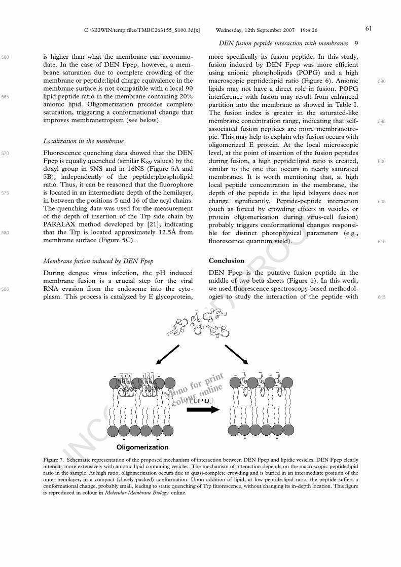
UNCORRECTED PROOF
is higher than what the membrane can accommo-
date. In the case of DEN Fpep, however, a mem-
brane saturation due to complete crowding of the
membrane or peptide:lipid charge equivalence in the
membrane surface is not compatible with a local 90
lipid:peptide ratio in the membrane containing 20%
anionic lipid. Oligomerization precedes complete
saturation, triggering a conformational change that
improves membranetropism (see below).
Localization in the membrane
Fluorescence quenching data showed that the DEN
Fpep is equally quenched (similar KSV values) by the
doxyl group in 5NS and in 16NS (Figure 5A and
5B), independently of the peptide:phospholipid
ratio. Thus, it can be reasoned that the fluorophore
is located in an intermediate depth of the hemilayer,
in between the positions 5 and 16 of the acyl chains.
The quenching data was used for the measurement
of the depth of insertion of the Trp side chain by
PARALAX method developed by [21], indicating
that the Trp is located approximately 12.5A from
membrane surface (Figure 5C).
Membrane fusion induced by DEN Fpep
During dengue virus infection, the pH induced
membrane fusion is a crucial step for the viral
RNA evasion from the endosome into the cyto-
plasm. This process is catalyzed by E glycoprotein,
more specifically its fusion peptide. In this study,
fusion induced by DEN Fpep was more efficient
using anionic phospholipids (POPG) and a high
macroscopic peptide:lipid ratio (Figure 6). Anionic
lipids may not have a direct role in fusion. POPG
interference with fusion may result from enhanced
partition into the membrane as showed in Table I.
The fusion index is greater in the saturated-like
membrane concentration range, indicating that self-
associated fusion peptides are more membranotro-
pic. This may help to explain why fusion occurs with
oligomerized E protein. At the local microscopic
level, at the point of insertion of the fusion peptides
during fusion, a high peptide:lipid ratio is created,
similar to the one that occurs in nearly saturated
membranes. It is worth mentioning that, at high
local peptide concentration in the membrane, the
depth of the peptide in the lipid bilayers does not
change significantly. Peptide-peptide interaction
(such as forced by crowding effects in vesicles or
protein oligomerization during virus-cell fusion)
probably triggers conformational changes responsi-
ble for distinct photophysical parameters (e.g.,
fluorescence quantum yield).
Conclusion
DEN Fpep is the putative fusion peptide in the
middle of two beta sheets (Figure 1). In this work,
we used fluorescence spectroscopy-based methodol-
ogies to study the interaction of the peptide with
560
565
570
575
580
585
590
595
600
605
610
615
Figure 7. Schematic representation of the proposed mechanism of interaction between DEN Fpep and lipidic vesicles. DEN Fpep clearly
interacts more extensively with anionic lipid containing vesicles. The mechanism of interaction depends on the macroscopic peptide:lipid
ratio in the sample. At high ratio, oligomerization occurs due to quasi-complete crowding and is buried in an intermediate position of the
outer hemilayer, in a compact (closely packed) conformation. Upon addition of lipid, at low peptide:lipid ratio, the peptide suffers a
conformational change, probably small, leading to static quenching of Trp fluorescence, without changing its in-depth location. This figure
is reproduced in colour in Molecular Membrane Biology online.
Monofor
colour o
nline
DEN fusion peptide interaction with membranes 9
C:/3B2WIN/temp files/TMBC263155_S100.3d[x] Wednesday, 12th September 2007 19:4:26 61

UNCORRECTED PROOF
model membranes. Analysis of our results shows: (i)
the peptide interacts with model membranes, in-
dependently of its lipid composition, as supported by
changes in fluorescence intensity and blue shifts
occurring upon addition of vesicles, but the presence
of anionic lipids leads to a more extensive partition;
(ii) partition curves with vesicles containing nega-
tively charged phospholipids show that the fluores-
cence intensity reaches a maximum at �1 mM of
lipids, followed by a progressive decrease, which is
not accompanied by lifetimes. Lifetimes reach a
plateau. This suggests that the interaction depends
on the global peptide:lipid ratio in the sample.
Moreover, that result indirectly suggests that the
peptide undergoes structural changes, probably
small, dependent of that ratio, leading to a static
quenching of Trp fluorescence; (iii) quenching
studies indicate that the DEN Fpep Trp residue is
buried in the middle of the outer hemilayer, inde-
pendently of the peptide:lipid ratio (5NS and 16NS
have similar quenching efficiency); (iv) DEN Fpep
is able to induce membrane fusion, but the process is
more efficient using vesicles containing anionic lipids
and at high macroscopic peptide:lipid ratio.
Based on the data described above, one can
hypothesize in general terms what might be the
mechanism of interaction between DEN Fpep and
membranes at the molecular level. As depicted in
Figure 7, the interaction depends on the global
peptide:lipid ratio in the sample. At high ratio, it is
possible to detect oligomerization. Until a certain
critical ratio, oligomerized peptides will be buried in
an intermediate depth in the outer hemilayer in a
conformation constrained by packing. This stage of
interaction only occurs with vesicles containing
anionic phospholipids, since quasi-saturation results
from the high affinity between the peptide and the
lipid. Upon addition of excess lipid, at low peptide:
lipid ratios, the peptide is conformationally unrest-
ricted because crowding disappears and undergoes
structural changes, probably small, without chan-
ging its in-depth location. The oligomerized, quasi-
saturated situation seems to be a suitable model for
studying the interaction of viral particles and cell
membranes. The micro-environment composed of
the cell membrane phospholipids and the viral
surface proteins has a high local peptide:lipid ratio.
We believe this may be the reason behind protein E
oligomerization for viral fusion, the fusion peptides
forming the tip of a trimer. Our results indicate that
membrane fusion is more efficient in this situation.
Moreover, a preferential interaction of the fusion
peptides of the E protein with anionic lipids may
drive the depth of insertion further to the interior of
the membrane, closer to the negatively charged inner
(cytoplasmatic) lipid hemilayer, which further per-
turbs membranes and favors fusion.
Altogether, these data provide novel insights on
the mechanism of dengue virus fusion peptide,
namely the frequently overlooked role of viral
proteins oligomerization.
Acknowledgements
We thank Sonia Troeira Henriques from Lisbon
University (Portugal) for valuable help.
This work was supported by grants from Conselho
Nacional de Desenvolvimento Cientıfico e Tecnolo-
gico (CNPq), Fundacao Carlos Chagas Filho de
Amparo a Pesquisa do Estado do Rio de Janeiro
(FAPERJ), Fundacao de Amparo a Pesquisa do
Estado de Sao Paulo (FAPESP), Coordenacao de
Aperfeicoamento de Pessoal de Nıvel Superior
(CAPES), Gabinete de Relacoes Internacionais da
Ciencia e do Ensino Superior (GRICES), Special
Programme for Research and Training in Tropical
Diseases (TDR) of World health Organization
(WHO), Third World Academy of Sciences (TWAS)
andFundacao para aCiencia eTecnologia (Portugal),
including a grant to M. Melo (SFRH/BD/24778/
2005). F. Stauffer was recipient of a fellowship from
CAPES (BEX 1279/05).
References
[1] Lindenbach BD, Rice CM. 2001. Flaviviridae: The viruses
and their replication. In: Fields BN, Howley PM, Griffin
DE, Lamb RA, Martin MA, Roizman B, Straus SE,
Knipe DM, editors. Fields’ virology, 4th ed. Philadelphia:
Lippincott Williams & Wilkins. p. 991�1041.[2] Crill WD, Roehrig JT. 2001. Monoclonal antibodies that
bind to domain III of dengue virus E glycoprotein are the
most efficient blockers of virus adsorption to Vero cells.
J Virol 75:7769�7773.[3] Kuhn RJ, Zhang W, Rossmann MG, Pletnev SV, Corver J,
Lenches E, Jones CT, Mukhopadhyay S, Chipman PR,
Strauss EG, Baker TS, Strauss JH. 2002. Structure of
dengue virus: implications for flavivirus organization, ma-
turation, and fusion. Cell 108:717�725.[4] Modis Y, Ogata S, Clements D, Harrison SC. 2003. A
ligand-binding pocket in the dengue virus envelope glyco-
protein. Proc Natl Acad Sci USA 100:6986�6991.[5] Modis Y, Ogata S, Clements D, Harrison SC. 2004.
Structure of the dengue virus envelope protein after mem-
brane fusion. Nature 427:313�319.[6] Wieprecht T, Beyermann M, Seelig J. 2002. Thermody-
namics of the coil-alpha-helix transition of amphipathic
peptides in a membrane environment: the role of vesicle
curvature. Biophys Chem 96:191�201.[7] Mayer LD, Hope MJ, Cullis PR. 1986. Vesicles of variable
sizes produced by a rapid extrusion procedure. Biochim
Biophys Acta 858:161�168.[8] Lakowicz JR, editor. 1999. Principles of fluorescence spec-
troscopy. 2nd ed. New York: Kluwer Academic/Plenum.
725 pp.
[9] Santos NC, Prieto M, Castanho MA. 2003. Quantifying
molecular partition into model systems of biomembranes: an
620
625
630
635
640
645
650
655
660
665
670
675
680
685
690
695
700
705
710
715
720
725
10 F. Stauffer et al.
C:/3B2WIN/temp files/TMBC263155_S100.3d[x] Wednesday, 12th September 2007 19:4:27 62

UNCORRECTED PROOF
emphasis on optical spectroscopic methods. Biochim Bio-
phys Acta 1612:123�135.[10] Melo MN, Castanho MARB. 2007. Omiganan interaction
with bacterial membranes and cell wall models. Assigning a
biological role to saturation. Biochim Biophys Acta
DOI:10.1016/j.bbamem.2007.02.005.
[11] Chiu SW, Jakobsson E, Subramaniam S, Scott HL. 1999.
Combined monte carlo and molecular dynamics simulation
of fully hydrated dioleyl and palmitoyl-oleyl phosphatidyl-
choline lipid bilayers. Biophys J 77:2462�2469.[12] Coutinho A, Prieto M. 1993. Ribonuclease T1 and alcohol
dehydrogenase fluorescence quenching by acrylamide.
J Chem Educ 70:425�428.[13] Santos NC, CastanhoMA. 2002. Fluorescence spectroscopy
methodologies on the study of proteins and peptides. Trends
Appl Spectrosc 4:113�125.[14] Caputo GA, London E. 2003. Cumulative effects of amino
acid substitutions and hydrophobic mismatch upon the
transmembrane stability and conformation of hydrophobic
alpha-helices. Biochemistry 42:3275�3285.[15] Fernandes MX, Garcia de la Torre J, Castanho MA. 2002.
Joint determination by Brownian dynamics and fluorescence
quenching of the in-depth location profile of biomolecules in
membranes. Anal Biochem 307:1�12.[16] Santos NC, Prieto M, Castanho MA. 1998. Interaction of
the major epitope region of HIV protein gp41 with mem-
brane model systems. A fluorescence spectroscopy study.
Biochemistry 37:8674�8682.[17] Wardlaw JR, Sawyer WH, Ghiggino KP. 1987. Vertical
fluctuations of phospholipid acyl chains in bilayers. FEBS
Lett 223:20�24.[18] Maier O, Oberle V, Hoekstra D. 2002. Fluorescent lipid
probes: some properties and applications (a review). Chem
Phys Lipids 116:3�18.[19] Haugland RP, editor. 2002. Handbook of fluorescent probes
and research products. 9th ed. New York: Molecular Probes.
[20] Pecheur EI, Martin I, Ruysschaert JM, Bienvenue A,
Hoekstra D. 1998. Membrane fusion induced by 11-mer
anionic and cationic peptides: a structure-function study.
Biochemistry 37:2361�2371.[21] Chattopadhyay A, London E. 1987. Parallax method for
direct measurement of membrane penetration depth utiliz-
ing fluorescence quenching by spin-labeled phospholipids.
Biochemistry 26:39�45.[22] Basanez G. 2002. Membrane fusion: the process and its
energy suppliers. Cell Mol Life Sci 59:1478�1490.
[23] Murray D, Arbuzova A, Honig B, McLaughlin S. 2002. The
role of electrostatic and nonpolar interactions in the associa-
tion of peripheral proteins with membranes. Curr Topics
Membr 52:271�302.[24] Feldman SA, Audet S, Beeler JA. 2000. The fusion
glycoprotein of human respiratory syncytial virus facilitates
virus attachment and infectivity via an interaction with
cellular heparan sulfate. J Virol 74:6442�6447.[25] Fry EE, Lea SM, Jackson T, Newman JW, Ellard FM,
Blakemore WE, Abu-Ghazaleh R, Samuel A, King AM,
Stuart DI. 1999. The structure and function of a foot-and-
mouth disease virus-oligosaccharide receptor complex.
Embo J 18:543�554.[26] Shukla D, Liu J, Blaiklock P, Shworak NW, Bai X, Esko JD,
Cohen GH, Eisenberg RJ, Rosenberg RD, Spear PG. 1999.
A novel role for 3-O-sulfated heparan sulfate in herpes
simplex virus 1 entry. Cell 99:13�22.[27] Chen Y, Maguire T, Hileman RE, Fromm JR, Esko JD,
Linhardt RJ, Marks RM. 1997. Dengue virus infectivity
depends on envelope protein binding to target cell heparan
sulfate. Nat Med 3:866�871.[28] Ludtke S, He K, Huang H. 1995. Membrane thinning
caused by magainin 2. Biochemistry 34:16764�16769.[29] Magzoub M, Graslund A. 2004. Cell-penetrating peptides:
small from inception to application. Q Rev Biophys 37:
147�195.[30] Gibbons DL, Vaney MC, Roussel A, Vigouroux A, Reilly B,
Lepault J, Kielian M, Rey FA. 2004. Conformational change
and protein-protein interactions of the fusion protein of
Semliki Forest virus. Nature 427:320�325.[31] Cheng SF, Wu CW, Kantchev EA, Chang DK. 2004.
Structure and membrane interaction of the internal fusion
peptide of avian sarcoma leukosis virus. Eur J Biochem
271:4725�4736.[32] Chang DK, Cheng SF, Deo Trivedi V, Yang SH. 2000. The
amino-terminal region of the fusion peptide of influenza
virus hemagglutinin HA2 inserts into sodium dodecyl sulfate
micelle with residues 16�18 at the aqueous boundary at
acidic pH. Oligomerization and the conformational flexibil-
ity. J Biol Chem 275:19150�19158.[33] Pecheur EI, Sainte-Marie J, Bienven e A, Hoekstra D. 1999.
Peptides and membrane fusion: towards an understanding
of the molecular mechanism of protein-induced fusion.
J Membr Biol 167:1�17.
730
735
740
745
750
755
760
765
770
775
780
785
790
795
800
805
810
AQ2
DEN fusion peptide interaction with membranes 11
C:/3B2WIN/temp files/TMBC263155_S100.3d[x] Wednesday, 12th September 2007 19:4:28 63

2.2 Parte II
Descoberta de um novo composto inativador viral baseado na inibição de fusão de
membranas e sua aplicação na formulação de uma vacina
A elucidação dos mecanismos moleculares envolvidos na reação de fusão de
membranas catalisada pelas glicoproteínas virais pode contribuir para a decoberta de possíveis
alvos para inibição da entrada dos vírus em suas células hospedeiras, auxiliando no
desenvolvimento de novos antivirais e de vacinas.
Na parte II desta tese, tivemos como objetivo descobrir um novo composto inativador
viral baseado em dados obtidos dos estudos básicos de elucidação do mecanismo de entrada
dos vírus nas células, realizados na parte I. Todos os passos utilizados na caracterização da
inativação viral baseada na inibição da fusão de membranas são resumidamente expostos a
seguir. Os resultados completos se encontram apresentados na forma de artigos científicos
(artigos 4 e 5).
Como citado na parte I dos resultados, nós descobrimos que o DEPC modifica
especificamente os resíduos de histidina da proteína G do VSV, inibindo as mudanças
conformacionais desta glicoproteína desencadeadas pelo pH ácido e consequentemente a
fusão de membranas mediada pelo VSV (artigo 2). Isto nos indicou que provavelmente o
tratamento viral com DEPC seria capaz de inativar o VSV. Para testar esta hipótese, fizemos
experimentos em modelos celular e animal, mostrando que a modificação viral com DEPC
abolia a infectividade e patogenicidade do VSV. Posteriormente, avaliamos por microscopia
eletrônica e ELISA de competição que as regiões antigênicas da glicoproteína viral estavam
preservadas. Estes resultados indicavam o potencial uso do DEPC na formulação de vacinas
inativadas baseada na inibição da fusão de membranas. Esta foi a primeira vez que o DEPC
foi utilizado como inativador viral, de forma que a partir dos resultados obtidos, depositamos
uma patente no Instituto Nacional da Propriedade Industrial (INPI) em 2004 (patente 1). Os
resultados foram posteriormente publicados no periódico Antiviral Research (artigo 4).
O próximo passo foi avaliar a imunogenicidade das partículas virais inativadas pelo
DEPC em camundongos. Observamos que o animal imunizado por via intraperitoneal com
três doses de VSV tratado com DEPC misturado com adjuvantes apresentava uma resposta
humoral semelhante àquela induzida pelo vírus nativo. Os anticorpos gerados eram capazes de
reconhecer e neutralizar o VSV não modificado, além de proteger de forma eficiente os
animais do desafio com uma dose letal de vírus. Em suma, nossos dados sugerem que a
inativação viral com DEPC pode ser utilizada para o desenvolvimento de vacinas. Estes dados
compõem o artigo 5 desta tese, praticamente aceito para publicação no periódico Vaccine.
64

2.2.1 Patente 1
Composição para Inativação Viral, Processo de Inativação Viral,
Vacina e Processo de Produção de Vacina
Fausto Stauffer, Joari M. de Miranda, Fabiana A. Carneiro, Gisele Machado e
Andrea T. Da Poian
Depositada em 2004 no INPI
Publicada em 2005 na Revista da Propriedade Industrial

1 / 18
Relatório descritivo Composição para Inativação Viral, Processo de Inativação Viral, Vacina e
Processo de Produção de Vacina
Campo da Invenção5
10
15
A presente invenção refere-se genericamente a uma composição para
inativação viral, a um processo de inativação viral, a uma vacina contra o VSV
e a um processo para a produção de vacina contra a VSV. Mais
especificamente, refere-se a uma composição que inibe a infecção e a
replicação viral, bem como o desenvolvimento da patologia estomatite vesicular
em animais. A presente invenção refere-se adicionalmente a um processo de
inativação viral através do contato do vírus da VSV com a substância DEPC. A
inativação do VSV possibilita a obtenção de uma vacina contras as patologias
causadas pelo mesmo.
Antecedentes da InvençãoA estomatite vesicular causada pelo VSV afeta bovinos, eqüinos e suínos,
e suas manifestações clínicas incluem vesiculação e/ou ulceração severa na
língua e nos tecidos orais, podendo, às vezes, provocar lesões nos pés e nas
tetas dos animais acometidos, o que resulta em uma substancial perda de
produtividade (Letchworth, G. J. et al. (1999) Vet. J. 157, 239-260). Os sintomas
desta doença são indistingüíveis daqueles observados na febre aftosa, exceto
por afetar também eqüinos. A estomatite vesicular causada pelo VSV ocorre
anualmente desde o sudeste dos Estados Unidos até o norte da América do Sul,
passando pelo México e por toda a América Central. Nestas regiões, os
sorotipos predominantes são VSV New Jersey (VSV-NJ) e VSV Indiana (VSV-
IN). No Brasil, é endêmico o sorotipo Indiana 3, ou Alagoas (Federer, K. E. et al.
(1967) Res. Vet. Sci. 8, 103-117; Andrade, C. M. et al. (1980) An. Microbiol. (Rio
de Janeiro) 25, 81-87). Outros vesiculovírus são endêmicos na Índia e na África.
Nos Estados Unidos os dois surtos mais recentes ocorreram em 1997 e 1998,
afetando principalmente eqüinos (McCluskey, B. J. et al. (1999) J. Am. Vet. Med.
20
25
30
66

2 / 18
Assoc. 215, 1259-1262). Em 1995, um grande surto atingiu rebanhos bovinos
causando um impacto significativo na indústria de carne do Colorado (Bridges,
V. E. et al. (1997) J. Am. Vet. Med. Assoc. 211, 556-560).
Para infectar uma célula, os vírus devem aderir à superfície celular,
penetrar na célula e tornar seu genoma suficientemente exposto às maquinarias
enzimáticas virais e celulares para sua transcrição e tradução. A identificação de
uma série de proteínas e enzimas virais essenciais para a replicação dos vírus e
suficientemente diferentes das proteínas celulares viabilizou o desenvolvimento
de drogas direcionadas exclusivamente a alvos virais que seriam, em princípio,
inofensivas às proteínas celulares. A maioria destas drogas inibe enzimas
envolvidas na replicação viral, como é o caso, por exemplo, do aciclovir, que
após processamento intracelular se torna um potente inibidor da DNA
polimerase do vírus do herpes (Crumpacker et al. (1979) Antimicrob. Agents
Chemother. 15, 642-645); ou dos inibidores nucleosídicos e não-nucleosídicos
da transcriptase reversa dos retrovírus (Autran, B. et al. (1997) Science 277,
112-116). Recentemente, o impedimento da entrada dos vírus nas células
hospedeiras também vem se mostrando um alvo importante em terapias anti-
virais.
5
10
15
A fusão de membranas no processo de infecção viral
Na classificação dos vírus um dos critérios de separação dos mesmos
ocorre em função da presença ou ausência de envelopes. Para efeitos da
presente invenção, serão abordados os aspectos envolvendo somente os vírus
envelopados.
20
25
30
A internalização dos vírus envelopados sempre envolve uma etapa de
fusão de membranas, que pode ser a fusão direta de seu envelope com a
membrana plasmática da célula, ou sua fusão com a membrana do
compartimento endossomal após a acidificação (Lanzrein, M. et al. (1994)
Biochem. J. 302, 313-320; Hernandez, L. D. et al. (1996) Annu. Rev. Cell Dev.
Biol. 12, 627-661; Weissnhorn, W. et al. (1999) Mol. Membr. Biol. 16, 3-9). A
fusão de membranas mediada pelos vírus envelopados é sempre catalisada
67

3 / 18
por glicoproteínas presentes no envelope viral, que sofrem mudanças
conformacionais seja pela interação com o receptor na membrana plasmática,
seja em decorrência da diminuição do pH no interior do endossoma, causando
a exposição de regiões hidrofóbicas que interagem com a membrana alvo
(Gaudin, Y. et al. (1995) J. Gen. Virol. 76, 1541-1556; Hernandez, L. D. et al.
(1996) Annu. Rev. Cell Dev. Biol. 12, 627-661; Weissnhorn, W. et al. (1999)
Mol. Membr. Biol. 16, 3-9).
5
10
15
20
25
No primeiro caso, o mecanismo de fusão melhor caracterizado é aquele
mediado pelas glicoproteínas gp120/gp41 do HIV-1 (Chan, D. C. & Kim, P. S.
(1998) Cell 93, 681-684). A interação entre a gp120 e o receptor celular para o
vírus (a molécula de CD4 e uma proteína co-receptora, CCR5 ou CXCR4)
resulta em uma série de mudanças conformacionais no complexo gp120/gp41
culminando com a inserção do peptídeo hidrofóbico presente na gp41 na
membrana da célula hospedeira. A proteína gp41 se dobra, formando uma
estrutura conhecida como hairpin (grampo de cabelo) (Chan, D. C. et al. (1997)
Cell 89, 263-273). Peptídeos derivados da região C-terminal da gp41 interferem
na formação desta estrutura e inibem de forma bastante efetiva a infecção do
HIV-1 (Wild, C. T. et al. (1994) Proc. Natl. Acad. Sci. USA. 91, 9770-9774). Um
destes peptídeos, denominado T-20, está atualmente em testes clínicos em
humanos, apresentando excelentes resultados (Kilby, J. M. et al. (1998) Nat.
Med. 4, 1302-1307).
No caso da fusão endossomal, a glicoproteína de fusão mais bem
estudada é a do vírus influenza, a hemaglutinina (HA), cuja estrutura foi
resolvida com resolução atômica tanto em pH neutro como em pH ácido (Wilson,
I. A. et al. (1981) Nature 289, 366-372, Bullough, P. A. et al. (1994) Nature 371,
37-43). Em resposta à diminuição do pH, a HA sofre uma grande mudança
conformacional, expondo um peptídeo hidrofóbico que se liga à membrana-alvo.
68

4 / 18
O vírus da estomatite vesicular
O vírus da estomatite vesicular (VSV) é o membro mais estudado da
família Rhabdoviridae, um grupo de vírus com grande abrangência de
hospedeiros (plantas, animais invertebrados e vertebrados). Os rabdovírus são
formados por um capsídeo ribonucleico helicoidal envolto por uma membrana
lipídica (Wagner, R. R. (1987) Em "The rhabdoviruses". R. R. Wagner, ed.
Plenum Press, New York, pp 9-74). O capsídeo ribonucleico é composto por
uma fita simples de RNA fortemente associado a uma proteína principal - a
proteína N - e a duas outras proteínas - L e P - que juntas constituem a RNA
polimerase viral. O envelope lipídico está associado a duas proteínas: a
glicoproteína G, integral à membrana, cujos trímeros formam as espículas virais,
e a proteína M, que interage com a face interna da membrana e com o capsídeo
ribonucleico. O VSV se liga aos receptores celulares através da proteína G,
sendo esta, então, responsável pela adsorção e infectividade viral. A entrada do
vírus na célula se dá por endocitose mediada por receptor e sua replicação
depende da acidificação endossomal (Matlin, K. S. et al. (1982) J. Mol. Biol. 156,
609-631).
5
10
15
A fusão de membranas mediada pelo vírus da estomatite vesicular
A membrana do VSV possui aproximadamente 1.200 moléculas da
proteína G, a glicoproteína de superfície deste vírus, sendo essa proteína
envolvida em pelo menos dois passos importantes do processo de infecção do
VSV: o reconhecimento da célula com conseqüente adsorção à superfície
desta, e o processo de fusão de membranas, fundamental à liberação do
genoma viral no citoplasma.
20
25
30
A fusão de membranas mediada pela proteína G depende da diminuição
do pH (White, J. et al. (1981) J. Cell Biol. 89, 674-679). As mudanças
conformacionais necessárias para a fusão têm sido motivo de vários estudos.
Até o presente mostrou-se que em pH ácido ocorre a estabilização do trímero
da proteína G (Doms, R. W. et al. (1987) J. Cell Biol. 105, 1957-1969). Os
trímeros da proteína G apresentam estabilidade diferencial na presença de
69

5 / 18
octilglicosídeo e Triton X-100, sugerindo que o ambiente que circunda a
seqüência de ancoramento na membrana também influencia a interação entre
as subunidades da proteína G (Wilcox, M. D. et al. (1992) Biochemistry 31,
10458-10464). A proteína G não apresenta nem um domínio de fusão típico,
nem uma outra seqüência hidrofóbica óbvia além da sua porção
transmembrana. Desta forma, o que se acredita é que haja a formação de um
domínio cuja estrutura terciária gere um ambiente hidrofóbico capaz de
interagir com a membrana e mediar a fusão. Durrer e colaboradores mostraram
que durante as transições conformacionais decorrentes da acidificação, um
segmento do ectodomínio tornava-se capaz de interagir com membranas
(Durrer, P. et al. (1995) J. Biol. Chem. 29, 17575-17581). A fragmentação da
proteína com brometo de cianogênio revelou que este segmento estava contido
entre os aminoácidos 59-221.
5
10
15
20
25
30
Substituições de aminoácidos em regiões do ectodomínio da proteína G
vêm sendo usadas como outra estratégia de localização do peptídeo de fusão
do VSV. Mutações em resíduos presentes na região 117-137, que são
altamente conservadas nos vesiculovírus, sugeriram que esta região continha o
domínio de fusão (Zhang, L. & Ghosh, H. P. (1994) J. Virol. 68, 2186-2193;
Fredericksen, B. e Whitt, M. A. (1995) J. Virol. 69, 1435-1443). A substituição
dos resíduos de glicina, prolina e ácido aspártico conservados ou aboliram a
fusão de membranas mediada pela proteína ou modificaram o pH ótimo da
reação. Mutações em outra região conservada, entre os aminoácidos 395-418,
também aboliu ou diminui a atividade de fusão (Shokralla, S. et al. (1998)
Virology 256, 119-129). A importância do domínio transmembrana na fusão de
membranas mediada pelo VSV também foi estudada (Odell, D. et al. (1997) J.
Virol. 71, 7996-8000; Cleverley, P. Z. & Lenard, J. (1998) Proc. Natl. Acad. Sci.
USA 95, 3425-3430). Estes trabalhos mostraram a importância de 6 resíduos
presentes nestes domínio (GLIIGL) e levaram à proposição de que seria
necessária a dobra deste segmento em uma das glicinas para que ocorresse a
formação do poro de fusão e a hemifusão propriamente dita.
70
6 / 18
Recentemente, as mudanças estruturais sofridas por esta proteína em
função da diminuição do pH e de sua interação com vesículas lipídicas foram
caracterizadas, simulando o processo de endocitose do vírus e a fusão de
membranas (Carneiro, F. A. et al. (2001) J. Biol. Chem. 276, 62-67). Nesse
trabalho, à medida que o pH diminui, uma dramática mudança conformacional
na proteína ocorre. Primeiramente, há a exposição de um domínio hidrofóbico
que interage com a membrana alvo, e em seguida ocorre perda de estruturas
secundária e terciária, que, em pHs mais baixos ainda, se reorganizam em uma
nova estrutura (Carneiro, F. A. et al. (2001) J. Biol. Chem. 276, 62-67). As
características e a termodinâmica da interação entre a proteína G e vesículas
de diferentes composições lipídicas também foram estudadas através do uso
da microscopia de força atômica, calorimetria e espectroscopia de
fluorescência (Carneiro, F. A. et al. (2002) J. Virol. 76, 3756-3764). Em
diferentes etapas do ciclo de infecção do vírus independente do
reconhecimento da membrana, da interação com esta membrana em pH baixo,
e da catálise da fusão de membranas propriamente dita, as interações
determinantes são interações eletrostáticas entre regiões da proteína
positivamente carregadas e fosfolipídeos negativamente carregados.
5
10
15
20
25
30
Esses resultados sugerem que as mudanças estruturais sofridas pela
proteína G durante a acidificação, assim como a aquisição da capacidade de
interagir com membranas negativamente carregadas ocorrem em uma faixa de
pH bastante estreita, entre 6,2 e 5,8. Esta é exatamente a faixa de pH na qual
ocorre a protonação da cadeia lateral do aminoácido histidina (pK ~ 6,04). Isso
sugeriu que a protonação de resíduos de histidina presentes na proteína G
seria necessária para o desencadeamento do processo de fusão. Por esse
motivo para efeitos da presente invenção, a substância Dietilpirocarbonato
(DEPC), cujo mecanismo de modificação específica de histidinas vem sendo
estudado há anos, foi utilizada no desenvolvimento da presente invenção.
Na faixa de pH entre 5.5 e 7.5, DEPC é razoavelmente específico para
reação com resíduos de histidina (Lundblad, R. L. & Noyes, C. M. (1984) em
“Chemical reagents for protein modification”, CRC Press, Boca Raton, Florida).
71

7 / 18
DEPC reage com resíduos de histidina em sistemas modelo e em proteínas, e
resulta na substituição de uma das posições de nitrogênio do anel imidazol,
gerando N-carbetoxihistidina (Miles, E. W. (1977) Meth. Enzymol. 47, 431-442),
como mostrado na equação 1:
5
A formação do N-carbetoxihistidina pode ser acompanhada
espectrofotometricamente, com um máximo de absorção entre 230 e 250 nm
(Ovádi, J. et al. (1967) Biochim. Biophys. Acta 2, 455-500).
10
15
20
25
Existem descritos na literatura vários exemplos do uso de DEPC para
estudar a função de resíduos de histidina em diversas proteínas (Lundblad, R. L.
& Noyes, C. M. (1984) em “Chemical reagents for protein modification”, CRC
Press, Boca Raton, Florida).
A partir de experimentos envolvendo vírus VSV e DEPC foi possível
mostrar que o vírus modificado perdia a capacidade de mediar a fusão de
membranas devido à inibição das mudanças conformacionais na proteína G.
Além disso, as seqüências modificadas pelo DEPC foram identificadas através
de experimentos de espectrometria de massa, o que permitiu, a partir do uso
de peptídeos sintéticos, identificar o peptídeo de fusão do VSV e caracterizar
os requerimentos necessários à sua participação na fusão de membranas
mediada pelo vírus (Carneiro, F. A. et al. (2003) J. Biol. Chem. 278, 13789-
13794).
O único relato encontrado na literatura patentária utilizando o DEPC
como um agente inativador viral foi descrito na patente norte americana US
986,046, de 1963. Esta patente descreve um processo detalhado para
produção de vacinas utilizando solução aquosa de DEPC, de modo que os
vírus inativados por este processo sejam imunogênicos, mas não patogênicos.
Foram utilizados como exemplos quatro tipos de vírus: sarampo (vírus
72

8 / 18
envelopado), varíola (vírus envelopado), pólio (vírus não envelopado) e
influenza A. A eficácia das vacinas foi avaliada por meio de titulação por TCID
(dose infecciosa em cultura de tecido) e dosagem de anticorpos no soro de
animais vacinados. Entretanto, a presente invenção propõe uma composição
para inativação viral capaz de adicionalmente inibir a infecção e a replicação do
vírus VSV.
5
10
15
20
25
30
Dentre as patentes relacionadas com a inativação do VSV, a patente
norte americana US 4,556,556, de 1985, refere-se a um composto químico que
atua sobre partícula viral, tornando-a inativa ou atenuada, e possibilitando sua
utilização em formulação de vacinas. Neste caso, o autor descreve uma vacina
contra o VSV contendo partículas virais inativadas pela associação psoraleno e
luz ultravioleta. Por intermédio da análise dos resultados de titulação viral,
dosagem de anticorpos em modelos animais e sobrevivência dos animais
vacinados expostos ao vírus selvagem, foi demonstrado que o VSV inativado
desta forma mantém sua imunogenicidade e é completamente não infeccioso,
podendo por isso ser administrado com segurança como vacina. Por outro
lado, a presente invenção produz efeito semelhante através da manipulação de
uma única substância.
A patente européia EP 0822819 propõe uma emulsão formada por um
solvente e um carreador de óleo para aplicação tópica a fim de inibir a infecção
causada particularmente pelos vírus envelopados de uma maneira geral. Essa
emulsão possui a capacidade de inibir a infecção viral e a replicação. Contudo,
a presente invenção produz efeito idêntico a partir da manipulação somente do
solvente orgânico DEPC (dietilpirocarbonato).
O pedido norte americano US 2004/0047844 propõe compostos alquil-
aminos capazes de inativar bactérias, fungos, leveduras, micoplasmas, células
de mamíferos ou de outros animais, vírus ou vírus envelopados como
Flaviviridae, Retroviridae, Togaviridae, Rhabdoviridae, Herpesviridae, VSV,
SFV, HIV, MuLV, BVDV, e CMV. Particularmente os ingredientes ativos são
aminas anfipáticas carregadas e/ ou óxidos de amina anfipáticos carregados.
73

9 / 18
Ao contrário da presente invenção, para que os compostos ativos funcionem de
forma eficaz, a solução deve estar dentro de uma faixa de pH específica (5,0 a
8,0), sendo preferencialmente 7,2, o que acarreta em maiores cuidados na
confecção do composto de inativação biológica da US 2004/0047844A1.
Um outro exemplo de inativação viral está descrito na patente norte
americana US 6369048. Nesse trabalho a inativação é realizada através do
contato, em condições específicas, da composição biológica com um solvente
orgânico pertencente ao grupo dos triacilfosfatos. Essa metodologia é útil na
inativação dos vírus não envelopados e dessa forma, diminui a capacidade,
bem como inibe a infecção e a replicação viral. Entretanto, a presente invenção
atua inativando o vírus VSV, que compreende um vírus envelopado.
5
10
15
20
25
30
O VSV também pode ser inativado por tratamento com formalina, com �-
propiolactona ou com luz UV (Bachmann M. F. et al. (1993) J. Virol. 67, 3917-
3922; Bachmann M. F. et al. (1994) Med. Microbiol. Immunol. 183, 95-104).
Nestes casos, o vírus inativado não foi capaz de induzir a produção de IgG,
embora a resposta de IgM tenha se mantido inalterada. Apenas o vírus
inativado por luz UV foi capaz suscitar resposta de linfócitos T citotóxicos
(Bachmann M. F. et al. (1994) Med. Microbiol. Immunol. 183, 95-104). Porém, a
presente invenção supera os resultados acima, ao impedir o aparecimento do
infiltrado inflamatório nos cortes histológicos de animais infectados e tratados
com a presente invenção, característico da reação inflamatória.
Mais recentemente, o rearranjo genético do VSV vem sendo apontado
como uma forma eficiente de atenuação viral (Wertz, G. W. et al. (1998) Proc.
Natl. Acad. Sci. USA 95, 3501-3506; Flanagan, E. B. et al. (2001) J. Virol. 75,
6107-6114). O afastamento do gene da proteína N da região promotora reduziu
os níveis de transcrição e da síntese da proteína N com conseqüente uma
atenuação da replicação do vírus e de sua letalidade para camundongos
(Wertz , G. W. et al. (1998) Proc. Natl. Acad. Sci. USA 95, 3501-3506). A
literatura patentária inclui o pedido de patente US2003/0124146, que propõe a
construção de um vetor recombinante Rhabdovírus expressando um
74

10 / 18
polipeptídeo antigênico do vírus da estomatite vesicular, por exemplo, a fim
resultar em uma resposta imune pelo hospedeiro. Pelo fato da presente
invenção não utilizar a tecnologia do DNA recombinante, a mesma permite o
barateamento das etapas da produção o que economicamente permite um
volume maior de produção e conseqüentemente, um beneficiamento para mais
animais.
5
10
15
20
25
Outras patentes referem-se a compostos químicos que interferem no
ciclo de vida dos vírus, ou seja, poderiam ser utilizados para o tratamento de
infecções virais (drogas antivirais). É o caso da patente norte americana US
4,396,628, de 1983, que demonstra que a dansilcadaverina é mais eficaz que a
amantadina e outros agentes antivirais no bloqueio da entrada do VSV em
células de camundongo. Outros compostos químicos, como compostos que
contêm hipericina ou pseudohipericina (US 4,898,891, de 1990); 3-
deazoadenosina (US 4,148,888, de 1979); e bases fracas parcialmente
solúveis em água (EP 0095833, de 1983), também foram patenteados como
antivirais. Contudo, tais mecanismos apenas atuam na diminuição da taxa de
replicação viral e não propõem o desenvolvimento de uma composição de
inativação viral capaz de inativar o vírus, ou seja, inibindo a infecção e a
replicação do vírus VSV, bem como impedindo o desenvolvimento patológico
da estomatite vesicular.
A presente invenção propõe a inativação do vírus VSV (vírus da
estomatite vesicular) através da utilização de uma substância orgânica, o
DEPC (dietilpirocarbonato). A eficiência dessa metodologia é dose dependente
de DEPC, porém a dose ideal não apresenta efeito citotóxico para as células
infectadas pelo vírus. Dessa forma, a presente invenção resolve o problema da
inativação do vírus VSV de forma simples, menos custosa e eficaz.
Sumário da invençãoÉ objeto da presente invenção uma composição para inativação viral,
mais especificamente a inativação do vírus VSV causador da doença
estomatite vesicular. Essa composição possui uma solução aquosa contendo o 30
75

11 / 18
solvente orgânico DEPC (dietilpirocarbonato) e o vírus VSV. Ao inativar o vírus
VSV, a presente invenção impede a infecção e a replicação do vírus VSV, bem
como o desenvolvimento patológico da estomatite vesicular.
É um adicional objeto da presente invenção o processo de inativação
viral através da contactação do mesmo com uma solução aquosa contendo o
composto orgânico DEPC (dietilpirocarbonato).
5
10
Em um aspecto, os vírus inativados pelo processo da presente invenção
são úteis na preparação de vacinas contra as doenças que os referidos vírus
acarretam. É, portanto, um adicional objeto da presente invenção uma vacina
contra as patologias causadas pelo VSV.
Em ainda outro aspecto, sendo, por tanto ainda outro objeto da presente
invenção, é provido um processo para a preparação de vacinas.
Breve Descrição das FigurasFigura 1: Microscopia ótica das células BHK21 tratadas ou não com DEPC
após 20 horas pós-infecção com o VSV. Tratamento com DEPC inibe o efeito
citopático induzido pelo VSV. Células BHK
15
20
25
30
21 foram infectadas com VSV tratado
ou não com DEPC em diferentes concentrações. As células foram examinadas
por microscopia ótica 20 horas pós-infecção. (A) Célula não infectada; (B)
célula infectada com VSV tratado com DEPC 0,5 mM; (C) 0,1 mM; (D) 0,05
mM; (E) 0,01 mM; e (F) célula infectada com VSV não tratado (F). Foi utilizada
uma multiplicidade de infecção (m.o.i.) de 0,1.
Figura 2: Gráfico representativo da taxa de mortalidade de camundongos
BALB/c infectados com VSV tratado ou não com DEPC acompanhados
diariamente por 5 semanas. Sobrevivência de camundongos BALB/c infectados
com VSV não tratado ou tratado com DEPC. Camundongos BALB/c foram
inoculados por via intranasal com 2,0 X 10⁶ TCID50 de VSV não tratado (�) ou
tratado com 0.5 mM de DEPC (�). A taxa de mortalidade dos camundongos foi
acompanhada diariamente por 5 semanas. Como grupo controle,
camundongos foram infectados somente com PBS contendo 0,5 mM de DEPC
(�).
76

12 / 18
Figura 3: Cortes histológicos longitudinais passando pelo ventrículo lateral de
camundongos inoculados com VSV ou com VSV tratado com DEPC. A e B -
Cortes longitudinais passando pelo ventrículo lateral de camundongos
inoculados com VSV (A) ou com VSV tratado com DEPC (B). Observa-se
presença de infiltrado inflamatório caracterizando ventriculite (*) em A, o que
não foi observado em B (*).HC – hipocampo (A e B barra = 50�m).
5
C e D -
Cortes longitudinais na região do bulbo olfatório corados por hematoxilina e
eosina mostram a presença de infiltrado inflamatório subpial em C (seta), o que
não foi observado em D (seta). CG - camada glomerular (C e D barra =
12,5�m). 10
Descrição Detalhada da Invenção
De acordo com a presente invenção, a composição de inativação viral
proposta impede a fusão de membranas mediada pelo vírus da estomatite
vesicular, inibindo sua replicação nas células-alvo e desenvolvimento da
patologia característica em modelo animal. Estes efeitos se devem à
modificação de resíduos de histidina presentes na glicoproteína, localizados na
superfície viral, a proteína G, pelo tratamento do vírus com dietilpirocarbonato
(DEPC). A fim de potencializar o resultado da presente invenção, outros
componentes podem ser usados em combinação com a composição da
presente invenção. Para efeitos da mesma, os componentes podem ser
químicos compreendendo substâncias orgânicas ou inorgânicas e/ ou outros
componentes tais como, a irradiação ultra-violeta, a irradiação ultra-sônica,
radiação iônica como a X, gama e beta ou radiação eletrônica. Adicionalmente,
para efeitos da presente invenção o termo “inativação” compreende inibir a
infecção e a replicação viral, bem como o desenvolvimento da patologia
característica da estomatite vesicular.
15
20
25
Os exemplos descritos a seguir ilustram, mas não limitam a presente invenção.
EXEMPLOSEXEMPLO 1- Efeitos do vírus VSV tratado com DEPC em amostras biológicas
30
77

13 / 18
A. Inibição do efeito citopático induzido pelo VSV pelo tratamento viral com
DEPC
Muitos vírus são capazes de induzir morte celular, levando a lise das
células infectadas (Levine et al. (1993) Nature 361, 739-742; Agol, V. I. et al.
(1998) Virology 252, 343-353). Nos últimos estágios da infecção pelo VSV,
mudanças morfológicas que antecedem a morte celular causada pelo vírus,
conhecidas como efeito citopático, podem ser observadas microscopicamente.
O efeito citopático induzido pelo VSV é caracterizado por células arredondadas
e encolhidas, núcleo deformado e cromatina condensada.
5
10
15
Durante a infecção de células BHK21, o efeito citopático clássico ocorre
16-20 horas pós-infecção. Sendo assim, as alterações morfológicas destas
células foram observadas por microscopia após 20 horas de infecção com VSV
controle e modificado com concentrações crescentes de DEPC (figura 1).
As imagens mostram uma inibição progressiva do efeito citopático induzido
pelo VSV a medida que a concentração de DEPC utilizada para modificação
viral aumenta. Estes resultados mostram ainda que o DEPC não é citotóxico
nestas concentrações.
B. Inativação do VSV modificado com DEPC
Para determinar o número de partículas infectantes remanescentes nas
amostras de VSV modificadas com DEPC, o VSV modificado por 15 minutos
com concentrações crescentes de DEPC (0.01, 0.05, 0.1 e 0.5 mM) foi titulado
pelo método de TCID
20
50 (tabela I).
78

14 / 18
Tabela I: Titulação das amostras de VSV modificadas com DEPC.
[DEPC] (mM) Título (pfu/ml)
0 1.80 x 10⁷
0.01 1.40 x 10⁷
0.05 1.60 x 10⁶
0.1 4.56 x 10⁴
0.5 0
Comparando os títulos obtidos para as amostras modificadas com
aquele obtido para o VSV controle, observamos que o tratamento com DEPC
inativa o vírus de forma dose dependente. A pré-incubação do VSV com DEPC
0.5 mM inibiu completamente a infecção, havendo uma perda completa do
título viral.
5
C. Inibição da replicação do VSV modificado com DEPC
Para avaliar a replicação do VSV, os sobrenadantes de células BHK21
infectadas com uma amostra de vírus controle e com uma amostra de vírus
modificado com 0.5 mM de DEPC foram coletados em 4, 8, 12, 16, 20 e 24
horas após a infecção. A replicação viral foi estimada pela presença de
partículas virais infectantes nos sobrenadantes coletados quantificadas pelo
método de TCID
10
1550 (tabela II).
Tabela II: Titulação dos sobrenadantes das culturas infectadas com VSV e VSV modificado com 0.5 mM DEPC.
Título (TCID50/ml)Tempo pós-infecção
(horas) VSV VSV-DEPC
4 4,52 x 10² 0 8 4,65 x 10² 0 12 8,00 x 10² 0 16 2,56 x 10⁴ 0 20 3,03 x 10⁴ 0 24 6,40 x 10³ 0
79

15 / 18
Os resultados obtidos mostram claramente que o tratamento com DEPC
0.5 mM inibe completamente a replicação do VSV.
D. Efeito da modificação do VSV com DEPC sobre a taxa de mortalidade de
camundongos infectados 5
10
A inativação do vírus in vivo foi analisada através de curvas de
sobrevivência de camundongos BALB/c inoculados com VSV controle e VSV
modificado por pré-incubação com DEPC 0.5 mM (figura 2). Os camundongos
controles foram inoculados somente com uma solução de DEPC 0.5 mM.
O percentual de sobrevivência de camundongos infectados com VSV
controle foi de aproximadamente 40%. Já no grupo que foi inoculado com VSV
tratado com DEPC, o perfil da curva de sobrevivência foi semelhante ao do
grupo controle, com percentual maior que 85% 35 dias após a inoculação.
E. Efeito do tratamento do VSV com DEPC na resposta inflamatória no SNC 15
20
25
O VSV, quando inoculado intranasal, inicialmente se replica nos
receptores olfatórios, sendo propagado para o SNC através do nervo olfatório
em menos de 12 horas. No bulbo olfatório, o vírus se replica de forma invasiva,
alcançando o ventrículo após 4-5 dias de infecção (Reiss e cols. (1998) Ann N
Y Acad Sci. 855, 751-761). Como resposta à replicação viral, ocorre uma
reação inflamatória nestas regiões do SNC. A fim de confirmar a inativação
viral in vivo, cortes histopatológicos do encéfalo corados pela H-E foram
examinados. Nos cortes de camundongos infectados com VSV não tratado,
observamos infiltrado inflamatório em resposta a infecção viral (figuras 3A e C).
No entanto, os cortes histológicos de camundongos infectados com VSV
tratado com DEPC 0,5 mM mostram que houve uma inibição completa da
resposta inflamatória (figuras 3B e D).
80

16 / 18
F. Efeitos da presente invenção nos vírus envelopados
A presente invenção compreende também a inativação dos vírus
pertencentes às famílas: Herpesviridae, Togaviridae, Flaviviridae,
Coronaviridae, Hepadnavirida, Filoviridae, Arenaviridae, Retroviridae, e
Rhabdoviridae, sendo preferencialmente o VSV. 5
G. Amostras Biológicas
As amostras biológicas cujo vírus presente na mesma possa ser
inativado compreendem os animais, mais especificamente os mamíferos e as
suspensões celulares, mais especificamente as de animais. 10
EXEMPLO 2: Ensaios realizados
A. Ensaios para propagação do vírus
VSV indiana foi propagado em monocamadas de células BHK21. As
células foram crescidas em placas, a 370C, em estufa de CO2, usando 10 mL
de meio de cultura “Dulbecco’s modified Eagle’s medium” (DMEM)
suplementado com 10% de soro fetal bovino (FBS), antibióticos - gentamicina
(5mg/mL), ampicilina (10mg/mL) - e anfotericina B (5mg/mL), tamponado com
bicarbonato de sódio. Após ficarem confluentes, o meio foi removido e a
monocamada de células foi infectada com VSV em uma multiplicidade de
infecção (m.o.i.) de 0,1. Após 30 minutos de infecção com o vírus na presença
de 9 mL de DMEM não suplementado, adicionamos 1 mL de FBS, e
mantivemos a cultura por 16-20 horas. Após a observação do efeito citopático,
coletamos o meio de cultura e centrifugamos em uma centrífuga Sorvall a
5.000 rpm, por 10 minutos, em rotor GSA.
15
20
25
B. Ensaios para a purificação do vírus
O sobrenadante clarificado foi centrifugado em rotor Beckman Ti45 a
30.000rpm, por 2,5 horas. O sobrenadante foi descartado e o precipitado foi
81

17 / 18
ressuspenso em tampão 3E (Tris 0,12M; acetado de sódio 0,06M; EDTA 3 mM;
pH 7,4) e centrifugado em gradiente contínuo de 5-40 % de sacarose (em
tampão 3E) por 1 hora a 39.000 rpm, em rotor Beckman SW-41. A banda
referente ao vírus foi coletada, dialisada contra tampão Tris-HCl 10mM pH 7,5.
O estoque de vírus foi mantido em freezer -700C. O título viral foi determinado
por método de TCID
5
50 (Reed, L. J. & Muench, H. (1938) Am. J. Hyg. 27, 493-
497) em monocamadas de células BHK21 com 80% de confluência em placas
de 96 poços.
C.Ensaios em células BHK21 do vírus VSV tratado com DEPC 10
15
20
25
Soluções de DEPC foram preparadas antes do experimento pela diluição
do reagente em etanol e posteriormente em PBS 1x. A concentração do
estoque de DEPC era determinada pela reação com imidazol 10mM (Miles, E.
W. (1977) Meth. Enzymol. 47, 431-442. Para inativação viral, VSV foi pré-
incubado com diferentes concentrações de DEPC por 15 minutos a 250C,
variando de 0,01 a 0,5 mM. A pré-incubação do VSV com volume equivalente
de etanol foi realizado como controle do experimento. A quantidade de etanol
na solução final de incubação era menor que 1%.
Para estudo da inibição de infecção in vitro, foi determinado o TCID50 do
VSV controle e modificado com DEPC 0,01; 0,05; 0,1; e 0,5 mM. Além disso,
monocamadas de células BHK21 em placas de 6 poços foram infectadas com
VSV controle e modificado com DEPC 0,005; 0,01; 0,05; e 0,1 mM numa m.o.i.
de 0,1. Após 20 horas de infecção, fotos foram tiradas em microscópio de
contraste de fase, acoplado a uma câmera fotográfica (aumento de 10x).
Para estudo da inibição de replicação, infectamos monocamadas de
células BHK21 em placas de 6 poços com VSV controle e modificado com 0,5
mM em uma m.o.i. de 0,1. Após 4, 8, 12, 16, 20 e 24 horas, os sobrenadantes
foram coletados para determinação do TCID50.
82

18 / 18
D.Ensaios em camundongos BALB/c do vírus VSV tratado com DEPC
Foram selecionados camundongos BALB/c do sexo masculino com seis
a oito semanas de idade. Para realização da curva de sobrevivência, estes
camundongos foram divididos em três grupos: controle, VSV e VSV-DEPC. No
grupo VSV, os camundongos foram infectados com dose de 2,0 x 10⁶ TCID50 5
10
de VSV. No grupo VSV-DEPC, antes da inoculação o VSV foi tratado com
DEPC 0,5 mM como descrito acima. No controle, foram inoculados somente
DEPC 0,5 mM diluído em PBS. Em todos os grupos, a inoculação foi feita por
via intranasal e com mesmo volume. Os camundongos foram acompanhados
diariamente por 45 dias.
E. Efeitos no sistema nervoso central (SNC)
Para avaliar a inibição da infecção do VSV tratado com DEPC no SNC,
camundongos infectados com VSV controle e que apresentavam sinais e
sintomas da doença foram sacrificados e seus encéfalos fixados com formalina
10% e incluídos em parafina. Cortes histológicos longitudinais com 5� e foram
corados por hematoxilina e eosina para observação de alterações
morfológicas. O mesmo foi feito com os encéfalos de camundongos infectados
com VSV tratado com 0,5 mM de DEPC. Os cortes foram analisados e
fotografados em microscópio Olympus BX50.
15
20
25
Os versados na arte apreciarão que a aplicação, a um animal, de uma
composição compreendendo o VSV inativado conforme a presente invenção
pode proporcionar a obtenção de resposta imune protetiva no referido animal.
Conseqüentemente, está compreendida no escopo da presente invenção uma
vacina contra as patologias causadas pelo VSV e também um processo de
produção da referida vacina.
83

1 / 1
ReivindicaçõesComposição para Inativação Viral, Processo de Inativação Viral, Vacina e
Processo de Produção de Vacina
1. Composição para inativação viral caracterizada pelo fato de compreender o
solvente orgânico DEPC (dietilpirocarbonato) em solução aquosa.
5
10
15
20
25
2. Composição, de acordo com a reivindicação 1, caracterizada pelo fato de
adicionalmente compreende anidrido etoxifórmico, dietiloxidiformato e
dietildicarbonato.
3. Composição, de acordo com a reivindicação 1, caracterizada pelo fato de
adicionalmente compreender outras substâncias úteis para a inativação viral.
4. Composição, de acordo com a reivindicação 1, caracterizada pelo fato de ser
destinada à inativação de vírus envelopados selecionados das famílias virais:
Rhabdoviridae, Togaviridae, Flaviviridae, Orthomyxoviridae, Coronaviridae,
Bunyaviridae, Arenaviridae, Hepadnavirida, Paramyxoviridae, Herpesviridae,
Filoviridae, e Retroviridae.
5. Processo de inativação viral caracterizado por compreender a contactação
do vírus com uma solução aquosa contendo DEPC (dietilpirocarbonato).
6. Processo de produção de vacina caracterizado pelo fato de compreender a
inativação do vírus VSV com um solvente orgânico DEPC (dietilpirocarbonato)
em solução aquosa.
7. Vacina caracterizada pelo fato de compreender o vírus VSV inativado com
um solvente orgânico DEPC (dietilpirocarbonato) em solução aquosa.
84

1 / 1
ResumoComposição para Inativação Viral, Processo de Inativação Viral, Vacina e
Processo de Produção de Vacina
A presente invenção refere-se a uma composição para inativação viral
que compreende solução aquosa de DEPC (dietilpirocarbonato); a referida
composição é útil para a inativação do vírus VSV em um processo de
inativação viral, que por sua vez é útil para a preparação de vacinas.
5
85

Figura 1
controle mM
A B C
D E F
0.5 DEPC 0.1 mM DEPC
0.05 mM DEPC 0.01 mM DEPC VSV não tratado
Figura 2
Tempo após inóculo (dias)
0 5 10 15 20 25 30 35
% S
obre
vivê
ncia
0
20
40
60
80
100
VSV
VSV-DEPC
controle
86

Figura 3
87

2.2.2 Artigo 4
Inactivation of vesicular stomatitis virus through inhibition of
membrane fusion by chemical modification of the viral
glycoprotein
Fausto Stauffer, Joari De Miranda, Marcos C. Schechter, Fabiana A. Carneiro,
Leonardo T. Salgado, Gisele F. Machado e Andrea T. Da Poian
Publicado em 2007 no periódico
Antiviral Research

Antiviral Research 73 (2007) 31–39
Inactivation of vesicular stomatitis virus through inhibition of membranefusion by chemical modification of the viral glycoprotein
Fausto Stauffer a,1, Joari De Miranda a,1, Marcos C. Schechter a, Fabiana A. Carneiro a,Leonardo T. Salgadob, Gisele F. Machado c, Andrea T. Da Poian a,∗
a Instituto de Bioquımica Medica, Programa de Biologia Molecular e Biotecnologia, Universidade Federal do Rio de Janeiro, Rio de Janeiro, RJ 21941-590, Brazilb Laboratorio de Biomineralizacao, Instituto de Ciencias Biomedicas, Universidade Federal do Rio de Janeiro, Rio de Janeiro, RJ 21941-590, Brazil
c Departamento de Cirurgia Clınica e Reproducao Animal, Escola de Medicina Veterinaria, Universidade Estadual Paulista,Aracatuba, SP 16050-680, Brazil
Received 18 January 2006; accepted 11 July 2006
Abstract
Membrane fusion is an essential step in the entry of enveloped viruses into their host cells triggered by conformational changes in viralglycoproteins.We have demonstrated previously that modification of vesicular stomatitis virus (VSV)with diethylpyrocarbonate (DEPC) abolishedconformational changes on VSV glycoprotein and the fusion reaction catalyzed by the virus. In the present study, we evaluated whether treatmentwith DEPCwas able to inactivate the virus. Infectivity and viral replication were abolished by viral treatment with 0.5mMDEPC. Mortality profileand inflammatory response in the central nervous system indicated that G protein modification with DEPC eliminates the ability of the virus tocause disease. In addition, DEPC treatment did not alter the conformational integrity of surface proteins of inactivated VSV as demonstrated bytransmission electron microscopy and competitive ELISA. Taken together, our results suggest a potential use of histidine (His) modification to thedevelopment of a new process of viral inactivation based on fusion inhibition.© 2006 Elsevier B.V. All rights reserved.
Keywords: Viral inactivation; Vesicular stomatitis virus; Membrane fusion; Diethylpyrocarbonate
1. Introduction
The development of new strategies to achieve viral inactiva-tion is an important aim in virus research. Despite the advancesin the field of immunology,molecular biology and genetics, viralinactivation remains an important procedure in basic research,since it is an easy and relatively cheap approach for produc-ing new effective and safe vaccines against several viruses. Theentry of enveloped viruses into a target cell always requiresvirus-mediated membrane fusion catalyzed by viral surface gly-coprotein (Eckert andKim, 2001;Hernandez et al., 1996; Skeheland Wiley, 2000). Virus-induced fusion may occur through twodifferent general mechanisms: (i) fusion between viral envelopeand host cell plasma membrane after virus interaction with itscellular receptor, or (ii) fusion with the endosomal membrane,
∗ Corresponding author. Tel.: +55 21 22706264; fax: +55 21 22708647.E-mail address: [email protected] (A.T. Da Poian).
1 These authors contributed equally to this work.
after virus internalization by receptor-mediated endocytosis. Inthe latter case, the decrease in the pH of the endosomal mediumtriggers conformational changes in viral glycoproteins. Thismechanism seems to be conserved among several viral families,whichmakes the fusion process a potentially attractive target forviral inactivation approaches.Vesicular stomatitis virus (VSV), amember ofRhabdoviridae
family, is composed by a helical ribonucleocapsid surroundedby a lipid bilayer covered by trimers of a single type of anintegral glycoprotein, named G protein. VSV causes an acutedisease that primarily affects cattle, horses and pigs. The clin-ical presentation of the disease is the development of vesiclesand ulcers in the oral cavity and, less frequently, in the teats andcoronary bands (Letchworth et al., 1999). Mortality rates aretypically very low, but production suffers because affected ani-mal lose weight and may develop lameness and mastitis. VSVenters into the cell by endocytosis followed by low-pH-inducedmembrane fusion (Da Poian et al., 1998; White et al., 1981).Cell recognition and fusion are mediated by the surface glyco-protein G. We have been studying the conformational changes
0166-3542/$ – see front matter © 2006 Elsevier B.V. All rights reserved.doi:10.1016/j.antiviral.2006.07.007
89

32 F. Stauffer et al. / Antiviral Research 73 (2007) 31–39
in VSV G protein and its interaction with the target membraneduring cellular recognition and fusion events (Carneiro et al.,2001, 2002). The observation of a dramatic reorganization inG protein structure at a very narrow pH range led us to pro-pose a crucial role for the histidine (His) residues of VSV Gprotein in membrane fusion mediated by the virus (Carneiroet al., 2003). Using diethylpyrocarbonate (DEPC) to modifyHis residues of G protein, we showed that VSV-induced fusionwas driven by His protonation at the pH range of endosomalmedium. DEPC is a widely used tool in chemical modificationof proteins because of the high selectivity of the reagent to his-tidyl residues (Miles, 1977). This compound covalentlymodifieshistidines and makes them unable to be protonated. Modifica-tion with DEPC was successfully used in inactivation studiesof various groups of enzymes, e.g., peroxidases, heparinasesand ATPases (Bhattacharyya et al., 1992; Dzhandzhugazyanand Plesner, 2000; Shriver et al., 1998). However, DEPC hasnot been previously evaluated as a potentially useful antiviralcompound.The main aim of the present study was to evaluate whether
viral treatment with DEPC was able to inactivate the virus. Forthis purpose, we analyzed the VSV infectivity after His modifi-cation in vitro and using an animal model. We showed that virustreatment with DEPC abolished virus replication in cell cultureand eliminated the ability of the virus to cause disease in mice.Moreover, although the modified virus was shown to be com-pletely inactivated in all the systems tested, virus structure andthe antigenic domains of modified G protein were preserved.These results suggest that modification of His residues of viralfusion proteins might be used as a new process of viral inacti-vation.
2. Material and methods
2.1. Virus propagation and purification
VSV Indiana was propagated in monolayer cultures ofBHK21 cells grown at 37 ◦C in petri dishes containing Dul-becco’s modified Eagle’s medium supplemented with 10% fetalbovine serum, 100�g/ml penicillin and 0.0085% streptomycinsulfate. When the cells reached confluence, the medium wasremoved, and the cell monolayer was infected with VSV at amultiplicity of infection (MOI) of 0.1. The cultures were keptat 37 ◦C for 16–20 h and the virus were harvested and purifiedby differential centrifugation followed by equilibrium sedimen-tation in a sucrose gradient, as described elsewhere (Da Poianet al., 1996). Purified virions were stored at −70 ◦C.
2.2. Virus modification with DEPC
Diethylpyrocarbonate solutions were freshly prepared bydilution of the reagent in cold ethanol. The concentration ofstock DEPC solution was determined by reaction with 10mMimidazole (Miles, 1977). VSV was diluted in PBS, pH 7.4, to afinal protein concentration of 60�g/ml and incubated for 15minat room temperature. The final concentration of DEPC rangedfrom 0.01 to 0.5mM.
2.3. Preparation of liposome and fusion assay
Phosphotidylcholine (PC) and phosphatidylserine (PS) frombovine brain (Sigma Chemical Co.) in a molar ratio of 1:3 weredissolved in chloroform and evaporated under nitrogen. Thelipid film formed was resuspended in 20mMMES, 30mM Trisbuffer (pH 6.0) at a final concentration of 1mM. The suspen-sion was vortexed vigorously for 5min. Small unilamellar vesi-cles (SUV) were obtained by sonicating the turbid suspensionusing a Branson Sonifier (Sonic Power Company, Danbury, CT)equipped with a titaniummicrotip probe, in an ice bath, alternat-ing cycles of 30 s at 20% full power, with 60 s resting intervalsuntil a transparent solution was obtained (approximately 10cycles). For fusion assays, equal amounts of unlabeled vesi-cles and vesicles labeled with 1mol% of each N-(7-nitro-2,1,3-benzoxadiazol-4-yl) phosphatidylethanolamine (NBD-PE) andN-(lissamineRhodamineB sulfonyl) phosphatidylethanolamine(Rh-PE) (Molecular Probes Inc., Eugene, OR) were prepared in20mM MES, 30mM Tris buffer (pH 6.0), at a final phospho-lipid concentration of 0.1mM. The fusion reaction was initi-ated by addition of the VSV (final concentration of 5�g/ml).Fusion was followed by the fluorescence resonance energytransfer (FRET) assay as described in Struck et al. (1981).In this assay, vesicles labeled with a combination of fluores-cence energy transfer donor and acceptor lipid probes, NBD-PE and Rh-PE, respectively, are mixed with unlabeled mem-branes. FRET, detected as rhodamine emission resulting fromNBD excitation, decreases when the average spatial separationof the probes is increased upon fusion of labeled membraneswith unlabeled membranes. In our experiments, the sampleswere excited at 465 nm and the fluorescence intensities werecollected at 530 and 590 nm for NBD-PE and Rh-PE, respec-tively, using a Hitachi F-4500 Fluorescence Spectrophotometer.The fusion index was calculated using as the 100% value, thefluorescence ratio after addition of 0.2% Triton X-100 to thevesicles.
2.4. Infection of BHK21 cells with VSV
Monolayers of the BHK21 cells in 6- or 24-well plates werefirst adsorbed with unmodified VSV or VSV treated with DEPCat aMOI of 0.1 for 1 h at 37 ◦C. In the case of the treated samples,the inoculum was based on the titer of untreated virus, whatmeans that all the samples contain the same number of virusparticles although the number of infectious viruses was lower inthe treated samples. After 1 h incubation, the unbound viruseswere removed by three gentlewashingswith serum-freemediumand fresh mediumwas added to each plate for further incubationat 37 ◦C.
2.5. Determination of virus titers by TCID50
Infectious particles of VSV were quantified by TCID50 onBHK21 cells for unmodified VSV and VSV treated with dif-ferent concentrations of DEPC, or for supernatants of infectedcultures of BHK21. The cells were grown at 37 ◦C in 96-wellplates until the confluence was reached. Samples were serially
90

F. Stauffer et al. / Antiviral Research 73 (2007) 31–39 33
diluted 10-fold in culture medium, and 100�l was added tocells in quadruplicate. Cells were cultured at 37 ◦C in 5% CO2for 24 h and stained with 0.2% crystal violet in 20% ethanol.The TCID50 was calculated by the method described by Reedand Muench (1938). To test the reversibility of inactivation,VSV modified with 0.5mM DEPC was incubated with 400 or1200mMhydroxylamine (from a 3M stock solution) for 30minor 2 h, at room temperature before the quantification of the infec-tious particles by TCID50.
2.6. Western blotting analysis
Cells were infected in 6-well plates and after 4, 12 and20 h of infection, the medium was removed, the cells werewashed with PBS and lysed with 100�l of lysis buffer (20mMTris–HCl, 0.2mM EDTA, pH 7.4, containing 1% Triton X-100, 2�g/ml leupeptin, 2�g/ml aprotinin and 1mM phenyl-methylsulfonyl fluoride). The lysates were diluted five-fold in30�l of SDS-PAGE buffer containing �-mercaptoethanol andboiled for 5min. Proteins were separated on 10% SDS-PAGEand transferred to a polyvinylidene difluoride (PVDF) mem-brane according to standard protocols. Blots were blocked in5% nonfat dry milk in PBS and incubated with a mouse mono-clonal antibody against VSV G protein (Alpha Diagnostic Int,San Antonio, TX), at a 1:5,000 dilution in blocking buffer.PVDF blots were washed with PBS, incubated with goat anti-mouse antibody conjugated with peroxidase (1:10,000 dilution)and revealed with ECL kit reagents (Amersham Biosciences).Molecular sizes were determined using prestained molecularweight markers (Sigma Chemical Co.). Purified VSV (100 ng)was added as positive control.
2.7. Immunofluorescence
BHK21 cells were grown on sterile coverslips in 24-welltissue culture dishes. At 12 h after infection, they were fixedand permeabilized with 100% methanol for 20min at −20 ◦C.Cells were blocked at room temperature using 4% goat serumin PBS for 2 h and incubated with mouse monoclonal anti-VSV G protein antibody (Alpha Diagnostic Int) diluted 1:200in PBS containing 1% goat serum. As secondary antibody, Cy3-labeled goat anti-mouse immunoglobulin G conjugate was usedat a 1:400 dilution (Sigma Chemical Co.). Finally, slides weremounted withN-propyl-galacto and examined with a Nikon epi-fluorescence microscope equipped for photomicroscopy.
2.8. Infection of mice
Four- to 5-weeks-old BALB/c male mice were slightly anes-thesized and inoculated intranasally with 2× 10⁶ infectiousunits of unmodified VSV or VSV treated with different con-centrations of DEPC in 5�l of sterile PBS. In the case of thetreated samples, the inoculumwas based on the titer of untreatedvirus, whichmeans that all the samples contain the same numberof virus particles although the number of infectious viruses waslower in the treated samples. Control mice received only PBS.After infection, mice were caged, maintained with free access to
food and observed daily for survival for at least 35 days. Animalswere housed under pathogen-free conditions.
2.9. Intracerebral inflammatory response
BALB/cmicewere inoculated intranasallywithVSVorVSVtreated with 0.5mM DEPC as described above. At day 5, theanimals were perfused with PBS followed by 4% paraformalde-hyde solution in phosphate buffer. The brains were removed andpost-fixed for 2 h in the same paraformaldehyde solution. Afterserial dehydration in ethanol and xylol solutions the whole brainwas embedded in paraffin. Serial sections of each brain weremounted in glass slides for staining with hematoxylin and eosin.
2.10. Electron microscopy
Unmodified VSV or VSV treated with 0.5mM DEPC werevisualized by transmission electron microscopy (TEM) afternegative staining. Briefly, a drop (5�l) of each sample wasplaced to a copper 300 mesh grid coated with a thin formvarfilm (20 nm). The liquidwas partially removedwith a filter paperafter 45 s. The wet grids were stained with 5% phosphotungsticacid solution during 45 s. The grids were completely dried andobserved in a JEOL 1200 EX TEM, operated at 80 kV.
2.11. Competitive ELISA
The antigenic properties of VSV G protein after virus treat-ment with DEPCwere evaluated by competitive ELISA. ELISAmicroplates were coated with 0.1�g VSV overnight at 4 ◦C.The plates were washedwith PBS/0.05%Tween-20 and blockedwith 2% milk powder in PBS/0.05% Tween-20 for 2 h at roomtemperature. After blocking, the plates were washed as before.Subsequently, immune sera collected from mice 7 days afterintranasal inoculation of 2× 10⁶ infectious units of unmodi-fied VSV (at a fixed dilution) were incubated at 37 ◦C for 1 htogether with serial dilutions of VSV modified with 0.5mMDEPC or virus denatured by boiling for 5min. As positive con-trol, the immune serum was incubated with unmodified virusin the same dilutions of treated viruses, and as negative controlalbumin at the same protein concentrationswas used. After incu-bation, the samples were transferred to coated ELISA plates andincubated for 1 h at room temperature. Plates were washed asbefore and bound IgGwas detected by addition of an anti-mousehorseradish peroxidase conjugate (1:5,000 dilution) (Santa CruzBiotechnology) followed by o-phenylenediamine (OPD) sub-strate as indicated by the manufacturer (Sigma Chemical Co.).The reaction was stopped with 3M sulfuric acid, and the opticaldensity at 492 nm was determined in an ELISA reader.
3. Results
3.1. Treatment with DEPC decreases VSV infectivity byinhibiting membrane fusion
During VSV infection, the pH-induced membrane fusion isa crucial step for the viral RNA evasion from the endosome
91

34 F. Stauffer et al. / Antiviral Research 73 (2007) 31–39
Fig. 1. DEPC treatment decreases the viral infectivity by inhibiting membrane fusion. (a) Equal amounts of unlabeled vesicles and vesicles labeled with NBD-PEand Rh-PE were incubated with purified VSV (�), or VSV pre-incubated with 0.01mM (©), 0.05mM (�), 0.1mM (�) and 0.5mM (�) DEPC. The vesicles werecomposed of PC:PS (1:3) and were prepared in 20mM MES, 30mM Tris buffer, pH 6.0, at a final phospholipid concentration of 0.1mM. VSV-induced membranefusion was measured by the decrease in the Rh-PE/NBD-PE fluorescence ratio after addition of the virus in a final protein concentration of 5�g/ml. The sampleswere excited at 465 nm and the fluorescence intensities were collected at 530 and 590 nm for NDB-PE and Rh-PE, respectively. (b) Titers of unmodified VSV or VSVtreated with different concentrations of DEPC were measured in BHK21 cells by TCID50. Data shown correspond to the results of four independent experiments.
into the cytoplasm. To quantify the VSV-induced membranefusion after virus modification with DEPC, we used a lipo-some fusion assay based on NBD-PE/Rh-PE energy transfer(Struck et al., 1981). The addition of the untreated virus to thevesicles at pH 6.0 induced a decrease in the Rh-PE/NBD-PEfluorescence ratio that indicates an effective membrane fusionprocess (Fig. 1a). In contrast, DEPC treatment inhibited mem-brane fusionmediated by the virus in a concentration-dependentmanner. To addresswhetherDEPC treatment also decreases viralinfectivity, we measured viral titers after treatment with differ-ent DEPC concentrations. Although the effect on viral titers wasless pronounced than on membrane fusion after treatment with0.05 and 0.1mM DEPC, the treatment with 0.5mM was suf-ficient to completely abolish VSV infectivity (Fig. 1b). Virustreated with the vehicle alone showed no change on the viraltiter, excluding the possibility that the viral manipulation duringthe DEPC treatment decreased the viral titer (not shown).To investigate the effect of DEPC treatment on viral replica-
tion, the viral protein synthesis and the viral titers recovered inthe culture medium at different times after infection were mea-sured (Fig. 2). When VSV was added to the culture mediumat a MOI of 0.1, the release of viral progeny from BHK21cells was maximal at 15–20 h after infection (Fig. 2a). Virustreatment with 0.1 and 0.5mM DEPC abolished completely theproduction of virus progeny in the cultured cells, while treatmentwith 0.05mM DEPC did not affect viral replication (Fig. 2b).Although virus growth was not observed for the sample treatedwith 0.1mM DEPC, this sample did give an infectious titerof approximately 105 TCID50/ml (Fig. 1b). This fact might beexplained by the absence of infectious virus in the inoculumafterthe dilution required to normalize the number of total particles tothat corresponding to theMOI of 0.1 used for the untreated sam-ple inoculum (see Section 2). To monitor viral protein synthesiswe performed an immunoblotting using anti-G protein antibody
(Fig. 2c). G protein expression correlates to the appearance ofviral progeny in the culture medium of the infected cells. It wasmaximal at 20 h after the infection and it was inhibited when thevirus was treated with 0.1 and 0.5mM DEPC. Treatment with0.05mMDEPC led to lower levels of G protein expression after20 h of infection. The effect of VSV treatment with DEPC on theviral protein synthesis was confirmed by immunofluorescence(Fig. 2d). In cells incubated with the unmodified virus, G proteinsynthesis was clearly detected 12 h after infection, with the fluo-rescence staining particularly concentrated around the nucleus.On the other hand, when the virus was pretreated with 0.5mMDEPC, no staining was observed in the cells.
3.2. DEPC is a safe approach for inactivating viruses
To be considered a safe method for viral inactivation, it isimperative that the infectivity is not restored in any condition.Hydroxylamine is a compound that specifically reverses DEPCmodification of His residues by removing the carbethoxy groupfrom imidazole group (Miles, 1977). Although the inhibition ofVSV-induced fusion by virus treatment with 0.02mM DEPCwas reverted by incubation of the modified virus with hydroxy-lamine at a final concentration of 400mM(Carneiro et al., 2003),fusion and infectivity were not restored after hydroxylaminetreatment, even when higher concentrations of hydroxylamineor longer incubation periods were tested (Table 1).Another concern is the possible cytotoxicity of DEPC. How-
ever, it is important to point out that free DEPC is very unstablein aqueous solution, being rapidly hydrolyzed (Berger, 1975;Miles, 1977), which suggests that no free DEPC would bepresent in the inactivated virus preparation. Indeed, time courseof DEPC hydrolysis in PBS, pH 7.4, the buffer solution used toperform viral inactivation, revealed a half-time of 5min (datanot shown).
92

F. Stauffer et al. / Antiviral Research 73 (2007) 31–39 35
Fig. 2. Viral replication is inhibited by virus modification with DEPC. (a) Virus progeny released from BHK21 infected with unmodified VSV was followed bytitrating the culture supernatants. (b) Virus release in supernatants of BHK21 cultures 20 h after incubation with control VSV or VSV treated with 0.05, 0.1 and 0.5mMDEPC were compared. Data shown correspond to the results of four independent experiments. (c) Lysates from BHK21 cells were prepared for immunoblotting withmonoclonal antibody against G protein. Purified VSV (100 ng) was used as positive control (lane 1). Lysates were prepared from non-infected cells (lane 2); fromcells infected with unmodified VSV after 4 h (lane 3), 12 h (lane 4) and 20 h (lane 5); and from cells after 20 h of incubation with VSV treated with 0.05mM DEPC(lane 6), 0.1mM DEPC (lane 7) and 0.5mM DEPC (lane 8). (d) BHK21 cells were infected with unmodified VSV (left) or VSV treated with 0.5mM DEPC (right)and were visualized by phase-contrast (top) or by immunofluorescence using a VSV G protein monoclonal antibody (bottom) after 12 h. Magnification was 400×.
3.3. Treatment with DEPC abolishes VSV pathogenesis inmice
To determine the effect of treatment with DEPC on the patho-genesis of VSV, the survival rate of mice inoculated intranasallywith VSV unmodified or treated with different concentrationsof DEPC (Fig. 3) and the intracerebral inflammatory responseto the virus (Fig. 4) were analyzed. It has been shown that when
VSV is applied to the nasal neuroepithelium, it initially repli-cates in olfactory receptor neurons, and then it is transmittedalong the olfactory nerve to the central nervous system (CNS)within 12 h (Reiss et al., 1998). In the olfactory bulb, the virusreplicates invasively through the layers of the olfactory bulb,reaching the cerebral ventricles by days 4–5 post-infection, andthe hindbrain by day 8 post-infection. In mice, infection causesencephalitis andmay result in a 50%mortality ratewhen 2× 10⁶
93
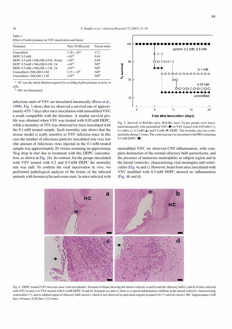
36 F. Stauffer et al. / Antiviral Research 73 (2007) 31–39
Table 1Effect of hydroxylamine on VSV inactivation and fusion
Treatment Titer (TCID50/ml) Fusion index
Unmodified 1.70× 10¹¹ 17.2DEPC 0.5mM <102a 0.69DEPC 0.5mM+NH2OH 0.4M, 30min <102a 0.89DEPC 0.5mM+NH2OH 0.4M, 2 h <102a NDb
DEPC 0.5mM+NH2OH 1.2M, 2 h <102a NDb
Unmodified +NH2OH 0.4M 2.15× 10⁹ NDb
Unmodified +NH2OH 1.2M <102a NDb
a 102 was the initial dilution required for avoiding hydroxylamine toxicity tocells.b ND: not determined.
infectious units of VSV are inoculated intranasally (Reiss et al.,1998). Fig. 3 shows that we observed a survival rate of approxi-mately 45% 7 days after mice inoculationwith unmodifiedVSV,a result compatible with the literature. A similar survival pro-file was obtained when VSV was treated with 0.05mM DEPC,while a mortality of 35%was observed for mice inoculated withthe 0.1mM treated sample. Such mortality rate shows that themouse model is really sensitive to VSV infection since in thiscase the number of infectious particles inoculated was very low(the amount of infectious virus injected in the 0.1mM treatedsample was approximately 20 virions assuming an approximate5log drop in titer due to treatment with this DEPC concentra-tion, as shown in Fig. 1b). In contrast, for the groups inoculatedwith VSV treated with 0.2 and 0.5mM DEPC the mortalityrate was null. To confirm the viral inactivation in vivo, weperformed pathological analysis of the brains of the infectedanimals with hematoxylin and eosin stain. In mice infected with
Fig. 3. Survival of BALB/c mice. BALB/c mice (10 per group) were inocu-lated intranasally with unmodified VSV (�) or VSV treated with 0.05mM (�),0.1mM (�), 0.2mM (�) and 0.5mM (�) DEPC. The mortality rate was evalu-ated daily during 3weeks. The control groupwas inoculatedwithPBScontaining0.5mM DEPC (�).
unmodified VSV, we observed CNS inflammation, with com-plete destruction of the normal olfactory bulb parenchyma, andthe presence of numerous neutrophiles at subpial region and inthe lateral ventricles, characterizing viral meningitis and ventri-culitis (Fig. 4a and c). However, brain frommice inoculatedwithVSV modified with 0.5mM DEPC showed no inflammation(Fig. 4b and d).
Fig. 4. DEPC-treated VSV does not cause viral encephalitis. Sections of brains showing the lateral ventricle (a and b) and the olfactory bulb (c and d) of mice infectedwith VSV (a and c) or VSV treated with 0.5mMDEPC (b and d). In panels (a) and (c), there is a typical inflammatory infiltrate in the lateral ventricle, characterizingventriculitis (*), and in subpial region of olfactory bulb (arrow), which is not observed in equivalent regions at panels (b) (*) and (d) (arrow). HC: hippocampus (A/Bline = 50mm); (C/D line = 12.5mm).
94

F. Stauffer et al. / Antiviral Research 73 (2007) 31–39 37
Fig. 5. Structural preservation of VSV after modification with DEPC. Transmission electron micrographs of (a) unmodified VSV, and (b) VSV treated with 0.5mMof DEPC (bar = 50 nm). (c) Inhibition of binding of an immune serum against untreated VSV to plates coated with VSV by VSVmodified with 0.5mMDEPC (�), orboiled (denaturated) VSV (�), assessed in a competitive ELISA. The maximal binding inhibition profile was determined using the untreated virus (�) as competitionantigen, and bovine albumin (�) was used as negative control. Unspecific binding of VSV (©) or VSV treated with 0.5mM DEPC (�) to sera was discarded usingpre-immune sera.
3.4. Treatment with DEPC does not alter VSV structure
Transmission electron microscopy revealed that virus treatedwith 0.5mMDEPC showed the samemorphology of the unmod-ified VSV, suggesting that the structure of the inactivated viruswas preserved (Fig. 5a and b). The preservation of G proteinantigenic domains afterDEPC treatmentwas analyzed by a com-petition ELISA (Fig. 5c). VSV modified with 0.5mM DEPCbound to an immune serum against unmodified VSV with thesame avidity as the unmodified virus. In contrast, virus dena-tured by boiling presented the same competitive activity asbovine albumin. These results show a clear difference betweenthe antigenic properties of the DEPC-treated and heat-denaturedviruses, although they are still preliminary to assure that the anti-genic properties are completely preserved afterDEPC treatment.
4. Discussion
In this study, we showed that treatment of VSV with DEPCresults in a complete virus inactivation. We observed in cell cul-ture that infectivity and viral replication were abolished by viraltreatment with 0.5mM DEPC and were partially inhibited atlower concentrations.Moreover, DEPC treatment also abolishedVSV lethality in mice. The fact that DEPC is very hydrophilic(Miles, 1977) suggests that it should not be readily transportedacross the membrane. This was supported experimentally bySpires and Begenisich (1990), who showed different effects ofDEPC on neurons when it was added to the intra- or to the
extracellular media. Thus viral treatment with DEPC would notmodify the enzymes located inside the virus particle, and themain mechanism of VSV inactivation by DEPC may reside inthe reaction of this compound with the viral glycoprotein. Ina previous study, we showed that 0.02mM DEPC specificallymodified G protein His residues, inhibiting pH-induced confor-mational changes on G protein and the fusion reaction catalyzedby the virus (Carneiro et al., 2003). However, we cannot excludethe possibility that other residues besides His could be modifiedafter virus treatment with the higher DEPC concentration usedhere, since fusion and inactivation were not restored by incuba-tion of the modified virus with hydroxylamine.The membrane of some enveloped viruses fuses directly with
the host cell plasma membrane through a pH-independent reac-tion, which is driven by virus binding to their cell receptor. Onthe other hand, many other enveloped viruses enter into the cellsby the endocytic pathway and, for them, the membrane fusionreaction depends on the acidification of the endosomal medium.In this case, the low pH triggers conformational changes in theviral glycoproteins necessary for their conversion to the fuso-genic state. For many viruses studied so far, the pH thresholdsfor fusion range between 5.0 and 6.5 (Chan and Kim, 1998;Gaudin et al., 1991; White et al., 1981), suggesting that His pro-tonation (pKa∼ 6.0) might be a more general requirement forthe conversion of viral glycoprotein from the non-fusogenic tothe fusogenic conformation. However, it could not be discardedthat His residues of any viral protein (even from viruses that fusedirectly to plasma membrane or non-enveloped viruses) may be
95

38 F. Stauffer et al. / Antiviral Research 73 (2007) 31–39
necessary for other events during the infection cycle, and thentheir modification with DEPC could inhibit infectivity througha mechanism different from fusion inhibition.Other three compounds described previously promote viral
inactivation by inhibiting membrane fusion: cyanovirin-N (Deyet al., 2000), hypericin and rose bengal (Lenard et al., 1993;Lenard and Vanderoef, 1993). Fusion inhibition due to the treat-ment of virus particles with hypericin or rose bengal resultsfrom the cross-linking of viral membrane proteins. The molec-ular mechanism of fusion inhibition promoted by cyanovirin-Ninvolves physical interactions with gp120 envelope proteins(Boyd et al., 1997). Further studies on this mechanism revealedthat cyanovirin-N binds to high-mannose oligosaccharides ongp120 (Bewley and Otero-Quintero, 2001; Bolmstedt et al.,2001; Shenoy et al., 2001). Cyanovirin-N was also shown tobe potently active against other enveloped viruses contain-ing similar oligosaccharides (Barrientos et al., 2003; Dey etal., 2000; O’Keefe et al., 2003). The immunogenic propertiesof virus inactivated by these compounds have not yet beenevaluated.In the past, the basic mechanism of enveloped viruses inacti-
vationwas to inhibit the virus entry in host cells at the adsorptionstage, e.g., formalin (Bachmann et al., 1993), detergent (Seitzet al., 2002) and UV light (Bay and Reichmann, 1979). How-ever, these inactivation procedures drastically impair inductionof neutralizing IgG responses for most viruses (Bachmann etal., 1994), since these treatments led to the denaturation of viralsurface proteins.The use of DEPC for virus inactivation opens new possibili-
ties for the development of safe vaccines. First, the inactivationby this compound is stable, since it covalently modifies his-tidines. Second, our present data indicate that DEPC treatmentseems to preserve VSV structure. Ultrastructural morphologyanalyses showed that VSV treated with DEPC is similar tounmodified VSV. Furthermore, in marked contrast to virus inac-tivated by heat, DEPC-treated virus was recognized by antibod-ies against the unmodified VSVwith the same avidity. However,although this data is suggestive that inactivated virus preservesthe antigenic properties of the surface G proteins, further workis needed to unequivocally show that virus antigenicity is notaltered, including studies on its reactivity against a panel ofmonoclonal antibodies. Nevertheless, the maintenance of con-formation integrity in DEPC-inactivated virions suggests thatsuch virions may be a useful in the pursuit of a potential vaccineantigen.
Acknowledgements
We thank Adriana Santos de Melo for technical assistance,Dr. Ada M.B. Alves and Maurıcio R.M.P. Luz for helpful sug-gestions andDr. Sergio T. Ferreira for the use of the fluorescencemicroscope.This work was supported by grants from Conselho Nacional
de Desenvolvimento Cientıfico e Tecnologico (CNPq), Cen-tro Argentino-Brasileiro de Biotecnologia (CABBIO) andFundacao Carlos Chagas Filho de Amparo a Pesquisa do Estadodo Rio de Janeiro (FAPERJ).
References
Bachmann, M.F., Kundig, T.M., Kalberer, C.P., Hengartner, H., Zinkernagel,R.M., 1993. Formalin inactivation of vesicular stomatitis virus impairs T-cell- but not T-help-independent B-cell responses. J. Virol. 67, 3917–3922.
Bachmann,M.F., Kundig, T.M., Hengartner, H., Zinkernagel, R.M., 1994. Regu-lation of IgG antibody titers by the amount persisting of immune-complexedantigen. Eur. J. Immunol. 24, 2567–2570.
Barrientos, L.G., O’Keefe, B.R., Bray, M., Sanchez, A., Gronenborn, A.M.,Boyd, M.R., 2003. Cyanovirin-N binds to the viral surface glycoprotein,GP1,2 and inhibits infectivity of ebola virus. Antiviral Res. 58, 47–56.
Bay, P.H., Reichmann, M.E., 1979. UV inactivation of the biological activityof defective interfering particles generated by vesicular stomatitis virus. J.Virol. 32, 876–884.
Berger, S.L., 1975. Diethyl pyrocarbonate: an examination of its properties inbuffered solutions with a new assay technique. Anal. Biochem. 67, 428–437.
Bewley,C.A.,Otero-Quintero, S., 2001. The potent anti-HIVprotein cyanovirin-N contains two novel carbohydrate binding sites that selectively bind toMan(8) D1D3 andMan(9) with nanomolar affinity: implications for bindingto the HIV envelope protein gp120. J. Am. Chem. Soc. 123, 3892–3902.
Bhattacharyya, D.K., Bandyopadhyay, U., Banerjee, R.K., 1992. Chemical andkinetic evidence for an essential histidine in horseradish peroxidase foriodide oxidation. J. Biol. Chem. 267, 9800–9804.
Bolmstedt, A.J., O’Keefe, B.R., Shenoy, S.R., McMahon, J.B., Boyd, M.R.,2001. Cyanovirin-N defines a new class of antiviral agent targetingN-linked,high-mannose glycans in an oligosaccharide-specific manner. Mol. Pharma-col. 59, 949–954.
Boyd,M.R., Gustafson, K.R.,McMahon, J.B., Shoemaker, R.H., O’Keefe, B.R.,Mori, T., Gulakowski, R.J., Wu, L., Rivera, M.I., Laurencot, C.M., Currens,M.J., Cardellina, I.J.H., Buckheit, R.W., Nara, P.L., Pannell, L.K., Sowder,I.R.C., Henderson, L.E., 1997. Discovery of cyanovirin-N, a novel humanimmunodeficiency virus-inactivating protein that binds viral surface enve-lope glycoprotein gp120: potential applications tomicrobicide development.Antimicrob. Agents Chemother. 41, 1521–1530.
Carneiro, F.A., Ferradosa, A.S., Da Poian, A.T., 2001. Low pH-induced confor-mational changes in vesicular stomatitis virus glycoprotein involve dramaticstructure reorganization. J. Biol. Chem. 276, 62–67.
Carneiro, F.A., Bianconi, M.L., Weissmuller, G., Stauffer, F., Da Poian, A.T.,2002.Membrane recognition by vesicular stomatitis virus involves enthalpy-driven protein–lipid interactions. J. Virol. 76, 3756–3764.
Carneiro, F.A., Stauffer, F., Lima, C.S., Juliano, M.A., Juliano, L., Da Poian,A.T., 2003. Membrane fusion induced by vesicular stomatitis virus dependson histidine protonation. J. Biol. Chem. 278, 13789–13794.
Chan, D.C., Kim, P.S., 1998. HIV entry and its inhibition. Cell 93, 681–684.Da Poian, A.T., Gomes, A.M.O., Oliveira, R.J.N., Silva, J.L., 1996. Migrationof vesicular stomatitis virus glycoprotein to the nucleus of infected cells.Proc. Natl. Acad. Sci. U.S.A. 93, 8268–8273.
Da Poian, A.T., Gomes, A.M.O., Coelho-Sampaio, T., 1998. Kinetics of intra-cellular viral disassembly and processing probed by Bodipy fluorescencedequenching. J. Virol. Methods 70, 45–58.
Dey, B., Lerner, D.L., Lusso, P., Boyd, M.R., Elder, J.H., Berger, E.A., 2000.Multiple antiviral activities of cyanovirin-N: blocking of human immun-odeficiency virus type 1 gp120 interaction with CD4 and coreceptor andinhibition of diverse enveloped viruses. J. Virol. 74, 4562–4569.
Dzhandzhugazyan, K.N., Plesner, L., 2000. Diethyl pyrocarbonate inactivatesCD39/ecto-ATPDase by modifying His-59. Biochim. Biophys. Acta 1466,267–277.
Eckert, D.M., Kim, P.S., 2001. Mechanisms of viral membrane fusion and itsinhibition. Annu. Rev. Biochem. 70, 777–810.
Gaudin, Y., Tuffereau, C., Segretain, D., Knossow, M., Flamand, A., 1991.Reversible conformational changes and fusion activity of rabies virus gly-coprotein. J. Virol. 65, 4853–4859.
Hernandez, L.D., Hoffman, L.R.,Wolfsberg, T.G.,White, J.M., 1996. Virus–celland cell–cell fusion. Annu. Rev. Cell Dev. Biol. 12, 627–661.
Lenard, J., Rabson, A., Vanderoef, R., 1993. Photodynamic inactivation of infec-tivity of human immunodeficiency virus and other enveloped viruses usinghypericin and rose bengal: inhibition of fusion and syncytia formation. Proc.Natl. Acad. Sci. U.S.A. 90, 158–162.
96

F. Stauffer et al. / Antiviral Research 73 (2007) 31–39 39
Lenard, J., Vanderoef, R., 1993. Photoinactivation of influenza virus fusion andinfectivity by rose bengal. Photochem. Photobiol. 58, 527–531.
Letchworth, G.J., Rodriguez, L.L., Del C. Barrera, J., 1999. Vesicular stomatitis.Vet. J. 157, 239–260.
Miles, E.W., 1977. Modification of histidyl residues in proteins by diethylpyro-carbonate. Methods Enzymol. 47, 431–442.
O’Keefe, B.R., Smee, D.F., Turpin, J.A., Saucedo, C.J., Gustafson, K.R., Mori,T., Blakeslee, D., Buckheit, R., Boyd, M.R., 2003. Potent anti-influenzaactivity of cyanovirin-N and interactions with viral hemagglutinin. Antimi-crob. Agents Chemother. 47, 2518–2525.
Reed, L.J., Muench, H., 1938. A simple method of estimating fifty per centendpoints. Am. J. Hyg. 27, 493–497.
Reiss, C.S., Plakhov, I.V., Komatsu, T., 1998.Viral replication in olfactory recep-tor neurons and entry into the olfactory bulb and brain. Ann. N. Y. Acad.Sci. 855, 751–761.
Seitz, H., Blumel, J., Schmidt, I., Willkommen, H., Lower, J., 2002. Compa-rable virus inactivation by bovine or vegetable derived Tween 80 duringsolvent/detergent treatment. Biologicals 30, 197–205.
Shenoy, S.R., O’Keefe, B.R., Bolmstedt, A.J., Cartner, L.K., Boyd, M.R.,2001. Selective interactions of the human immunodeficiency virus-inactivating protein cyanovirin-N with high-mannose oligosaccharides ongp120 and other glycoproteins. J. Pharmacol. Exp. Ther. 297, 704–710.
Shriver, Z., Hu, Y., Sasisekharan, R., 1998. Heparinase II from Flavobacteriumheparinum. Role of histidine residues in enzymatic activity as probed bychemical modification and site-directed mutagenesis. J. Biol. Chem. 273,10160–10167.
Skehel, J.J., Wiley, D.C., 2000. Receptor binding and membrane fusion invirus entry: the influenza hemagglutinin. Annu. Rev. Biochem. 69, 531–569.
Spires, S., Begenisich, T., 1990. Modification of potassium channel kinetics byhistidine-specific reagents. J. Gen. Physiol. 96, 757–775.
Struck,D.K., Hoekstra, D., Pagano, R.E., 1981.Use of resonance energy transferto monitor membrane fusion. Biochemistry 20, 4093–4099.
White, J., Matlin, K., Helenius, A., 1981. Cell fusion by Semliki Forest,influenza, and vesicular stomatitis viruses. J. Cell Biol. 89, 674–679.
97

2.2.3 Artigo 5
New chemical method of viral inactivation for vaccine
development based on membrane fusion inhibition
Fausto Stauffer, Joari M. Miranda, Marcos C. Schechter, Fernando A. Queiroz,
Nathalia O. Santos, Ada M.B. Alves e Andrea T. Da Poian
Aceito para publicação em 2007 no periódico
Vaccine

Author's personal copy
Available online at www.sciencedirect.com
Vaccine 25 (2007) 7885–7892
New chemical method of viral inactivation for vaccinedevelopment based on membrane fusion inhibition
Fausto Stauffer a, Joari De Miranda a, Marcos C. Schechter a,Fernando A. Queiroz a, Nathalia O. Santos a,Ada M.B. Alves b, Andrea T. Da Poian a,∗
a Instituto de Bioquımica Medica, Universidade Federal do Rio de Janeiro, Rio de Janeiro, RJ 21941-590, Brazilb Laboratorio de Imunopatologia, Instituto Oswaldo Cruz, Fundacao Oswaldo Cruz, Rio de Janeiro, RJ 21040-900, Brazil
Received 10 July 2007; received in revised form 30 August 2007; accepted 3 September 2007Available online 29 September 2007
Abstract
Membrane fusion is an essential step in the entry of enveloped viruses into their host cells. This process is triggered by conformational changesin viral surface glycoproteins. We have demonstrated previously that modification of vesicular stomatitis virus (VSV) with diethylpyrocarbonate(DEPC) abolished the conformational changes on VSV glycoprotein and the fusion reaction induced by the virus. Moreover, we observedthat viral treatment with DEPC inactivates the virus, preserving the conformational integrity of its surface proteins. In the present work, weevaluated the potential use of DEPC as a viral inactivating chemical agent for the development of useful vaccines. Pathogenicity and viralreplication in Balb/c mice were abolished by viral treatment with 0.5 mM DEPC. In addition, antibodies elicited in mice after intraperitonealimmunization with DEPC-inactivated VSV mixed with adjuvants were able to recognize and neutralize the native virus and efficiently protectedanimals against the challenge with lethal doses of VSV. These results together suggest that viral inactivation with DEPC seems to be a suitablemethod for the development of safe vaccines.© 2007 Elsevier Ltd. All rights reserved.
Keywords: Viral inactivation; Diethylpyrocarbonate; Vesicular stomatitis virus
1. Introduction
Vaccination is a valuable public health tool, being a safeand cost-effective strategy for controlling infectious diseases[1]. The progress in development and use of vaccines resultedin the decline and, in some cases, eradication of impor-tant infectious diseases, as smallpox [2]. Traditionally, thevaccines developed against viruses consist of attenuated orinactivated pathogens or subunits vaccines. Live attenuatedvaccines are based on the attenuation of the pathogen untilits virulence is greatly decreased but its immunogenicity isretained. Alternatively, inactivated vaccines consist of wholekilled virus, while subunit vaccines are based on only one orfew virus proteins. In the case of whole virus inactivated vac-
∗ Corresponding author. Tel.: +55 21 22706264; fax: +55 21 22708647.E-mail address: [email protected] (A.T. Da Poian).
cines, the inactivation treatment through different processes(for review, see [3,4]) must assure that all virus particles areinactivated in order for the vaccine to be safe. Despite theadvances in the field of immunology, molecular biology andgenetics, viral inactivation remains an important procedurein basic research, since it is an easy and relatively cheapapproach to produce new and safe vaccines.
The entry of enveloped viruses into a target cell alwaysrequires virus-mediated membrane fusion catalyzed by viralsurface glycoproteins [5–7]. Virus-induced fusion may occurthrough two different general mechanisms: (i) fusion betweenviral envelope and host cell plasma membrane after virusinteraction with its cellular receptor, or (ii) fusion with theendosomal membrane, after virus internalization by receptor-mediated endocytosis. In the latter case, the decrease in the pHof the endosomal medium triggers conformational changes inviral glycoproteins. This mechanism seems to be conserved
0264-410X/$ – see front matter © 2007 Elsevier Ltd. All rights reserved.doi:10.1016/j.vaccine.2007.09.025
99

Author's personal copy
7886 F. Stauffer et al. / Vaccine 25 (2007) 7885–7892
among several viral families what makes the fusion process apotentially attractive target for viral inactivation approaches.
VSV, a member of Rhabdoviridae family, is composed bya helical ribonucleocapsid surrounded by a lipid bilayer cov-ered with trimers of a single type of an integral glycoprotein,named G protein. VSV enters into the cell by endocytosisfollowed by low-pH-induced membrane fusion [8,9]. Cellrecognition and fusion are mediated by the surface glycopro-tein G. We have been studying the conformational changes inVSV G protein and its interaction with the target membraneduring cellular recognition and fusion events [10,11]. Theobservation of a dramatic reorganization in G protein struc-ture at a very narrow pH range (between pH 5.8 and 6.2) ledus to propose a crucial role for the protonation of histidineresidues (pKa ∼ 6.0) of VSV G protein in membrane fusionmediated by the virus [12]. Indeed, using diethylpyrocarbon-ate (DEPC) to modify His residues of G protein, we showedthat VSV-induced fusion was driven by His protonation atthe pH range of endosomal medium. DEPC is a widely usedtool in chemical modification of proteins because of the highselectivity of the reagent to histidyl residues [13]. This com-pound covalently modifies His and makes them unable to beprotonated. Recently, our group has shown that VSV treat-ment with DEPC inactivates the virus [14]. Moreover, theinactivated virus seemed to preserve the antigenic propertiesof the surface G proteins [14].
Based on these evidences, in the present work, we evalu-ated the potential use of DEPC as a viral inactivating chemicalagent for the development of useful vaccine antigens. Theresults demonstrated that, in the viral model studied, virustreatment with DEPC was able to abolish viral infectivityand pathogenicity, to elicit neutralizing antibodies in miceand to confer protection in these animals against challengeusing lethal doses of VSV.
2. Materials and methods
2.1. Virus propagation and purification
VSV Indiana was propagated in monolayer cultures ofBHK21 cells grown at 37 ◦C in petri dishes containing Dul-becco’s modified Eagle medium supplemented with 10%fetal bovine serum, 100 �g/mL penicillin, 0.0085% strep-tomycin sulfate. When the cells reached confluence, themedium was removed, and the cell monolayer was infectedwith VSV at a multiplicity of infection of 0.1. The cultureswere kept at 37 ◦C for 16–20 h and the virus were harvestedand purified by differential centrifugation followed by equi-librium sedimentation in a sucrose gradient as describedelsewhere [15]. Purified virions were stored at −70 ◦C.
2.2. Virus modification with DEPC
Diethylpyrocarbonate (DEPC) solutions were freshly pre-pared by dilution of the reagent in cold ethanol. The
concentration of stock DEPC solution was determined byreaction with 10 mM imidazole [13]. VSV (13.75 mg/mL)was diluted in PBS, pH 7.4 to a final protein concentra-tion of 1.375 mg/mL and incubated with 0.5 mM DEPC for15 min at room temperature. For mouse immunization, themodified virus was further diluted to a final protein concen-tration of 0.08 mg/mL, leading to a DEPC concentration inthe inoculum lower than 0.03 mM.
2.3. Immunofluorescence and determination of virustiters in mouse brain
Groups of three Balb/c mice (4-week-old) were inocu-lated intranasally with VSV or DEPC-inactivated VSV. Fivedays after inoculation, mice were sacrificed and perfusedwith normal saline (PBS) and brains were removed. Forimmunofluorescence, whole brains were embedded in cry-oprotetion solution (Tissue-Tek OCT compound, Sakura,USA) and frozen at −70 ◦C. Then, serial sections ofeach brain were incubated with mouse monoclonal anti-VSV G protein antibody (Alpha Diagnostic Int., SanAntonio, TX) diluted 1:200 in PBS containing 1% goatserum. As secondary antibody, FITC-labeled goat anti-mouseimmunoglobulin G conjugate was used at 1:400 dilution(Sigma Chemical Co.). Finally, slides were mounted with N-propil-galacto and examined with a Nikon epifluorescencemicroscope equipped for photomicroscopy. For determiningviral titer, brains were homogenized and infectious particlesof VSV in brain homogenates were quantified by TCID50 onBHK21 cells. The cells were grown at 37 ◦C in 96-well platesuntil they reach the confluence. Samples were serially diluted10-fold in culture medium, and 100 �L was added to cells inquadruplicate. Cells were cultured at 37 ◦C in 5% CO2 for24 h and stained with 0.2% crystal violet in 20% ethanol. TheTCID50 was calculated using the method described by Reedand Muench [16].
2.4. Breakdown of the blood brain barrier (BBB)
Groups of three Balb/c mice (4-week-old) were inoculatedintranasally with VSV or DEPC-inactivated VSV. Five daysafter inoculation, mice were injected intraperitoneally with200 �L of 2% Evans blue. One hour later, mice were sac-rificed and perfused with normal saline (PBS). Brains wereremoved and photos were taken.
2.5. Mice immunization and challenge
Groups of 15 Balb/c mice (4-week-old) were inoculated bythe intranasal (i.n.) or intraperitonial (i.p.) routes with VSV orDEPC-inactivated VSV, mixed or not with adjuvants. For i.n.immunization, mice were inoculated with 5 �L containing4 �g of inactivated VSV diluted in PBS, mixed or not with1 �g cholera toxin (CT). For i.p. immunization, mice wereinoculated with 50 �L containing 4 �g of inactivated VSVdiluted in PBS, mixed or not with 50 �L Freund’s adjuvant.
100

Author's personal copy
F. Stauffer et al. / Vaccine 25 (2007) 7885–7892 7887
Animals were separated in three groups, receiving one, twoor three doses of the vaccine, 14 days apart. For i.n. or i.p.infection, mice were inoculated once with 2 × 10⁶ TCID50VSV, diluted in 5 or 50 �L PBS, respectively. Blood wastaken on day 4 after inoculation for measuring IgM responseand on day 14 after each inoculation for measuring IgGresponse. Two weeks after the third i.p. dose, mice inoculatedwith DEPC-treated virus mixed with Freund’s adjuvant werechallenged with 2 × 10⁴ TCID50 of VSV. Survival rates andclinical signs of VSV infection (hyperexcitability, tremors,circling and paralysis) were recorded in the next 10 days.Animals were housed under pathogen-free conditions. Fortesting the durability of immune response, blood was takenonce a month for the period of 1 year after the third i.p.immunization.
2.6. Antibody assays
The anti-VSV antibodies in mouse sera were detected byELISA using VSV as solid-phase bound antigen. Briefly,ELISA microplates (Maxisorp, Nunc) were coated with0.1 �g VSV at 4 ◦C. The plates were washed with PBS/0.05%Tween-20 and blocked with 2% milk powder in PBS/0.05%Tween-20 for 2 h at room temperature. After blocking,plates were washed as before, and incubated for 1 h atroom temperature with serial dilutions (initial 1:50) ofimmune sera collected from immunized mice. Plates werethen washed as before and bound IgG was detected by theaddition of an anti-mouse horseradish peroxidase conju-gate (1:5000 dilution) (Santa Cruz Biotechnology) followedby o-phenylenediamine (OPD) substrate as indicated bythe manufacturer (Sigma Chemical Co.). The reaction wasstopped with 3 M sulfuric acid and the optical density at492 nm was determined in an ELISA reader. Titers wereestablished as the reciprocal of serum dilution, which gave anabsorbance above that of the respective pre-immune serum.
2.7. Serum neutralization test
The sera were initially heat inactivated for 30 min at 56 ◦C.Serial twofold dilutions in DMEM without FBS were mixedwith equal volumes of unmodified VSV diluted to contain 103
infectious particles. The mixture was incubated for 90 min at37 ◦C in an atmosphere with 5% CO2. Then the serum–virusmixture was transferred onto BHK21 cell monolayers in 96-well plates. After incubation for 24 h at 37 ◦C, the monolayerwas fixed and stained with 0.5% crystal violet. The 50% end-point dilution of each serum, corresponding to the dilution atwhich 50% of wells were completely protected from infec-tion, was determined according to the Reed–Muench method[16].
2.8. Competition ELISA
ELISA microplates were coated with 0.1 �g VSVovernight at 4 ◦C. Plates were washed with PBS/0.05%
Tween-20 and blocked with 2% milk powder in PBS/0.05%Tween-20 for 2 h at room temperature. After blocking, theplates were washed as before. Subsequently, immune serumcollected from vaccinated mice (animals after three i.p. inoc-ulation of 4 �g of DEPC-treated VSV mixed with adjuvant)was incubated at 37 ◦C for 1 h at a fixed dilution togetherwith serial dilutions of unmodified VSV. As positive con-trol, the immune serum collected from mice 7 days after i.n.inoculation of VSV was incubated with unmodified VSV inthe same dilutions of the other incubation. Albumin at thesame protein concentration was used with sera as a neg-ative control. After incubation, samples were transferredto antigen-coated ELISA plates and incubated for 1 h atroom temperature. Plates were washed as before and boundIgG was detected by addition of an anti-mouse horseradishperoxidase conjugate (1:5000 dilution) (Santa Cruz Biotech-nology) followed by o-phenylenediamine (OPD) substrateas indicated by the manufacturer (Sigma Chemical Co.).The reaction was stopped with 3 M sulfuric acid and theoptical density at 492 nm was determined in an ELISAreader.
3. Results
3.1. DEPC treatment abolishes VSV infectivity
To determine the effect of VSV treatment with DEPCin viral replication in mouse CNS, the viral protein synthe-sis in brain sections (Fig. 1a) and the viral titers in brainhomogenates (Fig. 1b) from mice 5 days after nasal inoc-ulation with unmodified or modified VSV were measured.Viral protein synthesis was followed by an immunofluores-cence assay using an anti-G protein antibody (Fig. 1a). Inbrains from mice infected with unmodified VSV, we observeda great fluorescence staining, indicating the presence of Gprotein synthesis. On the other hand, when the virus was pre-treated with 0.5 mM DEPC, no staining was observed in thebrain. In addition, viral titer recovered from brains of miceinoculated with unmodified VSV was 105 TCID50/mL, whileno infectious particles were recovered from mice inoculatedwith treated VSV (Fig. 1b).
To further confirm that DEPC treatment abolishes VSVinfectivity in mice, the integrity of blood brain barrier(Fig. 1c) and the survival rate (Fig. 1d) of mice inoculatedintranasally with VSV unmodified or treated with DEPC wereanalyzed. We observed that VSV-infected mice showed dis-ruption of the BBB, while mice inoculated with VSV treatedwith DEPC did not (Fig. 1c). Moreover, we observed a sur-vival rate of approximately 45% 7 days after inoculationwith unmodified VSV (Fig. 1d), which is in accordance toother reports [17]. In contrast, all animals inoculated withDEPC-treated VSV survived with no clinical signs of disease.Additionally, no death or clinical signs of chemical toxicitywere recorded among the control group inoculated with PBScontaining 0.5 mM DEPC.
101

Author's personal copy
7888 F. Stauffer et al. / Vaccine 25 (2007) 7885–7892
Fig. 1. Evaluation of the pathogenic potential of DEPC-treated VSV. (a) Mice were inoculated intranasally with unmodified VSV (top panel) or VSV treatedwith 0.5 mM DEPC (bottom panel) and after 5 days the brains were removed and an immunofluorescence using an anti-VSV G protein monoclonal antibodywere performed in brain sections. Magnification was 200×. (b) Viral replication in mouse brain was determined by TCID50 in brain homogenates on day4 post-intranasal inoculation of VSV or DEPC-treated VSV. (c) Breakdown of the BBB following mouse inoculation with unmodified VSV (top panel) orDEPC-treated virus (bottom panel) was analyzed through whole brain staining with Evans blue after an i.p. inoculation of the dye. The brain was removed 1 hafter dye inoculation from a mouse on day 5 post-inoculation. (d) BALB/c mice (n = 10 per group) were inoculated intranasally with unmodified VSV (blackbar), VSV treated with 0.5 mM DEPC (gray bar) or PBS containing 0.5 mM DEPC (white bar) and mortality rates were evaluated 1 week after inoculation.
3.2. Inactivated VSV induces B-cell response
VSV is known to induce an antibody response withIgM peak around day 4, while specific IgG rises betweendays 6 and 8, peaking around day 14 [18,19]. To deter-mine whether VSV inactivation with DEPC had an influence
Fig. 2. Analysis of IgM response induced by DEPC-inactivated VSV. Micewere inoculated by the i.n. (a) or i.p. (b) routes with native or inactivatedVSV, mixed or not with adjuvants (n = 10 per group). The cholera toxin (CT)and Freund’s adjuvant were used as adjuvants for i.n. and i.p. administra-tions, respectively. IgM titers were determined by ELISA on day 4 afterinoculation, using the native VSV as solid-phase bound antigen. Titers wereestablished as the reciprocal serum dilutions that gave an absorbance abovethat of pre-immune sera.
on the immunogenicity of viral antigens, the antibodyresponse of BALB/c mice immunized with 4 �g of inac-tivated VSV was compared to that of mice infected with2 × 106 TCID50 VSV. Mice were inoculated intranasallyor intraperitoneally. In the case of inactivated virus, ani-mal groups received one, two or three doses given 2 weeksapart, combined or not with adjuvants. Blood samples weretaken on day 4 after the first inoculation for the analysis ofIgM response (Fig. 2) and 2 weeks after each immunizationfor the detection of IgG response (Fig. 3). DEPC inactiva-tion led to a reduced IgM response, even in combinationwith adjuvants. Analysis of IgG response showed an induc-tion of antibody titers. The IgG titers obtained after threedoses of inactivated virus combined with adjuvant inocu-lated by the i.p. route were similar to that observed afteri.n. inoculation of unmodified virus. This result confirmedthat DEPC inactivation did not alter VSV immunogenic-ity. Time course of anti-VSV IgG response was analyzedafter i.n and i.p. inoculation of DEPC-inactivated virus,revealing that antibody titers increased after each dose andremained stable in both cases (Fig. 4). IgG titers decreasedapproximately 50% 6 months after the third inoculation(30,573 ± 1643) and detectable titers were still observed after1 year (14,629 ± 1360).
3.3. VSV is efficiently recognized and neutralized byserum antibodies induced in vaccinated mice
In order to evaluate the effectiveness of the humoralimmune response elicited in vaccinated mice, a neu-tralization assay (Fig. 5a) and a competition ELISA
102

Author's personal copy
F. Stauffer et al. / Vaccine 25 (2007) 7885–7892 7889
Fig. 3. Induction of IgG response induced by DEPC-inactivated VSV. Mice were inoculated by the i.n. (a) or i.p. (b) routes with native or inactivated VSV,mixed or not with adjuvants (n = 10 per group). The CT and Freund’s adjuvant were used as adjuvants for i.n. and i.p. administrations, respectively. One, twoor three doses were used in each group given 14 days apart, as shown in the figure. Sera were collected 14 days after each inoculation and IgG titers weredetermined by ELISA, using the native VSV as solid-phase bound antigen. Titers were established as the reciprocal serum dilutions that gave an absorbanceabove that of pre-immune sera.
(Fig. 5b) were performed. Fig. 5a shows that both seraobtained from mice inoculated with unmodified VSV(control) or inoculated with VSV treated with DEPC(vaccinated) presented similar neutralizing activity againstunmodified VSV. Supporting this result, we observed inFig. 5b that antibodies raised against VSV modified with0.5 mM DEPC bound to VSV with the same avidityas the control serum, produced against the unmodifiedVSV. These results indicate that the antibodies elicitedin vaccinated mice were able to recognize and neutralizeVSV.
3.4. Immunized mice become protected against lethalVSV challenge
Mice immunized with three i.p. doses of DEPC-treatedVSV mixed with Freund’s adjuvant were challenged witha sub-lethal dose of VSV given by the intracerebral routeand animals were monitored the next 10 days for mortal-ity and clinical signs of disease (hyperexcitability, tremors,circling and paralysis). A 100% protection was observed inmice immunized with DEPC-inactivated VSV while only30% of non-immunized animals survived after challenge
Fig. 4. Time course IgG response after intranasal or intraperitoneal immunization. Mice were inoculated intranasally (a) or intraperitoneally (b) with DEPC-inactivated VSV, mixed with the adjuvants cholera toxin (CT) and Freund’s adjuvant, respectively (n = 10 per group). One (�), two (�) or three (�) dosesgiven 14 days apart were used for each group. Serum samples were collected 14 days after each inoculation and IgG titers were determined by ELISA, usingthe native VSV as solid-phase bound antigen. Titers were established as the reciprocal serum dilutions that gave an absorbance above that of pre-immune sera.
103

Author's personal copy
7890 F. Stauffer et al. / Vaccine 25 (2007) 7885–7892
Fig. 5. Antibody recognition and neutralization of VSV. (a) Sera collectedfrom mice inoculated with three i.p. doses of DEPC-treated VSV mixedwith Freund’s adjuvant (vaccinated) or from mice after i.n. inoculation ofinfectious VSV (control) were tested in a neutralization assay for VSV. Neu-tralizing titer, in a log10 scale, represent the highest serum dilution thatreduced the number of virus plaques by 50%. (b) Competitive ELISA wasused to compare the VSV binding avidity of antibodies elicited in miceimmunized with native or DEPC-treated VSV. Immune sera collected fromvaccinated mice (animals after three i.p. inoculation of DEPC-treated VSVmixed with adjuvant) (�, �) or mice inoculated intranasally with nativeVSV (�, �) were incubated at a fixed dilution together with serial dilutionsof unmodified VSV (�, �) or bovine serum albumin as a control (�, �). Asanother control, pre-immune serum was incubated with serial dilutions ofthe native VSV (©). Serum samples were then assayed in an IgG ELISA,using the native VSV as solid phase antigen.
(Fig. 6). Such results confirmed data presented above show-ing that the inactivation of VSV with DEPC maintained itsimmunogenicity and its capacity of inducing protection. Datacollected from a group of challenge experiments suggest thatprotection was not achieved when the IgG titers were lowerthan 15,000 (not shown), suggesting that the three i.p. doseswere necessary to generate immunity.
4. Discussion
Whole inactivated virions have been used successfully asvaccines for numerous viruses [20,21]. The technique mostlyused for viral inactivation is the mild treatment of viruses withformalin or �-propiolactone. However, these methods havedisadvantages as long periods of incubation for successfulvirus inactivation and toxicity of the compound to the host.Moreover, these inactivation procedures drastically impairinduction of neutralizing IgG responses for most viruses [22],since these treatments led to the denaturation of viral surfaceproteins. Finally, there have been reports of failures on inac-tivation associated with both of these methodologies evenin the recent past, predominantly with formaldehyde [23],which makes the discovery of new procedures and substancesfor the virus inactivation extremely relevant and important.
Recently, our group has shown that DEPC is a virus-inactivating compound that seemed to preserve the viral
antigenic properties [14]. The mechanism of inactivation byDEPC may reside in the reaction of this compound with theviral glycoprotein, specifically modifying its His residues,since we have previously shown that His protonation isrequired for pH-induced conformational changes on VSV Gprotein necessary for the fusion reaction induced by the virus[12].
In the present report, we showed that the treatment ofVSV with DEPC completely abolishes virus infectivity andpathogenicity in mice. When VSV is applied to the nasalneuroepithelium, it initially replicates in olfactory receptorneurons, and then it is transmitted along the olfactory nerveto the central nervous system (CNS) within 12 h [17]. In theolfactory bulb, the virus replicates invasively through the lay-ers of the olfactory bulb, reaching the brain ventricles by day4–5 post-infection, and the hindbrain by day 8 post-infection.In mice, infection causes encephalitis, usually due to theblood brain barrier (BBB) disruption, and may result in a 50%mortality rate when 2 × 10⁶ infectious units of VSV are inoc-ulated intranasally [17]. Thus, the absence of a pathogeniceffect of DEPC-treated VSV inoculation was evaluated notonly by its lethality in mice but also by the BBB integrity. Allanimals injected with DEPC-treated VSV survived after inoc-ulation with no clinical signs. Furthermore, since dyes suchas Evans blue, which are normally excluded from the brain bythe intact BBB, are able to stain this organ when the integrity
Fig. 6. Survival after challenge. Non-immunized mice (control) and ani-mals inoculated with three i.p. doses of DEPC-treated VSV mixed withFreund’s adjuvant (vaccinated) were challenged with an intracranial injec-tion of 2 × 10⁴ TCID50 of VSV, 2 weeks after the third dose. Survival rateswere recorded 10 days after the challenge.
104

Author's personal copy
F. Stauffer et al. / Vaccine 25 (2007) 7885–7892 7891
of the BBB is broken, native VSV or DEPC-treated VSVinoculated mice were injected with this dye and brain stain-ning was compared. Results revealed that the BBB integritywas maintained only in DEPC-treated VSV. Additionally, thepresence of VSV in the central nervous system and virus anti-gens in brain sections were also investigated, showing thatthe treatment of VSV with DEPC completely abolished anyreplication of the virus in the brain of inoculated animals.
After the observation that DEPC-treated VSV had infact became inactivated, the immunogenic characteristicsof this virus were further evaluated. Mice inoculated onlywith DEPC-inactivated VSV presented low level of VSV-specific antibodies. Such results were already expected,since it is well known that inactivated vaccines, as well assubunit protein vaccines, usually induce a weak immuneresponse when administered without adjuvants [24]. In orderto increase the antibody response against DEPC-treatedVSV, animals were immunized with a mixture of virusand adjuvants, the cholera toxin (CT) and Freund’s adju-vant, for i.n. and i.p. immunizations, respectively. Bothadjuvants enhanced the antibody response against inacti-vated VSV, although immunization with Freund’s adjuvantinduced higher levels of VSV-specific IgG. Cholera toxinwas used in these experiments due to its high efficiency forthe induction of systemic and mucosal immune responses[25,26]. However, the effect of such adjuvant was not asstrong as the Freund’s adjuvants for the systemic antibodyresponse.
Although the differences in the magnitude of the humoralimmune response induced with both adjuvants, in the twocases, the levels of specific-VSV antibodies were dose-dependent, with maximal IgG titers attained after the thirdvirus dose. Furthermore, vaccination with three i.p. dosesof DEPC-inactivated VSV mixed with Freund’s adjuvantelicited specific antibody levels similar to those observedafter the infection with non-inactivated VSV. Moreover,antibody titers remained high for the following 6 months,indicating that the administration of DEPC-inactivated VSVis able to induce a long-lasting immune response. Besides,these antibodies recognized the native VSV with the sameavidity as antibodies raised against the unmodified virus andwere able to efficiently neutralize the native virus in in vitroexperiments. Such results indicated that the treatment ofVSV with DEPC did not modify conformational structuresof VSV proteins, preserving the immunogenic characteristicsof native VSV. In order to confirm and extend these obser-vations, the capacity of DEPC-inactivated VSV in providingprotection against the challenge with infectious VSV wasfurther analyzed. All the animals vaccinated with inactivatedvirus survived after challenge, while 75% of control animalsdied after inoculation with unmodified VSV.
All these results indicate that viral inactivation with DEPCseems to be a suitable method for the development of safevaccines by multiples reasons: (a) the inactivation by thiscompound is stable, since it covalently modifies His residuesin viral proteins [13]; (b) although DEPC is a cytotoxic agent,
it is important to point out that free DEPC is very unstablein aqueous solution, being rapidly hydrolyzed [13,27], whatassures that no free DEPC would be present in the inactivatedvirus preparation; (c) previous data indicated that DEPC treat-ment seems to preserve the structure of viral proteins [14];(d) our present data indicate that the antibodies elicited byimmunization with DEPC-inactivated VSV were able to rec-ognize and neutralize the native virus and efficiently protectanimals against the challenge with native virus.
However, it is important to point out that the proposedapproach is limited because it should only work when (a) theuptake of virus involve the low pH-dependent endocytosisentry pathway and the reorganization of viral glycoproteinsimplies the protonation of histidine residues and (b) antibodyresponse alone will be sufficient for inducing protection orlong-term control of viral replication.
Acknowledgements
This work was supported by grants from ConselhoNacional de Desenvolvimento Cientıfico e Tecnologico(CNPq), Fundacao Carlos Chagas Filho de Amparo aPesquisa do Estado do Rio de Janeiro (FAPERJ) andCoordenacao de Aperfeicoamento de Pessoal de Nıvel Supe-rior (CAPES).
References
[1] Ada GL, Ramsay AJ. Vaccines, vaccination, and the immune response.Philadelphia: Lippincott-Raven; 1997.
[2] Fenner F. Smallpox and its eradication. Geneve: WHO; 1988.[3] Ada G. Vaccines and vaccination. N Engl J Med 2001;345(14):
1042–53.[4] Stauffer F, El-Bacha T, Da Poian AT. Advances in the development
of inactivated virus vaccines. Recent Patents Anti-Infect Drug Discov2006;1(3):291–6.
[5] Eckert DM, Kim PS. Mechanisms of viral membrane fusion and itsinhibition. Annu Rev Biochem 2001;70:777–810.
[6] Hernandez LD, Hoffman LR, Wolfsberg TG, White JM. Virus–cell andcell–cell fusion. Annu Rev Cell Dev Biol 1996;12:627–61.
[7] Skehel JJ, Wiley DC. Receptor binding and membrane fusionin virus entry: the influenza hemagglutinin. Annu Rev Biochem2000;69:531–69.
[8] Da Poian AT, Gomes AM, Coelho-Sampaio T. Kinetics of intracellu-lar viral disassembly and processing probed by Bodipy fluorescencedequenching. J Virol Methods 1998;70(1):45–58.
[9] White J, Matlin K, Helenius A. Cell fusion by Semliki Forest, influenza,and vesicular stomatitis viruses. J Cell Biol 1981;89(3):674–9.
[10] Carneiro FA, Ferradosa AS, Da Poian AT. Low pH-inducedconformational changes in vesicular stomatitis virus glycoproteininvolve dramatic structure reorganization. J Biol Chem 2001;276(1):62–7.
[11] Carneiro FA, Bianconi ML, Weissmuller G, Stauffer F, Da Poian AT.Membrane recognition by vesicular stomatitis virus involves enthalpy-driven protein–lipid interactions. J Virol 2002;76(8):3756–64.
[12] Carneiro FA, Stauffer F, Lima CS, Juliano MA, Juliano L, Da PoianAT. Membrane fusion induced by vesicular stomatitis virus depends onhistidine protonation. J Biol Chem 2003;278(16):13789–94.
[13] Miles EW. Modification of histidyl residues in proteins by diethylpy-rocarbonate. Methods Enzymol 1977;47:431–42.
105

Author's personal copy
7892 F. Stauffer et al. / Vaccine 25 (2007) 7885–7892
[14] Stauffer F, De Miranda J, Schechter MC, Carneiro FA, Salgado LT,Machado GF, et al. Inactivation of vesicular stomatitis virus throughinhibition of membrane fusion by chemical modification of the viralglycoprotein. Antiviral Res 2007;73(1):31–9.
[15] Da Poian AT, Gomes AM, Oliveira RJ, Silva JL. Migration of vesicularstomatitis virus glycoprotein to the nucleus of infected cells. Proc NatlAcad Sci USA 1996;93(16):8268–73.
[16] Reed LJ, Muench H. A simple method of estimating fifty percent end-points. Am J Hyg 1938;27:493–7.
[17] Reiss CS, Plakhov IV, Komatsu T. Viral replication in olfactory receptorneurons and entry into the olfactory bulb and brain. Ann N Y Acad Sci1998;855:751–61.
[18] Charan S, Zinkernagel RM. Antibody mediated suppression of sec-ondary IgM response in nude mice against vesicular stomatitis virus. JImmunol 1986;136(8):3057–61.
[19] Leist TP, Cobbold SP, Waldmann H, Aguet M, Zinkernagel RM. Func-tional analysis of T lymphocyte subsets in antiviral host defense. JImmunol 1987;138(7):2278–81.
[20] Clemens R, Safary A, Hepburn A, Roche C, Stanbury WJ, Andre FE.Clinical experience with an inactivated hepatitis A vaccine. J Infect Dis1995;171(1):S44–9.
[21] Murdin AD, Barreto L, Plotkin S. Inactivated poliovirus vac-cine: past and present experience. Vaccine 1996;14(8):735–46.
[22] Bachmann MF, Bast C, Hengartner H, Zinkernagel RM. Immunogenic-ity of a viral model vaccine after different inactivation procedures. MedMicrobiol Immunol 1994;183(2):95–104.
[23] Brown F. Review of accidents caused by incomplete inactivation ofviruses. Dev Biol Stand 1993;81:103–7.
[24] O’Hagan DT, MacKichan ML, Singh M. Recent developmentsin adjuvants for vaccines against infectious diseases. Biomol Eng2001;18(3):69–85.
[25] Lycke N, Holmgren J. Strong adjuvant properties of cholera toxin on gutmucosal immune responses to orally presented antigens. Immunology1986;59(2):301–8.
[26] Elson CO, Ealding W. Generalized systemic and mucosal immunityin mice after mucosal stimulation with cholera toxin. J Immunol1984;132(6):2736–41.
[27] Berger SL. Diethyl pyrocarbonate: an examination of its propertiesin buffered solutions with a new assay technique. Anal Biochem1975;67:428–37.
106

DISCUSSÃO

3 Discussão
3.1 Parte I
Elucidação dos mecanismos da fusão de membranas mediada pelo VSV e pelo dengue
A entrada dos vírus na célula hospedeira é essencial para infecção viral. No caso dos
vírus envelopados, o mecanismo de entrada envolve uma etapa de fusão com membranas
celulares (plasmática ou endossomal). Este processo é mediado pelas proteínas de fusão que
estão presentes no envelope viral e desencadeado após a interação do vírus com seu receptor
celular específico ou exposição ao pH ácido do meio endossomal. Estes gatilhos acarretam na
mudança conformacional das proteínas de fusão, com a subsequente exposição do peptídeo de
fusão. Este segmento das glicoproteínas virais desestabiliza a membrana alvo, iniciando a
reação de fusão. Sendo assim, o conhecimento das mudanças conformacionais das proteínas
de fusão virais e de seus respectivos peptídeos de fusão e a identificação de resíduos de
aminoácidos essenciais para a fusão de membranas são etapas básicas para a elucidação dos
mecanismos moleculares do processo de fusão, permitindo o reconhecimento de possíveis
alvos para inibição da entrada dos vírus nas células. Nesta tese, acreditamos ter contribuído
para o avanço na elucidação do processo de fusão de membranas induzido pelo VSV e pelo
vírus da dengue.
3.1.1 VSV
As mudanças estruturais sofridas pela proteína G durante a acidificação, assim como a
aquisição da capacidade de interagir com membranas negativamente carregadas ocorrem em
uma faixa de pH bastante estreita, entre 6,2 e 5,8 (White et al., 1981; Carneiro et al., 2001).
Dentre os 20 aminoácidos que constituem as proteínas, 5 contém grupos ionizáveis presentes
em sua cadeia lateral, e, portanto, estão sujeitos a variações em sua carga em função de
variações no pH. Dentre eles, apenas a histidina apresenta pK de ionização de sua cadeia
lateral nesta faixa estreita de pH (pK ~ 6,04). Os outros 4 aminoácidos ou apresentam pKs
muito elevados - arginina (pK ~ 12,48), lisina (pK ~ 10.54), ou muito baixos - aspartato (pK ~
3,90) e glutamato (pK ~ 4,07). Isso nos sugeriu que a protonação de resíduos de histidina
presentes na proteína G seria necessária para o desencadeamento do processo de fusão.
Através de modificação das histidinas da proteína G com DEPC, demonstramos que
tanto a mudança conformacional quanto a fusão de membranas catalisadas pelo VSV são
dependentes da protonação de histidinas que ocorre durante a acidificação do meio
endossomal (artigo 2). Antes da publicação deste artigo, o envolvimento de resíduos de
108

histidina não havia sido avaliado como importante no processo de fusão mediado pelas
glicoproteínas virais.
Após a publicação do artigo 2 em 2003, outros grupos mostraram a importância de
resíduos de histidina, tanto na aquisição da conformação fusogênica (Stevens et al., 2004;
Roussel et al., 2006), quanto na infectividade e atividade fusogênica viral (Chanel-Vos e
Kielian, 2004). Em 2005, publicamos uma revisão sobre proteínas de fusão viral, ressaltando
a importância da protonação de resíduos de histidina para a aquisição da conformação
fusogênica e interação com a membrana alvo (anexo 2) (Da Poian et al., 2005). Mais
recentemente, foi proposto um mecanismo para a mudança conformacional das proteínas de
fusão desencadeada pelo pH ácido do meio endossomal, que envolve diretamente a
protonação das histidinas (fig. 15). Segundo este modelo, que foi em parte baseado no artigo 2
e na revisão de 2005 publicados por nós, a protonação de resíduos de histidina específicos,
localizados adjacentes a aminoácidos carregados positivamente, acarretaria no deslocamento
das histidinas com a subseqüente formação de pontes salinas que estabilizariam a nova
conformação adquirida (Kampmann et al., 2006).
Figura 15: Diagrama esquemático do mecanismo proposto para mudança conformacional induzida pelo pH ácido. Nesta figura, estão
representadas as interações dos resíduos de histidina nas proteínas de fusão viral nas conformações pré e pós-fusogênicas. No estado pré-
fusão, os resíduos de histidina (em vermelho) estão localizados próximo a um domínio (em vinho) composto de aminoácidos carregados
positivamente (representados pelo símbolo + em azul). No pH ácido, estes resíduos de histidina se tornam protonados e carregados
positivamente. Isto favorece interações eletrostáticas com cadeias laterais de aminoácidos carregados negativamente (em verde), acarretando
na movimentação dos resíduos de histidina com a formação de novas pontes salinas. Desta forma, a nova conformação adquirida torna-se
mais estável. As pontes de hidrogênio envolvendo resíduos de histidina como aceptores também são “pertubadas” pela protonação. Extraído
de Kampmann et al. (2006).
A formação das pontes salinas envolveria interações eletrostáticas entre os resíduos de
histidina carregados positivamente e cadeias laterais de aminoácidos carregados
negativamente, como os ácidos aspárticos e glutâmicos. Recentemente, nosso grupo
identificou que os resíduos D153 e E158 da proteína G do VSV são importantes para o processo
109

de fusão (Carneiro et al., 2006b). Foi sugerido que estes aminoácidos participariam de
interações eletrostáticas intermoleculares, estabilizando a conformação fusogênica adquirida
no pH ácido do meio endossomal (Carneiro et al., 2006b). Estas observações também
sustentam o modelo proposto por Kampmann et al., (2006).
Especificamente na proteína G do VSV, foram identificados resíduos de histidina,
conservados nos rabdovírus (H423, H178, H76), que estão associados na conformação pré-
fusogênica (Roche et al., 2007). A protonação destes resíduos provavelmente desestabiliza a
interação entre o segmento C-terminal e o domínio de fusão, sendo o gatilho para o
movimento inicial que dirige o loop de fusão para o topo da molécula, em direção a
membrana alvo (Roche et al., 2007). A conformação pós-fusogênica é estabilizada por novas
pontes de hidrogênio e pontes salinas, como a interação entre os resíduos H423 e D153 (Roche
et al., 2007). Nós já havíamos identificado estes aminoácidos como importantes para o
processo de fusão (artigo 2 e (Carneiro et al., 2006b)).
Ainda no artigo 2, propusemos um novo peptídeo relacionado com a fusão do VSV. A
identificação deste segmento da proteína G se iniciou com base nos dados de espectrometria
de massa, onde foram observados quatro fragmentos trípticos que tiveram pelo menos uma de
suas histidinas modificadas pelo DEPC: os segmentos entre os resíduos 3-63, 32-87, 110-168
e 392-417 (artigo 2, Tabela II).
O fragmento 110-168 apresenta, em sua seqüência, uma região de aminoácidos
conservada entre os rabdovírus, denominada peptídeo p2-like (Coll, 1995). No caso do VSV,
o peptídeo p2-like compreende os resíduos de aminoácidos 145-168 da proteína G. Existiam
evidências do envolvimento deste peptídeo no processo de fusão mediado pelo VSV.
Primeiro, neste segmento estão contidos os resíduos H148 e H149, que foram modificadas pelo
DEPC nos nossos experimentos. Além disso, havia sido demonstrado que este peptídeo
apresentava capacidade de ligar-se seletivamente a fosfatidilserina (PS) (Coll, 1997), um
fosfolipídio carregado negativamente que possui papel importante tanto na ligação do VSV à
membrana da célula hospedeira (Carneiro et al., 2006a) (anexo C) quanto no processo de
fusão mediado por este vírus (Carneiro et al., 2002) (anexo A).
O primeiro trabalho a identificar que esta seqüência da proteína G dos rabdovírus
estava envolvida na interação com membranas foi realizado com o vírus da septicemia
hemorrágica viral (VHSV). Este rabdovírus que infecta salmonídeos possuía um segmento
localizado entre os resíduos 82 e 109 da proteína G capaz de se ligar a PS e que foi
relacionado por evidências indiretas ao processo de fusão membranas (Estepa e Coll, 1996).
Este peptídeo foi denominado p2 e é homólogo ao segmento 145 a 168 da proteína G do VSV
110

(Coll, 1995). Posteriormente, esses autores demonstraram que um fragmento maior, contendo
o peptídeo p2, possuía atividade fusogênica e por isso estaria implicado na fusão mediada
pelo VHSV (Estepa et al., 2001).
No caso do VSV, o peptídeo p2-like foi definitivamente relacionado com a fusão de
membranas após a realização de experimento de fusão de lipossomas com diferentes
peptídeos sintéticos (artigo 2, fig. 6). Este segmento da proteína G foi tão eficiente quanto o
vírus inteiro em mediar a fusão de lipossomas. A reação de fusão catalisada pelo peptídeo p2-
like possuía as mesmas características daquela catalisada pelo VSV: dependência de pH ácido
e da presença de fosfolipídio negativo (PS). Já o suposto peptídeo de fusão do VSV
(segmento 117-137) (ver seção 1.3.3) (Zhang e Ghosh, 1994; Fredericksen e Whitt, 1995) não
era capaz de promover a fusão de membranas.
Tanto o segmento 145-168 (p2-like) quanto o
117-137 estão inseridos na porção da proteína G (59-
221) capaz de interagir com membranas durante as
transições conformacionais decorrentes da acidificação
(Durrer et al., 1995). Com a determinação da estrutura
cristalográfica da proteína G, sugeriu-se a existência de
um domínio de fusão (resíduos 69-188) (fig. 16), uma
estrutura composta de folhas beta, que contém dois
loops onde são encontrados resíduos aromáticos (W88,
Y89, Y132, A133) (Roche et al., 2006). Estes
aminoácidos estão no topo da molécula e seriam
responsáveis pela desestabilização inicial da membrana
alvo. Sendo assim, as seqüências de aminoácidos 145-
168 e 117-137 (assinaladas na fig. 16) podem não
representar especificamente o peptídeo de fusão da
proteína G do VSV, apesar de provavelmente
possuírem papéis importantes durante o processo de
fusão.
Figura 16: Estrutura cristalográfica do
domínio de fusão da proteína G do VSV
nas conformação pós fusogênica.
Segmentos 145-168 (p2-like) em
vermelho, 117-137 em azul e aminoácidos
aromáricos em amarelo.
3.1.2 Vírus da Dengue
Apesar dos estudos estruturais realizados com a glicoproteína E do vírus da dengue,
até o presente momento não existem trabalhos publicados com seu peptídeo de fusão. Este
111

segmento da proteína E, que compreende os aminoácidos 98-110, apresenta grande homologia
entre os flavivírus. Corresponde a um loop, no meio de folhas beta, localizado no domínio II,
que contém aminoácidos hidrofóbicos (W101, L107 e F108). Este peptídeo se encontra
“escondido” nos homodímeros de proteína E em pH neutro e exposto no topo da molécula nos
homotrímeros em pH ácido, iniciando o processo de fusão através da desestabilização da
membrana alvo. Estudos prévios de substituições de aminoácidos nesta região foram capazes
de abolir a fusão de membranas mediada pela proteína E de outros flavivírus (ex. TBE vírus)
(Allison et al., 2001).
Através do uso de metodologias baseadas em espectroscopia de fluorescência,
estudamos a interação do peptídeo de fusão do vírus da dengue com modelos de membranas
(artigo 3). Neste artigo, mostramos que o peptídeo interage com membranas e é capaz de
induzir fusão de lipossomas, indicando que realmente esta seqüência pode estar catalisando a
fusão de membranas mediada pelo vírus da dengue. Tanto a partição para membranas quanto
a reação de fusão foram mais intensas na presença de fosfolipídios negativos e são
dependentes do pH e da força iônica do meio, mostrando a importância de interações
eletrostáticas entre o peptídeo e componentes carregados negativamente das membranas alvo,
como já havíamos mostrado para o VSV (Carneiro et al., 2002; Carneiro et al., 2006b).
Adicionalmente, mostramos que provavelmente ocorre oligomerização do peptídeo da dengue
na presença de fosfolipídios negativos. Os peptídeos oligomerizados adquirem uma nova
conformação que supostamente favorece a desestabilização da membrana com subseqüente
reação de fusão mais eficiente, como foi observado.
A importância da oligomerização das glicoproteínas virais para o processo de fusão é
inquestionável (Harrison, 2005; Weissenhorn et al., 2007). Estudos prévios já demonstraram
que peptídeos de fusão oligomerizados são mais fusogênicos do que monômeros ou grandes
agregados (Lau et al., 2004; Yang et al., 2004) e que, geralmente, a oligomerização desses
peptídeos é desencadeada por mudanças na sua estrutura secundária (Han e Tamm, 2000;
Yang e Weliky, 2003; Li et al., 2004).
No caso da proteína E do vírus da dengue, ocorre a formação de trímeros e exposição
dos peptídeos de fusão no topo da molécula desencadeadas pela exposição ao pH ácido do
meio endossomal. Apesar da glicoproteína viral sofrer uma mudança estrutural durante este
processo, seu peptídeo de fusão mantém a mesma conformação em pH neutro e ácido (Modis
et al., 2004). Sendo assim, a oligomerização deste peptídeo não se deve a mudanças em sua
estrutura secundária. O processo parece ser desencadeado por interações eletrostáticas entre o
peptídeo (carga global +7, pois possui oito resíduos carregados positivamente e somente um
112

negativo) e componentes carregados negativamente nas membranas, sendo por isso somente
observado quando utilizamos fosfolipídios carregados negativamente.
Isso sugere que o mecanismo de interação e desestabilização de membranas do
peptídeo de fusão da proteína E é semelhante ao desempenhado pelos peptídeos
antimicrobianos, conhecido como modelo carpet (Pouny et al., 1992; Shai, 1999). Segundo
este modelo, acontece inicialmente uma interação entre a porção hidrofílica (carregada
positivamente) dos peptídeos com componentes negativamente carregados na membrana alvo
através de interações eletrostáticas. Posteriormente, ocorre a associação desses peptídeos na
superfície da membrana, ou seja, a oligomerização dos peptídeos. Isto acarreta na formação
de domínios hidrofóbicos, permitindo então o surgimento de interações hidrofóbicas que são
requeridas para a desestabilização da membrana (Jenssen et al., 2006).
O componente negativo das membranas alvo pode ser representado por qualquer
molécula carregada negativamente presente na membrana celular, como o heparan sulfato
(HS). Já foi demonstrado que este glicosaminoglicano (GAG) carregado negativamente pode
agir diretamente como um receptor celular para o vírus da dengue (Chen et al., 1997; Hung et
al., 1999; Hilgard e Stockert, 2000; Germi et al., 2002) ou ser apenas um fator presente na
superfície celular que agrega as partículas virais (Martinez-Barragan e Del Angel, 2001;
Thepparit e Smith, 2004).
Em face dos dados apresentados, foi proposto um mecanismo molecular de interação
entre o peptídeo de fusão do vírus da dengue e membranas (artigo 3, fig. 7). Na presença de
elevada razão [peptídeo]/[lipídio], ocorre oligomerização dos peptídeos e a fusão de
membranas é mais eficiente. A formação de oligômeros foi observada somente na presença de
fosfolipídios negativos, demonstrando a necessidade de interações eletrostáticas entre o
peptídeo e componentes carregados negativamente nas membranas alvo, como sugerido no
modelo carpet. Ao imaginarmos a interação vírus-célula, podemos especular que o
microambiente formado pelos fosfolipídios da membrana endossomal e os peptídeos de fusão
presentes no topo dos trímeros de proteína E possui uma elevada razão [peptídeo]/[lipídio],
justificando então a oligomerização desta glicoproteína para o processo de fusão mediado
pelo vírus da dengue.
113

3.2 Parte II
Descoberta de um novo composto inativador viral baseado na inibição de fusão de
membranas e sua aplicação na formulação de uma vacina
O desenvolvimento de novas estratégias de inativação viral é uma importante vertente
na área da virologia. Apesar dos avanços no campo da imunologia, biologia molecular e
genética, os mecanismos de inativação viral ainda são muito utilizados para produção de
vacinas, visto que são métodos eficazes, simples e mais econômicos. Além disso, o uso de
vírus inativados como antígenos de vacinas já foi utilizado com sucesso para diversas doenças
(Clemens et al., 1995; Murdin et al., 1996). Por último, as técnicas de inativação viral podem
ser utilizadas para o tratamento de doenças virais e também para a depuração viral de
produtos sanguíneos e de equipamentos.
As técnicas de inativação viral mais utilizadas até o dia de hoje para formulação de
vacinas de vírus inativados são baseadas no uso de compostos químicos, mais especificamente
a formalina e o beta proprionato (artigo 1, tabela 2). Estes métodos inibem a entrada dos vírus
nas células impedindo a ligação das partículas virais às células hospedeiras. No entanto,
acarretam na desnaturação das proteínas de superfície viral, modificando tanto a capacidade
antigênica quanto a imunogênica dos vírus (Bachmann et al., 1994). Outras desvantagens
incluem a necessidade de longos períodos de incubação para uma inativação eficaz, a
toxicidade do composto para o organismo, além de relatos na literatura de inativação
incompleta com estes compostos (Nathanson e Langmuir, 1963; Brown, 1993). Sendo assim,
a busca de novos compostos inativadores virais é de grande importância e relevância na área
da virologia.
Nos últimos cinco anos, grande destaque tem sido concedido aos agentes que atuam na
entrada dos vírus nas células hospedeiras, tanto na adsorção quanto na fusão de membranas
(Irurzun e Carrasco, 2001; Moore e Doms, 2003; Altmeyer, 2004; Moscona, 2005; Meanwell,
2006; Yeung et al., 2006; Este e Telenti, 2007; Yeung e Meanwell, 2007). Ao desvendar os
mecanismos de entrada do HIV-1 nas suas células alvo, foram descobertos importantes passos
da fusão da membrana viral com a celular, mediada pelas glicoproteínas gp120/gp41 do HIV
(Chan et al., 1997; Chan e Kim, 1998). Isto possibilitou o desenvolvimento de uma nova
classe de medicamentos anti-HIV, os inibidores de entrada viral, que são medicamentos
inibidores de fusão ou antagonistas de co-receptores (Este e Telenti, 2007).
Dentre os inibidores de fusão do HIV, um peptídeo de 36 resíduos de aminoácidos
construído com base na seqüência de aminoácidos da região helicoidal C-terminal da gp41,
conhecido como T20, enfuvirtida ou fuzeon®, foi o primeiro inibidor de entrada a ser
114

aprovado para uso clínico (Fletcher, 2003; Matthews et al., 2004; Morse e Maldarelli, 2007).
Outros peptídeos que interferem com o processo de fusão de diferentes vírus já foram
descritos (Cianci et al., 2005; Liao e Kielian, 2005; Kirschner et al., 2007). A descoberta e
caracterização destes novos inibidores é um exemplo de como estudos básicos utilizados para
elucidação do mecanismo de entrada dos vírus nas células hospedeiras podem levar ao
desenvolvimento de novos antivirais e auxiliar na formulação de vacinas.
Nesta tese, nós descobrimos que o DEPC modifica especificamente os resíduos de
histidina da proteína G do VSV, inibindo as mudanças conformacionais desta glicoproteína
desencadeadas pelo pH ácido e consequentemente a fusão de membranas mediada pelo VSV
(artigo 2). Posteriormente, mostramos que o tratamento do VSV com DEPC resulta na
completa inativação viral, tanto no modelo celular quanto no animal (artigos 4 e 5). Esta foi a
primeira vez que este composto foi utilizado como inativador viral, sendo por isso protegido
por patente depositada no INPI (Patente 1). Com base nesses dados, foi sugerido que o
mecanismo de inativação viral com DEPC se deve na modificação da glicoproteína viral com
a inibição da fusão de membranas mediada pelo VSV. Reforçando esta hipótese, temos o fato
do DEPC ser um composto muito hidrofílico (Miles, 1977), sugerindo que ele não atravessa a
membrana viral, o que impossibilitaria a modificação de enzima no interior da partícula viral.
Adicionalmente, mostramos que o vírus tratado com DEPC mantinha sua capacidade
antigênica (artigo 4) e imunogênica (artigo 5). A resposta imunológica foi avaliada através da
dosagem de anticorpos no soro de animais inoculados por via intranasal ou intraperitoneal
com VSV inativado. A produção de anticorpos era dependente da via e número de
inoculações. Também observamos a necessidade do uso de adjuvantes, visto que vacinas
inativadas geralmente induzem uma fraca resposta imune (O'hagan et al., 2001). Dos diversos
esquemas de imunização utilizados, a inoculação intraperitoneal de três doses de VSV
inativado com DEPC misturado com adjuvante desencadeou níveis de anticorpos equivalentes
ao induzido pela infecção ocasionada VSV. Estes anticorpos eram capazes de reconhecer e
neutralizar o vírus nativo (artigo 5, figura 5). Além disso, os títulos de anticorpos se
mantiveram estáveis durante seis meses, indicando que houve a indução de resposta imune de
longa duração. Por último, demonstramos que os animais vacinados, ou seja, inoculados com
o VSV inativado com DEPC, sobreviveram ao desafio com uma inoculação intracraniana do
vírus, enquanto que 75% dos animais controle morreram (artigo 5, figura 6).
Todos estes resultados indicam que o uso de DEPC para inativação viral é adequado
para o desenvolvimento de vacinas. Também podemos dizer que é um método seguro devido
a duas razões: primeiro, a inativação por este composto é estável, pois ele modifica
115

covalentemente os resíduos de histidina (Miles, 1977); segundo, esta substância livre é
instável em solução aquosa, sendo rapidamente hidrolisada (Berger, 1975; Miles, 1977), o que
assegura que não existe DEPC livre na preparação de vírus inativado.
Nossa hipótese é que o DEPC pode ser utilizado para formulação de vacinas para
diferentes vírus envelopados inativados (patente 1, artigo 5). No caso desses vírus cujo
mecanismo de entrada nas células hospedeiras se dá através de endocitose mediado por
receptor, a reação de fusão de membranas depende da acidificação do meio endossomal.
Nesta situação, o pH ácido desencadeia mudanças conformacionais nas glicoproteínas virais
que são necessárias para aquisição do estado fusogênico. A faixa de pH para a fusão de
diversos vírus envelopados estudados até então varia de 5.0 a 6.5 (fig. 17) (White et al., 1981;
Gaudin et al., 1995; Chan e Kim, 1998).
Figura 17: Dependência do pH para a aquisição da conformação fusogênica de diferentes vírus envelopados. Capacidade de fusão ao
longo do pH para os vírus indicados na figura. SFV - semliki forrest virus, VSV - vírus da estomatite vesicular, JIV - vírus influenza japonês,
FPV - fowl plaque virus, JIVp - precursor do vírus influenza japonês. Adaptado de White et al. (1981).
Dentre os 20 aminoácidos que constituem as proteínas, 5 contém grupos ionizáveis
presentes em sua cadeia lateral, e, portanto, estão sujeitos a variações em sua carga em função
de variações no pH. Dentre eles, apenas a histidina apresenta pK de ionização de sua cadeia
lateral na faixa de pH da fusão de membranas dos vírus estudados (pK ~ 6,04). Os outros 4
aminoácidos ou apresentam pKs muito elevados - arginina (pK ~ 12,48), lisina (pK ~ 10.54),
ou muito baixos - aspartato (pK ~ 3,90) e glutamato (pK ~ 4,07). Isto nos indica que a
protonação de resíduos de histidina (pKa ~ 6.0) nas glicoproteínas virais deve ser requerida
para a obtenção da conformação fusogênica de todos esses vírus, como já foi abordado na
parte I desta discussão (seção 3.1.1).
Cap
acid
ade
de fu
são
(%)
pH
JIVp
JIV FPV SFV VSV
Cap
acid
ade
de fu
são
(%)
JIV FPV SFV VSV
JIVp
pH
116

Sendo assim, é sugerido nesta tese que o tratamento com DEPC poderá ser utilizado
para a inativação de vírus envelopados através da inibição da fusão de membranas catalisada
pelas proteínas de superfície virais. Estes vírus inativados servem de antígenos para o
desenvolvimento de novas vacinas.
117

REFERÊNCIAS

REFERÊNCIAS
Allende, R. e P. M. Germano. Comparison of virus neutralisation and enzyme-linked immunosorbent assay for the identification of antibodies against vesicular stomatitis (Indiana 3) virus. Rev Sci Tech, v.12, n.3, Set, p.849-55. 1993.
Allison, S. L., J. Schalich, et al. Mutational evidence for an internal fusion peptide in flavivirus envelope protein E. J Virol, v.75, n.9, Maio, p.4268-75. 2001.
Allison, S. L., J. Schalich, et al.. Oligomeric rearrangement of tick-borne encephalitis virus envelope proteins induced by an acidic pH. J Virol, v.69, n.2, Fev, p.695-700. 1995.
Altmeyer, R. Virus attachment and entry offer numerous targets for antiviral therapy. Curr Pharm Des, v.10, n.30, p.3701-12. 2004.
Autran, B., G. Carcelain, et al. Positive effects of combined antiretroviral therapy on CD4+ T cell homeostasis and function in advanced HIV disease. Science, v.277, n.5322, Jul 4, p.112-6. 1997.
Bachmann, M. F., C. Bast, et al. Immunogenicity of a viral model vaccine after different inactivation procedures. Med Microbiol Immunol, v.183, n.2, Maio, p.95-104. 1994.
Bachmann, M. F., T. M. Kundig, et al. Formalin inactivation of vesicular stomatitis virus impairs T-cell- but not T-help-independent B-cell responses. J Virol, v.67, n.7, Jul, p.3917-22. 1993.
Barrientos, L. G., B. R. O'keefe, et al. Cyanovirin-N binds to the viral surface glycoprotein, GP1,2 and inhibits infectivity of Ebola virus. Antiviral Res, v.58, n.1, Mar, p.47-56. 2003.
Bay, P. H. e M. E. Reichmann. UV inactivation of the biological activity of defective interfering particles generated by vesicular stomatitis virus. J Virol, v.32, n.3, Dez, p.876-84. 1979.
Berger, S. L. Diethyl pyrocarbonate: an examination of its properties in buffered solutions with a new assay technique. Anal. Biochem., v.67, p.428-437. 1975.
Bewley, C. A. e S. Otero-Quintero. The potent anti-HIV protein cyanovirin-N contains two novel carbohydrate binding sites that selectively bind to Man(8) D1D3 and Man(9) with nanomolar affinity: implications for binding to the HIV envelope protein gp120. J Am Chem Soc, v.123, n.17, Maio 2, p.3892-902. 2001.
Bhattacharyya, D. K., U. Bandyopadhyay, et al. Chemical and kinetic evidence for an essential histidine in horseradish peroxidase for iodide oxidation. J Biol Chem, v.267, n.14, Maio 15, p.9800-4. 1992.
Bielefeldt-Ohmann, H., M. Meyer, et al. Dengue virus binding to human leukocyte cell lines: receptor usage differs between cell types and virus strains. Virus Res, v.73, n.1, Jan, p.81-9. 2001.
Bolmstedt, A. J., B. R. O'keefe, et al. Cyanovirin-N defines a new class of antiviral agent targeting N-linked, high-mannose glycans in an oligosaccharide-specific manner. Mol Pharmacol, v.59, n.5, Maio, p.949-54. 2001.
Bonafe, C. F., M. Glaser, et al. Virus inactivation by anilinonaphthalene sulfonate compounds and comparison with other ligands. Biochem Biophys Res Commun, v.275, n.3, Set 7, p.955-61. 2000.
Boyd, M. R., K. R. Gustafson, et al. Discovery of cyanovirin-N, a novel human immunodeficiency virus-inactivating protein that binds viral surface envelope glycoprotein gp120: potential applications to microbicide development. Antimicrob Agents Chemother, v.41, n.7, Jul, p.1521-30. 1997.
Bressanelli, S., K. Stiasny, et al. Structure of a flavivirus envelope glycoprotein in its low-pH-induced membrane fusion conformation. Embo J, v.23, n.4, Fev 25, p.728-38. 2004.
Bridges, V. E., B. J. Mccluskey, et al. Review of the 1995 vesicular stomatitis outbreak in the western United States. J Am Vet Med Assoc, v.211, n.5, Set 1, p.556-60. 1997.
119

Brown, F. Review of accidents caused by incomplete inactivation of viruses. Dev Biol Stand, v.81, p.103-7. 1993.
Bullough, P. A., F. M. Hughson, et al. Structure of influenza haemagglutinin at the pH of membrane fusion. Nature, v.371, n.6492, Set 1, p.37-43. 1994.
Burke, D. S. e T. P. Monath. Flaviviruses. In: D. M. Knipe e P. M. Howley (Ed.). Fields virology. Philadelphia: Lippincott/The Williams & Wilkins Co., 2001. Flaviviruses, p.1043-1125
Camara, F. P., R. L. Theophilo, et al. [Regional and dynamics characteristics of dengue in Brazil: a retrospective study]. Rev Soc Bras Med Trop, v.40, n.2, Mar-Abr, p.192-6. 2007.
Carneiro, F. A., M. L. Bianconi, et al. Membrane recognition by vesicular stomatitis virus involves enthalpy-driven protein-lipid interactions. J Virol, v.76, n.8, Abr, p.3756-64. 2002.
Carneiro, F. A., A. S. Ferradosa, et al. Low pH-induced conformational changes in vesicular stomatitis virus glycoprotein involve dramatic structure reorganization. J Biol Chem, v.276, n.1, Jan 5, p.62-7. 2001.
Carneiro, F. A., P. A. Lapido-Loureiro, et al. Probing the interaction between vesicular stomatitis virus and phosphatidylserine. Eur Biophys J, v.35, n.2, Jan, p.145-54. 2006a.
Carneiro, F. A., G. Vandenbussche, et al. Charged residues are involved in membrane fusion mediated by a hydrophilic peptide located in vesicular stomatitis virus G protein. Mol Membr Biol, v.23, n.5, Sep-Oct, p.396-406. 2006b.
Carr, C. M., C. Chaudhry, et al. Influenza hemagglutinin is spring-loaded by a metastable native conformation. Proc Natl Acad Sci U S A, v.94, n.26, Dez 23, p.14306-13. 1997.
Chan, D. C., D. Fass, et al. Core structure of gp41 from the HIV envelope glycoprotein. Cell, v.89, n.2, Abr 18, p.263-73. 1997.
Chan, D. C. e P. S. Kim. HIV entry and its inhibition. Cell, v.93, n.5, Maio 29, p.681-4. 1998.
Chandran, K., N. J. Sullivan, et al. Endosomal proteolysis of the Ebola virus glycoprotein is necessary for infection. Science, v.308, n.5728, Jun 10, p.1643-5. 2005.
Chanel-Vos, C. e M. Kielian. A conserved histidine in the ij loop of the Semliki Forest virus E1 protein plays an important role in membrane fusion. J Virol, v.78, n.24, Dez, p.13543-52. 2004.
Chen, L., J. J. Gorman, et al. The structure of the fusion glycoprotein of Newcastle disease virus suggests a novel paradigm for the molecular mechanism of membrane fusion. Structure, v.9, n.3, Mar 7, p.255-66. 2001.
Chen, Y., T. Maguire, et al. Dengue virus infectivity depends on envelope protein binding to target cell heparan sulfate. Nat Med, v.3, n.8, Ago, p.866-71. 1997.
Chen, Y. C., S. Y. Wang, et al. Bacterial lipopolysaccharide inhibits dengue virus infection of primary human monocytes/macrophages by blockade of virus entry via a CD14-dependent mechanism. J Virol, v.73, n.4, Abr, p.2650-7. 1999.
Cianci, C., N. Meanwell, et al. Antiviral activity and molecular mechanism of an orally active respiratory syncytial virus fusion inhibitor. J Antimicrob Chemother, v.55, n.3, Mar, p.289-92. 2005.
Clemens, R., A. Safary, et al. Clinical experience with an inactivated hepatitis A vaccine. J Infect Dis, v.171 Suppl 1, Mar, p.S44-9. 1995.
Cleverley, D. Z. e J. Lenard. The transmembrane domain in viral fusion: essential role for a conserved glycine residue in vesicular stomatitis virus G protein. Proc Natl Acad Sci U S A, v.95, n.7, Mar 31, p.3425-30. 1998.
120

Clyde, K., J. L. Kyle, et al. Recent advances in deciphering viral and host determinants of dengue virus replication and pathogenesis. J Virol, v.80, n.23, Dez, p.11418-31. 2006.
Coil, D. A. e A. D. Miller. Phosphatidylserine is not the cell surface receptor for vesicular stomatitis virus. JVirol, v.78, n.20, Out, p.10920-6. 2004.
Coll, J. M. Synthetic peptides reveal a phospholipid binding domain in the glycoprotein of VHSV, a salmonid rhabdovirus. Vet Res, v.26, n.5-6, p.399-407. 1995.
Coll, J. M. Synthetic peptides from the heptad repeats of the glycoproteins of rabies, vesicular stomatitis and fish rhabdoviruses bind phosphatidylserine. Arch Virol, v.142, n.10, p.2089-97. 1997.
Cotton, W. E. Vesicular stomatitis in its relation to the diagnosis of foot-and-mouth disease. J. Am. Vet. Med. Assoc., v.69, p.313-332. 1926.
Cotton, W. E. Vesicular stomatitis. Vet. Med., v.22, p.169-175. 1927.
Crumpacker, C. S., L. E. Schnipper, et al. Growth inhibition by acycloguanosine of herpesviruses isolated from human infections. Antimicrob Agents Chemother, v.15, n.5, Maio, p.642-5. 1979.
Da Poian, A. T., F. A. Carneiro, et al. Viral membrane fusion: is glycoprotein G of rhabdoviruses a representative of a new class of viral fusion proteins? Braz J Med Biol Res, v.38, n.6, Jun, p.813-23. 2005.
De Clercq, E. Antiviral drugs in current clinical use. J Clin Virol, v.30, n.2, Jun, p.115-33. 2004.
Dey, B., D. L. Lerner, et al. Multiple antiviral activities of cyanovirin-N: blocking of human immunodeficiency virus type 1 gp120 interaction with CD4 and coreceptor and inhibition of diverse enveloped viruses. J Virol, v.74, n.10, Maio, p.4562-9. 2000.
Dimitrov, D. S. Virus entry: molecular mechanisms and biomedical applications. Nat Rev Microbiol, v.2, n.2, Fev, p.109-22. 2004.
Durrer, P., Y. Gaudin, et al. Photolabeling identifies a putative fusion domain in the envelope glycoprotein of rabies and vesicular stomatitis viruses. J Biol Chem, v.270, n.29, Jul 21, p.17575-81. 1995.
Dzhandzhugazyan, K. N. e L. Plesner. Diethyl pyrocarbonate inactivates CD39/ecto-ATPDase by modifying His-59. Biochim Biophys Acta, v.1466, n.1-2, Jun 1, p.267-77. 2000.
Earp, L. J., S. E. Delos, et al. The many mechanisms of viral membrane fusion proteins. Curr Top Microbiol Immunol, v.285, p.25-66. 2005.
Eckert, D. M. e P. S. Kim. Mechanisms of viral membrane fusion and its inhibition. Annu Rev Biochem, v.70, p.777-810. 2001.
Eidelman, O., R. Schlegel, et al. pH-dependent fusion induced by vesicular stomatitis virus glycoprotein reconstituted into phospholipid vesicles. J Biol Chem, v.259, n.7, Abr 10, p.4622-8. 1984.
Elshuber, S., S. L. Allison, et al. Cleavage of protein prM is necessary for infection of BHK-21 cells by tick-borne encephalitis virus. J Gen Virol, v.84, n.Pt 1, Jan, p.183-91. 2003.
Epand, R. M. Fusion peptides and the mechanism of viral fusion. Biochim Biophys Acta, v.1614, n.1, Jul 11, p.116-21. 2003.
Este, J. A. e A. Telenti. HIV entry inhibitors. Lancet, v.370, n.9581, Jul 7, p.81-8. 2007.
Estepa, A. e J. M. Coll. Pepscan mapping and fusion-related properties of the major phosphatidylserine-binding domain of the glycoprotein of viral hemorrhagic septicemia virus, a salmonid rhabdovirus. Virology, v.216, n.1, Fev 1, p.60-70. 1996.
121

Estepa, A. M., A. I. Rocha, et al. A protein G fragment from the salmonid viral hemorrhagic septicemia rhabdovirus induces cell-to-cell fusion and membrane phosphatidylserine translocation at low pH. J Biol Chem,v.276, n.49, Dez 7, p.46268-75. 2001.
Fass, D., S. C. Harrison, et al. Retrovirus envelope domain at 1.7 angstrom resolution. Nat Struct Biol, v.3, n.5, Maio, p.465-9. 1996.
Federer, K. E., R. Burrows, et al. Vesicular stomatitis virus--the relationship between some strains of the Indiana serotype. Res Vet Sci, v.8, n.1, Jan, p.103-17. 1967.
Fields, B. N. e K. Hawkins. Human infection with the virus of vesicular stomatitis during an epizootic. N Engl J Med, v.277, n.19, Nov 9, p.989-94. 1967.
Figueiredo, L. T. M. e B. A. L. Fonseca. Dengue. In: R. Veronesi e R. Focaccia (Ed.). Tratado de Infectologia.Rio de Janeiro: Atheneu, 2002. Dengue, p.204-217
Figueroa, R. e C. Ramos. Dengue virus (serotype 3) circulation in endemic countries and its reappearance in America. Arch Med Res, v.31, n.4, Jul-Ago, p.429-30. 2000.
Flanagan, E. B., J. M. Zamparo, et al. Rearrangement of the genes of vesicular stomatitis virus eliminates clinical disease in the natural host: new strategy for vaccine development. J Virol, v.75, n.13, Jul, p.6107-14. 2001.
Fletcher, C. V. Enfuvirtide, a new drug for HIV infection. Lancet, v.361, n.9369, Maio 10, p.1577-8. 2003.
Fredericksen, B. L. e M. A. Whitt. Vesicular stomatitis virus glycoprotein mutations that affect membrane fusion activity and abolish virus infectivity. J Virol, v.69, n.3, Mar, p.1435-43. 1995.
Gaudin, Y. Rabies virus-induced membrane fusion pathway. J Cell Biol, v.150, n.3, Ago 7, p.601-12. 2000.
Gaudin, Y., R. W. Ruigrok, et al. Low-pH induced conformational changes in viral fusion proteins: implications for the fusion mechanism. J Gen Virol, v.76 ( Pt 7), Jul, p.1541-56. 1995.
Germi, R., J. M. Crance, et al. Heparan sulfate-mediated binding of infectious dengue virus type 2 and yellow fever virus. Virology, v.292, n.1, Jan 5, p.162-8. 2002.
Gibbons, D. L., M. C. Vaney, et al. Conformational change and protein-protein interactions of the fusion protein of Semliki Forest virus. Nature, v.427, n.6972, Jan 22, p.320-5. 2004.
Gubler, D. J. Epidemic dengue/dengue hemorrhagic fever as a public health, social and economic problem in the 21st century. Trends Microbiol, v.10, n.2, Fev, p.100-3. 2002.
Guirakhoo, F., F. X. Heinz, et al. Fusion activity of flaviviruses: comparison of mature and immature (prM-containing) tick-borne encephalitis virions. J Gen Virol, v.72 ( Pt 6), Jun, p.1323-9. 1991.
Guzman, M. G. e G. Kouri. Dengue: an update. Lancet Infect Dis, v.2, n.1, Jan, p.33-42. 2002.
Han, X. e L. K. Tamm. pH-dependent self-association of influenza hemagglutinin fusion peptides in lipid bilayers. J Mol Biol, v.304, n.5, Dez 15, p.953-65. 2000.
Hanson, R. P. Discussion of the natural history of vesicular stomatitis. Am J Epidemiol, v.87, n.2, Mar, p.264-6. 1968.
Harrison, S. C. Mechanism of membrane fusion by viral envelope proteins. Adv Virus Res, v.64, p.231-61. 2005.
Heinz, F. X. e S. L. Allison. The machinery for flavivirus fusion with host cell membranes. Curr Opin Microbiol,v.4, n.4, Ago, p.450-5. 2001.
Heinz, F. X. e S. L. Allison. Flavivirus structure and membrane fusion. Adv Virus Res, v.59, p.63-97. 2003.
122

Heldwein, E. E., H. Lou, et al. Crystal structure of glycoprotein B from herpes simplex virus 1. Science, v.313, n.5784, Jul 14, p.217-20. 2006.
Helle, F., C. Wychowski, et al. Cyanovirin-N inhibits hepatitis C virus entry by binding to envelope protein glycans. J Biol Chem, v.281, n.35, Set 1, p.25177-83. 2006.
Hernandez, L. D., L. R. Hoffman, et al. Virus-cell and cell-cell fusion. Annu Rev Cell Dev Biol, v.12, p.627-61. 1996.
Hilgard, P. e R. Stockert. Heparan sulfate proteoglycans initiate dengue virus infection of hepatocytes. Hepatology, v.32, n.5, Nov, p.1069-77. 2000.
Hirayama, J., H. Abe, et al. Photoinactivation of vesicular stomatitis virus with fullerene conjugated with methoxy polyethylene glycol amine. Biol Pharm Bull, v.22, n.10, Out, p.1106-9. 1999.
Hirayama, J., K. Ikebuchi, et al. Photoinactivation of virus infectivity by hypocrellin A. Photochem Photobiol,v.66, n.5, Nov, p.697-700. 1997.
Hofner, M. C., W. C. Carpenter, et al. A hemi-nested PCR assay for the detection and identification of vesicular stomatitis virus nucleic acid. J Virol Methods, v.50, n.1-3, Dez, p.11-20. 1994.
Hung, S. L., P. L. Lee, et al. Analysis of the steps involved in Dengue virus entry into host cells. Virology, v.257, n.1, Abr 25, p.156-67. 1999.
Irurzun, A. e L. Carrasco. Entry of poliovirus into cells is blocked by valinomycin and concanamycin A. Biochemistry, v.40, n.12, Mar 27, p.3589-600. 2001.
Jahn, R., T. Lang, et al. Membrane fusion. Cell, v.112, n.4, Fev 21, p.519-33. 2003.
Jeetendra, E., K. Ghosh, et al. The membrane-proximal region of vesicular stomatitis virus glycoprotein G ectodomain is critical for fusion and virus infectivity. J Virol, v.77, n.23, Dez, p.12807-18. 2003.
Jenssen, H., P. Hamill, et al. Peptide antimicrobial agents. Clin Microbiol Rev, v.19, n.3, Jul, p.491-511. 2006.
Jindadamrongwech, S., C. Thepparit, et al. Identification of GRP 78 (BiP) as a liver cell expressed receptor element for dengue virus serotype 2. Arch Virol, v.149, n.5, Maio, p.915-27. 2004.
Johnson, K. M., J. E. Vogel, et al. Clinical and serological response to laboratory-acquired human infection by Indiana type vesicular stomatitis virus (VSV). Am J Trop Med Hyg, v.15, n.2, Mar, p.244-6. 1966.
Kampmann, T., D. S. Mueller, et al. The Role of histidine residues in low-pH-mediated viral membrane fusion. Structure, v.14, n.10, Out, p.1481-7. 2006.
Kasermann, F. e C. Kempf. Photodynamic inactivation of enveloped viruses by buckminsterfullerene. Antiviral Res, v.34, n.1, Mar, p.65-70. 1997.
Kasermann, F. e C. Kempf. Inactivation of enveloped viruses by singlet oxygen thermally generated from a polymeric naphthalene derivative. Antiviral Res, v.38, n.1, Abr, p.55-62. 1998.
Kielian, M. e F. A. Rey. Virus membrane-fusion proteins: more than one way to make a hairpin. Nat Rev Microbiol, v.4, n.1, Jan, p.67-76. 2006.
Kirschner, A. N., A. S. Lowrey, et al. Binding site interactions between Epstein-Barr virus fusion proteins gp42 and gH/gL reveal a peptide that inhibits both epithelial and B-cell membrane fusion. J Virol, Jun 20. 2007.
Kuhn, R. J., W. Zhang, et al. Structure of dengue virus: implications for flavivirus organization, maturation, and fusion. Cell, v.108, n.5, Mar 8, p.717-25. 2002.
Lau, W. L., D. S. Ege, et al. Oligomerization of fusogenic peptides promotes membrane fusion by enhancing membrane destabilization. Biophys J, v.86, n.1 Pt 1, Jan, p.272-84. 2004.
123

Lenard, J., A. Rabson, et al. Photodynamic inactivation of infectivity of human immunodeficiency virus and other enveloped viruses using hypericin and rose bengal: inhibition of fusion and syncytia formation. Proc Natl Acad Sci U S A, v.90, n.1, Jan 1, p.158-62. 1993.
Lenard, J. e R. Vanderoef. Photoinactivation of influenza virus fusion and infectivity by rose bengal. Photochem Photobiol, v.58, n.4, Out, p.527-31. 1993.
Lescar, J., A. Roussel, et al. The Fusion glycoprotein shell of Semliki Forest virus: an icosahedral assembly primed for fusogenic activation at endosomal pH. Cell, v.105, n.1, Abr 6, p.137-48. 2001.
Letchworth, G. J., L. L. Rodriguez, et al. Vesicular stomatitis. Vet J, v.157, n.3, Maio, p.239-60. 1999.
Li, W., F. Nicol, et al. GALA: a designed synthetic pH-responsive amphipathic peptide with applications in drug and gene delivery. Adv Drug Deliv Rev, v.56, n.7, Abr 23, p.967-85. 2004.
Liao, M. e M. Kielian. Domain III from class II fusion proteins functions as a dominant-negative inhibitor of virus membrane fusion. J Cell Biol, v.171, n.1, Out 10, p.111-20. 2005.
Lim, D. S., S. H. Ko, et al. Photoinactivation of vesicular stomatitis virus by a photodynamic agent, chlorophyll derivatives from silkworm excreta. J Photochem Photobiol B, v.67, n.3, Jul, p.149-56. 2002.
Lindenbach, B. D. e C. M. Rice. Flaviviridae: the viruses and their replication. In: D. M. Knipe e P. M. Howley (Ed.). Fields virology. Philadelphia: Lippincott/The Williams & Wilkins Co., 2001. Flaviviridae: the viruses and their replication, p.991-1042
Lindenbach, B. D. e C. M. Rice. Molecular biology of flaviviruses. Adv Virus Res, v.59, p.23-61. 2003.
Lozach, P. Y., L. Burleigh, et al. Dendritic cell-specific intercellular adhesion molecule 3-grabbing non-integrin (DC-SIGN)-mediated enhancement of dengue virus infection is independent of DC-SIGN internalization signals. J Biol Chem, v.280, n.25, Jun 24, p.23698-708. 2005.
Lundblad, R. L. e C. M. Noyes. Chemical reagents for protein modification. Boca Raton, Fla.: CRC Press. 1984
Marsh, M. e A. Helenius. Virus entry: open sesame. Cell, v.124, n.4, Fev 24, p.729-40. 2006.
Martinez-Barragan, J. J. e R. M. Del Angel. Identification of a putative coreceptor on Vero cells that participates in dengue 4 virus infection. J Virol, v.75, n.17, Set, p.7818-27. 2001.
Matlin, K. S., H. Reggio, et al. Pathway of vesicular stomatitis virus entry leading to infection. J Mol Biol, v.156, n.3, Abr 15, p.609-31. 1982.
Matsuyama, S., S. E. Delos, et al. Sequential roles of receptor binding and low pH in forming prehairpin and hairpin conformations of a retroviral envelope glycoprotein. J Virol, v.78, n.15, Ago, p.8201-9. 2004.
Matthews, T., M. Salgo, et al. Enfuvirtide: the first therapy to inhibit the entry of HIV-1 into host CD4 lymphocytes. Nat Rev Drug Discov, v.3, n.3, Mar, p.215-25. 2004.
Mccluskey, B. J., H. S. Hurd, et al. Review of the 1997 outbreak of vesicular stomatitis in the western United States. J Am Vet Med Assoc, v.215, n.9, Nov 1, p.1259-62. 1999.
Meanwell, N. A. Hepatitis C virus entry: an intriguing challenge for drug discovery. Curr Opin Investig Drugs,v.7, n.8, Ago, p.727-32. 2006.
Miles, E. W. Modification of histidyl residues in proteins by diethylpyrocarbonate. Methods Enzymol, v.47, p.431-42. 1977.
Modis, Y., S. Ogata, et al. A ligand-binding pocket in the dengue virus envelope glycoprotein. Proc Natl Acad Sci U S A, v.100, n.12, Jun 10, p.6986-91. 2003.
124

Modis, Y., S. Ogata, et al. Structure of the dengue virus envelope protein after membrane fusion. Nature, v.427, n.6972, Jan 22, p.313-9. 2004.
Moore, J. P. e R. W. Doms. The entry of entry inhibitors: a fusion of science and medicine. Proc Natl Acad Sci U S A, v.100, n.19, Set 16, p.10598-602. 2003.
Morse, C. e F. Maldarelli. Enfuvirtide antiviral activity despite rebound viremia and resistance mutations: fitness tampering or a case of persistent braking on entering? J Infect Dis, v.195, n.3, Fev 1, p.318-21. 2007.
Moscona, A. Entry of parainfluenza virus into cells as a target for interrupting childhood respiratory disease. JClin Invest, v.115, n.7, Jul, p.1688-98. 2005.
Mothes, W., A. L. Boerger, et al. Retroviral entry mediated by receptor priming and low pH triggering of an envelope glycoprotein. Cell, v.103, n.4, Nov 10, p.679-89. 2000.
Mukhopadhyay, S., R. J. Kuhn, et al. A structural perspective of the flavivirus life cycle. Nat Rev Microbiol, v.3, n.1, Jan, p.13-22. 2005.
Murdin, A. D., L. Barreto, et al. Inactivated poliovirus vaccine: past and present experience. Vaccine, v.14, n.8, Jun, p.735-46. 1996.
Nathanson, N. e A. D. Langmuir. The Cutter Incident. Poliomyelitis Following Formaldehyde- Inactivated Poliovirus Vaccination in the United States During the Spring of 1955. Am J Hyg, v.78, Jul, p.16-60. 1963.
Navarro-Sanchez, E., R. Altmeyer, et al. Dendritic-cell-specific ICAM3-grabbing non-integrin is essential for the productive infection of human dendritic cells by mosquito-cell-derived dengue viruses. EMBO Rep, v.4, n.7, Jul, p.723-8. 2003.
Nogueira, R. M., M. P. Miagostovich, et al. Dengue virus type 3 in Rio de Janeiro, Brazil. Mem Inst Oswaldo Cruz, v.96, n.7, Out, p.925-6. 2001.
Nogueira, R. M., H. G. Schatzmayr, et al. Dengue virus type 3, Brazil, 2002. Emerg Infect Dis, v.11, n.9, Set, p.1376-81. 2005.
Nogueira, R. M., S. M. Zagner, et al. Dengue haemorrhagic fever/dengue shock syndrome (DHF/DSS) caused by serotype 2 in Brazil. Mem Inst Oswaldo Cruz, v.86, n.2, Abr-Jun, p.269. 1991.
O'hagan, D. T., M. L. Mackichan, et al. Recent developments in adjuvants for vaccines against infectious diseases. Biomol Eng, v.18, n.3, Out 15, p.69-85. 2001.
O'keefe, B. R., D. F. Smee, et al. Potent anti-influenza activity of cyanovirin-N and interactions with viral hemagglutinin. Antimicrob Agents Chemother, v.47, n.8, Ago, p.2518-25. 2003.
Odell, D., E. Wanas, et al. Influence of membrane anchoring and cytoplasmic domains on the fusogenic activity of vesicular stomatitis virus glycoprotein G. J Virol, v.71, n.10, Out, p.7996-8000. 1997.
Osanai, C. H., A. P. Travassos Da Rosa, et al. [Dengue outbreak in Boa Vista, Roraima. Preliminary report]. Rev Inst Med Trop Sao Paulo, v.25, n.1, Jan-Fev, p.53-4. 1983.
Ovadi, J., S. Libor, et al. Spectrophotometric determination of histidine in proteins with diethylpyrocarbonate. Acta Biochim. Biophys. Acad. Sci. Hung., v.2, p.455-458. 1967.
Pak, C. C., A. Puri, et al. Conformational changes and fusion activity of vesicular stomatitis virus glycoprotein: [125I]iodonaphthyl azide photolabeling studies in biological membranes. Biochemistry, v.36, n.29, Jul 22, p.8890-6. 1997.
Pinheiro, F. P. e S. J. Corber. Global situation of dengue and dengue haemorrhagic fever, and its emergence in the Americas. World Health Stat Q, v.50, n.3-4, p.161-9. 1997.
125

Pokidysheva, E., Y. Zhang, et al. Cryo-EM reconstruction of dengue virus in complex with the carbohydrate recognition domain of DC-SIGN. Cell, v.124, n.3, Fev 10, p.485-93. 2006.
Pouny, Y., D. Rapaport, et al. Interaction of antimicrobial dermaseptin and its fluorescently labeled analogues with phospholipid membranes. Biochemistry, v.31, n.49, Dez 15, p.12416-23. 1992.
Puri, A., J. Winick, et al. Activation of vesicular stomatitis virus fusion with cells by pretreatment at low pH. JBiol Chem, v.263, n.10, Abr 5, p.4749-53. 1988.
Quiroz, E., N. Moreno, et al. A human case of encephalitis associated with vesicular stomatitis virus (Indiana serotype) infection. Am J Trop Med Hyg, v.39, n.3, Set, p.312-4. 1988.
Rey, F. A., F. X. Heinz, et al. The envelope glycoprotein from tick-borne encephalitis virus at 2 A resolution. Nature, v.375, n.6529, Maio 25, p.291-8. 1995.
Reyes-Del Valle, J., S. Chavez-Salinas, et al. Heat shock protein 90 and heat shock protein 70 are components of dengue virus receptor complex in human cells. J Virol, v.79, n.8, Abr, p.4557-67. 2005.
Rigau-Perez, J. G., G. G. Clark, et al. Dengue and dengue haemorrhagic fever. Lancet, v.352, n.9132, Set 19, p.971-7. 1998.
Roche, S., S. Bressanelli, et al. Crystal structure of the low-pH form of the vesicular stomatitis virus glycoprotein G. Science, v.313, n.5784, Jul 14, p.187-91. 2006.
Roche, S. e Y. Gaudin. Characterization of the equilibrium between the native and fusion-inactive conformation of rabies virus glycoprotein indicates that the fusion complex is made of several trimers. Virology, v.297, n.1, Maio 25, p.128-35. 2002.
Roche, S., F. A. Rey, et al. Structure of the prefusion form of the vesicular stomatitis virus glycoprotein G. Science, v.315, n.5813, Fev 9, p.843-8. 2007.
Rodriguez, L. L. Emergence and re-emergence of vesicular stomatitis in the United States. Virus Res, v.85, n.2, Maio 10, p.211-9. 2002.
Rodriguez, L. L., G. J. Letchworth, et al. Rapid detection of vesicular stomatitis virus New Jersey serotype in clinical samples by using polymerase chain reaction. J Clin Microbiol, v.31, n.8, Ago, p.2016-20. 1993.
Rose, J. K. e M. A. Whitt. Rhabdoviridae: the viruses and their replication. In: D. M. Knipe e P. M. Howley (Ed.). Fields virology. Philadelphia: Lippincott/The Williams & Wilkins Co., 2001. Rhabdoviridae: the viruses and their replication, p.1221-1244
Roussel, A., J. Lescar, et al. Structure and interactions at the viral surface of the envelope protein E1 of Semliki Forest virus. Structure, v.14, n.1, Jan, p.75-86. 2006.
Schatzmayr, H. G., R. M. Nogueira, et al. An outbreak of dengue virus at Rio de Janeiro--1986. Mem Inst Oswaldo Cruz, v.81, n.2, Abr-Jun, p.245-6. 1986.
Schlegel, R., T. S. Tralka, et al. Inhibition of VSV binding and infectivity by phosphatidylserine: is phosphatidylserine a VSV-binding site? Cell, v.32, n.2, Fev, p.639-46. 1983.
Seitz, H., J. Blumel, et al. Comparable virus inactivation by bovine or vegetable derived Tween 80 during solvent/detergent treatment. Biologicals, v.30, n.3, Set, p.197-205. 2002.
Shai, Y. Mechanism of the binding, insertion and destabilization of phospholipid bilayer membranes by alpha-helical antimicrobial and cell non-selective membrane-lytic peptides. Biochim Biophys Acta, v.1462, n.1-2, Dez 15, p.55-70. 1999.
Shenoy, S. R., B. R. O'keefe, et al. Selective interactions of the human immunodeficiency virus-inactivating protein cyanovirin-N with high-mannose oligosaccharides on gp120 and other glycoproteins. J Pharmacol Exp Ther, v.297, n.2, Maio, p.704-10. 2001.
126

Shokralla, S., Y. He, et al. Mutations in a carboxy-terminal region of vesicular stomatitis virus glycoprotein G that affect membrane fusion activity. Virology, v.242, n.1, Mar 1, p.39-50. 1998.
Shriver, Z., Y. Hu, et al. Heparinase II from Flavobacterium heparinum. Role of histidine residues in enzymatic activity as probed by chemical modification and site-directed mutagenesis. J Biol Chem, v.273, n.17, Abr 24, p.10160-7. 1998.
Sieczkarski, S. B. e G. R. Whittaker. Dissecting virus entry via endocytosis. J Gen Virol, v.83, n.Pt 7, Jul, p.1535-45. 2002.
Sieczkarski, S. B. e G. R. Whittaker. Viral entry. Curr Top Microbiol Immunol, v.285, p.1-23. 2005.
Silva, J. L., P. Luan, et al. Effects of hydrostatic pressure on a membrane-enveloped virus: high immunogenicity of the pressure-inactivated virus. J Virol, v.66, n.4, Abr, p.2111-7. 1992.
Siqueira, J. B., Jr., C. M. Martelli, et al. Dengue and dengue hemorrhagic fever, Brazil, 1981-2002. Emerg Infect Dis, v.11, n.1, Jan, p.48-53. 2005.
Skehel, J. J. e D. C. Wiley. Receptor binding and membrane fusion in virus entry: the influenza hemagglutinin. Annu Rev Biochem, v.69, p.531-69. 2000.
Smith, A. E. e A. Helenius. How viruses enter animal cells. Science, v.304, n.5668, Abr 9, p.237-42. 2004.
Smith, T. Picture story. Does Dengue virus fuse using beta-barrels? Nat Struct Biol, v.9, n.4, Abr, p.244. 2002.
Stadler, K., S. L. Allison, et al. Proteolytic activation of tick-borne encephalitis virus by furin. J Virol, v.71, n.11, Nov, p.8475-81. 1997.
Stauffer, F., T. El-Bacha, et al. Advances in the Development of Inactivated Virus Vaccines Recent Patents on Anti-Infective Drug Discovery, v.1, n.3, p.291-296. 2006.
Stevens, J., A. L. Corper, et al. Structure of the uncleaved human H1 hemagglutinin from the extinct 1918 influenza virus. Science, v.303, n.5665, Mar 19, p.1866-70. 2004.
Stiasny, K., S. L. Allison, et al. Role of metastability and acidic pH in membrane fusion by tick-borne encephalitis virus. J Virol, v.75, n.16, Ago, p.7392-8. 2001.
Stiasny, K., S. L. Allison, et al. Structural requirements for low-pH-induced rearrangements in the envelope glycoprotein of tick-borne encephalitis virus. J Virol, v.70, n.11, Nov, p.8142-7. 1996.
Tassaneetrithep, B., T. H. Burgess, et al. DC-SIGN (CD209) mediates dengue virus infection of human dendritic cells. J Exp Med, v.197, n.7, Abr 7, p.823-9. 2003.
Teixeira, M. G., M. C. Costa, et al. Dengue and dengue hemorrhagic fever epidemics in Brazil: what research is needed based on trends, surveillance, and control experiences? Cad Saude Publica, v.21, n.5, Set-Out, p.1307-15. 2005.
Tesh, R. B., J. Boshell, et al. Natural infection of humans, animals, and phlebotomine sand flies with the Alagoas serotype of vesicular stomatitis virus in Colombia. Am J Trop Med Hyg, v.36, n.3, Maio, p.653-61. 1987.
Thepparit, C. e D. R. Smith. Serotype-specific entry of dengue virus into liver cells: identification of the 37-kilodalton/67-kilodalton high-affinity laminin receptor as a dengue virus serotype 1 receptor. J Virol, v.78, n.22, Nov, p.12647-56. 2004.
Wagner, R. R. Rhabdovirus biology and infection: an overview. In: R. R. Wagner (Ed.). The rhabdoviruses. New York: Plenum, 1987. Rhabdovirus biology and infection: an overview., p.9-74
Weissenhorn, W., A. Carfi, et al. Crystal structure of the Ebola virus membrane fusion subunit, GP2, from the envelope glycoprotein ectodomain. Mol Cell, v.2, n.5, Nov, p.605-16. 1998.
127

Weissenhorn, W., A. Dessen, et al. Structural basis for membrane fusion by enveloped viruses. Mol Membr Biol,v.16, n.1, Jan-Mar, p.3-9. 1999.
Weissenhorn, W., A. Hinz, et al. Virus membrane fusion. FEBS Lett, v.581, n.11, Maio 22, p.2150-5. 2007.
Wertz, G. W., V. P. Perepelitsa, et al. Gene rearrangement attenuates expression and lethality of a nonsegmented negative strand RNA virus. Proc Natl Acad Sci U S A, v.95, n.7, Mar 31, p.3501-6. 1998.
White, J., M. Kielian, et al. Membrane fusion proteins of enveloped animal viruses. Q Rev Biophys, v.16, n.2, Maio, p.151-95. 1983.
White, J., K. Matlin, et al. Cell fusion by Semliki Forest, influenza, and vesicular stomatitis viruses. J Cell Biol,v.89, n.3, Jun, p.674-9. 1981.
Whitehead, S. S., J. E. Blaney, et al. Prospects for a dengue virus vaccine. Nat Rev Microbiol, v.5, n.7, Jul, p.518-28. 2007.
Who. Dengue haemorrhagic fever: diagnosis, treatment prevention and control. WHO. Geneva, 1997. p.
Wilder-Smith, A. e E. Schwartz. Dengue in travelers. N Engl J Med, v.353, n.9, Set 1, p.924-32. 2005.
Wilks, C. R. Vesicular stomatitis and other vesiculovirus infections. In: J. A. W. Coetzer, G. R. Thomson, et al(Ed.). Infectious diseases of livestock with special reference to Southern Africa. Oxford: Oxford University Press, v.1, 1994. Vesicular stomatitis and other vesiculovirus infections., p.563-566
Wilson, I. A., J. J. Skehel, et al. Structure of the haemagglutinin membrane glycoprotein of influenza virus at 3 A resolution. Nature, v.289, n.5796, Jan 29, p.366-73. 1981.
Xu, Y., Y. Liu, et al. Structural basis for coronavirus-mediated membrane fusion. Crystal structure of mouse hepatitis virus spike protein fusion core. J Biol Chem, v.279, n.29, Jul 16, p.30514-22. 2004.
Xu, Y., Z. Lou, et al. Crystal structure of severe acute respiratory syndrome coronavirus spike protein fusion core. J Biol Chem, v.279, n.47, Nov 19, p.49414-9. 2004.
Yang, J., M. Prorok, et al. Oligomeric beta-structure of the membrane-bound HIV-1 fusion peptide formed from soluble monomers. Biophys J, v.87, n.3, Set, p.1951-63. 2004.
Yang, J. e D. P. Weliky. Solid-state nuclear magnetic resonance evidence for parallel and antiparallel strand arrangements in the membrane-associated HIV-1 fusion peptide. Biochemistry, v.42, n.40, Out 14, p.11879-90. 2003.
Yeung, K. S. e N. A. Meanwell. Recent developments in the virology and antiviral research of severe acute respiratory syndrome coronavirus. Infect Disord Drug Targets, v.7, n.1, Mar, p.29-41. 2007.
Yeung, K. S., G. A. Yamanaka, et al. Severe acute respiratory syndrome coronavirus entry into host cells: Opportunities for therapeutic intervention. Med Res Rev, v.26, n.4, Jul, p.414-33. 2006.
Yin, H. S., R. G. Paterson, et al. Structure of the uncleaved ectodomain of the paramyxovirus (hPIV3) fusion protein. Proc Natl Acad Sci U S A, v.102, n.26, Jun 28, p.9288-93. 2005.
Yin, H. S., X. Wen, et al. Structure of the parainfluenza virus 5 F protein in its metastable, prefusion conformation. Nature, v.439, n.7072, Jan 5, p.38-44. 2006.
Zhang, L. e H. P. Ghosh. Characterization of the putative fusogenic domain in vesicular stomatitis virus glycoprotein G. J Virol, v.68, n.4, Abr, p.2186-93. 1994.
128

ANEXOS

ANEXO A
Membrane Recognition by Vesicular Stomatitis Virus Involves
Enthalpy-Driven Protein-Lipid Interactions
Fabiana A. Carneiro, M. Lucia Bianconi, Gilberto Weissmüller, Fausto Stauffer
e Andrea T. Da Poian
Publicado em 2002 no periódico
Journal of Virology

JOURNAL OF VIROLOGY, Apr. 2002, p. 3756–3764 Vol. 76, No. 80022-538X/02/$04.00�0 DOI: 10.1128/JVI.76.8.3756–3764.2002Copyright © 2002, American Society for Microbiology. All Rights Reserved.
Membrane Recognition by Vesicular Stomatitis Virus InvolvesEnthalpy-Driven Protein-Lipid Interactions
Fabiana A. Carneiro,1 M. Lucia Bianconi,1 Gilberto Weissmüller,2 Fausto Stauffer,1and Andrea T. Da Poian1*
Departamento de Bioquımica Medica, Instituto de Ciências Biomédicas,1 and Instituto de Biofısica Carlos Chagas Filho,2
Universidade Federal do Rio de Janeiro, Rio de Janeiro, RJ 21941-590, Brazil
Received 26 October 2001/Accepted 16 January 2002
Vesicular stomatitis virus (VSV) infection depends on the fusion of viral and cellular membranes, which ismediated by virus spike glycoprotein G at the acidic environment of the endosomal compartment. VSV Gprotein does not contain a hydrophobic amino acid sequence similar to the fusion peptides found among otherviral glycoproteins, suggesting that membrane recognition occurs through an alternative mechanism. Here westudied the interaction between VSV G protein and liposomes of different phospholipid composition by forcespectroscopy, isothermal titration calorimetry (ITC), and fluorescence spectroscopy. Force spectroscopy ex-periments revealed the requirement for negatively charged phospholipids for VSV binding to membranes,suggesting that this interaction is electrostatic in nature. In addition, ITC experiments showed that VSVbinding to liposomes is an enthalpically driven process. Fluorescence data also showed the lack of VSVinteraction with the vesicles as well as inhibition of VSV-induced membrane fusion at high ionic strength.Intrinsic fluorescence measurements showed that the extent of G protein conformational changes depends onthe presence of phosphatidylserine (PS) on the target membrane. Although the increase in PS content did notchange the binding profile, the rate of the fusion reaction was remarkably increased when the PS content wasincreased from 25 to 75%. On the basis of these data, we suggest that G protein binding to the target membraneessentially depends on electrostatic interactions, probably between positive charges on the protein surface andnegatively charged phospholipids in the cellular membrane. In addition, the fusion is exothermic, indicatingno entropic constraints to this process.
Entry of enveloped animal viruses into their host cells alwaysinvolves a step of membrane fusion, which is mediated by viralenvelope glycoproteins (20, 23, 51). Two general mechanismshave been defined for the fusion reaction: (i) surface fusionbetween the viral envelope and host cell plasma membrane and(ii) fusion of the endosomal membrane with the viral envelopeafter virus particle internalization by receptor-mediated endo-cytosis. In the first case, a well-characterized fusion mechanismis that mediated by the human immunodeficiency virus (HIV)gp120 and gp41 glycoproteins (7). Interaction between gp120,the cellular CD4 molecule, and a coreceptor protein leads tothe insertion of the gp41 hydrophobic fusion peptide into theplasma membrane. gp41 forms a trimeric coiled-coil containingtwo interacting �-helical peptides that acquire a six-helix bun-dle structure (8, 28).Fusion at the endosome is triggered by conformational
changes in viral glycoproteins induced by the low pH of thiscellular compartment. The best-studied low-pH-activated viralfusion protein is the influenza virus glycoprotein hemaggluti-nin (HA). The X-ray structure of influenza virus HA wasdetermined at both neutral and fusogenic pHs (5, 53). Theconformational changes observed suggest that the hydrophobicfusion peptide moves to the tip of the molecule and is deliv-ered toward the target membrane (5). The conformational
transition occurs within a narrow pH range, corresponding tothe optimal pH of fusion, in which the protein acquires theability to interact with detergent micelles and lipid vesicles(46). This interaction leads to the insertion of the fusion pep-tide into the membrane, where a pore is formed (4, 48).Vesicular stomatitis virus (VSV) enters the cell by endocy-
tosis, followed by low-pH-induced membrane fusion mediatedby its spike glycoprotein, named the G protein (12, 30). Thisprotein is a trimeric type I glycoprotein of 67 kDa, which isanchored in the viral membrane via a single transmembraneanchor sequence close to the C terminus (39). Unlike mostviral fusion proteins, VSV G protein does not contain an apo-lar amino acid sequence similar to the fusion peptides (23).Most of the studies on G protein-mediated fusion have focusedon the description of the amino acids important for the low-pH-induced conformational change (9, 19, 45, 56). We haverecently shown that, at the fusogenic pH, a dramatic confor-mational change on VSV G protein takes place, including lossand reorganization of its secondary and tertiary structures (6).Our results also indicated that the G protein interacts withtarget membranes through the formation and/or exposure of ahydrophobic domain at pHs close to 6.0, although the mecha-nism and the nature of protein-lipid interactions during fusionstill remain unclear.Here we describe a study of VSV-membrane interaction by
force spectroscopy, isothermal titration calorimetry (ITC), andfluorescence spectroscopy. We show that VSV-membrane in-teractions as well as the fusion reaction mediated by the virusare highly dependent on the presence of negative charges onthe vesicle surface. In addition, both VSV binding to phospho-
* Corresponding author. Mailing address: Departamento de Bio-quımica Médica, Instituto de Ciências Biomédicas, Universidade Fed-eral do Rio de Janeiro, Rio de Janeiro, RJ 21941-590, Brazil. Phone:55-21-2270 6264. Fax: 55-21-2270 8647. E-mail: [email protected].
3756
131

lipid vesicles and the VSV-induced membrane fusion are en-thalpy-driven reactions, suggesting the involvement of electro-static interactions. This was confirmed by the lack of bindingand fusion reaction at high ionic strength. Our results showthat, although we cannot discard hydrophobic contributions inboth processes, VSV interaction with the target membrane isprobably driven by electrostatic interactions and H-bond for-mation.
MATERIALS AND METHODS
Virus propagation and purification. VSV strain Indiana was propagated inmonolayer cultures of BHK-21 cells. The cells were grown at 37°C in rollerbottles containing 150 ml of Dulbecco’s modified Eagle’s medium supplementedwith 10% fetal bovine serum (Cultilab, Campinas, SP, Brazil), 100 g of ampi-cillin, and 5 g of gentamicin per ml. When the cells reached confluence, themedium was removed, and the cell monolayer was infected with VSV at amultiplicity of 5 PFU/ml. The cultures were kept at 37°C for 16 to 20 h, and thevirus were harvested and purified by differential centrifugation, followed byequilibrium sedimentation in a sucrose gradient as described elsewhere (13).Purified virions were stored at �70°C.
Preparation of liposomes. Phospholipids were dissolved in chloroform andevaporated under nitrogen. The lipid film formed was resuspended in 20 mMMES (morpholineethanesulfonic acid)–30 mM Tris buffer (pH 7.5 or 6.0) at afinal concentration of 1 mM. The suspension was vortexed vigorously for 5 min.Small unilamellar vesicles were obtained by sonicating the turbid suspension witha Branson Sonifier (Sonic Power Company, Danbury, Conn.) equipped with atitanium microtip probe. Sonication was performed in an ice bath, alternatingcycles of 30 s at 20% full power with 60-s resting intervals until a transparentsolution was obtained (approximately 10 cycles). The vesicles used in this studywere composed of phosphatidylcholine (PC), phosphatidylethanolamine (PE),and phosphatidylserine (PS) at a 3:1:1 ratio, with 10% cholesterol; PC and PS atdifferent ratios, as indicated in the figure legends; PC and cardiolipin (CL), 3:1;and PC only. For fusion assays, 1% 1-hexadecanoyl-2-(1-pyrenedecanoyl)-sn-glycero-3-phosphocholine (10-PyPC; Molecular Probes Inc., Eugene, Oreg.) wasincorporated in PC-PS vesicles by vortexing for 10 min. Phospholipids werepurchased from Sigma Chemical Co.
Atomic force microscopy. The atomic force microscope used in this work wasbuilt in collaboration with the Ludwig-Maximilians-Universität Lehrstuhl fürAngewandte Physik in Munich, Germany. For all the experiments, the atomicforce microscope was used in force spectroscopy mode (18, 21, 58). Mica cov-erslips were glued to magnetic stainless steel punches and mounted in a fluid cellwithout using the O-ring. The mica surfaces were incubated with vesicles beforetransfer to the fluid cell (25, 35). Since the presence of calcium ions appears tofacilitate as well as to increase the rate of planar membrane formation fromvesicles (34, 38), mica surfaces were incubated with 20 l of the vesicle suspen-sion, containing 1 mM phospholipids, plus 10 l of 20 mM MES–30 mM Trisbuffer, pH 7.4, containing 1 mM CaCl2, for approximately half an hour at roomtemperature (25 0.5°C). After incubation, the slips were washed repeatedlywith the same buffer used to prepare vesicles.All experiments were performed at room temperature using standard V-
shaped cantilevers, containing a silicon nitride tip with a 4-m2 pyramidal base(Digital Instruments Inc.). The cantilevers have a spring constant of 0.06 N/m(manufacturer’s data) and were incubated with VSV as follows. The cantileverswere immersed in a virus suspension (total protein concentration, 0.28 mg/ml)for 24 h at 4 to 6°C. The instrument allows the performance of “approach-retraction” cycles, in which the maximal contact force, interaction time and theapproach-retracting rates can be controlled independently. The maximal forcewas limited to approximately 3 nN, the interaction time was set to zero, and theapproach-retracting rate was set to 7,500 nm/s.
Calorimetric measurements. The binding of VSV to lipid vesicles and themembrane fusion mediated by the G protein were studied at 35°C in an MCS-ITC microcalorimeter from MicroCal, Llc. (Northampton, Mass.). The imple-mentation of ITC was previously described by Wiseman et al. (54). For thebinding experiments, the samples were prepared at pH 7.5, and after equilibra-tion at 35°C, several preparations (2 to 10 l each) of a solution containing virus(28 g of protein/ml) were injected into the cell (volume � 1.38 ml) containingthe vesicles. The heat of dilution of the virus was measured by injecting the samesolution of VSV into buffer only. The calorimetric thermograms (� Q/� t as afunction of time) were analyzed by integrating the area under each peak todetermine the heat (Q) of injection.
For the membrane fusion experiments, the protein concentration in the VSVsample was 10-fold higher than that used for binding, and after a single 10-linjection the fusion process was followed for 30 min. The experiments were doneat pH 6.0 and pH 7.5, and the data were analyzed by integrating the calorimetricthermogram in order to obtain the heat released (�Q) as a function of time,which allows the analysis of the kinetics of fusion. Due to the high concentrationof the virus suspension, the heat of VSV dilution was very intense, making itdifficult to subtract from the raw data for fusion. In this case, the data wereanalyzed after the heat effect for the VSV dilution. In all the ITC experiments,the syringe was rotated at 400 rpm. The samples were degassed under vacuumprior to the titration. The data were analyzed with the Origin 5.0 softwareprovided by MicroCal. The changes in enthalpy and entropy for the association(�Hass and the �Sass, respectively) were calculated according to Hyre and Spicer(24).
Intrinsic fluorescence measurements. G protein conformational changes dur-ing VSV interaction with membranes of different phospholipid composition weremonitored by the changes in virus intrinsic fluorescence. VSV (final proteinconcentration, 70 g/ml) was incubated with a liposome suspension containing 1mM phospholipid in 20 mM MES–30 mM Tris buffer, pH 6.0. Intrinsic fluores-cence data were recorded using a Hitachi F-4500 fluorescence spectrometer,exciting the samples at 280 nm and collecting emissions between 300 and 420 nm.
Liposome fusion assay. A suspension of liposomes of different phospholipidcompositions containing equal amounts of unlabeled vesicles and vesicles labeledwith 10-PyPC were prepared in 20 mM MES–30 mM Tris buffer, pH 6.0 or 7.5,with a final phospholipid concentration of 0.1 mM. The emission spectrum ofpyrene-labeled vesicles exhibited a broad excimer fluorescence peak with maxi-mal intensity at 480 nm and two sharp peaks at 376 and 396 nm due to monomerfluorescence emission (not shown). The fusion reaction was initiated by additionof purified VSV. Fusion was followed by the decrease in the 10-PyPC excimer/monomer fluorescence intensity ratio, which was measured by exciting the sam-ple at 340 nm and collecting the fluorescence intensities of excimer and mono-mer at 480 and 376 nm, respectively.
RESULTS
VSV binding to membranes. Although the precise cellularreceptor for VSV is still unknown, the finding that PS specif-ically inhibits VSV cell binding and infectivity suggested thatPS is at least an important component of the VSV binding site(43). To study VSV binding to PS, we evaluated the interactionbetween the virus and membranes of different phospholipidcomposition using force spectroscopy (Fig. 1). This techniqueallows the direct determination of ligand-receptor interactionsby measuring rupture forces between the cantilever and thesurface, each of them covered with the molecules of interest.For this study, we chose three different membrane compo-
sitions: PC-PS (3:1), PC only, and PC-CL (3:1). VSV wasadsorbed on the cantilever, and several approach-retractioncycles were performed at a fixed rate of 7,500 nm/s. The con-tact between the tip containing adsorbed VSV and the PC-PSsurface gave rise to force-distance curves with negative peaks,indicative of adhesion (Fig. 1A). At least 200 curves could becollected with the same tip. In order to ensure the reproduc-ibility of the data, six to eight curves were collected with severaltips and substrate.Table 1 summarizes the values of the force of the adhesion
peaks obtained with six different cantilevers with VSV ad-sorbed and three mica surfaces covered with PC-PS (3:1). Themean adhesion force was 690 pN, with a variation range of 200to 1,920 pN. Most of the curves present peaks occurring from200 nm onwards, which corresponds to the virus length. Thisresult suggests that the whole virus particle bridges the tip andthe lipid film. No interaction between the virus and the mem-brane of PC only was observed (Fig. 1B). Of 120 curves ob-tained with mica substrate covered with 100% PC, only twoshowed a single small adhesion peak at 293 and 310 pN, both
VOL. 76, 2002 MEMBRANE RECOGNITION BY VSV G PROTEIN 3757
132

located very close to the surface, indicating nonspecific inter-actions. Again, to ensure reproducibility, the experiments werealso performed using different tips and substrates, and thesame results were obtained. No interaction was observed be-
tween bare tips and mica surfaces covered with PC or PC-PSmembranes (not shown).In order to verify whether the interaction observed in force
spectroscopy experiments was due to a specific binding be-tween PS and VSV or due to an electrostatic interaction be-tween positive charges in G protein and negatively chargedphospholipids, we substituted PS with another negativelycharged phospholipid, CL (Fig. 1C). The force-distance curvepresented in Fig. 1C is representative of several experimentsand showed a strong interaction between the virus and PC-CLmembranes, as found for PC-PS membranes. The mean adhe-sion force was 1,500 pN, ranging from 620 to 2,900 pN. Mul-tiple peaks were also obtained. This result suggests that, ratherthan being specific to PS, the interaction between VSV and themembranes probably depends on the presence of negativelycharged phospholipids.The interaction between VSV and vesicles of different phos-
pholipid compositions was also studied by ITC at pH 7.5, atwhich G protein-induced membrane fusion is negligible (33,52). As shown in the calorimetric traces in Fig. 2, each injectionof VSV into vesicles of PC-PE-PS the cholesterol (3:1:1 and10%) or PC-PS (1:1) results in a two-component reaction, onesharp exothermic peak followed by a broader exothermic com-ponent. A control experiment done by injecting VSV into
FIG. 1. Force-distance curves for VSV interaction with mem-branes. Force-distance curves were recorded on lipid-covered micasubstrates. Retracting curves were obtained with VSV adsorbed on thetip and mica substrates covered with PC-PS (3:1) (A), PC only (B), orPC-CL (3:1) (C). The negative values for the force peaks in panels Aand C indicate adhesion and are absent in panel B. Data were collectedin 20 mM MES–30 mM Tris, pH 7.5, at room temperature.
FIG. 2. Calorimetric measurement of VSV binding to liposomes at35oC. Typical calorimetric traces (heat flow as a function of time)obtained for four to eight injections (5 l each) of a VSV suspension(28 g/ml) into the cell containing unilamellar vesicles of PC-PE-PSwith cholesterol (3:1:1 and 10%), PC-PS (1:3), or PC only, in 20 mMMES–30 mM Tris, pH 7.5, at 35°C. The sharp peaks are due to theVSV dilution, as seen in control experiments of the injection of virusinto buffer (not shown). The phospholipid concentration was 1 mM.
TABLE 1. Force of adhesion peaks obtained with VSV and PC-PS(3:1) films on mica
Expt
Force (pN)
Substratea 1 Substrate 2 Substrate 3
Cantb 1 Cant 2 Cant 3 Cant 4 Cant 5 Cant 6
1 1,240 1,580 500 1,280 552 9572 540 1,920 425 980 332 7173 1,100 770 416 1,171 354 5874 402 825 200 810 420 7615 405 875 670 259 464 5216 503 927 667 301 266 5187 450 1,560 302 3458 410
a Substrate, mica surface covered with membranes.b Cant, cantilever.
3758 CARNEIRO ET AL. J. VIROL.
133

buffer showed that the sharp peaks were due to the heat ofdilution of the virus suspension. Injection of buffer only into acell containing vesicles gave rise to negligible heat effects andwas not considered for subtraction. On the other hand, VSVinjection into PC vesicles gave rise to sharp peaks similar tothose observed in the control experiment of virus dilution (Fig.2). Since no binding was observed with PC vesicles, the broader
exothermic component was related to the binding of VSV tothe PS-containing membranes. Several injections of VSV weredone until saturation.The plot of the total heat (QT) calculated for each peak
shows that similar binding isotherms can be obtained withvesicles containing 25, 50, or 75% PS (Fig. 3). The exothermicnature of the binding indicates that this is an enthalpicallydriven reaction, probably derived from electrostatic interac-tions, as suggested by the need for negatively charged lipids forthe binding to occur (Fig. 1). The QT calculated from theinjections of VSV into PC-only vesicles was negligible in com-parison with those obtained with vesicles containing PS (Fig.3).
Interactions between G protein and PS drive the conforma-tional changes involved in membrane fusion. It is well estab-lished that VSV induces membrane fusion at acidic pH. Thefusion reaction depends on VSV G protein and was character-ized by using isolated virus to promote fusion of model cells orliposomes in vitro (33, 52). We have recently shown that Gprotein interaction with liposomes at pH 6.0 resulted in dra-matic protein conformational changes, which can be followedby intrinsic fluorescence (6). In the presence of vesicles com-posed of PC and PS, a great increase in the tryptophan fluo-rescence of G protein occurred upon acidification of the me-dium, while a pH decrease led to intrinsic fluorescencequenching in the absence of liposomes (6). The time course offluorescence increase after VSV incubation with liposomes ofdifferent PS content, at pH 6.0, is shown in Fig. 4A. The extentof fluorescence increase was strongly dependent on the
FIG. 3. Binding isotherms. The total heat (QT) was calculated foreach peak of the calorimetric thermograms resulting from the injectionof VSV into vesicles (see Fig. 1). QT is plotted as a function of theprotein concentration in each injection, with the mean standarderror (SE) for five different experiments with PC-PS (F) and the meanof two experiments with PC-only vesicles (E) obtained with the sameVSV preparation. The data were essentially the same for the PC-PSvesicles containing 25, 50, or 75% PS. The conditions were the same asin Fig. 1. Bar, 0.05 cal s�1.
FIG. 4. VSV G protein conformational change during virus incubation with vesicles of different phospholipid compositions. (A) Intrinsicfluorescence of VSV was recorded after virus incubation with small unilamellar vesicles of PC-PS (1:3) (F), PC-PS (1:1) (Œ), PC-PS (3:1) (■), andPC only (E). The vesicles were prepared in 20 mMMES–30 mMTris buffer, pH 6.0, in a final phospholipid concentration of 0.1 mM. The excitationwavelength was 280 nm, and the emission was collected at 334 nm. The final protein concentration was 70 g/ml. (B) Purified virus was added toa sample containing equal amounts of unlabeled vesicles and vesicles labeled with 10-PyPC. VSV-induced membrane fusion was measured by thedecrease in the 10-PyPC excimer/monomer fluorescence intensity ratio. Vesicles used were PC-PS (1:3) (F), PC-PS (1:1) (Œ), and PC-PS (3:1) (■)at pH 6.0 and PC-PS (1:3) at pH 7.5 (E). 10-PyPC was excited at 340 nm, and the intensities were collected at 480 and 376 nm for the excimerand monomer, respectively. Experimental conditions were the same as described in the legend to panel A.
VOL. 76, 2002 MEMBRANE RECOGNITION BY VSV G PROTEIN 3759
134

amount of PS in the vesicle, and no increase in fluorescencewas observed when the experiment was performed with vesi-cles of PC only. This result indicates that the G protein con-formational changes that take place during protein-lipid inter-action are mediated by VSV binding to PS at acidic pH,suggesting the requirement for negative charges in the mem-brane surface.VSV-induced liposome fusion can be quantified by measur-
ing the decrease in pyrene phospholipid excimer fluorescence(33). The ability of VSV to mediate fusion was highly depen-dent on PS content in the liposome (Fig. 4B), suggesting thatinteractions between G protein and negatively charged phos-pholipids are also involved in the fusion reaction.In order to investigate the role of electrostatic interactions in
G protein conformational changes during membrane fusion,we evaluated the effect of high ionic strength on G proteinconformational changes and VSV fusion activity during incu-bation of the virus with PC-PS liposomes at pH 6.0. The ki-netics of increase in VSV intrinsic fluorescence was followed inthe presence of 250 mM KCl (Fig. 5A). At this salt concentra-tion, the interaction between VSV and the vesicles was com-pletely abolished. In addition, VSV-induced membrane fusionwas also inhibited at this ionic strength (Fig. 5B). Taken to-gether, these results corroborate the electrostatic nature of Gprotein-lipid interactions during VSV-induced membrane fu-sion.
Calorimetric studies of VSV-induced membrane fusion.Membrane fusion was also studied by ITC at 35°C by followingthe heat effect after injection of VSV into liposomes. Thefusion was studied with membranes of different compositions
in order to show the importance of negatively charged phos-pholipids in this process. At pH 6.0, at which the fusion occurs,there is a displacement of the heat flow to negative valuesrelative to the baseline after the heat effects for the dilution(Fig. 6). This effect was related to the VSV fusion to theliposomes, which is a slow process and can be followed forseveral minutes (6, 33). The heat flow always returns to thebaseline level, suggesting that the fusion is complete. The rateof the fusion reaction was dependent on the virus concentra-tion. The negative heat effect was not observed with liposomeslacking negatively charged lipids, such as PC-PE (Fig. 7) or PConly (not shown) vesicles. In agreement with the force spec-troscopy experiments, fusion can be studied with CL-contain-ing vesicles (Fig. 7).A control experiment was done with the same liposomes at
pH 7.5 (Fig. 6). At this pH, essentially no fusion is observed(52), although several vesicles can bind to the virus surface(43). In this case, with the same vesicles studied before, thenegative heat effect was not observed and a return of the heatflow to the baseline level was observed soon after the heat ofVSV dilution. The calorimetric thermograms are similar tothose obtained with PC or PC-PE liposomes, showing that theexothermic peak is a feature of systems in which fusion can beachieved.In Fig. 8, we show the integration of the calorimetric ther-
mograms obtained with PC-PS vesicles at both pH 6.0 and pH7.5 (from Fig. 6) and with PC only vesicles at pH 6.0 afterinjection of VSV. At pH 6.0, as the PS content in the mem-brane increased, there was an increase in the rate of the exo-thermic reaction. In this particular case, the rate calculated in
FIG. 5. Effect of high ionic strength on VSV G protein conformational changes during interaction with liposomes. (A) Intrinsic fluorescenceof VSV was recorded after virus incubation with vesicles composed of PC-PS (1:3) in the absence (F) and in the presence (E) of 250 mM KCl.The vesicles were prepared in 20 mMMES–30 mM Tris buffer, pH 6.0, in a final phospholipid concentration of 0.1 mM. The excitation wavelengthwas 280 nm, and the emission was collected at 334 nm. The final protein concentration was 70 g/ml. (B) VSV-induced membrane fusion measuredas in Fig. 4 after virus incubation with vesicles composed of PC-PS (1:3) in the absence (F) and in the presence (E) of 250 mM KCl. Experimentalconditions were the same as described in the legend to panel A.
3760 CARNEIRO ET AL. J. VIROL.
135

the steady state was 8.06 mcal min�1 for 25% PS, increasing to20.75 mcal min�1 for 75% PS. The process reached a plateauat 18 min with 75% PS and 21 min with 25% PS. At pH 7.5,however, the initial rate of the exothermic process was 1.04 and2.34 mcal min�1 for 25 and 75% PS, respectively, reaching a
plateau at around 10 min. For PC-only vesicles at pH 6.0, theheat effect was less intense than those observed with PS-con-taining vesicles at pH 7.5. This is in agreement with the resultsfound by force spectroscopy, where no binding was observedwith PC only (Fig. 1B). Furthermore, when the virus was in-activated by incubation at 50°C for 10 min, no heat effectrelated to the fusion could be observed (not shown).
DISCUSSION
Enveloped-virus infection depends on a series of events,which comprise cell recognition, interaction between a viralsurface protein and a cellular membrane, and membrane fu-sion induced by viral fusion proteins. Cell recognition by vi-ruses is mediated by the interaction between a viral surfaceprotein and a cellular receptor. Receptors used by virusesbelong to different classes of macromolecules, including pro-teins, carbohydrates, and lipids (2), and virus-receptor interac-tions may determine the cell tropism and the viral host range.Membrane fusion is always mediated by viral glycoproteins andoccurs either directly at the cell surface after virus binding toits receptor or at the acidic environment of the endosomalcompartment.In the case of VSV, cell recognition is mediated by its sur-
face glycoprotein G, which also interacts with the target mem-brane at acidic pH and catalyzes the fusion reaction. VSV hasa broad host range, extending from nearly all mammals to
FIG. 6. Calorimetric traces of the fusion of VSV with vesicles ofdifferent PS content at 35oC . The calorimetric traces were obtainedafter the injection of 10 l of VSV solution (0.28 mg/ml) into the cellcontaining 1 mM vesicles of PC-PS (1:3) (A) and PC-PS (3:1) (B) atpH 6.0 or pH 7.5, as indicated in each panel. After the heat due to theVSV dilution, there is a negative heat effect that can be associated withthe fusion process. The return to the baseline level indicates that thefusion was complete. At pH 7.5, only the heat effect associated with theVSV dilution and binding to the vesicles is observed. The samples wereprepared in 20 mM MES–30 mM Tris buffer, pH 7.5 or 6.0.
FIG. 7. Calorimetric traces of the fusion of VSV with vesicles ofdifferent PS content at 35oC . The calorimetric traces were obtained afterthe injection of 10l of VSV solution (0.28mg/ml) into the cell containing1 mM vesicles of PC-PE (1:1), PC-CL (3:1), and PC-PS (1:3). The vesicleswere prepared in 20 mM MES–30 mM Tris buffer, pH 6.0.
FIG. 8. Kinetics of VSV fusion with membranes at 35oC . The heatreleased after injection of VSV into the cell containing vesicles at pH6.0 (F, ■) or at pH 7.5 (E, �) was calculated by integrating thecalorimetric traces shown in Fig. 6 for PC-PS at ratios of 1:3 (F, E) and3:1 (■,�). Kinetics of the heat effects after VSV injection into vesiclesof PC only at pH 6.0 (Œ) was obtained from thermograms similar tothose in Fig. 6 (not shown).
VOL. 76, 2002 MEMBRANE RECOGNITION BY VSV G PROTEIN 3761
136

insects, suggesting that the VSV receptor is a widely distrib-uted molecule. Several efforts to identify the VSV binding siteon the cell surface pointed to a lipid as the VSV receptor.Schlegel and coworkers found that VSV binds with high affinityin a saturable site on Vero cells (42) and that the binding couldbe inhibited by a membrane extract, which was resistant toprotease, neuraminidase, and heating, and also inactivated bytreatment with phospholipase C (43). These findings, togetherwith the observation that only PS among various purified lipidswas able to inhibit VSV binding, led the authors to suggest thatPS could participate in the cellular binding site for VSV. Sim-ilar results were obtained with erythrocytes at acidic pH: onlythe lipid moiety of the cell membrane, specifically the nega-tively charged PS, phosphatidylinositol, and GM3 ganglioside,inhibited VSV attachment to cells (29). Removal of thecharged groups from these molecules greatly reduced theirinhibitory activities, suggesting an important role of electro-static interactions during cell recognition by VSV.Here we show direct evidences that VSV interacts very
strongly with membranes containing negatively charged phos-pholipids at neutral pH. Force-distance curves obtained byusing atomic force microscopy showed that VSV did not inter-act with membranes composed of PC only, whereas forces asstrong as 1,900 to 2,900 pN were observed when PS or CL, bothnegatively charged phospholipids, was present in the lipid film.This suggests that electrostatic interactions between positivelycharged G protein amino acid residues and the negativecharges present in the membrane surface are important formembrane recognition.Similar results were found in the ITC studies, where binding
isotherms were only observed with vesicles containing PS orCL. The binding gives rise to negative peaks, indicating thatthis process is enthalpically driven. ITC is the only techniquethat allows the direct thermodynamic analysis of biomolecularinteractions, providing the binding constant and stoichiometryin addition to the enthalpy and entropy of binding. Several ITCstudies showed that electrostatic binding is usually driven byenthalpy (3, 26, 31, 41, 50). However, it is important to pointout that the calorimetric enthalpy is actually a sum of all theheat effects, endothermic and exothermic, taking place duringthe interaction. Nevertheless, another evidence that electro-static interactions make a major contribution in the VSV in-teraction with membranes is the lack of binding and fusion athigh ionic strength, as discussed below.The requirement for electrostatic interactions for VSV bind-
ing to the cell surface has also been raised by Bailey et al., whoshowed that DEAE-dextran, a polycation, increased both VSVbinding to BHK cells and G protein-mediated membrane fu-sion (1). It is possible that DEAE-dextran interacts with thenegative charges on virus surface, increasing the density of thepositive charges involved in binding to the host cell.Identification of the amino acid residues involved in the
membrane binding site requires further investigation. Al-though it is possible that the binding domain is formed in thethree-dimensional structure of the G protein, recent studiesdemonstrated that the p2 peptide, a sequence presenting hep-tad repeats found in all rhabdovirus G proteins, binds PS (11).These heptad repeats (abcdefg) contain two hydrophobicamino acid residues at positions a and d, followed by a se-quence containing positively charged amino acid residues, and
are located in the amino-terminal part of the glycoproteins(10). For VSV, this sequence comprises the region betweenamino acid residues 134 and 161.The components of biological membranes are asymmetri-
cally distributed between the membrane surfaces, and PS ishighly segregated to the inner leaflet of plasma membranes(40), suggesting that the G protein-PS interaction is a veryimprobable event. However, recent findings showing that thebinding of a fragment of a salmonid rhabdovirus G protein tomodel membranes induces PS translocation from the inner tothe outer leaflet of the membrane (16) indicate that VSVbinding to PS could present more physiological relevance thanwas expected. Another possibility is that other negativelycharged molecules, such as glycosaminoglycans and ganglio-sides, could act as the physiological binding site for VSV. Therole of these molecules in VSV binding to cells and in themembrane fusion process is now under investigation.The force involved in G protein interaction with membranes
containing PS or CL was much stronger than that expected forsingle-molecule interactions (�100 pN) (35). This result couldbe explained if we consider that several G protein moleculesinteract with the membrane at a given point. Indeed, G proteinis densely distributed in the viral envelope, suggesting thatmultiple binding occurs. It should be pointed out that severalstudies used force spectroscopy to determine the strength be-tween molecular bonds (58) as well as to probe the adhesionforces between cells and surfaces (37). However, to our knowl-edge, the interaction between a virus and a target membranehas never been analyzed by this technique.Viruses are much smaller than whole cells, and it is difficult
to control the exact number of particles close to the tip apex.Considering the VSV dimensions (approximately 180 nm longand 65 nm wide), it should be expected that four to eightparticles will adsorb on the sides of the tip apex. This alsoexplains the multiple interaction peaks observed for the curvesobtained with PC-PS and PC-CL membranes. Control experi-ments using VSV adsorbed on the tip and a clean mica surface(mica is negatively charged) showed many interacting peaks,indicating binding to the surface (not shown). The averageforce obtained in these experiments was 281 pN, which isenough to ensure that virus is adsorbed on the tip and suffi-ciently low to reinforce the specificity of the VSV interactionwith membranes containing negative charges (much higherforce values).An attempt to calculate the calorimetric enthalpy (�Hcal) by
dividing QT (from the data in Fig. 2) by the amount of Gprotein present in each injection showed that �Hcal valuesseems to be far from real, since they can be as low as �1,400kcal/mol. As discussed before for the results with force spec-troscopy, this is probably due to the fact that the G protein isdensely distributed on the virus surface. Therefore, its localconcentration in the binding reaction is actually much higherthan that used for the calculations. If we consider the forcespectroscopy data, at least seven proteins are involved in thebinding, suggesting that the local concentration of G protein isat least seven times higher than that used for the calculationsof �Hcal. Therefore, the values found for �Hcal will be de-creased to around �200 kcal/mol. Although this value forenthalpy is large, it can be due to changes in the protein
3762 CARNEIRO ET AL. J. VIROL.
137

conformation, which may contribute to values as large as�150kcal/mol (47).Kozlov and Lohman (27) also showed that the SSB protein-
DNA interaction gives rise to a large negative enthalpy thatwas related to stacking interactions of aromatic amino acidresidues and lysine-phosphate or arginine-phosphate interac-tions. There is also a possibility that protonation of the proteinupon binding also contributes to a large negative �Hcal (14).From the analysis of the curve of �Hcal as a function of Gprotein concentration, it was found a large contribution for theassociation enthalpy (�Hass � �1,185 kcal/mol) and entropy(�Sass � �3,839 cal/mol · K). The thermodynamic parametersfor the association were also calculated considering the localconcentration of G protein as seven times higher, with �Hass ��166 kcal/mol and �Sass � �525 cal/mol · K. The unfavorableentropy can be due to different factors, such as the exposure ofhydrophobic surfaces to the solvent as well as the decrease inconformational motion in the protein and/or the membrane(49). Nevertheless, there are some examples in the literaturewhere hydrophobic interactions are driven by enthalpy and notby entropy, in the so-called nonclassical hydrophobic effect (fora review, see reference 44). Seelig (44) explains this bindingenthalpy of hydrophobic solutes into lipid bilayers as possiblyderived from (i) the van der Waals interaction energy and (ii)the increased hydration of the lipid-water interface.VSV binding to membranes at neutral pH did not induce
changes in VSV intrinsic fluorescence, suggesting that thebinding itself did not alter the G protein tryptophan environ-ment. On the other hand, G protein conformational changesinduced when the pH was decreased after VSV binding tomembranes can be followed by the increase in intrinsic fluo-rescence (6). Here, we found that the extent of these confor-mational changes depends on the number of negative chargesin the target membrane. G protein-mediated membrane fusioncan also be correlated to the PS content in the vesicles, prob-ably because it is driven by G protein conformational changes.Indeed, the electrostatic nature of VSV-membrane interac-tions was also demonstrated by the inhibition of G proteinconformational change and membrane fusion at high ionicstrength. An increase in ionic strength can abolish electrostaticinteractions by reducing charge-charge intermolecular or in-tramolecular interactions or even by decreasing the fraction offree water available to solvate the protein and/or the ligand.Although all these results unequivocally demonstrate the im-portance of electrostatic interactions for VSV fusion, we can-not discard the possibility that hydrophobic interactions arealso involved in the VSV interaction with membranes. In fact,exposure of hydrophobic domains has already been shown tooccur in G protein at low pH (6, 15).The electrostatic nature of VSV interaction with membranes
during fusion is an interesting result considering that for most ofthe viruses studied so far it is suggested that the binding to mem-brane occurs through hydrophobic interactions (22, 36). Verysimilar structures occur among several viral envelope glycopro-teins, such as those of influenza virus, HIV-1, Moloney murineleukemia virus, and respiratory syncytial virus, which form acoiled-coil trimer that is inserted into the target membrane (5, 8,17, 55, 57). Actually, since the �Hcal results from every singleevent, endothermic or exothermic, that is taking place during thebinding, it is possible that hydrophobic interactions also occur in
the case of VSV binding, but they are not the dominant energeticcontribution for the overall process.The ITC studies of VSV fusion to vesicles were done at pH
6.0. In these conditions, we clearly observed an exothermiceffect following the heat effects due to the VSV dilution. Thisprocess was slow and could be followed for several minutes.The calorimetric pattern is related to fusion, and aggregationcontributions can be excluded because the same pattern wasnot observed at pH 7.5. At this pH, the presence of multiplebinding sites both on the virus surface, represented by G pro-teins, and in the vesicles containing negative charges probablycauses aggregation. In fact, it was shown before by electronmicroscopy that several vesicles can bind to the surface of asingle VSV (43). Nevertheless, the ITC experiments in suchcondition showed that, after the heat effects due to VSV dilu-tion and binding to vesicles, there is a return to the baselinelevel, and the slow exothermic process is not observed.It is interesting that our result is the opposite of that found in
calorimetric studies of membrane fusion induced by influenzavirus HA (32). This study showed that the fusion is an endother-mic process, which could be explained by the increase in entropyupon both lipid mixing and the hydrophobic insertion of thefusion peptide into the lipid bilayer. It should be taken into ac-count that the fusion process probably comprises several steps,such as the virus-membrane interaction, organization and/or de-stabilization of the outer monolayers of the membranes, and alsothe formation of a fusion pore, and other events (23). Thus, theheat released during the fusion process can be reflecting the resultof all the changes that take place during this process as well as themixing of virus and vesicle contents that results from the fusion.Nevertheless, our data suggest an alternative mechanism involvedin VSV-induced membrane fusion compared to the calorimetricdata for influenza virus HA-induced fusion, which is also sup-ported by the fact that G protein does not contain an apolarfusion peptide. However, our results do not discard the partici-pation of hydrophobic interactions during the VSV-induced fu-sion reaction.Taken together, our results suggest that the interaction be-
tween VSV G protein and its target membrane seems to bemore electrostatic than hydrophobic at both neutral and fuso-genic pHs. We show that membrane recognition by VSV, Gprotein conformational changes induced by its interaction withthe membranes, and the membrane fusion reaction itself aredriven by electrostatic interactions between the viral G proteinand negatively charged phospholipids present in the targetmembranes.
ACKNOWLEDGMENTS
This work was supported by grants from Conselho Nacional deDesenvolvimento Cientıfico e Tecnológico (CNPq), Fundação deAmparo à Pesquisa do Estado do Rio de Janeiro (FAPERJ), Pro-grama de Apoio ao Desenvolvimento Cientıfico e Tecnológico(PADCT), Volkswagen Foundation, and Fundação Universitária JoséBonifácio (FUJB).We thank Leopoldo de Meis for the ITC facilities and Adalberto
Vieyra for the use of the fluorometer. We also thank Paulo H. dosSantos for technical assistance.
REFERENCES
1. Bailey, C. A., D. K. Miller, and J. Lenard. 1984. Effects of DEAE-dextran oninfection and hemolysis by VSV. Evidence that nonspecific electrostaticinteractions mediate effective binding of VSV to cells. Virology 133:111–117.
VOL. 76, 2002 MEMBRANE RECOGNITION BY VSV G PROTEIN 3763
138

2. Baranowski, E., C. M. Ruiz-Jarabo, and E. Domingo. 2001. Evolution of cellrecognition by viruses. Science 292:1102–1105.
3. Bathaie, S. Z., A. A. Moosavi-Movahedi, and A. A. Saboury. 1999. Energeticand binding properties of DNA upon interaction with dodecyl trimethylam-monium bromide. Nucleic Acids Res. 27:1001–1005.
4. Bonnafous, P., and T. Stegmann. 2000. Membrane perturbation and fusionpore formation in influenza hemagglutinin-mediated membrane fusion. Anew model for fusion. J. Biol. Chem. 275:6160–6166.
5. Bullough, P. A., F. M. Hughson, J. J. Skehel, and D. C. Wiley. 1994. Structureof influenza haemagglutinin at the pH of membrane fusion. Nature 371:37–43.
6. Carneiro, F. A., A. S. Ferradosa, and A. T. Da Poian. 2001. Low pH-inducedconformational changes in vesicular stomatitis virus glycoprotein involvedramatic structure reorganization. J. Biol. Chem. 276:67–72.
7. Chan, D. C., and P. S. Kim. 1998. HIV entry and its inhibition. Cell 93:681–684.8. Chan, D. C., D. Fass, J. M. Berger, and P. S. Kim. 1997. Core structure ofgp41 from the HIV envelope glycoprotein. Cell 89:263–273.
9. Cleverley, D. Z., and J. Lenard. 1998. The transmembrane domain in viralfusion: essential role for a conserved glycine residue in vesicular stomatitisvirus G protein. Proc. Natl. Acad. Sci. USA 95:3425–3430.
10. Coll, J. M. 1995. Heptad-repeat sequences in the glycoprotein of rhabdovi-rus. Virus Genes 10:107–114.
11. Coll, J. M. 1997. Synthetic peptides from the heptad repeats of the glyco-proteins of rabies, vesicular stomatitis and fish rhabdoviruses bind phospha-tidylserine. Arch. Virol. 142:2089–2097.
12. Da Poian, A. T., A. M. O. Gomes, and T. Coelho-Sampaio. 1998. Kinetics ofintracellular viral disassembly and processing probed by Bodipy fluorescencedequenching. J. Virol. Methods 70:45–58.
13. Da Poian, A. T., A. M. O. Gomes, R. J. N. Oliveira, and J. L. Silva. 1996.Migration of vesicular stomatitis virus glycoprotein to the nucleus of infectedcells. Proc. Natl. Acad. Sci. USA 93:8268–8273.
14. de Haseth, P. L., T. M. Lohman, and M. T. Record. 1977. The nonspecificbinding of lac-repressor to DNA, an association reaction driven by counte-rion release. Biochemistry 16:4783–4790.
15. Durrer, P., Y. Gaudin, W. H. Ruigrok, R. Graf, and J. Brunner. 1995. Photo-labeling identifies a putative fusion domain in the envelope glycoprotein ofrabies and vesicular stomatitis viruses. J. Biol. Chem. 270:17575–17581.
16. Estepa, A. M., A. I. Rocha, V. Mas, L. Pérez, J. A. Encinar, E. Nuñez, A.Fernandez, J. M. G. Ros, F. Gavilanes, and J. M. Coll. 2001. A protein Gfragment from the salmonid viral hemorrhagic septicemia rhabdovirus in-duces cell-to-cell fusion and membrane phosphatidylserine translocation atlow pH. J. Biol. Chem. 276:46268–46275.
17. Fass, D., S. C. Harrison, and P. S. Kim. 1996. Retrovirus envelope domainat 1.7 angstrom resolution. Nat. Struct. Biol. 3:465–469.
18. Florin, E. L., V. T. Moy, and H. E. Gaub. 1994. Adhesion forces betweenindividual ligand-receptor pairs. Science 264:415–417.
19. Fredericksen, B. L., and M. Whitt. 1995. Vesicular stomatitis virus glyco-protein mutations that affect membrane fusion activity and abolish virusinfectivity. J. Virol. 69:1435–1443.
20. Gaudin, Y., R. W. H. Ruigrok, and J. Brunner. 1995. Low-pH inducedconformational changes in viral fusion proteins: implications for the fusionmechanism. J. Gen. Virol. 76:1541–1556.
21. Gergely, C., J.-C. Voegel, P. Schaaf, B. Senger, M. Maaloum, J. K. H.Hörber, and J. Hemmerlé. 2000. Unbinding process of adsorbed proteinsunder external stress studied by atomic force microscopy spectroscopy. Proc.Natl. Acad. Sci. USA 97:10802–10807.
22. Harter, C., P. James, T. Bachi, G. Semenza, and J. Brunner. 1989. Hydro-phobic binding of the ectodomain of influenza hemagglutinin to membranesoccurs through the “fusion peptide.” J. Biol. Chem. 264:6459–6464.
23. Hernandez, L. D., L. R. Hoffman, T. G. Wolfsberg, and J. M. White. 1996.Virus-cell and cell-cell fusion. Annu. Rev. Cell Dev. Biol. 12:627–661.
24. Hyre, D. E., and L. D. Spicer. 1995. Thermodynamic evaluation of bindinginteractions in the methionine repressor system of Escherichia coli usingisothermal titration calorimetry. Biochemistry 34:3212–3221.
25. Jass, J., T. Tjärnhage, and G. Puu. 2000. From liposomes to supported,planar bilayer structures on hydrophilic and hydrophobic surfaces: an atomicforce microscopy study. Biophys. J. 79:3153–3163.
26. Jin, E., V. Katritch, W. K. Olson, M. Kharatisvili, R. Abagyan, and D. S.Pilch. 2000. Aminoglycoside binding in the major groove of duplex RNA: thethermodynamic and electrostatic forces that govern recognition. J. Mol. Biol.298:95–110.
27. Kozlov, A. G., and T. M. Lohman. 1998. Calorimetric studies of E. coli SSBprotein-single-stranded DNA interactions. Effects of monovalent salts onbinding enthalpy. J. Mol. Biol. 278:999–1014.
28. Lu, M., S. C. Blacklow, and P. S. Kim. 1995. A trimeric structural domain ofthe HIV-1 transmembrane glycoprotein. Nat. Struct. Biol. 2:1075–1082.
29. Mastromarino, P., C. Conti, P. Goldoni, B. Hauttecoeur, and N. Orsi. 1987.Characterization of membrane components of the erythrocyte involved invesicular stomatitis virus attachment and fusion at acidic pH. J. Gen. Virol.68:2359–2369.
30. Matlin, K. S., H. Reggio, A. Helenius, and K. Simons. 1982. Pathway of vesicularstomatitis virus entry leading to infection. J. Mol. Biol. 156:609–631.
31. Messana, I., M. Angeletti, M. Castagnola, G. De Sanctis, E. Di Stasio, B.Giardina, S. Pucciarelli, and M. Coletta. 1998. Thermodynamics of inositolhexabisphosphate interaction with human oxyhemoglobin. J. Biol. Chem.273:15329–15334.
32. Nebel, S., I. Bartoldus, and T. Stegman. 1995. Calorimetric detection ofinfluenza virus induced membrane fusion. Biochemistry 34:5705–5711.
33. Pal, R., Y. Barenholz, and R. R. Wagner. 1988. Pyrene phospholipid as abiological fluorescent probe for studying fusion of virus membrane withliposomes. Biochemistry 27:30–36.
34. Puu, G., and I. Gustafson. 1997. Planar lipid bilayers on solid supports fromliposomes—factors of importance for kinetics and stability. Biochim. Bio-phys. Acta 1327:149–161.
35. Puu, G., E. Artursson, I. Gustafson, M. Lundstro, and J. Jass. 2000. Distri-bution and stability of membrane proteins in lipid membranes on solidsupports. Biosensors Bioelectronics 15:31–41.
36. Rabenstein, M., and Y.-K. Shin. 1995. A peptide from the heptad repeat ofhuman immunodeficiency virus gp41 shows both membrane binding andcoiled-coil formation. Biochemistry 34:13390–13397.
37. Ratazos, A., Y.-L. Ong, M. M. Sharma, and G. Georgiou. 1998. Moleculardeterminants of bacterial adhesion monitored by atomic force microscopy.Proc. Natl. Acad. Sci. USA 95:11059–11064.
38. Reviakine, I., and A. Brisson. 2000. Formation of supported phospholipidbilayers from unilamellar vesicles investigated by atomic force microscopy.Langmuir 16:1806–1815.
39. Rose, J. K., and C. J. Gallione. 1981. Nucleotide sequences of the mRNA’sencoding the vesicular stomatitis virus G and M proteins determined fromcDNA clones containing the complete coding regions. J. Virol. 39:519–528.
40. Rothman, J. E., and J. Lenard. 1977. Membrane asymmetry. Science 195:743–753.
41. Sanderson, N. M., B. Guo, A. E. Jacob, P. S. Handley, J. G. Cunniffe, andM. N. Jones. 1996. The interaction of cationic liposomes with the skin-associated bacterium Staphylococcus epidermidis: effects of ionic strength andtemperature. Biochim. Biophys. Acta 1283:207–214.
42. Schlegel, R., M. C. Willingham, and I. H. Pastan. 1982. Saturable bindingsites for vesicular stomatitis virus on the surface of Vero cells. J. Virol.43:871–875.
43. Schlegel, R., T. S. Tralka, M. C. Willingham, and I. Pastan. 1983. Inhibitionof VSV binding and infectivity by phosphatidylserine: is phosphatidylserinea VSV-binding site? Cell 32:639–646.
44. Seelig, J. 1997. Titration calorimetry of lipid-peptide interactions. Biochim.Biophys. Acta 1331:103–116.
45. Shokralla, S., Y. He, E. Wanas, and H. P. Ghosh. 1998. Mutations in acarboxy-terminal region of vesicular stomatitis virus glycoprotein G thataffect membrane fusion activity. Virology 242:39–50.
46. Skehel, J. J., P. M. Bayley, E. B. Brown, S. R. Martin, M. D. Waterfield, J. M.White, I. A. Wilson, and D. C. Wiley. 1982. Changes in the conformation ofinfluenza virus hemagglutinin at the pH optimum of virus-mediated mem-brane fusion. Proc. Natl. Acad. Sci. USA 79:968–972.
47. Spolar, R. S., and M. T. Record, Jr. 1994. Coupling of local folding tosite-specific binding of proteins to DNA. Science 263:777–784.
48. Stegmann, T., J. M. Delfino, F. M. Richards, and A. Helenius. 1991. TheHA2 subunit of influenza hemagglutinin inserts into the target membraneprior to fusion. J. Biol. Chem. 266:18404–18410.
49. Tame, J. R. H., R. O’Brien, and J. E. Ladbury. 1998. Isothermal titrationcalorimetry of biomolecules, p. 27–38. In J. E. Ladbury and B. Z. Chowdhry(ed.), Biocalorimetry: applications of calorimetry in the biological sciences.John Wiley & Sons Ltd., London, England.
50. Thomas, P. G., and J. Seelig. 1993. Binding of the calcium antagonist flu-narizine to phosphatidylcholine bilayers: charge effects and thermodynamics.Biochem. J. 291:397–402.
51. Weissenhorn, W., A. Dessen, L. J. Calder, S. C. Harrinson, J. J. Skehel, andD. C. Wiley. 1999. Structural basis for membrane fusion by enveloped vi-ruses. Mol. Membr. Biol. 16:3–9.
52. White, J., K. Matlin, and A. Helenius. 1981. Cell fusion by Semliki Forest,influenza, and vesicular stomatitis viruses. J. Cell Biol. 89:674–679.
53. Wilson, I. A., J. J. Skehel, and D. C. Wiley. 1981. Structure of the haemag-glutinin membrane glycoprotein of influenza virus at 3 A resolution. Nature289:366–373.
54. Wiseman, T., S. Williston, J. F. Brandts, and L. N. Lin. 1989. Rapid mea-surement of binding constants and heats of binding using a new titrationcalorimeter. Anal. Biochem. 179:131–137.
55. Yu, Y. G., D. S. King, and Y.-K. Shin. 1994. Insertion of a coiled-coil peptidefrom influenza virus hemagglutinin into membranes. Science 266:274–276.
56. Zhang, L., and H. P. Ghosh. 1994. Characterization of the putative fusogenicdomain in vesicular stomatitis virus glycoprotein G. J. Virol. 68:2186–2193.
57. Zhao, X., M. Singh, V. N. Malashkevich, and P. S. Kim. 2000. Structuralcharacterization of the human respiratory syncytial virus fusion protein core.Proc. Natl. Acad. Sci. USA 97:14172–14177.
58. Zlatanova, J., S. M. Lindsay, and S. H. Leuba. 2000. Single molecule forcespectroscopy in biology using the atomic force microscope. Prog. Biophys.Mol. Biol. 74:37–61.
3764 CARNEIRO ET AL. J. VIROL.
139

ANEXO B
Viral membrane fusion: is glycoprotein G of rhabdoviruses a
representative of a new class of viral fusion proteins?
Andrea T. Da Poian, Fabiana A. Carneiro e Fausto Stauffer
Publicado em 2005 no periódico
Brazilian Journal of Medical and Biological Research

813
Braz J Med Biol Res 38(6) 2005
Rhabdovirus-mediated membrane fusion
Viral membrane fusion: is glycoproteinG of rhabdoviruses a representative ofa new class of viral fusion proteins?
Programa de Biologia Molecular e Biotecnologia, Instituto de Bioquímica Médica,Universidade Federal do Rio de Janeiro, Rio de Janeiro, RJ, Brasil
A.T. Da Poian,F.A. Carneiro and
F. Stauffer
Abstract
Enveloped viruses always gain entry into the cytoplasm by fusion oftheir lipid envelope with a cell membrane. Some enveloped virusesfuse directly with the host cell plasma membrane after virus binding tothe cell receptor. Other enveloped viruses enter the cells by theendocytic pathway, and fusion depends on the acidification of theendosomal compartment. In both cases, virus-induced membranefusion is triggered by conformational changes in viral envelope glyco-proteins. Two different classes of viral fusion proteins have beendescribed on the basis of their molecular architecture. Several struc-tural data permitted the elucidation of the mechanisms of membranefusion mediated by class I and class II fusion proteins. In this article,we review a number of results obtained by our laboratory and by othersthat suggest that the mechanisms involved in rhabdovirus fusion aredifferent from those used by the two well-studied classes of viralglycoproteins. We focus our discussion on the electrostatic nature ofvirus binding and interaction with membranes, especially throughphosphatidylserine, and on the reversibility of the conformationalchanges of the rhabdovirus glycoprotein involved in fusion. Takentogether, these data suggest the existence of a third class of fusionproteins and support the idea that new insights should emerge fromstudies of membrane fusion mediated by the G protein of rhabdovi-ruses. In particular, the elucidation of the three-dimensional structureof the G protein or even of the fusion peptide at different pH’s mightprovide valuable information for understanding the fusion mechanismof this new class of fusion proteins.
CorrespondenceA.T. Da Poian
Instituto de Bioquímica Médica
UFRJ
21941-590 Rio de Janeiro, RJ
Brasil
E-mail: [email protected]
Presented at the XI Congresso
Brasileiro de Biologia Celular,
Campinas, SP, Brazil, July 15-18,
2004.
Research supported by CNPq,
Centro Argentino-Brasileiro de
Biotecnologia (CABBIO) and FAPERJ.
Received July 16, 2004
Accepted February 10, 2005
Key words• Membrane fusion• Fusion proteins• Rhabdovirus• G protein• Glycoprotein
Brazilian Journal of Medical and Biological Research (2005) 38: 813-823ISSN 0100-879X Review
Introduction to viral-inducedmembrane fusion
The plasma membrane of eukaryotic cellsserves as a barrier against invading parasitesand viruses. To infect a cell, viruses must becapable of transporting their genome andaccessory proteins into the host cell, bypass-
ing or modifying the barrier properties im-posed by the plasma membrane. Entry intothe host cells always involves a step of mem-brane fusion for enveloped animal viruses.The fusion reaction may occur by two differ-ent general mechanisms (Figure 1): a) fusionbetween viral envelope and host cell plasmamembrane, and b) fusion of endosomal mem-
141

814
Braz J Med Biol Res 38(6) 2005
A.T. Da Poian et al.
brane with viral envelope, after virus par-ticle internalization by receptor-mediatedendocytosis (1,2). In both cases, membranefusion is mediated by specific viral surfaceglycoproteins which undergo structural re-organization to be converted from the non-fusogenic to the fusogenic conformation.
The membrane of some enveloped vi-ruses, such as paramyxoviruses, retrovirusesor herpesviruses, fuses directly with the hostcell plasma membrane after virus binding totheir cell receptor. The best-characterizedfusion mechanism that occurs at the plasmamembrane level is that mediated by HIVgp120/gp41 glycoproteins (3). Interactionbetween gp120, the cellular CD4 moleculeand a co-receptor protein leads to the inser-tion of gp41 hydrophobic fusion peptide intothe plasma membrane. gp41 forms a trimericcoiled coil containing two interacting α-he-lical peptides that acquire a six-helical bundle
structure (4).Other enveloped viruses such as ortho-
myxoviruses, alphaviruses or rhabdovirusesenter the cells by the endocytic pathway, andfusion depends on the acidification of theendosomal compartment. Fusion at the en-dosome level is triggered by conformationalchanges in viral glycoproteins induced bythe low pH of this cellular compartment. Thebest-studied low pH-activated viral fusionprotein is the influenza virus glycoproteinhemagglutinin (HA). The X-ray structure ofinfluenza virus HA was determined at bothneutral and fusogenic pH (5). The conforma-tional changes observed suggest that the hy-drophobic fusion peptide moves to the tip ofthe molecule and is delivered toward thetarget membrane. The conformational tran-sition occurs in a narrow pH range, corre-sponding to the optimal pH of fusion, inwhich the protein acquires the ability tointeract with detergent micelles and lipidvesicles. This interaction leads to the inser-tion of the fusion peptide into the membrane,where a pore is formed.
Viral fusion proteins
The viral fusion glycoproteins share anumber of common features: a) they are typeI integral membrane proteins presenting alarge ectodomain, a single transmembranesequence and a small C-terminal end insidethe viral membrane; b) they contain an N-terminal signal sequence that is cleaved afterdirecting the protein at the endoplasmicreticulum; c) they contain N-linked carbohy-drates; d) they form oligomers and occurat high density in the viral membrane; e)they contain a specific segment involved inmembrane fusion known as the fusion pep-tide.
Based on their molecular architecture,two classes of viral fusion proteins havebeen described. The main features that char-acterize the proteins of each class are sum-marized in Table 1. The HA from influenza
Figure 1. Entry of enveloped vi-rus into the host cells. First, thevirion attaches to the cell sur-face through the interaction be-tween a viral envelope proteinand a receptor molecule, lead-ing to (a) pH-independent fusionbetween viral envelope and hostcell plasma membrane or to (b)endocytosis of the virus particle,followed by fusion of the endo-somal membrane with the viralenvelope at low pH. After mem-brane fusion, the viral genomeis released into the cytosol to bereplicated, transcribed and trans-lated. In both cases, membranefusion is mediated by specificviral surface glycoproteins,which undergo structural reor-ganization to be converted fromthe nonfusogenic to the fuso-genic conformation.
a b
142

815
Braz J Med Biol Res 38(6) 2005
Rhabdovirus-mediated membrane fusion
virus is the prototype of class I fusion pro-teins, which also include glycoproteins frommyxo- and paramyxoviruses, retroviruses andfiloviruses (5). These proteins form trimericspikes of two-chain monomers generated bythe cleavage of a precursor protein. The C-terminal end is anchored to the viral mem-brane and the N-terminal created by the pre-cursor cleavage consists of a hydrophobicsegment containing approximately 20 aminoacids, which corresponds to the fusion pep-tide. After binding to a cellular receptor oron exposure to low pH, the protein assumesan extended conformation and the fusionpeptide inserts into the target membrane.The post-fusion conformation is a hairpin-like structure in which the N-terminal cen-tral coiled coil is surrounded by a sheath ofantiparallel chains, joining the fusion pep-tide and the membrane anchor at the sameend (6).
Class II includes the E protein fromflavivirus and E1 protein from alphaviruses(7). They have three domains that fold largelyon ß-sheets and are arranged in a continuousprotein lattice formed by dimers (8-10). Thefusion peptide is an internal loop betweentwo ß-strands, which is buried in the dimerinterface. The class II fusion proteins are notproteolytically cleaved during virus matura-tion, but are associated with another viral
membrane protein whose cleavage is requiredfor fusion. The post-fusion structure of Eprotein of dengue virus (11), tick-borne en-cephalitis virus (12), and E1 protein ofSemliki Forest virus (13) has been recentlydetermined, revealing a surprising conver-gence of the class I and class II fusion mechan-isms (14). The acidic pH of the endosomeinduces a disassembly of envelope proteindimers, which rearrange in trimers with thefusion peptide loops clustered at one end ofan elongated molecule.
Despite their structural differences, bothclass I and class II fusion proteins are synthe-sized in a metastable conformation, and it isbelieved that the irreversible transition to thepost-fusion state provides the energy formembrane fusion. However, several resultssuggest that there is at least one example ofviral fusion protein that catalyzes fusionthrough a completely different mechanism.This is the case for the rhabdovirus fusionglycoprotein.
The rhabdoviruses
The viruses that belong to the Rhabdoviri-dae family are widely distributed in natureand their hosts range from vertebrate andinvertebrate animals to many species ofplants. The rhabdoviruses that infect mam-
Table 1. Characteristics of viral fusion protein classes.
Class I Class II
Oligomeric structure Trimer Dimer
Secondary structure Predominantly α-helix Predominantly ß-sheet
Fusion peptide N-terminal Internal
Proteolysis during maturation Fusion protein Associated membrane protein
Virus families Retroviruses (HIV, SIV, MoLV, HTLV-1) Alphaviruses (SFV)
Orthomyxoviruses (influenza) Flaviviruses (dengue, TBE)
Paramyxoviruses (Sendai, SV5, HRSV)
Filoviruses (Ebola)
HIV = human immunodeficiency virus; SIV = simian immunodeficiency virus; MoLV = Moloney murineleukemia virus; HTLV-1 = human T-cell leukemia virus-1; SV5 = simian virus 5; HRSV = human respiratorysyncytial virus; SFV = Semliki Forest virus; TBE = tick-borne encephalitis virus.
143
816
Braz J Med Biol Res 38(6) 2005
A.T. Da Poian et al.
mals have been classified into two genera:the Lyssavirus (rabies and rabies-like vi-ruses) and the Vesiculovirus (whose name isderived from vesicular stomatitis virus(VSV), the prototype of the genus). Otherrhabdoviruses include those that infect fish,arthropods and plants. All rhabdovirusespresent a bullet-shaped structure measuring200 x 70 nm that is formed by two majorcomponents: the nucleocapsid and the enve-lope. The nucleocapsid contains a negativesingle-stranded genomic RNA that is tightlypacked by the nucleocapsid protein N. Theviral RNA-dependent RNA polymerase,composed of L and P proteins, is also associ-ated with the nucleocapsid core. The enve-lope is a lipid bilayer derived from the hostcell containing approximately 400 trimerictransmembrane spikes consisting of the singleviral glycoprotein G. The matrix protein islocalized inside the viral envelope betweenthe membrane and the nucleocapsid.
Entry of rhabdoviruses into host cells
The rhabdoviruses enter the cell by re-ceptor-mediated endocytosis followed bylow-pH-induced membrane fusion. Both thecell recognition and the fusion reaction aremediated by the surface glycoprotein G (15-19).
Structural features of rhabdovirus G protein
Rhabdoviruses possess a unique glyco-protein in their envelope, which presents ahighly conserved structure (20). G proteinscontain about 500 amino acids including asignal peptide, two sites of glycosylation,two acylated sites, and a hydrophilic cyto-plasmic C-terminal tail. Rabies virus andVSV G proteins are organized as trimersanchored to the viral membrane via a singletransmembrane sequence close to the C-ter-minus (21-24). The trimeric structure of VSVG protein is stabilized at mild acidic pH (22)but both rabies and VSV G protein trimers
seem to be less stable than the other trimericviral glycoproteins (24,25).
Rhabdovirus binding to the cell surface
The receptors for the attachment of rhab-doviruses have been difficult to identify be-cause of the generally broad host range ofthese viruses. It has been hypothesized thatthe nicotinic acetylcholine receptor is thereceptor for rabies virus (26,27), but recentevidence indicates that other proteins canalso act as receptors for this virus (28,29).
Binding to phospholipids seems to beimportant for rhabdovirus infection. Phos-pholipids from cellular membranes inhibitattachment and infection of rabies virus andVSV (30-32). Indeed, the VSV host rangeextends from nearly all mammals to insects,suggesting that the receptor for this virus is awidely distributed molecule. A high affinity,saturable binding site has been described forVSV on Vero cells (30). The binding wasinhibited by a membrane extract, which wasresistant to protease, neuraminidase and heat-ing, and was also inactivated by treatmentwith phospholipase C (33). These findings,taken together with the observation that onlyphosphatidylserine (PS) among various pu-rified lipids was able to inhibit VSV binding,led the authors to suggest that PS couldparticipate in the cellular binding site forVSV (33). The components of biologicalmembranes are asymmetrically distributedbetween the membrane surfaces and PS ishighly segregated to the inner leaflet ofplasma membranes, suggesting that G pro-tein-PS interactions were a very improbableevent. However, recent findings showingthat the binding of a fragment of a salmonidrhabdovirus G protein to model membranesinduces PS translocation from the inner tothe outer leaflet of the membrane (34) indi-cate that rhabdovirus binding to PS couldhave more physiological relevance than ex-pected. Another possibility is that other nega-tively charged molecules such as glycosami-
144

817
Braz J Med Biol Res 38(6) 2005
Rhabdovirus-mediated membrane fusion
Figure 2. Binding of the G protein of rhabdoviruses to the cell surface. A, Schematic representation of the interaction between viral G protein andphosphatidylserine (PS) in the plasma membrane, at neutral pH, showing the importance of electrostatic interactions for the binding. A specificsequence of G protein directly involved in the binding to the negatively charged phospholipid PS is shown in gray. This sequence presents similarstructural features in the G protein of all rhabdoviruses and is named p2 or p2-like peptide (20). B, PS-binding segments of rhabdovirus G proteincontain heptad repeats followed by a short segment containing at least one positively charged residue (20). The heptads are separated by a space forbetter visualization. The numbers shown on the left or right correspond to the first and the last amino acid residue in each sequence, respectively.VHSV = viral hemorrhagic septicemia virus; IHNV = infectious hematopoietic necrosis virus RB-1 strain; RAB = rabies B19 strain; VSVN = vesicularstomatitis virus New Jersey strain; VSVI = vesicular stomatitis virus Indiana strain.
noglycans and gangliosides could act as thephysiological binding site for VSV. The re-sults obtained by Mastromarino et al. (31)using erythrocytes at acidic pH showed that,besides PS, phosphatidylinositol and GM3ganglioside also inhibited VSV attachmentto cells. Removal of the charged groups fromthese negatively charged molecules greatlyreduced their inhibitory activities, suggest-ing an important role of electrostatic interac-tions during cell recognition by VSV.
We found direct evidence that VSV in-teracts very strongly with membranes con-taining negatively charged phospholipids atneutral pH. Isothermal titration calorimetryexperiments have shown that VSV bindingto liposomes is an enthalpically driven pro-cess, suggesting that electrostatic interac-tions are important for membrane recogni-tion (35). Using atomic force microscopy wedetermined the forces of VSV G proteinbinding to lipid films of different composi-tion (35,36). We measured forces as strongas 2 nN after virus interaction with a PS-containing lipid film, whereas no interactionbetween the virus and membranes composedof neutral phospholipids was observed (35).
Using other negatively charged phospho-lipids, we found that although G protein canbind to phosphatidylglycerol and cardiolipin,binding to PS seems to be much stronger(36), suggesting that membrane recognitionby VSV occurs through a specific interac-tion between G protein positively chargedamino acids and the negative charges of PSon the membrane surface (Figure 2A).
Viral hemorrhagic septicemia virus(VHSV), a fish rhabdovirus that infectssalmonids, also binds PS (37). The PS-bind-ing site of the VHSV G protein has beenmapped using pepscan and solid-phase phos-pholipid binding assays. The major PS-bind-ing regions were located between residues79 and 113 of VHSV G protein. This se-quence presents contiguous heptad repeatsfollowed by a short segment containing posi-tively charged amino acids, and led to thedesign of a peptide named p2 (from residues82 to 109), which contains both the heptadrepeat region and two arginine residues (Fig-ure 2B). Antibodies against this peptide wereable to inhibit both PS binding to VHSV andvirus-induced cell to cell fusion, suggestinga direct participation of p2 peptide in virus
145

818
Braz J Med Biol Res 38(6) 2005
A.T. Da Poian et al.
entry (37). p2-like regions have been foundin other rhabdoviruses (38). These sequencescontain heptad repeats followed by a shortsequence of 5 to 7 amino acids with at leastone positively charged residue, and show ahigh degree of sequence conservation withinthe same genus, but no sequence similarityamong the different genera (Figure 2B).
p2-like peptides from rabies (residues140 to 164), VSV (residues 134 to 161) andinfectious hematopoietic necrosis virus, an-other fish rhabdovirus (residues 99 to 119),bind PS (39). The identification of thesephospholipid-binding domains in severalrhabdovirus G proteins suggests that PS bind-ing is a common feature of rhabdovirus en-velope protein. These observations led us topropose that the binding of VSV to PS-containing membranes occurs through itsp2-like peptide (Figure 2A). Positive chargesin this segment might participate in electro-static interactions with the negatively chargedphospholipid PS during membrane recogni-tion.
Rhabdovirus-induced membranefusion
G protein conformational changes
The low pH-induced conformationalchanges of rhabdovirus G protein were firstinvestigated for VSV. It has been shown thatthe incubation of the protein at mild acidicpH leads to an exposure of a hydrophobicregion (25). Binding of the fluorescent probebis-ANS revealed that the exposure of hy-drophobic domains was maximal at pH 6.2(40). Between pH 6.0 and 5.6 a dramaticconformational change occurs, which in-cludes loss of secondary and tertiary struc-tures. For rabies virus G protein, it has beenshown that immediately after acidification,at pH below 6.7, the viruses become morehydrophobic allowing the interaction withmembranes (41). This conformation of Gprotein has been considered to be an acti-
vated state that may trigger the first step ofmembrane fusion. In the absence of mem-branes, however, the hydrophobic confor-mation of rabies and VSV G protein leads toits aggregation (18,40). Prolonged incuba-tion at low pH leads rabies G protein to aninactive state, which become sensitive toproteases and is antigenically distinct fromthe native structure (18). For VSV, G trimersare stabilized in the inactive state (22).
Role of phosphatidylserine
It seems that PS is not only involved inmembrane recognition but is also of crucialimportance in membrane fusion, especiallyin the case of VSV. Membrane fusion medi-ated by VSV G protein reconstituted in lipidvesicles showed a large preference for targetmembranes containing PS or phosphatidicacid (42). We found that the extent of pH-induced conformational changes of G pro-tein depends on the number of negativecharges in the target membrane and that Gprotein-mediated membrane fusion may becorrelated with the PS content in the vesicles(35). Although our results unequivocallydemonstrate the importance of electrostaticinteractions for VSV fusion, we cannot ex-clude the possibility that hydrophobic inter-actions are also involved in VSV interactionwith membranes during fusion.
Role of histidines
We have shown that VSV-induced fu-sion depends on a dramatic structure reorga-nization of G protein, which occurs within avery narrow pH range, between 5.8 and 6.2(40). This indicates that the protonation of asmall number of ionizable groups is requiredfor G protein structural changes, suggestingthat the protonation of the imidazole ring ofhistidyl residue(s) (pK = 6.0) is involved inG protein conformational changes requiredfor fusion. In addition, we have found thatVSV binding to membranes as well as the
146

819
Braz J Med Biol Res 38(6) 2005
Rhabdovirus-mediated membrane fusion
Histidine His protonatedInteractionInteractionInteractionInteractionInteraction
No interactionNo interactionNo interactionNo interactionNo interaction
Diethyl pyrocarbonate
Ethylcarboxamido-His Ethylcarboxamido-His
Phosphatidylserine
Figure 3. Role of His protonation in rhabdovirus-induced membrane fusion. His modification with diethyl pyrocarbonate (DEPC) abolishes pH-inducedconformational changes in G protein and also the fusion reaction catalyzed by the virus. As demonstrated in this figure, treatment of vesicularstomatitis virus with DEPC impairs His protonation, which seems to be necessary for the interaction between G protein and the negative chargesconferred by phosphatidylserine to the target membrane.
fusion reaction itself were highly dependenton electrostatic interactions between nega-tive charges on the membrane surface andpositively charged amino acids in G proteinat the fusion pH (35), and His becomespositively charged after protonation (Figure3). For rabies virus, Roche and Gaudin (43),showed that the pK of the transition from thenative to the fusion-inactive state of G pro-tein is 6.65, also suggesting that His residuesare involved. To investigate the role of Hisprotonation in VSV fusion, we modifiedthese residues using diethyl pyrocarbonate,a compound that reacts with a nitrogen atomof the imidazole ring of His forming N-carbethoxyhistidyl derivatives (44) (Figure3). His modification abolished pH-inducedconformational changes on G protein and
also the fusion reaction catalyzed by thevirus, suggesting that His protonation drivesG protein interaction with the target mem-brane at acidic pH (Figure 3). Mass spec-trometry analysis of tryptic fragments ofmodified G protein, together with the use ofsynthetic peptides, allowed the identifica-tion of His 148 and His 149 of VSV Gprotein as the putative active residues (44).These His residues are located in the p2-likepeptide of VSV, the PS-binding domain firstidentified in VHSV G protein and foundamong all rhabdoviruses (37,39).
Further evidence for the involvement ofHis protonation in rhabdovirus fusion wasobtained in the study of the pH-dependentequilibrium between the native and the fu-sion-inactive states of rabies virus G protein
147

820
Braz J Med Biol Res 38(6) 2005
A.T. Da Poian et al.
(43). Based on other studies using naturalmutants (45), the authors suggested that theprotonation of His 397 was required for thepH-dependent structural transitions. Thus,the protonation of this residue together withthe protonation of His 148 and 149 woulddrive the complete conformational changesof G protein during membrane fusion medi-ated by the virus.
Determination of the rhabdovirus fusionpeptide
The identification of the amino acid resi-dues essential for membrane fusion medi-ated by viral glycoproteins might contributeto the elucidation of the molecular mechan-isms underlying the fusion event.
Photolabeling studies of rabies and VSVG proteins have shown that interaction ofthese proteins with membranes strongly in-creased when the pH was lowered from 7.0to 6.0 (46). At the pH of fusion, the labelingsites are contained in a segment in theectodomain comprising amino acids 103 to179 for rabies and 59 to 221 for VSV. In thecase of VSV, mutational analysis has shownthat substitution of conserved Gly, Pro, orAsp located in the region between aminoacids 117 to 137 either abolished the fusionability of G protein or shifted the optimumpH of fusion (47-49). Based on these results,the authors proposed that this segment con-tained VSV G protein internal fusion pep-tide. However, although this segment ishighly conserved among the vesiculoviruses,there is less homology in the correspondingregion in rabies glycoprotein (46) and directevidence that this particular region interactswith the target membrane is still lacking.VSV putative fusion peptide contains noHis, and thus it cannot be modified by di-ethyl pyrocarbonate. In addition, we foundthat a synthetic peptide corresponding to thissegment failed to induce phospholipid vesiclefusion (44). Thus, further investigation isnecessary to determine whether the segment
between amino acids 117 to 137 of G proteindirectly participates in VSV fusion or whetherthe substitution of its conserved amino acidsaffects the conformation or the exposure ofother membrane-interacting sequence in Gprotein.
In contrast, we found that VSV p2-likepeptide was as efficient as the whole virus incatalyzing membrane fusion (44). In addi-tion, peptide-induced fusion showed the samerequirements as VSV-mediated fusion. Itoccurs at pH 6.0 but not at pH 7.5, anddepends on the presence of PS on the targetmembrane. The data obtained with p2 pep-tide of VHSV also indicate that this peptidemight play an active role in fusion: it medi-ates phospholipid vesicle fusion, lipid mix-ing, and leakage of liposome contents andinserts itself into liposome membranes (50).These results together suggest that p2-likepeptides directly participate in membranefusion mediated by rhabdoviruses probablythrough the protonation of their His resi-dues.
Another region of G protein, encompass-ing residues 395 to 418 for VSV or contain-ing residues 392 to 396 for rabies virus, hasbeen identified as a segment that affects thefusogenic activity of the protein by influenc-ing the low-pH-induced conformationalchanges (45,51). In addition, it has also beenshown that not only the ectodomain segmentbut also the membrane anchoring domain isrequired for VSV fusion activity (52,53).
Reversibility
It has been suggested that the fusogenicconformation of viral fusion glycoprotein isthermodynamically more stable than the na-tive structure of the protein, which is trappedin a metastable state (54). The native state isprevented from achieving the lower energyfusogenic conformation by a kinetic barrierimposed during the folding and/or matura-tion. In the case of influenza virus, for ex-ample, HA folds within the cell as the fu-
148
821
Braz J Med Biol Res 38(6) 2005
Rhabdovirus-mediated membrane fusion
sion-incompetent precursor, which subse-quently undergoes proteolytic cleavage togenerate the mature, two-chain native state.This metastability allows the coupling of anenergetically expensive membrane-fusionreaction to an energetically favorable con-formational change, a fact that could drivethe reaction toward complete membrane fu-sion (54).
The most striking observation with re-spect to the conformational changes of rhab-dovirus G proteins is the reversibility of thestructural transitions, suggesting that meta-stability is not absolutely required for viralmembrane fusion. For rabies virus, it hasbeen demonstrated that G protein can as-sume at least three different conformations:the native state detected on the viral surfaceat neutral pH, the activated hydrophobic state,and the fusion-inactive (I) state (41). Thesedifferent conformations occur in a pH-de-pendent equilibrium and the low-pH trig-gered conformational changes are completelyreversible (18,41). Although the metastabil-ity model predicts that any destabilizing re-agent will cause the same conformationalchange and membrane-fusion activity asacidic pH, for VSV, neither heat nor ureainduced the conformational changes leadingto membrane fusion (55).
An even more interesting observation isthat the low pH-induced conformationalchanges of rhabdovirus fusion proteins arereversible also after the interaction with themembrane (56,57). It has been shown thatafter the formation of the rabies virus pre-fusion complex, the lipids organized in thisstructure can be reversed to two bilayers(56). Photosensitized labeling experimentshave shown that VSV binding to membranesand the subsequent interaction at low pH
could be reversed after medium neutraliza-tion, suggesting a “velcro”-like attachmentof VSV G protein to the target membrane(57). This indicates that G protein interac-tion with the target membrane during fusionoccurs essentially on the membrane surface,and not through the insertion of a proteinsegment into the lipid bilayer. This hypo-thesis is in agreement with our results show-ing the electrostatic nature of G protein-lipidinteractions (35), and suggests a mechanismof membrane destabilization resembling theone promoted by the antimicrobial peptidesacting through a carpet model (58).
The increasing knowledge about virus-induced membrane fusion at the molecularlevel should provide new means to developantiviral drugs. Indeed, although most of theantiviral drugs have been developed againstviral enzymes involved in virus replication,recent studies have demonstrated that theentry events can also serve as a new target toblock viral infection (59,60). As an example,the progress in understanding the mechan-isms of HIV-1 cell entry into target cellspermitted the design of a new class of anti-HIV-1 drugs: compounds that act as fusionor entry inhibitors that are currently beingevaluated in clinical trials (60). Thus, newinsights concerning the fusion mechanismsmight be successfully applied to the devel-opment of entry inhibitors directed at otherviruses. The data presented in this reviewsuggest that the glycoprotein of the rhab-doviruses represents a new class of fusionproteins. The elucidation of the three-di-mensional structure of this glycoprotein oreven of its fusion peptide should providevaluable information for understanding thefusion mechanism of this new class of fusionproteins.
149

822
Braz J Med Biol Res 38(6) 2005
A.T. Da Poian et al.
References
1. Hernandez LD, Hoffman LR, Wolfsberg TG & White JM (1996).Virus-cell and cell-cell fusion. Annual Review of Cell and Develop-mental Biology, 12: 627-661.
2. Eckert DM & Kim PS (2001). Mechanisms of viral membrane fusionand its inhibition. Annual Review of Biochemistry, 70: 777-810.
3. Chan DC & Kim PS (1998). HIV entry and its inhibition. Cell, 93: 681-684.
4. Chan DC, Fass D, Berger JM & Kim PS (1997). Core structure ofgp41 from the HIV envelope glycoprotein. Cell, 89: 263-273.
5. Skehel JJ & Wiley DC (2000). Receptor binding and membranefusion in virus entry: the influenza hemagglutinin. Annual Review ofBiochemistry, 69: 531-569.
6. Skehel JJ & Wiley DC (1998). Coiled coils in both intracellularvesicle and viral membrane fusion. Cell, 95: 871-874.
7. Heinz FX & Allison SL (2001). The machinery for flavivirus fusionwith host cell membranes. Current Opinion in Microbiology, 4: 450-455.
8. Rey FA, Heinz FX, Mandl C, Kunz C & Harrison SC (1995). Theenvelope glycoprotein from tick-borne encephalitis virus at 2 Aresolution. Nature, 375: 291-298.
9. Lescar J, Roussel A, Wien MW, Navaza J, Fuller SD, Wengler G,Wengler G & Rey FA (2001). The fusion glycoprotein shell of SemlikiForest virus: an icosahedral assembly primed for fusogenic activa-tion at endosomal pH. Cell, 105: 137-148.
10. Kuhn RJ, Zhang W, Rossmann MG et al. (2002). Structure of den-gue virus: implications for flavivirus organization, maturation, andfusion. Cell, 108: 717-725.
11. Modis Y, Ogata S, Clements D & Harrison SC (2004). Structure ofthe dengue virus envelope protein after membrane fusion. Nature,427: 313-319.
12. Bressanelli S, Stiasny K, Allison SL, Stura EA, Duquerroy S, LescarJ, Heinz FX & Rey FA (2004). Structure of a flavivirus envelopeglycoprotein in its low-pH-induced membrane fusion conformation.EMBO Journal, 23: 728-738.
13. Gibbons DL, Vaney MC, Roussel A, Vigouroux A, Reilly B, Lepault J,Kielian M & Rey FA (2004). Conformational change and protein-protein interactions of the fusion protein of Semliki Forest virus.Nature, 427: 320-325.
14. Jardetzky TS & Lamb RA (2004). Virology: a class act. Nature, 427:307-308.
15. Matlin KS, Reggio H, Helenius A & Simons K (1982). Pathway ofvesicular stomatitis virus entry leading to infection. Journal of Mo-lecular Biology, 156: 609-631.
16. Superti F, Derer M & Tsiang H (1984). Mechanism of rabies virusentry into CER cells. Journal of General Virology, 65: 781-789.
17. Wunner WH, Reagan KJ & Koprowski H (1984). Characterization ofsaturable binding sites for rabies virus. Journal of Virology, 50: 691-697.
18. Gaudin Y, Tuffereau C, Segretain D, Knossow M & Flamand A(1991). Reversible conformational changes and fusion activity ofrabies virus glycoprotein. Journal of Virology, 65: 4853-4859.
19. Da Poian AT, Gomes AMO & Coelho-Sampaio T (1998). Kinetics ofintracellular viral disassembly and processing probed by Bodipyfluorescence dequenching. Journal of Virological Methods, 70: 45-58.
20. Coll JM (1995). The glycoprotein G of rhabdoviruses. Archives ofVirology, 140: 827-851.
21. Kreis TE & Lodish HF (1986). Oligomerization is essential for trans-port of vesicular stomatitis viral glycoprotein to the cell surface.
Cell, 46: 929-937.22. Doms RW, Keller DS, Helenius A & Balch WE (1987). Role for
adenosine triphosphate in regulating the assembly and transport ofvesicular stomatitis virus G protein trimers. Journal of Cell Biology,105: 1957-1969.
23. Whitt MA, Buonocore L, Prehaud C & Rose JK (1991). Membranefusion activity, oligomerization, and assembly of the rabies virusglycoprotein. Virology, 185: 681-688.
24. Gaudin Y, Ruigrok RW, Tuffereau C, Knossow M & Flamand A(1992). Rabies virus glycoprotein is a trimer. Virology, 187: 627-632.
25. Crimmins DL, Mehard WB & Schlesinger S (1983). Physical proper-ties of a soluble form of the glycoprotein of vesicular stomatitisvirus at neutral and acidic pH. Biochemistry, 22: 5790-5796.
26. Lentz TL, Burrage TG, Smith AL, Crick J & Tignor GH (1982). Is theacetylcholine receptor a rabies virus receptor? Science, 215: 182-184.
27. Gastka M, Horvath J & Lentz TL (1996). Rabies virus binding to thenicotinic acetylcholine receptor alpha subunit demonstrated by vi-rus overlay protein binding assay. Journal of General Virology, 77:2437-2440.
28. Reagan KJ & Wunner WH (1985). Rabies virus interaction withvarious cell lines is independent of the acetylcholine receptor. Ar-chives of Virology, 84: 277-282.
29. Thoulouze MI, Lafage M, Schachner M, Hartmann U, Cremer H &Lafon M (1998). The neural cell adhesion molecule is a receptor forrabies virus. Journal of Virology, 72: 7181-7190.
30. Schlegel R, Willingham MC & Pastan IH (1982). Saturable bindingsites for vesicular stomatitis virus on the surface of Vero cells.Journal of Virology, 43: 871-875.
31. Mastromarino P, Conti C, Goldoni P, Hauttecoeur B & Orsi N (1987).Characterization of membrane components of the erythrocyte in-volved in vesicular stomatitis virus attachment and fusion at acidicpH. Journal of General Virology, 68: 2359-2369.
32. Conti C, Mastromarino P, Ciuffarella MG & Orsi N (1988). Character-ization of rat brain cellular membrane components acting as recep-tors for vesicular stomatitis virus. Brief report. Archives of Virology,99: 261-269.
33. Schlegel R, Tralka TS, Willingham MC & Pastan I (1983). Inhibitionof VSV binding and infectivity by phosphatidylserine: is phosphati-dylserine a VSV-binding site? Cell, 32: 639-646.
34. Estepa AM, Rocha AI, Mas V et al. (2001). A protein G fragmentfrom the salmonid viral hemorrhagic septicemia rhabdovirus in-duces cell-to-cell fusion and membrane phosphatidylserine translo-cation at low pH. Journal of Biological Chemistry, 276: 46268-46275.
35. Carneiro FA, Bianconi ML, Weissmuller G, Stauffer F & Da Poian AT(2002). Membrane recognition by vesicular stomatitis virus involvesenthalpy-driven protein-lipid interactions. Journal of Virology, 76:3756-3764.
36. Weissmüller G, Moraes MB, Prazeres GMP et al. (2003). Multipleapplication of atomic force microscopy in biology. Acta Microsco-pica, 12: 29-33.
37. Estepa AM & Coll JM (1996). Pepscan mapping and fusion-relatedproperties of the major phosphatidylserine-binding domain of theglycoprotein of viral hemorrhagic septicemia virus, a salmonid rhab-dovirus. Virology, 216: 60-70.
38. Coll JM (1995). Heptad-repeat sequences in the glycoprotein ofrhabdoviruses. Virus Genes, 10: 107-114.
39. Coll JM (1997). Synthetic peptides from the heptad repeats of the
150

823
Braz J Med Biol Res 38(6) 2005
Rhabdovirus-mediated membrane fusion
glycoproteins of rabies, vesicular stomatitis and fish rhabdovirusesbind phosphatidylserine. Archives of Virology, 142: 2089-2097.
40. Carneiro FA, Ferradosa AS & Da Poian AT (2001). Low pH-inducedconformational changes in vesicular stomatitis virus glycoproteininvolve dramatic structure reorganization. Journal of BiologicalChemistry, 276: 67-72.
41. Gaudin Y, Ruigrok RW, Knossow M & Flamand A (1993). Low-pHconformational changes of rabies virus glycoprotein and their role inmembrane fusion. Journal of Virology, 67: 1365-1372.
42. Eidelman O, Schlegel R, Tralka TS & Blumenthal R (1984). pH-dependent fusion induced by vesicular stomatitis virus glycoproteinreconstituted into phospholipid vesicles. Journal of Biological Chem-istry, 259: 4622-4628.
43. Roche S & Gaudin Y (2002). Characterization of the equilibriumbetween the native and the fusion-inactive conformation of therabies virus glycoprotein indicates that the fusion complex is madeof several trimers. Virology, 297: 128-135.
44. Carneiro FA, Stauffer F, Lima CS, Juliano MA, Juliano L & Da PoianAT (2003). Membrane fusion induced by vesicular stomatitis virusdepends on histidine protonation. Journal of Biological Chemistry,278: 13789-13794.
45. Gaudin Y, Raux H, Flamand A & Ruigrok RW (1996). Identification ofamino acids controlling the low-pH-induced conformational changeof rabies virus glycoprotein. Journal of Virology, 70: 7371-7378.
46. Durrer P, Gaudin Y, Ruigrok WH, Graf R & Brunner J (1995).Photolabeling identifies a putative fusion domain in the envelopeglycoprotein of rabies and vesicular stomatitis viruses. Journal ofBiological Chemistry, 270: 17575-17581.
47. Li Y, Drone C, Sat E & Ghosh HP (1993). Mutational analysis of thevesicular stomatitis virus glycoprotein G for membrane fusion do-mains. Journal of Virology, 67: 4070-4077.
48. Zhang L & Ghosh HP (1994). Characterization of the putative fuso-genic domain in vesicular stomatitis virus glycoprotein G. Journal ofVirology, 68: 2186-2193.
49. Fredericksen BL & Whitt M (1995). Vesicular stomatitis virus glyco-protein mutations that affect membrane fusion activity and abolishvirus infectivity. Journal of Virology, 69: 1435-1443.
50. Nuñez E, Fernandez AM, Estepa A, Gonzalez-Ros JM, Gavilanes F &
Coll JM (1998). Phospholipid interactions of a peptide from thefusion-related domain of the glycoprotein of VHSV, a fish rhabdovi-rus. Virology, 243: 322-330.
51. Shokralla S, He Y, Wanas E & Ghosh HP (1998). Mutations in acarboxy-terminal region of vesicular stomatitis virus glycoprotein Gthat affect membrane fusion activity. Virology, 242: 39-50.
52. Odell D, Wanas E, Yan J & Ghosh HP (1997). Influence of mem-brane anchoring and cytoplasmic domains on the fusogenic activityof vesicular stomatitis virus glycoprotein G. Journal of Virology, 71:7996-8000.
53. Cleverley DZ & Lenard J (1998). The transmembrane domain in viralfusion: essential role for a conserved glycine residue in vesicularstomatitis virus G protein. Proceedings of the National Academy ofSciences, USA, 95: 3425-3430.
54. Carr CM, Chaudhry C & Kim PS (1997). Influenza hemagglutinin isspring-loaded by a metastable native conformation. Proceedings ofthe National Academy of Sciences, USA, 94: 14306-14313.
55. Yao Y, Ghosh K, Epand RF, Epand RM & Ghosh HP (2003). Mem-brane fusion activity of vesicular stomatitis virus glycoprotein G isinduced by low pH but not by heat or denaturant. Virology, 310: 319-332.
56. Gaudin Y (2000). Rabies virus-induced membrane fusion pathway.Journal of Cell Biology, 150: 601-611.
57. Pak CC, Puri A & Blumenthal R (1997). Conformational changes andfusion activity of vesicular stomatitis virus glycoprotein: [125I]-iodonaphthyl azide photolabeling studies in biological membranes.Biochemistry, 36: 8890-8896.
58. Shai Y (1999). Mechanism of the binding, insertion and destabiliza-tion of phospholipid bilayer membranes by alpha-helical antimicro-bial and cell non-selective membrane-lytic peptides. Biochimica etBiophysica Acta, 1462: 55-70.
59. Stoll V, Stewart KD, Maring CJ et al. (2003). Influenza neuramini-dase inhibitors: structure-based design of a novel inhibitor series.Biochemistry, 42: 718-727.
60. Moore JP & Doms RW (2003). The entry of entry inhibitors: a fusionof science and medicine. Proceedings of the National Academy ofSciences, USA, 100: 10598-10602.
151
ANEXO C
Probing the interaction between vesicular stomatitis virus and
phosphatidylserine
Fabiana A. Carneiro, Pedro A. Lapido-Loureiro, Sandra M. Cordo, Fausto
Stauffer, Gilberto Weissmüller, M. Lucia. Bianconi, Maria A. Juliano, Luiz
Juliano, Paulo M. Bisch e Andrea T. Da. Poian
Publicado em 2006 no periódico
European Biophysics Journal

ARTICLE
Fabiana A. Carneiro Æ Pedro A. Lapido-Loureiro
Sandra M. Cordo Æ Fausto Stauffer
Gilberto Weissmuller Æ M. Lucia. Bianconi
Maria A. Juliano Æ Luiz Juliano Æ Paulo M. Bisch
Andrea T. Da. Poian
Probing the interaction between vesicular stomatitisvirus and phosphatidylserine
Received: 20 May 2005 / Revised: 30 June 2005 / Accepted: 20 July 2005 / Published online: 24 September 2005� EBSA 2005
Abstract The entry of enveloped animal viruses intotheir host cells always depends on membrane fusiontriggered by conformational changes in viral envelopeglycoproteins. Vesicular stomatitis virus (VSV) infectionis mediated by virus spike glycoprotein G, which inducesmembrane fusion between the viral envelope and theendosomal membrane at the acidic environment of thiscompartment. In this work, we evaluated VSV interac-tions with membranes of different phospholipid com-positions, at neutral and acidic pH, using atomic forcemicroscopy (AFM) operating in the force spectroscopymode, isothermal calorimetry (ITC) and moleculardynamics simulation. We found that the binding forcesdiffered dramatically depending on the membranephospholipid composition, revealing a high specificity of
G protein binding to membranes containing phospha-tidylserine (PS). In a previous work, we showed that thesequence corresponding amino acid 145–164 of VSV Gprotein was as efficient as the virus in catalyzing mem-brane fusion at pH 6.0. Here, we used this sequence toexplore VSV–PS interaction using ITC. We found thatpeptide binding to membranes was exothermic, sug-gesting the participation of electrostatic interactions.Peptide–membrane interaction at pH 7.5 was shown tobe specific to PS and dependent on the presence of Hisresidues in the fusion peptide. The application of thesimplified continuum Gouy–Chapman theory to oursystem predicted a pH of 5.0 at membrane surface,suggesting that the His residues should be protonatedwhen located close to the membrane. Moleculardynamics simulations suggested that the peptide inter-acts with the lipid bilayer through its N-terminal resi-dues, especially Val145 and His148.
Introduction
The plasma membrane of eukaryotic cells serves as abarrier against invading parasites and viruses. To infecta cell, viruses must be capable of transporting theirgenome and accessory proteins into the cytosol or, insome cases, into the nucleus of the host cell, thusbypassing or modifying the barrier properties imposedby the plasma membrane. Enveloped viruses always gainentry to the cytoplasm by fusion of their lipid envelopewith the plasma or endosomal membranes (Hernandezet al. 1996; Skehel and Wiley 2000; Eckert and Kim2001), whereas nonenveloped viruses must use alterna-tive strategies to cross the membrane. Membrane fusioninduced by viruses is mediated by viral fusion glyco-proteins, which have already been identified for anumber of different viruses (Hernandez et al. 1996). Thefusion reaction depends on conformational changes inthe fusion glycoproteins that can be triggered either by
Fabiana A. Carneiro and Pedro A. Lapido-Loureiro contributedequally to this work
F. A. Carneiro Æ S. M. Cordo Æ F. Stauffer Æ M. L. BianconiA. T. Da. Poian (&)Instituto de Bioquımica Medica,Universidade Federal do Rio de Janeiro,Rio de Janeiro, 21941-590 BrazilE-mail: [email protected].: +55-21-22706264Fax: +55-21-22708647
P. A. Lapido-Loureiro Æ G. Weissmuller Æ P. M. BischF. A. CarneiroLaboratorio de Fısica Biologica,Instituto de Biofısica Carlos Chagas Filho,Universidade Federal do Rio de Janeiro,Rio de Janeiro, RJ, 21949-900 Brazil
S. M. CordoLaboratorio de Virologıa, Departamento de Quimica Biologica,Facultad de Ciencias Exactas y Naturales,Universidad de Buenos Aires, Buenos Aires, Argentina
M. A. Juliano Æ L. JulianoDepartamento de Biofısica, Escola Paulista de Medicina,UNIFESP, Rua Tresde Maio, 100,SaoPaulo, 04044-020 Brazil
Eur Biophys J (2006) 35: 145–154DOI 10.1007/s00249-005-0012-z
153

the interaction with a specific virus receptor on cellsurface, or by the acidic pH of the endosomal environ-ment.
Vesicular stomatitis virus (VSV) belongs to theRhabdoviridae family, a group of enveloped negativesingle strand RNA viruses. The VSV envelope containsapproximately 1,200 molecules of a single transmem-brane glycoprotein, the G protein, that form about 400trimeric spikes on the virus surface. VSV G protein isinvolved in both virus attachment to the host cell surfaceand in the membrane fusion mediated by the virus. VSV-induced membrane fusion occurs at the endosomalcompartment where the acidic pH induces conforma-tional changes on G protein, leading to the exposure ofhydrophobic domains (Crimmins et al. 1983; Durreret al. 1995; Pak et al. 1987), followed by dramaticstructural reorganization (Carneiro et al. 2001).
The cellular receptor for VSV is still to be deter-mined. There are several evidences in the literaturesuggesting an important role for phospholipids in therhabdovirus entry into the host cell (Schlegel et al.1982, 1983; Superti et al. 1984; Mastromarino et al.1987). A high affinity, saturable binding site has beendescribed for VSV on Vero cells, indicating that thebinding occurs through a specific receptor (Schlegelet al. 1982). The binding could be inhibited by mem-brane extracts, which were resistant to protease, neur-aminidase and heating, but inactivated by treatmentwith phospholipase C (Schlegel et al. 1983). Thesefindings, together with the observation that onlyphosphatidylserine (PS) among various purified lipidswas able to inhibit VSV binding to membranes, led theauthors to suggest that PS could participate in thecellular binding site for VSV (Schlegel et al. 1983). Onthe other hand, a recent work from Coil and Miller(2004) provided consistent evidences supporting thatthe VSV binding to PS is not a determinant event inthe context of the cellular plasma membrane. For theauthors, virus recognition at cell surface must occurthrough an unknown cellular receptor, and the bindingto PS should be important in a subsequent step of theentry process. Despite this controversy regarding theparticipation of PS in VSV binding site at the host cellsurface, several findings indicate that PS is essential forVSV–membrane interactions. Membrane fusion medi-ated by VSV G protein reconstituted in lipid vesiclesshowed a large preference for target membranes con-taining phosphatidylserine or phosphatidic acid (Eid-elman et al. 1984). In a previous work, we showed thatthe extent of pH-induced G protein conformationalchanges and the membrane fusion mediated by thisprotein could be correlated to the PS content in thevesicles (Carneiro et al. 2002). A PS-binding segmentwas mapped in G protein from several rhabdoviruses(Coll 1997). This sequence was first identified in viralhemorrhagic septicemia virus (VHSV), a fish rhabdo-virus that infects salmonids (Estepa and Coll 1996),and then found in rabies, VSV and infectious hae-matopoietic necrosis virus (IHNV), another fish
rhabdovirus (Coll 1997). We have demonstrated that asynthetic peptide corresponding to the VSV PS bindingsite (amino acids 145 to 164 of G protein) was asefficient as the whole virus to mediate fusion (Carneiroet al. 2003). This segment contains two His residues,whose substitution by Ala or modification withdiethylpyrocarbonate (DEPC) inhibits the fusogenicproperties of the peptide.
In the present work, we compared the interactionforces between VSV and membranes of different phos-pholipid composition and we found a high specificity forPS on VSV binding to vesicles. We evaluated the role ofthis His residues on membrane recognition at neutralbulk pH, a condition in which no fusion occurs. Weshow that the high specificity of VSV binding to mem-branes containing PS was due to His–PS interaction. Inaddition, a model for this interaction is proposed.
Materials and methods
Chemicals
Phosphatidylserine (PS), phosphatidylcholine (PC) andphosphatidylethanolamine (PE) from bovine brain,phosphatidylglycerol (PG) from egg yolk lecithin,phosphatidylinositol (PI) from bovine liver, anddiethylpyrocarbonate (DEPC) were purchased fromSigma Chemical Co., St. Louis, MO, USA. All otherreagents were of analytical grade.
Virus propagation and purification
VSV Indiana was propagated in monolayer cultures ofBHK21 cells. The cells were grown at 37�C in petridishes containing Dulbecco’s modified Eagle mediumsupplemented with 10% fetal bovine serum, 100 lg/mlpenicillin, 0.0085% streptomycin sulfate. When thecells reached confluence, the medium was removed, andthe cell monolayer was infected with VSV at a multi-plicity of infection (MOI) of 0.1. The cultures werekept at 37�C for 16–20 h and the virus was harvestedand purified by differential centrifugation followed byequilibrium sedimentation in a sucrose gradient as de-scribed elsewhere (Da Poian et al. 1996). For all theexperiments the purified virus was dialyzed against Tris10 mM buffer, pH 7.4 for 4 h. Purified virions werestored at �70�C.
Peptides synthesis
The VSV G protein peptide corresponding to the se-quence between amino acids 145 and 164,VTPHHVLVDEYTGEWVDSQF, and the same pep-tide except for the substitution of then two His for Alaresidues, VTPAAVLVDEYTGEWVDSQF, were syn-thesized by solid phase using the Fmoc methodology
146
154

and all protected amino acids were purchased fromCalbiochem–Novabiochem (San Diego, USA) or fromNeosystem (Strasbourg, France). The syntheses weredone in an automated bench-top simultaneous multiplesolid-phase peptide synthesizer (PSSM 8 system fromShimadzu). The final deprotected peptides were purifiedby semipreparative HPLC using an Econosil C-18 col-umn (10 lm, 22.5 · 250 mm) and a two-solvent system:(a) trifluoroacetic acid/H2O (1:1000, v/v) and (b) triflu-oroacetic acid/acetonitrile/H2O (1:900:100, v/v/v). Thecolumn was eluted at a flow rate of 5 ml min�1 with a 10or 30 to 50 or 60% gradient of solvent B over 30 or45 min. Analytical HPLC was performed using a binaryHPLC system from Shimadzu with a SPD-10AV Shi-madzu UV/vis detector, coupled to an Ultrasphere C-18column (5 lm, 4.6 · 150 mm), which was eluted withsolvent systems A1 (H3PO4/H2O, 1:1000, v/v) and B1(acetonitrile/H2O/H3PO4, 900:100:1, v/v/v) at a flow rateof 1.7 ml min�1 and a 10–80% gradient of B1 over15 min. The HPLC column eluted materials were mon-itored by their absorbance at 220 nm. The molecularmass and purity of synthesized peptides were checked byMALDI-TOF mass spectrometry (TofSpec-E, Micro-mass) and/or peptide sequencing using a protein se-quencer PPSQ-23 (Shimadzu Tokyo, Japan).
Sample modification with DEPC
DEPC solutions were freshly prepared by dilution of thereagent in cold ethanol. The concentration of stockDEPC solution was determined by reaction with 10 mMimidazole (Miles 1977). For modification with DEPC,VSV (0.6 mg/ml) was incubated with DEPC at finalconcentration of 0.5 mM for 15 min at room tempera-ture. Then VSV was diluted in 20 mM MES, 30 mMTris buffer, pH 7.5 to a final protein concentration of60 lg/ml. For modification of peptides, the process wascarried out under the same conditions except that theinitial and final protein concentrations were 10 mg/mland 400 lg/ml, respectively.
Preparation of liposomes
Phospholipids were dissolved in chloroform and evap-orated under nitrogen. The lipid film was resuspended in20 mM MES, 30 mM Tris buffer (pH 7.5 or 6.0) in afinal lipid concentration of 1 mM. The suspension wasvortexed vigorously for 5 min. Small unilamellar vesicleswere obtained by sonicating the turbid suspension usinga Branson Sonifier (Sonic Power Company, Danbury,CT, USA) equipped with a titanium microtip probe.Sonication was performed in an ice bath, alternatingcycles of 30 s at 20% full power, with 60 s restingintervals until a transparent solution was obtained(approximately ten cycles). The phospholipids used in
this study were composed of PC only, PC:PE, PC:PS,PC:PI and PC:PG at the proportions indicated in thefigure legends.
Atomic force microscopy
The AFM used in this work was built in collaborationwith the Ludwig-Maximilians-Universitat, Lehrstuhl furAngewandte Physik in Munchen, Germany. For all theexperiments, the AFM was used in force-spectroscopymode (Florin et al. 1994; Gergely et al. 2000; Zlatanovaet al. 2000). Mica coverslips were glued to magneticstainless steel punches, and mounted in a fluid cellwithout using the O-ring. The mica surfaces were pre-incubated with vesicles before transferring to the fluidcell (Jass et al. 2000; Puu et al. 2000). Since the presenceof calcium ions appears to facilitate as well as to increasethe rate of planar membrane formation from vesicles(Puu et al. 1997; Reviakine et al. 2000), mica surfaceswere incubated with 20 ll of the vesicle suspensioncontaining 1 mM phospholipids, plus 10 ll of 20 mMMES, 30 mM Tris buffer, pH 7.4 containing 1 mMCaCl2, for approximately half an hour at room tem-perature (25±0.5�C). After incubation, the slips werewashed repeatedly with the same buffer used to preparevesicles. All experiments were performed at room tem-perature using standard V-shaped cantilevers, contain-ing a silicon nitride tip with a 4 lm2 pyramidal base(Digital Instruments Inc.). The cantilevers have a springconstant of 0.06 N/m (manufacturer’s data) and werepre-incubated with VSV as follows. The cantilevers wereimmersed in a virus suspension (total protein concen-tration of 0.28 mg/ml) for 24 h at 4–6�C. The instrumentallows the performance of ‘‘approach-retraction’’ cycles,in which the maximal contact force, interaction time andthe approach-retracting rates can be controlled inde-pendently. The maximal force was limited to approxi-mately 3 nN, the interaction time was set to zero and theapproach-retracting rate was set to 7,500 nm/s. Tips canbe reused but they should be cleaned soon and shouldnot dry out before the cleaning procedure.
Calorimetric studies
Binding to lipid vesicles and membrane fusion inducedby VSV or wild type and mutant peptide [145–164] werestudied by isothermal titration calorimetry (ITC) using aMCS-ITC from MicroCal, Llc. (Northampton, MA,USA). Membrane fusion was studied by following theheat effect of four injections of 5 ll of a VSV suspension(60 lg/ml) or peptide solution (400 lg/ml) into thesample cell containing 1 mM PC:PS (1:3) vesicles in20 mM MES, 30 mM Tris buffer (pH 6.0), after equili-bration at 37�C. For the binding experiments, the sam-ples were prepared at pH 7.5, and four injections (5 lleach) of the virus suspension or peptide solution weredone into the sample cell (V= 1.38 ml) containing
147
155

1 mM PC:PS (1:3) or PC:PG (1:3) vesicles in 20 mMMES, 30 mM Tris buffer (pH 7.5), after equilibration at37 � C. The heat of dilution of the peptides was mea-sured by injecting the same solutions into buffer only.
Molecular dynamics simulations
Initially, we decided to simulate the peptide [145–164] insolution by molecular dynamics (MD) in order to eval-uate the consistency of the force field and compare itwith experimental NMR data in aqueous environment(C.S. Lima et al., unpublished results). The startingatomic coordinates were parameterized with the GRO-MOS96 united atom force field implemented in theGROMACS MD program (Lindahl et al. 2001). Thepeptide [145–164] with protonated His148 and His149 wassolvated in a SPC water box (Berendsen et al. 1987) with4,737 water molecules and two sodium ions, to neu-tralize the �2 e net charge. After energy minimizationwith constraints of 1,000 kJ/mol applied on C, N and Oatoms, we performed an unconstrained MD simulationin the NPT ensemble (fixed number of particles andconstant pressure/temperature) (Berendsen et al. 1984)for 10 ns, at 298 K and 1 bar.
To gain insight into the interaction between thepeptide [145–164] and charged membranes at anatomistic resolution, we chose to simulate a dimyri-stoylphosphosphatidylserine (DMPS) bilayer in the li-quid-crystalline phase. The last configuration of thepeptide [145–164] in the previous simulation was in-serted in the aqueous phase of a pre-equilibrated 126DMPS bilayer, with 7,347 SPC water molecules and 126sodium counterions. The system was energy minimizedwith constraints (see above) applied on the protein. Thelipids were simulated with the parameters taken fromPandit et al. (2002) and Chandrasekhar et al. (2003) inthe NPT ensemble, with anisotropic pressure scaling(i.e., six box dimensions xx, yy, zz, xy/yx, xz/zx and yz/zy were independently coupled to pressure ‘‘baths’’) for10 ns. In both systems, bonds were constrained with theSHAKE algorithm (Ryckaert et al. 1977) allowing a 2 fsintegration step. Electrostatic forces were calculatedusing the particle mesh Ewald method (Darden et al.1993) with 1.2 A grid spacing and a fourth-order splinefor interpolation. Van der Waals forces were computedwith a cut-off radius of 14 A .
Continuum electrostatics models
Assuming that the electrostatics of model lipid mem-branes is well described by the Gouy–Chapmanapproximation for interfaces of the generalized Poisson–Boltzmann equation (Cevc 1990), we can calculate thesurface potential as:
w0 ¼2kBTZe
� �sinh�1 Zerelk
2ee0kBT
� �
where, Y 0 is the surface potential, Z is the co- andcounter-ion valency, e, e0 and kB are the dielectric con-stant of water, the permittivity of free space and theBoltzmann constant, respectively. rel is the surfacecharge density and k is the Debye screening length (orthe width of the double layer), defined as:
k ¼ ee0kBT103NAe2RZ2
i ci
� �12
ð2Þ
According to the Boltzmann distribution, we have:
½ion�0 ¼ ½ion�bulk exp � ZeW0
kBT
� �ð3Þ
where, [ion]0 and [ ion]bulk are the ionic species concen-tration near the surface and in the bulk, respectively.
Results
Probing the VSV–membrane interaction using atomicforce microscopy
Force spectroscopy was used to measure the interactionforces between VSV and membranes of different phos-pholipid composition at pH 7.5, a condition that simu-lates binding to membrane but not fusion; and atpH 6.0, the optimum pH for VSV fusion. A set of fivelipid film compositions was used: three negativelycharged, composed of PC:PS (3:1), PC:PI (3:1), andPC:PG (3:1), and two neutral, composed of PC only andPC:PE (3:1). Sets of scans were acquired with delaytimes increasing from 0 to 800 ms between the approachand the retraction of the scanner (Figs. 1, 2).
At pH 7.5, the binding between the virus and lipidfilms containing PS was remarkably stronger whencompared to that observed for other lipid compositions(Fig. 1). Moreover, as shown in Table 1, the retractioncurves obtained for the interaction between VSV andPC:PS showed several rupture events that extended forhundreds of nanometers, while for other lipid films theinteraction peaks appear much closer to the contactpoint. The interaction between the virus and films ofPC:PS and PC:PG increased with contact time (Fig. 1).Although PC:PG films exhibited force curves with sev-eral rupture events after longer contact times, the forcepeaks were considerably smaller than that observed forPC:PS films. For PC:PI films, no significant interactionwas observed even after increasing the contact time,indicating that not only the charge is important forvirus–membrane interaction, but also the specific lipidhead group. Films containing PC:PE showed a very smallinteraction peak, close to the contact point, exhibitingonly a small variation with contact time. Furthermore,films containing PC only did not show any detectableinteraction even after 800 ms of contact (not shown).
In order to analyze the interactions under conditionsreflecting the protein–lipid interaction events that take
148
156

place during membrane fusion reaction, similar experi-ments were performed at pH 6.0 (Fig. 2). As observed atpH 7.5, for PS and PG containing lipid films, theinteraction forces increased with the increase in thecontact time and no interaction was detected betweenthe virus and PC:PI films. A small interaction close tothe contact point was observed with PC:PE films and asmall increase in the force peak was observed withlonger contact times. The films containing PC only didnot show any response to the pH change (not shown).
All experiments were repeated with different lipid andtip preparations where a set of at least ten scans wasacquired for each pH and delay time. With short contacttimes, we did not observe a significant variation in theadhesion peaks even after a large set of scans (20–30scans). However, as the contact time increased, thenumber of reproducible scans obtained with PC:PS films
was reduced when the interaction peaks become as largeas 3–4 nN. One explanation for this could be a stronginteraction between the virus and PS at pH 6.0 thatprobably involves insertion of VSV G protein into thelipid bilayer, resulting in the removal of VSV from thetip during retraction.
Calorimetric studies of VSV and peptide [145–164]interaction with vesicles
The role of PS in the interaction between membranesand VSV or G protein peptide [145–164] was alsostudied using microcalorimetry at the pHs of fusion andbinding. Membrane fusion was studied by ITC at 37� C,by following the heat flow after injection of VSV or thepeptide [145–164] into PC:PS (1:3) vesicles at pH 6.0.
Fig. 1 Interaction forces between VSV and membranes of differentphospholipid composition probed by atomic force microscopy atpH 7.5. Force–distance curves were recorded on lipid-coveredmica substrates. Retracting curves were obtained with VSV
adsorbed on the tip and mica substrates covered with PC:PS(3:1), PC:PG (3:1), PC:PI (3:1) or PC:PE (3:1) after a delay time of0 (a), 200 (b), 400 (c), 600 (d) and 800 (e) ms. Data were collected in20 mM MES, 30 mM Tris, pH 7.5, at room temperature
Fig. 2 Interaction forces between VSV and membranes of differentphospholipid composition probed by atomic force microscopy atpH 6.0. Force–distance curves were recorded on lipid-coveredmica substrates. Retracting curves were obtained with VSV
adsorbed on the tip and mica substrates covered with PC:PS(3:1), PC:PG (3:1), PC:PI (3:1) or PC:PE (3:1) after a delay time of0 (a), 200 (b), 400 (c), 600 (d) and 800 (e) ms. Data were collected in20 mM MES, 30 mM Tris, pH 6.0, at room temperature
149
157

Figure 3a shows that either the virus or the peptide wereable to cause membrane fusion in a similar way. Thekinetics of the fusion also seems to be very similar
occurring in a time frame of about 10 min, in agreementwith our previous observation using other techniques(Carneiro et al. 2001; 2003; Da Poian et al. 1998). Thecalorimetric thermograms obtained with VSV or withthe peptide showed a negative displacement of the heatflow from the baseline after sample injection. Intrigu-ingly, fusion stopped after one or two injections ofviruses, suggesting the newly injected viruses were notable to fuse with the virosomes resulting from previousfusion events. This was also observed when we measuredfusion by fluorescence energy transfer in labeled lipo-somes (not shown). However, the fusion process isprobably complex. It is probably causing a change inmacromolecular level to have a mixture of VSV proteinsand phospholipids, resulting in a different structure thanthat of a lipid bilayer.
On the other hand, when the VSV or the peptide wasinjected into vesicles prepared at pH 7.5, no fusion wasobserved and the binding could be measured. In bothcases we observed negative peaks indicative of an exo-thermic binding to the vesicles. In this case, after 2·5 lLinjections of the peptide (corresponding to 4 lg ofpeptide in the cell), there was no significant heat effect inthe thermogram indicating a binding saturation. Theseresults suggest that besides acting in membrane fusion,the binding of peptide [145–164] to PS-containingmembranes at pH 7.5 is similar to the exothermicbinding between the whole VSV and membranes con-taining PS. This encouraged us to explore the role ofHis148 and His149 on membrane recognition at neutralpH. We had shown earlier that the protonation of thesetwo His was involved in VSV fusion, since blockingthese His through their reaction with DEPC or substi-tuting these residues for Ala completely abolished pep-tide fusion activity (Carneiro et al. 2003). To addressthis point, binding experiments were done using thepeptide modified with DEPC or a mutant peptide con-taining two Ala residues replacing His148 and His149
(Fig. 4). In both cases, as compared to the intact pep-tide, after the injection of either peptide to the cellscontaining PC:PS (1:3) vesicles at 37�C, no significantheat effect could be observed after subtraction of theheat of dilution of the peptides (Fig. 4b, c). These resultsare indicative that the His148 and His149 are also essentialfor the binding to occur. Changing PS for PG in thevesicles abolished peptide binding (Fig. 4d), indicatingthat Peptide–membrane interaction occurs through thebinding between His and PS.
Table 1 Interaction between VSV and membranes of different lipid composition at pH 7.5
Film composition Force (pN) Maximal rupture distance (nm) Rupture events
PC:PS 1400±400 520±430 Up to 8PC:PG 660±140 68±22 Up to 2PC:PI 160±80 150±120 2PC:PE 450±90 40±15 1PC Not detected – –Mica 250±200 30±25 2
Fig. 3 Calorimetric measurements of membrane fusion (upperpanel) and binding (lower panel) by VSV or peptide [145–164] at37�C. Upper panel: Typical calorimetric traces (heat flow as afunction of time) obtained from 6 · 5 lL injections of VSV(0.06 mg/ml) or PEP (0.4 mg/ml) into the sample cell containing1 mM PC:PS (1:3) vesicles in 20 mM MES, 30 mM Tris, pH 6.0.The sharp peak that follows the injection is due to dilution effects,and the following slow heat effect is associated with the fusionprocess (broader peak). The negative heat effects indicate that thefusion is exothermic in nature. Lower panel: calorimetric traces formembrane binding of VSV (0.06 mg/ml) or PEP (0.4 mg/ml) intothe sample cell containing 1 mM PC:PS (1:3) vesicles in vesicles in20 mM MES, 30 mM Tris, pH 7.5. The negative peaks observedafter subtraction of the heat of dilution indicate the exothermicnature of the binding
150
158

Simulation of peptide [145–164] interactionwith PS-containing membranes
Since classical MD techniques are carried out with fixedpartial atomic charges, it was necessary to make a choiceregarding the protonation state of the His148 and His149
residues. By using the simplified continuum Gouy-Chapman theory (see Material and methods) an inter-facial H3O
+ concentration was calculated and the Hisimidazol ring protonation state was inferred, assuming apKa of 6.0. Applying Eqs. 1 and 2, a Debye screeninglength of approximately 10 A and a surface potential inthe range of �120 mV were obtained. The parametersused in the calculations were: surface charge density, rel,of 0.2 Cm�2, and ionic strength, S Zi
2 ci, of 100 mM.The surface charge density was calculated assuming anideal mixture of the PS and PC molecules and an areaper lipid of 55 and 64 A2, respectively. Inserting thesevalues into the Boltzmann distribution (Eq. 3) of H3O
+
ion and neglecting changes in H3O+ ion activity coeffi-
cient, a 10�5 mol l�1 hydronium concentration wasobtained, which corresponds to a local surface pH of 5.0(Bostrom et al. 2004). At this pH, the His imidazol ringis mostly protonated, displaying a net charge of +1e.
Although the starting structure of the DMPS +peptide [145–164] system was somewhat arbitrary, a10 ns-long MD simulation was probably sufficient tominimize any major artifacts arising from this initialchoice. Moreover, using the last 2 ns of simulation, anarea per lipid of 55±0.7 A2 was calculated (data notshown), in accordance with other simulations (Panditand Berkowitz 2002). This value, together with the factthat we used fully anisotropic pressure coupling (i.e., thebilayer was free to adjust its area), underlines the con-sistency of the lipid model.
As can be observed in Fig. 5, the peptide seems tointeract with the simulated membrane patch mainlythrough its N-terminal residues. The rest of the peptidechain displays larger fluctuations, with the negativelycharged residues (Asp and Glu) initially repelled fromand afterwards approximating the water-DMPS inter-face. In fact, by analyzing the distance between thecenters of mass of titratable aminoacids and the bilayercenter (see Fig. 6), it can be seen that Val145 and His148
are stabilized in their positions. On the contrary, at leastin the 10 ns time frame, the other charged residues(His149, Asp153,161 and Glu154,158) have not reached astable position along the normal to the membrane.
Discussion
The early events of envelope virus infection comprise atleast three distinct steps: (a) the cell recognition, whichoccurs generally through the interaction between thevirus and a specific receptor on cell surface; (b) theinteraction between a viral surface protein and a cellularmembrane; and (c) membrane fusion reaction inducedby the viral fusion proteins. In this work, we focused ondissecting the interaction between VSV and the mem-brane at neutral pH, which might take place afterbinding to the receptor but before the events involved inthe membrane fusion reaction. We have taken advantageof our previous demonstration that atomic forcemicroscopy (AFM) operating in the force spectroscopymode could efficiently measure the interaction forcesbetween a virus particle and a lipid bilayer (Carneiroet al. 2002). Using this technique, we showed that VSVinteracts very strongly with membranes containing PS,while no interaction was observed with membranescomposed of PC only. A question not completely an-swered was whether VSV–membrane interaction de-pends only on electrostatic interaction or it was specificfor PS. Although VSV fusion has already been testedvarying the phospholipid composition (Eidelman et al.1984; Hermann et al. 1990), the binding events at neu-tral pH were not explored so far. To address this pointwe measured the interaction forces between virus parti-cles and lipid films supported on mica surfaces atpH 7.5, a condition in which binding but not fusioncould occur. These experiments revealed a high speci-ficity for membrane-containing PS, suggesting that PS is
Fig. 4 Specificity of the peptide interaction with phospholipidvesicles. The heat flow as a function of temperature is shown aftersubtraction of the heat of dilution of the peptides. The calorimetrictraces were obtained at 37�C where the peptide [145–164] wasinjected in the sample cell containing 1 mM PC:PS 1:3 (a) orPC:PG 1:3 (d) vesicles. Traces b and c show the importance of theHis residues for the interaction as the mutant peptide (b) and theDEPC-modified peptide (c) since no significant heat effect isobserved
151
159

also important for VSV–membrane interactions at neu-tral pH. This does not mean that the receptor for thevirus is PS, but suggests that although other components
in the cell surface might act as the VSV receptors, asindicated by the results from Coil and Miller (2004), adirect interaction between G protein and PS in themembrane could take place before the acidification in-side the endosome.
The identification of G protein amino acid residuesdirectly involved in VSV binding to membranes is animportant point in the understanding of VSV–mem-brane interaction. Photolabeling studies of VSV Gprotein showed that its interaction with membranesstrongly increases when the pH is lowered from 7.0 to6.0 (Durrer et al. 1995). At the pH of fusion, the labeledsite was located in the ectodomain comprising the aminoacids 59 to 221. Based on several mutagenesis studies,the sequence between the residues 117 and 136 has beenproposed as the putative fusion peptide of VSV G pro-tein (Li et al. 1993; Zhang and Ghosh 1994; Frederick-sen and Whitt 1995). However, direct evidence that thisparticular region interacts with the target membrane isstill lacking and further investigation will be necessary toprovide unambiguous evidence whether the segmentbetween amino acids 117 and 136 of the G protein di-rectly participates in VSV fusion or whether the substi-tution of its conserved amino acids affects theconformation or the exposure of other membrane-
Fig. 5 Snapshots of the system configuration during 10 ns MDsimulation. The protein backbone is represented as a cyan tube,except that the titratable residues are depicted as van der Waalsspheres (cyan for the His and red for the Asp/Glu residues). Water
is represented as red lines. The peptide is seen in detail along thesimulation time. A representation of the water–DMPS interface isdepicted as a green line. The DMPS bilayer is shown with the lipidtails in green, after 10 ns of simulation
Fig. 6 Distance between titratable aminoacids and the bilayercenter during the 10 ns simulation. It can be seen that Val145 andHis148 display stable center of mass distances to the DMPS bilayer.The negatively charged aminoacids, on the contrary, displayunstable positions in relation to the membrane as well as beingmore distant to the bilayer center of mass
152
160
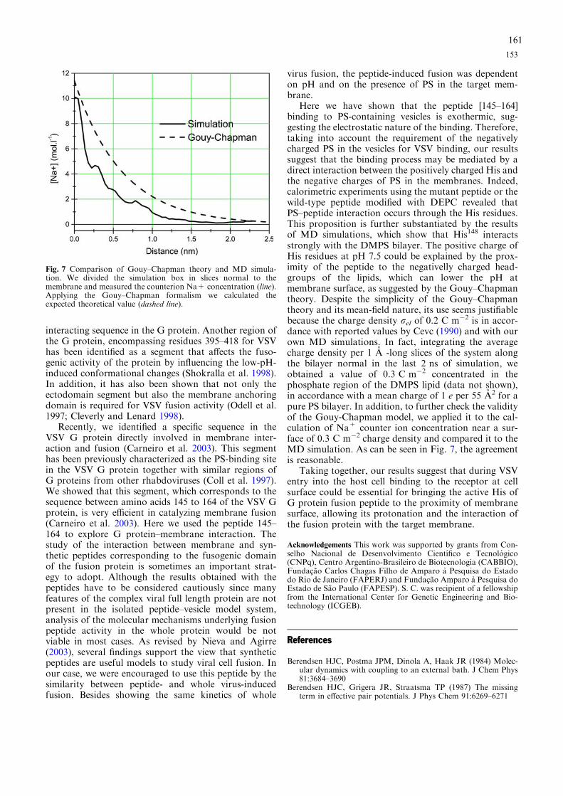
interacting sequence in the G protein. Another region ofthe G protein, encompassing residues 395–418 for VSVhas been identified as a segment that affects the fuso-genic activity of the protein by influencing the low-pH-induced conformational changes (Shokralla et al. 1998).In addition, it has also been shown that not only theectodomain segment but also the membrane anchoringdomain is required for VSV fusion activity (Odell et al.1997; Cleverly and Lenard 1998).
Recently, we identified a specific sequence in theVSV G protein directly involved in membrane inter-action and fusion (Carneiro et al. 2003). This segmenthas been previously characterized as the PS-binding sitein the VSV G protein together with similar regions ofG proteins from other rhabdoviruses (Coll et al. 1997).We showed that this segment, which corresponds to thesequence between amino acids 145 to 164 of the VSV Gprotein, is very efficient in catalyzing membrane fusion(Carneiro et al. 2003). Here we used the peptide 145–164 to explore G protein–membrane interaction. Thestudy of the interaction between membrane and syn-thetic peptides corresponding to the fusogenic domainof the fusion protein is sometimes an important strat-egy to adopt. Although the results obtained with thepeptides have to be considered cautiously since manyfeatures of the complex viral full length protein are notpresent in the isolated peptide–vesicle model system,analysis of the molecular mechanisms underlying fusionpeptide activity in the whole protein would be notviable in most cases. As revised by Nieva and Agirre(2003), several findings support the view that syntheticpeptides are useful models to study viral cell fusion. Inour case, we were encouraged to use this peptide by thesimilarity between peptide- and whole virus-inducedfusion. Besides showing the same kinetics of whole
virus fusion, the peptide-induced fusion was dependenton pH and on the presence of PS in the target mem-brane.
Here we have shown that the peptide [145–164]binding to PS-containing vesicles is exothermic, sug-gesting the electrostatic nature of the binding. Therefore,taking into account the requirement of the negativelycharged PS in the vesicles for VSV binding, our resultssuggest that the binding process may be mediated by adirect interaction between the positively charged His andthe negative charges of PS in the membranes. Indeed,calorimetric experiments using the mutant peptide or thewild-type peptide modified with DEPC revealed thatPS–peptide interaction occurs through the His residues.This proposition is further substantiated by the resultsof MD simulations, which show that His148 interactsstrongly with the DMPS bilayer. The positive charge ofHis residues at pH 7.5 could be explained by the prox-imity of the peptide to the negativelly charged head-groups of the lipids, which can lower the pH atmembrane surface, as suggested by the Gouy–Chapmantheory. Despite the simplicity of the Gouy–Chapmantheory and its mean-field nature, its use seems justifiablebecause the charge density rel of 0.2 C m�2 is in accor-dance with reported values by Cevc (1990) and with ourown MD simulations. In fact, integrating the averagecharge density per 1 A -long slices of the system alongthe bilayer normal in the last 2 ns of simulation, weobtained a value of 0.3 C m�2 concentrated in thephosphate region of the DMPS lipid (data not shown),in accordance with a mean charge of 1 e per 55 A2 for apure PS bilayer. In addition, to further check the validityof the Gouy-Chapman model, we applied it to the cal-culation of Na+ counter ion concentration near a sur-face of 0.3 C m�2 charge density and compared it to theMD simulation. As can be seen in Fig. 7, the agreementis reasonable.
Taking together, our results suggest that during VSVentry into the host cell binding to the receptor at cellsurface could be essential for bringing the active His ofG protein fusion peptide to the proximity of membranesurface, allowing its protonation and the interaction ofthe fusion protein with the target membrane.
Acknowledgements This work was supported by grants from Con-selho Nacional de Desenvolvimento Cientıfico e Tecnologico(CNPq), Centro Argentino-Brasileiro de Biotecnologia (CABBIO),Fundacao Carlos Chagas Filho de Amparo a Pesquisa do Estadodo Rio de Janeiro (FAPERJ) and Fundacao Amparo a Pesquisa doEstado de Sao Paulo (FAPESP). S. C. was recipient of a fellowshipfrom the International Center for Genetic Engineering and Bio-technology (ICGEB).
References
Berendsen HJC, Postma JPM, Dinola A, Haak JR (1984) Molec-ular dynamics with coupling to an external bath. J Chem Phys81:3684–3690
Berendsen HJC, Grigera JR, Straatsma TP (1987) The missingterm in effective pair potentials. J Phys Chem 91:6269–6271
Fig. 7 Comparison of Gouy–Chapman theory and MD simula-tion. We divided the simulation box in slices normal to themembrane and measured the counterion Na+ concentration (line).Applying the Gouy–Chapman formalism we calculated theexpected theoretical value (dashed line).
153
161

Bostrom M, Williams DRM, Ninham BW (2004) Specific ion ef-fects: Role of salt and buffer in protonation of cytochrome c.Eur Phys J 13:239–245
Carneiro FA, Ferradosa AS, Da Poian AT (2001) Low pH-inducedconformational changes in vesicular stomatitis virus glycopro-tein involve dramatic structure reorganization. J Biol Chem276:62–67
Carneiro FA, Bianconi ML, Weissmuller G, Stauffer F, Da PoianAT (2002) Membrane recognition by vesicular stomatitis virusinvolves enthalpy-driven protein-lipid interactions. J Virol76:3756–3764
Carneiro FA, Stauffer F, Lima CS, Juliano MA, Juliano L, DaPoian AT (2003) Membrane fusion induced by vesicular sto-matitis virus depends on histidine protonation. J Biol Chem278:13789–13794
Cevc G (1990) Membrane electrostatics. Biochim Biophys Acta1031(3):311–382
Chandrasekhar I, Kastenholz M, Lins RD, Oostenbrink C, SchulerLD, Tieleman DP, van Gunsteren WF (2003) A consistentpotential energy parameter set for lipids: dipal-mitoylphosphatidylcholine as a benchmark of the GROMOS9645 A3 force field. Eur Biophys J 32:62–67
Cleverley PZ, Lenard J (1998) The transmembrane domain in viralfusion: essential role for a conserved glycine residue in vesicularstomatitis virus G protein. Proc Natl Acad Sci USA 95:3425–3430
Coil DA, Miller AD (2004) Phosphatidylserine is not the cell sur-face receptor for vesicular stomatitis virus. J Virol 78:10920–10926
Coll JM (1997) Synthetic peptids from the heptad repeats of theglicoproteins of rabies, vesicular stomatitis and fish rhabdovi-ruses bind phosphatidylserine. Arch Virol 142:2089–2097
Crimmins DL, Mehard WB, Schlesinger S (1983) Physical prop-erties of a soluble form of the glycoprotein of vesicularstomatitis virus at neutral and acidic pH. Biochemistry22:5790–5796
Da Poian AT, Gomes AMO, Oliveira RJN, Silva JL (1996)Migration of vesicular stomatitis virus glycoprotein to the nu-cleus of infected cells. Proc Natl Acad Sci USA 93:8268–8273
Da Poian AT, Gomes AMO and Coelho-Sampaio T (1998)Kinetics of intracellular viral disassembly probed by bodipyfluorescence dequenching. J Virol Meth 70:45–58
Darden T, York D, Pedersen L (1993) Particle Mesh Ewald: anNlog (N) method for Ewald sums in large systems. J Chem Phys98:10089–10092
Durrer P, Gaudin Y, Ruigrok RW, Graf R and Brunner J (1995)Photolabeling identifies a putative fusion domain in the enve-lope glycoprotein of rabies and vesicular stomatitis viruses. JBiol Chem 270:17575–17581
Eckert DM, Kim PS (2001) Mechanisms of viral membrane fusionand its inhibition. Annu Rev Biochem 70:777–810
Eidelman O, Schlegel R, Tralka TS, Blumenthal R (1984) pH-dependent fusion induced by vesicular stomatitis virus glyco-protein reconstituted into phospholipid vesicles. J Biol Chem259:4622–4628
Estepa A, Coll JM (1996) Pepscan mapping and fusion relatedproperties of the major phosphatidylserine-binding domain ofthe glycoprotein of viral hemorragic septicemia virus, a sal-monid rhabdovirus. Virology 216:60–70
Florin EL, Moy VT, Gaub HE (1994) Adhesion forces betweenindividual ligand-receptor pairs. Science 264:415–417
Fredericksen B, Whitt MA (1995) Vesicular stomatitis virus gly-coprotein mutations that affect membrane fusion activity andabolish virus infectivity. J Virol 69:1435–1443
Gergely C, Voegel J-C, Schaaf P, Senger B, Maaloum M, HorberJKH, Hermmerle J (2000) Unbinding process of adsorbedproteins under external stress studied by atomic force micros-copy spectroscopy. Proc Natl Acad Sci USA 97:10802–10807
Hernandez LD, Hoffman LR, Wolfsberg TG, White JM (1996)Virus-cell and cell-cell fusion. Annu Rev Cell Dev Biol 12:627–661
Herrmann A, Clague MJ, Puri A, Morris SJ, Blumenthal R andGrimaldi S (1990) Effect of erythrocyte transbilayer phospho-lipid distribution on fusion with vesicular stomatitis virus.Biochemistry 29:4054–4058
Jass J, Tjarnhage T, Puu G (2000) From liposomes to supported,planar bilayer structures on hydrophilic and hydrophobic sur-faces: an atomic force microscopy study. Biophys J 79:3153–3163
Li Y, Drone C, Sat E, Ghosh HP (1993) Mutational analysis of thevesicular stomatitis virus glycoprotein G for membrane fusiondomains. J Virol 67:4070–4077
Lindahl E, Hess B, van der Spoel D (2001) GROMACS 3.0: Apackage for molecular simulation and trajectory analysis. J MolMod 7:306–317
Mastromarino P, Conti C, Goldoni P, Hauttecoeur B, Orsi N(1987) Characterization of membrane components of theerythrocyte involved in vesicular stomatitis virus attachmentand fusion at acidic pH. J Gen Virol 68:2359–2369
Miles EW (1977) Modification of histidyl residues in proteins bydiethyl pyrocarbonate. Meth Enzymol 47:431–442
Nieva JL, Agirre A (2003) Are fusion peptides a good model tostudy viral cell fusion? Biochim Biophys Acta 1614(1):104–115
Odell D, Wanas E, Yan J, Ghosh HP (1997) Influence of mem-brane anchoring and cytoplasmic domains on the fusogenicactivity of vesicular stomatitis virus glycoprotein G. J Virol71:7996–8000
Pak CC, Puri A, Blumenthal R (1987) Conformational changes andfusion activity of vesicular stomatitis virus glycoprotein: [125
I]iodonaphtyl azide photolabeling studies in biological mem-branes. Biochemistry 36:8890–8896
Pandit SA, Berkowitz ML (2002) Molecular dynamics simulationof dipalmitoylphosphatidylserine bilayer with Na+ counteri-ons. Biophys J 82:1818–1827
Puu G, Gustafson I (1997) Planar lipid bilayers on solid supportsfrom liposomes-factors of importance for kinetics and stability.Biochim Biophys Acta 1327:149–161
Puu G, Artursson E, Gustafson I, Lundstro M, Jass J (2000)Distribution and stability of membrane proteins in lipid mem-branes on solid supports. Biosensors Bioelectronics 15:31–41
Reviakine I, Brisson A (2000) Formation of supported phospho-lipid bilayers from unilamellar vesicles investigated by atomicforce microscopy. Langmuir 16:1806–1815
Ryckaert JP, Ciccott GI, Berendsen HJC (1977) Numerical inte-gration of the cartesian equations of motion of a system withconstraints: molecular dynamics of n-alkanes. J Comp Phys23:327–341
Schlegel R, Willingham MC, Pastan IH (1982) Saturable bindingsites for vesicular stomatitis virus on the surface of Vero cells. JVirol 43:871–875
Schlegel R, Tralka TS, Willingham MC, Pastan I (1983) Inhibitionof VSV binding and infectivity by phosphatidylserine: is phos-phatidylserine a VSV-binding site? Cell 32:639–646
Shokralla S, He Y, Wanas E, Ghosh HP (1998) Mutations in acarboxy-terminal region of vesicular stomatitis virus glycopro-tein G that affect membrane fusion activity. Virology 256:119–129
Skehel JJ, Wiley DC (2000) Receptor binding and membrane fu-sion in virus entry: the influenza hemagglutinin. Annu RevBiochem 69:531–569
Superti F, Seganti L, Tsiang H, Orsi N (1984) Role of phospho-lipids in rhabdovirus attachment to CER cells. Arch Virol81:321–328
Zhang L, Ghosh HP (1994) Characterization of the putative fus-ogenic domain in vesicular stomatitis virus glycoprotein G. JVirol 68:2186–2193
Zlatanova J, Lindsay SM, Leuba SH (2000) Single molecule forcespectroscopy in biology using the atomic force microscope.Prog Biophys Mol Biol 74:37–61
154
162