UNIVESIDADE FEDERAL DO RIO GRANDE – FURG PROGRAMA DE …
110
EFEITOS DO DESREGULADOR ENDÓCRINO BISFENOL A EM PARÂMETROS REPRODUTIVOS E SISTEMA NERVOSO CENTRAL DE CAMUNDONGOS Mus musculus (RODENTIA) Ana Cristina Kalb Tese defendida no âmbito do Programa de Pós-Graduação em Ciências Fisiológicas: Fisiologia Animal Comparada como parte dos requisitos para obtenção do título de DOUTOR em Fisiologia Animal Comparada. Orientador: Pof. Dr. Pablo Elías Martínez Rio Grande - RS 2012 UNIVESIDADE FEDERAL DO RIO GRANDE – FURG PROGRAMA DE PÓS GRADUAÇÃO EM CIÊNCIAS FISIOLÓGICAS : FISIOLOGIA ANIMAL COMPARADA
Transcript of UNIVESIDADE FEDERAL DO RIO GRANDE – FURG PROGRAMA DE …
EFEITOS DO DESREGULADOR ENDÓCRINO BISFENOL A EM
PARÂMETROS REPRODUTIVOS E SISTEMA NERVOSO CENTRAL DE
CAMUNDONGOS Mus musculus (RODENTIA)
Ana Cristina Kalb
Tese defendida no âmbito do Programa de
Pós-Graduação em Ciências Fisiológicas:
Fisiologia Animal Comparada como parte
dos requisitos para obtenção do título de
DOUTOR em Fisiologia Animal
Comparada.
Orientador: Pof. Dr. Pablo Elías Martínez
Rio Grande - RS
2012
UNIVESIDADE FEDERAL DO RIO GRANDE – FURG PROGRAMA DE PÓS GRADUAÇÃO EM
CIÊNCIAS FISIOLÓGICAS : FISIOLOGIA ANIMAL COMPARADA

Dedico este trabalho a todos aqueles que acreditam que a Ciência não é apenas mais um
afazer acadêmico, mas um complemento necessário à humanidade.

“Quem passou pela vida em brancas nuvens,
E num plácido repouso adormeceu,
Quem não sentiu o frio da desgraça;
Quem passou pela vida e não sofreu,
Foi espectro de Homem, não foi Homem,
Só passou pela vida, não viveu.”
Martin Luther king
Agradecimentos
Em primeiro lugar à força maior que rege o universo, que guia nossos passos e nossos
corações;
Aos meus pais, Max Edmundo e Ivoni, que por uma vida de dedicação, amor e trabalho
sempre possibilitaram a suas filhas a oportunidade de realizar sonhos e conquistas;
Ao meu querido avô Evaldo, que mesmo não podendo estar sempre perto, tenho certeza
que nós sempre estivemos e estaremos de mãos dadas em todos os desafios;
Ao meu orientador Pablo Martínez, que me acolheu em um momento muito difícil, me
deu exemplo de como ser apaixonado pelo que fazemos, e me proporcionou um
aprendizado e crescimento únicos.
A minha querida irmã e estagiária Ana Luiza que esteve sempre comigo, em todos os
dias de trabalho e da minha vida e que sem o seu auxílio, muito deste trabalho não teria
sido concretizado;
Ao meu querido e amado Josencler, que me ensinou a arte de amar, amar a vida, ser
uma pessoa melhor e consciente, e que compartilha comigo a paixão e a curiosidade
pela Biologia.
A minha querida amiga Mariângela que sempre me tratou com o carinho de uma mãe;
Ao meu querido amigo Vinícius Gonzalez que sempre foi ouvinte nas horas difíceis e
coadjuvante nos momentos felizes;
As amigas Camila Dalmolin, Sandra Isabel e Flavinha pela força e carinho, queria
muito poder ter vocês sempre por perto;
Ao Reuni pela bolsa cedida;
A todos os professores e funcionários da Fisiologia, por contribuírem de forma direta ou
indireta à minha formação;
Ao Curso de Pós-Graduação em Ciências Fisiológicas, onde me foi proporcionado não
apenas a oportunidade de realização desse trabalho, mas um ambiente ideal para o meu
desenvolvimento.
Sumário
Resumo..............................................................................................................................8
1.Introdução.......................................................................................................................9
1.1 Desreguladores endócrinos....................................................................................13
1.2 Bisfenol A..............................................................................................................15
2.Objetivo Geral..............................................................................................................19
2.1 Objetivos Específicos.............................................................................................19
Artigo 1............................................................................................................................21
Sperm impairment in male mice caused by maternal transference of bisphenol A during
lactation...........................................................................................................................22
Abstract........................................................................................................................23
1.Introduction...............................................................................................................24
2.Material and Methods...............................................................................................26
2.1 Animals and experimental protocol....................................................................26
2.2 Semen Quality....................................................................................................27
2.3 Histology............................................................................................................29
2.4 Measurement of total antioxidant capacity .......................................................29
2.5 Determination of glutamate cysteine ligase (GCL) activity and glutathione
(GSH) concentration................................................................................................30
2.6 Measurement of lipid peroxidation....................................................................31
3. Statistical analysis....................................................................................................32
4. Results......................................................................................................................32
4.1 Body weight gain, anogenital distance (AGD), testis and postate weight.........32
4.2 Sperm parameters...............................................................................................33
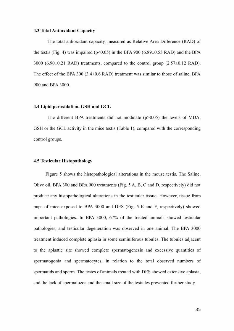
4.3 Total Antioxidant Capacity.................................................................................35
4.4 Lipid peroxidation, GSH and GCL....................................................................35
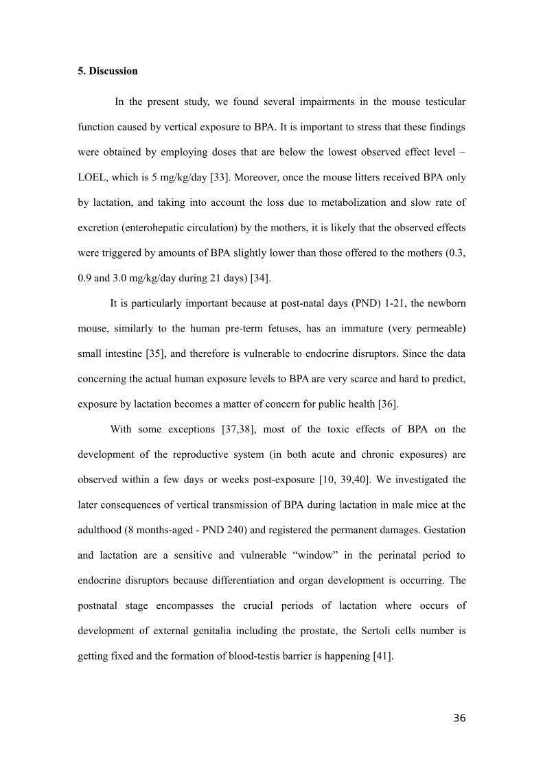
4.5 Testicular Histopathology ..................................................................................35
5. Discussion................................................................................................................36
6. Conflict of interest...................................................................................................40
7. Acknowledgements..................................................................................................41
Captions.......................................................................................................................47
Figure 1........................................................................................................................49
Figure 2........................................................................................................................50
Figure 3........................................................................................................................51
Figure 4........................................................................................................................52
Table 1..........................................................................................................................54
Artigo 2............................................................................................................................55
Effect of BPA on the central nervous system in lactating females..................................56
Abstract........................................................................................................................57
1.Introduction...............................................................................................................58
2. Materials and methods ............................................................................................61
2.1 Immunohistochemistry.......................................................................................61
3. Statistical analysis....................................................................................................63
4. Results......................................................................................................................63
4.1 GFAP marker......................................................................................................63
4.2 Tyrosine Hydroxylase.........................................................................................64
4.3 Synaptophysin....................................................................................................64
4.4 Histone 3 Phosphate...........................................................................................65
5. Discussion................................................................................................................66
6. Conflict of interest...................................................................................................70
7. Acknowledgements..................................................................................................70
8. References................................................................................................................71
Captions.......................................................................................................................76
Figure 1 .......................................................................................................................80
Figure 2........................................................................................................................81
Figure 3........................................................................................................................82
Figure 4........................................................................................................................83
Figure 5........................................................................................................................84
Figure 6........................................................................................................................85
Figure 7........................................................................................................................86
Figure 8........................................................................................................................87
Figure 9........................................................................................................................88
Figure 10......................................................................................................................89
Figure 11......................................................................................................................90
Conclusões ......................................................................................................................91
Anexo 1 ...........................................................................................................................92
Anexo 2 ...........................................................................................................................98
Referências....................................................................................................................104
Resumo
Bisfenol A (BPA) é conhecido por ser um desregulador endócrino com conhecida ação
no sistema reprodutor e sistema nervoso central. Fêmeas de camundongo com
aproximadamente 2 meses foram expostas via gavagem durante o período de
amamentação: dois grupos controle (salina 0,9% e azeite de oliva); BPA 300, 900, 3000
(µg/Kg/dia) e a dietilestilbestrol (DES, 650 µg/Kg/dia). Após 21 dias de exposição
(período de amamentação, exposição sub-crônica) as mães foram sacrificadas e seus
cérebros coletados para posterior análise de parâmetros neuronais. Os machos filhotes
foram separados, sua distância ano-genital medida, e após 240 dias sacrificados para
avaliação do impacto da transmissão vertical de BPA durante a amamentação em seus
testículos, nos quais foram analisados parâmetros espermáticos e estresse oxidativo.
Como resultado encontrou-se uma diminuição na distância ano-genital nos animais do
tratamento BPA 3000. BPA prejudica severamente os parâmetros espermáticos
analisados, tais como: motilidade do espermatozóide, morfologia, funcionalidade
mitocondrial, integridade de membrana, de acrossoma e de DNA, nos animais de todos
os tratamentos. Nas amostras de cérebro das mães expostas durante o período de
lactação, foram analisados o número de astrócitos (GFAP), enzima tirosina hidroxilase
(TH), quantidade de Sinaptofisina e a proliferação celular (H3P), nas regiões do Córtex,
Estriado e Hipocampo. Através da imunohistoquímica, como resultados observamos
uma diminuição do número de astrócitos em todas as regiões e diminuição de área de
fluorescência de TH na região CA3 do hipocampo no tratamento BPA300; No
tratamento BPA900, observou-se o decréscimo no número de astrócitos, aumento na
proliferação celular e diminuição do processo de formação sináptica na região CA3 do
hipocampo. A perda da função sináptica também se observou no córtex para este
tratamento. Os resultados gerais mostram proliferação celular induzida por BPA, mas
8

também reportam uma perda na quantidade de astrócitos, concentração de TH e de
densidade sináptica. Podemos concluir que BPA administrado indiretamente nos filhotes
causou um prejuízo nos parâmetros espermáticos. Quando administrado diretamente nas
mães por um período curto, mostrou-se como um potencial indutor de doenças
neurodegenerativas.
9
1. Introdução
O estrogênio é um hormônio esteróide, é produzidos principalmente nos ovários, corpo
lúteo e placenta. Adicionalmente, outros tecidos incluindo fígado, glândula adrenal,
mamas, tecido adiposo, e tecido nervoso contribuem para uma pequena parcela de
hormônio produzido (Judd & Fournet, 1994). Seu mecanismo de ação clássico ocorre
pela ligação aos seus receptores intracelulares a tecido/espécie específicos. Esses
receptores pertencem à superfamília de receptores hormonais nucleares que ao
formarem o complexo hormônio-receptor passam a ativar diferentes fatores de
transcrição gênica. Já foram identificados três tipos de receptores para estrógenos, o
receptor de estrógeno alfa (REα), o receptor de estrógeno beta (REβ) e o receptor de
estrógeno gama (REγ) (Matsushima et al. 2007; Taylor et al. 2009). Nos estrógenos o
complexo hormônio-receptor pode regular a expressão gênica sem se ligar diretamente
ao DNA por modular a função de outras classes de fatores de transcrição através da
interação proteína-proteína no núcleo (Gottlicher et al. 1998). Fatores de transcrição
como Fos/Jun (AP1- elemento de resposta) ou SP-1 são alguns exemplos de elementos
de resposta a estrógenos de ação genômica independente (Schreihofer et al. 2001). Um
terceiro mecanismo, também sem ação genômica, onde os elementos de resposta a
estrógenos estão associados a membrana celular (Deecher et al. 2003). Estrógenos se
ligam ao recetor de estrógeno acoplado a proteina G (GPR30), o qual é uma proteína
trans-membrana, levando a uma resposta da tradução relativamente rápida e transitória,
para estrógenos. Esta via de sinalização não genômica é largamente observada na
imediata estimulação de células e tecidos por estrógenos. (Qiu et al. 2003)
A síntese de hormônios esteróides envolve principalmente citocromos P450,
sendo limitante a primeira enzima a citocromo P450 desmolase (CYP11) que converte o
colesterol em pregnenolona. Isto acontece a nível mitocondrial e para o ingresso do
10
colesterol na mitocôndria é fator limitante a presença da proteína carregadora StaR
(Proteína reguladora aguda da esteroidogênese). Entretanto, modificações periféricas
são possíveis, assim os andrógenos podem ser convertidos em 3-dihidrotestosterona
pela ação da enzima 5α-reductase e permitir a masculinização da genitália externa e
outras estruturas. E ainda, os andrógenos podem se, perifericamente, transformados em
estrógenos pela ação da enzima aromatase. Os efeitos dos andrógenos ocorrem
principalmente através dos receptores de andrógenos (AR) e os estrógenos atuam por
meio de três receptores (REα, β e γ). (Lindberg et al. 2002).
Esses receptores de estrógenos apresentam diferentes expressões, dependentes
do tipo de tecido alvo, com a espécie envolvida e com a fase da vida do indivíduo, como
por exemplo, o receptor de estrogênio alfa é expresso no útero, no fígado (Kuiper et al.
1997), nos rins (Brunette e Leclerc, 2002), no sistema cardiovascular, nas glândulas
mamárias, no sistema nervoso, em células de câncer de mama (Cousen et al. 1997;
Shearman et al. 2003;). E o receptor de estrogênio beta é expresso nas células da
granulosa do folículo ovariano (Kuiper et al. 1997), no tecido ósseo, em núcleos do
sistema nervoso central (Taylor et al. 2009; Figtree et al. 2009). Os estrógenos podem
atuar através da via genômica (receptores nucleares) ou por vias não-genômicas
(receptores de membrana), os quais são amplamente expressos no sistema nervoso
central (SNC), incluindo o hipocampo, córtex e estriado e tem uma importante
influência direta e indireta em funções cognitivas superiores (Prange, Kiel & Rune ,
2006; Brann et al. 2007).
Assim, o hormônio esteróide (como também glicocorticóides,
mineralocorticóides, androgênios e progesteronas), ao entrar em contato com célula
alvo, atravessa sua membrana plasmática e interage com seus receptores intracelulares
específicos, formando o complexo hormônio-receptor (Nadal et al. 2001; Norman et al.
11
2004). Após sofrer o processo de dimerização, esse complexo, então, desloca-se para o
núcleo, atravessando pelo poro nuclear, onde atua como cofator de transcrição, no
nucleoplasma, através da ligação a regiões específicas do ácido desoxirribonucléico
(DNA), chamados de elementos responsivos ao estrogênio, o que por fim resulta na
regulação da expressão de genes-alvo (Silberger e Magleby, 1999).
Além desses receptores (REα e REβ), o receptor de estrogênio gama (REγ), que
foi inicialmente identificado no tecido testicular, nos rins, e no miocárdio humano, pode
ser expresso no tecido muscular esquelético e na retina (Giguere et al. 1988). Também
pode ser encontrado em tecidos humanos fetais como na placenta, no cérebro, no
coração, nos rins, nos pulmões e na musculatura esquelética, cujo mecanismo de ação
pode estar relacionado com a diferenciação e a maturação desses tecidos em indivíduos
adultos (Heard et al. 2000).
Os hormônios esteróides sintetizados no sistema nervoso (SN) de vertebrados a
partir do colesterol são conhecidos como neuroesteróides. Os hormônios esteróides
também podem ser sintetizados em glândulas esteroidogênicas periféricas como, por
exemplo, ovários e testículos, e regular funções neuronais importantes durante o
desenvolvimento do indivíduo que persistem nos vertebrados até a idade adulta (Chen et
al. 2009; Joseph et al. 2009). Hormônios esteróides periféricos, devido a sua
característica de lipossolubilidade, podem atravessar a barreira hemato-encefálica
atuando no tecido cerebral através de receptores intracelulares que regulam a transcrição
de genes específicos (Shao et al. 2012)
Células da glia estão envolvidas na formação de neuroesteróides e no
metabolismo do cérebro. Ambos astrócitos e oligodendrócitos podem ser considerados
locais primários de síntese de pregnenolona, o passo inicial da neuroesteroidogênese.
Há evidências de presença de enzimas esteroidogênicas como a citocromo P450 no
12
córtex cerebelar nas células de Purkinje, (Haraguchi et al. 2011), desta forma, com a
presença de grande quantidade de estrógenos é observado um aumento no crescimento
dendrítico, espinogênese e sinaptogênese nestas células (Price et al. 2000).
Outro papel importante dos estrógenos está relacionado a promoção de
neurogênese. Como exemplo de vias relacionadas podemos citar a fosforilação da
glicogênio sintase kinase 3 β. Os estrógenos também podem interagir com fatores de
crescimento como o fator de crescimento semelhante a insulina 1 (IGF-1) e o fator
neurotrófico derivado do cérebro (BDNF) (Garcia-Segura et al. 2006; Scharfman et al.
2006; Scharfman et al. 2012).
Os esteróides gonadais podem atuar no cérebro e influenciar comportamentos
reprodutivos em vertebrados como cópupa, corte, comportamento materno. Por
exemplo, algumas regiões do cérebro, que são responsáveis por controlar grande
variedade de comportamentos reprodutivos, contêm uma grande quantidade de células
que concentram andrógenos (Roselli e Resko, 1997; Tsutsui et al. 2000).
1.1 Desreguladores endócrinos
Nos últimos anos evidenciou-se que muitos produtos químicos, presentes no
meio ambiente, podem interferir com as ações fisiológicas dos hormônios endógenos.
Estas substâncias podem ser agonistas de receptores e interferir (imitando ou apenas
ocupando) com hormônios endógenos e, por isso, foram chamados de desreguladores
endócrinos (Darbre, 2006b). Os desreguladores endócrinos, que além de serem
encontrados no meio ambiente, podem também ser provenientes de produtos sintéticos,
podem atuar no genoma celular como agonistas ou antagonistas dos receptores de
esteróides.
Dentre os efeitos causados pela alteração hormonal, os disruptores endócrinos
podem alterar a função reprodutiva e causar feminilização por ligação com receptores
13
de estrogênio ou androgênio (Waring e Harris, 2005; Tabb e Blumberg, 2006) e também
interferir com o crescimento mamário, a lactação e predispor a doenças uterinas como
fibroses e endometriose (Mclachlan et al. 2006). Muitos disruptores endócrinos podem
se ligar a receptores tireoidianos e desregular o sistema neuroendócrino (Waring e
Harris, 2005; Whitehead e Rice, 2006). Os disruptores endócrinos podem ainda possuir
outros mecanismos de ação e, quando não agem no genoma, podem alterar a síntese
enzimática de subprodutos hormonais, causando alteração na função imune alteração
comportamental e da memória (Waring e Harris, 2005; Whitehead e Rice, 2006).
Muitos estudos concentram-se na avaliação da interferência dos disruptores
endócrinos durante o período pré-natal e pós-natal inicial, pois estas fases de
crescimento caracterizam-se pelo rápido crescimento e pela grande dependência de
ações hormonais. Perturbações no sistema endócrino durante estes períodos podem
causar, tardiamente, alterações anatômicas, fisiológicas, comportamentais e até mesmo
predispor ao desenvolvimento de doenças (Vandenberg, 2004; Dickerson e Gore, 2007).
Durante o desenvolvimento intra-uterino podem determinar crescimento intra-uterino
restrito bem como alteração na maturação do cérebro e das gônadas (Schoeters et al.
2008). Tardiamente podem causar puberdade precoce e aumento da incidência de
cânceres como o câncer vaginal e o câncer de próstata (Vandenberg, 2004; Dickerson e
Gore, 2007).
O estrogênio endógeno, mimetizado por disruptores endócrinos, possui papel
crucial na diferenciação sexual de estruturas do sistema nervoso central, controlando
algumas funções neuroendócrinas, cognitivas e comportamentais como, por exemplo, a
ativação de receptores de estrógenos no hipotálamo estimulando o comportamento
maternal logo após o parto. Durante o período pré-natal, o estradiol é responsável pelo
14
tipo de organização do cérebro em machos e em fêmeas (Wilson, et al. 2000;
Champagne, et al. 2001).
1.2 Bisfenol A
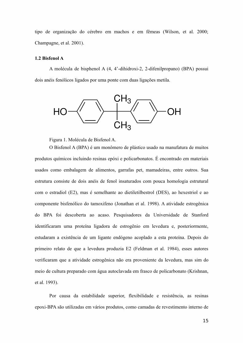
A molécula de bisphenol A (4, 4’-dihidroxi-2, 2-difenilpropano) (BPA) possui
dois anéis fenólicos ligados por uma ponte com duas ligações metila.
Figura 1. Molécula de Bisfenol A.
O Bisfenol A (BPA) é um monômero de plástico usado na manufatura de muitos
produtos químicos incluindo resinas epóxi e policarbonatos. É encontrado em materiais
usados como embalagem de alimentos, garrafas pet, mamadeiras, entre outros. Sua
estrutura consiste de dois anéis de fenol insaturados com pouca homologia estrutural
com o estradiol (E2), mas é semelhante ao dietiletilbestrol (DES), ao hexestriol e ao
componente bisfenólico do tamoxifeno (Jonathan et al. 1998). A atividade estrogênica
do BPA foi descoberta ao acaso. Pesquisadores da Universidade de Stanford
identificaram uma proteína ligadora de estrogênio em levedura e, posteriormente,
estudaram a existência de um ligante endógeno acoplado a esta proteína. Depois do
primeiro relato de que a levedura produzia E2 (Feldman et al. 1984), esses autores
verificaram que a atividade estrogênica não era proveniente da levedura, mas sim do
meio de cultura preparado com água autoclavada em frasco de policarbonato (Krishnan,
et al. 1993).
Por causa da estabilidade superior, flexibilidade e resistência, as resinas
epoxi-BPA são utilizadas em vários produtos, como camadas de revestimento interno de
15
latas de alimentos, complexos dentários para obturações e embalagens de remédios
(Brotons et al, 1995). Sua liberação no ambiente é possível quando a polimerização é
incompleta ou através de hidrolização causada por altas temperaturas (Krishnan et al,
1993; Feldman et al, 1984). A partir do revestimento das latas de alimentos, foi
detectada liberação de BPA em concentrações na faixa de 0,004 a 0,023mg/kg de
alimento (Feldman et al, 1984).
BPA administrado oralmente entra no organismo e é metabolizado
primariamente no intestino e fígado, é convertido pelo sistema de detoxificação no
fígado, pela citocromo P450 monooxigenase (Enzima de fase I) a 3-hidroxibisfenol A
(3-OH-BPA), que pode se ligar a molécula de DNA ou macromoléculas através da
formação de BPA-o-semiquinona ou BPA-o-quinona. A molécula de 3-OH-BPA, em
reação de autoxidação, forma o radical ânion superóxido (Nakagawa and Moore 2000).
BPA é metabolizado em fase II de detoxificação, principalmente pela reação de
conjugação pela UDP- glucoronosil transferase com o ácido glucurônico em ratos,
camundongos, macacos e humanos. Formando BPA-monoglucoronide (BPA-gluc)
formado é o maior metabólito produzido de BPA e é fisiologicamente inativo, não tendo
afinidade pelos receptores de estrógeno ou atividade estrogênica (Matthews et al. 2001;
Kurebayashi et al. 2003). BPA-gluc é excretado predominantemente pela via biliar nas
fezes e urina (Völkel et al. 2002; Kurebayashi et al. 2003).
BPA pode produzir efeitos em mamíferos a partir dos primeiros estágios de vida.
Essa exposição precoce pode afetar o desenvolvimento de células (como algumas do
sistema imune), órgão, tecidos e sistemas, assim como a produção de hormônios por
órgãos específicos. Seus efeitos podem persistir durante toda a vida do indivíduo e ter
impacto em sua prole. Precisa ser levado em conta o período crítico de desenvolvimento
de cada órgão e sua janela vulnerável de desenvolvimento (Rogers et al. 2013).
16
Alguns efeitos tóxicos podem ser observados durante o período perinatal de
exposição ao BPA onde podemos considerar sua passagem via placenta para o feto, e
durante o período de amamentação, onde uma grande quantidade de BPA é recebida
pelo lactente (Pryor et al. 2000; Doerge et al. 2010). Alguns efeitos observados no
sistema reprodutivo são diminuição no peso do epidídimo, aumento do peso da próstata
(Chitra et al. 2003; Kato et al. 2006), baixa produção de espermatozóides em roedores e
primatas, declínio na motilidade, alterações morfologia e dano de DNA (Saradha et al.
2006; Yang et al. 2010; Meeker et al.2010). Podemos relacionar a ação do BPA,
alterações em genes envolvidos na esteroidogênese e espermatogênese, o que pode levar
a um rompimento na barreira hemato-testicular e diminuição dos níveis de testosterona
em ratos (Li et al. 2009; Nakamura et al. 2010). A geração de espécies reativas de
oxigênio pela metabolização do BPA pode levar a um quadro de estresse oxidativo
causando peroxidação lipídica e diminuição da capacidade antioxidante, o que pode
levar a um quadro de patologia tecidual e apoptose via ativação de caspase 3 (Halliwell
and Gutteridge, 2007). Também pode aumentar a expressão da enzima aromatase e ter
efeito direto, por exemplo, na diminuição da distância ano-genital em machos, quando
administradas doses altas de BPA (Quignot et al. 2012).
O estrogênio está relacionado com os processos de formação da memória, possui
papel crítico na neurotransmissão do hipocampo associado com espinogênese ou
neuroproteção (Gould et al. 1990), regulação da sinaptogênese na região CA1 e
plasticidade sináptica (Miyagawa et al, 2007). Recentemente Miyagawa (2007) e
colaboradores demonstraram que a disrupção no desenvolvimento de neurônios
dopaminérgicos e também em outras neurotransmissões pode ser causada pela
exposição de ratos pré-natal e neonatal ao BPA. Estes mesmos autores sugerem que
altas doses de BPA causam uma piora na memória de ratos neste período. Por exemplo,
17
estudo usando imunohistoquímica, em animais expostos ao BPA neste mesmo período,
demonstram um decréscimo nas fibras colinérgicas nas regiões CA1, CA2 e CA3 no
hipocampo, como marcador foi usada a proteína acetiltransferase semelhante a colina
imunoreativa (ChAT-IR). A função colinérgica no hipocampo é importante no
aprendizado e na memória e pode ser afetada pela ação do BPA. A produção de espécies
reativas de oxigênio induzida por BPA está relacionada a ativação de caspase 3 e
conseqüente sinalização para apoptose de células da glia em diversas regiões do sistema
nervoso central (Obata and Kubota, 2000).
O BPA, além de possuir atividade semelhante ao estrogênio, também pode se
assemelhar ao hormônio da tireóide, este quando em baixas concentrações durante o
desenvolvimento do cérebro e de outros órgãos, pode causar disrupção na atividade das
enzimas do metabolismo central de acetilcolina (Donahue et al. 2004), sua ação ocorre
pela supressão da atividade transcricional por inibição competitiva com a triiodotironina
(T3) ao receptor de hormônio tireoidiano (TRα1 e TRβ1) (Suna et al. 2009).
Além dos efeitos no sistema reprodutor, sistema nervoso e tireóide, BPA pode afetar
profundamente o funcionamento das ilhotas de Langerhans, a unidade endócrina do
pâncreas. Combinado com o aumento da glicose, BPA promove a conversão de
fibroblastos em adipócitos, possui efeito estimulatório as células β do pâncreas para
uma maior produção de insulina, podendo ser através da estimulação pelo influxo de
Ca2+ (Nadal et al. 2009).
Sendo assim, uma aavaliação da ação de um desruptor endócrino nos estágios iniciais
de vida após o nascimento é de fundamental importância para o conhecimento sobre o
desenvolvimento do indivíduo.
18
2. Objetivo Geral
Verificar o efeito da exposição durante a amamentação ao Bisfenol A sobre aspectos
morfológicos e bioquímicos dos testículos dos filhotes e imunohistoquímica do sistema
nervoso central das mães em camundongos.
2.1 Objetivos Específicos
1. Verificar efeito da exposição pós-natal ao Bisfenol A sobre parâmetros
espermáticos de camundongos machos expostos durante o período de amamentação;
2. Verificar efeito da exposição pós-natal ao Bisfenol A sobre parâmetros de estresse
oxidativo em testículos de camundongos machos expostos durante o período de
amamentação;
3. Determinar o efeito da exposição pós-natal ao Bisfenol A sobre a
imunohistoquímica do córtex cerebral, núcleo estriado e hipocampo em camundongos
durante o período de lactação;
4. Verificar o efeito da exposição pós-natal ao Bisfenol A sobre a marcação
imunohistoquímica da enzima tirosina hidroxilase no córtex cerebral, núcleo estriado e
hipocampo em camundongos;
5. Verificar o efeito da exposição pós-natal ao Bisfenol A sobre a marcação
imunohistoquímica da proteína fibrilar ácida (GFAP) no córtex cerebral, núcleo estriado
e hipocampo em camundongos;
6. Verificar o efeito da exposição pós-natal ao Bisfenol A sobre a marcação
imunohistoquímica de Sinaptofisina no córtex cerebral, núcleo estriado e hipocampo em
camundongos;
19

7. Verificar o efeito da exposição pós-natal ao Bisfenol A sobre a marcação
imunohistoquímica de Histona-3-Fosfato no córtex cerebral, núcleo estriado e
hipocampo em camundongos;
20
Artigo 1
Revista : Reproductive Toxicology
(Fator de impacto: 3,22)
21
Sperm impairment in male mice caused by maternal transference of bisphenol A
during lactation
Ana Cristina Kalba, Ana Luiza Kalbb, Tainã Figueiredo Cardosoc, Carine Dahl Corcinic,
Antonio Sergio Varela Juniorb, Pablo Elías Martíneza, b, *
a Programa de Pós-Graduação em Ciências Fisiológicas: Fisiologia Animal Comparada,
Universidade Federal do Rio Grande, Rio Grande, RS, Brazil
b Instituto de Ciências Biológicas; Universidade Federal do Rio Grande, Rio Grande,
RS, Brazil
c REPROPEL- Faculdade de Veterinária; Universidade Federal de Pelotas, Pelotas, RS,
Brazil
* Corresponding author. Tel./fax: +55 53 32336848.
E-mail address: [email protected] (P.E. Martínez).
22
Abstract
The effect of bisphenol A (BPA) on the sperm quality of mammals has been
investigated. However in mouse lack extensive studies on sperm parameters associated
with testicular histology and oxidative stress. In this study mother mice were exposed to
BPA via gavage: two negative control groups: a 0.9% saline group and an olive oil
(vehicle) group; three BPA treatments: 300, 900 and 3000 groups (300, 900 and 3000
µg/kg/day); and a positive control group (diethylstilbestrol-DES 650 µg/kg/day). Mouse
pups were exposed to BPA while nursing from their mothers. At eight months old, male
mice were killed by cervical dislocation. The anogenital distance at weaning (21 days)
decreased significantly in the BPA 3000 group. BPA significantly impaired several
sperm parameters (motility; morphology; mitochondrial functionality; membrane,
acrosome and DNA integrity) and also induced histological testicular and oxidative
damage. Taken together, these results showed that exposure to BPA causes extensive
sperm impairment in mice.
Keyword: Bisphenol A; anogenital distance; spermatozoa membrane integrity;
acrosome integrity; spermatozoa DNA integrity; mitochondrial functionality.
Highlights
1. Bisphenol A (BPA) is a ubiquitous endocrine disrupter;2. We exposed mouse pups to BPA through lactation;3. BPA impaired reproductive and testicular oxidative-stress parameters;
23
1. Introduction
In recent years, bisphenol A [BPA, 2,2-bis-(4-hydroxyphenyl)propane] has been
widely used by industry to produce synthetic manufactured products such as resin
epoxy and plastic polycarbonates. BPA can be found in many end products, including
dental sealants, coatings for food cans, lining for metal cans, polyvinyl chloride, and
medical equipment, among others [1,2]. BPA is released into the environment through
sewage-treatment effluent, via hydrolysis from plastics, or from natural degradation of
polycarbonate plastics exposed to heat, acid or alkaline condition. BPA has received
heightened attention in the last decade because of its ubiquitous presence and because it
is an endocrine disruptor [3].
Endocrine-disrupting chemicals (ED) are biologically active compounds that can
mimic or antagonize the effects of endogenous hormones, causing many diseases
through systemic deleterious effects [4]. Endocrine-disrupting effects of BPA have been
reported in a number of animal models. In mammals, Zoeller et al. (2005) [5] observed
lower body-weight gain in female rats exposed during pregnancy and lactation, as well
as an increase in T4 concentration in their pups, harming the developing brains. Ropero
et al. (2008) [6] observed that BPA treatment in mice (100 µg/kg/day for 4 days)
disrupted function in pancreatic β-cells, producing insulin resistance.
While some toxic effects from BPA have been noted in adult animals, greater
attention has been paid to exposures during the perinatal period. This period altered
organizational programming and can confer increased susceptibility for diseases later in
life. The early postnatal period is also critical in the development of rodent reproductive
tracts [7,8]. Doerge et al. (2010) [9] reported significant effects from lactational transfer
24
of BPA suggest high potency and this fact can be related with BPA toxicity levels on
target tissue during the critical perinatal period affecting babies and fetuses.
Regarding the effects of BPA on the reproductive system, increased prostate weight,
decreased epididymis weight [10, 11] and lower sperm production were also reported in
adult rodents and primates [12, 13]. In humans, Meeker et al. (2010) [14] found declines
in sperm concentration, motility and morphology, and increased DNA damage in sperm.
Particularly in the neonatal period, BPA-induced effects can also appear as alterations of
gonadal organogenesis or function [15, 16]. For example, some alterations are evident
in genes involved in spermatogenesis and steroidogenesis, through epigenetic effects
that can result in disruption of the blood-testis barrier in rats [17] and decrease plasmatic
and testicular testosterone levels, reducing the expression of the steroidogenic enzymes
and cholesterol carrier protein in Leydig cells [18]. Prins et al. (2011) [19] also showed
increased susceptibility to prostate carcinogenesis in rat pups that received BPA during
the post-natal period (3, 5 and 21 days).
Pathological conditions caused by BPA may be related to the generation of reactive
oxygen species derived from its metabolization, affecting reproductive and sexual
characteristics by disturbing redox control systems [20]. Metabolization by the phase I
cytochrome P450 enzyme family (CYPs) causes free-radical generation as superoxide
anion (O2-•) via metabolic redox cycling between its quinone and hydroquinone forms.
Importantly, the following step in BPA detoxification involves its conjugation with
glucuronic acid by phase II enzyme UDP-glucuronosyltransferase, which is not fully
expressed in the neonatal period in mammals [21].
It is well-established that BPA exposure at early ages can impair the development of
the reproductive system. Nevertheless, the effects of BPA administered through vertical
transmission in the postnatal period (i.e. during lactation) on a number of important
25
sperm variables have not been fully investigated. In the present work, we demonstrated
that BPA can permanently impair the reproductive function of the male mice even when
some of the reproductive structures are formed, thus reinforcing the importance of the
oral exposure in this period. For this, several sperm variables were analyzed and
testicular histopathologies as well as key markers of the oxidative status of the testicular
tissue were registered.
2. Material and Methods
2.1 Animals and experimental protocol
All of the procedures involving animal subjects were reviewed and approved by the
Animal Ethics Committee of the Universidade Federal do Rio Grande-FURG, Rio
Grande, Rio Grande do Sul (Approval number: P006/2011). Healthy Swiss albino mice
obtained from the Central Animal Facility of the Universidade Federal do Rio
Grande-FURG, maintained by random breeding, were housed in standard polystyrene
cages at 23°C and 12-h light/12-h dark cycle, with water and a soybean-free diet ad
libitum (Nuvilab CR-1 NUVITAL, Jundiaí, SP, Brazil). Swiss albino mouse was chosen
due to its wide use in toxicological studies, many involving the effects of BPA on the
reproductive system of mammals. After the acclimatization period of one week, animals
(8 weeks old) were grouped in cages (5 females + 1 male) for random mating. The
presence of a vaginal plug was checked twice daily to confirm mating. All the females
that presented a vaginal plug were considered pregnant. Each pregnant female was
placed in a separate cage to give birth. The litter size chosen was about 10 pups with
similar numbers of male and female. From the birth of the pups to the end of the
26
lactation period (21 days), 6 treatment groups of 5 dams each received 200 µL/day of
one of the following solutions via gavage: two control groups, a 0.9% saline (Saline)
and an olive oil (vehicle) group; three BPA groups: BPA 300 - 300 µg/kg/day; BPA 900
- 900 µg/kg/day; BPA 3000 - 3000 µg/kg/day; and a positive control group DES
(diethylstilbestrol) 650 µg/kg/day. The dose of DES used was chosen for the purpose of
producing detectable alterations in testis morphology, since low doses do not induce
toxic effects in the reproductive system [7]. After the lactation period, the weanling
pups were sexed, the anogenital distance was measured, and the male pups were
separated in cages according to litter. Female pups are used for other study. The number
of male pups for each treatment was as follow: Saline - n=14; Olive oil – n=16; BPA300
– n=18; BPA 900 – n=14; BPA 3000 – n=17 and DES – n=12. At eight months old they
were killed by cervical dislocation [22]. The testes and prostate were removed by
laparotomy and weighted , and the epididymis tail and part of the vas deferens were
isolated and placed in a Petri dish (35 mm diameter; Corning) filled with 500 µL of
Sigma-M2 medium with HEPES (10 mM). For semen collection (n=10 per treatment),
the selected structures were disrupted with the aid of hypodermic needles (30 G) [23].
The remaining testicle tissue was immediately dissected out, weighed, and stored at
-80°C for biochemical analysis (n=5 per treatment) or placed in 4% paraformaldehyde
for histological analysis (n=6 per treatment). For all of the analysis, the individuals were
selected randomly to avoid the litter effect.
2.2 Semen Quality
The testes were removed by laparotomy, and the epididymis tail and part of the vas
deferens were isolated and placed in a Petri dish (35 mm diameter; Corning) filled with
500 μL of Sigma-M2 medium with HEPES (10 mM). For semen collection, the selected
27
structures were disrupted with the aid of hypodermic needles (30 G) [23]. The
remaining testicle tissue was immediately dissected out, weighed, and stored at -80°C
for biochemical analysis or placed in 4% paraformaldehyde for histological analysis.
Sperm quality evaluations were done after incubation of samples for 10 min at 37ºC in
M2 medium. Sperm motility was evaluated by putting 10 µL of sperm in a slide covered
with a coverslip, using phase-contrast microscopy at 200 x both pre-heated at 37° C
(BX 41 Olympus América, Inc., São Paulo, SP, Brazil) [23], always by the same trained
technician. Sperm morphology was determined as described by counting 200 cells with
phase contrast microscopy at 1000 x [24]. The evaluations of sperm membrane and
acrosome integrity was carried out with an epifluorescence microscope (Olympus BX
51, América INC, São Paulo - Brazil), with filter wave length of 450-520 nm. Sperm
membrane integrity was evaluated using carboxyfluorescein diacetate (CFDA; C5041)
and propidium iodide (PI; P4170) [ 25] at 400 x. In each slide, 200 cells were counted
and classified as intact and functional cell membrane (green fluorescence) or not intact
or functional cell membrane (red fluorescence or simultaneous red and green
fluorescence). Acrosome integrity was evaluated using FITC-PNA (L7381) by counting
200 cells in dry slides. Acrosomes were classified as intact, when presented red
fluorescence and normal conformation, or not intact, when presented green fluorescence
or no fluorescence and conformation distinct that from normal spermatozoa [26].
Mitochondrial functionality was evaluated after incubation of a 10 µL sperm sample
with a 40 µL rhodamine 123 solution (13 µM), at 20 °C for 10 min. Sperm with positive
rhodamine staining (green fluorescence) were considered as having functional
mitochondria. Conversely, nonfunctional mitochondria were characterized by negative
rhodamine staining (sperm with no fluorescence) [27]. The rate of mitochondrial
functionality was determined by the proportion of sperm emitting green fluorescence
28
compared with total sperm (green or no fluorescence). Sperm DNA integrity (Sperm
Chromatin Structure Assay) was evaluated after putting a 45 µL sperm sample in 50 µL
TNE (0.01 M Tris-HCl; 0.15 M NaCl; 0.001 M EDTA; pH 7.2). After 30 sec, 200 µL of
TritonX-100 solution (1%) was added and, 30 sec later, 50 µL of acridine orange was
added (2 mg/mL in deionized H2O). The evaluation was done after 5 min, without
exceeding 1 min of slide exposure [28]. Sperm with green fluorescence were considered
as having intact DNA, whereas those with red or orange fluorescence were considered
as having denatured DNA. The rate of DNA integrity was determined by the proportion
of sperm emitting green fluorescence compared with the total number of sperm (green,
red, or orange fluorescence) [28]. Assessments of mitochondrial function, membrane
integrity and DNA were performed in an epifluorescent microscope (Olympus BX51®,
America INC. São Paulo - Brazil) with 5mL of solution with sperm on slides
undercover slip (18x18mm), evaluating 200 cells per sample. The rates were expressed
as the percentage of viable cells/functional on the total cells evaluated.
2.3 Histology
The testicular tissue fixed in 4% paraformaldehyde was dehydrated in
increasing concentrations of ethanol and subsequently embedded in Paraplast X-TRA
(Sigma P3808). Sections 6 μm thick were cut with a rotary microtome (Leica RM 2255)
and stained with Hematoxylin-Eosin [29]. Histological examination was performed
using a light microscope (Olympus BX 51) with a high-resolution digital camera
(Olympus DP 72).
2.4 Measurement of total antioxidant capacity
29
The total antioxidant capacity against peroxyl radicals (ROO•) was determined
according to Amado et al. (2009) [30]. Aliquots of testis homogenates were placed in a
medium containing 30 mM HEPES (pH 7.2), 200 mM KCl, 1 mM MgCl2 and 40 M
of the fluorogenic compound 2´,7´-dichlorofluorescein diacetate (H2DCF-DA;
Invitrogen) in the presence or absence of 2,2´-azobis 2 methylpropionamidine
dihydrochloride (ABAP; 4 mM; Aldrich), which generates ROO• by thermal
decomposition at 37° C. Fluorescence was considered as a measure of ROS production
and was read in a spectrofluorimeter equipped with a microplate reader (Victor 2;
Perkin-Elmer) at wavelengths of 485 and 530 nm for excitation and emission,
respectively. Total fluorescence generation was calculated by integrating the
fluorescence units (FU) over the period of the measurement. The results were expressed
as area difference of FU x min in the same sample with and without ABAP addition, and
standardized to the ROS area without ABAP (background area). The relative difference
between the ROS area with and without ABAP was considered a measure of the
antioxidant competence of the testis, where area difference is inversely proportional to
the antioxidant capacity [30].
2.5 Determination of glutamate cysteine ligase (GCL) activity and glutathione
(GSH) concentration
GCL activity and GSH levels in the sample testis were determined according to
White et al. (2003) [31]. This method employs the reaction of naphthalene
dicarboxaldehyde (NDA) with GSH or -glutamylcysteine ( -GC) to form
fluorescent cyclic products. A reaction solution (25 μl) with 400 mM Tris-HCl, 40 mM
ATP, 20 mM glutamate, 2.0 mM EDTA, 20 mM sodium borate, 2 mM serine and 40
mM MgCl was prepared just before the assay, to prevent ATP degradation. After
30
addition of testis homogenates (25 μl), the plate was incubated at room temperature for
60 min and the on stopped by adding 50 μl of 5-sulfosalicylic acid (SSA, 200 mM).
After protein precipitation, the plate was centrifuged for 5 min at 2,000 × g and 20 μl of
supernatant from each well was transferred to a white plate, an NDA solution was added
to all wells, and after 30 min of incubation, the fluorescence intensity of the NDA-GSH
(or NDA- -GC) complex was read at excitation and emission wavelengths of 485 and
530 nm, respectively (Victor 2, Perkin-Elmer).
2.6 Measurement of lipid peroxidation
Determination of lipid peroxides was performed through estimation of the
malondialdehyde content in testis homogenates, employing the thiobarbituric
acid-reactive substances (TBARS) fluorimetric method, according to Oakes and Van der
Kraak (2003) [32]. Aliquots of sample extracts (10 µl) were added to a reaction solution
containing 150 µl of 20% acetic acid, 150 µl of thiobarbituric acid (0.8%), 50 µl of
Milli-Q water and 20 µl of sodium dodecyl sulfate (SDS, 8.1%). This mixture was
heated at 95 oC for 30 min for derivatization. Following cooling for 10 min, 100 µl of
Milli-Q water and 500 µl of n-butanol were added. After centrifugation (3,000 x g for
10 min at 15 oC), 150 µl of the organic phase was placed in a microplate reader and the
fluorescence recorded with wavelengths of 520 and 580 nm for excitation and emission,
respectively. Concentration of TBARS (nM/mg of wet tissue) was calculated,
employing a standard curve of tetramethoxypropane (TMP, Acros Organics) as MDA
equivalent.
31
3. Statistical analysis
Data were expressed as mean ± SEM. Once the assumptions of homogeneity and
normality of variance were verified, one-way ANOVA was first performed between
litters for each treatment, to exclude litter effects. Once litters from the same treatment
did not present statistical differences, statistical analysis was performed between
treatments by means of one-way ANOVA followed by Tukey post hoc test. The analysis
of prostate weight was performed through Kruskal-Wallis non-parametric ANOVA
followed by a Mann-Whitney test. The significance level adopted was 5% for all of the
cases.
4. Results
The litters exposed to the positive control (DES) presented a serious impairment
in their development and grew slowly. The development of the reproductive organs was
incomplete and many animals did not possess testis. For this reason, the few litters that
presented testis in the DES treatment were used for histopathological analysis, and
biochemical measurements as well as sperm parameters were not done in this treatment.
4.1 Body weight gain, anogenital distance (AGD), testis and postate weight
With respect to daily body weight gain over 180 days, mice treated with BPA
3000 had a significantly (p<0.05) lower weight gain (Fig. 1A). The anogenital distance
(Fig. 1B) was measured using a caliper at weaning (21 days). The results for anogenital
distance were expressed with respect to the body weight [anogenital distance (mm)/
body weight (g) at 21 days]. Exposure to BPA 3000 µg/kg/day resulted in a significant
decrease (p<0.05) in the AGD compared to the control groups. The DES group was not
considered for AGD measure due to the delay caused on the development, since male
32
and female litters were not sufficiently developed to show differences in the external
genitalia at 21 days. With respect to testis weight on 180 days, the DES treatment had a
significantly (p<0.05) lower weight compared to Control and BPA 900 groups (Fig. 2).
Did not differences weren’t observed on prostate weight.
4.2 Sperm parameters
4.2.1 Sperm Motility
BPA significantly (p<0.05) impaired the sperm motility (Fig. 3A). Sperm
motility for BPA 300 (57.00±5.15%); BPA 900 (48.00±5.27%) and BPA 3000
(27.00±5.97%) was less than for Saline (88.00±1.33%) and Olive oil (83.00±2.13%).
Sperm from the mice whose mothers were exposed to BPA 3000 showed the highest
motility impairment.
4.2.2 Normal Sperm Morphology
Figure 3B shows the effects of the BPA exposure on the spermatozoa
morphology. The BPA treatments significantly (p<0.05) lowered the number of
spermatozoa with normal morphology compared to the control. The percentages of
normal-appearing sperm obtained for each treatment were: BPA 300 (33.00±1.83%);
BPA 900 (33.90±1.39%); BPA 3000 (37.00±0.89%); Saline (46.10±2.82%) and Olive
oil (47.80±2.39%).
4.2.3 Spermatozoa Membrane Integrity
The BPA treatments significantly decreased (p<0.05) the spermatozoa membrane
integrity (Fig. 3C). The percentages of spermatozoa with normal membrane integrity
33
were: 40.80±7.19%, 26.40±4.07% and 3.10±0.52% for BPA 300, BPA 900 and BPA
3000, respectively. The membrane integrity was lower in the BPA treatments than in the
controls, Saline (94.30±1.22%) and Olive oil (92.80±1.33%).
4.2.4 Sperm Acrosome Integrity
Acrosome integrity (Fig. 3D) was significantly impaired (p<0.05) in the BPA
3000 (16.00±5.81%) and BPA 900 (52.10±6.51%) treatments compared to the controls,
Saline (92.20±2.13%) and Olive oil (94.70±1.41%) and to BPA 300 (94.70±3.30%).
Again, the BPA exposure elicited dose response, with no damage at the lowest
concentration (300 µg/kg/day).
4.2.5 Sperm DNA Integrity
The data for the spermatozoa DNA integrity (Fig. 3E) showed a significant
decrease (p<0.05) in all the BPA treatments. The two higher BPA concentrations, BPA
900 and BPA 3000, showed a more pronounced DNA-impairment effect, with
percentages of 16.08±3.00 and 17.03±8.14%, respectively, and BPA 300 showed an
intermediate effect (57.40±4.73%), compared with the control treatments, Saline
(89.80±2.19%) and Olive oil (88.00±1.87%).
4.2.6 Mitochondrial Functionality
Figure 3F shows the data for mitochondrial functionality of the spermatozoa.
BPA treatments significantly impaired (p<0.05) the mitochondrial functionality, with the
most damage when the mothers received BPA 3000 (8.50±2.11%). Administration of
BPA 300 (64.40±3.16%) and BPA 900 (51.50±5.39%) also impaired mitochondrial
function more than Saline (97.30±0.94%) and Olive oil (94.70±3.30%).
34
4.3 Total Antioxidant Capacity
The total antioxidant capacity, measured as Relative Area Difference (RAD) of
the testis (Fig. 4) was impaired (p<0.05) in the BPA 900 (6.89±0.53 RAD) and the BPA
3000 (6.90±0.21 RAD) treatments, compared to the control group (2.57±0.12 RAD).
The effect of the BPA 300 (3.4±0.6 RAD) treatment was similar to those of saline, BPA
900 and BPA 3000.
4.4 Lipid peroxidation, GSH and GCL
The different BPA treatments did not modulate (p>0.05) the levels of MDA,
GSH or the GCL activity in the mice testis (Table 1), compared with the corresponding
control groups.
4.5 Testicular Histopathology
Figure 5 shows the histopathological alterations in the mouse testis. The Saline,
Olive oil, BPA 300 and BPA 900 treatments (Fig. 5 A, B, C and D, respectively) did not
produce any histopathological alterations in the testicular tissue. However, tissue from
pups of mice exposed to BPA 3000 and DES (Fig. 5 E and F, respectively) showed
important pathologies. In BPA 3000, 67% of the treated animals showed testicular
pathologies, and testicular degeneration was observed in one animal. The BPA 3000
treatment induced complete aplasia in some seminiferous tubules. The tubules adjacent
to the aplastic site showed complete spermatogenesis and excessive quantities of
spermatogonia and spermatocytes, in relation to the total observed numbers of
spermatids and sperm. The testes of animals treated with DES showed extensive aplasia,
and the lack of spermatozoa and the small size of the testicles prevented further study.
35
5. Discussion
In the present study, we found several impairments in the mouse testicular
function caused by vertical exposure to BPA. It is important to stress that these findings
were obtained by employing doses that are below the lowest observed effect level –
LOEL, which is 5 mg/kg/day [33]. Moreover, once the mouse litters received BPA only
by lactation, and taking into account the loss due to metabolization and slow rate of
excretion (enterohepatic circulation) by the mothers, it is likely that the observed effects
were triggered by amounts of BPA slightly lower than those offered to the mothers (0.3,
0.9 and 3.0 mg/kg/day during 21 days) [34].
It is particularly important because at post-natal days (PND) 1-21, the newborn
mouse, similarly to the human pre-term fetuses, has an immature (very permeable)
small intestine [35], and therefore is vulnerable to endocrine disruptors. Since the data
concerning the actual human exposure levels to BPA are very scarce and hard to predict,
exposure by lactation becomes a matter of concern for public health [36].
With some exceptions [37,38], most of the toxic effects of BPA on the
development of the reproductive system (in both acute and chronic exposures) are
observed within a few days or weeks post-exposure [10, 39,40]. We investigated the
later consequences of vertical transmission of BPA during lactation in male mice at the
adulthood (8 months-aged - PND 240) and registered the permanent damages. Gestation
and lactation are a sensitive and vulnerable “window” in the perinatal period to
endocrine disruptors because differentiation and organ development is occurring. The
postnatal stage encompasses the crucial periods of lactation where occurs of
development of external genitalia including the prostate, the Sertoli cells number is
getting fixed and the formation of blood-testis barrier is happening [41].
36
Although several endocrine-disrupting effects are well-established, there are
some controversies concerning BPA toxicity due to the differences in the dose and mode
of exposure. For example, some authors have registered that BPA administered by
drinking water (0.01, 0.1, 1 or 10 ppm) [42] or subcutaneous injection (10 µg/kg/day
during 3 days) [43] caused no effect on the reproductive organs of rats. On the other
hand, a considerable number of studies have shown reproductive impairments
associated with BPA. Chitra et al. (2003) [10] exposed 45-day-old rats to different BPA
concentrations (0.2, 2 and 20 µg/kg/day for 45 days) and reported significant decreases
in epididymal sperm motility and sperm counts, such effects may be associated to the
inhibition of spermatogenesis, decreased numbers of elongated spermatids, and reduced
steroidogenic enzyme activity. Salian et al. (2009) [44] found that male offspring
exposed to low doses of BPA (1.2 – 2.4 µg/kg/day) in the uterus and in the neonatal
period resulted in reduced efficiency in daily sperm production, motility and
morphology. Other studies that investigated the BPA effects through the lactation period
of rats showed that BPA caused significant increase in mammary tumors in female
offspring [7,8,39], decrease in the anogenital distance and other reproductive changes in
male offspring [45].
In the present study, it was found a general decrease in sperm quality resulting
from maternal BPA exposure (Fig. 3). Concerning DNA fragmentation and
mitochondrial dysfunction, these processes are related to apoptosis and in the
seminiferous tubules its occurrence is a normal finding in spermatogenesis and
testicular homeostasis. However, the two main paths that lead to apoptosis – extrinsic
(via Fas/FasL) and intrinsic (mitochondrial pathway) – are apparently involved in
abnormal testicular apoptosis induced by exposure to BPA via gavage (160 or 480
mg/kg/day) [46]. We observed that BPA decreased sperm motility, and this may be due
37
to the effects of BPA on the mitochondrial function, that also affect ATP generation and
thus the motility of sperm (Fig. 3 A and F). Salian et al. (2009) [44], observed similar
results in rats, were that BPA induced declined on sperm counts and motility and
decrease in the levels of LH, FSH, testosterone and estrogen can be a possible
explanation.
The effect of BPA on mitochondrial functionality may occur both through its
involvement in the intrinsic pathway of apoptosis, considering its ability to generate
reactive oxygen species (ROS), or by direct action in mitochondrial estrogen receptors
(ERs), taking into account the role of estrogen and ERs in the regulation of the
mitochondrial respiratory chain [47]. On the other hand, these changes in the pituitary
gonadotrophins and steroidogenesis may be mediated by oxidative stress in the central
nervous system, adenohypophysis and testicle. As observed by Chitra et al. (2003) [10]
in rats and Kabuto et al. (2004) [15] in mice, BPA produces short or medium-term
oxidative damage in the brain, liver, kidney and testes. We studied the long-term effect
of BPA in testes of mice, and observed a significant (p<0.05) decrease in the total
antioxidant capacity in the treatments with BPA 900 and BPA 3000 (Fig. 4). The
testicles and the organisms had ample time for homeostatic mechanisms to compensate
for the effects of BPA exposure during lactation, but at 8 months of age, the male mice
still showed changes in their antioxidant capacity.
Oxidative stress, hormonal changes and apoptosis all can be inducing significant
changes in the testes. In fact, we observed significant changes in the testicular histology
in mice after the exposure to BPA 3000 (Fig. 5), including aplasia in the seminiferous
tubules and testicular degeneration. Hutanu (2011) [48] and Takahashi (2001) [49]
reported similar results, including disruption of the integrity of the hematotesticular
barrier and decrease in spermatogenesis in the high-dose BPA group (200 µg/kg/day).
38
From the same morphological pattern of response, we can also predict the mechanism of
action of BPA suspected of interfering with testosterone action or changes in the male
reproductive tract occurred during the perinatal window.
The histological testicular pathologies (Fig. 5), the spermatic impairment (Fig.
3), and the disturbance in oxidative stress (Fig. 4) observed in male mice exposed to
BPA during the lactation period suggest that BPA could have a long and lasting adverse
effects on the sperm quality of these offspring during adulthood.
The pups from dams exposed to BPA 3000 showed reduced anogenital distance
(Fig. 1). Androgens are very important to the male characters on development, (i.e. in
the seminiferous tubules, testosterone acts on spermatogenesis indirectly through Sertoli
cells to maintain spermatogenesis, germ cell development and ano-genital distance).
Our results are in agreement with those of Kobayashi et al. (2002) and Fujii et al.
(2001) [50, 51], who found that AGD decreases in boys born from mothers exposed to
BPA, indicating the potential role of BPA as an antiandrogen. However, exposure of
pregnant rats to 4 and 40 mg/kg/day of BPA from gestation day (GD) 6 and PND 20 had
no effect on the reproductive parameters evaluated (including AGD) [50]. On the other
hand, Gupta (2000) [51], for male mice exposed to BPA (50 µg/kg/day) during gestation
(GD 16-18), observed increased prostatic size, decreased epididymal weight and
increased anogenital distance. Similarly, but in females, Tyl et al. (2002) [37] observed
an AGD increase and lower body weight. Differences in the concentrations of BPA
exposure in the study of Gupta (2000) [52] and those used here, do not fully explain the
contrasting AGDs found in male mice. Furthermore, the observed decrease in AGDs in
human boys [45] argues that this measure can be of biological and/or toxicological
interest. In our work the decrease in the anogenital distance in the highest BPA
treatment could be due that serum conjugated-to-deconjugated estrogen ratios and its
39
estrogenic activity, and can be decreased or the aromatase expression was increased
[53]. Exposure to ED in early life leads to permanent changes in reproductive anatomy,
histology and physiology [7,11,15,19].
In summary, the present study demonstrates that the exposure of mice to
environmentally relevant doses of BPA during the early period results in long-term
adverse effects on the sperm quality and the importance of to make a screening the most
of sperm parameters. These findings should raise concerns for humans and vertebrates
in general, because BPA may cause changes in the redox status, reducing AGD,
spermatic impairment and its harmful effects manifest throughout life.
6. Conflict of interest
The author declared there is no conflict of interest.
40
7. References
[1] Thomson BM, Grounds PR. Bisphenol A in canned foods in New Zealand: exposure
assessment. Food Addit Contam 2005;22:65–72.
[2] Benuchour N, Aris A. Toxic effects of low doses of bisphenol-A on human placenta
cells. Toxicol Appl Pharmacol 2009;241:322–8.
[3] Quitmeyer A, Roberts R. Babies, bottles, and bisphenol A: the story of a
scientist-mother. PLoS Biol 2007;5:1399–402.
[4] Swedenborg, E, Pongratz I, Gustafsson JA. Endocrine disruptors targeting ERbeta
function. Int J Androl 2010;33:288-97.
[5] Zoeller RT, Bansal R, Parris C. Bisphenol-A, an environmental contaminant that acts
as a thyroid hormone receptor antagonist in vitro, increases serum thyroxine, and alters
RC3/neurogranin expression in the developing rat brain. Endocrinology
2005;146:607-12.
[6] Ropero AB, Alonso-Magdalena P, García-García E, Ripoll C, Fuentes E, Nadal A.
Bisphenol-A disruption of the endocrine pancreas and blood glucose homeostasis.
Internat J Androl 2008;31:194-200.
[7] Newbold RR, Jefferson WN, Padilla-banks E, Haseman J. Developmental exposure
to diethylstilbestrol (DES) alters uterine response to estrogens in prepubescent mice:
low versus high dose effects. Reprod Toxicol 2004;18:399-406.
[8] Pryor JL, Hughes C, Foster W, Hales BF, Robaire B. Critical windows of exposure
for children’s health: the reproductive system in animals and humans. Environ Health
Perspect 2000;108:491–503.
41
[9] Doerge DR, Vanlandingham M, Twaddle NC, Delclos KB. Lactational transfer of
bisphenolA in Sprague – Dawley rats. Toxicol Lett 2010;199:372-6.
[10] Chitra, KC, Latchoumycandane C, Mathur PP. Induction of oxidative stress by
bisphenol A in the epididymal sperm of rats. Toxicology 2003;185:119-27.
[11] Kato H, Furuhashi T, Tanaka M, Katsu Y, Watanabe H, Ohta Y, Iguchi T. Effects of
bisphenol A given neonatally on reproductive functions of male rats. Reprod Toxicol
2006;22:20-9.
[12] Saradha B, Mathur PP. Effect of environmental contaminants on male reproduction.
Environ Toxicol Pharmacol 2006;21:34-41.
[13] YangYJ, Lee SY, Kim KY, Hong YP. Acute Testis Toxicity of Bisphenol A
Diglycidyl Ether in Sprague-Dawley Rats. J Prev Med Public Health 2010;43:131-7.
[14] Meeker JD, Ehrlich S, Toth TL, Wright DL, Calafat AM, Trisini AT, Ye X, Hauser
R. Semen quality and sperm DNA damage in relation to urinary bisphenol A among
men from an infertility clinic. Reprod Toxicol 2010;30: 532-9.
[15] Kabuto H, Amakawa M, Shishibori T. Exposure to bisphenol A during
embryonic/fetal life and infancy increases oxidative injury and causes
underdevelopment of the brain and testis in mice. Life Sci 2004;74:2931-40.
[16] Braniste V, Jouault A, Gaultier E, Polizzi A, Buisson-Brenac C, Leveque M,
Martin PG, Theodorou V, Fioramonti J, Houdeau E. Impact of oral bisphenol A at
reference doses on intestinal barrier function and sex differences after perinatal
exposure in rats. Proc Natl Acad Sci USA 2010;107:448-53.
[17] Li MW, Mruk DD, Lee WM, Cheng Y. Disruption of the blood-testis barrier
integrity by bisphenol A in vitro: Is this a suitable model for studying blood-testis
barrier dynamics? Int J Biochem Cell Biol 2009;41:2302-14.
42
[18] Nakamura D, Yanagiba Y, Duan Z, Ito Y, Okamura A, Asaeda N, Tagawa Y, Li C,
Taya K, Zhang SY, Naito H, Ramdhan DH, Kamijima M, Nakajima T.. Bisphenol A
may cause testosterone reduction by adversely affecting both testis and pituitary
systems similar to estradiol. Toxicol Lett 2010;194:16-25.
[19] Prins GS, Ye SH, Birch L, Ho SM, Kannan, K. Serum bisphenol A
pharmacokinetics and prostate neoplastic responses following oral and subcutaneous
exposures in neonatal Sprague-Dawley rats. Reprod Toxicol 2011;31:1-9.
[20] Bindhumol V, Chitra KC, Mathur PP. Bisphenol A induces reactive oxygen species
generation in the liver of male rats. Toxicology 2003;188:117-24.
[21] Pritchett JJ, Kuester RK, Sipes IG. Metabolism of bisphenol A in primary cultured
hepatocytes from mice, rats, and humans. Drug Metab Dispos 2002;30:1180–5.
[22] Hogan B, Constantini F, Lacy E. Manipulating the mouse embryo: a laboratory
manual. Cold Spring Harbor Laboratory 1986;331.
[23] Sztein JM, Farley JS, Mobraaten LE. In vitro fertilization with cryopreserved
inbred mouse sperm. Biol Reprod 2000;63:1774-80.
[24] Tayama K, Fujita H, Takahashi H, Nagasawa A, Yano N, Yuzawa K, Ogata A.
Measuring mouse sperm parameters using a particle counter and sperm quality analyzer:
a simple and inexpensive method. Reprod Toxicol 2006;22:92–101.
[25] Harrison RAP, Vickers SE. Use of fluorescent probes to assess membrane integrity
in mammalian spermatozoa. J Reprod Fertil 1990;88:343-52.
[26] Jiménez I, González-Márquez H, Ortiz R, Herrera JA, Garcií A, Betancourt M,
Fierro R. Changes in the distribution of lectin receptors during capacitation and
acrosome reaction in boar spermatozoa. Theriogenology 2003;59:1171-80.
[27] Johnson LV, Walsh ML, Chen LB. Localization of mitochondria in living cells with
Rhodamine 123. Proc Natl Acad Sci USA 1980;77:990-4.
43
[28] Evenson D, Jost L. Sperm chromatin structure assay is useful for fertility
assessment. Methods Cell Sci 2000;22:169-89.
[29] Carson FL, Hladik C. Histotechnology: A Self-Instructional Text. 3rd Edition.
American Society for Clinical Pathology Press 2009:409p.
[30] Amado LL, Garcia ML, Ramos PB, Freitas RF, Zafalon B, Ferreira JLR, Yunes JS,
Monserrat JM. A method to measure total antioxidant capacity against peroxyl radicals
in aquatic organisms: Application to evaluate microcystins toxicity. Sci Total Environ
2009;407:2115-23.
[31] White CC, Viernes H, Kreysa CM, Botta D, Kavanagh TJ. Fluorescence-based
microtiter plate assay for glutamate-cysteine ligase activity. Anal Biochem 2003;
318:175-80.
[32] Oakes KD, Van der Kraak GJ. Utility of TBARS assay in detecting oxidative stress
in white sucker (Catostomus commersoni) populations exposed to pulp mill effluent.
Aquat Toxicol 2003;63:447-63.
[33] EFSA European Food Safety Authority. Scientific opinion on Bisphenol A:
evaluation of a study investigating its neurodevelopmental toxicity, review of recent
scientific literature on its toxicity and advice on the Danish risk assessment of
Bisphenol A. EFSA J 2010;8:1829.
[34] Snyder RW, Maness SC, Gaido KW, Welsch F, Sumner SC, Fennell TR.
Metabolism and disposition of bisphenolA in female rats.ToxicolApplPharmacol 2000;1
68:225-34.
[35] Drozdowski LA, Clandinin T, Thomson AB. Ontogeny, growth and development of
the small intestine: Understanding pediatric gastroenterology. World J Gastroenterol
2010;16:787-99.
44
[36] Vandenberg LN, Hauser R, Marcus M, Olea N, Welshons WV. Human exposure to
bisphenol A (BPA). Reprod Toxicol 2007; 24:139-177.
[37] Tyl RW, Myers CB, Marr MC, Thomas BF, Keimowitz AR, Brine DR, Veselica
MM, Fail PA, Chang TY, Seely JC, Joiner RL, Butala JH, Dimond SS, Cagen SZ,
Shiotsuka RN, Stropp GD, Waechter JM. Three-generation reproductive toxicity study
of dietary bisphenol A in CD Sprague-Dawley rats. Toxicol Sci 2002;68:121-46.
[38] Doshi T, Mehta SS, Dighe V, Balasinor N, Vanage G. Hypermethylation of
estrogen receptor promoter region in adult testis of rats exposed neonatally to bisphenol
A. Toxicology 2011;289:74-82.
[39] Doerge DR, Vanlandingham M, Twaddle NC, Delclos KB. Lactational transfer of
bisphenolA in Sprague – Dawley rats. Toxicol Lett 2010;199:372-6.
[40] D'Cruz SC, Jubendradass R, Jayakanthan M, Rani SJ, Mathur PP. Bisphenol A
impairs insulin signaling and glucose homeostasis and decreases steroidogenesis in rat
testis: an in vivo and in silico study. Food Chem Toxicol 2012;50:1124-33.
[41] Salian S, Doshi T, Vanage G. Perinatal exposure of rats to Bisphenol A affects
fertility of male offspring—An overview. Reprod Toxicol 2011; 31: 359-362.
[42] Cagen SZ, WaechterJr JM, Dimond SS, Breslin WJ, Butala JH, Jekat FW,Joimer
RL, Shiotsuka RN, Veenstra GE, Harris LR. Normal reproductive organ development in
Wistar rats exposed to Bisphenol A in the drinking water. Regul Toxicol Pharmacol
1999;30:130-9.
[43] Vandenberg LN, Maffini M V, Sonnenschein C, Rubin BS, Soto AM. Bisphenol-A
and the great divide: A review of controversies in the field of endocrine disruption.
Endocr Rev 2009;30:75–95.
[44] Salian S, Doshi T, Vanage G. Perinatal exposure of rats to Bisphenol A affects the
fertility of male offspring. Life Sci 2009;85:742–52.
45
[45] Miao M, Yuan W, He Y, Zhou Z, Wang J, Gao E, Li G, Li DK. In utero exposure to
bisphenol-A and anogenital distance of male offspring. Birth Defects Research A Clin
Molec Teratol 2011;91:867-72.
[46] Wang Q, Zhao XF, Ji YL, Wang H, Liu P, Zhang C, Zhang Y, Xu DX.
Mitochondrial signaling pathway is also involved in bisphenol A induced germ cell
apoptosis in testes. Toxicol Lett 2010;199:129-35.
[47] Chen JQ, Cammarata PR, Baines CP, Yager JD. Regulation of mitochondrial
respiratory chain biogenesis by estrogens/estrogen receptors and physiological,
pathological and pharmacological implications. Biochim Biophys Acta 2009;1793:
1540-70.
[48] Hutanu D. Experimental investigations reagarding [sic] the effects of bisphenol A
in adult mice spermatogenesis. Annals RSCB 2011;16:74-8.
[49] Takahashi O, Oishi S. Testicular toxicity of dietary 2,2-bis(4-hydroxyphenyl)
propane (bisphenol A) in F344 rats. Arch Toxicol 2001;75:42-51.
[50] Kobayashi K, Miyagawa M, Wang RS, Sekiguchi S, Suda M, Honma T. Effects of
in Utero and Lactational Exposure to Bisphenol A on Somatic Growth and Anogenital
Distance in F1 Rat Offspring. Ind Health 2002;40:375-81.
[51] Ema M, Fujii S, Furukawa M, Kiguchi M, Ikka T, Harazono A. Rat two-generation
reproductive toxicity study of bisphenol A. Reprod Toxicol 2001;15:505-23.
[52] Gupta C. Reproductive malformation of the male offspring following maternal
exposure to estrogenic chemicals. Proc Soc Exp Biol Med 2000;224:61–8.
[53] Quignot N, Arnaud M, Robidel F, Lecomte A, Barouki R, Lemazurier E.
Characterization of endocrine-disrupting chemicals based on hormonal balance
disruption in male and female adult rats. Reprod Toxicol 2012; 33:339- 352.
46
Captions
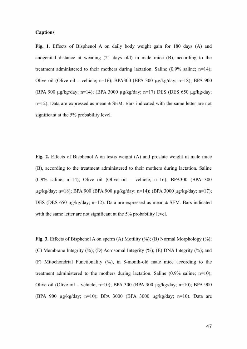
Fig. 1. Effects of Bisphenol A on daily body weight gain for 180 days (A) and
anogenital distance at weaning (21 days old) in male mice (B), according to the
treatment administered to their mothers during lactation. Saline (0.9% saline; n=14);
Olive oil (Olive oil – vehicle; n=16); BPA300 (BPA 300 µg/kg/day; n=18); BPA 900
(BPA 900 µg/kg/day; n=14); (BPA 3000 µg/kg/day; n=17) DES (DES 650 µg/kg/day;
n=12). Data are expressed as mean ± SEM. Bars indicated with the same letter are not
significant at the 5% probability level.
Fig. 2. Effects of Bisphenol A on testis weight (A) and prostate weight in male mice
(B), according to the treatment administered to their mothers during lactation. Saline
(0.9% saline; n=14); Olive oil (Olive oil – vehicle; n=16); BPA300 (BPA 300
µg/kg/day; n=18); BPA 900 (BPA 900 µg/kg/day; n=14); (BPA 3000 µg/kg/day; n=17);
DES (DES 650 µg/kg/day; n=12). Data are expressed as mean ± SEM. Bars indicated
with the same letter are not significant at the 5% probability level.
Fig. 3. Effects of Bisphenol A on sperm (A) Motility (%); (B) Normal Morphology (%);
(C) Membrane Integrity (%); (D) Acrosomal Integrity (%); (E) DNA Integrity (%); and
(F) Mitochondrial Functionality (%), in 8-month-old male mice according to the
treatment administered to the mothers during lactation. Saline (0.9% saline; n=10);
Olive oil (Olive oil – vehicle; n=10); BPA 300 (BPA 300 µg/kg/day; n=10); BPA 900
(BPA 900 µg/kg/day; n=10); BPA 3000 (BPA 3000 µg/kg/day; n=10). Data are
47
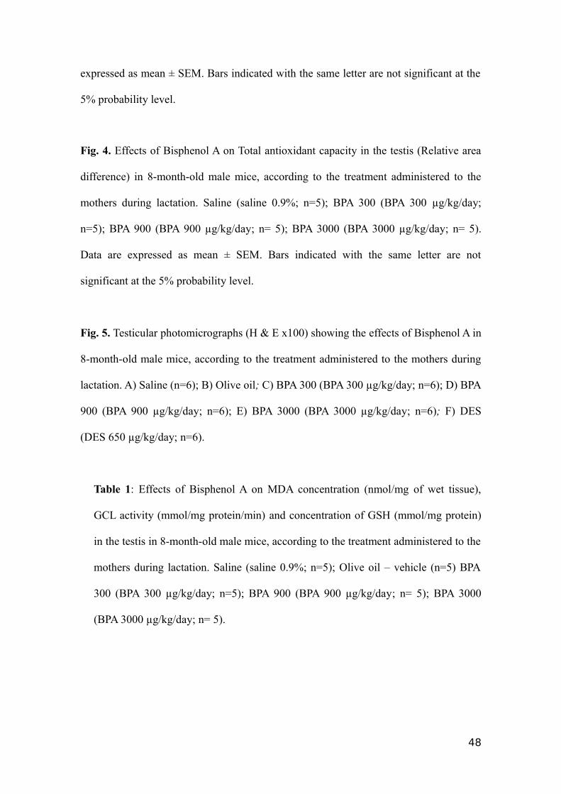
expressed as mean ± SEM. Bars indicated with the same letter are not significant at the
5% probability level.
Fig. 4. Effects of Bisphenol A on Total antioxidant capacity in the testis (Relative area
difference) in 8-month-old male mice, according to the treatment administered to the
mothers during lactation. Saline (saline 0.9%; n=5); BPA 300 (BPA 300 µg/kg/day;
n=5); BPA 900 (BPA 900 µg/kg/day; n= 5); BPA 3000 (BPA 3000 µg/kg/day; n= 5).
Data are expressed as mean ± SEM. Bars indicated with the same letter are not
significant at the 5% probability level.
Fig. 5. Testicular photomicrographs (H & E x100) showing the effects of Bisphenol A in
8-month-old male mice, according to the treatment administered to the mothers during
lactation. A) Saline (n=6); B) Olive oil; C) BPA 300 (BPA 300 µg/kg/day; n=6); D) BPA
900 (BPA 900 µg/kg/day; n=6); E) BPA 3000 (BPA 3000 µg/kg/day; n=6); F) DES
(DES 650 µg/kg/day; n=6).
Table 1: Effects of Bisphenol A on MDA concentration (nmol/mg of wet tissue),
GCL activity (mmol/mg protein/min) and concentration of GSH (mmol/mg protein)
in the testis in 8-month-old male mice, according to the treatment administered to the
mothers during lactation. Saline (saline 0.9%; n=5); Olive oil – vehicle (n=5) BPA
300 (BPA 300 µg/kg/day; n=5); BPA 900 (BPA 900 µg/kg/day; n= 5); BPA 3000
(BPA 3000 µg/kg/day; n= 5).
48
Figure 1
49
Figure 2
50
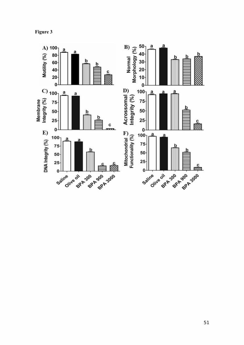
Figure 3
51
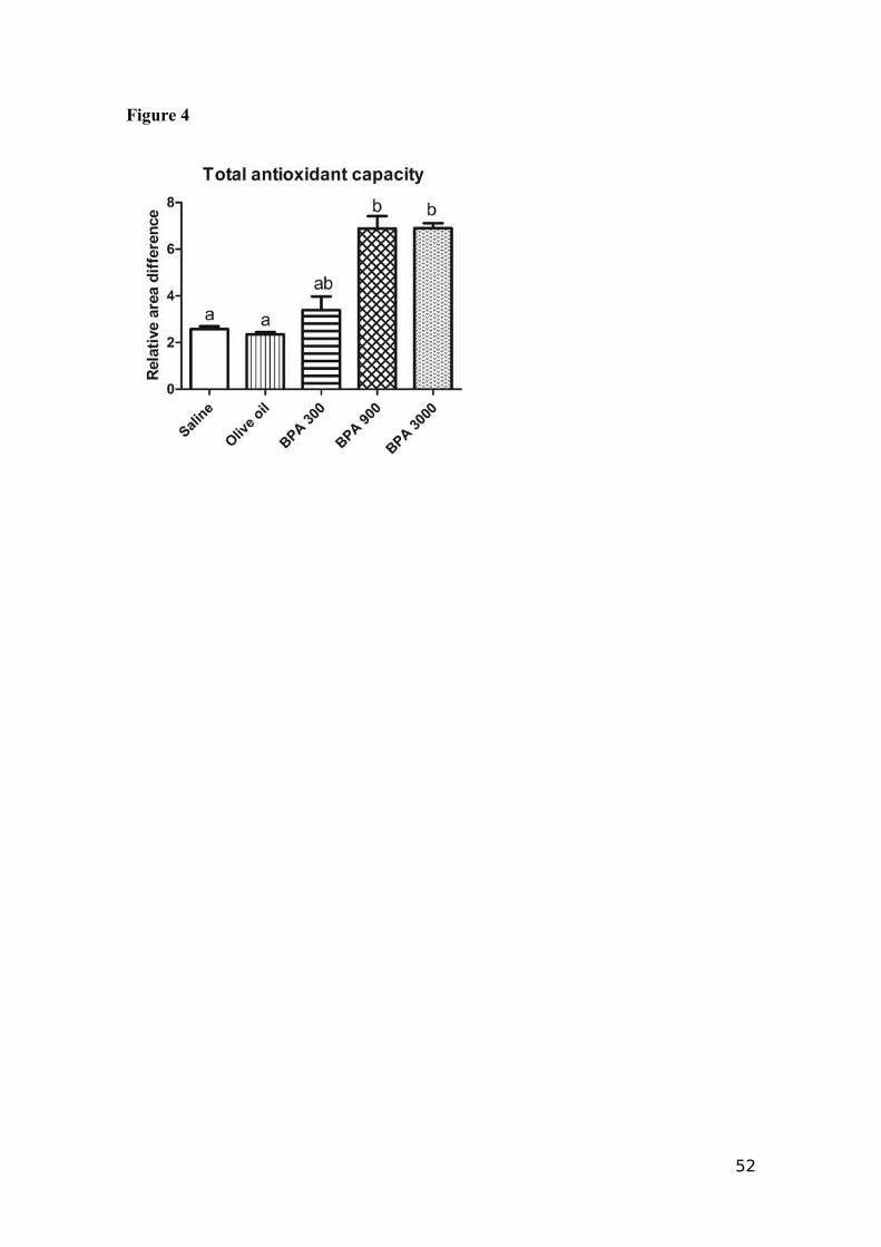
Figure 4
52
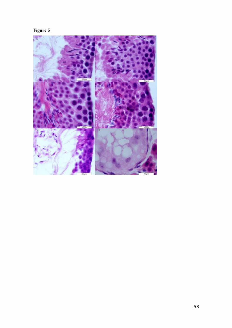
Figure 5
53
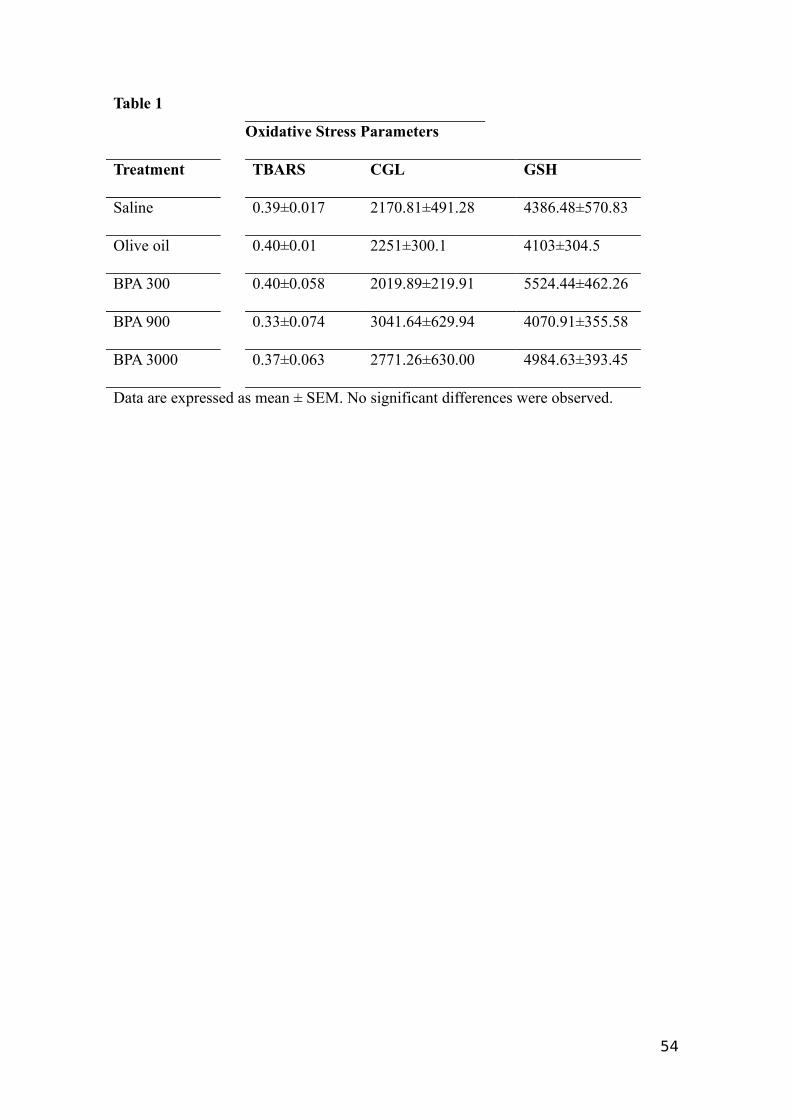
Table 1
Oxidative Stress Parameters
Treatment TBARS CGL GSH
Saline 0.39±0.017 2170.81±491.28 4386.48±570.83
Olive oil 0.40±0.01 2251±300.1 4103±304.5
BPA 300 0.40±0.058 2019.89±219.91 5524.44±462.26
BPA 900 0.33±0.074 3041.64±629.94 4070.91±355.58
BPA 3000 0.37±0.063 2771.26±630.00 4984.63±393.45
Data are expressed as mean ± SEM. No significant differences were observed.
54

Artigo 2
Revista : Toxicology Letters
(Fator de impacto: 3,23)
55
Effect of BPA on the central nervous system in lactating females
Ana Cristina Kalba, Ana Luiza Kalbb, Gabriela Hollmannc, Flavia Conde Kneipd,
Antonio Sergio Varela Juniorb, Silvana Allodic; Pablo Elías Martíneza, b, *
a Programa de Pós-Graduação em Ciências Fisiológicas: Fisiologia Animal Comparada,
Universidade Federal do Rio Grande, Rio Grande, RS, Brazil
b Instituto de Ciências Biológicas; Universidade Federal do Rio Grande, Rio Grande,
RS, Brazil
c Laboratório de Biofísica; Universidade Federal do Rio de Janeiro, Rio de Janeiro, RJ,
Brazil
d Instituto de Física e Matemática; Universidade Federal do Rio Grande, Rio Grande,
RS, Brazil
* Corresponding author. Tel./fax: +55 53 32336848.
E-mail address: [email protected] (P.E. Martínez).
56
Abstract
Bisphenol A (BPA) is a known endocrine disruptor that impairs brain function. We
employed immunohistochemistry tools to evaluate the effects in many regions of the
brain of lactant female mice exposed sub-chronically (21 days) to BPA (300, 900 and
3000 µg/kg/day) via gavage. The parameters observed were labels for: astrocytes - glial
acidic fibrillary protein (GFAP), cell proliferation - histone-3-phosphate (H3P), synapse
density synaptophisin (SYP), and catecholamine synthesis - tyrosine hydroxilase (TH).
The results showed that 300 µg/kg/day BPA decreased the number of astrocytes in all
brain regions and diminished TH fluorescence in the striatum and CA3 region of the
hippocampus. The dose of 900 µg/kg/day BPA also decreased the number of astrocytes
in the CA3 region and increased cell proliferation in the hippocampus, however both in
the cortex and in the CA3 region this treatment influenced synaptogenesis, reducing the
process. The overall results show that cell proliferation occurred in the brain of the
female mice but there were also losses induced by BPA on the number of astrocytes, in
TH concentration and synaptic density. These findings reinforce the potential
implication of BPA exposure in neurodegenerative diseases.
Keywords: bisphenol A; endocrine disruptor; brain;
Highlights
Bisphenol A (BPA) is a ubiquitous endocrine disrupter;
We exposed mouse mothers to BPA through lactation;
BPA impaired nervous central system parameters;
57
1. Introduction
Since the decade of 1950, the synthetic xenoestrogen, Bisphenol A (BPA), has been
employed in the manufacturing of some plastics with a range of uses, including dental
prosthesis and sealants and polycarbonate cans used to preserve foods and items as baby
bottles (Suzuki et al., 2000; Brede et al., 2003).
Previous studies regarding the biological and toxicological effects of BPA on the human
body have focused on its estrogenic action. This is because BPA seems to mimic some
estradiol-induced biological effects, such as sperm toxicity in humans (Hauser et al.,
2005) and fertility and fecundity decrease in mice (Cabaton et al., 2011). In addition to
the effects on reproductive tissues, BPA is likely to alter the morphological and
functional properties of neurons and astrocytes in the central nervous system (CNS).
Astrocytes, which may be identified by the intermediate filament glial fibrillary acidic
protein (GFAP) immunolabeling, play a key role in brain neurotransmission and express
estrogen receptors. BPA inhibits estradiol-induced hippocampal synaptogenesis
(MacLusky et al., 2005) and in doses of 1 pM or 1 µM) also amplifies the response of
dopaminergic neurons and astrocytes (Miyatake et al. 2006).
One important marker for dopamine is tyrosine hydroxylase (TH), which
catalyzes the enzymatic conversion of L-tyrosine to L-3, 4-dihydroxyphenylalanine
(L-dopa), the first step in the biosynthesis of chatecolamines. Xu et al. (2011) examined
the rapid effect of BPA, with a dose of 500 μg/kg on a single administration in the
hippocampus, on passive avoidance memory and N-methyl-D-aspartate receptor
(NMDAR) expression in the developing hippocampus of rats at the age of postnatal day
18. The authors observed that BPA or estradiol benzoate (EB) rapidly extended the
latency to step down from the platform 1 h after footshock and increased the
58
phosphorylation levels of NR1, NR2B (subunits of NMDAR), and mitogen-activated
extracellular signal-regulated kinase (ERK) in hippocampus within 1 h.
BPA interference in spine synapse formation has profound implications. Leranth
et al. (2008) observed, under a relative low exposure level (50 µg/Kg/day) to nonhuman
primates, a complete abrogation of the synaptogenic response to estradiol. Because sex
steroids are widely thought to play critical roles in higher brain activities, such as
cognition and mood through modulation of the structural and functional synaptic
plasticity, it is important to investigate the consequences of BPA exposure. BPA can
also antagonize spine synapse formation induced by estrogens and testosterone in limbic
brain areas of gonadectomized female and male rats. One important marker to
synaptogenesis is synaptophysin that can be found on the cytoplasmic surface of clear
pre-synaptic microvesicles of neurons. (Li et al., 2004; Neil et al., 2005; Liu et al.,
2008).
Phosphorylated histone H3 at serine 10 is a marker of cell proliferation.
Neuronal proliferation is well established in the subventricular zone (SVZ) of the lateral
ventricles and the subgranular zone (SGZ) of the dentate gyrus of the hippocampus
show constitutive neurogenesis under physiological (Sulllivan et al., 2007) and
pathological conditions (Nakayama et al., 2010). However, the central dogma of
non-renewal of cortical neurons was first called into question in 1962 by Altman in a
study that shows evidence of adult-born neurons in the human cortex (Altman, 1962).
Posteriorly Gould et al. (1999) recorded in primates, proliferation of neurons in the
prefrontal, inferior temporal and posterior parietal cortex. The new neurons may
originate in the subventricular zone and then migrate to the neocortex. These neurons
are added during adulthood, and may play important role in associative cortical
functions (Gould, et al., 1999).
59
Different physiological conditions such as aging, estrous cycle, pregnancy and
lactation can affect rodent neurogenesis (Pawluski et al., 2009). During the period of
maternity, there is an increase in neural plasticity, especially due to the induction of
maternal behavior and lactation. The hippocampus, although not traditionally associated
with this condition, has been hypothesized to be involved in the maternal behavior and
motherhood (Kinsley et al., 2006; Pawluski and Galea, 2006). As it is known,
reproductive conditions are affected by steroid hormones and prolactin (Pawluski et al.,
2009; Larsen and Gratan, 2012). A short time exposure to estradiol time increases
neurogenesis in the hippocampus, whereas long-term exposure shows the opposite
effect (Galea, 2008). The 17β-estradiol and BPA were also reported to affect
neurogenesis of stem and progenitor cells from telencephalum of rat embryos (Okada et
al., 2010).
The present study aimed to investigate the effects of BPA in adult female nursing
mice exposed sub-chronically, since there are few studies that investigate this particular
condition. Studies evaluating the effects of BPA on the CNS during lactation is an
innovative research. Therefore, we studied the effects of exposure to BPA on enzymes
and cell markers in different regions of the CNS in nursing female mice. For this
purpose, it was employed a immunohistochemical approach to estimate the number of
astrocytes, cellular proliferation, tyrosine hydroxylase and synaptophysin fluorescent
area, which are important parameters of brain function.
60
2. Materials and methods
2.1 Animals
All procedures involving animal subjects were reviewed and approved by the
Animal Ethics Committee of the Universidade Federal do Rio Grande (FURG), Rio
Grande, Rio Grande do Sul (Approval number: P006/2011). Healthy Swiss albino mice
obtained from the Central Animal Facility - FURG, maintained by random breeding,
were housed in standard polystyrene cages at 23°C and 12-h light/12-h dark cycle, with
water and a soybean-free diet ad libitum (Nuvilab CR-1 NUVITAL, Jundiaí, SP, Brazil).
After the acclimation period of one week, the animals (8 weeks old) were grouped in
cages (5 females + 1 male each) for random mating. The presence of the vaginal plug
was verified twice a day for mating confirmation and, if positive, the females were
considered pregnant. Pregnant females were placed into cages to give birth, and the
pups were maintained with its dams from the birth day to the end of the lactation period
(21 days). During this period, 5 treatment groups of 5 dams each received 200 µL/day
of the following solutions, via gavage: saline 0.9% to the control group (SALINE); 300
µg/w/day BPA (BPA 300); 900 µg/w/day BPA (BPA 900); 3000 µg/w/day BPA (BPA
3000) and 650 µg/w/day diethylstilbestrol (DES) to the positive control (DES). At the
day 21 after birth, the pups were separated for further analyses, and the dams
(sub-chronic exposure) were sacrificed by cervical dislocation (Hogan et al., 1986). The
brain tissue was dissected and pieces not larger than 0.5 cm were promptly submerged
into formaldehyde 4% freshly prepared from paraformaldehyde, for fixation during 24
h. After, the pieces were dehydrated in a graded series of ethanol up to 100%, cleared in
xylene and embedded in paraplast X-TRA (Sigma P3808). Six μm thick sections of the
61

striatum, cortex and hippocampus were obtained with a rotary microtome (Leica RM
2255) and mounted on poli-L-lisine-coated slides.
2.2 Immunohistochemistry
For the immunohistochemistry (IHC) assays, the brain sections were re-hydrated
in solutions with decreasing concentrations of ethanol. The slides were washed three
times, 10 min each, in a washing solution (PBS/Triton X-100 0.3%). The antigenic
recovery was performed with sodium citrate buffer 0.01% (pH 6.0) during 45 min at
95-98ºC. This step was employed to optimize the binding of the antibody to its
respective specific epitope. After, the unspecific binding sites of the tissue were blocked
with PBS/BSA 3% and PBS/NGS 10% during 2 h. The slides were then incubated
overnight at 4ºC with the primary antibody solutions (diluted 1:100 in PBS 0.1 M). The
primary antibodies employed were: monoclonal anti-GFAP produced in mouse (G3893,
Sigma-Aldrich), policlonal anti-TH produced in rabbit (T8700, Sigma-Aldrich),
policlonal anti- H3P serine 10, produced in rabbit (sc8656, Santa Cruz), and policlonal
anti-SYP produced in goat (sc7568, Santa Cruz). After the primary antibody incubation,
the slides were washed again with the washing solution (three times, 10 min each) and
incubated for 2 h at room temperature with the following secondary antibodies: CY3
(C2181, Sigma-Aldrich, anti-mouse IgG); CY3 anti-rabbit IgG (C2306, Sigma);
Alexa-Fluor 488 anti-rabbit IgG (A11094, Invitrogen) and CY3 anti-goat IgG (C2821,
Sigma-Aldrich). After, the slides were washed again with the washing solution (three
times, 10 min), incubated during 1 min with 4,6-diamidino-2 phenylindole (DAPI)
(D9542, Sigma-Aldrich), and mounted with aqueous medium mount (Fluoromount, Ref
F4680, Sigma-Aldrich). The structures were observed under a Zeiss Axioskop 2 Plus
epifluorescence microscope equipped with a color CCD camera (Media Cybernetics,
62
model Evolution MP). The fluorescent areas were analyzed using the IMAGE J
program.
3. Statistical analysis
Data were expressed as mean ± SEM. Once the assumptions of homogeneity and
normality of variance were verified, statistical analysis was performed between
treatments by means of one-way ANOVA. Post hoc comparisons were performed using
the Tukey test or orthogonal contrasts. The significant level adopted was 5% for all.
4. Results
4.1 GFAP marker
Representative images of astrocytes labeled with GFAP in the cortex, striatum
and hippocampus treated with five treatments are shown in Figures 1, 2 and 3. The
labeled astrocytes were counted and the difference of numbers of astrocytes in the
cortex, striatum and hippocampus (CA3) are shown in Figure 4 (A, B and C). The In
the cortex we observed a significant decrease (p<0.05) of astrocytes in the BPA 300 and
BPA 900 groups, as well as in the DES–treated animals when compared with the
control (Saline = 81±47; BPA 300 = 28±16; BPA 900 = 46±26; BPA 3000 = 68±39;
DES = 44±20). In the striatum, the BPA 300 and DES treatments resulted in
significantly decreased number of astrocytes (p<0.05) (BPA 300 = 8±4 and DES = 8±4)
when compared with the control group (Saline = 35±20, BPA 900 = 11±6 and BPA
3000 =15±9). However, BPA 900 and BPA 3000 groups showed a not significant
difference of number of astrocytes. The number of astrocytes in CA3 (Fig.4, C) region
was impaired (p<0.05) in the BPA 300 (16±9) and in the BPA 900 (24±14) treatments
when compared to the control group Saline (63±36). The BPA 3000 (39±23) and DES
(45±20) treatments did not statistically differentiate (p>0.05) either to the control or the
63
other treatments, but a slight decrease in astrocytes number with respect to the control
group can be noted.
4.2 Tyrosine Hydroxylase
Representative images of the striatum and hippocampus (CA3) labeled with TH
are shown in Figures 5 and 6. Figure 7 (A, B and C) shows the positive cells /
fluorescent area (µm2) in the cortex, striatum and hippocampus (CA3).The different
BPA and DES treatments did not affect the fluorescent area of the cortex (Fig. 5 A)
when compared with the corresponding control groups (Saline = 0.04269± 0.02465;
BPA 300 = 0.02670± 0.01542; BPA 900 = 0.03720± 0.02148; BPA 3000 = 0.03218±
0.01858; DES = 0.02507± 0.01448). The BPA 3000 (0.0137±0.0079) and DES
(0.0098±0.0056) treatments significantly decreased (p<0.05) the fluorescence area (Fig.
5 B) in the striatum, when compared with all the other groups (Saline = 0.0277±0.0160;
BPA 300 = 0.0192±0.0111; BPA 900 = 0.0180±0.0104). The data of fluorescent area
from CA3 region (Fig. 5 C) showed a significantly decrease in the BPA 3000 (p<0.05)
(Saline = 0.04458±0.02574; BPA 300 = 0.03710±0.02142; BPA 900 =
0.02842±0.01641; BPA 3000 = 0.01953±0.01128; DES = 0.02779±0.01605).
4.3 Synaptophysin
Representative image of synaptophysin labeling in the pre-frontal cortex is
shown in Figure 8. Figure 9 (A, B and C) shows the differences in the fluorescent area
occupied by synaptophysin positive cells in the cortex, striatum and hippocampus
(CA3) , respectively. In the cortex (Fig 9 A) the BPA 900 treatment showed a
significant decrease (0.02596±0.01079) (p<0.05) when compared with the saline
treatment (Saline = 0.04853±0.00176) however the other tresatments were not
significantly different from the control (BPA 300 = 0.05172±0.00256; BPA 3000 =
0.03354±0.00892; DES = 0.03060±0.00314). In the striatum (Fig 9 B) no difference
64
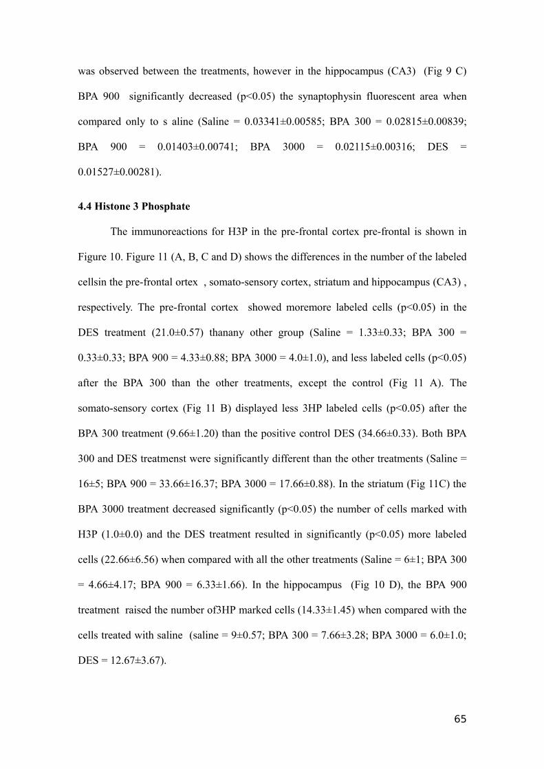
was observed between the treatments, however in the hippocampus (CA3) (Fig 9 C)
BPA 900 significantly decreased (p<0.05) the synaptophysin fluorescent area when
compared only to s aline (Saline = 0.03341±0.00585; BPA 300 = 0.02815±0.00839;
BPA 900 = 0.01403±0.00741; BPA 3000 = 0.02115±0.00316; DES =
0.01527±0.00281).
4.4 Histone 3 Phosphate
The immunoreactions for H3P in the pre-frontal cortex pre-frontal is shown in
Figure 10. Figure 11 (A, B, C and D) shows the differences in the number of the labeled
cellsin the pre-frontal ortex , somato-sensory cortex, striatum and hippocampus (CA3) ,
respectively. The pre-frontal cortex showed moremore labeled cells (p<0.05) in the
DES treatment (21.0±0.57) thanany other group (Saline = 1.33±0.33; BPA 300 =
0.33±0.33; BPA 900 = 4.33±0.88; BPA 3000 = 4.0±1.0), and less labeled cells (p<0.05)
after the BPA 300 than the other treatments, except the control (Fig 11 A). The
somato-sensory cortex (Fig 11 B) displayed less 3HP labeled cells (p<0.05) after the
BPA 300 treatment (9.66±1.20) than the positive control DES (34.66±0.33). Both BPA
300 and DES treatmenst were significantly different than the other treatments (Saline =
16±5; BPA 900 = 33.66±16.37; BPA 3000 = 17.66±0.88). In the striatum (Fig 11C) the
BPA 3000 treatment decreased significantly (p<0.05) the number of cells marked with
H3P (1.0±0.0) and the DES treatment resulted in significantly (p<0.05) more labeled
cells (22.66±6.56) when compared with all the other treatments (Saline = 6±1; BPA 300
= 4.66±4.17; BPA 900 = 6.33±1.66). In the hippocampus (Fig 10 D), the BPA 900
treatment raised the number of3HP marked cells (14.33±1.45) when compared with the
cells treated with saline (saline = 9±0.57; BPA 300 = 7.66±3.28; BPA 3000 = 6.0±1.0;
DES = 12.67±3.67).
65
5. Discussion
In thist study, we observed differences in the brain of female mice exposed
during 21 days (lactation period) to BPA (300, 900 and 3000 µg/kg/day) when
compared with the controls. These doses were below the No Observed Adverse Effect
Level (NOAEL) = 50mg/kg/b.w./day in rats (Tyl et al., 2002), which is no biologically
or statistically significant increase in the frequency or severity of any adverse effects in
the exposed population when compared to its appropriate control. In addition to the
effects on reproductive tissues and thyroid gland, BPA is likely to alter the
morphological and functional properties of neural cells in the central nervous system
(CNS). It was reported that the exposure to BPA in female rats during pregnancy and
lactation resulted in behavioral changes in dams, thus indicating that BPA altered neural
circuits (Della Seta et al., 2005). Here, we investigated the BPA effects in three distinct
regions of the brain: hippocampus, striatum and cortex. In these regions were evaluated
GFAP, TH, Synaptophysin and H3P.
Astrocytes are among of the most important target cells for Estradiol (E2) and
express nuclear and membrane estrogen receptors during development and in the adult
brain. These glial cells modulate synaptic transmissions and respond to
neurotransmitters released within the synapse by generating elevations in intracellular
Ca2+ concentration (Fellin and Carmignoto, 2004). In our study, in the striatum and
cortex areas (Figure 4), BPA 300 reduced the number of GFAP+ cells when compared
with the control. In the striatum, DES also decreased (P<0.05) the number of astrocytes.
In the hipppocampus (CA3) BPA 300 and 900 decrease (P<0.05) GFAP+ cells (Figure
4). BPA typically induces a non-monotonic response. However, the results suggested
that BPA effects can be mediated by its estrogenic activity.
66
Miyatake et al. (2006) administrated low doses of BPA in vitro on midbrain glial
cellular cultures (1fM to 1µM), and showed increase in GFAP+ astrocytes demonstrating
that the dopamine-induced Ca2+ responses in mixed cultures of neurons and astrocytes
were significantly enhanced by the treatment with BPA. Jang et al (2012) did not
observe GFAP increase in female mice in vivo. We observed that the exposure to the
lowest BPA concentration ( resulted in the lowest number of astrocytes when compared
with the control group, in all the brain regions studied. Therefore, another explanation
could be related with BPA-induced production of reactive oxygen species (ROS)
activating the caspase-3 pathway, which is a marker of neuronal cell death (Obata and
Kubota, 2000).
In our study, fluorescence of TH (rate-limiting enzyme for the catecholamines
synthesis) decreased by treatment with BPA in the Striatum and Hippocampus (CA3)
regions (Figure 7). It was observed in both regions that fluorescence in the BPA 3000
treatment significantly decreased (P<0.05) when compared to control. Ishido et al.
(2007) exposed pups to 600µg/day and demonstrated that BPA-induced alterations in
the dopamine (DA) system which can be pre-synaptic, affecting DA synthesis, DAT
expression, and DA release and turnover in midbrain. Honma et al. (2006) observed that
in female rats exposed perinatally (GD 6 to PND 20) to BPA (4 and 40mg/kg/day) the
levels of DA increased significantly in the group receiving 4mg/kg/day in hippocampus,
and no significant differences were observed in the cortex and striatum regions in all
treatments. Our results showed a decrease of TH in the hippocampus and striatum and
coincidentally not changes in the cortex (Figure 7).
BPA activates both of the major types of ERs (ERα and ERβ) in the anterior
pituitary to modify estrogenic activity. Consequently, BPA may regulate the
transcriptional activity of genes targeted by the estrogen system, including
67
dopaminergic genes (Ishido et al. 2007). Pre-natal BPA exposure decreased the number
of TH neurons observed in the rostral periventricular preoptic area in female rat
offspring in low BPA concentration and can be reflected on changes in the behavioral
activities in adult age (Sun et al. 2002). However, in our work the TH alterations could
be observed in high BPA concentrations in nursing female mice and were recorded in
specific brain areas. Similar results were found by Honma et al. (2006) whose observed
changes in dopamine and its metabolites in pups as well as dams. Furthermore, it has
been reported that BPA stimulates •OH generation in rat striatum (Obata and Kubota,
2000). Thus, it is most likely that the toxicity of BPA can be attributed to the
degeneration of dopaminergic neurons via oxidative stress effects. Nevertheless, we
could not eliminate the possibility that the estrogenicity of BPA should, at least in part,
be involved in this process. Ishido et al. (2007) demonstrated that the BPA oral
administration (exposed pups to 600µg/day) caused hyperactivity on first time and with
a prolonged administration (7 weeks) caused a large reduction of immunoreactivity for
TH in the midbrain This shows that BPA can lead to decreased levels on tyrosine
hydroxylase enzyme and the number of dopaminergic neurons. In our work, we found
similar results in different brain locals, an accentuate decrease on tyrosine hydroxylase
in the striatum and hippocampus.
BPA inhibits estradiol-induced hippocampal synaptogenesis (Mac Lusky et al.,
2005), alters dopamine responsiveness in neurons and astrocytes and suppresses the
estradiol-induced enhancement of long-term potentiation in the hippocampal CA1 area
(Mukai et al., 2006). Synaptophysin is a suitable indicator of synaptic density. In our
study synaptophysin+ area was decreased after using the highest dose in the cortex and
hippocampus (CA3) (Figure 10). These findings were similar to those reported by
Yokosuka et al. (2008) who administrated in hypothalamic cell culture 1µM of BPA and
68
this suppressed synaptic density (Synapsin+ area, and different synaptic marker, but with
similar function). On the other hand, a single exposure to BPA (0.32, 3.2 or 4.8 mg/kg)
in male and female mice (PND 10) resulted at 5 months old in an increase in the level of
synaptophysin (Viberg and Lee, 2012).
Iwakura et al. (2010) demonstrated that hypothalamic cell culture from fetuses
treated with 100 nM BPA increased the Synapsin I, but the change was not accompanied
by the increase in protein or mRNA expression levels ERs. The authors postulated that
BPA, similar to E2, induced effects on Synapsin I mainly through a non genomic
pathway. Alyea and Watson (2009) have demonstrated that low levels of xenoestrogens
including BPA act via membrane bound receptors (G protein coupled receptor 30 -
GPR30) but not via intracellular ones. Our exposure to BPA in female lactating mice
was subchronic, thus difficulting the comparison with other studies. However, in
general the results are consistent with the labeling observed for GFAP and TH.
H3P phosphorilated in serine 10 is an indicative of mitosis and meiosis. We
employed H3P as a labeling for cellular proliferation. The neurogenesis is influenced by
hormonal condition. In the postpartum period, Pawluski et al. (2009) observed increase
in neurogenesis in the lateral ventricle zone (SVZ) and gyrus dentatus of hippocampus.
Estrogen and progesterone increased the neurogenesis whereas chronic E2 expositions
result in decreasing of this process (Galea, 2008).
In physiological condition, a study with adult macaques employing BrdU
showed increased neurogenesis in the neocortex and gyrus dentatus (Gould et al., 2001).
Additionally, in adult rats thymidine - H3 as used as proliferation markershowed that
neurogenesis occurred in the neocortex, mesencephalon and diencephalon (Altman,
1962). In our study, neurogenesis was decreased after BPA 300 treatment in the
pre-frontal cortex and after BPA 3000 treatment in the striatum. tTheseresults indicate
69
that BPA may have anti-neurogenic effects. Kim et al. (2012) and Jang et al. (2012)
observed that neurogenesis in the hippocampal region was decreased by BPA in high
concentrations (20 mg/kg) and increased at low concentrations (1mg/kg), respectively.
The exposure to BPA (200µg/kg) in pregnant female mice resulted in increase in
neurogenesis of its brood (Komada et al., 2012). In our results on cortex pre-frontal and
striatum it was observed beneficial effects of DES in terms of cellular proliferation. This
indicates the proliferative sensitivity to high concentrations of DES in these regions,
although BPA showed no effect on hippocampus and cortex somato-sensory, however,
the proliferation was visually higher in this region. In the assessment of cellular
proliferation in the hippocampus it was included the gyrus dentatus and it was observed
an increase on proliferation in the BPA 900 when compared to the control. The
synaptophysin labeling was lowin CA3 after the treatment of BPA 900 and proliferation
in the same concentration in the hippocampus including gyrus dentatus was high. The
important proliferation observed in adult females during lactation can be positively
affected by this condition although other studies would be carried out to confirm these
findings.
In summary, cell proliferation occurred in the nervous system of adult female
mice but there were also losses induced by BPA or DES on the number of astrocytes, in
tyrosine hydroxylase concentration and synaptic density. This shows the potential
implication of BPA in neurodegenerative diseases.
6. Conflict of interest
The author declared there is no conflict of interest.
7. Acknowledgements
This study was supported by a grant from the Fundação de Amparo à Pesquisa
do Estado do Rio Grande do Sul (FAPERGS Proc. No. 1013533– Edital PqG) to the
70
Postgraduate Program in Physiological Sciences Post-graduate Program of the
Universidade Federal do Rio Grande. Ana Cristina Kalb received an undergraduate
fellowship from the Brazilian agency CAPES (Coordenação de Aperfeiçoamento de
Pessoal de Nível Superior) and Ana Luiza Kalb received a scientific initiation grant
from CNPq (Conselho Nacional de Desenvolvimento Científico e Tecnológico).
8. References
Alyea, R.A.,Watson, C.S. 2009. Differential regulation of dopamine transporter function
and location by low concentrations of environmental estrogens and 17beta stradiol.
Environ Health Perspect. 117,778–783.
Brede C., Fjeldal, P., Skjevrak I., Herikstad H. 2003. Increased migration levels of
bisphenol A from polycarbonate baby bottles after dishwashing, boiling and brushing,
Food Addit. Contam. 20, 684–689.
Della Seta, D., Minder, I., Dessì-Fulgheri, F., Farabollini, F. 2005. Bisphenol-A
exposure during pregnancy and lactation affects maternal behavior in rats. Brain Res
Bull. 65(3), 255-60.
Fellin, T., Carmignoto, G. 2004. Neurone-to-astrocyte signalling in the brain represents
a distinct multifunctional unit. J Physiol. 559, 3–15.
Galea, L.A. 2008. Gonadal hormone modulation of neurogenesis in the dentate gyrus of
adult male and female rodents. Brain Res Rev. 57(2), 332-41.
Gould, E., Vail, N., Wagers, M., Gross, C.G. 2000. Adult-generated hippocampal and
neocortical neurons in macaques have a transient existence PNAS. 98(19),
10910–10917.
Gould, E., Reeves, A.J., Graziano, M.S., Gross, C.G. 1999. Neurogenesis in the
neocortex of adult primates. Science 286, 548–552.
71
Hauser, R., Williams, P., Altshul, L.A.M. 2005. Evidence of Interaction between
Polychlorinated Biphenyls and Phthalates in Relation to Human Sperm Motility.
EnvironHealth. 113(4), 425-430.
Honma, T., Miyagawa, M., Suda, M., Wang, R.S., Kobayashi, K., Sekiguchi, S. 2006.
Effects of perinatal exposure to bisphenol A on brain neurotransmitters in female rat
offspring. Ind Health. 44,510–24.
Ishido M, Yonemoto J, Morita M. 2007. Mesencephalic neurodegeneration in the orally
administered bisphenol A-caused hyperactive rats. Toxicol Lett. 173, 66–72.
Jang, Y.J., Park, H.R., Kim, T.H., Yang, W.J., Lee, J.J., Choi, S.Y., Lee, E., Park, J.H.,
Kim, H.P., Kim, H.S., Lee, J. 2012. High dose bisphenol A impairs hippocampal
neurogenesis in female mice across generations. Toxicol. 296, 73– 82.
Kim, M.E., Park, H.R., Gong, E.J., Choi, S.Y., Kim, H.S., Lee, J., 2011. Exposure to
bisphenol A appears to impair hippocampal neurogenesis and spatial learning and
memory. Food Chem Toxicol. 49, 3383–3389.
Kinsley, C.H., Trainer, R., Stafisso-Sandoz, P. Quadros, L.K., Marcus, C., Hearon, E.A.,
Meyer, N., Hester, M., Morgan, F.J., Kozub, K.G. 2006. Motherhood and the hormones
of pregnancy modify concentrations of hippocampal neuronal dendritic spines. Horm.
Behav. 49, 131–142.
Komada, M., Asai, Y., Moric, M., Matsuki, M., Sato, M., Nagao, T. 2012. Maternal
bisphenol A oral dosing relates to the acceleration of neurogenesis in the developing
neocortex of mouse fetuses. Toxicol. 295, 31– 38.
Larsen, C.M., Grattan, D.R. 2012. Prolactin, neurogenesis, and maternal behaviors.
Brain Behav Immun. 26(2), 201-9.
72
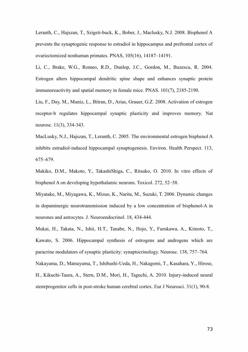
Leranth, C., Hajszan, T., Szigeti-buck, K., Bober, J., Maclusky, N.J. 2008. Bisphenol A
prevents the synaptogenic response to estradiol in hippocampus and prefrontal cortex of
ovariectomized nonhuman primates. PNAS, 105(16), 14187–14191.
Li, C., Brake, W.G., Romeo, R.D., Dunlop, J.C., Gordon, M., Buzescu, R. 2004.
Estrogen alters hippocampal dendritic spine shape and enhances synaptic protein
immunoreactivity and spatial memory in female mice. PNAS. 101(7), 2185-2190.
Liu, F., Day, M., Muniz, L., Bitran, D., Arias, Grauer, G.Z. 2008. Activation of estrogen
receptor-b regulates hippocampal synaptic plasticity and improves memory. Nat
neurosc. 11(3), 334-343.
MacLusky, N.J., Hajszan, T., Leranth, C. 2005. The environmental estrogen bisphenol A
inhibits estradiol-induced hippocampal synaptogenesis. Environ. Health Perspect. 113,
675–679.
Makiko, D.M., Makoto, Y., TakashiShiga, C., Ritsuko, O. 2010. In vitro effects of
bisphenol A on developing hypothalamic neurons. Toxicol. 272, 52–58.
Miyatake, M., Miyagawa, K., Mizuo, K., Narita, M., Suzuki, T. 2006. Dynamic changes
in dopaminergic neurotransmission induced by a low concentration of bisphenol-A in
neurones and astrocytes. J. Neuroendocrinol. 18, 434-444.
Mukai, H., Takata, N., Ishii, H.T., Tanabe, N., Hojo, Y., Furukawa, A., Kimoto, T.,
Kawato, S. 2006. Hippocampal synthesis of estrogens and androgens which are
paracrine modulators of synaptic plasticity: synaptocrinology. Neurosc. 138, 757–764.
Nakayama, D., Matsuyama, T., Ishibashi-Ueda, H., Nakagomi, T., Kasahara, Y., Hirose,
H., Kikuchi-Taura, A., Stern, D.M., Mori, H., Taguchi, A. 2010. Injury-induced neural
stem⁄progenitor cells in post-stroke human cerebral cortex. Eur J Neurosci. 31(1), 90-8.
73
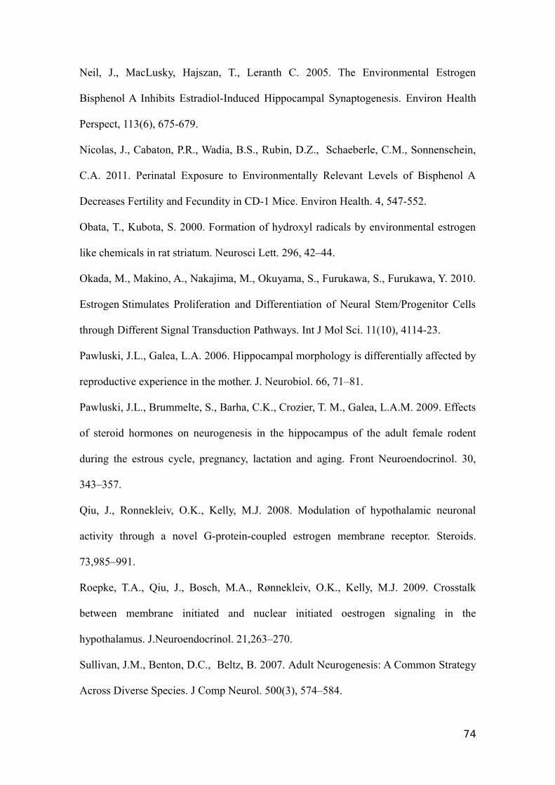
Neil, J., MacLusky, Hajszan, T., Leranth C. 2005. The Environmental Estrogen
Bisphenol A Inhibits Estradiol-Induced Hippocampal Synaptogenesis. Environ Health
Perspect, 113(6), 675-679.
Nicolas, J., Cabaton, P.R., Wadia, B.S., Rubin, D.Z., Schaeberle, C.M., Sonnenschein,
C.A. 2011. Perinatal Exposure to Environmentally Relevant Levels of Bisphenol A
Decreases Fertility and Fecundity in CD-1 Mice. Environ Health. 4, 547-552.
Obata, T., Kubota, S. 2000. Formation of hydroxyl radicals by environmental estrogen
like chemicals in rat striatum. Neurosci Lett. 296, 42–44.
Okada, M., Makino, A., Nakajima, M., Okuyama, S., Furukawa, S., Furukawa, Y. 2010.
Estrogen Stimulates Proliferation and Differentiation of Neural Stem/Progenitor Cells
through Different Signal Transduction Pathways. Int J Mol Sci. 11(10), 4114-23.
Pawluski, J.L., Galea, L.A. 2006. Hippocampal morphology is differentially affected by
reproductive experience in the mother. J. Neurobiol. 66, 71–81.
Pawluski, J.L., Brummelte, S., Barha, C.K., Crozier, T. M., Galea, L.A.M. 2009. Effects
of steroid hormones on neurogenesis in the hippocampus of the adult female rodent
during the estrous cycle, pregnancy, lactation and aging. Front Neuroendocrinol. 30,
343–357.
Qiu, J., Ronnekleiv, O.K., Kelly, M.J. 2008. Modulation of hypothalamic neuronal
activity through a novel G-protein-coupled estrogen membrane receptor. Steroids.
73,985–991.
Roepke, T.A., Qiu, J., Bosch, M.A., Rønnekleiv, O.K., Kelly, M.J. 2009. Crosstalk
between membrane initiated and nuclear initiated oestrogen signaling in the
hypothalamus. J.Neuroendocrinol. 21,263–270.
Sullivan, J.M., Benton, D.C., Beltz, B. 2007. Adult Neurogenesis: A Common Strategy
Across Diverse Species. J Comp Neurol. 500(3), 574–584.
74
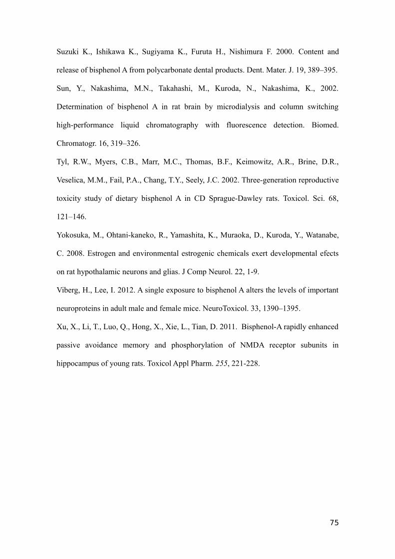
Suzuki K., Ishikawa K., Sugiyama K., Furuta H., Nishimura F. 2000. Content and
release of bisphenol A from polycarbonate dental products. Dent. Mater. J. 19, 389–395.
Sun, Y., Nakashima, M.N., Takahashi, M., Kuroda, N., Nakashima, K., 2002.
Determination of bisphenol A in rat brain by microdialysis and column switching
high-performance liquid chromatography with fluorescence detection. Biomed.
Chromatogr. 16, 319–326.
Tyl, R.W., Myers, C.B., Marr, M.C., Thomas, B.F., Keimowitz, A.R., Brine, D.R.,
Veselica, M.M., Fail, P.A., Chang, T.Y., Seely, J.C. 2002. Three-generation reproductive
toxicity study of dietary bisphenol A in CD Sprague-Dawley rats. Toxicol. Sci. 68,
121–146.
Yokosuka, M., Ohtani-kaneko, R., Yamashita, K., Muraoka, D., Kuroda, Y., Watanabe,
C. 2008. Estrogen and environmental estrogenic chemicals exert developmental efects
on rat hypothalamic neurons and glias. J Comp Neurol. 22, 1-9.
Viberg, H., Lee, I. 2012. A single exposure to bisphenol A alters the levels of important
neuroproteins in adult male and female mice. NeuroToxicol. 33, 1390–1395.
Xu, X., Li, T., Luo, Q., Hong, X., Xie, L., Tian, D. 2011. Bisphenol-A rapidly enhanced
passive avoidance memory and phosphorylation of NMDA receptor subunits in
hippocampus of young rats. Toxicol Appl Pharm. 255, 221-228.
75
Captions
Figure 1. Immunofluoresce analisys of glial fibrilary acid protein (GFAP) in cortex
in female mice (n=3 and 13 weeks old). 1: DAPI-labelling (blue), a nuclear marker, 2:
GFAP (red), astrocyte cells marker and 3: merged both images to each marker and
treatment. A: Saline; B: BPA 300µg/Kg./day (BPA 300); C: BPA 900µg//Kg./day (BPA
900); D: 3000µg//Kg./day (BPA 3000) and E: DES 650µg//Kg./day (DES). White
arrows indicate astrocytes. (Scale bar = 20µm).
Figure 2. Immunofluoresce analisys of glial fibrilary acid protein (GFAP) in
striatum in female mice (n=3 and 13 weeks old). 1: DAPI-labelling (blue), a nuclear
marker, 2: GFAP (red), astrocyte cells marker and 3: merged both images to each
marker and treatment. A: Saline; B: BPA 300µg/Kg./day (BPA 300); C: BPA
900µg//Kg./day (BPA 900); D: 3000µg//Kg./day (BPA 3000 and E: DES
650µg//Kg./day (DES). White arrows indicate astrocytes. (Scale bar = 20µm).
Figure 3. Immunofluoresce analisys of glial fibrilary acid protein (GFAP) in
hippocampus (CA3) in female mice (n=3 and 13 weeks old). 1: DAPI-labelling
(blue), a nuclear marker, 2: GFAP (red), astrocyte cells marker and 3: merged both
images to each marker and treatment. A: Saline; B: BPA 300µg/Kg./day (BPA 300); C:
BPA 900µg//Kg./day (BPA 900); D: 3000µg//Kg./day (BPA 3000) and E: DES
650µg/Kg./day (DES). White arrows indicate astrocytes. (Scale bar = 20µm).
76
Figure 4. (A), (B) and (C) shows the differences in female mice (n= 3 and 13 week old)
on astrocytes number on Cortex, Striatum and hippocampus (CA3) regions from brain,
respectively. Saline (0.9% saline); BPA300 (BPA 300 µg/kg/day); BPA 900 (BPA 900
µg/kg/day); (BPA 3000 µg/kg/day) DES (DES 650 µg/kg/day). Data are expressed as
mean ± SEM. Bars indicated with the same letter are not significant at the 5%
probability level.
Figure 5. Immunofluoresce analisys of tyrosine hydroxylase (TH) in striatum in
female mice (n=3 and 13 weeks old). 1: DAPI-labelling (blue), a nuclear marker, 2:
TH (red), TH positive cells marker and 3: merged both images to each marker and
treatment. A: Saline; B: BPA 300µg/Kg./day (BPA 300); C: BPA 900µg//Kg./day (BPA
900); D: 3000µg//Kg./day (BPA 3000) and E: DES 650µg//Kg./day (DES). White
arrows indicate TH positive cells marker. (Scale bar = 20µm).
Figure 6. Immunofluoresce analisys of tyrosine hydroxylase (TH) in hippocampus
(CA3) in female mice (n=3 and 13 weeks old). 1: DAPI-labelling (blue), a nuclear
marker, 2: TH (red), TH positive cells marker and 3: merged both images to each
marker and treatment. A: Saline; B: BPA 300µg/Kg./day (BPA 300); C: BPA
900µg//Kg./day (BPA 900); D: 3000µg//Kg./day (BPA 3000) and E: DES
650µg//Kg./day (DES). White arrows indicate TH positive cells marker. (Scale bar =
20µm).
Figure 7. (A), (B) and (C) shows Tyrosine hydroxylase positive cells in female mice (n=
3 and 13 week old) on Cortex, Striatum and hippocampus (CA3) regions from brain,
respectively. Saline (0.9% saline); BPA300 (BPA 300 µg/kg/day); BPA 900 (BPA 900
77
µg/kg/day); (BPA 3000 µg/kg/day) DES (DES 650 µg/kg/day). Data are expressed as
mean ± SEM. Bars indicated with the same letter are not significant at the 5%
probability level.
Figure 8. Immunofluoresce analisys of Synaptophysin in cortex in female mice (n=3
and 13 weeks old). 1: DAPI-labelling (blue), a nuclear marker, 2: Synaptophysin (red),
Synaptophysin positive cells marker and 3: merged both images to each marker and
treatment. A: Saline; B: BPA 300µg/Kg./day (BPA 300); C: BPA 900µg//Kg./day (BPA
900); D: 3000µg//Kg./day (BPA 3000) and E: DES 650µg//Kg./day (DES). White
arrows indicate synatophysin cells marker. (Scale bar = 20µm).
Figure 9. (A), (B) and (C) shows Synaptophysin positive cells in female mice (n= 3 and
13 week old) on Cortex, Striatum and hippocampus (CA3) regions from brain. Saline
(0.9% saline); BPA300 (BPA 300 µg/kg/day); BPA 900 (BPA 900 µg/kg/day); (BPA
3000 µg/kg/day) DES (DES 650 µg/kg/day). Data are expressed as mean ± SEM. Bars
indicated with the same letter are not significant at the 5% probability level.
Figure 10. Immunofluoresce analisys of histone 3 phosphate (H3P) in cortex
pre-frontal in female mice (n=3 and 13 weeks old). 1: DAPI-labelling (blue), a
nuclear marker, 2: Allexa 488 (green), H3P cells marker and 3: merged both images to
each marker and tratment. A: Saline; B: BPA 300µg/Kg./day (BPA 300); C: BPA
900µg//Kg./day (BPA 900); D: 3000µg//Kg./day (BPA 3000) and E: DES
650µg//Kg./day (DES). White arrows indicate histone 3 phosphate. (Scale bar = 20µm).
78
Figure 11. (A), (B), (C) and (D) shows number of histone 3 phosphate (H3P) in female
mice (n= 3 and 13 week old) on Cortex pre-frontal, Cortex somato-sensory, Striatum
and hippocampus (CA3) regions from brain. Saline (0.9% saline); BPA300 (BPA 300
µg/kg/day); BPA 900 (BPA 900 µg/kg/day); (BPA 3000 µg/kg/day) DES (DES 650
µg/kg/day). Data are expressed as mean ± SEM. Bars indicated with the same letter are
not significant at the 5% probability level.
79
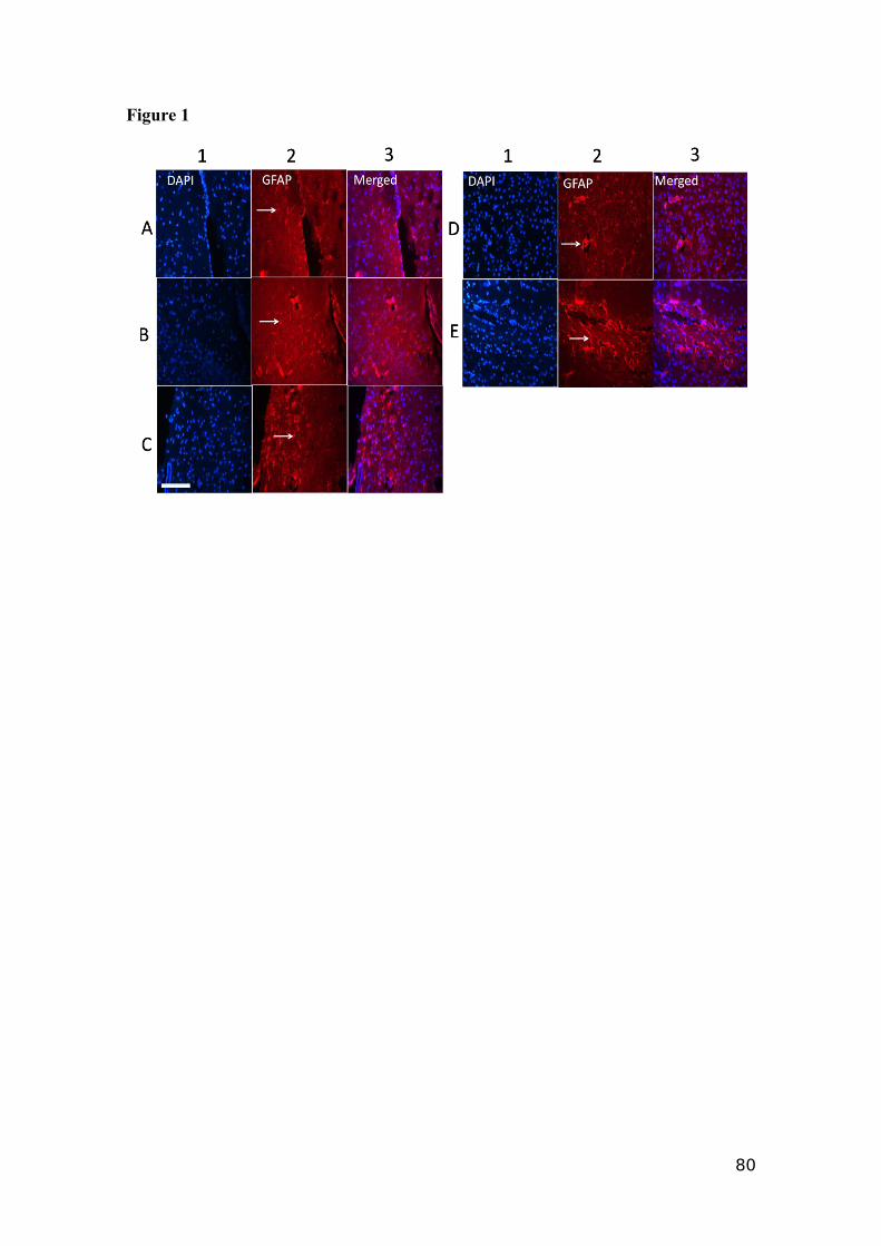
Figure 1
80
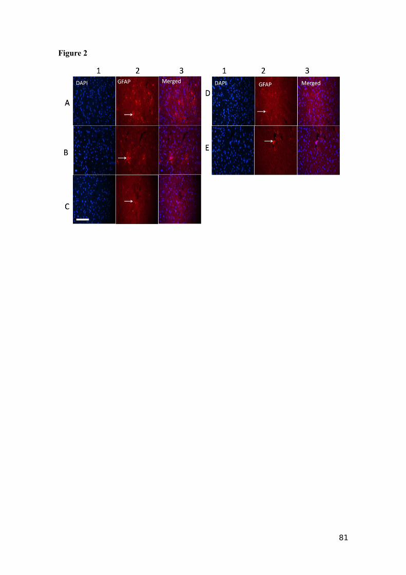
Figure 2
81
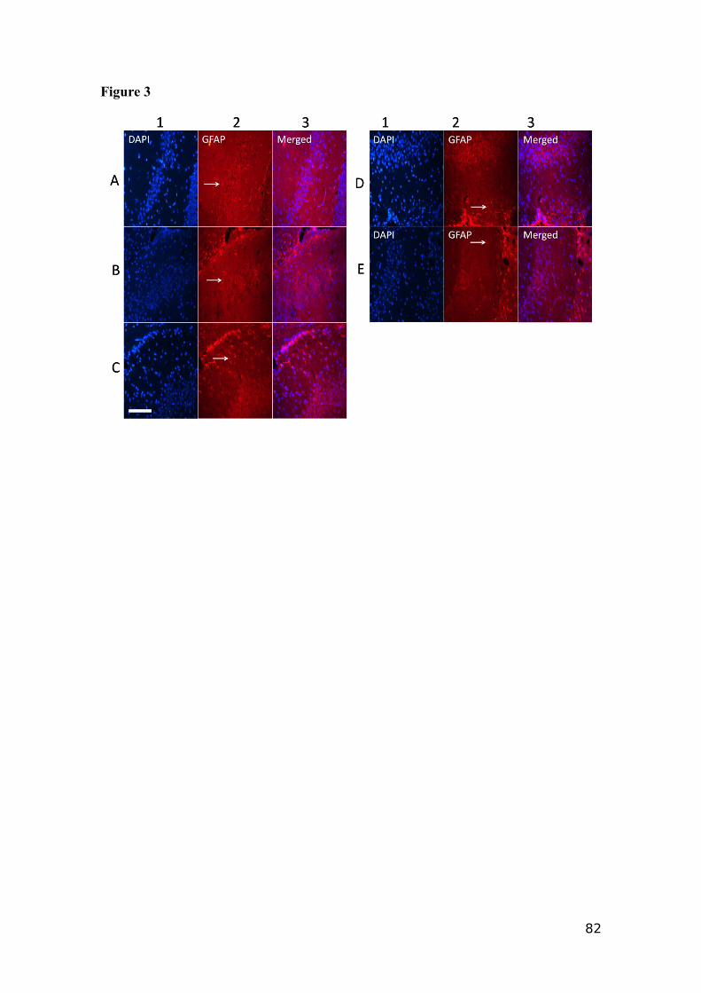
Figure 3
82
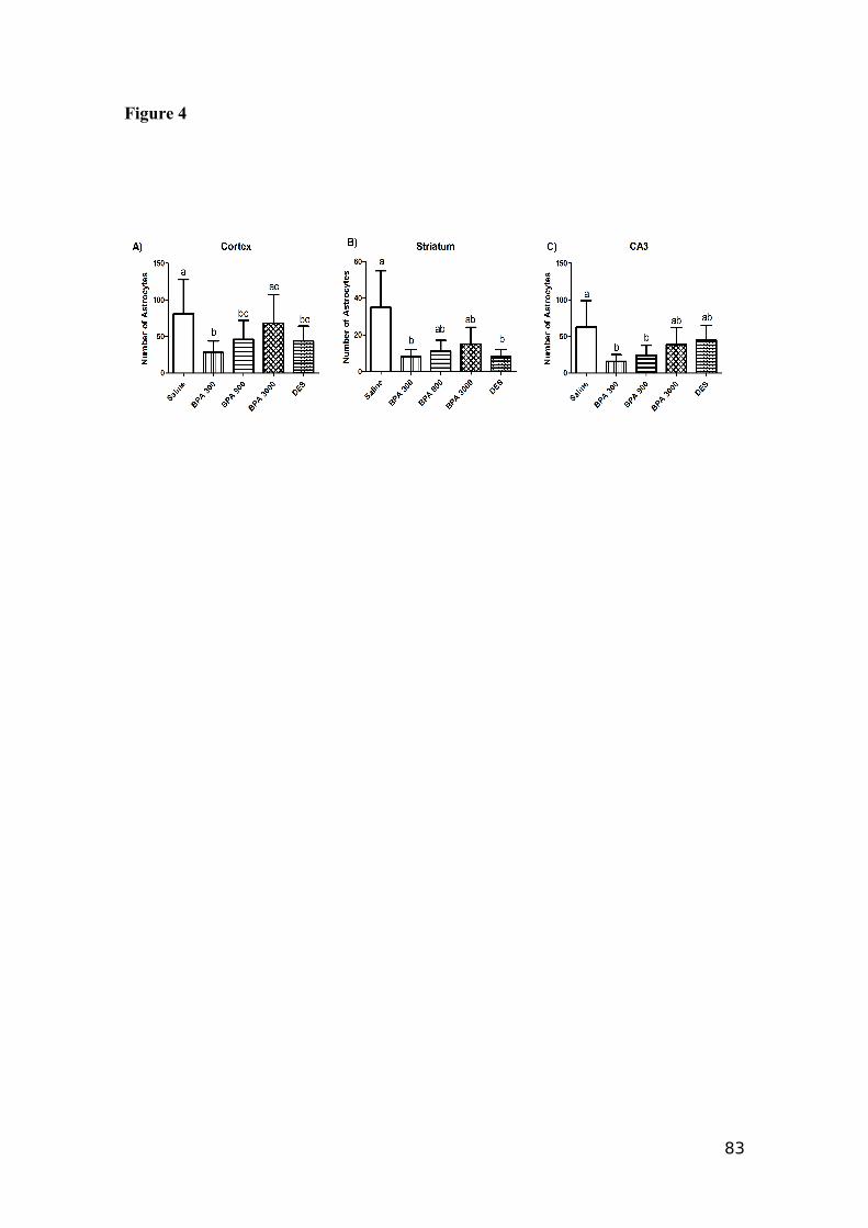
Figure 4
83
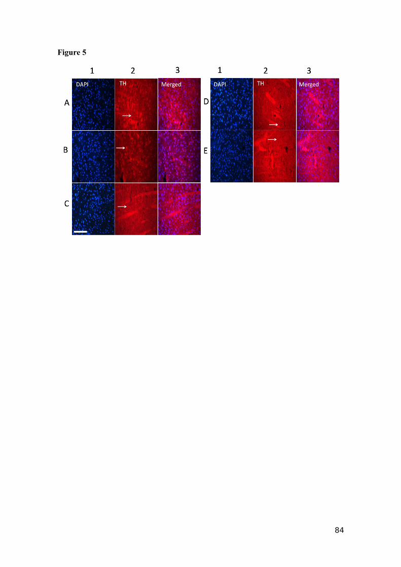
Figure 5
84
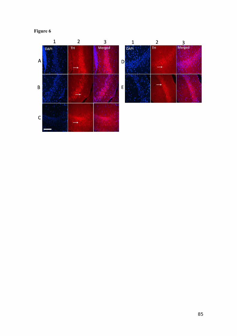
Figure 6
85
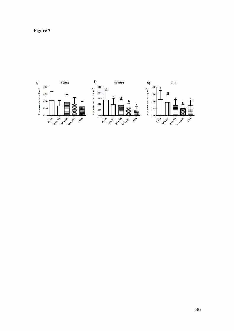
Figure 7
86
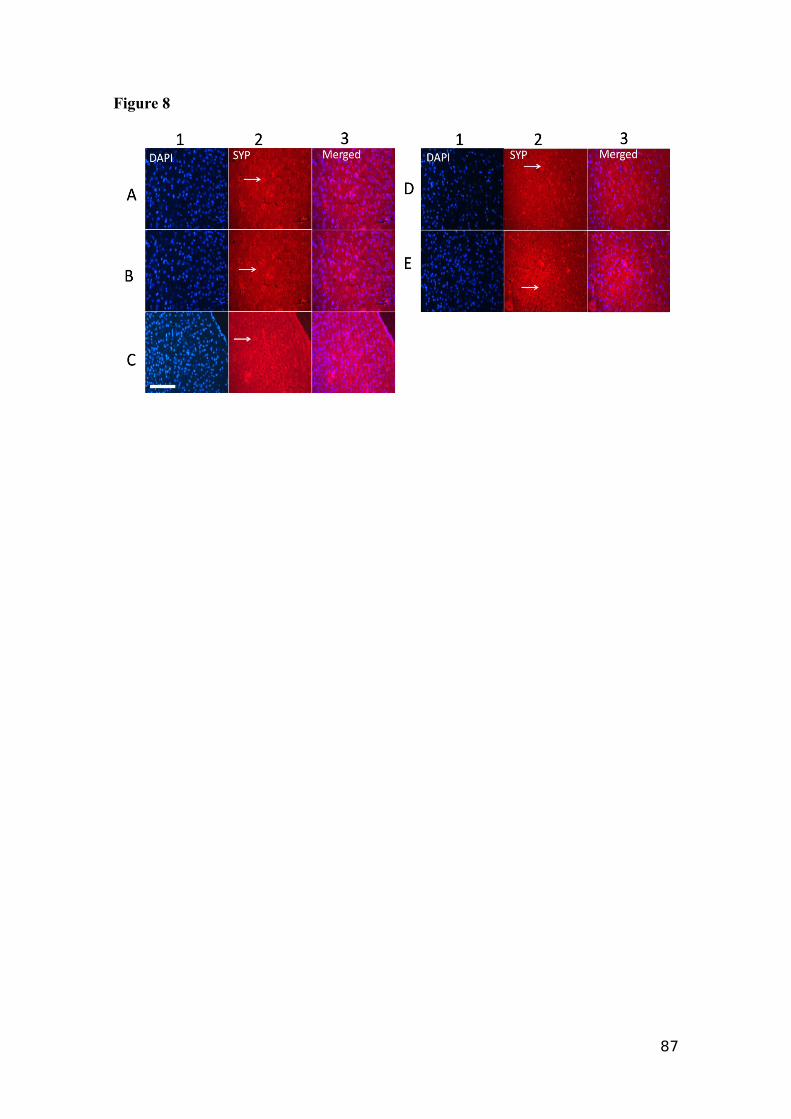
Figure 8
87
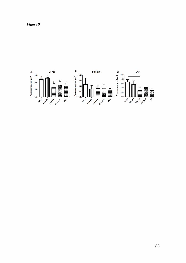
Figure 9
88
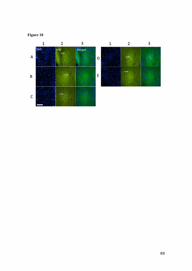
Figure 10
89
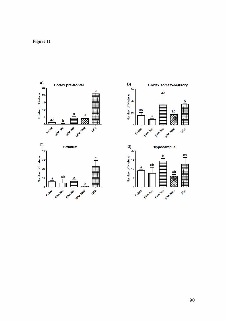
Figure 11
90
Conclusões
BPA quando administrado por via indireta (transmissão vertical) pode induzir a
danos em parâmetros espermáticos como motilidade do espermatozóide, morfologia,
funcionalidade mitocondrial, integridade de membrana, de acrossoma e de DNA.
Por diversas vias, dentre elas estresse oxidativo, o BPA pode causar alterações
morfológicas nos testículos dos animais. Também causa diminuição da distância
ano-genital destes animais.
Nas fêmeas que receberam BPA diretamente, o impacto nos parâmetros
analisados (número de astrócitos, quantidade da enzima tirosina hidroxilase, densidade
sináptica e proliferação celular), deixou evidente o papel deste tóxico na possível
indução de doenças neurodegenerativas.
91
Anexo 1
Normas da revista Reproductive Toxicology
92
93
94
95
96
97

Anexo 2
Normas da revista Toxicology Letters
98
99
100
101
102
103
Referências
Brann, D.W., Dhandapani, K., Wakade, C. Neurotrophic and neuroprotective actions of
estrogen: basic mechanisms and clinical implications. Steroids. 2007. 72: 381-405.
Brotons, J.A., Olea-Serrano, M.F., Villalobos, M., Pedraza, V., Olea, N. Xenoestrogens
released from lacquer coatings in food cans. Environ Health Perspect.1995. 103:608-12.
Brunette, M.G., Leclerc, M. Renal action of progesterone: effect on calcium
reabsorption. Mol Cell Endocrinol. 2002. 30(194):183-90.
Champagne, F., Diorio, J.S., Sharma, M.J. Naturally occur- ring variations in maternal
behavior in the rat are associated with differences in estrogen-inducible central oxytocin
receptors. Proc. Natl. Acad. Sci. U.S.A. 2001. 98: 12736–12741.
Chen, J., Cammarata, P.R., Baines, C.P., Yager, J.D. Regulation of mitochondrial
respiratory chain biogenesis by estrogens/estrogen receptors and physiological,
pathological and pharmacological implications. Bioch Bioph Acta.
2009.1793:1540-1570.
Chitra, K,C., Latchoumycandane, C., Mathur, P.P. Induction of oxidative stress by
bisphenol A in the epididymal sperm of rats. Toxicol. 2003.185:119-27.
Couse, J.F., Lindzey, J., Grandien, K. Tissue distribution and quantitative analysis of
estrogen receptor-alpha (ERalpha) and estrogen receptor-beta (ERbeta) messenger
ribonucleic acid in the wild-type and ERalpha-knockout mouse. Endocrinology 1997.
138: 4613-21.
Darbre, P. D. Preface. Best. Pract. Res. Clin. Endonol. Metab. v.20, n.1, p.vii-x, 2006b.
Deecher, D.C., Swiggard, P., Frail, D.E. Characterization of a membrane-associated
estrogen receptor in a rat hypothalamic cell line (D12). Endocrine. 2003. 22: 211-23.
Doerge, D.R., Vanlandingham, M., Twaddle, N.C., Delclos, K.B. Lactational transfer of
bisphenolA in Sprague – Dawley rats. Toxicol Lett. 2010.199:372-6.
104
Donahue, D.A., Dougherty, E.J., Meserve, L.A. Influence of a combination of two
tetrachloro biphenyl congeners (PCB47;PCB77) on thyroid status,
cholineacetyltransferase (ChAT) activity, and short and long-term memory in 30 day old
Sprague Dawley rats. Toxicol. 2004. 203:99–107.
Feldman, D., Tokes, L., Stathis, P.A. Identification of 17b-estradiol as the estrogenic
substance in Saccharomyces cerevisiae. Proc Natl Acad Sci USA. 1984. 81:4722-6.
Feldman, D. Estrogens from plastic - are we being exposed? Endocrinol.
1997.138:1777-9.
Figtree, G.A., Noonan, J.E., Bhindi, R. Estrogen receptor polymorphisms: significance
to human physiology, disease and therapy. Recent Pat DNA Gene Seq. 2009. 3: 164-71.
Garcia-Segura, L.M., Sanz, A., Mendez, P. Cross-talk between IGF-I and estradiol in the
brain: focus on neuroprotection. Neuroendocrinol. 2006. 84: 275-9.
Giguere, V. Identification of a new class of steroid hormone receptors. Nature.
1988.331:919–948.
Gottlicher, M., Heck, S., Herrlich, P. Transcriptional cross-talk, the second mode of
steroid hormone receptor action. J Mol Med. 1998. 76: 480-9
Gould, E., Woolley, C.S., Frankfurt, M., McEwen, B.S. Gonadal steroids regulate
dendritic spine density in hippocampal pyramidal cells in adulthood. J.Neurosci.
1990.101:286–1291,
Halliwell, B. and Gutteridge, J.M. Free radicals in biology and medicine. New York:
Oxford University Press ,2007.
Haraguchi, S., Katsunori, S., Hanako, S., Shin-ichiro, H., Kazuyoshi, T. Estradiol
Promotes Purkinje Dendritic Growth, Spinogenesis, and Synaptogenesis During
Neonatal Life by Inducing the Expression of BDNF. Cerebellum. 2012. 11:416-417.
105
Heard, D.J. Human ERRγ, a third member of the estrogen receptor-related receptor
(ERR) subfamily of orphan nuclear receptors: tissue-specific isoforms are expressed
during development and in the adult. Mol. Endocrinol. 2000.14: 382–392.
Jonathan, N. B., Steinmetz, R. Xenoestrogens: the emerging story of bisphenol A.
Trends Endocrinol Metab.1998. 9:124-8.
Joseph, A., Hess, R.A., Schaeffer, D.J. Absence of Estrogen Receptor Alpha Leads to
Physiological Alterations in the Mouse Epididymis and Consequent Defects in Sperm
Function. Biol of Reprod. 2010. 957:948-957.
Kato, H., Furuhashi, T., Tanaka, M., Katsu, Y., Watanabe, H., Ohta, Y., Iguchi, T. Effects
of bisphenol A given neonatally on reproductive functions of male rats. Reprod Toxicol.
2006. 22:20-9.
Kazuyoshi, T., Kazuyoshi, U., MarikoUsui, H. Novel brain function: biosynthesis and
actions of neurosteroids in Neurons. Neurosc Res. 2000. 36:261–273.
Kim, J.C., Shin, H.C., Cha, S.W., Koh, W.S., Chung, M.K., Han, S.S. Evaluation of
developmental toxicity in rats exposed to the environ- mental estrogen bisphenol A
during pregnancy. Life Sci. 2001. 69:2611–2625.
Krishnan, A.V., Stathis, P., Permuth, S.F., Tokes, L., Feldman, D. Bisphenol-A: an
estrogenic substance is released from polycarbonate flasks during autoclaving.
Endocrinol.1993.132:2279-86.
Kurebayashi, H., Betsui, H., Ohno,Y. Disposition of a lowdose of 14C-bisphenol A in
male rats and its main biliary excretion as BPA glucuronide. Toxicol. Sci. 2003. 73:
17–25.
Kuiper, G.G. Comparison of the ligand binding specificity and transcript tissue
distribution of estrogen receptors alfa and beta. Endocrinol. 1997. 138: 863–870.
106
Li, M.W., Mruk, D.D., Lee, W.M., Cheng, Y. Disruption of the blood-testis barrier
integrity by bisphenol A in vitro: Is this a suitable model for studying blood-testis
barrier dynamics? Int J Biochem Cell Biol. 2009. 41:2302-14.
Lindberg, M.K., Weihua, Z., Andersson, N.S., Movérare, H. Estrogen receptor speci
city for the effects of estrogen in ovariectomized mice. J Endocrinol. 2002. 167-178.
McLachlan, J. A., Simpson, E., Martin, M. Endocrine disrupters and female
reproductive health. Best. Pract. Res. Clin. Endocrinol. Metab. 2006. 20(1): 63-75,
Matsushima, A., Kakuta, Y., Teramoto, T., Koshiba, T., Liu, X., Okada, H., Tokunaga,
T., Kawabata, S., Kimura, M., Shimohigashi, Y. "Structural evidence for endocrine
disruptor bisphenol A binding to human nuclear receptor ERR gamma". J. Biochem.
2007. 142(4): 517–24
Matthews, J.B., Twomey, K., Zacharewski, T.R. In vitro and in vivo interactions of
bisphenol A and its metabolite, bisphenol A glucuronide, with estrogen receptors alpha
and beta. Chem. Res. Toxicol. 2001. 14:149–157.
Meeker, J.D., Ehrlich, S., Toth, T.L., Wright, D.L., Calafat, A.M., Trisini, A.T., Ye, X.,
Hauser, R. Semen quality and sperm DNA damage in relation to urinary bisphenol A
among men from an infertility clinic. Reprod Toxicol. 2010. 30: 532-9.
Miyatake, M., Miyagawa, K., Mizuo, K., Narita, M., Suzuki, T. Dynamic changes in
dopaminergic neurotransmission induced by alow concentration of bisphenol-A in
neurons and astrocytes. J.Neuroendocrinol. 2006. 18: 434–444.
Miyagawa, K., Narita, M., Narita, M., Akama, H., Suzuk, T. Memory impairment
associated with a dysfunction of the hippocampal cholinergic system induced by
prenatal and neonatal exposures to bisphenol-A. Neurosc Lett. 2007. 418:236–241.
107
Nadal, A., Alonso-Magdalena, P., Soriano, S., Quesada, I., Ropero, A. The pancreatic
β-cell as a target of estrogens and xenoestrogens: implications for blood glucose
homeostasis and diabetes. Molec Cell Endocrinol. 2009. 304: 63-68
Nadal, A. The estrogen trinity: membrane, cytosolic, and nuclear effects. News Physiol
Sci. 2001.16: 251–255.
Nakamura, D., Yanagiba, Y., Duan, Z., Ito, Y., Okamura, A., Asaeda, N., Tagawa, Y., Li,
C., Taya, K., Zhang, S.Y., Naito, H., Ramdhan, D.H., Kamijima, M., Nakajima, T.
Bisphenol A may cause testosterone reduction by adversely affecting both testis and
pituitary systems similar to estradiol. Toxicol Lett. 2010. 194:16-25.
Nakagawa, Y. and Tayama, S. Metabolism and cytotoxicity of bisphenol A and other
bisphenols in isolated rat hepatocytes. Arch Toxicol. 2000. 74: 99-105.
Norman, A.W. Steroid-hormone rapid actions, membrane receptors and a
conformational ensemble model. Nature Rev. 2004. 3: 27–40.
Obata,T., Kubota,S. Formation of hydroxyl radicals by environmental estrogen like
chemicals in rat striatum. Neurosci.Lett. 2000. 296, 42–44.
Ogiue-Ikedaa, M., Tanabe, N., Mukai, H., Hojoa, Y., Murakami, G. Rapid modulation of
synaptic plasticity by estrogens as well as endocrine disrupters in hippocampal neurons.
Brain Res. 2008. 57: 363 – 375.
Prange-Kiel, J., Rune, G.M. Direct and indirect effects of estrogen on rat hippocampus.
Neurosc. 2006. 138:765–772.
Price, R.H., Handa, R.J. Expression of estrogen receptor-β protein and mRNA in the
cerebellum of the rat. Neurosci Lett. 2000. 288:115–8.
Judd, H.L, Fournet, N. Changes of ovarian hormonal function with aging. Exp
Gerontol. 1994. 29: 285-98.
108
Pryor, J.L., Hughes, C., Foster, W., Hales, B.F., Robaire, B. Critical windows of
exposure for children’s health: the reproductive system in animals and humans. Environ
Health Perspect. 2000. 108:491–503.
Qiu, J., Bosch, M.A., Tobias, S.C. Rapid signaling of estrogen in hypothalamic neurons
involves a novel G-protein-coupled estrogen receptor that activates protein kinase C. J
Neurosci. 2003. 23: 9529- 40.
Quignot, N., Arnaud, M., Robidel, F., Lecomte, A., Barouki, R., Lemazurier, E.
Characterization of endocrine-disrupting chemicals based on hormonal balance
disruption in male and female adult rats. Reprod Toxicol. 2012. 33:339- 352.
Rogers, J.A., Metz, L. Review: Endocrine disrupting chemicals and immune
responses: A focus on bisphenol-A and its potential mechanisms. Molec Immun.
2013. 53: 421– 430
Roselli, C. e Resko J. Sex Differences in Androgen-regulated Expression of
Cytochrome P450 Aromatase in the Rat Brain. Steroid Bioch Mol Biol. 1997. 61(3-6):
365-374.
Saradha, B., Mathur, P.P. Effect of environmental contaminants on male reproduction.
Environ Toxicol Pharmacol. 2006. 21:34-41.
Scharfman, H.E., MacLusky, N.J. Estrogen and brain-derived neurotrophic factor
(BDNF) in hippocampus: complexity of steroid hormone-growth factor interactions in
the adult CNS. Front Neuroendocrinol. 2006. 27: 415-35.
SCHARFMAN, H.E. Neuroscience, 2012. Em impressão.
Schreihofer, D.A., Resnick, E.M., Lin, V.Y. Ligand-independent activation of pituitary
ER: on PKA-stimulated dependence pathways. Endocrinol. 2001. 142: 3361-8.
Shao, B., Cheng, Y., Jin, K. Estrogen, Neuroprotection and Neurogenesis after Ischemic
Stroke. DNA Seq. 2012:188-198.
109
Shearman, A.M. Association between estrogen receptor alpha gene variation and
cardiovascular disease. JAMA. 2003. 290: 2263–70.
Silberger, S.D., Magleby, K.L. Beting the odds with big K. Science. 1999.
285:1859–1860.
Suna, H.B., Shenb, O.X., Wang, X.R., Zhoua, L., Zhena, X.C. Anti-thyroid hormone
activity of bisphenol A, tetrabromobisphenol A and tetrachlorobisphenol A in an
improved reporter gene assay. Toxicol in Vitro. 2009. 23(5):950-4.
Tabb, M.M., Blumberg, B. New modes of action for endocrine-disrupting chemicals.
Mol. Endocrinol. 2006. 20(3):475-482.
Taylor, S. E. Oestrogen receptor splice variants in the pathogenesis of disease. Cancer
Lett. 2009.15.
Yang, Y.J., Lee, S.Y., Kim, K.Y., Hong, Y.P. Acute Testis Toxicity of Bisphenol A
Diglycidyl Ether in Sprague-Dawley Rats. J Prev Med Public Health 2010. 43:131-7.
Waring, R.H., Harris, R.M. Endocrine disrupters: a human risk? Mol. Cell. Endocrinol.
2005. 244(1-2):2-9.
Whitehaed, S. A., Rice, S. Endocrine-disrupting chemicals as modulators of sex steroid
synthesis. Best. Pract. Res. Clin. Endocrinol. Metab. 2006. 20(1):45-61.
Wilson, T.A., Garner, S.C., Anderson, J.J.B. Dietary protein source and serum estradiol
concentrations of rats during pregnancy, lactation and post-lactation, Nutr. Res. 2000.
20:1735–1747.
Völkel, W., Colnot, T., Csanady, G.A., Filser, J.G., Dekant,W. Metabolism and kinetics
of bisphenol A in humans at low doses following oral administration. Chem.
Res.Toxicol. 2002.15:1281–1287.
110