UNIVERSIDADE FEDERAL DO RIO GRANDE DO NORTE … · aplicação de soluções (gotas oftálmicas)...
66
UNIVERSIDADE FEDERAL DO RIO GRANDE DO NORTE CENTRO DE CIÊNCIAS DA SAÚDE PROGRAMA DE PÓS-GRADUAÇÃO EM CIÊNCIAS DA SAÚDE DESENVOLVIMENTO E CARACTERIZAÇÃO DE UM SISTEMA MICROEMULSIONADO CONTENDO ANFOTERICINA B PARA USO OFTALMOLÓGICO Walteçá Louis Lima da Silveira Natal 2009
Transcript of UNIVERSIDADE FEDERAL DO RIO GRANDE DO NORTE … · aplicação de soluções (gotas oftálmicas)...
UNIVERSIDADE FEDERAL DO RIO GRANDE DO NORTE
CENTRO DE CIÊNCIAS DA SAÚDE
PROGRAMA DE PÓS-GRADUAÇÃO EM CIÊNCIAS DA SAÚDE
DESENVOLVIMENTO E CARACTERIZAÇÃO DE UM SISTEMA
MICROEMULSIONADO CONTENDO ANFOTERICINA B PARA USO
OFTALMOLÓGICO
Walteçá Louis Lima da Silveira
Natal
2009

UNIVERSIDADE FEDERAL DO RIO GRANDE DO NORTE
CENTRO DE CIÊNCIAS DA SAÚDE
PROGRAMA DE PÓS-GRADUAÇÃO EM CIÊNCIAS DA SAÚDE
DESENVOLVIMENTO E CARACTERIZAÇÃO DE UM SISTEMA
MICROEMULSIONADO CONTENDO ANFOTERICINA B PARA USO
OFTALMOLÓGICO
Walteçá Louis Lima da Silveira
Dissertação a ser apresentada ao Programa de Pós-
Graduação em Ciências da Saúde como requisito para a
obtenção do Título de Mestre em Ciências da Saúde à
Universidade Federal do Rio Grande do Norte
Orientador: Prof. Dr. Eryvaldo Sócrates Tabosa do Egito
Natal
2009

UNIVERSIDADE FEDERAL DO RIO GRANDE DO NORTE
CENTRO DE CIÊNCIAS DA SAÚDE
PROGRAMA DE PÓS-GRADUAÇÃO EM CIÊNCIAS DA SAÚDE
Coordenador do Curso de Pós-Graduação:
Profa. Dra. Técia Maria de Oliveira Maranhão

iii
DESENVOLVIMENTO E CARACTERIZAÇÃO DE UM SISTEMA
MICROEMULSIONADO CONTENDO ANFOTERICINA B PARA USO
OFTALMOLÓGICO
BANCA EXAMINADORA
Presidente da banca: Prof. Dr. Eryvaldo Sócrates Tabosa do Egito
Membro Titulares:
Prof. Dr. Toshiyuki Nagashima Júnior (Universidade Federal de Campina
Grande - UFCG)
_______________________________________________________________
Prof. Dr. Arnóbio Antônio da Silva Júnior (Universidade Federal do Rio Grande
do Norte – UFRN)
_______________________________________________________________

iv
DEDICATÓRIA
Este trabalho é dedicado a todos aqueles que de uma forma, direta ou
indireta, contribuem para o desenvolvimento do ensino, da pesquisa e da
extensão neste país, promovendo e compartilhando o conhecimento a todas as
pessoas, sem distinção de classe social, idade ou sexo com a simples e pura
finalidade de educar e de querer transformar a dura realidade existente neste
país em educação.

v
AGRADECIMENTOS
Agradeço a Deus por me dar forças para seguir este caminho bastante
árduo; aos meus orientadores, Prof. Dr. Eryvaldo Sócrates Tabosa do Egito e
Profa. Dra. Ivonete Batista de Araújo, que com sua paciência, firmeza,
dedicação e ensinamentos, acreditaram e me incentivaram durante todo o
desenvolvimento deste trabalho, por me confiarem a competência para que eu
pudesse realizá-lo com ética e responsabilidade; aos meus pais e ao meu
irmão, que com muito apoio foram a base para a minha sustentação durante
toda a minha vida; a minha namorada e futura esposa, Nadja Maryelly de
Oliveira Gomes, por todo amor, carinho e compreensão que foi me dado
durante todo esse período de estudos e por ter me proporcionado o grande
privilégio de elaborar e desenvolver todo este trabalho; e a todos que fazem
parte da família LASID, em especial aos amigos, Bolívar Ponciano Goulart de
Lima Damasceno, Miguel Adelino da Silva Filho, Fernanda do Couto Brasil e
Airanuédida Silva Soares pela grande e importante ajuda demonstrada no dia-
a-dia dos trabalho e estudos.

vi
LISTA DE ABREVIATURAS
LASID Laboratório de Sistemas Dispersos
AmB Anfotericina B
ME Microemulsão
DFPT Diagrama de Fases Pseudo-Ternário
AmB ME Anfotericina B incorporada na Microemulsão
PPGCSA Programa de Pós-Graduação em Ciências da Saúde

vii
LISTA DE FIGURAS
Figura 1 Olho acometido por uma ceratomicose
Figura 2 Fórmula estrutural da Anfotericina B
Figura 3 Mecanismo de ação da Anfotericina B
Figura 4 Emulsão x Microemulsão
Figura 5 Classificação proposta por Winsor para sistemas dispersos (1948)
Figura 6 Microemulsão branca e Microemulsão com Anfotericina B
viii
RESUMO
As infecções fúngicas oculares estão sendo reconhecidas em todo o mundo
como uma importante causa de morbidade e cegueira. A baixa
biodisponibilidade dos fármacos como a anfotericina B, antifúngico bastante
utilizado no tratamento destas infecções, nos tecidos posteriores dos olhos
aliada às barreiras anatômicas oculares, que são capazes de limitar a sua
absorção e associado ao grave risco de desenvolvimento de reações adversas
levam, geralmente, a um insucesso no tratamento desejado e a sérias
conseqüências para os pacientes acometidos. Pesquisadores em todo o
mundo estão buscando novas alternativas para contornar esta problemática e
uma das principais linhas de pesquisa estabelecidas consiste no
desenvolvimento de novas formulações que visam melhorar a
biodisponibilidade e reduzir a toxicidade associada à aplicação da anfotericina
B. As microemulsões surgem como novos sistemas capazes de carrear este
fármaco para uso oftalmológico. O presente estudo objetivou a obtenção de
uma microemulsão biocompatível com a via de administração ocular contendo
anfotericina B a fim de se estabelecer uma nova forma farmacêutica viável para
a administração tópica nos olhos. O sistema obtido demonstrou, por meio dos
estudos de caracterização realizados, biocompatibilidade com a via de
administração pretendida e surge, portanto, como uma nova e interessante
apresentação farmacêutica contendo anfotericina B para ser utilizada, no
futuro, no tratamento de infecções fúngicas oculares.
Descritores : Anfotericina B; Soluções Oftálmicas; Carreadores de Fármacos;
Preparações Farmacêuticas; Microemulsões
ix
SUMÁRIO
Dedicatória _______________________________________________ iv
Agradecimentos ____________________________________________ v
Lista de Abreviaturas ________________________________________ vi
Lista de Figuras ___________________________________________ vii
Resumo __________________________________________________ viii
1. INTRODUÇÃO __________________________________________ 10
2. REVISÃO DA LITERATURA _______________________________ 13
3. ANEXAÇÃO DO ARTIGO ________________________________ 21
3.1. Título do Artigo __________________________________ 21
4. COMENTÁRIOS, CRÍTICAS E SUGESTÕES ________________ 54
5. APENDICE ____________________________________________ 58
5.1. Apêndice I ______________________________________ 58
6. REFERÊNCIAS BIBLIOGRÁFICAS (Vancouver) ______________ 62
Abstract ________________________________________________ 65

10
1. INTRODUÇÃO
As doenças oculares são principalmente tratadas topicamente pela
aplicação de soluções (gotas oftálmicas) contendo fármacos administrados por
meio de colírios. Estas formas farmacêuticas convencionais correspondem a
90% das formulações oftalmológicas disponíveis, entretanto, a rápida perda pré
corneana causada pela drenagem nasolacrimal e pela presença das lágrimas
tornam este fato um dos maiores problemas associados à absorção dos
fármacos nos olhos. Somente 5% da dose administrada são capazes de
penetrar através da córnea e alcançar os tecidos intra-oculares, sendo os
outros 95% eliminados através da absorção pela conjuntiva ou via ducto
nasolacrimal (1).
As infecções fúngicas oculares estão sendo cada vez mais reconhecidas
como uma importante causa de morbidade e cegueira em todo o mundo e o
tratamento destas infecções não foge ao contexto apresentado. As infecções
graves supurativas que levam a ulcerações nos tecidos envolvidos possuem
um prognóstico geralmente severo e o seu tratamento depende de um
diagnóstico rápido e eficiente. Entretanto, além dos desafios gerados pela
natureza do olho, o tratamento dessas patologias ainda gera incertezas quanto
aos protocolos clínicos que trazem, como fármaco de escolha, a anfotericina B
(AmB) (2-7).
A AmB tópica corresponde ao tratamento de escolha para as infecções
fúngicas superficiais que acometem o olho causadas por Candida e em locais
onde a natamicina (5%) não está aprovada para o uso. Sua formulação atual
(Fungizone®) contém desoxicolato de sódio (agente emulsificante necessário
para promover a micelização da AmB), que provoca durante a instilação

11
bastante irritação, o que leva a uma não adesão ao tratamento por parte do
paciente e, conseqüentemente, ao agravamento dos sintomas (8).
Entretanto, quando as lesões se mostram mais profundamente
localizadas nos tecidos oculares, este fármaco deve ser então administrado
através das vias intravenosa, intravitreal ou intracameral, devido a
incapacidade que a AmB tem em atravessar a córnea intacta, submetendo o
paciente aos desconfortos proporcionados por estas formas de administração e
aos efeitos adversos relacionados (9).
Diante de todos estes problemas associados ao uso da AmB no controle
de infecções fúngicas oculares, percebe-se que o desenvolvimento de novos
sistemas capazes de carrear este fármaco com a finalidade de melhorar sua
biodisponibilidade, estabilidade e diminuir sua toxicidade são, portanto,
necessários.
As microemulsões (MEs) correspondem a uma alternativa promissora
para melhorar a biodisponibilidade e diminuir os efeitos adversos causados
pelo uso da AmB. Estes veículos possuem propriedades que favorecem a sua
administração através desta via e consistem em sistemas transparentes
(isotropicamente translúcidos) e estáveis termodinamicamente de dois líquidos
imiscíveis estabilizados por um filme interfacial de compostos tensoativos que
se localizam na interface óleo/água, podendo ser facilmente preparadas e
esterilizadas. Estes sistemas oferecem ainda vantagens adicionais como: maior
capacidade de incorporar tanto fármacos hidrofílicos como lipofílicos, pequeno
tamanho de gotículas da fase interna e viscosidade adequada para a liberação
de fármacos nos olhos (10-13).
12
A idéia de utilizar tais sistemas como carreadores para a AmB com o
objetivo de diminuir a toxicidade deste fármaco, aumentar a sua estabilidade na
preparação farmacêutica e biodisponibilidade vem reforçar o objetivo de muitos
pesquisadores no mundo (14-16). O objetivo do presente trabalho visou,
portanto, desenvolver e caracterizar um sistema microemulsionado contendo
AmB pretendido para a administração tópica ocular por meio de colírios, forma
farmacêutica líquida e destinada a aplicação sobre as mucosas oculares.
O sistema foi, portanto, desenvolvido por meio de uma metodologia de
construção de diagramas de fase pseudo-ternário (DFPT) e caracterizado
físico-quimicamente segundo os ensaios referidos na literatura como
importantes, sendo suas características delineadas e avaliadas com a
finalidade de atender as particularidades da via de administração tópica ocular.
Com esta pesquisa, espera-se que a discussão sobre o tema seja mais
enfatizada sobre os aspectos clínicos, epidemiológicos, microbiológicos e
tecnológicos, fortalecendo a hipótese de que o tratamento sobre o ponto de
vista multidisciplinar é mais eficaz e seguro para o paciente no que diz respeito
à melhora de sua qualidade de vida. Com isso, o desenvolvimento desta nova
formulação poderá trazer, para o futuro, uma nova apresentação farmacêutica
para a AmB, mais eficaz, estável e com menos risco de proporcionar as
reações adversas referidas com o uso da formulação convencional.

13
2. REVISÃO DA LITERATURA
As infecções causadas por fungos na córnea, denominadas de
ceratomicoses e endoftalmites estão entre as mais freqüentemente
encontradas apresentando-se como lesões supurativas que se desenvolvem,
geralmente, por meio de úlceras. Estas infecções já são consideradas as
responsáveis por mais de 50% de todos os casos de infecções oculares
registrados, especialmente nas áreas tropicais e subtropicais do mundo, tendo
como um dos principais agentes etiológicos, fungos leveduriformes do gênero
Candida e filamentosos do gênero Fusarium (5) (Figura 1).
Figura 1: Olho acometido por uma ceratomicose (Thomas, PA. 2003)
A córnea corresponde a uma importante barreira mecânica e química,
pela qual limita o acesso de substâncias exógenas para o interior do olho,
protegendo, assim, os tecidos intra-oculares. Esta barreira consiste em uma
estrutura transparente e avascular com um diâmetro e uma espessura em torno
de 12 mm e 520 µm, respectivamente. Localiza-se no segmento anterior do
olho e a sua superfície consiste na principal rota de absorção de fármacos por

14
meio da via ocular, entretanto esta absorção é ineficiente e gera uma baixa
biodisponibilidade dos fármacos que são administrados topicamente (17).
Dois fatores rendem a córnea uma efetiva barreira de absorção dos
fármacos: O primeiro é a sua pequena área de superfície e a segunda é que
ela é relativamente impermeável. Outro fator bastante importante capaz de
reduzir a biodisponibilidade dos fármacos administrados reside na rápida perda
pré corneana ocasionada pela ação do sistema lacrimal e pela presença das
lágrimas (1, 18).
A eficácia clínica de agentes antifúngicos em micoses oftálmicas
depende, em grande parte, da concentração do fármaco e da sua capacidade
em alcançar o tecido ocular alvo. Isto depende, ainda, de um grande número
de fatores que incluem a massa molecular e a rota utilizada para a sua
administração, assim como a duração do tempo de contato do fármaco com o
tecido ocular alvo e a sua habilidade de penetração através da córnea.
Fármacos que apresentam grande massa molecular, como a AmB (924,10 Da),
excedendo 500 Da raramente são capazes de penetrar a estrutura intacta da
córnea (19).
A AmB é um antibiótico poliênico macrocíclico com uma potente ação
fungicida e fungistática contra uma grande faixa de espécies fúngicas
oportunistas e desde 1956 é considerada um dos fármacos mais eficazes
contra sérias infecções fúngicas sistêmicas tais como candidiases,
histoplasmoses e aspergiloses. Em oftalmologia, seu uso também é importante
e está relacionada, principalmente, ao tratamento de infecções causadas por
Candida desde 1959 (15, 20, 21).

15
Este fármaco foi primeiramente isolado em 1955 a partir de colônias de
Streptomices nodosus coletados na Venezuela e consiste em um pó amarelo-
alaranjado que apresenta duas propriedades físico-químicas: comportamento
anfifílico devido aos grupos polares e apolares do anel lactônico e anfotérico
devido à presença de grupos aminas e carboxílicos ionizáveis. Sua molécula é,
ainda, pouco solúvel em solventes aquosos e em muitos solventes orgânicos,
sendo sua solubilidade em água, no pH fisiológico (pH 6-7), menor do que 1
mg/mL (22) (Figura 2).
Figura 2: Fórmula estrutural da AmB
Massa molar (C47H73NO17) – 924,10 Da (Patrick, 1995)
A formulação comercial atual da AmB, Fungizone®, comercializado pela
Bristol-Myers Squibb, consiste em um sistema micelar que contém 41 mg de
desoxicolato de sódio e 50 mg de AmB. Para a administração tópica ocular,
uma solução extemporânea (0,15 a 0,3%) pode ser preparada diluindo-se esta
apresentação comercial em água esterilizada e posteriormente administrando o
medicamento a cada 30 a 60 minutos. Durante este intervalo, essa solução

16
deve ser armazenada sobre refrigeração e protegida da luz, por onde se reduz,
assim, a velocidade de degradação deste fármaco em solução (5, 23).
O principal mecanismo de ação da AmB consiste na habilidade que sua
molécula possui em interagir com os esteróis da membrana, colesterol das
membranas plasmáticas das células humanas e o ergosterol das membranas
celulares fúngicas, e formar, com isso, canais (poros), na qual há a liberação
de constituintes internos, tais como potássio, cálcio, magnésio e fosfato. Esta
liberação, seguida de um desequilíbrio na atividade enzimática das membranas
afeta, conseqüentemente, a vida celular, sendo esta, a razão para a sua
atividade fungicida (23) (Figura 3).
Figura 3: Mecanismo de ação da AmB
Fonte: Profa Dra Ivonete Batista de Araújo
Dois persistentes problemas relacionados à aplicação tópica da AmB
consistem na grande possibilidade de toxicidade na córnea (ceratites), pela
* Formação de poros pela AmB na bicamada lipídica das membranas celulares
17
qual poderia originar a uma não adesão ao tratamento por parte do paciente e
levar, com isso, a uma interpretação errônea de que o microorganismo não é
susceptível a terapia, assim como a sua baixa estabilidade quando em solução.
Essa toxicidade, que pode ainda se desenvolver por meio de blefarites e irites,
está associada ao desoxicolato de sódio e pode ser bastante reduzida quando
formulações lipídicas são usadas como veículos para a incorporação deste
fármaco sem o uso deste componente emulsificante (21, 24).
O uso de carreadores lipidícos, tais como as microemulsões (MEs),
utilizadas para incorporar a AmB, já vem sendo descrito por diversos
estudiosos, porém nenhum deles atribuiu o emprego deste sistema à via ocular
(14-16). As MEs aparecem como uma importante alternativa para a liberação
tópica ocular da AmB a ser estudada devido as suas propriedades intrínsecas e
estruturas específicas. Estes sistemas podem ser facilmente preparados por
emulsificação e esterilizados por filtração, possui viscosidade adequada e alta
capacidade para solubilizar tanto fármacos lipofílicos quanto hidrofílicos. Sua
capacidade em prolongar a liberação dos fármacos, diminuindo a freqüência de
administração das doses consiste na principal vantagem para a administração
ocular (10, 12, 13).
Além disso, devido a sua consistência aquosa, transparência,
estabilidade termodinâmica e sua capacidade em não influenciar na
visibilidade, quando comparada com as pomadas oftálmicas, podem levar uma
maior adesão ao tratamento por parte do paciente (10, 12, 13).
O conceito de ME foi introduzido nos anos quarenta por Hoar e
Schulman que geraram uma solução transparente, de única fase, por titulação
de uma emulsão de aspecto leitoso com hexanol. Posteriormente, seus
18
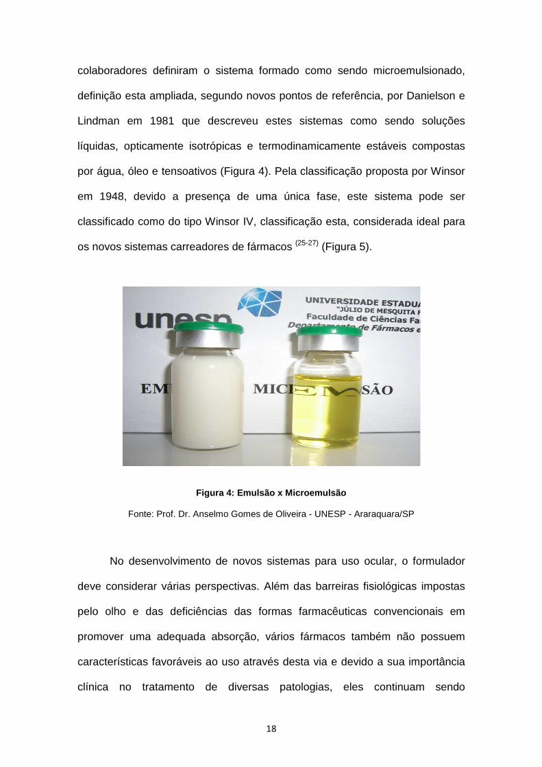
colaboradores definiram o sistema formado como sendo microemulsionado,
definição esta ampliada, segundo novos pontos de referência, por Danielson e
Lindman em 1981 que descreveu estes sistemas como sendo soluções
líquidas, opticamente isotrópicas e termodinamicamente estáveis compostas
por água, óleo e tensoativos (Figura 4). Pela classificação proposta por Winsor
em 1948, devido a presença de uma única fase, este sistema pode ser
classificado como do tipo Winsor IV, classificação esta, considerada ideal para
os novos sistemas carreadores de fármacos (25-27) (Figura 5).
Figura 4: Emulsão x Microemulsão
Fonte: Prof. Dr. Anselmo Gomes de Oliveira - UNESP - Araraquara/SP
No desenvolvimento de novos sistemas para uso ocular, o formulador
deve considerar várias perspectivas. Além das barreiras fisiológicas impostas
pelo olho e das deficiências das formas farmacêuticas convencionais em
promover uma adequada absorção, vários fármacos também não possuem
características favoráveis ao uso através desta via e devido a sua importância
clínica no tratamento de diversas patologias, eles continuam sendo

19
administrados por meio de condições adversas ao paciente. Sendo assim, após
a escolha dos prováveis sistemas microemulsionados a partir do DFPT, há a
necessidade de se caracterizar estes sistemas com e sem o fármaco
incorporado a fim de se obter a formulação em que abranja todos os requisitos
da via de administração ocular e que favoreçam os aspectos clínicos e
tecnológicos necessários (10, 12, 19, 28).
Figura 5: Classificação para sistemas dispersos pro posta por Winsor (Winsor, PA. 1948)
Portanto, os estudos de caracterização de uma ME desenvolvida com a
finalidade de se tornar um carreador para a AmB e com o objetivo de se
diminuir a toxicidade deste fármaco e aumentar a sua estabilidade vem reforçar
o objetivo de muitos pesquisadores no mundo, no entanto, a partir da grande
possibilidade desse sistema em possuir as características necessárias para a
administração tópica ocular, um novo foco de aplicação para este se torna
possível uma vez que, com o advento no número de casos de pacientes
acometidos por infecções fúngicas oculares no mundo e pela ausência de
formulações convencionais que contenham antifúngicos existentes no
20
mercado, o seu uso poderia contribuir com um tratamento mais eficaz e seguro,
permitindo uma melhora na qualidade de vida destes pacientes.

21
3. ANEXAÇÃO DE ARTIGO
3.1. Título do Artigo : Development and characterization of a microemulsion
system containing amphotericin B for ophthalmic use
REVISTA: Clinical and Experimental Ophthalmology – Fator de Impacto:
1,347
22
Original Article – Laboratory Science
Development and characterization of a
microemulsion system containing amphotericin
B with potential ophthalmic applications
Walteçá Louis Lima da Silveira1, Bolívar Ponciano Goulart de Lima
Damasceno1,2, Nadja Maryelly de Oliveira Gomes1, Fernanda do Couto Brasil1,
Airanuédida Silva Soares1, Aldo da Cunha Medeiros1, Anselmo Gomes de
Oliveira3, Ivonete Batista de Araújo1 and Eryvaldo Sócrates Tabosa do Egito1
1 Universidade Federal do Rio Grande do Norte (UFRN) – Centro de Ciências
da Saúde (CCS) – Programa de Pós-graduação em Ciências da Saúde
(PPGCSA) – Laboratório de Sistemas Dispersos (LASID) – 59.010-180, Natal-
RN-Brazil
2 Universidade Estadual da Paraíba (UEPB) – CCBSA – Campus V – 58.020-
540, João Pessoa-PB-Brazil
3 Universidade Estadual Paulista (UNESP) - Departamento de Fármacos e
Medicamentos – Faculdade de Ciências Farmacêuticas – 14.801-902,
Araraquara-SP-Brazil
Correspondence: Prof. Dr. Eryvaldo Sócrates Tabosa do Egito, Faculty of
Pharmacy of the Federal University of Rio Grande do Norte, Avenida Gal.
Gustavo Cordeiro de Farias, s/n Petrópolis, CEP 59010-180, Natal, RN, Brazil.
E-mail: [email protected] or [email protected]. Phone: 55 84 94 31 88 16;
Fax: 55 84 3342 9817 or 9808
AmB-microemulsion for ophthalmic use

23
ABSTRACT
Aim: The development of new ophthalmic drug delivery systems containing
antifungal agents has received considerable attention because of all drawbacks
presented with the use of conventional dosage forms. Topical amphotericin B
eye drops is the standard treatment for ocular infections caused by Candida.
However, its toxicity and poor bioavailability leads to no patient compliance and
aggravation of symptoms. The present work describes the development and
characterization of one microemulsion system containing amphotericin B,
aiming, in a long term, its use for ophthalmic applications.
Methods: The microemulsion was developed and prepared by the titration
technique. The physicochemical characteristics and stability were determined,
both in the absence and in the presence of amphotericin B. Its content was
investigated by spectrophotometric studies, which evaluated the influence of
sterilization process by filtration and the characterization of aggregation form
into the system.
Results: The performed studies showed that the presence of amphotericin B
incorporated into the system did not induce serious changes in its
physicochemical properties, which were, in general, maintained when compared
to the blank microemulsion. The spectrophotometric studies showed, however,
that not only the amphotericin B concentration decreased with the sterilization
process by filtration, but also an increase of amplitude of the band at 322 nm,
assigned to amphotericin B self-associated species, was revealed.
Conclusions: The characterization of these systems demonstrated that they
have compatible characteristics with the ophthalmic route, and could be eligible
24
as a new and interesting amphotericin B delivery dosage form to be used by
eye drops.
Key words: Amphotericin B; Eye drops; Drug carriers; Pharmaceutical
nanotechnology; Microemulsion

25
INTRODUCTION
Ocular fungal infections are being increasingly recognized as an
important cause of morbidity and blindness. The clinical efficacy of antifungal
agents on the treatment of these diseases depends to a great extent on the
concentration achieved in the target ocular tissue. A number of factors,
including the molecular mass, concentration of the drug and the route by which
it was administered, the duration of contact with the target ocular tissue and the
ability of the compound to penetrate the eye, moreover, the effects of corneal
resistance and the nasolachrymal drainage, lead for a poor bioavailability of
these drugs (1, 2).
Amphotericin B (AmB) is a broad spectrum antifungal agent mainly used
to treat invasive fungal infections (3). For ophthalmic applications, this drug has
been administered by intravenous, topical, intravitreal and intracameral routes,
however, many drawbacks can be described with its use by these several
routes (4). Topical AmB (1.5 – 3.0 mg/mL) is the standard treatment for ocular
infections due to Candida and related fungi and in the regions where natamycin
is not available (2, 5). The current formulation of AmB eye drops (micelle system,
Fungizone®, Bristol-Myers Squibb, USA) contains deoxycholate (AmB-M),
which renders their instillation painful and leads to poor compliance and
aggravation of symptoms (5). Moreover, the corneal penetration of AmB, in the
presence of an intact corneal epithelium, is, also, reduced and one persistent
concern is the possibility of corneal toxicity. Indeed, its solution is prepared
freshly with sterile water and must be refrigerated in a dark bottle to reduce the
speed of disintegration (2, 6-8). Because of all drawbacks related, the

26
development of stable and safe new pharmaceutical formulations for the topical
ocular administration of AmB might, therefore, be desirable.
Various systems as carriers to new drug delivery vehicles have been
developed to increase ocular absorption and reduce the toxicity related of drugs
(9). Microemulsions (MEs) are an interesting alternative to topical ocular
delivery, because of their intrinsic properties and specific structures (9). A ME is
a system which contains water and oil coexisting in thermodynamic equilibrium
due to the presence of a surfactant film at the oil-water interface (10). They can
be easily prepared through emulsification process and sterilized by filtration.
They all can have their viscosity adapted and possess a higher ability for
dissolving drugs (9). Their capacity of prolonged release of drugs, decreasing
the frequency of application of eye drops, per day, consists in an important
characteristic for ocular administration (9).
The aim of the present work was the development and characterization
of a ME system intended for topical ocular administration of AmB by eye drops.
The physicochemical characteristics were evaluated for attempted the
physicochemical particularities of the topical ophthalmic route.
METHODS
Materials
Monobasic and dibasic sodium phosphate, sodium hydroxide and
hydrochloric acid were purchased from Vetec Fine Chemicals (Brazil). Soy
phosphatidylcholine, Lipoid® S100, was a gift sample from Lipoid, Germany.
Tween® 80 and AmB were purchased from Sigma-Aldrich (USA). Miglyol® 812N
was a gift sample from Sasol, Germany. Methanol was purchased from Tedia
27
Company (USA). The micelle dosage form, AmB-M, was a gift sample from
UNICAT (Natal/RN, Brazil).
Pseudo-ternary phase diagram (PTPD)
For preparation of the PTPD, an adequate surfactant [(Lipoid® S100)/co-
surfactant (Tween® 80) (3:7)] and oil (Miglyol® 812N) weight ratios was used in
the range from 1:9 to 9:1, respectively, to obtain the phase diagram. To the
mixture, the aqueous phase (phosphate buffer 7.4) was titrated with an
automatic pipette, stirred with a sonicator for 1.5 min, and placed in an
ultrasound bath for 3 min. The transition from separated phase to optically clear
ME was observed and analyzed by naked eye.
Preparation of microemulsion
For preparation of the ME (Table 1), one point from the PTPD was
chosen in term of homogeneity, transparency and optical isotropy. This choice
show a formulation that has as first phase a phosphate buffer pH 7.4 solution
containing Lipoid® S100, and as the second phase Miglyol® 812N and Tween®
80. Both first and second phases were magnetically stirred, separately, for 20
and 3 min, respectively. The final production of the ME was concluded by
addition of the first phase in the second phase following by a sonication process
(1.5 min) and ultrasound bath (3 min). Three cycles in sonicator and ultrasound
bath were realized to form a true ME.
Amphotericin B loading process
AmB was incorporated at maximum concentration of 5 mg/mL (11) into
ME. Briefly, appropriated amounts of AmB were weight and added directly into
the ME previously prepared, under continuous magnetic stirring. After 1 min, the
AmB was dissolved by addition of sodium hydroxide (NaOH) solution (1 N) and

28
after solubilization, the pH of the preparation was neutralized to 7.0-7.5 with a
hydrochloric acid (HCl) solution (1 N). Finally, the AmB-loaded-microemulsion
(AmB-ME) was filtered through 0.45 µm membrane filters to remove the
suspended particles and, then, sterilized through a sterile filter of porous size of
0.22 µm (Sartorius Minisart® filters, Germany).
Physicochemical characteristics of ME and AmB-ME
Both the ME and AmB-ME were characterized in terms of macroscopic
aspects, pH, rheological behavior and viscosity, refractive index, conductivity,
particle size, surface tension and zeta potential. All parameters were
determined at 25º ± 1ºC and the analyses were carried out in triplicate.
The macroscopic parameters observed by naked eye were the color, the
consistency and the homogeneity. The transparent appearance was evaluated
by percentage of transmittance. The pH values were performed by digital pH
meter PG2000 (Gehaka, Brazil) equipped with a glass electrode, which was
previously calibrated with 4.0 and 7.0 pH standard solution. The electrode was
placed directly in the ME and AmB-ME samples, which were stored in a
scintillation flask (15 mL).
The rheological analyses (RA) of ME (10mL) and AmB-ME (10mL) were
performed using a controlled stress rheometer fitted with concentric cylinder
geometry (DC-41), connected with a hake K20-DC50 thermostatic bath. The
experiments were recorded with a shear rate in the range of 1 to 100 1/s and
performed for both up and down curves.
With an Abbé refractometer (Analytik, Jena, AG, Germany), the refractive
index (RI) was determined. The equipment was calibrated with distilled water,
29
and 30 µL of the samples were placed on the refractometer prism for RI
evaluation.
The conductivity values were measured by a conductimeter MC 226
(Toledo/Mettler, Brazil), equipped with an Inlab 730 electrode (TTLER/Toledo,
Brazil). The electrode was placed directly in the ME and AmB-ME stored in the
scintillation flask (15 mL).
Particle size analyses of samples were measured by photon correlation
spectroscopy using a Particle Sizing System by Dynamic Light Scattering (DLS)
– Brookhaven Instruments Corporation, EMI 9863 model (Holtsville, NY, USA)
with laser source He-Ne 10 mW, 532 nm-HUGHES, and self correlator with 64
channels. Before of the analysis, the samples were diluted in phosphate buffer
(pH 7.4) to yield a 1:32 solution and after were filtered with a membrane of 0.8
µm to remove eventual impurities. The size measurements were carried out at
fixed angle of 90o and the correlator was operated in parallel mode. The
cumulate method of analysis was used to calculate the droplet size according to
the intensity of scattered light. The RI was in accordance with the index of each
analyzed sample needed to perform these analyses.
The surface tension of the samples was measured using a Sensadyne
QC-6000 tensiometer by Chem-Dyne Research Corp. This parameter was
carried out through the bubble shape method using azotes atmosphere. Its
value was measured when the tension reached a constant value.
The electrophoretic mobility was measured determining the mobility of
particles by zetasizer nano equipment (Microtrac Zetatrac, USA). The samples
were placed directly into a cell where a potential difference (150 V) at a
modulation frequency of 1000 Hz was applied. Therefore, the movement of the
30
charged particles to electrode of opposite polarity and the speed of the charged
particle were observed.
Quantitative analyses and characterization of the a ggregation state of
AmB
AmB content, before and after of the filtration process through 0.45 and
0.22 µm membrane filters, respectively, was determined using the Libra S32
UV-visible spectrophotometer (Biochrom, Brazil). The values were recorded
over the wavelength of 405 nm, which is the maximum absorption to AmB
monomeric form (12). Prior to the determination of AmB into the AmB-ME, the
systems were diluted in methanol to yield concentrations of 5 x 10⁻⁶ mg/mL.
To evaluate the aggregation form, the AmB-ME samples were diluted in
phosphate buffer pH 7.4 and in the blank ME to yield a 5 x 10⁻⁶ mg/mL solution.
The spectrum of these solutions was recorded at the wavelength from 300 to
450 nm and compared to the ones obtained for the AmB-M in phosphate buffer
(pH 7.4) and pure AmB in methanol at the same concentration (5 x 10⁻⁶ mg/mL).
AmB entrapment efficiency for the MEs
The entrapment efficiency was assessed after centrifugation (11000 g ≈
14000 rpm) of 1 mL of each formulation for 10 min. The aim of this procedure
was not to settle ME droplets, but to settle both metastable systems and AmB
crystals not incorporated in the ME droplets. At the end of the centrifugation
cycle, the presence of crystals or AmB charged droplets was evaluated.
Stress testing
The samples were submitted, to a total of six freeze-thaw cycles. Each
cycle consisted of 24 h at 45ºC followed by 24 h at 8ºC during 12 days. These
cycles were important to determine the chemical stability of the dispersed

31
system and were examined for macroscopic aspects, pH, refractive index,
conductivity and AmB content as described earlier.
RESULTS AND DISCUSSION
Pseudo-ternary phase diagram (PTPD)
The phase behavior of simple ME containing oil, water and surfactants
can be studied with the aid of a PTPD in which each corner of the diagram
represents 100% of that particular component. The PTPD describes the
experimental conditions in which these components must be combined to form
clear preparations, important characteristic to their application in the eye (10, 13).
The area of existence of oil-in-water (O/W) ME containing Migliyol®
812N, phosphate buffer (pH 7.4) and an adequate combination of Lipoid® S100
and Tween® 80 (3:7) was denoted by Figure 1. The point chosen corresponds
an O/W ME, presenting homogeneity; transparency, and optical isotropy,
characteristics of one true ME (Table 1). This diagram describes, yet, the wide
range of possibilities to obtain areas of clear ME where O/W systems will
prevail.
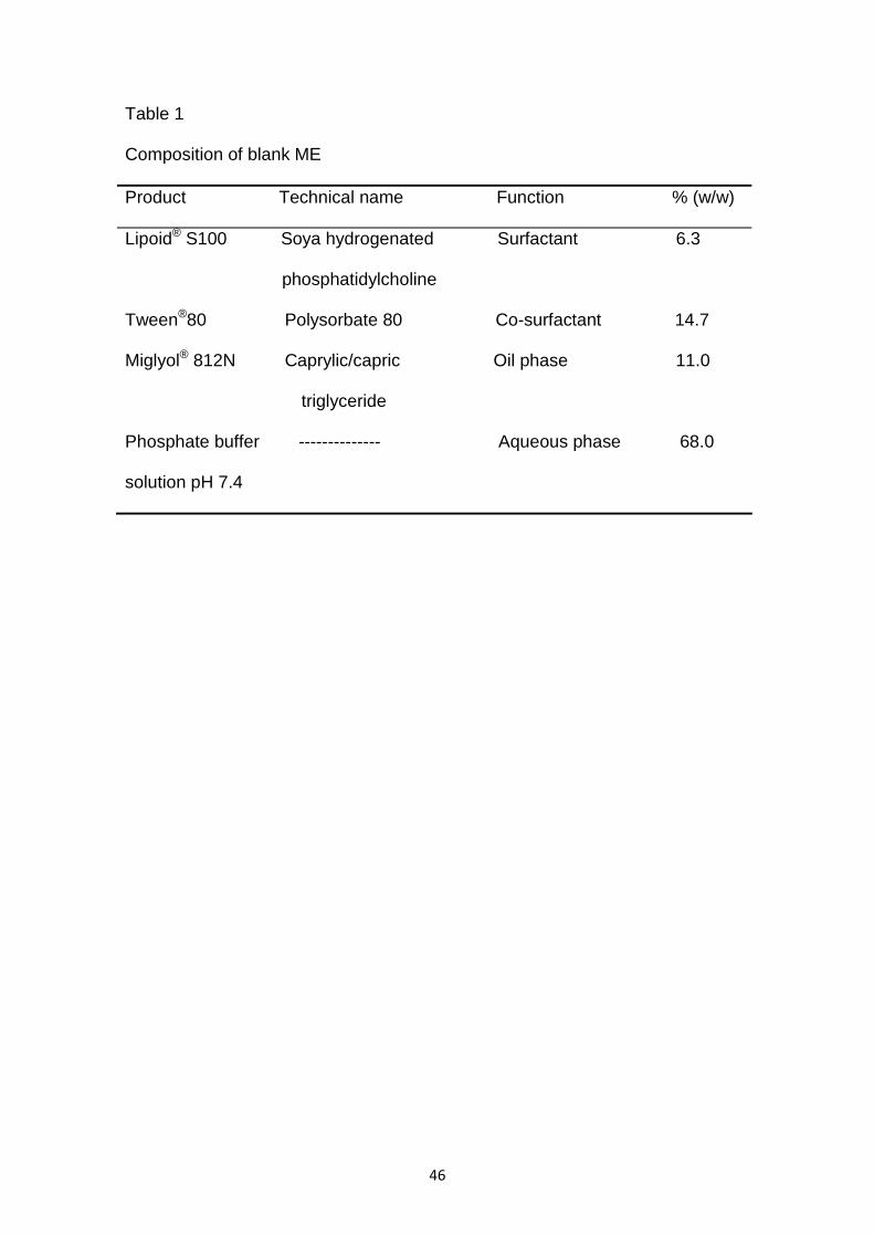
All the ME components were chosen based in their biocompatibility
characteristics to the eye (Table 1). The surfactant and co-surfactant
concentrations are usually high due the necessity to decrease the interfacial
tension and providing the thermodynamic stability of these systems. For
ophthalmic applications, this fact can be very important because it guarantees a
good spreading effect on the cornea and mixing with the precorneal film
constituents, thus possibly improving the contact between the drug and the
corneal epithelium (14). However, this high concentration can lead to ocular
toxicity (15). Therefore, the ionic surfactants, which are generally toxic, shall be

32
avoided and amphoteric and non-ionic surfactants, like soy phosphatidylcholine
and Tween® 80 are indicated for this use because they exhibit excellent
biocompatibility. The oil and the aqueous phases shall be chosen by its
conditions to form clear ME and their capacity of solubilization of drugs. They
also should be well tolerated by the eye. The most often oil phase used are the
triesters of glycerol, capric and caprylic acids (Miglyol® 812N) and phosphate
buffer for the aqueous phase (15).
Preparation of microemulsion
To prepare this ME, the proportion and addition order of components,
and the mechanical stirring and the heating process promoted by sonication
method were of utmost importance. In fact, the control of these parameters
speeds up the process of microemulsification. The sonication method is often
used, particularly in systems which contain non-ionic surfactants and was very
important for the process, allowing the formation of transparent system and
assisting in reducing of the size of dispersed droplet (16).
The resulting system was easily prepared and presented a yellow color,
a homogeneity aspect, a transparent appearance (Table 2) and the absence of
precipitates. Such characteristics is typical for a Winsor IV system, and,
therefore, it could be used as a drug delivery systems for ophthalmic use (17).
Amphotericin B loading process
The attraction of O/W ME is in their ability to incorporate drugs into the oil
phase, thereby, enhancing their solubility (18). Because of their high
solubilization capacity, these systems can be used as carriers for drugs which
having poor water solubility. The AmB molecule has one ring structure
comprising of a hydrophobic heptane chain and a hydroxyl rich hydrophilic

33
chain that imparts an amphoteric nature to the molecule. Because of this
intrinsic characteristic, the incorporation of this drug into the system is a
challenge since it has poor aqueous as well as oil solubility. However, many
works described the use of alkaline pH to solubilize this drug into the ME (12, 19).
The presence of AmB into the ME showed, after the addition of NaOH
solution (1 N), a complete incorporation of the drug into the system, maintained,
therefore, the characteristics of homogeneity and transparency of the blank ME.
Once the drug was dissolved, the system was neutralized to pH between 7.0-
7.5 with HCl solution (1 N) and this adjustment decreased the transparency
(Table 2) observed earlier, probably because of some interference on the
interfacial tension. This fact can be explained by the own chemical property of
AmB molecule characterized by its amphiphilic and amphoteric behavior.
However, the maintenance of the Winsor IV system aspect indicates that the
physicochemical stability of the ME was not perturbed (10, 17).
Physicochemical characteristics of ME and AmB-ME
The physicochemical characteristics of ME and AmB-ME (Table 2)
revealed a great compatibility with the characteristics ascribed for dosage forms
used for ophthalmic route (20). The blank ME appears as a promising carrier
system for the incorporation of drugs necessary to many ophthalmic diseases.
On the other hand, the incorporation of AmB did not induce important changes
in these parameters.
The adjustment of the initial pH at 7-8 is very important in order to
minimize the hydrolysis of the phospholipids presents, which can decrease the
pH of the ME. Indeed, the solubility, stability and corneal permeability of the
drugs are, also, quite dependent of this parameter. The physiological pH values

34
presented by the blank ME and the AmB-ME predicts the maximum comfort and
absence of eye irritation when this preparation would be instilled in the eye (9, 15,
21).
The ophthalmic solution of the conventional dosage form, represents
90% of the available ophthalmic formulations in the market. However, the rapid
pre-corneal loss and high tear fluid induce a low bioavailability of the drug into
the ocular structures. Developing new topical ocular delivery systems that
improve the ocular retention and increase the corneal drug absorption consist
on a big challenge (22). The AmB addition to the obtained blank ME did not have
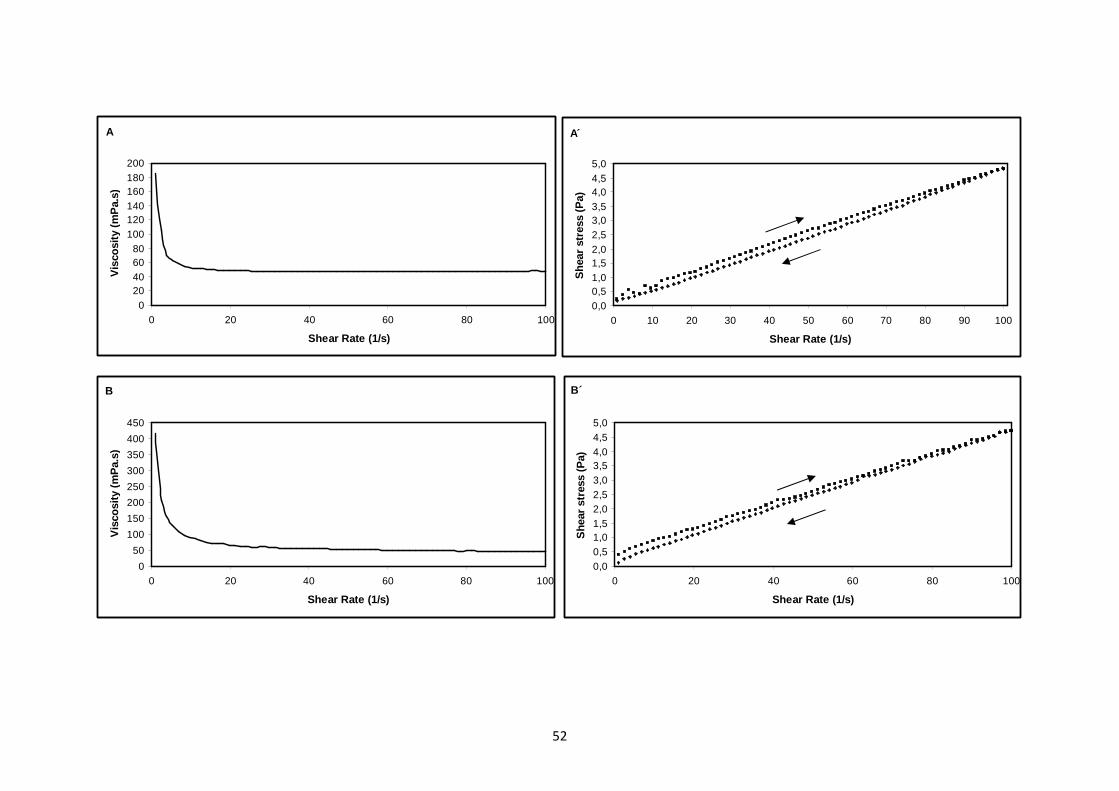
an effect on the rheology. However a slight decrease in the viscosity value of
AmB-ME was observed. Despite of that both systems presented a viscosity
value that allows sterile filtration. This viscosity value can still increase the
ocular residence time of AmB without the occurrence of discomfort, blurred
vision or foreign body sensation with the cornea surface, favoring, thereafter, a
better penetration of this drug into the eye. Indeed, on the beginning, the
system presented an initial deformation tension which after started to flow as a
Newtonian behavior, which ensures that blinking should not have effect on the
viscosity (9, 14) (Figure 2).
The presence of AmB into the ME decreased the transparency observed
in a blank ME as can be observed with the values obtained from percent of
transmittance. However, do not expected that these systems cause impairment
of vision and discomfort to the patient after instillation because of the proximity
of the RI values obtained with the water (1,33) and the cornea and lachrymal
fluid (1,34 – 1,36). The RI and percent of transmittance data prove the
transparence of the system (9, 14, 23).

35
The high value of conductivity found for the system allows us to classify
them as oil-in-water type. This characteristic was maintained after the
incorporation of the AmB, showing that this drug is not able to cause changes in
the intrinsic properties of the developed ME. This type of ME (O/W) is preferred
to use in the eye because of the droplet structure can be often retained during
the dilution process by the lachrymal fluid. Its administration could be
advantageous, because increases membrane permeability and facilitates the
corneal drug delivery, achieving prolonged release and higher penetration into
the deeper layers of the ocular structure and the aqueous humor than the native
drug (15, 16). Moreover, the increase in the value of conductivity after the
incorporation of the AmB can be explained by the addition of ionic solutions
(NaOH and HCl) necessary to incorporate this drug into the system.
The small droplet size of the ME system, presented in the range of
nanometers, is due to the interaction between the co-surfactant molecules with
the surfactant film which decrease the radius of curvature of the microdroplets,
forming transparent systems (13). The results of the droplet size of the MEs were
reported as X10, X50 and X90, being the droplet diameter for the 10th, 50th and
90th cumulative volume percentiles (24). They showed that the presence of AmB
increased slightly the droplet size. This increase can be explained because,
under the experimental conditions (pH 7.4), the AmB is amphoteric and very
slightly water soluble and then can be favorably partitioned into the oil phase,
increasing the droplet volume. Because of its amphiphilic characteristics, a
fraction of AmB molecules can, yet, be placed at the interface between the two
phases leading, also, to an increase in the droplet size (10). However, this fact

36
was not able to cause changes on the isotropic characteristic of the blank ME
(Figure 3).
Originally, several research groups suggested that for the ME formation,
a negative free energy is necessary. This can be obtained when large
reductions in surface tension are accompanied by significant favorable entropic
change (16). Therefore, the microemulsification is spontaneous and the resulting
dispersion has high concentration of surfactants that can lead to ocular toxicity
(15). Qualitatively, due the high concentration of aqueous phase which allows the
formation of oil-in-water (O/W) dispersion, the proportions of the components on
formulation and the nature of the surfactants used (amphoteric for Lipoid® S100
and non-ionic for Tween® 80), the spontaneous microemulsification was not
possible and a non-spontaneous preparation method (sonication) was needed.
Thus, because of this fact the results indicated high surface tension, both for
blank ME and for AmB-ME which suggests that there are not surfactant and co-
surfactant molecules free into the system and that all of them find out in the
interfacial area (15, 16).
The corneal surface is negatively charged. Therefore, the charges of the
droplets, which constitute the internal phase (oil) could influence their
absorption in the ophthalmic route. The studies showed that positively charged
ME are appropriate for this route. The electrostatic attraction between the
positively charged ME surface and negatively charged cell surface generate
bioadherence because of the opposite surface charges and provide
enhancement of drug absorption. So, the developed ME presented a positive
zeta potential value (+ 122.73 ± 2.25) which is appropriate for the ophthalmic
route because this is necessary to increase the contact time of the AmB with

37
the cornea surface. Moreover, the AmB changed the nature of the surface
charge without change the mean droplet size (25, 26).
Quantitative analyses and characterization of the a ggregation state of
AmB
The sterility of the systems was achieved by filtration through 0.22 µm
sterile filters. Microbiological tests have shown that this process was able to
sterilize the samples (data do not shown). UV-visible procedures were used to
determine the final amount of the drug incorporated into the ME. The results
suggest that there is a loss of AmB. The Table 3 shows the decrease in the
AmB concentration with the use of these filters, based in a nominal content of 5
mg/mL. The first filtration at 0.45 µm deleted the AmB suspended and the
probable drug excess of the dispersed system. Nevertheless, a small loss of
drug was also observed with the sterile filtration. This fact can be probably
ascribed to the adsorption entrapment on the filter considering the colloidal
nature of the disperse phase caused under this drug (19). Loss of AmB on
filtration of AmB formulations such as AmB colloidal dispersion for parenteral
use and from AmB-ME has been previously reported by several research
groups (12, 27-29).
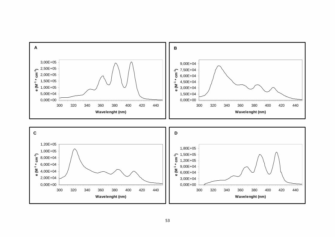
As described earlier, AmB is an amphiphilic molecule. Its hydrophobic
pole consists of a series of seven double bands in trans configuration, which
leads to an intense absorption spectra around 400 nm. The shape of the
spectra will depend on the state of aggregation of molecules and the relative
proportion of such different forms depends on concentration. The different
behavior was measured by the variation on the absorption spectra calculated by
the molar extinction coefficients (ε) and revealed that, in phosphate buffer, the

38
AmB-M presented four characteristics absorption bands, observed at 329, 364,
385 and 408 nm. In methanol, four bands with decreasing intensities at 406,
383, 363 and 345 nm can be seen. These spectral bands indicate the existence
of aggregates states and monomers, respectively. All of these results are in
agreement with the literature (30-32).
A comparison of UV-visible absorption spectra of AmB-M in phosphate
buffer, pure AmB in methanol and AmB-ME diluted in phosphate buffer and in
blank ME is presented in Figure 4. As it can be clearly seen on the spectra of
AmB-ME diluted in phosphate buffer, the presence of one intense band at 329
nm indicates the formation of self-associated AmB structure. However, the shift
of such band from 323 to 329 indicated that the complex between AmB and ME
is different of the complex formed by AmB and micelles. Indeed, when these
spectra were compared with the AmB-ME diluted in blank ME, it can be seem
that only the monomeric species is present (31, 32).
Clinically, the aggregation state of AmB is directly related to its
mechanism of action and toxicity (33, 34). The water soluble monomer is usually
related as a low toxic form to AmB while water-soluble oligomers have been
defined as the most toxic. The efficacy and toxicity of a lipidic based emulsion
for parenteral use containing AmB were showed by Araújo et al. Their assays,
using Red Blood Cells and Candida tropicalis as model cells, demonstrated that
their system was effective against these fungal cells maintaining the efficacy
and decreasing the toxicity presented by AmB-M. So, depending on its
aggregational state, AmB may present several patterns of activity against fungal
cells compared to the toxicity of mammalian cells, showing that a lipid carrier as

39
ME can modify the equilibrium between monomeric and oligomeric forms of this
drug in the aqueous media and change its overall activity and toxicity (35).
AmB entrapment efficiency for the MEs
After the centrifugation process, some drug precipitation was observed
which demonstrate that AmB was not properly incorporated to the disperse
system. The average entrapment efficiency value in the formulation was,
therefore, 70.2% (Table 3).
Stress testing
The freeze-thaw cycles consists of one stability test which designed to
determine the behavior of the samples under adverse condition of packaging.
The results obtained showed that the systems did not suffer any significant
changes on evaluated parameters maintaining the characteristics observed
earlier as can be seen through the table 4.
Conclusion
The results show that AmB-ME presents optimal characteristics to be
administered by the ophthalmic route (20) and this system could be used, in a
long term, has a probable new ophthalmic delivery system for AmB to treat
ocular fungal infections by eye drops. Their favorable characteristics and huge
potential to decrease the toxicity at the cornea caused by Fungizone® suggest
its therapeutic application. The blank ME appears, also, as an interesting
system for incorporation of both lipophilic and hydrophilic drugs needed to
control many ophthalmic diseases.

40
Acknowledgements
The authors wish to thank BNB and CNPq (Brasília, Brazil) for financial support
in the development in this work. They also acknowledge Glenn Hawes from the
American Language Program of the University of Georgia for editing this
manuscript.

41
References
1. Manzouri B, Vafidis GC, Wyse RK. Pharmacotherapy of fungal eye
infections. Expert Opin Pharmacother 2001; 2(11): 1849-57.
2. Thomas PA. Current perspectives on ophthalmic mycoses. Clin Microbiol
Rev 2003; 16(4): 730-97.
3. Esposito E, Bortolotti F, Menegatti E, Cortesi R. Amphiphilic association
systems for Amphotericin B delivery. Int J Pharm 2003; 260(2): 249-60.
4. Kaur IP, Rana C, Singh H. Development of effective ocular preparations of
antifungal agents. J Ocul Pharmacol Ther 2008; 24(5): 481-93.
5. Morand K, Bartoletti AC, Bochot A, Barratt G, Brandely ML, Chast F.
Liposomal amphotericin B eye drops to treat fungal keratitis: physico-chemical
and formulation stability. Int J Pharm 2007; 344(1-2): 150-3.
6. O'Day DM, Head WS, Robinson RD, Clanton JA. Corneal penetration of
topical amphotericin B and natamycin. Curr Eye Res 1986; 5(11): 877-82.
7. O'Day DM, Ray WA, Head WS, Robinson RD. Influence of the corneal
epithelium on the efficacy of topical antifungal agents. Invest Ophthalmol Vis Sci
1984; 25(7): 855-9.
8. Foster CS, Lass JH, Moran-Wallace K, Giovanoni R. Ocular toxicity of topical
antifungal agents. Arch Ophthalmol 1981; 99(6): 1081-4.
9. Fialho SL, da Silva-Cunha A. New vehicle based on a microemulsion for
topical ocular administration of dexamethasone. Clin Experiment Ophthalmol
2004; 32(6): 626-32.
10. Pestana KC, Formariz TP, Franzini CM, Sarmento VH, Chiavacci LA,
Scarpa MV, et al. Oil-in-water lecithin-based microemulsions as a potential

42
delivery system for amphotericin B. Colloids Surf B Biointerfaces 2008; 66(2):
253-9.
11. Wood TO, Tuberville AW, Monnett R. Keratomycosis and amphotericin B.
Trans Am Ophthalmol Soc 1985; 83: 397-409.
12. Moreno MA, Frutos P, Ballesteros MP. Lyophilized lecithin based oil-water
microemulsions as a new and low toxic delivery system for amphotericin B.
Pharm Res 2001; 18(3): 344-51.
13. Tenjarla S. Microemulsions: an overview and pharmaceutical applications.
Crit Rev Ther Drug Carrier Syst 1999; 16(5): 461-521.
14. Hasse A, Keipert S. Development and characterization of microemulsions
for ocular application. Eur J Pharm Biopharm 1997; 43(2): 179-83.
15. Vandamme TF. Microemulsions as ocular drug delivery systems: recent
developments and future challenges. Prog Retin Eye Res 2002; 21(1): 15-34.
16. Lawrence MJ, Rees GD. Microemulsion-based media as novel drug delivery
systems. Adv Drug Deliv Rev 2000; 45(1): 89-121.
17. Winsor PA. Hydrotropy, Solubilisation and Related Emulsification Processes
.1. to .4. Trans Faraday Soc 1948; 44(6): 376-98.
18. Karasulu HY. Microemulsions as novel drug carriers: the formation, stability,
applications and toxicity. Expert Opin Drug Deliv 2008; 5(1): 119-35.
19. Darole PS, Hegde DD, Nair HA. Formulation and evaluation of
microemulsion based delivery system for amphotericin B. AAPS PharmSciTech
2008; 9(1): 122-8.
20. Kaur IP, Kanwar M. Ocular preparations: the formulation approach. Drug
Dev Ind Pharm 2002; 28(5): 473-93.

43
21. Ali Y, Lehmussaari K. Industrial perspective in ocular drug delivery. Adv
Drug Deliv Rev 2006; 58(11): 1258-68.
22. Chan J, Maghraby GM, Craig JP, Alany RG. Phase transition water-in-oil
microemulsions as ocular drug delivery systems: in vitro and in vivo evaluation.
Int J Pharm 2007; 328(1): 65-71.
23. Ghosh PK, Majithiya RJ, Umrethia ML, Murthy RS. Design and development
of microemulsion drug delivery system of acyclovir for improvement of oral
bioavailability. AAPS PharmSciTech 2006; 7(3): 77.
24. Formiga FR, Fonseca IAA, Souza KB, Silva AKA, Macedo JPF, Araújo IB,
et al. Influence of a lipophilic drug on the stability of emulsions: an important
approach on the development of lipidic carriers. Int J Pharm 2007; 344(1-2):
158-60.
25. Tamilvanan S, Khoury K, Gilhar D, Benita S. Ocular delivery of cyclosporin
A I. Design and characterization of cyclosporin A-loaded positively-charged
submicron emulsion. Stp Pharm Sci 2001; 11(6): 421-6.
26. Abdulrazik M, Tamilvanan S, Khoury K, Benita S. Ocular delivery of
cyclosporin A II. Effect of submicron emulsion's surface charge on ocular
distribution of topical cyclosporin A. Stp Pharm Sci 2001; 11(6): 427-32.
27. Tripple M, Shadomy S, Espinel-Ingroff A. Availability of active amphotericin
B after filtration through membrane filters. Am Rev Respir Dis 1977; 115(5):
879-81.
28. Huber RC, Riffkin C. In line final filters for removing particles from
amphotericin B infusions. Am J Hosp Pharm 1975; 32(2): 173-6.

44
29. Brime B, Moreno MA, Frutos G, Ballesteros MP, Frutos P. Amphotericin B in
oil-water lecithin-based microemulsions: formulation and toxicity evaluation. J
Pharm Sci 2002; 91(4): 1178-85.
30. Egito LC, de Medeiros SR, Medeiros MG, Price JC, Egito EST. Evaluation
of the relationship of the molecular aggregation state of amphotericin B in
medium to its genotoxic potential. J Pharm Sci 2004; 93(6): 1557-65.
31. Egito EST, Araujo IB, Damasceno BP, Price JC. Amphotericin B/emulsion
admixture interactions: an approach concerning the reduction of amphotericin B
toxicity. J Pharm Sci 2002; 91(11): 2354-66.
32. Egito EST, Fessi H, Appel M, Puisieux F, Bolard J, Devissaguet JP. New
Techniques for Preparing Submicronic Emulsions - Application to Amphotericin-
B. Stp Pharm Sci 1994; 4(2): 155-62.
33. Bolard J, Legrand P, Heitz F, Cybulska B. One-sided action of amphotericin
B on cholesterol-containing membranes is determined by its self-association in
the medium. Biochemistry 1991; 30(23): 5707-15.
34. Lamy-Freund MT, Ferreira VF, Faljoni-Alario A, Schreier S. Effect of
aggregation on the kinetics of autoxidation of the polyene antibiotic
amphotericin B. J Pharm Sci 1993; 82(2): 162-6.
35. Araujo IB, Damasceno BP, de Medeiros TM, Soares LA, do Egito EST.
Decrease in Fungizone toxicity induced by the use of Lipofundin as a dilutent:
an in vitro study. Curr Drug Deliv 2005; 2(2): 199-205.

45
Figure legends
Figure 1. Pseudo-ternary phase diagrams of the system containing Miglyol®
812N (oil phase), phosphate buffer pH 7.4 (aqueous phase), Lipoid® S100
(surfactant) and Tween® 80 (co-surfactant), showing the area of existence of
microemulsions and the point chosen for characterization.
Figure 2: Particle size frequency and accumulative frequency of the
microemulsion (ME) (A) and amphotericin B-loaded-microemulsion (AmB-ME)
(B).
Figure 3: Viscosity (A and B) and rheological behavior (A’ and B’) of the
microemulsion (ME) (A and A’) and amphotericin B-loaded-microemulsion
(AmB-ME) (B and B’).
Figure 4: UV-visible absorption spectra of pure Amphotericin B (AmB) in
methanol (A), the micelle dosage form Amphotericin B (AmB-M) in phosphate
buffer (pH 7.4) (B) and AmB into the microemulsion diluted at phosphate buffer
(pH 7.4) (C) and in microemulsion (AmB-ME) (D).
46
Table 1
Composition of blank ME
Product Technical name Function % (w/w)
Lipoid® S100 Soya hydrogenated Surfactant 6.3
phosphatidylcholine
Tween®80 Polysorbate 80 Co-surfactant 14.7
Miglyol® 812N Caprylic/capric Oil phase 11.0
triglyceride
Phosphate buffer -------------- Aqueous phase 68.0
solution pH 7.4

47
Table 2
Evaluated parameters of ME and AmB-ME (Mean ± SD)
Parameter Blank ME AmB-ME
Percent transmittance (%) 98.0 60.0
pH 7.40 ± 0.04 7.16 ± 0.31
Viscosity (mPa.s) 48.28 ± 0.36 47.20 ± 0.31
Refractive index 1.373 ± 0.00 1.374 ± 0.00
Conductivity (µS) 733.00 ± 3.46 1820.00 ± 712.43
Particle size (nm)
X10 7.22 ± 0.41 7.39 ± 0.52
X50 9.79 ± 0.42 9.87 ± 0.64
X90 13.96 ± 0.49 14.15 ± 0.59
Surface tension (dyn/cm) 71.40 ± 0.57 71.10 ± 1.06
Zeta potential (mV) + 67.52 ± 0.59 + 122.73 ± 2.25

48
Table 3
AmB content (Mean ± SD)
Parameters AmB Content (mg/mL) AmB Content (%)
Without filtration 3.51 ± 0.35 70.20
After 0.45 µm 2.98 ± 0.33 59.60
After 0.22 µm 2.50 ± 0.16 50.00

49
Table 4
Results of stability of ME and AmB-ME after the freeze-thaw cycles (Mean ±
SD)
Parameter Blank ME AmB-ME
pH 7.48 ± 0.00 7.91 ± 0.10
Refractive index 1.378 ± 0.00 1.377 ± 0.00
Conductivity (µS) 829.03 ± 10.33 1692.3 ± 109.20
AmB content (mg/mL) ------------- 3.34 ± 0.54

50

51
0
5
10
15
20
25
30
4,525,37 6,39 7,6 9,03 10,7 12,815,218,121,525,630,4
Diameter (nm)F
requ
ency
(%
)
0
20
40
60
80
100
Accum
ulative frequency (%
)
0
5
10
15
20
25
30
4,525,376,39 7,6 9,03 10,712,815,218,121,5 25,6 30,4
Diameter (nm)
Fre
quen
cy (
%)
0
10
20
30
40
50
60
70
80
90
100 Accum
ulative frequency (%
)
52
B
0
50
100
150
200
250
300
350
400
450
0 20 40 60 80 100
Shear Rate (1/s)
Vis
cosi
ty (
mP
a.s)
A
020406080
100120140160180200
0 20 40 60 80 100
Shear Rate (1/s)
Vis
cosi
ty (
mP
a.s)
B´
0,00,51,01,52,02,53,03,54,04,55,0
0 20 40 60 80 100
Shear Rate (1/s)
She
ar s
tres
s (P
a)
A
0,00,51,01,52,02,53,03,54,04,55,0
0 10 20 30 40 50 60 70 80 90 100
Shear Rate (1/s)
She
ar s
tres
s (P
a)
53
A
0,00E+00
5,00E+04
1,00E+05
1,50E+05
2,00E+05
2,50E+05
3,00E+05
300 320 340 360 380 400 420 440
Wavelenght (nm)
e (M
-1 *
cm
-1)
C
0,00E+00
2,00E+04
4,00E+04
6,00E+04
8,00E+04
1,00E+05
1,20E+05
300 320 340 360 380 400 420 440
Wavelenght (nm)
e (M
-1 *
cm
-1)
B
0,00E+00
1,50E+04
3,00E+04
4,50E+04
6,00E+04
7,50E+04
9,00E+04
300 320 340 360 380 400 420 440
Wavelenght (nm)
e (M
-1 *
cm
-1)
D
0,00E+00
3,00E+04
6,00E+04
9,00E+04
1,20E+05
1,50E+05
1,80E+05
300 320 340 360 380 400 420 440
Wavelenght (nm)
e (M
-1 *
cm
-1)
54
4. COMENTÁRIOS, CRÍTICAS E SUGESTÕES
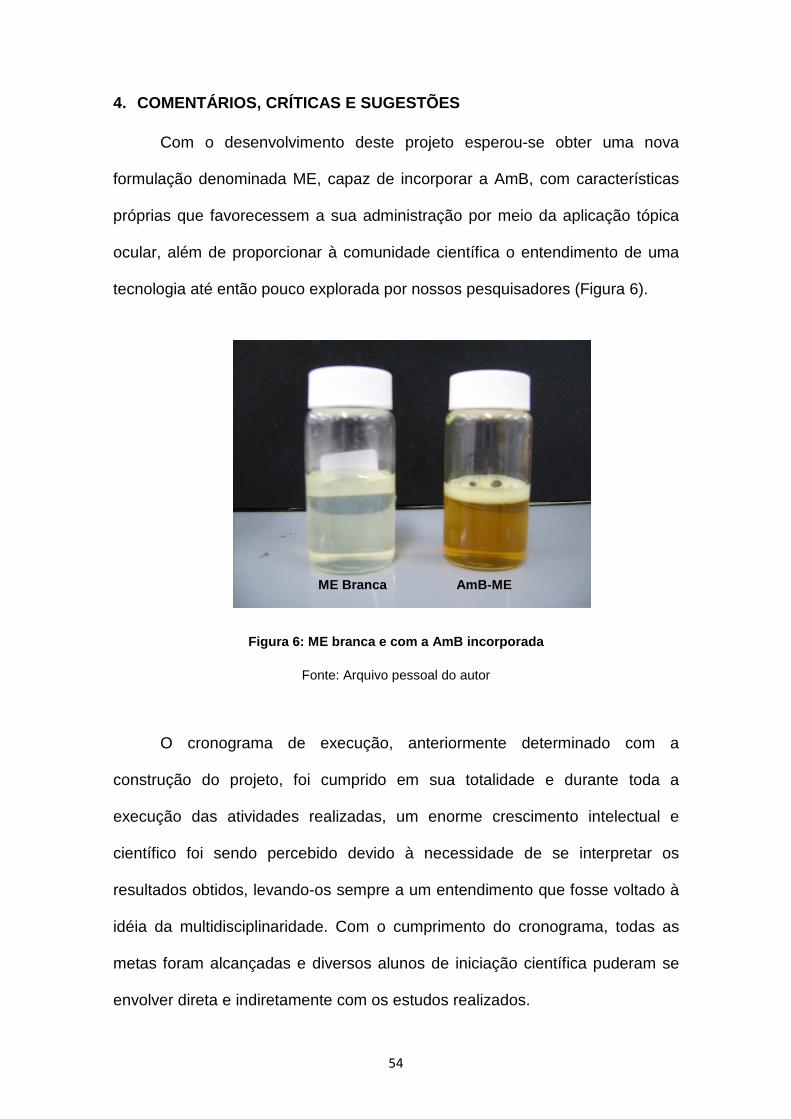
Com o desenvolvimento deste projeto esperou-se obter uma nova
formulação denominada ME, capaz de incorporar a AmB, com características
próprias que favorecessem a sua administração por meio da aplicação tópica
ocular, além de proporcionar à comunidade científica o entendimento de uma
tecnologia até então pouco explorada por nossos pesquisadores (Figura 6).
Figura 6: ME branca e com a AmB incorporada
Fonte: Arquivo pessoal do autor
O cronograma de execução, anteriormente determinado com a
construção do projeto, foi cumprido em sua totalidade e durante toda a
execução das atividades realizadas, um enorme crescimento intelectual e
científico foi sendo percebido devido à necessidade de se interpretar os
resultados obtidos, levando-os sempre a um entendimento que fosse voltado à
idéia da multidisciplinaridade. Com o cumprimento do cronograma, todas as
metas foram alcançadas e diversos alunos de iniciação científica puderam se
envolver direta e indiretamente com os estudos realizados.
AmB -ME ME Branca

55
Conforme pode ser verificado nos resultados gerados, a metodologia
utilizada foi capaz de atender a necessidade do desenvolvimento do projeto e
os sistemas desenvolvidos apresentaram resultados que nos permitem afirmar
que os mesmos podem ser, portanto, do ponto de vista tecnológico, aptos a
serem utilizados através da via de administração tópica ocular. Para a sua
realização, além dos nossos equipamentos, foram estabelecidos diversos
contatos bastante importantes com vários outros laboratórios da UFRN e da
UFPE, que gentilmente cederam seus equipamentos e instalações, dando um
suporte imprescindível à parte experimental do projeto e a quem eu devo
enormes agradecimentos.
A realização destes estudos mostrou ainda que foi possível a obtenção
de uma forma farmacêutica líquida e de fácil preparação, estéril, contendo um
fármaco bastante importante para a terapêutica antifúngica ocular e que a ME
branca pode servir para a incorporação de outros fármacos pouco solúveis em
água, ou ainda, que não possuam uma formulação oftalmológica em particular
necessários ao tratamento das mais diversas patologias oculares de forma
mais eficaz para a sociedade. Sendo assim, durante toda a fase experimental e
devido à esta grande diversidade de ensaios realizados, vários trabalhos
puderam ser apresentados em congressos locais, regionais, nacionais e
internacionais até o momento, incluindo a confecção de mais um artigo
científico que já está apto a ser submetido, além deste apresentado nesta
dissertação.
A publicação acima reforça a importância do tema para a comunidade
científica, pois a pobre biodisponibilidade da AmB gerada a partir das barreiras
anatômicas e fisiológicas do olho associados a grande possibilidade de

56
toxicidade ocular, além da baixa estabilidade da preparação extemporânea
utilizada para a aplicação tópica, faz com que se torne necessário o
desenvolvimento de um novo sistema carreador para a AmB, sistema este,
possibilitado devido ao enorme crescimento, recentemente, do uso das MEs
relacionadas para o uso farmacêutico.
Em decorrência disso, o objetivo de reduzir o potencial risco de reações
adversas gerado com o uso da AmB convencional, cuja formulação é mais
apropriada para a administração intravenosa, no tratamento de infecções
fúngicas oculares, além de proporcionar a chance de se ter uma forma
farmacêutica própria para aplicação tópica nos olhos para a AmB, ainda
inexistente no mercado, torna o referido artigo como uma referência básica e
primordial de orientação para o desenvolvimento de novas formulações para
uso oftalmológico somando-se àquelas poucas referências que são
encontradas hoje nos principais indexadores que trazem o assunto sobre a
utilização desses sistema para uso oftalmológico.
Entretanto, é evidente a necessidade ilimitada de desenvolver novas
alternativas terapêuticas para melhorar a qualidade da assistência à saúde e
acompanhar os avanços tecnológicos e científicos do dia-a-dia. Portanto, são
necessários que novos estudos sejam realizados com as formulações
desenvolvidas para que o entendimento seja claro e que atinja o equilíbrio
entre todas as áreas da saúde. Sendo assim, ensaios de eficácia clínica da
AmB-ME, farmacocinéticos, estudos de citotoxicidade e de toxicidade,
epidemiológicos, microbiológicos, entre outros, seriam de grande valia para a
complementaridade da idéia desenvolvida por meio deste trabalho, tornando o
tema aberto para a comunidade científica explorar, por meio de outros

57
projetos, dissertações ou teses. Em 2010, pretendo dar continuidade à vários
destes estudos por meio do curso de Doutorado no Programa de Pós-
Graduação em Ciências da Saúde.
Por fim, consideramos que o desenvolvimento desta pesquisa de
Mestrado bem como a pós-graduação foram fatores importantes que
contribuíram enormemente para a construção, execução, discussão e
divulgação desse tema que a cada ano torna-se mais importante para a
oftalmologia e que os objetivos estabelecidos da multidisciplinaridade
enfatizados pelo PPGCSA puderam ser alcançados com sucesso quando os
assuntos envolvidos no trabalho foram interdisciplinarizados.

58
5. APENDICE
5.1. APENDICE I
Resumos publicados em anais de congresso
1. MEDEIROS, Maria Izabel Priscila de Araújo, SILVEIRA, Walteçá Louis
Lima da, BRASIL, Fernanda do Couto, ARAÚJO, Ivonete Batista de, EGITO,
Eryvaldo Sócrates Tabosa do
Caracterização macroscópica de microemulsões em gel para uso
oftalmológico In: XVIII Congresso de Iniciação Científica da UFRN, 2007,
Natal/RN.
XVIII Congresso de Iniciação Científica . Natal/RN: 2007.
2. SILVEIRA, Walteçá Louis Lima da, BRASIL, Fernanda do Couto,
ARAÚJO, Ivonete Batista de, EGITO, Eryvaldo Sócrates Tabosa do
Desenvolvimento de microemulsões O/A para uso ocular In: IX Congresso
Científico da UNP, 2007, Natal/RN.
IX Congresso Científico da UNP . Natal/RN: 2007.
3. SILVEIRA, Walteçá Louis Lima da, DAMASCENO, Bolívar Ponciano
Goulart de Lima, BRASIL, Fernanda do Couto, SOARES, Airanuédida Silva,
ARAÚJO, Ivonete Batista de, OLIVEIRA, Anselmo Gomes, EGITO, Eryvaldo
Sócrates Tabosa do
Rheological behaviour of microemulsions intended for ophthalmic use In: I
International Symposium in Pharmaceutical Sciences of Northeast Brazil,
2008, Natal/RN.
I International Symposium in Pharmaceutical Science s of Northeast
Brazil , Natal/RN: 2008.

59
4. SILVEIRA, Walteçá Louis Lima da, SOARES, Airanuédida Silva,
BRASIL, Fernanda do Couto, ARAÚJO, Ivonete Batista de, OLIVEIRA,
Anselmo Gomes, EGITO, Eryvaldo Sócrates Tabosa do
Characterization of Oil-in-water microemulsion intended for ophthalmic use
In: I International Symposium in Pharmaceutical Sciences of Northeast
Brazil, 2008, Natal/RN.
I International Symposium in Pharmaceutical Science s of Northeast
Brazil , Natal/RN: 2008.
5. SILVEIRA, Walteçá Louis Lima da, BRASIL, Fernanda do Couto,
SOARES, Airanuédida Silva, FREIRE, Larissa Bandeira, ARAÚJO, Ivonete
Batista de, EGITO, Eryvaldo Sócrates Tabosa do
Evaluation of the autoclavation processo on the stability of oil-in-water
microemulsion intended for ophthalmic use In: III Congresso Norte-Nordeste
de Multirresistência bacteriana, II Workshop Sul-Americano de Ciência e
Tecnologias Farmacêuticas, I Fórum de Microbiologia Aplicada ao Controle
de Infecções em Serviços de Saúde e I Fórum Norte-Nordeste dos
LACEN´s, 2008, Olinda/PE.
III Congresso Norte-Nordeste de Multirresistência b acteriana .
Olinda/PE: 2008.
6. SILVEIRA, Walteçá Louis Lima da, BRASIL, Fernanda do Couto,
SOARES, Airanuédida Silva, CARVALHO, Juliana Fernandes, ARAÚJO,
Ivonete Batista de, EGITO, Eryvaldo Sócrates Tabosa do
Evaluation of the fungal contamination in microemulsion after the sterilization
process by filtration In: III Congresso Norte-Nordeste de Multirresistência
bacteriana, II Workshop Sul-Americano de Ciência e Tecnologias

60
Farmacêuticas, I Fórum de Microbiologia Aplicada ao Controle de Infecções
em Serviços de Saúde e I Fórum Norte-Nordeste dos LACEN´s, 2008,
Olinda/PE.
III Congresso Norte-Nordeste de Multirresistência b acteriana .
Olinda/PE: 2008.
7. BRASIL, Fernanda do Couto, SILVEIRA, Walteçá Louis Lima da, SILVA,
Maria Clara de Araújo, ARAÚJO, Ivonete Batista de, EGITO, Eryvaldo
Sócrates Tabosa do
Caracterização macroscópica de microemulsões contendo anfotericina B
para uso ocular In: XIX Congresso de Iniciação Científica da UFRN, 2008,
Natal/RN.
XIX Congresso de Iniciação Científica . Natal/RN: 2008.
8. FERREIRA, Laura da Fonseca, SILVEIRA, Walteçá Louis Lima da,
DAMASCENO, Bolívar, Ponciano Goulart de Lima, ARAÚJO, Ivonete Batista
de, EGITO, Eryvaldo Sócrates Tabosa do
Comportamento reológico de microemulsões contendo anfotericina B
destinados ao uso oftalmológico In: XIX Congresso de Iniciação Científica
da UFRN, 2008, Natal/RN.
XIX Congresso de Iniciação Científica . Natal/RN: 2008.
9. SILVEIRA, Walteçá Louis Lima da, DAMASCENO, Bolívar, Ponciano
Goulart de Lima, BRASIL, Fernanda do Couto, SOARES, Airanuédida Silva,
FERREIRA, Laura da Fonseca, SILVA, Maria Clara de Araújo, ARAÚJO,
Ivonete Batista de, EGITO, Eryvaldo Sócrates Tabosa do

61
Characterization of the aggregation form of anphotericin B in a
microemulsion intended for ophthalmic use In: Meeting on Nanotechnology,
Lipossome and Health, 2009, Itaparica/BA.
Meeting on Nanotechnology, Lipossome and Health . Itaparica/BA: 2009.
10. SOARES, Airanuédida Silva, SILVEIRA, Walteçá Louis Lima da,
BRASIL, Fernanda do Couto, ARAÚJO, Ivonete Batista de, EGITO, Eryvaldo
Sócrates Tabosa do
Estudos de caracterização físico-química de uma microemulsão contendo
anfotericina B para a aplicação tópica ocular In: XX Congresso de Iniciação
Científica da UFRN, 2009, Natal/RN.
XX Congresso de Iniciação Científica . Natal/RN: 2009.
11. SILVEIRA, Walteçá Louis Lima da, DAMASCENO, Bolívar, Ponciano
Goulart de Lima, BRASIL, Fernanda do Couto, SOARES, Airanuédida Silva,
ARAÚJO, Ivonete Batista de, EGITO, Eryvaldo Sócrates Tabosa do
Avaliação granulométrica de microemulsões contendo anfotericina B
visando o uso oftalmológico In: I Simpósio Nacional em Ciências
Farmacêuticas Básicas e Aplicadas, 2009, Natal/RN
I Simpósio Nacional em Ciências Farmacêuticas Básic as e Aplicadas .
Natal/RN: 2009.

62
6. REFERÊNCIAS BIBLIOGRÁFICAS
1. Carlfors J, Edsman K, Petersson R, Jornving K. Rheological evaluation of
Gelrite in situ gels for ophthalmic use. Eur J Pharm Sci. 1998; 6(2):113-9.
2. Chaumeil C, Bourcier T, Rostane H, Goldschmidt P, Nourry H, Zamfir O, et
al. Diagnosis and treatment of keratomycosis and fungal endophthalmitis. J
Mycol Med. 2007; 17:89-108.
3. Kaushik S, Ram J, Brar GS, Jain AK, Chakraborti A, Gupta A. Intracameral
amphotericin B: initial experience in severe keratomycosis. Cornea. 2001;
20(7):715-9.
4. Kuriakose T, Kothari M, Paul P, Jacob P, Thomas R. Intracameral
amphotericin B injection in the management of deep keratomycosis. Cornea.
2002; 21(7):653-6.
5. Thomas PA. Current perspectives on ophthalmic mycoses. Clin Microbiol
Rev. 2003; 16(4):730-97.
6. Yilmaz S, Ture M, Maden A. Efficacy of intracameral amphotericin B
injection in the management of refractory keratomycosis and endophthalmitis.
Cornea. 2007; 26(4):398-402.
7. Yoon KC, Jeong IY, Im SK, Chae HJ, Yang SY. Therapeutic effect of
intracameral amphotericin B injection in the treatment of fungal keratitis.
Cornea. 2007; 26(7):814-8.
8. Morand K, Bartoletti AC, Bochot A, Barratt G, Brandely ML, Chast F.
Liposomal amphotericin B eye drops to treat fungal keratitis: physico-chemical
and formulation stability. Int J Pharm. 2007; 344(1-2):150-3.
9. Kaur IP, Rana C, Singh H. Development of effective ocular preparations of
antifungal agents. J Ocul Pharmacol Ther. 2008; 24(5):481-93.
63
10. Fialho SL, da Silva-Cunha A. New vehicle based on a microemulsion for
topical ocular administration of dexamethasone. Clin Experiment Ophthalmol.
2004; 32(6):626-32.
11. Gasco MR, Gallarate M, Trotta M, Bauchiero L, Gremmo E, Chiappero O.
Microemulsions as topical delivery vehicles: ocular administration of timolol. J
Pharm Biomed Anal. 1989; 7(4):433-9.
12. Hasse A, Keipert S. Development and characterization of microemulsions
for ocular application. Eur J Pharm Biopharm. 1997; 43(2):179-83.
13. Vandamme TF. Microemulsions as ocular drug delivery systems: recent
developments and future challenges. Prog Retin Eye Res. 2002; 21(1):15-34.
14. Brime B, Moreno MA, Frutos G, Ballesteros MP, Frutos P. Amphotericin B
in oil-water lecithin-based microemulsions: formulation and toxicity evaluation.
J Pharm Sci. 2002; 91(4):1178-85.
15. Esposito E, Bortolotti F, Menegatti E, Cortesi R. Amphiphilic association
systems for Amphotericin B delivery. Int J Pharm. 2003; 260(2):249-60.
16. Moreno MA, Frutos P, Ballesteros MP. Lyophilized lecithin based oil-water
microemulsions as a new and low toxic delivery system for amphotericin B.
Pharm Res. 2001; 18(3):344-51.
17. Hornof M, Toropainen E, Urtti A. Cell culture models of the ocular barriers.
Eur J Pharm Biopharm. 2005; 60(2):207-25.
18. Davies NM. Biopharmaceutical considerations in topical ocular drug
delivery. Clin Exp Pharmacol Physiol. 2000; 27(7):558-62.
19. Manzouri B, Vafidis GC, Wyse RK. Pharmacotherapy of fungal eye
infections. Expert Opin Pharmacother. 2001; 2(11):1849-57.

64
20. Gallis HA, Drew RH, Pickard WW. Amphotericin B: 30 years of clinical
experience. Rev Infect Dis. 1990; 12(2):308-29.
21. Wood TO, Tuberville AW, Monnett R. Keratomycosis and amphotericin B.
Trans Am Ophthalmol Soc. 1985; 83:397-409.
22. Torrado JJ, Espada R, Ballesteros MP, Torrado-Santiago S. Amphotericin
B formulations and drug targeting. J Pharm Sci. 2008; 97(7):2405-25.
23. Araujo IB, Damasceno BP, de Medeiros TM, Soares LA, do Egito ES.
Decrease in Fungizone toxicity induced by the use of Lipofundin as a dilutent:
an in vitro study. Curr Drug Deliv. 2005; 2(2):199-205.
24. Kaji Y, Yamamoto E, Hiraoka T, Oshika T. Toxicities and
pharmacokinetics of subconjunctival injection of liposomal amphotericin B.
Graefes Arch Clin Exp Ophthalmol. 2009; 247(4):549-53.
25. Danielsson I, Lindman B. The definition of microemulsion. Colloides surf.
1981; 3:391-5.
26. Hoar TP, Schulman JH. Transparent water-in-oil dispersions: the
oleopathic hydromicelle. Nature. 1943; 152:102-05.
27. Winsor PA. Hydrotropy, Solubilisation and Related Emulsification
Processes .1. to .4. Transactions of the Faraday Soc. 1948; 44(6):376-98.
28. Kaur IP, Kanwar M. Ocular preparations: the formulation approach. Drug
Dev Ind Pharm. 2002; 28(5):473-93.

65
ABSTRACT
Ocular fungal infections are being increasingly recognized as an important
cause of morbidity and blindness of the world. The poor bioavailability of drugs,
like amphotericin B, antifungal enough used in the treatment of these infections,
in the posterior tissues of the eye combined with the anatomical ocular barriers,
which are able to limit their absorption, and associated with serious risks of
adverse reactions, generally, carry a treatment failure and serious
consequences for affected patients. Researchers around the world are looking
for new alternatives to overcome this problem and one great possibility is the
researches which involve the development of new formulations to improve the
bioavailability and reduce the toxicity associated with the application of
amphotericin B. Microemulsions appear as new systems capable of loading this
drug for ophthalmic use. This study aimed to obtain a biocompatible
microemulsion with the topical ocular route of administration containing
amphotericin B in order to establish a new and stable pharmaceutical dosage
form for topical administration in the eye. The system obtained demonstrated
through the characterization studies carried out biocompatibility with the route of
administration desired and, therefore, appears as an interesting and novel
dosage form containing amphotericin B for use, in future, in the treatment of
fungal eye infections.
Descriptors : Amphotericin B; Ophthalmic Solutions; Drug carriers;
Pharmaceutical preparations; Microemulsions.