UNIVERSIDADE FEDERAL DE PELOTAS Programa de Pós...
85
1 UNIVERSIDADE FEDERAL DE PELOTAS Programa de Pós-Graduação em Odontologia Tese Avaliação clínica dos efeitos de duas técnicas de isolamento do campo operatório no desempenho de restaurações Classe V e na condição periodontal Silvia Terra Fontes Pelotas, 2011
Transcript of UNIVERSIDADE FEDERAL DE PELOTAS Programa de Pós...
1
UNIVERSIDADE FEDERAL DE PELOTAS
Programa de Pós-Graduação em Odontologia
Tese
Avaliação clínica dos efeitos de duas técnicas de isolamento do campo
operatório no desempenho de restaurações Classe V e na condição
periodontal
Silvia Terra Fontes
Pelotas, 2011
2
SILVIA TERRA FONTES
Avaliação clínica dos efeitos de duas técnicas de isolamento do campo
operatório no desempenho de restaurações Classe V e na condição
periodontal
Orientador: Prof. Dr. Alexandre Severo Masotti
Co-orientadora: Profa. Dra. Fernanda de Oliveira Bello Corrêa
Pelotas, 2011
Tese apresentada ao Programa de Pós-Graduação em Odontologia da Universidade Federal de Pelotas, como requisito parcial à obtenção do título de Doutor em Odontologia com área de concentração em Dentística.

3
Dados de Catalogação da Publicação
F683a Fontes, Silvia Terra
Avaliação clínica dos efeitos de duas técnicas de isolamento do campo operatório no desempenho de restaurações Classe V e na condição periodontal / Silvia Terra Fontes ; orientador: Alexandre Severo Masotti ; co-orientador: Fernanda de Oliveira Bello Corrêa. - Pelotas: UFPel, 2011.
84 f. : fig. ; tab.
Tese (Doutorado) Dentística. Faculdade de Odontologia. Universidade Federal de Pelotas. Pelotas.
1. Adesivos dentinários. 2. Dentística operatória. 3. Dique de bor- racha. 4. Ensaio clínico. 5. Periodonto. I. Masotti, Alexandre Severo (orient.) II. Corrêa, Fernanda de Oliveira (co-orient.) III. Título.
D2
Bibliotecário: Fabiano Domingues Malheiro CRB -10/1955

4
Banca examinadora:
Profa. Dra. Fernanda de Oliveira Bello Corrêa (Co-orientadora)
Prof. Dr. Fabrício Aulo Ogliari
Profa. Dra. Marilene Issa Fernandes
Prof. Dr. Rafael Ratto Moraes
Prof. Dr. Thiago Marchi Martins
Prof. Dr. Eduardo Gonçalves Mota (Suplente)
5
DEDICATÓRIA
Dedico esta tese a meus pais e ao meu marido Ricardo por dividirem comigo as
alegrias e as dificuldades que tive com esta pesquisa.
6
AGRADECIMENTOS
Ao Programa de Pós-Graduação em Odontologia por tudo que aprendi e pela
oportunidade de realizar esta pesquisa.
Aos professores, Alexandre Severo Masotti, Fernanda de Oliveira Bello Corrêa,
Maximiliano Sérgio Cenci e Patrícia dos Santos Jardim pelos ensinamentos, pela
dedicação ao projeto e pela confiança que depositaram em mim.
Aos alunos, Ângelo Niemczewski Bobrowski, Aristênio Schimittz Bartsch, Caroline
Konzgen Barwaldt, Fernanda Weingartner Machado, Jordana Raquel Schneider,
Jorge Luiz da Costa Pereira Júnior, Karine Duarte da Silva, Ronaldo Luiz Rossi,
Thais Zorzoli Nunes, Thamires Pereira de Souza Meister, Vanessa Gebert e
Vanessa Reimers Guedes, que foram grandes colaboradores para a execução deste
estudo e, acima de tudo, amigos que jamais esquecerei.
Aos colegas Anelise Fernandes Montagner, Fabiana Chiapinotto, Fernanda Barbosa
Leal e Marcos Britto Correa pela disposição em me ajudar sempre que foi
necessário.
Às colegas Eduarda Dutra e Laura Pintado pelo encaminhamento de pacientes.
Às minhas amigas, Glória Beatriz Cubas de Azevedo, Maria Raquel Fernandez e
Sonia Luque Peralta, pela força e parceria durante estes anos de pós-graduação.
Agradeço também a todos os pacientes que participaram deste estudo.
7
NOTAS PRELIMINARES
A presente tese foi redigida segundo o Manual de Normas para Dissertações,
Teses e Trabalhos Científicos da Universidade Federal de Pelotas de 2006,
adotando o nível de descrição 4 – estruturas em artigos, que consta no Apêndice D
do referido manual. Disponível em:
<http://www.ufpel.tche.br/prg/sisbi/documentos/Manual_Normas_UFPel_2006.pdf>.
Acesso em: 10 fev. 2010, 15:03:20.
8
RESUMO
FONTES, Silvia Terra. Avaliação clínica dos efeitos de duas técnicas de
isolamento do campo operatório no desempenho de restaurações Classe V e
na condição periodontal. 2011. 84f. Tese (Doutorado) – Programa de Pós-
Graduação em Odontologia. Universidade Federal de Pelotas, Pelotas.
Durante os procedimentos restauradores, diferentes métodos de isolamento do campo operatório podem ser utilizados a fim de promover controle de umidade e retração de tecidos gengivais. O objetivo do presente ensaio clinico foi avaliar os efeitos de duas técnicas de isolamento no desempenho clínico de restaurações Classe V, bem como na condição periodontal dos sítios restaurados. Pacientes apresentando, no mínimo, duas lesões cervicais não cariosas (LCNCs) foram selecionados para este estudo. As LCNCs foram randomizadas nos seguintes grupos: (1) isolamento com lençol de borracha e grampo retrator de gengiva e (2) isolamento com rolos de algodão e fio retrator de gengiva. Um sugador de saliva foi utilizado em ambas as técnicas. Todos os procedimentos restauradores foram executados com sistema adesivo autocondicionante e compósito restaurador nanoparticulado, seguindo as instruções dos fabricantes. O desempenho clinico das restaurações foi avaliado em termos de fratura e retenção da restauração, adaptação marginal, manchamento marginal, sensibilidade pós-operatória e preservação da vitalidade pulpar após 1 semana e 6 meses da inserção das restaurações. A condição periodontal dos sítios restaurados foi avaliada com base na presença de placa supragengival, sangramento gengival marginal, profundidade de sondagem e recessão gengival relativa. Trinta pacientes foram incluídos no estudo, resultando num total de 136 restaurações (68 restaurações por grupo). Após seis meses de acompanhamento, uma restauração do grupo isolado com lençol de borracha e três restaurações do grupo isolado com rolos de algodão perderam a retenção. Neste período, uma maior incidência de pequenos defeitos marginais foi observada no grupo que recebeu isolamento com rolos de algodão (p= 0.01). Ambos os grupos apresentaram um aumento significativo do acúmulo de placa supragengival após 6 meses da inserção das restaurações (p< 0.05). Porém, não foram detectadas diferenças estatisticamente significativas entre os dois grupos testados considerando os critérios utilizados para avaliação das restaurações e da condição periodontal (p> 0.05). Dentro das limitações deste estudo, foi possível concluir que ambas as técnicas de isolamento resultaram em restaurações clinicamente aceitáveis, sem produzir efeitos negativos nos tecidos periodontais. Palavras-chave: Adesivos dentinários. Dentística operatória. Dique de borracha.
Ensaio clínico. Periodonto.

9
ABSTRACT
FONTES, Silvia Terra. Avaliação clínica dos efeitos de duas técnicas de
isolamento do campo operatório no desempenho de restaurações Classe V e
na condição periodontal. 2011. 84f. Tese (Doutorado) – Programa de Pós-
Graduação em Odontologia. Universidade Federal de Pelotas, Pelotas.
During restorative procedures, different isolation methods of the operative field can be used to promote moisture control and retraction of the gingival tissues. The aim of the present clinical trial was to evaluate the effects of two isolation techniques on the clinical performance of Class V restorations, as well on the periodontal conditions of restored sites. Patients presenting at least two noncarious cervical lesions (NCLs) were enrolled in this study. The NCLs were randomized into the following groups: (1) isolation performed with rubber dam and gingival retraction clamp and (2) isolation provided with cotton rolls and gingival retraction cord. Both techniques were used with a saliva suction device. All restorative procedures were performed using a self-etching adhesive system and a nanofilled composite resin according to the manufacturer’s instructions. The clinical performance of restorations was recorded in terms of fracture and retention of restoration, marginal adaptation, marginal staining, postoperative hypersensitivity, and preservation of tooth vitality at 1 week and 6 months after placement. The periodontal condition of restored sites was evaluated based on the presence of supragingival plaque, gingival marginal bleeding, probing depth, and relative gingival recession. Thirty patients were enrolled in the study, yielding a total of 136 restorations (68 restorations per group). At the 6-month follow-up, one restoration from the rubber dam group and three restorations from the cotton roll group lost retention. In this period, the highest incidence of small marginal defects was observed in the group isolated with cotton rolls (p= 0.01). Both groups showed a statistically significant increase in supragingival plaque at six months after restoration placement (p< 0.05). However, no significant differences were detected among the two groups tested with respect to any of the criteria used to evaluate the restorations or the periodontal condition (p> 0.05). Within the limits of this study, it can be concluded that both isolation techniques resulted in equally clinically acceptable restorations without producing negative effects on periodontal tissues. Keywords: Dentin-bonding agents. Operative dentistry. Rubber dam. Clinical trial. Periodontium.
10
LISTA DE FIGURAS
Projeto de pesquisa
Figura 1 Cartaz de divulgação do projeto....................................................... 19
Figura 2 Escala visual analógica ................................................................... 21
Artigo 1
Figure 1 Flowchart of the study participants…………………………………… 52
11
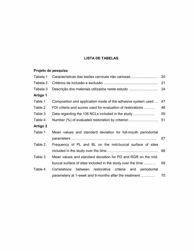
LISTA DE TABELAS
Projeto de pesquisa
Tabela 1 Características das lesões cervicais não cariosas .......................... 20
Tabela 2 Critérios de inclusão e exclusão ...................................................... 21
Tabela 3 Descrição dos materiais utilizados neste estudo ............................. 24
Artigo 1
Table 1 Composition and application mode of the adhesive system used … 47
Table 2 FDI criteria and scores used for evaluation of restorations ……… 48
Table 3 Data regarding the 136 NCLs included in the study ……………… 50
Table 4 Number (%) of evaluated restoration by criterion …………………… 51
Artigo 2
Table 1 Mean values and standard deviation for full-mouth periodontal
parameters ……………………………………………………………...
67
Table 2 Frequency of PL and BL on the mid-buccal surface of sites
included in the study over the time ……………………………………
68
Table 3 Mean values and standard deviation for PD and RGR on the mid-
buccal surface of sites included in the study over the time ……….
69
Table 4 Correlations between restorative criteria and periodontal
parameters at 1-week and 6-months after the treatment …………
70

12
SUMÁRIO
1 Projeto de pesquisa ..................................................................................... 13
1.1 Introdução.................................................................................................... 13
1.2 Justificativa .................................................................................................. 15
1.3 Objetivo ....................................................................................................... 16
1.4 Hipótese nula .............................................................................................. 17
1.5 Materiais e métodos .................................................................................... 18
Referências ....................................................................................................... 26
2 Relatório do trabalho de campo ................................................................. 28
3 Artigo 1........................................................................................................... 29
4 Artigo 2........................................................................................................... 53
Conclusões ........................................................................................................ 71
Referencias ....................................................................................................... 72
Orçamento ......................................................................................................... 76
Cronograma ...................................................................................................... 79
Apêndices .......................................................................................................... 81
Anexos .............................................................................................................. 84
13
1 Projeto de pesquisa
1.1 INTRODUÇÃO
Conforme a estratégia de união, os sistemas adesivos atualmente disponíveis
podem ser classificados em convencionais e autocondicionantes. Enquanto os
sistemas adesivos convencionais requerem tratamento do substrato com ácido
fosfórico, lavagem, subsequente aplicação e infiltração de monômeros resinosos; os
sistemas adesivos autocondicionantes dispensam a etapa preliminar de
condicionamento e lavagem do substrato, em função da presença de monômeros
ácidos na composição, os quais efetuam desmineralização e infiltração,
simultaneamente (DE MUNCK et al., 2005; VAN MEERBEEK et al., 2003).
Sistemas adesivos simplificados contemplam a demanda por produtos com
poucas etapas, rapidez e facilidade de aplicação, porém devem ser empregados
mediante conhecimento de suas limitações (TAY; PASHLEY, 2003). Contudo, os
sistemas adesivos convencionais de três passos são considerados padrão-ouro em
termos de estabilidade de união; já que, de forma geral, os produtos simplificados
não correspondem aos melhores resultados em termos de efetividade de adesão
(PEUMANS et al., 2005a).
Na prática clínica, restaurações de lesões cervicais não cariosas (LCNCs)
representam verdadeiros desafios para a adesão, pelo fato de combinarem
determinadas condições desfavoráveis ao processo de união, dentre as quais,
substratos altamente mineralizados e fibras colágenas desnaturadas (PERDIGÃO,
2010). Ademais, ensaios clínicos envolvendo restaurações Classe V são
preferencialmente utilizados para avaliação da efetividade do sistema adesivo pelo
fato de não apresentam quaisquer retenções macromecânicas, diferentemente das
demais cavidades (VAN MEERBEEK et al.,1998).
Para avaliação do desempenho clínico de restaurações, recomenda-se ainda
longo tempo de acompanhamento. Entretanto, é possível estabelecer critérios que
constituirão falhas prematuras das restaurações em curtos períodos, incluindo desde

14
sensibilidade pós-operatória até a perda da restauração (HICKEL et al., 2007). De
acordo com as orientações da American Dental Association (2001), o desempenho
dos sistemas adesivos é considerado clinicamente aceitável, se houver retenção de,
pelo menos, 95% das restaurações após 6 meses de acompanhamento. Para
aumentar o índice de sucesso das restaurações alguns autores sugerem a criação
de formas adicionais de retenção (KIM et al., 2009); outros propõem o
condicionamento seletivo das margens de esmalte (PEUMANS et al., 2005b).
Apesar da existência de diversos estudos de acompanhamento de
restaurações Classe V, a maioria tem-se restringido à avaliação do desempenho de
produtos que diferem ora em marca comercial, ora em estratégia de união
(PEUMANS et al., 2005a). Todavia, deve-se considerar que não apenas os materiais
utilizados em si, mas a técnica empregada para isolamento do campo operatório é
um fator que pode exercer influência significativa nos resultados e, portanto, devem
ser investigados (DE MUNCK et al., 2005).
De forma geral, os métodos de controle de umidade mais conhecidos
consistem no isolamento absoluto, conseguido essencialmente através do emprego
de lençol de borracha, e no isolamento relativo, obtido com uso de rolos de algodão.
Além disso, um sugador de saliva pode ser associado em ambos os métodos.
Embora haja reconhecimento da superioridade do isolamento absoluto no controle
de umidade, relutância e dificuldade em utilizá-lo são relatos frequentes (GILBERT
et al., 2010). No entanto, um estudo clínico prospectivo demonstrou a ausência de
diferenças significativas entre o desempenho clínico de restaurações em dentes
posteriores realizadas através de diferentes técnicas de isolamento (RASKIN et al.,
2000).
Métodos alternativos já foram sugeridos a fim de proporcionar controle de
umidade e acesso ao limite cervical da cavidade sem, concomitantemente, gerar
lesões iatrogênicas ao periodonto (BLUNCK, 2001; OWENS, 2006; PEREZ, 2010).
Nota-se, inclusive, limitarem-se os estudos longitudinais envolvendo restaurações de
LCNCs a mencionar, sucintamente, a etapa de isolamento do campo operatório,
sem descrevê-la de forma detalhada.
15
1.2 JUSTIFICATIVA
Pelo fato de a umidade do campo operatório exercer influência no desempenho
clínico de restaurações, percebe-se a necessidade de investigar clinicamente seu
efeito através de dois métodos diferentes de isolamento. Este tema consiste num
assunto de comum interesse a pesquisadores, estudantes e profissionais de
Odontologia, principalmente daqueles que lidam diariamente com procedimentos
adesivos.
Deve-se ainda considerar que o desenvolvimento e acompanhamento de um
ensaio clínico randomizado, controlado, envolvendo a influência da técnica utilizada
para isolamento do campo operatório no desempenho longitudinal de restaurações
Classe V, poderá esclarecer lacunas existentes acerca do tema, além de servir como
referência para o avanço da prática clínica baseada em evidências.

16
1.3 OBJETIVO
O objetivo do presente ensaio clínico randomizado será avaliar os efeitos de
duas técnicas de isolamento do campo operatório no desempenho clínico de
restaurações Classe V.

17
1.4 HIPÓTESE NULA
A hipótese nula a ser testada é que não haverá diferença no desempenho
clínico de restaurações Classe V realizadas com duas técnicas diferentes de
isolamento do campo operatório.
18
1.5 MATERIAIS E MÉTODOS
1.5.1 Considerações éticas
O presente estudo faz parte do projeto de pesquisa intitulado “Ensaio clínico
randomizado comparando diferentes sistemas adesivos em restaurações de lesões
cervicais não cariosas”, que foi submetido à apreciação do Comitê de Ética em
Pesquisa da Faculdade de Odontologia da Universidade Federal de Pelotas
(FOUFPEL), recebendo parecer favorável à sua execução (093/2009) (Anexo A).
Ademais, o referido projeto foi encaminhado para avaliação do Conselho
Coordenador do Ensino, Pesquisa e Extensão (COCEPE) desta universidade, sendo
considerado aprovado (4.02.01.046).
1.5.2 Desenho experimental e cálculo amostral
Este ensaio clínico randomizado controlado será delineado e conduzido
conforme as orientações do Consolidated Standards of Reporting of Trials
(CONSORT) (ALTMAN et al., 2001).
O tamanho amostral terá como base um estudo clínico prévio (LOGUÉRCIO
et al., 2006). Após início do presente estudo, o tamanho da amostra será
recalculado, considerando poder de 80% e nível de significância de 5%.
1.5.3 Recrutamento e seleção dos pacientes
A estratégia de busca dos indivíduos interessados em participar deste
ensaio clínico será realizada através da divulgação do projeto por meio da exposição
de cartazes e distribuição de panfletos (Figura 1) na FOUFPEL. Além disso, os
alunos líderes de turma, os professores chefes de disciplina e os dentistas da rede
de saúde pública serão informados sobre o estudo, visando aumentar a
receptividade e a taxa de adesão dos pacientes.

19
Figura 1. Cartaz de divulgação do projeto.
Todos os pacientes que forem encaminhados ou, diretamente, procurarem
atendimento odontológico, apresentando diagnóstico de lesão cervical não cariosa,
serão agendados para exame de avaliação. Um aluno realizará contato telefônico
com tais pacientes e agendamento das consultas por ordem de disponibilidade dos
mesmos.
Inicialmente, a presença de LCNCs será verificada através de inspeção com
auxílio de uma espátula de madeira. Em caso afirmativo, será preenchido um
prontuário do paciente, contendo dados de identificação, anamnese geral e
odontológica. Ademais, odontograma e periograma serão realizados, utilizando
espelho intrabucal, sonda exploradora, sonda periodontal milimetrada, pinça clínica,
rolos de algodão e sugador de saliva. A partir do exame clínico, as LCNCs serão
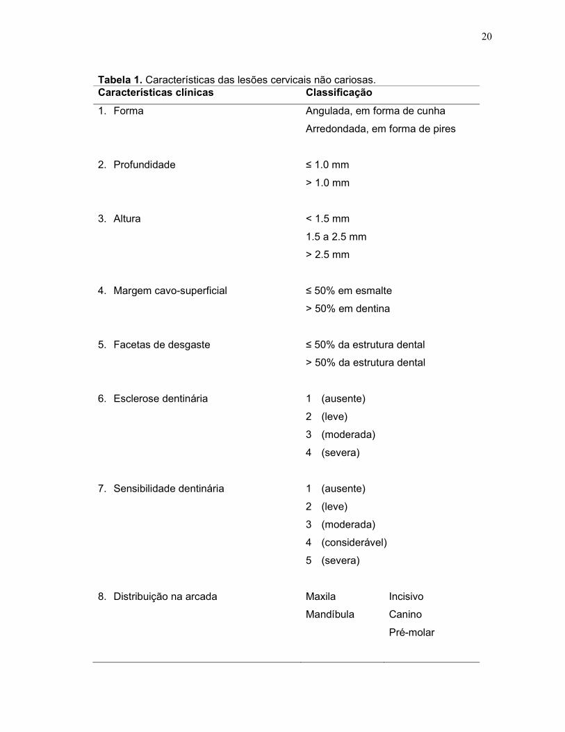
classificadas segundo avaliação de suas características (Tabela 1).
A sensibilidade dentinária será avaliada através da aplicação de jato de ar
na superfície vestibular. O grau de sensibilidade será registrado a partir de uma
escala visual analógica (Figura 2). A vitalidade pulpar será verificada através da
aplicação de spray refrigerante.
20
Tabela 1. Características das lesões cervicais não cariosas. Características clínicas Classificação
1. Forma Angulada, em forma de cunha
Arredondada, em forma de pires
2. Profundidade ≤ 1.0 mm
> 1.0 mm
3. Altura < 1.5 mm
1.5 a 2.5 mm
> 2.5 mm
4. Margem cavo-superficial ≤ 50% em esmalte
> 50% em dentina
5. Facetas de desgaste ≤ 50% da estrutura dental
> 50% da estrutura dental
6. Esclerose dentinária 1 (ausente)
2 (leve)
3 (moderada)
4 (severa)
7. Sensibilidade dentinária 1 (ausente)
2 (leve)
3 (moderada)
4 (considerável)
5 (severa)
8. Distribuição na arcada Maxila
Mandíbula
Incisivo
Canino
Pré-molar

21
Sensibilidade Nenhuma Leve Moderada Considerável Severa
Escores 1 2 3 4 5
Figura 2. Escala visual analógica.
Os pacientes que se enquadrarem nos critérios de inclusão e exclusão
(Tabela 2) receberão uma carta de informação sobre a natureza e proposição do
estudo (Apêndice A). Após a leitura, será solicitado que os voluntários assinem um
termo de consentimento livre e esclarecido (Apêndice B).
Tabela 2. Critérios de inclusão e exclusão.
Critérios
Inclusão � Presença de, no mínimo, duas lesões cervicais não cariosas na
face vestibular de dentes vitais anteriores ou pré-molares;
Exclusão � Fumantes;
� Pacientes com condição de saúde geral comprometida;
� Pacientes em tratamento ortodôntico;
� Pacientes com problemas oclusais;
� Presença de menos de 20 dentes em boca;
� Ausência de dente antagonista;
� Facetas de desgaste superior a 50% da estrutura incisal ou
oclusal;
� Presença de caries ou restaurações na face vestibular;
� Índice de placa visível ou índice de sangramento gengival
superior a 20%;
� Profundidade de sondagem e nível de inserção clínica igual ou
superior a 4 mm em qualquer sítio bucal;
� Pacientes sem disposição para comparecer às consultas de
reavaliação;
� Pacientes que se recusarem a participar do estudo.
Quatro semanas antes do início do estudo, os pacientes que necessitarem
serão submetidos a sessões de raspagem e polimento supragengival. Além disso,
22
eles receberão instrução para controle mecânico de placa, incluindo orientação
quanto à técnica de escovação e uso de fio dental. Durante o período de
acompanhamento, também será oferecido suporte odontológico aos pacientes
envolvidos no estudo.
1.5.4 Treinamento dos operadores
Seis candidatos a operadores (alunos da FOUFPEL) participarão de um
processo de treinamento para assegurar a padronização dos procedimentos clínicos
e minimizar as variações inerentes a diferentes operadores.
Primeira etapa: Será ministrada uma aula teórica, com duração de
aproximadamente 2 horas, consistindo na apresentação dos materiais e técnicas
disponíveis para controle de umidade no campo operatório, bem como restauração
de lesões cervicais não cariosas. Também será realizada exposição detalhada da
rotina a ser instituída durante os atendimentos clínicos. Um manual, contendo as
instruções de uso dos materiais e o protocolo dos procedimentos, será
disponibilizado aos alunos.
Segunda etapa: Os alunos passarão por atividades pré-clínicas, assistindo à
demonstração dos procedimentos operatórios e, posteriormente, realizando
restaurações Classe V em manequins. Num segundo momento, eles realizarão os
mesmos procedimentos em voluntários que, embora apresentem LCNCs com
necessidade restauradora, não serão incluídos neste estudo. Tais pacientes
receberão tratamento sob condições idênticas aos pacientes envolvidos no estudo,
porém não farão parte da amostra.
Ao final das etapas de treinamento, as funções da equipe de trabalho serão
delegadas, utilizando o desempenho individual dos alunos como critério de seleção.
Serão escolhidos dois operadores, que efetuarão os procedimentos operatórios, e
dois auxiliares para apoio dos operadores e preenchimento dos prontuários. Os
demais alunos ficarão encarregados da esterilização dos instrumentais,
agendamento das consultas, registro fotográfico, orientação de higiene bucal,
tratamento periodontal, dentre outros procedimentos odontológicos oferecidos aos
pacientes. Todas as etapas acima mencionadas serão realizadas sob supervisão
direta dos responsáveis pelo estudo.
1.5.5 Protocolo clínico

23
Inicialmente, a profilaxia do elemento a ser restaurado será feita com taça de
borracha e pasta a base de pedra-pomes e água. Em seguida, a cor da restauração
será selecionada com auxílio de uma escala de cores (Vitapan Classical, Vita
Zahnfabrik, Bad Sackingen, Alemanha). As LCNCs serão randomizadas em dois
grupos de acordo com o método de isolamento a ser efetuado. O processo de
randomização foi realizado por um membro da equipe não envolvido diretamente
nos procedimentos operatórios, nem na avaliação clínica das restaurações. Enfatiza-
se, ainda, que cada operador realizará o mesmo número de intervenções.
O isolamento relativo do campo operatório será realizado através do uso de
afastador labial, fio retrator #000 (Pro Retract, FGM, Joinville, SC, Brasil), roletes de
algodão e sugador de saliva. O primeiro elemento a ser introduzido na cavidade
bucal será o afastador labial, imprimindo afastamento de lábios e bochechas. Os
rolos de algodão serão posicionados no sulco vestibular superior, no sulco vestibular
inferior e na região sublingual, a fim de absorver o fluxo salivar proveniente,
principalmente, das glândulas salivares maiores. O fio retrator será inserido no
interior do sulco gengival com auxílio de espátula romba, sem gerar pressão
excessiva no periodonto. O isolamento absoluto do campo operatório consistirá na
utilização de lençol de borracha, arco de Young, grampo #212 (SS White-Duflex, Rio
de Janeiro, RJ, Brasil) e sugador de saliva. Uma pinça porta-grampo será utilizada
para distender e levar o grampo até a posição desejada, enquanto um perfurador
será empregado para realização de orifícios no lençol de borracha. Godiva de baixa
fusão e amarrias com fio dental serão utilizadas como dispositivos auxiliares para
estabilização do grampo e do lençol de borracha, respectivamente. Com a mesma
finalidade, outros modelos de grampos poderão ser posicionados na região mais
distal da arcada.
Previamente à execução da restauração, não será realizado nenhum tipo de
preparo cavitário, nem biselamento das margens da cavidade. Ambos os
procedimentos restauradores serão realizados com sistema adesivo convencional
(Adper Scotchbond Multi-Uso, 3M ESPE, St. Paul, MN, USA) e compósito
restaurador nanoparticulado (Filtek Z350, 3M ESPE, St. Paul, MN, USA), seguindo
rigorosamente as instruções de uso fornecidas pelo fabricante (Tabela 3). As
restaurações serão confeccionadas pela técnica incremental, utilizando
aproximadamente 2 ou 3 incrementos de compósito restaurador, conforme o
tamanho das LCNCs. Os incrementos serão levados e adaptados à cavidade com

24
espátulas, pinceis e pontas siliconadas para resina composta. Um aparelho LED
(Radii-Call, SDI, Bayswater, VI, Australia) será utilizado para fotoativação.
Finalmente, o acabamento das restaurações será realizado através da
utilização de pontas diamantadas de granulação fina e brocas multilaminadas, a fim
de remover excessos de material e/ou aperfeiçoar a forma de contorno das
restaurações. O polimento das mesmas será realizado com emprego de pontas
siliconadas, discos flexíveis de lixa (Sof-Lex Pop-On, 3M ESPE, St. Paul, MN, USA),
discos de feltro e pastas específicas para polimento.
Tabela 3. Descrição dos materiais utilizados neste estudo. Nome Fabricante Categoria Instruções de uso
Adper
Scotchbond
Multi-Uso
3M ESPE Sistema adesivo
convencional
1. Aplique o ácido fosfórico
em esmalte e dentina por
15 s.
2. Enxágue a superfície
condicionada por 15 s.
3. Aplique ar por 5 s.
4. Aplique o primer às
superfícies condicionadas
de dentina.
5. Seque levemente por 5 s.
6. Aplique o adesivo às
superfícies de esmalte e
dentina.
Filtek Z350 3M ESPE Resina composta
fotopolimerizável
1. Aplique incrementos de,
no máximo, 2 mm de
espessura.
2. Fotopolimerize cada
incremento por, no
mínimo, 20 s.

25
1.5.6 Avaliação das restaurações e análise estatística
Dois examinadores (professores da FOUFPEL, possuindo título de mestrado
e doutorado em Odontologia) passarão por um processo de treinamento e calibração
até que apresentem índice de concordância de, no mínimo, 80%. Caso ocorra
divergência quanto aos critérios de avaliação, os mesmos terão que entrar num
consenso através da reavaliação direta das restaurações e/ou por meio das
fotografias digitais. O registro fotográfico será feito antes da confecção da
restauração, bem como em cada período de avaliação das restaurações.
Após a etapa de treinamento e calibração, os avaliadores cegos, ou seja, sem
envolvimento algum com as condições clínicas as quais os pacientes foram
submetidos, procederão, independentemente, às avaliações das restaurações.
Neste momento, os avaliadores deverão utilizar espelho intrabucal, sonda
exploradora, sonda periodontal milimetrada, pinça clínica, rolos de algodão e
sugador de saliva. Dados referentes à sensibilidade dentária e vitalidade pulpar
também serão coletados através da aplicação de jato de ar e spray refrigerante,
respectivamente.
As avaliações serão realizadas em 1 semana (baseline) e 6 meses após a
inserção das restaurações, considerando os critérios clínicos aprovados pela FDI
World Dental Federation (HICKEL et al., 2007).
Os dados serão submetidos à análise estatística, considerando poder de 80%
e nível de significância de 5%.

26
REFERÊNCIAS
ADA Council on Scientific Affairs. Revised American Dental Association acceptance program guidelines: dentin and enamel adhesives. Chicago: American Dental Association, 2001. 9p.
ALTMAN, D. G.; SCHULZ, K. F.; MOHER, D.; EGGER, M.; DAVIDOFF, F.; ELMOUBE, D.; GOTZSCHE, P. C.; MA, T. L. The revised CONSORT statement for reporting randomized trials: explanation and elaboration. Annals of Intern Medicine, v.134, n.8, p.663-694, 2001. BLUNCK, Uwe. Improving cervical restorations: a review of materials and techniques. The Journal of Adhesive Dentistry, v.3, n.1, p.33-44, 2001. DE MUNCK, J.; VAN LANDUYT, K.; PEUMANS, M.; POITEVIN, A.; LAMBRECHTS, P.; BRAEM, M.; VAN MEERBEEK, B. A critical review of the durability of adhesion to tooth tissue: methods and results. Journal of Dental Research, v.84, n.2, p.118-132, 2005. HICKEL, R.; ROULET, J. F.; BAYNE, S.; HEINTZE, S. D.; MJOR, I. A.; PETERS, M.; ROUSSON, V.; RANDALL, R.; SCHMALZ, G.; TYAS, M.; VANHERLE, G. Recommendations for conducting controlled clinical studies of dental restorative materials. The Journal of Adhesive Dentistry, v.9, n.1, p.121-147, 2007.
GILBERT, G. H.; LITAKER, M. S.; PIHLSTROM, D. J.; AMUNDSON, C. W.; GORDAN, V. V. for The DPBRN Collaborative Group. Rubber dam use during routine operative dentistry procedures: findings from the Dental PBRN. Operative Dentistry, v. 35, n.5, p.491-499, 2010.
KIM, S. Y.; LEE, K. W.; SEONG, S. R.; LEE, M. A.; LEE, I. B.; SON, H. H.; KIM, H. Y.; OH, M. H.; CHO, B. H. Two-year clinical effectiveness of adhesives and retention form on resin composite restorations of non-carious cervical lesions. Operative Dentistry, v.34, n.5, p.507-515, 2009.
LOGUERCIO, A. D.; COSTENARO, A.; SILVEIRA, A. P.; RIBEIRO, N. R.; ROSSI, T. R.; REIS, A. A six-month clinical study of a self-etching and an etch-and-rinse adhesive applied as recommended and after doubling the number of adhesive coats. The Journal of Adhesive Dentistry, v.8, n.4, p.255-261, 2006. OWENS, Barry. Alternative rubber dam isolation technique for the restoration of class V cervical lesions. Operative Dentistry, v.31, n.2, p.277-280, 2006.

27
PERDIGÃO, Jorge. Dentin bonding - variables related to the clinical situation and the substrate treatment. Dental Materials, v.26, n.2, p.e24-e37, 2010.
PEREZ, Cesar Reis. Alternative technique for class V resin composite restorations with minimum finishing/polishing procedures. Operative Dentistry, v.35, n.3, p.375-379, 2010. PEUMANS, M.; KANUMILLI, P.; DE MUNCK, J.; VAN LANDUYT, K.; LAMBRECHTS, P.; VAN MEERBEEK, B. Clinical effectiveness of contemporary adhesives: a systematic review of current clinical trials. Dental Materials, v.21, n.9, p.864-881, 2005a.
PEUMANS, M.; DE MUNCK, J.; VAN LANDUYT, K.; LAMBRECHTS, P.; VAN MEERBEEK, B. Three-year clinical effectiveness of a two-step self-etch adhesive in cervical lesions. European Journal of Oral Science, v.113, n.6, p.512-518, 2005b.
RASKIN, A.; SETCOS, J. C.; VREVEN, J.; WILSON, N. H. F. Influence of the isolation method on the 10-year clinical behaviour of posterior resin composite restorations. Clinical Oral Investigations, v.4, n.3, p.148-152, 2000.
TAY, F. R.; PASHLEY, D. H. Have dentin adhesives become too hydrophilic? Journal of the Canadian Dental Association, v.69, n.11, p. 726-731, 2003. VAN MEERBEEK, B.; DE MUNCK, J.; YOSHIDA, Y.; INOUE, S.; VARGAS, M.; VIJAY, P.; VAN LANDUYT, K.; LAMBRECHTS, P.; VANHERLE, G. Buonocore memorial lecture. Adhesion to enamel and dentin: current status and future challenges. Operative Dentistry, v.28, n.3, p.215-235, 2003. VAN MEERBEEK, B.; PERDIGÃO, J.; LAMBRECHTS, P.; VANHERLE, G. The clinical performance of adhesives. Journal of Dentistry, v.26, p.1-20, 1998.

28
2 Relatório do trabalho de campo
Após qualificação deste projeto de pesquisa sob portaria no 13 de 1º de abril de
2010, as seguintes modificações foram realizadas:
• Em vez de um sistema adesivo convencional de três passos (Adper Scotchbond
Multi-Uso, 3M ESPE, St. Paul, MN, USA), utilizou-se um adesivo
autocondicionante de passo único (Adper Easy One, 3M ESPE, Seefeld,
Germany). Esta alteração foi estimulada pela ausência de estudos clínicos
avaliando o desempenho desse material. As instruções de uso fornecidas pelo
fabricante foram seguidas durante sua aplicação.
• Em 2010, os critérios clínicos utilizados para avaliações de restaurações
(HICKEL et al., 2007) sofreram modificações significativas. Por esse motivo,
optou-se em utilizar a versão mais atualizada (HICKEL et al., 2010) para
avaliação das restaurações deste estudo.
• Ademais, verificou-se a necessidade de utilizar parâmetros periodontais para
avaliação dos efeitos do tipo de isolamento na condição periodontal.
• Inicialmente, o tamanho amostral foi baseado num ensaio clínico com tempo de
acompanhamento de 6 meses (LOGUÉRCIO et al., 2006). Em posse dos
resultados preliminares do presente estudo, o tamanho amostral foi recalculado
observando-se a diferença entre os grupos com relação ao desfecho principal
(perda da restauração), poder de 80% e nível de significância de 5%.
Considerando as 136 restaurações realizadas em 30 pacientes, estima-se que
diferenças estatisticamente significantes, a favor do grupo isolado com dique de
borracha, serão observadas após 4 anos de acompanhamento.
• Embora resultados preliminares sejam utilizados para defesa desta tese, as
avaliações clínicas permanecerão ocorrendo semestralmente ou enquanto
houver pacientes dispostos a retornar às consultas de reavaliação.
29
3 Artigo 1
Title page
Title: Six-month evaluation of noncarious cervical restorations placed under two isolation
methods: a randomized controlled clinical trial
Short title: Effects of two methods to isolate the operative field
Silvia T. Fontesa,*, Fernanda O. B. Corrêaa, Maximiliano S. Cencia, Patrícia S. Jardima,
Márcia B. Pintoa, Alexandre S. Masottia
a Graduate Program in Dentistry, Federal University of Pelotas, Pelotas, RS, Brazil
*Corresponding author: Silvia Terra Fontes, Faculdade de Odontologia, Universidade
Federal de Pelotas, Rua Gonçalves Chaves 457, 96015-560, Pelotas, RS, Brasil, phone:
+55 53 32224439, fax: +55 53 32224439, E-mail address: [email protected]
_________________________________
Este artigo foi formatado segundo as normas do periódico Dental Materials. Disponível em: <http://www.elsevier.com/wps/find/journaldescription.cws_home/601024/authorinstructions> Acesso em: 01 nov. 2011, 09:35:38.

30
Abstract
Objective: This randomized controlled clinical trial evaluated the effects of two methods of
isolating the operative field on the 6-month performances of cervical restorations. Methods:
Thirty patients with at least two noncarious cervical lesions (NCLs) were enrolled in the
study. A total of 136 NCLs were randomized into the following groups: (1) isolation
performed with rubber dam and gingival retraction clamp, and (2) isolation provided with
cotton rolls and gingival retraction cord. Both methods were used with a saliva suction device.
An all-in-one self-etching adhesive (Adper Easy One, 3M ESPE) and a nanofilled composite
resin (Filtek Z350, 3M ESPE) were used for both groups. The restorations were blindly
assessed one week (baseline) and six months after placement, using the FDI criteria. Clinical
performance was recorded in terms of material fracture and restoration retention, marginal
adaptation, marginal staining, postoperative hypersensitivity and preservation of tooth vitality.
Results: The recall rate at 6 months was 100%. Four restorations were lost over this period,
resulting in an overall failure rate of 2.9%. Marginal defects increased in the cotton roll group
(p = 0.01). However, no significant differences were detected among groups regarding any of
the other evaluated criteria (p > 0.05). Significant decrease in sensitivity was observed
between the pre and postoperative measurements (p < 0.001). Significance: Both isolation
methods provided acceptable clinical performance of noncarious cervical restorations. After 6
months of clinical service, the overall restorations meet the ADA criteria for provisional
acceptance, regardless of the type of isolation used for restorations’ placement.
Keywords: adhesives; composite resins; controlled clinical trial; dental restoration; noncarious
cervical lesions; rubber dam, self-etch.
31
1 Introduction
Self-etch adhesives (SEA) use non-rinse acidic monomers that demineralise and
infiltrate the dental substrate simultaneously to the same depth [1]. Despite current trends
towards adhesives with simplified application procedures, an inefficient clinical performance
has been noted for the most tested one-step SEAs [2]. Fortunately, the latest generation of the
single-bottle or all-in-one adhesives definitely seems to perform better. Nevertheless, the
outcomes of bonded restorations not only depend on the adhesive category per se but also on
operative aspects, such as the method used to isolate the operative field from the rest of the
oral cavity [3]. It is generally accepted that adhesive restorations must be placed under well-
controlled moisture conditions [4], which suggests that the rubber dam is the ideal method to
accomplish this in routine restorative dentistry. Although a meta-analysis revealed no
influence of the isolation type on the clinical performance of cervical restorations [5], few
studies have addressed this important aspect. Furthermore, no randomized clinical trial (RCT)
has been designed specifically to evaluate the role of isolation method on the longevity of
restorations bonded with the newest generation of simplified SEAs.
Thus, the aim of this randomized controlled trial was to evaluate the effects of two
isolation methods of the operative field on the clinical performance of noncarious cervical
restorations. This study also provided information about the short-term clinical effectiveness
of a new all-in-one self-etch adhesive to restore noncarious cervical lesions (NCL). The null
hypothesis to be tested is that no significant differences among clinical performance of
restorations placed under different isolation methods will be detected after a 6-month follow-
up.

32
2 Materials and Methods
2.1 Study design and ethics considerations
This study was designed as a split-mouth, single-blind (clinical evaluators blinded),
prospective and randomized controlled clinical trial. NCLs were randomly assigned by two
different treatment groups, according to the method used to isolate the operative field. The
sample size was based on information from a previous 6-month clinical trial, which placed a
total of 116 restorations in 29 patients [6]. The protocol and the consent form for this study
were approved by the local ethics committee on investigations involving human subjects (#
093/2009). Before beginning the experiment, the participants signed a written informed
consent agreement to participate in the trial.
2.2 Patient and lesion selection
Recruitment of subjects was performed at the clinics of School of Dentistry (Federal
University of Pelotas, Pelotas, RS, Brazil) through advertisements, considering patients who
needed dental treatment of NCLs. Reasons for treatment were cervical tooth sensitivity,
aesthetic complaints and/or prevention of tooth further damage.
The eligibility criteria for volunteers were being at least 18 years old and presenting
more than one cervical lesion whose apical limit wasn’t located below the gingival margin,
with at least 1 mm of depth in a vital permanent incisor, canine, or premolar of the upper or
the lower jaw. Typically, these defects were situated at the facial surface of the teeth,
sometimes with a small part extending interproximally.
They were not admitted when any of the following criteria were presented: (1)
smoking habits; (2) severe systemic diseases; (3) active orthodontic treatment; (4)
malocclusion (Angle Class II or Class III); (5) less than 20 natural teeth in mouth; (6) absent
33
of antagonist tooth; (7) wear facets over more than 50% of the incisal/occlusion surface as a
result of tooth attrition; (8) caries or restorations in the area to be treated; (9) full-mouth
visible plaque index (VPI) or full-mouth gingival bleeding index (GBI) more than 20% [7];
(10) probing depth (PD) and clinical attachment level (CAL) values equal or greater than 4
mm with bleeding on probing (BOP); and (11) unwillingness to return for follow-ups or (12)
refuse to participate.
The screening of lesions was performed using a mouth mirror, an explorer, and a
periodontal probe (University of North Carolina, Hu-Friedy, Chicago, IL, USA). The NCLs
depths were measured by placing a probe into their deepest part, and their heights were
calculated by the distance of the most coronal and apical points of the cavity margins. The
degrees of dentin sclerosis were identified using a scale ranked from 1 to 4 [8]. Sensitivity
was measured by blowing a stream of compressed air for 3 s at a distance of 2 to 3 cm, while
shielding the adjacent teeth with fingers. Tooth vitality was tested by the application of an ice
stick on the tooth and comparing the reaction with that of the adjacent teeth. No attempt was
made to determine the aetiology of the cervical lesions.
Four weeks before the study began, the patients underwent a session of dental scaling
and polishing by a single operator using periodontal manual curettes (Gracey and McCall,
Trinity, São Paulo, SP, Brazil). They also received detailed oral hygiene instructions,
including a non-traumatic brushing technique (coronally directed roll technique) with a soft
toothbrush [9], and the use of the dental floss.
2.3 Random assignment
A preset random table was used to generate the random allocation sequence of the
treatment groups among participants. While the first randomly selected treatment was used for
the lowest quadrant number, the second treatment was used for the tooth with the second-
lowest quadrant number (according to the FDI system). This method was repeated for every

34
other quadrant that required a cervical restoration. In instances of an uneven number of NCLs
per patient, the unequal number of lesions of one group was adjusted by restoring one more
lesion in the other group in the next patient presenting with an unequal number of cervical
lesions. Opaque, sealed envelopes were employed to conceal the sequence until interventions
were assigned.
2.4 Interventions
The operative procedures were performed by two trained, skilled operators familiar
with adhesive restorative dentistry, under the supervision of an experienced clinician. They
received thorough pre-clinical training in the field isolation and adhesive procedures. Each
operator placed an equal number of restorations for each group.
Preoperatively, the teeth to be restored were cleaned with pumice and water in a
rubber prophylaxis cup. Subsequently, the colour of the restoration was determined using a
shade guide (Vitapan Classical, Vita, Zahnfabrik, Bad Sackingen, Germany). No additional
mechanical retention or enamel bevel was prepared.
In order to secure moisture control of the operative field, two isolation methods were
standardized by a detailed protocol, which is briefly summarized below.
(1) Rubber dam group: Moisture control was provided by a rubber dam and a gingival
retraction clamp placed in the cervical area of the tooth.
(2) Cotton roll group: Moisture control was provided using a labial retractor, cotton rolls
and gingival retraction cord placed into the gingival sulcus.
For both groups, a saliva suction device was held in position by an assistant during the
restorative procedure. If necessary, local anaesthesia was given to prevent patient discomfort
prior to treatment.

35
An all-in-one self-etch adhesive (Adper Easy One, 3M ESPE, Seefeld, Germany) was
used according to the manufacturer’s instructions (Table 1). The NCLs were restored with a
direct restorative nanocomposite (Filtek Z350, 3M ESPE, Irvine, CA, USA) applied in at least
two increments (not exceeding 2 mm in thickness), using a selected composite instrument
(Hu-Friedy, Chicago, IL, USA). Each increment was cured for 20 s with a LED light-curing
unit (Radii-Call, SDI, Bayswater, VI, Australia). All restorations were finished and polished
with fine- and ultra-fine-grain diamond burs (KG Sorensen, Barueri, SP, Brazil) under water
cooling, slow-speed flexible discs (Sof-Lex Pop-On, 3M ESPE, St Paul, MN, USA), polishing
paste (Prisma Gloss, Dentsply Caulk, Milford, DE, USA), and rubber points (Enhance,
Dentsply Caulk, Milford, DE, USA).
2.5 Clinical assessment
Criteria approved by the FDI World Dental Federation were used for clinical
assessment of restorations [10]. The primary clinical outcome was material fracture and
restoration retention. Secondary endpoints included the following criteria: (1) marginal
adaptation, (2) marginal staining, and (3) postoperative hypersensitivity and preservation of
tooth vitality. Each criterion was expressed with five scores, 3 for acceptable and 2 for
unacceptable (1 for reparable and 1 for replacement). Restorations that needed repairs or
replacements were considered clinical failures, receiving scores of 4 or 5, respectively (Table
2).
The evaluations were carried out by two independent examiners at 1 week (baseline)
and 6 months after the insertion of the restoration. They were not the operators and were fully
blinded to the assignment of interventions. A Web-based training and calibration tool called
e-calib (www.ecalib.info) was used to train and calibrate the evaluators. After that, they
evaluated cervical restorations in clinical settings. A pre-evaluation agreement of at least 80%

36
was obtained among them. When disagreement in evaluation occurred between the two
examiners, consensus was reached by immediate re-examination and discussion at chair side.
Photo documentation was made pre-operatively, at baseline and at recall.
2.6 Statistical analysis
Data were analyzed using the statistical software program SigmaStat (Version 3.5,
Systat, Richmond, CA, USA). Descriptive statistics were used to describe the frequency
distributions of the evaluated criteria. For each variable, the comparison between the groups
(the rubber dam group and cotton roll group) was performed with the Mann-Whitney Rank
Sum test. The Wilcoxon sign-ranked test was used to compare the changes across the periods
(baseline and 6 months). The significance level of 0.05 was adopted for all statistical analyses.

37
3 Results
3.1 Baseline data
During the recruiting period of March to December 2010, 80 subjects were assessed
for eligibility. Thirty patients (eight men and 22 women) were enrolled in the study, yielding
a total of 136 restorations (68 restorations per group). The age of the included patients varied
from 19 to 69 years (mean age of 48.5 ± 11.3 years). The average number of restorations per
patient was four. NCLs included in the study were pre-operatively categorized in terms of
tooth distribution, shape and height of the lesions, degree of sclerosis, and sensitivity (Table
3). No statistically significant differences were detected among groups for any of these
characteristics (p > 0.05). Seventy-five teeth (55.1%) were restored in the upper jaw. Most
restorations (72%) were placed on pre-molars. About 60% of all the lesions were pre-
operatively sensitive to air. The flow diagram indicates the number of participants through
each stage of the trial (Figure 1).
3.2 Evaluation results
Table 4 summarizes the evaluation criteria at baseline and at follow-up for both
groups. The overall recall rate at 6 months was 100%. One restoration from the rubber dam
group and three restorations from the cotton roll group were lost at 6 months, resulting in an
overall failure rate of 2.9%. With regard to marginal adaptation, the cotton roll group showed
a significant increase between the baseline and the 6-month findings (p = 0.01). This group
also presented an increase in marginal staining; however, the difference was not statistically
significant (p = 0.055). No significant differences were detected among groups for any of the
other evaluated criteria (p > 0.05). Considering the sensitivity to air stimulus, a significant
decrease was observed between the pre- and the postoperative measurements for all restored
teeth (p= 0.001).

38
4 Discussion
The most common methods to isolate the operative field include rubber dam and
cotton rolls, both frequently combined with saliva suction device. The literature emphasises
that resin-composite restorations cannot be placed successfully in a cavity surface that is
contaminated by blood or saliva [4], especially for restorations whose cervical margin is in
direct contact with the periodontal tissues (e.g., noncarious class-V lesions). Beyond moisture
control, the protection of the patient from possible aspiration and ingestion of dental foreign
objects is an advantage only offered by the use of the rubber dam in dental practise [11].
Meanwhile, most clinicians are not sure which isolation method to choose and also show
reluctance to use the rubber dam during the operative dentistry procedures [12]. According to
a 10-year clinical study, similar performances were reported for posterior restorations placed
under cotton rolls and rubber dam isolation [13]. Nevertheless, it is well recognised that only
studies involving cervical restorations should be considered to investigate adhesion
effectiveness, for a number of reasons previously discussed in the literature [14].
While the current trends favour simpler and faster clinical application steps, a
systematic review of clinical trials reported that the most common SEAs do not seem to meet
the expectations regarding bonding performance [2]. However, until now no peer-reviewed
clinical study has attempted to evaluate the performance of Adper Easy One Self-Etch
Adhesive, especially considering the influence of the isolation method on its clinical outcome.
This self-etch adhesive can be considered as the true all in one, due to the combination of all
the components into one single solution that does not require additional mixing. With regard
to the pH and the interaction depth of such solution at dentin, it has been also referred as an
ultra-mild self-etch approach (pH > 2.5) [1]. Although manufacturers introduced stronger
SEAs (pH ≤ 1.0) some years ago, serious problems (i.e., the hydrolytic instability of

39
methacrylates and intrinsic acid-based reaction of components) have apparently pushed them
to an ultra-mild self-etch approach [15].
Despite the limitations of a 6-month follow-up period, the present findings clearly
showed favourable results for the above-mentioned simplified adhesive regardless the method
used to isolate the operative field. At 6 months, only four restorations were scored as
clinically unacceptable (2.9% failure rate) due to the debonding of one restoration of the
rubber dam group and three restorations of the cotton roll group. Taking the American Dental
Association’s guidelines as reference, resin-based enamel-dentin adhesives gain ‘provisional
acceptance’ at 6 months if their retention loss in NCLs is less than 5% without mechanical
retention features [16]. In addition to the loss of restoration, another criterion that could
constitute an early failure at 6 months is severe postoperative hypersensitivity [17]. However,
both groups performed equally well without reporting abnormal postoperative
hypersensitivity after restoration. Yet, the frequency of tooth sensitivity to air stimulus was
reduced from 60% to approximately 11% at 1 week and 5% at 6 months. In part, this
favourable clinical benefit can be attributed to the less aggressive and more superficial
interaction of an ultra-mild adhesive with dentin [1].
Taking into account that the sealing capacity of restorations has often been assessed by
the integrity and colour changes along part or all of the margins [17], clinical signs of the
degradation of bonded interfaces were observed in restorations placed under the cotton roll
method. This group exhibited increased small marginal defects (especially at enamel margins)
and a slightly marginal staining at follow-up compared to the baseline evaluation. However,
findings were considered clinically acceptable, which suggests that the isolation method may
have played an important role in the early deterioration of marginal integrity. Additionally,
these minor shortcomings could be attributed to the superficial etching pattern of an ultra-mild
self-etch adhesive. A similar phenomenon of increased marginal defects and superficial

40
discoloration was also observed in a long-term clinical trial evaluating a mild two-step self-
etch adhesive [18]. That is why the literature so far indicates selective phosphoric-acid
etching of the enamel cavity margins, followed by applying an ultra-mild SEA. The purpose
of this combined approach is to provide a better self-etch interaction at enamel with
favourable perspectives at dentin [1].
Furthermore, maybe the main challenge for dental adhesives is to provide an equally
effective bond to hard tissues of a different nature. Noncarious class-V lesions exhibit
margins located in enamel, as well as in dentin, high degrees of sclerosis, heterogeneous
hyper-mineralized layer, and denatured collagen, seem to make difficult the bonding in such
clinically relevant substrates [19–21]. Nevertheless, lesions were not excluded from the
screening based on the proportion of margin involving enamel and dentin, nor on the degree
of dentin sclerosis. On the other hand, the authors recognize that some specific habits of
patients, such as poor oral hygiene, smoking, or bruxism, may influence the clinical outcomes
[17]. For this reason, patients presenting severe wear facets were excluded from the present
study. It was assumed that wear facets indicate a higher concentration of occlusal loads on the
area, contributing to a higher debonding rate for restorations. Then again, this exclusion
criterion prevents us from extrapolating our results to patients with parafunctional disorders.
With regard to the sample size, it was initially based on information from a previous 6-
month clinical study [6], although the sample size was recalculated at the 6-months follow-up,
taking into account the difference between groups with regard to our primary outcome (the
restoration retention). Thus, it is expected that a significant difference in retention rates
favouring the isolation method with a rubber dam will be detected over a period of 4 years,
considering a power of 0.80 and a type I error of 0.05.
Based on the results of the present investigation, the null hypothesis was accepted,
since no significant differences were detected among clinical performances of restorations

41
placed under different methods of isolation after a period of 6 months. However, the present
findings must be interpreted with caution, considering the short-term follow-up.

42
5 Conclusion
Within the period of 6 months, noncarious cervical restorations placed with both
isolation methods were equally successful. Isolation with cotton rolls had only some negative
effects on secondary clinical criteria, such as a progressive incidence of small marginal
defects. However, further long-term follow-up is needed to confirm the early effectiveness of
this all-in-one SEA with regard to the method used to isolate the operative field.
43
Acknowledgements
This paper is based on a thesis submitted to the Graduate Program in Dentistry, Federal
University of Pelotas, in partial fulfillment of the requirements for the first author’s PhD
degree. Fontes ST held a PhD scholarship from the National Council for Scientific and
Technological Development (CNPq) during this study.
The authors also thank the staff members, who were fundamental in the conduct of this
clinical study, especially Ângelo Niemczewski Bobrowski and Ronaldo Luiz Rossi for
placing all the composite restorations.

44
References
[1] Van Meerbeek B, Yoshihara K, Yoshida Y, Mine A, De Munck J, Van Landuyt KL. State
of the art of self-etch adhesives. Dent Mater, 2011; 27(1):17-28.
[2] Peumans M, Kanumilli P, De Munck J, Van Landuyt K, Lambrechts P, Van Meerbeek B.
Clinical effectiveness of contemporary adhesives: a systematic review of current clinical
trials. Dent Mater, 2005; 21(9):864-881.
[3] Demarco FF, Corrêa MB, Cenci MS, Moraes RR, Opdam NJ. Longevity of posterior
composite restorations: Not only a matter of materials. Dent Mater (2011) in press.
[4] ADA Council on Scientific Affairs. Direct and indirect restorative materials. J Am Dent
Assoc, 2003; 134(4):463-472.
[5] Heintze SD, Ruffieux C, Rousson V. Clinical performance of cervical restorations - a
meta-analysis. Dent Mater, 2010; 26(10):993-1000.
[6] Loguercio AD, Costenaro A, Silveira AP, Ribeiro NR, Rossi TR, Reis A. A six-month
clinical study of a self-etching and an etch-and-rinse adhesive applied as recommended
and after doubling the number of adhesive coats. J Adhes Dent, 2006; 8(4):255-261.
[7] Ainamo J, Bay I. Problems and proposals for recording gingivitis and plaque. Int Dent J,
1975; 25(4):229-235.
[8] Ritter AV, Heymann HO, Swift EJ Jr, Sturdevant JR, Wilder AD Jr. Clinical evaluation of
an all-in-one adhesive in non-carious cervical lesions with different degrees of dentin
sclerosis. Oper Dent, 2008; 33(4): 370-378.
[9] Santos VR, Lucchesi JA, Cortelli SC, Amaral CM, Feres M, Duarte PM. Effects of glass
ionomer and microfilled composite subgingival restorations on periodontal tissue and
subgingival biofilm: a 6-month evaluation. J Periodontol, 2007; 78(8): 1522-1528.
45
[10] Hickel R, Peschke A, Tyas M, Mjör I, Bayne S, Peters M, Hiller KA, Randall R,
Vanherle G, Heintze SD. FDI World Dental Federation - clinical criteria for the evaluation
of direct and indirect restorations. Update and clinical examples. J Adhes Dent, 2010;
12(4):259-272.
[11] Tiwana KK, Morton T, Tiwana PS. Aspiration and ingestion in dental practice: a 10-
year institutional review. J Am Dent Assoc, 2004; 135(9):1287-1291.
[12] Gilbert GH, Litaker MS, Pihlstrom DJ, Amundson CW, Gordan VV. Rubber dam use
during routine operative dentistry procedures: findings from the Dental PBRN. Oper Dent,
2010; 35(5):491-499.
[13] Raskin A, Setcos JC, Vreven J, Wilson NH. Influence of the isolation method on the
10-year clinical behaviour of posterior resin composite restorations. Clin Oral Investig,
2000; 4(3):148-152.
[14] De Munck J, Van Landuyt K, Peumans M, Poitevin A, Lambrechts P, Braem M, Van
Meerbeek B. A critical review of the durability of adhesion to tooth tissue: methods and
results. J Dent Res, 2005; 84(2):118-132.
[15] Moszner N, Salz U, Zimmermann J. Chemical aspects of self-etching enamel-dentin
adhesives: a systematic review. Dent Mater, 2005; 21(10):895-910.
[16] ADA Council on Scientific Affairs. Revised American Dental Association acceptance
program guidelines: dentin and enamel adhesives. Chicago: American Dental Association;
2001. p. 1-9.
[17] Hickel R, Roulet JF, Bayne S, Heintze SD, Mjör IA, Peters M, Rousson V, Randall R,
Schmalz G, Tyas M, Vanherle G. Recommendations for conducting controlled clinical
studies of dental restorative materials. Clin Oral Investig, 2007; 11(1):5-33.
46
[18] Peumans M, De Munck J, Van Landuyt KL, Poitevin A, Lambrechts P, Van Meerbeek
B. Eight-year clinical evaluation of a 2-step self-etch adhesive with and without selective
enamel etching. Dent Mater, 2010; 26(12): 1176-1184.
[19] Karan K, Yao X, Xu C, Wang Y. Chemical profile of the dentin substrate in non-
carious cervical lesions. Dent Mater, 2009; 25(10):1205-1212.
[20] Perdigão J. Dentin bonding-variables related to the clinical situation and the substrate
treatment. Dent Mater, 2010; 26(2):e24-37.
[21] Tay FR, Pashley DH. Resin bonding to cervical sclerotic dentin: A review. J Dent,
2004; 32(3):173-196.

47
Tables and figures
Table 1. Composition and application mode of the adhesive system used.
Product Manufacturer Composition Application procedure
Adper Easy
One Self-Etch
Adhesive
lot: #384060,
pH ≈ 2.7
3M ESPE AG,
Seefeld,
Germany
Bis-GMA,
HEMA,
methacrylated
phosphoric
esters,
1,6-hexanediol
dimethacrylate,
methacrylate
functionalized
polyalkenoic
acid, water,
ethanol, silica
filler, initiators
based on CQ and
stabilizers.
1. Apply the adhesive with the
disposable applicator for 20 s
to all surfaces of cavity.
2. Rewet the disposable
applicator as needed during
application.
3. Then, air thin the liquid for
approximately 5 s until the
film no longer moves,
indicating complete
vaporization of the solvent.
4. Light cure the adhesive for
10 s.
Abbreviations: Bis-GMA, bisphenol A diglycidyl methacrylate; CQ, camphorquinone;
HEMA, 2-hydroxyethyl methacrylate.

48
Table 2. FDI criteria and scores used for evaluation of restorations.
Score 1. Material
fracture
and
retention
2. Marginal
adaptation
3. Marginal
staining
4. Postoperative
hyper-
sensitivity
and tooth
vitality
1. Clinical
excellent /
very good
1.1 No
fractures/
cracks.
2.1 Harmonious
outline, no gaps,
no white or
discolored lines.
3.1 No
marginal
staining.
4.1 No
hypersensitivity,
normal vitality.
2. Clinically
good
(polishing
necessary)
1.2 Small
hairline crack.
2.2.1 Marginal
gap (< 150 µm),
white lines.
2.2.2 Small
marginal
fracture.
2.2.3 Slight
ditching, slight
step/ flashes,
minor
irregularities.
3.2 Minor
marginal
staining, easily
removable by
polishing.
4.2 Minor
hypersensitivity
for a limited
period of time,
normal vitality.
3. Clinically
sufficient /
satisfactory
1.3 Two or
more or larger
hairline cracks
and/ or
material chip
fracture not
affecting the
marginal
integrity.
2.3.1 Gap < 250
µm not
removable.
2.3.2 Several
small marginal
fractures.
2.3.3 Major
irregularities,
ditching or
flash, steps.
3.3 Moderate
marginal
staining, not
aesthetically
unacceptable.
4.3.1 Moderate
hypersensitivity.
4.3.2 Delayed/
mild sensitivity.
No subjective
complaints, no
treatment needed.

49
4. Clinically
unsatisfactory
(repair
necessary)
1.4.1 Material
chip fractures
which damage
marginal
quality.
1.4.2 Bulk
fractures with
partial loss
(less than half
of the
restoration).
2.4.1. Gap > 250
µm or dentin/
base exposed.
2.4.2 Severe
ditching or
marginal
fractures.
2.4.3 Larger
irregularities or
steps (repair
necessary).
3.4
Pronounced
marginal
staining. Major
intervention
necessary for
improvement.
4.4.1 Intense
hypersensitivity.
4.4.2 Delayed
with minor
subjective
symptoms.
4.3.3 No clinical
detectable
sensitivity.
Intervention
necessary, but not
replacement.
5. Clinically
poor
(replacement
necessary)
1.5 (Partial or
complete) loss
of restoration
or multiple
fractures.
2.5.1
Restoration
(complete or
partial) is loose
but in situ.
2.5.2
Generalized
major gaps or
irregularities.
3.5 Deep
marginal
staining, not
accessible for
intervention.
4.5 Intense, acute
pulpitis or
nonvital tooth.
Endodontic
treatment is
necessary and
restoration has to
be replaced.

50
Table 3. Data regarding the 136 NCLs included in the study.
Characteristics Rubber
dam
group
Cotton
roll
group
Total (%)
Tooth distribution Upper incisor
Upper canine
Upper premolar
Lower incisor
Lower canine
Lower premolar
02
11
25
02
02
26
04
11
22
03
03
25
06 (4.4)
22 (16.1)
47 (34.6)
05 (3.7)
05 (3.7)
51 (37.5)
Shape of the lesion Sharply defined, wedge-shaped
Rounded, saucer-shaped
26
42
29
39
55 (40.4)
81 (59.6)
Height of the lesion <1.5 mm
1.5 - 2.5 mm
> 2.5 mm
22
26
20
21
24
23
43 (31.6)
50 (36.8)
43 (31.6)
Degree of sclerosis No sclerosis evident
Slightly
Moderately
Severe
46
17
02
03
42
20
04
02
88 (64.7)
37 (27.2)
06 (4.4)
05 (3.7)
Pre-operative
sensitivity
No
Yes
26
42
30
38
56 (41.2)
80 (58.8)
Abbreviations: NCLs, noncarious cervical lesions.

51
Table 4. Number (%) of evaluated restoration by criterion.
Criteria Score Baseline 6 months
Rubber dam
group (n= 68)
Cotton roll
group (n= 68)
Rubber dam
group (n= 67)
Cotton roll
group (n= 65)
Material
fracture and
retention
1
2
3
4
5
68 (100)
0
0
0
0
68 (100)
0
0
0
0
67 (98.5)
0
0
0
01 (1.5)
65 (95.6)
0
0
0
03 (4.4)
Marginal
adaptation
1
2
3
4
5
57 (83.8)
11 (16.2)
0
0
0
62 (91.2)
06 (8.8)
0
0
0
52 (77.6)
15 (22.4)
0
0
0
48 (73.9)
17 (26.1)
0
0
0
Marginal
staining
1
2
3
4
5
66 (97.1)
02 (2.9)
0
0
0
65 (95.6)
03 (4.4)
0
0
0
62 (92.5)
04 (6.0)
01 (1.5)
0
0
57 (87.7)
06 (9.2)
02 (3.1)
0
0
Postoperativ
e hyper-
sensitivity
and tooth
vitality
1
2
3
4
5
57 (83.8)
11 (16.2)
0
0
0
63 (92.6)
04 (5.9)
01 (1.5)
0
0
59 (88.0)
04 (6.0)
04 (6.0)
0
0
60 (92.3)
03 (4.6)
02 (3.1)
0
0
Scores: 1. Clinical excellent / very good; 2. Clinically good; 3. Clinically sufficient /
satisfactory; 4. Clinically unsatisfactory; 5. Clinically poor. Regarding the variables ´marginal
adaptation`, ´marginal staining` and ´postoperative hyper-sensitivity and tooth vitality` only
retained restorations were considered.

52
Figure 1. Flowchart of the study participants.
Abbreviations: CAL, clinical attachment level; GBI, gingival bleeding index; Np, number of
patients; Nr, number of restorations; PD, probing depth; VPI, visible plaque index.

53
4 Artigo 2 Title page
Effect of two gingival retraction techniques on periodontal health: a 6-month randomized
controlled clinical trial
Silvia T. Fontes, MSc; Patrícia S. Jardim, DDs, MSc, PhD; Alexandre S. Masotti, DDs, MSc,
PhD; Fernanda O. B. Corrêa, DDs, MSc, PhD
*Silvia T. Fontes is a PhD student, Graduate Program in Dentistry, School of Dentistry,
Federal University of Pelotas, Pelotas, RS, Brazil
Dr. Jardim is an associate professor, Department of Operative Dentistry, School of Dentistry,
Federal University of Pelotas, Pelotas, RS, Brazil
Dr. Masotti is an associate professor, Department of Operative Dentistry, School of Dentistry,
Federal University of Pelotas, Pelotas, RS, Brazil
Dr. Corrêa an associate professor, Department of Operative Dentistry, School of Dentistry,
Federal University of Pelotas, Pelotas, RS, Brazil
*Corresponding author: Silvia Terra Fontes, Faculdade de Odontologia, Universidade
Federal de Pelotas, Rua Gonçalves Chaves 457, 96015-560, Pelotas, RS, Brasil, phone:
+55 53 32224439, fax: +55 53 32224439, E-mail address: [email protected]
_________________________________
Este artigo foi formatado segundo as normas do periódico The Journal of the American Dental Association. Disponível em: <http://www.ada.org/995.aspx> Acesso em: 01 nov. 2011, 23:40:13.

54
Abstract
Background. During placement of cervical restorations, different isolation techniques of the
operative field can be used to promote retraction of gingival tissues and moisture control of
cavity margins. Objective. This 6-month randomized controlled clinical trial evaluated the
effects of two gingival retraction techniques on the periodontal condition. Methods:
Noncarious cervical lesions (NCLs) were assigned randomly to receive resin composite
restorations under the following methods: (1) isolation performed with gingival retraction
clamp and rubber dam, and (2) isolation provided with gingival retraction cord and cotton
rolls. The periodontal condition of restored sites was record in terms of supragingival visible
plaque (PL), gingival marginal bleeding (BL), probing depth (PD), and relative gingival
recession (RGR) assessed as the distance from the apical gingival margin to the incisal border
of the tooth. These periodontal parameters were blindly evaluated at baseline (immediately
before the restorative procedures), 1-week and 6-months postoperatively. Intergroup and
intragroup statistics were determined by the Mann-Whitney Rank Sum test and the Kruskal-
Wallis one-way analysis of variance (ANOVA), respectively. The correlation analyses
between the restorative criteria (marginal staining and marginal adaptation of restorations)
and the periodontal parameters were performed with Spearman Ranks. The significance level
of 0.05 was adopted for all statistical analyses. Results. Thirty patients were enrolled in this
study, yielding a total of 136 NCLs (68 sites per group). No significant differences were
found among the two groups tested with respect to any of the parameters evaluated (P > 0.05).
However, both groups showed a significant increase in PL between the baseline and the 6-
month findings (P < 0.05). Additionally, the marginal staining of restorations was positively
correlated with RGR of restored sites (P < 0.026). Conclusion. Over a 6-month evaluation,
both gingival retraction approaches provided similar periodontal results. Key Words.
Controlled clinical trial; dental restoration; gingival retraction; periodontal health.

55
Introduction
Noncarious cervical lesions (NCLs) classically refer to the loss of tooth substance at
the cement-enamel junction by a process unrelated to dental caries. Despite disagreement
remains about the clinical management of NCLs, cervical restorations are one of the options
considered in their treatment.1 Because such lesions are more common on facial surfaces of
incisors, canines and premolars, it generally provides good access for restorative procedures.2
Meanwhile, dental fillings can not be placed successfully in cavities contaminated by blood,
saliva or sulcus fluid.3 This is especially true for Class V restorations that usually are rather
close to the periodontal tissues.4 Thus, the success of cervical restorations requires both an
effective moisture control and an adequate gingival retraction technique.
For many years, the periodontal-restorative interaction has been truly investigated.5,6
Periodontal literature recognizes that cervical defects can be successfully treated by different
restorative materials, without produce clinically detectable damage to the adjacent gingival
tissues.7 However, data concerning outcomes of the operative field isolation methods in
periodontium is lack. Therefore, the objective of this prospective randomized clinical trial
(RCT) was to evaluate the effects of two gingival retraction techniques used during the
operative field isolation on the periodontal conditions within the period of 6 months.
Additionally, this study also aimed to evaluate the short-term clinical features occurring on
the periodontium after placement of resin composite restorations. The null hypothesis to be
tested is that both gingival retraction approaches will perform no significant influence on the
periodontal clinic parameters in any of the periods of evaluation.

56
Subjects, materials and methods
Enrollment and sampling.
This study was designed as a split-mouth, single-blind (clinical evaluators blinded),
prospective and randomized controlled clinical trial. The sample size was based on
information from a previous 6-month clinical trial, which placed a total of 116 restorations in
29 patients.8 The protocol and the consent form for this study were approved by the local
ethics committee on investigations involving human subjects (# 093/2009). Patients were
recruited from the clinics of School of Dentistry (Federal University of Pelotas, Pelotas, RS,
Brazil) between March and December 2010. Before beginning the experiment, the
participants signed a written informed consent agreement to participate in the trial.
As inclusion criteria, volunteers were being at least 18 years old and presenting more
than one cervical lesion whose apical limit wasn’t located below the gingival margin, with at
least 1 mm of depth in a vital permanent incisor, canine, or premolar of the upper or the lower
jaw. Typically, these defects were situated at the facial surface of the teeth, sometimes with a
small part extending interproximally. They were not admitted when any of the following
criteria were presented: (1) smoking habits; (2) severe systemic diseases; (3) active
orthodontic treatment; (4) malocclusion (Angle Class II or Class III); (5) less than 20 natural
teeth in mouth; (6) absent of antagonist tooth; (7) wear facets over more than 50% of the
incisal/occlusion surface as a result of tooth attrition; (8) caries or restorations in the area to
be treated; (9) full-mouth visible plaque index (VPI) or full-mouth gingival bleeding index
(GBI) more than 20%;9 (10) probing depth (PD) and clinical attachment level (CAL) values
equal or greater than 4 mm with bleeding on probing (BOP); and (11) unwillingness to return
for follow-ups or (12) refuse to participate.
Four weeks before the study began, the patients underwent a session of dental scaling
and polishing by a single operator using periodontal manual curettes (Gracey and McCall,

57
Trinity, São Paulo, SP, Brazil). They also received detailed oral hygiene instructions,
including a non-traumatic brushing technique (coronally directed roll technique) with a soft
toothbrush,10 and the use of the dental floss. After this initial therapy, patients were subjected
to a full-mouth clinical examination performed in six sites per tooth (excluding third molars)
using a mouth mirror, an explorer and a periodontal probe (University of North Carolina, Hu-
Friedy, Chicago, IL, USA). Full-mouth VPI and full-mouth GBI were recorded as the % of
tooth surfaces with the presence of visible plaque and gingival bleeding. PD was measured, in
millimeters, as the distance from the gingival margin to the apical end of the gingival sulcus;
while CAL was calculated, in millimeters, as the distance between the cemento-enamel
junction and the end of the gingival sulcus. BOP was considered when bleeding occurred
within 15 s after gentle probing.
Restorative procedures.
NCLs were assigned randomly to receive resin composite restorations under the
following methods:
(1) Retraction clamp group: Isolation performed with gingival retraction clamp (SS White-
Duflex, Rio de Janeiro, RJ, Brazil) placed in the cervical area of the tooth, and rubber dam
for moisture control.
(2) Retraction cord group: Isolation provided with gingival retraction cord (Pro Retract,
FGM, Joinville, SC, Brazil) placed into the gingival sulcus, and cotton rolls for moisture
control.
For both groups, a saliva suction device was held in position by an assistant during the
restorative procedure. If necessary, local anaesthesia was given to prevent patient discomfort
prior to treatment. An all-in-one self-etch adhesive (Adper Easy One, 3M ESPE, Seefeld,
Germany) and a direct restorative nanocomposite (Filtek Z350, 3M ESPE, Irvine, CA, USA)

58
were used for restorations. Each increment was cured for 20 s with a LED light-curing unit
(Radii-Call, SDI, Bayswater, VI, Australia). All restorations were finished and polished with
fine- and ultra-fine-grain diamond burs (KG Sorensen, Barueri, SP, Brazil) under water
cooling, slow-speed flexible discs (Sof-Lex Pop-On, 3M ESPE, St Paul, MN, USA), polishing
paste (Prisma Gloss, Dentsply Caulk, Milford, DE, USA), and rubber points (Enhance,
Dentsply Caulk, Milford, DE, USA).
Periodontal assessments and statistical analyses.
All patients were subjected to an intra-oral clinical examination by a single trained
calibrated examiner (κ= 0.72, data not shown), who was blinded to the restorative procedures.
The following periodontal clinical parameters were recorded on the mid-buccal surface of
sites included in the study: (1) presence or absence of supragingival visible plaque (PL); (2)
presence or absence of gingival marginal bleeding (BL); (3) PD assessed as mentioned before;
and (4) relative gingival recession (RGR) measured, in millimeters, as the distance from the
apical gingival margin to the incisal border of the tooth. Assessments were carried out at
baseline (immediately before the restorative procedures), 1-week and 6-months
postoperatively. Criteria approved by the FDI World Dental Federation were used for clinical
assessment of restorations.11
Data were analyzed using the statistical software program SigmaStat (Version 3.5,
Systat, Richmond, CA, USA). For each clinical parameter, the comparison between the
groups (retraction clamp group and retraction cord group) was performed with the Mann-
Whitney Rank Sum test. The Kruskal-Wallis one-way analysis of variance (ANOVA) was
used to compare the changes across the periods (baseline, 1-week and 6 months). The
correlations were calculated by the coefficient of Spearman’s correlation between restorative
criteria (marginal staining and marginal adaptation) and periodontal parameters, considering

59
data from the two groups together. The significance level of 0.05 was adopted for all
statistical analyses.
Results
Thirty patients (8 men and 22 women) were enrolled in the present study. The age of
the included patients varied from 19 to 69 years (mean age of 48.5 ± 11.3 years). In total 136
NCLs were selected to be restored, yielding 68 sites randomly assigned in each group.
Seventy-five NCLs were located in the upper jaw (6 incisors, 22 canines and 47 premolars);
while 61 NCLs were located in the lower jaw (5 incisors, 5 canines and 51 premolars). Table
1 summarizes the full-mouth VPI, GBI, PD and CAL means of subjects participating of this
study.
Table 2 shows the frequency of supragingival visible plaque (PL) and gingival
marginal bleeding (BL) on the mid-buccal surface of sites included in the study over the time.
For these parameters, no statistically significant differences were detected among the groups
at any time point (P > 0.05). However, a significant increase in PL was observed at 6-months
for the retraction clamp group (P = 0.044) and the retraction cord group (P = 0.017).
Table 3 reports the means (± SD) for probing depth (PD) and relative gingival
recession (RGR) on the mid-buccal surface of sites included in the study over the time. No
significant differences were found among the groups neither at any time point, nor within the
groups over the time (P > 0.05).
The correlations between restorative criteria and periodontal parameters are presented
in Table 4. At 6-months, the marginal staining of overall restorations was positively
correlated with RGR of restored sites (P < 0.026).

60
Discussion
Considering there has been an increasing demand for aesthetic restorations in recent
years, the isolation method of the operative field has become of paramount importance in
restorative procedures, especially those involves adhesive approaches.3 Several clinical trials
have focused their attention on the effectiveness of adhesive restorations;12 whereas research
regarding the periodontal response to operative dentistry procedures is still scarce. Bennani
and colleagues13 described a variety of gingival retraction techniques used for making
impressions in fixed prosthodontics. However, little has been published about their use during
the operative field isolation.4
Despite the present investigation showed no significant differences among the two
gingival retraction methods; the null hypothesis was rejected since both groups allowed an
increase in plaque accumulation at 6 months. Therefore, non signs of gingival inflammation
were demonstrated by the absence of gingival bleeding in the buccal aspect of restored sites.
A reasonable explanation for these findings might be related to the high standard of oral
hygiene obtaining by the enrolled participants. Potentially limiting, the present study did not
include a measure of the full-month VPI and GBI at follow-ups, which was less than 20%
when assessed before the beginning of the study. The authors assume the most important
mechanism that dental restorations may affect the periodontal health is the enhancement of
plaque accumulation. In addition, investigators reported resin composite restorations seems to
play a negative role in the quantity and quality of subgingival plaque as compared with others
restorative materials.7
In terms of management of NCLs, clinical studies in periodontics have shown that
lesions associated with gingival recession can be successfully treated by restoration combined
with coronally positioned flap,14,15 with or without connective tissue graft.16 In the present
61
study, the authors recognize that the selection of a restorative technique as a single therapy to
treat such conditions may not content the esthetic demands of the patients, which often
concern for the clinical crown length of the restored teeth. In this context, aesthetic is the
main reason to require the combined use of both surgical and restorative procedures.17 Yet,
because the NCLs are frequently seen affecting part of the root and crow of the tooth, the
cemento-enamel junction (CEJ) might disappear with the progression of the lesion. In order to
avoid confusion regarding the identification of the CEJ location, the incisal border of the tooth
was used as reference point to assess the gingival margin position, namely RGR.
The major challenge of cervical restorations seems to be the apical cavity margin that
is almost always very close to the periodontal tissues, requiring the use of gingival retraction
methods.4 Although literature report various iatrogenic factors can lead to a periodontal
breakdown, which may be manifested clinically as gingivitis, increased probing depth or
migration of the marginal tissue to an apical position;6 the present investigation found no
significant changes in PD and RGR between baseline and postoperative periods for both
groups. It is important to emphasize that a 6-month RCT is a sufficient period to evaluate the
effects of gingival retraction techniques on the periodontal conditions, since the gingival
mechanical retractors (e.g. clamps or cords) are removed after the end of the operative
procedure.
Finally, no correlation was revealed between restorative criteria and the majority of
periodontal parameters recorded over the period of evaluation. This is probably due to the fact
that lesions whose apical margins were located below the gingival margin were not included
in this investigation, considering literature so far indicates supragingival restorative margins
as the most favorable position to periodontal health.6 However, a significant correlation was
found between the marginal staining of restorations and the relative gingival recession of
restored sites at 6 months. Despite that, all these marginal shortcomings were judged as

62
clinically acceptable without needing repair or replacement of restorations. Furthermore, it
must be mentioned that the marginal staining of restorations may not be exclusively linked to
the operative technique, but may also be influenced by the degradation of the dentin/enamel
bonding agent system.11 For this reason, the present results should be interpreted with caution
and long-term follow-up is strongly recommended to confirm such correlation.
63
Conclusion
Within the limits of this clinical trial, it can be concluded that cervical restorations
placed by gingival retraction clamp or gingival retraction cord during the operative field
isolation provided similar periodontal conditions in a 6-month period without negatively
affect the periodontal health.
64
Acknowledgements
This paper is based on a thesis submitted to the Graduate Program in Dentistry, Federal
University of Pelotas, in partial fulfillment of the requirements for the first author’s PhD
degree. Silvia T. Fontes held a PhD scholarship from the National Council for Scientific and
Technological Development (CNPq) during this study.
65
References
1. Wood I, Jawad Z, Paisley C, Brunton P. Non-carious cervical tooth surface loss: a
literature review. J Dent, 2008;36(10):759-766.
2. De Munck J, Van Landuyt K, Peumans M, Poitevin A, Lambrechts P, Braem M, Van
Meerbeek B. A critical review of the durability of adhesion to tooth tissue: methods and
results. J Dent Res, 2005;84(2):118-132.
3. ADA Council on Scientific Affairs. Direct and indirect restorative materials. J Am Dent
Assoc, 2003;134(4):463-472.
4. Blunck U. Improving cervical restorations: a review of materials and techniques. J Adhes
Dent. 2001;3(1):33-44.
5. Goldberg PV, Higginbottom FL, Wilson TG. Periodontal considerations in restorative and
implant therapy. Periodontol 2000,2001;25:100-109.
6. Padbury A Jr, Eber R, Wang HL. Interactions between the gingiva and the margin of
restorations. J Clin Periodontol, 2003;30(5):379-385.
7. Paolantonio M, D'ercole S, Perinetti G, Tripodi D, Catamo G, Serra E, Bruè C,
Piccolomini R. Clinical and microbiological effects of different restorative materials on
the periodontal tissues adjacent to subgingival class V restorations. J Clin Periodontol,
2004;31(3):200-207.
8. Loguercio AD, Costenaro A, Silveira AP, Ribeiro NR, Rossi TR, Reis A. A six-month
clinical study of a self-etching and an etch-and-rinse adhesive applied as recommended
and after doubling the number of adhesive coats. J Adhes Dent, 2006;8(4):255-261.
9. Ainamo J, Bay I. Problems and proposals for recording gingivitis and plaque. Int Dent J,
1975;25(4):229-235.
66
10. Santos VR, Lucchesi JA, Cortelli SC, Amaral CM, Feres M, Duarte PM. Effects of glass
ionomer and microfilled composite subgingival restorations on periodontal tissue and
subgingival biofilm: a 6-month evaluation. J Periodontol. 2007;78(8):1522-1528.
11. Hickel R, Peschke A, Tyas M, Mjör I, Bayne S, Peters M, Hiller KA, Randall R, Vanherle
G, Heintze SD. FDI World Dental Federation - clinical criteria for the evaluation of direct
and indirect restorations. Update and clinical examples. J Adhes Dent, 2010;12(4):259-
272.
12. Peumans M, Kanumilli P, De Munck J, Van Landuyt K, Lambrechts P, Van Meerbeek B.
Clinical effectiveness of contemporary adhesives: a systematic review of current clinical
trials. Dent Mater, 2005;21(9):864-881.
13. Bennani V, Schwass D, Chandler N. Gingival retraction techniques for implants versus
teeth: current status. J Am Dent Assoc, 2008;139(10):1354-1363.
14. Lucchesi JA, Santos VR, Amaral CM, Peruzzo DC, Duarte PM. Coronally
positioned flap for treatment of restored root surfaces: a 6-month clinical evaluation. J
Periodontol, 2007;78(4):615-623.
15. Santamaria MP, Suaid FF, Casati MZ, Nociti FH, Sallum AW, Sallum EA. Coronally
positioned flap plus resin-modified glass ionomer restoration for the treatment of gingival
recession associated with non-carious cervical lesions: a randomized controlled clinical
trial. J Periodontol, 2008;79(4):621-628.
16. Santamaria MP, Ambrosano GM, Casati MZ, Nociti Júnior FH, Sallum AW, Sallum EA.
Connective tissue graft plus resin-modified glass ionomer restoration for the treatment of
gingival recession associated with non-carious cervical lesion: a randomized-controlled
clinical trial. J Clin Periodontol, 2009;36(9):791-798.
17. Bouchard P, Malet J, Borghetti A. Decision-making in aesthetics: root coverage revisited.
Periodontol 2000, 2001;27:97-120.

67
Tables
Table 1. Mean values and standard deviation for full-mouth periodontal parameters.
Periodontal parameters Means (±SD)
VPI (%) 12.2 (± 4.8)
GBI (%) 12.6 (± 5.6)
PD (mm) 1.8 (± 0.2)
CAL (mm) 2.1 (± 0.4)
Abbreviations: CAL, clinical attachment level; GBI, gingival bleeding index; PD, probing
deep; VPI, visible plaque index.
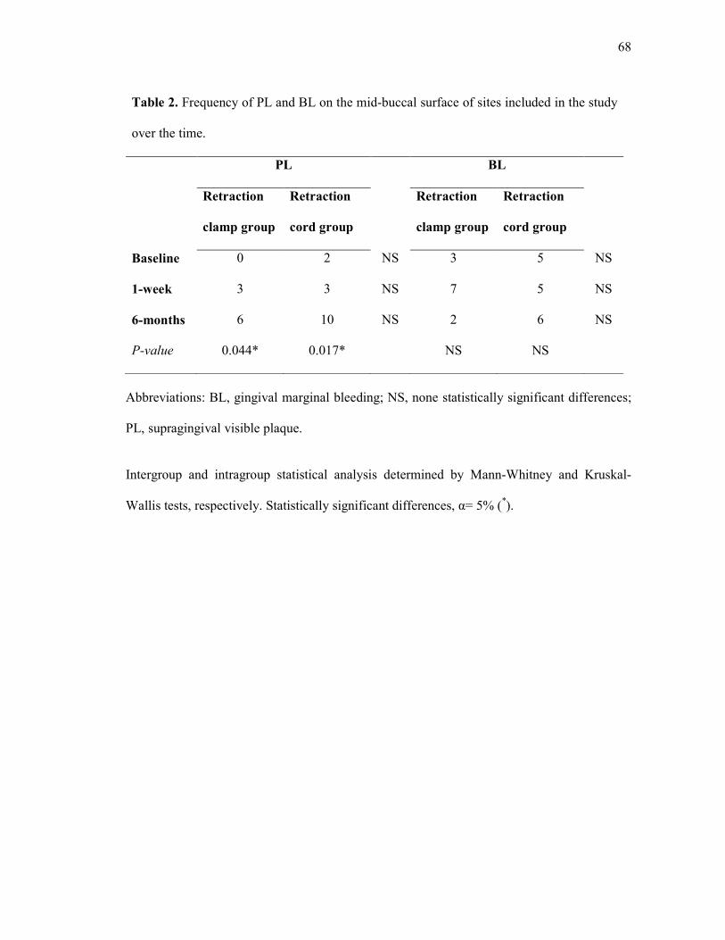
68
Table 2. Frequency of PL and BL on the mid-buccal surface of sites included in the study
over the time.
PL BL
Retraction
clamp group
Retraction
cord group
Retraction
clamp group
Retraction
cord group
Baseline 0 2 NS 3 5 NS
1-week 3 3 NS 7 5 NS
6-months 6 10 NS 2 6 NS
P-value 0.044* 0.017* NS NS
Abbreviations: BL, gingival marginal bleeding; NS, none statistically significant differences;
PL, supragingival visible plaque.
Intergroup and intragroup statistical analysis determined by Mann-Whitney and Kruskal-
Wallis tests, respectively. Statistically significant differences, α= 5% (*).

69
Table 3. Mean values and standard deviation for PD and RGR on the mid-buccal surface
of sites included in the study over the time.
PD (mm) RGR (mm)
Retraction
clamp group
Retraction
cord group
Retraction
clamp group
Retraction
cord group
Baseline 1.2 (± 0.4) 1.2 (± 0.4) NS 10.6 (± 1.7) 10.9 (± 1.7) NS
1-week 1.2 (± 0.4) 1.1 (± 0.3) NS 10.6 (± 1.7) 10.9 (± 1.7) NS
6-months 1.1 (± 0.4) 1.1 (± 0.4) NS 10.7 (± 1.6) 11.0 (± 1.9) NS
NS NS NS NS
Abbreviations: NS, none statistically significant differences; PD, probing depth; RGR,
relative gingival recession.
Intergroup and intragroup statistical analysis determined by Mann-Whitney and Kruskal-
Wallis tests, respectively.

70
Table 4. Correlations between restorative criteria and periodontal parameters at 1-week and
6-months after the treatment.
Marginal staining Marginal adaptation
PL BL PD RGR PL BL PD RGR
1-week -0.042 0.077 0.049 0.088 -0.081 0.039 0.007 0.054
6-months -0.040 -0.084 -0.099 0.194* 0.020 -0.070 0.059 0.116
Abbreviations: BL, gingival marginal bleeding; PD, probing depth; PL, supragingival visible
plaque; RGR, relative gingival recession.
Correlation analysis performed with Spearman Ranks. Statistically significant differences, α=
5% (*).
71
CONCLUSÕES
Com base na metodologia empregada neste estudo, pode-se concluir que as
restaurações Classe V realizadas com ambas as técnicas de isolamento do campo
operatório apresentaram-se clinicamente aceitáveis após seis meses de
acompanhamento. Observou-se, entretanto, uma incidência significativa de
pequenos defeitos marginais no grupo que recebeu isolamento com rolos de
algodão. Adicionalmente, verificou-se um aumento do acúmulo de placa
supragengival em ambos os grupos, sem produzir efeitos clinicamente significativos
na condição periodontal.
72
REFERÊNCIAS
ADA Council on Scientific Affairs. Direct and indirect restorative materials. The Journal of the American Dental Association, v.134, n.4, p.463-472, 2003.
ADA Council on Scientific Affairs. Revised American Dental Association acceptance program guidelines: dentin and enamel adhesives. Chicago: American Dental Association, 2001. 9p.
AINAMO, J.; BAY, I. Problems and proposals for recording gingivitis and plaque. International Dental Journal, v.25, n.4, p.229-235, 1975.
ALTMAN, D. G.; SCHULZ, K. F.; MOHER, D.; EGGER, M.; DAVIDOFF, F.; ELMOUBE, D.; GOTZSCHE, P. C.; MA, T. L. The revised CONSORT statement for reporting randomized trials: explanation and elaboration. Annals of Intern Medicine, v.134, n.8, p.663-694, 2001. BENNANI, V.; SCHWASS, D.; CHANDLER, N. Gingival retraction techniques for implants versus teeth: current status. The Journal of the American Dental Association, v.139, n.10, p.1354-1363, 2008.
BLUNCK, Uwe. Improving cervical restorations: a review of materials and techniques. The Journal of Adhesive Dentistry, v.3, n.1, p.33-44, 2001. BOUCHARD, P.; MALET, J.; BORGHETTI, A. Decision-making in aesthetics: root coverage revisited. Periodontology 2000, v.27, p.97-120, 2001. DE MUNCK, J.; VAN LANDUYT, K.; PEUMANS, M.; POITEVIN, A.; LAMBRECHTS, P.; BRAEM, M.; VAN MEERBEEK, B. A critical review of the durability of adhesion to tooth tissue: methods and results. Journal of Dental Research, v.84, n.2, p.118-132, 2005. DEMARCO, F. F.; CORRÊA, M. B.; CENCI, M. S.; MORAES, R. R.; OPDAM, N. J. Longevity of posterior composite restorations: Not only a matter of materials. Dental Materials (2011) in press.
GILBERT, G. H.; LITAKER, M. S.; PIHLSTROM, D. J.; AMUNDSON, C. W.; GORDAN, V. V. for The DPBRN Collaborative Group. Rubber dam use during routine operative dentistry procedures: findings from the Dental PBRN. Operative Dentistry, v. 35, n.5, p.491-499, 2010.
GOLDBERG, P. V.; HIGGINBOTTOM, F. L.; WILSON, T. G. Periodontal considerations in restorative and implant therapy. Periodontology 2000, v.25, p.100-109, 2001.

73
HEINTZE, S. D.; RUFFIEUX, C.; ROUSSON, V. Clinical performance of cervical restorations - a meta-analysis. Dental Materials, v.26, n.10, p.993-1000, 2010.
HICKEL, R.; PESCHKE, A.; TYAS, M.; MJÖR, I.; BAYNE, S.; PETERS, M.; HILLER, K. A.; RANDALL, R.; VANHERLE, G.; HEINTZE, S. D. FDI World Dental Federation - clinical criteria for the evaluation of direct and indirect restorations. Update and clinical examples. The Journal of Adhesive Dentistry, v.12, n.4, p.259-272, 2010.
HICKEL, R.; ROULET, J. F.; BAYNE, S.; HEINTZE, S. D.; MJOR, I. A.; PETERS, M.; ROUSSON, V.; RANDALL, R.; SCHMALZ, G.; TYAS, M.; VANHERLE, G. Recommendations for conducting controlled clinical studies of dental restorative materials. The Journal of Adhesive Dentistry, v.9, n.1, p.121-147, 2007.
KARAN, K.; YAO, X.; XU, C.; WANG, Y. Chemical profile of the dentin substrate in non-carious cervical lesions. Dental Materials, v.25,n.10, p.1205-1212, 2009.
KIM, S. Y.; LEE, K. W.; SEONG, S. R.; LEE, M. A.; LEE, I. B.; SON, H. H.; KIM, H. Y.; OH, M. H.; CHO, B. H. Two-year clinical effectiveness of adhesives and retention form on resin composite restorations of non-carious cervical lesions. Operative Dentistry, v.34, n.5, p.507-515, 2009.
LOGUERCIO, A. D.; COSTENARO, A.; SILVEIRA, A. P.; RIBEIRO, N. R.; ROSSI, T. R.; REIS, A. A six-month clinical study of a self-etching and an etch-and-rinse adhesive applied as recommended and after doubling the number of adhesive coats. The Journal of Adhesive Dentistry, v.8, n.4, p.255-261, 2006. LUCCHESI, J. A.; SANTOS, V. R.; AMARAL, C. M.; PERUZZO, D. C.; DUARTE, P. M. Coronally positioned flap for treatment of restored root surfaces: a 6-month clinical evaluation. Journal of Periodontology, v.78, n.4, p.615-623, 2007.
MOSZNER, N.; SALZ, U.; ZIMMERMANN, J. Chemical aspects of self-etching enamel-dentin adhesives: a systematic review. Dental Materials, v.21, n.10, p.895-910, 2005.
OWENS, Barry. Alternative rubber dam isolation technique for the restoration of class V cervical lesions. Operative Dentistry, v.31, n.2, p.277-280, 2006. PADBURY A JR, EBER R, WANG HL. Interactions between the gingiva and the margin of restorations. Journal of Clinical Periodontology, v.30, n.5, p.379-385, 2003. PAOLANTONIO, M,; D'ERCOLE, S.; PERINETTI, G.; TRIPODI, D.; CATAMO, G.; SERRA, E.; BRUÈ, C.; PICCOLOMINI, R. Clinical and microbiological effects of different restorative materials on the periodontal tissues adjacent to subgingival class V restorations. Journal of Clinical Periodontology, v.31, n.3, p.200-207, 2004.
PERDIGÃO, Jorge. Dentin bonding - variables related to the clinical situation and the substrate treatment. Dental Materials, v.26, n.2, p.e24-e37, 2010.
74
PEREZ, Cesar Reis. Alternative technique for class V resin composite restorations with minimum finishing/polishing procedures. Operative Dentistry, v.35, n.3, p.375-379, 2010.
PEUMANS, M.; DE MUNCK, J.; VAN LANDUYT, K. L.; POITEVIN, A.; LAMBRECHTS, P.; VAN MEERBEEK, B. Eight-year clinical evaluation of a 2-step self-etch adhesive with and without selective enamel etching. Dental Materials, v.26, n.12, p.1176-1184, 2010.
PEUMANS, M.; DE MUNCK, J.; VAN LANDUYT, K.; LAMBRECHTS, P.; VAN MEERBEEK, B. Three-year clinical effectiveness of a two-step self-etch adhesive in cervical lesions. European Journal of Oral Science, v.113, n.6, p.512-518, 2005b. PEUMANS, M.; KANUMILLI, P.; DE MUNCK, J.; VAN LANDUYT, K.; LAMBRECHTS, P.; VAN MEERBEEK, B. Clinical effectiveness of contemporary adhesives: a systematic review of current clinical trials. Dental Materials, v.21, n.9, p.864-881, 2005a.
RASKIN, A.; SETCOS, J. C.; VREVEN, J.; WILSON, N. H. F. Influence of the isolation method on the 10-year clinical behaviour of posterior resin composite restorations. Clinical Oral Investigations, v.4, n.3, p.148-152, 2000.
RITTER, A. V.; HEYMANN, H. O.; SWIFT, E. J.; STURDEVANT, J. R.; WILDER, A. D. Clinical evaluation of an all-in-one adhesive in non-carious cervical lesions with different degrees of dentin sclerosis. Operative Dentistry, v.33, n.4, p.370-378, 2008. SANTAMARIA, M. P.; SUAID, F. F.; CASATI, M. Z.; NOCITI, F. H.; SALLUM, A. W.; SALLUM, E. A. Coronally positioned flap plus resin-modified glass ionomer restoration for the treatment of gingival recession associated with non-carious cervical lesions: a randomized controlled clinical trial. Journal of Periodontology, v.79, n.4, p.621-628, 2008. SANTAMARIA, M. P.; AMBROSANO, G. M.; CASATI, M. Z.; NOCITI JÚNIOR, F. H.; SALLUM, A. W.; SALLUM, E. A. Connective tissue graft plus resin-modified glass ionomer restoration for the treatment of gingival recession associated with non-carious cervical lesion: a randomized-controlled clinical trial. Journal of Clinical Periodontology, v.36, n.9, p.791-798, 2009. SANTOS, V. R.; LUCCHESI, J. A.; CORTELLI, S. C.; AMARAL, C.M.; FERES, M.; DUARTE, P. M. Effects of glass ionomer and microfilled composite subgingival restorations on periodontal tissue and subgingival biofilm: a 6-month evaluation. Journal of Periodontology, v.78, n.8, p.1522-1528, 2007. TAY, F. R.; PASHLEY, D. H. Have dentin adhesives become too hydrophilic? Journal of the Canadian Dental Association, v.69, n.11, p.726-731, 2003. TAY, F. R.; PASHLEY, D. H. Resin bonding to cervical sclerotic dentin: A review. Journal of Dentistry, v.32, n.3, p.173-196, 2004.
75
TIWANA, K. K.; MORTON, T.; TIWANA, P. S. Aspiration and ingestion in dental practice: a 10-year institutional review. The Journal of the American Dental Association, v.135, n.9, p.1287-1291, 2004.
VAN MEERBEEK, B.; DE MUNCK, J.; YOSHIDA, Y.; INOUE, S.; VARGAS, M.; VIJAY, P.; VAN LANDUYT, K.; LAMBRECHTS, P.; VANHERLE, G. Buonocore memorial lecture. Adhesion to enamel and dentin: current status and future challenges. Operative Dentistry, v.28, n.3, p.215-235, 2003. VAN MEERBEEK, B.; PERDIGÃO, J.; LAMBRECHTS, P.; VANHERLE, G. The clinical performance of adhesives. Journal of Dentistry, v.26, n.1, p.1-20, 1998.
VAN MEERBEEK, B.; YOSHIHARA, K.; YOSHIDA, Y.; MINE, A.; DE MUNCK, J.; VAN LANDUYT, K. L. State of the art of self-etch adhesives. Dental Materials, v.27, n.1, p.17-28, 2011.
WOOD, I.; JAWAD, Z.; PAISLEY, C.; BRUNTON, P. Non-carious cervical tooth surface loss: a literature review. Journal of Dentistry, v.36, n.10, p.759-766, 2008.
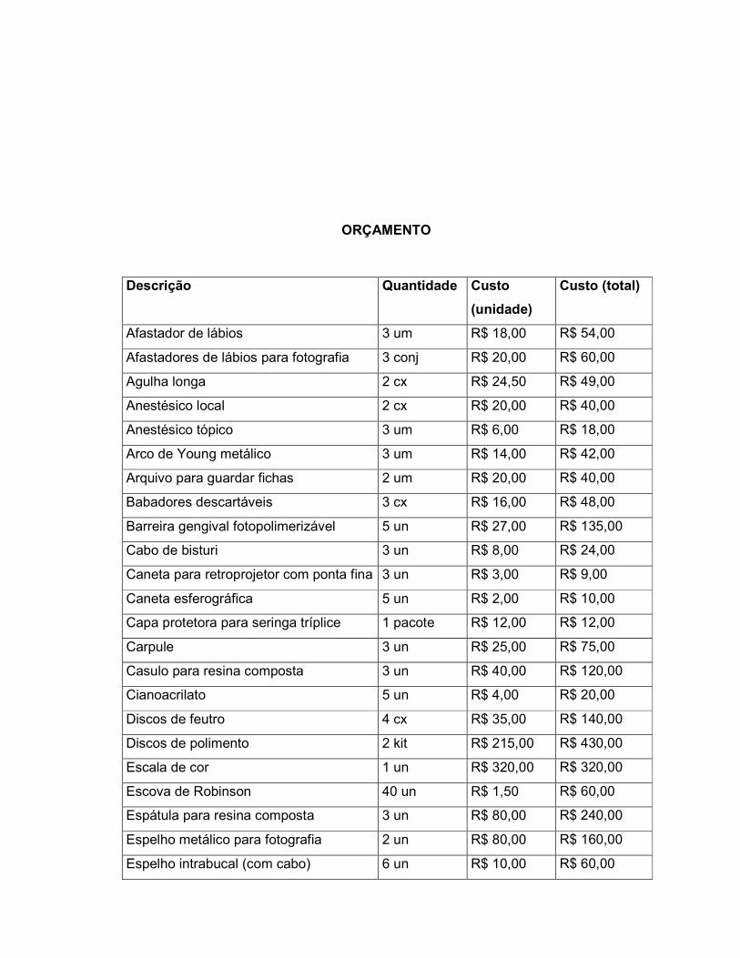
76
ORÇAMENTO
Descrição Quantidade Custo
(unidade)
Custo (total)
Afastador de lábios 3 um R$ 18,00 R$ 54,00
Afastadores de lábios para fotografia 3 conj R$ 20,00 R$ 60,00
Agulha longa 2 cx R$ 24,50 R$ 49,00
Anestésico local 2 cx R$ 20,00 R$ 40,00
Anestésico tópico 3 um R$ 6,00 R$ 18,00
Arco de Young metálico 3 um R$ 14,00 R$ 42,00
Arquivo para guardar fichas 2 um R$ 20,00 R$ 40,00
Babadores descartáveis 3 cx R$ 16,00 R$ 48,00
Barreira gengival fotopolimerizável 5 un R$ 27,00 R$ 135,00
Cabo de bisturi 3 un R$ 8,00 R$ 24,00
Caneta para retroprojetor com ponta fina 3 un R$ 3,00 R$ 9,00
Caneta esferográfica 5 un R$ 2,00 R$ 10,00
Capa protetora para seringa tríplice 1 pacote R$ 12,00 R$ 12,00
Carpule 3 un R$ 25,00 R$ 75,00
Casulo para resina composta 3 un R$ 40,00 R$ 120,00
Cianoacrilato 5 un R$ 4,00 R$ 20,00
Discos de feutro 4 cx R$ 35,00 R$ 140,00
Discos de polimento 2 kit R$ 215,00 R$ 430,00
Escala de cor 1 un R$ 320,00 R$ 320,00
Escova de Robinson 40 un R$ 1,50 R$ 60,00
Espátula para resina composta 3 un R$ 80,00 R$ 240,00
Espelho metálico para fotografia 2 un R$ 80,00 R$ 160,00
Espelho intrabucal (com cabo) 6 un R$ 10,00 R$ 60,00

77
Folhas de papel A4 para impressão 3 pacotes R$ 12,00 R$ 36,00
Filme PVC para proteção do equipo 5 rolos R$ 5,00 R$ 25,00
Filtro de papel recortado 2 cx R$ 5,00 R$ 10,00
Fio dental encerado 3 un R$ 4,00 R$ 12,00
Fio retractor gengival #000 2 un R$ 50,00 R$ 100,00
Fita para autoclave 3 un R$ 4,00 R$ 12,00
Fita “veda-rosca” 1 un R$ 3,00 R$ 3,00
Fototpolimerizador 2 un R$ 1.500,00 R$ 3.000,00
Gaveteiro para estoque de materiais 1 un R$ 100,00 R$ 100,00
Gaze 2 pacotes R$ 45,00 R$ 90,00
Gesso especial tipo IV 4 un R$ 25,00 R$ 100,00
Glutaraldeído 1 frasco R$ 25,00 R$ 25,00
Grampos para isolamento 20 un R$ 25,00 R$ 500,00
Godiva em bastão 3 cx R$ 20,00 R$ 60,00
Gorros descartáveis 1 cx R$ 15,00 R$ 30,00
Guardanapo de papel 6 pacotes R$ 3,00 R$ 18,00
Isqueiro 3 un R$ 4,00 R$ 12,00
Lâmina de bisturi #12 2 cx R$ 30,00 R$ 60,00
Lamparina à álcool 1 un R$ 20,00 R$ 20,00
Lençol de borracha 4 cx R$ 20,00 R$ 80,00
Lubrificante para alta e baixa rotação 1 un R$ 28,00 R$ 28,00
Lubrificante para lábios 1 un R$ 10,00 R$ 10,00
Luvas de procedimento descartáveis 4 cx R$ 20,00 R$ 60,00
Mandril para discos de polimento 3 un R$ 20,00 R$ 60,00
Máscaras descartáveis 3 cx R$ 10,00 R$ 30,00
Matriz de polyester 1 cx R$ 50,00 R$ 50,00
Óculos de proteção 3 un R$ 8,00 R$ 24,00
Papel articular 1 cx R$ 60,00 R$ 60,00
Pasta para polimento 10 un R$ 26,00 R$ 260,00
Pedra-pomes 1 un R$ 9,00 R$ 9,00
Perfurador de borracha 3 un R$ 46,00 R$ 138,00
Pilhas para máquina fotográfica 2 un R$ 30,00 R$ 60,00
Pinça clínica 6 un R$ 20,00 R$ 120,00

78
Pinça porta-agulha 3 un R$ 25,00 R$ 75,00
Pinça Miller 3 un R$ 20,00 R$ 60,00
Pinça porta-grampo 3 um R$ 50,00 R$ 150,00
Pinceis descartáveis para adesivo 4 cx R$ 10,00 R$ 40,00
Pinceis para resina composta 10 um R$ 15,00 R$ 150,00
Pontas diamantadas (3 tipos) 30 um R$ 4,50 R$ 135,00
Pontas multilaminadas 10 um R$ 4,50 R$ 45,00
Pontas siliconadas 3 kits R$ 150,00 R$ 450,00
Prancheta para anotações 2 um R$ 4,00 R$ 8,00
Prendedor para babador descartável 4 um R$ 5,00 R$ 20,00
Resina composta (8 cores) 20 um R$ 90,00 R$ 1.800,00
Rolos de algodão 12 pacotes R$ 2,00 R$ 24,00
Sacolé 2 pacotes R$ 4,00 R$ 8,00
Sacos para esterilização 2 rolos R$ 40,00 R$ 80,00
Sistema adesivo 2 um R$ 200,00 R$ 400,00
Sobre-luvas 2 pacotes R$ 20,00 R$ 40,00
Sonda exploradora 6 um R$ 4,00 R$ 24,00
Sonda periodontal milimetrada 2 um R$ 68,00 R$ 136,00
Spray para teste de vitalidade pulpar 4 um R$ 35,00 R$ 140,00
Sugador de saliva descartável 3 pacotes R$ 5,00 R$ 15,00
Tesoura cirúrgica 3 um R$ 12,00 R$ 36,00
Tira de lixa 1 cx R$ 50,00 R$ 50,00
Total R$ 11.484,00
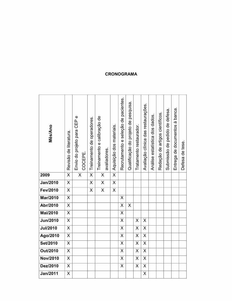
79
CRONOGRAMA
Mê
s/A
no
Revi
são d
e lit
era
tura
.
Envi
o do
pro
jeto
para
CE
P e
CO
CE
PE
.
Tre
inam
ento
de
opera
do
res.
Tre
inam
ento
e c
alib
raçã
o d
e
ava
liadore
s.
Aquis
içã
o dos
mate
riai
s.
Recr
utam
ento
e s
ele
ção
de p
aci
ente
s.
Qua
lific
açã
o do
pro
jeto
de p
esqu
isa.
Tra
tam
ento
rest
aur
ador.
Ava
liaçã
o cl
ínic
a d
as r
esta
ura
ções.
Anális
e e
statíst
ica d
os
dados.
Redaçã
o de
art
igos
cie
ntífic
os.
Subm
issã
o d
e ped
ido d
e de
fesa
.
Entr
ega
de d
ocu
mento
s à b
anca
.
Defe
sa d
e t
ese.
2009 X X X X X
Jan/2010 X X X X
Fev/2010 X X X X
Mar/2010 X X
Abr/2010 X X X
Mai/2010 X X
Jun/2010 X X X X
Jul/2010 X X X X
Ago/2010 X X X X
Set/2010 X X X X
Out/2010 X X X X
Nov/2010 X X X X
Dez/2010 X X X X
Jan/2011 X X

80
Fev/2011 X X
Mar/2011 X X
Abr/2011 X X
Mai/2011 X X
Jun/2011 X X X X
Jul/2011 X X X
Ago/2011 X X X
Set/2011 X X
Out/2011 X X X
Nov/2011 X
Dez/2011 X

81
Apêndices
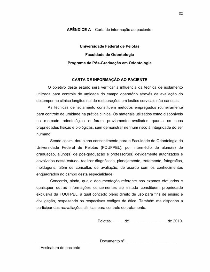
82
APÊNDICE A – Carta de informação ao paciente.
Universidade Federal de Pelotas
Faculdade de Odontologia
Programa de Pós-Graduação em Odontologia
CARTA DE INFORMAÇÃO AO PACIENTE
O objetivo deste estudo será verificar a influência da técnica de isolamento
utilizada para controle de umidade do campo operatório através da avaliação do
desempenho clínico longitudinal de restaurações em lesões cervicais não-cariosas.
As técnicas de isolamento constituem métodos empregados rotineiramente
para controle de umidade na prática clínica. Os materiais utilizados estão disponíveis
no mercado odontológico e foram previamente avaliados quanto as suas
propriedades físicas e biológicas, sem demonstrar nenhum risco à integridade do ser
humano.
Sendo assim, dou pleno consentimento para a Faculdade de Odontologia da
Universidade Federal de Pelotas (FOUFPEL), por intermédio de aluno(s) de
graduação, aluno(s) de pós-graduação e professor(es) devidamente autorizados e
envolvidos neste estudo, realizar diagnóstico, planejamento, tratamento, fotografias,
moldagens, além de consultas de avaliação, de acordo com os conhecimentos
enquadrados no campo desta especialidade.
Concordo, ainda, que a documentação referente aos exames efetuados e
quaisquer outras informações concernentes ao estudo constituem propriedade
exclusiva da FOUFPEL, à qual concedo pleno direito de uso para fins de ensino e
divulgação, respeitando os respectivos códigos de ética. Também me disponho a
participar das reavaliações clínicas para controle do tratamento.
Pelotas, _____ de _________________ de 2010.
_________________________ Documento no: _______________________
Assinatura do paciente
83
APÊNDICE B – Termo de consentimento livre e esclarecido.
Universidade Federal de Pelotas
Faculdade de Odontologia
Programa de Pós-Graduação em Odontologia
TERMO DE CONSENTIMENTO LIVRE E ESCLARECIDO
Título do projeto: Influência do isolamento do campo operatório em
restaurações Classe V: Ensaio clínico randomizado.
Pesquisador responsável pelo estudo: Silvia Terra Fontes
Nome do paciente: ___________________________________ Ficha no: _______
Por este instrumento que atende às exigências legais, o(a)
senhor(a)_________________________________________________________,
portador(a) da cédula de identidade nº________________________ SSP/___, após
leitura minuciosa da CARTA DE INFORMAÇÃO AO PACIENTE, detalhadamente
explicada pelos pesquisadores envolvidos neste estudo, ciente dos procedimentos
aos quais será submetido(a), não restando dúvidas a respeito do lido e do explicado,
firma CONSENTIMENTO LIVRE E ESCLARECIDO em concordância a participar da
pesquisa proposta no que lhe é cabível, conforme a carta de informação ao
paciente.
Fica claro que o paciente ou seu representante legal pode, a qualquer
momento, retirar seu consentimento livre e esclarecido, sem ser prejudicado no
tratamento, deixando de participar do estudo alvo da pesquisa e estando ciente que
todo trabalho realizado torna-se informação confidencial guardada por força do sigilo
profissional (Art. 9º do Código de Ética Odontológica).
Por estarem entendidos e conformados, assinam o presente termo.
Pelotas, _____ de _________________ de 2010.
_________________________ ____________________________
Assinatura do paciente Assinatura do pesquisador

84
Anexos
85
ANEXO A – Parecer do Comitê de Ética em Pesquisa.