UNIVERSIDADE FEDERAL DE PELOTAS Faculdade de...
104
Fernanda Valentini Formação de biofilme em reembasadores temporários para prótese dentária UNIVERSIDADE FEDERAL DE PELOTAS Faculdade de Odontologia Programa de Pós Graduação Área de Prótese Dentária Dissertação de Mestrado Pelotas, 2012
-
Upload
duongthuan -
Category
Documents
-
view
214 -
download
0
Transcript of UNIVERSIDADE FEDERAL DE PELOTAS Faculdade de...

Fernanda Valentini
Formação de biofilme em reembasadores
temporários para prótese dentária
UNIVERSIDADE FEDERAL DE PELOTAS Faculdade de Odontologia
Programa de Pós Graduação Área de Prótese Dentária
Dissertação de Mestrado
Pelotas, 2012

2
FERNANDA VALENTINI
FORMAÇÃO DE BIOFILME EM REEMBASADORES TEMPORÁRIOS PARA PRÓTESE DENTÁRIA
Orientadora: Profa. Dra. Tatiana Pereira Cenci Co-orientadora: Profa. Dra. Noéli Boscato
Pelotas, 2012
Dissertação de Mestrado apresentada à
Faculdade de Odontologia da Universidade
Federal de Pelotas obtenção do título de
Mestre em Odontologia (área do
conhecimento: Prótese Dentária).

3
Dados de Catalogação da Publicação
V161f Valentini, Fernanda
Formação de biofilme em reembasadores temporários para
prótese dentária / Fernanda Valentini ; orientador: Tatiana Pereira Cenci ; co-orientador: Noéli Boscato . – Pelotas: UFPel, 2012.
103 f. ; fig. ; tab.
Dissertação (Mestrado) Prótese dentária. Faculdade de Odontologia. Universidade Federal de Pelotas. Pelotas.
1. Condicionadores de tecido. 2. Reembasadores de prótese. 3. Biofilme. 4. Prótese totaI. 5. Candida. I. Cenci, Tatiana Pereira (orient.). II. Boscato, Noéli (co-orient.) III. Título.
D3
Bibliotecário: Fabiano Domingues Malheiro CRB -10/1955

4
Banca examinadora:
Profa. Dra. Tatiana Pereira Cenci
Profa. Dra. Rosemary Sadami Araí Shinkai
Profa. Dra. Fernanda Geraldes Pappen
Prof. Dr. Rafael Guerra Lund (suplente)

5
AGRADECIMENTOS
À Deus, por sempre iluminar meus caminhos.
À minha família, minha mãe, Dilene, meu pai, Celonir e ao meu irmão
Rafael, pelo amor sem limites, responsáveis pelo meu caráter. Estiveram sempre
juntos com palavras de estímulo e conforto e sem dúvida me ajudaram a seguir em
frente. Obrigada pela compreensão nos momentos em que estive ausente, pelo
carinho, pela preocupação e pela torcida. Sem vocês seria impossível realizar meus
sonhos. Um muito obrigado seria pouco pelo muito que fizeram.
“Aonde quer que eu vá levo vocês no olhar”. Amo vocês!
Ao meu namorado Rafael, meu anjo. Responsável pelo brilho no meu olhar.
Foi encantador em todos os momentos e fundamental para eu não desistir nos
momentos mais difíceis. Obrigada por tudo, pelos conselhos, pelo apoio, pelo
companheirismo e por acreditar em mim mais do que eu mesma. Te amo!
À Universidade Federal de Pelotas, a Faculdade de Odontologia e ao
Programa de Pós-Graduação em Odontologia, minha segunda casa, o qual me
orgulho por ser formada nesta instituição de tamanho gabarito e credibilidade.
Á minha orientadora Tatiana Pereira Cenci, pela paciência,
comprometimento, determinação, e exemplo de profissional que nunca mediu
esforços na colaboração para o desenvolvimento do meu trabalho. Obrigada pela
confiança e pelos valiosos ensinamentos. És um exemplo de educadora e
pesquisadora. Através da simplicidade dos teus ensinamentos aprendi muito de
prótese dentária e tive a oportunidade de seguir aprendendo e praticando a clínica.
É impossível, em palavras descrever o tamanho do meu carinho por você e da
minha eterna gratidão. Estará sempre do lado esquerdo do peito!
À minha co-orientadora Noéli Boscato, obrigada de coração pelo ombro
amigo pelos conselhos e ensinamentos que me fortaleceram ao longo desses anos.
Obrigada por acreditar em mim e me ensinar o que eu mais amo na odontologia,

6
prótese dentária. Através de teu ensinamento, almejo um ideal nesta profissão
clínica, te tenho como modelo a seguir.
Ao Prof. Dr. Maximiliano Sérgio Cenci, por contribuir direta e indiretamente
para o desenvolvimento deste trabalho. Obrigada pela ajuda, orientação e
serenidade.
A todos os professores do Programa de Pós-Graduação em Odontologia
pela convivência e pelos valiosos ensinamentos.
Ao Laboratório de Microbiologia em especial a Carmen que sempre se
mostrou solicita em ajudar no que foi preciso para o desenvolvimento desse
trabalho.
Ao Laboratório de Materias Dentários da UFRGS na pessoa do Prof. Dr.
Fabrício Mezzomo Collares, por gentilmente disponibilizar o uso do rugosímetro.
A todos os colegas do Programa de Pós-Graduação em Odontologia da
Universidade Federal de Pelotas. Em especial aos colegas de pós-graduação Mauro
Elias Mesko, Rafael Onofre, Jovito Adiel Skupien, pela agradável convivência,
parceria e amizade, tornando o 2° andar um lugar ainda mais agradável de
trabalhar. Adoro vocês!
Em especial ao graduando Murilo, pela ajuda e parceria no desenvolvimento
desse trabalho. Foi extremamente importante para dar seguimento a pesquisa.
Obrigada por tudo.
A todas as pessoas que direta ou indiretamente contribuíram para a execução
deste trabalho.

7
“Apesar dos nossos defeitos, precisamos enxergar que somos
pérolas únicas no teatro da vida e entender que não existem pessoas
de sucesso e pessoas fracassadas. O que existem são pessoas que
lutam pelos seus sonhos ou desistem dele.”
Augusto Curry

8
NOTAS PRELIMINARES
A presente dissertação foi redigida segundo o Manual de Normas para Dissertações,
Teses e Trabalhos Científicos da Universidade Federal de Pelotas de 2006,
adotando o Nível de Descrição 4 – estruturas em Artigos, que consta no Apêndice D
do referido manual. Disponível no endereço eletrônico:
(http://www.ufpel.tche.br/prg/sisbi/documentos/Manual_normas_UFPel_2006.pdf).

9
Resumo
VALENTINI, Fernanda. Formação de biofilme em reembasadores temporários para prótese dentária. 2012. 97f. Dissertação (Mestrado) – Programa de Pós-Graduação em Odontologia. Universidade Federal de Pelotas, Pelotas.
Os fungos oportunistas são responsáveis por doenças infecciosas na cavidade bucal
que aumentaram em prevalência nos últimos anos, especialmente em usuários de
prótese total. Assim, a colonização e o crescimento de espécies de Candida e outros
microrganismos em próteses tem fundamental importância clínica. Este estudo teve
por objetivo (i) fazer uma revisão sistemática para determinar se existe um protocolo
de prevenção ou tratamento da colonização por Candida em reembasadores de
prótese e (ii) avaliar clinicamente como a composição do biofilme é afetada por
diferentes materiais, tempo e a presença ou não de candidíase, em usuários de
prótese total. Estudos clínicos e in vitro foram avaliados quanto ao tratamento e / ou
prevenção da colonização por Candida e formação de biofilme em reembasadores
de prótese. Seis bases de dados eletrônicas foram pesquisadas (Lilacs, Scopus,
Pubmed / Medline, Scielo e Cochrane Database of Systematic Reviews) de 1950 a
2012 usando as palavras-chaves “denture liner”, “Candida”, “tissue conditioner”,
“denture stomatitis” e “antifungal agents”. Para o estudo in situ vinte e oito
voluntários usuários de prótese total, quinze portadores de estomatite por dentadura,
e doze pacientes com alguma espécie de Candida avaliados por screening inicial
foram selecionados. Foi quantificada a formação de biofilme sobre espécimes de
resina acrílica e reembasadores temporários (a base de resina acrílica ou silicone)
inseridos na parte interna da prótese total superior em duas fases de 21 dias. Os
espécimes foram removidos aleatoriamente no 7°, 14° e 21° dia. Amostras
representativas foram analisadas em MEV nos diferentes períodos de avaliação.
Unidades formadoras de colônia/mm2 de biofilme de estreptococos do grupo mutans,
lactobacilos, microrganismos totais e espécies de Candida foram determinados.
Através da revisão sistemática foi possível observar que a incorporação de nistatina
para prevenir o aparecimento da doença e a imersão em hipoclorito de sódio para
desinfectar reembasadores de tecidos são o tratamento mais frequentemente
encontrado. No entanto, os dados encontrados foram quase que exclusivamente
baseados em estudos in vitro, o que gera alto risco de viés. Para o estudo in situ, a
contagem de Candida não-albicans para reembasadores a base de silicone foi maior
10
em pacientes com candidíase (p=0,01). Pacientes com candidíase apresentaram
maiores contagens de estreptococos do grupo mutans após 7 dias (p=0,0041), mas
essa diferença desapareceu após 14-21 dias de formação de biofilme. Com isso,
reembasadores a base de silicone devem ser evitados em pacientes com candidíase
já que estes materiais apresentaram aumento da contagem de espécies de Candida
não-albicans, as quais são mais virulentas e resisitentes às terapias convencionais.
Palavras-chave: Condicionadores de Tecido. Biofilme. Prótese Total. Candida

11
Abstract
VALENTINI, Fernanda. Biofilm formation on temporary denture liners. 2012. 97f. Dissertação (Mestrado) – Programa de Pós-Graduação em Odontologia. Universidade Federal de Pelotas, Pelotas
Opportunistic fungi are responsible for infectious diseases in oral cavity that rose in
prevalence in the last years, especially in complete denture wearers. Thus,
colonization and growth of Candida species and other microorganisms are of clinical
importance. The aims of this study were (i) systematically review the literature to
determine if there is a protocol of prevention or treatment of Candida colonization in
denture liners and (ii) clinically assess how biofilm composition is affected by different
materials, time and the presence of candidiasis in denture wearers. In vitro and in
vivo studies were evaluated with regard to treatment and/or prevention of Candida
colonization and biofilm formation in denture liners. Six databases were searched
(Lilacs, Scopus, Pubmed / Medline, Scielo and Cochrane Database of Systematic
Reviews) from 1950 to 2012 using the keywords “denture liner”, “Candida”, “tissue
conditioner”, “denture stomatitis” and “antifungal agents”. For the in situ study,
twenty-eight volunteers, half with candidiasis, half healthy but Candida carriers
wearing complete dentures were selected to participate in this study. Biofilm formed
on acrylic resin and temporary denture liners (silicone based and acrylic resin based)
specimens mounted in the internal surface of the volunteers’ upper dentures were
collected in two phases of 21 days. Specimens were randomly removed on days 7,
14 and 21. Representative samples of the specimens were analyzed by SEM in the
various periods under evaluation. Colony forming units/mm2 of biofilm of mutans
streptococci, lactobacilli, total microorganisms and Candida species were
determined. Through the systematic review it was possible to observe that the
incorporation of nystatin to prevent the disease and the immersion in sodium
hypoclorite to disinfect denture liners was the most frequently found treatment.
However, as the data was in general derived from in vitro studies, there is a high risk
of bias. For the in situ study, non-albicans Candida species showed higher counts in
the silicone-based denture liner in diseased patients (p=0,01). Patients with
candidiasis showed higher counts of mutans streptococci after 7 days (p=0.0041),
but this difference disappeared after 14-21 days of biofilm formation. Thus, silicone-
based denture liners should be avoided in diseased patients, as they have shown

12
higher nonn-albicans Candida species, which are known to be more virulent and
resistant to conventional therapies.
Keywords: Tissue Conditioner. Biofilm. Complete Denture. Candida

13
Sumário
1 Projeto de Pesquisa..........……………....………………………………................. 15
1.1 Introdução......................................................................................................... 14
1.2 Objetivos........................................................................................................... 20
1.2.1 Gerais.............................................................................................................. 20
1.2.2 Específicos..................................................................................................... 20
1.3 Justificativa....................................................................................................... 21
1.4 Materiais e Métodos......................................................................................... 21
1.4.1 Delineamento Experimental.......................................................................... 22
1.4.2 Seleção dos Voluntários............................................................................... 24
1.4.3 Screening para a Presença de Candida...................................................... 25
1.4.4 Parte I – Estudo In Situ.................................................................................. 25
1.4.4.1 Preparo dos Espécimes............................................................................. 26
1.4.4.2 Rugosidade de Superfície.......................................................................... 26
1.4.4.3 Inserção dos Espécimes na Prótese........................................................ 26
1.4.4.4 Coleta do Material....................................................................................... 29
1.4.5 Parte II – Análise Microbiológica.................................................................. 30
1.4.6 Parte III – Análise Microscópica................................................................... 31
1.5 Cronograma de Execução............................................................................... 32
2 Relatório do trabalho de campo………………………………..………................. 34
2.1 Aspéctos Éticos................................................................................................ 34
2.2 Condições Gerais............................................................................................. 34

14
2.3 Rotinas Laboratoriais…………………………………………………………....… 35
2.3.1 Coleta e Processamento………………………………………………….…...... 35
2.3.2 Protocolo de Obtenção do Biofilme…………………………………………... 35
2.4 Alteração no Projeto Original.......................................................................... 36
2.4.1 Dificuldades Encontradas............................................................................. 36
Artigo 1………………………………………………………………….......................... 38
Artigo 2 …................................................................................................................ 71
3. Conclusões....................…………………………………………………................. 94
4. Referências......................................................................................................... 95
Apêndice................................................................................................................ 101
Anexos................................................................................................................... 103

15
1 Projeto de Pesquisa
1.1 Introdução
A cavidade bucal é colonizada por diversos microrganismos, os quais se
apresentam em número limitado, o que é determinado pelas condições que
seletivamente os favorecem, em condições de saúde (SAN MILLÁN et al., 2000). No
entanto, de acordo com a teoria da placa ecológica, sabe-se que a presença e
especialmente a proporção de algumas espécies propicia modificações que
transformam um estado de saúde em doença, muito mais do que a presença de
alguma espécie específica (MARSH, 1994).
Alguns dos microrganismos residentes na cavidade bucal são patógenos
oportunistas, dos quais destacam-se as espécies de Candida. Este microrganismo
eucariótico causa a candidíase bucal, comumente diagnosticada em humanos
(MUZYKA, 2005). O crescimento desse fungo sobre superfícies é natural no ciclo de
vida das espécies de Candida (KUMAMOTO; VINCES, 2005), o que pode explicar a
ocorrência comum da colonização fúngica nos usuários de próteses.
As lesões da mucosa bucal relacionadas às próteses removíveis são
reações agudas ou crônicas decorrentes da presença de biofilme dental, de
leveduras, de constituintes do material utilizado para a confecção das próteses e da
pouca retenção ou injúrias mecânicas oriundas do uso de próteses mal adaptadas
(BUDTZ-JORGENSEN, 1978; BUDTZ-JORGENSEN 1981; DOREY et al., 1985).
Entretanto, de todas as lesões que podem ocorrer, conforme supracitado, aquelas
ocasionadas pela candidíase podem interferir com o tratamento e principalmente ser

16
uma barreira para a saúde do paciente (PEREZOUS, 2005), uma vez que as
próteses podem servir como fonte de microrganismos para novas infecções
(MUZYKA, 2005), sendo a prevalência de até 67% nos usuários de próteses
removíveis (ARENDORF; WALKER, 1987; SPIECHOWICZ et al., 1991; RADFORD
et al., 1999).
Esta inflamação também é denominada estomatite induzida por prótese,
estomatite por dentaduras ou candidíase atrófica crônica, sendo que a Candida
albicans foi e continua sendo fortemente associada como o principal agente
etiológico desta patologia (WEBB et al., 1998; BARBEAU et al., 2003; ZAREMBA et
al., 2006). Entretanto, hoje é sabido que espécies de Candida não-albicans (C.
tropicalis, C. parapsilosis, C. glabrata, C. krusei e C. dubliniensis) podem ser
isoladas e responsáveis por mais de 50% dos casos de infecção
(SAMARANAYAKE; SAMARANAYAKE, 1994; COLEMAN et al., 1997; ELLEPOLA;
SAMARANAYAKE, 2001; ZAREMBA et al., 2006; FIGUEIRAL et al., 2007).
A candidíase bucal pode ser classificada a partir da presença de grandes
placas brancas pseudomembranosas na mucosa, língua e boca,
lesões palatais eritematosas características da candidíase atrófica crônica e queilite
angular nas comissuras labiais (SAMARANAYAKE, 1990; SCULLY et al., 1994;
SHAY et al., 1997). A candidíase pode ser classificada de acordo com Newton em
Tipo (I) lesões inflamatórias, eritematosas; Tipo (II) Eritema difuso, simples ou
generalizado em mucosa coberta por prótese e Tipo (III) lesões granulares ou
papilares comumente envolvendo a parte central do palato duro e rebordo alveolar.
A adesão de microrganismos em superfícies de biomateriais depende da
estrutura e composição de sua superfície e das propriedades físico-químicas da
superfície das células microbianas (BELLON-FONTAINE at al., 1990; BUSSCHER;

17
COWAN; VAN DER MEI, 1992), as quais vão aderir via formação de um biofilme.
Biofilme pode ser definido como uma película não calcificada, fortemente aderida às
superfícies dentais, resistindo a presença do fluxo salivar. O termo biofilme é usado
para denotar uma comunidade microbiana encapsulada em polímero que se
acumula em uma superfície, que também protege contra colonização de patógenos
exógenos (WILSON, 2001). O biofilme constitui-se de depósitos bacterianos e
constituintes salivares, com um crescimento contínuo, sendo considerada a principal
causa das doenças infecciosas e estomatites (ROSAN; LAMONT, 2000).
A formação de biofilmes multi-espécie, envolto por uma matriz extracelular,
protege o biofilme da ação de patógenos exógenos, da ação de alguns
medicamentos e da ação da própria saliva, isso aumenta a chance de sobrevivência
de todos os constituintes do ambiente bucal e é considerado o primeiro passo para a
colonização fúngica, levando a um processo infeccioso (CHANDRA et al., 2001;
CANNON; CHAFFIN, 1999; RAMAGE et al., 2004). Dessa forma, as espécies de
Candida podem aderir diretamente ou via uma camada de “placa de dentadura” às
bases de próteses (SAMARANAYAKE; MACFARLANE, 1980; BRANTING et
al.,1989; EDGERTON et al., 1993; COULTHWAITE; VERRAN , 2007).
Entretanto, pouco se sabe sobre o efeito de diferentes superfícies na
interação entre espécies de Candida e outros microrganismos, incluindo a superfície
de materiais que contem antifúngicos, como os reembasadores e condicionadores
de tecido (PEREIRA-CENCI et al., 2010). A utilização destes materiais é vantajosa
em diversas situações clínicas e tem aumentado nos últimos anos. Porém, um dos
problemas diretamente relacionados a estes materiais ainda é o acúmulo de biofilme
(BOSCATO et al., 2009) e a colonização por Candida.

18
Reembasadores de prótese são materiais macios temporários, usados em
prótese mucossuportadas totais e/ou parciais, com o intuito de realizar o forramento
desses aparelhos protéticos, em áreas submetidas a cirurgias ou que apresentam
inflamação na fibromucosa de revestimento, com o propósito de se conseguir
distribuição de forças mastigatórias mais homogêneas, reduzindo dessa forma
pressões localizadas sobre a mucosa e tecidos ósseos, originando mais conforto ao
paciente e facilitando a cicatrização (LOVATO et al., 2002) além de auxiliar no
restabelecimento da saúde da mucosa da área de suporte da prótese (HARRISON,
1981). São também utilizados em trauma mecânico por próteses mal adaptadas, na
instalação de próteses imediatas, como adjuvante em tratamento de estomatite
protética (HARRISON, 1981) em moldagens funcionais (BRADEN; CAUSTON, 1971;
MURATA, 2005), após a colocação de implantes osseointegrados (KULAK;
KAZAZOGLU, 1998) e para estabilizar a prótese total durante o registro das relações
maxilomandibulares.
Mesmo com o crescente aprimoramento, esses materiais resilientes
apresentam problemas de ordem físico-biológica que comprometem sua utilização
clínica por longos períodos de tempo. Segundo Qudah et al (1990), as limitações
são decorrentes do elevado índice de absorção dos fluidos bucais, levando a perda
da estabilidade dimensional, a má adaptação da prótese e a descoloração por
alguns agentes de limpeza impróprios, tais como o hipoclorito de sódio (NaOCl), que
causa a ruptura na adesão entre os materiais. A perda de água, plastificante e etanol
leva os materiais resilientes ao aumento de sua dureza e consequentemente a uma
superfície mais porosa, rugosa e áspera que facilita a contaminação por biofilme e
colonização por Candida albicans (NIKAWA et al., 2003; CRAIG, 2004).

19
Os reembasadores de prótese podem ser divididos em dois grupos
principais conforme o material que os compõem: resinas acrílicas ou silicones
(polímero dimetil siloxano). Ambos estão disponíveis nas formas térmicas e
quimicamente ativada (NIKAWA et al., 2000; RAZEK; MOHAMED,1980). Os
materiais constituídos de resina acrílica apresentam-se, geralmente, na forma de pó
e líquido. O pó é basicamente o poli (metacrilato de metila ou etila) e o líquido
contém monômero acrílico e plastificante (álcool etílico e/ou acetato de etila)
(DOUGLAS, 1987; VERRAN; MARYAN,1997). Os reembasadores de prótese à
base de silicone quimicamente ativado são fornecidos como um sistema de dois
componentes que polimerizam via reação por condensação (ANUSAVICE, 1996).
Até o presente momento, poucos estudos clínicos dedicaram-se a estudar
materiais diretamente inseridos nas bases das próteses dos pacientes. Além disso,
também existem poucos estudos clínicos comparando diferentes materiais e tempo
de formação de biofilme (PEREIRA-CENCI et al., 2008; BOSCATO et al., 2009;
2010).
1. 2 Objetivos
1.2.1 Geral
Os objetivos deste estudo serão avaliar a composição do biofilme formado
sobre a superfície de condicionadores de tecido temporários in situ, bem como a
influência da rugosidade de superfície nos padrões de colonização.

20
1.2.2 Específicos
1. Avaliar o percentual de diferentes espécies de Candida em relação a
microrganismos totais em pacientes usuários de prótese total superior;
2. Avaliar a possível alteração da rugosidade de superfície de
condicionadores de tecido temporários nos diferentes tempos de avaliação
1°, 7° e 14° dias;
3. Avaliar quantitativamente (através da contagem de UFC) e
qualitativamente (através de MEV) a influência do tempo de uso do
reembasador temporário inserido na base da prótese através de
microscopia eletrônica de varredura.
A hipótese testada é que haverá influência do tempo sobre os
reembasadores de prótese neste ensaio clínico, bem como as condição de saúde do
paciente irão influenciar na formação de biofilme in situ.

21
1.3 Justificativa
A alta prevalência de infecções causadas por Candida em usuários de
próteses removíveis é um problema para a saúde do indivíduo seja pela dificuldade
de diagnóstico ou de tratamento, sendo a remoção da chamada “placa de
dentadura” (denture plaque) (COULTHWAITE; VERRAN, 2007). essencial para
manutenção da saúde bucal. Adicionalmente, se um dado material perpetua a
condição de doença, justifica-se o esclarecimento das interações adesivas e a
longevidade efetiva de diferentes condicionadores de tecidos temporários.
1.4 Materiais e Métodos
Os materiais e equipamentos a serem utilizados neste trabalho estão
detalhados nas Tabelas 1 e 2.
Tabela 1 - Materiais
Produto Fabricante
Resina acrílica termopolimerizada Dentes artificiais BiotoneResmbasador de prótese temporário a base de resina acrílica Reembasador de prótese temporário a base de silicone
CLASSICO DENTSPLY DENCRIL ZHERMACK GMBH
Tubos Falcon 15 e 50 mL Meio de Cultura CHROMagar DIFCO Placa de Petri descartável Alça digalssica de plástico estéril e descartável (0,01 mL) para semeadura dos microrganismos
Newprov
Ponteiras para pipeta 0 – 200 Eppendorf Ponteiras 100 -1000 Eppendorf Tubos para microcentrífuga Eppendorf Lixas d’água número 320, 400 e 600 Swab Meio de cultura Blood Agar base DIFCO Meio de cultura MSB DIFCO Meio de cultura Agar Rogosa DIFCO

22
Tabela 2 - Equipamentos
Equipamento Fabricante
Mufla Metálica Dental Campineira Vibrador para Gesso Dental Campineira Espatulador de Gesso a vácuo Dental Campineira Agitador Orbital TE-420 Tecnal Estufa para Esterilização Fanem Gerador de anaerobiose Jarras para anaerobiose Jouan Rugosímetro Surf Corder SE 1700 Kozakalab Agitador de tubos AP 56 Phoenix Sonicador Bausch & Lomb Pipeta 0-200 Gilson Pipeta 100-1000 Gilson Câmera Fotográfica Cybershot 707 Sony
1.4.1 Delineamento Experimental
Este estudo terá uma avaliação clínica in situ, com duas fases de
formação de biofilme, onde cada fase terá 14 dias, aqueles voluntários que
participarem da primeira fase do estudo serão os mesmos à participar da segunda
fase de mais 14 dias. Serão convidados a fazer parte do estudo 20 voluntários
usuários de prótese total superior, com indicação para substituição, sendo
portadores do microrganismo Candida, avaliado por um screening inicial, através da
coletando de biofilme do palato com o auxílio de swab e posteriormente semeando
em placas CHROMagar Candida e incubadas em aerofilia a 37 ±1º C durante 24
horas para verificar a presença do microrganismo O cálculo do número de pacientes
a serem incluídos no estudo foi baseado em publicações prévias (PEREIRA-CENCI
et al., 2010), considerando perda de 10%. O estudo clínico envolveu um desenho
experimental cruzado, duplo-cego, com duas fases de acúmulo de biofilme. Cada
fase terá 14 dias, sendo os voluntários aleatoriamente designados a uma condição

23
experimental, de acordo com o tipo de reembasador de prótese temporário (a base
de resina acrílica ou a base de silicone).
Serão inserido na prótese superior antiga dos voluntários de forma
randomizada através do programa Microsoft Office Excel, 6 espécimes, de resina
acrílica termopolimerizável (controle) e 6 de reembasadores de prótese a base de
silicone ou a base de resina acrílica, dependendo da fase. Os recessos receberão
numeração de 1 a 6 de cada lado da prótese (Figura 1), onde os espécimes serão
alocados seguindo essa sequência de numeração. No 1º, 7º e 14º dia da fase, tanto o
biofilme formado como os espécimes serão removidos, dois a dois, sendo o biofilme
usado para análise microbiológica e os espécimes para reavaliação de sua
rugosidade de superfície (Ra) e também para análise em microscopia eletrônica de
varredura (MEV). Após a primeira fase haverá um intervalo mínimo de 07 dias
(washout). Terminado o estudo in situ, os voluntários receberam próteses novas
superiores e inferiores.
Figura 1 - Esquema da randomização da alocação dos espécimes.

24
1.4.2 Seleção dos Voluntários
Serão selecionados 20 voluntários que atendam os critérios de inclusão:
1. Adultos saudáveis portadores de prótese total superior com indicação para
substituição;
2. Que não apresentem histórico de uso de antifúngicos, antibióticos,
medicamentos para xerostomia ou anti-sépticos bucais nos últimos 03 meses;
3. Vinte pacientes portadores do fungo Candida residente;
5. Tenham disponibilidade para comparecerem a FO/UFPel nos dias pré-
determinados;
6. Concordem com o termo de consentimento livre e esclarecido, aprovado
pelo comitê de ética em pesquisa da FO/UFPel.
Critérios de exclusão:
1. Pacientes com doenças sistêmicas não controladas, portadores de
diabetes mellitus ou que façam uso de antibióticos ou medicação que
sabidamente diminuam o fluxo salivar;
2. Pacientes que não forem portadores do fungo Candida residente;
3. Pacientes que não forem usuários de prótese total superior.
1.4.3 Screening para a Presença de Candida
Para se certificar da presença do microrganismo Candida na cavidade bucal
será feito um screening inicial coletando biofilme do palato com o auxílio de swab e

25
semeado em placas CHROMagar Candida e incubadas em aerofilia a 37 ±1º C
durante 24 horas para verificar a presença do microrganismo. Os voluntários que
não apresentarem resultados positivos para a presença do microrganismo Candida
serão excluídos do estudo mas serão encaminhados para clínicas de referência para
confecção de novas próteses.
1.4.4 Parte I – Estudo In Situ
1.4.4.1 Preparo dos Espécimes
Reembasadores de prótese temporário a base de resina acrílica Soft
Confort- SC (Dencril, Pirassununga, Brazil) e a base de silicone Elite® Super Soft
Reling ESSR (Zhermack GmbH, Alemanha) serão proporcionados e manipulados de
acordo com as instruções dos fabricantes para a confecção de espécimes nas
dimensões de 0,5 x 0,5 x 0,2 cm. Para mimetizar as condições de reembasamento,
os espécimes de condicionadores serão fixados sobre bases de resina acrílica, os
quais serão reembasados contra uma placa de vidro. Os espécimes de resina
acrílica (controle) (Acron MC, GC America, Alsip, IL, Estados Unidos) receberão
acabamento com lixa d’água (320, 400 e 600). Já os confeccionados com o
reembasadores de prótese receberão somente acabamento. O acabamento para os
reembasadores e o polimento para a resina acrílica será realizados pois se fossem
reembasados diretamente na cavidade bucal de cada paciente, cada um dos
espécimes teria rugosidade variável e sabe-se que este fator influenciaria
diretamente na adesão microbiana.

26
1.4.4.2 Rugosidade de Superfície
A rugosidade de superfície de cada espécime será mensurada com
rugosímetro Surf Corder SE 1700 de resolução 0,01 µm, em temperatura ambiente.
Três mensurações em diferentes locais de cada espécime serão realizadas e a
média aritmética será o valor de rugosidade de superfície para o referido espécime.
Após a mensuração da rugosidade de superfície, os espécimes serão submetidos à
desinfecção em banho ultra-sônico durante 20 minutos (LUO; SAMARANAYAKE,
2002).
1.4.4.3 Inserção dos Espécimes na Prótese
A prótese total antiga de cada voluntário (as usadas pelo paciente quando o
mesmo será selecionado) será limpa com jato de óxido de alumínio para remoção de
cálculo e biofilme aderidos à superfície da prótese. Posteriormente a prótese serão
polida com escova e pedra-pomes , disco de feltro e branco de espanha, nesta
ordem. Desta forma, todos os aparelhos protéticos apresentarão as mesmas
condições superficiais de lisura e limpeza, para que este aspecto não interfira nos
resultados deste estudo. Após acabamento e polimento dos espécimes, estes serão
imediatamente colocados nas próteses antigas dos pacientes para simular o uso
clínico dos reembasadores.
Seis espécimes de cada lado da prótese (seis de um dos reembasadores e
seis de resina acrílica) serão fixados com cera pegajosa em um recesso medindo
0,6 x 0,6 x 0,3 cm previamente preparado na região palatina, correspondente a
localização dos pré-molares e molares da prótese (vertente palatina do rebordo
alveolar). O recesso incialmente desenhado com lápis na região padronizada da
base da prótese onde posteriormente com o micromotor, peça reta e fresa
apropriada, realizamos uma cavidade na delimitação do desenho. Estes recessos

27
estarão na parte interna da prótese total, diretamente em contato com o palato. Este
local será escolhido porque esta região de palato e prótese superior são os locais de
maior prevalência destes fungos (VANDEN ABBEELE et al., 2008; LUND et al.,
2010). Após realizados os procedimentos acima descritos, as próteses foram
devolvidas aos voluntários, e estes utilizaram as suas próteses normalmente.
(a)
(b) (c)
Figura 2 - Adequação das bases da prótese (a); esquema dos recessos para colocação dos espécimes (b); espécimes inseridos nos recessos na região palatina de uma prótese total superior (c).

28
Os voluntários utilizarão normalmente as suas próteses, sendo, em todos os
momentos, acompanhados pelos pesquisadores. Os mesmos se alimentaram
normalmente e dormiram com as próteses, removendo as mesmas 3x/dia para
higienização. O local onde serão fixados os espécimes será instruído a receber
somente a espuma feita com o dentifrício durante a escovação. A utilização apenas
da espuma é justificada pelo fato de que em reembasadores resilientes, está contra-
indicada escovação, uma vez que esta pode danificar a superfície do reembasador.
Não foi permitido autilização de quaisquer enxaguatórios ou medicamentos durante
as duas fases do estudo. Eventualmente se algum voluntário relatasse tal uso, o
mesmo será automaticamente excluído do estudo.
Todos os voluntários receberão instruções impressas de higiene oral,
cuidados com as próteses e esclarecimento da pesquisa.
1.4.4.4 Coleta do Material
Decorridos os tempos de 1, 7 e 14 dias, dois espécimes (de reembasadores
de prótese e de resina acrílica) coletaremos com auxílio de espátula estéril e
depositaremos em tubos para microcentrífuga previamente esterilizados; os tubos
serão mantidos em banho de gelo até o processamento. Os recessos serão limpos
e preenchidos com cera utilidade.
Decorridos 07 dias de intervalo (washout), os mesmos voluntários
participarão a segunda fase do experimento. Os espécimes serão coletados e
avaliados conforme já descritos para a fase 1. Ao final da segunda fase, os recessos
das próteses serão preenchidos com resina acrílica auto polimerizável que passará
por acabamento e polimento. O voluntário em momento algum ficará sem usar sua

29
prótese. Após finalizadas as duas fases, prosseguiremos a sequência clínica para
confecção de nova prótese total superior e inferiores.
1.4.5 Parte II – Análise Microbiológica
Os espécimes coletados serão acondicionados em tubo para
microcentrifuga, onde será adicionado 1mL de solução de NaCl a 0,9% esterilizada
e este conjunto será sonicado. A seguir, as amostras serão normalizadas por peso
seco. O peso seco, consiste no peso inicial do eppendorf, antes da colocação da
solução e do espécieme, subtraído pelo peso final do biofilme, após centrifugação.
A parte da suspensão restante será diluída serialmente até a proporção de
1:100.000.000 (10-7) em solução salina. As diluições serão semeadas em placas de
petri contendo os meios de cultura: a) mitis salivarius bacitracina (MSB), contendo
0,2 unidades de bacitracina/mL e 0,001% de telurito de potássio, para determinação
de estreptococos do grupo mutans; b) Meio Agar sangue, para determinação das
microrganismos totais; c) Meio CHROMagar Candida para determinação de
espécies de Candida; d) Meio Agar Rogosa, para determinação de lactobacilos.
Para o meio MSB será utilizado a diluição de 10-0 até 10-5; para o meio Agar
Sangue será utilizado a diluição de 10-3 até 10-7; para o meio CHROMagar Candida
será utilizado a diluição de 10-0 até 10-4; para o ,meio Agar Rogosa será utilizado a
diluição de 10-2 até 10-4.
A semeadura será realizada pela deposição de alíquotas (20 μL) destas
diluições em duplicata nas placas. As placas de CHROMagar Candida serão
incubadas em estufa a 37 ±1 o C por 48h. As placas de MSB, Rogosa, e Ágar
sangue serão incubadas em estufa a 37 ±1 oC por 72 h, em atmosfera anaerobiose.
As unidades formadoras de colônia (UFC) serão contadas, e os resultados serão

30
expressos em UFC/mg biofilme. Além disso, a porcentagem de estreptococos do
grupo mutans, lactobacilos e cada espécie de Candida em relação aos
microorganismos totais viáveis do biofilme serão calculadas. Para a contagem das
colônias, um microscópio estereoscópico será utilizado; colônias atípicas serão
identificadas através de coloração de Gram e bacterioscopia.
Os espécimes serão limpos com água destilada deionizada estéril, secos e
acondicionados em frascos plásticos até a segunda avaliação da rugosidade de
superfície e MEV.
Após a obtenção dos resultados, os mesmos serão tabulados e submetidos à
análise exploratória dos dados. A escolha do teste estatístico a ser utilizado
dependerá da homogeneidade dos resultados. O nível de significância de 5% será
utilizado nas análises.
1.4.6 Parte III – Análise Microscópica
Será realizada análise em microscópio eletrônico de varredura (MEV) com a
finalidade de ilustração da condição de superfície e da formação de biofilme nos três
tempos de formação de biofilme avaliados, a análise será feita com um espécime
para cada material e para cada tempo de avaliações (1°, 7° e 14° dias), totalizando
três espécimes de reembasadore de prótese a base de silicone, três espécimes de
reembasador de prótese a base de resina acrílica e três espécimes do grupo
controle de resina acrílica termopolimerizável. Os espécimes não serão avaliados
qualitativamente porque precisaríamos um numero muito grande de espécimes e
este não será nosso objetivo.
Os nove espécimes serão montados em um stub, secos com ar por
pulverização catódica revestido com ouro (Balzers Union MED 010 evaporador) e

31
examinadas com um microscópio eletrônico de varredura (SSX-550; Shimadzu) em
uma voltagem de aceleração de 15 kV para a superfície. Para esta análise, dois
voluntários, um de cada grupo serão selecionado conforme disponibilidade de inserir
um maior número de espécimes, ou seja, voluntários com próteses maiores para
que não sejam perdidos os espécimes de coleta de biofilme. Nesse item os
espécimes serão avaliados em todos os tempos, porém não em todos os
voluntários.
1.5 Cronograma de Execução
As etapas de execução do presente estudo serão:
1. Levantamento bibliográfico inicial;
2. Seleção e screening de pacientes;
3. Definição da metodologia e teste de equipamentos;
4. Execução dos testes experimentais;
5. Recolhimento e análise estatística dos resultados obtidos;
6. Levantamento bibliográfico adicional;
7. Redação de relatórios e artigo para publicação;
8. Divulgação em congressos e/ou seminários;
9. Defesa de Dissertação.
O cronograma de execução das etapas está detalhado na Tabela 3.

32
Tabela 3 - Cronograma de execução das etapas
2010
Jan Fev Mar Abr Mai Jun Jul Ago Set Out Nov Dez
1 1
2011
Jan Fev Mar Abr Mai Jun Jul Ago Set Out Nov Dez
2 2;3 4 3 4 4 5 5 6;7 6;7 7; 7;
2012
Jan Fev Mar Abr Mai Jun Jul Ago Set Out Nov Dez
7 7 7;8 8;9

33
2 Relatório de Trabalho de Campo
2.1 Aspectos éticos
O projeto qualificado foi submetido e aprovado pelo Comitê de Ética em
Pesquisa da Faculdade de Odontologia da Universidade Federal de Pelotas (FO-
UFPel/ RS) sob parecer nº191/2011 (Anexo A). Os voluntários assinaram um termo
de consentimento livre e esclarecido, a fim de autorizar sua participação no estudo
(Apêndice A).
2.2 Condições gerais
Para a revisão sistemática, dois avaliadores fizeram toda a busca e análise
de dados (JAS e TPC), conforme critérios de inclusão e exclusão, baseando-se nas
normas do PRISMA Statement. O estudo in situ foi completamente cego (quanto a
análise microbiológica) e aleatorizado. Os espécimes foram alocados na base das
próteses superiores respeitando uma sequência de alocação dos recessos
enumerada de 1 a 6 e removidos de dois em dois de forma aleatorizada. O estudo
foi dividido em dois grupos, totalizando trinta pacientes usuários de prótese total,
quinze portadores de estomatite por dentadura, clinicamente diagnosticada e quinze
pacientes com alguma espécie de Candida avaliados por screening inicial. Destes,
três voluntários foram perdidos devido ao uso de antibiótico e necessidades
cirúrgicas, resultando em doze pacientes com o microrganismo Candida.
2.3 Rotinas laboratoriais
2.3.1 Coleta e processamento

34
Decorridos os tempos de 7, 14 e 21 dias, dois espécimes (de
reembasadores de prótese e de resina acrílica) foram coletados com auxílio de
espátula estéril e depositados em tubos para microcentrífuga previamente
esterilizados; os tubos forma mantidos em banho de gelo até o processamento. Os
recessos forma limpos e preenchidos com cera. Decorridos 07 dias de intervalo
(washout), os mesmos voluntários participaram da segunda fase do experimento. Os
espécimes coletados foram acondicionados em tubo para microcentrifuga, onde foi
adicionado 1mL de solução de NaCl a 0,9% estéril e este conjunto foi sonicado e
diluído para o plaqueamento nos meios de cultura.
2.3.2 Protocolo de obtenção do biofilme
Os espécimes de resina acrílica (controle) e reembasadores (a base de
silicone e a base de resina) eram mantidos em contato com a cavidade bucal até a
remoção para avaliação microbiológica nos dias 7, 14 e 21 dias. Os espécimes
removidos eram colocados em tubo para microcentrífuga contendo 1mL de salina
estéril e então sonicados (Sonicador UNIQUE, Indaiatuba, SP, Brasil) com potência
de 30W, amplitude de 5%, com 3 pulsos de 10s cada, para obtenção do biofilme em
suspensão homogênea. Em seguida, as suspensões de biofilme era diluídas
serialmente e plaqueadas em meios de cultura para contagem de estreptococos do
grupo mutans, lactobacilos, espécies de Candida e microrganismos totais (CENCI,
2008.; TENUTA et al., 2006).
2.4 Alterações no projeto original
2.4.1 Dificuldades encontradas

35
Após sugestão da banca e aceite do comitê de ética em pesquisa, foi
acrescentado no projeto o grupo de voluntários portadores de estomatite por
dentadura, clinicamente diagnosticados. Desta forma, o número de voluntários
passou de 20 para 30, sendo divididos em dois grupos, 15 portadores de estomatite
por dentadura e 15 pacientes com alguma espécie de Candida. A fase de avaliação
teve alteração, passou de 14 dias de avaliação, para 21 dias de avaliação, uma vez
que a indicação do fabricante para utilização de reembasadores temporários pode
variar de 15 a 30 dias. Sendo assim, talvez 14 dias não fossem suficientes para
mostrar diferenças entre os materiais.
Em virtude do tempo dispendido na adequação da metodologia, preparo dos
espécimes e seleção de voluntários, o cronograma previsto para início dos
experimentos foi alterado. Adicionalmente, o rugosímetro de nossa escola quebrou e
levou 12 meses para ser consertado, o que também alterou o cronograma de início
dos experimentos. Adicionalmente, modificamos o protocolo de análise de biofilme
de UFC/mg de biofilme para UFC/mm2, já que não foi possível realizar a análise de
peso seco.
Em decorrência de problemas com o laboratório de prótese na confecção
das próteses, tivemos atraso para iniciarmos a 2° fase do estudo. Embora a
confecção das novas próteses fosse a partir da 1° fase do estudo, o primeiro
laboratório com o qual trabalhamos não cumpriu prazos e houve grande taxa de
repetição dos trabalhos, além de termos que lidar com a ansiedade dos pacientes
em obter suas novas próteses.
Aqueles com a doença, após o térmico do estudo, receberam terapia
antifúngica com Fluconazol 150 mg dose única e Nistatina creme 3 vezes ao dia
(SAMARANAYAKE et al., 2009; CANNON; FIRTH, 2006; NININ et al., 2010). Além

36
de instrução de higiene com escova de dente macia e pasta de dente, foi instruída a
desinfecção através da imersão em solução hipoclorito de sódio a 0,5% durante 10
minutos a cada 4 dias (FERREIRA et al., 2009) Todos os voluntários foram
acompanhada a cada 3 meses para avaliar a remissão totasl dos sinais clínicos da
infamação.
Acrescentamos uma revisão sistemática da literatura no projeto inicial, uma
vez que sentimos necessidade de pesquisar se existia na literatura um protocolo de
prevenção, tratamento ou desinfecção de reembasadores de prótese, baseado em
evidências científicas.

37
ARTIGO 1
Prevention and treatment of Candida colonization on denture liners: a systematic
review§
Fernanda Valentini, Jovito Adiel Skupien, Noéli Boscato, Tatiana Pereira-Cenci
Graduate Program in Dentistry, School of Dentistry, Federal University of Pelotas,
Brazil
Corresponding author: Rua Gonçalves Chaves, 457, Pelotas, RS, Brazil. 96015-560.
Tel./Fax: +55-53-3222-6690.
E-mail: [email protected]
§ Artigo formatado segundo as normas do periódico Journal of Prosthetic Dentistry

38
Abstract
Statement of Problem: Denture liners are well known for their poor physical properties that
favour the accumulation of plaque and colonization by Candida species, leading to irritation
of the oral tissues and therefore resulting in denture stomatitis.
Purpose: A systematic review was conducted to determine if there is a prevention protocol for
Candida colonization in denture liners and an effective treatment after the fungi has colonized
the material.
Material and Methods: Clinical and in vitro investigations that assessed the treatment and/or
prevention of Candida colonization and biofilm formation in denture liners were selected
according to PRISMA statement. Seven electronic databases were searched from 1950 to
April 2012 using the key words “denture liner” OR relin* OR “tissue conditioner” AND
Candida” OR “denture stomatitis” OR “antifungal agents” OR denture clean*. There was no
language restriction.
Results: Incorporation of nystatin into denture liners or tissue conditioners to prevent the
onset of the disease and immersion in sodium hypochlorite for disinfection were the most
often found in this systematic review and both were able to prevent or inhibit Candida
colonization depending on their concentrations. Due to a lack of standardized results
(especially considering the way microbial count was done), a meta-analysis could not be
performed.
Conclusion: It seems from the literature that the use of 0.5% sodium hypochlorite could be of
help to disinfect denture liners and tissue conditioners; however, to reach more consistent
results, randomized controlled trials are mandatory, as most of the studies were in vitro, which
could lead to overestimated results.

39
Key words: Candida; review; denture liners; tissue conditioning; denture stomatitis;
antifungal agents.
40
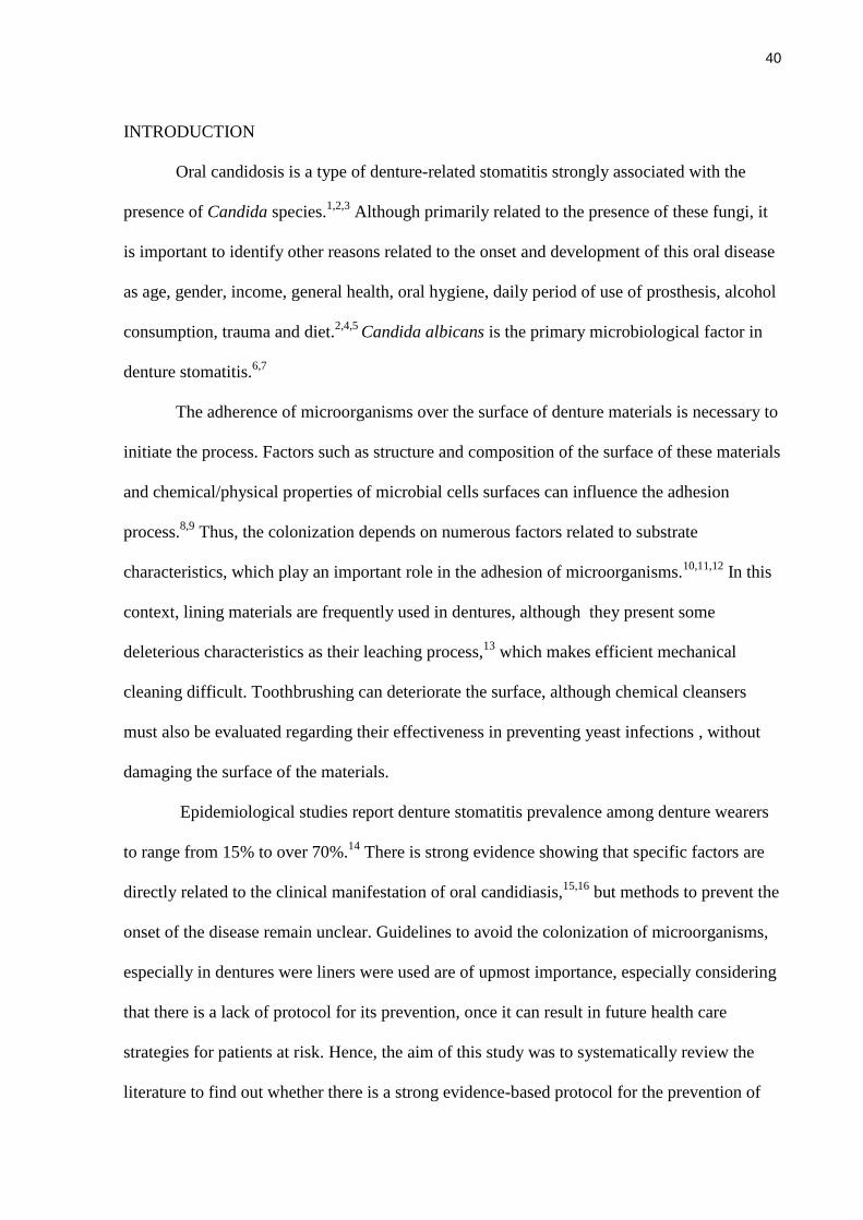
INTRODUCTION
Oral candidosis is a type of denture-related stomatitis strongly associated with the
presence of Candida species.1,2,3
Although primarily related to the presence of these fungi, it
is important to identify other reasons related to the onset and development of this oral disease
as age, gender, income, general health, oral hygiene, daily period of use of prosthesis, alcohol
consumption, trauma and diet.2,4,5
Candida albicans is the primary microbiological factor in
denture stomatitis.6,7
The adherence of microorganisms over the surface of denture materials is necessary to
initiate the process. Factors such as structure and composition of the surface of these materials
and chemical/physical properties of microbial cells surfaces can influence the adhesion
process.8,9
Thus, the colonization depends on numerous factors related to substrate
characteristics, which play an important role in the adhesion of microorganisms.10,11,12
In this
context, lining materials are frequently used in dentures, although they present some
deleterious characteristics as their leaching process,13
which makes efficient mechanical
cleaning difficult. Toothbrushing can deteriorate the surface, although chemical cleansers
must also be evaluated regarding their effectiveness in preventing yeast infections , without
damaging the surface of the materials.
Epidemiological studies report denture stomatitis prevalence among denture wearers
to range from 15% to over 70%.14
There is strong evidence showing that specific factors are
directly related to the clinical manifestation of oral candidiasis,15,16
but methods to prevent the
onset of the disease remain unclear. Guidelines to avoid the colonization of microorganisms,
especially in dentures were liners were used are of upmost importance, especially considering
that there is a lack of protocol for its prevention, once it can result in future health care
strategies for patients at risk. Hence, the aim of this study was to systematically review the
literature to find out whether there is a strong evidence-based protocol for the prevention of

41
Candida colonization in denture liners or at least disinfect these materials and if there is a
protocol to treat patients that use denture liners and had these materials colonized by Candida.

42
MATERIAL AND METHODS
Systematic literature search
This systematic review was performed according to PRISMA statement. Seven
databases were searched (Trip, Lilacs, Scopus, Pubmed/Medline, Scielo, Web of Science and
Cochrane Database of Systematic Reviews) using the following keywords: “denture liner” OR
relin* OR “tissue conditioner” AND “Candida” OR “denture stomatitis” OR “antifungal
agents” OR denture clean*. All papers found were evaluated and selected, following the
inclusion criteria, which was any in vitro, in situ or in vivo study, with protocols for
treatment, disinfection, cleaning or prevention of Candida colonization for denture liners or
tissue conditioners only. No restriction to language was made. The literature search was
carried out by two independent researchers (JAS and TPC) from November 2011 to April
2012, and all articles from 1950 to April 2012 were included. The references of papers
included in the review were carefully searched for additional papers that could be included,
including handsearch.
Selection criteria
According to the PRISMA statement,17
all abstracts were analyzed. A total of 152
articles were found. Abstracts were independently reviewed by two researchers. After the
screening and eligibility criteria had been individually accomplished, if a consensus was
reached, the article was included, if not, a third author was invited to discuss about the article.
Studies without protocol for treatment or prevention of Candida colonization were
excluded. The protocol should be tested in denture liners or tissue conditioners; if the
methodology was performed only using acrylic resin, the study was excluded. Studies could
also be related to other microorganisms involved in denture plaque development, dual or

43
multi species biofilm, but all studies had to contain Candida albicans as the main pathogen
related to denture stomatitis.
Three of the fifty two papers selected to have studies extracted could not be found
despite several attempts to contact the authors, the journals and libraries.18,19,20
Among all
studies included in the search strategy, 104 articles were included in the review. After
carefully reading the abstracts, 52 articles were selected to a full-text evaluation. Figure 1
indicates the step-by-step throughout the articles‟ selection. Thirteen articles were excluded
after assessed for eligibility due the reasons explained in Table 1.
Data collection and analysis
The study design, the type of microorganism and material found on articles were
recorded. The main findings of the studies, as results and conclusion were extracted.
Duo to a large variability of data, a meta-analysis was discarded. Different tools were
used to measure the antimicrobial activity, which resulted in distinct ways to quantify
Candida colonization, such as rate of pH decrease, colony forming unit count, weighing of
biofilm formation and the use of a kit to measure bioluminescence adenosine triphosphate
(ATP). Thus, only a qualitative investigation was possible and different comparisons were
made among studies trying to find out the best protocol to prevent, treat or eliminate Candida
colonization. Data were grouped in order to describe the main methods for prevention and
treatment.

44
RESULTS
Considering the 39 articles included in the study, articles were separated into two
categories, according to the strategy performed by the authors to achieve (1) a prevention
protocol or (2) a cleaning, treatment or disinfection strategy for denture liners. In the first
category (20 articles), all studies that had incorporation of any antimicrobial into a denture
liner were included (Table 2). Nystatin was often incorporated as antimicrobial agent in
denture liners (40%). Silver-based antimicrobials and fluconazole were present in four and
three studies respectively (20% and 15%), and chlorhexidine, amphotericin B and miconazole
were found to be incorporated in two studies (10%). Other antimicrobials were incorporated
in one study only such as zinc peroxide, clotrimazole, itraconazole, ketonazole, Melaleuca
alternifolia, human lactoferrin, magnesium oxide and triazine.
Category two included studies (19 articles) that had any protocol for cleaning, treating,
preventing or disinfecting denture liners that did not include the addition of an
antifungal/antibacterial agent (Table 3). The most frequent protocol tested (47.4%) was the
immersion of the liner into a sodium hypochlorite solution in various concentrations (0.5, 1, 2,
and 5.25%), with 0.5% already showing good results. Microwave irradiation and medical tabs
for dentures were also commonly used to prevent microbial colonization or disinfect denture
liners (26.3% and 31.6% respectively).

45
DISCUSSION
This systematic review has shown that attempts have been made to prevent denture
stomatitis or treat /disinfect denture liners commonly used for denture wearers. The
incorporation of fungicidal compounds into the denture liners or immersion of the denture
containing the denture liner in cleansing solutions was often performed to reach this objective.
The first idea to use readily available tools combining with other available agents is
pretty interesting to prevent the fungal infection. The addition of fungicidal compounds
directly to denture liners can be low cost, successful and especially attractive because it does
not require patient cooperation. In addition, denture cleansers may cause significant
deterioration of denture liners e.g. sodium hypochlorite.49
Tooth brushing may also cause
surface modifications, thereby facilitating colonization of microorganisms. The addition of
these compounds would lead to less detrimental effects when compared to the use of denture
cleansers. On the other hand, the amount of antifungal agents should be carefully planned as
they could be harmful to older people.69
In addition, the addition of antifungal to these liners
to prevent colonization by Candida should be carefully considered only for those patients at
high risk i.e., patients with xerostomia, previous history of denture stomatitis and motor
disabilities.
Despite the fact that there are differences between tissue conditioners and denture
liners, especially concerning viscosity no division on the results section was made particularly
because after the colonization of microorganisms, both seems to perform in a similar way
(and may also be used to treat denture stomatitis), providing no differentiation once a biofilm
is formed.70
In addition, once the aim of this review was to present an overview regarding
methods to decrease microorganisms counts, no comparisons among the materials concerning
their temporary or permanent use was performed once it is expected that temporary liners
will perform poorer than permanent ones.

46
Regarding the incorporation of antimicrobial agents into denture liner, nystatin seems
to be the gold standard in terms of prevention/treatment. All studies that used nystatin
presented at least a decrease in yeast levels. In a general way, the concentration of nystatin is
directly related to the inhibition of Candida growth. It is important to emphasize however,
that the concentration cannot be increased indiscriminately, because it can cause changes in
mechanical and chemical properties of these materials.49
Silver-based antimicrobial also
presented good results. All studies that mixed silver particles with denture liner showed a
fungicidal effect. Due to the few studies available, it was not possible to conclude if using
some specific agents may present beneficial antimicrobial effects or not. Once itraconozole
and miconazole were tested in only one study and showed good performance, or zinc peroxide
and triclosan (Microban), which demonstrated a poor result and again, were tested in only one
study, it is not possible to conclude the real potential of these agents. This is also true for the
use of Melaleuca alternifolia, which was effective in treating denture stomatitis in an in vivo
study.40
Several protocols were found to eliminate Candida colonization. Some denture
cleansers and irradiation (mainly immersed in water) with microwave presented decreased
yeasts counts, but immersion in sodium hypochlorite still remained the most effective
cleaning agent. Concentrations of 0.5, 1, 2 and 5.25% were tested and all presented a decrease
in Candida levels or complete elimination of these microorganisms. The ideal sodium
hypochlorite concentration must be studied once the immersion in this solution could
jeopardize the surface roughness over time, making yeast (re)colonization easier in long-term
analyses.60
Yet, it seems from the studies found that 0.5% sodium hypochlorite solution is
able to clean or disinfect denture liners, i.e. the lowest concentration could both clean and still
prevent surface deterioration caused by higher concentrations of NaOCl.

47
Strategies to prevent Candida colonization were also found. Olan-Rodriguez et al.54
tested in vivo the influence of sealers on denture liners, showing that this strategy could
decrease the colonization by yeasts and bacteria. Other strategy used was to compare
immersion technique vs. spraying technique through the use of chlorine dioxide as a denture
liner disinfectant. Although the immersion showed better results than the spray, both had poor
results,52
contrasting with Uludamar et al.,68
which demonstrated good results obtained with
the use of chlorine dioxide to treat palatal inflammation. Clinical studies have reported that
disrupting the biofilm may be more important than the use of antifungals or antimicrobials in
the prevention and treatment of denture stomatitis.40,41,59
As the etiology of the disease is
multifactorial, a set of attitudes together with treatment is necessary for the total elimination
of Candida.
The need of in vivo, prospective randomized clinical studies was evident. Only eight
studies were performed in a clinical scenario and in two of them, in vivo and in vitro studies
were performed together, thus demonstrating the lack of trials, prospective and retrospective
studies, which provide a better level of evidence. The results obtained in vitro do not
necessarily agree with the experience in vivo, because the oral cavity is an extremely rich
environment in saliva and nutrients, which could somewhat cancel the inhibitory effect
produced by the antimicrobials released from the liners in vitro. A possible reason why the
release of the antifungal agent included in the denture liner does not clinically stand is the
constant bathing in saliva in the mouth.68
In addition, in vitro the specimens are usually
smoother and have standardized surfaces, which gives a better picture for the antifungal tests;
in vivo, denture liners lose their plasticizers, becoming hardened and rough. Another
important reason why in vitro studies show good results that could not be real in clinical
practice is that usually in vitro studies are performed with planctonic cells or single species
biofilm. This means that the antifungal more easily penetrates into the biofilm when

48
compared to multi-species biofilms, which are complex communities with a matrix and
therefore improves the chances of survival for these microorganisms as they are more
protected.71
The only study found that tried to mimic the oral cavity used a microcosm biofilm
model and failed to show antifungal effect of triazine directly inserted in denture liners to
prevent Candida colonization.49
Due to these differences between in vitro and in vivo studies, a separation in the
results section depending on the design of the study was an option, but several protocols
tested have been tested only in vitro; thus it seemed more interesting to carry out an overview
about all the possibilities that could still be tested in vivo and what was not necessary to test
in laboratory studies based on our findings.
Unfortunately, due to the heterogeneous data, a meta-analysis could not be performed.
This does not mean that this review has no evidence, but increases the necessity to investigate
more protocols to in a near future, establish a definitive protocol, with the best material,
concentration or form of use of antifungals to achieve a good prognosis of preventing denture
related stomatitis when tissue conditioners or denture liners are being used. Still, it is possible
to state that based on in vitro results, nystatin could be of use mixed with denture liners, while
the best way to disinfect these materials is through immersion in a sodium hypochlorite
solution.

49
CONCLUSION
The addition of antifungal agents to denture liners appears to have some beneficial
effect to prevent Candida colonization, but a definitive concentration remains uncertain, as
the protocols found in literature are completely different. The use of 0.5% sodium
hypochlorite could be of help to disinfect denture liners. However, there is insufficient
reliable evidence to truly provide recommendations on which is the ideal cleaning method, or
whether the addition of antifungal agents is beneficial or not. Well designed randomized
controlled trials are needed to provide answers to these questions.

50
REFERENCES
1. Zomorodian K, Haghighi NN, Rajaee N, Pakshir K, Tarazooie B, Vojdani M, et al.
Assessment of Candida species colonization and denture-related stomatitis in complete
denture wearers. Med Mycol 2011;49:208-11.
2. Figueiral MH, Azul A, Pinto E, Fonseca PA, Branco FM, Scully C. Denture-related
stomatitis: identification of aetiological and predisposing factors - a large cohort. J Oral
Rehabil 2007;34:448-55.
3. Bilhan H, Sulun T, Erkose G, Kurt H, Erturan Z, Kutay O, et al. The role of Candida
albicans hyphae and Lactobacillus in denture-related stomatitis. Clin Oral Investig
2009;13:363-8.
4. Evren BA, Uludamar A, Işeri U, Ozkan YK. The association between socioeconomic
status, oral hygiene practice, denture stomatitis and oral status in elderly people living
different residential homes. Arch Gerontol Geriatr 2011;53:252-7.
5. Webb BC, Thomas CJ, Willcox MD, Harty DW, Knox KW. Candida- associated
denture stomatitis. Etiology and manage- ment: a review. Part I. factors influencing
distribution of Candida species in the oral cavity. Aus Dent J 1998;43:45-50.
6. Budtz-Jorgensen E. The significance of Candida albicans in denture stomatitis. Scand J
Dent Res 1974;82:151–90.
7. Arendorf TM, Walker DM. Denture stomatitis: a review. J Oral Rehabil 1987;14:217-
27.
8. Busscher HJ, Cowan MM, van der Mei HC. On the relative importance of specific and
non-specific approaches to oral microbial adhesion. FEMS Microbiol Rev 1992;8:199-
209.
9. Bellon-Fontaine MN, Mozes N, van der Mei HC, Sjollema J, Cerf O, Rouxhet PG et al.
A comparison of thermodynamic approaches to predict the adhesion of dairy

51
microorganisms to solid substrata. Cell Biophys 1990;17:93-106.
10. Minagi S, Miyake Y, Inagaki K, Tsuru H, Suginaka H. Hydrophobic interaction in
Candida albicans and Candida tropicalis adherence to various denture base resin
materials. Infect Immun 1985;47:11-4.
11. Pereira-Cenci T, Cury AA, Cenci MS, Rodrigues-Garcia RC. In vitro Candida
colonization on acrylic resins and denture liners: influence of surface free energy,
roughness, saliva, and adhering bacteria. Int J Prosthodont 2007;20:308-10.
12. Wright PS. The effect of soft lining materials on the growth of Candida albicans. J
Dent 1980;8:144-51.
13. Graham BS, Jones DW, Sutow EJ. An in vivo and in vitro study of the loss of
plasticizer from soft polymer-gel materials. J Dent Res 1991;70:870-3.
14. Gendreau L, Loewy ZG. Epidemiology and etiology of denture stomatitis. J
Prosthodont 2011;20:251-60.
15. Worthington HV, Clarkson JE, Bryan G, Furness S, Glenny AM, Littlewood A, et al.
Interventions for preventing oral mucositis for patients with cancer receiving treatment.
Cochrane Database Syst Rev 2011;13:CD000978.
16. de Souza RF, de Freitas Oliveira Paranhos H, Lovato da Silva CH, Abu-Naba'a L,
Fedorowicz Z, Gurgan CA. Interventions for cleaning dentures in adults. Cochrane
Database Syst Rev 2009;7:CD007395.
17. Moher D, Liberati A, Tetzlaff J, Altman DG, The PRISMA Group (2009). Preferred
Reporting Items for Systematic Reviews and Meta-Analyses: The PRISMA Statement.
PLoS Med 6:e1000097.
18. Tanaka T. The effect of tannic acid on attachment of Candida albicans to plate denture
lining material. Nichidai Koko Kagaku 1988;14:111-22.
19. el-Charkawi H, el-Said EA, Safouh HM, el-Raghi N. Effect of addition antimicrobial

52
agents to denture reliners. Egypt Dent J 1994;40:785-90.
20. Zhou M, Du L, Yang Z, Liao Y. A preliminary study of application of the antibacterial
solution containing silver ion to the surface of soft lining material. Sheng Wu Yi Xue
Gong Cheng Xue Za Zhi 2011;28:318-21.
21. Burns DR, Burns DA, DiPietro GJ, Gregory RL. Response of processed resilient
denture liners to Candida albicans. J Prosthet Dent 1987;57:507-12.
22. Granata JS, Staffanou RS. Evaluation of a new denture bath solution. J Prosthet Dent
1991;66:790-1.
23. Nikawa H, Yamamoto T, Hamada T, Rahardjo MB, Murata H. Commercial denture
cleansers--cleansing efficacy against Candida albicans biofilm and compatibility wit soft
denture-lining materials. Int J Prosthodont 1995;8:434-44.
24. Kulak Y, Kazazoglu E. In vivo and in vitro study of fungal presence and growth on
three tissue conditioning materials on implant supported complete denture wearers. J Oral
Rehabil. 1998;25:135-8.
25. Radford DR, Sweet SP, Challacombe SJ, Walter JD. Adherence of Candida albicans
to denture-base materials with different surface finishes. J Dent 1998;26:577-83.
26. McLain N, Ascanio R, Baker C, Strohaver RA, Dolan JW. Undecylenic acid inhibits
morphogenesis of Candida albicans. Antimicrob Agents Chemother 2000;44:2873-5.
27. Nevzatoğlu EU, Ozcan M, Kulak-Ozkan Y, Kadir T. Adherence of Candida albicans
to denture base acrylics and silicone-based resilient liner materials with different surface
finishes. Clin Oral Investig 2007;11:231-6.
28. Pereira-Cenci T, da Silva WJ, Cenci MS, Cury AA. Temporal changes of denture
plaque microbiologic composition evaluated in situ. Int J Prosthodont 2010;23:239-42.
http://www.ncbi.nlm.nih.gov/pubmed?term=McLain%20N%255BAuthor%255D&cauthor=true&cauthor_uid=10991877

53
29. Vural C, Ozdemir G, Kurtulmus H, Kumbuloglu O, Ozcan M. Comparative effects of
two different artificial body fluids on Candida albicans adhesion to soft lining materials.
Dent Mater J 2010;29:206-12.
30. Douglas WH, Walker DM. Nystatin in denture liners--an alternative treatment of
denture stomatitis. Br Dent J 1973;135:55-9.
31. Thomas CJ, Nutt GM. The in vitro fungicidal properties of Visco-gel, alone and
combined with nystatin and amphotericin B. J Oral Rehabil 1978;5:167-72.
32. Gettleman L, Fischer DJ, Farris C. Self-sanitizing soft denture liners: paradoxical
results. J Biomed Mater Res 1983;17:731-4.
33. Quinn DM. The effectiveness, in vitro, of miconazole and ketoconazole combined
with tissue conditioners in inhibiting the growth of Candida albicans. J Oral Rehabil
1985;12:177-82.
34. Schneid TR. An in vitro analysis of a sustained release system for the treatment of
denture stomatitis. Spec Care Dentist 1992;12:245-50.
35. Matsuura T, Abe Y, Sato Y, Okamoto K, Ueshige M, Akagawa Y. Prolonged
antimicrobial effect of tissue conditioners containing silver-zeolite. J Dent 1997;25:373-7.
36. Nikawa H, Yamamoto T, Hamada T, Rahardjo MB, Murata H, Nakanoda S.
Antifungal effect of zeolite-incorporated tissue conditioner against Candida albicans
growth and/or acid production. J Oral Rehabil 1997;24:350-7.
37. Chow CK, Matear DW, Lawrence HP. Efficacy of antifungal agents in tissue
conditioners in treating candidiasis. Gerodontology 1999;16:110-8.
38. Lefebvre CA, Wataha JC, Cibirka RM, Schuster GS, Parr GR. Effects of triclosan on
the cytotoxicity and fungal growth on a soft denture liner. J Prosthet Dent 2001;85:352-6.

54
39. Akiba N, Hayakawa I, Keh ES, Watanabe A. Antifungal effects of a tissue conditioner
coating agent with TiO2 photocatalyst. J Med Dent Sci 2005;52:223-7.
40. Catalán A, Pacheco JG, Martínez A, Mondaca MA. In vitro and in vivo activity of
Melaleuca alternifolia mixed with tissue conditioner on Candida albicans. Oral Surg Oral
Med Oral Pathol Oral Radiol Endod 2008;105:327-32.
41. Geerts GA, Stuhlinger ME, Basson NJ. Effect of an antifungal denture liner on the
saliva yeast count in patients with denture stomatitis: a pilot study. J Oral Rehabil
2008;35:664-9.
42. Yamamoto D, Shinohara Y, Nagadome H, Terada Y. Development of tissue
conditioner capable of binding with anti-microbial protein lactoferrin. J Prosthodont Res
2009;53:136-41.
43. Falah-Tafti A, Jafari AA, Lotfi-Kamran MH, Fallahzadeh H, Hayan RS. A
Comparison of the efficacy of Nystatin and Fluconazole Incorporated into Tissue
Conditioner on the In vitro Attachment and Colonization of Candida Albicans. Dent Res J
2010;7:18-22.
44. Radnai M, Whiley R, Friel T, Wright PS. Effect of antifungal gels incorporated into a
tissue conditioning material on the growth of Candida albicans. Gerodontology
2010;27:292-6.
45. Kanathila H, Bhat AM, Krishna PD. The effectiveness of magnesium oxide combined
with tissue conditioners in inhibiting the growth of Candida albicans: an in vitro study.
Indian J Dent Res 2011;22:613.
46. Uchimaru M, Sakai T, Moroi R, Shiota S, Shibata Y, Deguchi M, et al. Antimicrobial
and antifungal effects of tissue conditioners containing a photocatalyst. Dent Mater J
2011;30:691-9.

55
47. Chladek G, Mertas A, Barszczewska-Rybarek I, Nalewajek T, Zmudzki J, Król W, et
al. Antifungal activity of denture soft lining material modified by silver nanoparticles-a
pilot study. Int J Mol Sci 2011;12:4735-44.
48. Nam KY. In vitro antimicrobial effect of the tissue conditioner containing silver
nanoparticles. J Adv Prosthodont 2011;3:20-4.
49. de Moraes AP, Barwaldt CK, Nunes TZ, Sarkis-Onofre R, Ogliari FA, Boscato N, et
al. Effect of triazine derivative added to denture materials on a microcosm biofilm model.
J Biomed Mater Res Part B 2012:[in press]
50. Masella RP, Dolan CT, Laney WR. The prevention of the growth of Candida on
silastic 390 soft liner for dentures. J Prosthet Dent 1975;33:250-7.
51. Baysan A, Whiley R, Wright PS. Use of microwave energy to disinfect a long-term
soft lining material contaminated with Candida albicans or Staphylococcus aureus. J
Prosthet Dent 1998;79:454-8.
52. Furukawa KK, Niagro FD, Runyan DA, Cameron SM. Effectiveness of chlorine
dioxide in disinfection on two soft denture liners. J Prosthet Dent 1998;80:723-9.
53. Dixon DL, Breeding LC, Faler TA. Microwave disinfection of denture base materials
colonized with Candida albicans. J Prosthet Dent 1999;81:207-14.
54. Olan-Rodriguez L, Minah GE, Driscoll CF. Candida albicans colonization of surface-
sealed interim soft liners. J Prosthodont 2000;9:184-8.
55. Price C, Waters MG, Williams DW, Lewis MA, Stickler D. Surface modification of
an experimental silicone rubber aimed at reducing initial candidal adhesion. J Biomed
Mater Res 2002;63:122-8.
56. Glass RT, Bullard JW, Conrad RS, Blewett EL.Evaluation of the sanitization

56
effectiveness of a denture-cleaning product on dentures contaminated with known
microbial flora. An in vitro study. Quintessence Int 2004;35:194-9.
57. Yilmaz H, Aydin C, Bal BT, Ozçelik B. Effects of disinfectants on resilient denture-
lining materials contaminated with Staphylococcus aureus, Streptococcus sobrinus, and
Candida albicans. Quintessence Int 2005;36:373-81.
58. Mese, A. Mese, S. Effect of microwave energy on fungal growth of resilient denture
liner material. Biotechnol Biotec Eq 2007;21:91-3.
59. Zuluaga M, Javier D; Tovar A, Maritza E; Garcia R, Katherine J. Comparación de la
resolución de la estomatitis subprotesis tratada con acondicionador de tejido blando y
material de rebase duro autopolimerizable. Rev Fac Odontol Univ Antioq 2007;19:21-34.
60. Buergers R, Rosentritt M, Schneider-Brachert W, Behr M, Handel G, Hahnel S.
Efficacy of denture disinfection methods in controlling Candida albicans colonization in
vitro. Acta Odontol Scand 2008;66:174-80.
61. Mima EG, Pavarina AC, Neppelenbroek KH, Vergani CE, Spolidorio DM, Machado
AL. Effect of different exposure times on microwave irradiation on the disinfection of a
hard chairside reline resin. J Prosthodont 2008;17:312-7.
62. Boscato N, Radavelli A, Faccio D, Loguercio AD. Biofilm formation of Candida
albicans on the surface of a soft denture-lining material. Gerodontology 2009;26:210-3.
63. Ferreira MA, Pereira-Cenci T, Rodrigues de Vasconcelos LM, Rodrigues-Garcia RC,
Del Bel Cury AA. Efficacy of denture cleansers on denture liners contaminated with
Candida species. Clin Oral Investig 2009;13:237-42.
64. Gedik H, Ozkan YK. The effect of surface roughness of silicone-based resilient liner
materials on the adherence of Candida albicans and inhibition of Candida albicans with
different disinfectants. Oral Health Prev Dent 2009;7:347-53.

57
65. Boscato N, Delavi JD, Muller L, Pereira-Cenci T, Imanishi SW. Influence of varnish
application on a tissue conditioner: analysis of biofilm adhesion. Gerodontology
2010;27:207-10.
66. Vieira AP, Senna PM, Silva WJ, Del Bel Cury AA. Long-term efficacy of denture
cleansers in preventing Candida spp. biofilm recolonization on liner surface. Braz Oral
Res 2010;24:342-8.
67. Hahnel S, Rosentritt M, Bürgers R, Handel G, Lang R. Candida albicans biofilm
formation on soft denture liners and efficacy of cleaning protocols. Gerodontology 2011;
May 17.
68. Uludamar A, Özyeşil AG, Ozkan YK. Clinical and microbiological efficacy of three
different treatment methods in the management of denture stomatitis. Gerodontology
2011;28:104-10.
69. Nikawa H, Samaranayake LP, Tenovuo J, Hamada T. The effect of antifungal agents
on the in vitro susceptibility of Candida albicans to apo-lactoferrin. Arch Oral Biol
1994;39:921-3.
70. Marsh PD. Microbial ecology of dental plaque and its significance in health and
disease. Adv Dent Res 1994;8:263-71.
71. Cannon RD, Chaffin WL. Oral colonization by Candida albicans. Crit Rev Oral Biol
Med 1999;10:359-383.

58
Figure 1 – Selection criteria according PRISMA statement.

59
Table 1 - Reasons for exclusion of papers selected to assess the full-text.
Author Reason
Burns et al., 198721
Candida growth was analyzed in new denture liners; no
treatment/prevention protocol was tested.
Tanaka, 198818
Article not found.
Granata and Staffanou, 199122
The influence of immersion of denture liners in denture solutions
on surface hardness was studied.
el-Charkawi et al., 1994 19
Article not found.
Nikawa et al., 199523
Denture liners had their surface porosity tested after immersion in
denture cleansers.
Kulak and Kazazoglu, 199824
No protocol for prevention, cleaning or treatment was studied.
Radford et al., 199825
The surface roughness and its implication on Candida adhesion
were tested.
McLain et al., 200026
Only the effect of tissue conditioners on fungal growth was
studied.
Nevzatoğlu et al., 200727
The influence of different surface finishes on Candida albicans
adherence was studied.
Pereira-Cenci et al., 200711
No prevention protocol was tested, only Candida adhesion to
denture materials.
Pereira-Cenci et al., 201028
The influence of time, liner and roughness in the composition of
biofilm was analyzed; no treatment/prevention protocol was
tested.
Vural et al., 201029
The influence of polymerization process was tested on Candida
albicans adhesion.
Zhou et al., 201120
Article not found.

60
Table 2. Studies that had incorporation of antifungal agent to denture liners.
Manuscript Study
Design
Microorganism Material Results
Douglas and
Walker,
197330
In vitro and
In vivo
Candida albicans. Tempo; Coe Confort; Nystan/Tempo
(400000 and 800000 units).
In vitro: Tempo and Coe confort presented some
fungicidal effect, but Tempo was better.
Tempo/Nystan (400000 units): effective until day 20
Tempo/Nystan (800000 units): activity for at least 50
days. In vivo: There was no difference between
patients treated with Tempo + Nystatin Tablet or
Tempo/Nystatin mixture.
Tromas and
Nutt, 197831
In vitro Candida albicans;
Candida krusei;
Candida tropicalis.
Visco-Gel;
Nystatin (500000 and 1000000
units);
Amphotericin B (10mg and 20mg).
Visco-gel alone and mixed with amphotericin B was
not effective, only visco-gel/nystatin was efficient and
in higher concentrations the results were better.
Gettleman et
al., 198332
In vitro Candida albicans;
Staphylococcus
aureus.
Super-soft; Molloplast-B;
Lucitone 199; Syloid 244 silica and
alpha-cellulose fiber treated with zinc
peroxide (10%) into Molloplast-B
and Lucitone 199; Zinc peroxide (5
and 20%) into Molloplast-B and
Super-soft).
C. albicans presented more susceptibility to growth
inhibition. Silica filler showed worse results than
cellulose filler. Zinc peroxide (5 e 20%) was not
effective as antimicrobial.
Quinn, 198533
In vitro Candida albicans.
Ivoseal; Viscogel; Fitt; Nystatin
(500000 Units); Amphotericin B (10,
For amphotericin, no differences were found between
the concentrations and inhibitory effect was time and

61
20 mg); Miconazole (250 mg);
Ketonazole (200 mg).
material dependent. The best results were obtained
with nystatin, miconazole and ketonazole, while all
combinations presented inhibitory effect even after 15
days.
Schneid,
199234
In vitro Candida albicans.
Lynal Tissue Conditioner;
Temporary Reliner; Chlorhexidine
(250, 500, 1000 mg/unit);
Clotrimazole (250, 500, 1000
mg/unit); Fluconazole (250, 500,
1000 mg/unit); Nystatin (125, 250,
500 mg/unit).
Nystatin presented the best results. All groups showed
growth inhibition; however, they were time and
concentration dependent.
Matsuura et
al., 199735
In vitro Candida albicans;
Staphylococcus
aureus;
Pseudomonas
aeruginosa.
Visco-gel; GC Soft-Liner;
Fitt; SR-Ivoseal;
Shofu Tissue Conditioner;
Zeomic (Zeolite) - 2 (wt/wt)%.
All tissue conditioners mixed with zeomic presented
antimicrobial effects with or without immersion in
saliva for 4 weeks.
Nikawa et al.,
199736
In vitro Candida albicans.
GC Soft Liner;
Coe Confort;
Zeomic (Zeolite) - 1, 2, 3, 4 and 5
(wt/wt)% or 1, 2, 3, 4 and 5
(vol/vol)%
There was a dose-dependent effect. Zeolite specimens
showed higher antifungal effect than Coe Confort,
followed by Soft Liner. The antifungal effect of the
Zeomic was significantly decreased by the presence of
saliva.
Chow et al.,
199937
In vitro Candida albicans.
Coe Soft; Viscogel;
Fitt; Nystatin (1, 3, 5, 7, 9, 11%
In the presence of saliva, all groups had higher
fungicidal effect. The concentration of 5% wt/wt had

62
wt/wt); Fluconazole (1, 3, 5, 7, 9,
11% wt/wt); Itraconazole (1, 3, 5, 7,
9, 11% wt/wt);
the higher fungicidal activity for all combinations. The
best result was obtained with 5% wt/wt itraconazole
mixed with Coe Soft.
Lefebvre et
al., 200138
In vitro Candida albicans. PermaSoft;
Microban (1 part for 80);
No difference was found between groups with or
without microban.
Akiba et al.,
200539
In vitro Candida albicans. Fictioner;
Top Coat;
TiO2 – 0, 2 and 3g.
The viability significantly decreased with increased
concentration of TiO2 in the coating agents. The
viability also significantly decreased with increased
radiation time.
Catalan et al.,
200840
In vitro and
In vivo
Candida albicans. In vitro: Fitt; Lynal; Coe-Comfort;
Melaleuca alternifolia (0.5, 1, 2 and
4 ml); Nystatin (1ml).
In vivo: Coe-Comfort + M.
alternifolia (1ml); Coe-Comfort +
Nystatin (2ml); Coe-Comfort.
In vitro: Nystatin was effective in all tissue
conditioner. M. alternifolia was efficient mixed with
Fitt and Coe Confort at concentration of least 1 ml.
Lynal was ineffective in all concentratios.
In vivo: Coe-Confort mixed with nystatin and M.
alternifolia presented better results than control.
Geerts et al.,
200841
In vivo Patients with
denture-related
stomatitis
Visco gel;
Mycostatin (500000 U).
The treatment with liner and nystatin-incorporated
only showed a decrease in yeast counts until day 4.
After that, the control group significantly increased
and the test group remained with low counts until day
7, increasing until day 14, but still showed lower yeast
counts than day 0.
Yamamoto et
al., 200942
In vitro Candida albicans. Shofu Tissue Conditioner II
Antimicrobial protein solution,
Fungal viability was significantly lower than the
control in both concentrations, and between 4 and 8

63
human lactoferrin; Cation exchange
resin, Toyopearl CM650 4 an 8 wt%.
wt% was not significantly different.
Falah-Tafti et
al., 201043
In vitro Candida albicans. Acropars;
Nystatin (1, 3, 5, 10% wt/wt);
Fluconazole (1, 3, 5, 10% wt/wt).
Nystatin in all concentrations completely inhibited the
attachment and colonization of C. albicans, but in the
case of fluconazole, only the concentration of 10%
showed complete inhibition of Candida colonization.
Radnai et al.,
201044
In vitro Candida albicans. Visco Gel; Chlorhexidine (5, 10, 15,
20 and 25% v/v); Miconazole (5, 10,
15, 20 and 25% v/v).
Miconazole inhibits the growth of C. albicans and the
higher the concentration, the higher the inhibition.
Kanathila et
al., 201145
In vitro Candida albicans. Visco Gel;
GC Soft liner;
Magnesium oxide 1, 3, 5 and 7%.
GC presented best results. The inhibition of Candida
growth increased in mixtures with higher
concentrations of magnesium oxide.
Uchimaru et
al., 201146
In vitro E. coli; S. mutans;
S. aureus; C.
albicans.
Shofu Tissue Conditioner II;
Photohap;
Concentration of 0, 10, 15 and 20%.
The colony-forming units were lower in irradiated
specimens. Candida albicans decrease was not long-
time effective.
Chladek et al.,
201147
In vitro Candida albicans. Ufi Gel; AgNPs (10, 20, 40, 80,120
and 200 ppm).
The highest concentration of AgNPs leade to the best
antifungal efficacy.
Nam, 201148
In vitro S. aureus;
S. mutans;
Candida albicans.
GC Soft-Liner;
Silver nanoparticles (0, 0.1, 0.5, 1.0,
2.0 and 3.0% v/v).
Silver nanoparticle at 1.0% concentration was able to
eliminate S. aureus and S. mutans and 2.0% eliminated
C. albicans.
De Moraes, et
al., 201249
In vitro Streptococci and
Candida species
CoeSoft; SoftConfort; Kooliner;
Triazine (0, 2.5, 5 and 10%)
CoeSoft showed higher counts of Candida and
Streptococci and the addition of triazine did not result
in decreased counts of total microorganisms.

64
Table 3. Studies on cleaning or prevention protocols for denture liners without the incorporation of antifungals directly to the materials.
Manuscript Study
Design
Microorganism Material Results
Masella et al.,
197550
Prevention /
treatment –
In vitro
C. albicans; C.
glabrata; C. tropicalis;
C. parapsilosis.
Silastic 390; Pro-Kem; Zephiran;
Listerine; Mersene; Cidex; Water
60o C.; Zinc undecylenate.
All denture cleaners were effective at an appropriate
concentration, except for Zinc undecylenate (5%)
which was not totally effective.
Baysan et al.,
199851
Disinfection
– In vitro
Candida albicans;
Staphylococcus aureus.
Molloplast-B;
Sodium Hypochlorite 2%.
The best method for disinfection was immersion in
sodium hypochlorite, followed by microwave
irradiation.
Furukawa et
al. 199852
Disinfection
– In vitro
Candida albicans;
Eschericia coli;
Staphylococcus aureus.
Coe Soft;
Coe Comfort;
Chlorine Dioxide;
Immersion technique was more effective than spray
technique. However, chlorine dioxide was not able to
achieve the minimal disinfection standard for both
denture liners tested.
Dixon et al.,
199953
Disinfection
– In vitro
Candida albicans.
Molloplast-B;
Permaflex;
Lucitone 199.
C. albicans was killed by 5-minute irradiation, with
specimens immersed in water. Dry irradiation did not
effectively sterilize any of the materials.
Olan-
Rodriguez et
al., 200054
Prevention –
In vivo
Healthy patients. Coe-Soft;
Palaseal;
Mono-Poly.
The soft-denture liner sealed presented less
colonization by yeasts and bacteria. Among sealers,
no difference was found.
Price et al.,
200255
Prevention –
In vitro
Candida albicans.
Silicone material;
Silane;
Modification of surface through plasma increased
Candida adherence while silane treatment
significantly reduced it.
Glass et al., Cleaning – In Staphylococcus aureus; Methylmethacrylate dentures The use of Medical tabs for dentures eliminated C.

65
200456
vitro Pseudomas aeniginosa;
Bacillus cereus;
Candida albicans;
Herpes simplex virus 1;
without soft liners (hard
dentures);
Methylmethacrjlate dentures
containing processed soft liners
(soft-liner dentures);
Medical tabs for dentures.
albicans, S. aureus, P. aeruginosa and HSV-1 target
virus in soft denture liner. Hard denture eradicated C.
albicans and HSV-1 target virus and substantially
reduced populations of S. aureus and P. aeruginosa.
B cereus appeared to be the least affected by the one-
time use of Medical tabs.
Yilmaz et al.,
200557
Disinfection
– In vitro
Staphylococcus aureus;
Streptococcus sobrinus;
Candida albicans.
Tempo; Immediate; Flexacryl
Soft; Ufi Gel P; Deconex 5%;
Savlex 3.5%; Sodium
Hypochlorite 2%; Sodium
Hypochlorite 5.25%.
The best result was obtained by immersion in 5.25%
sodium hypochlorite for all liners, but in all groups
the disinfectant solution significantly reduced
microorganisms.
Meşe and
Meşe, 200758
Disinfection
– In vitro
Candida albicans.
Vertex Soft;
Sodium hypochlorite 2%;
Alkaline peroxide solutions.
Sodium hypochlorite showed the best results,
followed by exposition to microwave energy (650 W
for 2.5 minutes per side), but both were extremely
effective. The worst result was obtained with alkaline
peroxide solutions.
Zuluaga et al.,
200759
Treatment–
In vivo
Patients with denture-
related stomatitis
Coe Confort;
Kool Liner;
The treatment with both tissue conditioners was
effective.
Buergers et
al., 200860
Disinfection
– In vitro
Candida albicans.
Mucopren E; Hydrogen peroxide
3%; Sodium hypochlorite 1%;
Glutaraldehyde 2%; Household
vinegar; Listerine coolmint;
Plax (triclosan 0.3%); Blend-a-
The immersion in sodium hypochlorite (1%; 10 min),
microwave irradiation immersed in water (800 W; 6
min), and the immersion of effervescent cleansing
tabs (Blend-a-dent tabs; 10 min) proved to be
effective against C. albicans colonization.

66
dent 2 Phasen tabs.
Mima et al.,
200861
Disinfection
– In vitro
P. aeruginosa; S.
aureus; C. albicans;
Bacillus subtilis
Tokuso Rebase Fast Set. Irradiation for 3 minutes or more at 650 w showed
consistent sterilization to all microorganisms.
Boscato et al.,
200962
Prevention –
In vivo
Biofilm formation QuickLine; Colgate Triple Action
toothpaste; Sodium hypochlorite
0.5%.
Irrespective of time, daily prosthetic hygiene with
soft toothbrush and toothpaste presented the lowest
biolfilm formation.
Ferreira et al.,
200963
Cleaning – In
vitro
Candida albicans;
Candida glabrata.
CoeSoft; Kooliner; Ufi Gel P;
Polident; Efferdent; Sodium
hypochlorite 0.5%.
0.5% NaOCl was effective for both species. C.
glabrata showed higher number of remaining cells in
all treatments, except for NaOCl.
Gedik and
Ozkan, 200964
Disinfection
– In vitro
Candida albicans, Ufi Gel P; Ufi Gel C; Mollosil;
Soft-Liner; Moloplast B; Luci
Soft; Efferdent; Polident;
Steradent; Corega; Denclen;
Klorhex; Axion (5.25% NaOCl);
Cleaning with NaOCl led to significantly lower
counts compared to the other disinfectants tested.
Boscato et al.,
201065
Prevention –
In vivo
Biofilm formation Coe Confort;
Varnish;
The use of varnish was not recommended, as it
provided higher biofilm formation.
Vieira et al,,
201066
Cleaning – In
vitro
Candida albicans;
Candida glabrata.
Lucitone 550;
Kooliner;
Polident;
Efferdent;
Sodium hypochlorite 0.5%.
The only effective treatment to clean the liner
surfaces was the use of sodium hypochlorite. Both
denture cleansers were responsible to decrease
Candida counts. C. glabrata showed significantly
higher cell counts in comparison to C. albicans when
treated with both alkaline denture cleansers.

67
Hahnel et al.,
201167
Cleaning – In
vitro
Candida albicans.
Ufi Gel SC; GC Reline soft;
Silagum comfort; Mucopren soft;
Blend-A-Dent; 2% NaOCl;
Toothbrush Professional Care
9900 Braun Oral B and
Sensodyne.
The best result was achieved by immersion in
NaOCl. For long-term Candida albicans biofilm
analysis, NaOCl also showed better results; however,
mechanical cleaning was statistically superior to
chemical denture cleaner Blend-A-Dent.
Uludamar et
al., 201168
Treatment –
In vivo
Candida albicans.
Visco-gel;
chlorine dioxide (0.8%);
Corsodyl mouthrinse (0.2%)
The three treatments decreased palatal inflammation,
but 0.8% chlorine dioxide and 0.2% chlorhexidine
gluconate resulted in better results.
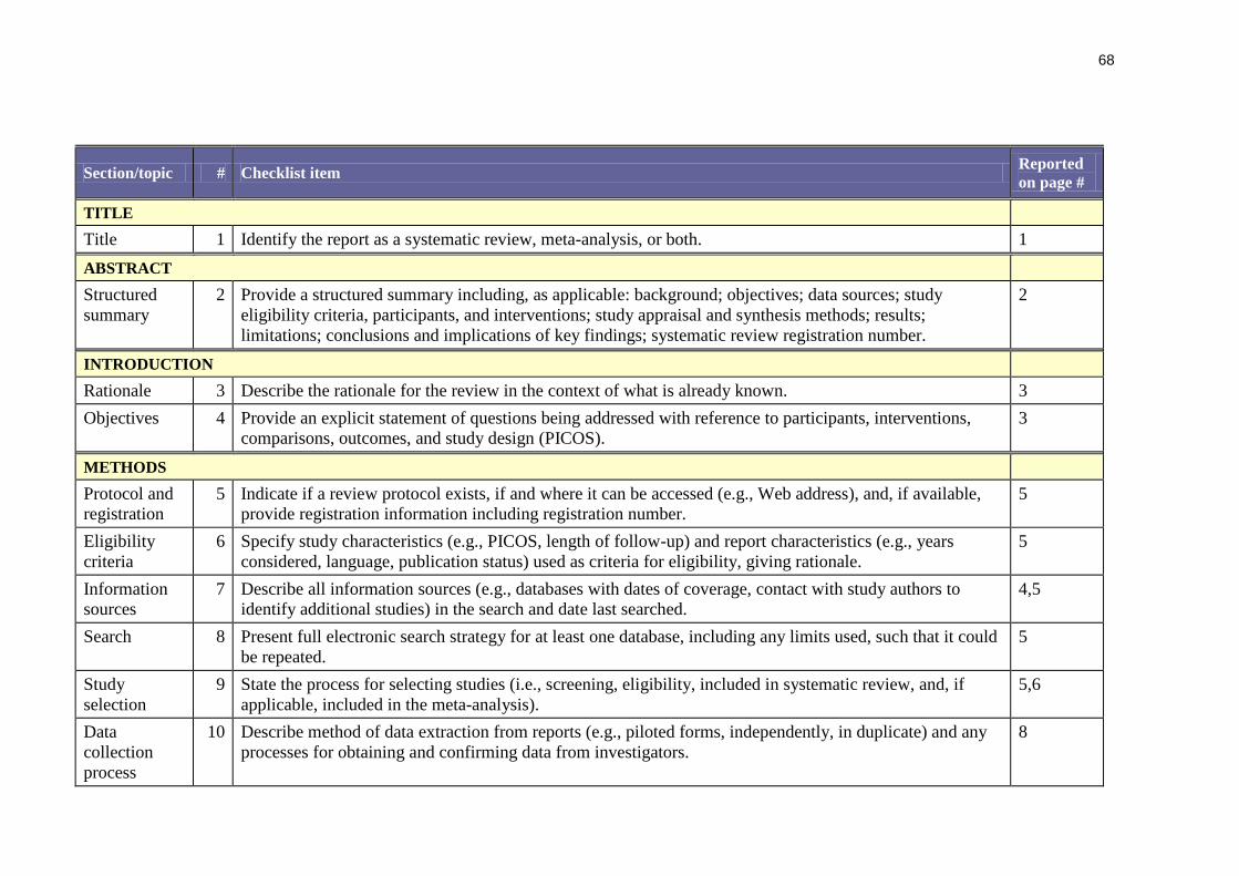
68
Section/topic # Checklist item Reported
on page #
TITLE
Title 1 Identify the report as a systematic review, meta-analysis, or both. 1
ABSTRACT
Structured
summary
2 Provide a structured summary including, as applicable: background; objectives; data sources; study
eligibility criteria, participants, and interventions; study appraisal and synthesis methods; results;
limitations; conclusions and implications of key findings; systematic review registration number.
2
INTRODUCTION
Rationale 3 Describe the rationale for the review in the context of what is already known. 3
Objectives 4 Provide an explicit statement of questions being addressed with reference to participants, interventions,
comparisons, outcomes, and study design (PICOS).
3
METHODS
Protocol and
registration
5 Indicate if a review protocol exists, if and where it can be accessed (e.g., Web address), and, if available,
provide registration information including registration number.
5
Eligibility
criteria
6 Specify study characteristics (e.g., PICOS, length of follow-up) and report characteristics (e.g., years
considered, language, publication status) used as criteria for eligibility, giving rationale.
5
Information
sources
7 Describe all information sources (e.g., databases with dates of coverage, contact with study authors to
identify additional studies) in the search and date last searched.
4,5
Search 8 Present full electronic search strategy for at least one database, including any limits used, such that it could
be repeated.
5
Study
selection
9 State the process for selecting studies (i.e., screening, eligibility, included in systematic review, and, if
applicable, included in the meta-analysis).
5,6
Data
collection
process
10 Describe method of data extraction from reports (e.g., piloted forms, independently, in duplicate) and any
processes for obtaining and confirming data from investigators.
8

69
Data items 11 List and define all variables for which data were sought (e.g., PICOS, funding sources) and any
assumptions and simplifications made.
8
Risk of bias
in individual
studies
12 Describe methods used for assessing risk of bias of individual studies (including specification of whether
this was done at the study or outcome level), and how this information is to be used in any data synthesis.
N/A
Summary
measures
13 State the principal summary measures (e.g., risk ratio, difference in means). 8
Synthesis of
results
14 Describe the methods of handling data and combining results of studies, if done, including measures of
consistency (e.g., I2) for each meta-analysis.
N/A
Page 1 of 2
Section/topic # Checklist item Reported
on page #
Risk of bias
across studies
15 Specify any assessment of risk of bias that may affect the cumulative evidence (e.g., publication bias,
selective reporting within studies).
N/A
Additional
analyses
16 Describe methods of additional analyses (e.g., sensitivity or subgroup analyses, meta-regression), if done,
indicating which were pre-specified.
N/A
RESULTS
Study
selection
17 Give numbers of studies screened, assessed for eligibility, and included in the review, with reasons for
exclusions at each stage, ideally with a flow diagram.
Table 1
and figure
1
Study
characteristics
18 For each study, present characteristics for which data were extracted (e.g., study size, PICOS, follow-up
period) and provide the citations.
Table 2
and 3
Risk of bias
within studies
19 Present data on risk of bias of each study and, if available, any outcome level assessment (see item 12). n/a
Results of
individual
studies
20 For all outcomes considered (benefits or harms), present, for each study: (a) simple summary data for each
intervention group (b) effect estimates and confidence intervals, ideally with a forest plot.
Table 2
and 3

70
Synthesis of
results
21 Present results of each meta-analysis done, including confidence intervals and measures of consistency. Not
applicable
Risk of bias
across studies
22 Present results of any assessment of risk of bias across studies (see Item 15). n/a
Additional
analysis
23 Give results of additional analyses, if done (e.g., sensitivity or subgroup analyses, meta-regression [see
Item 16]).
N/A
DISCUSSION
Summary of
evidence
24 Summarize the main findings including the strength of evidence for each main outcome; consider their
relevance to key groups (e.g., healthcare providers, users, and policy makers).
9,10,11,12
Limitations 25 Discuss limitations at study and outcome level (e.g., risk of bias), and at review-level (e.g., incomplete
retrieval of identified research, reporting bias).
12
Conclusions 26 Provide a general interpretation of the results in the context of other evidence, and implications for future
research.
13
FUNDING
Funding 27 Describe sources of funding for the systematic review and other support (e.g., supply of data); role of
funders for the systematic review.
Not
applicable
From: Moher D, Liberati A, Tetzlaff J, Altman DG, The PRISMA Group (2009). Preferred Reporting Items for Systematic Reviews and Meta-Analyses: The PRISMA Statement. PLoS Med 6(6): e1000097.
doi:10.1371/journal.pmed1000097
For more information, visit: www.prisma-statement.org

ARTIGO 2
Biofilm formation on denture liners in a randomised, controlled in situ trial§
Fernanda Valentini, Murilo Souza Luz, Noéli Boscato, Tatiana Pereira-Cenci
Graduate Program in Dentistry, Federal University of Pelotas, Pelotas, RS, Brazil
Running Title: Biofilm in denture liners in a controlled trial
Keywords: denture liners; biofilms; Candida; randomized trial; in situ; fungal
adherence
Corresponding author:
Rua Gonçalves Chaves, 457, Pelotas, RS, Brazil. 96015-560
Tel./Fax: +55-53-3222-6690
E-mail: [email protected]
§ Artigo formatado segundo as normas do periódico Archives of Oral Biology

72
Biofilm formation on denture liners in a randomised, controlled in situ trial
ABSTRACT
Objective: This randomized, in situ clinical trial assessed how biofilm composition is affected
by time and denture material type in denture wearers with and without denture stomatitis. The
randomized clinical trial design was performed using CONSORT statement.
Design: Twelve specimens of acrylic resin (control) and two denture liners (silicone-based or
acrylic resin based, depending on the experimental phase) were manufactured and inserted
into the surface intaglio of 28 denture wearers. Biofilm was formed in two phases of 21 days,
and counts of viable micro-organisms in the accumulating biofilm were determined and
converted to colony forming units per unit surface area after 7, 14 and 21days of biofilm
formation. The surface structure was analyzed by scanning electron microscopy (SEM). Data
were analyzed by three-way ANOVA followed by Tukey test to assess differences among
health condition (healthy or candidiasis), materials and time point.
Results: Non-albicans Candida species counts were higher in candidiasis patients with
silicone-based denture liners (p=0.01). Candidiasis patients showed higher mutans
streptococci counts after 7 days (p=0.0041), but this difference disappeared after 14-21 days
of biofilm formation. Lactobacilli and total micro-organisms counts were higher in denture
liners, irrespective of the health condition or aging. SEM analysis showed that denture liners
presented rougher surfaces while aging increases micro-organisms adhesion irrespective of
the material tested.
Conclusion: Silicone-based denture liners should be avoided in candidiasis patients as these
materials showed increased non-albicans species counts. Clinically, aging provided increased
roughness and therefore higher micro-organisms adhesion.
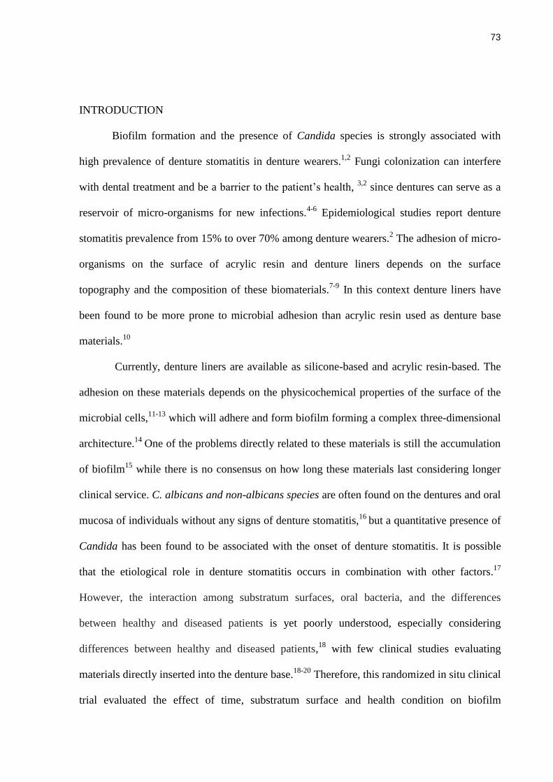
73
INTRODUCTION
Biofilm formation and the presence of Candida species is strongly associated with
high prevalence of denture stomatitis in denture wearers.1,2
Fungi colonization can interfere
with dental treatment and be a barrier to the patient‟s health, 3,2
since dentures can serve as a
reservoir of micro-organisms for new infections.4-6
Epidemiological studies report denture
stomatitis prevalence from 15% to over 70% among denture wearers.2 The adhesion of micro-
organisms on the surface of acrylic resin and denture liners depends on the surface
topography and the composition of these biomaterials.7-9
In this context denture liners have
been found to be more prone to microbial adhesion than acrylic resin used as denture base
materials.10
Currently, denture liners are available as silicone-based and acrylic resin-based. The
adhesion on these materials depends on the physicochemical properties of the surface of the
microbial cells,11-13
which will adhere and form biofilm forming a complex three-dimensional
architecture.14
One of the problems directly related to these materials is still the accumulation
of biofilm15
while there is no consensus on how long these materials last considering longer
clinical service. C. albicans and non-albicans species are often found on the dentures and oral
mucosa of individuals without any signs of denture stomatitis,16
but a quantitative presence of
Candida has been found to be associated with the onset of denture stomatitis. It is possible
that the etiological role in denture stomatitis occurs in combination with other factors.17
However, the interaction among substratum surfaces, oral bacteria, and the differences
between healthy and diseased patients is yet poorly understood, especially considering
differences between healthy and diseased patients,18
with few clinical studies evaluating
materials directly inserted into the denture base.18-20
Therefore, this randomized in situ clinical
trial evaluated the effect of time, substratum surface and health condition on biofilm

74
composition and surface characteristics of acrylic resin and denture liners. The hypothesis
tested was that there is influence of time, denture liner and health condition in the biofilm
formed in situ in this clinical trial.
MATERIALS & METHODS
Experimental design
This in situ, crossover, double-blinded (patient and biofilm analysis) study had a
completely randomized design with substratum type (acrylic resin or denture liner), biofilm
aging (7, 14 and 21 days) and health condition (healthy or candidiasis) as factors. The study
was approved by the Local Research and Ethics Committee (protocol 191/2011). The oral
health of the volunteers was assessed, and all participants signed written informed consent
before being accepted into the study. The randomized clinical trial design was performed
using CONSORT statement. Sixty- six patients wearing complete dentures were evaluated.
After explaining the study, thirty-six patients accepted to participate into the study had their
mouths and dentures swabbed for Candida species, but six patients could not be inclusion
criteria included, result in fifteen were identified as Candida carriers, and fifteen diagnosed
with denture stomatitis (candidiasis).
During 2 phases had inserted in recesses created in their palatal denture‟s flange 6
acrylic resin specimens and 6 temporary denture liner specimens (silicone or acrylic resin,
depending on the randomly assigned experimental phase). Respectively temporary denture
liners specimes the base of acrylic resin Soft Confort- SC (Dencril, Pirassununga, Brazil) and
a base the silicone Elite® Super Soft Reling ESSR (Zhermack GmbH, Alemanha).
Inclusion criteria included adults of both genders, with complete dentures but who had
not had a new or modified prosthesis within the previous 6 months, normal salivary flow rate
(0.3 – 0.5 mL/min), good general and oral health, ability to comply with the experimental

75
protocol, not having used antibiotics during the 3 months prior to the study, and not using any
other type of intraoral device. For the candidiasis patients, good general and oral health did
not apply, as they presented denture stomatitis. The exclusion criteria eliminated those taking
antifungal agents or using antiseptic mouth-washes and had a medical history that revealed
any disease or medical condition predisposing to oral candidosis (e.g. diabetes mellitus or iron
and vitamin deficiencies) that could insert a bias in the study (Fig 1).
Three patients withdrew the experiment (one had a surgery and the other had
antibiotics). In each phase, after 7, 14 and 21 days of clinical service, 2 specimens of each
material were randomly chosen and removed. The biofilm formed on the specimens was
processed for microbiological composition analysis, and the results were expressed in colony
forming units (CFU)/mm2. Specimens were analyzed by scanning electron microscopy
(SEM).
Panellists and Ethical Aspects
One examiner carried out intra-oral examination of oral soft tissues and dental
prostheses of all patients. These patients were screened for Candida species presence. This
step allowed the inclusion of volunteers who had Candida species in their oral habitat,
without however, having candidiasis, while the other group was classified according to
Newton's 21
classification: the clinical appearance of the inflamed mucosa was considered with
diffuse hyperemia and micropapules, inflammation and widespread, the mucosa was smooth
and swollen, covering the entire region covered by the prosthesis. Swabs were cultured in
CHROMagar™ Candida (Difco, Sparks, MD, USA) at 37oC for 48 h.
Patients were instructed to wear the dentures at all times and to brush their dentures
3x/day after the main mealtimes with a soft toothbrush and toothpaste (provided by the

76
researchers) except for the area containing the specimens, where only the slurry from the
toothpaste was spread during a 7-day pre-experimental period and the experimental period.
Preparation of specimens
All materials were prepared by a single operator at room temperature (25 ± 1.0ºC and
50 ± 5% relative humidity), under aseptic conditions. Specimens (5 x 5 x 2 mm) were
prepared according to manufacturers‟ recommendations acrylic resin (Acron MC, GC
America, Alsip, IL, USA), Elite® Super Soft Reling (Zhermack GmbH, Germany), and Soft
Confort (Dencril, Pirassununga, Brazil). The acrylic resin was processed as previously
described7
and were ground using progressively smoother aluminum oxide papers (320-, 400-,
and 600-grit) in a horizontal polisher. For the soft denture liners, surface roughness was
standardized by the contact with the glass slides.
Were prepared of twelve control specimes (acrylic resin), six denture liners silicone
based and six denture liners acrylic resin based for each peciente and for each experimental
phase. So, were inserted into the surface intaglio of 28 denture weares.
Denture preparation and clinical phase
Initially, the original patients prostheses received a standardised mechanic polishing
with a lathe, a brush wheel with pumice slurry and a felt cone with chalk powder were used so
that all the surfaces presented the same smooth baseline condition. Six recesses of 6 x 6 x 3
mm depth were made at each side of the intaglio surface of the maxillary denture in contact
with either normal or inflamed mucosa. Each specimen was positioned and fixed with wax in
the recess created. The specimens were randomly distributed according to the phase the
patient was designated. Considering that the study followed a crossover design, with the
patients participating in both phases, the subjects did not receive any instructions regarding

77
their daily diet. A washout period of 7 days was allowed between the two phases to eliminate
possible residual effects from the materials. Specimens were not reinserted and the recess was
cleaned and filled with wax.
Microbiological analysis of the biofilm
The biofilm formed and the specimens were collected on the 7th
, 14th
and 21th
day of
each experimental phase, in the morning and approximately 2 h after the last meal and
hygiene procedures. Two specimens of each substratum type (acrylic resin or denture liner)
were randomly selected to be removed. Specimens containing the biofilm were sonicated at
40 W and 5% amplitude with three pulses of 10 seconds each, serially diluted and inoculated
on specific media, and incubated at 37 °C in (anaerobiosis - blood agar, rogosa agar and mitis
salivarius bacitracin; aerobiosis - CHROMagar Candida) for 24 to 96 hours. The CFU were
counted using a stereomicroscope, and the results expressed in CFU/mm2. Different colony
morphologies were identified by Gram staining and morphology and biochemical tests of
sugar fermentation were used to confirm mutans streptococci and C. albicans and non-
albicans species. At the end of the second phase, all recesses were completed with acrylic
resin, finished and polished until a new pair of dentures was manufactured.
SEM Analysis
In order to observe the surface characteristics of all materials extra specimens were
also added to the dentures in the same way as previously described for each time point and
type material. Nine specimens were mounted on a stub, air-dried, sputter-coated with gold
(Balzers Union MED 010 evaporator) and examined with a scanning electron microscope
(SSX-550; Shimadzu) at an accelerating voltage of 15 kV for surface characterization after

78
the biofilm formation focusing on surface morphology and biofilm at each time point. This
methodology was merely illustrative did not have to qualifitu the surface.
Statistical analysis
Statistical analyses were done using SAS software (SAS InstituteInc., version 9.0,
Cary, NC, USA) employing a significance level fixed at 5%. The null hypotheses assumed
differences among substrata, time point and health condition assessed. A randomized block
design was used for the statistical analyses, considering the patients as statistical blocks, and
time points, substratum types and health condition as factors under study. For microbiological
analysis, data that violated the assumptions of equality of variances and normal distribution of
errors were transformed by rank and analyzed by ANOVA, followed by Tukey test.
RESULTS
Assessment of the three materials with SEM showed different degrees of surface
irregularities. Remarkably, large amounts of porosities and irregularities were observed in the
denture liner samples, with micro-organisms clusters on the surfaces. In general, C. albicans
adherence was observed in cluster forms and whole attached cells were viewed in blastospore
morphology (Figure 2).
Table 1 shows the microbiological results for C. albicans and non-albicans species.
There was no difference in C. albicans counts in all materials and time points studied
(p>0.05). Also, healthy or diseased patients did not show differences in C. albicans counts
(p>0.05). However, non-albicans Candida species counts showed statistically significant
differences in the silicone-based liner, with higher proportions of these species; candidiasis

79
patients showed highest counts of non-albicans species in the silicone based denture liner
(p=0.0111).
For mutans streptococci counts, there were statistically significant differences between
healthy and diseased patients only in the beginning of the experiment, i.e. 7 days, where
mutans streptococci counts were higher in candidiasis patients (p=0.0041); however, when the
biofilm matured for 14-21 days, this difference was no longer observed, irrespective of the
material tested.
For lactobacilli counts, the silicone-based liner showed higher counts when compared
to the other denture liner, in both healthy and diseased patients and for all time points
assessed (p=0.032). when considering total micro-organisms, the resin based denture liner
showed higher counts, irrespective of the time point assessed or the health condition of the
patient (p=0.0404).
DISCUSSION
This clinical study has shown that non-albicans Candida species are responsible for
higher counts in candidiasis patients. In addition, it seems from our study that liners will
always present higher counts compared to acrylic resin regularly used to fabricate dentures,
however, the time elapsed since the commencement of biofilm formation does not seem to
change biofilm composition. The present study evaluated denture wearers with and without
denture stomatitis, to understand how biofilm composition could be affected by time and
denture material type in healthy and diseased subjects. The biofilm was grown up to 21 days
to better understand if time would be responsible for changes in biofilm composition
especially in diseased subjects, as manufacturers usually indicate the use of these liners for
very short periods of time. Therefore, our hypothesis was accepted since there was difference

80
among time for mutans streptococci counts, differences between liners and between health
condition in the biofilm formed in situ in this clinical trial.
These new results are important since in vitro studies had already shown that denture
liners are easily colonised and deeply infected by Candida species,23,24
but no attempt to
evaluate mature biofilms or to compare the differences between subjects has been made.
Furthermore, intraorally a denture is rapidly coated with a salivary pellicle, modifying the
properties of the exposed surfaces, which is the reason why in vitro studies fail to show this
trend, as they rarely account for all the factors which likely play a role during biofilm
infection.25
In this study, we chose to analyze the surface structure by SEM, because it allows the
surface characterization after the biofilm formation focusing on surface morphology and
biofilm at each time point. The morphology of the materials‟ surface was examined and the
analysis revealed that the surface topography could affect microbial adhesion, with higher
numbers of cell clusters retained on the rougher surfaces (denture liners). The aging process
probably increases the surface irregularities and the likelihood of micro-organisms on the
surface (Figure 2). After 21 days, the biofilm will keep maturing and, with an increase of the
surface irregularities of the denture liner, the cells will be entrapped in the denture liners´
prorosities, thus making it more and more difficult to remove biofilm either mechanically or
chemically. However, the SEM images in this study are merely illustrative.
Biofilm formation is an important virulence factor for a number of Candida species, as
it confers significant resistance to antifungal therapy by limiting the penetration of substances
through the matrix and protecting cells from host immune responses.26,27
Moreover, biofilms
formed by C. albicans, C. parapsilosis, C. tropicalis and C. glabrata isolates have been
associated with higher morbidity and mortality rates compared with isolates unable to form
biofilms.28
Although the mechanisms of biofilm drug resistance are not fully understood, the

81
current consensus is that biofilm tolerance is a complex multifactorial phenomenon involving
different molecular mechanisms, restricted penetration of the drug through the matrix and the
presence of so-called „persister‟ cells within the biofilm, which survive exposure to the
agent.29,26,30
Although our study has shown no differences in C. albicans counts in any of the
conditions tested, Candida albicans is recognized as a contributing factor in the cause of
denture stomatitis since these fungi are capable of proliferating in healthy hosts by surviving
immune factors, demonstrating increased resistance to commonly used antifungal drug
therapies.27,31-33
Moreover, in this study, for mutans streptococci counts, there were
differences between healthy and diseased patients only in the beginning of the experiment, i.e.
where mutans streptococci counts were higher in candidiasis patients. These results are
important since mutans streptococci appear in the initial phases of biofilm development and
are known to have synergism with Candida species.22
For lactobacilli counts, the silicone-based liner showed higher counts when compared
with the other denture liner, in both healthy and diseased patients and for all time points
assessed. When considering total micro-organisms, the resin based denture liner showed
higher counts, irrespective of the time point assessed or the health condition of the patient.
Although these findings seem contradictory, the substratum may influence the composition
and the formation of the pellicle, together with host characteristics, which may be less
important than the surface properties of the dental materials.34
In addition, most studies
showing these differences are in vitro and again, may not account for the numerous factors
involved in vivo in biofilm formation, while antimicrobial properties of saliva may contribute
to the tissue/patient factors influence biofilme formation, not the substrate.18
A change in a key environmental factor (or factors) will trigger a shift in the balance
of the resident plaque microflora, and this might predispose a site to disease will trigger a

82
shift in the balance of the resident plaque microflora, and this might predispose a site to
disease.35
Microbial specificity in disease would be due to the fact that only certain species
are competitive under the new (changed) environmental conditions as it happened with non-
albicans Candida species and mutans streptococci.
In our study, denture hygiene was standardized with the same toothbrush and
toothpaste for all individuals, which had the same hygiene instructions. However, poor
denture hygiene is clearly accepted as a critical risk factor for denture stomatitis. Thus, it is
mandatory to carry out studies comparing different hygiene methods and the effect they will
promote in denture liners. While access to dental care is improving and teeth are still present
in the elderly patients, there is still a high incidence of individuals with complete dentures,
correlating the disease with other factors, ultimately preventing the disease that is still a
sizable at-risk population for denture stomatitis.36
Further studies are needed to increase our
understanding of the oral ecosystem and the clinically important micro-organisms/materials
interactions. Moreover, it is important to enphasize that the results obtained in this study
should be interpreted with caution, since individual factors may influence the findings,
according to age, gender, income, general health, oral hygiene, daily period of use of
prosthesis, time of use of the prosthesis alcohol consumption, trauma, diet and salivary
components.

83
CONCLUSIONS
The use of the silicone liners should be avoided in patients with denture stomatitis due
to an increase in non-albicans Candida species, known to be difficult to treat. In general,
denture liners evaluated in this study accumulate greater amount of biofilm, and therefore
their use should be cautious.
ACKNOWLEDGMENT
CNPq for the financial support (479090/2011-4).

84
REFERENCES
1- Zomorodian K, Haghighi NN, Rajaee N, Pakshir K, Tarazooie B, Vojdani M, et al.
Assessment of Candida species colonization and denture-related stomatitis in
complete denture wearers. Med Mycol 2011; 49 (2): 208-211.
2- Gendreau L, Loewy ZG.Epidemiology and etiology of denture stomatitis. J
Prosthodont 2011; 20 (4): 251-260.
3- Perezous LF, Flaitz CM, Goldschmidt ME; Engelmeier RL. Colonization of Candida
species in denture wearers with emphasis on HIV infection: a literature review. J
Prosthet Dent 2005; 93 (3): 288-93.
4- Muzika BC. Oral fungal infections. Dent Clin North Am 2005; 49 (1): 49-65.
5- Jainkittivong A, Aneksuk V, Langlais RP. Oral mucosal lesions in denture wearers.
Gerodontology 2010; 27 (1): 26-32.
6- Zamperini CA, Machado AL, Vergani CE, Pavarina AC, Giampaolo ET, da Cruz NC.
Adherence in vitro of Candida albicans to plasma treated acrylic resin.Effect of
plasma parameters, surface roughness and salivary pellicle. Arch Oral Biol 2010; 55
(10): 763-770.
7- Pires FR, Santos EB, Bonan PR, De Almeida OP, Lopes MA. Denture stomatitis and
salivary Candida in Brazilian edentulous patients. J Oral Rehabil. 2002; 29 (11):
1115-1119.
8- Vural C, Ozdemir G, Kurtulmus H, Kumbuloglu O, Ozcan M. Comparative effects of
two different artificial body fluids on Candida albicans adhesion to soft lining
materials. Dent Mater J 2010;29:206-12.
9- Kang SH, Lee HJ, Hong SH, Kim KH, Kwon TY. Influence of surface characteristics
on the adhesion of Candida albicans to various denture lining materials. Acta
Odontol Scand 2012 Mar; 20, in-press.
10- Verran J, Maryan CJ.Retention of Candida albicans on acrylic resin and silicone of
different surface topography. J Prosthet Dent 1997; 77(5): 535-539.

85
11- Busscher HJ, Cowan MM, Van Der Mei HC.On the relative importance of specific
and non-specific approaches to oral microbial adhesion. FEMS Microbiol Rev 1992;
8(3-4): 99-209.
12- Nikawa H, Jin C, Hamada T, Murata H. Interactions between thermal cycled resilient
denture lining materials, salivary and serum pellicles and Candida albicans „„in
vitro‟‟. Part I. Effects on fungal growth. J Oral Rehabil 2000; 27 (1): 41-51.
13- Vural C, Ozdemir G, Kurtulmus H, Kumbuloglu O, Özcan M. Comparative effects of
two different artificial body fluids on Candida albicans adhesion to soft lining
materials. Dent Mater J 2010; 29 (2): 206–212
14- Harriott MM, Noverr MC. Importance of Candida-bacterial polymicrobial biofilms in
disease. Trends Microbiol 2011; 19 (11): 557-563.
15- BoscatoN, Radavelli A, Faccio D, Loguercio AD. Biofilm formation of Candida
albicans on the surface of a soft denture-lining material. Gerodontology 2009 Sep; 26
(3): 210-3.
16- Abbeele AV, de Meel H, Ahariz M, et al. Denture contamination by yeasts in the
elderly. Gerodontology 2008; 25 (4): 222-228.
17- Coco BJ, Bagg J, Cross LJ, Jose A, Cross J, Ramage G. Mixed Candida albicans and
Candida glabrata populations associated with the pathogenesis of denture stomatitis.
Oral Microbiol Immunol 2008; 23 (5): 377-383.
18- Avon SL, Goulet JP, Deslauriers N. Removable acrylic resin disk as a sampling
system for the study of denture biofilms in vivo. J Prosthet Dent 2007; 97 (1) :32-8.
19- Bal BT, Yavuzyilmaz H, Yücel M. A pilot study to evaluate the adhesion of oral
microorganisms to temporary soft lining materials. J Oral Sci 2008 Mar; 50 (1): 1-8.
20- Pereira-Cenci T, Da Silva WJ; Cenci MS; Cury AA. Temporal changes of denture
plaque microbiologic composition evaluated in situ. Int J Prosthodont 2010; 23: 239-
242.
21- Newton AV. Denture sore mouth: a possible aetiology. British Dental Journal 1962;
112: 357-360.

86
22- Pereira-Cenci T, Cury AA, Cenci MS, Rodrigues-Garcia RC. In vitro Candida
colonization on acrylic resins and denture liners: influence of surface free energy,
roughness, saliva, and adhering bacteria. Int J Prosthodont 2007;20:308-10.
23- Ferreira FAM, Pereira-Cenci T, Vasconcelos RML, Rodrigues-Garcia MCR, Del Bel
Cury AA. Efficacy of denture cleansers on denture liners contaminated with Candida
species. Clin Oral Invest 2009; 13 (2): 237-242.
24- Nikawa H, Yamamoto H, Hamada T. Effect of components of resilient denture-lining
materilas on the growth, acid production and colonization of Candida albicans. J Oral
Rehabil 1995; 22(11):817-24.
25- Dagistan S, Esin Aktas A, Cgalayan F, et al: Differential diagnosis of denture-induced
stomatitis, Candida, and their variations in patients using complete denture: a clinical
and mycological study. Mycoses 2008; 53 (3):266-271.
26- Donlan RM, Costerton JW. Biofilms: survival mechanisms of clinically relevant
microorganisms. Clin Microbiol Rev 2002 Apr; 15(2):167-93.
27- Mukherjee P, Chandra J. Candida biofilm resistance. Drug Resist Update 2004;7 (4-
5): 301–309.
28- Kumamoto CA.Candida biofilms. Curr Opin Microbiol 2002 Dec; 5(6): 608-11.
Review.
29- Lewis K .Ridlle of biofilm resistance. Antimicrob Agent Ch 2001; 45(4): 999–1007.
30- Al-Fattani MA, Douglas LJ. Biofilm matrix of Candida albicans and Candida
tropicalis: chemical composition and role in drug resistance. J. Med. Microbiol 2006;
55 (8): 999–1008.
31- Nett J,Lincoln L, Marchillo K, Massey R, Holoyda K, Hoff B, VanHandel M, Andes
D. Putative role of beta-1,3 glucans in Candida albicans biofilm resistance.
Antimicrob.Agents Chemother 2007; 51(2): 510–520.
32- Ramage G, Vandewalle K, Wickes BL, Lopez-Ribot JL. Characteristics of biofilm
formation by Candida albicans. Rev. Iberoam. Micol 2001; 18 (4): 163–170
33- Douglas LJ.Candida biofilms and their role in infection.Trends Microbiol 2003 Jan;
11(1):30-6. Review.

87
34- Webb BC, Thomas CJ, Willcox MD, Harty DW, Knox KW. Candida-associated
denture stomatitis. Aetiology and management: a review. Part 1. Factors influencing
distribution of Candida species in the oral cavity. Aust Dent J 1998; 43(1): 45-50.
35- Marsh PD. Microbial ecology of dental plaque and its significance in health and
disease Adv Dent Res 1994; July; 8(2):263-271
36- Petersen PE, Yamamoto T: Improving the oral health of older people. The approach
of the WHO Global Oral Health Programme. Community Dent Oral Epidemiol 2005;
32 (2): 81-92.
.

88
CONSORT 2010 Flow Diagram
Figure 1. Selection criteria according CONSORT Statement.
Assessed for eligibility (n=66)
Excluded (n=36) Not meeting inclusion criteria (n=30) Declined to participate (n=3) Other reasons (n=3)
Analysed (n=15)
Excluded from analysis (give reasons) (n=0)
Lost to follow-up (give reasons) (n=0) Discontinued intervention (give reasons) (n=0)
Allocated to intervention (n=15)
Received allocated intervention (n=15)
Did not receive allocated intervention (give
reasons) (n=0)
Lost to follow-up (give reasons) (n=0) Discontinued intervention (starter using antibiotics) (n=3)
Allocated to intervention (n=15)
Received allocated intervention (n= 15)
Did not receive allocated intervention (give
reasons) (n=0)
Analysed (n=12)
Excluded from analysis (give reasons) (n=0)
Allocation
Analysis
Follow-Up
Randomized (n=30)
Enrollment

89
Figure 2. Representative SEM images of surface characteristics for each time point and type
of material. 7 days: A, acrylic resin; B, silicone-based denture liner; C, acrylic-based denture
liner (original magnification ×400, 100, 100, respectively); 14 days: D, acrylic resin; E,
silicone-based denture liner; F, acrylic-based denture liner (original magnification ×2400,
300, 1000, respectively); 21 days: G, acrylic resin; H, silicone-based denture liner; I, acrylic-
based denture liner (original magnification ×300, 1000, 8000, respectively).
A B
F
C
D E F
G H I
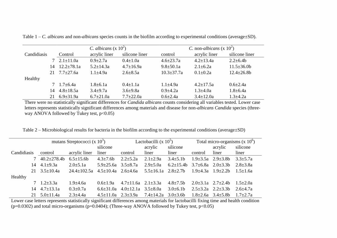
Table 1 – C. albicans and non-albicans species counts in the biofilm according to experimental conditions (average±SD).
C. albicans (x 102) C. non-albicans (x 10
2)
Candidiasis Control acrylic liner silicone liner control acrylic liner silicone liner
7 2.1±11.0a 0.9±2.7a 0.4±1.0a 4.6±23.7a 4.2±13.4a 2.2±6.4b
14 12.2±78.1a 5.2±14.3a 4.7±16.9a 9.8±50.1a 2.1±6.2a 11.5±36.0b
21 7.7±27.6a 1.1±4.9a 2.6±8.5a 10.3±37.7a 0.1±0.2a 12.4±26.8b
Healthy
7 1.7±6.4a 1.8±6.1a 0.4±1.1a 1.1±4.9a 4.2±17.5a 0.6±2.4a
14 4.8±18.5a 3.4±9.7a 3.6±9.8a 0.9±4.2a 1.3±4.0a 1.8±6.4a
21 6.9±31.9a 6.7±21.0a 7.7±22.0a 0.6±2.4a 3.4±12.0a 1.3±4.2a
There were no statistically significant differences for Candida albicans counts considering all variables tested. Lower case
letters represents statistically significant differences among materials and disease for non-albicans Candida species (three-
way ANOVA followed by Tukey test, p<0.05)
Table 2 – Microbiological results for bacteria in the biofilm according to the experimental conditions (average±SD)
mutans Streptococci (x 103) Lactobacilli (x 10
5) Total micro-organisms (x 10
6)
Candidiasis control acrylic liner
silicone
liner control
acrylic
liner
silicone
liner control
acrylic
liner
silicone
liner
7 40.2±278.4b 6.5±15.6b 4.3±7.6b 2.2±5.2a 2.1±2.9a 3.4±5.1b 1.9±3.5a 2.9±3.8b 3.3±5.7a
14 4.1±9.3a 2.0±5.1a 5.9±25.6a 3.5±8.7a 2.9±5.0a 6.2±15.4b 3.7±6.8a 2.0±3.3b 2.8±3.8a
21 3.5±10.4a 24.4±102.5a 4.5±10.4a 2.6±4.6a 5.5±16.1a 2.8±2.7b 1.9±4.3a 1.9±2.2b 1.5±1.6a
Healthy
7 1.2±3.3a 1.9±4.6a 0.6±1.9a 4.7±11.6a 2.1±3.3a 4.8±7.5b 2.0±3.1a 2.7±2.4b 1.5±2.0a
14 4.7±13.1a 0.3±0.7a 6.6±31.0a 4.0±12.1a 3.5±8.0a 3.0±6.1b 2.5±3.2a 2.2±3.3b 2.6±4.7a
21 5.0±11.4a 2.3±4.4a 4.5±11.0a 2.3±3.9a 7.4±14.2a 3.0±3.6b 1.8±2.6a 3.4±5.8b 1.7±2.7a
Lower case letters represents statistically significant differences among materials for lactobacilli fixing time and health condition
(p=0.0302) and total micro-organisms (p=0.0404); (Three-way ANOVA followed by Tukey test, p<0.05)

91
CONSORT 2010 checklist of information to include when reporting a randomised trial*
Section/Topic Item No Checklist item
Reported on page No
Title and abstract 1a Identification as a randomised trial in the title 1
1b Structured summary of trial design, methods, results, and conclusions (for specific guidance see CONSORT for abstracts) 1
Introduction Background and objectives
2a Scientific background and explanation of rationale 2,3
2b Specific objectives or hypotheses 3
Methods Trial design 3a Description of trial design (such as parallel, factorial) including allocation ratio 3
3b Important changes to methods after trial commencement (such as eligibility criteria), with reasons N/A
Participants 4a Eligibility criteria for participants 4
4b Settings and locations where the data were collected 3
Interventions 5 The interventions for each group with sufficient details to allow replication, including how and when they were actually administered
3,4
Outcomes 6a Completely defined pre-specified primary and secondary outcome measures, including how and when they were assessed
6,7
6b Any changes to trial outcomes after the trial commenced, with reasons N/A
Sample size 7a How sample size was determined 4
7b When applicable, explanation of any interim analyses and stopping guidelines N/A
Randomisation: Sequence
generation 8a Method used to generate the random allocation sequence 6
8b Type of randomisation; details of any restriction (such as blocking and block size) 7
Allocation concealment mechanism
9 Mechanism used to implement the random allocation sequence (such as sequentially numbered containers), describing any steps taken to conceal the sequence until interventions were assigned
6
Implementation 10 Who generated the random allocation sequence, who enrolled participants, and who assigned participants to 4,6

92
interventions
Blinding 11a If done, who was blinded after assignment to interventions (for example, participants, care providers, those assessing outcomes) and how
3
11b If relevant, description of the similarity of interventions N/A
Statistical methods 12a Statistical methods used to compare groups for primary and secondary outcomes 7
12b Methods for additional analyses, such as subgroup analyses and adjusted analyses N/A
Results Participant flow (a diagram is strongly recommended)
13a For each group, the numbers of participants who were randomly assigned, received intended treatment, and were analysed for the primary outcome
3
13b For each group, losses and exclusions after randomisation, together with reasons 3, 7
Recruitment 14a Dates defining the periods of recruitment and follow-up N/A
14b Why the trial ended or was stopped N/A
Baseline data 15 A table showing baseline demographic and clinical characteristics for each group N/A
Numbers analysed 16 For each group, number of participants (denominator) included in each analysis and whether the analysis was by original assigned groups
3,7
Outcomes and estimation
17a For each primary and secondary outcome, results for each group, and the estimated effect size and its precision (such as 95% confidence interval)
7
17b For binary outcomes, presentation of both absolute and relative effect sizes is recommended N/A
Ancillary analyses 18 Results of any other analyses performed, including subgroup analyses and adjusted analyses, distinguishing pre-specified from exploratory
8
Harms 19 All important harms or unintended effects in each group (for specific guidance see CONSORT for harms) N/A
Discussion Limitations 20 Trial limitations, addressing sources of potential bias, imprecision, and, if relevant, multiplicity of analyses 5
Generalisability 21 Generalisability (external validity, applicability) of the trial findings 11,12
Interpretation 22 Interpretation consistent with results, balancing benefits and harms, and considering other relevant evidence 11
Other information
Registration 23 Registration number and name of trial registry N/A
Protocol 24 Where the full trial protocol can be accessed, if available N/A
Funding 25 Sources of funding and other support (such as supply of drugs), role of funders 12

93
*We strongly recommend reading this statement in conjunction with the CONSORT 2010 Explanation and Elaboration for important clarifications on all the items. If
relevant, we also recommend reading CONSORT extensions for cluster randomised trials, non-inferiority and equivalence trials, non-pharmacological treatments, herbal
interventions, and pragmatic trials. Additional extensions are forthcoming: for those and for up to date references relevant to this checklist, see www.consort-statement.org.

3 Conclusões
Embora a adição de um agente antifúngico em reembasadores pareça ter
algum efeito benéfico para prevenir a colonização por Candida e a utilização de
hipoclorito de sódio pareça ajudar na sua desinfecção, não há evidências científicas
para fornecer recomendações definitivas sobre qual protocolo de desinfecção é o
ideal para reembasadores a base de prótese. No entanto, parece de acordo com os
estudos in vitro utilizados pela nossa revisão sistemática, que 0,5% de Hipoclorito de
Sódio é suficiente para limpar e desinfectar reembasadores de prótese sem danificar
a superfície dos materias.
A utilização de reembasadores a base de silicone, testados nesse estudo,
está contra indicada em pacientes com estomatite por dentadura devido a uma
maior colonização de C. não albicans, a qual é mais virulenta e resistente às
terapias convencionais. Em geral, os reembasadores de prótese, testados neste
estudo acumulam maior quantidade de biofilme e, portanto, seu uso deve ser
cauteloso.

95
4 Referências
<http://www.ncbi.nlm.nih.gov/pubmed/7023437?ordinalpos=47&itool=EntrezSystem2.PEntrez.Pubmed.Pubmed_ResultsPanel.Pubmed_RVDocSum>. Acessado em Novembro e Dezembro de 2010.
ARENDORF, T. M.; WALKER, D. M. Denture stomatitis: a review. Journal Oral Rehabilitation, v.14, n.3, p.217–227, 1987.
BARBEAU, J.; SEGUIN, J.; GOULET, J. P.; DE KONINCK, L.; AVON, S. L.; LALONDE, B. et al. Reassessing the presence of Candida albicans in denture-related stomatitis. Oral Surgery, Oral Medicine, Oral Pathology, Oral Radiology & Endodontics, v.95, n.1, p.51-59, 2003.
BELLON-FONTAINE, M. N.; MOZES, N.; VAN DER MEI, H. C.; SJOLLEMA, J.; CERF, O.; ROUXHET, P. G. et al. A comparison ofthermodynamic approaches to predict the adhesion of dairy microorganisms to solid substrata. Cellular Biophysics, v.17, n.1, p.93-106, 1990 .
BENTING, D. G.; PESUN, I. J.; HODGES, L. Compliance of resilient denture liners immersed in effervescent denture cleansers. Journal Prosthodont, v.14, n.3, p.175-83, 2005.
BOSCATO, N.; DELAVI, J. D.; MULLER, L.; PEREIRA-CENCI, T.; IMANISHI, S. W. Influence of varnish application on a tissue conditioner: analysis of biofilm adhesion. Gerodontology, v.21, n.3, p.207-210, 2010.
BOSCATO, N.; RADAVELLI, A.; FACCIO, D.; LOGUERCIO, A. D. Biofilm formation of Candida albicans on the surface of a soft denture-lining material. Gerodontology, v.3, n.26, p.210-213, 2009.
BRADEN, M.; CAUSTON, B. E. Tissue conditioners: III. Water immersion characteristics. Journal Dental Research, v.50, n.6, p.1544-1547, 1971. BRANTING, C.; SUND, M. L.; LINDER, L. E. The influence of Streptococcus mutans on adhesion of Candida albicans to acrylic surfaces in vitro. Archives of Oral Biology, v.34, n.5, p.347-353, 1989.
BUDTZ-JORGENSEN, E. Clinical aspects of Candida infection in denture wearers. The Journal of the American Dental Association, v.96, n.3, p.474-479, 1978.
BUDTZ-JORGENSEN, E.; THEILADE, E.; THEILADE, J.; ZANDER, H. A. Method for studying the development, structure and microflora of denture plaque. Scandinavian Journal of Dental Research, v.89, n.2, p.149-156, 1981.

96
BUSSCHER, H. J.; COWAN, M. M.; VAN DER MEI, H. C. On the relative importance of specific and non-specific approaches to oral microbial adhesion. FEMS Microbiology Reviews, v.8, n.3-4, p.99-209, 1992. CANNON, R. D.; CHAFFIN, W. L. Oral colonization by Candida albicans. Critical Reviews in Oral Biology & Medicine, v.10, n.3, p.359-383, 1999.
CANNON, R. D.; FIRTH, N. A. Fungi and fungal infections of the oral cavity. In: LAMONT, R. J.; BURNE, R. A.; LANTZ, M. S.; LEBLANC, D. J.; editors. Oral Microbiology and Immunology. Washington, DC: ASM Press, 2006. p.333-348.
CENCI, T. P. Avaliação da formação de biofilme de espécies de Candida sobre a superfície de resinas acrílicas para base e reembasamento de próteses removíveis. 2008. 101f. Tese (Doutorado em Clínica Odontológica)- Faculdade de Odontologia, Universidade de Campinas, Piracicaba.
CHANDRA, J.; MUKHERJEE, P. K.; LEIDICH, S. D.; FADDOUL, F. F.; HOYER, L. L.; DOUGLAS, L. J. et al. Antifungal resistance of candidal biofilms formed on denture acrylic in vitro. Jounal Dental Research, v.80, n.3, p.903-908, 2001.
COLEMAN, D. C.; SULLIVAN, D. J.; BENNETT, D. E.; MORAN, G. P.; BARRY, H. J.; SHANLEY, D. B. Candidiasis: the emergence of a novel species,Candida dubliniensis. AIDS, v.11, n.5, p.557-567, 1997.
COULTHWAITE, L.; VERRAN, J. Potential pathogenic aspects of denture plaque. Journal of Biomedical Science, v.64, n.4, p.180-189, 2007.
CRAIG, R.G. Inflammation, cardiovascular disease and destructive periodontal diseases. The evolving role of the dental profession. New York State Dental Journal, v.70, n.5, p.22-26, 2004.
DE SOUZA, R.F, DE FREITAS OLIVEIRA PARANHOS, H.; LOVATO DA SILVA, C. H.; ABU-NABA'A, L.; FEDOROWICZ, Z.; GURGAN, C. A. Interventions for cleaning dentures in adults. Cochrane Database Systematic Review, v.7, n.4, p.CD007395, 2009.
DOREY, J.L.; BLASBERG, B.; MACENTEE, M. I.; CONKLIN, R. J. Oral mucosal disorders in denture wearers. Journal of Prosthetic Dentistry, v.53, n.2, p.210-213, 1985.
DOUGLAS, L. J. Adhesion of Candida species to epithelial surfaces. Critical Reviews in Microbiology, v.15, n.1, p.27-43,1987.

97
EDGERTON, M.; SCANNAPIECO, F. A.; REDDY, M. S.; LEVINE, M. J. Human submandibular-sublingual saliva promotes adherence of Candida albicans to Polymethylmethacrylate. Infection and Immunity, v.61, n.2, p. 2644-2652, 1993.
ELLEPOLA, A. N.; SAMARANAYAKE, L. P. Inhalational and topical steroids, and oral candidosis: a mini review. Oral Dis Journal, v.7, n.4, p.211-216, 2001.
FERREIRA, M. A.; PEREIRA-CENCI, T.; VASCONCELOS, L. M.; RODRIGUES-GARCIA, R. C.; DEL BEL CURY, A. A. Efficacy of denture cleansers on denture liners contaminated with Candida species. Clinical Oral Investigations, v.13, n.2, p.237-242, 2009.
FIGUEIRAL, M. H.; AZUL, A.; PINTO, E.; FONSECA, P. A.; BRANCO, F. M.; SCULLY, C. Denture-related stomatitis: identification of aetiological and predisposing factors - a large cohort. Journal of Oral Rehabilitation, v.34, n.6, p. 448-455, 2007. HARRISON, A. Temporary soft lining materials. A review of their uses. British Dental Journal, n.151, v.12, p.419-422, 1981. KULAK, Y.; KAZAZOGLU, E. In vivo and in vitro study of fungal presence and growth on three tissue conditioning materials on implant supported complete denture wearers. Journal Oral Rehabilitation, v.25, n.2, p.135-138, 1998.
KUMAMOTO, C. A; VINCES, M. D. Alternative Candida albicans lifestyles: growth on surfaces. Annual Review Microbiology, v.59, p.113-133, 2005.
LUND, R. G.; DA SILVA NASCENTE, P.; ETGES, A.; RIBEIRO, G. A.; ROSALEN, P. L.; DEL PINO, F. A. Occurrence, isolation and differentiation of Candida spp. and prevalence of variables associated to chronic atrophic candidiasis. Mycoses, v.53, n.3, p.232-238, 2010.
LUO, G.; SAMARANAYAKE, L. P. Candida glabrata, an emerging fungal pathogen, exhibits superior relative cell surface hydrophobicity and adhesion to denture acrylic surfaces compared with Candida albicans. Acta Pathologica Microbiologica et Immunologica Scandinavica, v.110, n.9, p.601-610, 2002. MARSH, P. D. Microbial ecology of dental plaque and its significance in healthy and disease. Advances in Dental Research, v.8, n.2, p.263-271, 1994. MURATA, H.; CHIMORI, H.; HAMADA, T.; MCCABE, J. F. Viscoelasticity of Dental Tissue Conditioners during the Sol-gel Transition. Journal of Dental Research, v.84, n.4, p.376-381, 2005.
MUZIKA, B. C. Oral fungal infections. Dental Clinics of North America, v.49, n.1, p.49-65, 2005. NEWTON, A. V. Denture sore mouth: a possible aetiology. British Dental Journal, v.112, p.357-360, 1962.

98
NIKAWA, H.; JIN, C.; MAKIHIRA, S.; EGUSA, H.; HAMADA, T.; KUMAGAI, H. Biofilm formation of Candida albicans on the surfaces of deteriorated soft denture lining materials caused by denture cleansers in vitro. Jounal Oral Rehabilitation, v.30, n.3, p.243-250, 2003.
NIKAWA, H.; JIN, C.; HAMADA, T.; MAKIHIRA, S.; KUMAGAI, H.; MURATA, H. Interactions between thermal cycled resilient denture lining materials, salivary and serum pellicles and C. albicans in vitro: Part II.Effects on fungal colonization. Journal Oral Rehabilitation, v.27, n.2, p.124-130, 2000.
PEREIRA-CENCI , T.; DENG, D.M.; KRANEVELD, E.A.; MANDERS, E.M.; DEL BEL CURY, A. A.; TEN CATE, J. M.; et al. The effect of Streptococcus mutans and Candida glabrata on Candida albicans biofilms formed on different surfaces. Archives of Oral Biology, v.45, n.8, p.755-764, 2008.
PEREIRA-CENCI, T.; DA SILVA, W. J.; CENCI, M. S.; CURY, A. A. Temporal changes of denture plaque microbiologic composition evaluated in situ. The International Jounal of Prosthodontics, v.23, n.3, p.239-242, 2010.
PEREIRA-CENCI, T.; DEL BEL CURY, A. A.; CRIELAARD, W.; TEN CATE, J. M. Development of Candida-associated denture stomatitis: new insights. Journal of Applied Oral Science, v.16, n.2, p.86-94, 2008.
PEREZOUS, L. F.; FLAITZ, C. M.; GOLDSCHMIDT, M. E; ENGELMEIER, R. L. Colonization of Candida species in denture wearers with emphasis on HIV infection: a literature review. Jounal Prosthetic Dentistry, v.93, n.3, p.288-293, 2005.
QUDAH, S.; HARRISON, A.; HUGGETT, R. Soft lining materials in prosthetic dentistry: a review. The International Jounal of Prosthodontics, v.3, n.5, p.477-843, 1990.
RADFORD, D. R.; CHALLACOMBE, S. J.; WALTER, J. D. Denture plaque and adherence of Candida albicans to denture-base materials in vivo and in vitro. Critical Reviews in Oral Biology & Medicine, v.10, n.1, p.99-116,1999.
RAMAGE, G.; TOMSETT, K.; WICKES, B. L.; LOPEZ-RIBOT, J. L.; REDDING, S. W. Denture stomatitis: a role for Candida biofilms. Oral Surgery, Oral Medicine, Oral Pathology, Oral Radiology Endodontics, v.98, n.1, p.53-59, 2004.
RAZEK, M. K. A.; MOHAMED, Z. M. Influence of tissue conditioning materials on the oral bacteriologic status of complete denture wearers. Journal of Prosthetic Dentistry, v.44, n.2, p.137-142, 1980. ROSAN, B.; LAMONT, R. J. Dental plaque formation. Microbes and Infection, v.2, n.13, p.1599-1607, 2000.

99
SAMARANAYAKE, L. P. Host factors and oral candidosis. In: Oral candidosis. SAMARANAYAKE, L. P.; MACFARLANE, T. W.; editors. London: Butterworth, p.66-103, 1990.
SAMARANAYAKE, L. P.; MCCOURTIE, J.; MACFARLANE, T. W. Factors affecting th in-vitro adherence of Candida albicans to acrylic surfaces. Archives of Oral Biology, v.25, n.8-9, p.611-615, 1980.
SAMARANAYAKE, L. P.; FURGUSON, M. M. Delivery of antifungal agents to the oral cavity. Advanced Drug Delivery Reviews, v.13, n.1-2, p.161-179, 1994.
SAMARANAYAKE, L. P; KEUNG LEUNG, W.; JIN, L. Oral mucosal fungal infections. Periodontology, 2000, v.49, p.39-59, 2009.
SAN MILLÁN, R.; ELGUEZABAL, N.; REGÚLEZ, P.; MORAGUES, M. D.; UINDÓS, G.; PONTÓN, J. Effect of salivary secretory IgA on the adhesion of Candida albicans to polystyrene. Microbiology, v.146, n.9, p.2105-2112, 2000.
SCULLY, C.; EL-KABIR, M.; SAMARANAYAKE, L. P. Candida and oral candidosis: a review. Critical Reviews in Oral Biology & Medicine, v.5, n.2, p.125-157, 1994.
SHAY, K.; TRUHLAR, M. R.; RENNER, R. P. Oropharyngeal candidosis in the older patient. Journal of the American Geriatrics Society, v.45, n.7, p.863-870, 1997.
SILVA, C. H.; PARA NHOS, H. de O.; ITO, I.Y. Biofilm disclosing agents in complete denture: clinical and antimicrobial evaluation. Pesquisa Odontologica Brasileira, v.16, n.3, p.270-275, 2002.
SPIECHOWICZ, E.; RENNER, R. P.; POLLOCK, J. J.; SANTARPIA, R. P. 3RD; CIECHOWICZ, B.; KOWALCZYK, W.; et al. Sensitivity of the replica method in the detection of candidal infection among denture wearers with clinically healthy oral mucosa. Quintessence International, v.22, n.9, p.753-755, 1991.
TENUTA, L. M.; RICOMINI FILHO, A. P.; DEL BEL CURY, A. A.; CURY, J. A. Effect of sucrose on the selection of mutans streptococci and lactobacilli in dental biofilm formedin situ. Caries Research, v.40, n.2, p.546-549, 2006.
VANDEN ABBEELE, A.; DE MEEL, H.; AHARIZ, M.; PERRAUDIN, J. P.; BEYER, I.; COURTOIS, P. Denture contamination by yeasts in the elderly. Gerodontology, v.25, n.4, p.222-228, 2008.
VERRAN, J.; MARYAN, C. J. Retention of Candida albicans on acrylic resin and silicone of different surface topography. Journal of Prosthetic Dentistry, v.77, n.5, p.535-539, 1997.

100
WEBB, B. C.; THOMAS, C. J.; WILLCOX, M. D.; HARTY, D. W.; KNOX, K. W. Candida-associated denture stomatitis. Aetiology and management: a review. Part 1. Factors influencing distribution of Candida species in the oral cavity. Australian Dental Jounal, v.43, n.1 p.45-50, 1998.
WILSON, M. Bacterial biofilms and human disease. Science Progress, v.84, n.3, p.235-254, 2001.
ZAREMBA, M. L.; DANILUK, T.; ROZKIEWICZ, D.; CYLWIK-ROKICKA, D.; KIERKLO, A.; TOKAJUK, G.; et al. Incidence rate of Candida species in the oral cavity of middle-aged and elderly subjects. Advances in Medical Sciences, v.51, n.1, p.233-236, 2006.
MUKHERJEE, P. K; CHANDRA, J. Candida biofilm resistance. Drug Resistances Updates, v.7, n.4-5, p.301–309, 2004
ANUSAVICE, K. J. Phillips’ Science of Dental Materials, 10a ed., Philadelphia: W.B.Saunders, 1996, 806p.

101
APÊNDICE A – Termo de Consentimento Livre e Esclarecido
UNIVERSIDADE FEDERAL DE PELOTAS
FACULDADE DE ODONTOLOGIA
TERMO DE CONSENTIMENTO LIVRE E ESCLARECIDO
Você está convidado a participar, como voluntário, em uma pesquisa. Após ser
esclarecido sobre as informações a seguir, no caso de aceitar fazer parte do estudo,
assine ao final deste documento, que está em duas vias. Uma delas é sua e a outra
é das pesquisadoras responsáveis. Alertamos que não existem riscos envolvidos
neste estudo e em caso de recusa você não será penalizado de forma alguma.
Esclarecemos que a participação é decorrente de sua livre decisão, após receber
todas as informações que julgar necessárias, e que poderá ser a qualquer tempo,
retirada.
INFORMAÇÕES SOBRE A PESQUISA:
Título do Projeto: Avaliação clínica da formação de biofilme sobre condicionadores
de tecido temporários
Pesquisadora participante: Fernanda Valentini
Pesquisadora responsável: Profa. Dra. Tatiana Pereira Cenci e Profa. Dra. Noéli
Boscato

102
Prezado paciente, a nossa pesquisa tem como objetivo principal, avaliar a
composição da placa bacteriana que se forma sobre a superfície de diferentes
matérias usados na parte de dentro da dentadura para evitar que ela machuque sua
gengiva , bem como avaliar a rugosidade dessa superfície ao longo do tempo e
avaliar a colonização de Candida, que é um fungo comum na cavidade bucal e que
causa doença em alguns casos específicos, como por exemplo quando a prótese
fica muito tempo sem ser trocada ou sem adequadamente limpeza. Para isso será
realizado um estudo clínico em sua prótese total (dentadura) superior antiga ao
mesmo tempo em que se realizam as etapas clínicas para confecção de uma nova
prótese total. Os materiais estudados serão colocados na parte de dentro de sua
dentadura antiga sem provocar qualquer desconforto. Você terá que retornar à
Faculdade de Odontologia da UFPel após passados 1, 7 e 14 dias para que
possamos remover os matérias testados. Depois, haverá um intervalo de 07 dias e
colocaremos um segundo material para análise que ocorrerá da mesma maneira
que para a primeira fase , sendo necessário mais 14 dias, e você deverá retornar
para as consultas no 1, 7 e 14 dia da segunda fase, igual feito para primeira fase.
Ao final da segunda fase, seguirá a sequência clínica para a finalização da
confecção de nova prótese total superior.
CONSENTIMENTO DA PARTICIPAÇÃO DA PESSOA COMO SUJEITO E
RESPONSÁVEL LEGAL
Eu, _______________________________________________, RG/ CI
__________________________________, abaixo assinado, concordo em participar
do estudo sobre a avaliação clínica da formação de biofilme em condicionadores de
tecidos temporários realizado na clínica de prótese dentária do curso de Odontologia
da Universidade Federal de Pelotas. Fui devidamente informado e esclarecido sobre
a minha participação. Foi-me garantido que posso retirar meu consentimento a
qualquer momento, sem que isto leve a qualquer penalidade ou interrupção do
acompanhamento/ assistência/ tratamento.
Pelotas, _____de ______________ de 2010.

103
______________________________________________
Assinatura

104
ANEXO A. Parece do Comitê de Ética em Pesquisa