Understanding the roles of PIWIL3 and TACC3 proteins during...
58
Universidade de Lisboa Faculdade de Ciências Departamento de Biologia Animal UNDERSTANDING THE ROLES OF PIWIL3 AND TACC3 PROTEINS DURING BOVINE OOCYTE DEVELOPMENT ANA RITA CANHOTO LEITOGUINHO Dissertação Mestrado em Biologia Evolutiva e do Desenvolvimento 2012
Transcript of Understanding the roles of PIWIL3 and TACC3 proteins during...

Universidade de Lisboa
Faculdade de Ciências
Departamento de Biologia Animal
UNDERSTANDING THE ROLES OF PIWIL3 AND
TACC3 PROTEINS DURING BOVINE OOCYTE
DEVELOPMENT
ANA RITA CANHOTO LEITOGUINHO
Dissertação
Mestrado em Biologia Evolutiva e do Desenvolvimento
2012

Universidade de Lisboa
Faculdade de Ciências
Departamento de Biologia Animal
UNDERSTANDING THE ROLES OF PIWIL3 AND
TACC3 PROTEINS DURING BOVINE OOCYTE
DEVELOPMENT
ANA RITA CANHOTO LEITOGUINHO
Dissertação
Mestrado em Biologia Evolutiva e do Desenvolvimento
Dissertação orientada por:
Professora Dra. Maria Gabriela Rodrigues (DBA/FCUL)
Professor Dr. Bernard Roelen (BRC/UU)
2012

I
Acknowledgements
I would like to express my deep and sincere gratitude to Bernard for this opportunity
and for being always there when I needed. Also a special thanks to the entire lab, in
particular to Mahdi for his patience, help and great talks, to Eric for the tips on music and to
Leni for her constant guidance and assistance in the Lab.
From the warm and dry part of Europe, my sincere thanks to Gabriela for her support
and great advices.
Para a minha mãe, pai e irmã um muito obrigado por me providenciarem tudo o que
sempre precisei, mesmo nestes tempos difíceis. Nunca teria conseguido sem a vossa ajuda.
To all my friends, can’t thank you enough. Thank you to Nuno, Luís, Silvia, Yuri and
Filipa for your help during rough times and for your friendship. Also, to all Utrecht friends, a
big cheers for your welcoming feeling.
Last but not least, a loving gratitude to Stephin for his boundless wisdom, smile and
company.
This thesis is for all of you.
Ana Rita Leitoguinho / Toga
In loving memory of my grandfather

II
Abstract
In order to achieve a healthy embryo, the germline needs to properly develop while
protecting its genome against endogenous and exogenous adversities. The PIWI proteins, a
subgroup of Argonaute proteins, are involved in the search and silencing mechanism of
transposons ‐ genetic transposable elements‐ through a mechanism of complementarity
with piRNAs (Piwi‐interacting RNAs). These proteins are germline‐specific and are thought to
be also male‐specific in mammals. PIWIL3 (Piwi‐like 3) is part of this family and it is present
in the human and bovine genome, while rodents have PIWIL1, PIWIl2 and PIWIL4, but lack a
gene for PIWIl3. Recent findings on bovine cells suggested a role for PIWIL3 in the female
germline, the oocyte. Here, PIWIL3 protein was detected both in the bovine oocyte and in
somatic cells surrounding it, using an antibody directed against the Human PIWIL3
suggesting a role for this protein in protecting the oocyte genome.
Besides protecting the genome, the oocyte also needs to enroll meiosis. Transforming
acidic coiled‐coil 3 (TACC3) acts in a complex with ch‐TOG and clathrin to ensure mitotic
spindle stability and organization. TACC3 is phosphorylated and activated by Aurora‐A kinase
and it is responsible for TACC3’s proper placement in the centrosomes. The order of
recruitment of these proteins to the spindle is still open for debate, as it is the role of TACC3
during meiosis. Our antibody designed to identify bovine PIWIL3 was in fact recognizing
TACC3 and we could therefore study the spatiotemporal localization of TACC3 in the oocyte
and during embryo development. When bovine oocytes are matured with MLN8054, an
Aurora‐A inhibitor, they exhibit (i) dispersion of TACC3 protein in the cytoplasm (ii)
formation of an abnormal meiotic spindle (ii) meiotic arrest in the metaphase I stage and (iii)
decreased percentage of blastocyst formation suggesting a role of TACC3 in meiotic spindle
assembly, vital for oocyte and early embryo development.
Keywords: Bovine, Oocyte, Piwi‐like 3 (PIWIL3), TACC3 and Aurora‐A kinase

III
Resumo em Português
Todos os indivíduos que se reproduzem sexualmente combinam a sua informação genética
para criar um novo ser e apesar de todas as células possuírem esta informação, apenas as
células sexuais são capazes de a transmitir à descendência. É por isso vital proteger estas
células de fatores externos ou internos que possam afetar o seu genoma bem como
certificar um correto desenvolvimento do oócito com o intuito de formar um embrião
saudável.
O processo de maturação do oócito envolve o desenvolvimento da célula bem como a
criação de estruturas de suporte que o protegem de agressões externas. A Foliculogénese é
processo responsável pelo suporte físico do oócito, altamente regulado hormonalmente
pelo ovário, onde várias camadas celulares são formadas consecutivamente em volta do
oócito, contribuindo para que o resultado final seja uma célula sexual protegida, formando o
folículo antral. Concomitantemente, também a própria célula sofre alterações críticas.
Durante a maturação nuclear do oócito, ou oogénese, é essencial que este reduza o seu
número de cromossomas a metade, formando um gameta haplóide (com metade da
informação genética). Após fertilização, este resultará num zigoto diplóide (com o total da
informação genética) com informação materna e paterna que diferem não apenas na
informação genética mas também em fatores associados que são transmitidos não por via
genética, mas por via epigenética.
Para além dos rearranjos nucleares acima referidos, também o genoma da célula
sexual está sujeito a agressões endógenas, como os transposões, elementos móveis do
genoma que se podem inserir em diversas zonas, podendo interferir com a transcrição de
genes vitais para a célula. Existem vários mecanismos de defesa celular que silenciam estes
elementos, um destes é o RNA de interferência (RNAi) que, com ajuda de proteínas
Argonautas, consegue localizar e silenciar RNA mensageiro (mRNA) através de um
mecanismo de complementaridade, evitando a sua propagação pelo genoma. Esta família de
Argonautas contém o grupo de proteínas PIWI cuja expressão é restrita às células da linha
germinal. Recentemente descobriu‐se que estas proteínas PIWI são alvo de um tipo
particular de RNAi, os piRNAs (Piwi‐interacting RNAs) cuja principal função é silenciar
elementos transponíveis na linha germinal. Tendo como base mutantes PIWI em Drosophila
que ativam transposões, o estudo de proteínas PIWI e piRNA tem avançado bastante nos
últimos tempos revelando também em C. elegans, Zebrafish e ratinho o papel destas

IV
proteínas na formação da linha germinal. No ratinho existem três genes da família PIWI:
Miwi (Piwi‐like 1), Mili (Piwi‐like 2) e Miwi2 (Piwi‐like 4) e mutantes destes genes resultam
em esterilidade masculina, em contraste com Drosophila e Zebrafish onde ambas as linhas
germinais masculina e feminina são afetadas. Até à data não se sabe a função destas
proteínas na linha germinal feminina de mamíferos, razão pela qual a descoberta de um
quarto gene Piwi‐like (PIWIL3) em mamíferos que não o ratinho suscitou bastante interesse.
Recentemente, este gene foi descrito em oócitos de bovinos e a facilidade em obter oócitos
de vaca fizeram com que este organismo fosse o escolhido para o estudo desta proteína
PIWIL3.
Os nossos resultados revelaram primeiro que tudo que o anticorpo e primers
construídos anteriormente para a proteína PIWIL3 em vaca estavam mal construídos.
Propomos aqui um padrão de expressão proteico diferente do anteriormente documentado.
Com o uso de anticorpos anti‐humano dirigidos a PIWIL3, identificou‐se a proteína nas
células de suporte que rodeiam o oócito, sugerindo que a expressão desta proteína PIWI em
vaca não é específica para a linha germinal. Esta presença de PIWIL3 na linha somática que
rodeia o oócito pode ser essencial para a proteção do genoma do oócito, essencial para a
correta propagação da informação genética à descendência.
Aquando do estudo de PIWIL3, descobriu‐se que o anticorpo estava a reconhecer uma
outra proteína em vez de PIWIL3, a TACC3 (Transforming Acidid Coiled‐Coil protein 3), o que
fez com que este projeto se começasse a focar no papel desta outra proteína durante o
desenvolvimento do oócito.
Também uma correta divisão celular é essencial para uma célula sexual e as proteínas
TACC (Transforming acidic coiled‐coil proteins) estão de mãos dadas com esta prática. Estas
proteínas estão presentes em diferentes organismos e localizam‐se tanto nos microtúbulos
que se formam durante o processo de separação dos cromossomas como nos centrossomas,
estruturas que se localizam nos dois pólos opostos do fuso mitótico. Em ratinho são
conhecidas três proteínas TACC ‐ TACC1, TACC2 e TACC3 ‐ e sabe‐se que TACC3 é essencial
para a correta formação do fuso mitótico durante uma mitose. Mutantes TACC3 revelaram
um fuso mitótico anormal que, juntamente com cromossomas desalinhados na placa
metafásica, originam incorreta segregação cromossómica, resultando em células‐filha com
diferente conteúdo cromossómico. O recrutamento de TACC3 para os microtúbulos que
formam o fuso é regulado por uma kinase, a Aurora‐A, que é responsável pela fosforilação e
ativação desta proteína em diversas espécies como Drosophila, C.elegans, Xenopus e

V
Humanos. Quando esta kinase é inibida, por exemplo pelo composto MLN8054, a mitose
falha de forma semelhante aos mutantes de TACC3, contudo alguma células conseguem
dividir‐se, ainda que originando células aneuplóides ou tetraplóides. Porém, TACC3 não atua
sozinha e recentemente foi descoberto que o faz em conjunto com outras duas proteínas
num complexo proteíco denominado TACC3/ch‐TOG/clatrina. TACC3, clatrina e ch‐TOG
(Colonic‐hepatic Tumour Overexpressed Gene) atuam como pontes entre microtúbulos e
têm como função estabilizar estas fibras. Apesar de se saber que todos os intervenientes do
complexo são importantes, a ordem de recrutamento dasdiferentes proteínas ainda é alvo
de uma acesa discussão entre defensores que afirmam que clatrina recruta TACC3 e outros
que defendem ser TACC3 o primeiro componente do complexo, recrutando clatrina.
Apesar de ter sido estudada em oócitos de ratinho, informação sobre a função da
proteína TACC3 durante a meiose de mamíferos é quase nula, o que realça a importância
deste estudo. Os nossos resultados demonstram que a proteína se encontra rodeando a
cromatina do oócito e no fuso meiótico do mesmo durante a meiose, sugerindo um papel
semelhante ao documentado no ratinho quanto à manutenção dos microtúbulos do fuso.
Quanto à sua função, foi utilizado um inibidor de Aurora‐A, MLN8054, que resultou numa
paragem no desenvolvimento do oócito na metafase I, efeito semelhante aos previamente
descritos em células humanas. Também o fuso meiótico revelou sérias perturbações quando
os oócitos maturam num meio com o inibidor o que, juntamente com a dispersão da
proteína TACC3 no citoplasma e a diminuição da percentagem de blastocistos formados,
sugere que este inibidor estará a influenciar a expressão ou ativação de TACC3. Apesar de
não conseguirmos saber ao certo com este estudo se TACC3 esta de fato inibida, é essencial
averiguar o padrão de fosforilação desta proteína para poder estabelecer uma ligação causa‐
efeito entre o inibidor e a fosforilação/correta localização de TACC3. Também nesta tese é
proposto um modelo de recrutamento do complexo que se baseia na possibilidade de ser
TACC3 o primeiro e talvez principal interveniente.
Em suma, os resultados desta tese referentes a TACC3 e a PIWIL3 revelam que ambas
aparentam ter uma função durante o desenvolvimento da linha sexual feminina e é
considerado de extrema importância estudar ambas. Quanto aos entraves que o projeto
PIWIL3 sofreu, apesar de ter afetado parte da investigação da PIWIL3, serviu também para
conhecer esta nova proteína, a TACC3, em oócitos. Esta tese propõe um padrão de
expressão diferente de PIWIl3 com uma possível função também na linha somática em redor
do oócito. Quanto a TACC3 os nossos resultados revelaram que quando a sua ação é inibida,

VI
os oócitos não progridem na oogénese e revelam anomalias no fuso. Assim é sugerido que
esta proteína tenha um papel crucial no processo de formação e correto alinhamento dos
cromossomas na placa metafásica durante o desenvolvimento do oócito de vaca.
Palavras‐chave: Bovino, Oócito, Piwi‐like 3 (PIWIL3), TACC3, Aurora‐A
VII
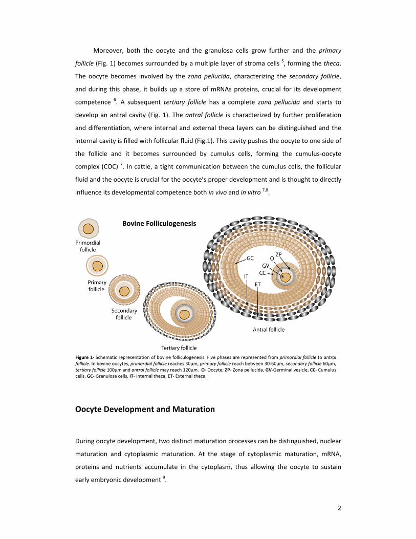
Table of Contents
Acknowledgements ............................................................................................................ I
Abstract ............................................................................................................................. II
Resumo em Português ...................................................................................................... III
Table of Contents ............................................................................................................. VII
Index of Figures .............................................................................................................. IX
Index of Tables ................................................................................................................ X
Index of Supplementary Tables ....................................................................................... X
Introduction ...................................................................................................................... 1
Follicle development protects the cell from outside adversities .................................... 1
Oocyte Development and Maturation ............................................................................ 2
Protecting germ cell’s integrity via Argonaute proteins ................................................. 5
Unraveling PIWI proteins in germ cells throughout species ........................................... 6
Disclosures in the PIWIL3 former research ..................................................................... 7
Transforming Acidic Coiled‐Coil (TACC) Proteins ............................................................ 9
Phosphorylation and activation of TACC3 by Aurora‐A kinase ..................................... 11
TACC3/ch‐TOG/clathrin complex stabilizes kinetochore fibers by inter‐microtubule
bridging .................................................................................................................................... 12
TACC3/ch‐TOG/clathrin complex activation, two hypotheses ..................................... 13
Material and methods ..................................................................................................... 15
Oocyte collection and in vitro maturation .................................................................... 15
In vitro fertilization and embryo culture ....................................................................... 15
Immunocytochemistry .................................................................................................. 16
Immunofluorescence .................................................................................................... 17
Western blotting ........................................................................................................... 18
Aurora‐A inhibition experiment .................................................................................... 19
Results ............................................................................................................................. 20
Sorting out the bovine Piwi‐like 3 antibody and unraveling Transforming Acidic Coiled‐
Coil protein 3 (TACC3) ............................................................................................................. 20

VIII
Detection of PIWIL3 expression in bovine oocytes using antibodies directed against
the human sequence ............................................................................................................... 22
TACC3’s expression pattern during oocyte and early zygote development: its presence
surrounding chromatin and the meiotic spindle ..................................................................... 24
TACC3’s presence surrounding the metaphase plate in the metaphase II ................... 26
TACC3 partially overlaps with α‐tubulin during oocyte and early zygote development
................................................................................................................................................. 27
Discerning TACC3 function: using MLN8054 compound, an Aurora‐A inhibitor,
decreases oocyte developmental progress and causes spindle anomalies ............................ 29
Effects of MLN8054 on TACC3 protein expression ....................................................... 31
Discussion ........................................................................................................................ 33
PIWIL3 during bovine oocyte development .................................................................. 33
TACC3 protein in bovine oocyte development ............................................................. 35
Future perspectives ....................................................................................................... 39
Concluding remarks ....................................................................................................... 39
References ....................................................................................................................... 40
Supplementary Information .............................................................................................. XI

IX
Index of Figures
Figure 1‐ Schematic representation of bovine folliculogenesis. .............................................. 2
Figure 2‐ Schematic representation of bovine oogenesis ......................................................... 3
Figure 3 ‐ Schematic representation of siRNA‐guided mRNA cleavage. ................................... 5
Figure 4 ‐ Homology between mammalian PIWI proteins. ....................................................... 7
Figure 5 ‐ mRNA quantification of PIWIl3 and PIWIL3 expression pattern ............................... 8
Figure 6 ‐ Centrosome and spindle localization of TACC proteins in C. elegans, D.
melanogaster, X. laevis and Human. ............................................................................. 10
Figure 7 ‐ Depletion of Xenopus’ TACC3 (xTACC3): effects on the spindle. ............................ 11
Figure 8 ‐ Two models representing the recruitment of the complex TACC3/ch‐TOG/clathrin
to the spindle. ............................................................................................................... 13
Figure 9 ‐ Schematic representation of two Piwi‐like 3 aminoacid sequences ....................... 20
Figure 10 ‐ Double stainings TACC3 and ’TACC3’ during oocyte maturation and blastocyst. 21
Figure 11 ‐ Merge channels of (A) DAPI and PIWIL1 and (B) DAPI and PIWIL2 expression
patterns during metaphase II. ....................................................................................... 22
Figure 12 ‐ Schematic representation of the human Piwi‐like 3 aminoacid sequence and
bovine Piwi‐like 3 aminoacid sequences ....................................................................... 22
Figure 13 ‐ HPIWIL3‐A and HPIWIL3‐B stainings on different staged oocytes and blastocysts.
....................................................................................................................................... 23
Figure 14 ‐ Immunocytochemistry results for HPIWIL3‐A and PIWIL3‐B in human testis,
bovine testis and bovine ovary. .................................................................................... 24
Figure 15 ‐ ‘TACC3’ expression pattern during oocyte development and early embryogenesis.
....................................................................................................................................... 25
Figure 16 ‐ Microfilament and microtubule stainings on metaphase II oocytes .................... 26
Figure 17 ‐ Double stainings ‘TACC3’ and actin on metaphase II oocytes. ............................. 27
Figure 18 – Double stainings of ‘TACC3’ and microtubules on oocytes and early zygotes. ... 29
Figure 19 ‐Effect of MLN8054 throughout oocyte developmental progress .......................... 30
Figure 20 – Effect of MLN8054 on oocyte polar body percentage and percentage of
blastocyst formation from cleaved embryos ................................................................ 30
Figure 21 ‐ Effects of MLN8054 on oocyte meiotic spindle during oocyte maturation process
....................................................................................................................................... 31
Figure 22 ‐ Effects of MLN8054 on TACC3 protein expression pattern during metaphase I and
II oocytes. ...................................................................................................................... 32

X
Figure 23 ‐ Western Blot on TACC3 protein and β‐actin in four different groups .................. 32
Figure 24 – Proposed TACC3/ch‐TOG/clathrin recruitment to the spindle and centrosomes
during bovine oocyte development .............................................................................. 38
Index of Tables
Table 1 ‐ Composition of multiple culture mediums used during oocyte in‐vitro maturation
and fertilization and embryo culture. ........................................................................... 15
Table 2 ‐ Detailed description of the antibodies used. ........................................................... 17
Table 3 ‐ Detailed description of the Buffers used on the Western Blot. ............................... 19
Table 4 ‐ Experimental design for the MLN8054 experiment. ................................................ 19
Index of Supplementary Tables
Supplementary Table 1 ‐ Effect of MLN8054 during oocyte meiosis progression .................. XI
Supplementary Table 2 ‐ Effect of MLN8054 during polar body extrusion. ............................ XI
Supplementary Table 3 ‐ Effect of MLN8054 on blastocyst formation. .................................. XI

1
Introduction
For centuries, the question of how a new being comes into existence has provided a
constant intellectual challenge and, since ancient Greece, several philosophers have
attempted to connect fertilization events with the creation of a new being. Once the in vivo
fertilization mechanism was understood, the molecular mechanisms underlying the process
presented the subsequent challenge. In 1978, the first human birth originating from a
successful in‐vitro‐fertilization (IVF) was announced 1, an accomplishment which earned
Robert Edwards the Nobel Prize in Physiology or Medicine in 2010.
The process of embryonic development goes through distinct stages with specific cell
types, which differ from somatic ones in their capacity to pass our genetic code to the next
generation. These are called germ cells and give rise to gametes in organisms with a sexual
reproductive system. Germline development involves specification of primordial germ cells
(PGCs) and their migration to special regions which will develop into the gonads, where the
germ cells can be stored and preserved. The PGCs in mammals arise through a complex
signaling process that, together with transcriptional regulators, inhibit somatic gene
expression while activating germline genes, thereby preserving germ cell pluripotency 2.
Later on, the germ cells undergo two divisions, a reductional and a non reductional one,
after which they can be called either an oocyte or a sperm cell. Despite oocyte development,
also the outside of the cell has to be protected and so, a follicle is matured in order to
protect the oocyte integrity.
Follicle development protects the cell from outside adversities
Whilst oocyte maturation is taking place, also the exterior of the oocyte undergoes
significant changes inside the ovarium through a process termed folliculogenesis (Fig. 1).
Coincident with the start of meiosis, oocytes become enclosed by a single layer of
somatic cells, the so‐called pre‐granulosa cells 3, thus forming the primordial follicles (Fig. 1)
that are thought to comprise the pool of resting follicles which defines postnatal ovarian life
span 4. When the follicles are recruited to undergo meiosis, the pre‐granulosa cells
surrounding the oocyte change from a flattened to a cuboidal shape and are then called
granulosa cells 5.
2
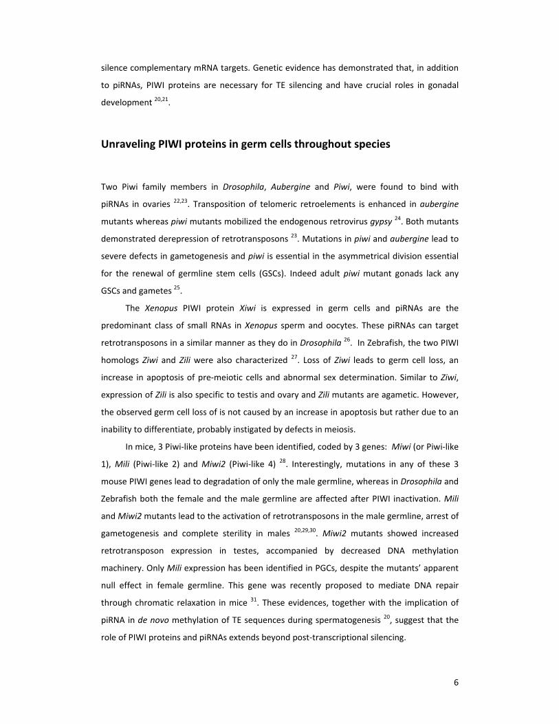
Moreover, both the oocyte and the granulosa cells grow further and the primary
follicle (Fig. 1) becomes surrounded by a multiple layer of stroma cells 5, forming the theca.
The oocyte becomes involved by the zona pellucida, characterizing the secondary follicle,
and during this phase, it builds up a store of mRNAs proteins, crucial for its development
competence 6. A subsequent tertiary follicle has a complete zona pellucida and starts to
develop an antral cavity (Fig. 1). The antral follicle is characterized by further proliferation
and differentiation, where internal and external theca layers can be distinguished and the
internal cavity is filled with follicular fluid (Fig.1). This cavity pushes the oocyte to one side of
the follicle and it becomes surrounded by cumulus cells, forming the cumulus‐oocyte
complex (COC) 7. In cattle, a tight communication between the cumulus cells, the follicular
fluid and the oocyte is crucial for the oocyte’s proper development and is thought to directly
influence its developmental competence both in vivo and in vitro 7,8.
Oocyte Development and Maturation
During oocyte development, two distinct maturation processes can be distinguished, nuclear
maturation and cytoplasmic maturation. At the stage of cytoplasmic maturation, mRNA,
proteins and nutrients accumulate in the cytoplasm, thus allowing the oocyte to sustain
early embryonic development 9.
Figure 1‐ Schematic representation of bovine folliculogenesis. Five phases are represented from primordial follicle to antralfollicle. In bovine oocytes, primordial follicle reaches 30µm, primary follicle reach between 30‐60µm, secondary follicle 60µm,tertiary follicle 100µm and antral follicle may reach 120µm. O‐ Oocyte; ZP‐ Zona pellucida, GV‐Germinal vesicle, CC‐ Cumuluscells, GC‐ Granulosa cells, IT‐ Internal theca, ET‐ External theca.
Bovine Folliculogenesis

3
All through mammalian nuclear maturation, the oocyte undergoes meiotic cell
division, which implies a reduction in chromosome number, accomplished through two
consecutive nuclear divisions termed meiosis I II. This process arrests in two distinct phases,
the prophase I and the metaphase II stage, and it is unique to diploid germ cells. The process
of oocyte meiosis, can be described through different stages 2,10 (Fig. 2).
Meiosis starts with the prophase I, the longest and most complex phase of the meiotic
cell division. This phase can be divided into five distinct stages: leptotene, zygotene,
pachytene, diplotene and diakinesis. At leptotene (Fig. 2, stage 1), the chromosomes, which
at this stage already consist of two sister chromatids, thicken and become visible while the
centrosomes begin to move toward opposite poles. During zygotene (Fig. 2, stage 2), each
chromosome seeks out its homologous partner and both are “zipped” together by a protein
structure called synaptonemal complex, in a process known as synapsis. This will only be
completed at the pachytene stage (Fig. 2, stage 3) where Crossing‐over occurs, i.e., the
genetic exchange between non‐sister chromatids of a homologous pair. In the penultimate
stage, termed diplotene (Fig. 2, stage 4), the synaptonemal complex dissolves and a tetrad of
four chromatids becomes visible. Additionally, the former crossing‐over points appear as
chiasmata, which hold nonsister chromatids together.Diplotene is the first regulatory check‐
point in oocyte meiosis, which can result in a meiotic arrest in a variety of species, including
cow and human. Prophase I is then completed by the diakinesis stage (Fig. 2, stage 5), which
Figure 2‐ Schematic representation of bovine oogenesis. The different phases are divided in 12 steps in which the first 5belong to the prophase I stage. The arrests represented in the figure concern the two known meiotic arrests known in bovineduring prophase I and metaphase II. A‐ First polar body B‐ Sperm cell C‐ Second polar body D‐ Male and female pronuclei.

4
is characterized by further chromatid condensation (Germinal Vesicle (GV)), breakdown of
the nuclear membrane ‐ in a process termed Germinal Vesicle Breakdown (GVBD) ‐ and
spindle formation 2,10.
Subsequently, the cells enter metaphase I, when the tetrad lines up along the
metaphase plate and each chromosome of a homologous pair attaches to microtubule fibers
from opposite poles. This is accomplished through the fusion of both sister chromatids’
kinetochore, thus providing each chromosome with only one functional kinetochore which
attaches to opposite spindles. Simultaneously, sister chromatids attach to microtubules from
the same pole (Fig. 2, stage 6) 2,10. At the onset of the next stage, the anaphase I, the
chiasmata dissolves, allowing the homologous to move towards opposite spindle poles. The
centrosome, however, does not divide, meaning that homologous chromosomes with sister
chromatids move towards opposite sides (Fig. 2, stage 7) 2,10. Succeeding anaphase I is the
telophase I, at the end of meiosis I. During this reductional division, each daughter cell
receives half the number of chromosomes, all consisting of two sister chromatids,
culminating with an asymmetric cell division and the extrusion of the first polar body, which
usually does not undergo the second meiotic division (Fig. 2, stage 8) 2,10.
The oocyte now enters the second division process, meiosis II. Starting the metaphase
II, the chromosomes align at the metaphase plate and the kinetochores of sister chromatids
attach to opposite microtubule spindle fibers. Most vertebrate oocytes arrest again at this
stage, where they can remain for a long time until fertilization occurs (Fig. 2, stage 9) 2,10.
Upon fertilization, the kinetochore of sister chromatids is disrupted and microtubule fibers
can attach to each centrosome of each sister chromatid, therefore pulling them on opposite
directions (Fig. 2, stage 10) 2,10. This stage is termed anaphase II and after it is completed, the
telophase II takes place, where another asymmetrical division is complete and the oocyte is
provided with the second polar body. The larger cell, the oocyte, occupies 95% of the
cytoplasm and is now a functional female pronuclei (Fig. 2, stage 11) 2,10.
By the end of oocyte meiosis, the zygote is formed (Fig. 2, stage 12) and the two
pronuclei, arising from the female and male progenitor, approach each other. Their
membranes break down and DNA replication takes place as they form a common mitotic
spindle and align in a common metaphase plate. A true diploid nucleus is first seen not in the
zygote but at the 2‐cell stage, after the first of many mitotic divisions that will ultimately
originate a full individual. Besides ascending from different genders, the pronuclei are not
equivalent and if the zygote’s genetic material is derived solely from one parent, normal
development will not take place. Different methylation patterns and epigenetic events

5
contribute to the difference in both genomes 2,11. Although interchange of the genome such
as the one that occurs during meiosis is important for evolutionary success, it is
simultaneously important to protect the germ cell genome from potentially precarious
mutations, particularly since these would be carried out throughout generations.
Protecting germ cell’s integrity via Argonaute proteins
In addition to follicular and cytosolic, also the nuclear environment is vital for a proper
oocyte formation. Germ cells exhibit an unforeseen diversity of RNA interference (RNAi)
mechanisms that are caught up in many gene‐regulatory mechanisms, such as genome
defense against viruses and transposable elements, developmental competence and
silencing activity 12,13.
Small interference RNA (siRNA) is a 20‐30 base‐pair double stranded fragment 14 and
one strand, the guide RNA, can be incorporated into the protein complex RISC (RNA‐Induced
Silencing Complex). The nuclease component of RISC known as Argonaute (AGO) uses these
small RNAs to select mRNA for degradation 12,14–16 and these proteins are central in RNA‐
mediated gene silencing processes.
The AGO proteins consist of four domains (Fig. 3): the N‐terminal domain; the PAZ
domain; the MID domain; and the PIWI domain, which has the important endonucleolytic
activity in some AGOs 17,18. Hence, Argonaute, the signature component of RISC, seems to
have slicer activity 16.
The PIWI subclass of Argonaute proteins has recently emerged in model organisms as
a target for a special kind of small RNAs: the Piwi‐interacting RNAs (piRNAs) 19. The main
currently known function of piRNAs is to silence transposable elements (TEs) in the germline,
and this role is highly conserved across animal species. Association of piRNAs with PIWI
proteins forms an active piRNA‐Induced Silencing Complex (piRISC) that can recognize and
Figure 3 ‐ Schematic representation of siRNA‐guided mRNAcleavage. The siRNA (yellow) binds with its 3’ end in the PAZ cleftand the 5’ is predicted to bind near the other end of the cleft. ThemRNA (brown) comes in between the N‐terminal and PAZdomains and out between the PAZ and middle domain. The activesite in the PIWI domain (shown as scissors) cleaves the mRNAopposite the middle of the siRNA guide
18
6
silence complementary mRNA targets. Genetic evidence has demonstrated that, in addition
to piRNAs, PIWI proteins are necessary for TE silencing and have crucial roles in gonadal
development 20,21.
Unraveling PIWI proteins in germ cells throughout species
Two Piwi family members in Drosophila, Aubergine and Piwi, were found to bind with
piRNAs in ovaries 22,23. Transposition of telomeric retroelements is enhanced in aubergine
mutants whereas piwi mutants mobilized the endogenous retrovirus gypsy 24. Both mutants
demonstrated derepression of retrotransposons 23. Mutations in piwi and aubergine lead to
severe defects in gametogenesis and piwi is essential in the asymmetrical division essential
for the renewal of germline stem cells (GSCs). Indeed adult piwi mutant gonads lack any
GSCs and gametes 25.
The Xenopus PIWI protein Xiwi is expressed in germ cells and piRNAs are the
predominant class of small RNAs in Xenopus sperm and oocytes. These piRNAs can target
retrotransposons in a similar manner as they do in Drosophila 26. In Zebrafish, the two PIWI
homologs Ziwi and Zili were also characterized 27. Loss of Ziwi leads to germ cell loss, an
increase in apoptosis of pre‐meiotic cells and abnormal sex determination. Similar to Ziwi,
expression of Zili is also specific to testis and ovary and Zili mutants are agametic. However,
the observed germ cell loss of is not caused by an increase in apoptosis but rather due to an
inability to differentiate, probably instigated by defects in meiosis.
In mice, 3 Piwi‐like proteins have been identified, coded by 3 genes: Miwi (or Piwi‐like
1), Mili (Piwi‐like 2) and Miwi2 (Piwi‐like 4) 28. Interestingly, mutations in any of these 3
mouse PIWI genes lead to degradation of only the male germline, whereas in Drosophila and
Zebrafish both the female and the male germline are affected after PIWI inactivation. Mili
and Miwi2 mutants lead to the activation of retrotransposons in the male germline, arrest of
gametogenesis and complete sterility in males 20,29,30. Miwi2 mutants showed increased
retrotransposon expression in testes, accompanied by decreased DNA methylation
machinery. Only Mili expression has been identified in PGCs, despite the mutants’ apparent
null effect in female germline. This gene was recently proposed to mediate DNA repair
through chromatic relaxation in mice 31. These evidences, together with the implication of
piRNA in de novo methylation of TE sequences during spermatogenesis 20, suggest that the
role of PIWI proteins and piRNAs extends beyond post‐transcriptional silencing.

7
Sasaki and colleagues in 2003 identified a Piwi‐like gene in human cells 32 , Piwi‐like 3
(PIWIL3), which does not have a murine homolog, but is present in other mammals such as
cattle (Fig. 4).
PIWIL3’s absence in the rodent genome jeopardizes the identification of its function
by gene knockout technology. A proper model to study the functions of this Piwi‐like 3 gene
is lacking in mammals, however the bovine reveals an easy and straightforward model to
approach this challenge.
Disclosures in the PIWIL3 former research
Some aspects of PIWIL3 protein were addressed recently in bovine oocytes which provided
new understandings into the function of this protein in mammalian germ cells. Bernard
Roelen’s lab developed a custom‐made rabbit‐anti‐PIWIL3 polyclonal antibody and one year
later we came to knowledge that it was not recognizing the protein (will be further
discussed). Still, a piRNA‐like population is present in bovine oocytes (Ketting & Roelen,
unpublished results) suggesting a unique function of PIWIL3 in oogenesis.
Preliminary results identified PIWIL3 mRNA in bovine ovaries, testes, cumulus cells
and oocytes 33 and also in bovine oocytes during oocyte maturation but not during pre‐
implantation development (Fig. 5, A), suggesting a protein maternal supply already
documented in Drosophila 33,34. With the PIWIL3 custom‐made antibody, co‐localization with
the meiotic spindle was suggested, after GVBD stage (Fig. 5, B) which ultimately lead to the
hypothesis that PIWIL3 has a role in oocyte maturation, especially during meiosis.
Figure 4 ‐ Homology betweenmammalian PIWI proteins. Mm=Mus musculus (mouse), Hs=Homo sapiens (human); Bt= Bostaurus (cattle). (Ketting, R.Roelen, B., unpublished results).

8
The sea urchin PIWI homolog Seawi presents a similar localization pattern 35 and it has
been suggested that Seawi associates with a complex of microtubule‐associated
ribonucleoproteins (MT‐RNP), providing a route for the spatial segregation of factors that
are important to establish morphogenetic gradients. Also in Xenopus oocytes, Xiwi protein
and piRNAs associate with microtubules of the meiotic spindle 26. However no defects in
spindle assembly were detected after depletion of Xiwi, suggesting that it is a mere
passenger of the microtubule network.
The idea of PIWIL3 localization in the spindle was an unexpected and interesting result
because no function of PIWI proteins in the meiotic spindle of mammalian oocytes had been
documented. So far, Piwi‐like 3 seemed to have a particular purpose, especially during
meiosis and we could speculate about the possibility of the protein and the piRNA being
maternally accumulated, suggesting their active participation in the transport and
localization of mRNA.
In the meantime, whilst studying PIWIL3, Transforming Acidic Coiled‐Coil protein 3
(TACC3) was identified in metaphase II oocytes (will be further discussed). Since the PIWIL3
results were compromised, we became interested in TACC3’s function during bovine oocyte
development.
A B
Figure 5 ‐ mRNA quantification of PIWIl3 and PIWIL3 expression pattern. (A): Relative mRNA expression levels of PIWIL3 duringoocyte maturation and early embryo development. X‐axis: developmental stages. Y‐axis: relative PIWIL3 amounts normalized tothe reference gene SDHA. Error bars represent standard deviation. (B): merge channel of DAPI and Piwi‐like 3 protein on ametaphase II oocyte, with metaphase plate magnified. Scale bar = 20 μM
33

9
Transforming Acidic Coiled‐Coil (TACC) Proteins
While trying to protect the genome against adversities that could endanger the upcoming
generations, cell division is also one of the most important processes the cell needs to face.
In order to accomplish it, the spindle needs to recruit microtubule fibers that are tightly
regulated by a main microtubule‐organizing center (MTOC), the centrosome in animal cells
36. A family group of proteins known to interact with centrosomal activity are the TACC
proteins.
TACC proteins were initially identified as a group of proteins implicated in cancer and
the first protein member was discovered whilst searching for amplified genomic regions in
breast cancer 37. These highly acidic proteins have, in contrast to a very diverse N‐terminus,
a predicted coiled‐coil domain on its C‐terminus, known as the TACC domain, the signature
of this protein family that is thought to carry most of the functional properties 38,39. In
Drosophila, the TACC domain provides the protein its correct localization on the spindle 39,40.
Being present in different organisms such as Schizosaccharomyces pombe, Drosophila
melanogaster, Caenorhabditis elegans, Xenopus laevis and mammals, TACC proteins have
different names. In some species there is only one TACC protein such as in
Schizosaccharomyces pombe (Alp7 also known as Mia1p), Drosophila melanogaster (D‐
TACC), Caenorhabditis elegans (TAC‐1) and Xenopus laevis (Maskin or xTACC3). On the other
hand, mammals have three TACC proteins: TACC1, TACC2 (also known as AZU‐1 and ECTACC)
and TACC3 (also known as AINT and ERIC1)39,41–43. TACC1 and TACC2 protein are implicated
in breast cancer 37 while TACC3 is associated with multiple myeloma and several cancer lines
44,45 (http://www.proteinatlas.org/ENSG00000013810). TACC3 is expressed in relatively few
adult tissues but it shows high levels in testis, ovary and in hematopoietic tissues. Together
with TACC3‐deficient mice displaying embryonic lethality, a reduced cell number and mitotic
defects, these data suggest an important role of TACC3 during early cell division 44,46,47.
Concerning the three mammalian TACC proteins, its expression pattern depends on
the cell phase cycle. At the interphase stage, TACC2 associates with centrosomes whereas
TACC1 and TACC3 are diffused both in the cytoplasm and in the nucleus, with TACC3 being
to some extent concentrated in the nucleus 38,39. During mitosis, TACC1 and TACC2 are
present in centrosomes while TACC3 is present in a more diffuse region around the
centrosomes 38,48.
10
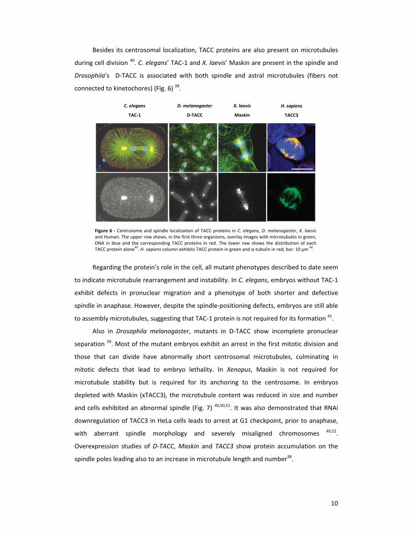
Besides its centrosomal localization, TACC proteins are also present on microtubules
during cell division 49. C. elegans’ TAC‐1 and X. laevis’ Maskin are present in the spindle and
Drosophila’s D‐TACC is associated with both spindle and astral microtubules (fibers not
connected to kinetochores) (Fig. 6) 39.
Regarding the protein’s role in the cell, all mutant phenotypes described to date seem
to indicate microtubule rearrangement and instability. In C. elegans, embryos without TAC‐1
exhibit defects in pronuclear migration and a phenotype of both shorter and defective
spindle in anaphase. However, despite the spindle‐positioning defects, embryos are still able
to assembly microtubules, suggesting that TAC‐1 protein is not required for its formation 41.
Also in Drosophila melanogaster, mutants in D‐TACC show incomplete pronuclear
separation 39. Most of the mutant embryos exhibit an arrest in the first mitotic division and
those that can divide have abnormally short centrosomal microtubules, culminating in
mitotic defects that lead to embryo lethality. In Xenopus, Maskin is not required for
microtubule stability but is required for its anchoring to the centrosome. In embryos
depleted with Maskin (xTACC3), the microtubule content was reduced in size and number
and cells exhibited an abnormal spindle (Fig. 7) 40,50,51. It was also demonstrated that RNAi
downregulation of TACC3 in HeLa cells leads to arrest at G1 checkpoint, prior to anaphase,
with aberrant spindle morphology and severely misaligned chromosomes 40,52.
Overexpression studies of D‐TACC, Maskin and TACC3 show protein accumulation on the
spindle poles leading also to an increase in microtubule length and number38.
C. elegans
TAC‐1 D. melanogaster
D‐TACC X. laevis
Maskin H. sapiens
TACC3
Figure 6 ‐ Centrosome and spindle localization of TACC proteins in C. elegans, D. melanogaster, X. laevisand Human. The upper row shows, in the first three organisms, overlay images with microtubules in green,DNA in blue and the corresponding TACC proteins in red. The lower row shows the distribution of eachTACC protein alone
49. H. sapiens column exhibits TACC protein in green and α‐tubulin in red, bar: 10 µm
48.

11
The rationale is that flaws in kinetochore‐microtubule attachment can lead to spindle
checkpoint activation and TACC3 regulates chromosome alignment by ensuring both proper
kinetochore microtubule attachment and spindle assembly checkpoint. 53
Phosphorylation and activation of TACC3 by Aurora‐A kinase
Recruitment of TACC3 proteins to microtubules is regulated by Aurora‐A kinase‐mediated
phosphorylation and this function appears to be conserved in Human, Xenopus, C. elegans
and Drosophila 41,48,54–56.
It has been proposed that Aurora‐A is vital during mitosis through phosphorylation of
a series of substrates that promote diverse critical events to maintain cell integrity 57. During
mitosis, Aurora‐A kinase associates with centrosomes and spindle, independently of
microtubules and D‐TACC and mutations of Aurora‐A lead to metaphase arrest and
decreased length of astral microtubules as well as prevention of D‐TACC centrosomal
association 54,56. Aurora A interacts with TACC3, phosphorylating it and allowing its targeting
to centrosomes 55. For Xenopus and Human TACC3, the exact phosphorylation sites have
already been identified 40,48,55. Suppression of Aurora‐A by siRNA caused mitosis failure, with
incorrect separation of centriole pairs, chromosome misalignment on the metaphase plate
and incomplete cytokinesis. Despite spindle abnormalities and unaligned chromosomes,
some cells lacking functional Aurora‐A are still able to divide, however segregation defects in
anaphase and chromatin bridges further develop into aneuploidies or tetraploidies which
ultimately lead to cell death 57.
Figure 7 ‐ Depletion of Xenopus’ TACC3 (xTACC3): effects on the spindle. The upper row shows the controlsituation and the lower one shows abnormal spindle and chromosome abnormalities in result if xTACC3depletion with RNAi
40.

12
Besides TACC3 vital interaction with Aurora‐A kinase, also its involvement with two
other other proteins, ch‐TOG and clathrin, is essential to a proper complex assembly and
function.
TACC3/ch‐TOG/clathrin complex stabilizes kinetochore fibers by inter‐
microtubule bridging
In higher organisms, the chromosome’s kinetochores are attached to the spindle via parallel
microtubules, named kinetochore fibers (K‐fibres) 58. The microtubules of K‐fibers are
connected by inter‐microtubule bridges that are thought to stabilize the fiber during
chromosome movement 59.
Clathrin interacts with these inter‐microtubule bridges in order to stabilize K‐fibers
60,61. Apart from being involved in coating vesicles during interphase, during cell division it
seems to target microtubule spindles 62. It acts as part of a complex involving two other
proteins: TACC3 and Colonic‐Hepatic Tumour Overexpressed Gene (ch‐TOG), forming the
TACC3/ch‐TOG/clathrin complex 61,63,64. ch‐TOG, or XMAP215, was identified as having an
essential role in spindle organization in Human during mitosis 65. Recently, clathrin was
identified as a binding protein for TACC3, and ch‐TOG was found to be associated with both
clathrin and TACC3 66. Depletion of clathrin, ch‐TOG or TACC3 result always in a mitotic
defected phenotype in different species, suggesting a consensus interaction across species
40,61,63,65.
Additionally, TACC3/ch‐TOG/clathrin bridges also protect the fiber from microtubule
loss 67 and promote growth by antagonizing the mitotic centromere‐associated kinesin
(MCAK) that promotes microtubule depolymerization 55,68.
Despite recent progresses in TACC3 research during mitosis, its role in mammalian
meiosis is relatively unknown. In mouse oocytes, TACC3 was identified during meiotic oocyte
maturation and the phosphorylated form of TACC3 accumulated from GVBD to the
metaphase II arrest. The phosphorylated protein was restricted to the spindle poles. The
effect of TACC3 depletion by siRNA was (i) inhibition of polar body extrusion (ii) arrested
meiosis I with spindle defects and (iii) lack of phosphorylated TACC3 labeling at the poles 69.

13
TACC3/ch‐TOG/clathrin complex activation, two hypotheses
The correct order of recruitment of these three proteins to the complex is yet unknown,
however two major ones were proposed during the last three years (Fig. 8).
In 2010, three teams 40,63,66 claimed that clathrin recruits phosphorylated TACC3 to
spindle poles (Fig. 8, A), however other findings point to a different direction. In 2011, Booth
et al. demonstrated that TACC3 and ch‐TOG bind to the spindle microtubules under Aurora‐
A regulation, followed by recruitment of clathrin to the microtubules forming complex with
TACC3 or TACC3/ch‐TOG subcomplex (Fig. 8, B). Furthermore, it was also proposed that the
complex is spindle‐specific 61.
According to Booth et al., clathrin cannot bind microtubules while maskin (TACC3) and
ch‐TOG can, therefore, clathrin would require an additional factor to bind these fibers,
however no such factor was identified. Also, the majority of clathrin in mitotic cells is not
restricted to the spindle whereas TACC3 and ch‐TOG are predominantly spindle‐associated.
In agreement with Booth’s theory, (i) overexpression of GFP‐CHC (Clathrin Heavy Chain) did
not cause more clathrin to accumulate at the spindle and did not influence TACC3 nor ch‐
Figure 8 ‐ Two modelsrepresenting the recruitment ofTACC3/ch‐TOG/clathrin complex tothe spindle. (A) Clathrin recruitsTACC3: Aurora‐A kinase activatesTACC3 by phosphorylating it in twosites (Ser620 and Ser626 inXenopus) and enables it to bindwith clathrin to form theclathrin/TACC3 complex. Finally,the clathrin‐associated TACC3 istargeted to spindle poles andspindle microtubules for properspindle assembly. Along withTACC3, ch‐TOG also targets tospindles to regulate spindlestability. (B) TACC3 recruitsclathrin. First, TACC3 alone orcomplexed with ch‐TOG isrecruited to the spindlemicrotubules in a phosphorylation‐dependent manner regulated byAurora‐A. Then, clathrin isrecruited to the spindle by TACC3or TACC3/ch‐TOG. Finally, clathrinforms an inter‐microtubule bridgeby interacting with additionalTACC3 or TACC3/ch‐TOG onadjacent microtubules to stabilizethe spindle microtubules.
89

14
TOG spindle localization (ii) overexpressing TACC3 increased the spindle location of ch‐TOG
and clathrin and (iii) inhibiting TACC3 spindle recruitment via inhibition of Aurora‐A kinase or
TACC3 mutation inhibited recruitment of clathrin to the spindle.
Taken together, recruitment of clathrin and TACC3 to the mitotic spindle remains
controversial, however in unanimity it is suggested that this complex stabilizes kinetochore
fibers by physically bridging between adjacent microtubules. In the light of these results,
TACC3 has an important role in mitosis as a controlled adaptor that can integrate several
other mitotic proteins into a complex on microtubules which holds kinetochore fibers
together.
TACC3 function during mammalian meiosis has already been documented 69; however
its role during bovine oocyte development is still blurred. Besides having clear implication in
human cancer lines, understanding its role during cell‐division is crucial when the matter of
mammalian oocyte developmental competence is addressed. The aim of this project will be
to get insights into TACC3’s function during meiosis and early embryo development and
ultimately propose a model for TACC3/ch‐TOG/clathrin complex regulation and recruitment
in cattle, which may mimic the Human situation.

15
Material and methods
Oocyte collection and in vitro maturation
Bovine ovaries were obtained from a local slaughterhouse and transported in thermal flasks.
Excess tissue was cut off and the ovaries were collected in flasks containing NaCl at 30ºC
supplemented with penicillin/streptomycin (1ml/L). Cumulus oocyte complexes (COCs) were
removed from the ovaries by suction of follicular fluid from antral follicles ranging 2‐8mm
and only those with reasonable cumulus investment were selected under a dissecting
microsope. The COCs were rinsed with medium B (Table 1) to avoid agglomeration. COCs
were transferred to 4‐well plates, each containing 500µL of maturation medium (Table 1) or
different concentrations of the inhibitor MLN8054 (by selleckchem®, 25 mM in DMSO) in
maturation medium. Between the wells, 1mL of sterile water was added to prevent
evaporation. After 22‐24 H of incubation at 39ºC with 5% CO2, the cultured oocytes were
either selected for in‐vitro fertilization or fixed in 4% paraformaldehyde.
In vitro fertilization and embryo culture
A straw of cryopreserved bull sperm was thawed in a 37ºC waterbath and the content
layered on a Percoll gradient and centrifuged for 30 minutes at 700G. The sperm’s correct
Medium Ingredients
B MilliQ water, M199 with Earle's salts, HEPES and glutamine
C LAL water, M199 with Earle's salts and glutamine, NaHCO3
PHE Penicillamine, hypotaurine and epinephrine
Maturation Medium C, Fetal Calf Serum, Pen/Strep, recombinant FSH
Fertilization LAL water, NaCl, KCl, NaHCO3, Na2HPO4, Na pyruvate, phenolred, CaCl2.2H2O,
MgCl2.6H2O, Pen/Strep, BSA
RD MilliQ water NaCl, KCl, NaHCO3, Na2HPO4, Na Lactate, HEPES, phenolred,
CaCl2.2H2O, MgCl2.6H2O, Na Pyruvate, Pen/Strep, BSA
SOF A LAL water, NaCL, KCl, KH2PO4, Na Lactate, MgSO4.7H2O, NaHCO3, CaCl2.2H2O,
phenolred, Non‐essential aminoacid solution
SOF B LAL water, Pen/Strep, Na‐pyruvate, L‐glutamine, BSA
SOF 8ml SOF A : 2ml SOF B
Table 1 ‐ Composition of multiple culture mediums used during oocyte in‐vitro maturation and fertilization and embryo culture.

16
concentration was determined by sampling a small volume of the sperm solution and
counting the number of sperm heads in a Bürker Turk chamber. Taking into account which
bull was used, the dilution factor was calculated and sperm was added to the fertilization
medium (Table 1). The COCs and sperm were incubated for 22 hr at 39ºC, 5% CO2 after
which the presumptive zygotes were stripped of cumulus cells by vortexing for 3 min in RD
medium (Table 1). Following denudation, the presumptive zygotes were transferred to a
new 4‐well plate with Synthetic Oviductal Fluid (SOF medium, Table 1) and incubated at
39ºC, in a 5% CO2 and 7% O2 environment70. At day 5 after culture, cleaved embryos were
scored and transferred to fresh SOF medium and at day 8, embryo development was
determined.
Immunocytochemistry
Sections of bovine testis, bovine ovaries and human testis (provided by University Medical
Center Utrecht) were used. To de‐paraffinise sections, slides were immersed 2x5min in
xylene and 2x5min in 100% ethanol followed by 20 min incubation in methanol‐3% H2O2 to
block endogenous peroxidase and reduce non‐specific background staining. Samples were
then rehydrated in 2 minutes series of 96/80/70% ethanol, rinsed shortly in distilled H2O.
For antigen retrieval, sodium‐citrate buffer (0.01M, pH 6.0) was heated until boiling point
and samples were boiled for 3 min. After a 30 min cool‐down, sections were washed in TBST
(0.05% Tween) for 2x5min, blocked with 5% NGS (Normal Goat Serum) in TBS for 30 minutes
at 37 ºC and left to incubate overnight with the first antibody (1:100) in 2% NGS in TBS at
room temperature. The three samples were each stained with Human anti‐PIWIL3 antibody
(ab77088), anti‐PIWIL3 antibody (ab93709) and IgG for control purposes (Table 2). On the
second day, after washing in TBS (0.05 M, pH 7.6) on the shaking table, incubation with the
second antibody (1:200) took place for 40 min at 37ºC followed by another washing step.
The samples were then incubated with ABC complex reagent (Vector elite kit 10µl A + 10µl B
+ 980 µl TBS) for 30 min at RT (ABC made 30 min prior to use). Sections were then washed in
TBS, incubated with DAB (diaminobenzidinetetrahydrochloride, mutagenic, photosensitive)
in TBS for 5‐10 min and washed again in distilled H2O. For counterstaining, slides were
stained with haematoxylin for 10 seconds and then washed thoroughly with H2O. Series of
70/80/96% ethanol were executed (2x 10 sec each) followed by xylene (2x2min). Lastly, the
samples were embedded in DEPEX.

17
Anti‐PIWIL3 antibody (ab77088)
Anti‐PIWIL3 antibody (ab93709)
Anti‐TACC3 antibody (ab56595)
Anti‐PIWIL3
antibody* (custom)
Anti‐PIWIL1 antibody (custom)
Anti‐PIWIL2 antibody (custom)
Description Mouse
polyclonal to PIWIL3
Rabbit polyclonal to
PIWIL3
Mouse monoclonal to TACC3
Rabbit polyclonal to PIWIL3
Rabbit polyclonal to PIWIL1
Rabbit polyclonal to PIWIL2
Host species Mouse Rabbit Mouse Rabbit Rabbit Rabbit
Tested applications
WB IHC‐P WB, IHC‐P ‐ ‐ ‐
Cross Reactivity
Reacts with Human
Reacts with Human
Reacts with Human
Reacts with
bovine*
Reacts with bovine
Reacts with bovine
Positive control
PIWIL3 transfected
293T cell lysate
Human testis tissue.
‐ ‐ ‐ ‐
Immunogen
Recombinant full length
human PIWIL3 (NP_001008496,
aa 1‐882)
Synthetic peptide from
intermediate residues of Human PIWIL3
100aa recombinant fragment
15aa long fragment
15aa long fragment
15aa long fragment
Immunofluorescence
Oocytes were stripped of cumulus cells before staining and fixed for a minimum of 15 min in
4% paraformaldehyde (PFA). After fixation the oocytes were briefly washed with 0.1% Triton
X‐100 and 10% FCS in PBS (PBST) and permeabilized for 30 min using 0.5% Triton X‐100 and
PBST. The oocytes were subsequently blocked for 1h in PBST and incubated with the primary
antibody overnight at 4ºC. For the immunofluorescence, all antibodies described in Table 2
were used. The oocytes were washed three times in PBST (15 min) followed by incubation in
the secondary antibody for 1 h in the dark. After several washes, the oocytes were stained
with 4',6‐diamidino‐2‐phenylindole (DAPI, 5min) and mounted on a slide with Vectashield
and isolated with Vaseline.
For α‐tubulin staining oocytes were incubated first for 30‐60 min in microtubule
stabilizing solution71 at 37 ºC and then fixed in 4% PFA. After fixation, oocytes were washed
in PBS with 0.1% (v/v) Tween‐20, incubated 5min in PBS with 2% (v/v) goat serum (Sigma‐
G6767) followed by 60 min incubation with mouse‐anti‐tubulin primary antibody at 37 ºC.
After washing, the oocytes were incubated in PBS + serum for 1 hour at 37 ºC, washed and
Table 2 ‐ Detailed description of the antibodies used. The (*) regarding the bovine PIWIL3 symbolizes the predicted details on theonset of this project. All the custom antibodies are thought not to be specific and further tests have to be done to address thisquestion.

18
incubated with the second antibody for 1 h. For microfilament staining, an additional
incubation of 30 min with phalloidin was performed. After DNA staining with DAPI, the
oocytes were mounted as mentioned before.
Slides were examined with Leyca® Confocal laser microscopy. Images were analyzed
with ImageJ® software and Adobe Illustrator®.
Western blotting
Cells were collected and frozen in ‐80ºC after which they were lysed with sample buffer 4x
diluted in RIPA buffer (table 3) and 1% protease inhibitor cocktail (table 3). Afterwards the
lysates were boiled at 100 ºC for 5 min and briefly centrifuged. The samples were resolved
by an SDS‐PAGE 8% running gel and 5% stacking gel (Table 3), while sponges, filter paper and
membrane were left for 10 min at 4 ºC in Blotting buffer. The proteins were then blotted on
nitrocellulose membranes (Trans‐Blot®, Bio‐Rad Laboratories) at 100V (±180mA) during 1H.
The membranes were incubated with blocking buffer (Table 3) for 1h under gentle shaking.
The membranes were incubated with mouse‐anti‐TACC3 (Table 2) diluted in blocking buffer
(1:1000) O/N at 4 ºC. After washing in TBS‐Tween (TBS with 0.05% Tween‐20) (3x5min) the
membranes were incubated with goat‐anti‐Mouse (sc‐2005, Santa Cruz Biotechnology Inc)
(1:5000) for 1H at RT; washed with TBS‐Tween and lastly with TBS. Finally the membranes
were placed between a plastic sheet under the SuperSignal® Chemiluminescent Substrate
(West Dura Extended Duration Substrate, Thermo scientific) consisting of 600µL of
Horseradish peroxidase (HRP) and phosphatase (PO) (1:1). The membrane was exposed to
X‐ray film (Fuji®) and the film was developed.
After developing the film, the membrane was stripped in stripping buffer for 5 min at
RT and the blot was incubated for 1H at 37 ºC with Goat‐anti‐β‐actin (1:5000, Santa Cruz
Biotechnology®, sc‐47778) and for 1H at RT with Rabbit‐anti‐Goat (1:10000, Santa Cruz
Biotechnology®,sc‐2768) just as described above.
19
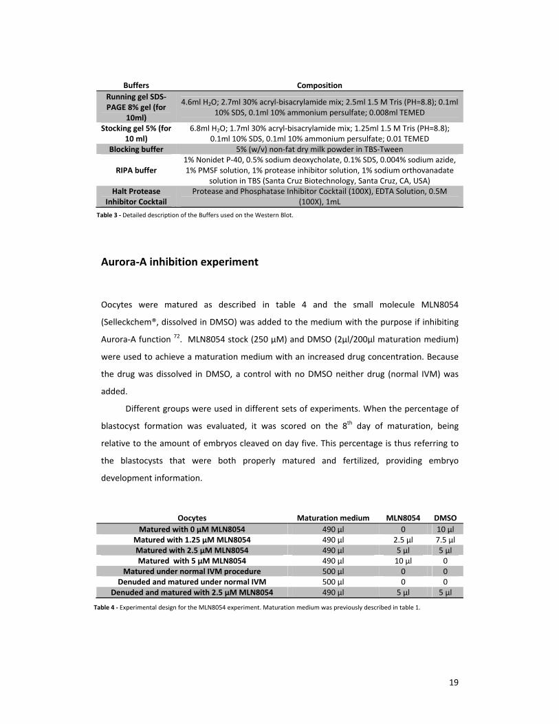
Aurora‐A inhibition experiment
Oocytes were matured as described in table 4 and the small molecule MLN8054
(Selleckchem®, dissolved in DMSO) was added to the medium with the purpose if inhibiting
Aurora‐A function 72. MLN8054 stock (250 µM) and DMSO (2µl/200µl maturation medium)
were used to achieve a maturation medium with an increased drug concentration. Because
the drug was dissolved in DMSO, a control with no DMSO neither drug (normal IVM) was
added.
Different groups were used in different sets of experiments. When the percentage of
blastocyst formation was evaluated, it was scored on the 8th day of maturation, being
relative to the amount of embryos cleaved on day five. This percentage is thus referring to
the blastocysts that were both properly matured and fertilized, providing embryo
development information.
Buffers Composition
Running gel SDS‐PAGE 8% gel (for
10ml)
4.6ml H2O; 2.7ml 30% acryl‐bisacrylamide mix; 2.5ml 1.5 M Tris (PH=8.8); 0.1ml 10% SDS, 0.1ml 10% ammonium persulfate; 0.008ml TEMED
Stocking gel 5% (for 10 ml)
6.8ml H2O; 1.7ml 30% acryl‐bisacrylamide mix; 1.25ml 1.5 M Tris (PH=8.8); 0.1ml 10% SDS, 0.1ml 10% ammonium persulfate; 0.01 TEMED
Blocking buffer 5% (w/v) non‐fat dry milk powder in TBS‐Tween
RIPA buffer 1% Nonidet P‐40, 0.5% sodium deoxycholate, 0.1% SDS, 0.004% sodium azide, 1% PMSF solution, 1% protease inhibitor solution, 1% sodium orthovanadate
solution in TBS (Santa Cruz Biotechnology, Santa Cruz, CA, USA) Halt Protease
Inhibitor Cocktail Protease and Phosphatase Inhibitor Cocktail (100X), EDTA Solution, 0.5M
(100X), 1mL
Oocytes Maturation medium MLN8054 DMSO
Matured with 0 µM MLN8054 490 µl 0 10 µlMatured with 1.25 µM MLN8054 490 µl 2.5 µl 7.5 µlMatured with 2.5 µM MLN8054 490 µl 5 µl 5 µlMatured with 5 µM MLN8054 490 µl 10 µl 0
Matured under normal IVM procedure 500 µl 0 0 Denuded and matured under normal IVM 500 µl 0 0
Denuded and matured with 2.5 µM MLN8054 490 µl 5 µl 5 µl
Table 3 ‐ Detailed description of the Buffers used on the Western Blot.
Table 4 ‐ Experimental design for the MLN8054 experiment. Maturation medium was previously described in table 1.

20
Results
Sorting out the bovine Piwi‐like 3 antibody and unraveling
Transforming Acidic Coiled‐Coil protein 3 (TACC3)
In order to detect bovine Piwi‐like 3 expression in oocytes a polyclonal antibody was
generated directed against a 15 aminoacid long fragment (Table 2). This peptide was
designed based on NCBI’s predicted 991aa bovine Piwi‐like 3 protein sequence and was
identical to the NH2 part of the predicted sequence. However, the ENSEMBL database
provides another predicted sequence for the PIWIL3 bovine protein. Besides having 88.2 %
similarity with the NCBI sequence, the ENSEMBL predicted sequence is shorter (875aa) and
lacks the fragment to which the antibody was directed (illustrated in Fig. 9). We have
reasons to believe that neither the ENSEMBL nor the NCBI protein sequences are correct
based on recent 5 prime race analyses and further sequencing (Roelen & Mahdipour,
unpublished results).
A mass spectrometry analysis was performed to evaluate potential PIWIL3 binding
proteins. For that purpose an Immunoprecipitation experiment was conducted using the
bovine PIWIL3 antibody followed by mass spectrometry (Roelen & Mahdipour, unpublished
results). Among the proteins expected to be detected were Argonaute proteins already
described as being associated with PIWI proteins. Interestingly the mass spectrometry
analysis revealed no proteins known to interact with PIWI proteins and, most importantly,
PIWIL3 itself was also not detected. Instead, TACC3 was identified as a prominent protein in
the IP fraction. It was proposed that the antibody used does not recognize bovine PIWIL3 but
instead bovine TACC3.
Figure 9 ‐ Schematic representation of two Piwi‐like 3 aminoacid sequences, one provided by NCBI database and the other by ENSEMBL database with 88.2% identity. Red represents the target sequence of our PIWIL3 custom made bovine antibody.

21
To test whether indeed the antibody that was designed to recognize bovine PIWL3
would recognize TACC3 instead, oocytes and blastocysts were double‐stained with the
custom‐made antibody and an antibody directed against human TACC3 (Table 2). Indeed
similar patterns of expression were identified (Fig. 10), being the human TACC3 antibody
more sensitive that the PIWIL3 antibody, particularly at the GV and blastocyst stage (Fig. 10,
A‐D and Q‐T).
Figure 10 ‐ Double stainings TACC3 and ’TACC3’ during oocyte maturation and blastocyst. ‘TACC3’ refers to the antibody designed against PIWIL3. Images (A‐D) represent a germinal vesicle stage, (E‐H) a metaphase I stage, (I‐L, M‐P) a metaphase II stage and (Q‐T) a blastocyst stage. Scale bar: 10µm.
DAPI A TACC3B ‘TACC3’ C DAPI TACC3 ‘TACC3’ D
E F G H
I J K L
M N O P
Q R S T

22
Detection of PIWIL3 expression in bovine oocytes using antibodies
directed against the human sequence
Preliminary findings suggested that in bovine oocytes PIWIL3 was localized around
chromatin during meiosis 33, in contrast with our results on the expression of PIWIL2 and
PIWIL1 (table 2) that appeared to be rather diffuse throughout the cytoplasm (Fig. 11, A and
B).
In order to characterize PIWIL3 expression in bovine oocytes, commercially available
Piwi‐like 3 antibodies directed against human PIWIL3 were used on bovine oocytes. The
human PIWIL3 protein sequence is significantly different from the predicted bovine NCBI
and ENSEMBL sequences, with just 54 and 52% of similarities, respectively. Two different
antibodies were available, one directed against the whole protein sequence of human
PIWIL3 (denominated αHPIWIL3‐A) and another one directed against a short 100aa fragment
(denominated αHPIWIL3‐B) (Fig. 12; Table 2).
Figure 12 ‐ Schematic representation of the human Piwi‐like 3 aminoacid sequence and bovine Piwi‐like 3 aminoacid sequences.The percentages indicate degrees of similarities between sequences. Red represents target sequences, the epitopes, ofrepresented antibodies.
DAPI PIWIL2
DAPI PIWIL1
A B Figure 11 ‐ Merge channels of (A) DAPI and PIWIL1 and (B)DAPI and PIWIL2 expression patterns during metaphase II. Oocyte limits are represented by the dashed circle line. Scale bar: 10µm.

23
Using the αHPIWIL3A antibody, no localized expression was detected in bovine
metaphase II staged oocytes (Fig. 13, A‐F) nor in blastocysts (Fig.13, G‐I). Instead, staining
was observed in a punctate pattern throughout the oocyte, similarly to the patterns
observed with bovine PIWIL1 and PIWIL2 (Fig 11). HPIWIL3‐B displayed a different pattern in
MII stage where its fluorescence was almost absent (Fig.13, M‐O), similar to the blastocyst
stage (Fig. 13, P‐R). Interestingly, cytoplasmic staining was observed in cumulus cells that
remained attached to the oocyte (Fig.13, J‐L).
In paraffin section of human testis, αHPIWIL3‐A revealed no staining whilst the
αHPIWIL3‐B was able to stain some cells (Fig. 14). These positive results are in consistence
with the positive control already documented and displayed (Fig. 14, Human testis and Table
2). In paraffin sections of bovine testis, αHPIWIL3‐A antibody only stained blood vessels,
whereas αHPIWIL3‐B stained the cytoplasm of cells at the final spermatogenesis stages
(Fig.14, bovine testis). In bovine ovarian sections, no staining was observed with the
αHPIWIL3‐A antibody. In contrast, the HPIWIL3‐B antibody stained (i) both the external and
internal theca (ii) granulosa cells (iii) cumulus cells surrounding the oocyte and (iv) the
oocyte itself (Fig. 14, bovine ovary).
F
I
DAPI
A
HPIWIL3‐A
B
DAPI HPIWIL3‐A
C
E D
G H
HPIWIL3‐B
K
DAPI HPIWIL3‐B
L
M N O
P Q R
DAPI
J
Figure 13 – HPIWIL3‐A and HPIWIL3‐B stainings on different staged oocytes and blastocysts. (A‐I) displays staining withHPIWIL3‐A and (J‐R) displays staining with HPIWIL3‐B. (A‐C) displays a germinal vesicle stage, (D‐F) zoom of a metaphase IIplate, (G‐I) a blastocyst, (J‐L) displays two cumulus cells still attached to the outside of the oocyte (M‐O) displays a metaphase IIstage and (P‐R) a blastocyst. Scale bar: 10 µm.

24
TACC3’s expression pattern during oocyte and early zygote
development: its presence surrounding chromatin and the meiotic
spindle
Because the custom‐made antibody doesn’t recognize bovine PIWIL3 but instead TACC3, we
chose to interpret the immunofluorescence results performed with this antibody as being
TACC3’s.
Oocytes were collected 12 and 23 H after in vitro maturation process and 6, 12 and 22
H after in vitro fertilization and images were selected to construct a time‐lapse figure ranging
oocytes from germinal vesicle up until blastocyst stage (Fig.15). TACC3 was first detected in a
condensed matter around the genetic material 12 H after maturation, when the chromatin
started displaying some degree of condensation (Fig.15, D‐F). Prior to this phase, TACC3
expression was not detected (Fig.15, A‐C). TACC3 was specifically localized around the
Bovine Ovary
Bovine Testis
IgG HPIWIL3‐A HPIWIL3‐B
Human
Testis
ET
IT
BM
GC
CC
CC
BM
GC
IT
ET
AA
O
Figure 14 ‐ Immunocytochemistry results for IgG, HPIWIL3‐A and PIWIL3‐B in human testis, bovine testis and bovine ovary. IgGis displayed as a negative control. Human and bovine testis row represent an epididymis tube cut transversely. Bovine ovary inIgG displays ovarian cortex (dashed circles represent primary follicles) and the other two pictures represent an antral folliclewith ET‐ External Theca; IT‐ Internal Theca; A‐ Antral cavity; CC‐ Cumulus Cells; GC‐ Granulosa Cells; BM‐ Basal Membrane; O‐Oocyte. Scale bar: 0.1mm.
25
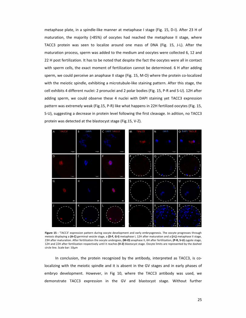
metaphase plate, in a spindle‐like manner at metaphase I stage (Fig. 15, D‐I). After 23 H of
maturation, the majority (>85%) of oocytes had reached the metaphase II stage, where
TACC3 protein was seen to localize around one mass of DNA (Fig. 15, J‐L). After the
maturation process, sperm was added to the medium and oocytes were collected 6, 12 and
22 H post fertilization. It has to be noted that despite the fact the oocytes were all in contact
with sperm cells, the exact moment of fertilization cannot be determined. 6 H after adding
sperm, we could perceive an anaphase II stage (Fig. 15, M‐O) where the protein co‐localized
with the meiotic spindle, exhibiting a microtubule‐like staining pattern. After this stage, the
cell exhibits 4 different nuclei: 2 pronuclei and 2 polar bodies (Fig. 15, P‐R and S‐U). 12H after
adding sperm, we could observe these 4 nuclei with DAPI staining yet TACC3 expression
pattern was extremely weak (Fig.15, P‐R) like what happens in 22H fertilized oocytes (Fig. 15,
S‐U), suggesting a decrease in protein level following the first cleavage. In adition, no TACC3
protein was detected at the blastocyst stage (Fig.15, V‐Z).
In conclusion, the protein recognized by the antibody, interpreted as TACC3, is co‐
localizing with the meiotic spindle and it is absent in the GV stages and in early phases of
embryo development. However, in Fig 10, where the TACC3 antibody was used, we
demonstrate TACC3 expression in the GV and blastocyst stage. Without further
Figure 15 ‐ ‘TACC3’ expression pattern during oocyte development and early embryogenesis. The oocyte progresses throughmeiosis displaying a (A‐C) germinal vesicle stage, a (D‐F, G‐I) metaphase I, 12H after maturation and a (J‐L) metaphase II stage,23H after maturation. After fertilization the oocyte undergoes, (M‐O) anaphase II, 6H after fertilization, (P‐R, S‐U) zygote stage,12H and 22H after fertilization respectively until it reaches (V‐Z) blastocyst stage. Oocyte limits are represented by the dashedcircle line. Scale bar: 10µm

26
characterization, it could only be speculated whether the protein was around the metaphase
plate or the polar body during metaphase II. .
TACC3’s presence surrounding the metaphase plate in the metaphase II
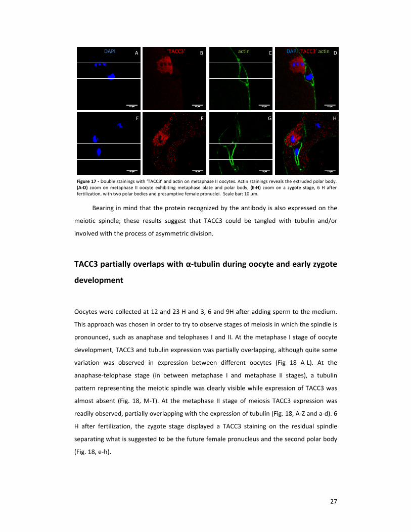
During meiosis, actin localizes specifically around the polar body 71 and a co‐staining of actin
and TACC3 was performed in MII stages oocytes. First, microfilaments were stained (Fig. 16)
to check whether the protocol worked for bovine oocytes. Indeed actin filaments were
identified specifically surrounding the extruded polar body (Fig. 16, B and F) while α‐tubulin
was present on both the polar body and the metaphase plate (Fig.16, C and G). In
metaphase II oocytes a distinct pattern was observed between the polar body and the
metaphase plate where the actin was surrounding the polar body on the periphery of the
oocyte and TACC3 protein was located specifically around the metaphase plate (Fig. 12, A‐
D). Later, at the zygote stage, two masses of DNA were enclosed by actin, suggesting the
presence of the first and the second polar body, 6H after fertilization (Fig. 12, G) and TACC3
expression accumulated in what, based on the actin distribution, is proposed to be the
second polar body, while no protein was surrounding the female pronucleus (Fig.12, E‐H).
A B C D
E F G H
DAPI Actin α‐tubulin DAPI Actin α‐tubulin
Figure 16 ‐ Microfilament and microtubule staining with actin (phalloidin) and α‐tubulin on (A‐H) metaphase II oocytes, 23Hafter maturation; (D‐H) zoom on the metaphase plate and polar body. Columns are divided per staining, as indicated. Scale bar:10 µm.
27
DAPI A ‘TACC3’ B actin C DAPI ‘TACC3’ actin D
E F G H
Bearing in mind that the protein recognized by the antibody is also expressed on the
meiotic spindle; these results suggest that TACC3 could be tangled with tubulin and/or
involved with the process of asymmetric division.
TACC3 partially overlaps with α‐tubulin during oocyte and early zygote
development
Oocytes were collected at 12 and 23 H and 3, 6 and 9H after adding sperm to the medium.
This approach was chosen in order to try to observe stages of meiosis in which the spindle is
pronounced, such as anaphase and telophases I and II. At the metaphase I stage of oocyte
development, TACC3 and tubulin expression was partially overlapping, although quite some
variation was observed in expression between different oocytes (Fig 18 A‐L). At the
anaphase‐telophase stage (in between metaphase I and metaphase II stages), a tubulin
pattern representing the meiotic spindle was clearly visible while expression of TACC3 was
almost absent (Fig. 18, M‐T). At the metaphase II stage of meiosis TACC3 expression was
readily observed, partially overlapping with the expression of tubulin (Fig. 18, A‐Z and a‐d). 6
H after fertilization, the zygote stage displayed a TACC3 staining on the residual spindle
separating what is suggested to be the future female pronucleus and the second polar body
(Fig. 18, e‐h).
Figure 17 ‐ Double stainings with ‘TACC3’ and actin on metaphase II oocytes. Actin stainings reveals the extruded polar body.(A‐D) zoom on metaphase II oocyte exhibiting metaphase plate and polar body, (E‐H) zoom on a zygote stage, 6 H afterfertilization, with two polar bodies and presumptive female pronuclei. Scale bar: 10 µm.
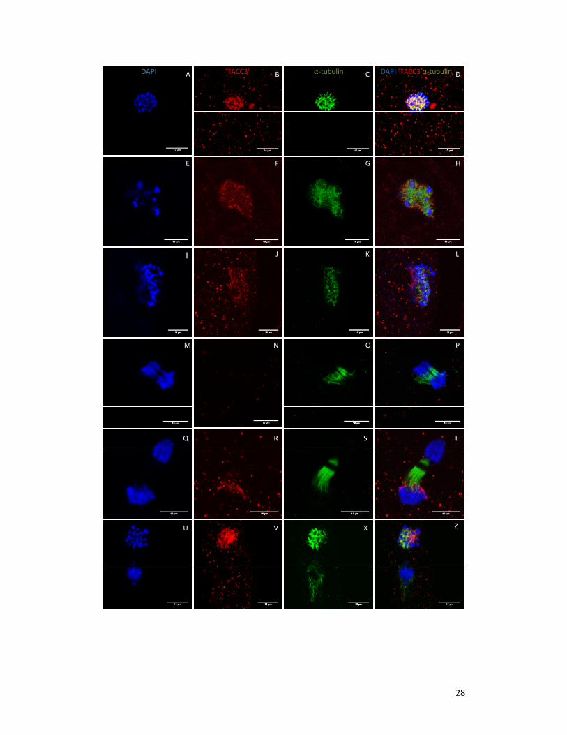
28
DAPI ‘TACC3’ α‐tubulin DAPI ‘TACC3’α‐tubulin A B C D
E F G H
I J K L
M N O P
U
R S T
V X Z
Q

29
Discerning TACC3 function: using MLN8054 compound, an Aurora‐A
inhibitor, decreases oocyte developmental progress and causes spindle
anomalies
Aurora‐A kinase, responsible for TACC3 phosphorylation and activation can be selectively
inhibited using a small compound, the MLN8054 72,73. This small molecule demonstrated
similar results to those of RNAi directed against Aurora‐A, including chromosomal defects
leading to aneuploidy 72. To study Aurora‐A kinase activity in TACC3 function during bovine
oocyte meiosis, oocytes were matured in the presence and absence of this Aurora‐A
inhibitor (Table 4).
23 h matured oocytes were collected and scored the ones in equal developmental
stage with an increased inhibitor concentration (Fig. 19, Supplementary Table 1). Firstly,
there were no significant differences between the normal IVM and maturation with 0 µM of
MLN8054 (10 µM DMSO), so DMSO had no effect on the oocyte. Secondly, oocytes
(denuded and non‐denuded) tend to arrest in metaphase I stage in a concentration
dependent matter, decreasing the percentage of oocytes at the metaphase II stage.
Figure 18 – Double stainings of ‘TACC3’ and microtubules (α‐tubulin) on oocytes and early zygotes. Images (A‐L) demonstrate a3 metaphase I oocytes, 12 hours after maturation (M‐T) anaphase I/telophase I oocytes, 12 hours after maturation (U‐d)metaphase II oocytes, 23 hours after maturation, and (e‐f) zygote stage, 6 hours after adding sperm to the medium. Scale bar:10 µm.
a b c d
e f g h
DAPI ‘TACC3’ α‐tubulin DAPI ‘TACC3’ α‐tubulin

30
In the presence of the inhibitor the percentage of MII stage oocytes (identified by the
presence of the first polar body) was reduced in a concentration dependent manner (Fig. 20,
Supplementary Table 2). Similarly, the percentage of blastocyst formation after embryo
cleavage also decreased with an increased drug concentration (Fig. 20, Supplementary Table
3). Moreover, there were no differences concerning cumulus cells growth and expansion in
the presence or absence of the inhibitor, however, a higher number of oocytes must be
analyzed to provide appropriate statistical conclusions. These results suggest that inhibition
of Aurora‐A kinase is affecting cell division and/or the process of polar body extrusion.
0%10%20%30%40%50%60%70%80%90%
100%
MLN8054 effect on oocyte developmental progress
Metaphase II
Telophase I
Metaphase I
Figure 19 ‐ Stacked column graphic representing the effect of the small molecule MLN8054 throughout oocyte developmentalprogress, namely during metaphase I, telophase I and metaphase II oocytes in different conditions.
82,98%
40,48% 8,51% 6,12%
R² = 0,8913
‐20%
0%
20%
40%
60%
80%
100%
120%
0 µM MLN8054 1.25µM MLN8054 2.5µM MLN8054 5µM MLN8054
Presence of polar body
71% 53% 58%
41% 38%
R² = 0,8511
0%
20%
40%
60%
80%
Normal IVM 0 µM MLN8054 1.25µM MLN8054 2.5µM MLN8054 5µM MLN8054
Blastocyst formation from cleaved embryos
Figure 20 ‐. Uppergraphic represents theeffect of MLN8054 onthe percentage ofoocytes with extrudedpolar body and the lowerone represents thepercentage of blastocystformation (day 8) fromalready cleaved embryos(day 5) with an increaseddrug concentration. Errorbars are indicated andred line represents alinear trendline with R
2
value indicated above.
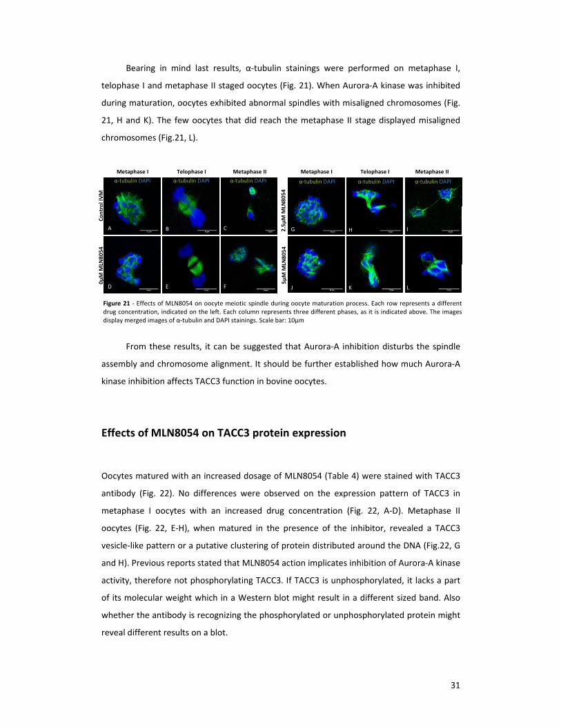
31
Bearing in mind last results, α‐tubulin stainings were performed on metaphase I,
telophase I and metaphase II staged oocytes (Fig. 21). When Aurora‐A kinase was inhibited
during maturation, oocytes exhibited abnormal spindles with misaligned chromosomes (Fig.
21, H and K). The few oocytes that did reach the metaphase II stage displayed misaligned
chromosomes (Fig.21, L).
From these results, it can be suggested that Aurora‐A inhibition disturbs the spindle
assembly and chromosome alignment. It should be further established how much Aurora‐A
kinase inhibition affects TACC3 function in bovine oocytes.
Effects of MLN8054 on TACC3 protein expression
Oocytes matured with an increased dosage of MLN8054 (Table 4) were stained with TACC3
antibody (Fig. 22). No differences were observed on the expression pattern of TACC3 in
metaphase I oocytes with an increased drug concentration (Fig. 22, A‐D). Metaphase II
oocytes (Fig. 22, E‐H), when matured in the presence of the inhibitor, revealed a TACC3
vesicle‐like pattern or a putative clustering of protein distributed around the DNA (Fig.22, G
and H). Previous reports stated that MLN8054 action implicates inhibition of Aurora‐A kinase
activity, therefore not phosphorylating TACC3. If TACC3 is unphosphorylated, it lacks a part
of its molecular weight which in a Western blot might result in a different sized band. Also
whether the antibody is recognizing the phosphorylated or unphosphorylated protein might
reveal different results on a blot.
Figure 21 ‐ Effects of MLN8054 on oocyte meiotic spindle during oocyte maturation process. Each row represents a differentdrug concentration, indicated on the left. Each column represents three different phases, as it is indicated above. The imagesdisplay merged images of α‐tubulin and DAPI stainings. Scale bar: 10µm
Control IVM
0µM M
LN8054
Metaphase I Telophase I Metaphase II
α‐tubulin DAPIα‐tubulin DAPI α‐tubulin DAPI
2.5µM M
LN8054
5µM M
LN8054
Metaphase I Telophase I Metaphase II
α‐tubulin DAPIα‐tubulin DAPI α‐tubulin DAPI
A B
D E
C
F
G H
J K
I
L

32
To address this question, oocytes were matured with 0 µM MLN8054 and with 5 µM
MLN8054 and 500 metaphase II oocytes and their cumulus cells were western blotted for
TACC3 (Fig. 23). Metaphase II oocytes matured with and without the drug displayed a band
between 115 and 180KDa. Concerning cumulus cells, we perceive a band (around 170KDa)
when matured with the drug, whilst no band is detected on the control. For quality check,
β‐actin staining was performed on the same blot and only the oocytes revealed a band.
Figure 22 ‐ Effects of MLN8054 on TACC3 protein expression pattern during metaphase I and II oocytes. TACC3 representedin green and DAPI in blue. The first row displays metaphase I oocytes and the lower row metaphase II ones. Columns aredivided according to the type of maturation process, as indicated on top. Scale bar: 10µm.
Figure 23 ‐ Western Blot on TACC3 protein and β‐actin in four different groups. First film reveals the results of the blot with TACC3 protein with 1min exposure to X‐ray film, second reveals β‐actin staining and some TACC3 left after 10 min of exposure. First lane represents lysates of cumulus cells from cells matured with 0µM MLN8054, second lane represents lysates of metaphase II oocytes matured also with 0µM MLN8054. Third and fourth lanes represent lysates of cumulus cells and metaphase II oocytes respectively matured with 5µM MLN8054. Ladder is represented in blue.
TACC3
β-actin
1min exposure 10min exposure
B D
F HGE
CA
5µM MLN8054 Control IVM 0µM MLN8054
Metap
hase I
Metaphase II
1.25µM MLN8054
TACC3 DAPI TACC3 DAPI TACC3 DAPI TACC3 DAPI

33
Discussion
PIWIL3 during bovine oocyte development
The custom‐made antibody designed to recognize bovine PIWIL3 did not identify bovine
Piwi‐like 3 protein because of incorrect protein sequences predicted by ENSEMBL and NCBI.
When the epitope peptide is blasted on the ENSEMBL protein database, no Piwi‐like protein
is matching whilst the NCBI protein database matches the peptide to the NH2 part of
PIWIL3. It was however discovered that the antibody specifically recognizes another
unrelated protein, TACC3.
On the basis of this project was the assumption that PIWIL3 has a specific role during
bovine oocyte development; this suggestion came from RT‐PCR experiments by which
bovine Piwi‐like 3 mRNA expression was detected in germinal vesicle and metaphase II stage
oocytes 33 (Fig. 5). When the expression patterns of Piwi‐like 1 and Piwi‐like 2 proteins were
analyzed (Fig. 11), it revealed a cytoplasmic staining in contrast to the specific expression
pattern that PIWIL3 was suggested to exhibit around the oocyte chromatin 33. Subcellular
localization of PIWIL1 and PIWIL2 in oocytes has not been documented, mostly because they
are restricted to testis, particularly in spermatocytes 32,74. Besides 34.3% identity between
PIWIL1 and PIWIL2 32, only MILI (PIWIL2) and Zili (Zebrafish homolog of Mili) proteins were
demonstrated to be also present in the female gonad 27,28 and PGCs of both sexes 28.
It can be suggested that the cytoplasmic expression of bovine PIWIL1 and PIWIL2
presented here are in accordance with documented cytoplasmic localization of MIWI
(PIWIL1) and MILI (PIWIL2) in mouse transfected cells 28,75 and with Ziwi (MIWI) protein
during early oogenesis in Zebrafish 27,76. However they differ from their human homologs
that are expressed in the nucleoplasm 74. It is possible that Piwi‐like proteins in humans have
different functions than their mouse homologs and the different subcellular localization
might support this notion which increases the importance of having a proper model‐
organism to study PIWI proteins in mammals. PIWI proteins might be involved in post‐
transcriptional mRNA processing in the nucleus and, alternatively, MIWI proteins might be
involved in post‐transcriptional regulation of mRNA in the cytoplasm. Nevertheless whether
the performed bovine PIWIL1 and PIWIL2 stainings are specific remains to be tested.
34
The commercially available human PIWIL3 antibodies are directed against different
epitopes (Fig. 12). Although cross‐reactivity has not been described (Table 2) the human
antibody’s epitope recognizes bovine Piwi‐like 3 when blasted against the whole bovine
genome. The human antibody directed against the whole peptide sequence could stain the
germ and somatic line. Besides not having proper positive controls, PIWI presence in
germline was already documented as being important to both male and female germline in
non‐mammalian organisms and for stem‐cell renewal in non‐mammal cells 25,27,77–79. It has
also been demonstrated that PIWI proteins are expressed in Drosophila melanogaster’s
follicular cells of somatic origin, being responsible for maintaining stemness of the germline
through a signaling mechanism 25,78,80. Despite PIWI presence in the female germline and
importance in maintaining germline stemness in other organisms, this relation has not yet
been documented in mammalian species.
If PIWIL3 is indeed present in bovine somatic tissues in close proximity to the oocyte,
it can be suggested that it may protect the germ cell line as described by controlling
retrotransposons activity or epigenetic markers. About Piwi‐like 3 protein, very few data can
be found given its absence in mice, however PIWIL3 expression in the male germline is in
agreement with previous reports 74. SNP (Small Nuclear polymorphisms) in this gene can be
responsible for human oligozoospermia, increasing the urgency to study this protein 81. The
different Piwi‐like proteins differ greatly in amino acid sequences 32 and the PIWIL1 has the
highest homology with PIWIL3 (Fig. 4)32. Miwi (PIWIL1) is a key regulator of
spermatogenesis, particularly during later stages and it was proposed to be involved in
mRNA processing and stability as well as with translational regulation 28,82.
Regarding the project aim to get new insights into PIWIL3 localization and function in
bovine oocytes, results with the anti‐human PIWIL3 antibody reveal expression in oocyte‐
surrounding cumulus cells, suggesting that its expression is not germline specific. Knock
down and overexpression studies of this protein with RNAi would contribute to additional
functional knowledge, and this can be achieved using transfection techniques described to
be promising in bovine oocytes 33. Recently our lab has discerned the correct full‐length Piwi‐
like 3 mRNA sequence which will contribute greatly to discern the challenge presented here.

35
TACC3 protein in bovine oocyte development
The mass spectrometry data together with the double staining ‘TACC3’ + TACC3 revealed
that the custom‐made antibody against PIWIL3 specifically binds to TACC3. Western blot
analysis revealed that both antibodies recognize an equally sized protein in metaphase II
oocytes (Roelen & Mahdipour, unpublished results). The reason why our antibody
recognized TACC3 protein remains unknown. The Immunoprecipitation with the custom‐
made PIWIL3 antibody on metaphase II oocytes followed by mass spectrometry revealed no
known PIWI binding proteins nor known TACC3 binding proteins such as clathrin or ch‐TOG,
known to tightly act as a complex 61,63. Immunoprecipitation studies have previously not
detected these proteins in mitotic cytosol and interphase cell stage, however the complex
was detected in the mitotic spindle 61, suggesting its specificity during stages where the
spindle is prominent, which can explain why we did not detect these protein complexes in
metaphase II oocytes.
Using the custom‐made antibody directed against PIWIL3 to describe TACC3, we could
demonstrate that the protein was surrounding the chromatin in a spindle‐like matter and
that it was present in close association with the oocyte‐specific DNA, in the metaphase plate
at the metaphase II (Fig. 15). If TACC3 is indeed associated with meiotic cell division and
spindle assembly as it was proposed before, it should be present within the mass of DNA
that will progress through cell division. In fact, our results display TACC3 stainings around the
metaphase plate, which will divide originating the female pronucleus and the second polar
body (Fig. 17). Also TACC3 expression on the blastocyst, a stage of high proliferation rate, is
in agreement with this hypothesis (Fig. 10). TACC3 is then proposed to be present in bovine
oocytes from GV up until blastocyst stage, which is in agreement with previous oocyte
stainings in mice 69. It was also proposed that TACC3 might be associated with the spindle
microtubules and indeed its partial overlap with α‐tubulin and its spindle‐like pattern during
telophase suggests so (Fig. 17).
TACC3 cDNA was observed in multiple tissues such as testis, ovary, uterus, cumulus
cells, granulosa cells, heart and lung (Roelen & Mahdipour, unpublished results) and the
presence in testis and lung, organs with high proliferation rate, are in accordance with
previous reports 46. Also, gene expression of TACC3 in ovary was also documented 83,84. qPCR
analysis on TACC3 demonstrated it to be present throughout oocyte development, from
germinal vesicle stage up until zygote (Roelen & Mahdipour, unpublished results), which is in
accordance with previous reports 69 and with our immunofluorescence results obtained with

36
the TACC3 antibody (Fig. 10). However, reaching the morula and blastocyst stage, TACC3
quantification was almost null (Roelen & Mahdipour, unpublished results), which contrasts
with the TACC3 stainings on blastocysts. This decrease was also documented when a
quantitative PCR on bovine PIWIL3 was performed 33. In cattle, embryonic genome activation
was reported to take place between the 6‐16 cell (morula) stages, where embryos overcome
their state of transcriptional repression, start transcription of their own genes while
destroying maternally inherited mRNAs 85. Due to its presence in the oocyte and zygote,
TACC3 is being maternally inherited and the protein expression seen in blastocyst stage can
be endogenous protein already transcribed by the embryo’s genome. TACC3 is known to be
important for proper early cell division and therefore correct embryo development 44,46,47
and its expression in bovine oocyte seems to be as equally important.
It is known that TACC3 proper phosphorylation and activation is essential for its
correct location on the centrosomes throughout species 51,54,69,86. To address TACC3 function
during bovine oocyte development, an Aurora‐A inhibitor was used, the MLN8054,
recommended as an attractive therapeutic intervention regarding cancer 73. This highly
specific small molecule is an ATP‐competitive, reversible inhibitor of Aurora‐A kinase 73. Our
results demonstrate a developmental arrest of oocytes in the meiosis I stage when matured
with increased concentration of MLN8054 (Fig. 19). A similar arrest was also demonstrated
in (i) cultured Human tumor cells with MLN805473, (ii) Hela cells deprived of TACC3 52, (iii)
Aurora‐A mutants in Drosophila 54,56 and (iv) TACC3 mutants in Xenopus 39. The extrusion of
the polar body (an important factor when analyzing oocyte progression 69) and blastocyst
formation (Fig. 20) also points to a cause‐effect relation between the drug and oocyte
developmental progress and indeed tubulin stainings revealed that abnormal spindles (Fig.
21) may be responsible for abnormal oogenesis. If the spindle assembly is inadequate,
chromosomes can become misaligned and, if not pulled correctly towards opposite poles,
cell division may result in different chromosome numbers in each end, leading to
aneuploidies 52,54,72. This can explain why oocytes were arrested in metaphase I, leading to
fewer oocytes on metaphase II stage (and consequently less polar body extrusion) and a
decrease in blastocyst formation.
It is worth noting, however, that some oocytes are still able to proceed with oogenesis
and embryo development, similar to what has been described for human cells 57. Regarding
our results, this occurs probably because the oocyte can recover from the damages caused
my MLN8054, since the oocyte is drug free after fertilization. Nevertheless, the drug causes
37
misalignment of chromosomes, which can result in aneuploidies in daughter cells like
previously reported 72. Microtubule rearrangements and misaligned chromosomes were
previously connected to TACC3 mutants in multiple species 39,40,50,51 which highlights TACC3’s
evolutionary conserved function.
The denuded and non‐denuded oocytes exhibited a similar drug‐response suggesting
that cumulus‐cells have little or no effect on the oocyte response to the drug. Also, judging
by pictures taken before and after maturation, the growth and expansion of cumulus cells
was not affected by the drug suggesting that cumulus cells could divide despite the drug.
Our results point to a proper Aurora‐A kinase inhibition with MLN8054 when
compared to previous reports on RNAi against this kinase, however we did not study this
kinase directly. TACC3 is known to be both centrosome, spindle and microtubule associated
during mitosis 38–40,48,49,51,54,61, being important for proper cell division 46. Therefore, TACC3
localization can be used to measure Aurora‐A activity in mitotic cells 48. If MLN8054 is
inactivating Aurora‐A, this protein will not phosphorylate TACC3 and the unphosphorylated
protein will not target the centrosomes as it should, being diffusely localized in cells,
similarly to what happens in TACC3 mutants 48,54,55,63,87. Our results regarding TACC3 protein
expression in oocytes matured with MLN8054 revealed a clustering in vesicles through the
cytoplasm during metaphase II (Fig. 22), where no preference for centrosomes or spindle
was verified. This can be due to the fact the antibody is recognizing both the phosphorylated
and non‐phosphorylated protein, being therefore impossible to distinguish between these
two forms using only immunofluorescence. However a diffuse‐like pattern of TACC3 has
been described in mitotic cells grown with MLN8054 48. In addition, we were expecting to
see a somehow different migration pattern of oocyte lysates incubated with and without
MLN8054, after electrophoresis. If indeed TACC3 phosphorylation pattern was being
affected, it could result in a reduced molecular weight of the non‐phosphorylated protein
and therefore longer migration. However the same sized band on both lanes (Fig. 23)
revealed that there was no clear difference on the TACC3 molecular weight. β‐actin staining
only on metaphase II suggested that only these two lanes were valid, therefore we cannot
conclude anything from the cumulus cells.
In mouse oocytes TACC3 is present during meiotic maturation and phosphorylated
TACC3 is present on the spindle poles, from GVBD to metaphase II, suggesting a possible
protein substitution 69. Our Immunofluorescence results (Fig. 10) agree with this study,
however we cannot conclude anything on TACC3 phosphorylation pattern. An efficient

38
inactivation of TACC3 would result in lack of P‐TACC3 on the poles, and our analysis could
not discern this 55,87.
One of the aims of this project was also to try to understand whether TACC3 would be
the core effector on the complex TACC3/ch‐TOG/clathrin. Since the background literature
reveals two different models, which can be applied to cattle? We have reasons to believe
that TACC3 is responsible for a proper spindle assemble in bovine meiosis, however whether
it recruits the other two proteins is still unknown. Depletion of both TACC3 and clathrin
destabilizes mitotic spindle and TACC3 strong localization to the spindles and binding to
microtubules suggest that TACC3 recruits ch‐TOG and clathrin to the spindle, stabilizing it by
inter‐microtubule bridging 61. Depletion of clathrin does not prevent TACC3 recruitment to
the spindle; instead it causes a reduction in TACC3 and ch‐TOG by preventing their
accumulation. Our results do not contribute directly to this discussion, however, a broad
model is proposed here where, together with literature gathered so far, it illustrates the
function of the main players in this topic (Fig. 24).
Figure 24 – Proposed TACC3/ch‐TOG/clathrin recruitment to the spindle and centrosomes during bovine oocytedevelopment. Black box represents the situation in the spindle and red one represents recruitment of phosphorylated TACC3 to the centrosomes. 1‐ TACC3 or TACC3/ch‐TOG is activated through Aurora‐A phosphorylation and is then recruited to the spindle. MLN8054 or TPX2 are Aurora‐A inhibitors that can inhibit TACC3 phosphorylation and activation. If so, TACC3 wont locate to the centrosomes, causing an oocyte arrest in the metaphase I stage. 2‐ Recruitment of Clathrin by TACC3 or by the complex TACC3/ch‐TOG to the spindle and centrosomes. 3‐ Clathrin forms an inter‐microtubule bridge together with ch‐TOG/TACC3 on adjacent microtubules to stabilize the spindle.

39
Future perspectives
Besides MLN8054, Aurora‐A kinase action can also be inhibited by TPX2 (Targeting Protein
for the Xenopus kinesin xklp2) 88 and it was proposed to be necessary for spindle assembly in
mouse oocytes via phosphorylation of TACC3 at the MTOCs 69. It would therefore be
interesting to investigate if TPX2 has the same effect in cattle as it has in mice, in respect to
meiosis progression in oocytes.
To properly address TACC3 phosphorylation, a Phos‐tag Western blotting is suggested
for detecting stoichiometric protein phosphorylation differences, as well as the use of
phospo‐dependent antibodies. To acknowledge an association between the Aurora‐A kinase
activity and TACC3, spindle length, assembly and/or number could be examined, together
with quantitative analyses of TACC‐3 / P‐TACC3 and analysis of their expression pattern on
oocytes. To test whether MLN8054 has the same effects as RNAi directed against TACC3 on
bovine oocytes, TACC3 knock‐downs with these small RNAs are suggested.
Concluding remarks
This thesis demonstrated a clear decrease in bovine oocyte developmental progress
throughout oogenesis when the oocytes are cultured with an Aurora‐A inhibitor. To our
understanding, Aurora‐A is being inhibited with the use of MLN8054 which causes TACC3
protein to diffuse in the cytoplasm and cause spindle defects responsible for oocyte
developmental arrest. Nevertheless we cannot yet settle a direct cause‐effect relationship
between the drug, Aurora‐A activity, TACC3 phosphorylation/activation and spindle
assembly, however big steps were taken towards that direction.

40
References
1. Edwards RG, Steptoe PC, Purdy JM. ESTABLISHING FULL‐TERM HUMAN PREGNANCIES USING CLEAVING
EMBRYOS GROWN IN VITRO. BJOG: An International Journal of Obstetrics & Gynaecology. 87(9).
2. Gilbert SF. Developmental Biology. Ninth edit. SINAUER; 2010.
3. Adams GP, Jaiswal R, Singh J, Malhi P. Progress in understanding ovarian follicular dynamics in cattle.
Theriogenology. 2008;69(1):72–80. Available at: http://www.ncbi.nlm.nih.gov/pubmed/17980420. Accessed
September 21, 2012.
4. Smitz JEJ, Cortvrindt RG. The earliest stages of folliculogenesis in vitro. Reproduction. 2002:185–202.
5. Braw‐Tal R, Yossefi S. Studies in vivo and in vitro on the initiation of follicle growth in the bovine ovary.
Journal of Reproduction and Fertility. 1997;(July).
6. Shard MA. RESUMPTION OF MEIOSIS : MECHANISM INVOLVED IN MEIOTIC PROGRESSION AND ITS
RELATION WITH DEVELOPMENTAL COMPETENCE. Theriogenology. 2001;(418):1241–1254.
7. Tol LV. Intra‐follicular interactions affecting mammalian oocyte maturation. 2009.
8. A. Tsafriri ND and SB‐A. The role of oocyte maturation inhibitor in follicular regulation of oocyte
maturation. Journals of Reproduction & Fertility. 1982.
9. Watson a J. Oocyte cytoplasmic maturation: a key mediator of oocyte and embryo developmental
competence. Journal of animal science. 2007;85(13 Suppl):E1–3. Available at:
http://www.ncbi.nlm.nih.gov/pubmed/17322120. Accessed September 14, 2012.
10. Hartwell, Hood, Goldberg, Reynolds, Silver V. Genetics, From Genes to Genomes. Third edit. (Hill M,
ed.).; 2008.
11. Reik W, Dean W, Cell M. Epigenetic Reprogramming in Mammalian Development. Science.
2001;293(August):1089–1093.
12. Aravin AA, Hannon GJ. Small RNA Silencing Pathways in Germ and Stem Cells. Cold Spring Harbor
Symposia on Quantitative Biology. 2008.
13. Lau NC. Small RNAs in the animal gonad: Guarding genomes and guiding development. International
Journal of Biochemistry. 2010;42(8):1334–1347.
14. Chapman EJ, Carrington JC. Specialization and evolution of endogenous small RNA pathways. Nature.
2007;8:884–896.
15. Nowotny M, Yang W. Structural and functional modules in RNA interference. NIH. 2009;19(3):286–
293.
16. Tolia NH, Joshua‐tor L, Rnas BOXS. Slicer and the Argonautes REVIEW. Nature Chemical Biology.
2006;3(1):36–43.
17. Boland A, Huntzinger E, Schmidt S, Izaurralde E, Weichenrieder O. Crystal structure of the MID‐PIWI
lobe of a eukaryotic Argonaute protein. PNAS. 2011;2011:1–6.
18. Song J‐J, Smith SK, Hannon GJ, Joshua‐Tor L. Crystal Structure of Argonaute and Its Implications for
RISC Slicer Activity. Science. 2004;1434.
19. Aravin AA. The Piwi‐piRNA Pathway Provides an Adaptive Defense in the Transposon Arms Race.
Science. 2007;761(2007).
20. Aravin AA. Developmentally Regulated piRNA Clusters Implicate MILI in Transposon Control. Science.
2007;744(2007).

41
21. Siomi MC, Sato K, Pezic D, Aravin AA. PIWI‐interacting small RNAs : the vanguard of genome defence.
Nature. 2011;12(4):246–258. Available at: http://dx.doi.org/10.1038/nrm3089.
22. Saito K, Nishida KM, Mori T, et al. Specific association of Piwi with rasiRNAs derived from
retrotransposon and heterochromatic regions in the Drosophila genome. Genes & Development. 2006:2214–
2222.
23. Vagin VV, Gvozdev V, Zamore PD. A Distinct Small RNA Pathway Silences Selfish Genetic Elements in
the Germline. Science. 2006;320.
24. Sarot E, Bucheton A, Pe A. Evidence for a piwi‐Dependent RNA Silencing of the gypsy Endogenous
Retrovirus by the Drosophila melanogaster flamenco Gene. Genetics Society of America.
2004;1321(March):1313–1321.
25. Cox DN, Chao A, Baker J, et al. A novel class of evolutionarily conserved genes defined by piwi are
essential for stem cell self‐renewal. Genes & Development. 1998:3715–3727.
26. Lau NC, Ohsumi T, Borowsky M, Kingston RE, Blower MD. Systematic and single cell analysis of
Xenopus Piwi‐interacting RNAs and Xiwi. The EMBO Journal. 2009;28(19):2945–2958. Available at:
http://dx.doi.org/10.1038/emboj.2009.237.
27. Houwing S, Kamminga LM, Berezikov E, et al. A Role for Piwi and piRNAs in Germ Cell Maintenance
and Transposon Silencing in Zebrafish. Cell. 2007:69–82.
28. Kuramochi‐miyagawa S, Kimura T, Yomogida K, Kuroiwa A. Two mouse piwi‐related genes : miwi and
mili q. Mechanisms of Development. 2001;108:121–133.
29. Carmell MA, Kant HJGVD, Bourc D, et al. MIWI2 Is Essential for Spermatogenesis and Repression of
Transposons in the Mouse Male Germline. Developmental Cell. 2007:503–514.
30. Kuramochi‐miyagawa S, Watanabe T, Gotoh K, et al. DNA methylation of retrotransposon genes is
regulated by Piwi family members MILI and MIWI2 in murine fetal testes. Genes & Development. 2008;1:908–
917.
31. Yin D, Wang Q, Chen L, et al. Germline Stem Cell Gene PIWIL2 Mediates DNA Repair through
Relaxation of Chromatin. PloS one. 2011;6(11).
32. Sasaki T, Shiohama A, Minoshima S, Shimizu N. Identification of eight members of the Argonaute
family in the human genome . Genomics. 2003;82:323–330.
33. Silva RAZ. PIWI PROTEINS IN MAMMALS: A COW’S PERSPECTIVE. 2011.
34. Rouget C, Robine N, Lai EC, Papin C, Boureux A. Maternal mRNA deadenylation and decay by the
piRNA pathway in the early Drosophila embryo ´. Nature. 2010.
35. Rodriguez AJ, Seipel SA, Hamill DR, et al. Seawi − a sea urchin piwi / argonaute family member is a
component of MT‐RNP complexes. RNA. 2005:646–656.
36. Bettencourt‐Dias M, Glover DM. Centrosome biogenesis and function: centrosomics brings new
understanding. Nature reviews. Molecular cell biology. 2007;8(6):451–63. Available at:
http://www.ncbi.nlm.nih.gov/pubmed/17505520. Accessed July 17, 2012.
37. Still IH, Hamilton M, Vince P, Wolfman a, Cowell JK. Cloning of TACC1, an embryonically expressed,
potentially transforming coiled coil containing gene, from the 8p11 breast cancer amplicon. Oncogene.
1999;18(27):4032–8. Available at: http://www.ncbi.nlm.nih.gov/pubmed/10435627.
38. Gergely F, Karlsson C, Still I, et al. The TACC domain identifies a family of centrosomal proteins that
can interact with microtubules. Proceedings of the National Academy of Sciences of the United States of America.

42
2000;97(26):14352–7. Available at:
http://www.pubmedcentral.nih.gov/articlerender.fcgi?artid=18922&tool=pmcentrez&rendertype=abstract.
39. Gergely F, Kidd D, Jeffers K, Wakefield JG, Raff JW. D‐TACC: a novel centrosomal protein required for
normal spindle function in the early Drosophila embryo. The EMBO journal. 2000;19(2):241–52. Available at:
http://www.pubmedcentral.nih.gov/articlerender.fcgi?artid=305558&tool=pmcentrez&rendertype=abstract.
40. Fu W, Tao W, Zheng P, et al. Clathrin recruits phosphorylated TACC3 to spindle poles for bipolar
spindle assembly and chromosome alignment. Journal of Cell Science. 2010;3:3645–3651.
41. Bot NL, Tsai M, Andrews RK, Ahringer J. TAC‐1 , a Regulator of Microtubule Length in the C . elegans
Embryo. Current Biology. 2003;13:1499–1505.
42. Sato M, Vardy L, Garcia MA, Koonrugsa N, Toda T. Interdependency of Fission Yeast Alp14 / TOG and
Coiled Coil Protein Alp7 in Microtubule Localization and Bipolar Spindle Formation □. Molecular Biology of the
Cell. 2004;15(April):1609–1622.
43. Still IH, Vettaikkorumakankauv AK, DiMatteo A, Liang P. Structure‐function evolution of the
transforming acidic coiled coil genes revealed by analysis of phylogenetically diverse organisms. BMC
evolutionary biology. 2004;4:16. Available at:
http://www.pubmedcentral.nih.gov/articlerender.fcgi?artid=441373&tool=pmcentrez&rendertype=abstract.
44. Still IH, Vince P, Cowell JK. The third member of the transforming acidic coiled coil‐containing gene
family, TACC3, maps in 4p16, close to translocation breakpoints in multiple myeloma, and is upregulated in
various cancer cell lines. Genomics. 1999;58(2):165–70. Available at:
http://www.ncbi.nlm.nih.gov/pubmed/10366448.
45. Ulisse S, Baldini E, Toller M, et al. Transforming acidic coiled‐coil 3 and Aurora‐A interact in human
thyrocytes and their expression is deregulated in thyroid cancer tissues. Endocrine‐related cancer.
2007;14(3):827–37. Available at:
http://www.pubmedcentral.nih.gov/articlerender.fcgi?artid=2216418&tool=pmcentrez&rendertype=abstract.
Accessed August 29, 2012.
46. Sadek CM, Pelto‐huikko M, Tujague M, et al. TACC3 expression is tightly regulated during early
differentiation. Gene Expression Patterns. 2003;3:203–211.
47. Piekorz RP, Hoffmeyer A, Duntsch CD, et al. The centrosomal protein TACC3 is essential for
hematopoietic stem cell function and genetically interfaces with p53‐regulated apoptosis. EMBO Journal.
2002;21(4):653–664.
48. Leroy PJ, Hunter JJ, Hoar KM, et al. Localization of Human TACC3 to Mitotic Spindles Is Mediated by
Phosphorylation on Ser 558 by Aurora A : A Novel Pharmacodynamic Method for Measuring Aurora A Activity.
Cancer Research. 2007:3–12.
49. Peset I, Vernos I. The TACC proteins : TACC‐ling microtubule dynamics and centrosome function. Cell.
2008;2:379–388.
50. Albee AJ, Wiese C. Xenopus TACC3 / Maskin Is Not Required for Microtubule Stability but Is Required
for Anchoring Microtubules at the Centrosome. Molecular Biology of the Cell. 2008;19:3347–3356.
51. O’Brien LL, Albee AJ, Liu L, et al. The Xenopus TACC Homologue, Maskin, Functions in Mitotic Spindle
Assembly. Molecular Biology of the Cell. 2005;16(June):2836–2847.
52. Schneider L, Essmann F, Kletke A, et al. The Transforming Acidic Coiled Coil 3 Protein Is Essential for
Spindle‐dependent Chromosome Alignment and Mitotic Survival. Journal of Biological Chemistry.
2007;282(40):29273–29283.

43
53. Musacchio A, Salmon ED. The spindle‐assembly checkpoint in space and time. Nature reviews.
Molecular cell biology. 2007;8(5):379–93. Available at: http://www.ncbi.nlm.nih.gov/pubmed/17426725.
Accessed July 13, 2012.
54. Giet R, Mclean D, Descamps S, et al. Drosophila Aurora A kinase is required to localize D‐TACC to
centrosomes and to regulate astral microtubules. Cell. 2002:437–451.
55. Kinoshita K, Noetzel TL, Pelletier L, et al. Aurora A phosporylation of TACC3/maskin is required for
centrosome‐dependent microtubule assembly in mitosis. Cell. 2005;170(7):1047–1055.
56. Barr AR, Gergely F. Aurora‐A : the maker and breaker of spindle poles. Journal of Cell Science. 2007.
57. Marumoto T, Honda S, Hara T, et al. Aurora‐A kinase maintains the fidelity of early and late mitotic
events in HeLa cells. The Journal of biological chemistry. 2003;278(51):51786–95. Available at:
http://www.ncbi.nlm.nih.gov/pubmed/14523000. Accessed August 1, 2012.
58. Rieder CL. Kinetochore fiber formation in animal somatic cells: dueling mechanisms come to a draw.
Chromosoma. 2005;114(5):310–8. Available at:
http://www.pubmedcentral.nih.gov/articlerender.fcgi?artid=2570760&tool=pmcentrez&rendertype=abstract.
Accessed August 30, 2012.
59. Compton DA. SPINDLE ASSEMBLY IN ANIMAL CELLS. Annu. Rev. Biochem. 2000:95–114.
60. OKAMOTO CT, MCKINNEY J, JENG YY. Clathrin in mitotic spindles. Am J Physiol Cell Physiol. 2000.
61. Booth DG, Hood FE, Prior IA, Royle SJ. A TACC3/ch‐TOG/clathrin complex stabilises kinetochore fibres
by inter‐microtubule bridging. The EMBO Journal. 2011;30(5):906–919. Available at:
http://dx.doi.org/10.1038/emboj.2011.15.
62. Brodsky FM, Chen CY, Knuehl C, Towler MC, Wakeham DE. Biological basket weaving: formation and
function of clathrin‐coated vesicles. Annual review of cell and developmental biology. 2001;17:517–68. Available
at: http://www.ncbi.nlm.nih.gov/pubmed/11687498.
63. Lin C‐H, Hu C‐K, Shih H‐M. Clathrin heavy chain mediates TACC3 targeting to mitotic spindles to
ensure spindle stability. The Journal of cell biology. 2010;189(7):1097–105. Available at:
http://www.pubmedcentral.nih.gov/articlerender.fcgi?artid=2894451&tool=pmcentrez&rendertype=abstract.
Accessed August 28, 2012.
64. Royle SJ. The role of clathrin in mitotic spindle organisation. Journal of Cell Science. 2012.
65. Gergely F, Draviam VM, Raff JW. The ch‐TOG / XMAP215 protein is essential for spindle pole
organization in human somatic cells. Genes & Development. 2003:336–341.
66. Hubner NC, Bird AW, Cox J, et al. Quantitative proteomics combined with BAC TransgeneOmics
reveals in vivo protein interactions. The Journal of cell biology. 2010;189(4):739–54. Available at:
http://www.pubmedcentral.nih.gov/articlerender.fcgi?artid=2872919&tool=pmcentrez&rendertype=abstract.
Accessed July 25, 2012.
67. Widlund PO, Stear JH, Pozniakovsky A, et al. XMAP215 polymerase activity is built by combining
multiple tubulin‐binding TOG domains and a basic lattice‐binding region. Proceedings of the National Academy of
Sciences of the United States of America. 2011;108(7):2741–6. Available at:
http://www.pubmedcentral.nih.gov/articlerender.fcgi?artid=3041093&tool=pmcentrez&rendertype=abstract.
Accessed August 8, 2012.
68. Holmfeldt P, Stenmark S, Gullberg M. Differential functional interplay of TOGp/XMAP215 and the KinI
kinesin MCAK during interphase and mitosis. The EMBO journal. 2004;23(3):627–37. Available at:

44
http://www.pubmedcentral.nih.gov/articlerender.fcgi?artid=1271808&tool=pmcentrez&rendertype=abstract.
Accessed August 31, 2012.
69. Brunet S, Dumont J, Lee KW, et al. Meiotic Regulation of TPX2 Protein Levels Governs Cell Cycle
Progression in Mouse Oocytes. PloS one. 2008;3(10).
70. van Tol HT a, van Eerdenburg FJCM, Colenbrander B, Roelen B a J. Enhancement of Bovine oocyte
maturation by leptin is accompanied by an upregulation in mRNA expression of leptin receptor isoforms in
cumulus cells. Molecular reproduction and development. 2008;75(4):578–87. Available at:
http://www.ncbi.nlm.nih.gov/pubmed/17886271. Accessed November 22, 2011.
71. Woudenberg ARB, Tol HTAV, Roelen BAJ, Colenbrander B, Bevers MM. Estradiol and Its Membrane‐
Impermeable Conjugate ( Estradiol‐Bovine Serum Albumin ) During In Vitro Maturation of Bovine Oocytes :
Effects on Nuclear and Cytoplasmic Maturation , Cytoskeleton , and Embryo Quality. Biology of Reproduction.
2004;1474(January):1465–1474.
72. Hoar K, Chakravarty A, Rabino C, et al. MLN8054, a Small‐Molecule Inhibitor of Aurora A, Causes
Spindle Pole and Chromosome Congression Defects Leading to Aneuploidy. MOLECULAR AND CELLULAR
BIOLOGY. 2007;27(12):4513–4525.
73. Manfredi MG, Ecsedy JA, Meetze KA, et al. Antitumor activity of MLN8054 , an orally active small‐
molecule inhibitor of Aurora A kinase. PNAS. 2006;104(10):1–6.
74. Sugimoto K, Kage H, Aki N, et al. The induction of H3K9 methylation by PIWIL4 at the p16 Ink4a locus.
Biochemical and Biophysical Research Communications. 2007;359(2007):497–502.
75. Kuramochi‐Miyagawa S, Kimura T, Ijiri TW, et al. Mili, a mammalian member of piwi family gene, is
essential for spermatogenesis. Development (Cambridge, England). 2003;131(4):839–49. Available at:
http://www.ncbi.nlm.nih.gov/pubmed/14736746. Accessed July 28, 2011.
76. Huang HY. Functional analysis of Tudor‐domain‐containing proteins in the zebrafish germline. 2012.
Available at: http://www.narcis.nl/publication/RecordID/oai:dspace.library.uu.nl:1874/221006. Accessed
October 12, 2012.
77. Donnell KAO, Boeke JD. Mighty Piwis Defend the Germline against Genome Intruders. Cell.
2007;(2007):37–44.
78. Cox DN, Chao a, Lin H. Piwi Encodes a Nucleoplasmic Factor Whose Activity Modulates the Number
and Division Rate of Germline Stem Cells. Development (Cambridge, England). 2000;127(3):503–14. Available at:
http://www.ncbi.nlm.nih.gov/pubmed/10631171.
79. Wilczynska A, Minshall N, Armisen J, Miska EA, Standart N. Two Piwi proteins , Xiwi and Xili , are
expressed in the Xenopus female germline. RNA. 2009;(2009):337–345.
80. Malone CD, Brennecke J, Dus M, et al. Specialized piRNA pathways act in germline and somatic tissues
of the Drosophila ovary. Cell. 2009;137(3):522–35. Available at:
http://www.pubmedcentral.nih.gov/articlerender.fcgi?artid=2882632&tool=pmcentrez&rendertype=abstract.
Accessed October 13, 2012.
81. Gu A, Ji G, Shi X, et al. Genetic variants in Piwi‐interacting RNA pathway genes confer susceptibility to
spermatogenic failure in a Chinese population. Cell. 2010;0(0):1– 7.
82. Deng W, Lin H. Miwi, a Murine Homolog of Piwi, Encodes a Cytoplasmic Protein Essential for
Spermatogenesis. Developmental cell. 2002;2(6):819–30. Available at:
http://www.ncbi.nlm.nih.gov/pubmed/12062093.

45
83. Adjaye J, Herwig R, Brink TC, et al. Conserved molecular portraits of bovine and human blastocysts as
a consequence of the transition from maternal to embryonic control of gene expression. Physiological genomics.
2007;31(2):315–27. Available at: http://www.ncbi.nlm.nih.gov/pubmed/17595343. Accessed October 15, 2012.
84. Hao Z, Stoler MH, Sen B, et al. TACC3 Expression and Localization in the Murine Egg and Ovary.
Molecular Reproduction and Development. 2002;299(May):291–299.
85. Memili E, Dominko T, First NL. Onset of transcription in bovine oocytes and preimplantation embryos.
Molecular reproduction and development. 1998;51(1):36–41. Available at:
http://www.ncbi.nlm.nih.gov/pubmed/9712315.
86. Hood FE, Royle SJ. Pulling it together: The mitotic function of TACC3. Bioarchitecture. 2011;1(3):105–
109. Available at:
http://www.pubmedcentral.nih.gov/articlerender.fcgi?artid=3173962&tool=pmcentrez&rendertype=abstract.
Accessed July 13, 2012.
87. Barros TP, Kinoshita K, Hyman A a, Raff JW. Aurora A activates D‐TACC‐Msps complexes exclusively at
centrosomes to stabilize centrosomal microtubules. The Journal of cell biology. 2005;170(7):1039–46. Available
at: http://www.pubmedcentral.nih.gov/articlerender.fcgi?artid=2171528&tool=pmcentrez&rendertype=abstract.
Accessed July 18, 2012.
88. Gruss OJ, Vernos I. The mechanism of spindle assembly: functions of Ran and its target TPX2. The
Journal of cell biology. 2004;166(7):949–55. Available at:
http://www.pubmedcentral.nih.gov/articlerender.fcgi?artid=2172015&tool=pmcentrez&rendertype=abstract.
Accessed October 16, 2012.
89. Fu W, Jiang Q, Zhang C. Novel functions of endocytic player clathrin in mitosis. Cell research.
2011;21(12):1655–61. Available at: http://www.ncbi.nlm.nih.gov/pubmed/21709692. Accessed July 14, 2012.

XI
Supplementary Information
Stages Normal IVM
0 µM MLN8054
1.25 µM MLN8054
2.5 µM MLN8054
5 µM MLN8054
Denuded normal IVM
Denuded 2.5 µM
MLN8054
Metaphase I 14 8 10 34 37 4 13
Telophase I 2 4 2 2
Metaphase II 22 24 8 5 16 6
N Total 38 36 18 41 39 20 19 Supplementary Table 1 ‐ Effect of MLN8054 during oocyte meiosis progress. Numbers (N) represent the oocytes scored in each phase and in each experiment. Results were are also illustrated in the Stacked column graphic in Fig. 19
Drug Concentration Extruded polar bodies
N %
0 µM MLN8054 39/47 82,98%
1.25µM MLN8054 17/42 40,48%
2.5µM MLN8054 4/47 8,51%
5µM MLN8054 3/49 6,12%
Supplementary Table 2 ‐ Effect of MLN8054 during polar body extrusion. N=Number of oocytes. Results are also illustrated in Fig. 20
Drug Concentration
Blastocyst formation on day 5 Blastocyst formation on day 8
Percentage of
Blastocysts from
cleaved embryos
N % N %
Normal IVM 17/20 85% 12/20 60% 71%
0 µM MLN8054 19/20 95% 10/20 50% 53%
1.25µM MLN8054 12/20 60% 7/20 35% 58%
2.5µM MLN8054 17/20 85% 7/20 35% 41%
5µM MLN8054 14/20 70% 5/20 25% 38%
Supplementary Table 3 ‐ Effect of MLN8054 on blastocyst formation. N=Number of oocytes. Blastocysts were scored on day 5 and day 8 of maturation. The final percentage of blastocysts at day 8 is relative not to the total of oocytes (20) but to those that were cleaved on day 5. Results are also illustrated in Fig.20


![The Roles of Justice John Paul Stevens in Criminal Justice Cases · 2018. 10. 9. · SMITH_ARTICLE_FINAL.DOC 4/15/2006 11:10 AM 2006] ROLES OF JUSTICE JOHN PAUL STEVENS IN CRIMINAL](https://static.fdocumentos.tips/doc/165x107/60c96abf28be5e2cb3016216/the-roles-of-justice-john-paul-stevens-in-criminal-justice-cases-2018-10-9.jpg)