
RODRIGO RODENBUSCH - core.ac.uk · de responsabilidade e natureza individual, ... A compreensão da...
60
RODRIGO RODENBUSCH ANÁLISE DE SNPs EM GENES DE PIGMENTAÇÃO HUMANA EM INDIVÍDUOS COM ALTO OU BAIXO CONTEÚDO DE MELANINA Tese apresentada como requisito para a obtenção do grau de Doutor pelo Programa de Pós- Graduação em Biologia Celular e Molecular da Faculdade de Biociências da Pontifícia Universidade Católica do Rio Grande do Sul Orientador Profa. Dra. Clarice Sampaio Alho Porto Alegre 2014 i
Transcript of RODRIGO RODENBUSCH - core.ac.uk · de responsabilidade e natureza individual, ... A compreensão da...

RODRIGO RODENBUSCH
ANÁLISE DE SNPs EM GENES DE PIGMENTAÇÃO HUMANA EM INDIVÍDUOS
COM ALTO OU BAIXO CONTEÚDO DE MELANINA
Tese apresentada como requisito para a obtenção
do grau de Doutor pelo Programa de Pós-
Graduação em Biologia Celular e Molecular da
Faculdade de Biociências da Pontifícia Universidade
Católica do Rio Grande do Sul
Orientador
Profa. Dra. Clarice Sampaio Alho
Porto Alegre
2014
i

AGRADECIMENTOS
Primeiramente, gostaria de agradecer à minha orientadora Profª. Drª. Clarice Sampaio Alho, que
tive a oportunidade e alegria em ser seu primeiro aluno de Iniciação Científica na PUCRS, onde durante
estes mais de 15 anos levei não só ensinamentos profissionais mas também ensinamentos para a minha
vida pessoal.
Aos queridos colegas e amigos do Laboratório de Investigação de Paternidade, agradeço por toda a
compreensão e apoio durante os momentos de ausência, momentos estes essenciais para a execução
deste trabalho. Agradeço, principalmente ao colega André Gastaldo, pela ajuda nas correções da língua
portuguesa.
Às bolsistas, e hoje mestrandas, Thayane Rispoli e Mayara Jorgens Prado, pela ajuda nas coletas e
parceria nas viagens pelo Estado, a fim de conseguirmos todas as amostras necessárias para esta pesquisa.
Aos colegas, do LabGen da PUCRS, Diego Gubert, Fernanda Sawitzki, Eduardo Capelesso e Bruna Schroeder
Oliveira, por abraçarem esta causa da predição fenotípica e ajudarem em todas as etapas da coleta e
validação da mesma. Agradeço também por toda ajuda com o levantamento populacional dos bancos de
dados de frequência e conferência dos mesmos; ao Eduardo por passar horas organizando, normalizando e
formatando as fotos dos olhos dos indivíduos, tarefafa esta que foi muito ardúa.
À minha família, pela base e apoio durante as horas de inquietude em busca de amostras, de
resultados contundentes e na preparação dos seminários de acompanhamento. Pela vibração deles pelas
minhas conquistas.
Agradeço do fundo do meu coração, à minha mãe Sirlei (in memoriam), pelo carinho incondicional,
pelas incontáveis noites que passamos no hospital, onde eu, nervoso, preparando os seminários, tinha o
seu apoio sempre dizendo que daria tudo certo. Embora ela não esteja mais neste plano conosco, sei que
está muito orgulhosa do caçula dela.
Ao meu filho Vinícius Rodenbusch, agradeço pelo carinho e ao mesmo tempo me desculpo pela
ausência em alguns momentos durante a execução deste trabalho. Filho, tu és uma das pedras
fundamentais que sustentam a minha saúde afetiva.
Por fim, e não menos importante, à minha esposa, Jéssica Pegoraro Rodenbusch, agradeço pelo
amor e compreensão ao longo desta caminhada, pelo incentivo e por nunca me deixar desistir nos
momentos de maior aflição. Agradeço também a ela e a Deus pela chegada nos próximos meses da nossa
filha Valentina, que será muito amada pelos seus pais e iluminará nossa família.
Aqueles não lembrados ou não citados me perdoem pelo esquecimento, pois apesar desta tese ser
de responsabilidade e natureza individual, sem o apoio e incentivo de todos, não seria possível alcançar
este objetivo.
ii

RESUMO
A compreensão da função e expressão dos genes envolvidos nos traços externamente visíveis (EVC;
do inglês externally visible characteristics) têm sido amplamente utilizada em vários estudos de evolução
humana e investigações forenses. Para este último propósito, vários esforços têm sido feitos para descobrir
um modelo eficiente e fácil para a predição da cor da pele e dos olhos em seres humanos. A vantagem
óbvia da predição de tais EVCs, através da utilização do DNA, é sua incorporação na rotina em laboratórios
forenses, sendo assim aplicada em investigações policiais. Em nosso estudo, relacionamos o genótipo de
oito SNPs em genes relacionados com a pigmentação (rs4778138 - OCA2; rs12913832 - HERC2; rs16891982
- SLC45A2; rs8045560 - MC1R; rs1426654 - SLC24A5; rs2733832 - TYRP1; rs1042602 - TYR; rs916977 -
HERC2) com diferentes abordagens analíticas. Este painel de SNPs considerou as frequências alélicas
obtidas de dados do HapMap e Alfred a partir de indivíduos com Alto Conteúdo de Melanina (HMC; do
inglês High Melanin Content; a partir de populações africanas), e Baixo Conteúdo de Melanina (LMC; do
inglês Low Melanin Content; a partir de populações europeias) e definiu os alelos H (para predizer os HMC)
e alelos L (para predizer os LMC). A distribuição cumulativa dos alelos H e L nos dois grupos com
características de pigmentação fenotipicamente distintas, dos 134 indivíduos da nossa população, mostrou
que 82% dos indivíduos HMC (N = 61) tinham oito ou mais alelos H e 100% dos indivíduos de LMC (N = 73)
tinham menos de oito alelo H, com o valor de precisão de 96,3%. Outras abordagens como AUC (do inglês;
Area Under the Receiver Operating Characteristic Curve), cálculo de PGL (do inglês; Pathway Genetic Load)
e GP (do inglês; Genetic Probability) foram realizadas. A análise AUC foi de 0,99 tanto para a predição
fenotípica dos HMC quanto LMC; as análises PGL, para o painel com 8 SNPs, teve 93% de concordância
genótipo-fenótipo nos HMC ou LMC; e a abordagem GP mostrou 91% de concordância para predição dos
fenótipos HMC e LMC. Nossa tecnologia de genotipagem de alto rendimento, combinada com diferentes
abordagens analíticas, chegou a uma precisão muito alta para predizer os fenótipos extremos de
pigmentação humana. Acreditamos que esta técnica de fenotipagem forense pelo DNA (FDP; do inglês
forensic DNA phenotyping), seria particularmente útil nos casos em que os perfis genéticos de locais de
crime não fossem encontrados no bancos de dados de DNA ou para ajudar a classificar cadáveres
degradados, esqueletos, ou vestígios biológicos de pessoas desaparecidas.
PALAVRAS CHAVE: Pigmentação Humana, DNA Forense, Predição Fenotípica.
iii
ABSTRACT
The understanding of gene function in the externally visible characteristcs (EVC) expression has
several uses in human population evolution studies or in forensic investigations. To this last, some effort
has been done to discover an efficient and easy model for prediction of skin and eye color in humans. The
obvious advantage of the prediction of such EVCs through the use of DNA is to be incorporated as routine
in forensic labs and to be applied to police investigations. In our study we combined the genotyping of eight
SNPs in pigment-related genes (rs4778138 - OCA2; rs12913832 - HERC2; rs16891982 - SLC45A2; rs8045560
- MC1R; rs1426654 - SLC24A5; rs2733832 - TYRP1; rs1042602 - TYR; rs916977 - HERC2) with different
analytical approaches. Considering this SNP panel we evaluated allele frequencies from HAPMAP and
ALFRED data obtained from subjects with High Melanin Content (HMC; from African populations), and Low
Melanin Content (LMC; from European populations) and defined the alleles H (to predict HMC subjects)
and alleles L (to predict LMC subjects). The cumulative distribution of alleles H and alleles L in two
phenotypically different color groups of 134 South Brazilian subjects showed that 82% of HMC subjects (N =
61) had eight or more allele H and 100% of LMC subjects (N = 73) had less than eight allele H, with accuracy
value of 96.3%. We performed other analyses using AUC (Area Under the Receiver Operating Characteristic
Curve), PGL (Calculation of Pathway Genetic Load), and GP (Genetic Probability) approaches. The AUC was
0.99 in predicting both HMC and LMC phenotypes; PGL showed the eight SNPs panel had 93% of
concordance between genotype and HMC or LMC phenotypes; and GP approach showed 91% of
concordance between prediction and HMC or LMC phenotypes. Our high-throughput genotyping
technology combined with different analytical approaches reached very high accuracy to predict the
extreme phenotypes of human pigmentation. We believe this forensic DNA phenotyping (FDP) technique
would be particularly useful in cases in which the genetic profiles of crime scenes were not found in the
DNA data banks or to help classify degraded cadavers skeletons, or biological clues of dismissed people.
KEYWORDS: Human Pigmentation, Forensic DNA, Phenotypic Prediction
iv
LISTA DE ABREVIATURAS
EVC Características externamente visíveis; do inglês externally visible characteristcs
SNP do inglês, Single Nucleotide Polymorphism
HMC Alto conteúdo de melanina; do inglês High Melanin Content
LMC Baixo conteúdo de melanina; do inglês Low Melanin Content
AUC do inglês Area Under the Receiver Operating Characteristic Curve
PGL do inglês Pathway Genetic Load
GP do inglês Genetic Probability
FDP fenotipagem forense pelo DNA; do inglês Forensic DNA Phenotyping
MC1R do inglês, melanocortin 1 receptor
ASIP do inglês, Agouti signaling protein
MATP do inglês, Membrane associated transporter protein
SLC45A2 do inglês, Solute Carrier Family 45, member 2
SLC24A5 do inglês, Solute Carrier Family 24, member 5
TYR do inglês, Tyrosinase
TYRP1 do inglês, Tyrosinase-related protein 1
OCA2 do inglês, Oculocutaneous albinism type II
HERC2 do inglês, HECT domain and RCC1-like domain-containing protein 2
VNTR do inglês, Variable Number of Tandem Repeat
STR do inglês, Short of Tandem Repeat
AIM do inglês, Ancestry Informative Marker
PCR do inglês, Polymerase Chain Reaction
CREB do inglês, cyclicAMP response-element binding protein
MITF do ingles, Microphthalmia-associated Transcription Factor
DQ dopaquinona
UV ultravioleta
v
SUMÁRIO
CAPÍTULO 1 - APRESENTAÇÃO DO TEMA E OBJETIVOS 01
1- REFERENCIAL TEÓRICO 01
1.1 Importância da Predição de Fenótipo 02
1.2 Biologia da Pigmentação 02
1.2.1 Melanogênese ____ 02
1.2.2 Pigmentação da Pele 04
1.2.3 Pigmentação do Olho 05
1.3 Pigmentação e Ancestralidade 06
1.4 Genes e SNPs Envolvidos na Determinação de Pigmentação de Pele e Olhos 08
1.4.1 Gene HERC2: rs916977 e rs12913832 09
1.4.2 Gene OCA2: rs4778138 10
1.4.3 Gene SLC24A5: rs1426654 11
1.4.4 Gene SLC45A2: rs16891982 12
1.4.5 Gene TYR: rs1042602 12
1.4.6 Gene TYRP1: rs2733832 13
1.4.7 Gene MC1R: rs8045560 13
1.5 Frequências alélicas dos SNPs em populações Africanas e Européias 13
2- OBJETIVOS 15
CAPÍTULO 2 - ARTIGO CIENTÍFICO 16
CAPÍTULO 3 - CONSIDERAÇÕES FINAIS 43
REFERÊNCIAS BIBLIOGRÁFICAS 44
ADENDOS 50
vi

CAPÍTULO 1 - APRESENTAÇÃO DO TEMA E OBJETIVOS
1- REFERENCIAL TEÓRICO
Ao longo dos anos foram implantadas diferentes técnicas a fim de caracterizar e individualizar
pessoas, como por exemplo os exames antropométricos, impressões digitais e estudos envolvendo
marcadores genéticos e bioquímicos (STRs, VNTRs, polimorfismos dos sistemas HLA, ABO e de outras
proteínas séricas) (Kobachuk, 2012). Os marcadores moleculares conhecidos como STRs (Short tandem
repeat) são altamente polimórficos e amplamente utilizados na identificação humana em casos forenses
há quase trinta anos (Jobling et al., 2004). Na atualidade, o uso dos marcadores STR é muito vasto no
sistema forense, sendo aplicado nos casos de identificação forense criminal e em testes de paternidade ou
parentesco (Yoshida et al., 2011). A confiabilidade elevada do resultado de uma análise de STR permite que
ele seja usado como prova respeitável nos processos penal ou cível. Além disto, análises conjuntas de
marcadores moleculares do tipo SNPs (single nucleotide polymorphisms) podem também auxiliar na
identificação humana, pois permitem a obtenção de perfis genômicos únicos para cada indivíduo
(Kobachuk, 2012). SNPs, que representam a classe mais abundante de polimorfismos humanos, podem
servir tanto para a discriminação individual como para a predição de Características Externas Visíveis
(EVCs). Sua abundância é a principal razão do grande interesse atual no campo forense; e o mapeamento
do genoma humano tornou possível o desenvolvimento de mapas de genótipos e de haplótipos que podem
caracterizar fenotipicamente um ser humano (Stacey et al., 2002).
Entre as EVCs, a variação de pigmentação em humanos tem sido um bom alvo. Mutações
polimórficas do tipo SNPs, podem determinar substituições de aminoáciodos na proteína, alterando as
propriedades funcionais da proteína traduzida e sendo expressa em fenótipos distintos (O’rahilly, 2009). Os
traços fenotípicos mais promissores para a identificação forense são aqueles relacionados à pigmentação
da pele, olhos e cabelos, por serem características muito marcantes, de fácil visualização e constituírem um
dos fenótipos mais variáveis na população humana (Jablonski e Chaplin, 2000; Sturm et al., 2001; Parra,
2007; Kayser e Schneider, 2009). O desenvolvimento de técnicas quantitativas para mensurar a
pigmentação proporcionou uma maior objetividade na classificação da ampla variação de pigmentação
existente, permitindo identificar de forma mais precisa os genes que de alguma forma exercem influência
sobre essa característica (Jablonski, 2004).
Apesar de ser promissora a utilização de marcadores genéticos de traços fenotípicos na
identificação humana para fins forenses, a descrição de genes e do mecanismo pelo qual eles influenciam a
definição das características fenotípicas é de difícil elucidação, uma vez que essas características
apresentam um padrão de herança complexo, por serem determinadas por múltiplos genes (poligenia) e
por sofrerem forte influência do meio ambiente (Sturm e Larsson, 2009). Não obstante, estudos
demonstram que a base genética da variação normal da pigmentação é passível de ser decifrada (Van Daal,
1

2008) se forem avaliadas as variantes genéticas adequadas (Liu et al., 2009). Nós estamos, neste momento,
tentando identificar quais são, portanto, elas.
1.1 – Importância da Predição de Fenótipo
Estudos de associação genótipo-fenótipo sobre a cor do cabelo, a cor da íris e a pigmentação da
pele têm identificado SNPs em genes diretamente envolvidos na síntese de pigmentos (Sturm, 2009; Walsh
et al., 2011a; Walsh et al., 2011b; Walsh et al., 2013). Todos eles sustentam que há uma necessidade
grande por parte dos órgãos policiais de possuir uma base de dados com perfis genéticos que possam
auxiliar na solução de crimes, excluindo ou inserindo indivíduos em processos criminais que buscam
identificar suspeitos ou vitimas (Pulker et al., 2007). Especialmente no caso de não se ter informações sobre
o perfil genético de referência (para confronto de alelos de STRs), a amostra questionada pode ser
estudada para fins da predição do fenótipo do individuo (Kayser et al., 2011).
1.2 - Biologia da Pigmentação
Acredita-se que existam mais de 120 genes de alguma forma envolvidos nas vias de pigmentação,
os quais agem em diferentes estágios do processo de produção da melanina (Sturm, 2006; Branicki et al.,
2009). Tais genes agem em caráter quantitativo, influenciando cumulativamente nas diferentes tonalidades
de coloração (Tully, 2007).
A compreensão da biologia da pigmentação depende do entendimento dos eventos intramelanos-
somais. Os melanossomas (compartimentos sub-celulares, produzidos pelos melanócitos, que sintetizam e
estocam polímeros de melanina) são os principais responsáveis pela coloração dos tecidos humanos (Sulem
et al., 2007) por causa do seu conteúdo de melanina. Melaninas são produzidas em dois tipos distintos
quimicamente: eumelanina (coloração castanho-preto) e feomelanina (coloração amarelo-avermelhada)
(Ito et al., 2006). Eumelaninas são escuras, opacas e altamente polimerizadas (formato oval), enquanto que
as feomelaninas, por possuírem sulfidrila e cisteína em seus passos de conjugação, são mais leves e menos
polimerizadas (formato esférico), o que ocasiona uma deposição rândomica no lúmen. O acúmulo, a
transferência, o conteúdo e a forma destes componentes estão diretamente ligados à intensidade da
coloração de pele e de olho (Sturm, 2006; Kondo e Hearing, 2011).
1.2.1- Melanogênese
O regulador da síntese de melanina mais importante é o receptor de melanocortina 1 (MC1R). Na
sua via, o MC1R ativa a CREB (cyclicAMP response-element binding protein) que aumenta a expressão de
MITF (Microphthalmia-associated Transcription Factor) e sua ativação, por fosforilação, estimula a
transcrição de tirosinase (TYR), de proteina 1 relacionada a tirosinase (TYRP1) e de dopacromo tautomerase
(DCT) (Liu et al., 2013). O primeiro passo na melanogênese é a criação da dopaquinona (DQ) a partir da
oxidação de tirosina pela tirosinase (TYR) com 3,4-dihidroxifenilalanina (DOPA) como agente intermediário
(Kondo e Hearing, 2011) (Figura 1A).
2

Eumelanogênese: Após a produção de DQ a sua espontânea ciclização acontece, dando origem a
ciclodopa, que então faz uma reação de redução com uma molécula de DQ, resultando em uma molécula
de DOPAcromo e uma de DOPA (Land et al., 2003). O DOPAcromo, por sua vez, é decomposto
espontaneamente por descarboxilação em pH neutro para formar 5,6-dihidroxindole (DHI) e ácido 5,6-
dihidroxindole-2-carboxílico (DHICA) numa razão de 70:1 (Palumbo et al., 1987). A enzima DOPAcromo
tautomerase (DCT) tautomeriza o DOPAcroma produzindo somente DHICA (Palumbo et al., 1991). DHI e
DHICA são oxidadas e polimerizadas para dar origem a eumelanina (Figura 1B).
Feomelanogênese: A feomelanogênese se dá pela adição de cisteína na oxidação da DQ para
produzir dois isômeros principais de DOPAcisteinila (CD): a 5SCD (do inglês 5-S-cysteinyldopa) e a 2SCD ((do
inglês 2-S-cysteinyldopa). Ocorre uma troca redox entre a CD e a DQ para produzir CD-quinonas e DOPA. A
desidratação da CD-quinona faz com que ocorra a ciclização dando origem a orto-quinona que é
rearranjada e forma o intermediário 1,4-benzotiazina, que se polimeriza dando origem a feomelaninas
(Napolitano et al., 1994; Napolitano et al., 1999; Napolitano et al., 2000; Greco et al., 2009; Wakamatsu et
al., 2009) (Figura 1C).
Figura 1 – Via biossintética da produção de eumelanina/feomelanina a partir de tirosina. As atividades daTRY, TYRP1 e DCT estão envolvidas na produção da eumelanina, mas apenas TYR e cisteína são necessários
para a produção de feomelanina a partir de DQ (Ito e Wakamatsu, 2010).
1.2.2- Pigmentação na Pele
A melanina é gradualmente produzida nos melanossomas que, quando maduros, são transportados
ao longo das projeções dendríticas dos melanócitos até os queratinócitos adjacentes da pele (Sturm et al.,
3

2006). O número de melanócitos entre os diferentes tons de pele é constante, mas peles mais escuras
apresentam melanossomas mais densos e individualmente dispersos, enquanto peles mais claras
apresentam melanossomas menos densos e menores (Sturm et al., 2012).
Os melanossomas desenvolvem gradualmente o pigmento enquanto amadurecem (Slominski et al.,
2004; Lin e Fisher, 2007; Ho et al., 2011). Após sintetizado, o pigmento é passado aos queratinócitos
circundantes da pele e do folículo piloso (Sturm, 2006) e, por fim, para as células do epitélio pigmentar da
retina (Kondo e Hearing, 2011). O acúmulo de melanina e a distribuição do melanossomas variam
quantitativamente entre indivíduos de diferentes grupos étnicos (Alaluf et al., 2002; Sturm, 2006). Os
melanossomas são divididos em quatro estágios de maturação (I a IV), determinados pela sua estrutura,
quantidade, qualidade e arranjo da melanina produzida (Kushimoto et al., 2001; Costin e Hearing, 2007)
(Figura 2). O melanossoma no estágio I é uma organela esférica, com ausência da enzima tirosinase (enzima
que promove a quebra da tirosina, formando os elementos precursores da eumelanina e feimelanina) e,
ausência também, de componentes internos estruturais. No estágio II a organela apresenta uma estrutura
ovoide, atividade de TYR e a expressão de uma proteína da matriz estrutural, conhecida como PMEL17,
necessária para a produção da matriz fibrilar interna, e pela deposição mínima de melanina (Berson et al.,
2001; Sturm, 2006; Costin e Hearing, 2007; Kondo e Hearing, 2011). No estágio III, a síntese de melanina é
iniciada e o pigmento é depositado sobre fibras internas. No estágio IV de desenvolvimento, o
melanossoma apresenta formato elíptico ou elipsoide. Estes estádios de desenvolvimento referem à
eumelanossomos (que sintetizam a eumelanina), e nos feomelanossomos (que sintetizam a feomelanina) o
processo é similar com a diferença que durante o processo de maturação, há ausência de fibrilas (Costin e
Hearing, 2007; Brenner e Berking, 2010).
Figura 2 - Esquema de um melanócito: O mecanismo para a produção da melanina. Os quatro estágios (I, II,III e IV) de desenvolvimento dos melanossomos são mostrados deslocando-se em direção à periferia dacélula. À esquerda (ampliado) está um melanossomo maduro (estágio IV) onde se vê a via de síntese de
eumelanina e feomelanina de forma simplificada (Figura do autor).1.2.3- Pigmentação no Olho
Os melanócitos dos olhos têm duas origens distintas, os que se encontram no epitélio pigmentoso
da íris (IPE, do inglê iris pigment epithelium) que têm origem neuroectodérmica, e os melanócitos
estromais, que têm a mesma origem embrionária dos melanócitos dérmicos (oriundos da crista neural), e
4

migram através do trato uveal durante o desenvolvimento. Nas íris de coloração castanha existe uma
abundância de melanina na camada basal anterior e no estroma, enquanto que nas íris de coloração azul
essas camadas contêm muito pouco de melanina (Sturm e Larsson, 2009).
A íris, região ocular composta por tecido conectivo e muscular, é responsável pelo controle da
entrada da luz incidente através de sua abertura central denominada pupila, regulando a formação das
imagens na retina. Essa região é subdividida em cinco camadas, sendo a mais externa denominada borda
anterior, seguida do estroma, esfíncter e músculos dilatadores e, mais internamente, o epitélio posterior
pigmentado (Sturm e Frudakis, 2004). Dessas, a borda anterior e o estroma são as principais camadas
contribuintes para a variação da coloração da íris (Wilkerson et al., 1996), uma vez que o epitélio posterior
pigmentado apresenta a mesma pigmentação em todos as cores observadas, não contribuindo de forma
relevante para a variação fenotípica final (Sturm e Frudakis, 2004). Íris de coloração marrom apresentam
melanócitos com alto conteúdo de melanina na borda anterior e no estroma, enquanto em íris azuis, essas
camadas possuem baixo teor de melanina, permitindo maior passagem da luz e consequente refletância de
ondas curtas azuis pelas fibras de colágeno presentes. No estroma, os dendritos dos melanócitos são
geralmente paralelos à superfície da íris, os quais tendem a se agrupar na borda anterior, e correspondem a
aproximadamente 66% das células componentes dessas camadas, independentemente da coloração da íris
(Wilkerson et al., 1996).
Ao contrário do que ocorre na pele, onde a melanina é continuamente produzida e secretada, os
melanossomos da íris não são secretados dos melanócitos, mas sim retidos e acumulados no citoplasma
destes. As variações na coloração observadas são resultado das diferentes quantidades e tipos de
melanossomos presentes nos melanócitos, sendo a proporção de eumelanina/feomelanina geralmente
maior em colorações mais escuras, e colorações mais claras apresentando maior concentração do
pigmento feomelanina (Sturm e Larsson, 2009).
Sabe-se que, apesar de todas as colorações de olhos possuírem um número similar de
melanócitos, os olhos azuis contêm um mínimo de pigmentos e poucos melanossomos; os olhos verdes são
resultado de moderados níveis de pigmentos, intensidade de melanina e grande quantidade de partículas
melanossomais (Sturm e Frudakis, 2004) (Figura 3).
5
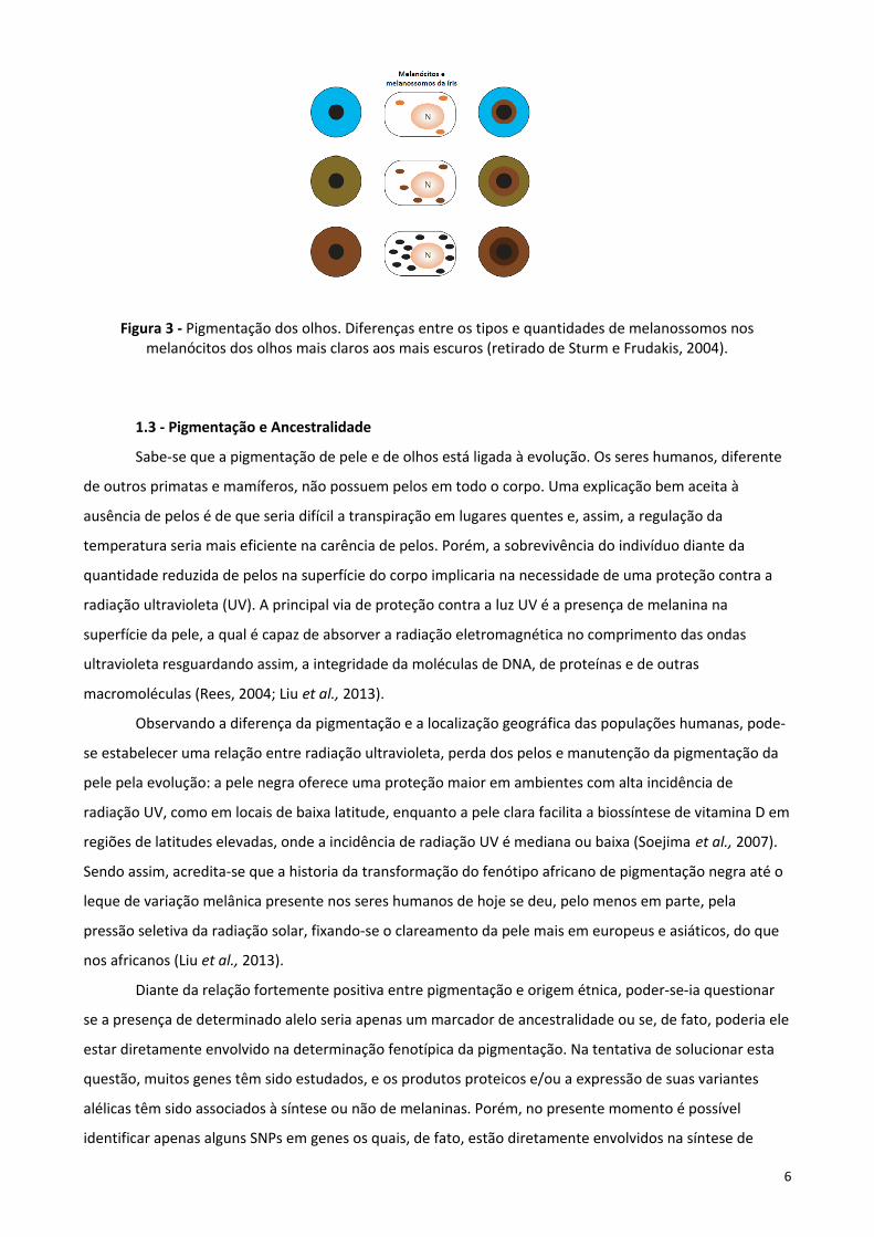
Figura 3 - Pigmentação dos olhos. Diferenças entre os tipos e quantidades de melanossomos nosmelanócitos dos olhos mais claros aos mais escuros (retirado de Sturm e Frudakis, 2004).
1.3 - Pigmentação e Ancestralidade
Sabe-se que a pigmentação de pele e de olhos está ligada à evolução. Os seres humanos, diferente
de outros primatas e mamíferos, não possuem pelos em todo o corpo. Uma explicação bem aceita à
ausência de pelos é de que seria difícil a transpiração em lugares quentes e, assim, a regulação da
temperatura seria mais eficiente na carência de pelos. Porém, a sobrevivência do indivíduo diante da
quantidade reduzida de pelos na superfície do corpo implicaria na necessidade de uma proteção contra a
radiação ultravioleta (UV). A principal via de proteção contra a luz UV é a presença de melanina na
superfície da pele, a qual é capaz de absorver a radiação eletromagnética no comprimento das ondas
ultravioleta resguardando assim, a integridade da moléculas de DNA, de proteínas e de outras
macromoléculas (Rees, 2004; Liu et al., 2013).
Observando a diferença da pigmentação e a localização geográfica das populações humanas, pode-
se estabelecer uma relação entre radiação ultravioleta, perda dos pelos e manutenção da pigmentação da
pele pela evolução: a pele negra oferece uma proteção maior em ambientes com alta incidência de
radiação UV, como em locais de baixa latitude, enquanto a pele clara facilita a biossíntese de vitamina D em
regiões de latitudes elevadas, onde a incidência de radiação UV é mediana ou baixa (Soejima et al., 2007).
Sendo assim, acredita-se que a historia da transformação do fenótipo africano de pigmentação negra até o
leque de variação melânica presente nos seres humanos de hoje se deu, pelo menos em parte, pela
pressão seletiva da radiação solar, fixando-se o clareamento da pele mais em europeus e asiáticos, do que
nos africanos (Liu et al., 2013).
Diante da relação fortemente positiva entre pigmentação e origem étnica, poder-se-ia questionar
se a presença de determinado alelo seria apenas um marcador de ancestralidade ou se, de fato, poderia ele
estar diretamente envolvido na determinação fenotípica da pigmentação. Na tentativa de solucionar esta
questão, muitos genes têm sido estudados, e os produtos proteicos e/ou a expressão de suas variantes
alélicas têm sido associados à síntese ou não de melaninas. Porém, no presente momento é possível
identificar apenas alguns SNPs em genes os quais, de fato, estão diretamente envolvidos na síntese de
6

pigmentos de pele e de olhos, independentemente da origem étnica da população em estudo (Kenny et al.,
2012). Por exemplo, como será citado a seguir, o SNP rs12913832 do gene HERC2, o qual se encontra a
21kb upstream do promotor de OCA2, regula a expressão de OCA2 pelo dobramento da cromatina que
permite o ancoramento dos fatores de transcrição de OCA2. O produto da expressão de OCA2, a proteína P,
é essencial para a síntese de melanina. O alelo T (ou alelo A, considerando a fita complementar) do SNP
rs12913832 ao permitir a abertura de cromatina, e o recrutamento dos fatores de transcrição de OCA2,
induz à coloração mais escura, enquanto o alelo C (ou alelo G, considerando a fita complementar) mantém
a cromatina mais fechada, tendo menos eficácia no recrutamento destes fatores, ocasionando uma
coloração mais clara (Figura 4).
Figura 4- Modelo de determinação da cor de Iris castanho (A) e azul (B) influenciado pelo SNP rs12913832do gene HERC2, segundo Sturm e Larsson, 2009.
Mesmo se localizando no interior de segmentos gênicos chave para a produção de melanina,
poucos os alelos foram, contudo, já reconhecidos como sendo os próprios responsáveis pelo efeito variante
de síntese de pigmento. Por não ter sido ainda completamente elucidada a implicação efetiva no fenótipo
de cada variante alélica, algumas delas podem estar associadas à pele/olho escuro ou à pele/olho claro
apenas por estarem em desequilíbrio de ligação com uma variante, de fato, fenótipo-efetiva. De todas as
formas, em geral, têm sido usados para explicar a coloração escura aqueles alelos cuja frequência está mais
elevada nas populações africanas negras (alelos originais/ancestrais) e os alelos derivados, cuja frequência
está mais elevada nas populações europeias, para explicar a coloração clara. O desafio na análise da
maioria dos SNPs ainda é saber se o marcador é de fenótipo ou de ancestralidade. No intuito de focar nos
marcadores de fenótipo, têm sido retirados dos estudos associativos e/ou de predição de fenótipo aqueles
alelos derivados (isto é, mutações polimórficas mais recentes) cujas frequências estão mais aumentadas
nas populações africanas, como é o caso do alelo derivado (mais recente) do SNP rs26722 do gene SLC45A2
(regulador do pH que controla a via da síntese de melanina, no interior do melanócito). Algumas
populações africanas de fenótipo escuro mostram a presença de um alelo derivado raro (alelo A; Lys272)
deste SNP e, a despeito do seu fenótipo claro, populações caucasianas têm em frequência o alelo original
deste SNP (alelo G; Glu272) similares às de aborígenes australianas escuros (frequência de G ~0,98) (Graf et
7
al., 2005). Devido ao fato de o alelo original (G; Glu272) estar presente em grupos populacionais com
diferentes fenótipos de pigmentação (caucasianos claros e aborígenes escuros), e ao fato de populações
africanas de cor escura conterem ambas variantes (Lys272 e Glu272), Graf et al. (2005) concluíram que este
polimorfismo não estaria associado com a determinação de pigmentação, mas sim que se trataria de uma
variação associada à distribuição/isolamento geográfico. Assim, o alelo raro A surgiu como uma mutação
recente apenas em africanos, e não interferiu na expressão da cor.
1.4 - Genes e SNPs envolvidos na determinação de pigmentação de pele e olhos
Mais de uma centena de genes com efeitos conhecidos já foram descritos em modelos animais e
seus homólogos identificados em humanos (IFPCS, 2009). Esses genes podem ser divididos em grupos
funcionais de acordo com a influência que seus produtos proteicos exercem na produção e regulação da
pigmentação como segue: 1- genes que codificam fatores de crescimento e transcrição para o controle do
crescimento e diferenciação dos melanoblastos em melanócitos; 2- genes que codificam proteínas
componentes dos melanossomos; 3- genes que controlam a biossíntese de organelas relacionadas; 4-
genes que determinam a produção de eumelanina versus feomelanina e 5- genes envolvidos no transporte
dos melanossomos.
Com base na literatura disponível, um estudo de revisão do nosso grupo selecionou oito SNPs em
genes candidatos ao estudo de previsão de pigmentação em diversas populações, por estarem
significantemente relacionados com a manifestação da pigmentação em seres humanos. A base do nosso
estudo foi: Tully, 2007; Sulem et al., 2007; Sturm et al., 2008; Branicki et al., 2009; Giardina et al., 2011;
Walsh et al., 2011a; Walsh et al., 2011b, Donnelly et al., 2012; Allwood et al., 2013; Liu et al., 2013; Walsh
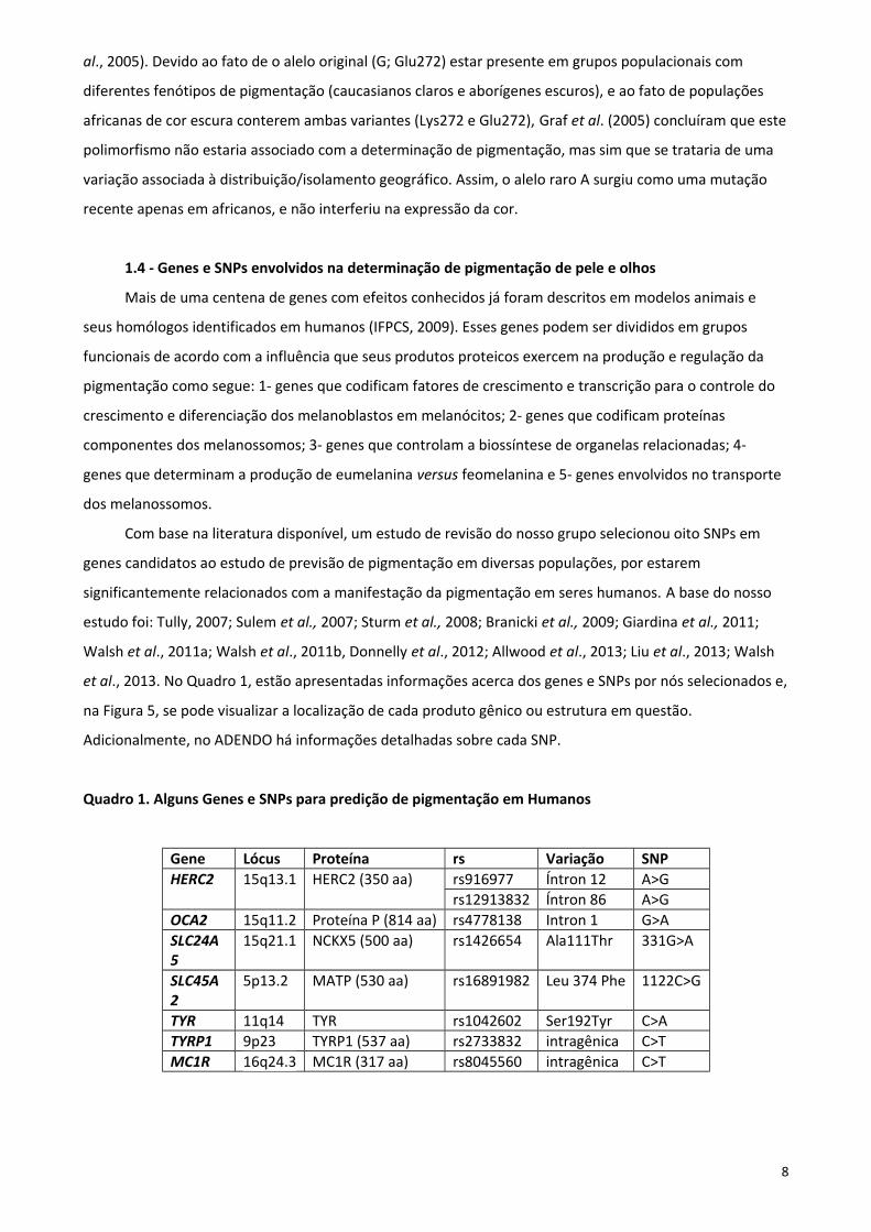
et al., 2013. No Quadro 1, estão apresentadas informações acerca dos genes e SNPs por nós selecionados e,
na Figura 5, se pode visualizar a localização de cada produto gênico ou estrutura em questão.
Adicionalmente, no ADENDO há informações detalhadas sobre cada SNP.
Quadro 1. Alguns Genes e SNPs para predição de pigmentação em Humanos
Gene Lócus Proteína rs Variação SNPHERC2 15q13.1 HERC2 (350 aa) rs916977 Íntron 12 A>G
rs12913832 Íntron 86 A>GOCA2 15q11.2 Proteína P (814 aa) rs4778138 Intron 1 G>ASLC24A5
15q21.1 NCKX5 (500 aa) rs1426654 Ala111Thr 331G>A
SLC45A2
5p13.2 MATP (530 aa) rs16891982 Leu 374 Phe 1122C>G
TYR 11q14 TYR rs1042602 Ser192Tyr C>ATYRP1 9p23 TYRP1 (537 aa) rs2733832 intragênica C>TMC1R 16q24.3 MC1R (317 aa) rs8045560 intragênica C>T
8

Figura 5- Indicação na estrutura celular dos genes (citados no quadro1) e/ou seus produtos proteicos parapredição de pigmentação em Humanos (Figura do autor)
1.4.1- Gene HERC2: rs916977 e rs12913832
O gene HERC2 (lócus 15q13.1) codifica a proteína HECT domain - RCC1-like domain-containing
protein 2 (ou E3 ubiquitin-protein ligase HERC2) envolvida no tráfego de proteínas. Ainda que a função do
gene HERC2 seja desconhecida, a sequencia de DNA no interior de HERC2 desempenha um papel estrutural
no genoma e na regulação de pigmentação (Donnelly et al., 2012). De acordo com Sturm et al. (2008), a
proteína HERC2 não está diretamente envolvida na síntese de pigmento, mas a sequência de DNA dentro
da região do gene HERC2 controla a expressão do gene OCA2 o que, por sua vez, controla a produção de
melanina (Sturm et al, 2008). Tais sequências no interior de HERC2 são enhancers do promotor do gene
OCA2 (Visser et al., 2012).
Visser et al.(2012) mostraram que o SNP rs12913832 de HERC2, que se encontra a 21kb upstream
do promotor de OCA2, regula a expressão deste gene pelo dobramento da cromatina que gera um loop de
longo alcance que acaba por promover a expressão de OCA2. Esta atividade é mediada pelos fatores de
transcrição HLTF, LEF1 e MITF. Porém, diferentes versões alélicas de HERC2 impedem o desenrolamento da
cromatina, o que reduz a ligação dos fatores de transcrição de OCA2, reduzindo consequentemente sua
taxa transcricional. O alelo T (ou alelo A, considerando a fita complementar) do SNP rs12913832 permite a
abertura da cromatina e consegue recrutar os fatores de transcrição de OCA2, o que permite a produção de
melanina e ocasiona uma coloração mais escura, enquanto o alelo C (ou alelo G, considerando a fita
complementar) apresenta uma diminição no recrutamento destes fatores, impedindo a síntese plena de
pigmento e ocasionando uma coloração mais clara. Assim, via controle da expressão de OCA2, o gene
HERC2 tem o papel predominante na determinação de cor. Dados populacionais indicam que os SNPs
rs916977 e rs12913832, localizados no íntron 12 e íntron 86, respectivamente, são melhores preditores de
9
cor de olho do que três SNPs já descritos no interior do íntron 1 do próprio gene OCA2 (rs7495174,
rs6497268 e rs11855019) (Branicki et al, 2009). Estudos em outras populações também indicam o SNP
rs12913832 como sendo o SNP com maior efeito na predição da cor de olhos (Allwood et al., 2013).
Adicionalmente, se observou que o alelo A do SNP rs1667394, localizado no íntron 4 do gene
HERC2, tem um padrão de associação com cor de cabelo e de olhos em um gradiente de redução da
pigmentação: menor frequência alélica em indivíduos com cabelos e olhos castanhos e com a maior
frequência em indivíduos com cabelo loiro e olhos azuis. O alelo A é encontrado em uma frequência de 80-
90% na população Norte da Europa (Sulem et al., 2007).
1.4.2- Gene OCA2: rs4778138
O gene que, ao conter mutações deletérias, ocasiona o albinismo oculocutaneo tipo 2 (gene OCA2),
codifica uma proteína com 12 domínios transmembrana denominada proteína P (Sturm et al., 2008).
Acredita-se que a proteína P esteja envolvida no transporte de ânions, na regulação do pH melanossomal
(Visser et al., 2012), e que esteja envolvida no tráfego de proteínas internas como a tirosinase (TYR) e as
proteínas associadas a tirosinase 1 (TYRP1) (Valenzuela et al., 2011). OCA2 por si só, independente de
HERC2, também desempenha um papel na variação de coloração comum em olhos, cabelo e pele. Três
SNPs que se encontram no intron 1 de OCA2 mostraram ter uma forte associação com a coloração dos
olhos (Liu et al., 2013). Além disto, o SNP rs1800407 encontrado no exón 13 do gene OCA2 ocasiona uma
troca do tipo Arg419Gln na sequência polipeptídica da proteína P (Donelly et al., 2012), esta troca aumenta
a penetrância do fenótipo de olhos azuis associados ao SNP rs12913832 (Sturm et al., 2008; Walsh, et al.,
2011a).
A proteína P, relacionada ao transporte de sódio/sulfeto (família SLC13), pode estar diretamente
implicada na regulação do pH melanossomal (Kondo e Hearing, 2011). Foi observado que a proteína P
atuava na permuta de cátions Na+/H+, e/ou como um transportador de glutamato. Ambas as funções
indicam que este produto do gene OCA2 esteja envolvido no fornecimento de substrato para a TYR. A
proteína P pode ainda estar envolvida no tráfego intracelular da enzima TYR, durante a maturação dos
melanossomos (Eiberg et al., 2008). O pH do melanossomo regula a atividade da TYR afetando, portanto,
uma série de fatores na síntese da melanina, como a taxa de produção de melanina, a proporção de
eumelanina/feomelanina e a maturação de melanossomos nos melanócitos e células de melanoma.
Resultados experimentais indicaram que a produção de melanina é ideal em ambiente celular com pH
próximo a pH-neutro (cerca de pH= 6.8). Paralelamente, a síntese da melanina é quase ausente em pH
menor que 5.5. Em um pH menos ácido, por outro lado, a melanogênese pode prosseguir sem qualquer
alteração na abundância de proteínas melanogênicas (Ancans et al., 2001; Fuller et al., 2001).
Outro fator envolvido ainda na regulação do pH é a enzima ATPase-vacuolar (V-ATPase), presente
no interior do melanossomo, a qual atua na acidificação dos melanossomos. Em estudo realizado por Fuller
e colaboradores foi relatado que a V-ATPase está em menor quantidade ou, até mesmo, ausente em
melanossomos originados em pele negra (Fuller et al., 2001).
10

1.4.3- Gene SLC24A5: rs1426654
Baseado no que se conhece do NCKX5 (Na+/Ca2+/K+ exchanger 5), a regulação do pH
melanossomal pode ser o papel da proteína codificada pelo gene SLC24A5 (Tully, 2007). O gene codifica
uma proteína de 500 aminoácidos, que faz parte da família das proteínas envolvidas nas trocas de
sódio/cálcio dependente de potássio, localiza-se próxima à membrana melanossomal e atua no transporte
de membrana do melanossomo (Sturm, 2006; Norton et al, 2007; Liu et al., 2013). Lamason et al. (2005)
descobriram que NCKX5 está localizado em melanossomos ou seus precursores, e assumiram que ele
poderia atuar acumulando Ca2+ no melanossomo. O gene SLC24A5 foi descoberto primeiramente em
zebra-fish, no qual está relacionado ao fenótipo de uma listra clara significante. O homólogo foi identificado
e caracterizado em humanos. O gene humano mostrou que uma substituição de Ala111Thr, resultante do
SNP rs1426654 (de Ala111-alelo G para Thr111-alelo A) tinha um efeito similar na pigmentação humana,
isto é, conferia redução de melanina (fenótipo claro). De acordo com Ginger et al. (2008) o SLC24A5 está
associado com a rede trans-Golgi. Neste mesmo estudo, eles descobriram que o alelo G (Ala111) do SNP
rs1426654, associado com pele mais escura, teve maior atividade de troca de íons em relação ao alelo A
(Thr111), o qual está associado com pele mais clara. O alelo ancestral evolutivamente conservado (Ala111)
do gene SLC24A5 também foi encontrado em alta frequência em populações africanas e asiáticas, e que o
alelo derivado (Thr111) atingiu a fixação em europeus. Especificamente, a frequência para a variante
Thr111 varia entre 98,7 e 100% entre as amostras da população de origem europeia, enquanto Ala111 tem
uma frequência de 93-100% em amostras de africanos, nativos americanos e população do Leste Asiático
(Dimisianos et al., 2008)
1.4.4- Gene SLC45A2: rs16891982
Originalmente identificado como um antígeno do melanoma humano (AIM1) e recentemente
renomeado carregador de soluto família 45, membro 2, o gene SLC45A2 contém sete éxons que se
estendem a uma região de aproximadamente 40kb e codifica a proteína MATP de 530 aminoácidos que
supõe-se ter 12 domínios transmembrana (Graf et al., 2007). Mutações no gene SLC45A2 causam uma
forma de albinismo em humanos (OCA4) (Vierkötter et al., 2012; Tully, 2007; Sturm, 2006). Graf et al.
(2005) relataram dois SNPs em SLC45A2 que não estavam associados a doenças, mas sim associados com a
variação normal da pigmentação humana, e também a variações entre diferentes populações: Glu272Lys
(rs26722) e Leu374Phe (rs16891982). A proteína MATP desempenha um papel crucial no processamento e
tráfego intracelular da tirosinase (TYR), uma das enzimas cruciais e necessárias para síntese de melanina,
assim como na via de outras proteínas melanossomais (Branicki et al, 2009; Vierkötter et al., 2012).
O SNP rs16891982 gera o polimorfismo proteico Leu374Phe e regula a função de transporte da região
transmembrana da proteína MATP. A variante Leu374 (alelo G) desempenha um papel importante no
transporte de prótons, resultando em um pH ótimo intramelanossomal, o qual permite a atividade da
tirosinase (TYR) e consequentemente a produção adequada da eumelanina (fenótipo pigmentado). A
11
variante Phe374 (alelo C) pode alterar o transporte, o pH e a síntese do pigmento (Graf et al, 2005; Tully,
2007; Vierkötter et al., 2012). Nakayama et al. (2006) referiram o alelo C (Phe374) como alelo “tipo-
Caucasiano”, já que ele resultou na redução da função da MATP, por alterar o tráfego intracelular de
elementos melanossomais, criando um ambiente de decréscimo na produção de melanina. Estudando o
mesmo gene, Graf et al. (2005) observaram que o alelo Lys272 do gene SLC45A2 mostrou um significante
aumento nas populações asiáticas e afro-americanas quando comparadas a caucasianos. Contudo, as
populações aborígenes australianas mostraram frequências alélicas similares às das populações de
caucasianos. Devido ao fato do fenótipo de pigmentação dos dois últimos grupos populacionais serem bem
diferentes, os autores sugeriram que o polimorfismo rs26722 não estaria associado com a determinação de
pigmentação, mas apenas seria uma variação associada à distribuição ou isolamento geográfico.
1.4.5- Gene TYR: rs1042602
O gene TYR transcreve a tirosinase, uma importante enzima para a fase inicial da melanogênese
(Liu et al., 2013). O gene TYR foi indicado como um loci para a susceptibilidade ao vitiligo generalizado (Jin
et al., 2012). Variações polimórficas em TYR estão diretamente associadas com alterações na coloração de
olhos, cabelos e pele (Sulem et al., 2007). Em especial, dois polimorfismos de TYR, rs1042602 (Ser192Tyr) e
rs1126809 (Ala402Gly), têm mostrado frequências alélicas associadas a fenótipos de diferentes colorações:
alelos que aparecem em alta frequência em populações europeias e estão ausentes em populações
africanas (Stokowski et al., 2007). As análises enzimáticas in vitro revelaram que existe uma redução de
aproximadamente 40% na atividade catalítica de tirosinase devido à variação Ser192Tyr (Chaki et al., 2011).
1.4.6- Gene TYRP1: rs2733832
Localizado no cromossomo 9p23, o gene TYRP1 codifica a proteína relacionada à tirosinase 1, que
faz parte do complexo da enzima tirosinase durante a produção de melanina (Parra, 2007; Liu et al., 2013).
O impacto da proteína TYRP1, assim como a da DCT, na estabilidade da proteína TYR e na produção de
outras enzimas envolvidas na formação catalítica de melanina mostra que alterações no gene TYRP1 têm
um papel importante na variação normal da pigmentação humana (Sturm, 2006). Há mais de dez anos, viu-
se evidências que polimorfismos no TYRP1 estão associados à coloração de olhos em europeus (Frudakis et
al., 2003). Estudos proteicos mostraram que a proteína codificada por TYRP1 foi encontrada estando
elevada 2,6 vezes na pele de africanos e de indianos (com pigmentação escura) se comparada à pele de
Mexicanos, Chineses e Europeus (com pigmentação clara) (Alaluf et al, 2003). Foi ainda observado que
mutações raras (não polimórficas) em TYRP1 são responsáveis pelo albinismo oculocutaneo tipo 3 (OCA3)
(Sulem et al., 2007).
1.4.7- Gene MC1R: rs8045560
Até o momento, o gene MC1R é o mais bem caracterizado dentre os genes descritos que influenciam
a variação normal de pigmentação em humanos (Gerstenblith et al., 2007). Este gene apresenta um padrão
12
peculiar de distribuição dos seus alelos entre as populações humanas: populações africanas e outras
caracterizadas pela pele escura apresentam as menores variações alélicas desse gene, enquanto em
populações asiáticas e europeias, esse gene é altamente polimórfico (Makova e Norton, 2005). Estudos em
diferentes populações têm demonstrado que a região codificadora do gene MC1R apresenta mais de 70
variantes já identificadas (Gerstenblith et al., 2007), com impacto significativo no fenótipo de pigmentação
(Harding et al., 2000; Strum, 2006).
O gene MC1R transcreve para o receptor de melanocortina 1 (MC1R), uma proteína-G localizada na
membrana de melanócitos (Valenzuela et al., 2011) e diretamente responsável pela regulação da síntese de
eumelanina/feomelanina (Makova et al., 2005). A ligação do hormônio paracrino alfa-estimulante dos
melanócitos (α-MSH) ao gene MC1R causa uma cascata de sinalização de cAMP aumentando a produção de
eumelanina (Makova et al., 2005; Valenzuela et al., 2011), enquanto que a ligação de seu antagonista, a
proteína de sinalização agouti (ASIP), resulta em menos produção de eumelanina e no aumento da
produção de feomelanina (Voisey et al., 2006; Liu et al., 2012).
1.5 - Frequências alélicas dos SNPs envolvidos na determinação de pigmentação de pele e olhos em
populações Africanas e Europeias
O Quadro 2 apresenta uma levantamento realizado com dados de indivíduos oriundos de populações
Africanas e Europeias descritas nos bancos de dados ALFRED (The ALlele FREquency Database) e HAPMAP.
O ALFRED (da U.S. National Science Foundation) foi projetado para tornar os dados de frequência de alelos
em amostras de populações humanas prontamente disponíveis para utilização pelas comunidades; e o
projeto internacional HAPMAP é resultado de uma parceria de cientistas e agências de financiamento do
Canadá, China, Japão, Nigéria, Reino Unido e Estados Unidos, que tem por finalidade desenvolver e manter
um recurso público para ajudar pesquisadores a encontrar genes e estes SNPs resultantes de estudos
populacionais. Consultando estas duas bases de dados, compilamos resultados de todas as populações já
estudadas até maio de 2013.
Neste levantamento não foram consideramos dados de indivíduos oriundos de populações asiáticas
devido a sua baixa representatividade na população do Rio Grande do Sul. Todos os dados foram plotados
em uma planilha prévia para, finalmente, se obter o número total de indivíduos já genotipados e a
frequência de cada alelo. Estes dados estão apresentados no Quadro 2.
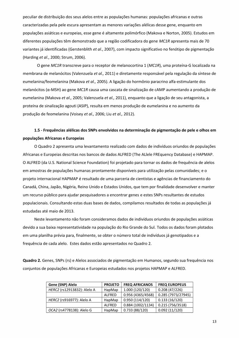
Quadro 2. Genes, SNPs (rs) e Alelos associados de pigmentação em Humanos, segundo sua frequência nos
conjuntos de populações Africanas e Europeias estudados nos projetos HAPMAP e ALFRED.
Gene (SNP) Alelo PROJETO FREQ AFRICANOS FREQ EUROPEUSHERC2 (rs12913832): Alelo A HapMap 1.000 (120/120) 0.208 (47/226)
ALFRED 0.956 (4365/4568) 0.285 (7973/27945)HERC2 (rs916977): Alelo A HapMap 0.950 (114/120) 0.133 (16/120)
ALFRED 0.884 (1002/1134) 0.215 (756/3518)OCA2 (rs4778138): Alelo G HapMap 0.733 (88/120) 0.092 (11/120)
13

ALFRED 0.746 (1047/1404) 0.166 (945/5706)SLC24A5 (rs1426654): Alelo G HapMap 0.987 (223/226) 0.000 (0/116)
ALFRED 0.707 (2630/3720) 0.011 (49/4271)SLC452A (rs16891982): Alelo C* HapMap 1.000 (114/114) 0.017 (2/116)
ALFRED 0.919 (3478/3786) 0.071 (1840/25961)TYR (rs1042602): Alelo C HapMap 1.000 (120/120) 0.571 (129/226)
ALFRED 0.940 (1757/1870) 0.705 (3544/5026)TYRP1 (rs2733832): Alelo C HapMap 0.951 (215/226) 0.398 (90/226)
ALFRED 0.879 (181/206) 0.419 (447/1066)MC1R (rs8045560): Alelo C HapMap 0.951 (215/226) 0.465 (105/226)
ALFRED 0.909 (1225/1348) 0.438 (1624/3706) * Ou alelo G, considerando a fita complementar
FONTE: http://hapmap.ncbi.nlm.nih.gov/; http://alfred.med.yale.edu/alfred/index.asp.
14

2- OBJETIVOS
Objetivo Geral
Identificar e analisar os SNPs mais significativos para a previsão de pigmentação em humanos e
medir suas frequências alélicas e genotípicas em indivíduos com fenótipo de alto ou baixo conteúdo de
melanina em pelo e olhos.
Objetivos Específicos
I. Selecionar, na literatura científica, SNPs significativos para a previsão de pigmentação em
humanos.
II. Identificar dois grupos de indivíduos fenotipicamente distintos: Grupo 1: indivíduos com muita
pigmentação (olhos negros e pele escura); Grupo 2: indivíduos com pouca pigmentação (olhos azuis
claro e pele muito clara).
III. Genotipar os SNPs selecionados em genes de pigmentação.
IV. Determinar as frequências alélicas e genotípicas para cada SNP nos dois grupos de estudo.
V. Comparar os grupos de acordo com a relação genótipo-fenótipo.
15

CAPÍTULO 2 - ARTIGO CIENTÍFICO
Analysis of eight SNPs in pigmentation-related genes in Southern Brazilian subjects with simultaneous
high or low skin and eye melanin content.
ABSTRACT
The understanding of gene function in the determination of externally visible characteristics (EVCs)
has several uses in human population evolutionary studies and in forensic investigations. With regards to
the latter, a fair amount of effort has been made to discover an easy and efficient model for the prediction
of eye and skin color in humans. The obvious advantage of the prediction of such EVCs through the use of
DNA is that it can be incorporated as routine procedure in forensic labs and be applied to police
investigations. In the present study, we combined the genotypes of eight SNPs in pigmentation-related
genes (rs4778138 - OCA2; rs12913832 - HERC2; rs16891982 - SLC45A2; rs8045560 - MC1R; rs1426654 -
SLC24A5; rs2733832 - TYRP1; rs1042602 - TYR; rs916977 - HERC2) with multiple analytical approaches.
Considering this SNP panel, we evaluated allele frequencies of subjects with High Melanin Content (HMC;
from African populations) and Low Melanin Content (LMC; from European populations), as obtained from
the HAPMAP and ALFRED databases, and defined the alleles H (to predict HMC subjects) and alleles L (to
predict LMC subjects). The cumulative distribution of alleles H and alleles L in the two phenotypically
different color groups comprised of a total of 134 southern Brazilian subjects showed that 82% of the HMC
subjects (N = 61) had eight or more alleles H, and 100% of the LMC subjects (N = 73) had less than eight
alleles H, with an accuracy value of 96.3%. We also performed analyses using the AUC (Area Under the
Receiver Operating Characteristic Curve), PGL (Calculation of Pathway Genetic Load), and GP (Genetic
Probability) approaches. The AUC for the prediction of both HMC and LMC phenotypes was 0.99; the PGL
showed 93% concordance between genotype and HMC or LMC phenotypes across the eight SNPs panel;
and the GP showed 91% concordance between predicted and observed HMC or LMC phenotypes. The
combination of our high-throughput genotyping technology and multiple analytical approaches has
achieved very high accuracy in the prediction of the extreme phenotypes for human pigmentation. We
believe this forensic DNA phenotyping (FDP) technique can be particularly useful in instances where classic
DNA profiling fails to generate a database match from crime scene DNA or where the need to classify
skeletal or biological clues of dismissed people or degraded corpses arises.
16

BACKGROUND
Different techniques, such as anthropometric examinations, fingerprinting and genetic
studies, have been largely implemented in order to characterize and individualize people. For nearly
twenty years, autosomal sets of short tandem repeat markers (STRs) and, more recently, single
nucleotide polymorphisms (SNPs) have been used for human identification in forensic cases [1].
SNPs, which represent the most abundant class of human polymorphisms, may determine amino acid
substitutions altering the functional properties of proteins and expressing distinct phenotypes [2].
SNP abundance has sparked the interest in forensics and the mapping of the human genome has
made possible the development of sets of genotypes and haplotypes that can phenotypically
characterize a human being [3]. Thus, SNP studies may serve for individual discrimination as well as
for the prediction of externally visible characteristics (EVCs). Among the EVCs, the pigmentation of
skin, eyes, and hair has become a good target for the prediction of phenotypes. Genotype-phenotype
association studies regarding iris and skin color have been focusing on detecting SNPs within genes
directly involved in the synthesis of pigments [4-16]. There are more than one hundred genes
involved in the pigmentation process [17, 18] and they have a cumulative effect. However, for
forensic purposes, despite the small number of known genes determining human pigmentary traits,
it is possible to predict with high confidence the color of a person’s eye, hair, and/or skin based on
this small number of DNA polymorphisms [19]. Thus, it is fair to say that some genes have alleles
which can definitely determine the extremes of melanin pigmentation patterns, i.e. , light and dark
patterns.
In this work, we selected eight SNPs in seven genes that are linked to the melanin synthesis in
order to investigate their significance in skin and eye color phenotypes for forensic purposes. SNPs
were selected in the following human pigmentation associated genes: HERC2, OCA2, SLC24A5,
SLC45A2, TYR, TYRP1, and MC1R; SNPs in the first three were shown to significantly alter the melanin
content in human melanocytes, supporting their functional role in pigmentation [20].
OCA2, rs4778138: The OCA2 gene encodes the P protein involved in anion transport and in
the melanosomal pH regulation, operating in the tyrosinase (TYR) and the tyrosinase related protein
1 (TYRP1) functions [21-25]; changes on OCA2 may interfere with melanin synthesis due to pH
changes. The rs4778138 is a G>A SNP in the intron region of OCA2 and it is strongly associated with
eye color [19].
HERC2, rs12913832: The Hect Domain and RCC1-like Domain 2 gene is located 10 Kb
upstream of OCA2 and it acts as a regulatory enhancer region of OCA2. The function of HERC2 is still
unknown. The rs12913832 is an A>G SNP in an intronic area of the HERC2, within the OCA2 enhancer
region. The derived allele G of this SNP is associated with the blue iris phenotype, being common in
Europeans , particularly those of northwestern and eastern European descent [25]. The allele A (or
allele T, if in the complementary DNA strand) allows for the opening of the chromatin and
17
recruitment of the OCA2 transcription factors, which leads to a darker iris, while the allele G (or allele
C, if in the complementary DNA strand) maintains the chromatin closed, being less effective in
recruiting the aforementioned factors, which results in a lighter iris [20, 23].
SLC45A2, rs16891982: The solute carrier family 45 member 2 gene encodes the MATP
protein [26], which plays a crucial role in the processing and intracellular trafficking of tyrosinase
(TYR) and of other enzymes necessary for the synthesis of melanin [18, 28]. The rs16891982 is the
1122C>G SNP in exon 5 of the SLC45A2 gene. This SNP results in the non-synonymous substitution of
Leucine (allele C; codon TTG on the coding/reverse DNA strand) by Phenylalanine (allele G; codon
TTC on the coding/reverse DNA strand) at amino acid 374 (Leu374Phe). The rs16891982 is associated
with the normal variation of human skin pigmentation , since it regulates the MATP function; the
Leu374 variant (allele C – complementary to G on codon TTG on the coding/reverse DNA strand)
plays an important role in the transport of protons, resulting in an optimum intramelanosomal pH,
which allows the activity of tyrosinase (TYR) and, consequently, the adequate production of
eumelanin (brow-black spectrum), while the derived Phe374 variant (allele G – complementary to C
on codon TTC on the coding/reverse DNA strand) may change the transport, the pH and the synthesis
of the pigment [28-30]. A strong association between rs16891982 and eye color has been reported
[19, 27].
MC1R, rs8045560: MC1R gene transcribes the melanocortin 1 receptor (alpha melanocyte
stimulating hormone receptor), a G-protein located in the melanocyte membrane [24] which is
directly responsible for the regulation of eumelanin / pheomelanin synthesis [19, 31, 32]. The MC1R
gene is the best characterized among the genes that influence the normal variation of human skin
pigmentation, and the ancestral allele C of rs8045560 (C>T SNP) is associated with darker skin
populations [17, 31, 33].
SLC24A5, rs1426654: The solute carrier family 24, member 5, is responsable for the
accumulating Ca+2 in the melanosome [34, 35]. The rs1426654 (G>A SNP in the coding region of
SLC24A5 gene) results in non-synonymous substitution of Alanine (GCA) by Threonine (ACA) at amino
acid 111 Ala111Thr. This SNP has shown evidence of natural selection; the derived allele A (Thr111) is
associated with light skin pigmentation and is common in Europe, southwest Asia, and central Asia
[25]. The ancestral allele (G allele; Ala111) is associated with darker skin [20, 36].
TYRP1, rs2733832: Located at 9p23, this gene encoding to tyrosinase-related protein 1, which is
part of the enzyme tyrosinase in melanin production [4, 17, 19]. TYRP1 polymorphisms are associated with
coloring eyes in Europeans [37], and the presence of TYRP1 protein has been found to be 2.6 times higher
in the skin of Africans and Indians (darker pigmentation) than in the skin of Mexicans, Chinese, and
Europeans [38]. The ancestral allele C of rs2733832 (C>T SNP) is associated with darker skin populations.
TYR, rs1042602: TYR gene transcribes the tyrosinase, an important enzyme for the initial process of
melanogenesis [19]. Mutations in the TYR gene are associated with vitiligo susceptibility [39] and
oculocutaneous albinism type I [40], but polymorphic variations in the TYR are directly associated with
18

changes in the color of eye, hair, and skin [4, 41]. The rs1042602 (C>A SNP) results in the non-synonymous
substitution of Serine (TCT) by Tyrosine (TAT) at amino acid 192 (Ser192Tyr) and in a reduction of about
40% in the catalytic activity of tyrosinase [42]. The ancestral allele C is associated with darker skin
populations.
HERC2, rs916977: The derived allele G of rs916977 (A>G SNP) has been associated with blue eyes
[4, 43].
In this paper, we have processed different analyses with these SNP variants and genotyped subjects
from two southern Brazilian populations with either high or low melanin content using a multiplex system.
We used this panel to predict color phenotype for forensic uses.
MATERIAL AND METHODS
Design, subjects, and approval. This observational cohort study was conducted with data and DNA
collected from adult subjects (over 18 years old) enrolled between March 1st, 2012 and October 31st, 2013.
Subjects were selected based on their skin/eye phenotype characteristics. All subjects were from southern
Brazil, an area that presents a distinctive genetic background: the majority of subjects were of European
origin (Portuguese, Italian, Spanish, and German ancestry) and a smaller amount of individuals were of
African origin [44]. A form containing information about gender, age, origin, place of birth and residence
was filled out by all individuals. This COLOR-genotyping project was approved by the Research Ethics
Committee of the Pontifical Catholic University of Rio Grande do Sul (protocol #11-05722, Of.CEP-0295/12
and Of.CEP-1041/12), and the informed written consent for the study was obtained from all subjects.
Skin Phenotyping. The skin color of each participant was identified using the Fitzpatrick score,
ranging from Type 1 to Type 6, where: 1- Highly sensitive skin, always burns, never tans; 2- Very sun-
sensitive skin, burns easily, tans minimally; 3- Sun-sensitive skin, sometimes burns, slowly tans to light
brown; 4- Minimally sun-sensitive skin, burns minimally, always tans to moderate brown; 5- Sun-insensitive
skin, rarely burns, tans well; 6- Sun-insensitive skin, never burns, deeply pigmented. We also measured the
skin amount of red (R), green (G), and blue (B) pigments in an inner and hairless portion (below elbow) of
the right arm using the ACR-1023 (Instrutherm, São Paulo, Brazil), a battery-portable color analyzer
equipment which employs a spectral analysis method to determine the color of the sample. Each R, G, and
B values range from zero to 1023, where the minimum value (zero) represents the complete absence of
color, and the maximum (1023), its complete presence. In this system, total white has a value of R=1023,
G=1023, and B=1023, and total black has a value of R=0, G=0, and B=0. The RGB skin values of each person
were measured three times and the average was recorded. The equipment was calibrated before each use
by measuring a white plate. Potential subjects that reported having a recent or intense tan were excluded.
Eye Phenotyping. The eye color of each participant was classified from Type 1 to Type 6, where: 1-
light blue; 2- dark blue; 3- green; 4- hazel or light brown; 5-dark brown; 6-black. We photographed both
eyes using a Nikon COOLPIX L110, with MacroZoom, ISO 80, with Flash at 5500K (D55 illuminant), and a
picture resolution of 4,000 X 3,000 pixels. To take the pictures, the camera was fixed on a tripod stand and
19

placed at a distance of 6cm ± 0.5cm from the subject’s eye, having its focus locked onto the central area of
the iris. The images were taken in a room with standardized artificial light conditions. The participants
wearing contact lenses were required to remove them. We measured both eye amounts of R, G, and B
pigments by analyzing each photo with the software COLOURS [45]. A triangular-shaped representative
area of the iris covering approximately 2.500 pixels was selected in each picture and the R, G, and B values
of each pixel were measured on a scale of zero to 255, where the minimum value (zero) represents the
complete absence of a given color and the maximum (255) represents its complete presence. To calculate
the R value, the software divides the total amount of red pigment present in each pixel of the selected area
by the product of 255 times the total amount of pixels in the same area. For example (Figure 1, picture on
the right): 264002 / (255 x 2542) = 0.41 – as the total amount of red in the selected area is 264002 and the
total amount of pixels in the same area is 2542. The same mathematical formula is used to G and B
calculations. In this system, total black has a value of R=0, G=0, and B=0, and total white has a value of R=1,
G=1, and B=1. The RGB eye values were measured on both eyes and the average was recorded. The primary
RGB values were then converted into HSV (hue, saturation, value of brightness) system values using
standard formulas. HSV is a cylindrical-coordinate representation of points in an RGB color model. This
representation maps the values into a cylinder (color wheel), where: the angle around the central vertical
axis corresponds to hue (H) and the distance from the axis corresponds to saturation (S). The height
corresponds to a third value (V), the system's representation of the perceived luminance (brightness) in
relation to the saturation. Potential subjects with Heterochromia iridum (excessively different colored
irises) were excluded from the experiment.
Genotyping. Genomic DNA was extracted with a NucleoSpin® Blood kit (Macherey-Nagel Inc.).
Amplification of regions flanking the SNPs were made by multiplex PCR in a volume of 25 μL with 2 to 10 ng
genomic DNA, 0.2 μM of each primer (Table 1), and 1X of Qiagen Multiplex PCR Master Mix (© QIAGEN,
Invitrogen). The amplification consisted of 95°C for 5 min, followed by 30 cycles of 30s at 94°C, 90s at 57°C,
90s at 72°C, and a final extension for 10 min at 72°C. After the amplification, the enzymatic purification of
the PCR product using USB® ExoSAP-IT® PCR Product Cleanup (Affymetrix®) was carried out according to the
user’s manual recommendations. SNP analysis was performed by the SNaPshot® Multiplex System ABI
Prism (Applied Biosystems®, São Paulo, Brazil), using eight SNaPshot primers (Table 1). Reactions were
performed in a final volume of 10 μL, containing 3.0 μL of purified multiplex PCR product, 5.0 μL of
SNaPshot Multiplex Ready Reaction Mix, 1.0 μL of pooled SNaPshot primers (each primer at 1 μM final),
and 1.0 μL of sterile water. Multiplex single base extensions were carried out for 28 cycles according to the
following program: 10 seconds at 96°C, 5 seconds at 50°C, and 30 seconds at 60°C. SNaPshot products were
then treated at 37°C for 1 hour with 0.8 μL of shrimp alkaline phosphatase (1.0 U/μl) and 1.2 μL of 10X SAP
buffer reaction, added directly to 10 μL of the SNaPshot product. After heat inactivation of shrimp alkaline
phosphatase for 15 minutes at 75°C, 1uL of the labeled products were mixed with 9.5 μL of HiDi formamide
and 0.5 μL of Genescan-120 LIZ size standard. They were then separated using an ABIPRISM® 3130xl Genetic
Analyzer (Applied Biosystems) with POP-4 matrix and with respective run parameters: injection voltage of
20

1.2 Kv, injection time of 23s, run voltage of 15Kv and run time of 1200s, in a capillary of 36cm. The analysis
was performed using the GeneMapper ID software version 3.2.1 (Applied Biosystems). In order to confirm
the genotyping system, we performed a sequence analysis in the ABIPRISM® 3130xl Genetic Analyzer
(Applied Biosystems) using the same designed primers in 10% of our sample.
Candidate SNP parameters: At each locus, we dubbed ‘allele H’ each of the alleles strongly
associated with subjects from African populations (individuals with High Melanin Content; HMC), and ‘allele
L’ the alleles strongly associated with subjects from European populations (individuals with Low Melanin
Content; LMC). Based on the SNP frequencies per locus available on the HAPMAP and ALFRED databases,
we constructed a 2 x 2 table with number of alleles, where: the True Positive (TP; correctly identified) was
allele H in HMC subjects, True Negative (TN; correctly rejected) was allele L in LMC subjects, False Positive
(FP; incorrectly identified or Type I error) was allele H in LMC subjects; False Negative (FN; incorrectly
rejected or Type II error) was allele L in HMC subjects. We then calculated the following parameters:
Sensitivity (ability to identify positive results) = TP / (TP + FN); Specificity (ability to identify negative results)
= TN / (FP + TN); Predictive value to allele H in HCM subjects = TP / (TP + FP); Predictive value for the
presence of allele L in LMC subjects = TN / (FN + TN); Relative risk (ratio of the probability of allele H in HMC
group to the probability of allele H in LMC group) = [TP / (TP + FP)] / [FN / (FN + TN)]; and Odds ratio (to
identify the probability of exposure and non-exposure) = (TP x TN) / (FP x FN). We calculated the sensitivity,
specificity and accuracy values joying the set of all 16 alleles. The Accuracy value, or the detection of how
the test correctly identifies or excludes the predicted phenotype, was calculated as = (TP + TN) / (TP + FP +
TN + FN).
Area under the Receiver Operating Characteristic curve: A Receiver Operating Characteristic (ROC)
curve was constructed by computing the sensitivity and specificity of the set of all 16 SNPs. The accuracy in
this test was measured by the area under the ROC curve (AUC) with a parametric method using a maximum
likelihood estimator. The AUC measures the ability of the set of SNPs to correctly classify those with and
without the phenotype, as an overall measure for prediction accuracy. An area of 1 represents a perfect
test; an area of 0.5 represents a worthless test. A recommended guide for classifying the accuracy would be
0.90-1 = excellent; 0.80-0.90 = good; 0.70-0.80 = fair; 0.60-0.70 = poor; 0.50-0.60 = fail.
Calculation of Pathway Genetic Load (PGL): Genotypes were scored as 0 for the LL homozygotes, 1
for heterozygotes, and 2 for the HH homozygotes. Unweighted PGL was calculated by summing the
numerical scores for genotypes across all SNPs as described in Huebinger et al [46]. Individual PGL scores
potentially ranged from zero (homozygous for alleles L at all loci) to 16 (homozygous for alleles H at all loci).
PGL scores weighted for color were created by multiplying the allele H count at each locus by its adjusted
odds ratio (OR) – as determined from previously published HAPMAP and ALFRED frequencies. Weighted
PGL scores for each locus were then computed to create a weighted PGL score for each individual. PGL and
other continuous data were compared by the Mann-Whitney U test. Categorical data was compared using
chi-square. The BioEstat 5.0 Statistical Package was used for all calculations.
21
Cluster analysis using STRUCTURE Software: We used the eight SNPs and the STRUCTURE software
to estimate clusters among subjects. The STRUCTURE Software v2.3.4
(http://pritch.bsd.uchicago.edu/structure.html; 47) analyses the data plotting probabilistically the
individuals in K populations by characterization of the allele frequencies set in each locus. The individuals
may be assigned in one, two or more populations if their genotype shows an admixed pattern. Our data file
was composed by: row of makers (SNP and gene data), column of labels of each individual, column of
population data (1 for HMC; 2 for LMC), and the genotype data. The ADMIXTURE MODEL and CORRELATED
ALLELE FREQUENCIES MODEL were chosen because the admixture between populations is a common
characteristic of real genetic data, since subjects may have recent ancestors in more than one population
[48]. To estimate the K value, the data was analyzed with twenty replicates for K=1 to K=10, all runs were
performed with 10,000 burn-in period and 100,000 Markov Chain Monte Carlo (MCMC) repeats after burn-
in, ADMIXTURE MODEL, CORRELATED ALLELE. The K values were estimated using the lnPr(X|K) values for
each simulation. With these values, it is possible to calculate the probability for each K and estimate the
best K [47]. The method used to estimate the value of K calculates the delta K and then selects the
appropriate K value [49, 50]. Graphics were constructed using Clumpp [51] and Distruct [52] software.
RESULTS
Table 2 presents the allele frequencies of the selected SNPs – based on population data available
on the HAPMAP and ALFRED websites – obtained from African and European subjects (the database used
to construct Table 2 is shown in a Supplementary Excel file). An association Chi-square test was performed
to examine the relationship between allele frequencies in the aforementioned populations (Africans and
Europeans). There was a highly significant association between one of the alleles with Africans and the
other with Europeans for all the SNPs tested. We dubbed ‘allele H’ each allele strongly associated with
subjects with High Melanin Content (HMC; from African populations), and ‘allele L’ each allele strongly
associated with subjects with Low Melanin Content (LMC; from European populations). Considering the
relationship between alleles and populations, we calculated (as explained in Material and Methods) the
sensibility, specificity, predictive values, relative ratio, and odds ratio for each SNP present in table 1. The
eight SNPs in pigmentation-related genes presented remarkable results in all of the parameters and they
were considered robust markers for color prediction (Table 2).
We analyzed 134 southern Brazilian subjects belonging to two color categories: HMC – individuals
with high melanin content (N = 61) – and LMC – individuals with low melanin content (N = 73). The
phenotype data of these subjects is presented in Table 3. As expected by the previous selective sampling,
the clustering toward the color was evident between these two groups: the HMC group presented only
people with Types 5 and 6 of skin and eye classification, and with RGB values closer to zero (black); and the
LMC group presented only people with Types 1 and 2 of skin and eye classification, and with RGB values
closer to the highest values (white) (Table 3). However, the ancestral origin reported by the subjects was
not clearly discriminated into the groups, especially in the HMC group, where 21% of the people assumed
22

they had European origin. We genotyped these 134 southern Brazilian subjects using the SNaPshot®
Multiplex System with a set of eight primer pairs to analyze the eight target SNPs. Table 4 shows a highly
significant association between ‘alleles H’ and HMC people, and ‘alleles L’ and LMC people. The other
parameters (sensibility, specificity, predictive values, relative ratio, odds ratio) were equally remarkable.
Although not dealing with such a clear ethnic background in our sample, the allele frequencies evaluated
confirmed the eight SNPs as robust markers for color prediction.
To observe the cumulative distribution of alleles H and alleles L in our two phenotypically different
color groups, we plotted the frequency data from southern Brazilian subjects considering the total number
of each allele H per person in the two groups, comprised of 61 HMC subjects and 73 LMC subjects,
respectively. Figure 2 shows that LMC subjects had between zero and seven alleles H, while HMC subjects
had between four and 14 alleles H. In our sample, no subject had 15 or 16 alleles H. The average of alleles H
among the HMC subjects was 12.0 (SD=3.78) , and 82% (56/61) of the HMC individuals had eight or more
alleles H. The average of alleles H among the LMC subjects was 3.3 (SD=1.49), and 96% (70/73) of the LMC
individuals had up to five alleles H, and 100% had seven or less alleles H. Some results are especially
interesting because they present a divergence between the quantity of alleles H and the phenotype
category: one HMC person had only four alleles H, one HMC person had five alleles H, two HMC people had
six alleles H and one HMC person had seven alleles H. The individual allele analyses of these five subjects
show that all loci were in some way involved in homo or heterozygosis, i.e. , there was no evidence of any
locus to explain the high skin/eye melanin content even with such few alleles H. We also analyzed one LMC
person (HC120) with seven alleles H: this person was homozygote HH to rs8045560 (MC1R) and
heterozygote to rs4778138 (OCA2), rs12913832 (HERC2), rs2733832 (TYRP1), rs1042602 (TYR), and
rs916977 (HERC2). The analysis of these people could be important to identify which loci are more critical
to color prediction – however, a conclusion about the matter was not possible with the present data
because of the size and heterogeneity of the sample.
Using the threshold of 8 alleles H, the sensitivity, specificity, and accuracy values of our system
were, respectively, 91.8% (56/61), 100% (73/73), and 96.3% (129/134). Thus, we changed the decision
threshold from zero to 16 alleles H and plotted the "sensitivity" and "1 - specificity" values as "x" and "y"
coordinate values of points on a graph. The axes of this graph both range from zero to one because these
are the limits of all possible combinations of "sensitivity" and "1 - specificity" values. The points of the curve
inevitably passed through the graph origin (where "sensitivity" = 0 and "1 - specificity" = 0) and through the
upper right corner of the graph (where "sensitivity" = 1 and "1 - specificity" = 1). The resulting graph was a
curve which is called Receiver Operating Characteristic (ROC) curve since "it describes the inherent
detection characteristics of the test" and since "the receiver of the test information can operate at any
point on the curve by using an appropriate decision threshold" [53]. Based on the same assessment that Liu
et al (2009) made in their work, we measured the ability of the set of SNPs to correctly classify those with
and without the phenotypic trait evaluating the area under the ROC curve (AUC) [19]. Considering the
cumulative distribution of alleles H (the total number of alleles H per person) in the two phenotypically
23

different color groups we obtained the very high value of AUC at 0.9908 in the prediction of both HMC and
LMC phenotypes (SE= 0.0088; IC 95% = 0.99736-1.0).
For another cumulative analysis, we used the Pathway Genetic Load (PGL) , which allows for a
simultaneous evaluation of multiple loci with cumulative effect on the phenotype [46]. Individual
unweighted PGL scores ranged from zero (homozygous for allele L at all eight loci) to 14, as previously
shown in Figure 2. PGL weighted scores (PGL-WSs) for color were obtained by multiplying the allele H count
(HH=2.0, HL=1.0, LL=0.0) per locus by its adjusted odds ratio (OR) – previously determined by HAPMAP and
ALFRED data (presented in Table 2). For example: to the following loci rs4778138 (OCA), rs12913832
(HERC2), rs16891982 (SLC452A), rs8045560 (MC1R), rs1426654 (SLC24A5), rs2733832 (TYRP1), rs1042602
(TYR), and rs916977 (HERC2), the complete genotype of subject HC123 was, respectively: LL – LL – LL – HL –
LL – HL – HH – LL. Using respective OR values by locus, we obtained to HC123 subject this values: PGL-WS=
(0.0 x 14.81) + (0.0 x 54.26) + (0.0 x 103.28) + (1.0 x 13.38) + (0.0 x 226.12) + (1.0 x 15.47) + (2.0 x 7.37) +
(0.0 x 29.73) = 43.59. The PGL analysis in all of the 134 southern Brazilian subjects showed that the
individual PGL-WSs in HMC population ranged from 65.29 to 870.92, with the median in 567.37 (mean=
584.63; SD=262.46); and the individual PGL-WSs in LMC population ranged from zero to 148.40, with the
median in 38.31 (mean= 44.80; SD=30.93). The PGL-WS for the eight SNPs tested were significantly
different between HMC and LMC groups (P<0.0001; Mann-Whitney Test), 95% (58/61) of the HCM subjects
had the PGL-WSs higher than the maximum value among LMC subjects (148.40), and 88% (67/73) of the
LCM subjects had the PGL-WSs lower than the minimum value among HMC subjects ( 65.29). Considering
these two cut points (more than 148.40 to HMC, and less than 65.29 to LMC), the PGL analysis showed that
the current eight SNPs genotyping system had 93% (125/134) concordance between genotype and HMC or
LMC phenotype.
Based on the HAPMAP and ALFRED allele frequencies, we calculated each expected genotype
frequency to HH, HL, and LL genotypes, by locus, in HMC and LMC groups (Table 5). Then, we examined the
complete genotype of each South Brazilian subject and calculated the probability of the complete genotype
belonging to expected HMC and LMC groups. For example: to the following loci rs4778138 (OCA),
rs12913832 (HERC2), rs16891982 (SLC452A), rs8045560 (MC1R), rs1426654 (SLC24A5), rs2733832 (TYRP1),
rs1042602 (TYR), and rs916977 (HERC2) the complete genotype of subject HC123 was, respectively: LL – LL
– LL – HL – LL – HL – HH – LL. Using each genotype frequency by locus obtained from HMC population, we
had the following values for subject HC123: 0.06554 - 0.00185 - 0.01416 - 0.15555 - 0.0756 - 0.15222 -
0.89492 - 0.01210 (see Table 5). The product of these values was 3.33 x 10 -11, which means the probability
of this complete genotype is present in the HMC population. Then, using each genotype frequency by locus
obtained from LMC population, we had the following values for subject HC123: 0.69890 - 0.50694 - 0.87049
- 0.49280 - 0.97812 - 0.48555 - 0.49280 - 0.61780 (see Table 5). The product of these last values was: 0.022,
which means the probability of this complete genotype is present in the LMC population. In conclusion,
comparing the two probabilities, the complete genotype of the HC123 subject has 6.6 x 10⁸ times higher
chance of belonging to the LMC population than to the HML population. This finding was in perfect
24

agreement with the actual HC123 phenotype data obtained: a male with light skin color (Type 2), dark blue
eyes (Type 2), high R-G-B values to skin (317-239-200) and eyes (0.385-0.365-0.530), and of European
origin.
We applied this model to predict color in the HMC or LMC phenotype categories based on the
genotype probability (GP) in all 134 South Brazilian subjects; we observed that 80% (49/61) of the
genotypes of our HMC subjects had a higher chance of belonging to the HMC group than to the LMC group,
with rates from 1.95 to 8.27 x 10⁷ (mean= 7.92 x 10⁶). Analyzing the genotypes of our LMC subjects, we
noticed that 100% (73/73) had a higher chance of belonging to the LMC group, with rates from 1.46 x 10⁴
(in subject HC120, with seven alleles H) to 7.77 x 10¹² (mean= 2.02 x 10¹¹). This GP analysis using the current
eight SNPs genotyping system showed 91% (122/134) concordance between predicted and observed HMC
or LMC phenotype.
As a complementary approach, we used the eight SNPs and the STRUCTURE software to
estimate clusters among subjects. The program assigned to each subject a value, which represented the
probability of belonging to each of two phenotypic clusters. This value clustered the subjects into HMC or
LMC groups. Figure 3 shows the dot plot of probability values in each phenotypic cluster; the set of SNPs
separated the individuals into the two extreme HMC or LMC groups indicating a substructure across the
total population.
DISCUSSION
Forensic DNA phenotyping (FDP, a relatively young field within forensic genetics, aims to predict
appearance traits of a sample donor from DNA left behind at a crime scene; an efficient FDP technology
would be especially useful in cases where the Police have no clues (including conventional DNA profiling) as
to who the crime scene sample donor might be [5]. As with other applications of DNA data in the forensic
arena, FDP has to be backed up by the legislation as, for instance, is currently the case of forensic DNA-
based eye color prediction in the Netherlands [19]. Skin and eye color are important externally visible traits
(EVTs) in forensics, since they are highly heritable genetic traits and are the most obvious and distinguishing
externally visible characteristics to be used in human identification.
Melanin is gradually produced in melanosomes inside melanocytes. Although the number of
melanocytes in different skin colors is essentially constant, the distribution of melanosomes is not so, as
darker skins have denser and individually dispersed melanosomes, while lighter skins have less dense
melanosomes [54]. In the eyes, pigments are distributed in the epithelium and in the stromal cells of the
iris; brown iris has an abundance of melanin while blue iris contains low levels of melanin allowing light
transmittance and reflectance which results in a blue shortwave due to the presence of collagen fibers.
Variations in both skin and eye color also result from the ratio of eumelanin/pheomelanin [55] and are
strictly connected to human evolution as well. The presence of melanin is an important protection against
ultraviolet radiation (UV): darker skin provides greater protection in environments with high UV radiation
(low latitudes), while light skin facilitates the biosynthesis of vitamin D in regions where UV radiation is low
25
(high latitudes) [56]. Since pigmentation and ethnicity are correlated, it is important to understand if the
presence of a particular allele in populations with high or low melanin content could be directly used in
determining phenotypic pigmentation or if it is only a marker of ancestry.
It is still unclear whether a gene is expressed only in the skin or only in the iris, and for this reason
we have selected subjects with HMC or LMC in both skin and eye phenotypes. Our results for the
association study, sensibility, specificity or accuracy parameters, or the final analysis using different
interpretation models would still have been the same even if only the eye color data had been considered.
We have, nonetheless, preferred to work, in a first moment, with subjects with whole (skin and eye) HMC
or LMC phenotypes, even though there are many Brazilian subjects with intermediary or ambiguous
phenotypes, such as dark eye / light skin or light eye / dark skin. Further studies with the last subjects could
select genes with preferential expression to skin or eye. In the same way, further studies with subjects with
intermediate pigmentation phenotype should be mandatorily conducted. Although genetic markers have
been informative to predict phenotypes in human pigmentation, this field of study still requires many
efforts to be really applicable. Thus, we chose to design a panel that could at least detect the extreme
phenotypes (subjects with high or low melanin content).
We have not analyzed the hair color because the melanin content in the hair is more strongly
changed by intrinsic or extrinsic factors; hair color is also age-dependent , changing during late childhood,
adolescence, adulthood, and old age, becoming darker, gray or white at any given stage [57]. These
changes, added to the occurrence of baldness, make hair a not very useful trait to be useful for practical
forensic DNA phenotyping applications.
Human skin and eye pigmentations are continuous traits and there is not a worldwide model to
determine the limits of each mate category. However, in FDP studies, the use of categorized information
about these continuous traits is inevitable. Thus, more accurate and more detailed pigmentation
determinations may guarantee better conclusions in these FDP studies [16, 19]. To avoid any mistakes in
the true color categorizations and to concentrate on high accuracy and high level of detail in color
phenotype description, we have only worked with representatives of the two extremes of the continuous
skin and eye color distribution. Considering only two extremely stratified groups of high (dark) and low
(light) melanin content phenotypes and using reproducible methods to quantify the skin-eye pigmentation,
we have obtained well-characterized information on both skin (using a surface color analyzer ) and eye
color (using digital photographs and a specially designed color quantifier software).
During the length of this study, important advances in FDP technology considering skin, eye, and/or
hair color prediction were made. In 2009, Liu et al. constructed a model-building set using six SNPs from six
genes (HERC2 rs12913832, OCA2 rs1800407, SLC24A4 rs12896399, SLC45A2 rs16891982, TYR rs1393350,
and IRF4 rs12203592) and tested it in a group of more than two thousand people from Rotterdam. They
performed a multinomial logistic analysis for the interpretation of the six SNPs from six genes that revealed
excellent accuracy for the prediction of eye color, with an overall area under the receiver characteristic
operating curve (AUC) of 0.93 for brown, 0.91 for blue, and 0.72 for intermediate colored eyes. Nine
26

additional SNPs (from TYRP1, OCA2, HERC2, and ASIP) were also tested, but had only minimal additive
effect on their sample. The remaining nine SNPs had no additive value to the predictive accuracy, though
they were all significantly associated with eye color in the single-SNP analysis. The prediction accuracies
presented by Liu et al. have improved considerably in comparison to their previous attempt, using three
SNPs in OCA2 and HERC2 (e.g. AUC = 0.82 for brown eyes), as presented by Kayser et al. [54], or in
comparison to another prediction analysis based on four SNPs in OCA2, HERC2, SLC24A4, and TYR, carried
out applying a different methodology [4].
Mengel-From et al. [8] investigated in 395 unrelated, healthy Danish donors, the predictive values
of various combinations of SNP alleles in the HERC2, OCA2, and SLC45A2 (MATP) genes and compared the
results to the eye colors as described by the individuals themselves. The highest predictive value of typing
either the HERC2 SNPs rs1129038 and/or rs12913832 (which are in strong linkage disequilibrium) was
observed when eye color was split into two groups: light (blue, grey, and green) versus dark (brown and
hazel). Sequence variations in rs11636232 and rs7170852 in HERC2, rs1800407 in OCA2 and rs16891982 in
SLC45A2 showed further association with eye color in addition to the effect of HERC2 rs1129038. In this
work, the diplotype analysis of three sequence variations in HERC2 and one sequence variation in OCA2
showed the best discrimination between light and dark eye colors, with a 29.3 likelihood ratio.
In 2011, Walsh et al. [9, 10] presented a DNA tool, termed IrisPlex, for the accurate prediction of
eye color performing a single multiplex assay based on SNaPshot chemistry and capillary electrophoresis.
The researchers used a multiplex genotyping system based on the six eye-color informative SNPs:
rs12913832, rs1800407, rs12896399, rs16891982, rs1393350 and rs12203592 from the HERC2, OCA2,
SLC24A4, SLC45A2 (MATP), TYR and IRF4 genes, respectively. The IrisPlex eye color prediction
interpretation was based on a multinomial, logistic regression model with three outcomes (blue,
intermediate and brown). Their data revealed prevalence-adjusted prediction accuracies of over 90% for
both blue and brown eye color in more than six thousand Dutch Europeans. They also presented a
prediction model to correctly classify an individual’s eye color via probability estimation based exclusively
on DNA data and demonstrated the accuracy of the developed prediction test on 40 individuals from
various geographic origins.
In the same year, Spichenok et al. [11] performed a TaqMan PCR assay studying and validating
seven SNPs (7-plex-system), all located in and nearby genes known for their important role in pigmentation,
on 554 samples donated from non-related individuals of various populations. Six SNPs were used to predict
the eye color, and all seven were used to describe the skin color of the subjects. The outcome revealed that
the markers rs12913832, rs1545397, rs16891982, rs1426654, rs885479, rs6119471, and rs12203592 can be
applied to all populations to predict skin and eye colors of individuals, independently of their ethnic origin,
with very low error-rates, even though the skin coloration varied between populations and demonstrated
some level of complexity. In the following year, Pneuman et al. (2012) validated the eye and skin color
predictors presented by Spichenok et al. on new sample sets and confirmed that the predictors can be
applied to various populations, including African-American, South Asian (dark), East Asian (light), European,
27

and mixed populations. Prediction was made using classification trees for all samples and the average
error-rate was calculated at approximately 3%, which corroborated the original publication of 7-plex-
system [11]. The error-rates varied – they were lower for the predictors that describe the eye and skin color
exclusively (non-brown or non-blue and non-white or non-dark, respectively) and higher for the predictor
that describes individual eye colors (blue, brown, and intermediate/green) – due to uncertainties with the
green eye color prediction.
More recently, Ruiz et al. [14] typed 37 SNPs in pigmentation-associated genes in order to
investigate the genotype prediction of eye, skin, and hair color variation. The developed assays were
subsequently used to evaluate the performance of eye color predictors in 416 subjects from six populations
of north and south Europe through AUC analysis and a modified Bayesian likelihood-based classification
approach (Snipper classifier). Using the online classifier provided two advantages to the researchers,
namely the ability to submit incomplete SNP profiles (a common occurrence when typing challenging DNA)
as well as handle physically linked SNPs showing independent effect (by allowing the user to input
frequencies from SNP pairs). The study indicated that the SNP pair rs12913832 and rs1129038
encompasses the two SNPs most closely associated to eye color. The Snipper classifier was used to include
the submission of data for these two SNPs.
Virtually at the same time, Allwood et al. [15] developed another model for prediction of eye color
performing an assay with 19 SNPs studied in 101 unrelated individuals from the New Zealand population
with different ancestral backgrounds. They constructed classification tree models with SNPs of known
association with eye color phenotypes in three categories: blue versus non-blue, brown versus non-brown
and intermediate versus non-intermediate. Four alternate models were capable of predicting eye color
from the DNA genotypes of SNPs located within the HERC2, OCA2, TYR, and SLC24A4 genes by using
probability calculation and the classification trees. The final model exhibited high levels of accuracy for both
blue (89%) and brown eye color (94%). The models using probability calculation and the classification trees
were also assessed with a test set of 25 blind samples where phenotype was unknown, with blue and
brown eye color predicted correctly. The authors avowed that classification trees offer an aesthetically
simple and comprehensible model to predict blue and brown eye color.
Recently, Andersen et al. [16] focused their study on a new method for measuring the eye color
phenotype, developing a continuous well-classified scale of different shades of eye color. They used the
custom designed software Digital Iris Analysis Tool (DIAT) for automatic iris identification and creation of
high resolution digital images. The correlations between the PIE-scores and the six IrisPlex SNPs (HERC2
rs12913832, OCA2 rs1800407, SLC24A4 rs12896399, TYR rs1393350, SLC45A2 rs16891982 and IRF4
rs12203592) were analyzed in 570 individuals. Significant differences in the PIE-scores were observed in
individuals typed as HERC2 rs12913832 G (PIE=0.99) and rs12913832 GA (PIE=-0.71) or A (PIE=-0.87).
After an adjustment for the effect of HERC2 rs12913832, the quantitative PIE-scores were significantly
associated with SNPs with minor effects (OCA2 rs1800407, SLC24A4 rs12896399, and TYR rs1393350) on
the eye color. They found good concordance with the phenotype prediction from individuals typed as
28

HERC2 rs12913832 when the multinomial, logistic regression IrisPlex [10] and the modified Bayesian
likelihood-based Snipper classifier models were used [14].
In our study, we have combined the genotyping of eight SNPs (rs4778138 - OCA2; rs12913832 -
HERC2; rs16891982 - SLC45A2; rs8045560 - MC1R; rs1426654 - SLC24A5; rs2733832 - TYRP1; rs1042602 –
TYR; rs916977 – HERC2) with classic and new analytical approaches. Considering this SNP panel, we
evaluated allele frequencies – as obtained from the HAPMAP and ALFRED databases – of populations with
subjects with High Melanin Content (HMC; from African populations), and Low Melanin Content (LMC; from
European populations) and defined the alleles H (to predict HMC subjects) and alleles L (to predict LMC
subjects). The cumulative distribution of alleles H and alleles L in the two phenotypically different color
groups comprised of a total of 134 Southern Brazilian subjects showed that 82% of the HMC subjects (N =
61) had eight or more alleles H, and 100% of the LMC subjects (N = 73) had less than eight alleles H, with an
accuracy value of 96.3%. Considering the cumulative distribution of alleles H in the two phenotypically
different color groups, we obtained the very high AUC value of 0.9908 for the prediction of both HMC and
LMC phenotypes. PGL analysis, which allows simultaneous evaluation of multiple loci with cumulative
effect on the phenotype, showed 93% concordance between genotype and HMC or LMC phenotypes across
the eight SNPs panel. The last interpretation analysis, based on genotype probability (GP), showed 91%
concordance between predicted and observed HMC or LMC phenotypes. As a complementary approach,
the individuals were analyzed using STRUCTURE; the results confirmed our previous findings, showing two
well-separated clusters.
In our interpretation analysis, we considered the cumulative effect of each allele on the whole set
of SNPs. Even though the relationship between the two alleles in each SNP is unclear, markers in
heterozygosis were considered co-dominant and were included in the analysis. We did not assume
complete dominance with respect to any genes, based on previous assays with cultured human
melanocytes where rs16891982 - SLC45A2, rs1426654 - SLC24A5, and rs4778138 - OCA2 heterozygotes
presented intermediate expression [20].
Since the epistasis, pleiotropic, expressivity, or penetrance effects among the genes are still
uncertain, we also considered equal contribution for each allele, independently of SNP or gene. In
previous studies, HERC2 SNPs were recognized to have the most important effect on dark
phenotypes. Subsequently, other approaches used dichotomy classification trees starting in the
presence or absence of informative alleles in HERC2 SNPs [12, 15] before continuing the evaluation
with other SNPs. In our sample, 48.4% (15/61) and 50.8% (31/61) of HMC subjects had alleles L to
rs12913832 and rs916977 HERC2 SNPs, respectively. This could be explained by the different genetic
background of our population. According to the ALFRED and HAPMAP databases, the global
frequency of alleles L in HMC populations is 0.043 and 0.110 to rs12913832 and rs916977 HERC2
SNPs, respectively. The use of dichotomy classification tree models considering first the rs12913832
and rs916977 HERC2 SNPs in our population would cause a deviation from the better correct result.
29

Finally, we investigated a set of eight SNPs in previously confirmed pigmentation-related genes
using a SNaPshot multiplex panel. Such high-throughput genotyping technology combined with multiple
analytical approaches reached very high accuracy in the prediction of the extreme phenotypes for human
pigmentation. We believe this forensic DNA phenotyping (FDP) technology could be particularly useful in
instances where classic DNA profiling fails to generate a database match from crime scene DNA or where
the need to classify skeletal or biological clues of dismissed people or degraded corpses arises.
REFERENCES
1. Kobachuk LDG (2012) Estudo de freqüências alélicas de dez locos STRs do cromossomo X na
população do estado do Paraná e sua contribuição na identificação humana. Dissertation, Federal
University of Paraná
2. O’Rahilly, S (2009) Human genetics illuminates the path to metabolic disease. Nature 462:307-314
3. Stacey GB, Schaffner SF, Nguyen H, Moore JM, Roy J, Blumenstiel B, Higgins J, Defelice M, Lochner
A, Faggart M, Liu-Cordero SN, Rotimi C, Adeyemo A, Cooper R, Ward R, Lander ES, Daly MJ,
Altshuler D (2002) The structure of haplotype blocks in the human genome. Science 296:2225–2229
4. Sulem P, Gudbjartsson DF, Stacey SN, Helgason A, Rafnar T, Magnusson KP, Manolescu A, Karason
A, Palsson A, Thorleifsson G, Jakobsdottir M, Steinberg S, Pálsson S, Jonasson F, Sigurgeirsson B,
Thorisdottir K, Ragnarsson R, Benediktsdottir KR, Aben KK, Kiemeney LA, Olafsson JH, Gulcher J,
Kong A, Thorsteinsdottir U, Stefansson K (2007) Genetic determinants of hair, eye and skin
pigmentation in Europeans, Nat Genet 39:443–1452
5. Kayser M, Schneider P (2009) DNA-based prediction of human externally visible characteristics in
forensics: Motivations, scientific challenges, and ethical considerations. Forensic Science
International 3:154–161
6. Sturm RA (2009) Molecular genetics of human pigmentation diversity. Hum Mol Genet Review
Issue 18:R9-R17
7. Liu F, van Duijn K, Vingerling JR, Hofman A, Uitterlinden AG, Janssens ACJW, Kayser M (2009) Eye
color and the prediction of complex phenotypes from genotypes. Curr. Biol. 19:R192–R193
8. Mengel-From J, Børsting C, Sanchez JJ, Eiberg H, Morling N (2010) Human eye colour and HERC2,
OCA2 and MATP. Forensic Sci Int Genet 4(5):323-328.
9. Walsh S, Lidenbrgh A, Zuniga SB, Sijen T, Knijff P, Kayser M, Ballantyne KN (2011a) Developmental
validation of the IrisPlex system: Determination of blue and brown iris colour for forensic
intelligence. Forensic Sci Int Genet 5 (5): 464-471.
10. Walsh S, Liu F, Ballantyne KN, van Oven M, Lao O, Kayser M (2011b) IrisPlex: a sensitive DNA tool
for accurate prediction of blue and brown eye colour in the absence of ancestry information.
Forensic Sci Int Genet 5(3):170-180.
30

11. Spichenok O, Budimlija ZM, Mitchell AA, Jenny A, Kovacevic L, Marjanovic D, Caragine T, Prinz M,
Wurmbach E. (2011) Prediction of eye and skin color in diverse populations using seven SNPs.
Forensic Sci Int Genet 5(5):472-478.
12. Pneuman A, Budimlija ZM, Caragine T, Prinz M, Wurmbach E (2012) Verification of eye and skin
color predictors in various populations. Legal Medicine 14(2):78-83.
13. Walsh S, Liu F, Wollstein A, Kovatsi L, Ralf A, Kosiniak-Kamysz A, Branicki W, Kayser M (2013) The
HIrisPlex system for simultaneous prediction of hair and eye colour from DNA. Forensic Sci Int
Genet 7:98-115
14. Ruiz Y, Phillips C, Gomez-Tato A, Alvarez-Dios J, Casares de Cal M, Cruz R, Maroñas O, Söchtig J,
Fondevila M, Rodriguez-Cid MJ, Carracedo A, Lareu MV (2013) Further development of forensic eye
color predictive tests. Forensic Sci Int Genet 7(1):28-40.
15. Allwood JS, Harbison S (2013) SNP model development for the prediction of eye colour in New
Zealand. Forensic Sci Int Genet 7(4):444-452.
16. Andersen JD, Johansen P, Harder S, Christoffersen SR, Delgado MC, Henriksen ST, Nielsen MM,
Sørensen E, Ullum H, Hansen T, Dahl AL, Paulsen RR, Børsting C, Morling N (2013) Genetic analyses
of the human eye colours using a novel objective method for eye colour classification. Forensic Sci
Int Genet 7(5):508-515.
17. Sturm RA (2006) A golden age of human pigmentation genetics. Trends Genet 22:464-468
18. Branicki W, Brudnik U, Wojas-Pel A (2009) Interactions Between HERC2, OCA2 and MC1R May
Influence Human Pigmentation Phenotype. Annals of Hum Genet 73:160-170.
19. Liu F, Wenb B, Kayser M. (2013) Colorful DNA polymorphisms in humans. Semin Cell Dev Biol 24(6-
7):562-575.
20. Cook AL, Chen W, Thurber AE, Smit DJ, Smith AG, Bladen TG, Brown DL, Duffy DL, Pastorino L,
Bianchi-Scarra G, Leonard JH, Stow JL, Sturm RA. (2009) Analysis of Cultured Human Melanocytes
Based on Polymorphisms within the SLC45A2/MATP, SLC24A5/NCKX5, and OCA2/P Loci. J Invest
Dermatol 129(2):392-405.
21. Sturm RA, Duffy DL, Zhao ZZ, Leite FPN, Stark MS, Hayward NK, Martin NG, Montgomery GW
(2008) A single SNP in an evolutionary conserved region within intron 86 of the HERC2 gene
determines human blue-brown eye color. The American Journal of Human Genetics 82:424-431.
22. Kondo T, Hearing VJ (2011) Update on the regulation of mammalian melanocyte function and skin
pigmentation. Expert Rev Dermatol 6:97–108.
23. Visser M, Kayser M, Palstra RJ (2012) HERC2 rs12913832 modulates human pigmentation by
attenuating chromatin-loop formation between a long-range enhancer and the OCA2 promoter.
Genome Res 22:446-455.
24. Valenzuela RK, Ito S, Wakamatsu K, Brilliant MH (2011) Prediction model validation: normal human
pigmentation variation, J Forensic Res 2:139.
31

25. Pulker H, Lareu MV, Phillips C, Carracedo A (2007) Finding genes that underlie physical traits of
forensic interest using genetic tools. Forensic Sci Int Genet 1(2):100-104.
26. Graf J, Voisey J, Hughes I, Van Daal A (2007) Promoter polymorphisms in the MATP (SLC24A5) gene
are associated with normal skin color variation. Hum Mutat 28:710-717.
27. Vierkötter A, Krämer U, Sugiri D, Morita A, Yamamoto A, Kaneko N, Matsui M, Krutmann J (2012)
Development of lentigines in german and japanese women correlates with variants in the SLC24A5.
Gene Journal of Investigative Dermatology 132:733–736.
28. Graf J, Hodgson R, Van Daal A (2005) Single nucleotide polymorphisms in the MATP gene are
associated with normal human pigmentation variation. Hum Mutat 25:278-284.
29. Nakayama K, Soemantri A, Jin F, Dashnyam B, Ohtsuka R, Duanchang P, Isa MN, Settheetham-Ishida
W, Harihara S, Ishida T (2006) Identification of novel functional variants of the melanocortin 1
receptor gene originated from Asians. Human Genet 119:322-330.
30. Tully G (2007) Genotype versus phenotype: Human pigmentation. Forensic Sci Int Genet 1(2):105-
110.
31. Makova K, Norton H (2005) Worlwide polymorphism at the MC1R locus and normal pigmentation
variation in humans. Peptides 26:1901-1908.
32. Voisey J, Gomez-Cabrera MC, Smit DJ, Leonard JH, Sturm RA, van Daal A (2006) A polymorphism in
the agouti signalling protein (ASIP) is associated with decreased levels of mRNA. Pigment Cell Res
19:226–231.
33. Gerstenblith MR, Goldstein AM, Fargnoli MC, Peris K, Landi MT (2007) Comprehensive evaluation of
allele frequency differences of MC1R variants across populations. Human Mutat 28: 495-505.
34. Lamason RL, Mohideen MA, Mest JR, Wong AC, Norton HL, Aros MC, Jurynec MJ, Mao X,
Humphreville VR, Humbert JE, Sinha S, Moore JL, Jagadeeswaran P, Zhao W, Ning G, Makalowska I,
McKeigue PM, O'donnell D, Kittles R, Parra EJ, Mangini NJ, Grunwald DJ, Shriver MD, Canfield VA,
Cheng KC (2005) SLC24A5, a putative cation exchanger, affects pigmentation in zebrafish and
humans, Science 310:1782–1786.
35. Ginger RS, Askew SE, Ogborne RM, Wilson S, Ferdinando D, Dadd T, Smith AM, Kazi S, Szerencsei
RT, Winkfein RJ, Schnetkamp PP, Green MR (2008) SLC24A5 encodes a trans-Golgi network protein
with potassium-dependent sodium–calcium exchange activity that regulates human epidermal
melanogenesis. J Biol Chem 283:5486–5495.
36. Parra EJ (2007) Human pigmentation variation: Evolution, genetic basis, and implications for public
health, Year book of physical anthropology. 50:85-105.
37. Frudakis T, Thomas M, Gaskin Z, Venkateswarlu K, Chandra KS, Ginjupalli S, Gunturi S, Natrajan S,
Ponnuswamy VK, Ponnuswamy KN (2003) Sequences associated with human iris pigmentation.
Genetics 165:2071–2083.
38. Alaluf S, Atkins D, Barrett K, Carter N (2003) Ethnic variation in tyrosinase and TYRP1 expression in
photoexposed and photoprotected human skin, Pigment Cell Res 16 35-42.
32

39. Jin Y, Birlea SA, Fain PR, Ferrara TM, Ben S, Riccardi SL, Cole JB, Gowan K, Holland PJ, Bennett DC,
Luiten RM, Wolkerstorfer A, van der Veen JP, Hartmann A, Eichner S, Schuler G, van Geel N,
Lambert J, Kemp EH, Gawkrodger DJ, Weetman AP, Taïeb A, Jouary T, Ezzedine K, Wallace MR,
McCormack WT, Picardo M, Leone G, Overbeck A, Silverberg NB, Spritz RA (2012) Genome-wide
association analyses identify 13 new susceptibility loci for generalized vitiligo. Nature Genetics
44:676–680
40. Norton HL, Kittles RA, Parra E, Mckeigue P, Mao X, Cheng K, Canfield VA, Bradley DG, Mceovoy B,
Shriver M (2007) Genetic evidence for the convergent evolution of light skin in Europeans and East
Asian. Mol Biol Evol 24:710-22
41. Stokowski RP, Pant PVK, Dadd T, Fereday A, Hinds DA, Jarman C, Filsell W, Ginger RS, Green MR,
van der Ouderaa FJ, Cox DR (2007) A genomewide association study of skin pigmentation in a south
asian population, The American Journal of Human Genetics 81:1119–1132
42. Chaki M, Sengupta M, Mondal M, Bhattacharya A, Mallick S, Bhadra R, Ray K (2011) Molecular and
functional studies of tyrosinase variants among indian oculocutaneous albinism type 1. Patients
Journal of Investigative Dermatology 131:260–262
43. Kayser M, Liu F, Janssens AC, Rivadeneira F, Lao O, van Duijn K, Vermeulen M, Arp P, Jhamai MM,
van Ijcken WF, den Dunnen JT, Heath S, Zelenika D, Despriet DD, Klaver CC, Vingerling JR, de Jong
PT, Hofman A, Aulchenko YS, Uitterlinden AG, Oostra BA, van Duijn CM (2008) Three genome-wide
association studies and a linkage analysis identify HERC2 as a human iris color gene. Am J Hum
Genet 82(2):411-423. doi:10.1016/j.ajhg.2007.10.003
44. Parra FC, Amado RC, Lambertucci JR, Rocha J, Antunes CM, Pena SD (2003) Color and genomic
ancestry in Brazilians. Proc Natl Acad Sci USA 100(1):177-182
45. Otaka I, Kumagai K, Inagaki Y, Shimoyama M, Saegusa K, Hara T. (2002) Simple and inexpensive
software designed for the evaluation of color. Am J Ophthalmol 133(1):140-142.
46. Huebinger RM, Garner HR, Barber RC (2010) Pathway genetic load allows simultaneous evaluation
of multiple genetic associations. Burns 36(6):787-792.
47. Pritchard JK, Stephens M, Donnelly P (2000) Inference of population structure using
multilocus genotype data. Genetics 155:945–959.
48. Falush D, Stephens M, Pritchard JK (2003) Inference of population structure: Extensions to
linked loci and correlated allele frequencies. Genetics 164:1567–1587.
49. Evanno G, Regnaut S, Goudet J (2005) Detecting the number of clusters of individuals using
the software STRUCTURE: a simulation study. Molecular Ecology 14:2611–2620.
50. Earl DA, Vonholdt BM (2012) STRUCTURE HARVESTER: a website and program for visualizing
STRUCTURE output and implementing the Evanno method. Conservation Genetics Resources
4(2): 359-361.
33
51. Jakobsson M, Rosenberg NA (2007) CLUMPP: a cluster matching and permutation program
for dealing with label switching and multimodality in analysis of population structure.
Bioinformatics 23:1801-1806.
52. Rosenberg NA (2004) Distruct: a program for the graphical display of population structure.
Molecular Ecology Notes 4:137-138.
53. Metz CE (1978) Basic principles of ROC analysis. Semin Nucl Med 8(4):283-298
54. Sturm RA, Duffy DL (2012) Human pigmentation genes under environmental selection. Genome
Biololgy 13:248. doi:10.1186/gb-2012-13-9-248
55. Sturm RA, Larsson M (2009) Genetics of human íris colour and patterns. Pigment Cell Melanoma
Ress 22:544-562
56. Soejima M, Koda Y (2007) Population differences of two coding SNPs in pigmentation-related genes
SLC24A5 and SLC24A5, Int J Legal Med 121:36-39
57. Rees JL (2003) Genetics of hair and skin color. Annu Rev Genet 37:67-90
34

FIGURE 1- Layout ofthe COLOURS software (Otakaet al.,2002). A triangular-shaped representative area of the iris covering approximately 2.500 pixelswas selected in each picture.The R, G, and B values of each pixelwere measured on a scale of 0 (zero) to 255 – where the minimum value represents the complete absence of a givencolor and the maximum represents its complete presence. To calculate the R value, COLOURS divides the total amount of redpigment presentin each pixel of the selected area by the product of255 times the total amount of pixels in the same area. Thus, in the picture on the right,264002/ (255 x 2542) = 0.41– asthe total amount of red in the selected area is 264002 and the total amount of pixels in the same area is2542.The same mathematical formula is used for the calculation ofG and B values.
35
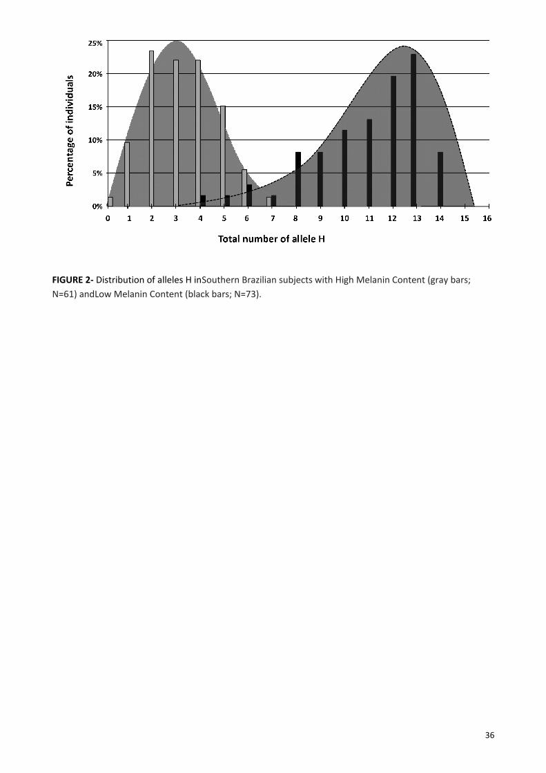
FIGURE 2- Distribution of alleles H inSouthern Brazilian subjects with High Melanin Content (gray bars; N=61) andLow Melanin Content (black bars; N=73).
36

FIGURE 3- STRUCTURE cluster plot for optimum K:3.Graph of the probability of each subject (each bar) belonging to two phenotype clusters: with high melanin content (in white) or with low melanin content (in black). Each vertical bar represents one subject (134 in total). The vertical black line separates the two different pre-categorized color groups, identified by the labels at the bottom.
37

TABLE 1- Primer sequences for conventional PCR and for SNapShot multiplex system.
38

TABLE 2- Allele frequencies and other parameters of the selected SNPs based on population data from HAPMAP and ALFRED (the database used to create Table 1 is presented in a Supplementary Excel file).
1- HMC (High Melanin Content) Populations: populations of Africans, as available on the HAPMAP and ALFRED websites. 2- LMC (Low Melanin Content) Populations: populations of Europeans, as available on theHAPMAP and ALFRED websites. ‘Allele H’: allele strongly associated with people with HMC (from African populations). ‘Allele L’: allele strongly associated with people with LMC (from European populations). Sensit: sensitivity. Specif: specificity. PV-1: predictive value for the presence of allele H in HMC populations. PV-2: predictive value for the presence of allele L in LMC populations. RR: relative ratio. OR: odds ratio.
39
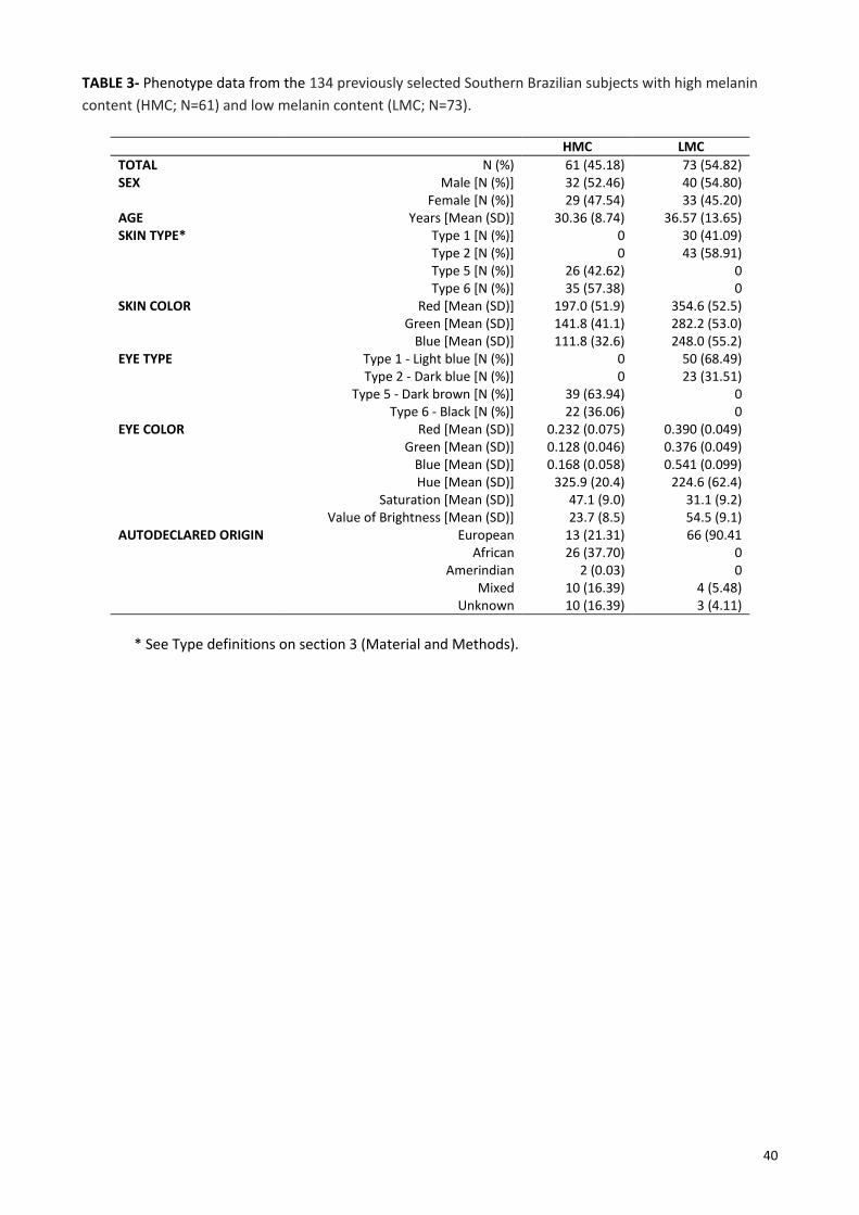
TABLE 3- Phenotype data from the 134 previously selected Southern Brazilian subjects with high melanin content (HMC; N=61) and low melanin content (LMC; N=73).
HMC LMCTOTAL N (%) 61 (45.18) 73 (54.82)SEX Male [N (%)] 32 (52.46) 40 (54.80) Female [N (%)] 29 (47.54) 33 (45.20)AGE Years [Mean (SD)] 30.36 (8.74) 36.57 (13.65)SKIN TYPE* Type 1 [N (%)] 0 30 (41.09) Type 2 [N (%)] 0 43 (58.91)
Type 5 [N (%)] 26 (42.62) 0Type 6 [N (%)] 35 (57.38) 0
SKIN COLOR Red [Mean (SD)] 197.0 (51.9) 354.6 (52.5) Green [Mean (SD)] 141.8 (41.1) 282.2 (53.0) Blue [Mean (SD)] 111.8 (32.6) 248.0 (55.2)EYE TYPE Type 1 - Light blue [N (%)] 0 50 (68.49) Type 2 - Dark blue [N (%)] 0 23 (31.51)
Type 5 - Dark brown [N (%)] 39 (63.94) 0 Type 6 - Black [N (%)] 22 (36.06) 0EYE COLOR Red [Mean (SD)] 0.232 (0.075) 0.390 (0.049) Green [Mean (SD)] 0.128 (0.046) 0.376 (0.049) Blue [Mean (SD)] 0.168 (0.058) 0.541 (0.099)
Hue [Mean (SD)] 325.9 (20.4) 224.6 (62.4) Saturation [Mean (SD)] 47.1 (9.0) 31.1 (9.2) Value of Brightness [Mean (SD)] 23.7 (8.5) 54.5 (9.1)AUTODECLARED ORIGIN European 13 (21.31) 66 (90.41 African 26 (37.70) 0 Amerindian 2 (0.03) 0 Mixed 10 (16.39) 4 (5.48) Unknown 10 (16.39) 3 (4.11)
* See Type definitions on section 3 (Material and Methods).
40
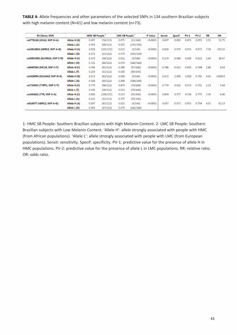
TABLE 4- Allele frequencies and other parameters of the selected SNPs in 134 southern Brazilian subjects with high melanin content (N=61) and low melanin content (n=73).
1- HMC SB People: Southern Brazilian subjects with High Melanin Content. 2- LMC SB People: Southern Brazilian subjects with Low Melanin Content. ‘Allele H’: allele strongly associated with people with HMC (from African populations). ‘Allele L’: allele strongly associated with people with LMC (from European populations). Sensit: sensitivity. Specif: specificity. PV-1: predictive value for the presence of allele H in HMC populations. PV-2: predictive value for the presence of allele L in LMC populations. RR: relative ratio. OR: odds ratio.
41

TABLE 5- Genotype frequencies of HH, HL and LL of the selected SNPs calculated from allele frequencies based on population data from HAPMAP and ALFRED (the database used to create Table 1 is presented in the Supplementary Excel file).
HMC Populations1 HH HL LLrs4778138 (OCA2; H=G, L=A) 0.55503 0.38144 0.06554rs12913832 (HERC2; H=A, L=G) 0.91585 0.08230 0.00185rs16891982 (SLC452A; H=C, L=G) 0.77616 0.20968 0.01416rs8045560 (MC1R; H= C, L=T) 0.83723 0.15555 0.00723rs1426654 (SLC24A5; H=G, L=A) 0.52563 0.39875 0.07563rs2733832 (TYRP1; H=C, L=T) 0.84089 0.15222 0.00689rs1042602 (TYR; H=C, L=A) 0.89492 0.10217 0.00292rs916977 (HERC2; H=A, L=G) 0.79210 0.19580 0.01210LMC Populations2 HH HL LLrs4778138 (OCA2; H=G, L=A) 0.02690 0.27421 0.69890rs12913832 (HERC2; H=A, L=G) 0.08123 0.40584 0.50694rs16891982 (SLC452A; H=C, L=G) 0.00449 0.12502 0.87049rs8045560 (MC1R; H= C, L=T) 0.19360 0.49280 0.31360rs1426654 (SLC24A5; H=G, L=A) 0.00012 0.02176 0.97812rs2733832 (TYRP1; H=C, L=T) 0.17223 0.48555 0.34223rs1042602 (TYR; H=C, L=A) 0.49280 0.41839 0.08880rs916977 (HERC2; H=A, L=G) 0.04580 0.33641 0.61780
1- HMC (High Melanin Content) Populations: populations of Africans, as available on the HAPMAP and ALFRED websites. 2- LMC (Low Melanin Content) Populations: populations of Europeans, as available on theHAPMAP and ALFRED websites. ‘Allele H’: allele strongly associated with people with HMC (from African populations). ‘Allele L’: allele strongly associated with people with LMC (from European populations).
42

CAPÍTULO 3 - CONSIDERAÇÕES FINAIS
Recentemente, novas tecnologias começaram a ser aplicadas largamente na área em estudo.
Podemos citar, por exemplo, a genotipagem de SNPs através de chips semicondutores. Estas tecnologias de
genotipagem de SNPs em larga escala, associadas com a descoberta de novas mutações capazes de
predizer características externas visíveis, nos darão o norte para o desenvolvimento de novas pesquisas.
De qualquer forma, os resultados obtidos nesta tese, assim como a natureza geopolítica da região
sul do Brasil, em especial a do Estado do Rio Grande do Sul, demostraram que o nosso painel em SNaPShot,
para genotipagem de oito SNPs, pode ser uma ferramenta para predição fenotípica com alto nível de
acerto.
Fazendo uso do progresso observável neste campo, novos estudos que possam prever
características fenotípicas devem ser desenvolvidos, a fim de esclarecer as mais complexas associações e
interações entre os mais de 120 genes envolvidos na pigmentação humana.
43

REFERÊNCIAS BIBLIOGRÁFICAS
A. Napolitano, C. Costantini, O. Crescenzi, G. Prota. Characterization of 1,4- benzothiazine intermediates in
the oxidative conversion of 5-S-cysteinyldopa to pheomelanins, Tetrahedron Lett 35 (1994) 6365–
6368.
A. Napolitano, P. Di Donato, G. Prota, E.J. Land. Transient quinonimines and 1,4-benzothiazines of
pheomelanogenesis: new pulse radiolytic and spectrophotometric evidence, Free Radic Biol Med.
27 (1999) 521–528.
A. Napolitano, P. Di Donato, G. Prota. New regulatory mechanisms in the biosynthesis of pheomelanins:
rearrangement vs. redox exchange reaction routes of a transient 2H-1,4-benzothiazine-o-
quinonimine intermediate, Biochim Biophys Acta. 1475 (2000) 47–54.
A. Palumbo, M. d’Ischia, G. Prota. Tyrosinase-promoted oxidation of 5,6-dihydroxyindole-2-carboxylic acid
to melanin. Isolation and characterization of oligomer intermediates, Tetrahedron, 43 (1987) 4203–
4206.
A. Palumbo, F. Solano, G. Misuraca, P. Aroca, J.C. Garcia-Borron, J.A. Lozano, G. Prota. Comparative action
of dopachrome tautomerase and metal ions on the rearrangement of dopachrome, Biochim
Biophys Acta. 1115 (1991) 1–5.
A. Slominski, D. J. Tobin, S. Shibahara, J. Wortsman. Melanin pigmentation in mammalian skin and Its
hormonal regulation. American Physiological Reviews 84(2004) 1155–1228.
A. Van Daal, A. The genetic basis of human pigmentation, Forensic Science International 1(2008). 541–543.
A. Vierkötter, U. Krämer, D. Sugiri, A. Morita, A. Yamamoto, N. Kaneko, M. Matsui, J. Krutmann.
Development of lentigines in german and japanese women correlates with variants in the SLC24A5,
Gene Journal of Investigative Dermatology 132 (2012) 733–736.
C.L. Wilkerson, N.A. Syed, M.R. Fisher, N.L. Robinson, H.L. Wallow, D.M. Albert. Melanocytes and iris color.
Light microscopic findings, Archives of Ophthalmology 114(1996)437–442.
E. Giardina, A. Spinella, G. Novelli. Past, present and future of forensic DNA typing, Nanomedicine 6 (2011)
257–270.
E.E. Kenny, N.J. Timpson, M.Sikora, M.C. Yee, A. Moreno-Estrada, C. Eng, S. Huntsman, E.G.
Burchard, M. Stoneking, C.D. Bustamante, S. Myles. Melanesian blond hair is caused by an amino acid
change in TYRP1, Science 336 (2012) 554.
E.J. Land, S. Ito, K. Wakamatsu, P.A. Riley. Rate constants for the first two chemical steps of
Eumelanogenesis, Pigment Cell Res. 16 (2003) 487–493.
E.J. Parra. Human pigmentation variation: Evolution, genetic basis, and implications for public health, Year
book of physical anthropology. 50 (2007) 85-105.
F. Liu, K. van Duijn, J.R. Vingerling, A. Hofman, A.G. Uitterlinden, A.C.J.W. Janssens, M. Kayser. Eye color and
the prediction of complex phenotypes from genotypes, Curr. Biol. 19 (2009) R192–R193.
F. Liu, B. Wenb, M. Kayser. Colorful DNA polymorphisms in humans, Semin Cell Dev Biol (2013).
44

G.B. Stacey, S.F. Schaffner, H. Nguyen, J.M. Moore, J. Roy, B. Blumenstiel, J. Higgins, M. Defelice, A.
Lochner, M. Faggart, S.N. Liu-Cordero, C. Rotimi, A. Adeyemo, R. Cooper, R. Ward, E.S. Lander, M.J.
Daly, D. Altshuler. The structure of haplotype blocks in the human genome, Science 296 (2002)
2225–2229.
G. Dimisianos, I. Stefanaki, V. Nicolaou, V. Sypsa, C. Antoniou, M. Poulou, O. Papadopoulos, H. Gogas, E.
Kanavakis, E. Nicolaidou, A. D. Katsambas, A. J. Stratigos. A study of a single variant allele
(rs1426654) of the pigmentation-related gene SLC24A5 in Greek subjects, Experimental
Dermatology 18 (2009)175–7.
G. E Costin, V. J Hearing. Human skin pigmentation: melanocytes modulate skin color in response to stress,
The Journal of the Federation of American Societies for Experimental Biology 21 (2007) 976-994.
G. Greco, K. Wakamatsu, L. Panzella, S. Ito, A. Napolitano, M. D´Ischia. Isomeric cysteinyldopas provide a
(photo)degradable bulk component and a robust structural element in red human hair
pheomelanin, Pigment Cell Melanoma Res. 22 (2009) 319–327.
G. Tully. Genotype versus phenotype: Human pigmentation, Forensic Sci Int. Genet. 1 (2007) 100-104.
H. Ho, R. Kapadia, S. Al-Tahan, S.; et al. WIPI1 Coordinates Melanogenic Gene Transcription and
Melanosome Formation via TORC1 Inhibition, The Jornal of Biological Chemistry 286, n. 14. (2011)
12509-12523.
H.L. Norton, R.A Kittles, E. Parra, P. Mckeigue, X. Mao, K. Cheng, V. A. Canfield, D.G Bradley, B. Mceovoy, M.
Shriver. Genetic evidence for the convergent evolution of light skin in Europeans and East Asian,
Mol Biol Evol 24(2007) 710-22.
H. Pulker, M.V. Lareu, C. Phillips, A. Carracedo. Finding genes that underlie physical traits of forensic
interest using genetic tools, Forensic Sci Int Genet. 1 (2007) 100-104.
INTERNATIONAL FEDERATION OF PIGMENT CELL SOCIETIES – IFPCS 2009: Disponível em:
http://www.espcr.org/micemut/ - Acessado em abril de 2013.
J. Berson, D. C. Harper, D. Tenza, G. Raposo, M.S. Marks. Pmel17 initiates premelanosome morphogenesis
within multivesicular bodies, Molecular Biology of the cell 12(2001) 3451-3464.
J. Graf, J. Voisey, I. Hughes, A.Van Daal. Promoter polymorphisms in the MATP (SLC24A5) gene are
associated with normal skin color variation, Hum. Mutat. 28 (2007) 710-717.
J. Graf, R. Hodgson, A.Van Daal. Single nucleotide polymorphisms in the MATP gene are associated with
normal human pigmentation variation, Hum. Mutat. 25 (2005) 278-284.
J.L. Rees. The genetics of Sun sensivity in humans, Am. J. Hum. Genet. 75 (2004) 739-751.
J.R.Landis, G.G Koch. The measurement of observer agreement for categorical data, Biometrics
33(1977)159-174.
J.S. Allwood, S.A. Harbison. SNP model development for the prediction of eye colour in New Zealand,
Forensic Science International: Genetics 7 (2013) 444–452.
45

J. Voisey, M.C. Gomez-Cabrera, D.J. Smit, J.H. Leonard, R.A. Sturm, A. van Daal. A polymorphism in the
agouti signalling protein (ASIP) is associated with decreased levels of mRNA Pigment Cell Res. 19
(2006) 226–231.
J.Y Lin, D.E Fisher. Melanocyte biology and skin pigmentation, Nature 445 (2007) 843-850.
K. Makova, H. Norton. Worlwide polymorphism at the MC1R locus and normal pigmentation variation in
humans, Peptides 26 (2005) 1901-1908.
K. Nakayama, A. Soemantri, F.J, Bumbein Dashnyam, R. Ohtsuka, P. Duanchang, M. N. Isa, W. Settheetham-
Ishida, S. Harihara, T. Ishida. Identification of novel functional variants of the melanocortin 1
receptor gene originated from asians, Human Genet 119(2006) 322-30.
K. Wakamatsu, K. Ohtara, S. Ito. Chemical analysis of late stages of pheomelanogenesis: conversion of
dihydrobenzothiazine to a benzothiazole structure, Pigment Cell Melanoma Res. 22 (2009) 474–
486.
K. Yoshida, K. Yayama, A. Hatanaka, K. Tamaki. Efficacy of extended kinship analyses utilizing commercial
STR kit in establishing personal identification, Leg. Med. 13 (2011) 12–15.
L.D.G. Kobachuk. Estudo de freqüências alélicas de dez locos STRs do cromossomo X na população do
estado do Paraná e sua contribuição na identificação humana, (2012).
L. Excoffier, H.E.L. Lischer. Arlequin suite ver 3.5: a new series of programs to perform population genetics
analyses under Linux and Windows, Mol Ecol Resour 10 (2010) 564-67.
M.A. Jobling, P. Gill. Encoded evidence: DNA in forensic analysis, Nature Rev. Genet. 5 (2004) 739–751.
M. Brenner, M. Berking. Grundlagen der Hautpigmentierung: Biochemie und regulation der
melaninsynthese, Der Hautarzt 61(2010) 554-560.
M. Chaki, M. Sengupta, M. Mondal, A. Bhattacharya, S. Mallick, R. Bhadra, K. Ray. Molecular and functional
studies of tyrosinase variants among indian oculocutaneous albinism type 1, Patients Journal of
Investigative Dermatology 131 (2011) 260–262.
M. Kayser, P. Schneider. DNA-based prediction of human externally visible characteristics in forensics:
Motivations, scientific challenges, and ethical considerations, Forensic Science International
3(2009)154–161.
M. Kayser, P. Knijff. Improving human forensics through advances in genetics, genomics and molecular
biology, Nature Reviews 12 (2011) 179-192.
M.P. Donnelly, P. Paschou, E. Grigorenko, D. Gurwitz, C. Barta, R.B. Lu, O.V. Zhukova, J.J. Kim, M. Siniscalco,
M. New, H. Li, S.L.B. Kajuna, V.G. Manolopoulos, W.C. Speed, A.J. Pakstis, J.R. Kidd, K.K. Kidd. A
global view of the OCA2-HERC2 region and pigmentation, Hum Genet 131 (2012) 683-696.
M.R, Gerstenblith, A.M, Goldstein, M.C, Fargnoli, K. Peris, M.T Landi. Comprehensive evaluation of allele
frequency differences of MC1R variants across populations, Human Mutat 28(2007) 495-505.
M. Soejima, Y. Koda. Population differences of two coding SNPs in pigmentation-related genes SLC24A5 and
SLC24A5, Int J Legal Med 121 (2007) 36-39.
46

M. Visser, M. Kayser, R.J. Palstra. HERC2 rs12913832 modulates human pigmentation by attenuating
chromatin-loop formation between a long-range enhancer and the OCA2 promoter Genome Res.22
(2012) 446-455.
N. Jablonski, G. Chaplin. The evolution of human skin coloration, Journal of Human Evolution, v. 39(2000)
57-106.
N. Jablonski. The Evolution of Human Skin and Skin Color, Annual Review of Anthropology v. 33 (2004) 585–
623.
O’Rahilly, S. Human genetics illuminates the path to metabolic disease, Nature 462(2009) 307-14.
P. Sulem, D.F. Gudbjartsson, S.N. Stacey, A. Helgason, T. Rafnar, K.P. Magnusson, A. Manolescu, A. Karason,
A. Palsson, G. Thorleifsson, M. Jakobsdottir, S. Steinberg, S. Palsson, F. Jonasson, B. Sigurgeirsson, K.
Thorisdottir, R. Ragnarsson, K.R. Benediktsdottir, K.K. Aben, L.A. Kiemeney, J.H. Olafsson, J. Gulcher,
A. Kong, U. Thorsteinsdottir, K. Stefansson. Genetic determinants of hair, eye and skin pigmentation
in Europeans, Nat. Genet. 39 (2007) 1443–1452.
R.A Sturm, R. Teasdale, N. Box. Human pigmentation genes: Identification, Structure and Consequences of
Polymorphic Variation, Gene 277(2001) 49–62.
R.A Sturm, T.N Frudakis. Eye colour: portals into pigmentation genes andancestry, Trends in Genetics
20(2004) 327–332.
R.A. Sturm. A golden age of human pigmentation genetics, Trends Genet. 22 (2006) 464-468.
R.A. Sturm, D.L. Duffy, Z.Z. Zhao, F.P.N. Leite, M.S. Stark, N.K. Hayward, N.G. Martin, G.W. Montgomery, A
single SNP in an evolutionary conserved region within intron 86 of the HERC2 gene determines
human blue-brown eye color. The American Journal of Human Genetics 82 (2008) 424-431.
R.A. Sturm. Molecular genetics of human pigmentation diversity, Hum. Mol. Genet. Review Issue 18 (2009)
R9-R17.
R.A. Sturm, M. Larsson. Genetics of human íris colour and patterns, Pigment Cell Melanoma Ress. 22 (2009)
544-562.
R.A. Sturm, D.L. Duffy. Human pigmentation genes under environmental selection, Genome Biololgy (2012)
13:248. doi:10.1186/gb-2012-13-9-248
R.K. Valenzuela, S. Ito, K. Wakamatsu, M.H. Brilliant. Prediction model validation: normal human
pigmentation variation, J. Forenseci Res. 2 (2011). doi:10.4172/2157-7145.1000139
R.L. Lamason, M.A. Mohideen, J.R. Mest, A.C. Wrong, H.L. Norton, M.C. Aros, M.J. Jurynec, X. Mao, V.R.
Humphreville, J.E. Humbert, S. Sinha, J.L. Moore, P. Jagadeeswaran, W. Zhao, G. Ning, I.
Makalowska, P.M. McKeigue, D, O’Donnnell, R. Kittles, E.J. Parra, N.J. Mangini, D.J. Grunwald, M.D.
Sriver, V.A. Canfield, K.C. Cheng. SLC24A5, a putative cation exchanger, affects pigmentation in
zebrafish and humans, Science 310(2005) 1782–1786.
R. M. Harding, E. Healy, A. J. Ray, N. S. Ellis, N. Flanagan, C. Todd, C. Dixon, A. Sajantila, I. J. Jackson, M. A.
Birch-Machin, J. L. Rees. Evidence for Variable Selective Pressures at MC1R, Am J Human Genet
66(2000)1351-61.
47
R.P. Stokowski, P.V.K. Pant, T. Dadd, A. Fereday, D.A. Hinds, C. Jarman, W. Filsell, R.S. Ginger, M.R. Green,
F.J. van der Ouderaa, D.R. Cox. A genomewide association study of skin pigmentation in a south
asian population, The American Journal of Human Genetics. 81 (2007) 1119–1132.
R.S. Ginger, S. E. Askew, R.M. Ogborne, R.M Wilson, D. Ferdinando, T. Dadd , A.M Smith, S. Kazi, R.T
Szerencsei, R.J Winkfein, P.P Schnetkamp, M.R Green. SLC24A5 encodes a trans-Golgi network
protein with potassium-dependent sodium–calcium exchange activity that regulates human
epidermal melanogenesis, J Biol Chem 283(2008) 5486–5495.
S. Alaluf, D. Atkins, K. Barrett, M. Blout, N. Carter, A. Heath. Ethnic variation in melanin content and
composition in photoexposed and photoprotected human skin, Pigment Cell Res 15(2002) 112-118.
S. Alaluf, K. Barrett, M. Blount, N. Carter. Ethnic variation in tyrosinase and TYRP1 expression in
photoexposed and photoprotected human skin, Pigment. Cell. Res. 16 (2003) 35-42.
S. Ito, K. Wakamatsu, Chemistry of Melanins. In: J.J. Nordlund, R.E. Boissy, V.J. Hearing, R.A. King, W.S.
Oetting, J.P. Ortonne (Eds.). The Pigmentary System. Physiology and Pathophysiology, Blackwell
Publishing; Oxford, 2006, pp. 282-310.
S. Walsh, A. Lidenbrgh, S.B. Zuniga, T. Sijen, P. Knijff, M. Kayser, K.N. Ballantyne. Developmental validation
of the IrisPlex system: Determination of blue and brown iris colour for forensic intelligence,
Forensic Sci Int. Genet. 5 (2011a) 464-471.
S. Walsh, F. Liu, K.N. Ballantyne, M. van Oven, O. Lao, M. Kayser. IrisPlex: a sensitive DNA tool for accurate
prediction of blue and brown eye colour in the absence of ancestry information, Forensic Sci Int.
Genet. (2011b). doi:10.1016/j.fsigen.2010.02.004
S. Walsh, F. Liu, A. Wollstein, L. Kovatsi, A. Ralf, A. Kosiniak-Kamysz, W. Branicki, M. Kayser. The HIrisPlex
system for simultaneous prediction of hair and eye colour from DNA, Forensic Sci Int. Genet. 7
(2013) 98-115.
T. Frudakis, M. Thomas, Z. Gaskin, K. Venkateswarlu, K.S. Chandra, S. Ginjupalli, S. Gunturi, S. Natrajan, V.K.
Ponnuswamy, K.N. Ponnuswamy. Sequences associated with human iris pigmentation, Genetics
165 (2003) 2071–2083.
T. Kondo, V.J. Hearing. Update on the regulation of mammalian melanocyte function and skin
pigmentation, Expert Rev Dermatol. 6 (2011) 97–108.
T. Kushimoto, V. Basrur,J. Valencia, J. Matsunaga, W.D. Vieira, V.J. Ferrans, J. Muller, E. Appella, V.J.
Hearing. A model for melanosome biogenesis based on the purification and analysis of early
melanosomes. Proceedings of the National Academy of Sciences of the United States of America.
98(2001) 10698-10703.
W. Branicki, U. Brudnik, A. Wojas-Pel. Interactions Between HERC2, OCA2 and MC1R May Influence Human
Pigmentation Phenotype, Annals of Hum Genet. 73 (2009) 160-170.
Y. Jin, S.A. Birlea, P.R Fain, T.M. Ferrara, S. Ben, S.L. Riccardi, J.B. Cole, K. Gowan, P.J. Holland, D.C. Bennett,
R.M. Luiten, A. Wolkerstorfer, J.P.W. van der Veen, A. Hartmann, S. Eichner, G. Schuler, N. van Geel,
J. Lambert, E.H. Kemp, D.J. Gawkrodger, A.P. Weetman, A. Taïeb, T. Jouary, K. Ezzedine, M.R.
48

Wallace, W.T. McCormack, M. Picardo, G. Leone, A. Overbeck, N.B. Silverberg, R.A. Spritz. Genome-
wide association analyses identify 13 new susceptibility loci for generalized vitiligo, Nature Genetics
44 (2012) 676–680.
49

ADENDOS
MATERIAL INFORMATIVO APRESENTADO AOS PARTICIPANTES DA PESQUISA
50

MATERIAL INFORMATIVO APRESENTADO AOS PARTICIPANTES DA PESQUISA
51

PROCEDIMENTO OPERACIONAL PADRÃO PARA COLETA DE DADOS
52

TERMO DE CONSENTIMENTO LIVRE E ESCLARECIDO
Você está sendo convidado para participar de uma pesquisa da PUCRS. O título da pesquisa é “ANÁLISE DESNPs EM GENES DE PIGMENTAÇÃO HUMANA EM INDIVÍDUOS GENETICAMENTE DISTINTOS” , que tem por objetivoestudar partes do material genético que possam estar relacionadas à cor da pele e à cor dos olhos das pessoas. Paraisso precisamos: 1- registrar a cor da pele do seu braço com um medidor; 2- fotografar cada um de seus olhos [não orosto]; 3- coletar células da sua bochecha com um cotonete esterilizado.
Este estudo não trará benefícios para você diretamente, nesse momento. Mas, haverá benefícios para asociedade se conseguir encontrar relações entre o material genético e a cor da pele e olhos da pessoa. Isso poderáajudar a identificar pessoas desaparecidas, por exemplo.
Sinta-se à vontade para fazer qualquer pergunta sobre o estudo e esclarecer suas dúvidas. Os responsáveispela pesquisa garantidamente lhe responderão. Caso você tenha vontade de ser retirado da pesquisa, diga isso aqualquer momento, sem necessidade de explicar. Você não será prejudicado se resolver tomar essa decisão. Apenascomunique aos pesquisadores se quiser abandonar a pesquisa e nós respeitaremos a sua decisão.
Toda e qualquer informação atualizada sobre este estudo estará à disposição de todos os que participam dapesquisa. Os resultados deste estudo poderão ser comunicados a você, sempre que você quiser. A dignidade e aprivacidade dos participantes desta pesquisa, em relação a qualquer dado, serão mantidas. Os dados dos participantesda pesquisa estarão protegidos pelos pesquisadores responsáveis contra qualquer tipo de uso que não os aquiprevistos. Além disto, os dados receberão um número para garantir o anonimato dos participantes da pesquisa. Noestudo dos dados, a identidade do participante será desconhecida tanto pela equipe do trabalho como por qualqueroutra pessoa externa.
A partir do mesmo material poderão ser futuramente feitos outros estudos. Para isso, você poderá permitirque sua amostra seja usada em novos estudos. Mesmo que esta permissão seja dada agora, estudos futuros só serãorealizados se forem aprovados por um Comitê de Ética em Pesquisa.
Eu, ______________________________________ (preencher com nome do participante da pesquisa) fuiinformado dos objetivos da pesquisa acima de maneira clara e objetiva. Recebi informação a respeito dosprocedimentos do estudo e esclareci minhas dúvidas. Sei que em qualquer momento poderei solicitar novasinformações e modificar minha decisão se assim eu o desejar. A equipe da Dra. Clarice S. Alho certificou-me de quetodos os dados referentes ao participante desta pesquisa serão confidenciais, bem como sua rotina de vida não serámodificada em razão desta pesquisa e que, ainda, terei a liberdade de retirar meu consentimento de participação napesquisa se assim o desejar. Assim, como sujeito de pesquisa, dou consentimento para participar deste estudo. Eutambém permito que a amostra de material genético seja armazenada na universidade, sob responsabilidade dosPesquisadores Coordenadores, para estudos futuros. Minha permissão vale desde que os novos estudos sejam sempredevidamente aprovados por um Comitê de Ética em Pesquisa e estejam sob responsabilidade dos mesmosPesquisadores Coordenadores. Fui informado também que, nesta situação, caso alguma informação consideradaimportante seja identificada no material genético analisado em decorrência da pesquisa, os pesquisadores poderãobuscar a mim para oferecer acesso a tal informação.
Caso eu tenha novas perguntas sobre este estudo, posso telefonar a Dra. Clarice S. Alho nos telefones (51)91136334 ou (51) 3320 3545 ramal 4534, ou para esclarecer sobre os meus direitos como participante deste estudo,ou se penso que fui prejudicado pela minha participação, posso chamar também os responsáveis pelo Comitê de Éticaem Pesquisa – CEP-PUCRS no telefone (51) 3320 3000 ramal 3345. As ligações para os celulares poderão ser realizadasa cobrar. Declaro também que recebi cópia do presente Termo de Consentimento.
Assinatura do Participante Nome do Participante _____/_____/20_____
Assinatura do Pesquisador Nome do Pesquisador _____/_____/20_____
Na impossibilidade de autonomia para a leitura deste Termo de Consentimento Livre e Esclarecido, o mesmofoi lido para ______________________________________ (preencher com o nome do doador) pelo pesquisador_____________________ (preencher com o nome do pesquisador) enquanto eu estava presente como testemunha.
Assinatura da Testemunha Nome da Testemunha _____/_____/20_____
53

INSTRUMENTO DE COLETA DE DADOS
54