Rio de Janeiro, Vol. 92(2): 221-232, Mar./Apr. 1997 221 ... · Egg Structures of Anopheles...
12
221 221 221 221 221 Mem Inst Oswaldo Cruz, Rio de Janeiro, Vol. 92(2): 221-232, Mar./Apr. 1997 Egg Structures of Anopheles fluminensis and Anopheles shannoni LP Lounibos/ + , D Duzak, JR Linley † , R Lourenço-de-Oliveira* Florida Medical Entomology Laboratory, University of Florida, 200 9 th St. SE, Vero Beach, Florida 32962, U.S.A. *Departamento de Entomologia, Instituto Oswaldo Cruz, Av. Brasil 4365, 21045-900 Rio de Janeiro, RJ, Brasil Eggs of two species belonging to the Arribalzagia Series of the Laticorn Section of Anopheles (Anopheles) collected in Brazil are described from scanning electron micrographs. The An. fluminensis egg is long with shallow floats displaced far dorsally. The narrow deck region is overlain by a frill modified into prominent ridges that are nearly continuous to both ends of the egg. Slightly opened decks at both poles contain an average of four lobed tubercles. Polygonal, plastron-type chorionic cells cover the lateral and dorsal surfaces. The egg of An. shannoni is unique in possessing 22-27 fingerlike fila- ments that project with regular spacing from each of its massive floats. These filaments and their bases are highly perforated and are believed to trap air and support flotation of the egg with the dorsal surface up, contrary to the usual orientation for anophelines. The eggs are compared with those of related species bearing similar structures, notably An. fluminensis with An. mediopunctatus s.s and An. shannoni with An. peryassui. Key words: Anopheles - Brazil - eggs - malaria - morphology - scanning electron microscopy Species identifications of malaria vectors by morphological characters of adults are problem- atic and often unsatisfactory. The first evidence of an anopheline species complex, whose members were indistinguishable in the adult stage, came from comparative examinations of eggs of Anopheles maculipennis s.l. Meigen from southern Europe (Falleroni 1926, Hackett & Missiroli 1935). The identification of cryptic species by egg structures led to a surge of interest among entomologists and malariologists in the systematics potential of this neglected life stage (e.g. Galvão 1938, Rozeboom 1938, 1942, Kumm 1941). A comprehensive study based on over 28,000 ovipositions by 30 species led to an illustrated key to the eggs of Brazilian Anopheles (Causey et al. 1944). Limitations to this research included multiple egg morphs within some species and the low resolution of the light micro- scope for observing the intricate details of egg structures. This work was supported by grants from the National Institutes of Health (AI-31034) of the U.S.A. and CNPq, PAPES/FIOCRUZ and FNS of Brazil, and is Univer- sity of Florida IFAS Journal Series No. R-05309. + Corresponding author. Fax: +561-778.7205. E-mail: [email protected] † Deceased Received 13 August 1996 Accepted 10 October 1996 For many subsequent years anopheline eggs received scant attention until Hinton (1968) rec- ognized the potential of the scanning electron mi- croscope (SEM) for visualizing egg microstruc- tures and, by extension, new morphological char- acters for species recognition. However, only in the present decade has the SEM been applied ex- tensively to eggs of New World Anopheles for describing structural details (e.g. Linley & Lounibos 1993, 1994), intraspecific variation (Rodriguez et al. 1992), geographic differentiation (Linley et al. 1996), and for separating members of species complexes (Linley et al. 1993) or sepa- rate species mistakenly synonymized (Lounibos et al. manuscript in preparation). The present study describes the ultrastructure of eggs of two anopheline species that had previ- ously been known only at the light microscope level. Anopheles (Anopheles) fluminensis was origi- nally described by Root (1927) from the State of Rio de Janeiro, and its egg was first depicted by Causey et al. (1944) from Amazonian collections of this species. Although it has not been regarded as a vector in malarious regions of Brazil (Cerqueira 1961), mosquitoes identified as An. sp. near fluminensis were incriminated as vectors of human malaria in eastern Peru (Hayes et al. 1987). The first illustration of the egg of An. shannoni Davis, at the time undescribed, was by Bonne and Bonne-Wepster (1925) (as An. mediopunctatus [Theobald]). The highly modified egg of this spe- cies was first described by Causey et al. (1944)
Transcript of Rio de Janeiro, Vol. 92(2): 221-232, Mar./Apr. 1997 221 ... · Egg Structures of Anopheles...

221221221221221Mem Inst Oswaldo Cruz, Rio de Janeiro, Vol. 92(2): 221-232, Mar./Apr. 1997
Egg Structures of Anopheles fluminensis andAnopheles shannoni
LP Lounibos/+, D Duzak, JR Linley†, R Lourenço-de-Oliveira*
Florida Medical Entomology Laboratory, University of Florida, 200 9th St. SE, Vero Beach, Florida 32962,U.S.A. *Departamento de Entomologia, Instituto Oswaldo Cruz, Av. Brasil 4365, 21045-900 Rio de Janeiro,
RJ, Brasil
Eggs of two species belonging to the Arribalzagia Series of the Laticorn Section of Anopheles(Anopheles) collected in Brazil are described from scanning electron micrographs. The An. fluminensisegg is long with shallow floats displaced far dorsally. The narrow deck region is overlain by a frillmodified into prominent ridges that are nearly continuous to both ends of the egg. Slightly opened decksat both poles contain an average of four lobed tubercles. Polygonal, plastron-type chorionic cells coverthe lateral and dorsal surfaces. The egg of An. shannoni is unique in possessing 22-27 fingerlike fila-ments that project with regular spacing from each of its massive floats. These filaments and their basesare highly perforated and are believed to trap air and support flotation of the egg with the dorsal surfaceup, contrary to the usual orientation for anophelines. The eggs are compared with those of relatedspecies bearing similar structures, notably An. fluminensis with An. mediopunctatus s.s and An. shannoniwith An. peryassui.
Key words: Anopheles - Brazil - eggs - malaria - morphology - scanning electron microscopy
Species identifications of malaria vectors bymorphological characters of adults are problem-atic and often unsatisfactory. The first evidence ofan anopheline species complex, whose memberswere indistinguishable in the adult stage, came fromcomparative examinations of eggs of Anophelesmaculipennis s.l. Meigen from southern Europe(Falleroni 1926, Hackett & Missiroli 1935). Theidentification of cryptic species by egg structuresled to a surge of interest among entomologists andmalariologists in the systematics potential of thisneglected life stage (e.g. Galvão 1938, Rozeboom1938, 1942, Kumm 1941). A comprehensive studybased on over 28,000 ovipositions by 30 speciesled to an illustrated key to the eggs of BrazilianAnopheles (Causey et al. 1944). Limitations to thisresearch included multiple egg morphs within somespecies and the low resolution of the light micro-scope for observing the intricate details of eggstructures.
This work was supported by grants from the NationalInstitutes of Health (AI-31034) of the U.S.A. and CNPq,PAPES/FIOCRUZ and FNS of Brazil, and is Univer-sity of Florida IFAS Journal Series No. R-05309.+Corresponding author. Fax: +561-778.7205. E-mail:[email protected]†DeceasedReceived 13 August 1996Accepted 10 October 1996
For many subsequent years anopheline eggsreceived scant attention until Hinton (1968) rec-ognized the potential of the scanning electron mi-croscope (SEM) for visualizing egg microstruc-tures and, by extension, new morphological char-acters for species recognition. However, only inthe present decade has the SEM been applied ex-tensively to eggs of New World Anopheles fordescribing structural details (e.g. Linley &Lounibos 1993, 1994), intraspecific variation(Rodriguez et al. 1992), geographic differentiation(Linley et al. 1996), and for separating membersof species complexes (Linley et al. 1993) or sepa-rate species mistakenly synonymized (Lounibos etal. manuscript in preparation).
The present study describes the ultrastructureof eggs of two anopheline species that had previ-ously been known only at the light microscopelevel. Anopheles (Anopheles) fluminensis was origi-nally described by Root (1927) from the State ofRio de Janeiro, and its egg was first depicted byCausey et al. (1944) from Amazonian collectionsof this species. Although it has not been regardedas a vector in malarious regions of Brazil(Cerqueira 1961), mosquitoes identified as An. sp.near fluminensis were incriminated as vectors ofhuman malaria in eastern Peru (Hayes et al. 1987).The first illustration of the egg of An. shannoniDavis, at the time undescribed, was by Bonne andBonne-Wepster (1925) (as An. mediopunctatus[Theobald]). The highly modified egg of this spe-cies was first described by Causey et al. (1944)
222222222222222 Eggs of Brazilian Anophelines • LP Lounibos et al.
from collections in the State of Pará, Brazil, thetype locality of An. shannoni (Davis 1931).Anopheles shannoni has not been suspected as avector of human malaria (Deane et al. 1948,Cerqueira 1961), in part because it prefers to bitein the canopy of forested regions (Deane et al.1953). However, such acrodendrophilic host-seek-ing behavior makes An. shannoni a possible main-tenance vector of simian malaria where this mos-quito species and monkeys co-occur in the Ama-zon region (Lourenço-de-Oliveira & Luz 1996).
Both An. fluminensis and An. shannoni belongto the Arribalzagia Series of the Laticorn Section(Reid & Knight 1961) of the subgenus Anopheles.Wilkerson and Peyton (1990) inferred a mono-phyletic origin of the Arribalzagia Series basedupon wingspot characters. Since eggs of severalother species of this Series have been describedrecently using the SEM (Linley & Lounibos 1994,Linley & Milstrey 1995, Lounibos et al. in prepa-ration), we infer in this paper possible species af-finities based on egg structures.
MATERIALS AND METHODS
Female An. fluminensis were collected in Janu-ary 1995 from human bait at Picinguaba, State ofSão Paulo, Brazil (23o23’S, 44o50’W) and femaleAn. shannoni were collected by the same methodin July 1994 at Samuel Ecological Station, Stateof Rondônia, Brazil (9o07’S, 63o16’W). Blood-fedfemales, identified with the keys of Consoli andLourenço-de-Oliveira (1994), were isolated indi-vidually in small vials with damp filter paper foroviposition. Laid eggs were allowed 24 hr at 26oCto embryonate before preservation in alcoholicBouin’s fixative. A few eggs of An. shannoni wereimmersed in water prior to fixation in order to al-low float filaments to unfurl, as accomplished pre-viously for An. peryassui Dyar and Knab (Linley& Lounibos 1994). In preparation for microscopy,eggs were removed from fixative, washed twice in80% ethanol, then dehydrated in an ethanol seriesof 5% concentration increments. After critical pointdrying, eggs were mounted on stubs coated withsticky tape, sputter-coated with gold/palladium, andexamined in an Hitachi S-510 SEM.
Electron micrographs of eggs were eitherscanned and rendered as computer images for mea-surements with SigmaScan software (Jandel Sci-entific, San Rafael, CA, U.S.A.), or measurementswere made with a digitizing tablet and tabulatedwith the same software. Except where otherwisenoted, measurements of An. fluminensis were madeon 10-12 eggs laid by three females, and of An.shannoni on 4-6 eggs from one female. Mean val-ues in the text are followed by ± 1 SE. The de-scriptive terminology follows that of Harbach and
Knight (1980) except for “plastron” which con-forms to the usage of Hinton (1968) to describethe network of structures that form a physical gillbeneath the water line of anopheline eggs. Voucherspecimens of adult females have been depositedin the Department of Entomology, FIOCRUZ.
DESCRIPTIONS
Anopheles fluminensis (Figs 1-4)
Size: egg length 515.4-546.2 µm (mean 530.8± 3.3 µm, n=10), width 180.8-203.8 µm (mean187.7 ± 2.2 µm, n=10), length/width ratio 2.68-2.91 (mean 2.83 ± .03, n=10). Color: black. Over-all appearance: long and wide across floats, espe-cially anteriorly, but tapering abruptly where floatsterminate before anterior and posterior ends (Fig.1a); egg boat-shaped in lateral view, dorsal sur-face concave but ventral surface flat (Fig. 1b); floatsshallow and displaced unusually far dorsally (Fig.1b).
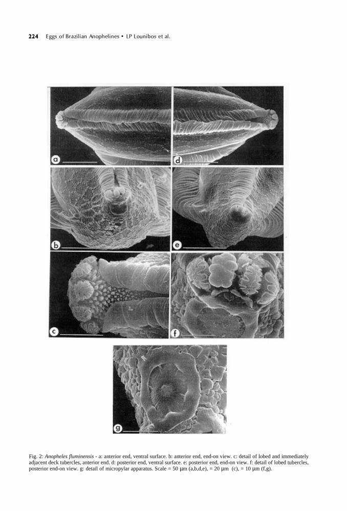
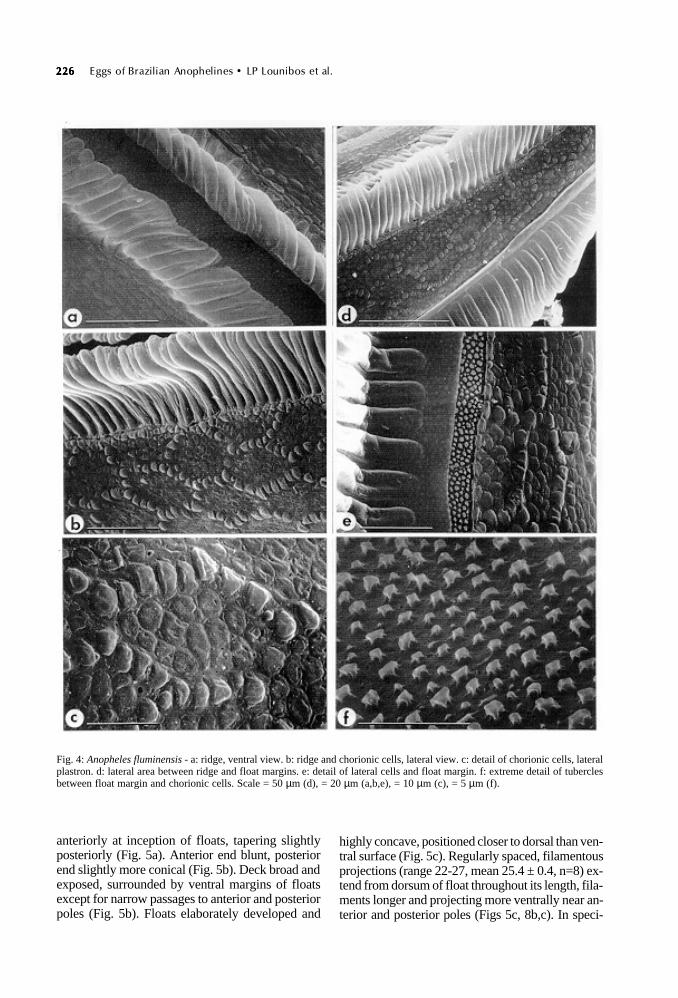
Ventral (upper) surface: deck usually hiddenby overlapping frills which form ventral ridges forlength of the egg except for anterior and posteriorpoles (Figs 2a,d). When visible, deck deeply re-cessed between ridges which are deeply groovedin both ventral and lateral views (Figs 4a,d). Bothanterior and posterior decks open, containing 3-5mushroom-like, lobed tubercles (mean anterior 4.0+ 0.2, mean posterior 3.8 ± 0.2, n=12) (Figs 2c,f).Smaller tubercles of both anterior and posteriordecks irregular, some star-shaped, and domed withbuttressed walls (Figs 3a,c). Tubercles of middledeck, exposed infrequently because of ridge over-lap, less densely packed and shorter, some ma-rooned on chorionic islets (Fig. 3b).
Ventral plastron flanking ridges and deck, wideand occupied by hexagonal chorionic cells withboundaries distinguished by raised tubercles (Figs1b, 4b). Cells longer than wide, these dimensionsconsistent for length of egg (mean cell length 35.6± 0.5 µm, mean width 16.0 ± 0.6 µm, n=20). Inte-rior of chorionic cells with rounded, tightly packedtubercles, less raised than perimeter tubercles (Fig.4c).
Anterior end, micropyle: anterior end blunt, frillextending around lobed tubercles but reduced be-tween tubercles and micropyle (Fig. 2b). Chori-onic cells continuous with lateral hexagonal ones,more compressed and irregularly shaped anteriorly(Fig. 2b). Micropylar disc divided into 6-7 sectors(mean 6.8 ± 0.1, n=10) by short rays from collar(Fig. 2g). Micropylar collar generally smooth butwith shallow pits; disc surface rugose. Micropylarorifice set in low mound (Fig. 2g).
Posterior end: blunt in end-on view (Fig. 2e),otherwise similar in conformation to ventral view

223223223223223Mem Inst Oswaldo Cruz, Rio de Janeiro, Vol. 92(2), Mar./Apr. 1997
Fig. 1: Anopheles fluminensis - a: entire egg, ventral view, anterior end at top. b: entire egg, lateral view, ventral surface at left,anterior end at top. Scale = 100 µm.
of anterior end (Figs 2a cf. 2d).Dorsal (lower) and lateral surfaces: outer
chorionic cells of dorsal plastron tending to formpolygons, although some with rounded borders(Fig. 3e), length 24.7-34.7 µm (mean 27.8 ± 1.2
µm, n=10), width 16.9-20.4 µm (mean 17.1 ± 0.7µm, n=10). Interior of cells with tightly packedtubercles like smooth cobblestone pavement (Fig.3f). Exochorion continuous except for infrequentperforations between tubercles (Fig. 3f). Floats
224224224224224 Eggs of Brazilian Anophelines • LP Lounibos et al.
Fig. 2: Anopheles fluminensis - a: anterior end, ventral surface. b: anterior end, end-on view. c: detail of lobed and immediatelyadjacent deck tubercles, anterior end. d: posterior end, ventral surface. e: posterior end, end-on view. f: detail of lobed tubercles,posterior end-on view. g: detail of micropylar apparatus. Scale = 50 µm (a,b,d,e), = 20 µm (c), = 10 µm (f,g).

225225225225225Mem Inst Oswaldo Cruz, Rio de Janeiro, Vol. 92(2), Mar./Apr. 1997
Fig. 3: Anopheles fluminensis - a: detail anterior deck tubercles. b: detail middle deck tubercles. c: detail posterior deck tu-bercles. d: ventral margin of float at junction with dorsal plastron. e: dorsal plastron, middle of egg. f: detail of chorionic cells,dorsal plastron. Scale = 50 µm (e), = 20 µm (d), = 10 µm (a,b,c,f).
deeply grooved to margin with dorsal surface (Fig.3d), the grooved sutures undulating near egg mid-line in lateral view (Fig. 1b). Boundary betweenfloat and lateral chorionic cells separated by a nar-row strip of tiny, densely packed tubercles (Fig.4e); high magnification reveals these tubercles tobe of varying sizes with smooth domes and but-tressed roots, some with fine projections from roots(Fig. 4f). Lateral chorionic cells similar in detail todorsal counterparts in polygonal shape, boundaries
with smooth, raised tubercles, cobblestone-likeinterior and occasional perforations (Figs 4b-d).
Anopheles shannoni (Figs 5-8)
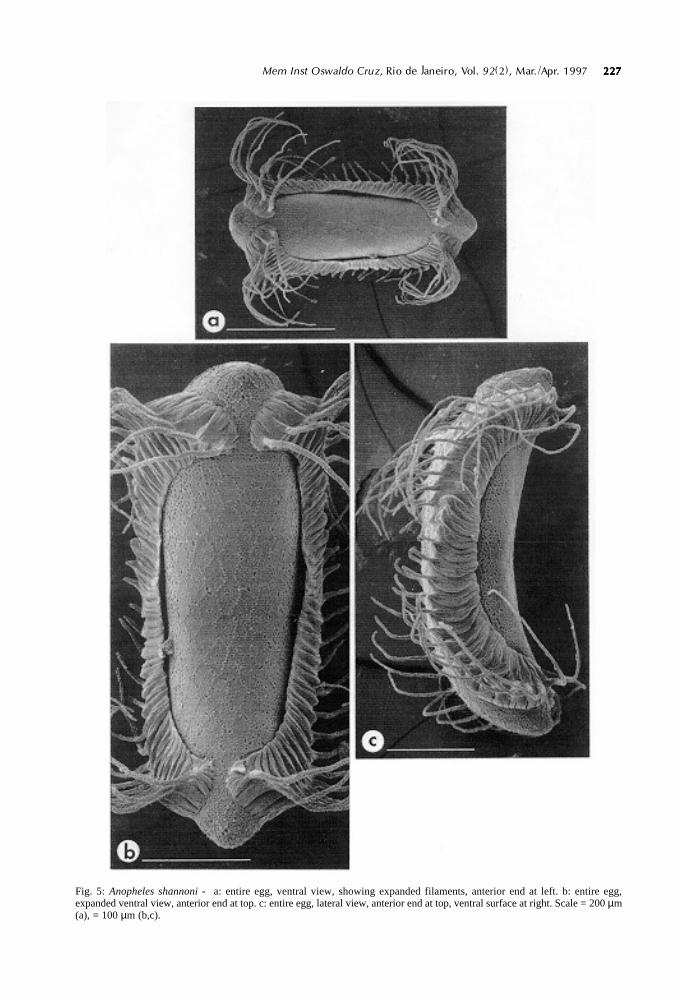
Size: egg length 450.9-493.8 µm (mean 471.8±6.9 µm, n=6), width 167.1-189.7 µm (mean 177.7 ±3.0 µm, n=6), length/width ratio 2.39-2.85 (mean2.66 ± 0.07, n=6). Color: black. Shape, overall ap-pearance: egg boat-shaped in lateral view, curved inboth dorsal and ventral surfaces (Fig. 5c), widest
226226226226226 Eggs of Brazilian Anophelines • LP Lounibos et al.
anteriorly at inception of floats, tapering slightlyposteriorly (Fig. 5a). Anterior end blunt, posteriorend slightly more conical (Fig. 5b). Deck broad andexposed, surrounded by ventral margins of floatsexcept for narrow passages to anterior and posteriorpoles (Fig. 5b). Floats elaborately developed and
highly concave, positioned closer to dorsal than ven-tral surface (Fig. 5c). Regularly spaced, filamentousprojections (range 22-27, mean 25.4 ± 0.4, n=8) ex-tend from dorsum of float throughout its length, fila-ments longer and projecting more ventrally near an-terior and posterior poles (Figs 5c, 8b,c). In speci-
Fig. 4: Anopheles fluminensis - a: ridge, ventral view. b: ridge and chorionic cells, lateral view. c: detail of chorionic cells, lateralplastron. d: lateral area between ridge and float margins. e: detail of lateral cells and float margin. f: extreme detail of tuberclesbetween float margin and chorionic cells. Scale = 50 µm (d), = 20 µm (a,b,e), = 10 µm (c), = 5 µm (f).
227227227227227Mem Inst Oswaldo Cruz, Rio de Janeiro, Vol. 92(2), Mar./Apr. 1997
Fig. 5: Anopheles shannoni - a: entire egg, ventral view, showing expanded filaments, anterior end at left. b: entire egg,expanded ventral view, anterior end at top. c: entire egg, lateral view, anterior end at top, ventral surface at right. Scale = 200 µm(a), = 100 µm (b,c).
228228228228228 Eggs of Brazilian Anophelines • LP Lounibos et al.
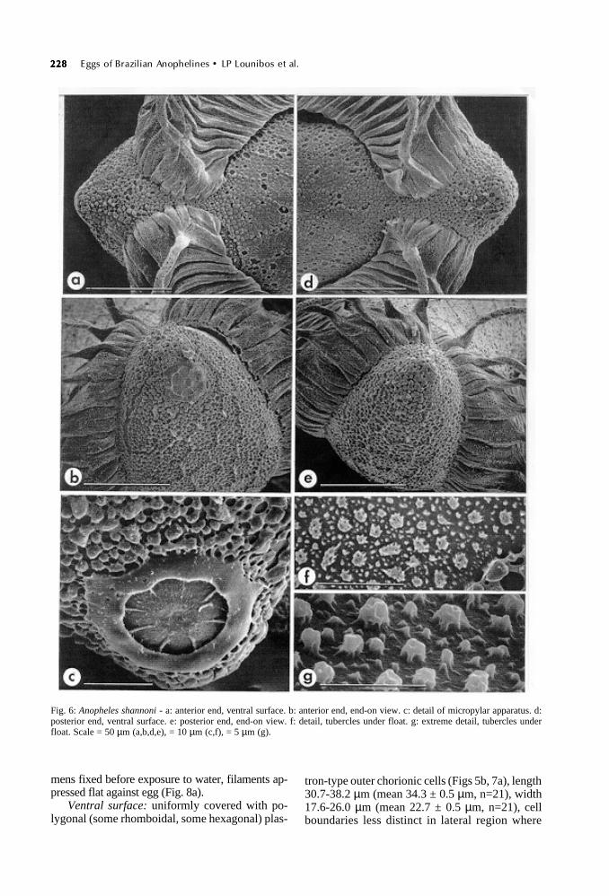
Fig. 6: Anopheles shannoni - a: anterior end, ventral surface. b: anterior end, end-on view. c: detail of micropylar apparatus. d:posterior end, ventral surface. e: posterior end, end-on view. f: detail, tubercles under float. g: extreme detail, tubercles underfloat. Scale = 50 µm (a,b,d,e), = 10 µm (c,f), = 5 µm (g).
mens fixed before exposure to water, filaments ap-pressed flat against egg (Fig. 8a).
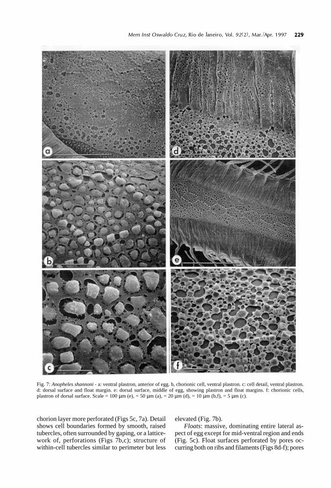
Ventral surface: uniformly covered with po-lygonal (some rhomboidal, some hexagonal) plas-
tron-type outer chorionic cells (Figs 5b, 7a), length30.7-38.2 µm (mean 34.3 ± 0.5 µm, n=21), width17.6-26.0 µm (mean 22.7 ± 0.5 µm, n=21), cellboundaries less distinct in lateral region where
229229229229229Mem Inst Oswaldo Cruz, Rio de Janeiro, Vol. 92(2), Mar./Apr. 1997
Fig. 7: Anopheles shannoni - a: ventral plastron, anterior of egg. b, chorionic cell, ventral plastron. c: cell detail, ventral plastron.d: dorsal surface and float margin. e: dorsal surface, middle of egg, showing plastron and float margins. f: chorionic cells,plastron of dorsal surface. Scale = 100 µm (e), = 50 µm (a), = 20 µm (d), = 10 µm (b,f), = 5 µm (c).
chorion layer more perforated (Figs 5c, 7a). Detailshows cell boundaries formed by smooth, raisedtubercles, often surrounded by gaping, or a lattice-work of, perforations (Figs 7b,c); structure ofwithin-cell tubercles similar to perimeter but less
elevated (Fig. 7b).Floats: massive, dominating entire lateral as-
pect of egg except for mid-ventral region and ends(Fig. 5c). Float surfaces perforated by pores oc-curring both on ribs and filaments (Figs 8d-f); pores

230230230230230 Eggs of Brazilian Anophelines • LP Lounibos et al.
Fig. 8: Anopheles shannoni - a: lateral view of float with filaments unexpanded. b: dorsal view near middle of egg, filamentsextended. c: ventral view of filament bases at anterior end. d: filament bases, dorsal view. e: detail of filament bases at anteriorend. f: detail of pores in extended filaments. Scale = 100 µm (a,b), = 50 µm (c), = 20 µm (d,e), = 10 µm (f).
less frequent on ventral margins (Figs 5b, 8a,c).Filaments originate from separate float segmentsof similar width (range 21.6-33.0 µm, mean 26.5± 0.9 µm, n=16) (Figs 8b,d). Filaments longer at
either end of egg, where they arch dorsally (Figs5c, 8b), becoming progressively shorter towardmidline (Figs 5a, 8b). Filaments tubular, diameter4.1-5.6 µm (mean 4.6 ± 0.2 µm, n=10), perfora-

231231231231231Mem Inst Oswaldo Cruz, Rio de Janeiro, Vol. 92(2), Mar./Apr. 1997
tions oval, length 0.9-1.9 µm (mean 1.3 ± 0.1 µm,n=20), width 0.7-1.6 µm (mean 1.0 ± 0.04 µm,n=20), regularly distributed over filament surface(Fig. 8f). Chorion beneath float with denselypacked tubercles of varying sizes (Fig. 6f), whichare dome-shaped with buttressed walls under highmagnification (Fig. 6g).
Anterior end, micropyle: anterior end somewhatconical, floats terminating in ventral surface be-fore pole, left and right sides separated by narrowchorion strip (Fig. 6a). Micropylar apparatus lo-cated dorsally to an agglomeration of cauliflower-like tubercles (Fig. 6b). Micropylar collar smooth,slightly scalloped on interior margin (Fig. 6c), ra-dial arms extend from collar approximately half-way into disc area. Micropylar orifice recessed inlow mound (Fig. 6c).
Posterior end: no obvious distinctions fromanterior end in ventral view, floats terminatingdorsomedially before pole (Fig. 6d). End-on viewshows cauliflower-like tubercles clustered at poleand distinctive from those forming polygonal cellsof dorsal chorion (Fig. 6e).
Dorsal surface: area covered by polygonal(mostly hexagonal) chorionic cells of length 30.3-35.9 ± µm (mean 33.1 ± 0.6 µm, n=12), width 18.5-23.4 µm (mean 19.6-0.4 µm, n=12), area occupiedby cells narrow in middle of egg owing to dorsalposition of floats (Fig. 7e). Interior of cells com-posed of rounded, slightly raised tubercles withmany round or ovoid pores exposing lower chorionlayer (Fig. 7f). No change in surface structure nearmargin with float (Fig. 7d).
DISCUSSION
The light microscopic descriptions by Causeyet al. (1944) of eggs of An. fluminensis and An.shannoni are accurate insofar as their limited reso-lution allows. These authors depict the frill of An.fluminensis “surrounding (a) wide, elliptical blackarea”, which indicates that the deck of their speci-mens was more exposed than those of the currentstudy. For An. shannoni drawn by Causey et al.(1944), the segments of floats near the midline donot give rise to filamentous projections, and thefloats are positioned more ventrally than in ourspecimens.
The egg of An. fluminensis, distinctive for itsventral ridges and dorsally positioned floats, is notlikely to be confused with that of any other de-scribed anopheline species. Among related speciesexamined with the SEM, the egg of An.mediopunctatus s.s. most resembles that of An.fluminensis. However, the central deck of An.mediopunctatus s.s. is more exposed than in An.fluminensis and its dorsal ridges are coalesced into
whorls (Lounibos et al. in preparation). Interest-ingly, the type locality of both these species issoutheastern Brazil. Examinations of eggs of themalaria vector An. sp. near fluminensis from east-ern Peru would be valuable for establishing its re-lationship to An. fluminensis from the type local-ity.
The filamentous projections of the floats of An.shannoni invite comparisons to the homologousextensions of the floats of An. peryassui (Linley &Lounibos 1994), which species is also placed inthe Arribalzagia Series. Filaments of both speciesare compressed laterally until the newly laid eggtouches water and are highly perforated, presum-ably to entrap air and assist flotation.
However, float filaments differ between An.shannoni and An. peryassui in many structural de-tails. In the former species, these extensions projectregularly from the complete length of massivefloats. In An. peryassui, filaments are confined tothe anterior and posterior ends, and the floats ofthis related species are much more slender thanthose of An. shannoni. Further, perforations onthe margins and bases of the floats are larger andmore abundant in An. peryassui. Additional fea-tures distinguish the eggs of the two species: An.shannoni eggs have no frill or crown, which occurat both ends of the An. peryassui egg [althoughCausey et al. (1944) show some An. peryassui with-out frills or crowns]; the mean egg length/widthratio is 4.0 for An. peryassui and 2.7 for An.shannoni; the ventral surface is highly concave inAn. shannoni but only slightly concave in An.peryassui.
Observations on An. peryassui eggs laid inwater led to the conclusion that the natural flota-tion position of this species is dorsal side up (Linley& Lounibos 1994). In view of the many structuralsimilarities of eggs of the two species, we antici-pate that An. shannoni eggs also float in this posi-tion, which is unique to these species of Anophe-les. The ventral deck of both species, which wouldbe submerged, is covered with porous, plastron-like chorionic cells, usually typical of dorsal sur-faces, which entrap air for respiration. The larvalhabitat of An. shannoni is reported to be filled withleaves, branches and tree trunks (Deane et al. 1948).As conjectured for eggs of An. peryassui whichalso occur in detritus-laden habitats, the filamentsof An. shannoni eggs may support their flotationby attachment to buoyant debris.
ACKNOWLEDGEMENTS
To D Couto Lima, J Conn and M Motta for theirhelp in field collections; to J Conn and R Wilkerson forcomments on a draft of this manuscript.
232232232232232 Eggs of Brazilian Anophelines • LP Lounibos et al.
REFERENCES
Bonne C, Bonne-Wepster J 1925. The Mosquitoes ofSurinam. Med Kolon Inst Amst, Trop Hyg No. 13,558 pp.
Causey OR, Deane LM, Deane MP 1944. An illustratedkey to the eggs of thirty species of Braziliananophelines with several new descriptions. Am J Hyg39: 1-7.
Cerqueira NL 1961. Distribuição geográfica dos mos-quitos da Amazônia. Rev Bras Ent 10: 111-168.
Consoli RAGB, Lourenço-de-Oliveira R 1994.Principais Mosquitos de Importância Sanitária noBrasil. Editora FIOCRUZ, Rio de Janeiro, 225 pp.
Davis NC 1931. A new anopheline mosquito from Pará,Brazil. Am J Hyg 13: 345-348.
Deane LM, Causey OR, Deane MP 1948. Notas sôbre adistribuição e a Biologia dos Anofelinos das regiõesNordestina e Amazônica do Brasil. Rev Serv Esp SaúPúb 1: 827-965.
Deane LM, Damasceno RG, Arouck R 1953.Distribuição vertical de mosquitos em uma florestados arredores de Belém, Pará. Fol Clin Biol 20: 101-110.
Falleroni D 1926. Fauna anofelica italiana e suo “habi-tat” (paludi, risaie, canali). Metodi di lotta contra lamalaria. Riv Malariol 5: 553-593.
Galvão ALA 1938. Observações sôbre algumas espéciesdo subgênero Nyssorhynchus, com especialreferência à morfologia dos ovos. Rev Biol Hyg 9:51-60.
Hackett LW, Missiroli A 1935. The varieties of Anophe-les maculipennis and their relation to the distribu-tion of malaria in Europe. Riv Malariol 14: 45-109.
Harbach RE, Knight KL 1980. Taxonomists’ Glossaryof Mosquito Anatomy. Plexus Publ. Inc., Marlton,New Jersey, xi + 413 pp.
Hayes J, Calderón G, Falcón R, Zambrano V 1987.Newly incriminated anopheline vectors of humanmalaria parasites in Junín Department, Peru. J AmMosq Control Assoc 3: 418-422.
Hinton HE 1968. Observations on the biology and tax-onomy of the eggs of Anopheles mosquitoes. BullEnt Res 57: 495-508.
Kumm HW 1941. The eggs of some Costa Ricaanophelines. Am J Trop Med 21: 91-98.
Linely JR, Lounibos LP 1993. The eggs of Anopheles(Nyssorhynchus) rangeli and Anopheles(Nyssorhynchus) dunhami (Diptera: Culicidae).Mosq Syst 25: 157-169.
Linley JR, Lounibos LP 1994. The remarkable egg ofAnopheles peryassui (Diptera: Culicidae). Mosq Syst26: 25-34.
Linley JR, Milstrey EG 1995.The eggs of Anopheles(Anopheles) mattogrossensis and Chagasia fajardi(Diptera: Culicidae). Mosq Syst 27: 27-39.
Linley JR, Kaiser PE, Cockburn AF 1993. A descrip-tion and morphometric study of the eggs of speciesof the Anopheles quadrimaculatus Complex(Diptera: Culicidae). Mosq Syst 25: 124-147.
Linley JR, Lounibos LP, Conn J, Duzak D, NishimuraN 1996. A description and morphometric compari-son of eggs from eight geographic populations ofthe South American malaria vector Anopheles(Nyssorhynchus) nuneztovari (Diptera: Culicidae).J Am Mosq Control Assoc 12: 275-292.
Lourenço-de-Oliveira R, Luz SLB 1996. Simian malariaat two sites in the Brazilian Amazon - II. Verticaldistribution and frequency of anopheline species in-side and outside the forest. Mem Inst Oswaldo Cruz91: 687-694.
Reid JA, Knight KL 1961. Classification within the sub-genus Anopheles (Diptera, Culicidae). Ann Trop MedParasit 55: 474-488.
Rodriguez MH, Chavez B, Orozco A, Loyola EG,Martinez-Palomo A 1992. Scanning electron micro-scopic observations of Anopheles albimanus(Diptera: Culicidae) eggs. J Med Ent 29: 400-406.
Root FM 1927. Studies on Brazilian mosquitoes. IV.Notes on some Brazilian species of Anopheles. AmJ Hyg 7: 599-605.
Rozeboom LE 1938. The eggs of the Nyssorhynchusgroup of Anopheles (Culicidae) in Panama. Am JHyg 27: 95-107.
Rozeboom LE 1942. Subspecific variations amongneotropical Anopheles mosquitoes and their impor-tance in the transmission of malaria. Am J Trop Med22: 235-255.
Wilkerson RC, Peyton EL 1990. Standardized nomen-clature for the costal wing spots of the genus Anophe-les and other spotted-wing mosquitoes (Diptera:Culicidae). J Med Ent 27: 207-224.