Programa de Psiquiatria Orientador: Prof. Dr. Rodrigo ......Anatomia da mitocôndria e a cadeia...
138
RAFAEL TEIXEIRA DE SOUSA Fisiopatologia do Transtorno de Humor Bipolar e efeito do tratamento com lítio: enfoque em neuroproteção e função mitocondrial Programa de Psiquiatria Orientador: Prof. Dr. Rodrigo Machado-Vieira São Paulo - 2014 -
Transcript of Programa de Psiquiatria Orientador: Prof. Dr. Rodrigo ......Anatomia da mitocôndria e a cadeia...

RAFAEL TEIXEIRA DE SOUSA
Fisiopatologia do Transtorno de Humor Bipolar e efeito do
tratamento com lítio: enfoque em neuroproteção e função
mitocondrial
Programa de Psiquiatria
Orientador: Prof. Dr. Rodrigo Machado-Vieira
São Paulo
- 2014 -

Dados Internacionais de Catalogação na Publicação (CIP)
Preparada pela Biblioteca da Faculdade de Medicina da Universidade de São Paulo
©reprodução autorizada pelo autor
Sousa, Rafael Teixeira de Fisiopatologia do Transtorno de Humor Bipolar e efeito do tratamento com lítio : enfoque em neuroproteção e função mitocondrial / Rafael Teixeira de Sousa. -- São Paulo, 2014.
Tese(doutorado)--Faculdade de Medicina da Universidade de São Paulo. Programa de Psiquiatria.
Orientador: Rodrigo Machado-Vieira. Descritores: 1.Transtorno bipolar 2.Mitocôndrias 3.Lítio 4.Estresse oxidativo
5.Complexo de proteínas da cadeia de transporte de elétrons 6.Ciclo do ácido cítrico 7.DNA mitocondrial 8.Óxido nítrico 9.Efeito neuroprotetor
USP/FM/DBD-042/14

ii
Dedicatória
Dedico esta tese aos meus pais, que representam a minha origem e foram a minha
grande influência nas minhas escolhas.
Dedico também esta tese à minha irmã e ao meu irmão, com quem dividi a maior
parte da minha vida.
Dedico a tese ao meu cunhado, que já é um irmão.
E por fim, dedico a tese aos amigos que dividiram tanto tempo comigo que já são
da minha família.

iii
Agradecimentos
Agradeço aos meus pais por tudo o que fizeram por mim e, em especial, pelos valores que me
incutiram; sem estes valores eu não teria ganas de almejar a mais na minha vida.
Agradeço também aos meus irmãos e ao meu cunhado pelo companheirismo e apoio, que são a
base para que se possa lutar.
Agradeço muito ao meu orientador Prof. Dr. Rodrigo Machado-Vieira por ter sido amigo e por
ter sido duro comigo, tentando tirar de mim o melhor.
Agradeço ao Prof. Wagner F. Gattaz por proporcionar um ambiente de alto nível científico no
LIM27, por apoiar o meu projeto, confiar no meu trabalho e por todo o auxílio que me deu.
Agradeço ao Prof. Dr. Marcus Zanetti pelo suporte científico que me deu inúmeras vezes, sendo
quase um coorientador.
Agradeço a toda a equipe do LIM27 pela amizade, pelo companheirismo e pelo trabalho
eficiente, que permitiu a execução do meu projeto.
Agradeço à Profa. Dra. Elisa Higa e à equipe do Laboratório de Óxido Nítrico da Unifesp pela
frutuosa colaboração.

iv
Agradeço ao Prof. Dr. Emílio Streck e à equipe do Laboratório de Bioenergética da Unesc pelo
produtivo trabalho juntos.
Agradeço à Profa. Dra. Suely K. N. Marie e aos colegas do LIM15 pelo profícuo trabalho em
conjunto.
Agradeço ao Prof. Dr. Geraldo Busatto e à equipe do LIM21, que trabalhou conosco com
grande companheirismo e eficácia.
Agradeço à Fapesp pelo auxílio que financiou o projeto e suas dosagens.
Agradeço à Universidade de São Paulo e ao Departamento de Psiquiatria da FMUSP pelo apoio
na divulgação dos meus resultados.
Agradeço a tantas pessoas que não vou poder citar aqui textualmente, mas que fizeram muita
diferença com seus conselhos, com o seu apoio e com os horizontes que me descortinaram.
Ainda que de forma indireta, esta ajuda foi certamente decisiva para que conseguisse concluir o
meu trabalho.

v
Privados de um trabalho significativo, homens e mulheres perdem a sua razão para existir
Dostoievski

vi
NORMALIZAÇÃO ADOTADA
publicaç
adaptado de International Committee of Medical Journals Editors (Vancouver).
Documentaç
Elaborado por Anneliese Carneiro
da Cunha, Maria Julia de A. L. Freddi, Maria F. Crestana, Marinalva d
2011.
List of Journals Indexed in Index
Medicus.

vii
Sumário
LISTA DE FIGURAS .................................................................................................................. x
LISTA DE TABELAS ............................................................................................................... xi
LISTA DE SIGLAS E SÍMBOLOS ........................................................................................ xii
RESUMO .................................................................................................................................. xiii
ABSTRACT ................................................................................................................................ xv
1. INTRODUÇÃO ........................................................................................................................ 1
1.1. A mitocôndria .................................................................................................................... 2
1.2. Função mitocondrial no THB .......................................................................................... 8
1.3. Alterações de metabolismo energético em estudos de neuroimagem no THB ............ 9
1.4. Estresse oxidativo no THB ............................................................................................. 12
1.5. O estresse oxidativo está associado a alterações em neurotransmissores no THB ... 15
1.6. Associação do THB com genes mitocondriais e sua expressão diferencial no
transtorno ............................................................................................................................... 16
2. JUSTIFICATIVA .................................................................................................................. 19
2.1. Neuroprogressão no THB e a investigação de biomarcadores ................................... 19
2.2. Subtipo e fase no THB .................................................................................................... 21
3. OBJETIVOS E HIPÓTESES ............................................................................................... 22
3.1. Objetivos gerais ............................................................................................................... 22
3.2. Objetivos específicos ....................................................................................................... 22
viii
3.3. Hipóteses .......................................................................................................................... 23
4. MÉTODOS ............................................................................................................................. 26
4.1. Seleção da amostra .......................................................................................................... 26
4.2. Desenho do estudo ........................................................................................................... 27
4.3. Análises laboratoriais ..................................................................................................... 28
4.3.1. Colaborações e participação do candidato ao doutoramento em ensaios .................. 28
4.3.2. Dados específicos dos ensaios laboratoriais .............................................................. 29
4.3.3. Atividade dos complexos I, II, II-III e IV e das enzimas citrato sintase, malato
desidrogenase e succinato desidrogenase ............................................................................ 30
4.3.4. Atividade das enzimas citrato sintase, malato desidrogenase e succinato
desidrogenase ....................................................................................................................... 31
4.3.5. Avaliação de parâmetros de estresse oxidativo ......................................................... 32
4.3.6. Avaliação do conteúdo de DNAmt ............................................................................ 34
4.4. Análise estatística de dados demográficos e clínicos .................................................... 36
5. RESULTADOS ...................................................................................................................... 37
5.1. Descrição da amostra e dados clínicos .......................................................................... 37
5.2. Resultados publicados .................................................................................................... 39
5.3. Resultados submetidos .................................................................................................... 40
5.4. Resultado a ser submetido: Atividade das Enzimas do Ciclo do Ácido cítrico no
THB e o Efeito do Tratamento com Lítio ............................................................................ 41
5.4.1. Justificativa ................................................................................................................ 41
5.4.2. Análise estatística....................................................................................................... 42
5.4.3. Resultados .................................................................................................................. 42
5.4.4. Discussão ................................................................................................................... 45

ix
6. ANÁLISE CRÍTICA DOS ACHADOS ............................................................................... 47
6.1. Neurobiologia do THB .................................................................................................... 47
6.2. Efeito do tratamento com lítio ....................................................................................... 50
6.3. Limitações e pontos fortes .............................................................................................. 53
6.4. Conclusão e perspectivas ................................................................................................ 53
7. ANEXOS ................................................................................................................................. 55
7.1. Artigos publicados .......................................................................................................... 55
7.1.1. Leukocyte mitochondrial DNA copy number in bipolar disorder ............................. 55
7.1.2. Oxidative stress in early stage Bipolar Disorder and the association with response to
lithium .................................................................................................................................. 60
7.2. Artigos submetidos ......................................................................................................... 67
7.2.1. Lithium Increases Nitric Oxide Levels in Subjects with Bipolar Disorder during
Depressive Episodes ............................................................................................................ 67
7.2.2. Lithium Increases Leukocyte Mitochondrial Complex I in Short-Term Bipolar
Disorder................................................................................................................................ 82
8. REFERÊNCIAS ................................................................................................................... 102

x
LISTA DE FIGURAS
Figura 1. Anatomia da mitocôndria e a cadeia transportadora de elétrons: os complexos I-
V são parte da membrana mitocondrial interna. O NADH e o FADH2 doam elétrons para
os complexos I e II, respectivamente. A cada passagem de elétrons de um transportador
para o outro, há liberação de energia e é gerado um gradiente eletroquímico através da
membrana mitocondrial interna. Através do complexo V os íons de hidrogênio passam de
volta para a matriz, produzindo ATPs. Cada complexo compreende várias proteínas.
(Adaptado: Clay et al., 2011). ..................................................................................................... 3
Figura 2. Via aeróbica e via anaeróbica de produção de energia. A via anaeróbica (de
baixa produção energética) compreende a quebra de glicose em piruvato, que é
transformado em lactato no citosol. Na via aeróbica (de alta produção energética) o
piruvato derivado da glicose entra na mitocôndria, onde é transformado em acetil-
coenzima A (acetil-CoA), que vai ser transformada em citrato pela enzima citrato sintase
(CS). Em seguida a Acetil-CoA vai se converter em α-cetoglutarato e depois em succinil-
coenzima A (succinil-coA) e então succinato. O succinato será transformado em fumarato
pela succinato desidrogenase (SDH), tornando-se depois malato. O malato será mudado
em oxaloacetato com o auxílio da malato desidrogenase (MDH). ........................................... 6
Figura 3. Estresse oxidativo e enzimas antioxidantes. O radical superóxido (O2-
) é
convertido em peróxido de hidrogênio (H2O2) pela enzima superóxido dismutase (SOD). O
H2O2 é transformado em H2O+1/2O2 pela ação das enzimas catalase (CAT) e glutationa
peroxidase (GPx). O óxido nítrico (NO) é transformado em peroxinitrito (ONOO-).
ONOO- e H2O2 causam dano oxidativo, refletido na peroxidação lipídica com aumento de
substâncias reativas ao ácido tiobarbitúrico (TBARS). ......................................................... 14
Figura 4. Atividades das enzimas citrato sintase, malato desidrogenase e succinato
desidrogenase em pacientes com Transtorno de Humor Bipolar I (THB I) e THB II
comparados com controles saudáveis. *p<0,05. ...................................................................... 44
Figura 5. Modelo neurobiológico proposto para o Transtorno de Humor Bipolar de início
recente. A fase inicial do THB é marcada por mecanismos compensatórios como o
aumento de atividade da catalase e da glutationa peroxidase, que diminuem o estresse
oxidativo e preservam as atividades da cadeia transportadora de elétrons e das enzimas do
ciclo do ácido cítrico e o número de mitocôndrias (indicado pelo conteúdo de DNA
mitocondrial e pela atividade da citrato sintase). ................................................................... 49
Figura 6. Modelo proposto para a ação neuroprotetora do lítio no Transtorno de Humor
Bipolar (THB). O lítio aumenta a atividade da cadeia transportadora de elétrons e
diminui a atividade da superóxido dismutase no THB, gerando assim diminuição do
estresse oxidativo. O lítio também aumenta os níveis do neuromodulador óxido nítrico. .. 51

xi
LISTA DE TABELAS
Tabela 1. Características clínicas e demográficas de pacientes em episódio depressivo de
Transtorno de Humor Bipolar (THB) e controles saudáveis. ................................................ 38
Tabela 2. Atividades das enzimas citrato sintase, malato desidrogenase e succinato
desidrogenase em pacientes com depressão bipolar antes e depois do tratamento em
comparação com controles saudáveis. ...................................................................................... 43

xii
LISTA DE SIGLAS E SÍMBOLOS
CAT - catalase
CGI - Impressão Clínica Global
DNAmt - DNA mitocondrial
DSM-IV - 4º Manual Diagnóstico e Estatístico de Transtornos Mentais
ERM - Espectroscopia por Ressonância Magnética
GPx - glutationa peroxidase
H2O2 - peróxido de hidrogênio
NO - óxido nítrico
O2-
- radical superóxido
ONOO- - peroxinitrito
SCID - Entrevista Clínica Estruturada para Transtornos do DSM-IV
SOD - superóxido dismutase
TBARS - substâncias reativas ao ácido tiobarbitúrico
THB - Transtorno de Humor Bipolar

xiii
RESUMO
de Sousa, R. T. Fisiopatologia do Transtorno de Humor Bipolar e efeito do tratamento com
lítio: enfoque em neuroproteção e função mitocondrial [Tese]. São Paulo: Faculdade de
Medicina, Universidade de São Paulo; 2014
Introdução: Diversas evidências apontam para um papel da disfunção mitocondrial no
Transtorno de Humor Bipolar (THB), mas pouco se sabe sobre isso no THB de início recente.
Na mitocôndria a atividade da cadeia transportadora de elétrons (CTE) atua juntamente com o
ciclo do ácido cítrico na produção de energia, mas não está claro se estão alteradas no THB. O
DNA mitocondrial (DNAmt) codifica diversas proteínas da CTE e está associado ao estresse
oxidativo, mas nunca foi avaliado em pacientes no THB in vivo. O estresse oxidativo está
associado ao THB e à disfunção mitocondrial, mas não se sabe muito das atividades das
enzimas antioxidantes no THB de início recente. O óxido nítrico (NO) é uma molécula com
efeitos neuromoduladores, mas com um papel no THB ainda não elucidado. O lítio é um
tratamento padrão-ouro no THB, tendo mostrado efeitos neuroprotetores. Apesar disso, pouco
se conhece do efeito do lítio na CTE, nas enzimas do ciclo do ácido cítrico, no conteúdo de
DNAmt e na regulação de NO em humanos. Também não está claro o papel antioxidante do
lítio no THB. Metódos: Pacientes com THB em depressão (n=31), não medicados em sua
maioria (84%), foram tratados por 6 semanas com lítio. Antes e depois do tratamento,
verificaram-se em leucócitos as atividades dos complexos I-IV da CTE, atividades das enzimas
citrato sintase, succinato desidrogenase e malato desidrogenase e também o conteúdo de
DNAmt; em plasma foram analisados os níveis de NO, substâncias reativas ao ácido
tiobarbitúrico (TBARS) e as atividades de catalase (CAT), glutationa peroxidase (GPx),
superóxido dismutase (SOD) e razão de SOD/CAT. Os pacientes com depressão bipolar foram
comparados com 28 controles saudáveis. Resultados: Em comparação com controles, os
pacientes com THB tiveram um aumento de GPx (p<0,001) e CAT (p=0,005) e uma diminuição
de SOD/CAT (p=0,001), sem outras diferenças nos demais biomarcadores. Pacientes com THB
I mostraram uma diminuição de citrato sintase (p=0,02) e uma discreta diminuição do conteúdo
de DNAmt (p=0,05) em comparação com o THB II; o conteúdo de DNAmt esteve ligeiramente
diminuído no THB I comparado com controles (p=0,05). Do início ao fim do tratamento com
lítio houve aumento da atividade do complexo I da CTE (p=0,02), diminuição de TBARS
(p=0,02) e SOD (p=0,03) e aumento de NO (p=0,02), sem haver alteração de outros parâmetros.
Depois do tratamento, o TBARS se mostrou diminuído em respondedores comparados a não
respondedores (p=0,02) e diminuído no THB II em comparação com o THB I (p=0,04).
Discussão: No THB de início recente, houve poucas alterações em biomarcadores. Os achados
sugerem aumento de CAT e GPx na depressão bipolar de início recente e uma diminuição de
conteúdo mitocondrial no THB I comparado com o THB II, que devem ser confirmadas por
outros estudos. Os resultados reforçam um papel neuroprotetor do lítio, sugerindo que a droga
aumente a atividade do complexo I da CTE mitocondrial e aumente os níveis de NO na
depressão bipolar. Além disso, o lítio reforçou o seu papel antioxidante e modulador das
enzimas antioxidantes no THB.

xiv
Descritores: 1) Transtorno Bipolar 2) Mitocôndrias 3) Lítio 4) Estresse Oxidativo 5) Complexo
de Proteínas da Cadeia de Transporte de Elétrons 6) Ciclo do Ácido Cítrico 7) DNA
Mitocondrial 8) Óxido Nítrico 9) Fármacos Neuroprotetores

xv
ABSTRACT
de Sousa, R. T. Bipolar Disorder pathophysiology and the effect of lithium treatment: focus on
neuroprotection and mitochondrial function [Thesis]. São Paulo: Faculdade de Medicina,
Universidade de São Paulo; 2014
Background: Several evidences point to a role for mitochondrial dysfunction in Bipolar
Disorder (BD), but few is known about it on short-term BD. In mitochondria the electron
transport chain (ETC) acts jointly with citric acid cycle to produce energy, but it is not clear if
they are altered in BD. Mitochondrial DNA (mtDNA) encodes several ETC proteins and is
associated with oxidative stress, but it was never evaluated in BD in vivo. Oxidative stress is
associated with BD and with mitochondrial dysfunction, but few is known about the activities
of antioxidant enzymes in short-term BD. Nitric oxide (NO) is a molecule with
neuromodulatory effects, but with an unclear role in BD. Lithium is a gold-standard treatment
for BD, which has shown neuroprotective effects. However, few is known about lithium effect
on ETC, citric acid cycle, mtDNA content, and NO regulation in humans. Also, lithium's
antioxidant role in BD is unclear. Methods: Patients with BD depression (n=31) unmedicated
in majority (84%) received lithium treatment for 6 weeks. Before and after treatment, in
leukocytes the activities of ETC complex I-IV, citrate synthase, succinate dehydrogenase, and
malate dehydrogenase, and mtDNA content were evaluated; in plasma, NO levels,
thiobarbituric acid reactive substances (TBARS), the activities of catalase (CAT), glutathione
peroxidase (GPx), and superoxide dismutase (SOD), and SOD/CAT ratio were evaluated.
Bipolar depression patients were compared with 28 healthy controls. Results: When compared
with controls, BD patients showed an increase in GPx (p<0.001) and CAT (p=0.005) and a
decrease in SOD/CAT (p=0.001), but showed no difference for other biomarkers. Patients with
BD I showed a decrease in citrate synthase (p=0.02) and a slight decrease in mtDNA content
(p=0.05) when compared to BD II; mtDNA content was slightly decreased in BD I compared to
controls (p=0.05). From baseline to endpoint, there was an increase in ETC complex I activity
(p=0.02), a decrease in TBARS (p=0.02) and SOD (p=0.03) and an increase in NO (p=0.02),
without change in other parameters. After treatment, TBARS was decreased in responders
compared to non-responders (p=0.02) and decreased in BD II compared to BD I (p=0.04).
Discussion: In short-term BD few alterations were observed on biomarkers. The findings
suggest increase on CAT and GPX in short-term bipolar depression and mitochondrial content
decrease in BD I when compared to BD II, which deserve other studies for confirmation. The
results reinforce a lithium's neuroprotective role and suggest that lithium increases ETC
complex I activity and NO levels in bipolar depression. Moreover, lithium reinforced its role as
antioxidant and as a modulator of antioxidant enzymes in BD.
Descriptors: 1) Bipolar Disorder 2) Mitochondria 3) Lithium 4) Oxidative stress 5) Electron
Transport Chain Complex Proteins 6) Citric acid cycle 7) Mitochondrial DNA 8) Nitric Oxide
9) Neuroprotective Agents

1
1. INTRODUÇÃO
O Transtorno de Humor Bipolar (THB) é um distúrbio mental grave e crônico, que
muitas vezes põe em risco a vida dos acometidos. Apesar da sua gravidade e da sua incidência
em cerca de 2% na população (Merikangas et al., 2007), os mecanismos fisiopatológicos que
estão por trás da clínica estão longe de estar elucidados, o que limita o desenvolvimento de
novos tratamentos.
Ainda que o THB não seja uma doença neurodegenerativa típica, diversos estudos têm
mostrado associação do THB com estresse celular e morte de células da glia e neurônios
(Machado-Vieira et al., 2009). Os achados em cérebros post-mortem no THB mostram
diminuição volumétrica de áreas reguladoras do humor, como o córtex pré-frontal, o córtex
cingulado anterior e a amígdala (Rajkowska et al., 2001). Ongur et al. (1998) encontraram uma
diminuição de 41% de células da glia no córtex pré-frontal em pacientes com THB que tinham
história familiar de transtornos de humor. Para alguns tipos específicos de neurônios analisados
no THB, encontrou-se redução de densidade neuronal em córtex pré-frontal (Rajkowska et al.,
2001) e em córtex cingulado anterior (Benes et al., 2001).
Os estudos de neuroimagem também corroboram estes achados, mostrando alterações de
volume e estruturais em áreas relacionadas ao controle do humor no THB. Diversas alterações
morfométricas foram descritas no THB, entre elas reduções de volume de córtex pré-frontal,
cíngulo anterior, tálamo, núcleo caudado e putâmen, amígdala e hipocampo (Beyer & Krishnan,
2002; Sheline, 2003).
Em regiões importantes para a regulação de humor, observaram-se ainda alterações de
níveis de N-acetil-aspartato, colina e mio-Inositol através de Espectroscopia por Ressonância
Magnética (ERM) de prótons (Stork & Renshaw, 2005). Estes marcadores têm relação com

2
neurotrofismo (Clark, 1998; Govindaraju et al., 2000; Silverstone et al., 2005) e também estão
ligados ao controle do metabolismo energético (Stork & Renshaw, 2005).
Na tentativa de compreender as alterações presentes no THB, inicialmente o foco das
pesquisas foi a desregulação de neurotransmissores, porém logo passando a ser a sinalização
intracelular (Hahn & Friedman, 1999; Gould & Manji, 2002); mais recentemente, o
metabolismo energético mitocondrial, o estresse oxidativo e a neurogênese tem sido alvo do
enfoque nos estudos em THB (Berk et al., 2011). Além disso, levantou-se a hipótese de que a
perda de suporte neurotrófico, o estresse oxidativo e a inflamação estejam associados a estádios
mais avançados do THB, ocorrendo progressivamente (Berk et al., 2013). Neste panorama, a
função mitocondrial e a neuroproteção são particularmente relevantes no THB.
1.1. A mitocôndria
A mitocôndria é uma organela composta por uma membrana externa e uma interna,
sendo o espaço entre ambas denominado espaço intermembranoso (Figura 1). Na parte interior
existe a matriz mitocondrial, onde acontece o ciclo do ácido cítrico.
A mitocôndria é a única organela celular que contém DNA próprio, codificando 37
genes ligados à produção de RNAs transportadores, RNAs ribossomais e proteínas da cadeia
transportadora de elétrons. Apesar disso, a maior parte das proteínas mitocondriais é codificada
pelo DNA nuclear. O DNA mitocondrial (DNAmt) é herdado quase exclusivamente da mãe, já
que o gameta feminino contém muito mais citoplasma que o masculino. O padrão de herança
materno encontrado por alguns estudos no THB (McMahon et al., 1995; Stine et al., 1995) é
também uma evidência a favor do envolvimento da organela no transtorno.

3
Figura 1. Anatomia da mitocôndria e a cadeia transportadora de elétrons: os complexos I-
V são parte da membrana mitocondrial interna. O NADH e o FADH2 doam elétrons para
os complexos I e II, respectivamente. A cada passagem de elétrons de um transportador
para o outro, há liberação de energia e é gerado um gradiente eletroquímico através da
membrana mitocondrial interna. Através do complexo V os íons de hidrogênio passam de
volta para a matriz, produzindo ATPs. Cada complexo compreende várias proteínas.
(Adaptado: Clay et al., 2011).

4
Outra característica particular da mitocôndria é a heteroplasmia. Células diferentes têm
números e tipos diferentes de mitocôndrias. Daí a disfunção mitocondrial ser capaz de se
manifestar de uma maneira muito específica ou regional (Quiroz et al., 2008), podendo, por
exemplo, acometer só o sistema nervoso central. O DNAmt tem variação do número de
moléculas entre os diferentes tecidos (Clay Montier et al., 2009). O conteúdo de DNAmt
(medido pelo número de cópias de DNAmt) é conhecido por refletir a estabilidade dos genes
mitocondriais e o número de mitocôndrias (Malik & Czajka, 2013), que é essencial para o bom
funcionamento celular (Clay Montier et al., 2009). Há evidência ligando a diminuição de
conteúdo de DNAmt à depressão (Kim et al., 2011) e à pior cognição em idosos (Lee et al.,
2010), porém nenhum estudo avaliou o conteúdo de DNAmt in vivo em pacientes com THB.
A mitocôndria tem função de conversão da energia de açúcares em ATPs. Tal processo
se dá com o auxílio de enzimas do ciclo do ácido cítrico. Inicialmente acontece a glicólise,
quebra de glicose em piruvato (Figura 2). Na via anaeróbica, a produção de piruvato dá origem
ao lactato, produzindo pouca energia. Na via aeróbica, de grande produção energética, o
piruvato derivado da glicose é transformado em acetil-coenzima A que entra no ciclo do ácido
cítrico. Junto com o oxaloacetato, a acetil-coenzima A vai ser transformada em citrato pela
enzima citrato sintase. Em seguida vai se converter α-cetoglutarato e depois em succinil-
coenzima A e então succinato. O succinato será transformado em fumarato pela succinato
desidrogenase, tornando-se depois malato. O malato será mudado em oxaloacetato com o
auxílio da malato desidrogenase. Apesar da relevância das enzimas do ciclo do ácido cítrico na
produção de energia, nenhum estudo avaliou a atividade das enzimas em células periféricas no
THB.
No ciclo do ácido cítrico são produzidos NADH2 e FADH, que transferem elétrons para
a cadeia transportadora de elétrons. O NADH+H transfere seus elétrons para o complexo I da

5
cadeia transportadora de elétrons (ou cadeia respiratória) (Figura 1). Na matriz mitocondrial
ocorre produção de succinato, que leva elétrons para o complexo II da cadeia. Os elétrons
passam dos complexos I ou II para a coenzima Q, seguindo para o complexo III. Indo para o
complexo IV, os elétrons se combinam com O2 para produzir H2O. A cada passagem de elétrons
de um transportador para o outro, há liberação de energia e é gerado um gradiente eletroquímico
através da membrana mitocondrial interna. Através do complexo V os íons de hidrogênio
passam de volta para a matriz, produzindo ATPs.
Encontrou-se menor expressão de genes que codificavam proteínas relacionadas aos
complexos I, III, IV e V em cérebros de pacientes com THB (Konradi et al., 2004; Sun et al.,
2006). O achado indica acometimento da cadeia transportadora de elétrons, com provável
impacto na produção de energia por ATPs. O lítio, por sua vez, aumentou a expressão do
complexo I em cérebros de pacientes com THB (Sun et al., 2006), sugerindo assim uma ação
neuroprotetora da droga através da melhora do metabolismo energético.

6
Figura 2. Via aeróbica e via anaeróbica de produção de energia. A via anaeróbica (de
baixa produção energética) compreende a quebra de glicose em piruvato, que é
transformado em lactato no citosol. Na via aeróbica (de alta produção energética) o
piruvato derivado da glicose entra na mitocôndria, onde é transformado em acetil-
coenzima A (acetil-CoA), que vai ser transformada em citrato pela enzima citrato sintase
(CS). Em seguida a Acetil-CoA vai se converter em α-cetoglutarato e depois em succinil-
coenzima A (succinil-coA) e então succinato. O succinato será transformado em fumarato
pela succinato desidrogenase (SDH), tornando-se depois malato. O malato será mudado
em oxaloacetato com o auxílio da malato desidrogenase (MDH).

7
Em células periféricas, a atividade da cadeia transportadora de elétrons está alterada em
transtornos psiquiátricos como o autismo (Giulivi et al., 2010) e a esquizofrenia (Dror et al.,
2002). Devido à facilidade de avaliação, a atividade de complexo I foi proposta como marcador
de estado na esquizofrenia (Dror et al., 2002). Só dois estudos em células periféricas avaliaram
o potencial da atividade dos complexos da cadeia transportadora de elétrons no THB, obtendo
resultados negativos, mas contando com amostras bastante pequenas (n=10 e n=12,
respectivamente) (Ben-Shachar et al., 1999; Gubert et al., 2013).
A atividade da cadeia transportadora de elétrons culmina na produção de espécies
reativas de oxigênio. Se a produção de espécies reativas de oxigênio é excessiva ou se a sua
eliminação é ineficiente, as espécies reativas de oxigênio vão causar estresse oxidativo. O
estresse oxidativo é nocivo para a célula, estando ligado ao dano na transdução de sinal, na
plasticidade estrutural e na resiliência (Massaad & Klann, 2011). A fosforilação oxidativa
inadequada leva à deficiência na produção de ATPs, podendo se refletir no potencial de
membrana celular, que ficaria alterado porque as bombas de cálcio não teriam energia para
controlar o fluxo do íon.
A atuação da mitocôndria na homeostase do cálcio intracelular não se restringe à
produção de energia, mas compreende também o tamponamento do cálcio citosólico,
prevenindo que altos níveis do íon no citosol induzam estresse e excitotoxicidade (Baron et al.,
2003; Rizzuto & Pozzan, 2006). Níveis aumentados de cálcio basal intracelular (potencialmente
tóxicos) foram observados em plaquetas e linfócitos no THB (Hough et al., 1999). Além disso,
houve maiores elevações de cálcio em resposta a estímulo com diversas substâncias em células
periféricas no THB (Dubovsky et al., 1989; Kusumi et al., 1992; Berk et al., 1994; Hough et
al., 1999; Suzuki et al., 2001), sendo o lítio capaz de regular a resposta de cálcio à provocação
(Wasserman et al., 2004). As evidências tornam provável que o desequilíbrio no cálcio

8
intracelular visto no THB possa estar relacionado à atividade mitocondrial alterada (Kato,
2008).
Agressões celulares como excesso de influxo de cálcio ou potencial de membrana
mitocondrial diminuído podem levar à maior permeabilidade da membrana mitocondrial.
Freqüentemente isso se dá através da abertura do poro de transição de permeabilidade
mitocondrial. O poro de transição de permeabilidade mitocondrial é um complexo de proteína
que une o interior e o exterior da membrana mitocondrial. A sua abertura libera proteínas
mitocondriais como o citocromo C e pró-caspases, que são facilitadoras de apoptose (Hotchkiss
et al., 2009; Machado-Vieira et al., 2009). Os mecanismos pró-apoptóticos deflagrados podem
ser muito nocivos mesmo se não causam morte celular, pois estão ligados à perda de sinapses
(ou "apoptose sináptica") (Quiroz et al., 2008).
O THB está longe de ser considerado uma doença mitocondrial clássica, pois não
apresenta os acometimentos típicos destas doenças (e.g., atrofia de retina, ataxia, miopatia,
intolerância ao exercício físico, etc.). As evidências, porém, apontam para disfunções sutis
ligadas a níveis mais básicos, provavelmente de codificação nuclear.
1.2. Função mitocondrial no THB
A mitocôndria tem função crucial de resiliência celular e manejo do estresse. Este papel
é importante, já que o estresse celular pode ativar cascatas de apoptose. Quando estas não levam
à morte celular dos neurônios, podem induzir atrofia de sinapses e neurites, mecanismos ligados
à fisiopatogenia do THB (Quiroz et al., 2008). Diversos fatores que determinam a sobrevivência
e o crescimento neuronal são afetados pelo lítio (Machado-Vieira et al., 2009), droga padrão-
ouro no tratamento do THB.

9
Muitas evidências apontam uma associação entre disfunção mitocondrial e THB. É bem
conhecido o papel fundamental que a mitocôndria tem na produção de energia celular. No THB,
o metabolismo de energia mostrou anormalidades tanto pelo aumento de lactato nos estudos
avaliando líquor (Regenold et al., 2009), como nos estudos de ressonância magnética com
espectroscopia (Stork & Renshaw, 2005; Frey et al., 2007; Ongür et al., 2009), sugerindo
comprometimento mitocondrial. Pesquisas enfocando a expressão de genes e proteínas no THB
mostram uma diminuição de fatores e enzimas envolvidos na geração e estoque de ATP
(Konradi et al., 2004; Iwamoto et al., 2005; MacDonald et al., 2006; Pennington et al., 2008).
Além disso, a associação do THB com genes de função mitocondrial está referenciada
também por estudos genéticos (Shao et al., 2008; Xu et al., 2008; Rollins et al., 2009; Zhang et
al., 2009). Reforçando a relevância destes achados, há relatos de doenças classicamente
mitocondriais com apresentação semelhante ao THB (Grover et al., 2006; Mancuso et al.,
2008). Esta semelhança também sugere que a fisiopatogenia do THB possa estar vinculada à
mitocôndria.
1.3. Alterações de metabolismo energético em estudos de neuroimagem no THB
Espectroscopia por Ressonância Magnética (ERM) é uma técnica que permite a
visualização da química cerebral in vivo, incluindo metabólitos relacionados com energia, pH e
compostos do metabolismo de fosfolípides. Estudos com ERM de prótons (1H) encontraram
níveis anormais de N-acetil-aspartato, glutamato/glutamina, lactato, compostos contendo colina
e mio-inositol no THB, enquanto estudos com ERM de fósforo (31
P) acharam anormalidades em
concentrações de fosfocreatinina e fosfomonoésteres e pH intracelular em pacientes com THB.
10
Acredita-se que a disfunção mitocondrial no THB englobe alterações na fosforilação
oxidativa, levando a um desvio para a produção de energia por via anaeróbica (Figura 2) com
pior rendimento energético e consequente prejuízo do metabolismo de fosfolípide anormal.
O N-acetil-aspartato é um amino-ácido especialmente envolvido em produção de
energia neuronal e cujos níveis refletem a função mitocondrial (Madhavarao et al., 2003).
Vários estudos de ERM de prótons mostraram diminuição de níveis de N-acetil-aspartato
cerebral no THB (Deicken et al., 1995; Winsberg et al., 2000; Cecil et al., 2002; Bertolino et al.,
2003; Chang et al., 2003; Sassi et al., 2005; Atmaca et al., 2006).
O pH cerebral se mostrou diminuído nos estudos no THB em especial no lobo frontal de
pacientes eutímicos (Kato et al., 1992, 1993; Kato et al., 1998). Os pacientes em depressão
(Kato et al., 1992) e em mania (Kato et al., 1993) tiveram níveis de pH mais alto do que
pacientes eutímicos, o que levou Kato & Kato (2000) a propor o pH baixo como um traço do
THB, independente da presença de sintomas. A diminuição do pH cerebral pode ser explicada
também pelo aumento de lactato cerebral no giro cingulado (Dager et al., 2004) e aumento de
lactato encontrado em líquor de pacientes com THB (Regenold et al., 2009).
O lactato cerebral em região frontal se correlacionou com a gravidade da mania
mensurada pela escala de mania de Young (YMRS) (Kim et al., 2007), sugerindo que o lactato
possa ser um marcador de estado no THB. Tendo em conta que o desvio do metabolismo para a
via anaeróbica implica em maior produção de lactato, o achado reforça o modelo proposto.
Anormalidades em glutamato/glutamina também foram encontradas no THB. O
aumento de níveis de glutamato intracelular pode induzir hiperativação neuronal, então
aumentando a demanda energética com um subsequente desvio glicolítico (Dager et al., 2004).
Uma meta-análise recente mostrou níveis cerebrais aumentados de glutamato/glutamina em
pacientes com THB medicados e não-medicados (Gigante et al., 2012). Além disso, encontrou-

11
se normalização de níveis cerebrais de glutamato/glutamina pelo tratamento com lítio do THB
(Friedman et al., 2004; Shibuya-Tayoshi et al., 2008), o que é coincidente com outros achados
de efeitos protetores mitocondriais do fármaco.
Outro composto estudado no THB é a fosfocreatina, um composto altamente energético
que funciona como estoque de ATP. Concentrações alteradas de fosfocreatina refletem aporte
insuficiente de ATP para a célula. Níveis diminuídos de fosfocreatina no lobo frontal foram
achados em mania, depressão e eutimia no THB (Kato et al., 1995; Shi et al., 2012), ainda que
Shi et al. (2012) tenham encontrado aumento de fosfocreatina em lobo frontal no THB.
A manutenção da membrana celular requer mais de 13% do ATP produzido na célula
(Purdon & Rapoport, 1998). Neste contexto, a produção de energia deficiente causada pela
disfunção mitocondrial poderia comprometer o metabolismo de fosfolípide, explicando as
alterações em níveis cerebrais de colina, mioinositol e fosfomonoéster encontradas no THB.
Diversos estudos mostraram níveis aumentados de colina ou razões aumentadas de
colina/creatina + fosfocreatina no THB, especialmente em gânglios da base (Kato et al., 1996;
Hamakawa et al., 1999; Moore et al., 2000; Senaratne et al., 2009), indicando uma provável
produção aumentada de fosfatidilcolina para a síntese de membrana celular (Farber et al., 2000).
Também apontam para alteração do metabolismo de membrana alguns estudos de ERM
de prótons que encontraram aumento (e tendência a aumento) de níveis cerebrais de mioinositol
no THB (Winsberg et al., 2000; Davanzo et al., 2003). Há evidências de normalização e até
mesmo diminuição das concentrações cerebrais de mioinositol com o uso de lítio no THB
(Moore et al., 1999; Davanzo et al., 2001; Silverstone et al., 2002).
Os níveis cerebrais de fosfomonoéster estão diminuídos em pacientes com THB
eutímicos (Kato et al., 1992, 1993; Deicken et al., 1995; Yildiz et al., 2001) e aumentados em
pacientes com mania e depressão bipolar (Kato et al., 1991; Kato et al., 1992, 1993; Kato et al.,

12
1994; Yildiz et al., 2001), indicando que algumas alterações no metabolismo de membrana
possam ser estado-dependentes. O aumento de fosfomonoéster (com concomitante aumento de
produção de membrana) pode refletir uma tentativa de normalização de processos celulares
desregulados pela disfunção mitocondrial (Stork & Renshaw, 2005).
1.4. Estresse oxidativo no THB
Estresse oxidativo é o resultado da desregulação do balanço pró-oxidativo-antioxidativo
em favor da oxidação, levando a dano (Sies, 1991). O cérebro é um órgão especialmente
vulnerável ao estresse oxidativo pela sua alta demanda energética e limitada capacidade
antioxidante (Reiter, 1995). Existem cada vez mais evidências de uma relação entre a
fisiopatologia dos transtornos neuropsiquiátricos e o aumento de estresse oxidativo e disfunção
mitocondrial (Kuloglu et al., 2002; Ranjekar et al., 2003; Steckert et al., 2010).
O metabolismo normal das mitocôndrias gera espécies reativas de oxigênio na cadeia
transportadora de elétrons de todas as células do corpo (Chinopoulos & Adam-Vizi, 2006). Em
circunstâncias normais, as espécies reativas de oxigênio são eliminadas por mecanismos
celulares antioxidantes. Entre eles, a ação da enzima superóxido dismutase (SOD), que converte
o radical superóxido (O2-
) em peróxido de hidrogênio (H2O2), da catalase (CAT) e da glutationa
peroxidase (GPx), que transformam o H2O2 em H2O + 1/2 O2 (Figura 3). Embora as espécies
reativas de oxigênio tenham um papel fisiológico, se as espécies reativas de oxigênio estão
presentes em excesso podem causar dano celular, levando à peroxidação lipídica, oxidação de
proteínas e dano ao DNA.
Outra molécula com potencial oxidativo é o óxido nítrico (NO), que reage com o O2-
formando peroxinitrito (ONOO-), espécie muito citotóxica (Pacher et al., 2007). Observou-se
aumento de dano oxidativo induzido pelo ONOO-, medido pelos níveis de 3-nitrotirosina em

13
cérebros post-mortem de pacientes com THB (Andreazza et al., 2010). Também se encontrou
aumento nos níveis de NO em soro e plasma de pacientes com THB (Savas et al., 2002; Yanik
et al., 2004; Savas et al., 2006; Gergerlioglu et al., 2007; Selek et al., 2008), o que é coincidente
com o achado em cérebros post-mortem. Apesar disso, Ozcan et al. (2004) e Aykut et al. (2012)
encontraram diminuição de NO no THB.
Apesar de o NO ter um papel no estresse oxidativo, o NO também está associado à
neuromodulação e à neuroproteção (Calabrese et al., 2007). A modulação do NO se mostrou
associada à liberação de neurotransmissores (Prast & Philippu, 2001) e à plasticidade sináptica
(Bon & Garthwaite, 2003). O papel do NO na neuroproteção está associado com a diminuição
do influxo de Ca2+
com a consequente inibição dos mecanismos pró-apoptóticos e de morte
celular (Liu & Stamler, 1999). Além disso, o NO aumentou as proteínas neuroprotetoras Akt
(Ciani et al., 2002) e a proteína de ligação ao elemento de resposta ao AMP cíclico (CREB)
(Riccio et al., 2006). Os efeitos do NO dependem das suas concentrações. Em níveis
fisiológicos o NO tem um papel neuromodulador e neuroprotetor, enquanto em altas
concentrações o NO se mostra neurotóxico e está associado ao estresse oxidativo (Calabrese et
al., 2007).

14
Figura 3. Estresse oxidativo e enzimas antioxidantes. O radical superóxido (O2-
) é
convertido em peróxido de hidrogênio (H2O2) pela enzima superóxido dismutase (SOD). O
H2O2 é transformado em H2O+1/2O2 pela ação das enzimas catalase (CAT) e glutationa
peroxidase (GPx). O óxido nítrico (NO) é transformado em peroxinitrito (ONOO-).
ONOO- e H2O2 causam dano oxidativo, refletido na peroxidação lipídica com aumento de
substâncias reativas ao ácido tiobarbitúrico (TBARS).
O dano oxidativo pode ser medido pelos níveis de substâncias reativas ao ácido
tiobarbitúrico (TBARS), um marcador de peroxidação lipídica, processo que gera distúrbios nos
sistemas de sinalização de mensagem de lipídios (Bazan et al., 2005). Em cérebros post-mortem
de pacientes com THB, Andreazza et al. (2010) encontraram aumento de oxidação de proteínas,
mensurado por carbonilação proteica. Há também diversas evidências de aumento de estresse
oxidativo em THB verificadas em sangue periférico. Aumentos nos níveis de TBARS foram
encontrados em estudos usando plasma e soro, tanto em pacientes com THB medicados
(Kuloglu et al., 2002; Andreazza et al., 2007) como em pacientes com o transtorno não
medicados (Machado-Vieira et al., 2007).
15
A atividade da enzima CAT esteve diminuída em diversos estudos (Andreazza et al.,
2007; Ozcan et al., 2004; Ranjekar et al., 2003), estando aumentada apenas em um estudo, em
pacientes com início recente de THB (Machado-Vieira et al., 2007). Os resultados para a
atividade da enzima SOD mostram aumento na maior parte dos estudos (Kuloglu et al., 2002;
Savas et al., 2006; Andreazza et al., 2007; Machado-Vieira et al., 2007), embora tenha sido
encontrada diminuída em alguns estudos (Gergerlioglu et al., 2007; Selek et al., 2008). O
desequilíbrio da razão SOD/CAT tem sido sugerida como parâmetro mais significativo que a
SOD e a CAT isoladamente para avaliação de estresse oxidativo, já que uma enzima age
sequencialmente depois da outra na eliminação das espécies reativas de oxigênio (Machado-
Vieira et al., 2007; Khairova et al., 2012). Houve diminuição da razão SOD/CAT com o
tratamento com o lítio em pacientes em mania (Machado-Vieira et al., 2007) e em voluntários
saudáveis (Khairova et al., 2012). Os achados da atividade de GPx são ainda inconclusivos, pois
acharam-se tanto aumento (Andreazza et al., 2007) como diminuição (Ozcan et al., 2004) em
pacientes com THB.
1.5. O estresse oxidativo está associado a alterações em neurotransmissores no THB
O estresse oxidativo se associou também ao desequilíbrio de neurotransmissores
envolvidos na fisiopatologia do THB. Há evidências que associam o estresse oxidativo à
hiperativação dos sistemas glutamatérgico e dopaminérgico (Berk et al., 2011), bem como a
uma diminuição da transmissão gabaérgica (Brambilla et al., 2003) no THB.
A hiperatividade glutamatérgica leva a um influxo de cálcio aumentado (Plein & Berk,
2001), que aumenta o estresse oxidativo (Shao et al., 2005). Por outro lado, o aumento do
estresse oxidativo também se associou à produção aumentada de glutamato (Volterra et al.,
1994; Lovell et al., 2000).

16
A produção dopaminérgica excessiva aumenta o estresse oxidativo devido à produção de
espécies reativas de oxigênio no metabolismo de dopamina (Miyazaki & Asanuma, 2008).
Além disso, o mecanismo oposto acontece quando o aumento de estresse oxidativo impede a
recaptação de dopamina, aumentando portanto a atividade dopaminérgica (Kim & Andreazza,
2012).
A peroxidação lipídica da membrana plasmática, induzida quando ocorre aumento de
estresse oxidativo, se associou a uma diminuição de liberação de ácido gamma-aminobutírico
(GABA) em sinaptossomos (Palmeira et al., 1993).
1.6. Associação do THB com genes mitocondriais e sua expressão diferencial no transtorno
O papel da mitocôndria no THB também foi ressaltado por estudos genéticos e de
expressão. Estudos de ligação mostraram associação do THB com genes de função mitocondrial
(Kirk et al., 1999; Washizuka et al., 2003b; Xu et al., 2008; Zhang et al., 2009). Além disso,
polimorfismos e mutações de DNAmt estão associados com THB (Kato et al., 1997; Kato et al.,
2000, 2001; Munakata et al., 2004; Munakata et al., 2005; Shao et al., 2008).
O gene nuclear NDUFV2 codifica uma proteína de subunidade do complexo I
mitocondrial. Estudos mostraram associação de polimorfismos de NDUFV2 com THB
(Washizuka et al., 2003b; Munakata et al., 2004; Xu et al., 2008; Zhang et al., 2009; Doyle et
al., 2011); além disso, o gene NDUFV2 também teve expressão diferencial em células
periféricas (Washizuka et al., 2003b; Washizuka et al., 2005; Washizuka et al., 2009) e em
cérebros post-mortem de pacientes com THB (Nakatani et al., 2006; Ben-Shachar & Karry,
2008).
Polimorfismos de DNAmt em posições 5178 e 10398 estão associados ao THB (Kato et
al., 2000, 2001). Pacientes portadores do polimorfismo 10398A tiveram melhor resposta ao
17
tratamento de manutenção com lítio (Washizuka et al., 2003a) e em células linfoblastóides
houve diferentes respostas evocadas de Ca+2
comparando-se os haplótipos 5178/10398 de
DNAmt (Kato et al., 2003).
Encontrou-se associação entre a mutação de DNAmt 3644T>C e THB, havendo
diminuição de potencial da membrana mitocondrial e piora de atividade de complexo I em
cíbridos transmitocondriais com a mutação (Munakata et al., 2004). De forma semelhante,
achou-se associação do THB com o gene nuclear DISC-1 (Thomson et al., 2005; Perlis et al.,
2008; Hennah et al., 2009; Schosser et al., 2010; Xiao et al., 2011; Ram Murthy et al., 2012),
que codificou proteínas mitocondriais de estrutura alterada em células linfoblastóides no THB
(Eykelenboom et al., 2012).
Os estudos de expressão genética têm corroborado com a hipótese de disfunção
mitocondrial na fisiopatogenia do THB. Em cérebros post-mortem, Konradi et al. (2004)
encontraram uma expressão diminuída de vários genes que codificam subunidades dos
complexos I-V em hipocampo de pacientes com THB. Alguns dos genes encontrados também
foram expressos em menor quantidade no córtex frontal de pacientes com THB (Sun et al.,
2006).
Dentre os genes que tiveram expressão alterada no THB, cabe ressaltar alguns genes de
função mitocondrial associados à fosforilação oxidativa que tiveram achados que já foram
replicados na literatura. Os genes que codificam proteínas da ATP sintase ATP5C1 e ATP5G3
tiveram expressão diferencial tanto em córtex frontal (Sun et al., 2006) como em hipocampo de
pacientes com THB (Konradi et al., 2004). Sun et al. (Sun et al., 2006) encontraram expressão
diferencial dos genes codificadores de proteínas do complexo I NDUFS7, do complexo III
UQCRC2 e da citocromo C oxidase COX6C em córtex frontal no THB, dado corroborado por
achados de expressão diferencial destes genes em células periféricas de pacientes com THB

18
(Washizuka et al., 2005; Naydenov et al., 2007). Washizuka et al. (2005) e Naydenov et al.
(2007) encontraram expressão diferencial dos genes codificadores de protéinas do complexo I
NDUFA6 e NDUFV2 em células periféricas de pacientes com THB.
O efeito do lítio na expressão genética é coincidente com os achados. Em cérebros post-
mortem de pacientes tratados com lítio observou-se um aumento da expressão do gene
NDUFS7, que codifica uma proteína do complexo I da cadeia transportadora de elétrons (Sun et
al., 2006). O achado reforça a hipótese de que o lítio tenha um efeito neuroprotetor na cadeia
transportadora de elétrons mitocondrial; contudo o efeito do lítio nunca foi estudado na
atividade da cadeia transportadora de elétrons em humanos in vivo.

19
2. JUSTIFICATIVA
Nesta seção se justifica a seleção da amostra. As justificativas para as dosagens
específicas são abordadas nos manuscritos elaborados (nos anexos) e no setor "5.4. Resultado a
ser submetido".
2.1. Neuroprogressão no THB e a investigação de biomarcadores
Há evidências crescentes de que neurobiologia do THB mude de acordo com a
neuroprogressão da doença. Clinicamente, o maior número de episódios se relaciona com uma
pior resposta à medicação, mais sintomas, declínio de qualidade de vida (Magalhães et al.,
2012) e perda neurocognitiva (López-Jaramillo et al., 2010).
A piora crescente nos parâmetros clínicos é coincidente com os achados biológicos. Nos
estudos de neuroimagem em THB, observa-se aumento de ventrículos laterais, alterações não
específicas em substância cinzenta e hiperintensidades em substância branca (Kempton et al.,
2008). Ainda que o dado seja controverso, sugere-se que as alterações de neuroimagem
progridam com o tempo e o número de episódios (Strakowski et al., 2002; Lisy et al., 2011).
Além disso, observa-se um progressivo aumento de inflamação. Verificaram-se aumento
de TNF-alfa e diminuição de interleucina-10 (IL-10) e de interleucina-6 (IL-6) em pacientes
com THB em fases tardias comparados aos pacientes com início recente (Kauer-Sant'Anna et
al., 2009); o aumento de TNF-alfa e a diminuição de IL-6 se correlacionaram com o aumento de
tempo de doença.
O suporte neurotrófico parece diminuir com a progressão do THB. Os pacientes com
THB em fase tardia, mas não os pacientes com THB na fase inicial, tiveram diminuição de
brain-derived neurotrophic factor (BDNF) em comparação com controles (Kauer-Sant'Anna et

20
al., 2009). De forma semelhante ao que se viu nos parâmetros inflamatórios, a diminuição de
BDNF se correlacionou com o aumento de tempo de doença (Kauer-Sant'Anna et al., 2009).
No estresse oxidativo, um estudo encontrou aumento de atividades de glutationa
redutase e de glutationa S-transferase em pacientes com THB em fase tardia comparados a
controles, enquanto pacientes com THB em fase inicial não mostraram esta alteração
(Andreazza et al., 2009). Houve aumento de atividade de catalase em pacientes não-medicados
com THB de início recente (Machado-Vieira et al., 2007), enquanto se observou diminuição de
catalase em pacientes medicados com THB em fase mais avançada (Andreazza et al., 2007). Os
estudos sugerem que seja possível uma diferença na regulação do estresse oxidativo com o
progredir do THB.
A proposta da neuroprogressão vem da teoria de que em transtornos de humor de início
recente haja alterações compensatórias para preservar a homeostase cerebral (Post, 2007). Nas
fases iniciais, fatores adaptativos atuariam auxiliando a melhora clínica. Com o tempo, essas
alterações compensatórias iriam sendo perdidas, piorando a resposta clínica e aumentando a
severidade do transtorno. Apesar da proposta de Post (2007), pouco se conhece acerca das
alterações iniciais adaptativas na biologia do THB.
Os dados ainda são insuficientes para propor algo que auxilie na clínica, embora
diversos progressos tenham sido realizados no conhecimento acerca da neurobiologia do THB.
Os marcadores validados no THB são poucos e ainda não se conhece bem a associação dos
parâmetros clínicos com eles (Machado-Vieira, 2012). Dentro deste contexto, as alterações
mitocondriais em pacientes com THB de início recente também foram pouco estudadas.

21
2.2. Subtipo e fase no THB
Juntamente com o tempo de história, outro aspecto relevante na clínica é o subtipo do
THB. Sabe-se que o THB I e o THB II tem curso clínico e resposta diferente ao tratamento
(Benazzi, 2007). Apesar disso, e do fato de a genética dos dois subtipos do THB também se
mostrar diferente, as diferenças neurobiológicas do THB I e do THB II foram pouco estudadas
(D'Addario et al., 2012; Huang et al., 2012).
A maior parte dos estudos em neurobiologia do THB enfoca o subtipo I ou não separa os
dois subtipos nas análises (Vieta & Suppes, 2008). O estudo dos padrões neurobiológicos de
cada um dos subtipos poderia permitir adequar o tratamento a necessidades específicas.
Embora o THB se caracterize por fases de mania, depressão ou estado misto, as
evidências mostram que os pacientes passam mais tempo com sintomas depressivos do que com
sintomas de mania ou mistos. A história natural do THB mostra que isso acontece no THB I, no
qual os pacientes passam 3 vezes mais tempo com sintomas depressivos do que com sintomas
de hipomania e mania e 5 vezes mais tempo com sintomas depressivos que com sintomas
mistos (Judd et al., 2002). Contudo, isso é ainda mais acentuado no THB II, no qual os sintomas
depressivos estão presentes cerca de 40 vezes mais que os de hipomania e 22 vezes mais que os
mistos (Judd et al., 2003).
Apesar de a depressão bipolar ser clinicamente relevante, as opções terapêuticas com
boa evidência de eficácia são limitadas (Grunze et al., 2010; Yatham et al., 2013). Conhecer
melhor a neurobiologia da depressão bipolar poderia permitir identificar alvos terapêuticos
relevantes para tratamento do THB. Muito embora seja interessante estudar pacientes eutímicos
para entender a fisiopatologia do THB, os pacientes eutímicos mais comumente estão em uso de
medicações que podem influenciar nos parâmetros biológicos (Modabbernia et al., 2013).

22
3. OBJETIVOS E HIPÓTESES
3.1. Objetivos gerais
É objetivo do estudo conhecer melhor a neurobiologia do THB: entender o
envolvimento da fosforilação oxidativa e do metabolismo mitocondrial na fisiopatologia do
THB, bem como compreender o papel do estresse oxidativo, das enzimas antioxidantes e do NO
e o controle do número de mitocôndrias no transtorno. Além disso, é objetivo do estudo
entender o efeito do tratamento com lítio por 6 semanas nestes alvos.
3.2. Objetivos específicos
Quanto ao estudo da neurobiologia do THB, os objetivos específicos são estudar na
depressão bipolar (antes do tratamento):
1) O estresse oxidativo em plasma através da avaliação das atividades da SOD, da CAT,
da GPx e da dosagem de TBARS em pacientes comparados com controles;
2) O conteúdo de DNAmt em leucócitos em pacientes comparados com controles;
3) Os níveis de NO plasmático em pacientes comparados com controles;
4) As atividades dos complexos I, II, II-III e IV da cadeia transportadora de elétrons em
leucócitos em pacientes comparados com controles;
5) As atividades das enzimas citrato sintase, malato desidrogenase e succinato
desidrogenase em leucócitos em pacientes comparados com controles;
6) A correlação de cada um destes marcadores (mencionados de 1 a 5) com os escores
da escala de depressão de Hamilton (de 21 itens) (Hamilton, 1960);
Pretende-se também avaliar o efeito do lítio:

23
7) Comparando antes e depois tratamento da depressão bipolar cada um dos
biomarcadores (mencionados de 1 a 5);
8) Correlacionando a mudança de biomarcadores (mencionados de 1 a 5) de antes para
depois do tratamento com lítio com a mudança na escala de depressão de Hamilton (de 21 itens)
do pré-tratamento para o pós-tratamento;
9) Comparando os marcadores (citados de 1 a 5) entre respondedores e não
respondedores e entre pacientes que remitiram e que não remitiram.
3.3. Hipóteses
Hipótese 0
Não há diferença entre controles saudáveis e pacientes com depressão bipolar antes do
tratamento em relação:
1) Ao estresse oxidativo em plasma através da avaliação das atividades da SOD, da
CAT, da GPx e da dosagem de TBARS;
2) Ao conteúdo de DNAmt em leucócitos;
3) Aos níveis de NO plasmático;
4) Às atividades dos complexos I, II, II-III e IV da cadeia transportadora de elétrons em
leucócitos;
5) Às atividades das enzimas citrato sintase, malato desidrogenase e succinato
desidrogenase em leucócitos;
Além disso:

24
6) Não há correlação dos escores da escala de depressão de Hamilton (21 itens) com os
marcadores (citados de 1 a 5);
Quanto ao efeito do lítio:
7) Não há diferença antes e depois tratamento da depressão bipolar em cada um dos
biomarcadores (citados de 1 a 5);
8) Não há correlação da mudança de biomarcadores (citados de 1 a 5) de antes para
depois do tratamento com lítio com a mudança na escala de depressão de Hamilton (de 21 itens)
do pré-tratamento para o pós-tratamento;
9) Não há diferença de biomarcadores (citados de 1 a 5) entre respondedores e não
respondedores e entre pacientes que remitiram e que não remitiram.
Hipótese 1
Há diferença entre controles saudáveis e pacientes com depressão bipolar antes do
tratamento em relação:
1) Ao estresse oxidativo em plasma através da avaliação das atividades da SOD, da
CAT, da GPx e da dosagem de TBARS;
2) Ao conteúdo de DNAmt em leucócitos;
3) Aos níveis de NO plasmático;
4) Às atividades dos complexos I, II, II-III e IV da cadeia transportadora de elétrons em
leucócitos;
5) Às atividades das enzimas citrato sintase, malato desidrogenase e succinato
desidrogenase em leucócitos;
Além disso:

25
6) Há correlação dos escores da escala de depressão de Hamilton (21 itens) com os
marcadores (citados de 1 a 5);
Quanto ao efeito do lítio:
7) Há diferença antes e depois tratamento da depressão bipolar em cada um dos
biomarcadores (citados de 1 a 5);
8) Há correlação da mudança de biomarcadores (citados de 1 a 5) de antes para depois
do tratamento com lítio com a mudança na escala de depressão de Hamilton (de 21 itens) do
pré-tratamento para o pós-tratamento;
9) Há diferença de biomarcadores (citados de 1 a 5) entre respondedores e não
respondedores e entre pacientes que remitiram e que não remitiram.

26
4. MÉTODOS
4.1. Seleção da amostra
A captação de pacientes com THB se deu através de divulgação do estudo pela Internet
e pela mídia. A partir de 251 emails, o candidato ao doutoramento, MVZ e RMV fizeram
triagem por telefone de 155 pessoas, das quais 42 foram avaliadas pessoalmente no ambulatório
do IPq-HCFMUSP. Deste total, foram incluídos 31 pacientes com THB em fase depressiva.
Os critérios de inclusão no estudo foram:
a) Idade entre 18 e 45 anos de idade;
b) Diagnóstico de THB tipo I ou II, em episódio depressivo pelos critérios da 4a edição
do Manual Diagnóstico Estatístico de Transtornos Mentais (DSM-IV) através da Entrevista
Clínica Estruturada para Transtornos do DSM-IV (SCID) (First et al., 1995);
c) Pontuação mínima de 18 na escala de depressão de Hamilton ;
Foram critérios de exclusão do estudo:
a) Mais de 5 anos de história de transtorno de humor, com intuito de excluir pacientes
com THB com alterações mais tardias (Berk et al., 2013);
b) Uso de lítio nas oito semanas prévias à inclusão;
c) Outro transtorno mental do eixo I.
Foi permitido aos pacientes com THB I estar em uso de um antipsicótico ou um
estabilizador de humor ao ingressar no estudo, enquanto os pacientes com THB II estavam ao
menos 6 semanas sem uso de outras drogas (exceto hipnótico). Para controles, foram
selecionados candidatos entre comunidades próximas à nossa instituição, pareados por idade
(3 anos), podendo haver mais de um controle por paciente e mais de um paciente
correspondendo a um controle.
27
Todos os controles foram avaliados pela SCID pelo candidato ao doutoramento e por
outros colegas, sendo critérios de exclusão:
a) Presença de qualquer transtorno mental ao longo da vida, incluindo transtornos
relacionados a uso de substância (exceto uso de nicotina e fobia específica);
b) Familiares de 1º grau com transtorno psiquiátrico;
c) Retardo mental;
d) Presença de condição neurológica ou doença clínica com potencial de afetar sistema
nervoso central;
e) Antecedente de trauma crânio-encefálico com perda de consciência.
O estudo foi aprovado pelo comitê de ética (protocolo 58/10, aprovado em 24/8/2010) e
todos os participantes foram informados a respeito dos objetivos e procedimentos realizados e
os mesmos ou seus familiares assinaram termo de consentimento para entrada no estudo.
4.2. Desenho do estudo
Este estudo é o braço de um projeto Jovem Pesquisador da Fapesp (2010/16643-0), que
avaliou outras variáveis laboratoriais e de neuroimagem entre agosto de 2010 e julho de 2012.
O presente estudo analisou parâmetros de função mitocondrial, estresse oxidativo e NO em
pacientes com THB e controles saudáveis. Os pacientes estavam em episódio depressivo e
foram tratados por 6 semanas com lítio. A dose inicial foi de 450mg/dia de carbonato de lítio,
com ajustes de dose ao longo do estudo, controlando a litemia sérica e tendo em conta a
melhora clínica.
Antes de receberem medicação, na primeira visita (V1) os pacientes fizeram coleta de
sangue e foram avaliados por escalas psicométricas: escala de depressão de Hamilton de 21
itens para quantificação de sintomas depressivos (HAM-D) (Hedlund &Vieweg, 1979), escala
28
de mania de Young para avaliação de sintomas de mania (Young et al., 1978) e a escala de
Impressão Clínica Global (CGI) – gravidade e a CGI – melhora (Guy, 1976) para avaliação de
gravidade de sintomas. Os avaliadores foram o candidato ao doutoramento e outros psiquiatras
com experiência em aplicação de escalas psicométricas. As escalas foram aplicadas também 1
semana (V2), 2 semanas (V3), 4 semanas (V4) e 6 semanas depois do tratamento com lítio
(V5), ao final do estudo. Na V5 os pacientes fizeram também nova coleta de sangue além das
avaliações por escalas.
A resposta foi caracterizada por melhora de ao menos 50% dos valores iniciais da
HAM-D, enquanto a remissão de sintomas compreendeu pontuação de HAMD8 e de
YMRS8.
4.3. Análises laboratoriais
4.3.1. Colaborações e participação do candidato ao doutoramento em ensaios
Depois de definido o enfoque do projeto na neuroproteção e função mitocondrial no
THB, o candidato ao doutoramento procurou se inteirar de outras análises possíveis dentro da
mesma linha de pesquisa. Então firmou uma colaboração com a Profa. Dra. Suely K. N. Marie
do Laboratório de Investigação em Neurologia (LIM15) do HC-FMUSP, onde foram realizadas
a extração e a quantificação do conteúdo de DNAmt dos leucócitos.
O candidato ao doutoramento também firmou uma colaboração com o Prof. Dr. Emílio
Streck do Laboratório de Bioenergética da Universidade do Extremo Sul Catarinense, Criciúma,
onde foram realizadas em leucócitos as dosagens das atividades dos complexos I-IV da cadeia
transportadora de elétrons e das enzimas do ciclo do ácido cítrico citrato sintase, succinato
desidrogenase e malato desidrogenase.

29
A última colaboração firmada pelo candidato foi com a Profa. Dra. Elisa M. Higa, do
Laboratório de Óxido Nítrico da Escola Paulista de Medicina-Unifesp, onde a dosagem de NO
plasmático foi realizada no Nitric Oxide Analyzer, um aparelho específico para a dosagem de
NO.
O candidato ao doutoramento assistiu a todos os ensaios, exceto à dosagem de atividade
de SOD e GPx. Além de assistir aos ensaios, o candidato realizou ativamente os seguintes
ensaios: avaliação da atividade da cadeia transportadora de elétrons e das enzimas do ciclo de
Krebs, avaliação de atividade do NO e verificação do conteúdo de DNAmt.
4.3.2. Dados específicos dos ensaios laboratoriais
Os ensaios laboratoriais foram realizados com leucócitos e plasma sanguíneo coletados
de pacientes com THB no início do estudo e ao final do ensaio clínico com lítio. Os pacientes e
controles não tiveram todas as amostras incluídas em cada análise devido à complexidade do
estudo. Os pacientes fizeram a coleta de sangue através de punção venosa em veia antecubital,
no período matutino (entre 8h e 10h), em tubos contendo EDTA e citrato. Todos os sujeitos
estavam em jejum de ao menos 8 horas.
Para a obtenção de leucócitos, após a coleta, centrifugaram-se os tubos de sangue
contendo EDTA junto com ficoll (5mL) a 700xg por 40 minutos a 20ºC, retirando-se a camada
de leucócitos. Adicionou-se PBS gelado à camada de leucócitos e então centrifugaram-se as
amostras por 30 minutos a 700xg a 20ºC, em seguida removendo o sobrenadante e lavando com
PBS. Depois se procedeu à centrifugação a 700xg por 20 minutos a 20ºC, posteriormente
descartando o sobrenadante. A amostra foi então ressuspensa em PBS e DMSO 5%, sendo

30
congelada progressivamente a -10ºC por 30 minutos, a -20ºC por mais 30 minutos, para depois
ser armazenada a -80ºC.
Para a separação de plasma, após a coleta dos tubos, retirou-se parte do sangue, que foi
então centrifugado em temperatura ambiente a 1620xg por 15 minutos. Retirou-se então o
plasma com transfer e acondicionou-se o mesmo em criotubos, congelando a -80ºC até as
análises.
4.3.3. Atividade dos complexos I, II, II-III e IV e das enzimas citrato sintase, malato
desidrogenase e succinato desidrogenase
Depois de descongeladas as amostras, centrifugaram-se os tubos por 10 minutos a 3 mil
rpm. Retirou-se o DMSO, adicionando PBS, homogeneizando a amostra. Em seguida,
centrifugaram-se as amostras por 10 minutos a 3 mil rpm, retirando-se o PBS. O procedimento
de lavagem com PBS foi repetido uma vez.
Procedeu-se então à dosagem de proteínas pelo método de Lowry et al. (1951), usando
albumina bovina como padrão.
4.3.1.1. Atividade do complexo I
A atividade da NADH desidrogenase foi avaliada pelo método descrito por Cassina &
Radi (1996) pela taxa de NADH-dependente da redução do ferricianeto a 420 nm.
4.3.1.2. Atividades do complexo II
As atividades enzimáticas foram medidas pelo método descrito por Fischer et al. (1985),
onde a diminuição da absorbância do 2,6-DCIP em 600 nm é usada para o cálculo da atividade
do complexo II.

31
4.3.1.3. Atividades do complexo II-III
A atividade do citocromo c oxiredutase (complexo II–III) foi determinada de acordo
com Fischer et al. (1985), onde foi medido pela redução do citocromo c para succinato.
4.3.1.4. Atividades do complexo IV
A atividade do complexo IV foi determinada de acordo com Rustin et al. (1994),
calculada pela diminuição da absorbância causada pela oxidação do citocromo c reduzido,
medido em 550 nm.
4.3.4. Atividade das enzimas citrato sintase, malato desidrogenase e succinato
desidrogenase
A citrato sintase foi medida pelo método de Shepherd & Garland (1969) em meio de
incubação contendo DTNB, acetil-Coenzima A e Triton X-100. A reação foi iniciada pela
adição de oxaloacetato e acompanhada em 412 nm.
A atividade NADH-específica da malato desidrogenase foi avaliada
espectrofotometricamente conforme o método descrito por Kitto (1969). A reação foi iniciada
após a adição de oxaloacetato e o consumo de NADH foi acompanhado através da redução da
absorbância em 340 nm durante 3 a 5 minutos a 37ºC.
Para medir a atividade da enzima succinato desidrogenase, no meio de incubação
contendo tampão fosfato de potássio 62,5 mM pH 7,4, Triton X-100 0,1 %, succinato de sódio 1
mM e dicloroindofenol (DCIP) 9 M foi adicionada amostra contendo cerca de 80 a 140 g de
proteína. Os sistemas foram pré-incubados por 30 minutos a 30°C em banho-maria e, após,
foram adicionados azida sódica 4,3 mM, rotenona 7 M, metassulfato de fenazina 1 mM e
DCIP 42 M. A redução do DCIP foi determinada em 600 nm durante 5 minutos a 25°C
(Fischer et al., 1985). A atividade está expressa em nmol. (min . mg de proteína)-1
.

32
4.3.5. Avaliação de parâmetros de estresse oxidativo
As dosagens de atividade de SOD, CAT e GPx e de níveis de TBARS foram feitas em
duplicata por meio de kits disponíveis comercialmente, enquanto a avaliação de atividade de
NO foi feita por quimioluminiscência em aparelho específico (Nitric Oxide Analyzer).
Calculou-se a razão SOD/CAT, indicador de estresse oxidativo.
4.3.5.1. Avaliação da atividade de SOD em plasma
A avaliação da atividade de SOD se deu através do Superoxide Dismutase Assay Kit
segundo protocolo da Cayman Chemical Company®. Descongelaram-se as amostras de plasma.
Em seguida adicionaram-se detector de radical e amostra em poços. Depois de adicionar xantina
oxidase nos poços, por alguns segundos a placa foi agitada com cuidado e então coberta.
Procedeu-se à incubação da placa em agitador por 20 minutos em temperatura ambiente. Então
foi lida a absorbância da placa através de espectrofotômetro em 440-460nm.
4.3.5.2. Avaliação da atividade de CAT em plasma
A avaliação da atividade de catalase se deu através do Catalase Assay Kit segundo
protocolo da Cayman Chemical Company®. Descongelaram-se as amostras de plasma. Em
seguida adicionaram-se tampão, metanol e amostra em poços. Depois de adicionar peróxido de
hidrogênio nos poços, pôs-se a placa em agitador por 20 minutos. Em seguida, hidróxido de
potássio e cromógeno foram colocados nos poços e a placa ficou por mais 10 minutos no
agitador. Então, periodato de potássio foi adicionado e a placa foi lida através de
espectrofotômetro em 540nm.
4.3.5.3. Avaliação da atividade de GPx em plasma
A avaliação da atividade de GPx se deu através do Glutathione Peroxidase Assay Kit
segundo protocolo da Cayman Chemical Company®. Descongelaram-se as amostras de plasma.

33
Em seguida adicionaram-se tampão, hidroperóxido de cumeno e amostra em poços. Por alguns
segundos a placa foi agitada com cuidado e então procedeu-se à leitura da absorbância através
de espectrofotômetro em 340nm a cada minuto por 5 minutos.
4.3.5.4. Dosagem de TBARS em plasma
A dosagem de TBARS é uma medida da peroxidação lipídica, indicador de estresse
oxidativo. A dosagem de TBARS foi realizada através do TBARS Assay Kit segundo protocolo
da Cayman Chemical Company®. Descongelaram-se as amostras de plasma, colocando-as em
tubos. Em seguida, adicionaram-se às amostras SDS e reagente de cor, colocando as amostras
em água fervendo. Os tubos foram incubados em gelo por 10 minutos para depois passarem por
centrifugação a 1600xg a 4ºC por 10 minutos. As amostras foram colocadas em poços de uma
placa e leu-se a absorbância em 530-540 nm.
4.3.5.5. Avaliação de atividade de NO em plasma
A avaliação da atividade de NO foi feita através de quimioluminiscência, método de alta
sensibilidade para a detecção da molécula (Hampl et al., 1996). Para isso, usou-se o Nitric
Oxide Analyzer (NOATM
280, Sievers Instruments, Inc. Boulder, CO, USA), aparelho específico
para a medida.
Amostras de plasma a -80ºC foram descongeladas. Procedeu-se à desproteinização do
plasma adicionando sulfato de zinco (ZnSO4) a 10% e hidróxido de sódio (NaOH) 0,5 molar.
Após agitação por 30 segundos em vórtex, a amostra foi deixada em repouso por 15 minutos.
Em seguida, procedeu-se à centrifugação por 5 minutos em 12 mil rpm. Uma fase sólida ficou
ao fundo do tubo, sendo retirado o sobrenadante para o experimento. As amostras extraídas de
sobrenadante foram então congeladas a -20ºC.
Em um segundo momento, as amostras desproteinizadas foram descongeladas.
Colocaram-se as amostras para reagir com vanádio. O nitrito (NO2-) e o nitrato (NO3
-),

34
metabólitos estáveis do NO presentes nas amostras, foram reduzidos a NO através da reação
com vanádio. O NO, passando à forma de gás, foi captado por um compartimento do aparelho e
reagiu então com ozônio, emitindo luz conforme a reação:
NO + O3 NO2- + O2
NO2- NO2 + energia luminosa
A emissão de energia a partir de um elétron de NO2 na forma de espectro infravermelho
é detectada por um tubo fotomultiplicador. Esta leitura é então processada através de um
software (NOAnalysisTM
software) e os resultados medidos em M de nitrito produzido. A
sensibilidade do NOA para a medida do NO em fase gasosa é de aproximadamente 1 picomol.
4.3.6. Avaliação do conteúdo de DNAmt
A avaliação do conteúdo de DNAmt se fez através da contagem do número de cópias de
DNAmt relativo ao número de cópias do DNA nuclear.
4.3.6.1. Extração de DNA
Fez-se a extração de DNA de leucócitos periféricos através de papa de leucócitos
segundo o protocolo descrito pelo Kit QIAamp® DNA Mini (Qiagen Inc, Hilden, Germany),
que permite extrair DNA nuclear e mitocondrial.
Após lavagem do DMSO das amostras com PBS (como descrito acima para as
atividades de complexos da cadeia respiratória), fez-se a lise das proteínas das amostras com
proteinase K. Então se procedeu à centrifugação das amostras em colunas para retenção de
DNA nas membranas de sílica. Fez-se então uma dupla lavagem das colunas com DNA com
dois tampões diferentes para retirada de impurezas. Em seguida, foi realizada a eluição das
amostras por duas vezes com tampão de eluição, retirando o DNA purificado, que recebeu
adição de tampão AE e foi guardado a 4ºC.

35
A qualidade das amostras foi verificada através de corrida eletroforética em gel de
agarose 1% e as concentrações das mesmas foram determinadas através de leitura em
espectrofotômetro em comprimento de onda de 260nm no aparelho ND-1000
Spectrophotometer (NanoDrop Technologies, Inc), em que se considerou satisfatórias as razões
em 260/280nm que variavam de 1,8 a 2,0. As amostras-mãe foram estocadas a 4ºC em dois
tubos (primeira e segunda eluição) e uma alíquota de DNA foi utilizada para a reação de PCR
quantitativo em tempo real.
4.3.6.2. PCR quantitativo em tempo real (qRT-PCR)
O número de cópias relativo de DNAmt foi analisado por PCR quantitativo em tempo
real (qRT-PCR), utilizando-se 2,5 ng de DNA total extraído de leucócitos periféricos na reação.
Um gene de cópia única, hemoglobina beta (HBb), foi utilizado como referência (Kersting et
al., 2004). As sequências dos primers utilizados (5´- 3`) foram: DNAmt F sense
(CCTAGCCGTTTACTCAATCCT), DNAmt antisense (TGATGGCTAGGGTGACTTCAT),
HBb sense (GTGAAGGCTCATGGCAAGA) e HBb antisense,
(AGCTCACTCAGTGTGGCAAAG). A síntese dos primers foi realizada pela IDT (Coralville,
IA, EUA).
Todos os ensaios foram f f 10μL
2 5μL N 5μL 2x x ® Y R G q R x (
Life Sciences, Burlington Ontario, Canada) 2 5μL primer em uma concentração de
100nM.
As condições de reação fo 50˚ 2 NG 10
DNA polimerase a 95ºC, 40 ciclos de 15s a 95ºC e um minuto de pareamento e extensão a
60ºC, seguido de um estágio de curva de dissociação, realizado em um aparelho ABI Prism

36
7500 (Applied Biosystems). As reações foram repetidas quando os valores de Ct ultrapassaram
um desvio padrão de 0,4.
A concentração final utilizada dos primers foi de 100nM, o tamanho dos produtos de
PCR para DNAmt e HBb foi de 116pb e 94pb, respectivamente, sendo as eficiências de
amplificação dos primers na reação de 100%. Os produtos de PCR foram também analisados
por eletroforese em gel de agarose, corados com brometo de etídeo, para confirmação da
amplificação do tamanho correto do produto. Para cada reação, foram testadas concentrações
finais mínimas de primers que não prejudicassem a eficiência da reação. A eficiência de
amplificação (E= 10(-1/slope)
-1) foi calculada utilizando-se diluições seriadas de DNA.
A equação 2-Ct
foi aplicada no cálculo do número de cópias de DNAmt relativo para
E=100 ± 10%, onde Ct = média Ct DNAmt – média Ct HBb e Ct = Ct paciente após
tratamento com lítio- Ct paciente pré tratamento com lítio (Livak & Shmittgen, 2001). A
equação 2-Ct
, foi aplicada no cálculo do número de cópias de DNAmt absoluto, onde Ct =
média Ct DNAmt – média Ct HBb.
4.4. Análise estatística de dados demográficos e clínicos
Para comparar pacientes e controles quanto ao gênero e idade, utilizaram-se o teste de
Qui-quadrado e o teste t de Student, respectivamente. Para comparar os valores das escalas de
depressão de Hamilton e de mania de Young antes e depois do tratamento, utilizou-se o teste de
Wilcoxon sinalizado (tendo em conta a distribuição não-normal). O programa utilizado foi o
SPSS 21.0. O nível de significância estabelecido foi p<0,05.
A análise estatística do "Resultado a ser submetido" é descrita na seção dos Resultados
(no item 5.4.2).

37
5. RESULTADOS
5.1. Descrição da amostra e dados clínicos
A amostra compreendeu 31 pacientes, 8 (26%) homens e 23 (74%) mulheres, com
média de idade de 28,5 (5,4) anos (Tabela 1). Dos pacientes incluídos, 12 (39%) tinham
diagnóstico de THB I e 19 (61%) de THB II, estando todos em episódio depressivo. Dois
pacientes não completaram o estudo; um paciente descontinuou o tratamento por problemas
pessoais não relacionados com o estudo e outro foi excluído por má adesão ao tratamento
(níveis de lítio indetectáveis). O tempo médio de doença foi de 36,3 (±19,4) meses, 22 (71%)
pacientes eram virgens de tratamento e 26 (84%) estavam não-medicados por ao menos 6
semanas antes da inclusão no estudo. Somente 4 (13%) pacientes tinham história prévia de
sintomas psicóticos.
Durante o estudo, houve diminuição significativa da pontuação da escala de depressão
de Hamilton do início (22,5±3,5) para o final (7,3±5,9) (z=-4,43, p<0,001) e diminuição
significativa da pontuação da escala de mania de Young do início (6,1±5,5) até o final (3,8±8,6)
do ensaio (z=-3,12, p=0,002). Ao final do estudo, 25 pacientes (80%) responderam ao
tratamento e 18 (58%) remitiram. Houve ainda uma diminuição de escores totais da CGI do
início (4,3±0,6) para o fim do estudo (2,1±1,0) (p<0,001). A litemia média ao fim das 6
semanas de estudo foi 0,49 (±0,20) mEq/L.
Vinte e oito voluntários saudáveis, 16 (57%) homens e 12 (43%) mulheres, com média
de idade 28,3 (8,0) anos foram usados para as comparações com os pacientes.

38
Tabela 1. Características clínicas e demográficas de pacientes em episódio depressivo de
Transtorno de Humor Bipolar (THB) e controles saudáveis.

39
5.2. Resultados publicados
1) de Sousa RT, Uno M, Zanetti MV, Shinjo SM, Busatto GF, Gattaz WF, Marie SK,
Machado-Vieira R.
Leukocyte mitochondrial DNA copy number in bipolar disorder.
Prog Neuropsychopharmacol Biol Psychiatry. 2014 Jan 3;48:32-5.
Fator de Impacto: 3,552 (2012)
2) de Sousa RT, Zarate CA Jr, Zanetti MV, Costa AC, Talib LL, Gattaz WF, Machado-Vieira
R.
Oxidative stress in early stage Bipolar Disorder and the association with response to lithium.
J Psychiatr Res. 2014 Mar;50:36-41
doi: 10.1016/j.jpsychires.2013.11.011. Epub 2013 Dec 6.
Fator de Impacto: 4,066 (2012)

40
5.3. Resultados submetidos
1) de Sousa RT, Zanetti MV, Busatto GF, Mouro MG, Zarate CA Jr, Gattaz WF, Higa EM,
Machado-Vieira R.
Lithium Increases Nitric Oxide Levels in Subjects with Bipolar Disorder during Depressive
Episodes
Journal of Psychiatric Research
Fator de Impacto: 4,066 (2012)
2) de Sousa RT, Streck EL, Zanetti MV, Ferreira GK, Diniz BS, Brunoni AR, Busatto GF,
Gattaz WF, Machado-Vieira R.
Lithium Increases Leukocyte Mitochondrial Complex I in Short-Term Bipolar Disorder
Journal of Affective Disorders
Fator de Impacto: 3,295 (2012)

41
5.4. Resultado a ser submetido: Atividade das Enzimas do Ciclo do Ácido cítrico no THB e
o Efeito do Tratamento com Lítio
5.4.1. Justificativa
Dentro da produção de energia mitocondrial o ciclo do ácido cítrico tem um papel
importante, já que é responsável por reações que geram substratos para a fosforilação oxidativa
acontecer na cadeia transportadora de elétrons. Alterações no ciclo do ácido cítrico portanto
podem modificar a produção energética (Blass & Brown, 2000).
Em cérebros post-mortem de pacientes com THB observou-se uma diminuição de
expressão de RNAm da enzima do ciclo do ácido cítrico malato desidrogenase (Lee et al.,
2007). Além disso, em modelos animais de mania induzida por anfetamina e por metanfetamina
houve diminuição de atividades da citrato sintase, malato desidrogenase e succinato
desidrogenase (Feier et al., 2013; Valvassori et al., 2013). Apesar disso, as atividades da citrato
sintase, malato desidrogenase e succinato desidrogenase nunca foram avaliadas no THB.
O lítio, medicação padrão-ouro no tratamento do THB, foi capaz de atenuar a
diminuição de atividades das enzimas do ciclo do ácido cítrico observada no modelo animal de
mania induzida por metanfetamina (Feier et al., 2013). O lítio aumentou a atividade da enzima
succinato desidrogenase em cérebros post-mortem de sujeitos sem transtornos
neuropsiquiátricos (Maurer et al., 2009). Em humanos in vivo, contudo, nunca se avaliou o
efeito do lítio nas atividades da citrato sintase, malato desidrogenase e succinato desidrogenase.

42
5.4.2. Análise estatística
Tendo em vista que as variáveis tinham distribuição não-normal, optou-se por fazer as
comparações de pacientes antes do tratamento e controles com o teste Mann-Whitney e
pacientes antes e depois do tratamento com o teste Wilcoxon sinalizado; as correlações foram
calculadas pelo teste de Spearman. O programa utilizado foi o SPSS 21.0. O nível de
significância estabelecido foi p<0,05.
5.4.3. Resultados
As atividades das enzimas do ciclo do ácido cítrico não foram diferentes em pacientes
comparados com controles
Em comparação com controles saudáveis, os pacientes com THB (antes do tratamento)
não mostraram diferença de atividades de citrato sintase (z=-0,30, p=0,76), malato
desidrogenase (z=-0,52, p=0,60) e succinato desidrogenase (z=-0,99, p=0,32) (Tabela 2).
Como houve um desequilíbrio entre o grupo com THB e o grupo controle quanto a
gênero (p=0,04), compararam-se as atividades de citrato sintase, malato desidrogenase e
succinato desidrogenase de homens com mulheres em pacientes (pré-tratamento) e depois de
homens com mulheres em controles saudáveis para confirmar a confiabilidade dos resultados.
Não se observou diferença em pacientes homens comparados com mulheres quanto às
atividades da enzima citrato sintase (p=0,16), malato desidrogenase (p=0,23) e succinato
desidrogenase (p=0,37). De forma semelhante, não houve diferença em controles saudáveis
homens comparados com mulheres em relação às atividades da enzima citrato sintase (p=0,09),
malato desidrogenase (p=0,68) e succinato desidrogenase (p=0,69). Portanto a diferença de
gênero na amostra não teve influência significativa nos dados da amostra.

43
Tabela 2. Atividades das enzimas citrato sintase, malato desidrogenase e succinato
desidrogenase em pacientes com depressão bipolar antes e depois do tratamento em
comparação com controles saudáveis.
Pacientes com THB I tiveram citrato sintase diminuída em comparação com THB II
A comparação dos subtipos de THB mostrou diminuição de citrato sintase de pacientes
com THB I (0,78±0,68nmol/mg de proteína/min) em comparação com pacientes com THB II
(4,87±11,71) (z=-2,32, p=0,02) (Figura 4). Em relação à enzima malato desidrogenase não
houve diferença nas atividades do THB I (48,56±64,16nmol/mg de proteína/min) em comparação
com THB II (53,06±100,88) (p>0,99), tampouco houve diferença de atividades de succinato
desidrogenase do THB I (6,00±2,90nmol/mg de proteína/min) comparado ao THB II (8,64±8,08)
(z=-0,40, p=0,69).
Não houve diferença do THB I e controles quanto às atividades de citrato sintase
(p=0,09), malato desidrogenase (p=0,75) e succinato desidrogenase (p=0,40) (Figura 4).
Tampouco houve diferença do THB II e controles quanto às atividades de citrato sintase
(p=0,46), malato desidrogenase (p=0,62) e succinato desidrogenase (p=0,43).

44
Figura 4. Atividades das enzimas citrato sintase, malato desidrogenase e succinato
desidrogenase em pacientes com Transtorno de Humor Bipolar I (THB I) e THB II
comparados com controles saudáveis. *p<0,05.
No pré-tratamento, as atividades das enzimas do ciclo do ácido cítrico não se correlacionaram
com os sintomas depressivos
Os escores pré-tratamento da escala de depressão de Hamilton não se correlacionaram
com as atividades pré-tratamento das enzimas do ciclo do ácido cítrico citrato sintase (ρ=0,32,
p=0,12), malato desidrogenase (ρ=0,32, p=0,12) e succinato desidrogenase (ρ=0,05, p=0,80).
O tratamento com lítio não alterou as atividades das enzimas do ciclo do ácido cítrico
Do início ao fim do tratamento com lítio, não houve mudanças significantes nas
atividades da citrato sintase (z=-1,31, p=0,19), malato desidrogenase (z=-0,76, p=0,45) e
succinato desidrogenase (z=-0,70, p=0,50).
45
Não houve associação da resposta com as atividades das enzimas do ciclo do ácido cítrico
depois do tratamento
Não houve diferença entre respondedores e não respondedores quanto aos valores pós-
tratamento de citrato sintase (z=-0,86, p=0,39), malato desidrogenase (z=-0,16, p=0,87) e
succinato desidrogenase (z=-1,14, p=0,26). Também não houve diferença entre pacientes que
remitiram e que não remitiram quanto aos valores depois do tratamento de citrato sintase (z=-
0,79, p=0,43), malato desidrogenase (z=-0,76, p=0,45) e succinato desidrogenase (z=-0,31,
p=0,75).
5.4.4. Discussão
No presente estudo não se encontrou alteração de atividade das enzimas do ciclo do
ácido cítrico citrato sintase, malato desidrogenase ou succinato desidrogenase em pacientes com
depressão bipolar. Contudo houve uma diminuição de citrato sintase no subtipo I do THB em
comparação ao subtipo II. O lítio não alterou a atividade de nenhuma das enzimas do ciclo do
ácido cítrico. Este é o primeiro estudo que verifica a atividade das enzimas citrato sintase,
malato desidrogenase e succinato desidrogenase em pacientes com THB.
Não se encontrou alteração nas atividades das enzimas citrato sintase, malato
desidrogenase e succinato desidrogenase em pacientes comparados com sujeitos saudáveis. É
possível que no THB de início recente haja mecanismos que impeçam a alteração destas
enzimas do ciclo do ácido cítrico (Post, 2007; Berk et al., 2013). Por outro lado, é possível
também que a ausência de alteração aconteça só na fase depressiva e não na mania, como
sugerem as alterações das enzimas do ciclo do ácido cítrico no modelo de mania induzido por
anfetamina (Valvassori et al., 2013) e por metanfetamina (Feier et al., 2013).

46
Encontrou-se uma diminuição de citrato sintase em pacientes com THB I comparados ao
THB II. A citrato sintase é o primeiro passo do ciclo do ácido cítrico e sua atividade tem sido
utilizada como marcador de conteúdo de mitocôndrias intactas (Marco et al., 1974). Um achado
coincidente com este foi a diminuição do conteúdo de DNAmt encontrada em pacientes com
THB de subtipo I em comparação ao subtipo II nesta mesma amostra (de Sousa et al., 2014a);
de fato, tanto o conteúdo de DNAmt como a atividade de citrato sintase são marcadores de
conteúdo mitocondrial. O presente achado sugere que haja um menor número de mitocôndrias
em pacientes com THB do subtipo I em relação ao subtipo II.
Alinhado ao presente achado, em células linfoblastóides houve diminuição de expressão
de genes mitocondriais somente no THB I, mas não no THB II (Washizuka et al., 2005). Isso
levanta a hipótese de um maior acometimento da função mitocondrial no THB I e sugere que
possa haver perfis neurobiológicos diferentes nos dois subtipos de THB.
Não se observou nenhuma alteração nas atividades das enzimas do ciclo do ácido cítrico
depois do tratamento com lítio no presente estudo. Pode ser que o efeito do lítio se observe
somente quando há diminuição de atividade das enzimas ou em doses muito altas de lítio.
Assim, observou-se o aumento de atividade de succinato desidrogenase após tratamento com
lítio de cérebros post-mortem de sujeitos sem transtornos neuropsiquiátricos (Maurer et al.,
2009).
As atividades de citrato sintase, malato desidrogenase e succinato desidrogenase podem
estar inalteradas no THB. Os achados sugerem uma diferença neurobiológica entre os subtipos I
e II do THB. É possível que o THB I apresente diminuição de conteúdo mitocondrial em
relação ao THB II. Estudos com amostras maiores são necessários para a confirmação destes
dados preliminares.
47
6. ANÁLISE CRÍTICA DOS ACHADOS
6.1. Neurobiologia do THB
No presente estudo, pacientes com THB de início recente tiveram aumento de atividade
de enzimas antioxidantes CAT e GPx, mas sem aumento de peroxidação lipídica (medida por
TBARS). As atividades da cadeia transportadora de elétrons e do ciclo do ácido cítrico, os
parâmetros de conteúdo mitocondrial e o NO não estavam alterados no THB de início recente.
O aumento das enzimas antioxidantes CAT e GPx encontrado e a diminuição da razão
SOD/CAT podem refletir um mecanismo antioxidante compensatório, que estaria presente no
THB na sua fase inicial (Figura 5). Evidências clínicas e laboratoriais sugerem que haja uma
neuroprogressão no THB, onde a neurobiologia se agrava ao longo do tempo. Propõe-se
também a existência de mecanismos compensatórios no estágio precoce do THB que
inicialmente impeçam a ocorrência das alterações patológicas (Post, 2007; Berk et al., 2013). A
regulação das enzimas antioxidantes poderia diminuir o estresse oxidativo (de Sousa et al.,
2014b); a diminuição do estresse oxidativo por sua vez poderia permitir preservar inalterados os
parâmetros mitocondriais.
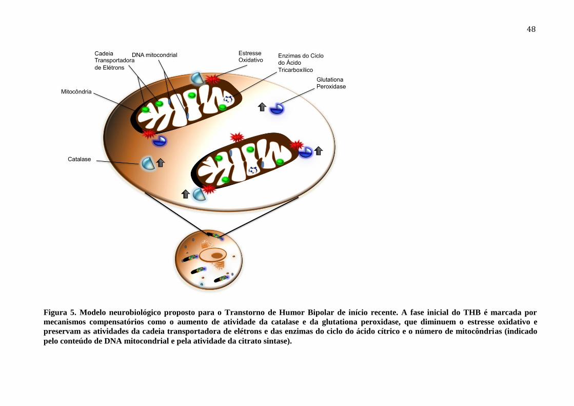
48
Figura 5. Modelo neurobiológico proposto para o Transtorno de Humor Bipolar de início recente. A fase inicial do THB é marcada por
mecanismos compensatórios como o aumento de atividade da catalase e da glutationa peroxidase, que diminuem o estresse oxidativo e
preservam as atividades da cadeia transportadora de elétrons e das enzimas do ciclo do ácido cítrico e o número de mitocôndrias (indicado
pelo conteúdo de DNA mitocondrial e pela atividade da citrato sintase).

49
O aumento do estresse oxidativo é prejudicial para lipídios, proteínas e DNA
(Halliwell & Gutteridge, 2007). Por isso, leva a um pior funcionamento da cadeia
transportadora de elétrons (Tretter et al., 2004) e das enzimas do ciclo do ácido cítrico
(Kamboj et al., 2008). A ausência de aumento de estresse oxidativo no presente
estudo é coincidente com a ausência de alteração da atividade da cadeia
transportadora de elétrons. Os processos da cadeia transportadora de elétrons geram a
maior parte da energia produzida na mitocôndria, que é a organela responsável pela
produção de energia celular. Os danos à cadeia transportadora de elétrons estão
associados a uma diminuição de produção de energia, que por sua vez
retroalimentaria o estresse oxidativo (Tretter et al., 2004; Adam-Vizi, 2005). O início
recente do THB também poderia explicar a ausência de alteração das enzimas do ciclo
do ácido cítrico.
Não se encontrou alteração do conteúdo de DNAmt nos pacientes com THB.
Tanto o DNAmt como a atividade de citrato sintase são indicadores de número de
mitocôndrias na célula (Marco et al., 1974; Malik & Czajka, 2013). Sabe-se que o
estresse oxidativo pode modular o DNAmt. Há evidências de que o estresse oxidativo
inicialmente aumente o DNAmt numa resposta adaptativa; com a cronificação do
estresse oxidativo, haveria diminuição de DNAmt (Malik & Czajka, 2013). Pode ser
que haja preservação de número de mitocôndrias nas fases iniciais do THB, quando a
neurobiologia e a apresentação clínica estão menos comprometidas.
Alinhado a essa hipótese, o THB I, subtipo mais grave do THB, mostrou
diminuição de número de mitocôndrias em comparação com THB II, subtipo menos
grave; esta diferença se observou com dois indicadores que refletem o número de
mitocôndrias, o conteúdo de DNAmt e a atividade de citrato sintase. A comparação
com controles saudáveis também mostrou discreta diminuição de número de

50
mitocôndrias no THB I, mas em só um dos marcadores, o DNAmt. É possível que a
neurobiologia do subtipo II possibilite a preservação do número de mitocôndrias, por
ser menos grave. Isso seria coincidente com a diminuição de expressão de genes
mitocondriais em células linfoblastóides somente em pacientes com THB I, mas não
no THB II (Washizuka et al., 2005). Além disso, a apresentação clínica mais branda
do subtipo II do THB reforça a hipótese.
O presente estudo não mostrou alteração de níveis de NO em pacientes com
THB em fase inicial. Embora seja possível argumentar que o achado negativo se deva
ao estádio inicial em que os pacientes se encontravam, é importante também recordar
que o NO tem propriedades neuromoduladoras e neuroprotetoras quando presente em
quantidades fisiológicas e não é necessariamente nocivo (Calabrese et al., 2007).
6.2. Efeito do tratamento com lítio
Observou-se um efeito do lítio deste estudo com potencial para neuroproteção.
O tratamento com lítio diminuiu a peroxidação lipídica (medida pelo TBARS) e
diminuiu a atividade da SOD nos pacientes com depressão bipolar, bem como
aumentou a atividade da cadeia transportadora de elétrons e os níveis de NO (Figura
6).
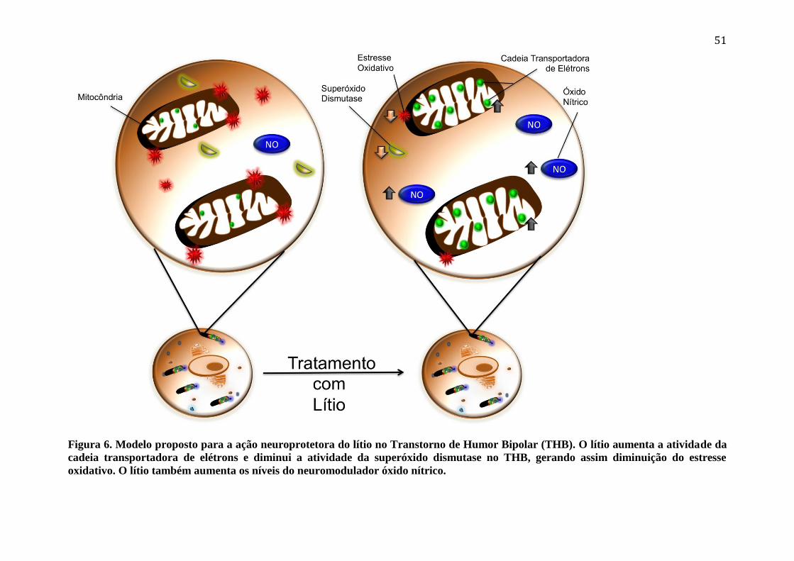
51
Figura 6. Modelo proposto para a ação neuroprotetora do lítio no Transtorno de Humor Bipolar (THB). O lítio aumenta a atividade da
cadeia transportadora de elétrons e diminui a atividade da superóxido dismutase no THB, gerando assim diminuição do estresse
oxidativo. O lítio também aumenta os níveis do neuromodulador óxido nítrico.

52
O lítio aumentou a atividade do complexo I da cadeia transportadora de
elétrons mitocondrial. Pode ser que o aumento na eficácia do complexo I esteja
associado à diminuição de estresse oxidativo observada aqui nos pacientes com
depressão bipolar, já que o contrário se observa: discretas diminuições na atividade do
complexo I da cadeia transportadora de elétrons estão associadas ao aumento de
estresse oxidativo em diversos estudos (Adam-Vizi, 2005). A regulação da enzima
SOD pelo tratamento com lítio é uma outra possível explicação para a diminuição do
estresse oxidativo observada no presente estudo; uma melhora do equilíbrio das
enzimas antioxidantes se mostrou eficaz contra o estresse oxidativo (Andreazza et al.,
2007).
Pode ser que uma ação antioxidante do lítio seja favorecida por alguma
especificidade da neurobiologia do subtipo II do THB. Depois do tratamento com lítio
houve diminuição de peroxidação lipídica somente no subtipo II, mas não no subtipo I
do THB. Além disso, em comparação com o subtipo I, os pacientes com subtipo II do
THB mostraram aumento de citrato sintase e conteúdo de DNAm, ambos marcadores
do número de mitocôndrias. Um número maior de mitocôndrias poderia favorecer
uma melhor ação do lítio contra a peroxidação lipídica no THB.
Coincidente com um efeito protetor antioxidante do lítio nos presentes
resultados, achou-se também um aumento de NO na depressão bipolar. O NO tem
sido sugerido como molécula importante para neuroproteção e para a fisiologia
normal em doses não-tóxicas (Calabrese et al., 2007). O NO se mostrou capaz de
modular o ácido gama-aminobutírico, que tem evidências de transmissão diminuída
no THB (Guidotti et al., 2011). Houve aumento da liberação de ácido gama-
53
aminobutírico em resposta ao NO em cultura de neurônios de camundongos (Ohkuma
et al., 1996) e em cérebro de ratos in vivo (Getting et al., 1996).
6.3. Limitações e pontos fortes
São limitações deste estudo o tamanho da amostra, a presença de pacientes
somente em fase depressiva, bem como o fato de a maior parte dos pacientes ter o
subtipo II do THB. Por outro lado, o início recente do THB, o fato de a maioria dos
pacientes não estar medicados (84%) e a maior parte dos pacientes ser virgem de
tratamento (71%) são fatores que aumentam a especificidade do presente estudo,
evitando algumas influências na neurobiologia que poderiam interferir nos parâmetros
estudados.
6.4. Conclusão e perspectivas
O presente estudo sugere que possa haver um perfil específico de alterações
em fases iniciais do THB, com a presença de mecanismos compensatórios, reforçando
a proposta de investigar o estadiamento neurobiológico no THB. A expectativa
otimista é de que o estadiamento permita intervenções iniciais mais amplas que
melhorem o curso do THB e o prognóstico (Berk et al., 2013).
Estudos avaliando coortes de pacientes com THB com medidas repetidas de
biomarcadores são necessários para a confirmação dos achados no THB de início
recente e para esclarecer o curso do estresse oxidativo e da função mitocondrial nos
diferentes estágios do THB.
Os achados sugerem que o papel da mitocôndria na neurobiologia dos subtipos
I e II do THB possa ser diferente e que o papel do lítio contra o estresse oxidativo

54
possa ser diferente nos dois subtipos. Estudos com amostras maiores poderiam
confirmar estes achados preliminares.
Os achados reforçam um papel do lítio contra o estresse oxidativo, sugerindo
que a droga atue em humanos aumentando a atividade do complexo I da cadeia
transportadora de elétrons mitocondrial de forma dose-dependente e possa regular as
enzimas antioxidantes em humanos. Além disso, os presentes resultados sugerem que
o lítio possa modular o sistema nitrérgico no THB.
55
7. ANEXOS
7.1. Artigos publicados
7.1.1. Leukocyte mitochondrial DNA copy number in
bipolar disorder
Progress in Neuro-Psychopharmacology & Biological Psychiatry 48 (2014) 32–35
Contents lists available at ScienceDirect
Progress in Neuro-Psychopharmacology & BiologicalPsychiatry
j ourna l homepage: www.e lsev ie r .com/ locate /pnp
Leukocyte mitochondrial DNA copy number in bipolar disorder
Rafael T. de Sousa a, Miyuki Uno b, Marcus V. Zanetti a,c,d, Sueli M.O. Shinjo b, Geraldo F. Busatto c,d,Wagner F. Gattaz a,c, Sueli K.N. Marie b, Rodrigo Machado-Vieira a,c,⁎a Laboratory of Neuroscience, LIM-27, Institute and Department of Psychiatry, University of Sao Paulo, Brazilb Department of Neurology, School of Medicine, University of São Paulo, São Paulo, São Paulo, Brazilc Center for Interdisciplinary Research on Applied Neurosciences (NAPNA), University of São Paulo, Brazild Laboratory of Psychiatric Neuroimaging, LIM-21, Department and Institute of Psychiatry, University of Sao Paulo, Brazil
Abbreviations: BD, Bipolar disorder; CGI, Clinical Gltransport chain; HAM-D, 21-item Hamilton DepressionmtDNA, mitochondrial DNA; SCID, Structured Clinical IDisorders; YMRS, Young Mania Rating Scale.⁎ Corresponding author at: Institute and Department
Paulo, Rua Ovidio Pires de Campos, 785, São Paulo, SP, BraE-mail address: [email protected] (R. Macha
0278-5846/$ – see front matter. Published by Elsevier Inchttp://dx.doi.org/10.1016/j.pnpbp.2013.09.002
a b s t r a c t
a r t i c l e i n f oArticle history:
Received 18 July 2013Received in revised form 4 September 2013Accepted 5 September 2013Available online 13 September 2013Keywords:AntidepressantBipolar disorderDepressionLithiumMitochondriaTreatment
Background: Evidence supports the role formitochondrial impairment in the pathophysiology of bipolar disorder(BD). BD has been associated with decreased mitochondrial electron transport chain activity and increased oxi-dative stress. Also, mitochondrial DNA (mtDNA) encodes mitochondrial electron transport chain proteins andhas been associated with altered oxidative stress. Preclinical studies showed that lithium treatment increasedmtDNA content, but no study has directly assessed mtDNA content in subjects with BD in vivo. Also, the effectsof lithium treatment on mtDNA content have never been evaluated in humans.Methods: LeukocytemtDNA content using real time-PCRwas evaluated in subjectswith BD (n = 23) in a depres-sive episode (≥18 in the 21-item Hamilton Depression Rating Scale) before and after 6-week lithium treatmentversus healthy controls (n = 24).Results: mtDNA content showed no significant difference between subjects with BD at baseline and controls(p = 0.46); also no differencewas observedwhen comparing before and after lithium treatment. A trend for de-creased mtDNA content was specifically observed in BD type I compared to controls and BD type II (p = 0.05).
Importantly, endpoint mtDNA copy number was significantly correlated with age.Conclusion: In BD subjects who were younger, unmedicated and had a shorter duration of illness, no change wasobserved in mtDNA copy number. More studies with larger samples are warranted to evaluate mtDNA contentchanges in BD and its potential role as a treatment target, especially in BD type I and its association with aging.Published by Elsevier Inc.
1. Introduction
Evidences of different lines support a role of mitochondrial impair-ment in the pathophysiology of bipolar disorder (BD) (Clay et al.,2011). Mitochondrial dysfunction was initially suggested in subjectswith BDbased on brainmagnetic resonance spectroscopy studies show-ing energy shortage and low intracellular pH (Stork and Renshaw,2005). Also, increased oxidative stress was found in the periphery andpost-mortem brains of subjects with BD (Andreazza et al., 2008,2010), reinforcing the mitochondrial dysfunction in the illness. Post-mortem studies in BD have also found decreased expression of genesencoding mitochondrial electron transport chain (ETC) (Konradi et al.,
obal Impression; ETC, electronScale; HBb, Hemoglobin Beta;nterview for Axis I DSM-IV-TR
of Psychiatry, University of Saozil.do-Vieira).
.
2004; Sun et al., 2006) and lower mitochondrial ETC complex I activity(Andreazza et al., 2010).
ThemtDNA is a double-stranded, closed circularmolecule located inmitochondrion, encoding 37 genes essential for normal mitochondrialfunctioning. Abnormal mtDNA number content has been associatedwith disturbed mitochondrial function and increased oxidative stress(Malik and Czajka, 2013). In BD, only one post-mortem study foundsubtle changes in mtDNA (Vawter et al., 2006), while others found nodifferences compared to controls (Kakiuchi et al., 2005; Sabunciyanet al., 2007; Torrell et al., 2013). Importantly, a study of elderly patientsshowed decreased mtDNA content associated with depressive symp-toms (Kim et al., 2011). Lithium is a gold standard treatment for moodepisodes and maintenance in BD (Machado-Vieira et al., 2009). Currentguidelines recommend the use of lithium as a first line treatment foracute bipolar depression (Haeberle et al., 2012; Yatham et al., 2013).Also, data from the European drug surveillance program shows thatlithium is themost prescribed agent for bipolar depression in combinedtherapy (33%) (Haeberle et al., 2012).
Its efficacy has been associated with the mtDNA 10398A polymor-phism (Washizuka et al., 2003). Also, Struewing et al. (2007) found

Table 1Demographic and clinical characteristics of bipolar depression patients and healthycontrols.
Controls (n = 24) Bipolar (n = 23) p
GenderMale/female, n (%) 14 (58.3)/10 (41.7) 6 (26.1)/17 (73.9) 0.01⁎,a
Age, years 28.3 (±7.3) 28.5 (±6.1) 0.61b
Bipolar disorder typeType I/type II, n (%) 7 (30.4)/16 (69.6)
Duration of illness, months 36.3 (±18.2)Drug-naive, n (%) 16 (69.6)Medication-free, n (%) 21 (91.3)History of psychosis, n (%) 4 (17.4)HAM-D
Baseline/endpoint 22.0 (±2.7)/7.6 (±6.4)YMRS
Baseline/endpoint 5.6 (±4.8)/4.7 (±9.7)Response, n (%) 18 (78.3)Remission, n (%) 13 (56.6)Dropout, n (%) 1 (4.3)Endpoint serum lithium,mEq/L
0.50 (±0.20)
HAM-D— Hamilton Depression Scale, YMRS — Young Mania Rating Scale.⁎ Significantly different.a Chi-square.b Student's t test.
33R.T. de Sousa et al. / Progress in Neuro-Psychopharmacology & Biological Psychiatry 48 (2014) 32–35
lithium increasing mtDNA content and in spinal cord of rodents. SincemtDNA encodes part of complex I proteins, lithium may enhancemtDNA content and lead to altered complex I transcription, observedin themitochondrial complex INDUFS7 gene in subjectswith BD treatedwith lithium (Sun et al., 2006).
Up to date, no studyhas assessedmtDNAcontent in subjectswithBDin vivo, nor examined mtDNA content association with depressivesymptoms in BD. Furthermore, no study evaluated lithium effects inmtDNA content in humans and its potential clinical relevance. The pres-ent study evaluatesmtDNA content in bipolar depressive episodes com-pared to healthy controls, testing if leukocyte mtDNA content is alteredin BD or modulated by lithium over time (6 weeks) in a clinically rele-vant paradigm.
2. Methods
2.1. Subjects
Between August 2010 and June 2012, 23 outpatients, 17 (73.9%)women, with a mean age of 28.5 (±6.1) years and a diagnosis of BD,currently experiencing a depressive episode, as diagnosed by StructuredClinical Interview for Axis I DSM-IV-TR Disorders (SCID) (First, 1995),entered the study. Patients were recruited through newspaper, internetand radio advertisement and evaluated at the Institute of Psychiatry,University of Sao Paulo, Brazil.
Patients who presented a score ≥18 on the 21-item Hamilton De-pression Scale (HAM-D) were eligible for the study. Diagnosis and psy-chometric assessments were performed by experienced psychiatrists.Presence of any current neurological disorders or medical illness, otheraxis I diagnosis (including current substance abuse or dependence) ormental retardation was excluded from the present investigation.
Twenty-four age-matched (±3 years) healthy controls recruitedthrough advertisement in the community, 14 (58.3%) men and 10(41.7%) women (mean age = 28.3 ± 7.3 years), were also studied.Controls were interviewed and excluded if they had a lifetime historyof anymental disorder (as assessed with the SCID), including substanceabuse/dependence or any neurological/medical disease or any first-degree relative with mood or psychotic disorder. This study was ap-proved by the local institutional review board, and all participants pro-vided written informed consent before entering the study.
2.2. Study design
Blood samples were collected at baseline and at endpoint (week 6),while healthy controls had only one-point sample collection. At baseline,patients were started on lithium carbonate at 450 mg/day, with flexibledose adjusted according to clinical improvement, also controlling plasmalithium levels. Most patients were in lithium monotherapy, althoughhypnotic use as needed was allowed and 2 patients were also in use ofother drugs. Psychometric assessments were made at baseline, onweek 1, week 2, week 4, and week 6 (endpoint). Assessment of symp-toms was performed with HAM-D, Young Mania Rating Scale (YMRS),and Clinical Global Impression (CGI)— Severity and Improvement. Clin-ical response was defined as a decrease of 50% or more in the HAM-D atendpoint and remission as HAM-D b 8 and YMRS b 8 at endpoint.
2.3. Assays
Blood samples were collected from 8 to 10 am from subjects whowere in 8-hour fasting. Samples were centrifuged at 20 °C and 700 ×gfor 40 min and leukocyte pellets were obtained. PBS and DMSO wereadded to the pellet and the samples were gradually frozen at −10 °Cfor 30 min, at −20 °C for 30 min, and finally stored at −80 °C. DNAwas extracted from leukocyte pellet following the protocol describedby Kit QIAamp® DNAMini (Qiagen Inc., Hilden, Germany). DNA qualitywas verified in agarose gel electrophoresis andDNA concentrationswere
determined at 260 nm wavelength using ND-1000 Spectrophotometer(NanoDrop Technologies, Inc.). Ratios between 1.8 and 2.0 at260/280 nm were considered satisfactory. The mtDNA copy numberwas determined with quantitative real-time PCR, using 2.5 ng fromtotal DNA extracted. A nuclear, single copy gene, Hemoglobin Beta(HBb) was used as reference (Kersting et al., 2004). Primer sequencesused at a final concentration of 100 nM on ABI Prism 7500 sequencedetector (Applied Biosystems, Foster City, CA) were (5′–3′): mtDNAF: CCTAGCCGTTTACTCAATCCT, mtDNA R: TGATGGCTAGGGTGACTTCAT, HBb F: GTGAAGGCTCATGGCAAGA, and HBb R: AGCTCACTCAGTGTGGCAAAG (IDT, Coralville, IA, USA). Reaction conditions were 2 minat 50 °C and 10 min at 95 °C, followed by 40 cycles of 15 s at 95 °C,and 1 min at 60 °C. All assays were carried out in duplicate. mtDNAcopy number was calculated by the equation 2−ΔΔCt (Livak andSchmittgen, 2001).
2.4. Statistics
Statistical analysis was performed using SPSS 14.0 and “R” package.Chi-square test was used to compare gender in patients and controls.For samples with normal distribution Student's t test was used andwhen samples had non-normal distribution, comparisons wereperformed with Mann–Whitney and Wilcoxon Signed Ranks tests, andcorrelations evaluated with Spearman test. For comparing patientsand controls generalized linear model was performed to take genderdifference into account. Significance level was set at b0.05 (two-tailed).
3. Results
3.1. Demographic and clinical data
Demographic and clinical data of patients and controls are summa-rized in Table 1. From 23 patients enrolled, 7 (30.4%) had a diagnosisof BD type I and 16 (69.6%) of BD type II; 21 (91.3%) patients weremedication-free for at least 6 weeks prior to their enrollment in thestudy and 16 (69.6%) were drug-naïve. Patients had mean duration ofillness of 36.3 months (±18.2) and history of previous mood episodewith psychosis was present in 4 (17.4%) patients. Patients had a signif-icant decrease in depressive symptomsmeasured byHAM-D frombase-line (22.0 ± 2.7) to endpoint (7.6 ± 6.4) (z = −4.07, p b 0.001).

Fig. 1. Comparison of mitochondrial DNA (mtDNA) copies in healthy controls, bipolardisorder (BD) patients before and after 6-week lithium treatment.
34 R.T. de Sousa et al. / Progress in Neuro-Psychopharmacology & Biological Psychiatry 48 (2014) 32–35
Eighteen (78.3%) patients responded to treatment and 13 (56.6%) pa-tients had remission.
3.2. mtDNA copy number in subjects with BD and controls
Subjects with BD and healthy controls had a gender difference,but on themodel comparing patients and controls genderwas not asso-ciated with mtDNA copies (t = −0.52, p = 0.61). Even taking genderinto account on the model, subjects with BD and controls did not differregarding to mtDNA copies (BD patients = 1241.9 ± 1671.4; healthycontrols = 914.2 ± 640.0) (t = 0.75, p = 0.46) (Fig. 1). BaselineHAM-D scores andmtDNA copies did not show a significant correlation(ρ = 0.27, p = 0.21).
There was a non-significant decrease in mtDNA copies of type I BDgroup (490.5 ± 421.5) in comparison to healthy controls (914.2 ±640.0) (z = −1.937, p = 0.05). No differences were observed inmtDNA copies between patients with type II BD and healthy controls(z = −0.44, p = 0.66). Also, a non-significant decrease in mtDNAcopies of type I BD (490.5 ± 421.5) compared to type II BD(1570.7 ± 1909.8) was observed (z = −1.938, p = 0.05).
Number of mtDNA copies at baseline associated with treatment sta-tus at baseline (drug naïve z = −0.64, p = 0.53; drug-free z = −0.46,p = 0.65) and the presence psychosis history (z = −1.46, p = 0.14).Also, baseline mtDNA copies were not correlated with any of the vari-ables analyzed: age, CGI score, and duration of illness (data not shown).
3.3. Lithium treatment did not change mtDNA copies (but was correlatedwith age)
Lithium treatment did not change mtDNA copies from baseline(1241.9 ± 1671.4) to endpoint (1010.0 ± 810.6) (z = −0.50, p =0.62) (Fig. 1).mtDNA copies at endpointwere not different between re-sponders vs. non-responders (z = −0.68, p = 0.50) or remitters vs.non-remitters (t = 0.25, p = 0.80). Also, endpoint mtDNA copy num-ber was significantly correlated with age (ρ = −0.564, p = 0.006),but not with any other variable analyzed: endpoint HAM-D, CGI score,and duration of illness (data not shown).
4. Discussion
The present study found no difference in mtDNA copy number be-tween subjects with BD depression and healthy controls. Also, mtDNAcontent did not significantly change after 6-week lithium treatment.However, a significant association with endpoint mtDNA copy numberwas significantly correlated with age.
To the best of our knowledge, this is the first study evaluatingmtDNA content in vivo in subjects with BD (in any mood state) andalso the only investigation on lithium effects at mtDNA content inhumans.
The evidence of mitochondrial dysfunction in BD comes from differ-ent lines of research, including reduced high-energy phosphates andlow pH in neuroimaging of BD patients (Stork and Renshaw, 2005) aswell increased prevalence of mtDNA polymorphisms and changes inmitochondrial function in microarray and biochemical studies of sub-jects with BD compared to healthy subjects (Clay et al., 2011; Quirozet al., 2008). Based on studies showing increased oxidative stress inBD (Andreazza et al., 2008, 2010; Machado-Vieira et al., 2007) and de-creasedmitochondrial ETC activity (Andreazza et al., 2010), also associ-ated with mtDNA content alteration (Malik and Czajka, 2013), wehypothesized that altered mtDNA content in BD patients would occur;however this finding was not observed here.
The present findings do not contradict evidences of mitochondrialdysfunction in BD. Although mtDNA encodes ETC proteins whichlower its transcription in BD (Konradi et al., 2004; Sun et al., 2006), ev-idence challenges the association between mtDNA content and mtDNAtranscription (Torrell et al., 2013; Vawter et al., 2006). Rather, it is pos-sible that mtDNA sequence plays a more crucial role in mtDNA expres-sion (Reinecke et al., 2009). Moreover, oxidative stress may onlyinitially increase (reactive) mtDNA content (Liu et al., 2003), while lon-ger periods of elevated oxidative stress are associatedwith a decrease inmtDNA content (Malik and Czajka, 2013), which may explain the hereobserved lack of alteration in mtDNA copy number.
The present results are in accordance with three studies in BD post-mortem brains which showed no alteration in mtDNA content(Kakiuchi et al., 2005; Sabunciyan et al., 2007; Torrell et al., 2013). Incontrast with our findings, one study found thatmtDNA content slightlyincreased in post-mortemBD subjects (Vawter et al., 2006). ThemtDNAcontent in peripheral cells may reflect similar mtDNA content in othertargets (such as brain) involved different diseases (Malik and Czajka,2013), which reinforces the validity of our findings in leukocytes.
Regarding mtDNA in peripheral cells, Kim et al. (2011) found de-creased mtDNA content in leukocytes of patients with depression.Their study, however, did not use any diagnostic tool. Moreover, Kimet al. (2011) evaluated elderly patients with depressive symptomswhile our sample comprised young adults with bipolar depression.Our study used standard tools for diagnosis and symptom evaluation,enhancing the validity of the findings.
Here, subjects with BD treated with lithium showed no changes inmtDNA content in bipolar depression. Present findings are in contrastwith increased mtDNA content found in bovine endothelial cellsincubated with lithium (Struewing et al., 2007) and in spinal cord ofmice treated with lithium (Fornai et al., 2008). In humans, lithiummight have no effect inmtDNAcontent regulation, especially in youngerage.
Our findings showed a slight decrease in baseline mtDNA content intype I BD compared to controls (p = 0.05) and a small reduction inbaselinemtDNA of type I BD compared to type II BD (p = 0.05). Indeed,most studies inmitochondria report data on BD type I while the presentsample consisted in majority of BD type II subjects (69.6%). A differencein mitochondrial role in the neurobiology of these two subtypes is sup-ported byWashizuka et al. (2005). They found differential expression ofmitochondrial genes only in BD type I but not II. The presence of mostBD type II heremight explain the lack of alteration found inmtDNA con-tent of BD patients.
Regarding potential limitations, the small BD sample size may limitthe extrapolation of the present findings. However, the drug-naïve/drug status of most part of the sample at baseline strengthens the find-ings. Also, this is the first study evaluatingmtDNA content in bipolar de-pression and lithium effects on mtDNA content. Importantly this studyonly includes unmedicated young subjects with BD short course of theillness, reinforcing the specificity of the findings.

35R.T. de Sousa et al. / Progress in Neuro-Psychopharmacology & Biological Psychiatry 48 (2014) 32–35
Further studies with larger samples are warranted to assess mtDNAcontent in BD, especially in BD type I and its association with aging andillness progression.
Acknowledgments
This study was sponsored by Sao Paulo Research Foundation(Fapesp, Brazil). The Laboratory of Neuroscience is supported by theAssociação Beneficente Alzira Denise Hertzog da Silva (ABADHS).
References
Andreazza AC, Kauer-Sant'anna M, Frey BN, Bond DJ, Kapczinski F, Young LT, et al. Oxida-tive stress markers in bipolar disorder: a meta-analysis. J Affect Disord 2008;111:135–44.
Andreazza AC, Shao L, Wang JF, Young LT. Mitochondrial complex I activity and oxidativedamage to mitochondrial proteins in the prefrontal cortex of patients with bipolardisorder. Arch Gen Psychiatry 2010;67:360–8.
Clay HB, Sillivan S, Konradi C. Mitochondrial dysfunction and pathology in bipolar disor-der and schizophrenia. Int J Dev Neurosci 2011;29:311–24.
First M. Structured clinical interview for DSM-IV Axis I disorders. Patient ed. New York:Biometrics Research Department, New York Psychiatric Institute; 1995.
Fornai F, Longone P, Cafaro L, Kastsiuchenka O, Ferrucci M, Manca ML, et al. Lithium delaysprogression of amyotrophic lateral sclerosis. Proc Natl Acad Sci U S A 2008;105:2052–7.
Haeberle A, Greil W, Russman S, Grohmann R. Mono- and combination drug therapies inhospitalized patients with bipolar disorder. Data from the European drug surveillanceprogram AMSP. BMC Psychiatry 2012;12:153.
Kakiuchi C, Ishiwata M, KametaniM, Nelson C, Iwamoto K, Kato T. Quantitative analysis ofmitochondrial DNA deletions in the brains of patients with bipolar disorder andschizophrenia. Int J Neuropsychopharmacol 2005;8:515–22.
Kersting C, Tidow N, Schmidt H, Liedtke C, Neumann J, BoeckerW, et al. Gene dosage PCRand fluorescence in situ hybridization reveal low frequency of egfr amplifications de-spite protein overexpression in invasive breast carcinoma. Lab Invest 2004;84:582–7.
KimMY, Lee JW, Kang HC, Kim E, Lee DC. Leukocyte mitochondrial DNA (mtDNA) contentis associated with depression in old women. Arch Gerontol Geriatr 2011;53:e218–21.
Konradi C, EatonM, MacDonald ML, Walsh J, Benes FM, Heckers S. Molecular evidence formitochondrial dysfunction in bipolar disorder. Arch Gen Psychiatry 2004;61:300–8.
Liu CS, Tsai CS, Kuo CL, Chen HW, Lii CK, Ma YS, et al. Oxidative stress-related alteration ofthe copy number of mitochondrial DNA in human leukocytes. Free Radic Res 2003;37:1307–17.
Livak KJ, Schmittgen TD. Analysis of relative gene expression data using real-time quanti-tative PCR and the 2(-Delta Delta C(T)) Method. Methods 2001;25:402–8.
Machado-Vieira R, Andreazza AC, Viale CI, Zanatto V, Cereser V, Silva Vargas R, et al.Oxidative stress parameters in unmedicated and treated bipolar subjects duringinitial manic episode: a possible role for lithium antioxidant effects. Neurosci Lett2007;421:33–6.
Machado-Vieira R,Manji HK, Zarate CA. The role of lithium in the treatment of bipolar dis-order: convergent evidence for neurotrophic effects as a unifying hypothesis. BipolarDisord 2009;11:92–109.
Malik AN, Czajka A. Is mitochondrial DNA content a potential biomarker of mitochondrialdysfunction? Mitochondrion 2013;13:481–92.
Quiroz JA, Gray NA, Kato T, Manji HK. Mitochondrially mediated plasticity in the patho-physiology and treatment of bipolar disorder. Neuropsychopharmacology 2008;33:2551–65.
Reinecke F, Smeitink JA, van der Westhuizen FH. OXPHOS gene expression and control inmitochondrial disorders. Biochim Biophys Acta 2009;1792:1113–21.
Sabunciyan S, Kirches E, Krause G, Bogerts B, Mawrin C, Llenos IC, et al. Quantification oftotal mitochondrial DNA and mitochondrial common deletion in the frontal cortex ofpatients with schizophrenia and bipolar disorder. J Neural Transm 2007;114:665–74.
Stork C, Renshaw PF. Mitochondrial dysfunction in bipolar disorder: evidence from mag-netic resonance spectroscopy research. Mol Psychiatry 2005;10:900–19.
Struewing IT, Barnett CD, Tang T, Mao CD. Lithium increases PGC-1alpha expression andmitochondrial biogenesis in primary bovine aortic endothelial cells. FEBS J 2007;274:2749–65.
Sun X, Wang JF, Tseng M, Young LT. Downregulation in components of the mitochondrialelectron transport chain in the postmortem frontal cortex of subjects with bipolardisorder. J Psychiatry Neurosci 2006;31:189–96.
Torrell H, Montaña E, Abasolo N, Roig B, Gaviria AM, Vilella E, et al. Mitochondrial DNA(mtDNA) in brain samples from patients with major psychiatric disorders: gene ex-pression profiles, mtDNA content and presence of the mtDNA common deletion.Am J Med Genet B Neuropsychiatr Genet 2013;162B:213–23.
Vawter MP, Tomita H, Meng F, Bolstad B, Li J, Evans S, et al. Mitochondrial-related geneexpression changes are sensitive to agonal-pH state: implications for brain disorders.Mol Psychiatry 2006;11:663–79.
Washizuka SA, Ikeda A, Kato N, Kato T. Possible relationship between mitochondrial DNApolymorphisms and lithium response in bipolar disorder. Int J Neuropsychopharmacol2003;6:421–4.
Washizuka S, Kakiuchi C, Mori K, Tajima O, Akiyama T, Kato T. Expression ofmitochondria-related genes in lymphoblastoid cells from patients with bipolar disor-der. Bipolar Disord 2005;7:146–52.
Yatham LN, Kennedy SH, Parikh SV, Schaffer A, Beaulieu S, AldaM, et al. Canadian Networkfor Mood and Anxiety Treatments (CANMAT) and International Society for BipolarDisorders (ISBD) collaborative update of CANMAT. guidelines for the managementof patients with bipolar disorder: update 2013. Bipolar Disord 2013;15:1–44.

60
7.1.2. Oxidative stress in early stage Bipolar Disorder and
the association with response to lithium

lable at ScienceDirect
Journal of Psychiatric Research 50 (2014) 36e41
Contents lists avai
Journal of Psychiatric Research
journal homepage: www.elsevier .com/locate/psychires
Oxidative stress in early stage Bipolar Disorder and the associationwith response to lithium
Rafael T. de Sousa a, Carlos A. Zarate Jr. b, Marcus V. Zanetti a,c, Alana C. Costa a,Leda L. Talib a, Wagner F. Gattaz a,c, Rodrigo Machado-Vieira a,b,c,*
a Laboratory of Neuroscience, LIM-27, Institute and Department of Psychiatry, University of Sao Paulo, Brazilb Experimental Therapeutics and Pathophysiology Branch (ETPB), National Institute of Mental Health, NIH, Bethesda, MD, USAcCenter for Interdisciplinary Research on Applied Neurosciences (NAPNA), University of Sao Paulo, Brazil
a r t i c l e i n f o
Article history:Received 4 September 2013Received in revised form8 November 2013Accepted 26 November 2013
Keywords:Oxidative stressLithiumBipolar DisorderDepressionNeuroprotection
* Corresponding author. Institute and Departmenticine, Rua Ovidio Pires de Campos, 785, CEP 01060-9
E-mail addresses: [email protected],(R. Machado-Vieira).
0022-3956/$ e see front matter Published by Elseviehttp://dx.doi.org/10.1016/j.jpsychires.2013.11.011
a b s t r a c t
Background: Several studies have described increased oxidative stress (OxS) parameters and imbalanceof antioxidant enzymes in Bipolar Disorder (BD) but few is know about the impact of treatment at thesetargets. However, no study has evaluated OxS parameters in unmedicated early stage BD and their as-sociation with lithium treatment in bipolar depression.Methods: Patients with BD I or II (n ¼ 29) in a depressive episode were treated for 6 weeks withlithium. Plasma samples were collected at baseline and endpoint, and were also compared to age-matched controls (n ¼ 28). The thiobarbituric acid reactive substances (TBARS), and the antioxidantenzymes superoxide dismutase (SOD), catalase (CAT), and glutathione peroxidase (GPx) activitieswere measured.Results: Subjects with BD depression at baseline presented a significant increase in CAT (p ¼ 0.005) andGPx (p < 0.001) levels, with lower SOD/CAT ratio (p ¼ 0.001) and no changes on SOD or TBARS comparedto healthy controls. Regarding therapeutics, lithium only induced a decrease in TBARS (p ¼ 0.023) andSOD (p ¼ 0.029) levels, especially in BDII. Finally, TBARS levels were significantly lower at endpoint inlithium responders compared to non-responders (p ¼ 0.018) with no difference in any biomarkerregarding remission.Conclusion: The present findings suggest a reactive increase in antioxidant enzymes levels duringdepressive episodes in early stage BD with minimal prior treatment. Also, decreased lipid peroxidation(TBARS) levels were observed, associated with lithium’s clinical efficacy. Overall, these results reinforcethe role for altered oxidative stress in the pathophysiology of BD and the presence of antioxidant effectsof lithium in the prevention of illness progression and clinical efficacy.
Published by Elsevier Ltd.
1. Introduction
It has been proposed a progressive course of Bipolar Disorder(BD) associated with longer illness duration, cognitive decline,decreased functioning and impaired cellular resilience leading todeleterious consequences on signal transduction and synapticplasticity (Machado-Vieira et al., 2013). Thus, intervention in theearly stage of BD may be a valuable tool to improve the course ofthe illness and provide a better prognosis (Berk et al., 2013).
of Psychiatry, School of Med-70 Sao Paulo, SP, [email protected]
r Ltd.
Increased Oxidative Stress (OxS) generates deleterious conse-quences on signal transduction, synaptic plasticity, and cellularresilience, especially by inducing lipid peroxidation in mem-branes, proteins and DNA (Grintzalis et al., 2013; Mahadik et al.,2001; Soeiro-de-Souza et al., 2013). DNA damage, which can beinduced by oxidative stress, has been found to be associated withthe severity of depressive symptoms in BD (Andreazza et al.,2007b).
Thiobarbituric acid reactive substances (TBARS) levels are adirect index of cell lipid peroxidation whereas antioxidant systeminvolves coordinated effects of antioxidant enzymes superoxidedismutase (SOD), catalase (CAT), and glutathione peroxidase (GPx)(Reddy et al., 1991).
Imbalance in antioxidant enzymes has been reported in severalstudies of BD and is consistently associated with increased OxS

R.T. de Sousa et al. / Journal of Psychiatric Research 50 (2014) 36e41 37
(Andreazza et al., 2008). Only one study evaluated OxS parametersin BD patients with short duration of illness and found increasedactivity of antioxidant system in mania (Machado-Vieira et al.,2007). No study, however, has evaluated OxS in recent-onset BDpatients in unmedicated depressive episodes.
Also, consistent evidences support the role of a subtle mito-chondrial compromise in BD (Clay et al., 2011; Manji et al., 2012).Initially, magnetic resonance spectroscopy studies showedincreased lactate levels in BD, suggesting a metabolic shift(reviewed in Stork and Renshaw, 2005). Post-mortem studies usingbrains of BD patients have shown decreased expression of mito-chondrial electron transport chain genes (Konradi et al., 2004; Sunet al., 2006). Mitochondrial dysfunction increases production ofreactive oxygen species, leading to enhanced OxS. OxS parametershave been found to be increased in BD post-mortem brains(Andreazza et al., 2010) and also in the peripheral blood of subjectswith BD (Andreazza et al., 2008). Only one study evaluated subjectswith BD during a depressive episode in a smaller sample, and founda slight increase in CAT levels compared to controls (Andreazzaet al., 2007a).
Evidence suggests that the presence of OxS might be associatedwith the consistently found hyperactivation of the glutamatergicand dopaminergic systems in BD (Berk et al., 2011). Glutamatergichyperactivity leads to increased calcium influx (Plein and Berk,2001) which increases OxS (Shao et al., 2005). On the other hand,enhanced OxS has been suggested to increase glutamate (Lovellet al., 2000; Volterra et al., 1994). The excessive dopamine pro-duction increases OxS due to the production of reactive oxygenspecies in dopamine metabolism (Miyazaki and Asanuma, 2008).Moreover, the opposite mechanism also happens when OxS in-duces dopamine uptake, thus increasing dopamine activity (Kimand Andreazza, 2012) in a vicious cycle.
Other systems associated with BD are g-Aminobutyric acid(Brambilla et al., 2003) and serotonergic systems (Fountoulakiset al., 2012), which are found to show decreased activity. Similarto other systems, OxS is associated with decreased g-Aminobutyricacid release (Palmeira et al., 1993). Increased metabolism of sero-tonin, which decreases serotonergic function, is found to be asso-ciated with increased oxidative stress (Bianchi et al., 2005; Nocitoet al., 2007). Consistent with these findings, evidences suggestthat treatment with antidepressants can decrease OxS (Bilici et al.,2001).
Lithium is recommended as a first-line treatment for bipolardepression (Haeberle et al., 2012; Yatham et al., 2013) and it hasshown several neuroprotective and neurotrophic actions(Machado-Vieira et al., 2009). Lithium has been shown to increasebrain-derived neurotrophic factor (BDNF) (de Sousa et al., 2011),regulate intracellular Caþ2 (Wasserman et al., 2004), activateCREB (Ozaki and Chuang, 1997), increase Akt (Yazlovitskaya et al.,2006), and inhibit apoptotic caspase-3 (Ghribi et al., 2002).Similarly, lithium has been shown to protect against gluta-matergic excitotoxicity (Nonaka et al., 1998). This agent hasshown to decrease OxS in preclinical models (Schäfer et al., 2004),in bipolar mania (Machado-Vieira et al., 2007) and healthy vol-unteers (Khairova et al., 2012). Nonetheless, the effects of lithiumon OxS parameters specifically in bipolar depression have neverbeen studied.
The present study evaluated OxS parameters (TBARS, SOD, CATand GPx) in unmedicated bipolar depression versus controls as wellas the potential antioxidant effects of lithium in a therapeuticallyrelevant paradigm. Our hypothesis was that subjects in a bipolardepression episode would present increased OxS (TBARS) andimbalance of antioxidant enzymes during depressive episodes andthat lithium would lower OxS levels and enhance antioxidant en-zymes levels.
2. Methods
2.1. Subjects
Subjects were evaluated between August 2010 and June 2012 atthe Institute of Psychiatry, University of Sao Paulo, Brazil. Twenty-nine patients, 21 (72.4%) women, with 28.4 (�5.5) years of age anddiagnosis of BD I (38%) or BD II (62%) in a depressive episode wereincluded, as diagnosed by the Structured Clinical Interview for AxisI DSM-IV-TR Disorders (SCID) (First et al., 1995). Patients who had ascore �18 on the 21-item Hamilton Depression Scale (HAM-D)(HAMILTON, 1960) were eligible for the study. Also, 26 (89.6%)patients were drug-free for at least 6 weeks prior to their enroll-ment and 21 (72.4%) subjects were treatment-naïve at baseline.Exclusion criteria included presence of chronic medical illness,comorbid substance abuse or dependence in the past year, rapidcycling in the past 12 months, previous head trauma, currentmajor axis I psychiatric disorder, subjects submitted to electro-convulsive therapy and current significant abnormal laboratorytests.
The comparison group was constituted by 28 age-matchedhealthy controls (within 3 years of difference of BD patients);these 16 men (57.1%) and 12 women (42.8%) (age ¼ 28.0 � 7.2)were recruited through advertisement in the local community.Controls were excluded if they had lifetime history of any mentaldisorder (by SCID), including substance abuse or dependence, orif they had any disease with central nervous system involvementor any first-degree relative with a mental disorder. This studywas approved by the local institutional review board, and allparticipants provided written informed consent before studyentry.
2.2. Study design
Patients had blood samples collected at baseline and at endpoint(week 6), while healthy controls had only one-point samplecollection. At baseline, patients were started on open-label lithiumcarbonate at 450 mg/day, with flexible doses increase according toclinical improvement, controlling plasma lithium levels to ensurecompliance and avoid toxicity (<1.2 mEq/L).
Most patients were on lithium monotherapy, although hypno-tic (benzodiazepine or zolpidem) use as needed was allowed and 4patients were also in use of antipsychotics or mood stabilizer.Psychometric assessments were made at baseline and on week 1,week 2, week 4, and week 6 (endpoint). Assessment of symptomswas performed with the HAM-D, Young Mania Rating Scale(YMRS) (Young et al., 1978), and Clinical Global Impression. Clinicalresponse was defined as a decrease of 50% or more in the HAM-Dat endpoint and remission as HAM-D < 8 and YMRS < 8 atendpoint.
2.3. Assays
Blood samples were collected from 8:00 to 10:00 AM usingvacutainer tubes. All subjects were in 8-h fasting. Samples werecentrifuged at 20 �C and 1620 � g for 15 min. Plasma was obtained,frozen, and stored at �80 �C. Given the complexity of the study, notall the patients and controls had samples available to be included inall analyses. All samples were assessed in duplicate. TBARS levels(malondialdehyde e thiobarbituric acid adduct) and SOD, CAT, andGPx activities were determined using spectrophotometry accord-ing to commercially available kits from Cayman ChemicalCompany�. Since SOD and CAT act sequentially, the results are alsoexpressed as SOD/CAT ratio. CAT and GPx levels are presented asnM/min/mL, SOD as U/mL and TBARS as nM/mL.

R.T. de Sousa et al. / Journal of Psychiatric Research 50 (2014) 36e4138
2.4. Statistics
Student’s t test and ManneWhitney test were used for intra-group comparisons with normal and non-normal distributions ofvariables, respectively. Changes in OxS measures and enzyme ac-tivities before and after lithium treatment in the BD group werecompared using paired student’s t test and Wilcoxon signed rankstest. KruskaleWallis and ANOVA were used to compare two sub-groups of patients with controls. Significance level was set at<0.05(two-tailed). Statistical analysis was performed using the SPSS 14.0and last observation carried forward was used in one patient whodiscontinued treatment.
3. Results
3.1. Clinical and demographical data
Demographic and clinical data are summarized in Table 1; pa-tients and controls showed similar age, but a trend for differentgender distribution (p¼ 0.05). Patients had a significant decrease indepressive symptoms measured by HAM-D from baseline(22.5 � 3.5) to endpoint (7.3 � 5.9) (z ¼ �4.68, p < 0.001). Twenty-five (86.2%) patients responded to treatment and 18 (62.1%) ach-ieved symptomatic remission at week 6. Mean duration of illnesswas 3.0 years (�1.6).
3.2. Antioxidant enzymes are imbalanced in drug-free bipolardepression compared to controls
TBARS levels in BD patients at baseline (n ¼ 29) and controls(n ¼ 22) were not different (p ¼ 0.95) (Fig. 1A) (Table 2). BaselineSOD levels in BD patients (n ¼ 25) and controls (n ¼ 28) weresimilar (p ¼ 0.56) (Fig. 1B). CAT was increased in BD patients(n ¼ 29) in comparison to controls (n ¼ 22) (p ¼ 0.005) (Fig. 1C).SOD/CAT ratio (n ¼ 25) in bipolar depression was decreasedcompared to controls (n¼ 22) (p¼ 0.001) (Fig. 1D). Finally, baselineGPx in subjects with BD (n ¼ 25) was increased in comparison tocontrols (n ¼ 27) (t ¼ 4.19, p < 0.001) (Fig. 1E).
Since the BD and control groups had a trend for unbalance ingender, we compared OxS parameters of males versus females in
Table 1Demographic and clinical characteristics of bipolar disorder patients and healthycontrols.
Controls (n ¼ 28) Bipolar (n ¼ 29) p
GenderMale/Female, n (%) 16 (57.1)/12 (42.9) 8 (27.6)/21 (72.4) 0.05*a
Age, years 28.0 (�7.2) 28.4 (�5.5) 0.60b
Bipolar Disorder typeType I/Type II, n (%) 11 (37.9)/18 (62.1)
Mood PhaseDepressive/mixed 27 (93.1)/2 (6.9)
Duration of illness, years 3.0 (�1.6)Drug-naïve, n (%) 21 (72.4)Drug-free, n (%) 26 (89.6)History of psychosis, n (%) 4 (13.8)HAM-DBaseline/endpoint 22.6 (�3.6)/7.3 (�5.9)
YMRSBaseline/endpoint 6.1 (�5.6)/3.8 (�8.6)
Response, n (%) 25 (86.2)Remission, n (%) 18 (62.1)Dropout, n (%) 1 (3.4)Endpoint serum lithium,
mEq/L0.49 (�0.20)
HAM-D e Hamilton Depression Scale, YMRS e Young Mania Rating Scale.*Significantly different. a e Chi-square, b e Student’s t test.
patients and then in controls to assure that the gender differencedid not influence the results. After analyses (data not shown), theeffects of gender difference in the sample did not influence in theresults.
3.3. Lithium treatment decreases TBARS and SOD in bipolardepression
There was a significant decrease in TBARS from baseline toendpoint (n ¼ 28, p ¼ 0.023) (Fig. 1A) (Table 2). SOD activitydecreased after lithium treatment (n ¼ 24, p ¼ 0.029) (Fig. 1B),while CAT levels did not show difference (n¼ 28, p¼ 0.85) (Fig. 1C).SOD/CAT ratio at endpoint showed no difference from baseline(n ¼ 24, p ¼ 0.48) (Fig. 1D). GPx levels were not different frombaseline after lithium treatment (n¼ 24, t¼ 0.47, p¼ 0.64) (Fig. 1E).
3.4. TBARS at endpoint was decreased in responders compared tonon-responders at endpoint
At endpoint, TBARS was decreased in responders (31.4 � 32.9)compared to non-responders (76.7 � 30.2) (p ¼ 0.018) (Fig. 2A),although endpoint TBARS values from remitters (n ¼ 18,30.5 � 32.7) and non-remitters (n ¼ 10, 51.2 � 39.1) showed nosignificant difference (p ¼ 0.14). There was no association betweenany other biological measure with response or remission (data notshown). Finally, lithium levels showed no association with anymarker at endpoint (data not shown).
3.5. BD II but not BD I patients showed decrease in TBARS afterlithium treatment
A selective decrease of TBARS in BD II (58.63 � 46.23 nM/mL) toendpoint (29.14 � 31.86 nM/mL) was observed (n ¼ 18, p ¼ 0.039),whereas in BD I, TBARS showed no alteration from baseline(67.86� 46.06 nM/mL) to endpoint (53.62� 38.87 nM/mL) (n¼ 10,t ¼ 0.71, p ¼ 0.50). Endpoint TBARS in BD II was significantlydecreased compared to endpoint TBARS in BD I (p ¼ 0.04) (Fig. 2B).Other OxS parameters were not associated with BD subtype (datanot shown).
4. Discussion
The present findings showed an imbalance of antioxidant en-zymes in bipolar depression, with increased CAT and GPx anddecreased SOD/CAT ratio in recent-onset drug-free subjectscompared to healthy controls. Also, lithium significantly decreasedplasma TBARS and SOD activity after 6 weeks of lithium treatment;importantly, TBARS levels at endpoint were associated with clinicalresponse. TBARS decrease was only relevant in subjects with BD II.A number of different factors might contribute to the heterogeneityof results observed in the literature of OxS enzymes in BD (Kulogluet al., 2002; Montero et al., 2012). Nevertheless, it has been sug-gested that enzymes imbalance is more associated with overallchanges on OxS parameters than a specific alteration affecting asingle protein (Andrades et al., 2005; Andreazza et al., 2007a).
We found increased CAT and GPx, and decreased SOD/CAT ratioin bipolar depression, which is consistent with imbalance andincreased OxS found in other studies. Previously, our group foundincreased CAT activity in BD patients during drug naïve mania(Machado-Vieira et al., 2007). Only one study evaluated BD patientsduring a depressive episode (Andreazza et al., 2007a), but subjectswere under diverse treatments and compared with other moodstates. Other analyses comprised patients without depression orsamples including symptomatic and asymptomatic patients andfound decreased CAT levels (Andreazza et al., 2007a; Ozcan et al.,

Fig. 1. OxS parameters in patients with bipolar disorder in a depressive episode before (black bar) and after lithium treatment (grey bar) compared to healthy controls (white bar):A) TBARSe Thiobarbituric Acid Reactive Substances; B) SOD e Superoxide Dismutase; C) CAT e Catalase; D) SOD/CAT ratio, and E) GPx e Glutathione Peroxidase; *p < 0.05,**p < 0.01.
R.T. de Sousa et al. / Journal of Psychiatric Research 50 (2014) 36e41 39
2004; Raffa et al., 2012; Ranjekar et al., 2003) or unaltered(Fontoura et al., 2012) in BD versus healthy subjects. Also, ourfinding of increased GPx levels in bipolar depression is in line withAndreazza et al. (2007a), while other studies did not find a signif-icant difference in different mood states (Abdalla et al., 1986;Andreazza et al., 2009; Kuloglu et al., 2002; Raffa et al., 2012;Ranjekar et al., 2003). In spite of the fact that imbalance of anti-oxidant enzymes is an indicative of OxS present across mania,depression, and euthymia, BD studies do not suggest a specificantioxidant enzyme alteration associated with any mood phase.
Although the mechanisms underlying CAT and GPx balance areunclear, increases in CAT and GPx found here in early stage BD arecompatible with a compensatory mechanism, proposed to be presentin early stages of BD (Berk et al., 2013). Loss of neurotrophic support,increasedoxidative stressand inflammationareassociatedwith illnessprogression in BD (Berk et al., 2013). Oxidative stress and loss ofneurotrophic support play key roles in the development of severalneuropsychiatric disorders, including BD. Oxidative stress can down-regulate neurotrophic factors, while neurotrophic factors promote theexpression of antioxidant proteins, also limiting inflammation.
The increases in CAT and GPx found in our study might explainthe unaltered TBARS values found in our BD patients at baseline, incontrast with increased TBARS in most BD studies (Andreazza et al.,2008). Increases in CAT were also found in early stage BD duringmania (Machado-Vieira et al., 2007). Importantly, CAT has shownefficacy against dopamine-induced OxS in mitochondria (isolatedfrom) rat brain (Berman and Hastings,1999) and in one study in SH-SY5Y neuroblastoma human cells (Lai and Yu, 1997) although not in
Table 2OxS parameters in bipolar disorder patients in a depressive episode before and after lith
Before treatment (n ¼ 29) After treatment (n ¼ 28) Hea
TBARS (nM/mL) 60.77 � 45.14 37.88 � 35.85 62.SOD (U/mL) 1.52 � 0.23 1.38 � 0.30 1.CAT (nM/min/mL) 21.59 � 9.49 22.17 � 12.70 5.SOD/CAT ratio 0.11 � 0.11 0.15 � 0.22 0.GPx (nM/min/mL) 70.32 � 15.62 68.76 � 19.53 49.
TBARS e Thiobarbituric Acid Reactive Substances, SOD e Superoxide Dismutase, CAT e
another study performed in the same cells (Emdadul Haque et al.,2003). Moreover, an elevation in GPx activity was the response tooxygen reactive species exposure in mice (Esposito et al., 1999).Glutathione, which antioxidant reactions are catalyzed by theenzyme GPx, was protective from dopamine-induced OxS in vitro(Kuhn et al., 1999), in SH-SY5Y neuroblastoma human cells(Emdadul Haque et al., 2003; Kuhn et al., 1999), and in rat striatum(Hastings et al., 1996). Regarding the glutamatergic system, gluta-thione decreased glutamatergic activity through reducing gluta-mate binding to glutamate receptors (Ogita et al., 1986; Varga et al.,1997). Also, our results showing decreased SOD/CAT ratio in BDpatients relative to controls might be seen as compensatory, sincedecreased SOD/CAT ratio as well as increased CAT and GPx favorsscavenge of the reactive oxygen species H2O2 (Gsell et al., 1995;Khairova et al., 2012). The role of CAT and GPx as markers of earlystage bipolar depression warrants further investigation.
Regarding the effects of lithium on OxS parameters, it wasobserved a clinically relevant decrease in lipid peroxidation(TBARS) levels. Our findings are in line with previous studiesshowing lower TBARS levels in lithium treated BD after mania(Machado-Vieira et al., 2007) and euthymia (Banerjee et al., 2012).In the study of Banerjee et al. (2012), lithium’s antioxidant effectwas correlated with the activity of Naþ-Kþ-ATPase enzyme, point-ing to a possible antioxidant mechanism of the drug. Thus, ourresults provide further support to the idea that lithium possesses anantioxidant effect.
Importantly, the response to lithium treatment was associatedwith decrease in TBARS levels. This finding reinforces the presence
ium treatment compared to healthy controls.
lthy controls (n ¼ 22) Patients vs. controls p Before vs. after treatment p
74 � 37.58 0.95 0.023*46 � 0.45 0.56 0.029*96 � 2.67 0.005** 0.8531 � 0.17 0.001** 0.4891 � 19.10 <0.001** 0.64
Catalase, GPx e Glutathione Peroxidase, *p < 0.05, **p < 0.01.

Fig. 2. A) Endpoint thiobarbituric acid reactive substances (TBARS) levels in responders compared with non-responders to lithium treatment. B) Endpoint TBARS levels in BipolarDisorder I compared to Bipolar Disorder II; *p < 0.05.
R.T. de Sousa et al. / Journal of Psychiatric Research 50 (2014) 36e4140
of an association of central and peripheral levels of OxS markers,which also was previously suggested by studies in acute neurolog-ical disorders such as stroke (Polidori et al., 1998). The decrease inTBARS levels in responders compared to non-responders warrantsfurther research to confirm TBARS as amarker of treatment efficacy.
Interestingly, TBARS decreased after lithium treatment only insubjects with BD II. Although BD I and BD II are associated withdifferent clinical course and response to treatment, neurobiologicaldifferences between the two types of BD remain understudied(D’Addario et al., 2012; Huang et al., 2012). Lithium treatment alsodecreased SOD activity in our sample. A decrease in SOD was alsoobserved in healthy subjects taking lithium (Khairova et al., 2012),reinforcing that its antioxidant profile may involve SOD activity,independently of disorder and treatment outcome. Since SOD cat-alyzes a dismutation reaction that converts superoxide in hydrogenperoxide (Liochev and Fridovich, 1999), SOD decrease couldpotentially lower hydrogen peroxide formation.
The study is limited by the lack of a randomized double-blinddesign, also lacking a second point blood collection for controls,gender unbalance between BD and control groups, and the rela-tively small sample size. Also, the absence of patients studiedduring mania and euthymia in the sample may limit the general-ization of the findings to all phases of BD, thus not addressing thespecificity of our findings for bipolar depression. The younger age,short length of illness and minimal prior treatment (most drug-free/naïve patients) are major strengths of this investigation andalso a novelty in OxS research in BD during depression.
Overall, the present findings suggest a compensatory elevationin antioxidant enzymes CAT and GPx during depressive episodes inearly stage of BD, with a decrease in lipid peroxidation (TBARS)associated with lithium’s antidepressant actions. The present re-sults reinforce the role for altered oxidative stress in the patho-physiology of BD and potential clinically relevant antioxidanteffects of lithium in the prevention of illness progression.
Conflict of interest
CAZ is listed as co-inventor on a patent for the use of ketaminein major depression and have assigned their patent rights on ke-tamine to the US government. The other authors report no conflictof interest.
Contributors
All authors contributed to manuscript writing or data analysis,and agreed to submit the final version for publication.
Role of the funding source
Sao Paulo Research Foundation (Fapesp) funded the study, buthad no role in study design or analysis.
Acknowledgments
This study was sponsored by Sao Paulo Research Foundation(Fapesp, Brazil). The Laboratory of Neuroscience is supported by theAssociação Beneficente Alzira Denise Hertzog da Silva (ABADHS).
References
Abdalla DS, Monteiro HP, Oliveira JA, Bechara EJ. Activities of superoxide dismutaseand glutathione peroxidase in schizophrenic and manic-depressive patients.Clin Chem 1986;32:805e7.
Andrades M, Ritter C, Moreira JC, Dal-Pizzol F. Oxidative parameters differencesduring non-lethal and lethal sepsis development. J Surg Res 2005;125:68e72.
Andreazza AC, Cassini C, Rosa AR, Leite MC, de Almeida LM, Nardin P, et al. SerumS100B and antioxidant enzymes in bipolar patients. J Psychiatr Res 2007a;41:523e9.
Andreazza AC, Frey BN, Erdtmann B, Salvador M, Rombaldi F, Santin A, et al. DNAdamage in bipolar disorder. Psychiatry Res 2007b;153:27e32.
Andreazza AC, Kapczinski F, Kauer-Sant’Anna M, Walz JC, Bond DJ, Gonçalves CA,et al. 3-Nitrotyrosine and glutathione antioxidant system in patients in theearly and late stages of bipolar disorder. J Psychiatry Neurosci 2009;34:263e71.
Andreazza AC, Kauer-Sant’anna M, Frey BN, Bond DJ, Kapczinski F, Young LT, et al.Oxidative stress markers in bipolar disorder: a meta-analysis. J Affect Disord2008;111:135e44.
Andreazza AC, Shao L, Wang JF, Young LT. Mitochondrial complex I activity andoxidative damage to mitochondrial proteins in the prefrontal cortex of patientswith bipolar disorder. Arch Gen Psychiatry 2010;67:360e8.
Banerjee U, Dasgupta A, Rout JK, Singh OP. Effects of lithium therapy on Naþ-Kþ-ATPase activity and lipid peroxidation in bipolar disorder. Prog Neuro-psychopharmacol Biol Psychiatry 2012;37:56e61.
Berk M, Berk L, Dodd S, Cotton S, Macneil C, Daglas R, et al. Stage managing bipolardisorder. Bipolar Disord 2013 Jun 20. http://dx.doi.org/10.1111/bdi.12099. [Epubahead of print].
Berk M, Kapczinski F, Andreazza AC, Dean OM, Giorlando F, Maes M, et al. Pathwaysunderlying neuroprogression in bipolar disorder: focus on inflammation,oxidative stress and neurotrophic factors. Neurosci Biobehav Rev 2011;35:804e17.
Berman SB, Hastings TG. Dopamine oxidation alters mitochondrial respiration andinduces permeability transition in brain mitochondria: implications for Par-kinson’s disease. J Neurochem 1999;73:1127e37.
Bianchi P, Kunduzova O, Masini E, Cambon C, Bani D, Raimondi L, et al. Oxidativestress by monoamine oxidase mediates receptor-independent cardiomyocyteapoptosis by serotonin and postischemic myocardial injury. Circulation2005;112:3297e305.
Bilici M, Efe H, Köro�glu MA, Uydu HA, Bekaro�glu M, De�ger O. Antioxidative enzymeactivities and lipid peroxidation in major depression: alterations by antide-pressant treatments. J Affect Disord 2001;64:43e51.
Brambilla P, Perez J, Barale F, Schettini G, Soares JC. GABAergic dysfunction in mooddisorders. Mol Psychiatry 2003;8:721e37. 715.
Clay HB, Sillivan S, Konradi C. Mitochondrial dysfunction and pathology in bipolardisorder and schizophrenia. Int J Dev Neurosci 2011;29:311e24.

R.T. de Sousa et al. / Journal of Psychiatric Research 50 (2014) 36e41 41
D’Addario C, Dell’Osso B, Palazzo MC, Benatti B, Lietti L, Cattaneo E, et al. SelectiveDNA methylation of BDNF promoter in bipolar disorder: differences amongpatients with BDI and BDII. Neuropsychopharmacol 2012;37:1647e55.
de Sousa RT, van de Bilt MT, Diniz BS, Ladeira RB, Portela LV, Souza DO, et al. Lithiumincreases plasma brain-derived neurotrophic factor in acute bipolar mania: apreliminary 4-week study. Neurosci Lett 2011;494:54e6.
Emdadul Haque M, Asanuma M, Higashi Y, Miyazaki I, Tanaka K, Ogawa N.Apoptosis-inducing neurotoxicity of dopamine and its metabolites via reactivequinone generation in neuroblastoma cells. Biochim Biophys Acta 2003;1619:39e52.
Esposito LA, Melov S, Panov A, Cottrell BA, Wallace DC. Mitochondrial disease inmouse results in increased oxidative stress. Proc Natl Acad Sci U S A 1999;96:4820e5.
First M, Spitzer RL, Gibbon M, Williams JBW. In: Structured clinical interview forDSM-IV axis I disorders. Patient ed. New York: Biometrics Research Depart-ment, New York Psychiatric Institute; 1995.
Fontoura PC, Pinto VL, Matsuura C, Resende A e C, de Bem GF, Ferraz MR, et al.Defective nitric oxide-cyclic guanosine monophosphate signaling in patientswith bipolar disorder: a potential role for platelet dysfunction. Psychosom Med2012;74:873e7.
Fountoulakis KN, Kelsoe JR, Akiskal H. Receptor targets for antidepressant therapyin bipolar disorder: an overview. J Affect Disord 2012;138:222e38.
Ghribi O, Herman MM, Spaulding NK, Savory J. Lithium inhibits aluminum-inducedapoptosis in rabbit hippocampus, by preventing cytochrome c translocation,Bcl-2 decrease, Bax elevation and caspase-3 activation. J Neurochem 2002;82:137e45.
Grintzalis K, Zisimopoulos D, Grune T, Weber D, Georgiou CD. Method for thesimultaneous determination of free/protein malondialdehyde and lipid/proteinhydroperoxides. Free Radic Biol Med 2013;59:27e35.
Gsell W, Conrad R, Hickethier M, Sofic E, Frölich L, Wichart I, et al. Decreasedcatalase activity but unchanged superoxide dismutase activity in brains of pa-tients with dementia of Alzheimer type. J Neurochem 1995;64:1216e23.
HAMILTON M. A rating scale for depression. J Neurol Neurosurg Psychiatry 1960;23:56e62.
Haeberle A, Greil W, Russmann S, Grohmann R. Mono- and combination drugtherapies in hospitalized patients with bipolar depression. Data from the Eu-ropean drug surveillance program AMSP. BMC Psychiatry 2012 Sep 21;12:153.
Hastings TG, Lewis DA, Zigmond MJ. Role of oxidation in the neurotoxic effects ofintrastriatal dopamine injections. Proc Natl Acad Sci U S A 1996;93:1956e61.
Huang CC, Chang YH, Lee SY, Chen SL, Chen SH, Chu CH, et al. The interaction be-tween BDNF and DRD2 in bipolar II disorder but not in bipolar I disorder. Am JMed Genet B Neuropsychiatr Genet 2012;159B:501e7.
Khairova R, Pawar R, Salvadore G, Juruena MF, de Sousa RT, Soeiro-de-Souza MG,et al. Effects of lithium on oxidative stress parameters in healthy subjects. MolMed Rep 2012;5:680e2.
Kim HK, Andreazza AC. The relationship between oxidative stress and post-translational modification of the dopamine transporter in bipolar disorder.Expert Rev Neurother 2012;12:849e59.
Konradi C, Eaton M, MacDonald ML, Walsh J, Benes FM, Heckers S. Molecular evi-dence for mitochondrial dysfunction in bipolar disorder. Arch Gen Psychiatry2004;61:300e8.
Kuhn DM, Arthur RE, Thomas DM, Elferink LA. Tyrosine hydroxylase is inactivatedby catechol-quinones and converted to a redox-cycling quinoprotein: possiblerelevance to Parkinson’s disease. J Neurochem 1999;73:1309e17.
Kuloglu M, Ustundag B, Atmaca M, Canatan H, Tezcan AE, Cinkilinc N. Lipid per-oxidation and antioxidant enzyme levels in patients with schizophrenia andbipolar disorder. Cell Biochem Funct 2002;20:171e5.
Lai CT, Yu PH. Dopamine- and L-beta-3,4-dihydroxyphenylalanine hydrochloride (L-Dopa)-induced cytotoxicity towards catecholaminergic neuroblastoma SH-SY5Y cells. Effects of oxidative stress and antioxidative factors. Biochem Phar-macol 1997;53:363e72.
Liochev SI, Fridovich I. The relative importance of HO* and ONOO� in mediating thetoxicity of O*�. Free Radic Biol Med 1999;26:777e8.
Lovell MA, Xie C, Markesbery WR. Acrolein, a product of lipid peroxidation, inhibitsglucose and glutamate uptake in primary neuronal cultures. Free Radic BiolMed 2000;29:714e20.
Machado-Vieira R, Andreazza AC, Viale CI, Zanatto V, Cereser V, da Silva Vargas R,et al. Oxidative stress parameters in unmedicated and treated bipolar subjectsduring initial manic episode: a possible role for lithium antioxidant effects.Neurosci Lett 2007;421:33e6.
Machado-Vieira R, Manji HK, Zarate CA. The role of lithium in the treatment ofbipolar disorder: convergent evidence for neurotrophic effects as a unifyinghypothesis. Bipolar Disord 2009;11(Suppl. 2):92e109.
Machado-Vieira R, Soeiro-De-Souza MG, Richards EM, Teixeira AL, Zarate Jr CA.Multiple levels of impaired neural plasticity and cellular resilience in bipolardisorder: developing treatments using an integrated translational approach.World J Biol Psychiatry 2013 Sep 2.
Mahadik SP, Evans D, Lal H. Oxidative stress and role of antioxidant and omega-3essential fatty acid supplementation in schizophrenia. Prog Neuro-psychopharmacol Biol Psychiatry 2001;25:463e93.
Manji H, Kato T, Di Prospero NA, Ness S, Beal MF, Krams M, et al. Impaired mito-chondrial function in psychiatric disorders. Nat Rev Neurosci 2012;13:293e307.
Miyazaki I, Asanuma M. Dopaminergic neuron-specific oxidative stress caused bydopamine itself. Acta Med Okayama 2008;62:141e50.
Montero D, Walther G, Perez-Martin A, Roche E, Vinet A. Endothelial dysfunction,inflammation, and oxidative stress in obese children and adolescents: markersand effect of lifestyle intervention. Obes Rev 2012;13:441e55.
Nocito A, Dahm F, JochumW, Jang JH, Georgiev P, Bader M, et al. Serotonin mediatesoxidative stress and mitochondrial toxicity in a murine model of nonalcoholicsteatohepatitis. Gastroenterol 2007;133:608e18.
Nonaka S, Hough CJ, Chuang DM. Chronic lithium treatment robustly protectsneurons in the central nervous system against excitotoxicity by inhibiting N-methyl-D-aspartate receptor-mediated calcium influx. Proc Natl Acad Sci U S A1998;95:2642e7.
Ogita K, Kitago T, Nakamuta H, Fukuda Y, Koida M, Ogawa Y, et al. Glutathione-induced inhibition of Naþ-independent and -dependent bindings of L-[3H]glutamate in rat brain. Life Sci 1986;39:2411e8.
Ozaki N, Chuang DM. Lithium increases transcription factor binding to AP-1 andcyclic AMP-responsive element in cultured neurons and rat brain. J Neurochem1997;69:2336e44.
Ozcan ME, Gulec M, Ozerol E, Polat R, Akyol O. Antioxidant enzyme activities andoxidative stress in affective disorders. Int Clin Psychopharmacol 2004;19:89e95.
Palmeira CM, Santos MS, Carvalho AP, Oliveira CR. Membrane lipid peroxidationinduces changes in gamma-[3H]aminobutyric acid transport and calcium up-take by synaptosomes. Brain Res 1993;609:117e23.
Plein H, Berk M. The platelet as a peripheral marker in psychiatric illness. HumPsychopharmacol 2001;16:229e36.
Polidori MC, Frei B, Cherubini A, Nelles G, Rordorf G, Keaney JF, et al. Increasedplasma levels of lipid hydroperoxides in patients with ischemic stroke. FreeRadic Biol Med 1998;25:561e7.
Raffa M, Barhoumi S, Atig F, Fendri C, Kerkeni A, Mechri A. Reduced antioxidantdefense systems in schizophrenia and bipolar I disorder. Prog Neuro-psychopharmacol Biol Psychiatry 2012;39:371e5.
Ranjekar PK, Hinge A, Hegde MV, Ghate M, Kale A, Sitasawad S, et al. Decreasedantioxidant enzymes and membrane essential polyunsaturated fatty acids inschizophrenic and bipolar mood disorder patients. Psychiatry Res 2003;121:109e22.
Reddy R, Sahebarao MP, Mukherjee S, Murthy JN. Enzymes of the antioxidant de-fense system in chronic schizophrenic patients. Biol Psychiatry 1991;30:409e12.
Schäfer M, Goodenough S, Moosmann B, Behl C. Inhibition of glycogen synthasekinase 3 beta is involved in the resistance to oxidative stress in neuronal HT22cells. Brain Res 2004;1005:84e9.
Shao L, Young LT, Wang JF. Chronic treatment with mood stabilizers lithium andvalproate prevents excitotoxicity by inhibiting oxidative stress in rat cerebralcortical cells. Biol Psychiatry 2005;58:879e84.
Soeiro-de-Souza MG, Andreazza AC, Carvalho AF, Machado-Vieira R, Young LT,Moreno RA. Number of manic episodes is associated with elevated DNAoxidation in bipolar I disorder. Int J Neuropsychopharmacol 2013;16:1505e12.
Stork C, Renshaw PF. Mitochondrial dysfunction in bipolar disorder: evidence frommagnetic resonance spectroscopy research. Mol Psychiatry 2005;10:900e19.
Sun X, Wang JF, Tseng M, Young LT. Downregulation in components of the mito-chondrial electron transport chain in the postmortem frontal cortex of subjectswith bipolar disorder. J Psychiatry Neurosci 2006;31:189e96.
Varga V, Jenei Z, Janáky R, Saransaari P, Oja SS. Glutathione is an endogenous ligandof rat brain N-methyl-D-aspartate (NMDA) and 2-amino-3-hydroxy-5-methyl-4-isoxazolepropionate (AMPA) receptors. Neurochem Res 1997;22:1165e71.
Volterra A, Trotti D, Racagni G. Glutamate uptake is inhibited by arachidonic acidand oxygen radicals via two distinct and additive mechanisms. Mol Pharmacol1994;46:986e92.
Wasserman MJ, Corson TW, Sibony D, Cooke RG, Parikh SV, Pennefather PS, et al.Chronic lithium treatment attenuates intracellular calcium mobilization. Neu-ropsychopharmacol 2004;29:759e69.
Yatham LN, Kennedy SH, Parikh SV, Schaffer A, Beaulieu S, Alda M, et al. CanadianNetwork for Mood and Anxiety Treatments (CANMAT) and International Soci-ety for Bipolar Disorders (ISBD) collaborative update of CANMAT guidelines forthe management of patients withbipolar disorder: update 2013. Bipolar Disord2013 Feb;15(1):1e44.
Yazlovitskaya EM, Edwards E, Thotala D, Fu A, Osusky KL, Whetsell WO, et al.Lithium treatment prevents neurocognitive deficit resulting from cranial irra-diation. Cancer Res 2006;66:11179e86.
Young RC, Biggs JT, Ziegler VE, Meyer DA. A rating scale for mania: reliability, val-idity and sensitivity. Br J Psychiatry 1978;133:429e35.

67
7.2. Artigos submetidos
7.2.1. Lithium Increases Nitric Oxide Levels in Subjects with
Bipolar Disorder during Depressive Episodes
68
Lithium Increases Nitric Oxide Levels in Subjects with Bipolar
Disorder during Depressive Episodes
Rafael T. de Sousa1, Marcus V. Zanetti
1,2,3, Geraldo F. Busatto
2,3, Margaret G. Mouro
5,
Carlos A. Zarate Jr.4, Wagner F. Gattaz
1,2, Elisa M. Higa
4, Rodrigo Machado-Vieira
1,2,4*
1Laboratory of Neuroscience, LIM-27, Institute and Department of Psychiatry, University of
Sao Paulo, Brazil 2Center for Interdisciplinary Research on Applied Neurosciences (NAPNA), University of
Sao Paulo, Brazil 3Laboratory of Psychiatric Neuroimaging, LIM-21, Department and Institute of Psychiatry,
University of Sao Paulo, Brazil 4Experimental Therapeutics and Pathophysiology Branch (ETPB), National Institute of
Mental Health, NIH, Bethesda, MD, USA 5Department of Medicine, Nephrology Division, UNIFESP/Escola Paulista de Medicina, Sao
Paulo, Brazil
Running title: Lithium Increases Nitric Oxide
Abbreviations
BD, Bipolar Disorder; CGI, Clinical Global Impression; CREB, cyclic-AMP-responsive-
element-binding protein; HAM-D, 21-item Hamilton Depression Scale; MDD, Major
Depressive Disorder; NO, Nitric Oxide; eNOS, endothelial nitric oxide synthase; iNOS,
inducible nitric oxide synthase; nNOS, neuronal nitric oxide synthase; SCID, Structured
Clinical Interview for Axis I DSM-IV-TR Disorders; YMRS, Young Mania Rating Scale.
Keywords: Bipolar disorder, nitric oxide, lithium, treatment, depression,plasticity
Corresponding author:
*Rodrigo Machado-Vieira, MD, PhD
Laboratory of Neuroscience (LIM27),
Department and Institute of Psychiatry,
University of Sao Paulo, Brazil.

69
Abstract
Background: Altered nitric oxide (NO) signaling has been associated with the
pathophysiology of Bipolar Disorder (BD), directly affecting neurotransmitter release
and synaptic plasticity cascades. Lithium showed to regulate NO in preclinical
models. However, no study has addressed peripheral NO levels in unmedicated BD.
Also, ’ ff NO
Methods: Plasma NO was evaluated in subjects with BD I and II during a depressive
( =26) j f ≥18 21-item Hamilton Depression
Rating Scale and were followed-up during a 6-week trial with lithium. Plasma NO
levels were also compared to matched healthy controls (n=28). NO was determined
by chemiluminescence method.
Results: Lithium treatment significantly increased plasma NO levels after 6 weeks of
treatment in comparison to baseline levels in bipolar depression (p=0.016). Baseline
NO levels during depressive episodes showed no difference when matching up to
healthy controls (p=0.66).
Conclusion: The present findings suggest that lithium modulates NO signaling in
humans. Specifically, in unmedicated BD with short illness duration, lithium
increased plasma NO levels after 6 weeks of therapy. Plasma NO levels showed no
changes during depressive episodes in BD compared to healthy controls. Further
studies with larger samples are needed to confirm the effects of lithium on NO
pathway and its association with neuroprotection and synaptic plasticity.

70
Introduction
Recent studies have suggested that nitric oxide (NO) signaling is a potential
therapeutic target in mood disorders (Ghasemi and Dehpour, 2011). Post-mortem
studies in MDD showed less neuronal nitric oxide synthase levels in the locus
coeruleus and lower number and density of nitric oxide synthase-immunoreactive
neurons in the hypothalamic nuclei compared to healthy controls (Bernstein et al.,
2005; Bernstein et al., 2002; Bernstein et al., 1998; Karolewicz et al., 2004).
Regarding peripheral NO levels in MDD, Suzuki et al. (2001) found increased plasma
NO levels, whereas other study found no alteration (Kim et al 2006). In medication-
free depression, decreased NO levels were found in different studies (Chrapko et al.,
2004; García et al., 2011; Herken et al., 2007; Selley, 2004). Regarding investigations
in BD, it was found increased NO levels during different mood states (Andreazza et
al., 2008), including depressive episodes (Selek et al., 2008). In Selek et al (2008),
patients were evaluated in a naturalistic setting using diverse psychotropic drugs.
The NO pathway is especially relevant in neuropsychiatric disorders. NO modulation
was shown to affect the release of neurotransmitters (Prast and Philippu, 2001) and
synaptic plasticity (Bon and Garthwaite, 2003). NO has dose-dependent effects; at
high concentrations it has neurotoxic properties and when at physiological
concentrations has a neuromodulator and a neuroprotective role (Calabrese et al.,
2007). Its role in neuroprotection is associated with decreased Ca2+
influx and
consequently inhibition of cell death (Liu and Stamler, 1999). Also, NO increases the
neuroprotective proteins Akt (Ciani et al., 2002) and cyclic-AMP-responsive-element-
binding protein (CREB) (Riccio et al., 2006), also inducing the production of
bilirubin, a potent antioxidant (Sergent et al., 1997).

71
NO effects are consistent with the neuroprotective and neurotrophic actions of lithium
(de Sousa et al., 2011; Machado-Vieira et al., 2009). Lithium is a standard treatment
for BD and used as first option treatment for bipolar depression (Yatham et al 2013).
Its mechanism of action is complex, affecting multiple intracellular signaling
pathways. Several animal models showed that lithium regulates central and peripheral
NO levels (Ghasemi and Dehpour, 2011). Studies in rodents have shown that lithium
regulates the expression of the enzyme NO synthase (Anai et al., 2001; Bagetta et al.,
1993; Bhalla et al., 2010; Feinstein, 1998) and NO activity (Harvey et al., 1994;
Maruta et al., 2005), but results are mixed.
The present study compared plasma NO levels in unmedicated subjects with BD
patients during depressive episodes in comparison to healthy controls. To date, NO
levels have not been studied in drug-free patients with bipolar depression. In addition,
although many studies were performed in animal models or in vitro ’ ff
on NO have not been systematically evaluat ’ ff
NO levels were evaluated in bipolar depression in a 6-week trial.
Methods
Subjects
Between August 2010 and June 2012, 26 outpatients, 7 (26.9%) men and 19 (73.1%)
women, with a mean age of 27.7 (±4.8) years and a diagnosis of BD, experiencing a
major depressive episode, diagnosed by Structured Clinical Interview for Axis I
DSM-IV-TR Disorders (SCID) (First et al., 1995), were enrolled in the study. Patients
were recruited through media advertisement and evaluated at the Institute of
Psychiatry, University of Sao Paulo, Brazil.
72
Patients were required to have a score ≥18 on the 21-item Hamilton Depression Scale
(HAM-D) (Hamilton, 1960) when enrolled. Assessment of symptoms was also made
with Young Mania Rating Scale (YMRS), and Clinical Global Impression – Severity
(CGI-S) (Petkova et al., 2000). Diagnosis and psychometric assessments were
performed by experienced psychiatrists. Patients were excluded if they had any
medical disorder that could affect the central nervous system, current substance abuse
or dependence or mental retardation.
For comparison with patients, 28 age-matched (±3 years) healthy controls, 16 (57.1%)
men and 12 (42.9%) women, with a mean age of 28.0 years (±7.2) were also studied.
Controls were excluded from the present investigation if there was a lifetime history
of any mental disorder (as assessed with SCID), including substance disorders, or any
disease affecting central nervous system or any first-degree relative with mood or
psychotic disorder.
This study had approval by the local institutional review board, and all participants
provided written informed consent prior to entering the study.
Study Design
Patients had blood samples collected before treatment (at baseline) and after treatment
(at endpoint), and were matched with samples from healthy controls. At baseline,
patients received lithium carbonate at 450 mg/day, with flexible increases in doses
according to clinical improvement. Most patients were taking lithium monotherapy,
although the use of hypnotics as needed was allowed and 2 patients who were also
using antipsychotics/mood stabilizers. Psychometric assessments were made on week
0 (baseline) and weeks 1, 2, 4 and 6 (endpoint). Criterion for clinical response was a

73
decrease of 50% or more in the HAM-D at endpoint and for remission a HAM-D <8
and YMRS<8 at endpoint.
Assays
Blood samples were collected from 8:00 to 10:00AM in vacutainer tubes from
patients and controls in 8-hour fasting. Samples were centrifuged at 20ºC and 1620 x
g for 15 minutes to obtain plasma. Plasma was frozen and stored at -80ºC.
Since Griess reaction has been shown not to be accurate for NO measures (Hunter et
al., 2013), NO plasma samples were determined by chemiluminescence using Model
280 N Ox (NO ™) f I I ( O
USA), which is a high-sensitive detector for measuring nitric oxide (Hunter et al.,
2013), based on gas-phase chemiluminescent reaction between nitric oxide and ozone:
NO + O3 NO2- + O2 and NO2
- NO2 + hv. The photon emission from
electrically excited nitrogen dioxide was detected by a thermoelectrically cooled
photomultiplier tube. Measurement of NO and its reaction products in liquid samples
has a sensitivity around 1 pmol (Hampl et al., 1996).
Statistics
Chi-square test was used to compare gender in patients and controls. For samples with
normal distribution Student's t test was used for comparisons and when samples had
non-normal distribution, comparisons were performed with Mann-Whitney and
Wilcoxon Signed Ranks tests. Correlations were evaluated with Spearman test.
Statistical analysis was performed using SPSS 14.0. Significance level was set at
<0.05 (two-tailed).

74
Results
Demographic and clinical data
Demographic and clinical data of BD patients and controls are summarized in Table
1. From the 26 patients enrolled, 9 (34.6%) had diagnosis of type I BD and 17
(65.4%) of type II BD; 24 (92.3%) patients were medication-free for at least 6 weeks
before enrollment in the study and among those, 20 (76.9%) were drug-naïve. Patients
had illness duration of mean 36.6 months (±19.4) and history of previous psychotic
mood episode was present in only 3 subjects (11.5%) patients. A significant decrease
in depressive symptoms measured by HAM-D was observed from baseline (22.6±2.9)
to endpoint (7.0±6.2) (z=-4.35, p<0.001), 22 (84.6%) patients responded to treatment
and 16 (61.5%) patients achieved remission.
[Table 1 here]
Lithium treatment increased NO plasma levels
Lithium treatment significantly increased NO levels from baseline (78.820.4M) to
endpoint (99.153.6M) (z=-2.42, p=0.016) (Figure 1). NO at endpoint was not
different in responders compared to non-responders (z=-0.79, p=0.43) and in remitters
versus non-remitters (t=-0.70, p=0.48). Also, NO change (endpoint NO — baseline
NO) was not correlated with HAM-D change (ρ=-0.10, p=0.63). NO levels at
endpoint did not correlate with endpoint serum lithium (=-0.24, p=0.25), age
(p=0.28), lithium serum (p=0.25), illness duration (p=0.60), or any other
demographic/clinical variable (data not shown).
[Figure 1 here]
Similar plasma NO levels in bipolar depression and controls
Plasma NO levels in bipolar depression at baseline (78.619.8M) were not
significantly different from NO levels in healthy controls (88.439.9M) (z=-0.48,

75
p=0.63) (Figure 1). Although BD and control groups showed difference for gender,
no significant differences were observed between males and females with regard to
NO levels (z=-0.45, p=0.66). Baseline HAM-D scores and NO levels did not show
significant correlation (ρ=-0.35, p=0.08). Also, baseline NO was not correlated with
age (ρ=0.03, p=0.90), illness duration (ρ=0.06, p=0.79) or any other variable (data not
shown).
Discussion
To our best knowledge, this is the first study reporting the effects of lithium at NO
pathway in humans. It was shown that lithium significantly increased NO levels in
BD depression after 6 weeks of treatment. No significant difference in NO levels was
observed in unmedicated bipolar depression compared to matched healthy controls.
For the first time, NO levels were evaluated in a sample where most of the BD
patients were also drug-naive. Also, no association between lithium-induced NO
increase and clinical improvement was observed.
The present findings suggest that NO may be regulated by lithium in humans. Similar
to our findings, a growing body of preclinical evidence suggests that lithium has
direct effects targeting at NO signaling (reviewed in Ghasemi & Dehpour, 2011).
Lithium has been shown to increase NO synthase (NOs) mRNA expression in glial
cultured cells (Feinstein, 1998). It also increased NOs expression in the hypothalamus
(Anai et al., 2001) and hippocampus (Bagetta et al., 1993) as well as enhanced
cortical NO metabolites (Harvey et al., 1994) in rodents. Other preclinical studies,
however, found lithium decreasing NO metabolites in rat neural tissue (Bhalla et al.,
2010; Maruta et al., 2005).

76
In the present investigation, plasma NO levels in bipolar depression were not different
from healthy controls. In mood disorders, studies on NO showed mixed results. In
MDD, NO was decreased evaluating drug-free patients with during depressive
episodes (Chrapko et al., 2004; García et al., 2011; Herken et al., 2007; Selley, 2004),
while increased (Suzuki et al., 2001) or unaltered (Kim et al., 2006) NO levels were
also observed. Meanwhile, in BD, increased NO levels was observed in most of the
studies (Andreazza et al., 2008). Only one study, however, evaluated NO during a
depressive episode in BD (Selek et al., 2008). This study showed elevated NO levels
in subjects with bipolar depression under multiple medications (Ikenouchi-Sugita et
al., 2009; Lara et al., 2003; Maia-de-Oliveira et al., 2012). The fact that our sample
comprised drug-free patients with short duration of illness is a possible explanation
for the discordance with the other study in bipolar depression. It is also possible that
NO may have a more important role in bipolar depression later in the course of the
illness, after patients have been exposed to chronic insults such as recurrent episodes,
medications effects, comorbidities and others. Another difference is that more than
half of the patients enrolled in this study were BD II subjects, while other studies
evaluating NO included predominantly evaluated BD I.
A strength of our study was that we used a chemiluminescence analyzer for the
measurement of NO levels, a method that was shown to be more accurate than the
Griess reaction (Hunter et al., 2013) which was used to evaluate NO levels in
previous studies in MDD (García et al., 2011; Kim et al., 2006) and in all studies in
BD (Andreazza et al., 2008). Moreover, our sample comprised young patients, with
short duration of illness, and mostly drug-free, increasing specificity of findings. The
relative small sample size of the study may represent a limitation.
77
Overall, the present findings suggest that lithium modulates NO signaling in subjects
with BD during depressive episode, with no direct association with the
pathophysiology of the illness. These findings may support a potential role for NO
signaling in the neurotrophic and neuroprotective effects of lithium observed in BD
and other neuropsychiatric disorders. Further studies with larger samples, however,
are needed to confirm these preliminary findings.
References
Alderton, W. K., C. E. Cooper, and R. G. Knowles, 2001, Nitric oxide synthases: structure,
function and inhibition: Biochem J, v. 357, p. 593-615.
Anai, H., Y. Ueta, R. Serino, M. Nomura, Y. Nakashima, and H. Yamashita, 2001, Activation
of hypothalamic neuronal nitric oxide synthase in lithium-induced diabetes insipidus
rats: Psychoneuroendocrinology, v. 26, p. 109-20.
Andreazza, A. C., M. Kauer-Sant'anna, B. N. Frey, D. J. Bond, F. Kapczinski, L. T. Young,
and L. N. Yatham, 2008, Oxidative stress markers in bipolar disorder: a meta-
analysis: J Affect Disord, v. 111, p. 135-44.
Bagetta, G., M. T. Corasaniti, G. Melino, A. M. Paoletti, A. Finazzi-Agrò, and G. Nisticò,
1993, Lithium and tacrine increase the expression of nitric oxide synthase mRNA in
the hippocampus of rat: Biochem Biophys Res Commun, v. 197, p. 1132-9.
Bernstein, H. G., A. Heinemann, D. Krell, H. Dobrowolny, H. Bielau, G. Keilhoff, and B.
Bogerts, 2005, Hypothalamic nitric oxide synthase in affective disorder: focus on the
suprachiasmatic nucleus: Cell Mol Biol (Noisy-le-grand), v. 51, p. 279-84.
Bernstein, H. G., A. Heinemann, D. Krell, C. Mawrin, H. Bielau, P. Danos, S. Diekmann, G.
Keilhoff, B. Bogerts, and B. Baumann, 2002, Further immunohistochemical evidence
for impaired NO signaling in the hypothalamus of depressed patients: Ann N Y Acad
Sci, v. 973, p. 91-3.
Bernstein, H. G., A. Stanarius, B. Baumann, H. Henning, D. Krell, P. Danos, P. Falkai, and B.
Bogerts, 1998, Nitric oxide synthase-containing neurons in the human hypothalamus:
reduced number of immunoreactive cells in the paraventricular nucleus of depressive
patients and schizophrenics: Neuroscience, v. 83, p. 867-75.
Bhalla, P., N. Singla, and D. K. Dhawan, 2010, Potential of lithium to reduce aluminium-
induced cytotoxic effects in rat brain: Biometals, v. 23, p. 197-206.
Bielau, H., R. Brisch, J. Bernard-Mittelstaedt, H. Dobrowolny, T. Gos, B. Baumann, C.
Mawrin, H. G. Bernstein, B. Bogerts, and J. Steiner, 2012, Immunohistochemical
evidence for impaired nitric oxide signaling of the locus coeruleus in bipolar disorder:
Brain Res, v. 1459, p. 91-9.
Bon, C. L., and J. Garthwaite, 2003, On the role of nitric oxide in hippocampal long-term
potentiation: J Neurosci, v. 23, p. 1941-8.
Bschor, T., and M. Bauer, 2006, Efficacy and mechanisms of action of lithium augmentation
in refractory major depression: Curr Pharm Des, v. 12, p. 2985-92.
Calabrese, V., C. Mancuso, M. Calvani, E. Rizzarelli, D. A. Butterfield, and A. M. Stella,
2007, Nitric oxide in the central nervous system: neuroprotection versus
neurotoxicity: Nat Rev Neurosci, v. 8, p. 766-75.

78
Chrapko, W. E., P. Jurasz, M. W. Radomski, N. Lara, S. L. Archer, and J. M. Le Mellédo,
2004, Decreased platelet nitric oxide synthase activity and plasma nitric oxide
metabolites in major depressive disorder: Biol Psychiatry, v. 56, p. 129-34.
Ciani, E., M. Virgili, and A. Contestabile, 2002, Akt pathway mediates a cGMP-dependent
survival role of nitric oxide in cerebellar granule neurones: J Neurochem, v. 81, p.
218-28.
Contestabile, A., and E. Ciani, 2004, Role of nitric oxide in the regulation of neuronal
proliferation, survival and differentiation: Neurochem Int, v. 45, p. 903-14.
de Sousa, R. T., M. T. van de Bilt, B. S. Diniz, R. B. Ladeira, L. V. Portela, D. O. Souza, O.
V. Forlenza, W. F. Gattaz, and R. Machado-Vieira, 2011, Lithium increases plasma
brain-derived neurotrophic factor in acute bipolar mania: a preliminary 4-week study:
Neurosci Lett, v. 494, p. 54-6.
Feinstein, D. L., 1998, Potentiation of astroglial nitric oxide synthase type-2 expression by
lithium chloride: J Neurochem, v. 71, p. 883-6.
First, M., S. RL, G. M, and W. JBW, 1995, Structured Clinical Interview for DSM-IV Axis I
Disorders. Patient Ed. : New York: Biometrics Research Department, New York
Psychiatric Institute.
García, R. G., J. G. Zarruk, C. Barrera, A. Pinzón, E. Trillos, W. D. Arenas, C. Luengas, C.
Tomaz, and P. López-Jaramillo, 2011, Plasma nitrate levels and flow-mediated
vasodilation in untreated major depression: Psychosom Med, v. 73, p. 344-9.
Ghasemi, M., and A. R. Dehpour, 2011, The NMDA receptor/nitric oxide pathway: a target
for the therapeutic and toxic effects of lithium: Trends Pharmacol Sci, v. 32, p. 420-
34.
Ghribi, O., M. M. Herman, N. K. Spaulding, and J. Savory, 2002, Lithium inhibits aluminum-
induced apoptosis in rabbit hippocampus, by preventing cytochrome c translocation,
Bcl-2 decrease, Bax elevation and caspase-3 activation: J Neurochem, v. 82, p. 137-
45.
Giovannoni, G., S. J. Heales, J. M. Land, and E. J. Thompson, 1998, The potential role of
nitric oxide in multiple sclerosis: Mult Scler, v. 4, p. 212-6.
Hamilton, M., 1960, A rating scale for depression: J Neurol Neurosurg Psychiatry, v. 23, p.
56-62.
Hampl, V., C. Waters, and S. Ascher, 1996, Determination of nitric oxide by the
chemiluminescence reaction with ozone., in S. J. S. E. F. M, ed., Methods in Nitric
Oxide, John Wiley & Sons Ltd, p. 309–318.
Harvey, B. H., M. E. Carstens, and J. J. Taljaard, 1994, Evidence that lithium induces a
glutamatergic: nitric oxide-mediated response in rat brain: Neurochem Res, v. 19, p.
469-74.
Herken, H., A. Gurel, S. Selek, F. Armutcu, M. E. Ozen, M. Bulut, O. Kap, M. Yumru, H. A.
Savas, and O. Akyol, 2007, Adenosine deaminase, nitric oxide, superoxide
dismutase, and xanthine oxidase in patients with major depression: impact of
antidepressant treatment: Arch Med Res, v. 38, p. 247-52.
Hudetz, A. G., H. Shen, and J. P. Kampine, 1998, Nitric oxide from neuronal NOS plays
critical role in cerebral capillary flow response to hypoxia: Am J Physiol, v. 274, p.
H982-9.
Hunter, R. A., W. L. Storm, P. N. Coneski, and M. H. Schoenfisch, 2013, Inaccuracies of
nitric oxide measurement methods in biological media: Anal Chem, v. 85, p. 1957-
63.
Ikenouchi-Sugita, A., R. Yoshimura, H. Hori, W. Umene-Nakano, N. Ueda, and J. Nakamura,
2009, Effects of antidepressants on plasma metabolites of nitric oxide in major
depressive disorder: comparison between milnacipran and paroxetine: Prog
Neuropsychopharmacol Biol Psychiatry, v. 33, p. 1451-3.
Karolewicz, B., K. Szebeni, C. A. Stockmeier, L. Konick, J. C. Overholser, G. Jurjus, B. L.
Roth, and G. A. Ordway, 2004, Low nNOS protein in the locus coeruleus in major
depression: J Neurochem, v. 91, p. 1057-66.
79
Kim, Y. K., J. W. Paik, S. W. Lee, D. Yoon, C. Han, and B. H. Lee, 2006, Increased plasma
nitric oxide level associated with suicide attempt in depressive patients: Prog
Neuropsychopharmacol Biol Psychiatry, v. 30, p. 1091-6.
Lara, N., S. L. Archer, G. B. Baker, and J. M. Le Mellédo, 2003, Paroxetine-induced increase
in metabolic end products of nitric oxide: J Clin Psychopharmacol, v. 23, p. 408-12.
Lipton, S. A., D. J. Singel, and J. S. Stamler, 1994, Nitric oxide in the central nervous system:
Prog Brain Res, v. 103, p. 359-64.
Liu, L., and J. S. Stamler, 1999, NO: an inhibitor of cell death: Cell Death Differ, v. 6, p. 937-
42.
Machado-Vieira, R., H. K. Manji, and C. A. Zarate, 2009, The role of lithium in the treatment
of bipolar disorder: convergent evidence for neurotrophic effects as a unifying
hypothesis: Bipolar Disord, v. 11 Suppl 2, p. 92-109.
Maia-de-Oliveira, J. P., C. Trzesniak, I. R. Oliveira, M. J. Kempton, T. M. Rezende, S. Iego,
G. B. Baker, S. M. Dursun, J. P. Machado-de-Sousa, and J. E. Hallak, 2012, Nitric
oxide plasma/serum levels in patients with schizophrenia: a systematic review and
meta-analysis: Rev Bras Psiquiatr, v. 34 Suppl 2, p. S149-55.
Mannick, J. B., C. Schonhoff, N. Papeta, P. Ghafourifar, M. Szibor, K. Fang, and B. Gaston,
2001, S-Nitrosylation of mitochondrial caspases: J Cell Biol, v. 154, p. 1111-6.
Maruta, S., E. Suzuki, M. Yokoyama, T. Sato, K. Inada, S. Watanabe, and H. Miyaoka, 2005,
Effects of intraperitoneally injected lithium, imipramine and diazepam on nitrate
levels in rat amygdala: Psychiatry Clin Neurosci, v. 59, p. 358-61.
Ozaki, N., and D. M. Chuang, 1997, Lithium increases transcription factor binding to AP-1
and cyclic AMP-responsive element in cultured neurons and rat brain: J Neurochem,
v. 69, p. 2336-44.
Petkova, E., F. M. Quitkin, P. J. McGrath, J. W. Stewart, and D. F. Klein, 2000, A method to
quantify rater bias in antidepressant trials: Neuropsychopharmacology, v. 22, p. 559-
65.
Prast, H., and A. Philippu, 2001, Nitric oxide as modulator of neuronal function: Prog
Neurobiol, v. 64, p. 51-68.
Riccio, A., R. S. Alvania, B. E. Lonze, N. Ramanan, T. Kim, Y. Huang, T. M. Dawson, S. H.
Snyder, and D. D. Ginty, 2006, A nitric oxide signaling pathway controls CREB-
mediated gene expression in neurons: Mol Cell, v. 21, p. 283-94.
Selek, S., H. A. Savas, H. S. Gergerlioglu, F. Bulbul, E. Uz, and M. Yumru, 2008, The course
of nitric oxide and superoxide dismutase during treatment of bipolar depressive
episode: J Affect Disord, v. 107, p. 89-94.
Selley, M. L., 2004, Increased (E)-4-hydroxy-2-nonenal and asymmetric dimethylarginine
concentrations and decreased nitric oxide concentrations in the plasma of patients
with major depression: J Affect Disord, v. 80, p. 249-56.
Sergent, O., B. Griffon, I. Morel, M. Chevanne, M. P. Dubos, P. Cillard, and J. Cillard, 1997,
Effect of nitric oxide on iron-mediated oxidative stress in primary rat hepatocyte
culture: Hepatology, v. 25, p. 122-7.
Suzuki, E., G. Yagi, T. Nakaki, S. Kanba, and M. Asai, 2001, Elevated plasma nitrate levels
in depressive states: J Affect Disord, v. 63, p. 221-4.
Wasserman, M. J., T. W. Corson, D. Sibony, R. G. Cooke, S. V. Parikh, P. S. Pennefather, P.
P. Li, and J. J. Warsh, 2004, Chronic lithium treatment attenuates intracellular
calcium mobilization: Neuropsychopharmacology, v. 29, p. 759-69.
Yatham, L.N. et al., 2013. Canadian Network for Mood and Anxiety Treatments (CANMAT)
and International Society for Bipolar Disorders (ISBD) collaborative update of
CANMAT guidelines for the management of patients with bipolar disorder: update 2013.
Bipolar disorders, 15(1), pp.1–44.
Yazlovitskaya, E. M., E. Edwards, D. Thotala, A. Fu, K. L. Osusky, W. O. Whetsell, B.
Boone, E. T. Shinohara, and D. E. Hallahan, 2006, Lithium treatment prevents
neurocognitive deficit resulting from cranial irradiation: Cancer Res, v. 66, p. 11179-86.

80

81
Fig. 1. Comparison of nitric oxide levels in bipolar depression patients before
treatment, after treatment, and in healthy controls. Bars display standard error mean.
*p=0.016.

82
7.2.2. Lithium Increases Leukocyte Mitochondrial Complex
I in Short-Term Bipolar Disorder

83
Lithium Increases Leukocyte Mitochondrial Complex I in
Short-Term Bipolar Disorder
Rafael T. de Sousa1, Emilio L. Streck
2, Marcus V. Zanetti
1,3,4, Gabriela K.
Ferreira2, Breno S. Diniz
1, Andre R. Brunoni
1 Geraldo F. Busatto
3,4, Wagner F.
Gattaz1,3
, Rodrigo Machado-Vieira1,3,5*
1Laboratory of Neuroscience, LIM-27, Institute and Department of Psychiatry, University of
Sao Paulo, Brazil 2Laboratory of Bioenergetics, Programa de Pós-graduação em Ciências da Saúde,
Universidade do Extremo Sul Catarinense, Criciúma, SC, Brazil 3Center for Interdisciplinary Research on Applied Neurosciences (NAPNA), University of
Sao Paulo, Brazil 4Laboratory of Psychiatric Neuroimaging, LIM-21, Department and Institute of Psychiatry,
University of Sao Paulo, Brazil 5Experimental Therapeutics and Pathophysiology Branch (ETPB), National Institute of
Mental Health, NIH, Bethesda, MD, USA
Corresponding author:
*Rodrigo Machado-Vieira, MD, PhD
Department and Institute of Psychiatry
University of Sao Paulo, Brazil.
Tel. (301) 443-3721 (U.S.)
Running title: Lithium Increases Mitochondrial Complex I

84
Abstract
Background: Different lines of evidence suggest that mitochondrial electron
transport chain (ETC) dysfunction may be implicated in bipolar disorder (BD)
pathophysiology. ETC activity, however, was never assessed in vivo in short-term BD
or in unmedicated BD patients. Regarding treatment, lithium has been shown to
increase ETC gene expression/activity in preclinical models and in post-mortem
brains of BD subjects. Nevertheless, no study to date has evaluated lithium effects on
ETC activity in humans in vivo.
Methods: BD patients presenting a depressive episode (n=25) were treated for 6
weeks with lithium. Leukocytes were collected at baseline and endpoint and ETC
complexes I-IV activities were measured and compared to healthy controls (n=24).
Results: ETC complexes I-IV activities were not significantly different in
unmedicated BD patients at baseline when compared to healthy controls. Lithium
significantly increased complex I activity from baseline to endpoint (p=0.022), with
no changes in complexes II-IV after 6 weeks. Interestingly, plasma lithium levels
were significantly correlated with mitochondrial complex I activity after treatment
(p=0.003).
Limitations: Most of the patients had BD II disorder, and the sample was relatively
small.
Conclusion: Our findings demonstrate that lithium selectively increases ETC
complex I activity in subjects with BD, positively associated with plasma lithium
levels.
Abbreviations
BD, Bipolar Disorder; ETC, Electron Transport Chain; CGI, Clinical Global Impression;
HAM-D, 21-item Hamilton Depression Scale; SCID, Structured Clinical Interview for Axis I
DSM-IV-TR Disorders; TBARS, Thiobarbituric Acid Reactive Substances; YMRS, Young
Mania Rating Scale.

85
Keywords: Complex I, Mitochondria, Lithium, Bipolar Disorder, Depression,
Treatment

86
1. Introduction
Converging lines of evidence suggest the presence of mitochondrial
dysfunction in the pathophysiology of bipolar disorder (BD) (Clay et al., 2011;
Machado-Vieira et al., 2013). Initial in vivo studies performed with brain magnetic
resonance spectroscopy in BD patients showed energy shortage and enhanced lactate
levels, indirect indicators of mitochondrial dysfunction (Stork and Renshaw, 2005).
Subsequent studies reported increased oxidative stress in post-mortem brains and
peripheral blood samples of BD subjects (Andreazza et al., 2008; Andreazza et al.,
2010; Machado-Vieira et al., 2007), suggesting energy production dysfunctional at
mitochondrial electron transport chain (ETC) complexes. A decrease in mitochondrial
ETC complex I activity was observed in post-mortem brains of BD patients,
associated with elevated oxidative stress parameters (Andreazza et al., 2010). Also,
there has been evidence of lower expression of genes encoding mitochondrial ETC
(Konradi et al., 2004; Soeiro-de-Souza et al., 2012; Sun et al., 2006) in post-mortem
brains of subjects with BD, and decreased mRNA expression of several complex I
subunits in lymphoblastoid cells (Washizuka et al., 2005). Moreover, a genome-wide
association study has shown a significant association between the diagnosis of BD
and allelic variations of ETC complex I-encoding genes (Consortium, 2007). Taken
together, these findings suggest that abnormal mitochondrial ETC activity, especially
in complex I, may play a role in the pathophysiology of BD and may also represent a
potentially relevant target for BD treatment.
To date, ETC activity in vivo was directly evaluated in BD patients in only one
study, with no changes in complex I activity compared to healthy controls (Gubert et
al., 2013). However, the sample investigated was small and included chronic BD
patients using multiple drugs, which might interfere with ETC activity measurements.
87
Also, ETC activity has never been assessed in vivo in short-term BD or in
unmedicated subjects with BD. Moreover, no in vivo study to date evaluated the
relationship between indices of ETC activity and the severity of specific mood
episodes in BD.
Lithium is a gold standard treatment for BD; however, its mechanisms associated with
therapeutic efficacy are still not fully elucidated (Diniz et al., 2013). In animal
models, lithium decreased oxidative stress in rat cerebral cortical cells (Shao et al.,
2005); counteract the complex I -inhibitor rotenone (Toker et al., 2013); and protect
against amphetamine-induced ETC impairment (Valvassori et al., 2010). In addition,
lithium increases ETC activity and gene expression in the brains of rodents
(McQuillin et al., 2007; Toker et al., 2013). Brain post-mortem studies have shown
that lithium treatment was associated with increased gene expression of complex I
subunits in BD patients (Sun et al., 2006), as well as with increased ETC activity in
subjects without neuropsychiatric disorders (Maurer et al., 2009). Nonetheless, there
is no study which addressed the effects of lithium on ETC activity in vivo.
Therefore, this study aims to evaluate ETC complexes I-IV activities in
leucocytes of BD patients in acute depressive episode in comparison to healthy
controls. In addition, we evaluated the effect of lithium treatment on ETC complexes
I-IV activities in a 6-week lithium treatment trial. Our predictions were two-fold: that
ETC activity would be decreased in unmedicated bipolar depression patients relative
to controls; and that lithium treatment would significantly increase ETC activity in
bipolar depression patients, associated with symptom improvement.

88
2. Methods
2.1. Subjects
This study included 25 BD outpatients during a depressive episode, with 6
(24.0%) men and 19 (76.0%) women, and mean age of 28.6 (±5.9) years. The
diagnosis of BD was confirmed using the Structured Clinical Interview for Axis I
DSM-IV-TR Disorders (SCID) (First et al., 1995). Patients were recruited through
media advertisement and were evaluated and followed-up at the Institute of
Psychiatry, University of Sao Paulo, Brazil.
Inclusion criteria for BD were scores ≥18 on the 21-item Hamilton Depression Scale
(HAM-D) (Hamilton, 1960) at the first evaluation. Assessment of manic symptoms
was performed with the Young Mania Rating Scale (YMRS) (Young et al., 1978) by
experienced psychiatrists. BD patients were excluded if they presented any medical
disorder that could affect the central nervous system, current substance abuse or
dependence, or mental retardation.
The control group included 24 age-matched (±3 years) healthy volunteers (14
men and 10 women, with a mean age of 27.6±6.6 years). Controls were excluded from
this investigation if there was: a lifetime personal history of any psychiatric disorder
(as assessed with the SCID), including substance disorders; presence of any disease
affecting central nervous system; or a history of mood or psychotic disorders in any
first-degree relatives. This study was approved by the local Ethics Committee and all
participants gave written informed consent prior to entering the study.
2.2. Study Design
At baseline, patients were started on lithium carbonate at 450 mg/day, with
flexible increases in doses according to clinical improvement. Plasma lithium levels

89
were monitored to avoid toxicity (<1.2 mEq/L). Most BD subjects were kept on
lithium monotherapy, with only 3 patients also using other mood stabilizers. The use
of hypnotics as needed was also allowed. Psychometric assessments were carried out
at week 0 (baseline), week 1, week 2, week 4, and week 6 (endpoint). The criterion
for response was a decrease of 50% or more in the HAM-D at endpoint, while
remission criteria were HAM-D scores <8 and YMRS scores <8 at endpoint.
2.3. Blood sampling and laboratorial analysis
Patients had blood samples collected at baseline and after 6 weeks on lithium
treatment, while controls had one collection. Blood samples were collected from 8:00
to 10:00 AM after 8-hour fasting. Samples were centrifuged at 20ºC and 700 x g for
40 minutes and leukocyte pellets were obtained. Phosphate buffered saline (PBS) and
dimethyl sulfoxide (DMSO) were added to the pellets and the samples were gradually
frozen at -10ºC for 30 minutes, at -20ºC for 30 minutes, and finally stored at -80ºC.
After unfreezing, samples were centrifuged, DMSO was withdrawn, and PBS was
used to wash the samples. Protein content was determined by the method described by
Lowry et al. (1951).
Regarding complexes I-IV activities, the techniques are briefly described as
follows: reduced Nicotinamide adenine dinucleotide (NADH) dehydrogenase
(complex I) activity was evaluated according to Cassina & Radi (1996) by
determining the rate of NADH- f λ = 420
Regarding complex II, Succinate-2,6-dichloroindophenol (DCIP)-oxidoreductase
activity was determined using the method described by Fischer et al. (1985). By
following the decrease in absorbance due to the reduction of 2,6- I λ = 600 ,
complex II activity was measured. The activity of succinate:cytochrome c
oxidoreductase (complex III) was evaluated according to Fischer et al. (1985). The

90
activity of complex II-III was measured by cytochrome c reduction using succinate as
λ = 550 Finally, cytochrome c oxidase (complex IV) activity was
evaluated according to the method of Rustin et al. (1994) and measured by following
the absorbance decrease due to the oxidation of previously reduced cytochrome c
(prepared by reduction of cytochrome with NaBH4 and HCl) λ = 550 with 580
nm as reference wavelength (ε = 19.1 mM−1
cm−1
). The activity of mitochondrial
respiratory chain complexes I-IV were calculated as nmol.min-1
x mg protein-1
.
2.4. Statistics
Prior to statistical analysis, Complex I-IV activities were log-transformed to fit
requirements for parametric analyses. Chi-square test was used to compare the
distribution of gender of patients versus controls. We used independent t-test to
compare demographic and clinical characteristics, and complex I-IV activity between
BD patients and controls. In addition, we used paired t-tests to compare complex I-IV
activity before and after lithium treatment in BD patients. Correlations between
demographic and clinical characteristics and complex I-IV activity were evaluated
with Pearson tests. All statistical analysis were performed using SPSS 21.0 and
significance level was set at <0.05 (two-tailed).
3. Results
3.1. Demographic and clinical data
Demographic and clinical data are summarized in Table 1. From the 25 BD
patients enrolled, 9 (36.0%) had a diagnosis of type I BD and 16 (64.0%) of type II
BD; 23 (92.0%) patients were medication-free for at least 6 weeks prior to their
enrollment in the study and 18 (72.0%) were drug-naïve. Patients had a mean duration

91
of illness of 35.2 months (±18.7), and a history of a previous mood episode with
psychotic features was present in 4 (16.0%) patients. Depressive symptoms (HAM-D
scores) significantly decreased from baseline (22.3±3.4) to endpoint (7.5±6.2) (z=-
4.26, p<0.001); twenty (80.0%) patients responded to treatment and 14 (56.0%)
patients achieved remission. One BD patient discontinued the treatment due to
personal problems unrelated to the trial; one BD patient was excluded due to non-
adherence to treatment. For one BD patient, the analysis of complex II-III activity
showed technical problems and could not be repeated.
[Table 1 here]
3.2. Similar ETC complexes I-IV activities in bipolar depression and controls
ETC complexes activities in patients before lithium treatment were not
different from controls (Fig.1): complex I in patients=938.1±853.5nmol/mg of
protein/min vs. controls=1141.5±662.1 (t=-0.73, p=0.47), complex II in
patients=4.22±7.92nmol/mg of protein/min vs. controls=2.70±2.17 (t=-0.28, p=0.79),
complex II-III in patients=3.05±2.83nmol/mg of protein/min vs. controls=3.47±3.39
(t=0.77, p=0.94), complex IV in patients=57.63±82.90nmol/mg of protein/min vs.
controls=62.49±42.24 (t=-1.57, p=0.12).
Since the BD and control groups showed and imbalance for gender, we
compared the activity of ETC complexes of males versus females in BD patients and
then in controls, but no significant differences were observed (data not shown).
Baseline ETC complexes were not significantly correlated with baseline HAM-D
scores, length of illness, or age (data not shown).
3.3. Lithium treatment significantly increased ETC complex I activity
Lithium treatment significantly increased ETC complex I activity from
baseline (938.1±853.5nmol/mg of protein/min) to endpoint (1547.6±1831.3) (t=-2.47,

92
p=0.022) (Fig.1). However, the 6-week lithium treatment did not change the activities
of other ETC complexes: complex II pre-treatment=4.22±7.92 nmol/mg of protein/min vs
post-treatment=4.16±5.32 (t=-0.74, p=0.47), complex II-III pre-treatment=3.05±2.83
nmol/mg of protein/min vs post-treatment=3.39±3.70 (t=-0.32, p=0.75), and complex IV
pre-treatment=57.63±82.90 nmol/mg of protein/min vs post-treatment=86.90±176.04 (t=-
0.11, p=0.91).
[Figure 1 here]
3.4. Serum lithium level at endpoint was correlated with ETC complex I activity after
treatment
Endpoint serum lithium levels were significantly correlated with ETC
complex I activity after treatment (r=0.59, p=0.003) (Fig.2). However, endpoint
serum lithium levels were not correlated with ETC complex II (r=0.49, p=0.86),
complex II-III (r=0.39, p=0.07), and complex IV (r=0.13, p=0.56) activities.
[Figure 2 here]
3.5. Activities of ETC complexes I-IV were not associated with clinical improvement
Overall change in HAM-D scores was not significantly correlated with ETC
complexes I-IV changes (Baseline activity — Endpoint activity) (complex I, p=0.77;
complex II, p=0.17; complex II-III, p=0.83; complex IV, p=0.20). Also, endpoint
activities of ETC complexes I-IV were not different comparing responders with non-
responders (complex I, p=0.87; complex II, p=0.46; complex II-III, p=0.62; complex
IV, p=0.17). Similarly, when comparing remitters to non-remitters,ETC complexes I-
IV also showed no difference (complex I, p=0.91; complex II, p=0.97; complex II-III,
p=0.47; complex IV, p=0.19).

93
4. Discussion
To the best of our knowledge, this is the first study reporting the effects of
lithium treatment on mitochondrial ETC complexes activity in vivo short-term BD.
Lithium increased mitochondrial complex I activity after six weeks of treatment by
around 65%; moreover, plasma lithium levels were positively correlated with
endpoint mitochondrial complex I activity. Baseline mitochondrial ETC, however,
showed no alteration in unmedicated bipolar depression compared to healthy controls.
The lack of difference in ETC complexes activities observed between bipolar
depression patients and healthy controls in this study was unexpected, since previous
post-mortem studies in BD pointed out to a decrease in complex I activity (Andreazza
et al., 2010) and ETC complexes subunit proteins expression (Konradi et al., 2004;
Sun et al., 2006). In addition, complex I subunit protein expression was decreased in
peripheral cells of BD patients (Washizuka et al., 2005). A possible explanation for
our negative finding could be our recruitment of early stage and unmedicated subjects
with BD. It is possible that complex I activity may be selectively altered in later
stages of BD when increased oxidative stress is more prominent, since elevated
oxidative stress leads to ETC dysfunction (Adam-Vizi, 2005). On the other hand, our
findings are consistent with previous studies showing no significant changes in
mitochondrial DNA content (de Sousa et al., 2014) or TBARS levels (de Sousa et al.,
2013). Also, in a smaller study with euthymic BD subjects (n=12), oxidative stress
levels (as measured by TBARS) and ETC complex I activity showed no changes
compared to controls (Gubert et al., 2013). No changes in ETC complexes in early
stages of BD is compatible with models suggesting that loss of neurotrophic support,
increased oxidative stress, and inflammation would not be significantly severe in the
early stages of BD (Berk et al., 2013; de Sousa et al., 2013).

94
ETC is the major component of mitochondria and consists in a series of
protein complexes responsible for mitochondrial energy production. The ETC
complex I is one of the main sites where electrons are leaked, and impairment in its
activity may be an important source of reactive oxygen species, generating oxidative
stress (Adam-Vizi, 2005). The lithium-induced increase in ETC complex I activity
found in the present study is thus compatible with lithium's efficacy against oxidative
stress. Lithium has been found to decrease oxidative stress in subjects with bipolar
mania (Machado-Vieira et al., 2007), bipolar depression (de Sousa et al., 2013) and in
healthy volunteers (Khairova et al., 2012).
Consistent with the lithium-induced increase in ETC complex I activity, it was
demonstrated that lithium counteracts behavioral effects of the ETC complex I
inhibitor rotenone in preclinical models of depression and mania (Toker et al., 2013).
Also, the brain complex I subunits NDUFB9, NDUFAB1, and NDUFS7 expression
increased in rodents treated with lithium (McQuillin et al., 2007; Toker et al., 2013).
Furthermore, lithium increased prefrontal cortex complex I activity in models of
mania (Valvassori et al., 2010). Maurer et al. (2009) recently found that lithium was
associated with elevated complex I activity in post-mortem brain from healthy
controls. In summary, studies support the effects of lithium targeting at mitochondrial
complex I activity in BD.
The increase in ETC complex I activity observed in our study showed a
significant positive correlation with serum lithium levels. This is consistent with the
dose-dependent increase in complex I activity found by Maurer et al. (2009) in human
post-mortem brain tissue treated with lithium, reinforcing the validity of the present
findings. Consistently, lithium has shown neurotrophic and neuroprotective properties
in different models (de Sousa et al., 2011; Diniz et al., 2013). Also, lithium treatment
95
reduced cytotoxicity, cytochrome c release and caspase-3 activation induced by
complex I inhibitor rotenone induced in SH-SY5Y cells (Lai et al., 2006). One
mechanism associated with lithium's neuroprotective effect might be the increase in
ETC complex I activity observed in our study.
Our present analyses were performed in leukocytes rather than in brain tissue.
However, mitochondrial ETC in peripheral cells have been shown activities similar to
those of other tissues (Chretien et al., 1994), and leukocyte ETC activity is widely
used as a surrogate marker of brain activity in neurological disorders (Ghiasi et al.,
2012). This suggests that the values observed herein in leukocytes are highly likely to
reflect ETC activity in the brain. Limitations include the fact that most of the patients
had BD II disorder, and the relatively small sample size. The latter aspect may have
prevented us from detecting a biomarker association with symptom improvement.
However, the young age, short length of illness, uniform presence of depressive
symptoms in all BD subjects, and use of lithium monotherapy in most BD patients, all
increase the specificity of this investigation, avoiding the added variability introduced
by aging, illness chronicity, symptom variability at baseline, and action of other drugs
on ETC activity.
5. Conclusion
Overall, our findings showed that lithium selectively increases ETC complex I
activity in subjects with BD, also correlated with plasma lithium levels. Moreover, the
present study provides evidence that ETC deficiency may not be present in early
stages of BD. Future longitudinal studies evaluating representative BD cohorts with
repeated biomarker measurements are necessary to confirm these findings and clarify

96
the course of oxidative stress and ETC activity abnormalities across different stages
of BD.
Acknowledgments
This study was sponsored by Sao Paulo Research Foundation (Fapesp, Brazil). The
Laboratory of Neuroscience is supported by the Associação Beneficente Alzira
Denise Hertzog da Silva (ABADHS).
References
Adam-Vizi, V., 2005. Production of reactive oxygen species in brain mitochondria:
contribution by electron transport chain and non-electron transport chain sources.
Antioxid. Redox Signal 7, 1140-1149.
Andreazza, A.C., Kauer-Sant'anna, M., Frey, B.N., Bond, D.J., Kapczinski, F.,
Young, L.T., Yatham, L.N., 2008. Oxidative stress markers in bipolar disorder: a
meta-analysis. J. Affect. Disord. 111, 135-144.
Andreazza, A.C., Shao, L., Wang, J.F., Young, L.T., 2010. Mitochondrial complex I
activity and oxidative damage to mitochondrial proteins in the prefrontal cortex of
patients with bipolar disorder. Arch. Gen. Psychiatry 67, 360-368.
Berk, M., Berk, L., Dodd, S., Cotton, S., Macneil, C., Daglas, R., Conus, P.,
Bechdolf, A., Moylan, S., Malhi, G.S., 2013. Stage managing bipolar disorder.
Bipolar Disord.
Cassina, A., Radi, R., 1996. Differential inhibitory action of nitric oxide and
peroxynitrite on mitochondrial electron transport. Arch. Biochem. Biophys. 328, 309-
316.
Chretien, D., Rustin, P., Bourgeron, T., Rötig, A., Saudubray, J.M., Munnich, A.,
1994. Reference charts for respiratory chain activities in human tissues. Clin. Chim.
Acta 228, 53-70.
Clay, H.B., Sillivan, S., Konradi, C., 2011. Mitochondrial dysfunction and pathology
in bipolar disorder and schizophrenia. Int. J. Dev. Neurosci. 29, 311-324.
Consortium, W.T.C.C., 2007. Genome-wide association study of 14,000 cases of
seven common diseases and 3,000 shared controls. Nature 447, 661-678.
de Sousa, R.T., Uno, M., Zanetti, M.V., Shinjo, S.M., Busatto, G.F., Gattaz, W.F.,
Marie, S.K., Machado-Vieira, R., 2014. Leukocyte mitochondrial DNA copy number
in bipolar disorder. Prog. Neuropsychopharmacol. Biol. Psychiatry 48, 32-35.
de Sousa, R.T., van de Bilt, M.T., Diniz, B.S., Ladeira, R.B., Portela, L.V., Souza,
D.O., Forlenza, O.V., Gattaz, W.F., Machado-Vieira, R., 2011. Lithium increases
plasma brain-derived neurotrophic factor in acute bipolar mania: a preliminary 4-
week study. Neurosci. Lett. 494, 54-56.
de Sousa, R.T., Zarate, C.A., Zanetti, M.V., Costa, A.C., Talib, L.L., Gattaz, W.F.,
Machado-Vieira, R., 2013. Oxidative stress in early stage Bipolar Disorder and the
association with response to lithium. J. Psychiatr. Res.
97
Diniz, B.S., Machado-Vieira, R., Forlenza, O.V., 2013. Lithium and neuroprotection:
translational evidence and implications for the treatment of neuropsychiatric
disorders. Neuropsychiatr. Dis. Treat. 9, 493-500.
First, M., RL, S., M, G., JBW, W., 1995. Structured Clinical Interview for DSM-IV
Axis I Disorders. Patient Ed. , New York: Biometrics Research Department, New
York Psychiatric Institute.
Fischer, J.C., Ruitenbeek, W., Berden, J.A., Trijbels, J.M., Veerkamp, J.H.,
Stadhouders, A.M., Sengers, R.C., Janssen, A.J., 1985. Differential investigation of
the capacity of succinate oxidation in human skeletal muscle. Clin. Chim. Acta 153,
23-36.
Ghiasi, P., Hosseinkhani, S., Noori, A., Nafissi, S., Khajeh, K., 2012. Mitochondrial
complex I deficiency and ATP/ADP ratio in lymphocytes of amyotrophic lateral
sclerosis patients. Neurol. Res. 34, 297-303.
Gubert, C., Stertz, L., Pfaffenseller, B., Panizzutti, B.S., Rezin, G.T., Massuda, R.,
Streck, E.L., Gama, C.S., Kapczinski, F., Kunz, M., 2013. Mitochondrial activity and
oxidative stress markers in peripheral blood mononuclear cells of patients with
bipolar disorder, schizophrenia, and healthy subjects. J. Psychiatr. Res. 47, 1396-
1402.
Hamilton, M., 1960. A rating scale for depression. J. Neurol. Neurosurg. Psychiatry
23, 56-62.
Khairova, R., Pawar, R., Salvadore, G., Juruena, M.F., de Sousa, R.T., Soeiro-de-
Souza, M.G., Salvador, M., Zarate, C.A., Gattaz, W.F., Machado-Vieira, R., 2012.
Effects of lithium on oxidative stress parameters in healthy subjects. Mol. Med. Rep.
5, 680-682.
Konradi, C., Eaton, M., MacDonald, M.L., Walsh, J., Benes, F.M., Heckers, S., 2004.
Molecular evidence for mitochondrial dysfunction in bipolar disorder. Arch. Gen.
Psychiatry 61, 300-308.
Lai, J.S., Zhao, C., Warsh, J.J., Li, P.P., 2006. Cytoprotection by lithium and
valproate varies between cell types and cellular stresses. Eur. J. Pharmacol. 539, 18-
26.
Lowry, O.H., Rosenbrough, N.J., Farr, A.L., Randall, R.J., 1951. Protein
measurement with the Folin phenol reagent. J. Biol. Chem. 193, 265-275.
Machado-Vieira, R., Andreazza, A.C., Viale, C.I., Zanatto, V., Cereser, V., da Silva
Vargas, R., Kapczinski, F., Portela, L.V., Souza, D.O., Salvador, M., Gentil, V., 2007.
Oxidative stress parameters in unmedicated and treated bipolar subjects during initial
manic episode: a possible role for lithium antioxidant effects. Neurosci. Lett. 421, 33-
36.
Machado-Vieira, R., Soeiro-De-Souza, M.G., Richards, E.M., Teixeira, A.L., Zarate,
C.A., 2013. Multiple levels of impaired neural plasticity and cellular resilience in
bipolar disorder: Developing treatments using an integrated translational approach.
World J. Biol. Psychiatry.
Maurer, I.C., Schippel, P., Volz, H.P., 2009. Lithium-induced enhancement of
mitochondrial oxidative phosphorylation in human brain tissue. Bipolar Disord. 11,
515-522.
McQuillin, A., Rizig, M., Gurling, H.M., 2007. A microarray gene expression study
of the molecular pharmacology of lithium carbonate on mouse brain mRNA to
understand the neurobiology of mood stabilization and treatment of bipolar affective
disorder. Pharmacogenet. Genomics 17, 605-617.
98
Rustin, P., Chretien, D., Bourgeron, T., Gérard, B., Rötig, A., Saudubray, J.M.,
Munnich, A., 1994. Biochemical and molecular investigations in respiratory chain
deficiencies. Clin. Chim. Acta 228, 35-51.
Shao, L., Young, L.T., Wang, J.F., 2005. Chronic treatment with mood stabilizers
lithium and valproate prevents excitotoxicity by inhibiting oxidative stress in rat
cerebral cortical cells. Biol. Psychiatry 58, 879-884.
Soeiro-de-Souza, M.G., Dias, V.V., Figueira, M.L., Forlenza, O.V., Gattaz, W.F.,
Zarate, C.A., Machado-Vieira, R., 2012. Translating neurotrophic and cellular
plasticity: from pathophysiology to improved therapeutics for bipolar disorder. Acta
Psychiatr. Scand. 126, 332-341.
Stork, C., Renshaw, P.F., 2005. Mitochondrial dysfunction in bipolar disorder:
evidence from magnetic resonance spectroscopy research. Mol. Psychiatry 10, 900-
919.
Sun, X., Wang, J.F., Tseng, M., Young, L.T., 2006. Downregulation in components of
the mitochondrial electron transport chain in the postmortem frontal cortex of subjects
with bipolar disorder. J. Psychiatry Neurosci. 31, 189-196.
Toker, L., Bersudsky, Y., Plaschkes, I., Chalifa-Caspi, V., Berry, G., Buccafusca, R.,
Moechars, D., Belmaker, R.H., Agam, G., 2013. Inositol-Related Gene Knockouts
Mimic Lithium's Effect on Mitochondrial Function. Neuropsychopharmacology.
Valvassori, S.S., Rezin, G.T., Ferreira, C.L., Moretti, M., Gonçalves, C.L., Cardoso,
M.R., Streck, E.L., Kapczinski, F., Quevedo, J., 2010. Effects of mood stabilizers on
mitochondrial respiratory chain activity in brain of rats treated with d-amphetamine. J.
Psychiatr. Res. 44, 903-909.
Washizuka, S., Kakiuchi, C., Mori, K., Tajima, O., Akiyama, T., Kato, T., 2005.
Expression of mitochondria-related genes in lymphoblastoid cells from patients with
bipolar disorder. Bipolar Disord. 7, 146-152.
Young, R.C., Biggs, J.T., Ziegler, V.E., Meyer, D.A., 1978. A rating scale for mania:
reliability, validity and sensitivity. Br. J. Psychiatry 133, 429-435.

99

100
Fig.1. Activities of electron transport chain complexes I-IV in bipolar depression
subjects at baseline (black bars) and after 6-week lithium treatment (grey bars)
compared with controls (white bars).

101
Fig. 2. Correlation of endpoint complex I activity and lithium serum levels in bipolar
depression patients after treatment.
102
8. REFERÊNCIAS
Adam-Vizi V. Production of reactive oxygen species in brain mitochondria:
contribution by electron transport chain and non-electron transport chain
sources. Antioxid Redox Signal 2005; 7: 1140-1149.
Andreazza AC, Cassini C, Rosa AR, Leite MC, de Almeida LM, Nardin P, Cunha
AB, Ceresér KM, Santin A, Gottfried C, Salvador M, Kapczinski F, &
Gonçalves CA. Serum S100B and antioxidant enzymes in bipolar patients. J
Psychiatr Res 2007; 41: 523-529.
Andreazza AC, Kapczinski F, Kauer-Sant'Anna M, Walz JC, Bond DJ, Gonçalves
CA, Young LT, & Yatham LN. 3-Nitrotyrosine and glutathione antioxidant
system in patients in the early and late stages of bipolar disorder. J Psychiatry
Neurosci 2009; 34: 263-271.
Andreazza AC, Shao L, Wang JF, & Young LT. Mitochondrial complex I activity and
oxidative damage to mitochondrial proteins in the prefrontal cortex of patients
with bipolar disorder. Arch Gen Psychiatry 2010; 67: 360-368.
Atmaca M, Yildirim H, Ozdemir H, Poyraz AK, Tezcan E, & Ogur E. Hippocampal
1H MRS in first-episode bipolar I patients. Prog Neuropsychopharmacol Biol
Psychiatry 2006; 30: 1235-1239.
Aykut DS, Tiryaki A, Özkorumak E, & Karahan C. Nitric oxide and asymmetrical
dimethylarginine levels in acute mania. 2012 (p. 10-16): Klinik
Psikofarmakoloji Bulteni
Baron KT, Wang GJ, Padua RA, Campbell C, & Thayer SA. NMDA-evoked
consumption and recovery of mitochondrially targeted aequorin suggests
increased Ca2+ uptake by a subset of mitochondria in hippocampal neurons.
Brain Res 2003; 993: 124-132.

103
Bazan NG, Marcheselli VL, & Cole-Edwards K. Brain response to injury and
neurodegeneration: endogenous neuroprotective signaling. Ann N Y Acad Sci
2005; 1053: 137-147.
Ben-Shachar D, & Karry R. Neuroanatomical pattern of mitochondrial complex I
pathology varies between schizophrenia, bipolar disorder and major depression.
PLoS One 2008; 3: e3676.
Ben-Shachar D, Zuk R, Gazawi H, Reshef A, Sheinkman A, & Klein E. Increased
mitochondrial complex I activity in platelets of schizophrenic patients. Int J
Neuropsychopharmacol 1999; 2: 245-253.
Benazzi F. Bipolar II disorder : epidemiology, diagnosis and management. CNS
Drugs 2007; 21: 727-740.
Benes FM, Vincent SL, & Todtenkopf M. The density of pyramidal and
nonpyramidal neurons in anterior cingulate cortex of schizophrenic and bipolar
subjects. Biol Psychiatry 2001; 50: 395-406.
Berk M, Berk L, Dodd S, Cotton S, Macneil C, Daglas R, Conus P, Bechdolf A,
Moylan S, & Malhi GS. Stage managing bipolar disorder. Bipolar Disord 2013;
Berk M, Bodemer W, van Oudenhove T, & Butkow N. Dopamine increases platelet
intracellular calcium in bipolar affective disorder and controls. Int Clin
Psychopharmacol 1994; 9: 291-293.
Berk M, Kapczinski F, Andreazza AC, Dean OM, Giorlando F, Maes M, Yücel M,
Gama CS, Dodd S, Dean B, Magalhães PV, Amminger P, McGorry P, & Malhi
GS. Pathways underlying neuroprogression in bipolar disorder: focus on
inflammation, oxidative stress and neurotrophic factors. Neurosci Biobehav Rev
2011; 35: 804-817.
Bertolino A, Frye M, Callicott JH, Mattay VS, Rakow R, Shelton-Repella J, Post R,
& Weinberger DR. Neuronal pathology in the hippocampal area of patients with
bipolar disorder: a study with proton magnetic resonance spectroscopic
imaging. Biol Psychiatry 2003; 53: 906-913.
104
Beyer JL, & Krishnan KR. Volumetric brain imaging findings in mood disorders.
Bipolar Disord 2002; 4: 89-104.
Blass JP, & Brown AM. Lower activity of Krebs cycle enzymes than of
electron transport in human brain: disease implications. 2000;(p. 81):
Neurobiology of Aging , 81.
Bon CL, & Garthwaite J. On the role of nitric oxide in hippocampal long-term
potentiation. J Neurosci 2003; 23: 1941-1948.
Brambilla P, Perez J, Barale F, Schettini G, & Soares JC. GABAergic dysfunction in
mood disorders. Mol Psychiatry 2003; 8: 721-737, 715.
Calabrese V, Mancuso C, Calvani M, Rizzarelli E, Butterfield DA, & Stella AM.
Nitric oxide in the central nervous system: neuroprotection versus neurotoxicity.
Nat Rev Neurosci 2007; 8: 766-775.
Cassina A, & Radi R. Differential inhibitory action of nitric oxide and peroxynitrite
on mitochondrial electron transport. Arch Biochem Biophys 1996; 328: 309-
316.
Cecil KM, DelBello MP, Morey R, & Strakowski SM. Frontal lobe differences in
bipolar disorder as determined by proton MR spectroscopy. Bipolar Disord
2002; 4: 357-365.
Chang K, Adleman N, Dienes K, Barnea-Goraly N, Reiss A, & Ketter T. Decreased
N-acetylaspartate in children with familial bipolar disorder. Biol Psychiatry
2003; 53: 1059-1065.
Chinopoulos C, & Adam-Vizi V. Calcium, mitochondria and oxidative stress in
neuronal pathology. Novel aspects of an enduring theme. FEBS J 2006; 273:
433-450.
Ciani E, Virgili M, & Contestabile A. Akt pathway mediates a cGMP-dependent
survival role of nitric oxide in cerebellar granule neurones. J Neurochem 2002;
81: 218-228.

105
Clark J. N-acetyl aspartate: a marker for neuronal loss or mitochondrial dysfunction
1998;(p. 271–276): Dev Neurosci
Clay Montier LL, Deng JJ, & Bai Y. Number matters: control of mammalian
mitochondrial DNA copy number. J Genet Genomics 2009; 36: 125-131.
D'Addario C, Dell'Osso B, Palazzo MC, Benatti B, Lietti L, Cattaneo E, Galimberti
D, Fenoglio C, Cortini F, Scarpini E, Arosio B, Di Francesco A, Di Benedetto
M, Romualdi P, Candeletti S, Mari D, Bergamaschini L, Bresolin N,
Maccarrone M, & Altamura AC. Selective DNA methylation of BDNF
promoter in bipolar disorder: differences among patients with BDI and BDII.
Neuropsychopharmacology 2012; 37: 1647-1655.
Dager SR, Friedman SD, Parow A, Demopulos C, Stoll AL, Lyoo IK, Dunner DL, &
Renshaw PF. Brain metabolic alterations in medication-free patients with
bipolar disorder. Arch Gen Psychiatry 2004; 61: 450-458.
Davanzo P, Thomas MA, Yue K, Oshiro T, Belin T, Strober M, & McCracken J.
Decreased anterior cingulate myo-inositol/creatine spectroscopy resonance with
lithium treatment in children with bipolar disorder. Neuropsychopharmacology
2001; 24: 359-369.
Davanzo P, Yue K, Thomas MA, Belin T, Mintz J, Venkatraman TN, Santoro E,
Barnett S, & McCracken J. Proton magnetic resonance spectroscopy of bipolar
disorder versus intermittent explosive disorder in children and adolescents. Am
J Psychiatry 2003; 160: 1442-1452.
de Sousa RT, Uno M, Zanetti MV, Shinjo SM, Busatto GF, Gattaz WF, Marie SK, &
Machado-Vieira R. Leukocyte mitochondrial DNA copy number in bipolar
disorder. Prog Neuropsychopharmacol Biol Psychiatry 2014a; 48: 32-35.
de Sousa RT, Zarate CA, Zanetti MV, Costa AC, Talib LL, Gattaz WF, & Machado-
Vieira R. Oxidative stress in early stage Bipolar Disorder and the association
with response to lithium. J Psychiatr Res 2014b; 50: 36-41.
Deicken RF, Fein G, & Weiner MW. Abnormal frontal lobe phosphorous metabolism
in bipolar disorder. Am J Psychiatry 1995; 152: 915-918.

106
Doyle GA, Dahl JP, Bloch PJ, Weller AE, Lohoff FW, Ferraro TN, & Berrettini WH.
Association study of polymorphisms in the autosomal mitochondrial complex I
subunit gene, NADH dehydrogenase (ubiquinone) flavoprotein 2, and bipolar
disorder. Psychiatr Genet 2011; 21: 51-52.
Dror N, Klein E, Karry R, Sheinkman A, Kirsh Z, Mazor M, Tzukerman M, & Ben-
Shachar D. State-dependent alterations in mitochondrial complex I activity in
platelets: a potential peripheral marker for schizophrenia. Mol Psychiatry 2002;
7: 995-1001.
Dubovsky SL, Christiano J, Daniell LC, Franks RD, Murphy J, Adler L, Baker N, &
Harris RA. Increased platelet intracellular calcium concentration in patients
with bipolar affective disorders. Arch Gen Psychiatry 1989; 46: 632-638.
Eykelenboom JE, Briggs GJ, Bradshaw NJ, Soares DC, Ogawa F, Christie S,
Malavasi EL, Makedonopoulou P, Mackie S, Malloy MP, Wear MA, Blackburn
EA, Bramham J, McIntosh AM, Blackwood DH, Muir WJ, Porteous DJ, &
Millar JK. A t(1;11) translocation linked to schizophrenia and affective
disorders gives rise to aberrant chimeric DISC1 transcripts that encode
structurally altered, deleterious mitochondrial proteins. Hum Mol Genet 2012;
21: 3374-3386.
Farber SA, Slack BE, & Blusztajn JK. Acceleration of phosphatidylcholine synthesis
and breakdown by inhibitors of mitochondrial function in neuronal cells: a
model of the membrane defect of Alzheimer's disease. FASEB J 2000; 14:
2198-2206.
Feier G, Valvassori SS, Varela RB, Resende WR, Bavaresco DV, Morais MO, Scaini
G, Andersen ML, Streck EL, & Quevedo J. Lithium and valproate modulate
energy metabolism in an animal model of mania induced by methamphetamine.
Pharmacol Biochem Behav 2013; 103: 589-596.
First MB, Spitzer RL, Gibbon M, & Williams JBW. Structured Clinical Interview for
DSM-IV Axis I Disorders, Patient Edition (SCID-I/P) 1995;(p. New York:
Biometrics Research, New York State Psychiatry Institute.

107
Fischer JC, Ruitenbeek W, Berden JA, Trijbels JM, Veerkamp JH, Stadhouders AM,
Sengers RC, & Janssen AJ. Differential investigation of the capacity of
succinate oxidation in human skeletal muscle. Clin Chim Acta 1985; 153: 23-
36.
Frey BN, Stanley JA, Nery FG, Monkul ES, Nicoletti MA, Chen HH, Hatch JP,
Caetano SC, Ortiz O, Kapczinski F, & Soares JC. Abnormal cellular energy and
phospholipid metabolism in the left dorsolateral prefrontal cortex of medication-
free individuals with bipolar disorder: an in vivo 1H MRS study. Bipolar Disord
2007; 9 Suppl 1: 119-127.
Friedman SD, Dager SR, Parow A, Hirashima F, Demopulos C, Stoll AL, Lyoo IK,
Dunner DL, & Renshaw PF. Lithium and valproic acid treatment effects on
brain chemistry in bipolar disorder. Biol Psychiatry 2004; 56: 340-348.
Gergerlioglu HS, Savas HA, Bulbul F, Selek S, Uz E, & Yumru M. Changes in nitric
oxide level and superoxide dismutase activity during antimanic treatment. Prog
Neuropsychopharmacol Biol Psychiatry 2007; 31: 697-702.
Getting SJ, Segieth J, Ahmad S, Biggs CS, & Whitton PS. Biphasic modulation of
GABA release by nitric oxide in the hippocampus of freely moving rats in vivo.
Brain Res 1996; 717: 196-199.
Gigante AD, Bond DJ, Lafer B, Lam RW, Young LT, & Yatham LN. Brain glutamate
levels measured by magnetic resonance spectroscopy in patients with bipolar
disorder: a meta-analysis. Bipolar Disord 2012; 14: 478-487.
Giulivi C, Zhang YF, Omanska-Klusek A, Ross-Inta C, Wong S, Hertz-Picciotto I,
Tassone F, & Pessah IN. Mitochondrial dysfunction in autism. JAMA 2010;
304: 2389-2396.
Gould TD, & Manji HK. Signaling networks in the pathophysiology and treatment of
mood disorders. J Psychosom Res 2002; 53: 687-697.
Govindaraju V, Young K, & Maudsley AA. Proton NMR chemical shifts and
coupling constants for brain metabolites. NMR Biomed 2000; 13: 129-153.

108
Grover S, Padhy SK, DAS CP, Vasishta RK, Sharan P, & Chakrabarti S. Mania as a
first presentation in mitochondrial myopathy. Psychiatry Clin Neurosci 2006;
60: 774-775.
Grunze H, Vieta E, Goodwin GM, Bowden C, Licht RW, Möller HJ, Kasper S, &
Disorders WTFOTGFB. The World Federation of Societies of Biological
Psychiatry (WFSBP) Guidelines for the Biological Treatment of Bipolar
Disorders: Update 2010 on the treatment of acute bipolar depression. World J
Biol Psychiatry 2010; 11: 81-109.
Gubert C, Stertz L, Pfaffenseller B, Panizzutti BS, Rezin GT, Massuda R, Streck EL,
Gama CS, Kapczinski F, & Kunz M. Mitochondrial activity and oxidative stress
markers in peripheral blood mononuclear cells of patients with bipolar disorder,
schizophrenia, and healthy subjects. J Psychiatr Res 2013; 47: 1396-1402.
Guidotti A, Auta J, Chen Y, Davis JM, Dong E, Gavin DP, Grayson DR, Matrisciano
F, Pinna G, Satta R, Sharma RP, Tremolizzo L, & Tueting P. Epigenetic
GABAergic targets in schizophrenia and bipolar disorder. Neuropharmacology
2011; 60: 1007-1016.
Guy WE. Clinical Global Impressions, ECDEU Assessment Manual for
Psychopharmacology, revised 1976;(p. Rockville, MD: National Institute of
Mental Health
Hahn CG, & Friedman E. Abnormalities in protein kinase C signaling and the
pathophysiology of bipolar disorder. Bipolar Disord 1999; 1: 81-86.
Halliwell B, & Gutteridge JM. Free Radicals in Biology and Medicine 2007;(p.
United States: Oxford University Press Inc., New York.
Hamakawa H, Kato T, Shioiri T, Inubushi T, & Kato N. Quantitative proton magnetic
resonance spectroscopy of the bilateral frontal lobes in patients with bipolar
disorder. Psychol Med 1999; 29: 639-644.
Hamilton M. A rating scale for depression. J Neurol Neurosurg Psychiatry 1960; 23:
56-62.

109
Hampl V, Waters C, & Ascher S. Determination of nitric oxide by the
chemiluminescence reaction with ozone. In F. M SJSE editor, Methods in Nitric
Oxide 1996;(p. 309–318): John Wiley & Sons Ltd.
Hedlund JL, & Vieweg BW. The Hamilton rating scale for depression: a
comprehensive review. 1979;(p. 149-165): Journal of Operational Psychiatry
Hennah W, Thomson P, McQuillin A, Bass N, Loukola A, Anjorin A, Blackwood D,
Curtis D, Deary IJ, Harris SE, Isometsä ET, Lawrence J, Lönnqvist J, Muir W,
Palotie A, Partonen T, Paunio T, Pylkkö E, Robinson M, Soronen P, Suominen
K, Suvisaari J, Thirumalai S, St Clair D, Gurling H, Peltonen L, & Porteous D.
DISC1 association, heterogeneity and interplay in schizophrenia and bipolar
disorder. Mol Psychiatry 2009; 14: 865-873.
Hotchkiss RS, Strasser A, McDunn JE, & Swanson PE. Cell death. N Engl J Med
2009; 361: 1570-1583.
Hough C, Lu SJ, Davis CL, Chuang DM, & Post RM. Elevated basal and
thapsigargin-stimulated intracellular calcium of platelets and lymphocytes from
bipolar affective disorder patients measured by a fluorometric microassay. Biol
Psychiatry 1999; 46: 247-255.
Huang CC, Chang YH, Lee SY, Chen SL, Chen SH, Chu CH, Huang SY, Tzeng NS,
Lee IH, Yeh TL, Yang YK, & Lu RB. The interaction between BDNF and
DRD2 in bipolar II disorder but not in bipolar I disorder. Am J Med Genet B
Neuropsychiatr Genet 2012; 159B: 501-507.
Iwamoto K, Bundo M, & Kato T. Altered expression of mitochondria-related genes in
postmortem brains of patients with bipolar disorder or schizophrenia, as
revealed by large-scale DNA microarray analysis. Hum Mol Genet 2005; 14:
241-253.
Judd LL, Akiskal HS, Schettler PJ, Coryell W, Endicott J, Maser JD, Solomon DA,
Leon AC, & Keller MB. A prospective investigation of the natural history of the
long-term weekly symptomatic status of bipolar II disorder. Arch Gen
Psychiatry 2003; 60: 261-269.
110
Judd LL, Akiskal HS, Schettler PJ, Endicott J, Maser J, Solomon DA, Leon AC, Rice
JA, & Keller MB. The long-term natural history of the weekly symptomatic
status of bipolar I disorder. Arch Gen Psychiatry 2002; 59: 530-537.
Kamboj SS, Kumar V, Kamboj A, & Sandhir R. Mitochondrial oxidative stress and
dysfunction in rat brain induced by carbofuran exposure. Cell Mol Neurobiol
2008; 28: 961-969.
Kato T. Role of mitochondrial DNA in calcium signaling abnormality in bipolar
disorder. Cell Calcium 2008; 44: 92-102.
Kato T, Hamakawa H, Shioiri T, Murashita J, Takahashi Y, Takahashi S, & Inubushi
T. Choline-containing compounds detected by proton magnetic resonance
spectroscopy in the basal ganglia in bipolar disorder. J Psychiatry Neurosci
1996; 21: 248-254.
Kato T, Ishiwata M, Mori K, Washizuka S, Tajima O, Akiyama T, & Kato N.
Mechanisms of altered Ca2+ signalling in transformed lymphoblastoid cells
from patients with bipolar disorder. Int J Neuropsychopharmacol 2003; 6: 379-
389.
Kato T, & Kato N. Mitochondrial dysfunction in bipolar disorder. Bipolar Disord
2000a; 2: 180-190.
Kato T, Kunugi H, Nanko S, & Kato N. Association of bipolar disorder with the 5178
polymorphism in mitochondrial DNA. Am J Med Genet 2000b; 96: 182-186.
Kato T, Kunugi H, Nanko S, & Kato N. Mitochondrial DNA polymorphisms in
bipolar disorder. J Affect Disord 2001; 62: 151-164.
Kato T, Murashita J, Kamiya A, Shioiri T, Kato N, & Inubushi T. Decreased brain
intracellular pH measured by 31P-MRS in bipolar disorder: a confirmation in
drug-free patients and correlation with white matter hyperintensity. Eur Arch
Psychiatry Clin Neurosci 1998; 248: 301-306.

111
Kato T, Shioiri T, Murashita J, Hamakawa H, Inubushi T, & Takahashi S.
Phosphorus-31 magnetic resonance spectroscopy and ventricular enlargement in
bipolar disorder. Psychiatry Res 1994; 55: 41-50.
Kato T, Shioiri T, Murashita J, Hamakawa H, Takahashi Y, Inubushi T, & Takahashi
S. Lateralized abnormality of high energy phosphate metabolism in the frontal
lobes of patients with bipolar disorder detected by phase-encoded 31P-MRS.
Psychol Med 1995; 25: 557-566.
Kato T, Shioiri T, Takahashi S, & Inubushi T. Measurement of brain
phosphoinositide metabolism in bipolar patients using in vivo 31P-MRS. J
Affect Disord 1991; 22: 185-190.
Kato T, Stine OC, McMahon FJ, & Crowe RR. Increased levels of a mitochondrial
DNA deletion in the brain of patients with bipolar disorder. Biol Psychiatry
1997; 42: 871-875.
Kato T, Takahashi S, Shioiri T, & Inubushi T. Brain phosphorous metabolism in
depressive disorders detected by phosphorus-31 magnetic resonance
spectroscopy. J Affect Disord 1992; 26: 223-230.
Kato T, Takahashi S, Shioiri T, & Inubushi T. Alterations in brain phosphorous
metabolism in bipolar disorder detected by in vivo 31P and 7Li magnetic
resonance spectroscopy. J Affect Disord 1993; 27: 53-59.
Kauer-Sant'Anna M, Kapczinski F, Andreazza AC, Bond DJ, Lam RW, Young LT, &
Yatham LN. Brain-derived neurotrophic factor and inflammatory markers in
patients with early- vs. late-stage bipolar disorder. Int J Neuropsychopharmacol
2009; 12: 447-458.
Kempton MJ, Geddes JR, Ettinger U, Williams SC, & Grasby PM. Meta-analysis,
database, and meta-regression of 98 structural imaging studies in bipolar
disorder. Arch Gen Psychiatry 2008; 65: 1017-1032.
Khairova R, Pawar R, Salvadore G, Juruena MF, de Sousa RT, Soeiro-de-Souza MG,
Salvador M, Zarate CA, Gattaz WF, & Machado-Vieira R. Effects of lithium on
oxidative stress parameters in healthy subjects. Mol Med Rep 2012; 5: 680-682.

112
Kim DJ, Lyoo IK, Yoon SJ, Choi T, Lee B, Kim JE, Lee JS, & Renshaw PF. Clinical
response of quetiapine in rapid cycling manic bipolar patients and lactate level
changes in proton magnetic resonance spectroscopy. Prog
Neuropsychopharmacol Biol Psychiatry 2007; 31: 1182-1188.
Kim HK, & Andreazza AC. The relationship between oxidative stress and post-
translational modification of the dopamine transporter in bipolar disorder.
Expert Rev Neurother 2012; 12: 849-859.
Kim MY, Lee JW, Kang HC, Kim E, & Lee DC. Leukocyte mitochondrial DNA
(mtDNA) content is associated with depression in old women. Arch Gerontol
Geriatr 2011; 53: e218-221.
Kirk R, Furlong RA, Amos W, Cooper G, Rubinsztein JS, Walsh C, Paykel ES, &
Rubinsztein DC. Mitochondrial genetic analyses suggest selection against
maternal lineages in bipolar affective disorder. Am J Hum Genet 1999; 65: 508-
518.
Kitto, & GB. Intra- and extramitochondrial malate dehydrogenases from chicken and
tuna heart. 1969;(p. 106-116): Methods Enzymol
Konradi C, Eaton M, MacDonald ML, Walsh J, Benes FM, & Heckers S. Molecular
evidence for mitochondrial dysfunction in bipolar disorder. Arch Gen
Psychiatry 2004; 61: 300-308.
Kuloglu M, Ustundag B, Atmaca M, Canatan H, Tezcan AE, & Cinkilinc N. Lipid
peroxidation and antioxidant enzyme levels in patients with schizophrenia and
bipolar disorder. Cell Biochem Funct 2002; 20: 171-175.
Kusumi I, Koyama T, & Yamashita I. Thrombin-induced platelet calcium
mobilization is enhanced in bipolar disorders. Biol Psychiatry 1992; 32: 731-
734.
Lee BD, Walss-Bass C, Thompson PM, Dassori A, Montero PA, Medina R, Contreras
S, Armas R, Ramirez M, Pereira M, Salazar R, Leach RJ, Quezada P, Raventos
H, & Escamilla MA. Malic enzyme 2 and susceptibility to psychosis and mania.
Psychiatry Res 2007; 150: 1-11.
113
Lee JW, Park KD, Im JA, Kim MY, & Lee DC. Mitochondrial DNA copy number in
peripheral blood is associated with cognitive function in apparently healthy
elderly women. Clin Chim Acta 2010; 411: 592-596.
Lisy ME, Jarvis KB, DelBello MP, Mills NP, Weber WA, Fleck D, Strakowski SM,
& Adler CM. Progressive neurostructural changes in adolescent and adult
patients with bipolar disorder. Bipolar Disord 2011; 13: 396-405.
Liu L, & Stamler JS. NO: an inhibitor of cell death. Cell Death Differ 1999; 6: 937-
942.
Lovell MA, Xie C, & Markesbery WR. Acrolein, a product of lipid peroxidation,
inhibits glucose and glutamate uptake in primary neuronal cultures. Free Radic
Biol Med 2000; 29: 714-720.
Lowry OH, Rosebrough NJ, Farr AL, & Randall RJ. Protein measurement with the
Folin phenol reagent. J Biol Chem 1951; 193: 265-275.
López-Jaramillo C, Lopera-Vásquez J, Gallo A, Ospina-Duque J, Bell V, Torrent C,
Martínez-Arán A, & Vieta E. Effects of recurrence on the cognitive
performance of patients with bipolar I disorder: implications for relapse
prevention and treatment adherence. Bipolar Disord 2010; 12: 557-567.
MacDonald ML, Naydenov A, Chu M, Matzilevich D, & Konradi C. Decrease in
creatine kinase messenger RNA expression in the hippocampus and dorsolateral
prefrontal cortex in bipolar disorder. Bipolar Disord 2006; 8: 255-264.
Machado-Vieira R. Tracking the impact of translational research in psychiatry: state
of the art and perspectives. J Transl Med 2012; 10: 175.
Machado-Vieira R, Andreazza AC, Viale CI, Zanatto V, Cereser V, da Silva Vargas
R, Kapczinski F, Portela LV, Souza DO, Salvador M, & Gentil V. Oxidative
stress parameters in unmedicated and treated bipolar subjects during initial
manic episode: a possible role for lithium antioxidant effects. Neurosci Lett
2007; 421: 33-36.

114
Machado-Vieira R, Manji HK, & Zarate CA. The role of lithium in the treatment of
bipolar disorder: convergent evidence for neurotrophic effects as a unifying
hypothesis. Bipolar Disord 2009; 11 Suppl 2: 92-109.
Madhavarao CN, Chinopoulos C, Chandrasekaran K, & Namboodiri MA.
Characterization of the N-acetylaspartate biosynthetic enzyme from rat brain. J
Neurochem 2003; 86: 824-835.
Magalhães PV, Dodd S, Nierenberg AA, & Berk M. Cumulative morbidity and
prognostic staging of illness in the Systematic Treatment Enhancement Program
for Bipolar Disorder (STEP-BD). Aust N Z J Psychiatry 2012; 46: 1058-1067.
Malik AN, & Czajka A. Is mitochondrial DNA content a potential biomarker of
mitochondrial dysfunction? Mitochondrion 2013; 13: 481-492.
Mancuso M, Ricci G, Choub A, Filosto M, DiMauro S, Davidzon G, Tessa A,
Santorelli FM, Murri L, & Siciliano G. Autosomal dominant psychiatric
disorders and mitochondrial DNA multiple deletions: report of a family. J
Affect Disord 2008; 106: 173-177.
Marco R, Pestaña A, Sebastian J, & Sols A. Oxaloacetate metabolic crossroads in
liver. Enzyme compartmentation and regulation of gluconeogenesis. Mol Cell
Biochem 1974; 3: 53-70.
Massaad CA, & Klann E. Reactive oxygen species in the regulation of synaptic
plasticity and memory. Antioxid Redox Signal 2011; 14: 2013-2054.
Maurer IC, Schippel P, & Volz HP. Lithium-induced enhancement of mitochondrial
oxidative phosphorylation in human brain tissue. Bipolar Disord 2009; 11: 515-
522.
McMahon FJ, Stine OC, Meyers DA, Simpson SG, & DePaulo JR. Patterns of
maternal transmission in bipolar affective disorder. Am J Hum Genet 1995; 56:
1277-1286.
Merikangas KR, Akiskal HS, Angst J, Greenberg PE, Hirschfeld RM, Petukhova M,
& Kessler RC. Lifetime and 12-month prevalence of bipolar spectrum disorder

115
in the National Comorbidity Survey replication. Arch Gen Psychiatry 2007; 64:
543-552.
Miyazaki I, & Asanuma M. Dopaminergic neuron-specific oxidative stress caused by
dopamine itself. Acta Med Okayama 2008; 62: 141-150.
Modabbernia A, Taslimi S, Brietzke E, & Ashrafi M. Cytokine alterations in bipolar
disorder: a meta-analysis of 30 studies. Biol Psychiatry 2013; 74: 15-25.
Moore GJ, Bebchuk JM, Hasanat K, Chen G, Seraji-Bozorgzad N, Wilds IB, Faulk
MW, Koch S, Glitz DA, Jolkovsky L, & Manji HK. Lithium increases N-acetyl-
aspartate in the human brain: in vivo evidence in support of bcl-2's neurotrophic
effects? Biol Psychiatry 2000; 48: 1-8.
Moore GJ, Bebchuk JM, Parrish JK, Faulk MW, Arfken CL, Strahl-Bevacqua J, &
Manji HK. Temporal dissociation between lithium-induced changes in frontal
lobe myo-inositol and clinical response in manic-depressive illness. Am J
Psychiatry 1999; 156: 1902-1908.
Munakata K, Iwamoto K, Bundo M, & Kato T. Mitochondrial DNA 3243A>G
mutation and increased expression of LARS2 gene in the brains of patients with
bipolar disorder and schizophrenia. Biol Psychiatry 2005; 57: 525-532.
Munakata K, Tanaka M, Mori K, Washizuka S, Yoneda M, Tajima O, Akiyama T,
Nanko S, Kunugi H, Tadokoro K, Ozaki N, Inada T, Sakamoto K, Fukunaga T,
Iijima Y, Iwata N, Tatsumi M, Yamada K, Yoshikawa T, & Kato T.
Mitochondrial DNA 3644T-->C mutation associated with bipolar disorder.
Genomics 2004; 84: 1041-1050.
Nakatani N, Hattori E, Ohnishi T, Dean B, Iwayama Y, Matsumoto I, Kato T, Osumi
N, Higuchi T, Niwa S, & Yoshikawa T. Genome-wide expression analysis
detects eight genes with robust alterations specific to bipolar I disorder:
relevance to neuronal network perturbation. Hum Mol Genet 2006; 15: 1949-
1962.
Naydenov AV, MacDonald ML, Ongur D, & Konradi C. Differences in lymphocyte
electron transport gene expression levels between subjects with bipolar disorder

116
and normal controls in response to glucose deprivation stress. Arch Gen
Psychiatry 2007; 64: 555-564.
Ohkuma S, Katsura M, Chen DZ, Narihara H, & Kuriyama K. Nitric oxide-evoked
[3H] gamma-aminobutyric acid release is mediated by two distinct release
mechanisms. Brain Res Mol Brain Res 1996; 36: 137-144.
Ongür D, Drevets WC, & Price JL. Glial reduction in the subgenual prefrontal cortex
in mood disorders. Proc Natl Acad Sci U S A 1998; 95: 13290-13295.
Ongür D, Prescot AP, Jensen JE, Cohen BM, & Renshaw PF. Creatine abnormalities
in schizophrenia and bipolar disorder. Psychiatry Res 2009; 172: 44-48.
Ozcan ME, Gulec M, Ozerol E, Polat R, & Akyol O. Antioxidant enzyme activities
and oxidative stress in affective disorders. Int Clin Psychopharmacol 2004; 19:
89-95.
Pacher P, Beckman JS, & Liaudet L. Nitric oxide and peroxynitrite in health and
disease. Physiol Rev 2007; 87: 315-424.
Palmeira CM, Santos MS, Carvalho AP, & Oliveira CR. Membrane lipid peroxidation
induces changes in gamma-[3H]aminobutyric acid transport and calcium uptake
by synaptosomes. Brain Res 1993; 609: 117-123.
Pennington K, Beasley CL, Dicker P, Fagan A, English J, Pariante CM, Wait R, Dunn
MJ, & Cotter DR. Prominent synaptic and metabolic abnormalities revealed by
proteomic analysis of the dorsolateral prefrontal cortex in schizophrenia and
bipolar disorder. Mol Psychiatry 2008; 13: 1102-1117.
Perlis RH, Purcell S, Fagerness J, Kirby A, Petryshen TL, Fan J, & Sklar P. Family-
based association study of lithium-related and other candidate genes in bipolar
disorder. Arch Gen Psychiatry 2008; 65: 53-61.
Plein H, & Berk M. The platelet as a peripheral marker in psychiatric illness. Hum
Psychopharmacol 2001; 16: 229-236.
Post RM. Kindling and sensitization as models for affective episode recurrence,
cyclicity, and tolerance phenomena. Neurosci Biobehav Rev 2007; 31: 858-873.
117
Prast H, & Philippu A. Nitric oxide as modulator of neuronal function. Prog
Neurobiol 2001; 64: 51-68.
Purdon AD, & Rapoport SI. Energy requirements for two aspects of phospholipid
metabolism in mammalian brain. Biochem J 1998; 335 ( Pt 2): 313-318.
Quiroz JA, Gray NA, Kato T, & Manji HK. Mitochondrially mediated plasticity in the
pathophysiology and treatment of bipolar disorder. Neuropsychopharmacology
2008; 33: 2551-2565.
Rajkowska G, Halaris A, & Selemon LD. Reductions in neuronal and glial density
characterize the dorsolateral prefrontal cortex in bipolar disorder. Biol
Psychiatry 2001; 49: 741-752.
Ram Murthy A, Purushottam M, Kiran Kumar HB, ValliKiran M, Krishna N,
Jayramu Sriharsha K, Janardhan Reddy YC, Ghosh S, & Jain S. Gender-specific
association of TSNAX/DISC1 locus for schizophrenia and bipolar affective
disorder in South Indian population. J Hum Genet 2012; 57: 523-530.
Ranjekar PK, Hinge A, Hegde MV, Ghate M, Kale A, Sitasawad S, Wagh UV,
Debsikdar VB, & Mahadik SP. Decreased antioxidant enzymes and membrane
essential polyunsaturated fatty acids in schizophrenic and bipolar mood disorder
patients. Psychiatry Res 2003; 121: 109-122.
Regenold WT, Phatak P, Marano CM, Sassan A, Conley RR, & Kling MA. Elevated
cerebrospinal fluid lactate concentrations in patients with bipolar disorder and
schizophrenia: implications for the mitochondrial dysfunction hypothesis. Biol
Psychiatry 2009; 65: 489-494.
Reiter RJ. Oxidative processes and antioxidative defense mechanisms in the aging
brain. FASEB J 1995; 9: 526-533.
Riccio A, Alvania RS, Lonze BE, Ramanan N, Kim T, Huang Y, Dawson TM,
Snyder SH, & Ginty DD. A nitric oxide signaling pathway controls CREB-
mediated gene expression in neurons. Mol Cell 2006; 21: 283-294.
118
Rizzuto R, & Pozzan T. Microdomains of intracellular Ca2+: molecular determinants
and functional consequences. Physiol Rev 2006; 86: 369-408.
Rollins B, Martin MV, Sequeira PA, Moon EA, Morgan LZ, Watson SJ, Schatzberg
A, Akil H, Myers RM, Jones EG, Wallace DC, Bunney WE, & Vawter MP.
Mitochondrial variants in schizophrenia, bipolar disorder, and major depressive
disorder. PLoS One 2009; 4: e4913.
Rustin P, Chretien D, Bourgeron T, Gérard B, Rötig A, Saudubray JM, & Munnich A.
Biochemical and molecular investigations in respiratory chain deficiencies. Clin
Chim Acta 1994; 228: 35-51.
Sassi RB, Stanley JA, Axelson D, Brambilla P, Nicoletti MA, Keshavan MS, Ramos
RT, Ryan N, Birmaher B, & Soares JC. Reduced NAA levels in the dorsolateral
prefrontal cortex of young bipolar patients. Am J Psychiatry 2005; 162: 2109-
2115.
Savas HA, Gergerlioglu HS, Armutcu F, Herken H, Yilmaz HR, Kocoglu E, Selek S,
Tutkun H, Zoroglu SS, & Akyol O. Elevated serum nitric oxide and superoxide
dismutase in euthymic bipolar patients: impact of past episodes. World J Biol
Psychiatry 2006; 7: 51-55.
ş H H k H Yü k k H Z ğ O
& Akyol O. Possible role of nitric oxide and adrenomedullin in bipolar affective
disorder. Neuropsychobiology 2002; 45: 57-61.
Schosser A, Gaysina D, Cohen-Woods S, Chow PC, Martucci L, Craddock N, Farmer
A, Korszun A, Gunasinghe C, Gray J, Jones L, Tozzi F, Perry J, Muglia P,
Owen MJ, Craig IW, & McGuffin P. Association of DISC1 and TSNAX genes
and affective disorders in the depression case-control (DeCC) and bipolar
affective case-control (BACCS) studies. Mol Psychiatry 2010; 15: 844-849.
Selek S, Savas HA, Gergerlioglu HS, Bulbul F, Uz E, & Yumru M. The course of
nitric oxide and superoxide dismutase during treatment of bipolar depressive
episode. J Affect Disord 2008; 107: 89-94.

119
Senaratne R, Milne AM, MacQueen GM, & Hall GB. Increased choline-containing
compounds in the orbitofrontal cortex and hippocampus in euthymic patients
with bipolar disorder: a proton magnetic resonance spectroscopy study.
Psychiatry Res 2009; 172: 205-209.
Shao L, Martin MV, Watson SJ, Schatzberg A, Akil H, Myers RM, Jones EG,
Bunney WE, & Vawter MP. Mitochondrial involvement in psychiatric
disorders. Ann Med 2008; 40: 281-295.
Shao L, Young LT, & Wang JF. Chronic treatment with mood stabilizers lithium and
valproate prevents excitotoxicity by inhibiting oxidative stress in rat cerebral
cortical cells. Biol Psychiatry 2005; 58: 879-884.
Sheline YI. Neuroimaging studies of mood disorder effects on the brain. Biol
Psychiatry 2003; 54: 338-352.
Shepherd D, & Garland PB. The kinetic properties of citrate synthase from rat liver
mitochondria. Biochem J 1969; 114: 597-610.
Shi XF, Kondo DG, Sung YH, Hellem TL, Fiedler KK, Jeong EK, Huber RS, &
Renshaw PF. Frontal lobe bioenergetic metabolism in depressed adolescents
with bipolar disorder: a phosphorus-31 magnetic resonance spectroscopy study.
Bipolar Disord 2012; 14: 607-617.
Shibuya-Tayoshi S, Tayoshi S, Sumitani S, Ueno S, Harada M, & Ohmori T. Lithium
effects on brain glutamatergic and GABAergic systems of healthy volunteers as
measured by proton magnetic resonance spectroscopy. Prog
Neuropsychopharmacol Biol Psychiatry 2008; 32: 249-256.
Sies H. Oxidative stress: from basic research to clinical application. Am J Med 1991;
91: 31S-38S.
Silverstone PH, McGrath BM, & Kim H. Bipolar disorder and myo-inositol: a review
of the magnetic resonance spectroscopy findings. Bipolar Disord 2005; 7: 1-10.
Silverstone PH, Wu RH, O'Donnell T, Ulrich M, Asghar SJ, & Hanstock CC. Chronic
treatment with both lithium and sodium valproate may normalize

120
phosphoinositol cycle activity in bipolar patients. Hum Psychopharmacol 2002;
17: 321-327.
Steckert AV, Valvassori SS, Moretti M, Dal-Pizzol F, & Quevedo J. Role of oxidative
stress in the pathophysiology of bipolar disorder. Neurochem Res 2010; 35:
1295-1301.
Stine OC, Xu J, Koskela R, McMahon FJ, Gschwend M, Friddle C, Clark CD,
McInnis MG, Simpson SG, Breschel TS, Vishio E, Riskin K, Feilotter H, Chen
E, Shen S, Folstein S, Meyers DA, Botstein D, Marr TG, & DePaulo JR.
Evidence for linkage of bipolar disorder to chromosome 18 with a parent-of-
origin effect. Am J Hum Genet 1995; 57: 1384-1394.
Stork C, & Renshaw PF. Mitochondrial dysfunction in bipolar disorder: evidence
from magnetic resonance spectroscopy research. Mol Psychiatry 2005; 10: 900-
919.
Strakowski SM, DelBello MP, Zimmerman ME, Getz GE, Mills NP, Ret J, Shear P,
& Adler CM. Ventricular and periventricular structural volumes in first- versus
multiple-episode bipolar disorder. Am J Psychiatry 2002; 159: 1841-1847.
Sun X, Wang JF, Tseng M, & Young LT. Downregulation in components of the
mitochondrial electron transport chain in the postmortem frontal cortex of
subjects with bipolar disorder. J Psychiatry Neurosci 2006; 31: 189-196.
Suzuki K, Kusumi I, Sasaki Y, & Koyama T. Serotonin-induced platelet intracellular
calcium mobilization in various psychiatric disorders: is it specific to bipolar
disorder? J Affect Disord 2001; 64: 291-296.
Thomson PA, Wray NR, Millar JK, Evans KL, Hellard SL, Condie A, Muir WJ,
Blackwood DH, & Porteous DJ. Association between the TRAX/DISC locus
and both bipolar disorder and schizophrenia in the Scottish population. Mol
Psychiatry 2005; 10: 657-668, 616.
Tretter L, Sipos I, & Adam-Vizi V. Initiation of neuronal damage by complex I
deficiency and oxidative stress in Parkinson's disease. Neurochem Res 2004;
29: 569-577.

121
Valvassori SS, Bavaresco DV, Budni J, Bobsin TS, Gonçalves CL, de Freitas KV,
Streck EL, & Quevedo J. Effects of tamoxifen on tricarboxylic acid cycle
enzymes in the brain of rats submitted to an animal model of mania induced by
amphetamine. Psychiatry Res 2013;
Vieta E, & Suppes T. Bipolar II disorder: arguments for and against a distinct
diagnostic entity. Bipolar Disord 2008; 10: 163-178.
Volterra A, Trotti D, & Racagni G. Glutamate uptake is inhibited by arachidonic acid
and oxygen radicals via two distinct and additive mechanisms. Mol Pharmacol
1994; 46: 986-992.
Washizuka S, Ikeda A, Kato N, & Kato T. Possible relationship between
mitochondrial DNA polymorphisms and lithium response in bipolar disorder.
Int J Neuropsychopharmacol 2003a; 6: 421-424.
Washizuka S, Iwamoto K, Kakiuchi C, Bundo M, & Kato T. Expression of
mitochondrial complex I subunit gene NDUFV2 in the lymphoblastoid cells
derived from patients with bipolar disorder and schizophrenia. Neurosci Res
2009; 63: 199-204.
Washizuka S, Kakiuchi C, Mori K, Kunugi H, Tajima O, Akiyama T, Nanko S, &
Kato T. Association of mitochondrial complex I subunit gene NDUFV2 at
18p11 with bipolar disorder. Am J Med Genet B Neuropsychiatr Genet 2003b;
120B: 72-78.
Washizuka S, Kakiuchi C, Mori K, Tajima O, Akiyama T, & Kato T. Expression of
mitochondria-related genes in lymphoblastoid cells from patients with bipolar
disorder. Bipolar Disord 2005; 7: 146-152.
Wasserman MJ, Corson TW, Sibony D, Cooke RG, Parikh SV, Pennefather PS, Li
PP, & Warsh JJ. Chronic lithium treatment attenuates intracellular calcium
mobilization. Neuropsychopharmacology 2004; 29: 759-769.
Winsberg ME, Sachs N, Tate DL, Adalsteinsson E, Spielman D, & Ketter TA.
Decreased dorsolateral prefrontal N-acetyl aspartate in bipolar disorder. Biol
Psychiatry 2000; 47: 475-481.
122
Xiao Y, Zhang J, Wang Y, Wang P, Li X, Ji J, Yang F, Feng G, He L, & He G.
Limited association between Disrupted in Schizophrenia 1 (DISC1) gene and
bipolar disorder in the Chinese population. Psychiatr Genet 2011; 21: 42-46.
Xu C, Li PP, Kennedy JL, Green M, Hughes B, Cooke RG, Parikh SV, & Warsh JJ.
Further support for association of the mitochondrial complex I subunit gene
NDUFV2 with bipolar disorder. Bipolar Disord 2008; 10: 105-110.
Y k H k H Z ğ ş H H k H K ğ K ş
H, & Akyol O. The role of the arginine-nitric oxide pathway in the pathogenesis
of bipolar affective disorder. Eur Arch Psychiatry Clin Neurosci 2004; 254: 43-
47.
Yatham LN, Kennedy SH, Parikh SV, Schaffer A, Beaulieu S, Alda M, O'Donovan C,
Macqueen G, McIntyre RS, Sharma V, Ravindran A, Young LT, Milev R, Bond
DJ, Frey BN, Goldstein BI, Lafer B, Birmaher B, Ha K, Nolen WA, & Berk M.
Canadian Network for Mood and Anxiety Treatments (CANMAT) and
International Society for Bipolar Disorders (ISBD) collaborative update of
CANMAT guidelines for the management of patients with bipolar disorder:
update 2013. Bipolar Disord 2013; 15: 1-44.
Yildiz A, Sachs GS, Dorer DJ, & Renshaw PF. 31P Nuclear magnetic resonance
spectroscopy findings in bipolar illness: a meta-analysis. Psychiatry Res 2001;
106: 181-191.
Young RC, Biggs JT, Ziegler VE, & Meyer DA. A rating scale for mania: reliability,
validity and sensitivity. Br J Psychiatry 1978; 133: 429-435.
Zhang J, Li X, Wang Y, Ji J, Yang F, Feng G, Wan P, Lindpaintner K, He L, & He G.
Association study on the mitochondrial gene NDUFV2 and bipolar disorder in
the Chinese Han population. J Neural Transm 2009; 116: 357-361.