LE ENTESI : CLINICA E IMAGINGfisiokinesiterapia-news.it/download/entesi.pdf · Radiografia standard...
147
LE ENTESI : CLINICA E IMAGING www.fisiokinesiterapia.biz
Transcript of LE ENTESI : CLINICA E IMAGINGfisiokinesiterapia-news.it/download/entesi.pdf · Radiografia standard...

LE ENTESI : CLINICA E IMAGING
www.fisiokinesiterapia.biz

CASO CLINICOCASO CLINICO
Sesso maschileAnni 39, altezza 178 cm, peso 77 KgNessuna patologia pregressa significativaNel 2008 dolore e limitazione funzionale agli achillei Terapia con FANS e antidolorifici a dosaggio pieno con risultati parziali e temporanei

CASO CLINICOCASO CLINICO
Maratoneta (60-80 km di corsa alla settimana, miglior tempo 2h 28’)Non riesce più ad allenarsi per esacerbazione del dolore dopo lo sforzoIl dolore è accentuato al mattino al risveglio marcata difficoltà a muovere i primi passi con progressivo miglioramento con il movimento (circa 30’)

CASO CLINICOCASO CLINICO
Trattato con riposo, FANS e terapia Trattato con riposo, FANS e terapia infiltrativainfiltrativa con steroidi con beneficio con steroidi con beneficio 6 mesi dopo sviluppa dolore oculare a 6 mesi dopo sviluppa dolore oculare a destra con difficoltdestra con difficoltàà alla visione alla visione Visita Oculistica : uveite anteriore acutaVisita Oculistica : uveite anteriore acutaTrattamento locale steroideo con beneficioTrattamento locale steroideo con beneficio

Complicanze oculari : uveite Complicanze oculari : uveite anterioreanteriore
CASO CLINICOCASO CLINICO
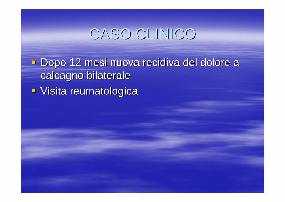
Dopo 12 mesi nuova recidiva del dolore a Dopo 12 mesi nuova recidiva del dolore a calcagno bilateralecalcagno bilateraleVisita reumatologicaVisita reumatologica

CASO CLINICOCASO CLINICO
EOLimitazione motilità rachide lombareBASDAI : 5.8 cmBASDAI entesi : 8.6 cmBASFI : 6.5 cmPCR 3.85 mg/dl, VES 32 mm/prima oraHLA-B27 +Indice entesitico Maastrict : 7

www.fisiokinesiterapia.biz

Tendine di Achille in longitudinale prima della infiltrazione
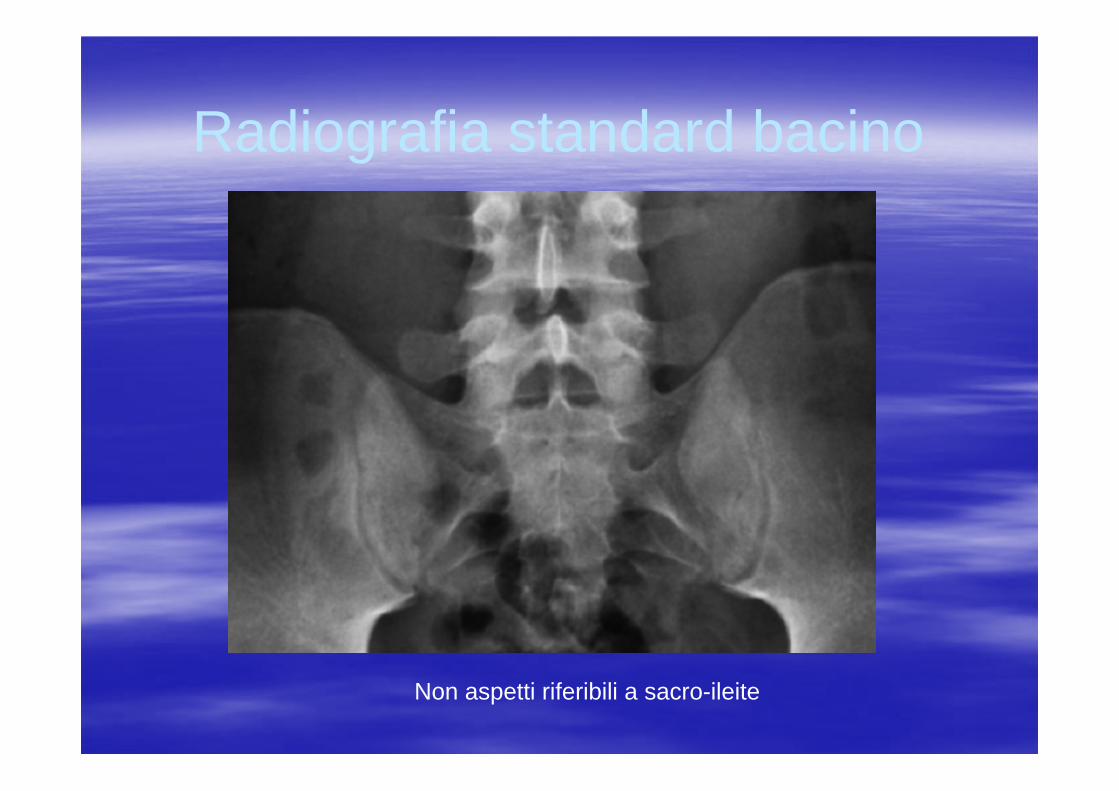
Radiografia standard bacino
Non aspetti riferibili a sacro-ileite

RMN delle sacro-iliache
Risonanza magnetica delle articolazioni sacro-iliache, sequenza STIR : sacro-ileite bilaterale (frecce) in proiezione semicoronale.

Problemi clinici
Spondilite HLA-B27 positivaTerapia con FANS a dosaggio pienoDa 3 mesi dolore al calcagno, molto intenso al mattino con difficoltà alla deambulazione

DefinitionsDefinitions EULAREULAR
EntesopatiaEntesopatia : ogni modificazione patologica : ogni modificazione patologica di una di una entesientesiEntesiteEntesite : ogni modificazione infiammatoria : ogni modificazione infiammatoria di una di una entesientesi

Cosa Cosa èè ll’’entesientesi
Il luogo di inserzione di un tendine, Il luogo di inserzione di un tendine, ligamento, capsula o fascia nellligamento, capsula o fascia nell’’ossoosso

Tendinite ed Tendinite ed entesiteentesite achilleaachillea

Schema struttura anatomica Schema struttura anatomica delldell’’entesientesi
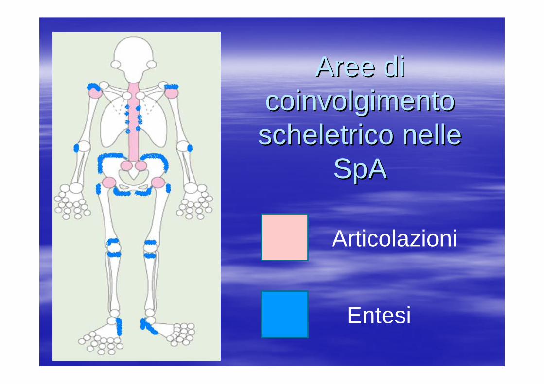
Aree di Aree di coinvolgimento coinvolgimento scheletrico nelle scheletrico nelle
SpASpA
Articolazioni
Entesi

Rilevanza dellRilevanza dell’’entesiteentesite nelle nelle SpASpA
Sebbene lSebbene l’’entesiteentesite achillea o della fascia plantare sia achillea o della fascia plantare sia una manifestazione clinica ben riconosciuta una manifestazione clinica ben riconosciuta edlleedllespondiliti fino alla fine degli anni spondiliti fino alla fine degli anni ‘‘90 era considerata 90 era considerata indipendente dalle altre manifestazioni cliniche delle indipendente dalle altre manifestazioni cliniche delle spondiliti come la sinovite, la sacrospondiliti come la sinovite, la sacro--ileite o lileite o l’’osteite osteite ((Calin&Calin& TaurogTaurog, 1998). , 1998). McGonagleMcGonagle etet al. (1998) hanno potuto dimostrare con al. (1998) hanno potuto dimostrare con ll’’impiego della RM che limpiego della RM che l’’entesiteentesite èè comune nelle comune nelle articolazioni sinoviali delle SPAarticolazioni sinoviali delle SPAInoltre Inoltre èè associata ad una diffusa osteite delassociata ad una diffusa osteite del’’osso osso immediatamanteimmediatamante adiacente adiacente sugerendosugerendo come lcome l’’etesiteetesiteòpossaòpossa essere essere iiliil concetto concetto uniiacanteuniiacante della SPA della SPA ((McGonagleMcGonagle etet al. 1998).al. 1998).
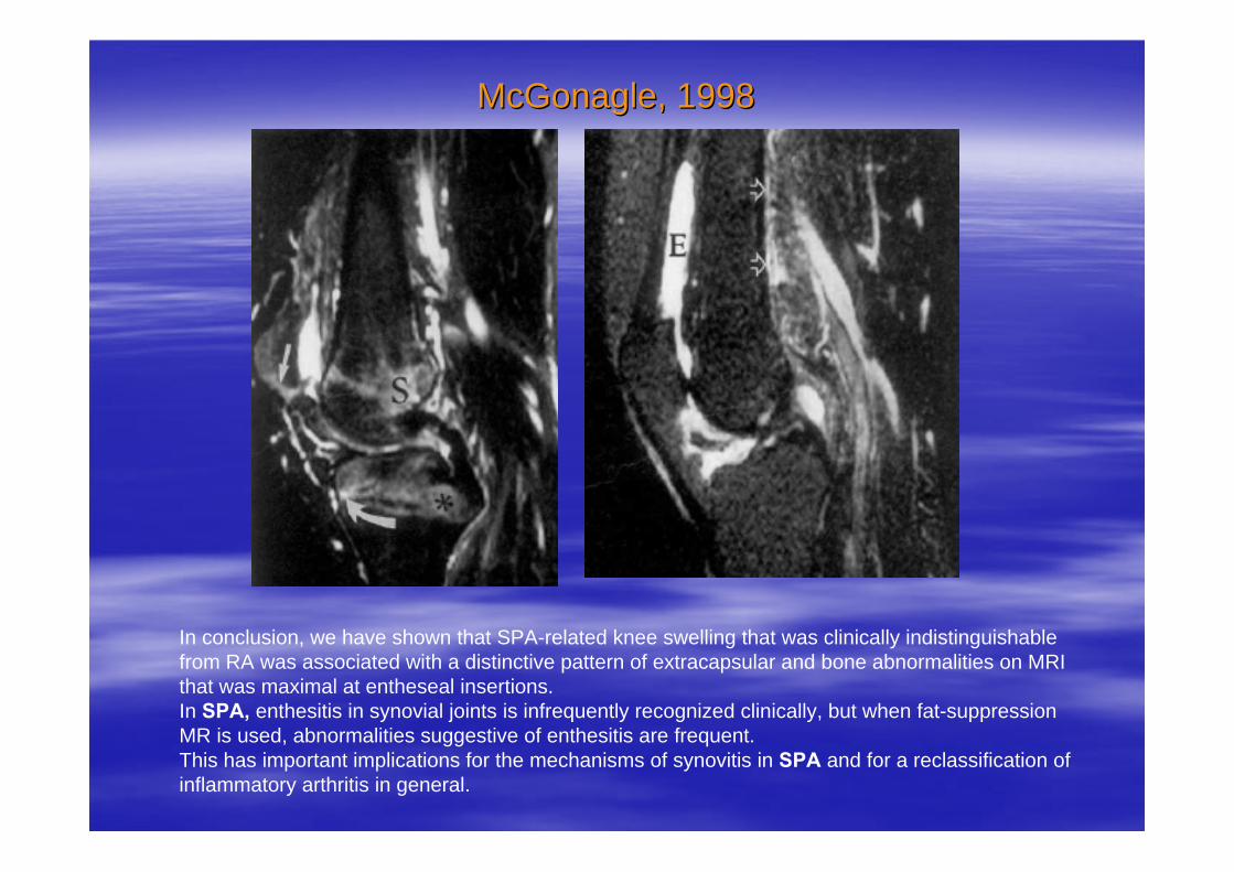
McGonagleMcGonagle, 1998, 1998
In conclusion, we have shown that SPA-related knee swelling that was clinically indistinguishablefrom RA was associated with a distinctive pattern of extracapsular and bone abnormalities on MRI that was maximal at entheseal insertions. In SPA, enthesitis in synovial joints is infrequently recognized clinically, but when fat-suppressionMR is used, abnormalities suggestive of enthesitis are frequent. This has important implications for the mechanisms of synovitis in SPA and for a reclassification ofinflammatory arthritis in general.

Classificazione delle malattie Classificazione delle malattie articolari infiammatorie articolari infiammatorie
((McGonagleMcGonagle,1996),1996)
SinoviticheSinovitiche (AR)(AR)EntesiticheEntesitiche (SA)(SA)

LL’’entesiteentesite nelle SpA nelle SpA
Ball per primo ha affermato che lBall per primo ha affermato che l’’entesiteentesite èè il il segno patognomonico della SA (segno patognomonico della SA (Ball J. Ball J. AnnAnn RheumRheum DisDis 1971, 1971,
30:21330:213––223223).).LL’’ipotesi ipotesi entesiticaentesitica èè stata poi estesa a tutte le stata poi estesa a tutte le SpA (SpA (PaolaggiPaolaggi JB, JB, etet al. al. RevRev Rhum Mal Rhum Mal OsteoarticOsteoartic 1984, 51:4511984, 51:451––456, 456, GaucherGaucher A, A, etet al. Presse al. Presse MedMed 1986, 15:6231986, 15:623––624, 624, McGonagleMcGonagle D, D, etet al.Arthritisal.ArthritisRheumRheum 1998, 41:6941998, 41:694––700700).).EE’’ stato anche affermato che la sinovite delle SpA stato anche affermato che la sinovite delle SpA sia una conseguenza della sia una conseguenza della entesiteentesite adiacente adiacente ((McGonagleMcGonagle D, D, GibbonGibbon W, W, EmeryEmery P. P. LancetLancet 1998, 353:11371998, 353:1137––11401140).).
Caratteristiche clinicheCaratteristiche cliniche
Segno della Segno della entesiteentesite periferica periferica èè il dolore il dolore Può essere però asintomaticaPuò essere però asintomaticaPuò essere dimostrata solo con Può essere dimostrata solo con ll’’imagingimaging tipo gli ultrasuoni (US), specie tipo gli ultrasuoni (US), specie se combinati con il se combinati con il powerpower--DopplerDoppler, dalla , dalla radiologia convenzionale o dalla RMradiologia convenzionale o dalla RM
Caratteristiche clinicheCaratteristiche cliniche
Il coinvolgimento delle Il coinvolgimento delle entesientesi pipiùùsuperficiali (tendine achilleo, tendini superficiali (tendine achilleo, tendini patellari o epicondilo laterale del gomito) patellari o epicondilo laterale del gomito) può essere associato ad una può essere associato ad una tumefazione sottocutanea ben evidente.tumefazione sottocutanea ben evidente.
Caratteristiche clinicheCaratteristiche cliniche
Al contrario il coinvolgimento delle Al contrario il coinvolgimento delle entesientesisituate pisituate piùù in profonditin profonditàà, come quelle della , come quelle della cresta iliaca, la sinfisi pubica, le tuberositcresta iliaca, la sinfisi pubica, le tuberositààischiatiche, il piccolo e grande trocantere ischiatiche, il piccolo e grande trocantere dimostrano solo dimostrano solo dolorabilitdolorabilitàà alla pressione e alla pressione e tumefazione palpabile.tumefazione palpabile.In questi casi quando la unica manifestazione In questi casi quando la unica manifestazione clinica clinica èè il dolore il dolore èè necessario escludere la necessario escludere la presenza di una presenza di una fibromialgiafibromialgia e documentare la e documentare la eventuale presenza di eventuale presenza di entesiteentesite con metodiche con metodiche di di imagingimaging..

Caratteristiche clinicheCaratteristiche clinicheIl dolore da Il dolore da entesiteentesite può essere molto intenso, può essere molto intenso, disabilitante e continuo, in certi casi (era disabilitante e continuo, in certi casi (era prepre biologica) biologica) può durare diversi anni. può durare diversi anni. GesterGester (2003) ha proposto dei criteri clinici per (2003) ha proposto dei criteri clinici per classificare lclassificare l’’entesiteentesite del calcagnodel calcagnoLa La talalgiatalalgia èè considerata modesta quando il dolore considerata modesta quando il dolore èèpresente occasionalmente sotto carico, si risole presente occasionalmente sotto carico, si risole rapidamente con il riposo ed rapidamente con il riposo ed èè scatenato da una scatenato da una pressione moderata o marcatapressione moderata o marcataViene invece classificata severa quando il dolore Viene invece classificata severa quando il dolore èèsempre presente sotto carico, si riduce lentamente sempre presente sotto carico, si riduce lentamente solo dopo riposo prolungato ed solo dopo riposo prolungato ed èè scatenato da scatenato da modesta pressione.modesta pressione.

Caratteristiche clinicheCaratteristiche cliniche
Spesso il dolore Spesso il dolore entesiticoentesitico èè massimo al massimo al carico dopo un periodo di riposo, specie carico dopo un periodo di riposo, specie al mattino al risveglio.al mattino al risveglio.EE’’ molto intenso durante i primi molto intenso durante i primi movimenti e migliora con il cammino.movimenti e migliora con il cammino.
Epidemiologia Epidemiologia entesiteentesite
PeripheralPeripheral enthesitisenthesitis maymay bebe observedobserved in in allall formsformsofof SpA SpA In In allall phasesphases ofof diseasedisease evolutionevolution..IsIs particularlyparticularly frequentfrequent in in juvenilejuvenile--onsetonset SpA.SpA.In In primaryprimary AS, the AS, the frequencyfrequency ofof peripheralperipheralenthesitisenthesitis hashas beenbeen foundfound toto bebe betweenbetween 25 and 25 and 58%. 58%. SometimesSometimes, , especiallyespecially in in juvenilejuvenile--onsetonset formsforms, , ititprecedesprecedes in in associationassociation or or notnot withwith peripheralperipheralarthritisarthritis spinalspinal symptomssymptoms and and radiologicalradiological findingsfindings..
Valutazione clinica delle Valutazione clinica delle entesitientesiti
Anamnesi Anamnesi Esame obiettivo : ispezione delle Esame obiettivo : ispezione delle entesientesisuperficiali per tumefazione e arrossamento superficiali per tumefazione e arrossamento cutaneo, cutaneo, dolorabilitdolorabilitàà alla pressione nelle alla pressione nelle sedi anatomiche sedi anatomiche

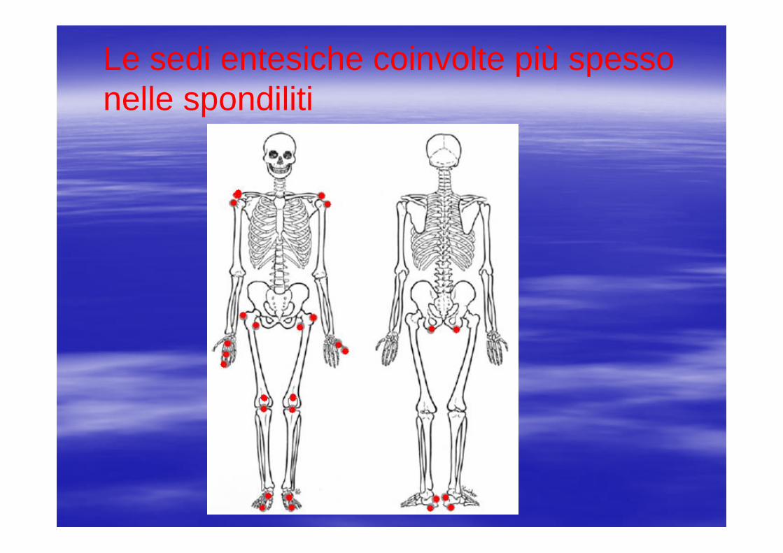
Le sedi entesiche coinvolte più spesso nelle spondiliti (Eshed I., 2007)
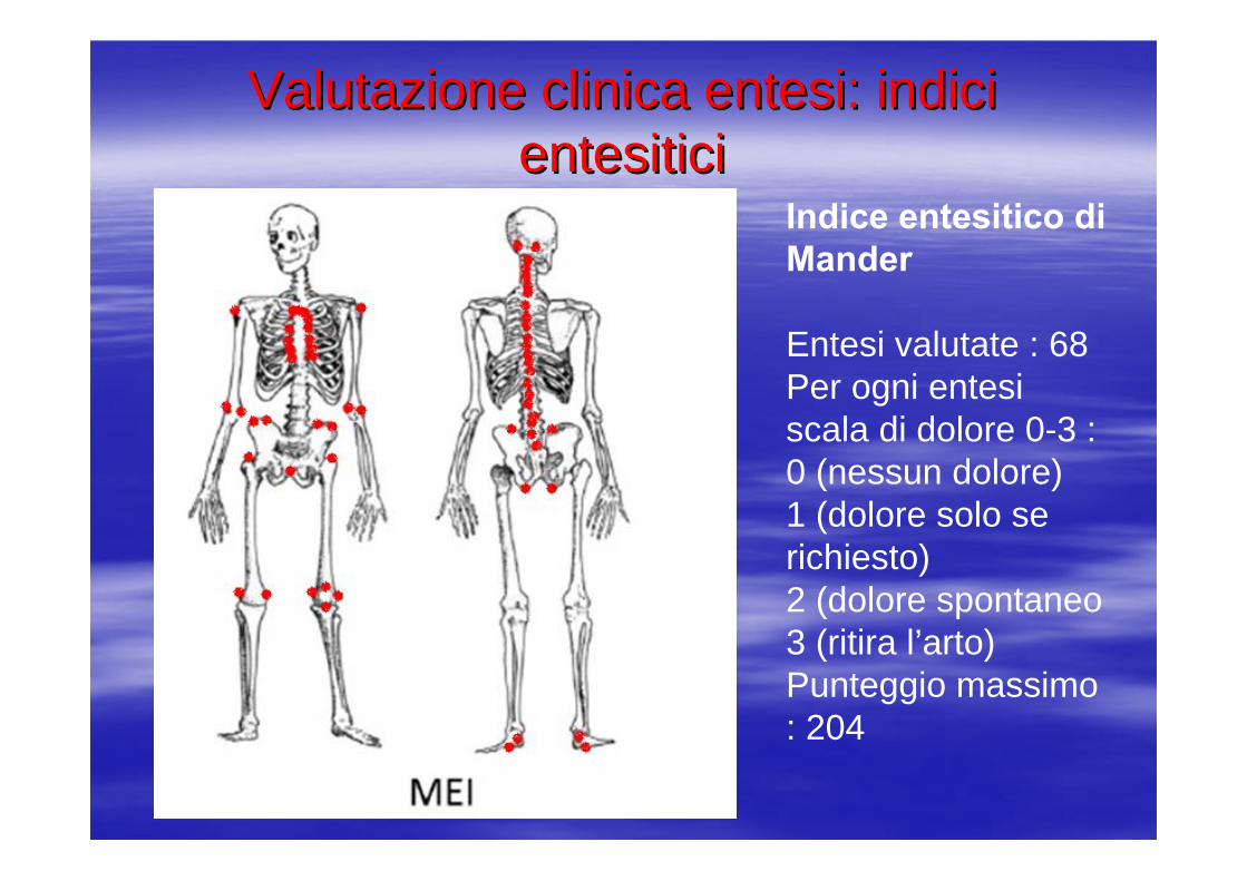
Valutazione clinica Valutazione clinica entesientesi: indici : indici entesiticientesitici
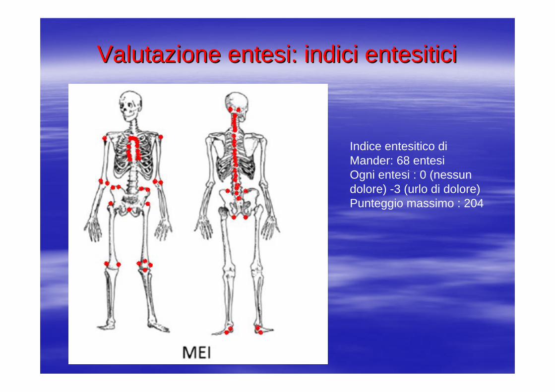
Indice entesitico di Mander
Entesi valutate : 68 Per ogni entesiscala di dolore 0-3 : 0 (nessun dolore)1 (dolore solo se richiesto)2 (dolore spontaneo 3 (ritira l’arto)Punteggio massimo : 204

Valutazione clinica Valutazione clinica entesientesi: indici : indici entesiticientesitici
Indice entesitico di Maastricht
Numero entesivalutate :13Valore per ogni entesi0-3Valore massimo :39

Valutazione clinica Valutazione clinica entesientesi: indici : indici entesiticientesitici
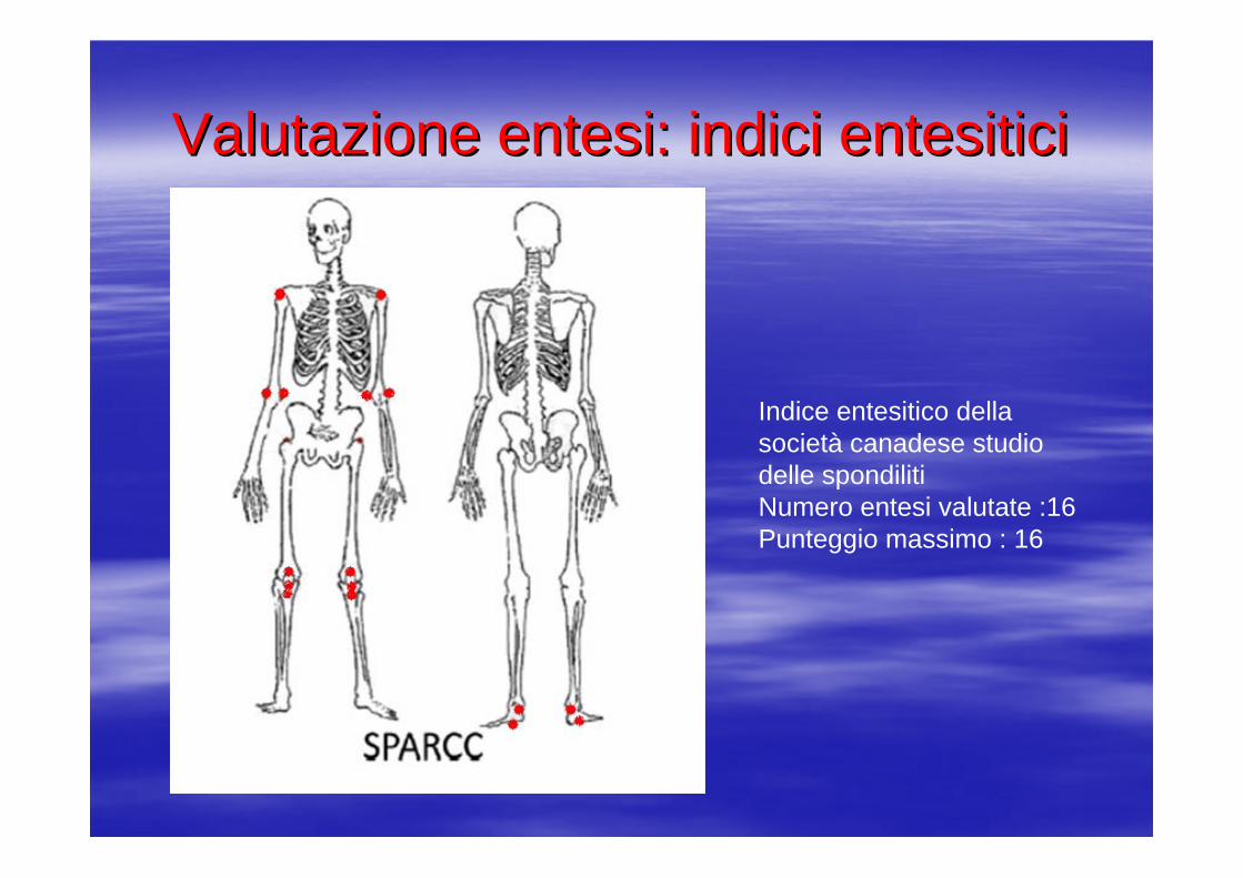
SPARCC: SpondyloarthritisResearch Consortium ofCanada Enthesitis Index
Numero entesi valutate :16Punteggio massimo : 16

Valutazione clinica Valutazione clinica entesientesi: indici : indici entesiticientesitici
Indice entesitico di Leeds
Numero entesivalutate : 6Valore massimo : 6

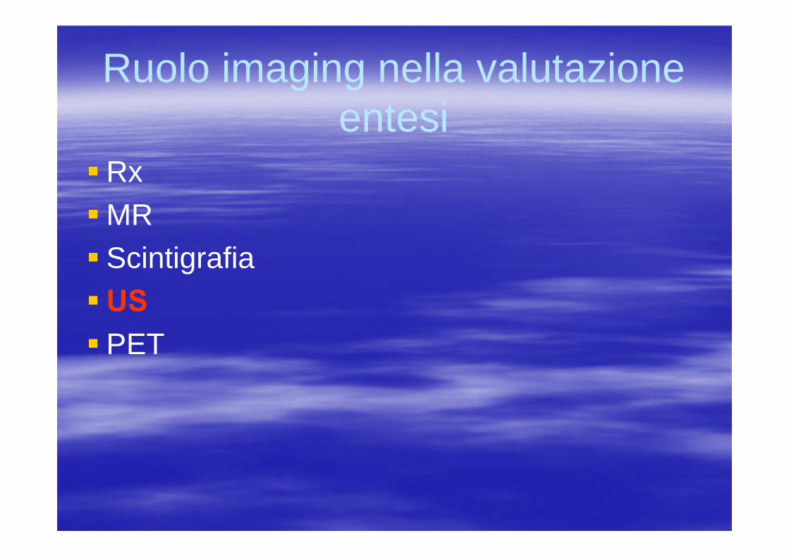
Ruolo imaging nella valutazione entesi
RadiografiaMRScintigrafiaUSPET

Valutazione radiologica delle Valutazione radiologica delle entesientesi
VantaggiVantaggiFacile disponibilitFacile disponibilitààCosto limitatoCosto limitatoIndagine standardIndagine standardCorrela con il dato clinicoCorrela con il dato clinico
SvantaggiSvantaggiUso di radiazioniUso di radiazioniScarsa sensibilitScarsa sensibilitàà nelle lesioni inizialinelle lesioni inizialiDimostra solo lesioni stabilizzateDimostra solo lesioni stabilizzateNon utilizzabile per la valutazione delle flogosi Non utilizzabile per la valutazione delle flogosi Poco utile nel followPoco utile nel follow--upup

Valutazione radiologica delle Valutazione radiologica delle entesientesi
LESIONI ELEMENTARILESIONI ELEMENTARI
ErosioniErosioni osseeosseeentesofitientesofitiIspessimentoIspessimento entesicoentesico. .

RxRx entesientesi : erosioni e crescita : erosioni e crescita osseaossea
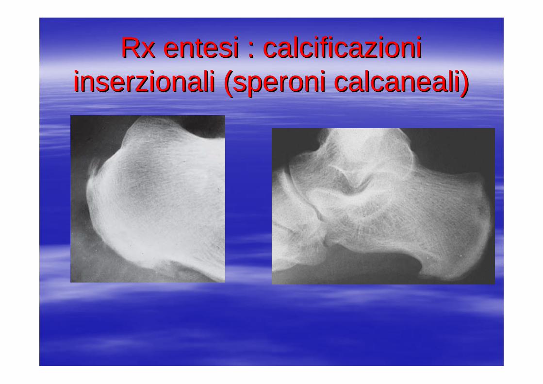
RxRx entesientesi : calcificazioni : calcificazioni inserzionaliinserzionali (speroni (speroni calcanealicalcaneali))

Lateral radiographs of the knees and heels were obtained bilaterally and used to assess the following entheses: patellar insertion of the quadriceps tendon, proximal and distal insertions of the patellar tendon, calcaneal insertions of the Achilles tendon and plantar fascia. At each of the 10 entheseal sites, the following three radiographic criteria were recorded on a standardized form: bony erosion, enthesophyte, entheseal thickening. Each criterion was scored 1 if present and 0 if absent (range 0-30).
Ruolo imaging nella valutazione entesi
RxMRScintigrafiaUSPET
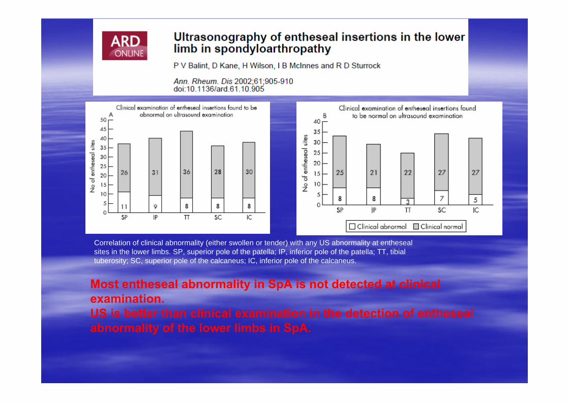
Most entheseal abnormality in SpA is not detected at clinical examination. US is better than clinical examination in the detection of enthesealabnormality of the lower limbs in SpA.
Correlation of clinical abnormality (either swollen or tender) with any US abnormality at enthesealsites in the lower limbs. SP, superior pole of the patella; IP, inferior pole of the patella; TT, tibialtuberosity; SC, superior pole of the calcaneus; IC, inferior pole of the calcaneus.
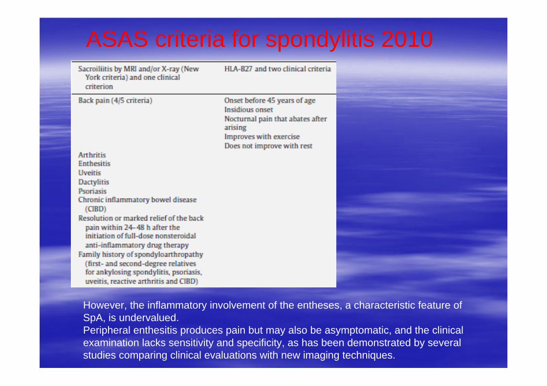
ASAS criteria for spondylitis 2010
However, the inflammatory involvement of the entheses, a characteristic feature of SpA, is undervalued. Peripheral enthesitis produces pain but may also be asymptomatic, and the clinical examination lacks sensitivity and specificity, as has been demonstrated by several studies comparing clinical evaluations with new imaging techniques.
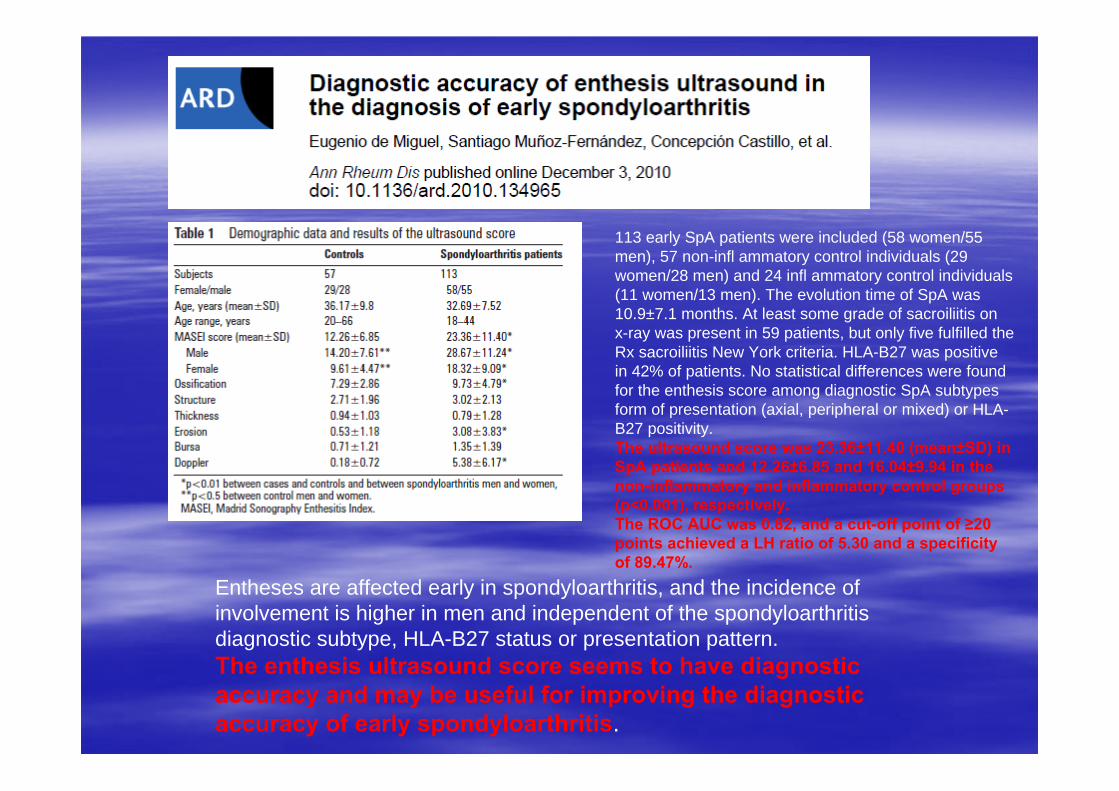
Entheses are affected early in spondyloarthritis, and the incidence of involvement is higher in men and independent of the spondyloarthritisdiagnostic subtype, HLA-B27 status or presentation pattern. The enthesis ultrasound score seems to have diagnostic accuracy and may be useful for improving the diagnostic accuracy of early spondyloarthritis.
113 early SpA patients were included (58 women/55 men), 57 non-infl ammatory control individuals (29 women/28 men) and 24 infl ammatory control individuals (11 women/13 men). The evolution time of SpA was 10.9±7.1 months. At least some grade of sacroiliitis on x-ray was present in 59 patients, but only five fulfilled the Rx sacroiliitis New York criteria. HLA-B27 was positive in 42% of patients. No statistical differences were found for the enthesis score among diagnostic SpA subtypes form of presentation (axial, peripheral or mixed) or HLA-B27 positivity. The ultrasound score was 23.36±11.40 (mean±SD) in SpA patients and 12.26±6.85 and 16.04±9.94 in the non-inflammatory and inflammatory control groups (p<0.001), respectively. The ROC AUC was 0.82, and a cut-off point of ≥20 points achieved a LH ratio of 5.30 and a specificity of 89.47%.
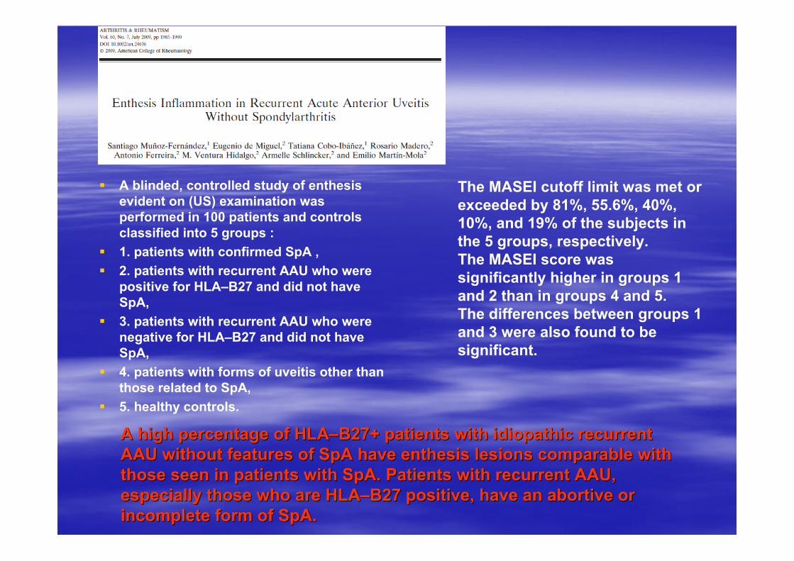
A blinded, controlled study of enthesisevident on (US) examination was performed in 100 patients and controls classified into 5 groups :1. patients with confirmed SpA ,2. patients with recurrent AAU who were positive for HLA–B27 and did not have SpA, 3. patients with recurrent AAU who were negative for HLA–B27 and did not have SpA, 4. patients with forms of uveitis other than those related to SpA,5. healthy controls.
The MASEI cutoff limit was met or exceeded by 81%, 55.6%, 40%, 10%, and 19% of the subjects in the 5 groups, respectively. The MASEI score was significantly higher in groups 1 and 2 than in groups 4 and 5. The differences between groups 1 and 3 were also found to be significant.
A high percentage of HLAA high percentage of HLA––B27+ patients with idiopathic recurrent B27+ patients with idiopathic recurrent AAU without features of AAU without features of SpASpA have have enthesisenthesis lesions comparable with lesions comparable with those seen in patients with those seen in patients with SpASpA. Patients with recurrent AAU, . Patients with recurrent AAU, especially those who are HLAespecially those who are HLA––B27 positive, have an abortive or B27 positive, have an abortive or incomplete form of incomplete form of SpASpA..

Lesioni ultrasonografiche elementari
DEFINITIONEnthesisEnthesis thickening and thickening and hypoechogenicityhypoechogenicity are evaluated relative toare evaluated relative to
the body of the tendon. CalcificCalcific depositdeposit at the enthesis are hyperechoic spots or lines at the
preinsertional area of the tendons, with or without acoustic shadowing, seen in 2 perpendicular planes. Bone erosionBone erosion is a discontinuity of the entheseal bone surface, seen in
2 perpendicular planes. EnthesophyteEnthesophyte is a hyperechoic prominence at the end of the
entheseal bone contour, seen in 2 perpendicular planes.BursitisBursitis is a well circumscribed hypoechoic or anechoic collection at
the site of an anatomic bursa.

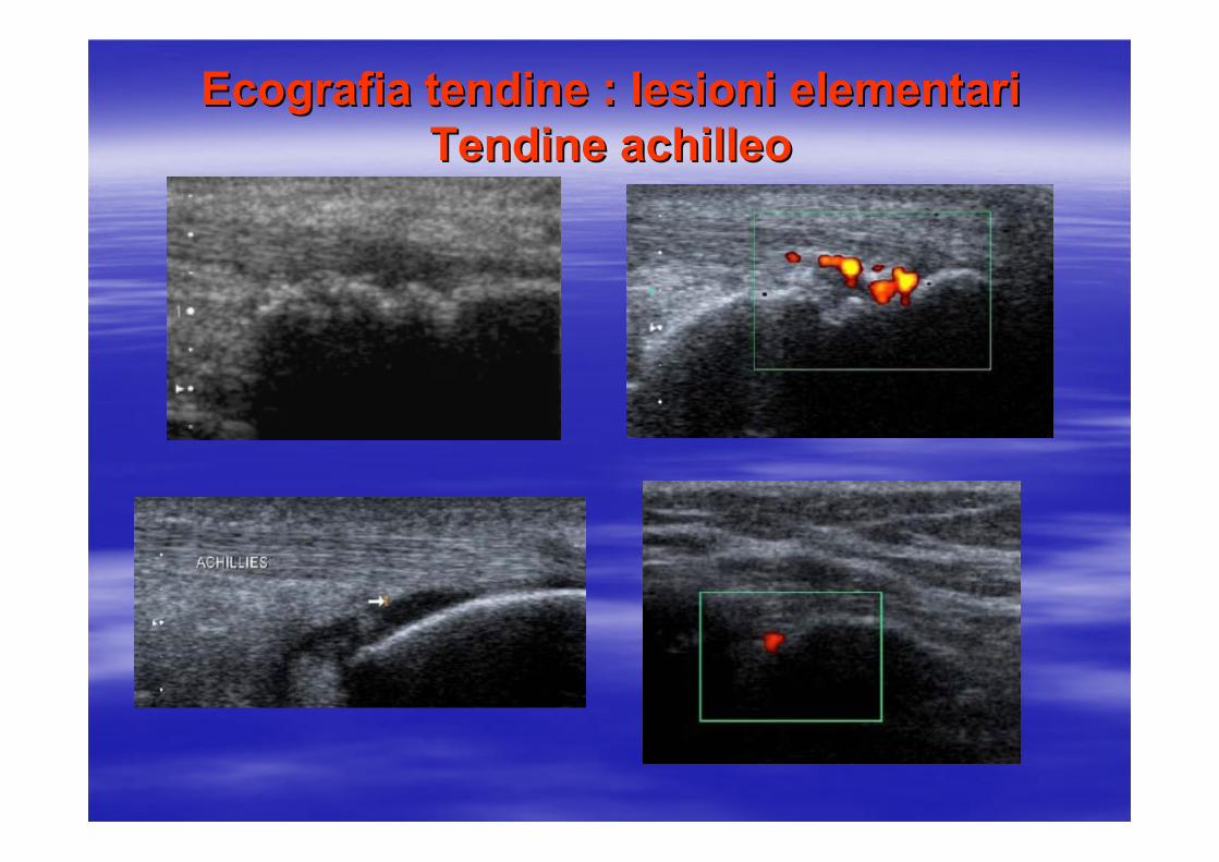
Ecografia tendine : lesioni elementariEcografia tendine : lesioni elementariTendine achilleoTendine achilleo
AchillesAchilles tendontendon. . LongitudinalLongitudinal scanscan. . PresencePresenceof of hypoechogenicityhypoechogenicity of the tendon structure (due of the tendon structure (due
to to intrafibrillarintrafibrillar oedemaoedema) and ) and retrocalcanearretrocalcanearbursitis (asterisk), with intense PD signal. bursitis (asterisk), with intense PD signal. Note the integrity of the bone profile. Note the integrity of the bone profile.
Achilles tendon. Longitudinal scan.Tendonitis. Note the hypoechogenicity of the structure of the tendon and the peritendineousoedema (e) with PD signal
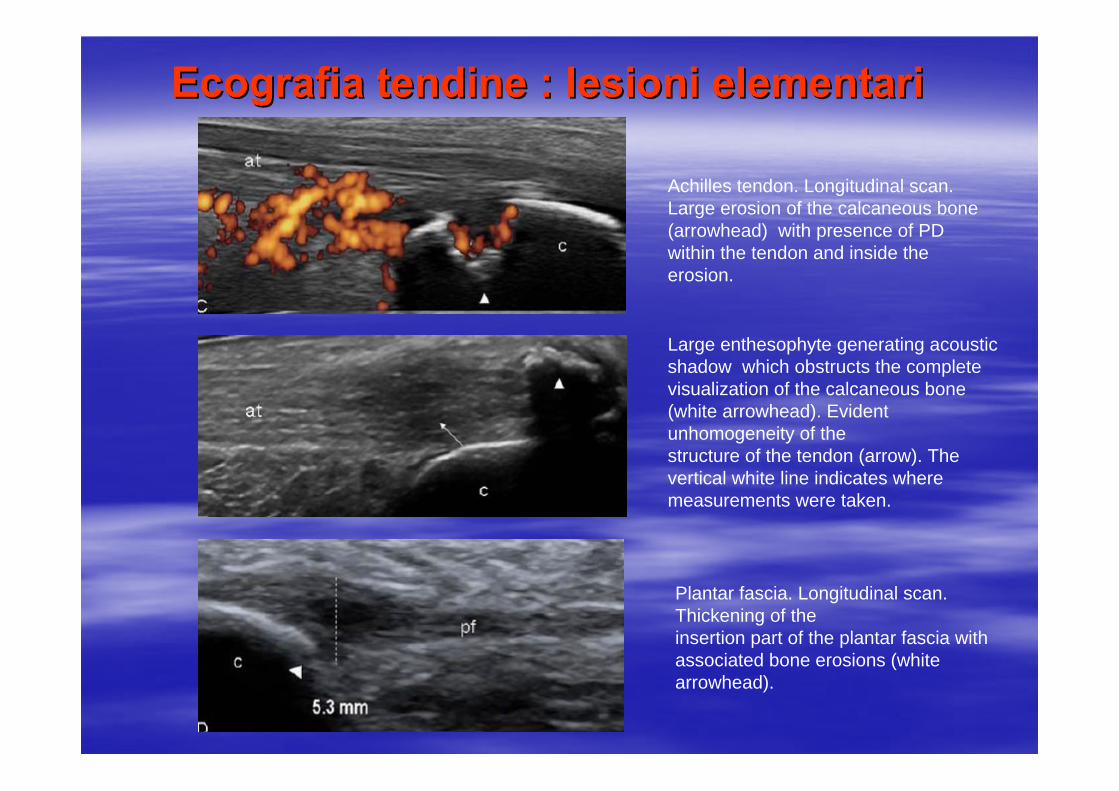
Ecografia tendine : lesioni elementariEcografia tendine : lesioni elementari
Achilles tendon. Longitudinal scan. Large erosion of the calcaneous bone (arrowhead) with presence of PD within the tendon and inside the erosion.
Large enthesophyte generating acoustic shadow which obstructs the complete visualization of the calcaneous bone (white arrowhead). Evident unhomogeneity of thestructure of the tendon (arrow). The vertical white line indicates where measurements were taken.
Plantar fascia. Longitudinal scan. Thickening of theinsertion part of the plantar fascia with associated bone erosions (whitearrowhead).
Ecografia tendine : lesioni elementariEcografia tendine : lesioni elementariTendine sottorotuleoTendine sottorotuleo
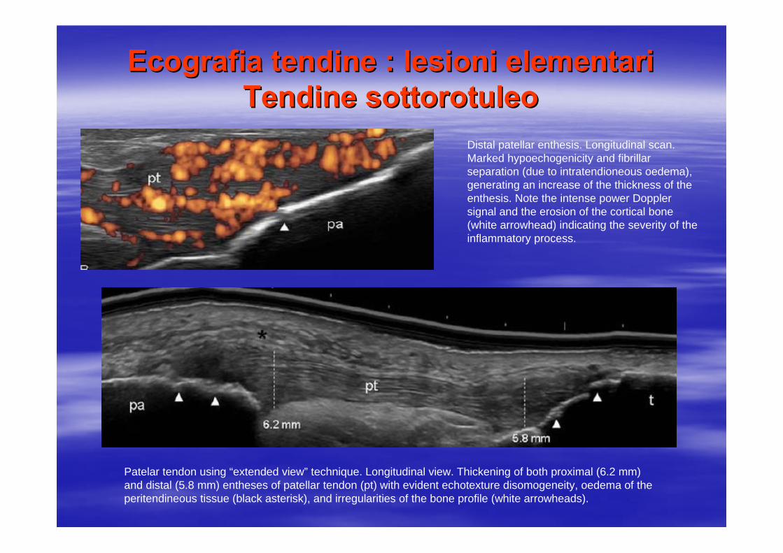
Patelar tendon using “extended view” technique. Longitudinal view. Thickening of both proximal (6.2 mm)and distal (5.8 mm) entheses of patellar tendon (pt) with evident echotexture disomogeneity, oedema of the peritendineous tissue (black asterisk), and irregularities of the bone profile (white arrowheads).
Distal patellar enthesis. Longitudinal scan. Marked hypoechogenicity and fibrillar separation (due to intratendioneous oedema), generating an increase of the thickness of the enthesis. Note the intense power Doppler signal and the erosion of the cortical bone (white arrowhead) indicating the severity of the inflammatory process.

Ecografia tendine : lesioni elementariEcografia tendine : lesioni elementariTendine achilleoTendine achilleo
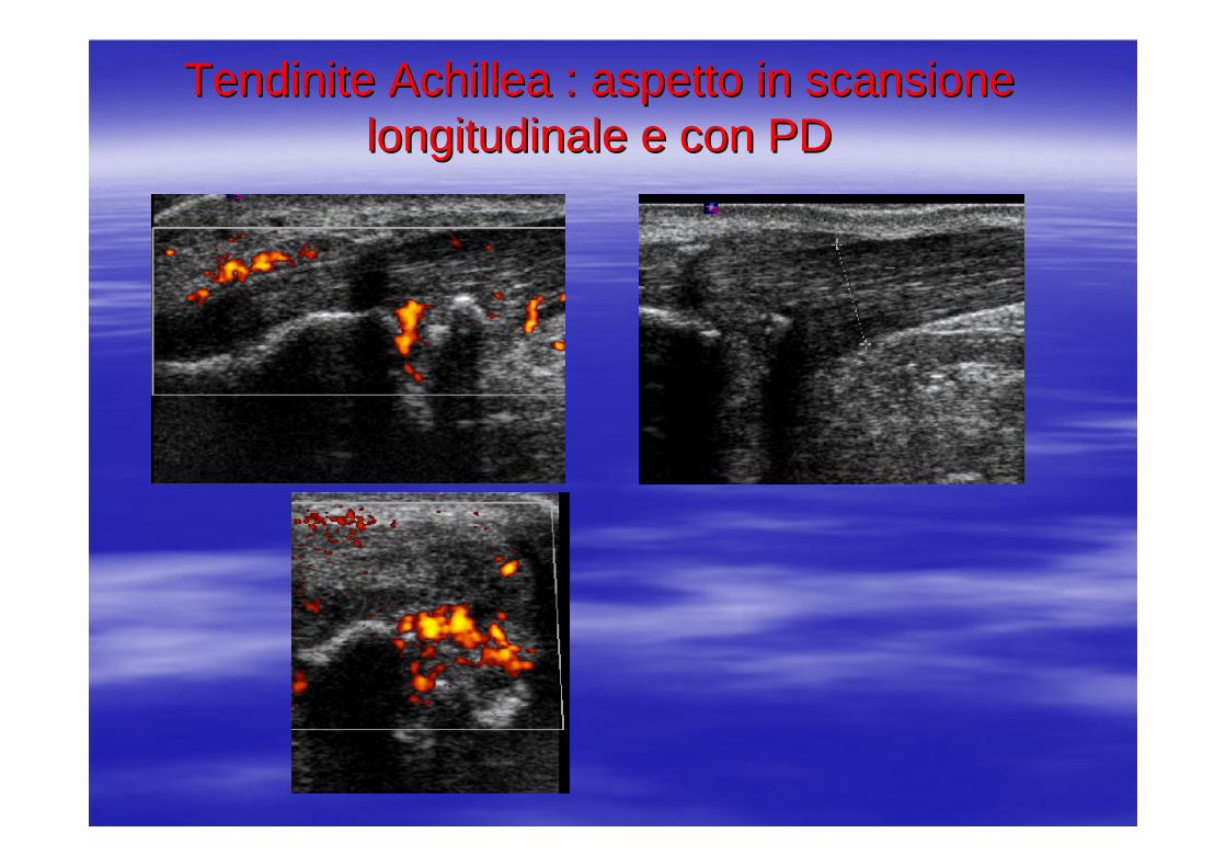
US appearance of grey-scale enthesitis of Achilles tendon enthesitis in a longitudinal scan. Enthesophytes (E) andhypoechogenicity (H).
US appearance of grey-scale and PD enthesitis of Achilles tendon enthesitis in a longitudinal scan: erosions (A), PD signal (B); Hypoechogenicity and incresead of thickness (C), enthesophites (D).
Ecografia tendine : lesioni elementariEcografia tendine : lesioni elementariTendine achilleoTendine achilleo

Ecografia tendine : lesioni elementariEcografia tendine : lesioni elementariFascia plantareFascia plantare

Ecografia tendine : lesioni elementariEcografia tendine : lesioni elementariTendine ACHILLEOTendine ACHILLEO
A : Erosione con segnale Doppler B: Ridotta ecogenicitàC: Entesofita con segnale Doppler
AA(A)
(B)
(C)

Lesioni US elementari. Lesioni US elementari.
Tendine achilleo in sezione longitudinale.Tendine achilleo in sezione longitudinale.
A. Calcificazione intratendinea; B. Erosione; C. Edema peritendineo; D.Disomogeneità del tendine; E. Vascolarizzazione peritendinea; F. Vascolarizzazione intratendinea
(A)
(B)
(C)
(D)
(E)
(F)
Ruolo imaging nella valutazione entesi
RxMRScintigrafiaUSPET

Valutazione entesite in MRIThe normal tendon on MR images has homogeneous low signal intensity in all sequences.
When evaluating MR images for enthesitis one should evaluate:thickness and signal intensitythickness and signal intensity of tendons and ligaments.periperi--enthesalenthesal soft tissuessoft tissues for swelling or oedema.adjacent bone marrow to detect adjacent bone marrow to detect oedemaoedema, best appreciated as high signal in fat suppressed sequences.adjacent bone for erosionsadjacent bone for erosions (cortical bone defects and contour irregularities) and enthesophytesenthesophytes (extensions of marrow contents isointense to the medullary bone), (best appreciated on T1-weighted sequences).additional findings in adjacent structures (joint or joint or bursalbursal fluidfluid forexample).
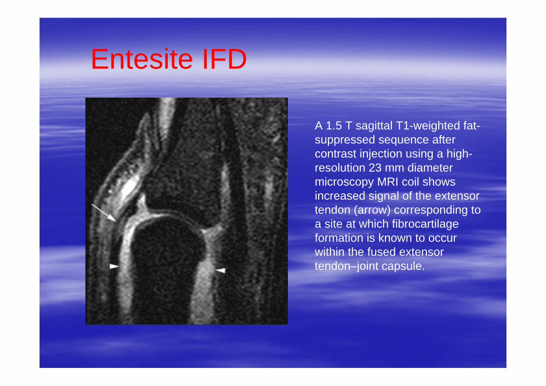
A 1.5 T sagittal T1-weighted fat-suppressed sequence after contrast injection using a high-resolution 23 mm diameter microscopy MRI coil showsincreased signal of the extensor tendon (arrow) corresponding to a site at which fibrocartilageformation is known to occur within the fused extensortendon–joint capsule.
Entesite IFD

Entesite IFDA 1.5 T coronal high-resolution T1-weightedfat-suppressed sequence after contrast injection. The diffuse pattern of periarticular bone marrow oedema (arrows) as well as peri-entheseal soft tissueoedema (arrowheads) isseen.
www.fisiokinesiterapia.biz
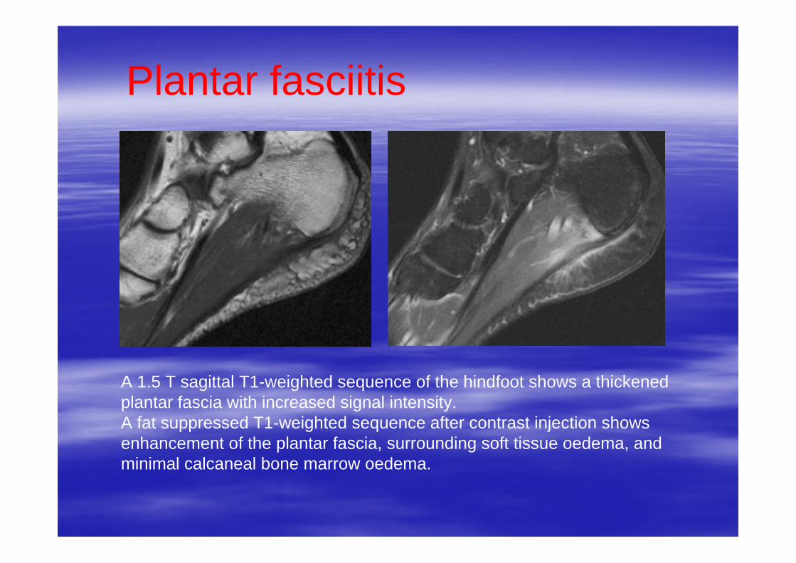
A 1.5 T sagittal T1-weighted sequence of the hindfoot shows a thickened plantar fascia with increased signal intensity. A fat suppressed T1-weighted sequence after contrast injection shows enhancement of the plantar fascia, surrounding soft tissue oedema, and minimal calcaneal bone marrow oedema.
Plantar fasciitis

1.5 T sagittal and axial fat-suppressed T1-weighted sequences after contrast injection of the knee. Localised bone marrow oedema at the posterior aspect of the lateral femoralcondyle (arrowheads) at the insertion site of the lateral collateral ligament and the origins of the lateral head of the gastrocnemius and the popliteus muscles is depicted.
Entesite ginocchio

Entesite anca e bacino
A 1.5 T transverse STIR sequence showing extensive bone marrow oedema(white arrows) in the femoral head and acetabulum as well as hip joint effusion (white arrowheads). Same sequence more caudally shows bursitis (black arrowhead) and enthesisrelated bone marrow oedema at the lesser femoral trochanter (black arrow), ischial tubercles (white asterisks) and pubic bones (black asterisks).

RMN: lesioni elementari. Edema osseo.
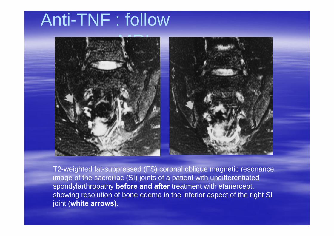
T2-weighted fat-suppressed (FS) coronal oblique magnetic resonance image of the sacroiliac (SI) joints of a patient with undifferentiated spondylarthropathy showing bone edema in the inferior aspect of the right SI joint (white arrows).

RMN: lesioni elementari. Edema osseo.
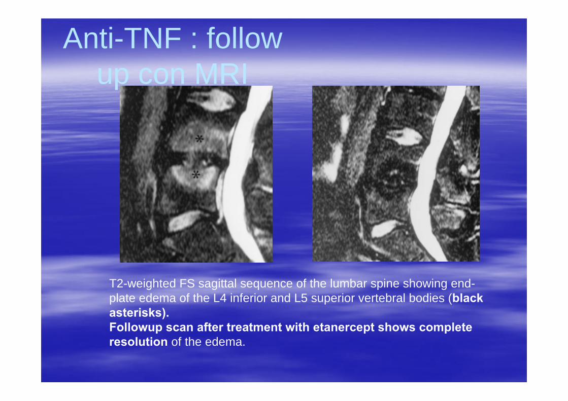
T2-weighted FS sagittal sequence of the lumbar spine showing end-plate edema of the L4 inferior and L5 superior vertebral bodies (black asterisks).

RMN: lesioni elementari. Edema osseo e flogosi tessuto perientesico.
T1-weighted fat-suppressed post-gadolinium coronal sequence of the left second distal IP joint of a patient with SpA.
Extensive subcutaneous edema (small black asterisk) bone marrow edema (large blackasterisk) and inflammatory change within the collateral ligament (black arrow).
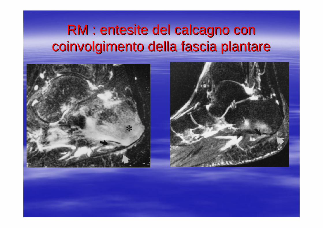
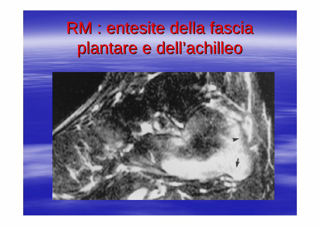
RM : RM : entesiteentesite del calcagno con del calcagno con coinvolgimento della fascia plantarecoinvolgimento della fascia plantare

RM : RM : entesiteentesite della fascia della fascia plantare e dellplantare e dell’’achilleoachilleo

RM : RM : entesiteentesite della caviglia e della caviglia e della IFPdella IFP

RM : RM : entesiteentesite della inserzione della inserzione sovraspinatosovraspinato
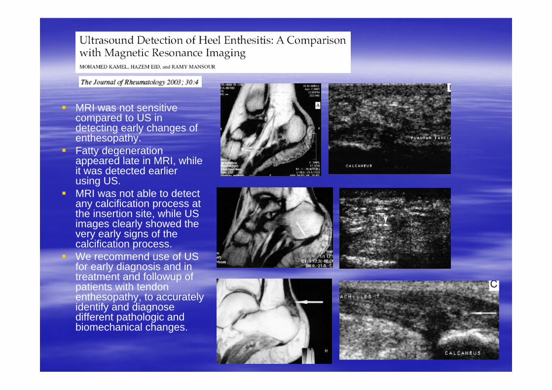
MRI was not sensitive compared to US in detecting early changes ofenthesopathy. Fatty degenerationappeared late in MRI, whileit was detected earlierusing US. MRI was not able to detectany calcification process at the insertion site, while US images clearly showed the very early signs of the calcification process. We recommend use of US for early diagnosis and in treatment and followup ofpatients with tendonenthesopathy, to accuratelyidentify and diagnosedifferent pathologic and biomechanical changes.

Quale imaging nelle entesopatie?RISONANZA MAGNETICAVANTAGGIDimostrazione di lesioni acute infiammatorie, Correla con il dato clinicoUtile nel follow-upNon uso i radiazioni
SVANTAGGIScarsa sensibilità per il riconoscimento di calcificazioni, Scarsa sensibilità nel riconoscimento di alterazioni degenerative Non facile disponiblitàCosto elevato

Quale imaging nelle entesopatie?ULTRASONOGRAFIAMetodo di scelta, Ottima sensibilità e specificità, Buona riproducibilità, Possibile correlazione con il dato clinicoNon uso di radiazioni ionizzantiUtile nel follow-upCosto limitatoFacile disponibilità.

Quale imaging nelle entesopatie?
ULTRASONOGRAFIASVANTAGGISVANTAGGIOperatore dipendenzaOperatore dipendenzaNon sufficiente standardizzazioneNon sufficiente standardizzazioneMinore esperienzaMinore esperienza
Quale imaging ?Nessuna : scelta possibile non linee guida sull’imaging periferico nelle SpA Scarsa sensibilità rispetto all’imaging.RX : dimostrazione di lesioni stabilizzate, scarsa sensibilità nelle lesioni iniziali, correla come indice con il dato clinico MRI : dimostrazione di lesioni acute, scarsa sensibilità per il riconoscimento di calcificazioni, limitazioni dalla scarsa disponibilità, non radiazioni, correla con il dato clinico.

Quale imaging ?Scintigrafia : uso di radiazioni, buona sensibilità, multiple sedi valutate contemporaneamente Ultrasuoni : metodo di scelta, ottima sensibilità e specificità, buona riproducibilità, possibile correlazione con il dato clinico.PET : ancora sperimentale, radiazioni, buona correlazione con il dato clinico, multiple sedi contemporaneamenteRMN total body : ancora scarsa disponibilità, non radiazioni, multiple sedi contemporaneamente (entesi, articolari, assiali, periferiche)

Quale terapiaInfiltrazione locale con steroidi (prevista dalle linee guida ma non indicati i dosaggi, non studi controllati, solo case-report, non indicate le metodologie (intra-tendineo, peritendineo, metodo palpatorio o imaging guidato) FANS a dosaggio pieno (pochi studi, probabile efficacia, poche indicazioni sulla durata e sul dosaggio.)Sulfasalazina(pochi studi, non efficace.)Methotrexate(pochi studi, non efficace.)Anti-TNF(farmaci di scelta nei casi non responder alle terapie locali o ai FANS, previsti da tutte le linee guida internazionali e nazionali. Solo recente comparsa di studi randomizzati specifici per questo item.)

Tendine di Achille in longitudinale prima della infiltrazione

6 tendons in 5 patients were evaluated withGS US and CD before and after US-guidedintratendinous GC injection. Pain at rest and at activity was evaluated on a VAS.Results:With CD all tendons had intratendinous flow. Pain and CD activity decreased during a meanfollow-up of 182 days (range 92–309 days).One tendon relapsed after 199 days. Conclusion:Intratendinous GC injections seem to have a marked effect on both symptoms and CDfindings.CD adds significant information to GSUS withregard to diagnosis, location and follow-up ofAT.

Injection of 7 mg (1 ml) of betamethasone and 0.5 ml of 1% lidocaine into the inflamedproximal plantar fascia was performed under US guidance or palpation.VAS-measured levels of pain improvedsignificantly after CS injection in both groups(p < 0.001). Thickness decreased significantly afterinjection (p < 0.01 in the palpationguidedgroup; p < 0.001 in the US guided group). The number of patients with hypoechogenicityat the proximal plantar fascia decreased afterCS injection in both groups (p < 0.01 for bothgroups). The recurrence rate of plantar fasciitis in patients of the palpation-guided group (6/13) was significantly higher than that of the US guided group (1/12) (p < 0.05).
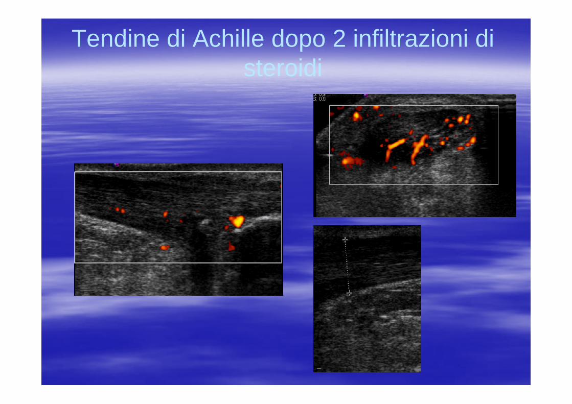
Tendine di Achille dopo 2 infiltrazioni di steroidi

Ø Nine patients had a total of 44 MRI-detectable entheseal lesions. These were seen in the SI joints in 6 patients (n 5 = 15 lesions), in the lumbar or cervical spine in 9 patients (n 5 = 22 lesions), and in peripheral joints in 5 patients (n 5 = 7 lesions). Ø Overall, 86% of MRI detected entheseal lesions either regressed completely or improved. Ø No new lesions developed.Ø Conclusion. TNFa blockade with etanercept is markedly effective in controlling the clinical manifestations of SpAthat is resistant to disease-modifying antirheumatic drugs. This is associated with marked improvement of enthesitis and associated osteitis pathology as determined by MRI.

Definizione di entesite in MRIMRI enthesitis was defined on T2 FS images as bone edema (identified by high or intermediate marrow signal) and/or soft tissue edema (high signal in the extracapsular connective tissues) adjacent to entheses. MRI scoring. In the SI joints, 4 quadrants were assessed: right upper, left upper, right lower, and left lower. Each quadrant was subdivided into ilialand sacral aspects. In the spine, lesions were classified as present within the vertebrae or in the paraspinal soft tissues
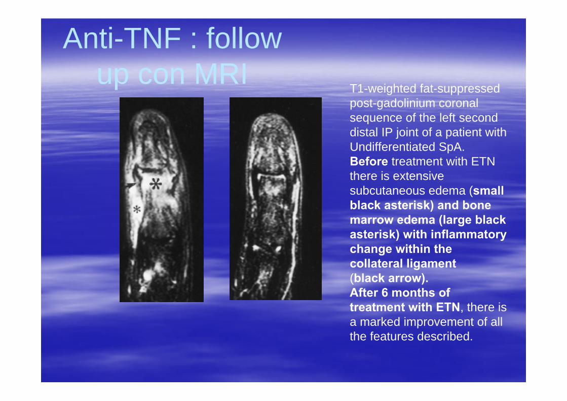
Anti-TNF : followup con MRI
T2-weighted fat-suppressed (FS) coronal oblique magnetic resonance image of the sacroiliac (SI) joints of a patient with undifferentiated spondylarthropathy before and after treatment with etanercept, showing resolution of bone edema in the inferior aspect of the right SIjoint (white arrows).
Anti-TNF : followup con MRI
T2-weighted FS sagittal sequence of the lumbar spine showing end-plate edema of the L4 inferior and L5 superior vertebral bodies (black asterisks). Followup scan after treatment with etanercept shows complete resolution of the edema.
Anti-TNF : followup con MRI
T1-weighted fat-suppressed post-gadolinium coronal sequence of the left second distal IP joint of a patient withUndifferentiated SpA. Before treatment with ETN there is extensive subcutaneous edema (small black asterisk) and bone marrow edema (large blackasterisk) with inflammatory change within the collateral ligament(black arrow). After 6 months of treatment with ETN, there is a marked improvement of all the features described.

Sagittal fat-suppressed T1- weighted MR images of the left knee following contrast administration: prior to, at 6 weeks, and 2 years after starting etanercept.

Gli US nel follow-up delle entesopatie
At each enthesis, the following elementary lesions were assessed (1) entheseal morphologicabnormalities (i.e., hypoechogenicityand/or thickening); (2) entheseal calcific deposits; (3) entheseal cortical abnormalities (i.e., bone erosion and/or enthesophytes); (4) adjacent bursitis;
DEFINITIONØ Enthesis thickening and hypoechogenicitywere evaluated relative to the body of the tendon. Ø Calcific deposit at the enthesis was defined as hyperechoic spots or lines at the preinsertional area of the tendons, with or without acoustic shadowing, seen in 2 perpendicular planes. Ø Bone erosion was defined as a discontinuity of the entheseal bone surface, seen in 2 perpendicular planes. Ø Enthesophyte was defined as a hyperechoicprominence at the end of the entheseal bone contour, seen in 2 perpendicular planes.Ø Bursitis was defined as a well circumscribed hypoechoic or anechoic collection at the site of an anatomic bursa.

Gli US nel follow-up delle entesopatie

Gli US nel follow-up delle
entesopatie ØThere was no correlation between clinical and laboratory measures and PDUS abnormalities. ØThere was no correlation between US entheseal findings and clinical (BASDAI, BASFI) and laboratory (CRP, ESR) measures. ØThere was no correlation between MASES and PDUS findings. ØResponsive PDUS abnormalities seemed to be markers of SpA activity independent of conventional clinicaland laboratory indicators.ØEntheseal morphologic abnormalities, PD signal, and bursitis were US abnormalities that were responsive to anti-TNF therapy in SpA. ØPDUS can be a reproducible method for multicenter monitoring of therapeutic response in enthesitis of SpA.

MANCARELLA et al,
Clin ExpRheumatol,
2008Longitudinal US images showed a mild thickening and reduced echogenicity of normal fibrillar echotexture of the Achilles tendon related to inflammatory oedema (star), distension of retrocalcaneal bursae (white solid arrow), bonecortical erosions in the proximal part of the enthesis with signsof hypervascularisation on power Doppler imaging inside the erosion (open arrow), before therapy.
Sagittal MRI fat-sat images showing irregularities ofcortical bone at the enthesis of the Achilles tendon(star), retrocalcaneal bursitis (white solid arrow), diffuse bone marrow oedema (arrowheads), particularly at the insertions of both the Achilles tendon and plantarfascia, soft tissue oedema (black solid arrow), and plantar fasciitis (open arrow).

as shown after : 3 months (B,E) and 6 months
(C,F) of therapy.

STORIA CLINICA
TERAPIAInizia farmaco anti-TNFDopo 6 settimane di terapia Dolore entesi : 10BASDAI : - 2.5BASFI : - 3.4PCR normalizzataVES normalizzataEco achillea non più PD signal
Sezione interattiva Proseguire terapia
con biologiciProseguire sempre :
(per la terapia convenzionale con FANS è stata dimostrata la maggiore efficacia della terapia cronica rispetto alla strategia on demand. Nessuna linea guida prevede la sospensione della terapia con biologici.)Interrompere basandosi sul dato clinico :
( pochi dati. Frequenti riprese della malattia)I t b d i l d t di

Short tau inversion recovery image showing increased signal extending from the right femoral head to the intertrochanteric region due to bone marrow edema. 8 weeks after the first examination and the beginning of IFX therapy, showing dramatic improvement of bone edema at the right hip and regression at the left side.
To date, the disease has remained in remission, and the patient has taken no medication.A third MRI performed at month 6 was normal.
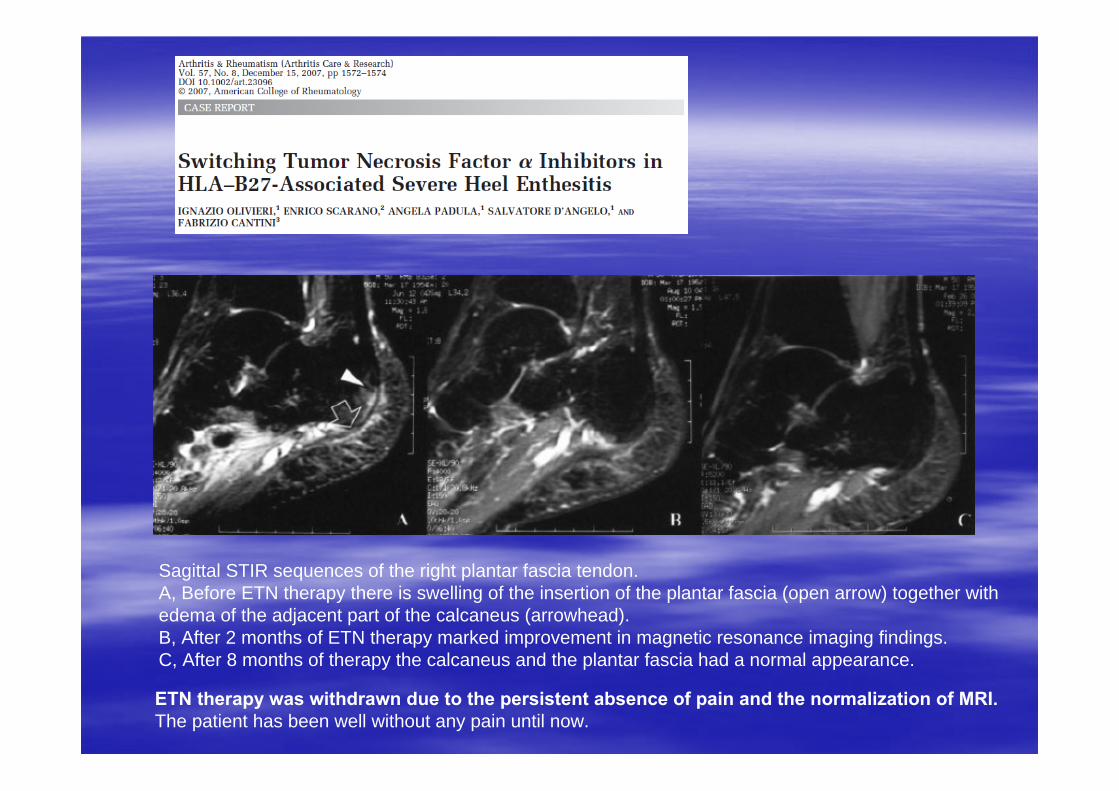
Sagittal STIR sequences of the right plantar fascia tendon. A, Before ETN therapy there is swelling of the insertion of the plantar fascia (open arrow) together with edema of the adjacent part of the calcaneus (arrowhead). B, After 2 months of ETN therapy marked improvement in magnetic resonance imaging findings. C, After 8 months of therapy the calcaneus and the plantar fascia had a normal appearance.
ETN therapy was withdrawn due to the persistent absence of pain and the normalization of MRI. The patient has been well without any pain until now.

Sagittal STIR (short inversion recovery) T2-weighted sequences of the left Achillestendon. (A) Imaging obtained the day before the beginning of adalimumab therapy, showing a diffuse oedema of the calcaneus (arrowheads) together with swelling of Achilles tendon(open arrow) and distension of the retrocalcaneal bursa by fluid collection (solid arrow). (B), 1 month(C), two months
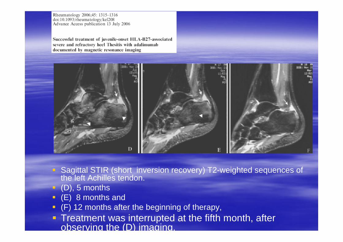
Sagittal STIR (short inversion recovery) T2-weighted sequences ofthe left Achilles tendon.(D), 5 months(E) 8 months and (F) 12 months after the beginning of therapy, Treatment was interrupted at the fifth month, afterobserving the (D) imaging.

Tendinite ed Tendinite ed entesiteentesite achilleaachillea

Schema struttura anatomica Schema struttura anatomica delldell’’entesientesi
Le sedi entesiche coinvolte più spesso nelle spondiliti
Valutazione Valutazione entesientesi: indici : indici entesiticientesitici
Indice entesitico di Mander: 68 entesiOgni entesi : 0 (nessun dolore) -3 (urlo di dolore)Punteggio massimo : 204

Valutazione Valutazione entesientesi: indici : indici entesiticientesitici
Indice entesitico di MaastrichtNumero entesi valutate :13Valore massimo :13
Valutazione Valutazione entesientesi: indici : indici entesiticientesitici
Indice entesitico della società canadese studio delle spondilitiNumero entesi valutate :16Punteggio massimo : 16

Valutazione Valutazione entesientesi: indici : indici entesitientesiticici
Indice entesitico di LeedsNumero entesi valutate : 6Valore massimo : 6

RM : RM : entesiteentesite del calcagno con del calcagno con coinvolgimento della fascia plantarecoinvolgimento della fascia plantare
RM : RM : entesiteentesite della fascia della fascia plantare e dellplantare e dell’’achilleoachilleo
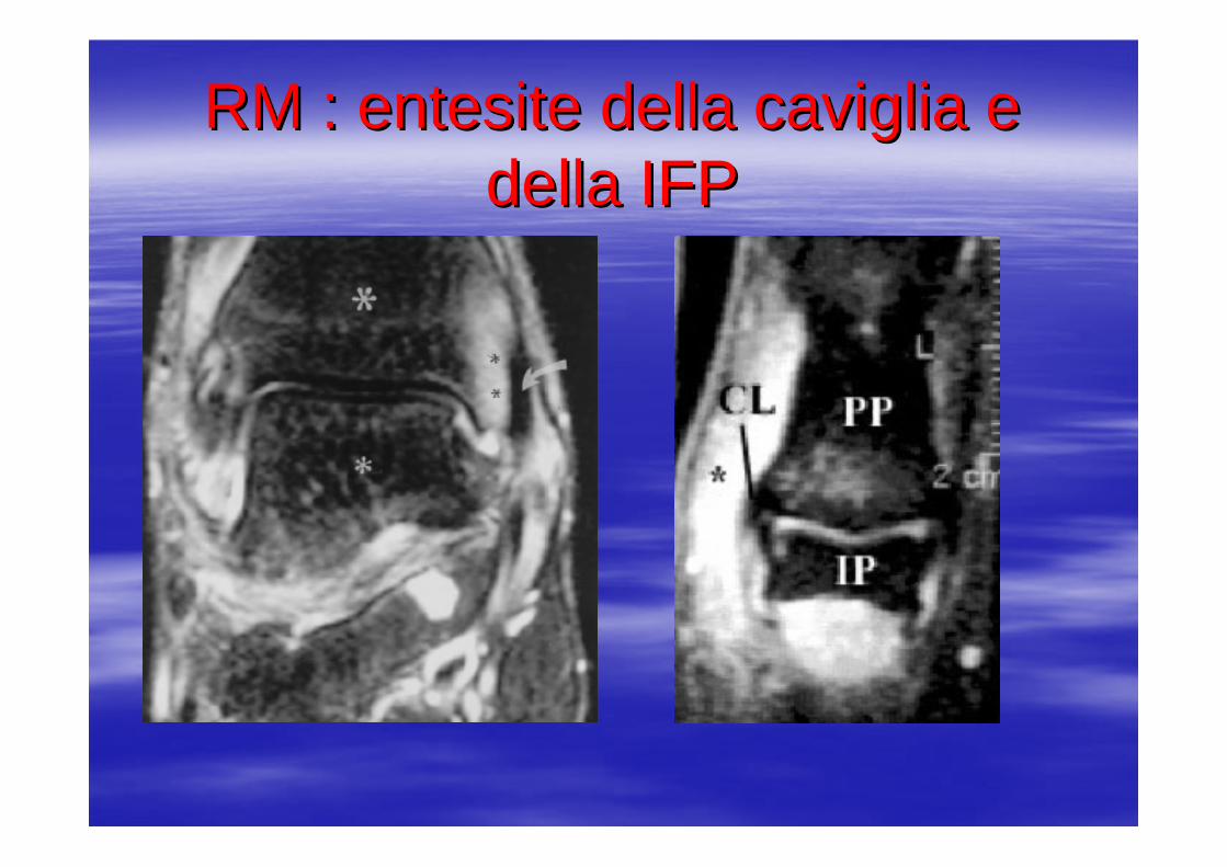
RM : RM : entesiteentesite della caviglia e della caviglia e della IFPdella IFP

RM : RM : entesiteentesite della inserzione della inserzione sovraspinatosovraspinato
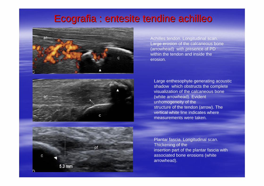
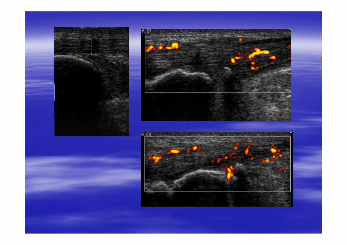
Ecografia : Ecografia : entesiteentesite tendine achilleotendine achilleo
Achilles tendon. Longitudinal scan. Large erosion of the calcaneous bone (arrowhead) with presence of PD within the tendon and inside the erosion.
Large enthesophyte generating acoustic shadow which obstructs the complete visualization of the calcaneous bone (white arrowhead). Evident unhomogeneity of thestructure of the tendon (arrow). The vertical white line indicates where measurements were taken.
Plantar fascia. Longitudinal scan. Thickening of theinsertion part of the plantar fascia with associated bone erosions (whitearrowhead).

Tendinite Achillea : aspetto in scansione Tendinite Achillea : aspetto in scansione longitudinale e con PDlongitudinale e con PD


Pazienti e metodiPazienti e metodi
Thirty patients with psoriasis and 30 controls Thirty patients with psoriasis and 30 controls underwent underwent ultrasonographicultrasonographic evaluation of evaluation of Achilles, quadriceps, Achilles, quadriceps, patellarpatellar enthesesentheses and and plantarplantar aponeurosisaponeurosis..UltrasonographicUltrasonographic findings were scored findings were scored according to the Glasgow Ultrasound according to the Glasgow Ultrasound EnthesitisEnthesitis Scoring System (GUESS).Scoring System (GUESS).
RisultatiRisultati
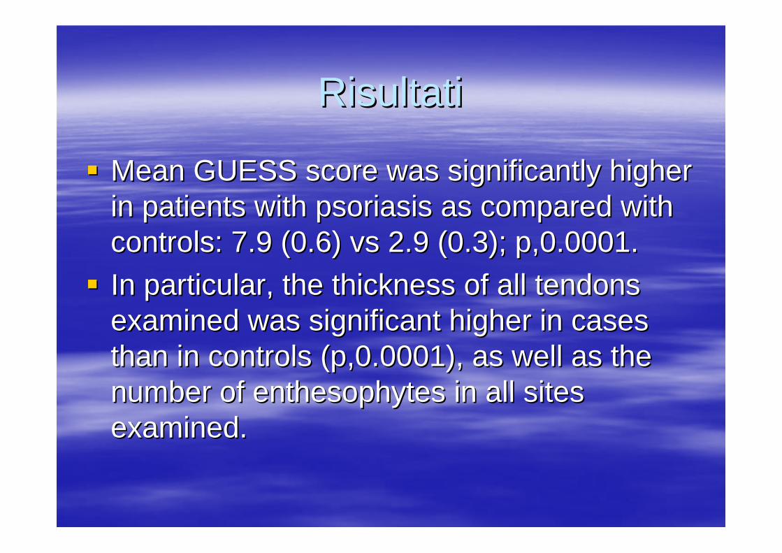
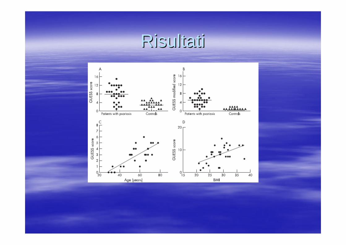
Mean GUESS score was significantly higher Mean GUESS score was significantly higher in patients with psoriasis as compared with in patients with psoriasis as compared with controls: 7.9 (0.6) controls: 7.9 (0.6) vsvs 2.9 (0.3); p,0.0001. 2.9 (0.3); p,0.0001. In particular, the thickness of all tendons In particular, the thickness of all tendons examined was significant higher in cases examined was significant higher in cases than in controls (p,0.0001), as well as the than in controls (p,0.0001), as well as the number of number of enthesophytesenthesophytes in all sites in all sites examined. examined.
RisultatiRisultati
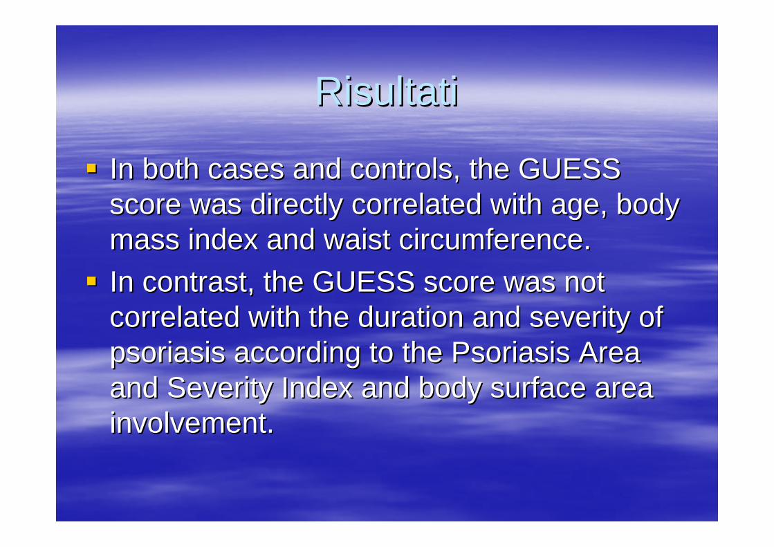
In both cases and controls, the GUESS In both cases and controls, the GUESS score was directly correlated with age, body score was directly correlated with age, body mass index and waist circumference.mass index and waist circumference.In contrast, the GUESS score was not In contrast, the GUESS score was not correlated with the duration and severity of correlated with the duration and severity of psoriasis according to the Psoriasis Area psoriasis according to the Psoriasis Area and Severity Index and body surface area and Severity Index and body surface area involvement.involvement.
RisultatiRisultati

ConclusioniConclusioni
EnthesealEntheseal abnormalities can be documented abnormalities can be documented by by ultrasonographyultrasonography in clinically in clinically asymptomatic patients with psoriasis. asymptomatic patients with psoriasis. These findings could be related to a These findings could be related to a subclinical subclinical enthesealentheseal psoriatic inflammation. psoriatic inflammation. We suggest close followWe suggest close follow--up of patients with up of patients with psoriasis with psoriasis with enthesealentheseal abnormalities for abnormalities for early diagnosis of psoriatic arthritis.early diagnosis of psoriatic arthritis.


RESULTSRESULTS
A total of 450 A total of 450 enthesesentheses in 45 patients with psoriasis were in 45 patients with psoriasis were evaluated by US. evaluated by US. In 148 of 450 (32.9%) In 148 of 450 (32.9%) enthesesentheses, grayscale US found signs , grayscale US found signs indicative of indicative of enthesopathyenthesopathy..In 4/450 (0.9%) In 4/450 (0.9%) enthesesentheses PD signal was detected. PD signal was detected. In the healthy population, US found signs of In the healthy population, US found signs of enthesopathyenthesopathyin 38 of 450 (8.4%) in 38 of 450 (8.4%) enthesesentheses and no PD signal was and no PD signal was detected. detected. The GUESS score was significantly higher in patients with The GUESS score was significantly higher in patients with psoriasis than in healthy controls (psoriasis than in healthy controls (P 0.0001). P 0.0001).

Normal Normal enthesealentheseal insertions on longitudinal scaninsertions on longitudinal scan

AbnormalitiesAbnormalities

DistributionDistribution ofof the the abnormalitiesabnormalities

CONCLUSIONSCONCLUSIONS
Both grayscale US and PD findings Both grayscale US and PD findings indicative of indicative of enthesopathyenthesopathy were more were more frequent in patients with psoriasis. frequent in patients with psoriasis. The US ability to detect signs of subclinical The US ability to detect signs of subclinical enthesopathyenthesopathy should be the object of should be the object of longitudinal investigations to define its value longitudinal investigations to define its value in predicting the clinical onset of psoriatic in predicting the clinical onset of psoriatic arthritis.arthritis.

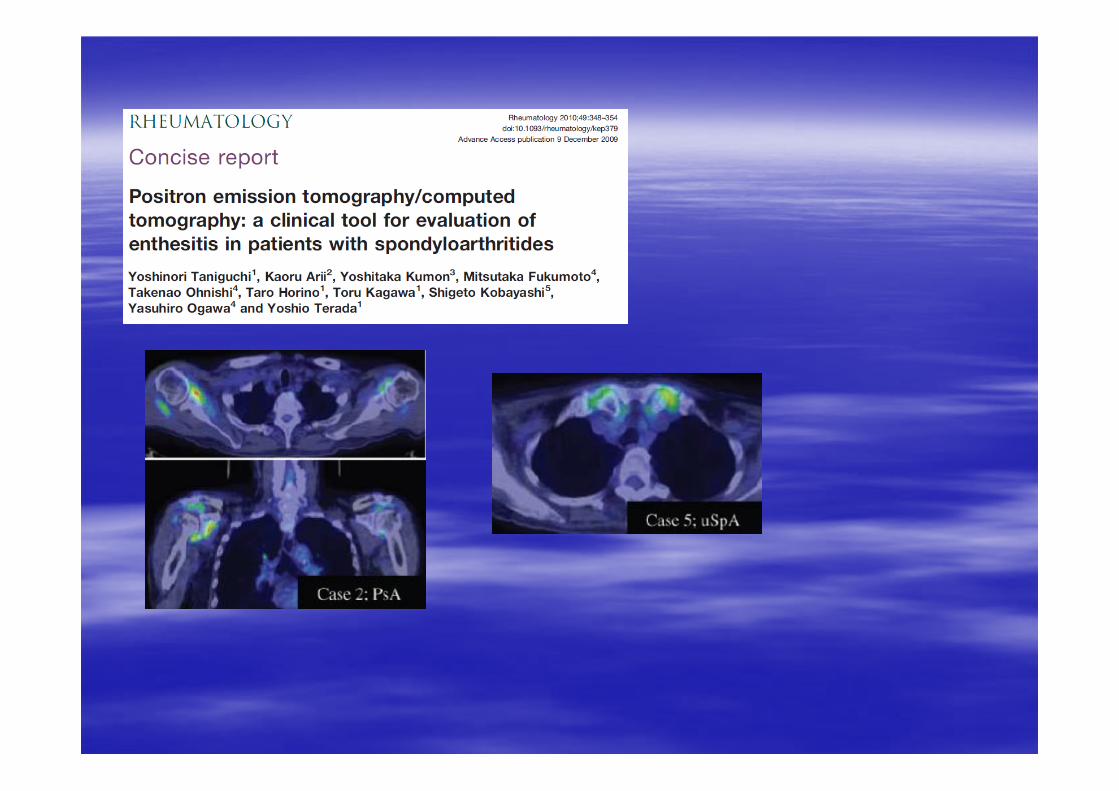
ScintigrafiaScintigrafia
ScintigraphyScintigraphy hashas beenbeen shownshown toto bebe effectiveeffectiveat at detectingdetecting synovialsynovial inflammationinflammation and and correlationcorrelation hashas beenbeen shownshown betweenbetweenscintigraphicalscintigraphical evidenceevidence ofof synovitissynovitis and and laterlater progressionprogression ofof joint joint erosionserosions..HoweverHowever, the , the rangerange ofof radiotracersradiotracers usedusedand the and the varyingvarying methodologiesmethodologies reportedreported in in the the literatureliterature makemake comparisonscomparisons ofof existingexistingtrialstrials difficultdifficult..


MetodiMetodi
47 consecutive 47 consecutive patientspatients: 29 : 29 hadhad definite definite PsAPsA and and 18 18 hadhad the the ““sinesine psoriasispsoriasis”” subset.subset.InclusionInclusion criteriacriteria werewere articulararticular and/or and/or enthesealenthesealinvolvementinvolvement ofof lessless 12 12 weeksweeks’’ durationduration and the and the exclusiveexclusive useuse, , beforebefore enrollmentenrollment, , ofof AINS AINS drugsdrugs totocontrolcontrol articulararticular symptomssymptoms..AllAll patientspatients underwentunderwent clinicalclinical examinationexamination, , bloodbloodteststests, , totaltotal--bodybody bonebone scintigraphyscintigraphy, , articulararticularultrasonographyultrasonography, and , and radiographyradiography ofof clinicallyclinicallyinvolvedinvolved jointsjoints and/or and/or enthesesentheses..
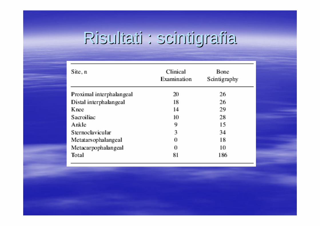
Risultati : scintigrafiaRisultati : scintigrafia

Scintigrafia : Scintigrafia : entesientesi
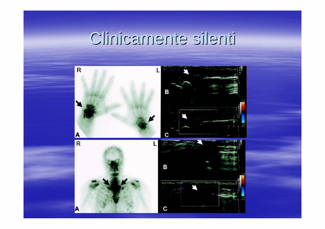
Clinicamente silentiClinicamente silenti
ConclusioniConclusioni
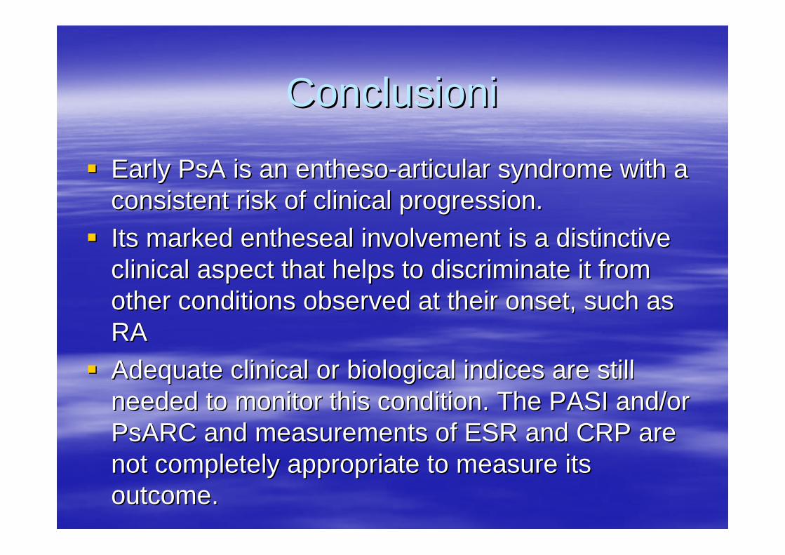
EarlyEarly PsAPsA isis anan enthesoentheso--articulararticular syndromesyndrome withwith a a consistentconsistent riskrisk ofof clinicalclinical progressionprogression. . ItsIts markedmarked enthesealentheseal involvementinvolvement isis a a distinctivedistinctiveclinicalclinical aspectaspect thatthat helpshelps toto discriminate discriminate itit fromfromotherother conditionsconditions observedobserved at at theirtheir onsetonset, , suchsuch asasRARAAdequateAdequate clinicalclinical or or biologicalbiological indicesindices are are stillstillneededneeded toto monitor monitor thisthis conditioncondition. The PASI and/or . The PASI and/or PsARCPsARC and and measurementsmeasurements ofof ESR and CRP are ESR and CRP are notnot completelycompletely appropriate appropriate toto measuremeasure itsitsoutcomeoutcome..

Quale imaging ?Nessuna : scelta possibile non linee guida sull’imaging periferico nelle SpA Scarsa sensibilità rispetto all’imaging.RX : dimostrazione di lesioni stabilizzate, scarsa sensibilità nelle lesioni iniziali, correla come indice con il dato clinico MRI : dimostrazione di lesioni acute, scarsa sensibilità per il riconoscimento di calcificazioni, limitazioni dalla scarsa disponibilità, non radiazioni, correla con il dato clinico.

SPONDILITE ANCHILOSANTE SPONDILITE ANCHILOSANTE (SA): DEFINIZIONE(SA): DEFINIZIONE
Malattia reumatica sistemica cronicaMalattia reumatica sistemica cronicaPredilezione per lo scheletro assialePredilezione per lo scheletro assialeInfiammazione nella sede di inserzione Infiammazione nella sede di inserzione dei legamenti nelldei legamenti nell’’osso (osso (entesientesi) )

SA: DEFINIZIONESA: DEFINIZIONE
Aspetto peculiare Aspetto peculiare èè ll’’infiammazione delle infiammazione delle articolazioni sacroarticolazioni sacro--iliacheiliacheHa una forte predisposizione genetica Ha una forte predisposizione genetica legata alla presenza delllegata alla presenza dell’’HLAHLA--B27B27
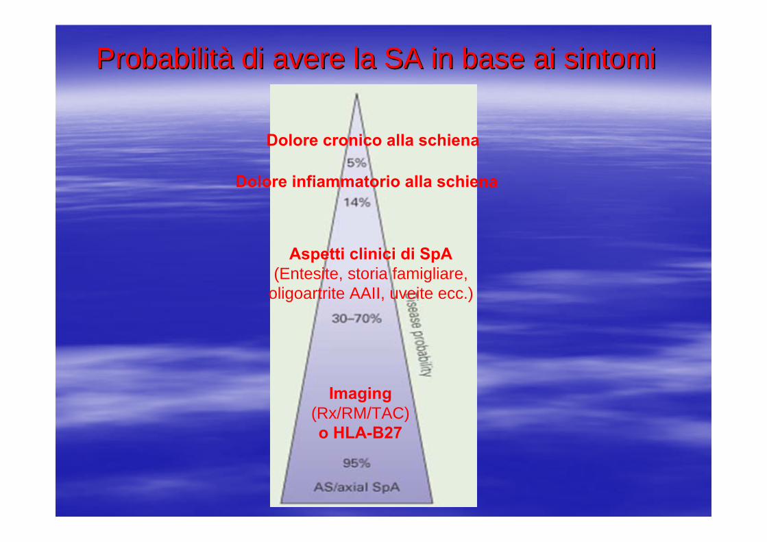
ProbabilitProbabilitàà di avere la SA in base ai sintomidi avere la SA in base ai sintomi
Dolore cronico alla schiena
Dolore infiammatorio alla schiena
Aspetti clinici di SpA (Entesite, storia famigliare,
oligoartrite AAII, uveite ecc.)
Imaging(Rx/RM/TAC) o HLA-B27

Criteri classificativi Criteri classificativi
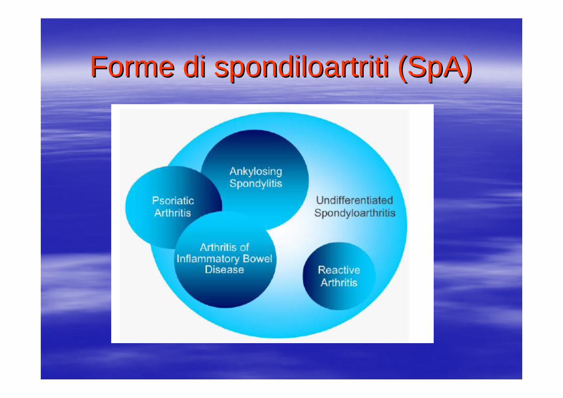
Forme di spondiloartriti (SpA)Forme di spondiloartriti (SpA)

Ecografia : Ecografia : entesiteentesite tendine achilleotendine achilleo
Achilles tendon. Longitudinal scan. Large erosion of the calcaneous bone (arrowhead) with presence of PD within the tendon and inside the erosion.
Large enthesophyte generating acoustic shadow which obstructs the complete visualization of the calcaneous bone (white arrowhead). Evident unhomogeneity of thestructure of the tendon (arrow). The vertical white line indicates where measurements were taken.
Plantar fascia. Longitudinal scan. Thickening of theinsertion part of the plantar fascia with associated bone erosions (whitearrowhead).
Tendinite Achillea : aspetto in scansione Tendinite Achillea : aspetto in scansione longitudinale e con PDlongitudinale e con PD
Epidemiologia Epidemiologia entesiteentesiteIn In reactivereactive arthritisarthritis ((ReAReA), ), peripheralperipheralenthesitisenthesitis hashas beenbeen foundfound in 33in 33––58% 58% ofofcasescases. . In a In a NorwegianNorwegian epidemiologicalepidemiological studystudy on on ReAReA, , enthesitisenthesitis waswas infrequentinfrequent and and seenseen in in 8% 186 8% 186 patientspatients..In In anotheranother studystudy, 5 out , 5 out ofof 11 11 patientspatients withwithReAReA fromfrom a a cohortcohort ofof 84 84 patientspatients withwithSalmonella Salmonella enteritidisenteritidis foodfood poisoningpoisoning hadhadperipheralperipheral enthesitisenthesitis. . In In twotwo ofof thesethese, , peripheralperipheral enthesitisenthesitis waswas the the onlyonly clinicalclinical manifestationmanifestation ofof the salmonella the salmonella inducedinduced ReAReA and the and the termterm ‘‘reactivereactiveenthesitisenthesitis’’ waswas proposedproposed. .
Epidemiologia Epidemiologia entesiteentesite
Oriente Oriente foundfound peripheralperipheral enthesitisenthesitis in 20% in 20% ofof hishispatientspatients withwith psoriaticpsoriatic arthritisarthritis ((PsAPsA), ), withwith a a peakpeakvaluevalue ofof 30% in the 30% in the spondyliticspondylitic pattern. pattern. In In ourour studystudy on the on the prevalenceprevalence and the and the clinicalclinicalspectrumspectrum ofof musculoskeletalmusculoskeletal manifestationsmanifestations in in ananinceptioninception cohortcohort ofof patientspatients withwith inflammatoryinflammatorybowelbowel diseasedisease foundfound a a frequencyfrequency ofof enthesitisenthesitis ofof10% (Salvarani 10% (Salvarani etet al.)al.)

Epidemiologia Epidemiologia entesiteentesite
The The clinicalclinical spectrumspectrum ofof uSpAuSpA isis wide due wide due toto the the variousvarious combinationscombinations ofof clinicalclinical and and radiologicalradiologicalmanifestationsmanifestations ofof SpA, SpA, I.e. I.e. peripheralperipheral enthesitisenthesitis, , peripheralperipheral arthritisarthritis, , dactylitisdactylitis, , inflammatoryinflammatory spinalspinal painpain, , sacroiliitissacroiliitis, , aorticaortic regurgitationregurgitation withwith conductionconduction disturbancesdisturbances, , uveitisuveitis and and conjunctivitisconjunctivitis. . EachEach ofof thesethese maymay alsoalso occuroccur alone alone asas the the onlyonlyclinicalclinical manifestationmanifestation ofof the B27the B27--associated associated diseasedisease processprocess..
Epidemiologia Epidemiologia entesiteentesite
UsuallyUsually, , peripheralperipheral enthesitisenthesitis coco--existsexists withwithotherother clinicalclinical manifestationsmanifestations ofof SpA, SpA, especiallyespecially peripheralperipheral arthritisarthritis and and dactylitisdactylitis. . ThisThis situation situation isis more more frequentfrequent in in juvenilejuvenile--onsetonset SpA SpA wherewhere the the enthesopathyenthesopathy and and arthropathyarthropathy syndromesyndrome hashas beenbeen identifiedidentified..

Epidemiologia Epidemiologia entesiteentesite
ThereThere are are uSpAuSpA patientspatients presentingpresenting isolatedisolatedperipheralperipheral enthesitisenthesitis. . In In suchsuch casescases, HLA , HLA typingtyping maymay bebe usefuluseful forfor ananearlyearly diagnosisdiagnosis ofof SpA. SpA. LongLong--lastinglasting isolatedisolated HLAHLA--B27B27--associated associated peripheralperipheral enthesitisenthesitis hashas beenbeen describeddescribed in in childrenchildren and and adolescentsadolescents, , youngyoung and and middlemiddle--agedagedadultsadults, and in , and in olderolder individualsindividuals..

Epidemiologia Epidemiologia entesiteentesite
In a In a studiesstudies byby OlivieriOlivieri on consecutive on consecutive patientspatientswithwith uSpAuSpA seenseen in in theirtheir tertiarytertiary referralreferral centrescentres, , isolatedisolated peripheralperipheral enthesitisenthesitis waswas seenseen in in twotwo(14%) out (14%) out ofof 14 14 patientspatients withwith juvenilejuvenile--onsetonsetdiseasedisease, and , and twotwo (9%) out (9%) out ofof 23 23 patientspatients withwith latelate--onsetonset diseasedisease. . ThereThere isis alsoalso a subset a subset ofof PsAPsA withwith isolatedisolatedenthesitisenthesitis and/or and/or dactylitisdactylitis (Salvarani (Salvarani etet al.)al.)

![[Recensão a:] Emanuele Coccia, La trasparenza delle ......Emanuele Coccia, La trasparenza delle immagini. Averroè e l’averroismo. Introduzione di Giorgio Agamben. Milano: Bruno](https://static.fdocumentos.tips/doc/165x107/611a1895ab673212bd5ccc0f/recenso-a-emanuele-coccia-la-trasparenza-delle-emanuele-coccia-la.jpg)


![ESPECIALIZAÇÃO REFLEXOLOGIA SACRO CRANIANA FORMAÇÃO 17.10.2020... · especializaÇÃo reflexologia sacro craniana [+ no verso] portfÓlio de prÁtica: portfÓlio estudo em casa](https://static.fdocumentos.tips/doc/165x107/5e8a5619d528900ea46afa18/especializafo-reflexologia-sacro-craniana-formafo-17102020-especializafo.jpg)