Domínio catalítico da enzima fosfodiesterase 5 humana...
53
Domínio catalítico da enzima fosfodiesterase 5 humana: síntese da região codificante, clonagem, expressão e purificação Bruna Pelegrim Selbach
Transcript of Domínio catalítico da enzima fosfodiesterase 5 humana...

Domínio catalítico da enzima fosfodiesterase 5 humana: síntese da
região codificante, clonagem, expressão e purificação
Bruna Pelegrim Selbach
Pontifícia Universidade Católica do Rio Grande do Sul Faculdade de Biociências
Programa de Pós-Graduação em Biologia Celular e Molecular
Domínio catalítico da enzima fosfodiesterase 5 humana: síntese da região codificante, clonagem, expressão e purificação
Dissertação apresentada ao Programa de Pós-Graduação em Biologia Celular e Molecular como requisito para a obtenção do grau de Mestre.
Autor
Bruna Pelegrim Selbach
Orientador Prof. Diógenes Santiago Santos
Co-orientador Prof. Luiz Augusto Basso
Porto Alegre, RS Março, 2008
Índice Resumo ................................................................................................... iv Abstract ................................................................................................... vi Capítulo 1 1.1 Introdução ......................................................................................... 7 1.2 Hipótese ............................................................................................ 21 1.3 Objetivos ........................................................................................... 22 Capítulo 2 Manuscrito .............................................................................................. 23
2.1 Abstract .................................................................................. 24 2.2 Introduction ............................................................................ 25 2.3 Materials and Methods ........................................................... 27 2.4 Results and Discussion .......................................................... 31
2.5 References ............................................................................. 38 Capítulo 3 3.1 Considerações finais e Perspectivas ................................................ 42 Anexo I Mapa do vetor de clonagem pCRBlunt ................................................... 43 Anexo II Mapa do vetor de expressão pET23a(+) ................................................. 44 1.4 Referências bibliográficas ................................................................. 45
iv
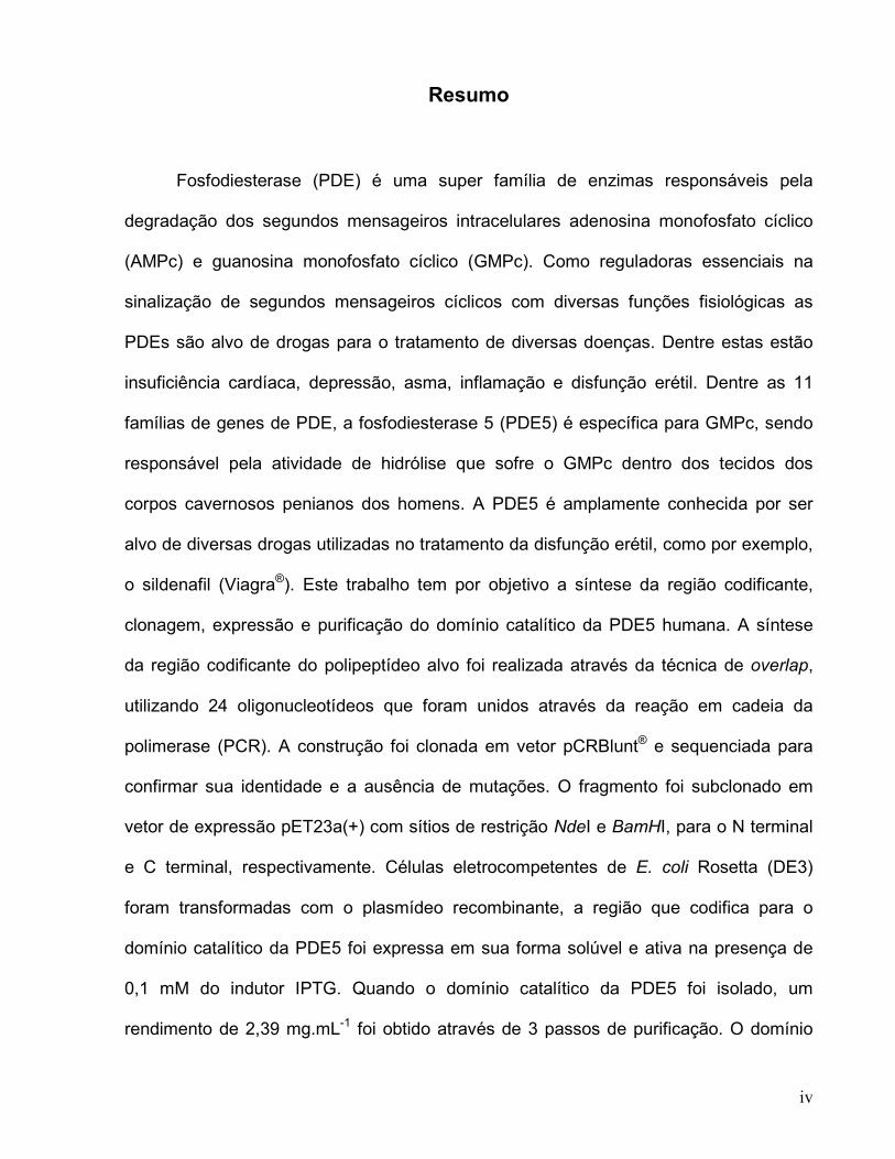
Resumo
Fosfodiesterase (PDE) é uma super família de enzimas responsáveis pela
degradação dos segundos mensageiros intracelulares adenosina monofosfato cíclico
(AMPc) e guanosina monofosfato cíclico (GMPc). Como reguladoras essenciais na
sinalização de segundos mensageiros cíclicos com diversas funções fisiológicas as
PDEs são alvo de drogas para o tratamento de diversas doenças. Dentre estas estão
insuficiência cardíaca, depressão, asma, inflamação e disfunção erétil. Dentre as 11
famílias de genes de PDE, a fosfodiesterase 5 (PDE5) é específica para GMPc, sendo
responsável pela atividade de hidrólise que sofre o GMPc dentro dos tecidos dos
corpos cavernosos penianos dos homens. A PDE5 é amplamente conhecida por ser
alvo de diversas drogas utilizadas no tratamento da disfunção erétil, como por exemplo,
o sildenafil (Viagra®). Este trabalho tem por objetivo a síntese da região codificante,
clonagem, expressão e purificação do domínio catalítico da PDE5 humana. A síntese
da região codificante do polipeptídeo alvo foi realizada através da técnica de overlap,
utilizando 24 oligonucleotídeos que foram unidos através da reação em cadeia da
polimerase (PCR). A construção foi clonada em vetor pCRBlunt® e sequenciada para
confirmar sua identidade e a ausência de mutações. O fragmento foi subclonado em
vetor de expressão pET23a(+) com sítios de restrição NdeI e BamHI, para o N terminal
e C terminal, respectivamente. Células eletrocompetentes de E. coli Rosetta (DE3)
foram transformadas com o plasmídeo recombinante, a região que codifica para o
domínio catalítico da PDE5 foi expressa em sua forma solúvel e ativa na presença de
0,1 mM do indutor IPTG. Quando o domínio catalítico da PDE5 foi isolado, um
rendimento de 2,39 mg.mL-1 foi obtido através de 3 passos de purificação. O domínio
v
catalítico da PDE5 puro permitirá a realização de uma triagem de extratos vegetais
oriundos da biodiversidade brasileira. Com o intuito de buscar novos compostos que
possam se tornar possíveis inibidores de PDE5, e assim serem utilizados no tratamento
da disfunção erétil.
Palavras – chave: AMPc, GMPc, disfunção erétil, fosfodiesterase 5.
vi
Abstract
Phosphodiesterases (PDEs) are a super family of enzymes which degrade the
intracellular second messengers cyclic guanosine monophosphate (cGMP) and cyclic
adenosine monophosphate (cAMP). As essential regulators in cyclic nucleotide
signaling with diverse physiological functions, PDEs are drug targets for the treatment of
various diseases including heart failure, depression, asthma, inflammation, and erectile
dysfunction. Among the eleven PDE gene families, the cGMP-specific PDE5 is the
principal cGMP-hydrolyzing activity in the human corpus cavernosum tissue. It is well
known as the target of many drugs used in the treatment of erectile dysfunction, like
sildenafil. This work aims the synthesis of the coding region, cloning, expression and
purification of the catalytic domain of human PDE5 gene which encodes the
phosphodiesterase 5. The synthesis of the coding region of the target polypeptide was
accomplished by using 24 primers, which were ligated by overlap extension using the
polymerase chain reaction. The construction was cloned into pCRBlunt® vector and
sequenced to confirm its identity and the absence of mutations. The fragment was
subcloned into pET23a(+) expression vector with NdeI and BamHI restriction sites.
Escherichia coli Rosetta (DE3) electrocompetent cells were transformed with the
recombinant plasmid and the coding region of the catalytic domain of the PDE5 was
expressed in its soluble form with 0.1mM of IPTG induction. The purification yielded 2.39
mg.mL-1 through 3 steps of purification. The pure catalytic domain of PDE5 will allow
screening for the discovery of new natural inhibitors from the brazilian biodiversity that
could be used in the treatment of erectile dysfunction.
7
1. Capítulo 1
1.1 Introdução
1.1.1 Disfunção erétil e Ereção
A Disfunção Erétil (DE), popularmente conhecida por impotência, é a
incapacidade de se obter ou manter uma ereção adequada para a prática da relação
sexual. Não deve ser confundida com a falta ou diminuição da libido, nem com a
dificuldade em ejacular ou em atingir o orgasmo. Dados da Organização Mundial de
Saúde (OMS), sugerem que 30% da população economicamente ativa manifesta algum
tipo de DE, o que no Brasil representa cerca de 11 a 15 milhões de homens.
A ereção é um fenômeno hemodinâmico, dependente do relaxamento do
músculo liso cavernoso e das arteríolas do corpo cavernoso, levando a um aumento do
fluxo sanguíneo para o espaço sinusoidal. O aumento do fluxo arterial distende o
espaço lacunar dos sinusóides, comprimindo, passivamente, as vênulas entre os
sinusóides e a túnica albugínea dos corpos cavernosos. A relativa ausência de
distensão da túnica albugínea resulta na veno-oclusão, que eleva a pressão
intracavernosa até valores próximos à pressão arterial média, gerando uma ereção
plena [1]. Após o estímulo sexual, o óxido nítrico (NO) é liberado por terminações
nervosas parasimpáticas e pelas células endoteliais que possui as isoformas neuronal
(nNOS) e endotelial (eNOS) da óxido nítrico sintase [2,3]. O óxido nítrico por ser gasoso
se difunde nas células da musculatura lisa vascular no corpo cavernoso do pênis,
interfere na conformação da guanilil ciclase solúvel tornando-a ativa e aumentando os
níveis de guanosina monofosfato cíclica (GMPc) nessas células [4]. Este processo leva
à ativação da proteína quinase dependente de GMPc (PKG), à fosforilação de outras
proteínas e à diminuição de cálcio por seqüestro intracelular ou à redução da
8
sensibilidade por cálcio via ativação de canais de potássio, o que resulta em um
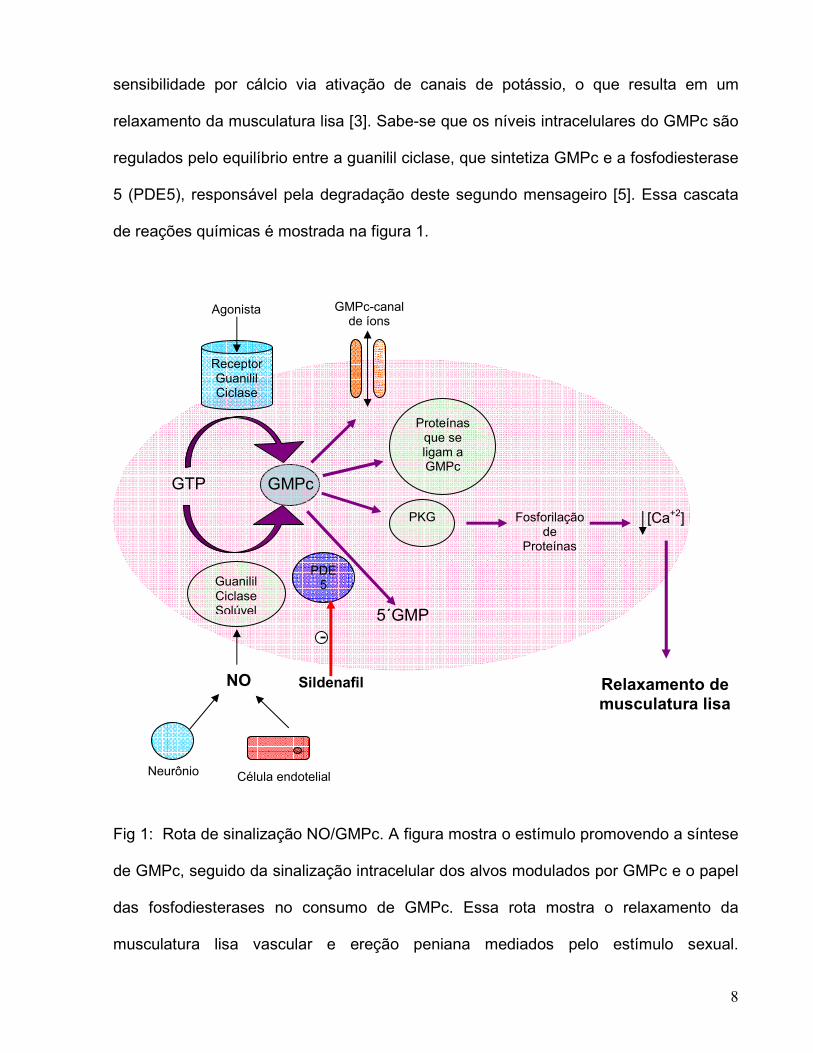
relaxamento da musculatura lisa [3]. Sabe-se que os níveis intracelulares do GMPc são
regulados pelo equilíbrio entre a guanilil ciclase, que sintetiza GMPc e a fosfodiesterase
5 (PDE5), responsável pela degradação deste segundo mensageiro [5]. Essa cascata
de reações químicas é mostrada na figura 1.
Fig 1: Rota de sinalização NO/GMPc. A figura mostra o estímulo promovendo a síntese
de GMPc, seguido da sinalização intracelular dos alvos modulados por GMPc e o papel
das fosfodiesterases no consumo de GMPc. Essa rota mostra o relaxamento da
musculatura lisa vascular e ereção peniana mediados pelo estímulo sexual.
Célula endotelial Neurônio
NO
Guanilil Ciclase Solúvel
Receptor Guanilil Ciclase
Agonista
GTP GMPc
GMPc-canal de íons
Proteínas que se ligam a GMPc
PDE 5
PKG Fosforilação de
Proteínas
[Ca+2]
Relaxamento de
musculatura lisa
5´GMP
Sildenafil
-
9
Relaxamento de musculatura lisa é em parte mediado pela ativação de PKG,
subseqüente abertura dos canais de potássio e a redução nos níveis de cálcio por
seqüestro intracelular. PDE5 é o alvo para sildenafil assim como para outros inibidores
utilizados no tratamento de disfunções vasculares crônicas.
1.1.2 Fosfodiesterases
As fosfodiesterases (PDEs) pertencem a uma grande família de 11 diferentes
genes. Essas enzimas são estruturalmente relacionadas, porém funcionalmente
distintas, e altamente reguladas [6]. São fosfohidrolases que seletivamente catalisam a
hidrólise de ligações 3´ de fosfato cíclico de adenosina e/ou guanosina 3´,
5´monofosfato cíclico.
Logo após a descoberta da adenosina monofosfato cíclica (AMPc) por
Sutherland e colaboradores a atividade de PDE sobre nucleotídeos cíclicos foi descrita
[7]. Com a descoberta do GMPc observou-se que tanto o AMPc quanto o GMPc podiam
ser hidrolisados pelo mesmo tipo de atividade, por exemplo, a hidrólise da ligação 3´ do
fosfato cíclico. Baseado nos estudos de competição por substratos tornou-se claro que
pelo menos algumas dessas atividades deviam ter o mesmo sítio catalítico. De fato,
muitos dos estudos preliminares em nucleotídeos cíclicos foram direcionados para
entender a atividade PDE, pois, na época era mais fácil medir a atividade da PDE do
que as atividades de AMPc ou GMPc, ou mesmo das enzimas que catalisavam suas
sínteses. Com o aprimoramento dos ensaios biológicos utilizando substratos radioativos
tornou-se clara a existência de múltiplas formas de PDEs com diferentes propriedades
cinéticas e regulatórias [8,9].
Devido à complexidade do sistema de nucleotídeos cíclicos de PDE fez-se
necessário adotar abordagens mais sofisticadas e complexas para entender o papel

10
das PDEs na regulação de GMPc e AMPc na célula. Hoje está claro que um único tipo
celular pode expressar diferentes PDEs e que a natureza e a localização dessas
enzimas parece ser um grande regulador de GMPc e AMPc dentro da célula. As PDEs
não são apenas reguladas em nível genético, mas também por diversos mecanismos
bioquímicos, os quais incluem fosforilação e desfosforilação, ligação alostérica de
GMPc ou AMPc, ligação de Ca+2 / calmodulina e diferentes interações proteína-proteína
[10]. O principal conceito sobre o papel das PDEs é de que elas modulam a forma
tridimensional, a amplitude e a duração dos nucleotídeos cíclicos dentro da célula que
está em expansão. Algumas PDEs, sem dúvida, funcionam apenas para impedir que
esses nucleotídeos se espalhem para regiões inapropriadas das células e outras
servem para regular o local de acesso de receptores específicos tanto de AMPc quanto
de GMPc nos locais intracelulares específicos. Acredita-se também que nem todas as
PDEs funcionam controlando a hidrólise de nucleotídeos cíclicos, e sim atuem como
proteínas mediadoras alterando interações proteína-proteína utilizando as mudanças
alostéricas induzidas pela ligação dos nucleotídeos cíclicos. Esse último conceito é
possível, porém não há nenhuma demonstração evidente de tal fato até hoje [11].
11
1.1.3 Familias de PDE
A maior parte das famílias de PDE possuem de mais de um gene (~20 genes de
PDE), que geram múltiplos produtos de proteínas (>50 proteínas de PDE) através de
splicing alternativo de RNAm ou utiliza diferentes sítios iniciadores de promotores de
transcrição. Grande parte das células possui múltiplos genes de famílias de PDE,
porém em diferentes quantidades, proporções e localizações. As fosfodiesterases 5, por
exemplo, são relativamente abundantes em musculatura lisa, incluindo vasculatura
pulmonar e corpos cavernosos penianos, onde aparentemente regulam a hidrólise de
GMPc que modula a vasodilatação.
As PDEs de mamíferos exibem uma organização estrutural bastante comum
entre elas, com um domínio catalítico conservado nas porções C-terminal das
moléculas e diferentes domínios regulatórios nas porções N-terminal. O domínio
catalítico, altamente conservado entre os membros de cada família possui um motivo de
assinatura, comum a todas PDEs, e domínios de ligação a metal. Além disso, os
domínios catalíticos possuem seqüências específicas para cada família, que são
responsáveis pelas diferentes afinidades por substratos, atividades catalíticas e
também sensibilidade a inibidores específicos. Algumas famílias de PDE são
relativamente específicas para AMPc, GMPc ou também podem hidrolisar ambos [12]
(Tabela 1).

12
Tabela 1: Especificidade de substrato para cada família de PDE
Família PDE Subfamília (no de variantes) Substrato
1 A (4), B (1), C (5) AMPc / GMPc
2 A (3) AMPc / GMPc
3 A (1), B (1) AMPc / GMPc
4 A (8), B (3), C (4), D (5) AMPc
5 A (3) GMPc
6 A (1), B (1), C (1) GMPc
7 A (3), B (1) AMPc
8 A (5), B (1) AMPc
9 A (6) GMPc
10 A (2) AMPc / GMPc
11 A (4) AMPc / GMPc
1.1.4 PDEs como alvos terapêuticos
Imediatamente após a descoberta da atividade PDE, descobriu-se que a cafeína
era um inibidor efetivo de PDE assim como outros análogos da mesma, como teofilina.
No entanto, o princípio pelo qual a inibição da atividade de PDE seria válida como um
alvo terapêutico não era claro, até porque pouco se sabia sobre esses inibidores e
grande parte desses inibiam a maioria, senão todas, as PDEs nos tecidos. Uma razão
importante para utilizar as PDEs como alvos terapêuticos está relacionada com o
princípio farmacológico básico, de que a regulação de degradação de qualquer ligante
13
ou segundo mensageiro pode, freqüentemente, realizar uma maior alteração na
concentração dos mesmos quando comparados com a regulação nas taxas de síntese.
Isso é verdade tanto para alterações farmacocinéticas em nível de drogas ou para
alterações nas quantidades de moléculas de regulação celulares endógenas ou
metabólitos. Essa propriedade intrínseca estaria aumentada se o processo celular
responsável pela degradação apresentar um valor de Vmax maior do que aquele do
processo de síntese. Sabe-se, que a maioria dos tecidos contém pelo menos uma
ordem de magnitude maior de atividade de PDE do que atividade ciclase, tanto para
AMPc quanto para GMPc e acredita-se ser improvável que a maioria das PDEs atuem
abaixo do valor de Vmax para os substratos em condições fisiológicas. A idéia atual é de
que em muitos compartimentos celulares os níveis de substrato podem ser altos e, no
entanto, essa grande atividade estaria presente. Outra razão para que as PDEs sejam
possíveis bons alvos de drogas está relacionada com a concentração de seus
substratos na célula. Normalmente os níveis de AMPc e GMPc na maioria das células é
de 1 a 10 µM. Isto significa que um inibidor competitivo não precisaria competir com
altos níveis de substrato endógeno para ser efetivo [11].
A razão mais importante para o reconhecimento das PDEs como bons alvos de
drogas é o fato de que existem diferentes isoformas. Em função do grande número de
distintas formas de PDEs expressas em tecidos de mamíferos, há uma forte evidência
de que muitas dessas PDEs estão relacionadas a diferentes funções fisiológicas no
organismo, assim como a diferentes condições patológicas. Acredita-se ser possível o
desenvolvimento de inibidores seletivos as isoformas, que podem ter como alvo funções
específicas e condições patológicas sem a alta probabilidade de causar efeitos
colaterais indesejáveis [13].

14
1.1.5 Estruturas de domínios catalíticos
Nos últimos quatro anos, estruturas cristalográficas para os domínios catalíticos
de sete diferentes famílias de PDE foram resolvidas incluindo a PDE1B [14], a PDE3B
[15], a PDE4B [16,17,18,19], a PDE4D [18,19,20,21,22,23], a PDE5A [14,18,22,24], a
PDE7A [25] e a PDE9A [26]. No entanto, nenhuma estrutura de alta resolução para
qualquer holoenzima de PDE foi descrita, por isso pouco se sabe sobre os detalhes
moleculares de como os domínios regulatórios influenciam na catálise, porém sabe-se
que de fato possuem papéis importantes em relação à especificidade por substrato e
ligação de inibidores.
A maior parte dos dobramentos e dos elementos estruturais funcionais para cada
domínio catalítico são bastante semelhantes, apesar de apresentarem,
aproximadamente, de 25 a 35% de identidade entre elas. Todos esses domínios
catalíticos contem três subdomínios compostos por 16 hélices e o sítio ativo é formado
na junção das hélices por resíduos que são altamente conservados entre as PDEs. Na
parte superior do bolsão de ligação do substrato está o sítio de ligação para dois metais
divalentes (Fig. 2A). Os metais zinco e magnésio são coordenados por resíduos
localizados em cada um dos três diferentes domínios. O sítio de ligação do metal zinco
possui três resíduos de histidina e dois de aspartato que são absolutamente
conservados entre todas as PDEs estudadas até hoje. Por serem resíduos de histidina
e aspartato imagina-se que eles atuem para coordenar cátions divalentes, que serão
necessários para a atividade dessas proteínas (Fig. 2B). O metal magnésio liga-se mais
fracamente ao bolsão catalítico provavelmente em função da presença de moléculas de
água que coordenam esse metal [16]. Possivelmente isso é o que ocorre nas PDEs
[25,27].

15
Fig.2: Estrutura terciária do domínio catalítico da PDE5. A) Bolsão de ligação do
substrato. B) Sítio de ligação dos metais; Mg2+ (Rosa), Zn2+ (Laranja).
Outra idéia interessante que surgiu a partir de estudos de estrutura é a proposta
para o mecanismo molecular dos nucleotídeos cíclicos para especificidade. Em cada
uma das PDEs cuja estrutura foi resolvida, parece existir invariavelmente uma
glutamina que estabiliza a ligação do anel purínico no bolsão de ligação [14]. Para as
PDEs que são seletivas para AMPc em baixas concentrações, essa glutamina possui
uma orientação, mediada por resíduos vizinhos, que favorece a ligação do AMPc. Já
para as PDEs que preferem GMPc essa glutamina (Q817) está disposta em uma outra
orientação que então permite a ligação do GMPc [14] (Fig. 3). No caso de PDEs que
hidrolisam tanto AMPc como GMPc essa glutamina deve estar livre para se
movimentar. Essa hipótese chamada de glutamine switch parece formar a base
molecular para muitas das seletividades por substratos observadas entre as diferentes
16
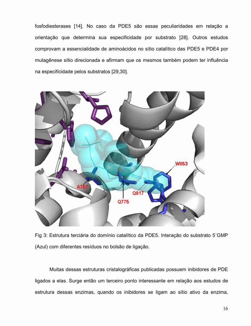
fosfodiesterases [14]. No caso da PDE5 são essas peculiaridades em relação a
orientação que determina sua especificidade por substrato [28]. Outros estudos
comprovam a essencialidade de aminoácidos no sítio catalítico das PDE5 e PDE4 por
mutagênese sítio direcionada e afirmam que os mesmos também podem ter influência
na especificidade pelos substratos [29,30].
Fig 3: Estrutura terciária do domínio catalítico da PDE5. Interação do substrato 5´GMP
(Azul) com diferentes resíduos no bolsão de ligação.
Muitas dessas estruturas cristalográficas publicadas possuem inibidores de PDE
ligados a elas. Surge então um terceiro ponto interessante em relação aos estudos de
estrutura dessas enzimas, quando os inibidores se ligam ao sítio ativo da enzima,
17
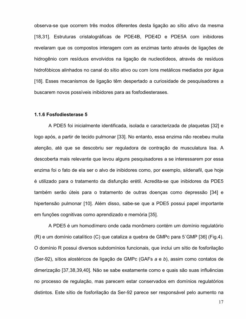
observa-se que ocorrem três modos diferentes desta ligação ao sítio ativo da mesma
[18,31]. Estruturas cristalográficas de PDE4B, PDE4D e PDE5A com inibidores
revelaram que os compostos interagem com as enzimas tanto através de ligações de
hidrogênio com resíduos envolvidos na ligação de nucleotídeos, através de resíduos
hidrofóbicos alinhados no canal do sítio ativo ou com íons metálicos mediados por água
[18]. Esses mecanismos de ligação têm despertado a curiosidade de pesquisadores a
buscarem novos possíveis inibidores para as fosfodiesterases.
1.1.6 Fosfodiesterase 5
A PDE5 foi inicialmente identificada, isolada e caracterizada de plaquetas [32] e
logo após, a partir de tecido pulmonar [33]. No entanto, essa enzima não recebeu muita
atenção, até que se descobriu ser reguladora de contração de musculatura lisa. A
descoberta mais relevante que levou alguns pesquisadores a se interessarem por essa
enzima foi o fato de ela ser o alvo de inibidores como, por exemplo, sildenafil, que hoje
é utilizado para o tratamento da disfunção erétil. Acredita-se que inibidores da PDE5
também serão úteis para o tratamento de outras doenças como depressão [34] e
hipertensão pulmonar [10]. Além disso, sabe-se que a PDE5 possui papel importante
em funções cognitivas como aprendizado e memória [35].
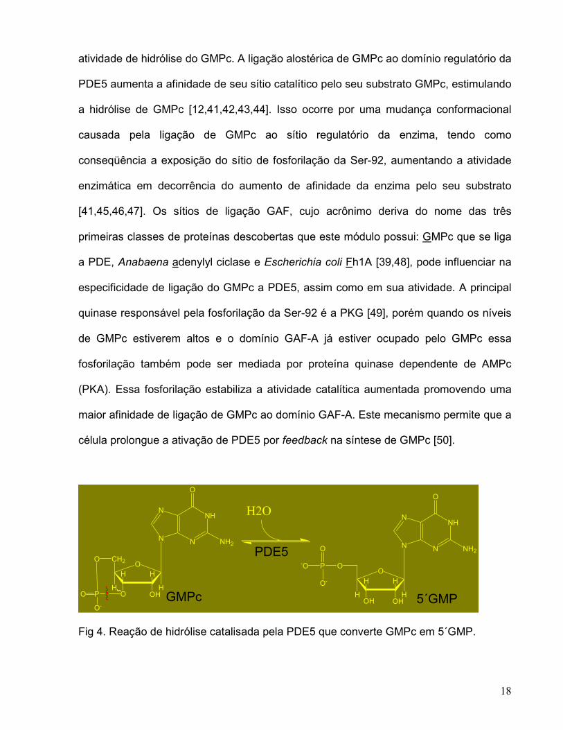
A PDE5 é um homodímero onde cada monômero contém um domínio regulatório
(R) e um domínio catalítico (C) que cataliza a quebra de GMPc para 5´GMP [36] (Fig.4).
O domínio R possui diversos subdomínios funcionais, que inclui um sítio de fosforilação
(Ser-92), sítios alostéricos de ligação de GMPc (GAFs a e b), assim como contatos de
dimerização [37,38,39,40]. Não se sabe exatamente como e quais são suas influências
no processo de regulação, mas parecem estar conservados em domínios regulatórios
distintos. Este sítio de fosforilação da Ser-92 parece ser responsável pelo aumento na
18
atividade de hidrólise do GMPc. A ligação alostérica de GMPc ao domínio regulatório da
PDE5 aumenta a afinidade de seu sítio catalítico pelo seu substrato GMPc, estimulando
a hidrólise de GMPc [12,41,42,43,44]. Isso ocorre por uma mudança conformacional
causada pela ligação de GMPc ao sítio regulatório da enzima, tendo como
conseqüência a exposição do sítio de fosforilação da Ser-92, aumentando a atividade
enzimática em decorrência do aumento de afinidade da enzima pelo seu substrato
[41,45,46,47]. Os sítios de ligação GAF, cujo acrônimo deriva do nome das três
primeiras classes de proteínas descobertas que este módulo possui: GMPc que se liga
a PDE, Anabaena adenylyl ciclase e Escherichia coli Fh1A [39,48], pode influenciar na
especificidade de ligação do GMPc a PDE5, assim como em sua atividade. A principal
quinase responsável pela fosforilação da Ser-92 é a PKG [49], porém quando os níveis
de GMPc estiverem altos e o domínio GAF-A já estiver ocupado pelo GMPc essa
fosforilação também pode ser mediada por proteína quinase dependente de AMPc
(PKA). Essa fosforilação estabiliza a atividade catalítica aumentada promovendo uma
maior afinidade de ligação de GMPc ao domínio GAF-A. Este mecanismo permite que a
célula prolongue a ativação de PDE5 por feedback na síntese de GMPc [50].
Fig 4. Reação de hidrólise catalisada pela PDE5 que converte GMPc em 5´GMP.
NH
N
N
O
NH2N
O
OH
HH
HHOH
OP-O
O
O-
H2ONH
N
N
O
NH2N
O
OH
HH
H
CH2
HO
O
P
O-
O GMPc
PDE5
5´GMP
19
1.1.7 Inibidores de PDE5
Diversos compostos que inibem a PDE5 foram recentemente sintetizados, dentre
estes, três são utilizados clinicamente para o tratamento da DE no homem. Após o
estímulo sexual, essas drogas inibem a PDE5 gerando um acúmulo de GMPc nas
células dos tecidos penianos, desencadeando uma cascata que leva à ativação da
PKG, a fosforilação de outras proteínas e a diminuição de cálcio celular ou à redução
da sensibilidade ao cálcio, o que resulta em relaxamento da musculatura lisa [3].
O sildenafil (Viagra®) foi o pioneiro desta classe de medicamentos a ser utilizado
para o tratamento da DE que apresenta como mecanismo principal de ação a inibição
da PDE5. Entretanto, esta droga também é promissora no tratamento de outras
disfunções relacionadas à musculatura lisa, como por exemplo, a hipertensão pulmonar
supracitado. Em 1998 este medicamento foi aprovado pelo FDA (Food and Drug
Administration) nos Estados Unidos para o tratamento da DE e, em 2002, representou
90% das vendas como medicamento para tratar a DE. As vendas mundiais do Sildenafil
excederam US$1,5 bilhões em 2001 [51]. Outros inibidores, como o Vardenafil
(Levitra®) e o Tadalafil (Cialis®), foram desenvolvidos para o tratamento de DE com
base no mesmo mecanismo de ação do Sildenafil (Viagra®). A disponibilidade desses
inibidores fornece ferramentas importantes para o estudo do domínio catalítico da
PDE5, abrindo assim a possibilidade de se propor novos fármacos que atuem nesse
alvo terapêutico.
Efeitos adversos como distúrbios visuais em pacientes que fazem o uso de
inibidores da PDE5 [52,53] refletem a necessidade de um estudo mais aprofundado do
mecanismo de ação dessas drogas, assim como o desenvolvimento de novos
potenciais inibidores. Estudos com extratos de plantas já demonstraram sua ação

20
relaxante em endotélio do corpo cavernoso de roedores [54], porém não se sabe
exatamente qual é o alvo desses compostos naturais. Existe uma série de compostos e
extratos potenciais a serem testados como possíveis inibidores da PDE5 [55], porém
testes adicionais relacionados com mecanismo de ação, efeitos colaterais indesejáveis
e toxicidade precisam ser concluídos.
21
1.2 Hipótese
Inibidores seletivos para diferentes familias de PDEs estão em desenvolvimento
para diversas indicações. Drogas que possuem como alvo as PDEs são fortemente
consideradas por suas propriedades cardiotônica, vasodilatadora, relaxantes de
musculatura lisa, antitrombótica, antinflamatória e antiasmática. Elas também estão
sendo desenvolvidas como antidepressivos e para melhorarem funções cognitivas
como aprendizado e memória [35]. Inibidores de PDE5 como sildenafil, vardenafil e
tadalafil estão sendo amplamente utilizados com sucesso para o tratamento de
disfunção erétil em homens e recentemente foram aprovados para o tratamento de
hipertensão pulmonar [10].
No entanto, um maior entendimento da topologia do bolsão catalítico será de
extrema importância para o desenvolvimento de drogas mais seletivas. Este projeto visa
à obtenção do domínio catalítico da PDE5 em sua foma solúvel e ativa. Com o mesmo,
será possível a realização de uma triagem de extratos vegetais oriundos da
biodiversidade vegetal brasileira com o intuito de buscar novos inibidores da PDE5 para
o tratamento da disfunção erétil.

22
1.3 Objetivo Geral
O Objetivo deste trabalho é isolar o domínio catalítico da fosfodiesterase 5 em
sua forma solúvel e ativa.
1.4 Objetivos específicos
1) Desenhar os oligonucleotídeos sintéticos para a construção da região
codificante do domínio catalítico da PDE5;
2) Unir os primers para a construção da região codificante do domínio catalítico
da PDE5 utilizando a reação em cadeia da polimerase (PCR);
3) Clonar o fragmento amplificado em pCR-Blunt®, e posterior subclonagem em
vetor de expressão de células procarióticas pET23a(+);
4) Seqüenciar o plasmídeo recombinante final para verificação da ausência de
mutações;
5) Obter expressão da proteína recombinante em células de Escherichia coli
Rosetta (DE3) utilizando como controle o vetor de expressão sem o inserto.
6) Purificação do domínio catalítico da PDE5 na sua forma homogênea;
7) Seqüenciamento N-terminal da proteína pura, análise de pureza e
determinação da massa molecular da subunidade por espectroscopia de massas.

23
Capítulo 2 – Manuscrito a ser submetido para revista Biochemical and
Biophysical Research Communications (BBRC).
Human phosphodiesterase 5 catalytic domain: synthesis of the coding
region, cloning, expression and purification.
Bruna P. Selbach1, Isabel Osório da Fonseca1,2, Luiz Augusto Basso1, Diógenes
Santiago Santos1*.
___________________________________
1 Centro de Pesquisas em Biologia Molecular e Funcional, Pontifícia Universidade
Católica do Rio Grande do Sul, Porto Alegre, RS 90619-900, Brasil;
2 Current address: Virginia Bioinformatics Institute, Virginia Tech, Blacksburg, VA 24061,
USA.
*To whom correspondence may be addressed: E-mail: [email protected] Phone: +55
51 33203629.
24
2.1 Abstract
Phosphodiesterases (PDEs) are a superfamily of enzymes which degrade the
intracellular second messengers cGMP and cAMP. As essential regulators in cyclic
nucleotide signaling with diverse physiological functions, PDEs are drug targets for the
treatment of various diseases including heart failure, depression, asthma, inflammation,
and erectile dysfunction. Among the eleven PDE gene families, the cGMP-specific PDE5
has the main cGMP-hydrolyzing activity in the human corpus cavernosum tissue. It is
well known as the target of many drugs used in the treatment of erectile dysfunction, like
sildenafil. This work aims at the synthesis of the coding region, cloning, expression and
purification of the catalytic domain of human PDE5 gene which encodes
phosphodiesterase 5. The pure catalytic domain of PDE5 will allow screening for the
discovery of new natural inhibitors from the Brazilian biodiversity that can be used in the
treatment of erectile dysfunction.
Key words: erectile dysfunction, PDE5, nitric oxide, cGMP, cAMP, natural inhibitors,
phosphodiesterase, corpus cavernosum, catalytic domain, purification.
25
2.2 Introduction
The superfamily of cyclic nucleotide phosphodiesterases (PDEs) comprises
eleven known families of PDEs that vary in substrate specificity, regulatory properties,
and tissue distribution [1]. PDEs play a critical role in maintaining the celular level of
cyclic adenosine monophosphate (cAMP) and cyclic guanosine monophosphate (cGMP)
[2]. The second messengers cAMP and cGMP, mediate the response of cells to a wide
variety of hormones and neurotransmitters, and modulate many metabolic processes
such as cardiac and smooth muscle contraction [3]. As essential regulators in cyclic
nucleotide signaling with diverse physiological functions, PDEs are drug targets for the
treatment of several diseases including heart failure, depression, asthma, inflammation
and erectile dysfunction. Within the eleven PDE gene families, the cGMP-specific PDE5
is the major cGMP-hydrolyzing activity in the human corpus cavernosum [4].
Penile erection is a hemodynamic event that is regulated by relaxation of the
corpus cavernosum smooth muscle cells, mediated via the NO-cGMP pathway. Upon
sexual arousal, NO is the putative principal neurotransmitter released from non-
cholinergic non-adrenergic parasympathetic nerve endings in the walls of the arteries
and sinusoids of the penis, mediating erection [5]. NO binds to soluble guanylyl cyclase
triggering increased synthesis of cGMP from guanosine triphosphate, thus elevating
intracellular cGMP concentrations. This leads to activation of cGMP-dependent protein
kinase (PKG), phosphorylation of several proteins, and lowering of cellular calcium level,
which will be responsible for the relaxation of the smooth muscle, increased blood flow,
simultaneously increased penile tumescence, and, eventually, erection [6].
26
It is known that cGMP relaxes vascular smooth muscle and that cellular cGMP
concentration is determined by the balance between its synthesis by guanylyl ciclases
and its breakdown to 5`GMP by cyclic nucleotide phosphodiesterase5 (PDE5).
Therefore, modulating intracellular cGMP levels has been targeted for pharmacological
intervention, for example sildenafil, in disorders directly influenced by vascular smooth
muscle activity, which includes erectile dysfunction [7].
The catalytic domain of PDE5 is the direct target of PDE5 inhibitors, but the
regulatory domain can influence PDE5 inhibitor actions on the enzyme in certain
instances [6, 8]. In PDE5, the effect of allosteric cGMP binding is not much clear, but
cGMP binding to the PDE5 regulatory domain controls phosphorylation of a specific
serine that is near the amino terminus of the regulatory domain [9, 10]. Phosphorylation
at this serine activates both PDE5 catalytic and allosteric cGMP-binding activities [6].
Studies of several PDEs have suggested that the isolated catalytic domain alone is
sufficient for catalytic activity [3, 11].
This work presents the synthesis, cloning, sequencing, expression, and
purification of the catalytic domain of human PDE5. This pure catalytic domain will allow
us to perform a screening of natural plant extracts to search for new natural inhibitors
from the brazilian biodiversity that could be used in the treatment of erectile dysfunction.
27
2.3 Materials and Methods
Construction of the coding region of the catalytic domain of PDE5
The coding region of the PDE5 catalytic domain was constructed using 24
primers which were designed based on the genomic DNA sequence of PDE5 Homo
sapiens sp from the Gene Bank (Gene ID: 8654). These primers were ligated by overlap
extension using the polymerase chain reaction (PCR) with Pyrococcus furiosus DNA
polymerase (Pfu; Stratagene) [12], which is a thermostable polymerase that exhibits low
error rate, thus lowering the likelihood of introducing unwanted mutations.
Cloning of the catalytic domain of PDE5
Based on the genomic sequence of the PDE5 Homo sapiens sp catalytic domain
the synthetic oligonucleotide primers for PCR amplification of PDE5 catalytic domain
construction were designed (5´- aacatatggaagaaacaagagagctacagtcgttagcgg -3´ and 5´-
aaggatcctcactgctgttctgcaagggcctgccatttc 3´). These primers were complementary to,
respectively, the amino-terminal coding and carboxi-terminal noncoding strands of the
PDE5 coding region of the catalytic domain containing 5´ NdeI and 3´BamHI restrictions
sites, which are in bold. These pair of primers were used to amplify the coding region of
the catalytic domain of the PDE5 (970 bp) from the constructed fragment using standard
PCR conditions and the enzyme Pfu DNA Polymerase (Stratagene).
The PCR product was purified from the agarose gel by a ConcertTM Nucleic Acid
Purification System (GIBCO BRL) kit, cloned into the pCRBlunt® (Invitrogen) vector and
digested with the NdeI and BamHI (GIBCO BRL). Following that, the fragment was
cloned into pET23a(+) (Novagen) expression vector, which was previously digested with
the same restriction enzymes. To both confirm the identity of the cloned catalytic

28
domain, and ensure that no mutations were introduced by the PCR amplification step,
the DNA was automatically sequenced by Mega Bace 1000 (Healthcare).
Expression of the PDE5 catalytic domain
The recombinant plasmid pET23a(+)-PDE5 was transformed into
electrocompetent E. coli Rosetta (DE3) cells (Novagen) and selected on LB agar plates
containing carbenicillin 50 µg.mL-1 and chloramphenicol 34 µg.mL-1. Single colonies
were used to inoculate 50 mL LB medium containing the same antibiotics and
concentrations of LB solid medium, and grown at 37°C, 180 rpm until it reached the
OD600 = 0.7. Then 0.1 mM of isopropyl β-D-thiogalactopyranoside (IPTG) was added for
further growth at 15°C and 180 rpm for 24 h [3]. Cells were harvested by centrifugation
at 12,000 g for 20 min at 4°C, and stored at -20°C. For protein expression analysis, 10
mg of stored cells were resuspended in 500 µL Buffer A (100 mM Tris-HCl, pH 7,8),
disrupted by sonication, and cell debris were removed by centrifugation. Both soluble
and insoluble fractions were analysed by SDS-PAGE 12% [13]. Control experiments
were performed under the same experimental conditions except that E. coli host cells
were transformed with the expression vector lacking the target gene.
Purification of recombinant PDE5 catalitic domain
All steps of the purification protocol of recombinant PDE5 were performed on ice
or at 4°C. Approximately 10g of cells were collected by centrifugation (48,000 g for 20
min) from 9 L of LB medium. Frozen cells were thawed, ressuspended in Buffer A (4 mL
of buffer per gram of cell paste), 0.2 mg.mL-1 of lysozyme was added, and the mixture
was stirred for 30 minutes. Cells were disrupted by sonication, and cell debris were
removed by centrifugation at 48.000 g for 30 minutes. The supernatant containing

29
soluble PDE5 was incubated with 1% (w/v) of streptomycin sulfate for 30 min and
centrifuged at 48,000 g for 30 min. The supernatant was dialyzed twice against Buffer A,
using a dialysis tubing with molecular weight exclusion limit of 12000–14000 Da. This
sample was clarified by centrifugation (48,000 g for 30 min) and loaded onto an FPLC
Q-Sepharose fast flow column (GE Healthcare) pre-equilibrated with the same buffer.
The column was washed with 5 column volumes of buffer A, and the absorbed material
was eluted with a linear gradient (0–100%) of 20 column volumes of 100 mM Tris–HCl,
pH 7.8, 1 M NaCl (buffer B). The PDE5–containing fractions were pooled and
ammonium sulfate was added to a final concentration of 1 M, followed by centrifugation
(48,000 g for 30 min). The supernatant was loaded onto a Butyl-Sepharose fast flow
(GE Healthcare) column pre-equilibrated with 100 mM Tris–HCl, pH 7.8, 1M (NH4)2SO4
(buffer C). The column was washed with 5 column volumes of buffer C and bounded
proteins were eluted with a 20 column volume linear gradient (0–100%) of buffer A. The
fractions containing PDE5 catalitic domain were pooled and concentrated to less than 4
mL using an Amicon ultrafiltration cell (MWCO 10,000 Da), and loaded onto a Sephacryl
S-100 (GE Healthcare) column pre-equilibrated with buffer A. Elution profiles were
followed at 280 and 215 nm. Homogeneous PDE5 catalitic domain was eluted in a total
volume of 15 mL and stored at -80°C. Protein purification was monitored by SDS–PAGE
[13], and the protein concentration was determined by the method of Bradford et al. [14]
using the Bio-Rad protein assay kit (Bio-Rad) and bovine serum albumin as standard.
PDE5 catalytic domain enzyme assay
Enzyme activity of recombinant PDE5 catalytic domain was assayed in the
forward direction by a coupled spectrophotometric method with the enzyme GMP
reductase. The GMP reductase catalyzes the irreversible reductive deamination of

30
5´GMP to IMP (5´GMP + NADPH + H+ IMP + NH3 + NADP+). This assay was
coupled with the GMP reductase because the substrate and the product of the reaction
catalyzed by the catalytic domain of the PDE5 (hydrolysis of the cGMP to 5´GMP) did
not have significant spectrophotometric differences. All reactions were carried out at
25°C and initiated with the addition of the GMP reductase to a reaction mixture
containing: 100 mM Tris-HCl, pH 7.0, 1 mM cGMP (Sigma), 180 µM NADPH (Sigma),
130 µg of the PDE5 catalytic domain, and the oxidation of NADPH was monitored at 340
nm (ε = 6220 M-1cm-1). One unit of enzyme (U) is defined as the amount of enzyme
catalyzing the conversion of 1 µmol NADPH min-1 at 25°C in a 1 cm optical path.
Mass spectrometry analysis
The subunit molecular mass of PDE5 catalytic domain was determined by ESI-
MS, adjusting the mass spectrometer to give a peak width at half-height of 1 mass unit,
and the cone sample to skimmer lens voltage controlling the ion transfer to mass
analyzer was set to 38 V. About 50 pmol sample was injected into electrospray transport
solvent. The ESI spectrum was obtained in the multi-channel acquisition mode,
scanning from m/z 500 to 2,000 at scan time of 7 s. The mass spectrometer is equipped
with MassLynx and Transform softwares for data acquisition and spectra handling.
N-terminal amino acid sequencing
The N-terminal amino acid residues of purified recombinant PDE5 catalytic domain were
identified by automated Edman degradation sequencing using a PPSQ 21A gas-phase
sequencer (Shimadzu).
31
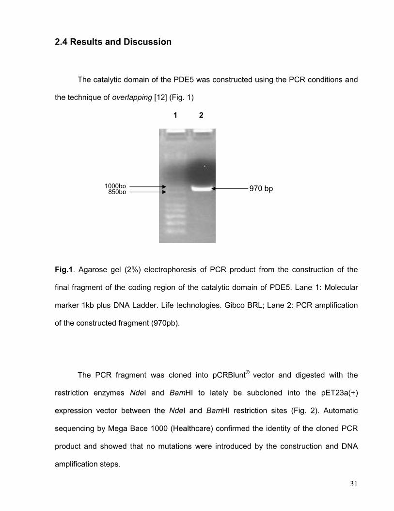
2.4 Results and Discussion
The catalytic domain of the PDE5 was constructed using the PCR conditions and
the technique of overlapping [12] (Fig. 1)
1 2
Fig.1. Agarose gel (2%) electrophoresis of PCR product from the construction of the
final fragment of the coding region of the catalytic domain of PDE5. Lane 1: Molecular
marker 1kb plus DNA Ladder. Life technologies. Gibco BRL; Lane 2: PCR amplification
of the constructed fragment (970pb).
The PCR fragment was cloned into pCRBlunt® vector and digested with the
restriction enzymes NdeI and BamHI to lately be subcloned into the pET23a(+)
expression vector between the NdeI and BamHI restriction sites (Fig. 2). Automatic
sequencing by Mega Bace 1000 (Healthcare) confirmed the identity of the cloned PCR
product and showed that no mutations were introduced by the construction and DNA
amplification steps.
850bp 1000bp 970 bp
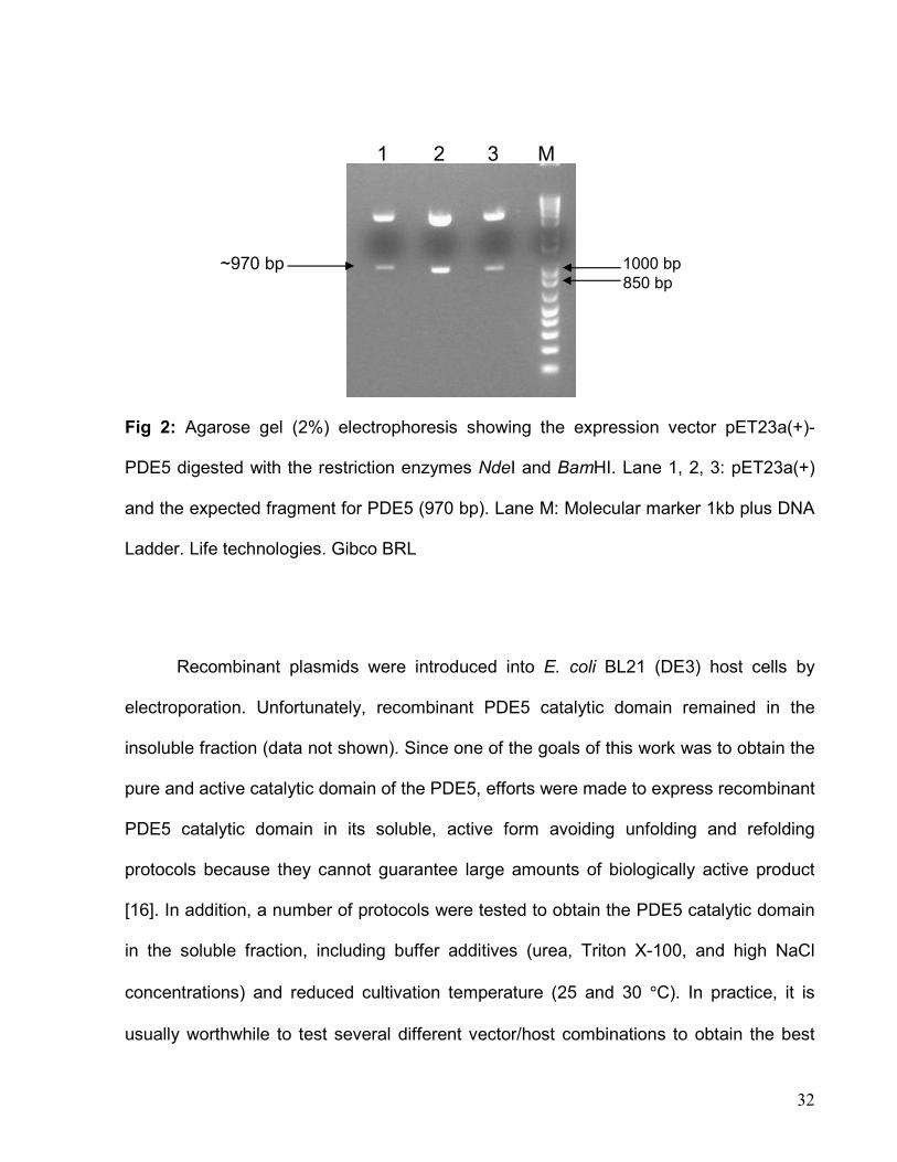
32
Fig 2: Agarose gel (2%) electrophoresis showing the expression vector pET23a(+)-
PDE5 digested with the restriction enzymes NdeI and BamHI. Lane 1, 2, 3: pET23a(+)
and the expected fragment for PDE5 (970 bp). Lane M: Molecular marker 1kb plus DNA
Ladder. Life technologies. Gibco BRL
Recombinant plasmids were introduced into E. coli BL21 (DE3) host cells by
electroporation. Unfortunately, recombinant PDE5 catalytic domain remained in the
insoluble fraction (data not shown). Since one of the goals of this work was to obtain the
pure and active catalytic domain of the PDE5, efforts were made to express recombinant
PDE5 catalytic domain in its soluble, active form avoiding unfolding and refolding
protocols because they cannot guarantee large amounts of biologically active product
[16]. In addition, a number of protocols were tested to obtain the PDE5 catalytic domain
in the soluble fraction, including buffer additives (urea, Triton X-100, and high NaCl
concentrations) and reduced cultivation temperature (25 and 30 °C). In practice, it is
usually worthwhile to test several different vector/host combinations to obtain the best
~970 bp
1 2 3 M
1000 bp 850 bp
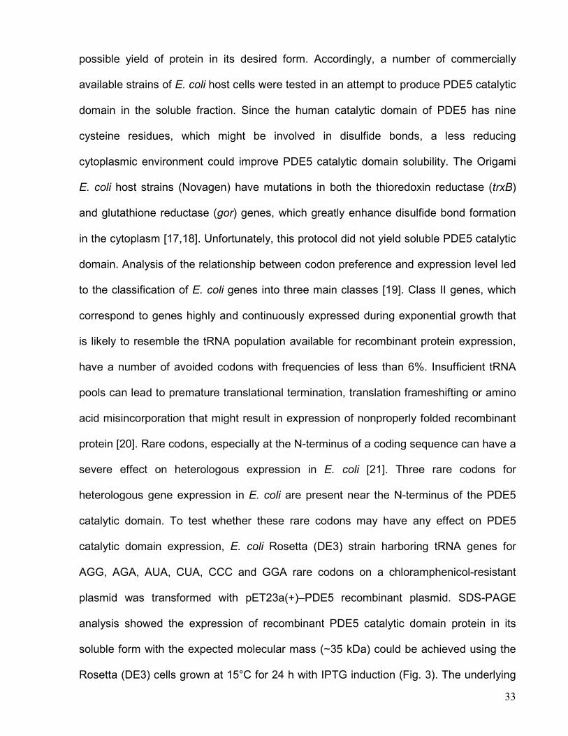
33
possible yield of protein in its desired form. Accordingly, a number of commercially
available strains of E. coli host cells were tested in an attempt to produce PDE5 catalytic
domain in the soluble fraction. Since the human catalytic domain of PDE5 has nine
cysteine residues, which might be involved in disulfide bonds, a less reducing
cytoplasmic environment could improve PDE5 catalytic domain solubility. The Origami
E. coli host strains (Novagen) have mutations in both the thioredoxin reductase (trxB)
and glutathione reductase (gor) genes, which greatly enhance disulfide bond formation
in the cytoplasm [17,18]. Unfortunately, this protocol did not yield soluble PDE5 catalytic
domain. Analysis of the relationship between codon preference and expression level led
to the classification of E. coli genes into three main classes [19]. Class II genes, which
correspond to genes highly and continuously expressed during exponential growth that
is likely to resemble the tRNA population available for recombinant protein expression,
have a number of avoided codons with frequencies of less than 6%. Insufficient tRNA
pools can lead to premature translational termination, translation frameshifting or amino
acid misincorporation that might result in expression of nonproperly folded recombinant
protein [20]. Rare codons, especially at the N-terminus of a coding sequence can have a
severe effect on heterologous expression in E. coli [21]. Three rare codons for
heterologous gene expression in E. coli are present near the N-terminus of the PDE5
catalytic domain. To test whether these rare codons may have any effect on PDE5
catalytic domain expression, E. coli Rosetta (DE3) strain harboring tRNA genes for
AGG, AGA, AUA, CUA, CCC and GGA rare codons on a chloramphenicol-resistant
plasmid was transformed with pET23a(+)–PDE5 recombinant plasmid. SDS-PAGE
analysis showed the expression of recombinant PDE5 catalytic domain protein in its
soluble form with the expected molecular mass (~35 kDa) could be achieved using the
Rosetta (DE3) cells grown at 15°C for 24 h with IPTG induction (Fig. 3). The underlying
34
reason for this result is unclear because other E. coli strains with the availability to
express rare codons were tested and the results were not satisfactory (data not shown).
However, it underscores the need for optimization of vector/host combinations to
achieve soluble recombinant protein expression before attempting any unfolding /
refolding protocols.
Fig. 3. SDS-PAGE analysis of soluble protein extracts, 24 h growth at 15°C. Lane 1:
Rosetta (DE3) pET23a(+)-PDE5 with 0.1mM of IPTG. Lane 2: Rosetta (DE3) pET23a(+)
with 0.1mM of IPTG. Lane 3: Rosetta (DE3) pET23a(+)-PDE5 without IPTG. Lane 4:
Rosetta (DE3) pET23a(+) without IPTG. Lane 5: Rosetta (DE3) pET23a(+)-PDE5 with
0.4mM of IPTG. Lane 6: Rosetta (DE3) pET23a(+) with 0.4mM of IPTG. Lane 7:
BenchMark Protein Ladder (GIBCO).
It should be pointed out that a screening of experimental conditions was carried
out to obtain high yield of recombinant protein expression, including temperature of
growth, culture aeration, medium type, hours of growth after IPTG induction, and hours
10 kDa
20 kDa
25 kDa
30 kDa
40 kDa
50 kDa
2 1 3 4 5 6 7
35
of growth in the absence of IPTG. The best results were obtained from Rosetta (DE3) E.
coli cells grown for 24 h at 15°C in LB medium with the IPTG induction. The pET system
makes use of the powerful T7 RNA polymerase, under control of IPTG-inducible lacUV5
promoter, to transcribe target genes of interest [22]. The IPTG binds to the repressor
protein LacI, stopping its activity, making it possible for the T7 RNA polymerase to
activate the transcription of the gene. Besides this, the high processing of this RNA
polymerase makes high concentrations of mRNA, consequently the production of the
protein of interest is also high, reaching 40 to 50% from the total protein extract [16].
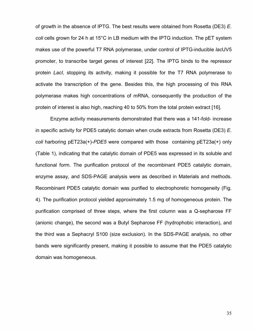
Enzyme activity measurements demonstrated that there was a 141-fold- increase
in specific activity for PDE5 catalytic domain when crude extracts from Rosetta (DE3) E.
coli harboring pET23a(+)-PDE5 were compared with those containing pET23a(+) only
(Table 1), indicating that the catalytic domain of PDE5 was expressed in its soluble and
functional form. The purification protocol of the recombinant PDE5 catalytic domain,
enzyme assay, and SDS-PAGE analysis were as described in Materials and methods.
Recombinant PDE5 catalytic domain was purified to electrophoretic homogeneity (Fig.
4). The purification protocol yielded approximately 1.5 mg of homogeneous protein. The
purification comprised of three steps, where the first column was a Q-sepharose FF
(anionic change), the second was a Butyl Sepharose FF (hydrophobic interaction), and
the third was a Sephacryl S100 (size exclusion). In the SDS-PAGE analysis, no other
bands were significantly present, making it possible to assume that the PDE5 catalytic
domain was homogeneous.
36
Table 1 Measurements of recombinant PDE5 catalytic domain enzyme activity
Cell extract a Specific activity b (SA, U mg-1) SA cloned / SA control
Control 0.0007 1
PDE5 catalytic domain 0.0987 141
a Crude cell extract in 100 mM Tris-HCl, pH 7.0. b U mL -1/mg mL-1.
Fig. 4. SDS–PAGE (12.5%) analysis of pooled fractions from the various steps of the
purification protocol of PDE5 catalytic domain . Lane 1, crude extract; lane 2, Q-
Sepharose fast flow ion exchange; lane 3, Butyl-Sepharose hydrophobic interaction;
lane 4, Sephacryl S-200 gel permeation; lane 5, MW marker high Range (Gibco).
The subunit molecular mass of the active catalytic domain of PDE5 was
determined to be 35164.3 Da by ESI-MS, consistent with the post translational removal
of the N-terminal methionine residue from the full length gene product (predicted mass:
35295.43Da). The first 15 N-terminal amino acids residues of the recombinant protein
14.3kDa
18.4kDa
29kDa
43kDa
68kDa 97.4kDaa
1 2 3 4 5
37
were identified as EETRELQSLAAAVVP by the Edman degradation chemistry protocol.
This result unambiguously identifies the homogeneous recombinant protein as the
catalytic domain of PDE5 and confirms the removal of the N-terminal methionine
residue, a common co/post-translational modification of proteins synthesized in
prokaryotic cells in modification at their N-termini. Methionine aminopeptidase catalyzed
cleavage of initiator methionine is usually directed by the penultimate amino acid
residues with the smallest side chain radii of gyration (glycine, alanine, serine, threonine,
proline, valine and cysteine) [23]. According to the amino acid sequence, this is not valid
for the PDE5, since the penultimate amino acid is a glutamate (Glu), considered as a
large amino acid. Some middle sized amino acids can also undergo N-terminal
processing (Asn, Asp, Leu, and Ile) [24] but this is not the case either.
The major intracellular receptors for cGMP, for example PDE5, are abundant in
vascular smooth muscle cells, including those of the penis. Because cGMP elevation is
known to relax vascular smooth muscle, there has been interest in developing inhibitors
of PDE5 that would block cGMP degradation. To this end, PDE5 has been used to
screen for potentially potent and selective PDE5 inhibitors. The homogeneous PDE5
catalytic domain will provide protein in quantities necessary for the screening of natural
plant extracts from the Brazilian biodiversity. This screening will allow us to research for
a natural and specific inhibitor for the catalytic domain of PDE5 and hopefully develop a
new drug for erectile dysfunction with reduced side effects.
38
2.5 References
[1] S.H. Francis, E.P. Bessay, J. Kotera, K.A. Grimes, L. Liu, W.J. Thompson, and J.D.
Corbin Phosphorylation of Isolated Human Phosphodiesterase-5 Regulatory Domain
Induces an Apparent Conformational Change and Increases cGMP Binding Affinity, J.
Biol. Biochem (2002) 277: 47581-47587.
[2] G.L. Card et al. Structural Basis for the Activity of Drugs that Inhibit
Phosphodiesterases, Structure (2004)12: 2233-2247.
[3] Q. Huai et al. Crystal Structures of Phosphodiesterases 4 and 5 in Complex with
Inhibitor 3-isobutyl-1-methylxanthine Suggest a Conformation Determinant of Inhibitor
Selectivity, J. Biol. Biochem (2004) 279: 13095-13101.
[4] D. Rotella Phosphodiesterase 5 Inhibitors: Current Status and Potencial
Applications, Nat. Rev. Drug. Discov (2002) 1: 647-682.
[5] J.D. Corbin and S.H. Francis Cyclic GMP Phosphodiesterase-5: Target of Sildenafil,
J. Biol. Chem (1999) 247: 13728-13732.
[6] J.D. Corbin, I.V. Turko, A. Beasley, S.H. Francis Phosphorylation of
Phosphodiesterase-5 by cyclic nucleotide-dependent protein kinase alters its catalytic
and allosteric cGMP-binding activities, Eur. J. Biochem (2000) 267: 2760-2767.
39
[7] J.D. Corbin, S.H. Francis, D.J. Webb Phosphodiesterase type 5 as a pharmacologic
Target in Erectile Dysfunction, Urology (2002) 60: (supplement 2B) 4-11.
[8] J.D. Corbin et al. [3H] Sildenafil Binding to Phosphodiesterase-5 Is Specific,
Kenetically, and Stimulated by cGMP, Mol. Pharmacol (2003) 63: 1364-1372.
[9] M.K. Thomas, S.H. Francis, and J.D. Corbin Substrate and kinase-directed
regulation of phosphorylation os a cGMP-binding phosphodiesterase by cGMP, J. Biol.
Chem (1990) 265: 14971-14978.
[10] V.I. Turko, S.H. Francis, and J.D. Corbin Binding of cGMP to both allosteric sites of
cGMP-binding cGMP-specific phosphodiesterase (PDE5) is required for its
phosphorilation, Biochem. J (1998) 329: 505-510.
[11] T.L. Fink, S.H. Francis, A. Beasley, K.A. Grimes, J.D. Corbin Expression of an
Active, Monomeric Catalytic Domain of the cGMP- binding cGMP-specific
Phosphodiesterase (PDE5), J. Biol. Chem (1999) 274: 34613-34620.
[12] S.N. Ho, H.D. Hunt, R.M. Horton, J.K. Pullen and L.R. Pease Site-directed
mutagenesis by overlap extension using the polymerase chain reaction, Gene (1989)
77: 51-59.
[13] U.K. Laemmli Cleavage of structural proteins during the assembly of the head of
bacteriophage T4, Nature (1970) 227: 680-685.
40
[14] M.M. Bradford, R.A McRorie, W.L. Williams A rapid and sensitive method for the
quantification of microgram quantities of protein utilizing the principle of protein-dye
biding, Anal. Biochem 72 (1976) 248-254.
[15] H. Chassaigne, R. Lobinski, Characterization of horse kidney metallothionein
isoforms by electrospray MS and reversed-phase HPLC-electrospray MS, Analyst 123
(1998) 2125-2130.
[16] C.H. Schein, Production of soluble recombinant proteins in bacteria, Bio/technology
7 (1989) 1141-1149.
[17] W.A. Prinz, F. Aslund, A. Holmgren, J. Beckwith, The role of the thioredoxin and
glutaredoxin pathways in redox-facilitate attempts at crystallizing and elucidating the
protein disulfide bonds in the Escherichia coli cytoplasm, J. Biol. Chem 272 (1997)
15661-15667.
[18] F. Baneyx, Recombinant protein expression on Escherichia coli, Curr. Opin.
Biotechnol 10 (1999) 411-421.
[19] A. Hénaut, A. Danchin, Analysis and predictions from Escherichia coli sequences,
or E. coli in silico, in: F.C. Neidhardt (Ed.), Escherichia coli and Salmonella: Cellular and
Molecular Biology, ASM Press, Washington, DC, 1996, pp. 2047-2066.
[20] C. Kurland, J. Gallant, Error of heterologous protein expression, Curr. Opin.
Biotechnol 7 (1996) 489-493.
41
[21] Y. Nakamura, T. Gojobori, T. Ikemura, Codon usage tabulated from international
DNA sequence databases: status for the year 2000, Nucleic Acids Res. 28 (2000) 292.
[22] K.C. Kelley, K.J. Huestis, D.A. Austen, C.T. Sanderson, M.A. Donoghue, S.K.
Stickel, E.S. Kawasaki, M.S. Osburne Regulation of CD4-183 gene expression from
phage-T7-based vectors in Escherichia coli. Gene (1995) 156: 33-36.
[23] W.T. Lowther, B.W. Matthews, Structure and function of the methionine
aminopeptidases, Biochim. Biophys. Acta 1477 (2000) 157-167.
[24] P.H. Hirel, J.M. Schmitter, P. Dessen, G. Fayat, S. Blanquet, Extent of N-terminal
methionine excision from Escherichia coli proteins is governed by the side chain length
of the penultimate amino acid, Proc. Natl. Acad. Sci. USA 86 (1989) 8247-8251.
42
Capítulo 3
3.1 Considerações Finais e Perspectivas
A região codificante do domínio catalítico da fosfodiesterase 5 foi sintetizado
utilizando a técnica de overlaping por PCR. O fragmento desejado foi clonado em vetor
de expressão pET23a(+) e seqüenciado para confirmação da ausência de mutações.
Uma triagem de diversos experimentos como testes de diferentes cepas E. coli para
expressão de proteínas heterólogas, aeração, pH, composição de meio de cultura e
temperatura foram realizados com o intuito de se obter o domínio catalítico da PDE5 em
sua forma homogênea e ativa. O plasmídeo recombinante foi transformado em células
de E. coli Rosetta (DE3) e apresentou expressão na fração solúvel. Com a obtenção do
domínio catalítico da PDE5 em sua forma solúvel testes espectrofotométricos
confirmaram sua atividade. Sabendo que este domínio era expresso em sua forma
solúvel e ativa iniciou-se a determinação de seu protocolo de purificação. Através de
três etapas de purificação, utilizando colunas de troca iônica, interação hidrofóbica e de
exclusão por tamanho o domínio catalítico da PDE5 foi obtido em sua forma
homogênea.
Como perspectivas neste trabalho tem-se a utilização do BIACORE,
equipamento que utiliza a técnica de ressonância plasmônica de superfície, onde se
utilizará a proteína pura para realização de uma triagem, utilizando extratos de plantas
já conhecidos da biodiversidade brasileira, em busca de possíveis candidatos a
inibidores naturais para a fosfodiesterase 5, plantas como por exemplo a catuama são
fortes candidatas a serem testadas [54].
43
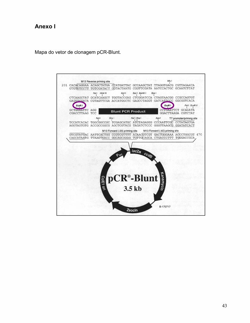
Anexo I
Mapa do vetor de clonagem pCR-Blunt.
44
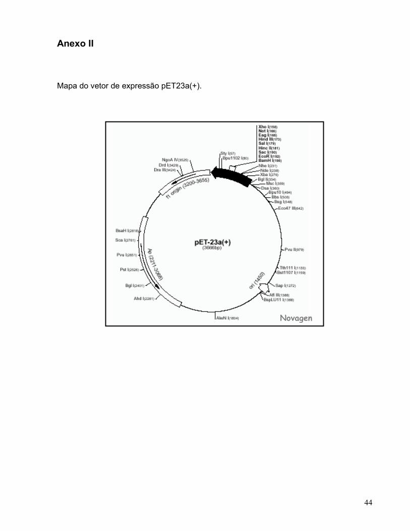
Anexo II Mapa do vetor de expressão pET23a(+).
Novagen
en
45
1.5 Referências Bibliográficas
[1] Corbin JD, Francis SH and Webb DJ. Phosphodiesterase type 5 as a pharmacologic
Target in Erectile Dysfunction. Urology. 2002; 60 Suppl 2B: 4-11.
[2] Champion HC, Bivalacqua TJ, Takimoto E, Kass DA and Burnett AL.
Phosphodiesterase - 5A dysregulation in penile erectile tissue is a mechanism of
priapism. PNAS. 2005; 102: 1661-1666.
[3] Corbin JD, Beasley A, Blount, MA and Francis SH. Vardenafil: structural basis for
higher potency over sildenafil in inhibiting cGMP-specific phosphodiesterase-5 (PDE5).
Neurochem Int. 2004; 45: 859-863.
[4] Loughney K, Hill TR, Florio VA, Uher L, Rosman GJ, Wolda SL, et al. Isolation and
characterization of cDNAs encoding PDE5A, a human cGMP-binding, cGMP-specific
3´,5´-cyclic nucleotide phosphodiesterase. Gene. 1998; 216: 139-147.
[5] Stacey P, Rulten S, Dapling A and Phillips SC. Molecular Cloning and Expression of
Human cGMP-binding cGMP-specific Phosphodiesterase (PDE5). Biochem Biophys
Res Commun. 1998; 247: 249-254.
[6] Francis SH, Turko and Corbin JD. Cyclic Nucleotide phosphodiesterases: relating
structure and function. Prog Nucleic Acid Res Mol Biol. 2001; 65: 1-52.
46
[7] Butcher RW, Sutherland EW. Adenosine 3´,5´ - phosphate in biological materials. J
Biol Chem. 1962; 237: 1244-1250.
[8] Thompson WJ, Terasaki WL, Epstein PM and Strada SJ. Assay of cyclic nucleotide
phosphodiesterase and resolution of multiple molecular forms of the enzyme. Adv Cyclic
Nucleotide Res. 1979; 10: 69-92.
[9] Beavo JA, Hansen RS, Harrison SA, Hurwitz RL, Martins TJ and Mumby MC.
Identification and properties of cyclic nucleotide phosphodiesterase. Mol Cel Endocrinol.
1982; 28: 387-410.
[10] Ghofrani HA, Osterloh IH, Grimminger F. Sildenafil: from angina to erectile
dysfunction to pulmonary hypertension and beyond. Nat Rev Drug Discov. 2006; 5 (8):
689-702.
[11] Bender AT, Beavo JA. Cyclic nucleotide phosphodiesterases: Molecular Regulation
to clinical use. Pharmacol Rev. 2006; 58: 488-520.
[12] Corbin JD and Francis SH. Cyclic GMP Phosphodiesterase-5: Target of Sildenafil. J
Biol Chem. 1999; 247: 13728-13732.
[13] Lin G, Xin ZC, Lue TF, Lin CS. Phosphodiesterase – 5 isoforms: differencial cyclic
guanyl monophosphate binding and cyclic guanyl monophosphate catalytic activities,
and inhibitory effects od sildenafil and vardenafil. J Urol. 2006; 176 (3): 1242-1247.
47
[14] Zhang KY, Card GL, Suzuki Y, Artis DR, Fong D, Gillette S, et al. A Glutamine
Switch Mechanism for Nucleotide Selectivity by Phosphodiesterases. Mol Cell. 2004; 15:
279-286.
[15] Scapin G, Patel SB, Chung C, Vernerin JP, Edmondson SD, Mastracchio A, et al.
Crystal Structure of human phosphodiesterase 3B: atomic basis for substrate and
inhibitor specificity. Biochemistry. 2004; 43: 6091-6100.
[16] Xu RX, Hassell AM, Vanderwall D, Lambert MH, Holmes WD, Luther MA, et al.
Atomic structure of PDE4: insights into phosphodiesterase mechanism and specificity.
Science. 2000; 288: 1822-1825.
[17] Xu RX, Rocque WJ, Lambert MH, Vanderwall DE, Luther MA, and Nolte RT. Crystal
structures of the catalytic domain of phosphodiesterase 4B complexed with AMP, 8-Br-
AMP, and rolipram. J Mol Biol. 2004; 337: 355-365.
[18] Card GL, England BP, Suzuki Y, Fong D, Powell B, Lee B, et al. Structural Basis for
the Activity of Drugs that Inhibit Phosphodiesterases. Structure. 2004; 12: 2233-2247.
[19] Card GL, Blasdel L, England BP, Zhang C, Suzuki Y, Gillette S, et al. A family of
phosphodiesterase inhibitors discovered by cocrystallography and scaffold-based drug
design. Nat Biotechnol. 2005; 23: 201-207.
[20] Lee ME, Markowitz J, Lee JO and Lee H. Crystal Structure of phosphodiesterase
4D and inhibitor complex. FEBS Lett. 2000; 530: 53-58.

48
[21] Huai Q, Wang H, Sun Y, Kim HY, Liu Y and Ke H. Three-dimensional structures of
PDE4D in complex with roliprams and implication on inhibitor selectivity. Structure.
2003; 11: 865-873.
[22] Huai Q, Liu Y, Francis SH, Corbin JD and Ke H. Crystal Structures of
Phosphodiesterases 4 and 5 in Complex with Inhibitor 3-isobutyl-1-methylxanthine
Suggest a Conformation Determinant of Inhibitor Selectivity. J Biol Chem. 2004; 279:
13095-13101.
[23] Huai Q, Colicelli J and Ke H. The crysral structure of AMP-bound PDE4 suggests a
mechanism for phosphodiesterase catalysis. Biochemistry. 2003; 42: 13220-13226.
[24] Sung BJ, Hwang KY, Jeon HJ, Lee J, Heo YS, Kim JH, et al. Structure of the
catalytic domain of human phosphodiesterase 5 with bound drug molecules. Nature.
2003; 425: 98-102.
[25] Wang H, Liu Y, Chen Y, Robinson H and Ke H. Multiple elements jointly determine
inhibitor selectivity of cyclic nucleotide phosphodiesterases 4 and 7. J Biol Chem. 2005;
280: 30949-30955.
[26] Huai Q, Wang H, Zhang W, Colman RW, Robinson H and Ke H. Crystal structure of
phosphodiesterase 9 shows orientation variation of inhibitor 3-isobutyl-1-methylxanthine
binding. Proc Natl Acad Sci USA. 2004; 101: 9624-9629.
49
[27] Ke H. Implications of PDE4 structure on inhibitor selectivity across PDE families. Int
J Impot Res. 2004; 16 Suppl 1: S24-S27.
[28] Zoraghi R, Corbin JD and Francis SH. Phosphodiesterase-5 Gln-817 is critical for
cGMP, vardenafil, or sildenafil affinity: its orientation impacts cGMP but not cAMP
affinity. J Biol Chem. 2006; 281: 5553-5558.
[29] Turko VI, Francis SH and Corbin JD. Binding of cGMP to both allosteric sites of
cGMP-binding cGMP-specific phosphodiesterase (PDE5) is required for its
phosphorilation. Biochem J. 1998; 329: 505-510.
[30] Zoraghi R, Francis SH, Corbin JD. Critical amino acids in phosphodiesterase 5
catalytic site that provide for high affinity interaction with cyclic guanosine
monophosphate and inhibitors. Biochemistry. 2007; 46: 13554-13563.
[31] Jeon YH, Heo YS, Kim CM, Hyun YL, Lee TG, Ro S and Cho JM.
Phosphodiesterase: verview of protein structures, potencial therapeutic applications and
recent process in drug development. Cell Mol Life Sci. 2005; 62: 1198-1220.
[32] Coquil JF, Franks DJ, Wells JN, Dupuis M and Hamet P. Characteristics of a new
binding protein distinct from the kinase for guanosine 3´,5´-monophosphate in rat
platelets. Biochim Biophys Acta. 1980; 631: 148-165.
[33] Francis SH, Lincoln TM and Corbin JD. Characterization of a novel cGMP binding
protein from rat lung. J Biol Chem. 1980; 255: 620-626.
50
[34] Campos MM, Fernandes ES, Ferreira J, Bortolanza LB, Santos AR, Calixto JB.
Pharmacological and neurochemical evidence for antidepressant-like effects of the
herbal product catuama. Pharmacol Biochem Behav. 2004; 78: 757-764.
[35] Prickaerts j, Sik A, van Staveren WC, Koopmans G, Steinbusch HW, van der Staay
FJ, et al. Phosphodiesterase type 5 inhibition improves early memory consolidation of
object information. Neurochem Int. 2004; 45: 915-928.
[36] Corbin JD and Francis SH. Cyclic GMP Phosphodiesterase-5: Target of Sildenafil. J
Biol Chem. 1999; 247: 13728-13732.
[37] Thomas MK, Francis SH, Beebe SJ, Gettys TW, Corbin JD. Partial mapping of
cyclic nucleotide sites and studies of regulatory mechanisms of phosphodiesterases
using cyclic nucleotide analogues. Adv Second Messenger Phosphoprotein Res. 1992;
25: 45-53.
[38] McAllister-Lucas LM, Haik TL, Colbran JL, Sonnenburg WK, Seger D, Turko IV, et
al. An essencial aspartic acid at each of two allosteric cGMP-binding sites of a cGMP
specific phosphodiesterase. J Biol Chem. 1995; 270: 30671-30679.
[39] Zoraghi R, Bessay EP, Corbin JD and Francis SH. Structural and functional features
in human PDE5A1 regulatory domain that provide for allosteric cGMP binding,
dimerization, and regulation. J Biol Chem. 2005; 280: 12051-12063.
51
[40] Blount MA, Zoraghi R, Ke H, Bessay EP, Corbin JD and Francis SH. A 46-amino
acid segment in pjospjodiesterase 5 GAF-B domain provides for high vardenafil potency
over sildenafil and tadalafil and is involved in PDE5 dimerization. Mol Pharmacol. 2006;
70: 1822-1831.
[41] Thomas MK, Francis SH, Corbin JD. Substrate and kinase-directed redulation of
phosphorylation of a cGMP-binding phosphodiesterase by cGMP. J Biol Chem. 1990;
265: 14971-14978.
[42] Corbin JD, Blount MA, Weeks II JL, Beasley A, Kuhn KP, Ho YSJ, et al. [3H]
Sildenafil Binding to Phosphodiesterase-5 Is Specific, Kenetically, and Stimulated by
cGMP. Mol Pharmacol. 2003; 63: 1364-1372.
[43] Mullershausen F, Russwurm M, Thompson WJ, Liu L, Koesling D and Friebe A.
Rapid nitric oxide-induced desensitization of the cGMP response is caused by increase
activity of phosphodiesterase 5 paralleled by phosphorilation of the enzyme. J Cell Biol.
2001; 155: 271-278.
[44] Rybalkin SD, Rybalkina IG, Feil R, Hofmann F and Beavo JA. Regulation of cGMP-
specific phosphodiesterase (PDE5) phosphorylation in smooth muscle cells. J Biol
Chem. 2002; 277: 3310-3317.
[45] Murthy KS. Activation of phosphodiesterase 5 and inhibition of guanylate cyclase by
cGMP-dependent protein kinase in smooth muscle cells. Biochem J. 2001; 360: 199-
208.
52
[46] Mullershausen F, Friebe A, Feil R, Thompson JW, Hofmann F, Koesling D. Direct
activation of PDE5 by cGMP: long-term effects within NO/cGMP signaling. J Cell Biol.
2003; 160 (5): 719-727.
[47] Rybalkin SD, Yan C, Bornfeldt KE, Beavo JA. Cyclic GMP phosphodiesterases and
regulation of smooth muscle function. Circ Res. 2003; 93(4): 280-291.
[48] Corbin JD and Francis SH. Molecular biology and pharmacology of PDE5-inhibitor
therapy for erectile dysfunction. J Androl. 2003; 24 Suppl 6: S38-S41.
[49] Corbin JD, Turko IV, Beasley A and Francis SH. Phosphorylation of
Phosphodiesterase-5 by cyclic nucleotide-dependent protein kinase alters its catalytic
and allosteric cGMP-binding activities. Eur J Biochem. 2000; 267: 2760-2767.
[50] Blount MA, Beasley A, Zoraghi R, Sekhar KR, Bessay PB, Francis SH and Corbin
JD. Binding of Tritiated Sildenafil, Tadalafil, or Vardenafil to the Phosphodiesterase-5
Catalytic Site Displays Potency, Specificity, Heterogeneity, and cGMP Stimulation. Mol
Pharmacol. 2004; 66: 144-152.
[51] Rotella D. Phosphodiesterase 5 Inhibitors: Current Status and Potencial
Applications. Nature Review. 2002; 1: 647-682.
53
[52] Pomeranz HD and Bhavsar AR. Nonarteretic ischemic optic neuropathy developing
soon after use of sildenafil (Viagra): A report of seven new cases. J Neuro-Ophthalmol.
2005; 25: 9-13.
[53] Sowka JW, Neiberg MN and Vollmer L. Optic atrophy after sildenafil use.
Optometry. 2007; 78: 122-128.
[54] Calixto JB, Cabrini DA. Herbal medicine catuama induces endothelium-dependent
and –independent vasorelaxant action on isolated vessels from rats, guinea-pigs and
rabbits. Phytother Res. 1997; 11: 32-38.
[55] Drewes SE, George J, Khan F. Recent findings on natural products with erectile-
dysfunction activity. Phytochemistry. 2003; 62 (7): 1019-1025.
[56] Lewinsohn TM e Prado PI. Biodiversidade Brasileira: Síntese do estado atual do
conhecimento. 2002; 1ª edição. Editora Contexto, São Paulo.