Disfunção de Tecidos Moles: a Pista que Faltava no...
12
Revista Internacional de Ortopedia Funcional / International Journal of Jaw Functional Orthopedics 2005; 1(4):351-9 Revisão da Literatura / Literature Review Ramirez-Yañez GO, Farrel C. Disfunção de tecidos moles: a pista que faltava no tratamento de más-oclusões. Ortop Rev Int Ortop Func 2005; 1(5/6):483-94. O tratamento atual de más-oclusões está relacionado ao efeito das atividades musculares na oclusão. O tratamento de más- oclusões envolve alinhamento dentário, posicionamento do maxilar, estabilização da atividade dos músculos mastigatórios, dos músculos da língua, bochechas e lábios, para a obtenção de equilíbrio entre as forças aplicadas aos arcos e dentes por todos os músculos envolvidos nas funções bucais. Qualquer alteração na atividade muscular desses músculos é denominada pelos autores como “disfunção do tecido mole”. É preciso que dentistas envolvidos no tratamento de más-oclusões estejam atentos a essa disfunção e aprendam a reconhecê-la. Este trabalho apresenta uma revisão da biologia dos músculos das bochechas, lábios e língua, e como a ação desses músculos ação pode afetar a oclusão. Os objetivos de um tratamento ideal de más-oclusões, como aparelhos baseados na fisiologia bucal devem ser projetados para se obter maior grau de sucesso e maior estabilidade no tratamento também são discutidos. PALAVRAS-CHAVE: Disfunção de tecidos moles; Atividade mus- cular; Bochechas; Lábios; Língua. Disfunção de Tecidos Moles: a Pista que Faltava no Tratamento de Más-oclusões Soft Tissue Dysfunction: a Missing Clue when Treating Malocclusions German O Ramirez-Yañez 1 Chris Farrell 2 Ramirez-Yañez GO, Farrel C. Soft tissue dysfunction: a missing clue when treating malocclusions. Ortop Rev Int Ortop Func 2005; 1(5/6):483-94. Contemporaneous treatment of malocclusions concerns about the effect of the muscular activity in the occlusion. Treatment of malocclusion involves dental alignment, jaw repositioning, stabilization of the activity of the mastica- tory muscles, the muscles of the tongue and the muscles of the cheeks and lips in order to achieve a balance be- tween the forces delivered on the arches and teeth by all the muscles involved in oral functions. Any alteration in the muscular activity of these muscles is what the authors term “soft tissue dysfunction”. It is necessary that dentists treating malocclusion be aware of this dysfunction and acquire the required knowledge to recognize it. The current paper reviews the biology of the muscles of the cheeks, lips and tongue and how their action may affect the occlusion. The goals for an ideal treatment of maloc- clusions and how appliances based in the oral physiology must be designed to get better success and more stability in the treatment are also discussed. KEYWORDS: Soft tissue dysfunction; Muscular activity; Cheeks; Lips; Tongue. 1 DDS, MDSc, PhD (cursando); Myofunctional Research Co.; Helensvale, QLD; Australia; Oral Biology & Pathology; The University of Queensland; School of Dentistry; Brisbane, QLD; Australia; St Lucia Campus; Brisbane, QLD 4072; Australia; e-mail: [email protected] 2 BDSc; Myofunctional Research Co.; Helensvale, QLD; Australia 1 DDS, MDSc, PhD (in course); Myofunctional Research Co.; Helensvale, QLD; Australia; Oral Biology & Pathology; The University of Queensland; School of Dentistry; Brisbane, QLD; Australia; Oral Biology & Pathology; School of Dentistry; St Lucia Campus; Brisbane, QLD 4072; Australia; e-mail: german@ myoresearch.com 2 BDSc; Myofunctional Research Co.; Helensvale, QLD; Australia
Transcript of Disfunção de Tecidos Moles: a Pista que Faltava no...
Revista Internacional de Ortopedia Funcional / International Journal of Jaw Functional Orthopedics 2005; 1(4):351-9
Revi
são
da L
itera
tura
/ Li
tera
ture
Rev
iew
Ramirez-Yañez GO, Farrel C. Disfunção de tecidos moles: a pista que faltava no tratamento de más-oclusões. Ortop Rev Int Ortop Func 2005; 1(5/6):483-94.
O tratamento atual de más-oclusões está relacionado ao efeito das atividades musculares na oclusão. O tratamento de más-oclusões envolve alinhamento dentário, posicionamento do maxilar, estabilização da atividade dos músculos mastigatórios, dos músculos da língua, bochechas e lábios, para a obtenção de equilíbrio entre as forças aplicadas aos arcos e dentes por todos os músculos envolvidos nas funções bucais. Qualquer alteração na atividade muscular desses músculos é denominada pelos autores como “disfunção do tecido mole”. É preciso que dentistas envolvidos no tratamento de más-oclusões estejam atentos a essa disfunção e aprendam a reconhecê-la. Este trabalho apresenta uma revisão da biologia dos músculos das bochechas, lábios e língua, e como a ação desses músculos ação pode afetar a oclusão. Os objetivos de um tratamento ideal de más-oclusões, como aparelhos baseados na fisiologia bucal devem ser projetados para se obter maior grau de sucesso e maior estabilidade no tratamento também são discutidos.
PALAVRAS-CHAVE: Disfunção de tecidos moles; Atividade mus-cular; Bochechas; Lábios; Língua.
Disfunção de Tecidos Moles: a Pista que Faltava no Tratamento de Más-oclusõesSoft Tissue Dysfunction: a Missing Clue when Treating Malocclusions
German O Ramirez-Yañez1
Chris Farrell2
Ramirez-Yañez GO, Farrel C. Soft tissue dysfunction: a missing clue when treating malocclusions. Ortop Rev Int Ortop Func 2005; 1(5/6):483-94.
Contemporaneous treatment of malocclusions concerns about the effect of the muscular activity in the occlusion. Treatment of malocclusion involves dental alignment, jaw repositioning, stabilization of the activity of the mastica-tory muscles, the muscles of the tongue and the muscles of the cheeks and lips in order to achieve a balance be-tween the forces delivered on the arches and teeth by all the muscles involved in oral functions. Any alteration in the muscular activity of these muscles is what the authors term “soft tissue dysfunction”. It is necessary that dentists treating malocclusion be aware of this dysfunction and acquire the required knowledge to recognize it. The current paper reviews the biology of the muscles of the cheeks, lips and tongue and how their action may affect the occlusion. The goals for an ideal treatment of maloc-clusions and how appliances based in the oral physiology must be designed to get better success and more stability in the treatment are also discussed.
KEYWORDS: Soft tissue dysfunction; Muscular activity; Cheeks; Lips; Tongue.
1DDS, MDSc, PhD (cursando); Myofunctional Research Co.; Helensvale, QLD; Australia; Oral Biology & Pathology; The University of Queensland; School of Dentistry; Brisbane, QLD; Australia; St Lucia Campus; Brisbane, QLD 4072; Australia; e-mail: [email protected]; Myofunctional Research Co.; Helensvale, QLD; Australia1DDS, MDSc, PhD (in course); Myofunctional Research Co.; Helensvale, QLD; Australia; Oral Biology & Pathology; The University of Queensland; School of Dentistry; Brisbane, QLD; Australia; Oral Biology & Pathology; School of Dentistry; St Lucia Campus; Brisbane, QLD 4072; Australia; e-mail: [email protected] BDSc; Myofunctional Research Co.; Helensvale, QLD; Australia
Disfunção de Tecidos.indd 483 11-04-2006 17:27:39
Disfunção de Tecidos Moles: a Pista que Faltava no Tratamento de Más-oclusõesSoft Tissue Dysfunction: a Missing Clue when Treating Malocclusions
Revista Internacional de Ortopedia Funcional / International Journal of Jaw Functional Orthopedics 2005; 1(5/6):483-94484
A Odontologia estuda a fisiologia e a patologia do sistema bucal. O sistema bucal realiza diversas funções, sendo que três delas se destacam como as mais freqüentes: mastigação, fala para comunicação e deglutição. Ao longo dos anos, os profissionais da área odontológica têm tratado os fatores que afetam a posição dos dentes, alterando o padrão funcional normal descrito como uma disfunção. Vários estudos já demonstraram que a disfunção bucal não é causada somente pelo posicionamento incorreto de dentes e mandíbula, mas também fortemente associada pela hiper e/ou hipoatividade da musculatura envolvida na função do sistema bucal1,2,3,4,5.
Em geral, a avaliação de um paciente com má-oclusão busca identificar alterações nos tecidos min-eralizados (ossos e dentes) e não avalia alterações na função dos tecidos moles (bochechas, lábios e língua). Os modos de avaliação de alterações em tecidos mineralizados já foram exaustivamente de-scritos na literatura por meio de diferentes medidas em modelos, ortopantomogramas e cefalogramas laterais. Entretanto, a avaliação das alterações da função dos tecidos moles é baseada em observa-ções clínicas e requer um profundo conhecimento da fisiologia oral. Por exemplo, cirurgiões-dentistas interagem com pacientes que podem ter desordens de fala, e é importante que tenhamos consciência de que as estruturas pelas quais somos responsáveis são fundamentais, não somente para a mastigação e a deglutição, mas também para a comunicação por meio da fala6, e que uma desordem da fala pode piorar o problema que estamos avaliando. Portanto, um alto nível de observação da expressão facial do paciente é necessário para se detectar qualquer des-vio de padrão muscular que poderia estar associado à disfunção oral7.
Um tratamento ideal deve levar em conta os prob-lemas musculares subjacentes associados à má-oclusão8. A intenção deste estudo é descrever o que os autores denominam “disfunção dos tecidos moles” e como essa disfunção altera o formato dos arcos e a posição dos dentes.
As BochechasO bucinador é o maior músculo das bochechas.
O músculo é ligado à maxila e à mandíbula9. Em repouso, a pressão exercida pela atividade mus-cular das bochechas no lado vestibular dos dentes
Dentistry focuses in the study of the physiology and pathology of the oral system. The oral system performs several functions with three of them ris-ing as the most frequently performed: mastication, speech for communication and swallowing. Over the years, dental professionals have dealt with those factors affecting the position of the teeth, and so, altering the normal functional pattern which is described as a dysfunction. Numerous studies have shown that oral dysfunction is not only caused by an incorrect position of the teeth and jaws, but also highly associated to hyper- and/or hypo-activity of the musculature involved in the function of the oral system1,2,3,4,5.
In general, the evaluation of a patient with malocclusion seeks for alterations in the mineral-ized tissues (bone and teeth), and do not evaluate alterations in the function of the soft tissues (cheeks, lips and tongue). How to evaluate alterations in the mineralized tissues has been extensively explained in the literature through different measurements on cast models, orthopantomograms and lateral cephalograms. However, the evaluation of altera-tions in the function of the soft tissues is based on clinical observations and requires a deep knowledge in oral physiology. For example, dentists interact with patients who may have speech disorders, and it is important for us to realize that the structures for which we are responsible play a vital role not only for chewing and swallowing but also for speech communication6, and that a speech disorder may worsen the problem that we are evaluating. So, a high level of observation of the patient’s facial ex-pression is required to detect any deviation in the muscular pattern that may be associated with the oral dysfunction7.
An ideal treatment must address the underlying muscular problems associated to the malocclusion8. This paper intends to describe what authors term “soft tissue dysfunction” and, how this dysfunction alters the shape of the arches and the position of the teeth.
The CheeksThe buccinator is the major muscle of the
cheeks. This muscle is attached to both the maxilla and the mandible9. At rest, the pressure exerted by the muscular activity of the cheeks on the buccal
INTRODUÇÃO/INTRODUCTION
Disfunção de Tecidos.indd 484 11-04-2006 17:27:39
485
Disfunção de Tecidos Moles: a Pista que Faltava no Tratamento de Más-oclusõesSoft Tissue Dysfunction: a Missing Clue when Treating Malocclusions
Revista Internacional de Ortopedia Funcional / International Journal of Jaw Functional Orthopedics 2005; 1(5/6):483-94
posteriores mandibulares é semelhante à exercida pela atividade muscular da língua no lado lingual, aproximadamente 2g/cm2. Entretanto, a pressão nos dentes posteriores maxilares é maior no lado vestibular do que no lado lingual, 2,7 e 1,0g/cm2 respectivamente10. Essa situação muda durante a mastigação e a deglutição, quando a pressão no lado lingual dos dentes posteriores maxilares é maior do que no lado vestibular10. Assim sendo, o equilíbrio é atingido e a posição dos dentes posteriores é mantida pelas bochechas e pela língua.
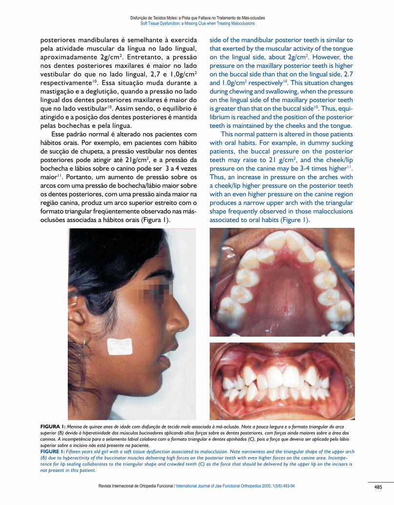
Esse padrão normal é alterado nos pacientes com hábitos orais. Por exemplo, em pacientes com hábito de sucção de chupeta, a pressão vestibular nos dentes posteriores pode atingir até 21g/cm2, e a pressão da bochecha e lábios sobre o canino pode ser 3 a 4 vezes maior11. Portanto, um aumento de pressão sobre os arcos com uma pressão de bochecha/lábio maior sobre os dentes posteriores, com uma pressão ainda maior na região canina, produz um arco superior estreito com o formato triangular freqüentemente observado nas más-oclusões associadas a hábitos orais (Figura 1).
side of the mandibular posterior teeth is similar to that exerted by the muscular activity of the tongue on the lingual side, about 2g/cm2. However, the pressure on the maxillary posterior teeth is higher on the buccal side than that on the lingual side, 2.7 and 1.0g/cm2 respectively10. This situation changes during chewing and swallowing, when the pressure on the lingual side of the maxillary posterior teeth is greater than that on the buccal side10. Thus, equi-librium is reached and the position of the posterior teeth is maintained by the cheeks and the tongue.
This normal pattern is altered in those patients with oral habits. For example, in dummy sucking patients, the buccal pressure on the posterior teeth may raise to 21 g/cm2, and the cheek/lip pressure on the canine may be 3-4 times higher11. Thus, an increase in pressure on the arches with a cheek/lip higher pressure on the posterior teeth with an even higher pressure on the canine region produces a narrow upper arch with the triangular shape frequently observed in those malocclusions associated to oral habits (Figure 1).
FIGURA 1: Menina de quinze anos de idade com disfunção de tecido mole associada à má-oclusão. Note a pouca largura e o formato triangular do arco
superior (B) devido à hiperatividade dos músculos bucinadores aplicando altas forças sobre os dentes posteriores, com forças ainda maiores sobre a área dos
caninos. A incompetência para o selamento labial colabora com o formato triangular e dentes apinhados (C), pois a força que deveria ser aplicada pelo lábio
superior sobre o incisivo não está presente na paciente.
FIGURE 1: Fifteen years old girl with a soft tissue dysfunction associated to malocclusion. Note narrowness and the triangular shape of the upper arch
(B) due to hyperactivity of the buccinator muscles delivering high forces on the posterior teeth with even higher forces on the canine area. Incompe-
tence for lip sealing collaborates to the triangular shape and crowded teeth (C) as the force that should be delivered by the upper lip on the incisors is
not present in this patient.
Disfunção de Tecidos.indd 485 11-04-2006 17:27:40
Disfunção de Tecidos Moles: a Pista que Faltava no Tratamento de Más-oclusõesSoft Tissue Dysfunction: a Missing Clue when Treating Malocclusions
Revista Internacional de Ortopedia Funcional / International Journal of Jaw Functional Orthopedics 2005; 1(5/6):483-94486
Os LábiosOs músculos orbiculares superior e inferior da boca
são as principais estruturas dos lábios. Sua atividade mútua permite o fechamento dos lábios, que veda a entrada da cavidade oral. Em repouso, com selamento labial a atividade do mentual é desprezível em pacientes com oclusão normal5. Portanto, para o selamento labial deve haver atividade do músculo orbicular da boca, mas não atividade do mentual. A força produzida pelo músculo orbicular da boca é leve e con-tínua, e foi estimada por meio da força transmitida a placas labiativas (lip bumpers) em 12 a 14g em repouso e 33 a 36g durante a deglutição12. A força da língua na superfície lingual dos incisivos foi calculada em 0,8 a 1,6g/cm2 em repouso13,14. Entretanto, a força pode aumentar para até 3 a 7g/cm2 em pacientes respiradores bucais15. Estudos indicam que o músculo orbicular da boca pode exercer uma força sobre uma placa labiativa (lip bumpers) de até 300g e a língua pode exercer forças de até 500g16,17. Alguns autores sugeriram que a pressão da língua sobre os incisivos é maior do que as forças produzidas pela musculatura labial18,19,20. Um estudo recente mostrou que, em repouso, a pressão exercida pelos lábios diretamente sobre os incisivos superiores e inferiores é muito baixa, até negativa5,21. Portanto, o equilíbrio é atingido e a correta posição dos incisivos é mantida porque uma força mais alta, porém descontínua, é aplicada pela língua sobre a superfície lingual dos incisivos, ao passo que uma força menor, porém contínua, é aplicada pelos lábios à superfície vestibular dos dentes anteriores22,23.
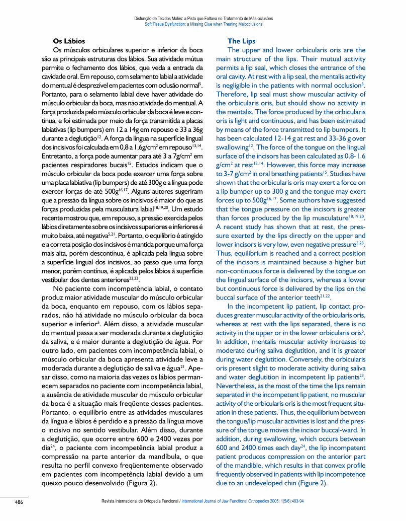
No paciente com incompetência labial, o contato produz maior atividade muscular do músculo orbicular da boca, enquanto em repouso, com os lábios sepa-rados, não há atividade no músculo orbicular da boca superior e inferior5. Além disso, a atividade muscular do mentual passa a ser moderada durante a deglutição da saliva, e é maior durante a deglutição de água. Por outro lado, em pacientes com incompetência labial, o músculo orbicular da boca apresenta atividade leve a moderada durante a deglutição de saliva e água21. Ape-sar disso, como na maioria das vezes os lábios perman-ecem separados no paciente com incompetência labial, a ausência de atividade muscular do músculo orbicular da boca é a situação mais freqüente desses pacientes. Portanto, o equilíbrio entre as atividades musculares da língua e lábios é perdido e a pressão da língua move o incisivo no sentido vestibular. Além disso, durante a deglutição, que ocorre entre 600 e 2400 vezes por dia24, o paciente com incompetência labial produz a compressão na parte anterior da mandíbula, o que resulta no perfil convexo freqüentemente observado em pacientes com incompetência labial devido a um queixo pouco desenvolvido (Figura 2).
The LipsThe upper and lower orbicularis oris are the
main structure of the lips. Their mutual activity permits a lip seal, which closes the entrance of the oral cavity. At rest with a lip seal, the mentalis activity is negligible in the patients with normal occlusion5. Therefore, lip seal must show muscular activity of the orbicularis oris, but should show no activity in the mentalis. The force produced by the orbicularis oris is light and continuous, and has been estimated by means of the force transmitted to lip bumpers. It has been calculated 12-14 g at rest and 33-36 g over swallowing12. The force of the tongue on the lingual surface of the incisors has been calculated as 0.8-1.6 g/cm2 at rest13,14. However, this force may increase to 3-7 g/cm2 in oral breathing patients15. Studies have shown that the orbicularis oris may exert a force on a lip bumper up to 300 g and the tongue may exert forces up to 500g16,17. Some authors have suggested that the tongue pressure on the incisors is greater than forces produced by the lip musculature18,19,20. A recent study has shown that at rest, the pres-sure exerted by the lips directly on the upper and lower incisors is very low, even negative pressure5,23. Thus, equilibrium is reached and a correct position of the incisors is maintained because a higher but non-continuous force is delivered by the tongue on the lingual surface of the incisors, whereas a lower but continuous force is delivered by the lips on the buccal surface of the anterior teeth21,22.
In the incompetent lip patient, lip contact pro-duces greater muscular activity of the orbicularis oris, whereas at rest with the lips separated, there is no activity in the upper or in the lower orbicularis oris5. In addition, mentalis muscular activity increases to moderate during saliva deglutition, and it is greater during water deglutition. Conversely, the orbicularis oris present slight to moderate activity during saliva and water deglutition in incompetent lip patients23. Nevertheless, as the most of the time the lips remain separated in the incompetent lip patient, no muscular activity of the orbicularis oris is the most frequent situ-ation in these patients. Thus, the equilibrium between the tongue/lip muscular activities is lost and the pres-sure of the tongue moves the incisor buccal-ward. In addition, during swallowing, which occurs between 600 and 2400 times each day24, the lip incompetent patient produces compression on the anterior part of the mandible, which results in that convex profile frequently observed in patients with lip incompetence due to an undeveloped chin (Figure 2).
Disfunção de Tecidos.indd 486 11-04-2006 17:27:40
487
Disfunção de Tecidos Moles: a Pista que Faltava no Tratamento de Más-oclusõesSoft Tissue Dysfunction: a Missing Clue when Treating Malocclusions
Revista Internacional de Ortopedia Funcional / International Journal of Jaw Functional Orthopedics 2005; 1(5/6):483-94
A LínguaA língua é um órgão estruturalmente complexo e ex-
tremamente flexível que ocupa uma grande parte da cavi-dade oral. A posição de repouso da língua é a posição re-produzível, fisiologicamente relaxada do órgão, observada quando os lábios estão suavemente fechados, ou levemente separados, e a mandíbula está ligeiramente aberta25. Nessa posição, a língua produz a menor pressão nas estruturas adjacentes, ao passo que durante seu movimento, para participar em funções orais, a pressão aumenta ou diminui em diferentes locais, em momentos diferentes10.
A atividade mais desempenhada pela língua é a obser-vada durante a deglutição. Essa função oral é dividida em seis estágios26. No primeiro, a língua está em posição de repouso, com sua extremidade atrás da superfície lingual dos incisivos superiores, tocando levemente o palato an-terior. O dorso fica paralelo e próximo ao palato duro e a maior parte posterior da língua fica em contato com o palato mole. No segundo estágio, a extremidade da língua se posiciona em sua posição mais posterior. O terceiro estágio é caracterizado pela fixação da extremidade da língua no palato anterior e pelo abaixamento do dorso da língua. No quarto estágio, o palato mole se eleva até
The TongueThe tongue is a structurally complex and extremely
flexible organ occupying a large portion of the oral cav-ity. The tongue resting position refers to a reproducible physiologically relaxed position of the organ observed when the lips are smoothly sealed, or slightly separated, and the mandible is slightly opened25. At this position, the tongue produces the lowest pressure on the sur-rounding structures, whereas during its movements to participate in the oral functions, the pressure increases or reduces at different sites at different times10.
The most performed activity of the tongue is that observed during swallowing. This oral function is di-vided into six stages26, starting from a resting position where the tip of the tongue is positioned behind the lingual surface of the upper incisors, slightly touching the anterior palate, the dorsum running parallel and near to the hard palate and the posterior part of the tongue mostly in contact with the soft palate. At the second stage, the tip of the tongue locates at its most backward position. Stage three is characterized by the fixation of the tongue tip on the anterior palate and a descending of the dorsum of the tongue. At stage four,
FIGURA 2: Menina com onze anos de idade com disfunção de tecido mole associado a distoclusão (B). Esta má-oclusão resulta da hiperatividade do mentual
que produz uma força para trás na área anterior da mandíbula, em oposição ao crescimento mandibular normal (C).
FIGURE 2: Eleven years girl with soft tissue dysfunction associated to disto-occlusion (B). This malocclusion results from hyperactivity of the mentalis which
produces a backward force on the anterior area of the mandible, and so, opposites to normal mandibular growth (C).
Disfunção de Tecidos.indd 487 11-04-2006 17:27:40

Disfunção de Tecidos Moles: a Pista que Faltava no Tratamento de Más-oclusõesSoft Tissue Dysfunction: a Missing Clue when Treating Malocclusions
Revista Internacional de Ortopedia Funcional / International Journal of Jaw Functional Orthopedics 2005; 1(5/6):483-94488
o nível mais alto, com a maior parte da superfície dorsal da língua em contato com o palato. No quinto estágio, o palato mole desce à sua posição mais baixa, e no último, a língua retorna à sua posição inicial.
A língua se move de maneira precisa, contribuindo significativamente para os complexos movimentos oro-faciais27. Todos esses movimentos são produzidos pelo trabalho coordenado de dois conjuntos de músculos, os extrínsecos e os intrínsecos. Os músculos extrínsecos são o genioglosso, que é o principal protrusor da língua, o estiloglosso e o hioglosso, que agem como retratores da língua28,29. Entretanto, a ação dos músculos extrínsecos da língua não é simples. A ativação simultânea do músculo protrusor e dos retrusores produz a retração da língua30, a contração simultânea do genioglosso (protrusor) e esti-loglosso (retrusor), durante a abertura da boca, contribui para moldar o formato da língua para reunir o alimento31. Por outro lado, os músculos intrínsecos são classificados em transverso, vertical e longitudinal28. Os músculos intrínsecos tem uma arquitetura e composição de fibras características que diferem das dos músculos orofaciais e mastigatórios32,33. As fibras dos intrínsecos são responsáveis pelas ações rápidas e flexíveis no posicionamento e formato da língua durante as funções orais33, e permitem que o dorso da língua se enrijeça para pressionar o alimento durante a mastigação e para mover o alimento em direção à região posterior para deglutição32.
Portanto, a ação dos músculos da língua é coordenada internamente, mas, além disso, a ação da língua é também associada à função dos músculos mastigatórios. Durante a abertura da boca, o genioglosso aumenta sua atividade em relação ao aumento de atividade do digástrico31,34. Durante o fechamento da boca, um aumento na atividade do estilo-glosso é observada em relação ao aumento de atividade do masseter34. Os músculos mastigatórios e extrínsecos são ativos e coordenados durante a mastigação. Um aumento na atividade de um grupo (genioglosso/digástrico ou estilo-glosso/masseter) inibe a atividade do grupo antagonista34. Mas isso não ocorre nos dois grupos de músculos. O efeito inibitório do masseter sobre o digástrico é maior do que o produzido pelo genioglosso sobre o estiloglosso34. A intera-ção entre a língua e os músculos mastigatórios é conhecida como reflexo mandíbula-língua35.
No nascimento, todos os indivíduos possuem uma posição normal da língua, e a posição anormal é desenvolvida com a idade e por hábitos de deglutição anormais36. Os movimentos da língua, que são não dife-renciados no bebê, transformam-se em diferenciados e refinados na criança jovem37. A posição da base da língua está associada à posição do maxilar e à rotação vertical mandibular, e assim influi no crescimento maxilo-man-
the soft palate rises to the highest level with the dorsal surface of the tongue mostly in contact with the palate. At stage five, the soft palate descends to the lowest position, and at the last stage, the tongue returns to the initial position.
The tongue moves in a precise manner contribut-ing highly to the complex orofacial movements27. All these movements are produced by a coordinated performance of two sets of muscles, the extrinsic and the intrinsic muscles. The extrinsic muscles are the ge-nioglosus, which is the principal tongue protruder, and the styloglosus and the hyoglosus, which act as tongue retractors28,29. However, the action of the tongue ex-trinsic muscles is not as simple. Co-activation of the protruder and retractor muscles produces tongue re-traction30, and co-contraction of the genioglosus (pro-truder) and styloglosus (retractor) during jaw-opening, contributes to shape the tongue for gathering the food stuff31. On the other hand, the intrinsic muscles are classified as transverses, verticalis and longitudinalis28. The intrinsic muscles have a characteristic architecture and fiber composition which differs from those of orofacial and masticatory muscles32,33. The fibers in the intrinsic are responsible for the fast and flexible actions in positioning and shaping the tongue during the oral functions33, and enable the dorsum of the tongue to harden for pressing food during mastication and shifting the food posteriorly for swallowing32.
Thus, the action of the muscles of the tongue is coordinated within them, but furthermore, tongue action is also associated to function of the mastica-tory muscles. During jaw opening, the genioglosus increases its activity in correlation to the increase in the digastrics activity31,34. During jaw closing, an increase in styloglosus activity is observed in correlation to the increase in the activity of the masseter34. Both mastica-tory and extrinsic muscles are active and coordinated during mastication. An increase in the activity of one group (genioglosus/digastrics or styloglosus/masseter) inhibits the activity of the antagonist group34. But, this is not analogous between the two muscle groups. The inhibitory effect of the masseter on the digastrics is higher than that produced by the genioglosus on the styloglosus34. This interaction between the tongue and the masticatory muscles is referred as the jaw-tongue reflex35.
At birth, all subjects posses a normal tongue position and the abnormal one is developed by aging accompanied by abnormal swallowing habits36. The movements of the tongue transform from undiffer-entiated movements in the infant, to differentiated
Disfunção de Tecidos.indd 488 11-04-2006 17:27:40
489
Disfunção de Tecidos Moles: a Pista que Faltava no Tratamento de Más-oclusõesSoft Tissue Dysfunction: a Missing Clue when Treating Malocclusions
Revista Internacional de Ortopedia Funcional / International Journal of Jaw Functional Orthopedics 2005; 1(5/6):483-94
dibular38. Portanto, a atividade da língua define a posição dos dentes, como citado acima e também determina a posição da mandíbula. Assim, o equilíbrio entre pressão da língua pelo lado de dentro e pressão lábio-bucal pelo lado de fora precisa existir39 associado à correta posição da base da língua38, de modo a desenvolver padrões corretos durante o crescimento dento-maxilo-mandibular.
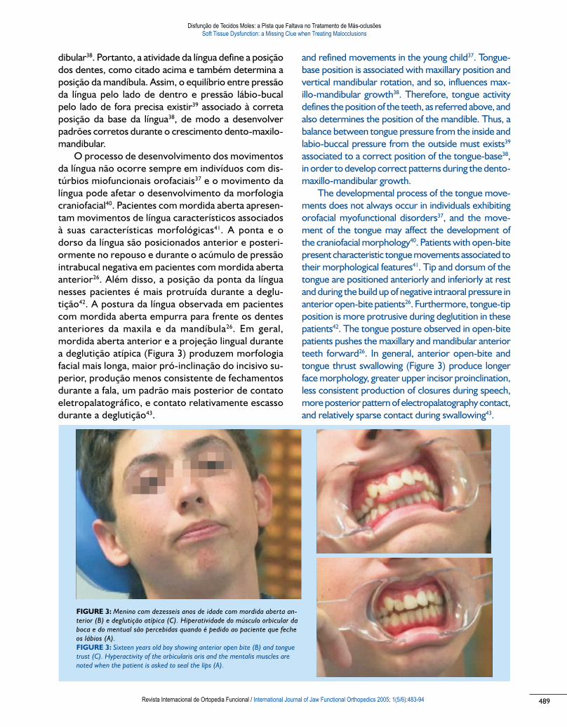
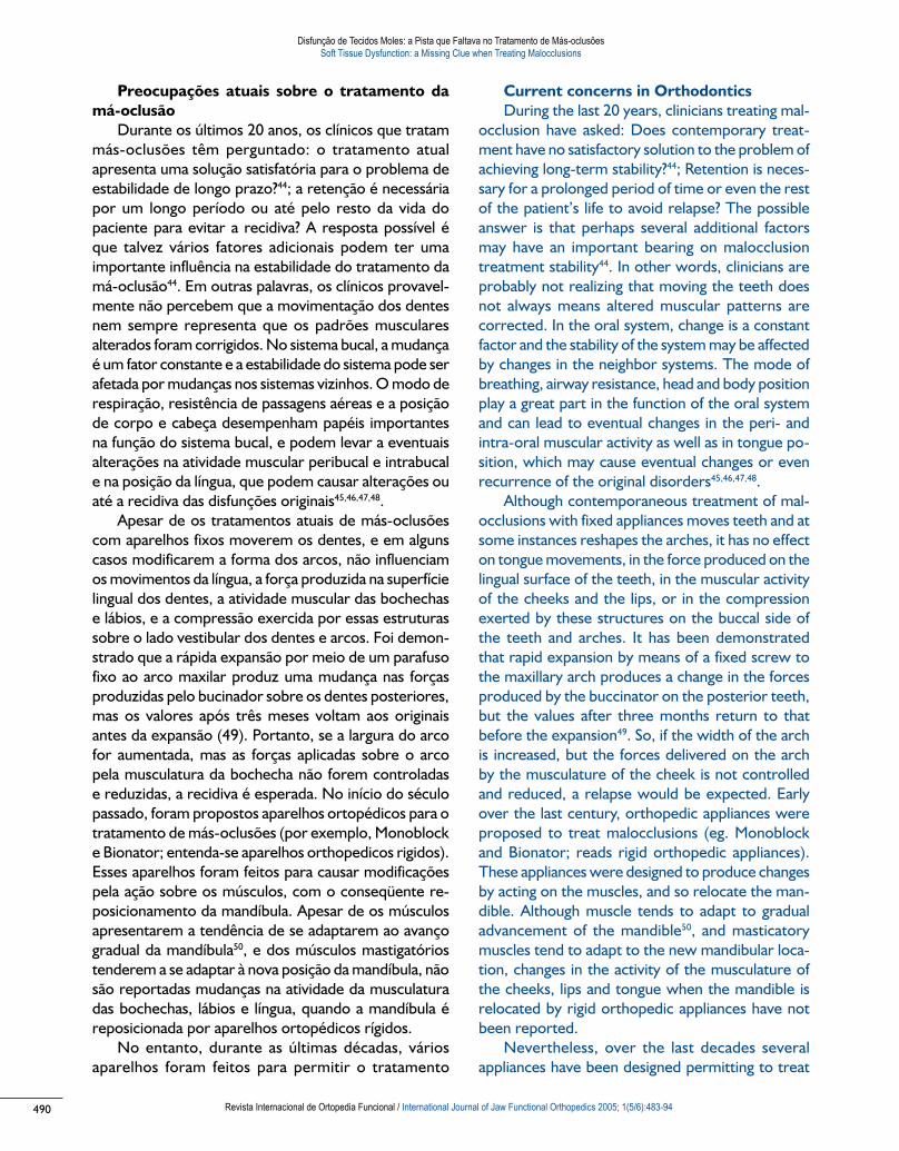
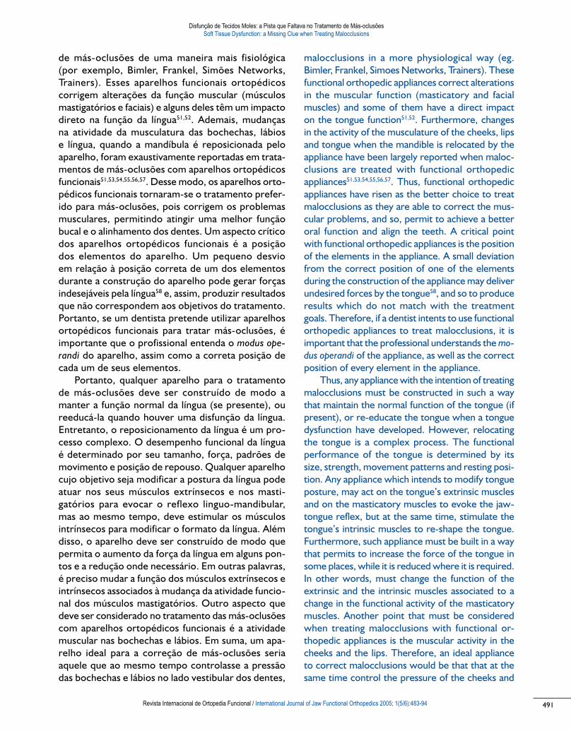
O processo de desenvolvimento dos movimentos da língua não ocorre sempre em indivíduos com dis-túrbios miofuncionais orofaciais37 e o movimento da língua pode afetar o desenvolvimento da morfologia craniofacial40. Pacientes com mordida aberta apresen-tam movimentos de língua característicos associados à suas características morfológicas41. A ponta e o dorso da língua são posicionados anterior e posteri-ormente no repouso e durante o acúmulo de pressão intrabucal negativa em pacientes com mordida aberta anterior26. Além disso, a posição da ponta da língua nesses pacientes é mais protruída durante a deglu-tição42. A postura da língua observada em pacientes com mordida aberta empurra para frente os dentes anteriores da maxila e da mandíbula26. Em geral, mordida aberta anterior e a projeção lingual durante a deglutição atípica (Figura 3) produzem morfologia facial mais longa, maior pró-inclinação do incisivo su-perior, produção menos consistente de fechamentos durante a fala, um padrão mais posterior de contato eletropalatográfico, e contato relativamente escasso durante a deglutição43.
and refined movements in the young child37. Tongue-base position is associated with maxillary position and vertical mandibular rotation, and so, influences max-illo-mandibular growth38. Therefore, tongue activity defines the position of the teeth, as referred above, and also determines the position of the mandible. Thus, a balance between tongue pressure from the inside and labio-buccal pressure from the outside must exists39 associated to a correct position of the tongue-base38, in order to develop correct patterns during the dento-maxillo-mandibular growth.
The developmental process of the tongue move-ments does not always occur in individuals exhibiting orofacial myofunctional disorders37, and the move-ment of the tongue may affect the development of the craniofacial morphology40. Patients with open-bite present characteristic tongue movements associated to their morphological features41. Tip and dorsum of the tongue are positioned anteriorly and inferiorly at rest and during the build up of negative intraoral pressure in anterior open-bite patients26. Furthermore, tongue-tip position is more protrusive during deglutition in these patients42. The tongue posture observed in open-bite patients pushes the maxillary and mandibular anterior teeth forward26. In general, anterior open-bite and tongue thrust swallowing (Figure 3) produce longer face morphology, greater upper incisor proinclination, less consistent production of closures during speech, more posterior pattern of electropalatography contact, and relatively sparse contact during swallowing43.
FIGURE 3: Menino com dezesseis anos de idade com mordida aberta an-
terior (B) e deglutição atípica (C). Hiperatividade do músculo orbicular da
boca e do mentual são percebidas quando é pedido ao paciente que feche
os lábios (A).
FIGURE 3: Sixteen years old boy showing anterior open bite (B) and tongue
trust (C). Hyperactivity of the orbicularis oris and the mentalis muscles are
noted when the patient is asked to seal the lips (A).
Disfunção de Tecidos.indd 489 11-04-2006 17:27:41
Disfunção de Tecidos Moles: a Pista que Faltava no Tratamento de Más-oclusõesSoft Tissue Dysfunction: a Missing Clue when Treating Malocclusions
Revista Internacional de Ortopedia Funcional / International Journal of Jaw Functional Orthopedics 2005; 1(5/6):483-94490
Preocupações atuais sobre o tratamento da má-oclusão
Durante os últimos 20 anos, os clínicos que tratam más-oclusões têm perguntado: o tratamento atual apresenta uma solução satisfatória para o problema de estabilidade de longo prazo?44; a retenção é necessária por um longo período ou até pelo resto da vida do paciente para evitar a recidiva? A resposta possível é que talvez vários fatores adicionais podem ter uma importante influência na estabilidade do tratamento da má-oclusão44. Em outras palavras, os clínicos provavel-mente não percebem que a movimentação dos dentes nem sempre representa que os padrões musculares alterados foram corrigidos. No sistema bucal, a mudança é um fator constante e a estabilidade do sistema pode ser afetada por mudanças nos sistemas vizinhos. O modo de respiração, resistência de passagens aéreas e a posição de corpo e cabeça desempenham papéis importantes na função do sistema bucal, e podem levar a eventuais alterações na atividade muscular peribucal e intrabucal e na posição da língua, que podem causar alterações ou até a recidiva das disfunções originais45,46,47,48.
Apesar de os tratamentos atuais de más-oclusões com aparelhos fixos moverem os dentes, e em alguns casos modificarem a forma dos arcos, não influenciam os movimentos da língua, a força produzida na superfície lingual dos dentes, a atividade muscular das bochechas e lábios, e a compressão exercida por essas estruturas sobre o lado vestibular dos dentes e arcos. Foi demon-strado que a rápida expansão por meio de um parafuso fixo ao arco maxilar produz uma mudança nas forças produzidas pelo bucinador sobre os dentes posteriores, mas os valores após três meses voltam aos originais antes da expansão (49). Portanto, se a largura do arco for aumentada, mas as forças aplicadas sobre o arco pela musculatura da bochecha não forem controladas e reduzidas, a recidiva é esperada. No início do século passado, foram propostos aparelhos ortopédicos para o tratamento de más-oclusões (por exemplo, Monoblock e Bionator; entenda-se aparelhos orthopedicos rigidos). Esses aparelhos foram feitos para causar modificações pela ação sobre os músculos, com o conseqüente re-posicionamento da mandíbula. Apesar de os músculos apresentarem a tendência de se adaptarem ao avanço gradual da mandíbula50, e dos músculos mastigatórios tenderem a se adaptar à nova posição da mandíbula, não são reportadas mudanças na atividade da musculatura das bochechas, lábios e língua, quando a mandíbula é reposicionada por aparelhos ortopédicos rígidos.
No entanto, durante as últimas décadas, vários aparelhos foram feitos para permitir o tratamento
Current concerns in OrthodonticsDuring the last 20 years, clinicians treating mal-
occlusion have asked: Does contemporary treat-ment have no satisfactory solution to the problem of achieving long-term stability?44; Retention is neces-sary for a prolonged period of time or even the rest of the patient’s life to avoid relapse? The possible answer is that perhaps several additional factors may have an important bearing on malocclusion treatment stability44. In other words, clinicians are probably not realizing that moving the teeth does not always means altered muscular patterns are corrected. In the oral system, change is a constant factor and the stability of the system may be affected by changes in the neighbor systems. The mode of breathing, airway resistance, head and body position play a great part in the function of the oral system and can lead to eventual changes in the peri- and intra-oral muscular activity as well as in tongue po-sition, which may cause eventual changes or even recurrence of the original disorders45,46,47,48.
Although contemporaneous treatment of mal-occlusions with fixed appliances moves teeth and at some instances reshapes the arches, it has no effect on tongue movements, in the force produced on the lingual surface of the teeth, in the muscular activity of the cheeks and the lips, or in the compression exerted by these structures on the buccal side of the teeth and arches. It has been demonstrated that rapid expansion by means of a fixed screw to the maxillary arch produces a change in the forces produced by the buccinator on the posterior teeth, but the values after three months return to that before the expansion49. So, if the width of the arch is increased, but the forces delivered on the arch by the musculature of the cheek is not controlled and reduced, a relapse would be expected. Early over the last century, orthopedic appliances were proposed to treat malocclusions (eg. Monoblock and Bionator; reads rigid orthopedic appliances). These appliances were designed to produce changes by acting on the muscles, and so relocate the man-dible. Although muscle tends to adapt to gradual advancement of the mandible50, and masticatory muscles tend to adapt to the new mandibular loca-tion, changes in the activity of the musculature of the cheeks, lips and tongue when the mandible is relocated by rigid orthopedic appliances have not been reported.
Nevertheless, over the last decades several appliances have been designed permitting to treat
Disfunção de Tecidos.indd 490 11-04-2006 17:27:41
491
Disfunção de Tecidos Moles: a Pista que Faltava no Tratamento de Más-oclusõesSoft Tissue Dysfunction: a Missing Clue when Treating Malocclusions
Revista Internacional de Ortopedia Funcional / International Journal of Jaw Functional Orthopedics 2005; 1(5/6):483-94
de más-oclusões de uma maneira mais fisiológica (por exemplo, Bimler, Frankel, Simões Networks, Trainers). Esses aparelhos funcionais ortopédicos corrigem alterações da função muscular (músculos mastigatórios e faciais) e alguns deles têm um impacto direto na função da língua51,52. Ademais, mudanças na atividade da musculatura das bochechas, lábios e língua, quando a mandíbula é reposicionada pelo aparelho, foram exaustivamente reportadas em trata-mentos de más-oclusões com aparelhos ortopédicos funcionais51,53,54,55,56,57. Desse modo, os aparelhos orto-pédicos funcionais tornaram-se o tratamento prefer-ido para más-oclusões, pois corrigem os problemas musculares, permitindo atingir uma melhor função bucal e o alinhamento dos dentes. Um aspecto crítico dos aparelhos ortopédicos funcionais é a posição dos elementos do aparelho. Um pequeno desvio em relação à posição correta de um dos elementos durante a construção do aparelho pode gerar forças indesejáveis pela língua58 e, assim, produzir resultados que não correspondem aos objetivos do tratamento. Portanto, se um dentista pretende utilizar aparelhos ortopédicos funcionais para tratar más-oclusões, é importante que o profissional entenda o modus ope-
randi do aparelho, assim como a correta posição de cada um de seus elementos.
Portanto, qualquer aparelho para o tratamento de más-oclusões deve ser construído de modo a manter a função normal da língua (se presente), ou reeducá-la quando houver uma disfunção da língua. Entretanto, o reposicionamento da língua é um pro-cesso complexo. O desempenho funcional da língua é determinado por seu tamanho, força, padrões de movimento e posição de repouso. Qualquer aparelho cujo objetivo seja modificar a postura da língua pode atuar nos seus músculos extrínsecos e nos masti-gatórios para evocar o reflexo linguo-mandibular, mas ao mesmo tempo, deve estimular os músculos intrínsecos para modificar o formato da língua. Além disso, o aparelho deve ser construído de modo que permita o aumento da força da língua em alguns pon-tos e a redução onde necessário. Em outras palavras, é preciso mudar a função dos músculos extrínsecos e intrínsecos associados à mudança da atividade funcio-nal dos músculos mastigatórios. Outro aspecto que deve ser considerado no tratamento das más-oclusões com aparelhos ortopédicos funcionais é a atividade muscular nas bochechas e lábios. Em suma, um apa-relho ideal para a correção de más-oclusões seria aquele que ao mesmo tempo controlasse a pressão das bochechas e lábios no lado vestibular dos dentes,
malocclusions in a more physiological way (eg. Bimler, Frankel, Simoes Networks, Trainers). These functional orthopedic appliances correct alterations in the muscular function (masticatory and facial muscles) and some of them have a direct impact on the tongue function51,52. Furthermore, changes in the activity of the musculature of the cheeks, lips and tongue when the mandible is relocated by the appliance have been largely reported when maloc-clusions are treated with functional orthopedic appliances51,53,54,55,56,57. Thus, functional orthopedic appliances have risen as the better choice to treat malocclusions as they are able to correct the mus-cular problems, and so, permit to achieve a better oral function and align the teeth. A critical point with functional orthopedic appliances is the position of the elements in the appliance. A small deviation from the correct position of one of the elements during the construction of the appliance may deliver undesired forces by the tongue58, and so to produce results which do not match with the treatment goals. Therefore, if a dentist intents to use functional orthopedic appliances to treat malocclusions, it is important that the professional understands the mo-
dus operandi of the appliance, as well as the correct position of every element in the appliance.
Thus, any appliance with the intention of treating malocclusions must be constructed in such a way that maintain the normal function of the tongue (if present), or re-educate the tongue when a tongue dysfunction have developed. However, relocating the tongue is a complex process. The functional performance of the tongue is determined by its size, strength, movement patterns and resting posi-tion. Any appliance which intends to modify tongue posture, may act on the tongue’s extrinsic muscles and on the masticatory muscles to evoke the jaw-tongue reflex, but at the same time, stimulate the tongue’s intrinsic muscles to re-shape the tongue. Furthermore, such appliance must be built in a way that permits to increase the force of the tongue in some places, while it is reduced where it is required. In other words, must change the function of the extrinsic and the intrinsic muscles associated to a change in the functional activity of the masticatory muscles. Another point that must be considered when treating malocclusions with functional or-thopedic appliances is the muscular activity in the cheeks and the lips. Therefore, an ideal appliance to correct malocclusions would be that that at the same time control the pressure of the cheeks and
Disfunção de Tecidos.indd 491 11-04-2006 17:27:41
Disfunção de Tecidos Moles: a Pista que Faltava no Tratamento de Más-oclusõesSoft Tissue Dysfunction: a Missing Clue when Treating Malocclusions
Revista Internacional de Ortopedia Funcional / International Journal of Jaw Functional Orthopedics 2005; 1(5/6):483-94492
reduzisse a atividade dos músculos no queixo até um valor mínimo ou desprezível, corrigisse a postura da língua, reposicionasse a mandíbula para frente ou para trás, expandisse o arco maxilar e o mandibular, e alinhasse os dentes. Em outras palavras, um trata-mento ideal precisa não somente movimentar os dentes, mas também se preocupar com os problemas miofuncionais subjacentes causados ou associados à disfunção.
Isso não é uma novidade. Tem sido relatado na literatura, há muitos anos59, que a influência de lábios e bochechas, assim como da posição da língua, não modificam a forma dos arcos, e que quase toda má-oclusão oferece alguma manifestação perceptível e variada de suas funções. Além disso, já foi declarado que é imperativo que os clínicos que tratam más-oclusões avaliem a atividade muscular e conduzam o tratamento de modo que o resultado reflita um equilíbrio entre modificações estruturais e as novas forças funcionais atuantes sobre os arcos e dentes8. Um tratamento baseado somente na movimenta-ção dos dentes significa tratar somente parte do problema e a recidiva pode ser esperada. Alguns clínicos incluem aparelhos ortopédicos rígidos em seus tratamentos para obter melhores resultados. Mesmo que essa seja uma melhor maneira para se abordar o problema, ainda precisam combinar aparelhos fixos para alinhar os dentes e aparelhos ortopédicos para reposicionar a mandíbula e mel-horar a atividade muscular. Em muitos casos, os pacientes se recusam a usar todos estes aparelhos e não cooperam com o tratamento proposto. Apesar de os aparelhos ortopédicos funcionais serem a mel-hor escolha para o tratamento de más-oclusões, até hoje não há um aparelho único que cumpra todos os objetivos. Cada caso é diferente e pode responder diferentemente ao tratamento conforme a etiologia da má-oclusão e a biologia do paciente. Portanto, é preciso entender a fisiologia de todas as estruturas envolvidas nas funções orais, como o sistema bu-cal funciona e como funciona em conjunto com os sistemas vizinhos. Portanto, pouco a pouco estamos conseguindo desenvolver técnicas mais simples que tratam ao mesmo tempo alterações nos tecidos mineralizados (maxilares e dentes) e alterações da atividade muscular dos tecidos moles (músculos mas-tigatórios, bochechas, lábios e língua). Entretanto, pesquisas adicionais sobre a etiologia da má-oclusão e sobre a individualidade das respostas do paciente são necessárias para desenvolver técnicas melhores e mais simples para tratar más-oclusões.
the lips on the buccal side of the teeth, reduce the activity of the muscles at the chin to a minimum or even negligible, correct tongue posture, relocate the mandible forward or backward, expand the maxillary and mandibular arches, and align the teeth. In other words, an ideal treatment must not only move teeth, but also must address the underlying myofunctional problems causing or associated to the disorder.
This is not a new revelation. The fact that the influence of the lips and cheeks, as well as the position of the tongue, modifies the form of the arches and that almost every malocclusion offers some noticeable and varying manifestation of their function has been reported in the literature since long time ago59. Furthermore, it has been stated that it is imperative that those clinicians treating malocclusions appraise muscle activity and that they conduct their therapy in such a manner that the finished result reflects a balance between the structural changes and the new functional forces acting on the arches and teeth8. A treatment based only in moving teeth is treating only part of the problem and relapse could be expected. Some clini-cians have included rigid orthopedic appliances in their treatment to improve their results. Although this is a better way to approach the problem, they still have to combine fixed appliances to align the teeth and orthopedic appliances to relocate the mandible and to improve muscular activity. In many cases patients refuse all these devices and do not cooperate with the proposed treatment. Although, functional orthopedic appliances have risen as the better choice to treat malocclusions, up to date it appears that there is a unique appliance which achieves all the goals at once. Every case is different and may respond to the treatment accordingly to the etiology of the malocclusion and the biology of the patient. Therefore, it is necessary to understand the physiology of all the structures involved in the oral functions, how the oral system functions and how it functions in association with the surrounding systems. Thus, little by little we are being able to develop simpler techniques which treat at the same time alterations in the mineralized tissues (jaws and teeth) and alterations in the muscular activity of the soft tissues (masticatory muscles, cheeks, lips and tongue). However, further research on the etiol-ogy of malocclusions and on the individuality of the patient’s response are necessary to develop better and simpler techniques to treat malocclusions.
Disfunção de Tecidos.indd 492 11-04-2006 17:27:41
493
Disfunção de Tecidos Moles: a Pista que Faltava no Tratamento de Más-oclusõesSoft Tissue Dysfunction: a Missing Clue when Treating Malocclusions
Revista Internacional de Ortopedia Funcional / International Journal of Jaw Functional Orthopedics 2005; 1(5/6):483-94
Esta revisão mostrou que a posição da língua e lábios, juntamente com a atividade muscular dos músculos da língua, lábios e bochechas durante as funções orais, afeta significativamente o formato dos arcos e a posição dos dentes. Considerando que alterações de postura e funcionais prenunciam alte-rações dento-esqueléticas59,60,61,62,63,64,65, as atividades dos músculos mastigatórios, da língua e dos músculos das bochechas e lábios são um dos mais importantes fatores que determinam a presença de má-oclusão. Qualquer alteração no padrão normal de atividade dos músculos que atuam durante as funções orais é denominada pelos autores de “Disfunção do Tecido Mole”.
Os dentistas devem avaliar em cada paciente se há disfunção da atividade muscular e como essa dis-função do tecido mole afeta a oclusão. Além disso, um planejamento de tratamento para correção da má-oclusão deve incluir aparelhos que eliminem a disfunção de tecidos moles nos músculos das boche-chas, lábios e língua, enquanto corrigem os dentes e a posição dos maxilares. Por conseguinte, o sucesso e a estabilidade de qualquer tratamento cujo objetivo é corrigir más-oclusões estão, pelo menos em parte, diretamente relacionados à obtenção do equilíbrio entre as forças produzidas pela língua e as produzidas pelas bochechas e lábios. Nesse contexto, aparelhos ortopédicos funcionais aparecem como a melhor escolha para a correção da disfunção do tecido mole e para a obtenção de resultados melhores e mais estáveis no tratamento de más-oclusões.
This review has shown that the position of the tongue and the lips as well as the muscular activ-ity of the muscles of the tongue, lips and cheeks during the oral functions significantly affects the shape of the arches and the position of the teeth. Considering that postural and functional alterations anticipate dento-skeletal changes59,60,61,62,63,64,65, the activities of the masticatory muscles, of the tongue, and of the muscles of cheeks and lips are one of the most important factors determining if a malocclu-sion is or not present. Any alteration in the normal pattern of activity of the muscles acting during the oral functions is what the authors term “Soft Tissue Dysfunction”.
Dentists must be aware and evaluate in each patient if a dysfunction in the muscular activity is present and how this soft tissue dysfunction has affected the occlusion. Furthermore, a plan of treatment intending to correct a malocclusion must include appliances which eliminate soft tissue dys-function acting on the muscles of the cheeks, lips and tongue at the time it is correcting teeth and jaws position. Therefore, the success and the stability of any treatment which intends to correct a malocclu-sion are, at least in part, in direct relationship to that achievement in the equilibrium between the forces produced by the tongue and those produced by the cheeks and lips. In this context, functional orthopedic appliances raise as the better choice to correct soft tissue dysfunction and to achieve better and more stable results when treating malocclusions.
CONCLUSÕES/CONCLUSIONS
REFERÊNCIAS/REFERENCES1. Subtelny JD, Sakuda M. Muscle function, oral malformation, and growth changes. Am J Orthod 1966; 52:495-517.2. Gugny PJ, Kenesi MC. Development of muscular activity and occlusal relations during maturation. Orthod Fr 1968; 39:263-79.3. Miralles R, Hevia R, Contreras L, Carvajal R, Bull R, Manns A. Patterns of elec-tromyographic activity in subjects with different skeletal facial types. Angle Orthod 1991; 61:277-84.4. Karjalainen S, Ronning O, Lapinleimu H, Simell O. Association between early weaning, non-nutritive sucking habits and occlusal anomalies in 3-year-old Finnish children. Int J Paediatr Dent 1999; 9:169-73.5. Tosello DO, Vitti M, Berzin F. EMG activity of the orbicularis oris and mentalis muscles in children with malocclusion, incompetent lips and atypical swallowing – part II. J Oral Rehabil 1999; 26:644-9.6. Bressmann T. Speech. In: Miles TS, Nauntofte B, Svensson P, editors. Clinical oral physiology. Copenhagen: Quintessence; 2004. p.255-68.7. Farrel C. Habit correction in the growing child. Dental Pract 2003.8. Graber T. The three M’s: muscles, malformation and malocclusion. Am J Orthod Dentofacial Orthop 1963; 49:418-50.9. Tomo S, Tomo I, Nakajima K, Townsend GC, Hirata K. Comparative anatomy of
the buccinator muscle in cat. Anat Rec 2002; 267:78-86.10. Thuer U, Sieber R, Ingervall B. Cheek and tongue pressures in the molar areas and the atmospheric pressure in the palatal vault in young adults. Eur J Orthod 1999; 21:299-309.11. Lindner A, Hellsing E. Cheek and lip pressure against maxillary dental arch during dummy sucking. Eur J Orthod 1991; 13:362-6.12. O’Donnell S, Nanda RS, Ghosh J. Perioral forces and dental changes resulting from mandibular lip bumper treatment. Am J Orthod Dentofacial Orthop 1998; 113:247-55.13. Christiansen RL, Evans CA, Sue SK. Resting tongue pressures. Angle Orthod 1979; 49:92-7.14. Takahashi S, Ono T, Ishiwata Y, Kuroda T. Effect of wearing cervical headgear on tongue pressure. J Orthod 2000; 27:163-7.15. Takahashi S, Ono T, Ishiwata Y, Kuroda T. Effect of changes in the breathing mode and body position on tongue pressure with respiratory-related oscillations. Am J Orthod Dentofacial Orthop 1999; 115:239-46.16. Sakuda M. Study of the lip bumper. J Dent Res 1970; 69:667.
adult swallowing. Archs Oral Biol 1972; 17:555-63.
Disfunção de Tecidos.indd 493 11-04-2006 17:27:42
Disfunção de Tecidos Moles: a Pista que Faltava no Tratamento de Más-oclusõesSoft Tissue Dysfunction: a Missing Clue when Treating Malocclusions
Revista Internacional de Ortopedia Funcional / International Journal of Jaw Functional Orthopedics 2005; 1(5/6):483-94494
by the perioral and lingual musculature in acceptable occlusion. Am J Orthod 1958; 44:64-5.19. Lear C, Moorrees CFA. Buccolingual muscle force and dental arch form. Am J Orthod 1969; 56:379-93.
Dent Res 1964;43:555-62.21. Tosello DO, Vitti M, Berzin F. EMG activity of the orbicularis oris and mentalis muscles in children with malocclusion, incompetent lips and atypical swallowing-part I. J Oral Rehabil 1998; 25:838-46.
Angle Orthod 1978; 48:175-86.23. Lowe AA, Takada K. Association between anterior temporal, masseter, and orbicularis oris muscle activity and craniofacial morphology in children. Am J Orthod 1984; 86:319-30.24. Carvajal R, Miralles R, Cauvi D, Berger B, Carvajal A, Bull R. Superior orbicularis oris muscle activity in children with and without cleft lip and palate. Cleft Palate Craniofac J 1992; 29:32-7.25. Cookson AM. Tongue resting position. A method for its measurement and correla-tion to skeletal and occlusal patterns. Dental Pract 1967; 18:115.26. Kawamura M, Nojima K, Nishi Y, Yamaguchi H. A cineradiographic study of deglutive tongue movement in patients with anterior open bite. Bull Tokyo Dent Coll 2003; 44:133-9.27. Sawczuk A, Mosier KM. Neural control of tongue movements with respect to respiration and swallowing. Crit Rev Oral Biol Med 2001; 12:18-37.28. McClung JR, Goldberg SJ. Functional anatomy of the hypoglosal innervated muscles of the rat tongue: a model for elongation and protrusion of the mammalian tongue. Anat Rec 2000; 260:378-86.29. Sokoloff AJ. Localization and contractile properties of intrinsic longitudinal motor units of the rat tongue. J Neurophysiol 2000; 84:827-35.
-chanics in the rat. J Physiol 1999; 519(601-13).31. Naganuma K, Inoue M, Yamamura K, Hanada K, Yamada Y. Tongue and jaw muscle activities during chewing and swallowing in freely behaving rabbits. Brain Res 2001; 915:185-94.32. Saito H, Itoh I. Three-dimensional architecture of the intrinsic tongue muscles, particularly the longitudinal muscle, by the chemical-maceration method. Anat Sci Int 2003; 78:168-76.33. Stal P, Marklund S, Thornell LE, De Paul R, Eriksson PO. Fibre composition of human intrinsic tongue muscles. Cells Tissues Organs 2003;173:147-61.34. Kakizaki Y, Uchida K, Yamamura K, Yamada Y. Coordination between the masti-
activities during natural chewing. Brain Res 2002; 929:210-7.
pathways, and synaptic potentials in hypoglosal motoneurons in the cat. J Dent Res 2000; 79:1626-34.
dentures. J Prosthet Dent 1966; 16:414-30.37. Meyer PG. Tongue lip and jaw differentiation and its relationship to orofacial myofunctional treatment. Int J Orofacial Myology 2000; 26:44-52.38. Niikuni N, Nakajima I, Akasaka M. The relationship between tongue-base posi-tion and craniofacial morphology in preschool children. J Clin Pediatr Dent 2004; 28:131-4.39. Hotokezaka H, Matsuo T, Nakagawa M, Mizuno A, Kobayashi K. Severe dental open bite malocclusion with tongue reduction after orthodontic treatment. Angle Orthod 2001; 71:228-36.40. Cheng CF, Peng CL, Chiou HY, Tsai CY. Dentofacial morphology and tongue function during swallowing. Am J Orthod Dentofacial Orthop 2002; 122:491-9.41. Fujiki T, Inoue M, Miyawaki S, Nagasaki T, Tanimoto K, Takano-Yamamoto T. Relationship between maxillofacial morphology and deglutive tongue movement in patients with anterior open bite. Am J Orthod Dentofacial Orthop 2004; 125:160-7.42. Fujiki T, Takano-Yamamoto T, Noguchi H, Yamashiro T, Guan G, Tanimoto K.
A cineradiographic study of deglutive tongue movement and nasopharingeal closure in patients with anterior open bite. Angle Orthod 2000; 70:284-9.
cephalometric assesment of tongue function in open bite and non-open bite subjects. Eur J Orthod 2000; 22:463-74.44. Nanda RS, Nanda SK. Considerations of dentofacial growth in long-term retention and stability: is active retention needed? Am J Orthod Dentofacial Orthop 1992; 101:297-302.45. Hellsing E, L’Estrange P. Changes in lip pressure following extension and
Orthop 1987; 91:286-94.46. Otopalik HB. Long term concerns. Am J Orthod Dentofacial Orthop 1998; 113:14A-15A.47. Song HG, Pae EK. Changes in orofacial muscle activity in response to changes in respiratory resistance. Am J Orthod Dentofacial Orthop 2001; 119:436-42.48. Takahashi S, Ono T, Ishiwata Y, Kuroda T. Breathing modes, body positions, and suprahyoid muscle activity. J Orthod 2002; 29:307-13.49. Kucukkeles N, Ceylanoglu C. Changes in the lip, cheek, and tongue pres-sures after rapid maxillary expansion using a diaphragm pressure transducer. Angle Orthod 2003; 73:662-8.50. Du X, Hagg U. Muscular adaptation to gradual advancement of the mandible. Angle Orthod 2003; 73:525-31.
rehabilitacion neuro-oclusal. 3a ed. São Paulo: Artes Medicas; 2003.52. Quadrelli C, Gheorgiu M, Marcheti, C, Ghiglione V. Early myofunctional approach to skeletal class II. Mondo Orthod 2002; 2:109-22.
masseter muscles during functional jaw orthopedic treatment in children. Am J Orthod Dentofacial Orthop 1986; 90:410-9.54. Frankel, R. The Frankel appliance. In: TM Graber, B Newmann (Eds).
p.526-66.55. Hiyama S, Ono PT, Ishiwata Y, Kuroda T, McNamara JA. Neuromuscular and skeletal adaptations following mandibular forward positioning induced by Herbst appliance. Angle Orthod 2000; 70:442-53.56. Kalogirou K, Ahlgren J, Klinge B. Effects of buccal shields on the maxillary dentoalveolar structures and the midpalatal suture-histologic and biometric studies in rabbits. Am J Orthod Dentofacial Orthop 1996; 109:521-30.57. McNamara JA, Howe RP, Dischinger TG. A comparison of the Herbst and Frankel appliances in the treatment of Class II malocclusion. Am J Orthod Dentofacial Orthop 1990; 98:134-44.58. Chiba Y, Motoyoshi M, Namura S. Tongue pressure on loop transpalatal arch during deglutition. Am J Orthod Dentofacial Orthop 2003; 123:29-34.59. Angle EH. The treatment of malocclusion of the teeth. 7ª ed. 1907.
practice. J Clin Pediatr Dent 1996; 21(1):1-8.61. Gribel MN. Planas direct tracks in the early treatment of unilateral Crossbite
2002; 3.
63. Ramirez-Yañez GO. Planas direct tracks for early crossbite correction. J Clin Orthod 2003; 37(6):294-8.64. Valera FC, Travitzki LV, Mattar SE, Matsumoto MA, Elias AM, Anselmo-Lima
enlarged adenoids and tonsils. Int J Pediatr Otorhinolaryngol 2003.65. Ramirez-Yañez GO. The mandibular condylar cartilage: a review. Int J Jaw Funct Orthop 2004; 1:85-94.
Recebido para publicação em/ Received for publication on: 25/02/05Enviado para análise em/ Submited to analysis on: 12/06/05 Aceito para publicação em/ publication on: 20/07/05
Disfunção de Tecidos.indd 494 11-04-2006 17:27:42