DIMINUIÇÃO DA RESPOSTA IMUNE AO TOXOIDE TETÂNICO … · Aos amigos da secretaria do Serviço de...
104
UNIVERSIDADE FEDERAL DA BAHIA FACULDADE DE MEDICINA DA BAHIA PROGRAMA DE PÓS-GRADUAÇÃO EM CIÊNCIAS DA SAÚDE DIMINUIÇÃO DA RESPOSTA IMUNE AO TOXOIDE TETÂNICO EM INDIVÍDUOS INFECTADOS PELO HTLV-1 Anselmo de Santana Souza Tese de Doutorado Salvador (Bahia), 2012
Transcript of DIMINUIÇÃO DA RESPOSTA IMUNE AO TOXOIDE TETÂNICO … · Aos amigos da secretaria do Serviço de...

UNIVERSIDADE FEDERAL DA BAHIA FACULDADE DE MEDICINA DA BAHIA
PROGRAMA DE PÓS-GRADUAÇÃO EM
CIÊNCIAS DA SAÚDE
DIMINUIÇÃO DA RESPOSTA IMUNE AO
TOXOIDE TETÂNICO EM INDIVÍDUOS
INFECTADOS PELO HTLV-1
Anselmo de Santana Souza
Tese de Doutorado
Salvador (Bahia), 2012

Ficha catalográfica elaborada pela Biblioteca Universitária de Saúde, SIBI - UFBA.
S729 Souza, Anselmo de Santana
Diminuição da resposta imune ao toxoide tetânico em
indivíduos infectados pelo HTLV-1 / Anselmo de Santana
Souza. – Salvador, 2012.
64 f. il.
Orientador: Prof. Dr. Edgar Marcelino de Carvalho Filho
Tese (Doutorado) – Universidade Federal da Bahia.
Faculdade de Medicina da Bahia, 2012.
1. HTLV-1. 2. Linfócitos. 3. Monócitos. 4. Imunologia. I.
Carvalho Filho, Edgar Marcelino de. II. Universidade Federal
da Bahia. III. Título.
CDU 616.83

iii
UNIVERSIDADE FEDERAL DA BAHIA FACULDADE DE MEDICINA DA BAHIA
PROGRAMA DE PÓS-GRADUAÇÃO EM
CIÊNCIAS DA SAÚDE
DIMINUIÇÃO DA RESPOSTA IMUNE AO
TOXOIDE TETÂNICO EM INDIVÍDUOS
INFECTADOS PELO HTLV-1
Anselmo de Santana Souza
Professor-orientador: Edgar Marcelino de Carvalho
Tese apresentada ao Colegiado do PROGRAMA
DE PÓS-GRADUAÇÃO EM CIÊNCIAS DA
SAÚDE, da Faculdade de Medicina da
Universidade Federal da Bahia, como pré-requisito
obrigatório para a obtenção do grau de Doutor em
Ciências da Saúde, da área de concentração em
Imunologia.
Salvador (Bahia), 2012
iv
COMISSÃO EXAMINADORA
Membros Titulares:
Dra. Maria Fernanda Rios Grassi, Pesquisadora Titular da Fundação Oswaldo Cruz e
Professora Adjunta da Escola Bahiana de Medicina e Saúde Pública.
Dra. Luciana Santos Cardoso, Professora auxiliar da Universidade Estadual da Bahia.
Dra. Silvane Maria Braga Santos, Professora Adjunta da Universidade Estadual de
Feira de Santana.
Dr. Roque Pacheco de Almeida, Professor Adjunto da Universidade Federal de
Sergipe.
Dr. Sérgio Marcos Arruda, professor adjunto da Escola Bahiana de Medicina e Saúde
Pública.
Membro Suplente:
Dr. Edgar Marcelino de Carvalho Filho, Professor Titular da Universidade Federal da
Bahia.

v
“Aprender é a única coisa de que a mente
nunca se cansa, nunca tem medo e nunca
se arrepende.”
Leonardo da Vinci

vi
À minha família, pelo apoio e
companheirismo durante esta longa
jornada.

vii
EQUIPE
Silvane Maria Braga Santos, doutora em Imunologia.
Maria de la Glória Orge, psicóloga do Ambulatório Multidisciplinarde HTLV-1.
Thaís Delavechia, psicóloga do Ambulatório Multidisciplinar de HTLV-1.
Camila Amorim, mestranda do Programa de Pós-graduação em Ciências da Saúde.
Natália Carvalho, doutora em Imunologia pela Universidade Federal de Minas Gerais.
Lúcia Passos, enfermeira do Ambulatório Multidisciplinar de HTLV-1.
Davi Tanajura, neurologista e doutorando do Programa de Pós-graduação em Ciências
da Saúde.
Tânia Luna, doutora em Imunologia.

viii
INSTITUIÇÕES PARTICIPANTES
UNIVERSIDADE FEDERAL DA BAHIA
Complexo do Hospital Universitário Professor Edgard Santos (COM-HUPES).
o Serviço de Imunologia.
o Ambulatório Magalhães Neto

ix
FONTES DE FINANCIAMENTO
1. Conselho Nacional de Desenvolvimento Científico e Tecnológico (CNPq).
2. National Institute of Health (NIH).

x
AGRADECIMENTOS
Aos colaboradores do Ambulatório Multidisciplinar de HTLV-1, por prestarem um
excelente atendimento aos pacientes e estarem sempre à disposição para esclarecimentos
diversos: Dra. Valéria Bittencourt, Dr. Davi Tanajura, Dr. André Muniz, Dra. Maria de
Lourdes, Dr. Matheus Tannus, Dra. Rosana Andrade, Dislene Santos, Lúcia Passos, Glória
Orge e Thaís Delavechia.
Aos amigos da secretaria do Serviço de Imunologia (SIM), Orlando, Elbe, Lúcia Reis,
Érica Castilho, Cristiano e Dílson, pelo ótimo trabalho que executam e pela amizade
construída.
À Dilma Simplício e Dorival Silva, pela constante ajuda na coleta de sangue dos
pacientes e controles sadios.
Aos amigos pós-graduandos e pós-graduados do SIM, Pedro Paulo, Ludmila Polari,
Andréa Magalhães, Luís Henrique, Adriano Queirós, Luciane Lima, Aline Báfica, pela
convivência harmoniosa e pelos momentos de concentração e distração.
Aos grandes amigos, Thaís Delavechia, Angela Giudice, Rosana Sousa, Márcia
Nascimento, Joyce Moura, Juliana Almeida, Lílian Medina, Viviane Magalhães, Silvana
Santos, Kátia Salgado, Thiago Marconi, Aline Muniz e Lucas Fedrigo. Amizades verdadeiras
e pessoas maravilhosas para os estudos, happy hours e o que ocorrer.
Ao gerente de projetos Paulo Lessa, sem ele os trabalhos no SIM ficariam difíceis de
serem executados.
Aos chefes dos laboratórios do SIM, Dra. Olívia Bacellar, Dra. Maria Ilma Araújo, Dr.
Paulo Machado, Dr. Albert Schriefer, Dra. Lea Castellucci, Dra. Sara Passos e Dr. Lucas
Carvalho. Obrigado pela atenção, disponibilidade de ajudar nos trabalhos, suas coerentes e
sábias sugestões para a pesquisa e, obviamente, amizade e cordialidade.
Aos membros do Laboratório de HTLV-1 do SIM, Camila Amorim, Dra. Natália
Carvalho, Glória Orge, Thaís Delavechia, Dra. Tânia Luna, Adriana Dourado e Dra. Silvane
Santos. Obrigado pela ótima convivência, harmonia, amizade e, sobretudo, paixão pela
pesquisa.
À Dra. Jaci Andrade do Centro de Referência para Imunobiológicos Especiais (CRIE),
HUPES, UFBA, pelo apoio na elaboração do protocolo de imunização para tétano e no
manejo dos pacientes.

xi
À minha teacher Silvane Santos, exemplo de mulher, mãe e pesquisadora. Obrigado
por ter me recebido de braços abertos no Laboratório de HTLV-1; pelos ensinamentos,
conselhos e opiniões que, com certeza, ficarão para meu futuro profissional.
Agradecimento especial para meu professor-orientador Dr. Edgar Carvalho. Exemplo
de médico-professor-pesquisador. Sua inteligência e perspicácia são empolgantes e
influenciam seus próximos. Obrigado por ter me aceitado para trabalhar no Laboratório de
HTLV-1 e no SIM.
12
ÍNDICE
Índice de tabelas 13
Índice de figuras 14
Lista de Abreviaturas 15
RESUMO 17
INTRODUÇÃO 18
1) Epidemiologia da infecção pelo HTLV-1 18
2) Estrutura do HTLV-1 19
3) Manifestações clínicas associadas ao HTLV-1 20
4) Resposta imune na infecção pelo HTLV-1 21
5) Resposta imune a antígenos virais e não virais na infecção pelo HTLV-1 24
HIPÓTESES 26
OBJETIVOS 27
MATERIAIS E MÉTODOS 28
1) População de estudo 28
2) Protocolo de imunização e sorologia para toxoide tetânico (TT) 29
3) Obtenção de células mononucleares do sangue periférico (CMSP) 30
4) Determinação da produção de citocinas 30
5) Avaliação da expressão de citocinas em linfócitos 30
6) Avaliação da expressão de moléculas coestimulatórias e citocinas em monócitos 31
7) Análise estatística 32
8) Desenho experimental 33
Resultados Gerais 34
1) Características da população de estudo 34
2) Resposta imune humoral para TT 34
3) Determinação da produção de citocinas 35
4) Expressão de citocinas em linfócitos 37
5) Expressão de HLA-DR, CD80, CD86 e citocinas em monócitos 40
Discussão 45
Propostas de Estudo 51
Resumo dos resultados 52
Conclusão 53
Summary 54
Referências Bibliográficas 55
Anexos 64
Anexo 1. Termo de Consentimento Livre e Esclarecido
Anexo 2. Ofício do Comitê de Ética em Pesquisa da MCO - UFBA
Anexo 3. Artigo 1 – Decrease of the immune response against tetanus toxoid in
HTLV-1 infected subjects.
Anexo 4. Normas de publicação da revista Retrovirology.
Anexo 5. Publicação científica no período de 2009-2012.

13
ÍNDICE DE TABELAS
TABELA 1. Idade e gênero dos indivíduos infectados pelo HTLV-1 e controles
sadios.
34
14
ÍNDICE DE FIGURAS
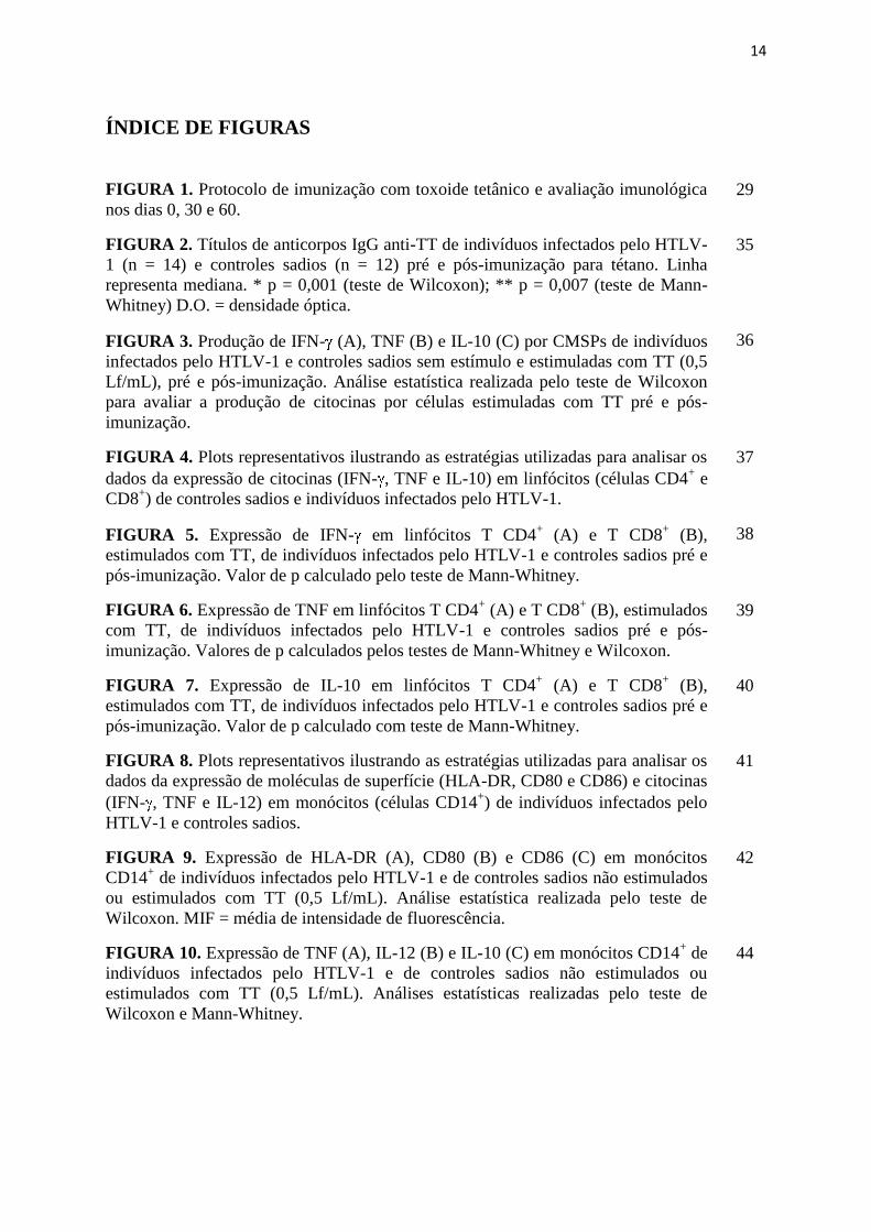
FIGURA 1. Protocolo de imunização com toxoide tetânico e avaliação imunológica
nos dias 0, 30 e 60.
29
FIGURA 2. Títulos de anticorpos IgG anti-TT de indivíduos infectados pelo HTLV-
1 (n = 14) e controles sadios (n = 12) pré e pós-imunização para tétano. Linha
representa mediana. * p = 0,001 (teste de Wilcoxon); ** p = 0,007 (teste de Mann-
Whitney) D.O. = densidade óptica.
35
FIGURA 3. Produção de IFN- (A), TNF (B) e IL-10 (C) por CMSPs de indivíduos
infectados pelo HTLV-1 e controles sadios sem estímulo e estimuladas com TT (0,5
Lf/mL), pré e pós-imunização. Análise estatística realizada pelo teste de Wilcoxon
para avaliar a produção de citocinas por células estimuladas com TT pré e pós-
imunização.
36
FIGURA 4. Plots representativos ilustrando as estratégias utilizadas para analisar os
dados da expressão de citocinas (IFN- , TNF e IL-10) em linfócitos (células CD4+ e
CD8+) de controles sadios e indivíduos infectados pelo HTLV-1.
37
FIGURA 5. Expressão de IFN- em linfócitos T CD4+ (A) e T CD8
+ (B),
estimulados com TT, de indivíduos infectados pelo HTLV-1 e controles sadios pré e
pós-imunização. Valor de p calculado pelo teste de Mann-Whitney.
38
FIGURA 6. Expressão de TNF em linfócitos T CD4+ (A) e T CD8
+ (B), estimulados
com TT, de indivíduos infectados pelo HTLV-1 e controles sadios pré e pós-
imunização. Valores de p calculados pelos testes de Mann-Whitney e Wilcoxon.
39
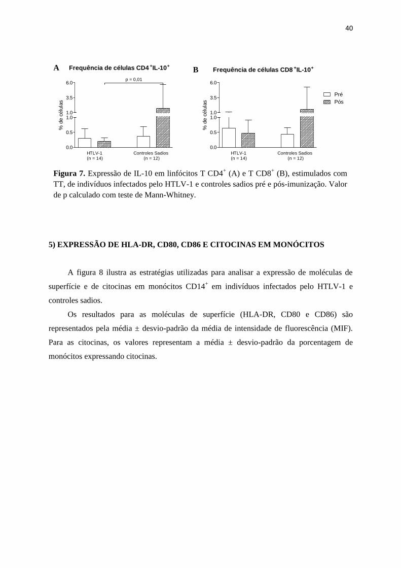
FIGURA 7. Expressão de IL-10 em linfócitos T CD4+ (A) e T CD8
+ (B),
estimulados com TT, de indivíduos infectados pelo HTLV-1 e controles sadios pré e
pós-imunização. Valor de p calculado com teste de Mann-Whitney.
40
FIGURA 8. Plots representativos ilustrando as estratégias utilizadas para analisar os
dados da expressão de moléculas de superfície (HLA-DR, CD80 e CD86) e citocinas
(IFN- , TNF e IL-12) em monócitos (células CD14+) de indivíduos infectados pelo
HTLV-1 e controles sadios.
41
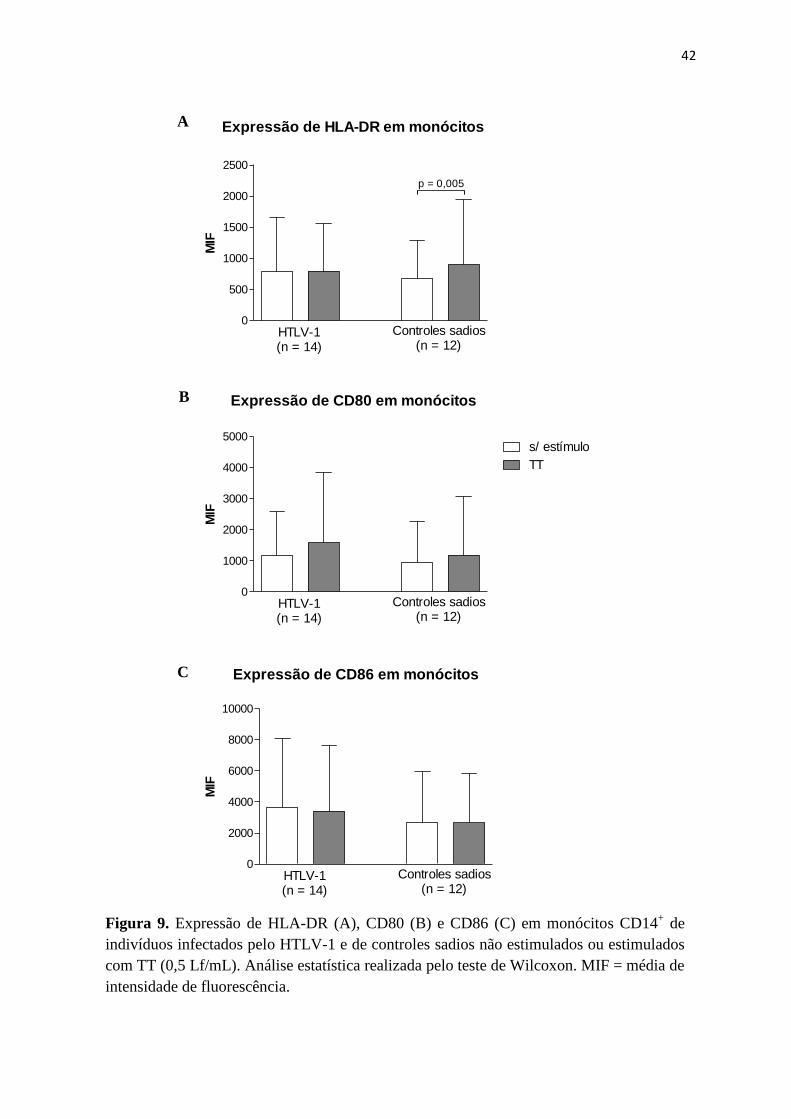
FIGURA 9. Expressão de HLA-DR (A), CD80 (B) e CD86 (C) em monócitos
CD14+ de indivíduos infectados pelo HTLV-1 e de controles sadios não estimulados
ou estimulados com TT (0,5 Lf/mL). Análise estatística realizada pelo teste de
Wilcoxon. MIF = média de intensidade de fluorescência.
42
FIGURA 10. Expressão de TNF (A), IL-12 (B) e IL-10 (C) em monócitos CD14+ de
indivíduos infectados pelo HTLV-1 e de controles sadios não estimulados ou
estimulados com TT (0,5 Lf/mL). Análises estatísticas realizadas pelo teste de
Wilcoxon e Mann-Whitney.
44
15
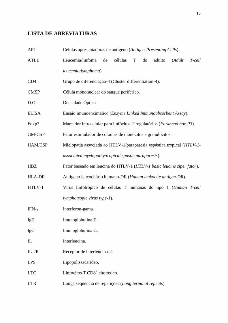
LISTA DE ABREVIATURAS
APC Células apresentadoras de antígeno (Antigen-Presenting Cells).
ATLL Leucemia/linfoma de células T do adulto (Adult T-cell
leucemia/lymphoma).
CD4 Grupo de diferenciação-4 (Cluster differentiation-4).
CMSP Célula mononuclear do sangue periférico.
D.O. Densidade Óptica.
ELISA Ensaio imunoenzimático (Enzyme Linked Immunoabsorbent Assay).
Foxp3 Marcador intracelular para linfócitos T regulatórios (Forkhead box P3).
GM-CSF Fator estimulador de colônias de monócitos e granulócitos.
HAM/TSP Mielopatia associada ao HTLV-1/paraparesia espástica tropical (HTLV-1-
associated myelopathy/tropical spastic paraparesis).
HBZ Fator baseado em leucina do HTLV-1 (HTLV-1 basic leucine ziper fator).
HLA-DR Antigeno leucocitário humano-DR (Human leukocite antigen-DR).
HTLV-1 Vírus linfotrópico de células T humanas do tipo 1 (Human T-cell
lymphotropic vírus type-1).
IFN- Interferon-gama.
IgE Imunoglobulina E.
IgG Imunoglobulina G.
IL Interleucina.
IL-2R Receptor de interleucina-2.
LPS Lipopolissacarídeo.
LTC Linfócitos T CD8+ citotóxico.
LTR Longa sequência de repetições (Long terminal repeats).

16
NF B Fator de transcrição nuclear kappa-beta.
pNPP para-nitrofenil fosfato.
PPD Derivado proteico purificado de Mycobacterium tuberculosis.
TH1 Linfócitos T CD4+ auxiliar-1
TNF Fator de necrose tumoral (Tumor Necrosis Factor).
TT Toxoide tetânico.

17
RESUMO
DIMINUIÇÃO DA RESPOSTA IMUNE AO TOXOIDE TETÂNICO EM
INDIVÍDUOS INFECTADOS PELO HTLV-1. O HTLV-1 é o agente etiológico da
leucemia/linfoma de células T do adulto (ATLL) e da mielopatia associada ao HTLV-1
(HAM/TSP). Tem-se documentado que células mononucleares de indivíduos infectados não
proliferam quando estimuladas com antígenos não relacionados ao vírus como, por exemplo,
derivado proteico purificado de Mycobacterium tuberculosis (PPD) e toxoide tetânico (TT).
Alguns fatores que podem estar relacionados a essa falta de resposta são as funções de células
T regulatórias e disfunção de células apresentadoras de antígeno. Objetivo: Avaliar a resposta
imune de indivíduos infectados pelo HTLV-1 ao toxoide tetânico. Materiais e Métodos:
Foram selecionados portadores assintomáticos do HTLV-1 baixo produtor de IFN- e
controles sadios. Realizou-se sorologia para TT. Os indivíduos soronegativos para TT foram
imunizados. Antes e após imunização, fez-se a sorologia para TT e avaliação da expressão de
citocinas (IFN- , TNF e IL-10) por linfócitos T CD4+ e T CD8
+ estimulados com TT. Os
monócitos dos pacientes e controles, estimulados com TT, foram avaliados para a expressão
de HLA-DR, CD80, CD86, TNF, IL-12 e IL-10 antes da imunização. Resultados: Após
imunização, os pacientes apresentaram menores títulos de IgG anti-TT quando comparados
com os controles (p = 0,007). As células mononucleares dos pacientes, estimuladas com TT,
não aumentaram a produção de IFN- , TNF e IL-10 após imunização. A frequência de
linfócitos T CD4+ expressando IFN- , TNF e IL-10, após estímulo, foi menor nos pacientes
do que nos controles pós-imunização (p = 0,01, p = 0,04 e p = 0,01, respectivamente). Os
monócitos dos pacientes não aumentaram a expressão de HLA-DR após estímulo com TT. A
expressão de TNF e IL-12 por monócitos de pacientes elevaram-se após estímulo com TT (p
= 0,009 e p = 0,006, respectivamente). Conclusões: Os indivíduos infectados pelo HTLV-1,
após esquema de vacinação, apresentaram diminuição da resposta imune humoral e celular
contra TT. Os monócitos destes pacientes exibiram uma disfunção na apresentação antigênica
através do mecanismo de expressão de HLA-DR, porém, o segundo sinal (expressão de
CD80 e CD86) e expressão de citocinas não apresentaram anormalidades. Tais resultados
sugerem que estes mecanismos imunológicos podem participar no aumento da
susceptibilidade dos indivíduos infectados pelo HTLV-1 a adquirir outras doenças
infecciosas.
Palavras-chave: 1. HTLV-1; 2. Linfócitos; 3. Monócitos; 4. Imunologia.
18
INTRODUÇÃO
1) EPIDEMIOLOGIA DA INFECÇÃO PELO HTLV-1
O vírus linfotrópico de células T humanas do tipo 1 (HTLV-1) foi o primeiro retrovírus
identificado (Poiesz, Ruscetti et al., 1980).
Mundialmente aproximadamente 10 a 20 milhões de pessoas podem estar infectadas
pelo HTLV-1 (Edlich, Hill et al., 2003). As regiões com maiores prevalências da infecção são
o Japão, Caribe, África subsaariana e a América do Sul (Maloney, Murphy et al., 1991;
Gessain e De The, 1996; Mueller, Okayama et al., 1996; Catalan-Soares, Carneiro-Proietti et
al., 2005).
No Brasil, a prevalência da infecção pelo HTLV-1 é variada nas diversas regiões do
país (Catalan-Soares, Carneiro-Proietti et al., 2005). Em estudo de base populacional, foi
registrado que a cidade de Salvador, no estado da Bahia, possui a maior prevalência da
infecção pelo HTLV-1: 1,8% da população geral (Dourado, Alcantara et al., 2003). É
necessário que sejam realizados estudos epidemiológicos para a atualização deste dado.
As informações sobre a infecção pelo HTLV-1 são obtidas, principalmente, a partir do
rastreamento da presença de anticorpos específicos para o vírus em doadores de sangue.
Galvão-Castro e cols. (1997) demonstraram que 1,38% dos doadores de sangue da cidade de
Salvador, Bahia, estavam infectados pelo HTLV-1, sendo, portanto, a maior prevalência do
país (Galvao-Castro, Loures et al., 1997). Dentre as Unidades da Federação, a Bahia,
juntamente com Pará e Maranhão, possui uma das maiores prevalências de doadores de
sangue infectados pelo HTLV-1, acima de 9/1000 indivíduos (Catalan-Soares, Carneiro-
Proietti et al., 2005). É importante salientar que, neste mesmo estudo, foi possível identificar
a diminuição de doadores de sangue infectados pelo HTLV-1, o que pode ser explicado pela
obrigatoriedade dos bancos de sangue em detectar a presença de anticorpos para HTLV-1/2
em doadores.
O HTLV-1 é transmitido pelas vias sexual, vertical e pelo compartilhamento de
seringas e agulhas contaminadas entre usuários de drogas (Manns, Wilks et al., 1992). Uma
das principais vias de transmissão é a vertical, da mãe para o filho, através do aleitamento
materno (Kinoshita, Hino et al., 1984; Ureta-Vidal, Angelin-Duclos et al., 1999).

19
2) ESTRUTURA DO HTLV-1
O HTLV-1 pertence à família Retroviridae, subfamília Orthoretrovirinae, gênero
Deltaretrovirus. O envelope viral é composto por proteínas de superfície e transmembrana,
codificadas pelo gene env. O capsídeo é composto por proteínas que são codificadas pelo
gene gag, possui uma forma icosaédrica e abriga o pequeno genoma viral. No interior do
capsídeo está presente o genoma e as enzimas transcriptase reversa e integrase (Yoshida,
2001).
O genoma do HTLV-1 consiste de um RNA de fita simples. As duas extremidades do
RNA possuem sequências repetidas de nucleotídeos chamadas de LTR (long terminal
repeats), que auxiliam na integração do genoma viral ao genoma humano. Os genes
responsáveis pela regulação transcricional do HTLV-1 estão presentes na LTR (Yoshida,
2001). O DNA proviral é composto por 9 mil pares de bases (Seiki, Hattori et al., 1983).
A extremidade 3’ do RNA viral é a região pX, em que é encontrada o gene tax, que
codifica a proteína Tax, importante para a manutenção do vírus (Kiyokawa, Seiki et al., 1984;
Lee, Coligan et al., 1984; Yoshida, 2001). Tax induz ativação da transcrição do provírus e o
estado de imortalidade da célula hospedeira (Younis e Green, 2005)
Recentemente, um novo gene tem sido alvo de estudo: o fator de zíper de leucina
básico do HTLV-1 (hbz, HTLV-1 basic leucine zíper fator). O hbz é encontrado na
extremidade 3’ da LTR e codificado pela cadeia complementar do genoma viral (Gaudray,
Gachon et al., 2002). A proteína HBZ suprime os efeitos da ativação mediada por Tax
(Basbous, Arpin et al., 2003). O mRNA de HBZ é intensamente expresso em células de
pacientes com leucemia/linfoma de células T do adulto (ATLL), induzindo o aumento da
proliferação de células T (Mesnard, Barbeau et al., 2006). Além disso, HBZ parece participar
da patogênese da mielopatia associada ao HTLV-1/paraparesia espástica tropical (HAM/TSP,
HTLV-1 associated mielopathy/tropical spastic paraparesis), visto que foi correlacionado
com a severidade da doença, carga proviral e níveis elevados de neopterina (Saito, Matsuzaki
et al., 2009).

20
3) MANIFESTAÇÕES CLÍNICAS ASSOCIADAS AO HTLV-1
Na década de 80, o HTLV-1 foi identificado nos Estados Unidos a partir de linhagens
de células T de paciente com linfoma cutâneo de células T (Poiesz, Ruscetti et al., 1980).
Anteriormente, pesquisadores japoneses descreveram uma doença que atingia células T
denominada leucemia/linfoma de células T do adulto (ATLL) que, mais tarde, foi associada à
infecção pelo HTLV-1 (Uchiyama, Yodoi et al., 1977; Takatsuki, 2005).
A ATLL é uma forma agressiva de leucemia/linfoma, consistindo de uma expansão
oligoclonal ou monoclonal de células T CD4+ e células T CD4
+CD25
+Foxp3
+ (Higuchi e
Fujii, 2009; Shembade e Harhaj, 2010). Caracteriza-se pela presença de infiltrados celulares
na pele, fígado, trato gastrointestinal e pulmões, hipercalcemia e presença de linfócitos
atípicos em formato de flor (flower cells) no sangue periférico (Matsuoka, 2005).
Na década de 60, descreveu-se a paraparesia espástica tropical (tropical spastic
paraparesis - TSP) em indivíduos da Índia e Jamaica. Após a descoberta do HTLV-1,
observou-se que mais de 50% dos indivíduos com TSP possuíam anticorpos anti-HTLV-1/2
(Mani, Mani et al., 1969; Gessain, Barin et al., 1985). Em 1986, pesquisadores japoneses
associaram a infecção pelo HTLV-1 a uma mielopatia crônica, que passou a ser denominada
mielopatia associada ao HTLV-1 (HTLV-1 associated myelopathy - HAM) (Osame, Usuku et
al., 1986). Devido à presença de anticorpos anti-HTLV-1 tanto na TSP quando em HAM,
adotou-se o termo HAM/TSP.
A HAM/TSP é uma síndrome de desmielinização de início insidioso caracterizada por
um dano no sistema nervoso central (SNC), especialmente na porção mais baixa do cordão
espinhal (Bangham, 2000; Ribas e Melo, 2002). Paraparesia, sinais piramidais e sintomas
urinários são observados em quase 100% dos indivíduos com HAM/TSP. Além destes
sintomas, constipação intestinal, diminuição da libido, impotência sexual, dor lombar e
atrofia muscular são relatados nestes pacientes (Caskey, Morgan et al., 2007; Carod-Artal,
Mesquita et al., 2008).
Apesar de essas duas doenças serem intensamente associadas à infecção pelo HTLV-1,
somente 5 a 10% dos portadores desenvolvem ATLL ou HAM/TSP (Maloney, Cleghorn et
al., 1998), enquanto a maioria dos indivíduos infectados permanece como portadores
assintomáticos. Outras doenças têm sido associadas à infecção pelo HTLV-1: síndrome de
Sjögren (Nakamura, Ikebe-Hiroki et al., 1997; Giozza, Santos et al., 2008); artropatias
(Nishioka, Maruyama et al., 1989); uveíte (Mochizuki, Watanabe et al., 1992) e dermatite

21
infecciosa (Lagrenade, Hanchard et al., 1990). Além destas alterações, osteoporose
(Schachter, Cartier et al., 2003) e periodontite (Garlet, Giozza et al., 2010) também têm sido
relacionadas à infecção pelo HTLV-1.
Tem sido demonstrada uma maior frequência de sintomas neurológicos, disfunção erétil
e distúrbios urinários nos portadores do HTLV-1, quando comparados com indivíduos
controles, sugerindo que o espectro de doenças associadas ao vírus é maior do que o descrito
na literatura (Caskey, Morgan et al., 2007; Oliveira, Castro et al., 2010). Um dos principais
sintomas neurológicos presentes na infecção pelo HTLV-1, na ausência de HAM/TSP, é a
bexiga hiperativa (Castro, Rodrigues et al., 2007; Silva, Coutinho et al., 2009). Noctúria,
urgência e incontinência são os principais sintomas urinários da bexiga hiperativa.
Comparados com indivíduos soronegativos, portadores do HTLV-1 tem uma maior
frequência destes sintomas (Caskey, Morgan et al., 2007; Poetker, Porto et al., 2011). Em
relação aos fatores urodinâmicos, hiperreflexia do detrusor foi encontrado na maioria dos
portadores do HTLV-1 sem HAM/TSP. Portadores do HTLV-1 com bexiga hiperativa são
considerados como oligossintomáticos, sendo a bexiga hiperativa apontada como uma
manifestação inicial da HAM/TSP (Castro, Freitas et al., 2007).
Uma das evidências de que a bexiga hiperativa é uma manifestação inicial da
mielopatia é o aumento da carga proviral. É conhecido que pacientes com HAM/TSP
apresentam uma elevada carga proviral quando comparados com portadores assintomáticos
(Nagai, Usuku et al., 1998; Matsuzaki, Nakagawa et al., 2001; Olindo, Lezin et al., 2005) e
os portadores de bexiga hiperativa encontram-se como um grupo intermediário (Costa, Santos
et al., 2012; Santos, Oliveira et al., 2012).
4) RESPOSTA IMUNE NA INFECÇÃO PELO HTLV-1
O HTLV-1 possui um tropismo preferencial para os linfócitos T CD4+ (Chen, Quan et
al., 1983; Richardson, Edwards et al., 1990; Yamano, Cohen et al., 2004). Porém, outros
tipos celulares também podem ser alvos do vírus: linfócitos T CD8+, células dendríticas,
macrófagos e linfócitos B (De Revel, Mabondzo et al., 1993; Knight, Macatonia et al., 1993;
Yamano, Cohen et al., 2004).
A proteína Tax é responsável pela ativação espontânea das células infectadas pelo
vírus. Consequentemente, há indução da produção de interleucina(IL)-2 e seu receptor, IL-2R
22
(Ballard, Bohnlein et al., 1988), gerando proliferação celular e produção espontânea de
citocinas (Jacobson, Zaninovic et al., 1988).
A produção espontânea de citocinas pelos linfócitos T CD4+ e T CD8
+ pode ser
observada tanto em portadores assintomáticos quanto em pacientes com HAM/TSP. Quando
comparadas com portadores assintomáticos, células mononucleares de indivíduos com
HAM/TSP produzem níveis elevados de TNF e IFN- sem a necessidade de estímulo com
mitógeno (Santos, Porto et al., 2004). Esta observação demonstra um perfil de resposta imune
do tipo TH1 na infecção pelo HTLV-1, mas citocinas do perfil TH2, como IL-10 e IL-5,
também são detectadas em culturas de células mononucleares de indivíduos portadores
assintomáticos do vírus, o que não é observado em cultura de células de indivíduos sadios
(Carvalho, Bacellar et al., 2001).
Células mononucleares de portadores assintomáticos do HTLV-1 produzem níveis
elevados de IL-10 quando comparadas com células de indivíduos sadios. A produção desta
citocina pode estar relacionada à manutenção do status clínico destes indivíduos, impedindo a
produção exacerbada de citocinas inflamatórias (Brito-Melo, Peruhype-Magalhaes et al.,
2007). Em estudo in vitro, a adição exógena de IL-10 em cultura de células mononucleares
provenientes de portadores assintomáticos é capaz de modular a produção de IFN- , ao
contrário do que é observado em células de pacientes com HAM/TSP (Santos, Porto et al.,
2006).
Além das citocinas inflamatórias, as quimiocinas do perfil TH1, CXCL9 e CXCL10,
também são espontaneamente produzidas em portadores do HTLV-1. Níveis elevados destas
quimiocinas são detectados em pacientes com HAM/TSP quando comparados com
portadores assintomáticos (Narikawa, Fujihara et al., 2005; Guerreiro, Santos et al., 2006;
Santos, Oliveira et al., 2012).
Devido à intensa resposta TH1, os indivíduos infectados pelo HTLV-1 podem
apresentar modulação da resposta TH2, o que pode influenciar na defesa contra outros agentes
infecciosos como Strongyloides stercoralis (Gotuzzo, Terashima et al., 1999). Neste caso, os
indivíduos coinfectados pelo HTLV-1/S. stercoralis apresentam uma diminuição da produção
de citocinas do tipo TH2 (IL-4, IL-5 e IL-13) e do anticorpo IgE, os quais são essenciais para
a eliminação do helminto (David, Vadas et al., 1980; Carvalho e Da Fonseca Porto, 2004).
Consequentemente, a frequência das formas grave e disseminada da estrongiloidíase é maior
entre os indivíduos coinfectados (Porto, Alcantara et al., 2005).

23
Por outro lado, o perfil de resposta imune do tipo TH1 influencia negativamente no
controle de agentes infecciosos como, por exemplo, Mycobacterium tuberculosis. Estudo
realizado em Salvador, Bahia, a ocorrência de óbitos foi maior entre os indivíduos
coinfectados quando comparados com indivíduos somente infectados pela bactéria (Pedral-
Sampaio, Martins Netto et al., 1997). Recentemente foi demonstrado que indivíduos
coinfectados apresentaram níveis de produção espontânea de IFN- e TNF maiores do que
nos indivíduos somente infectados pela tuberculose. Quando as células mononucleares foram
estimuladas com derivado proteico purificado (PPD) de M. tuberculosis, os níveis de TNF
foram menores nos casos (HTLV-1 e tuberculose) do que nos controles (somente
tuberculose), o que sugere que há uma anormalidade na resposta imune inicial contra o M.
tuberculosis nos indivíduos coinfectados (Bastos, Santos et al., 2012)
Além dos linfócitos T CD4+, os linfócitos T CD8
+ citotóxicos (LTCs) também são
importantes na resposta imune contra o vírus. Por serem capazes de reconhecer a proteína
Tax, tornam-se o principal mecanismo de defesa na infecção pelo HTLV-1 (Nishiura,
Nakamura et al., 1996; Hanon, Hall et al., 2000). Assim como as células T CD4+, os LTCs
contribuem para a produção espontânea de IL-2, IFN- e TNF em portadores assintomáticos e
são as principais fontes de citocinas proinflamatórias em indivíduos com HAM/TSP (Goon,
Igakura et al., 2003; Santos, Porto et al., 2004). A progressão da infecção viral está associada
com expressão elevada da proteína HBZ. Pela sua capacidade de suprimir a ação de Tax e sua
apresentação aos LTCs, HBZ diminui a capacidade destas células em eliminar o vírus
(Macnamara, Rowan et al., 2010). Mesmo com a baixa expressão de Tax, os LTCs
permanecem ativados e produzem citocinas proinflamatórias, as quais podem estar associadas
ao desenvolvimento da HAM/TSP (Biddison, Kubota et al., 1997; Kubota, Kawanishi et al.,
1998). Adicionalmente, a diminuição da expressão de moléculas coestimulatórias (CD28,
CD80, CD86 e CD152) é mais frequente em pacientes com HAM/TSP do que em portadores
assintomáticos (Sabouri, Usuku et al., 2008).
As células dendríticas também são infectadas pelo vírus, principalmente em indivíduos
com HAM/TSP (Macatonia, Cruickshank et al., 1992). Recentemente foi demonstrado que
células dendríticas das linhagens plasmocitoide, linfoide ou derivadas de monócitos, também
são infectadas pelo HTLV-1 e são capazes de transmitir o vírus para as células T in vitro
(Jones, Petrow-Sadowski et al., 2008). As células dendríticas são essenciais para a
apresentação da proteína Tax para os linfócitos T, o que induz a ativação, a proliferação e a
produção de citocinas por estas células (Jain, Ahuja et al., 2007). Monócitos de indivíduos

24
sadios são capazes de amadurecer para células dendríticas, aumentando a expressão de
moléculas de superfície celular como CD1a e HLA-DR. No entanto, monócitos de indivíduos
infectados pelo HTLV-1 são incapazes de diferenciarem para células dendríticas, tanto na
morfologia quanto na expressão destas moléculas de superfície (Nascimento, Lima et al.,
2011). Adicionalmente, há diminuição da expressão de HLA-DR em células dendríticas de
pacientes com ATLL (Makino, Wakamatsu et al., 2000).
Apesar da intensa informação sobre os linfócitos T, pouco se sabe do papel das células
apresentadoras de antígeno (APC), principalmente os monócitos/macrófagos, no mecanismo
imunológico associado à infecção pelo HTLV-1.
5) RESPOSTA IMUNE A ANTÍGENOS VIRAIS E NÃO VIRAIS NA INFECÇÃO
PELO HTLV-1
A proteína viral Tax é importante para a manutenção da carga viral e o principal alvo
da resposta imune na infecção pelo HTLV-1. Os LTCs são essenciais para a eliminação de
células infectadas pelo vírus, desde que haja apresentação antigênica. As células T CD4+ são
capazes de apresentar o antígeno Tax para os LTCs, que destrói a célula infectada por
mecanismo de apoptose (Hanon, Hall et al., 2000). No entanto, estudo recente demonstrou
que os linfócitos T CD8+ específicos para Tax são raros e não totalmente funcionais tanto em
portadores assintomáticos quanto em indivíduos com ATLL (Takamori, Hasegawa et al.,
2011), evidenciando um mecanismo de persistência da infecção viral.
Além de Tax, o HTLV-1 expressa p30, uma proteína regulatória. O aumento de p30
inibe a expressão do receptor tipo-Toll-4 (Toll-like receptor-4, TLR4) em macrófagos de
pacientes com ATLL. Esta alteração dificulta o início da resposta imune contra bactérias
Gram-negativas e diminui a produção de citocinas e quimiocinas inflamatórias como, por
exemplo, TNF, IL-8 e CCL2, porém, aumenta a produção de IL-10 (Datta, Sinha-Datta et al.,
2006). Estas mudanças na resposta imune inata podem influenciar na ocorrência de infecções
oportunistas em indivíduos portadores do HTLV-1 (White, Zaknoen et al., 1995).
É conhecido que, in vitro, as células mononucleares de indivíduos infectados pelo
HTLV-1 proliferam na ausência de mitógenos (Popovic, Flomenberg et al., 1984; Kramer,
Jacobson et al., 1989; Jacobson, Gupta et al., 1990). Tal proliferação poderia auxiliar na
eliminação de microrganismos causadores de doenças infecciosas. No entanto, tal fator de

25
proteção parece não ocorrer e as doenças infectocontagiosas são mais frequentes entre os
indivíduos infectados pelo HTLV-1 do que nos indivíduos soronegativos para o vírus. Essa
constatação serviu de base para estudos que tinham como objetivo avaliar a resposta imune
contra antígenos não relacionados ao vírus.
No Japão, onde os indivíduos infectados pelo HTLV-1 possuíam uma resposta
intradérmica ao PPD menor do que os soronegativos (Tachibana, Okayama et al., 1988),
observou-se que o nível de proliferação celular e produção de IFN- , após estímulo com PPD,
era menor entre os indivíduos infectados pelo HTLV-1 não respondedores ao teste
intradérmico (Suzuki, Dezzutti et al., 1999).
Em estudo realizado no Brasil, após identificar indivíduos infectados pelo HTLV-1 que
não apresentavam proliferação celular espontânea, observou-se que as células destes
pacientes não proliferaram após estímulo com PPD, toxoide tetânico e antígeno de Candida
albicans (Mascarenhas, Brodskyn et al., 2006).
Alguns fatores imunológicos podem estar relacionados à anergia contra este antígenos.
Visto que a adição exógena de IL-12 restaurou a produção de IFN- específico para PPD
(Suzuki, Dezzutti et al., 1999), foi sugerido que as células apresentadoras de antígeno (APC)
poderiam estar envolvidas nesta anormalidade.
Além de produzirem citocinas proinflamatórias, os indivíduos portadores do HTLV-1
também produzem IL-10 espontaneamente (Carvalho, Bacellar et al., 2001). A produção de
IL-10 em portadores assintomáticos parece estar envolvida no controle do desenvolvimento
de manifestações neurológicas, o que pode ser observado in vitro, onde a adição exógena
desta citocina é capaz de modular a produção de IFN- e TNF, o que não ocorre nos pacientes
com HAM/TSP (Santos, Porto et al., 2006).
Uma vez que indivíduos infectados pelo HTLV-1 não apresentam uma resposta imune
adequada contra antígenos não relacionados ao vírus, faz-se necessária a investigação de
quais mecanismos imunológicos podem estar envolvidos nesta anormalidade. O presente
estudo aborda aspectos da imunidade humoral e celular na resposta ao toxoide tetânico, assim
como a atividade de monócitos de indivíduos infectados pelo HTLV-1 quando estimulados
com este antígeno.
26
HIPÓTESES
Hipótese 1 – Os indivíduos infectados pelo HTLV-1 não aumentam a produção de
anticorpos anti-toxoide tetânico após imunização e há anergia da resposta imune celular
contra este antígeno.
Hipótese 2 – Monócitos de indivíduos infectados pelo HTLV-1 possuem uma
incapacidade de aumentar a expressão de moléculas coestimulatórias e citocinas após
estímulo com toxoide tetânico.

27
OBJETIVOS
1) OBJETIVO GERAL
Avaliar a resposta imune de indivíduos infectados pelo HTLV-1 ao toxoide tetânico.
2) OBJETIVOS ESPECÍFICOS
1) Avaliar a resposta imune humoral ao toxoide tetânico em indivíduos infectados
pelo HTLV-1 antes e após imunização para tétano.
2) Avaliar a expressão de citocinas (IFN- , TNF e IL-10) em linfócitos de
indivíduos infectados pelo HTLV-1 antes e após imunização para tétano.
3) Avaliar o estado de ativação e expressão de citocinas em monócitos de
indivíduos infectados pelo HTLV-1 após estímulo com toxoide tetânico.

28
MATERIAIS E MÉTODOS
1) POPULAÇÃO DE ESTUDO
Participaram deste estudo indivíduos infectados pelo HTLV-1 que são acompanhados
no Ambulatório Multidisciplinar de HTLV-1 (AMH), localizado no Ambulatório Magalhães
Neto do Complexo Hospitalar Universitário Professor Edgard Santos, Universidade Federal
da Bahia, Salvador, Bahia.
O diagnóstico de HTLV-1 foi realizado através de teste imunoenzimático (ELISA)
(Murex HTLV-1+2, Abbot, Dartford, UK) e confirmado por Western Blot (Genelabs HTLV
2.3-2.4, Singapore).
No AMH, desde 2004, vem sendo executado um estudo de coorte, cujo objetivo geral é
acompanhar os indivíduos infectados pelo HTLV-1, buscando identificar a mudança de status
clínico dos pacientes. O acompanhamento consiste na avaliação imunológica e determinação
da carga proviral a cada dois anos e/ou quando ocorre mudança do status clínico. A produção
espontânea de IFN- , TNF, IL-10 e IL-5 é um dos parâmetros observados na avaliação
imunológica. Adicionalmente, os soros são obtidos e armazenados a -20 oC.
O desenho deste estudo é um corte transversal seguido de intervenção. A amostra foi de
conveniência e os pacientes foram selecionados a partir de informações do banco de dados da
coorte.
Os critérios de inclusão foram indivíduos de ambos os gêneros, de 18 a 65 anos de
idade, com ausência de manifestações neurológicas associadas com a infecção pelo HTLV-1
e que concordaram em participar do estudo após assinar o Termo de Consentimento Livre e
Esclarecido (TCLE).
Os critérios de exclusão foram: indivíduos que apresentavam coinfecções como o HIV,
vírus das hepatites B e C, tuberculose e helmintíases; portadores de doenças crônicas e
autoimunes; grávidas; em tratamento com corticoides; e que não assinaram o TCLE.
Como grupo de comparação, os controles sadios foram constituídos de indivíduos
(estudantes e funcionários) do Serviço de Imunologia do COM-HUPES, negativos para
HTLV-1 e que assinaram o TCLE.
O estudo foi aprovado pelo Comitê de Ética em Pesquisa da Maternidade Climério de
Oliveira, UFBA (Parecer/Resolução 154/2009).
29
2) PROTOCOLO DE IMUNIZAÇÃO E SOROLOGIA PARA TOXOIDE TETÂNICO
Os pacientes selecionados para participarem do estudo, a partir de informações do
banco de dados da coorte, foram questionados sobre situação vacinal para tétano. Os
indivíduos que referiram imunização prévia há mais de 10 anos e/ou comprovaram através de
cartão de vacinação foram incluídos no desenho do estudo, após assinatura do TCLE. Em
seguida, obteve-se amostra de sangue destes indivíduos para realização de sorologia para
toxoide tetânico (TT) e avaliação da resposta imune celular. Adicionalmente, os indivíduos
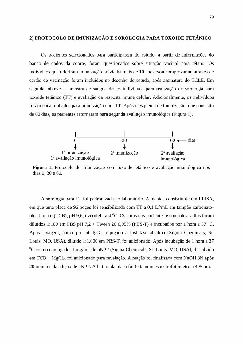
foram encaminhados para imunização com TT. Após o esquema de imunização, que consistiu
de 60 dias, os pacientes retornaram para segunda avaliação imunológica (Figura 1).
A sorologia para TT foi padronizada no laboratório. A técnica consistiu de um ELISA,
em que uma placa de 96 poços foi sensibilizada com TT a 0,1 Lf/mL em tampão carbonato-
bicarbonato (TCB), pH 9,6, overnight a 4 oC. Os soros dos pacientes e controles sadios foram
diluídos 1:100 em PBS pH 7,2 + Tween 20 0,05% (PBS-T) e incubados por 1 hora a 37 oC.
Após lavagem, anticorpo anti-IgG conjugado à fosfatase alcalina (Sigma Chemicals, St.
Louis, MO, USA), diluído 1:1.000 em PBS-T, foi adicionado. Após incubação de 1 hora a 37
oC com o conjugado, 1 mg/mL de pNPP (Sigma Chemicals, St. Louis, MO, USA), dissolvido
em TCB + MgCl2, foi adicionado para revelação. A reação foi finalizada com NaOH 3N após
20 minutos da adição de pNPP. A leitura da placa foi feita num espectrofotômetro a 405 nm.
0 30 60 dias
1ª imunização
1ª avaliação imunológica 2ª imunização
2ª avaliação
imunológica
Figura 1. Protocolo de imunização com toxoide tetânico e avaliação imunológica nos
dias 0, 30 e 60.

30
3) OBTENÇÃO DE CÉLULAS MONONUCLEARES DO SANGUE PERIFÉRICO
(CMSP)
Os pacientes e controles sadios selecionados para o estudo tiverem seu sangue coletado
em tubos contendo heparina. As células mononucleares do sangue periférico (CMSP) foram
isoladas por gradiente de densidade Ficoll-Hypaque (LSM, Organon Teknika Corporation,
Durham, NS, USA). AS células foram cultivadas em meio RPMI 1640 (Life Technologies
GibcoBRL, Grand Island, NY, USA) contendo 10% de soro fetal bovino (Gibco, Grand
Island, NY, USA) e antibióticos.
4) DETERMINAÇÃO DA PRODUÇÃO DE CITOCINAS
Antes e após imunização com TT, 3x10⁶ células/mL de pacientes e controles sadios
foram distribuídas em placas de 24 poços. As CMSPs foram incubadas por 72 horas, a 37 oC
com 5% de CO2. Neste tempo, as células foram estimuladas com fitohemaglutinina (PHA) e
toxoide tetânico (TT) a 0,5 Lf/mL. Adicionaram-se poços contendo células não estimuladas.
Os sobrenadantes das culturas foram coletados e armazenados a -20 oC.
Utilizou-se a técnica de ELISA sanduíche para dosagem das citocinas IFN- , TNF e IL-
10 nos sobrenadantes, de acordo com as instruções do fabricante (BD Bioscience
Pharmingen, San Jose, CA, USA). Os resultados foram expressos em pg/mL, a partir de uma
curva-padrão gerada por citocinas recombinantes.
As concentrações das citocinas, produzidas por CMSP estimuladas com TT, foram
comparadas antes e depois da imunização em ambos os grupos de estudo.
5) AVALIAÇÃO DA EXPRESSÃO DE CITOCINAS EM LINFÓCITOS
Avaliou-se a expressão de citocinas em linfócitos T CD4+ e T CD8
+ de pacientes e
controles sadios antes e após imunização para TT.
Parte das CMSPs obtidas na coleta de sangue dos pacientes e controles foram
cultivadas por 20 horas a 37 oC, 5% de CO2 na ausência ou presença de TT (0,5 Lf/mL). Nas
últimas 4 horas de incubação, adicionou-se Brefeldin A (1 µg/mL). Lavaram-se as células
31
com PBS com posterior marcação para moléculas de superfície, em que os anticorpos
monoclonais anti-CD4-FITC e anti-CD8-PE-Cy5 (Pharmingen, San Diego, CA, USA)
permaneceram por 20 minutos a 4 oC. Após lavagem e centrifugação com PBS, fixou-se as
células com paraformaldeído a 2%. Para marcação intracitoplasmática, as células foram
permeabilizadas com saponina, lavadas e centrifugadas com PBS e, em seguida, marcadas
com os anticorpos anti-IFN- -PE, anti-IL-10-PE e anti-TNF-PE (Pharmingen, San Diego,
CA, USA) por 30 minutos à temperatura ambiente. Repetiu-se o processo de lavagem e
centrifugação para, posteriormente, aquisição das células no citômetro FACScanto II.
Realizou-se a análise no software FlowJo, versão 7.6.1.
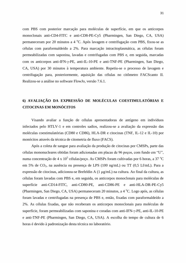
6) AVALIAÇÃO DA EXPRESSÃO DE MOLÉCULAS COESTIMULATÓRIAS E
CITOCINAS EM MONÓCITOS
Visando avaliar a função de células apresentadoras de antígeno em indivíduos
infectados pelo HTLV-1 e em controles sadios, realizou-se a avaliação da expressão das
moléculas coestimulatórias (CD80 e CD86), HLA-DR e citocinas (TNF, IL-12 e IL-10) por
monócitos através da técnica de citometria de fluxo (FACS).
Após a coleta de sangue para avaliação da produção de citocinas por CMSPs, parte das
células mononucleares obtidas foram adicionadas em placas de 96 poços, com fundo em “U”,
numa concentração de 4 x 10⁵ células/poço. As CMSPs foram cultivadas por 6 horas, a 37
oC
em 5% de CO2, na ausência ou presença de LPS (100 ng/mL) ou TT (0,5 Lf/mL). Para a
expressão de citocinas, adicionou-se Brefeldin A (1 µg/mL) na cultura. Ao final da cultura, as
células foram lavadas com PBS e, em seguida, os anticorpos monoclonais para moléculas de
superfície anti-CD14-FITC, anti-CD80-PE, anti-CD86-PE e anti-HLA-DR-PE-Cy5
(Pharmingen, San Diego, CA, USA) permaneceram 20 minutos, a 4 oC. Logo após, as células
foram lavadas e centrifugadas na presença de PBS e, então, fixadas com paraformaldeído a
2%. As células fixadas, que não receberam os anticorpos monoclonais para moléculas de
superfície, foram permeabilizadas com saponina e coradas com anti-IFN- -PE, anti-IL-10-PE
e anti-TNF-PE (Pharmingen, San Diego, CA, USA). A escolha do tempo de cultura de 6
horas é devido à padronização desta técnica no laboratório.

32
Realizou-se a aquisição pelo citômetro FACScanto II (BD, San Jose, CA, USA). No
total, 100.000 células foram obtidas. Ao final do processo, seguiu-se a análise dos dados no
software FlowJo, versão 7.6.1. (Tree Star, Ashland, OR, USA).
7) ANÁLISE ESTATÍSTICA
Os títulos de anticorpos IgG anti-TT e concentração de citocinas foram expressos como
mediana e intervalo interquartil (IQ). Os dados de citometria de fluxo foram representados
com média e desvio-padrão.
Para comparação de frequências, o teste exato de Fisher foi utilizado.
O teste de Wilcoxon foi utilizado para comparação dos dados antes e após imunização e
frequência de células após estímulo com TT.
O teste não-paramétrico de Mann-Whitney foi utilizado para comparar os resultados
entre os dois grupos.
O valor de p < 0,05 foi considerado estatisticamente significante.
Os dados foram analisados no software GraphPad Prism 5 (San Diego, CA, USA).
33
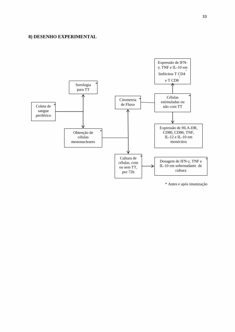
8) DESENHO EXPERIMENTAL
Coleta de
sangue
periférico
Sorologia
para TT
Obtenção de
células
mononucleares
Cultura de
células, com
ou sem TT,
por 72h
Dosagem de IFN- , TNF e
IL-10 em sobrenadante de
cultura
Citometria
de Fluxo
Expressão de IFN-
, TNF e IL-10 em
linfócitos T CD4+
e T CD8+
Expressão de HLA-DR,
CD80, CD86, TNF,
IL-12 e IL-10 em
monócitos
*
*
*
*
*
*
Células
estimuladas ou
não com TT
*
* Antes e após imunização
34
RESULTADOS GERAIS
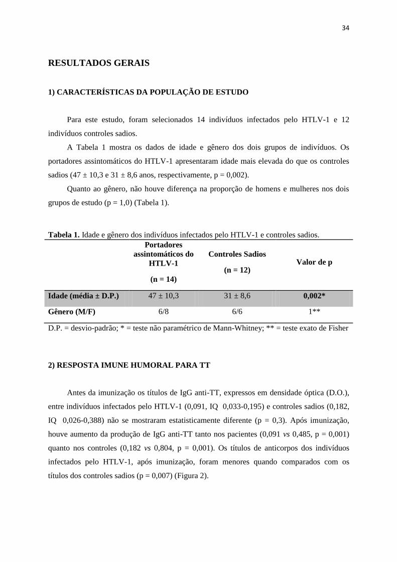
1) CARACTERÍSTICAS DA POPULAÇÃO DE ESTUDO
Para este estudo, foram selecionados 14 indivíduos infectados pelo HTLV-1 e 12
indivíduos controles sadios.
A Tabela 1 mostra os dados de idade e gênero dos dois grupos de indivíduos. Os
portadores assintomáticos do HTLV-1 apresentaram idade mais elevada do que os controles
sadios (47 ± 10,3 e 31 ± 8,6 anos, respectivamente, p = 0,002).
Quanto ao gênero, não houve diferença na proporção de homens e mulheres nos dois
grupos de estudo (p = 1,0) (Tabela 1).
Tabela 1. Idade e gênero dos indivíduos infectados pelo HTLV-1 e controles sadios.
Portadores
assintomáticos do
HTLV-1
(n = 14)
Controles Sadios
(n = 12) Valor de p
Idade (média ± D.P.) 47 ± 10,3 31 ± 8,6 0,002*
Gênero (M/F) 6/8 6/6 1**
D.P. = desvio-padrão; * = teste não paramétrico de Mann-Whitney; ** = teste exato de Fisher
2) RESPOSTA IMUNE HUMORAL PARA TT
Antes da imunização os títulos de IgG anti-TT, expressos em densidade óptica (D.O.),
entre indivíduos infectados pelo HTLV-1 (0,091, IQ 0,033-0,195) e controles sadios (0,182,
IQ 0,026-0,388) não se mostraram estatisticamente diferente (p = 0,3). Após imunização,
houve aumento da produção de IgG anti-TT tanto nos pacientes (0,091 vs 0,485, p = 0,001)
quanto nos controles (0,182 vs 0,804, p = 0,001). Os títulos de anticorpos dos indivíduos
infectados pelo HTLV-1, após imunização, foram menores quando comparados com os
títulos dos controles sadios (p = 0,007) (Figura 2).

35
3) DETERMINAÇÃO DA PRODUÇÃO DE CITOCINAS
As CMSPs dos indivíduos infectados pelo HTLV-1, após estímulo com TT, produziram
níveis menores de IFN- após imunização com TT (2002 pg/mL, IQ 440-2647 pg/mL vs 748
pg/mL; IQ 17,5-2408 pg/mL, p = 0,2). As células mononucleares dos controles sadios
aumentaram a produção de IFN- após imunização com TT (5 pg/mL, IQ 0-158 pg/mL vs
151 pg/mL, IQ 0-682 pg/mL, p = 0,08) (Figura 3A).
Quanto à produção de TNF por células dos pacientes estimuladas com TT, a
concentração desta citocina antes da imunização obteve mediana de 1240 pg/mL (IQ 438-
1742) e, após imunização, 434 pg/mL (IQ 28-2758) (p = 0,9). As CMSPs dos controles
sadios, após estímulo com TT, secretaram, antes da imunização, 0 pg/mL (IQ 0-27) de TNF
e, após imunização, 45 pg/mL (IQ 0-905) (p = 1,0) (Figura 3B).
Os níveis de IL-10 produzidos por CMSPs dos pacientes estimuladas com TT foram: 0
pg/mL (IQ 0-70) antes da imunização; e 0 pg/mL (IQ 0-20) após imunização (p = 0,6).
Dentre os controles sadios, as concentrações desta citocina por células mononucleadas
estimuladas com TT, antes e após imunização, foram de 0 pg/mL (IQ 0-139) e 61 pg/mL (IQ
3,5-276), respectivamente (p = 0,8) (Figura 3C).
Figura 2. Títulos de anticorpos IgG anti-TT de indivíduos infectados pelo HTLV-1 (n =
14) e controles sadios (n = 12) pré e pós-imunização para tétano. Linha representa
mediana. * p = 0,001 (teste de Wilcoxon); ** p = 0,007 (teste de Mann-Whitney) D.O. =
densidade óptica.
Pré Pós Pré Pós0.0
0.2
0.4
0.6
0.8
1.0
1.2
HTLV-1 Controles sadios(n = 14) (n = 12)
**
**
D.O
. 405 n
m

36
Figura 3. Produção de IFN- (A), TNF (B) e IL-10 (C) por CMSPs de indivíduos
infectados pelo HTLV-1 e controles sadios sem estímulo e estimuladas com TT (0,5
Lf/mL), pré e pós-imunização. Análise estatística realizada pelo teste de Wilcoxon para
avaliar a produção de citocinas por células estimuladas com TT pré e pós-imunização.
IFN-
0
1000
2000
3000
4000
s/ estímulo
TT
Pré PréPós Pós
Controles sadios
(n = 12)
HTLV-1
(n = 14)
p = 0,08
s/ estímulo
TT
pg
/mL
TNF
0
1000
2000
3000
4000
5000 s/ estímulo
TT
s/ estímulo
TT
Pré PréPós Pós
Controles sadios (n = 12)
HTLV-1(n = 14)
pg
/mL
IL-10
0
500
1000
1500s/ estímulo
TT
s/ estímulo
TT
Pré PréPós Pós
Controles sadios
(n = 12)
HTLV-1
(n = 14)
pg
/mL
A
B
C

37
4) EXPRESSÃO DE CITOCINAS EM LINFÓCITOS
A figura 4 ilustra as estratégias utilizadas para analisar os dados da expressão de
citocinas em linfócitos de indivíduos infectados pelo HTLV-1 e controles sadios.
Os linfócitos T CD4+ dos indivíduos infectados pelo HTLV-1, após estímulo com TT,
apresentaram a mesma expressão de IFN- antes (0,2 ± 0,1%) e depois (0,2 ± 0,1%) da
imunização (p = 0,8). Quanto aos controles sadios, houve um aumento na expressão desta
citocinas após imunização (0,4 ± 0,3% vs 2,8 ± 5,3%), porém, sem diferença estatística (p =
0,4). Após imunização, a frequência de células expressando esta citocina foi menor entre os
pacientes quando comparados com os controles sadios (p = 0,01) (Figura 5A).
Figura 4. Plots representativos ilustrando as estratégias utilizadas para analisar os dados da
expressão de citocinas (IFN- , TNF e IL-10) em linfócitos (células CD4+ e CD8
+) de
controles sadios e indivíduos infectados pelo HTLV-1.
CD4+
CD8+
Cit
oci
na+
C
ito
cin
a+

38
A frequência de linfócitos T CD8+IFN-
+ estimulados dos pacientes aumentou após
imunização (0,6 ± 0,4% vs 0,9 ± 0,6%), porém, sem significância estatística (p = 0,4).
Resultado semelhante foi encontrado nas células de controles sadios (0,4 ± 0,3% vs 2,6 ±
4,8%, p = 0,2) (Figura 5B).
Antes da imunização, a frequência de linfócitos T CD4+TNF
+ dos pacientes, após
estimulados, não aumentou após imunização com TT (0,3 ± 0,2% vs 0,3 ± 0,1%, p = 0,7).
Houve elevação da frequência destas células nos controles sadios pós-imunização com TT
(0,3 ± 0,1% vs 0,6 ± 0,5%, p = 0,04). Quando comparadas as frequências de células T
CD4+TNF
+ de pacientes e controles sadios, após imunização, as células dos indivíduos
infectados expressaram menos TNF do que as células dos controles (p = 0,04) (Figura 6A).
A frequência de linfócitos T CD8+TNF
+ após estímulo com TT, nos indivíduos
infectados pelo HTLV-1, não aumentou significativamente após imunização (0,5 ± 0,3% vs
0,6 ± 0,5%, p = 0,7). A não elevação da frequência destas células também foi observada nos
controles sadios (0,3 ± 0,2% vs 0,5 ± 0,7%, p = 0,8) (Figura 6B).
Figura 5. Expressão de IFN- em linfócitos T CD4+ (A) e T CD8
+ (B), estimulados com
TT, de indivíduos infectados pelo HTLV-1 e controles sadios pré e pós-imunização. Valor
de p calculado pelo teste de Mann-Whitney.
Frequência de células CD4 +IFN- +
0
1
22
6
10 p = 0,01
HTLV-1(n = 14)
Controles Sadios (n = 12)
% d
e c
élu
las
Frequência de células CD8 +IFN- +
0
1
22
6
10Pré
Pós
HTLV-1
(n = 14)
Controles Sadios
(n = 12)
% d
e c
élu
las
A B

39
Em relação à frequência de linfócitos T CD4+IL-10
+ estimulados com TT, houve uma
diminuição nos indivíduos infectados pelo HTLV-1 após imunização (0,3 ± 0,3% vs 0,2 ±
0,1%, p = 1). Quanto aos controles sadios, ocorreu o aumento da frequência destas células
pós-imunização (0,4 ± 0,3% vs 1,7 ± 3,9%, p = 0,1), porém, sem significância estatística. Ao
comparar a frequência dos linfócitos T CD4+ expressando IL-10 de ambos os grupos,
observou-se uma diferença estatisticamente significante entre pacientes e controles sadios
imunizados com TT (p = 0,01) (Figura 7A).
Não houve elevação da expressão de IL-10 em linfócitos T CD8+
estimulados de
pacientes pós-imunização com TT (0,6 ± 0,5% vs 0,5 ± 0,4%, p = 0,5). Observou-se o mesmo
resultado nos controles sadios após serem imunizados com TT (0,4 ± 0,2% vs 1,5 ± 3,7%, p =
0,9). Não houve diferença estatística quando se considerou a frequência de células T CD8+IL-
10+ dos pacientes e controles após imunização com TT (p = 0,9) (Figura 7B).
Figura 6. Expressão de TNF em linfócitos T CD4+ (A) e T CD8
+ (B), estimulados com
TT, de indivíduos infectados pelo HTLV-1 e controles sadios pré e pós-imunização.
Valores de p calculados pelos testes de Mann-Whitney e Wilcoxon.
Frequência de células CD4 +TNF+
0.0
0.5
1.0
1.5
p = 0,04
p = 0,04
HTLV-1(n = 14)
Controles Sadios (n = 12)
% d
e c
élu
las
Frequência de células CD8 +TNF+
0.0
0.5
1.0
1.5
Pré
Pós
HTLV-1(n = 14)
Controles Sadios (n = 12)
% d
e c
élu
las
A B
40
5) EXPRESSÃO DE HLA-DR, CD80, CD86 E CITOCINAS EM MONÓCITOS
A figura 8 ilustra as estratégias utilizadas para analisar a expressão de moléculas de
superfície e de citocinas em monócitos CD14+ em indivíduos infectados pelo HTLV-1 e
controles sadios.
Os resultados para as moléculas de superfície (HLA-DR, CD80 e CD86) são
representados pela média ± desvio-padrão da média de intensidade de fluorescência (MIF).
Para as citocinas, os valores representam a média ± desvio-padrão da porcentagem de
monócitos expressando citocinas.
Figura 7. Expressão de IL-10 em linfócitos T CD4+ (A) e T CD8
+ (B), estimulados com
TT, de indivíduos infectados pelo HTLV-1 e controles sadios pré e pós-imunização. Valor
de p calculado com teste de Mann-Whitney.
Frequência de células CD4 +IL-10+
0.0
0.5
1.01.0
3.5
6.0p = 0,01
HTLV-1(n = 14)
Controles Sadios (n = 12)
% d
e c
élu
las
Frequência de células CD8 +IL-10+
0.0
0.5
1.01.0
3.5
6.0
Pré
Pós
HTLV-1(n = 14)
Controles Sadios (n = 12)
% d
e c
élu
las
A B

41
A expressão de HLA-DR em monócitos de indivíduos infectados pelo HTLV-1 não
aumentou após estímulo com TT (781 ± 873 vs 793 ± 761, p = 0,3). Os monócitos de
controles sadios aumentaram a expressão desta molécula após estímulo (681 ± 615 vs 898 ±
1055, p = 0,005) (Figura 9A).
Os monócitos dos pacientes aumentaram a expressão de CD80 quando estimulados com
TT (1167 ± 1410 vs 1579 ± 2272), porém, sem diferença estatisticamente significante (p =
0,07). Resultado semelhante foi encontrado em células dos controles (936 ± 1318 vs 1176 ±
1890, p = 0,2) (Figura 9B).
Referente à expressão de CD86, houve uma diminuição da expressão nos monócitos
dos pacientes após estimulação com TT (3628 ± 4447 vs 3394 ± 4529, p = 0,07). Quanto aos
monócitos dos controles, a expressão desta molécula não foi alterada (2686 ± 3240 vs 2647 ±
3169, p = 0,5) (Figura 9C).
CD14+
CD14+HLA-DR
+
CD14+Citocina
+
Figura 8. Plots representativos ilustrando as estratégias utilizadas para analisar os dados
da expressão de moléculas de superfície (HLA-DR, CD80 e CD86) e citocinas (IFN- ,
TNF e IL-12) em monócitos (células CD14+) de indivíduos infectados pelo HTLV-1 e
controles sadios.
42
Figura 9. Expressão de HLA-DR (A), CD80 (B) e CD86 (C) em monócitos CD14+ de
indivíduos infectados pelo HTLV-1 e de controles sadios não estimulados ou estimulados
com TT (0,5 Lf/mL). Análise estatística realizada pelo teste de Wilcoxon. MIF = média de
intensidade de fluorescência.
Expressão de HLA-DR em monócitos
0
500
1000
1500
2000
2500
p = 0,005
HTLV-1(n = 14)
Controles sadios (n = 12)
MIF
Expressão de CD80 em monócitos
0
1000
2000
3000
4000
5000
HTLV-1(n = 14)
Controles sadios (n = 12)
s/ estímulo
TT
MIF
Expressão de CD86 em monócitos
0
2000
4000
6000
8000
10000
HTLV-1(n = 14)
Controles sadios (n = 12)
MIF
A
B
C
43
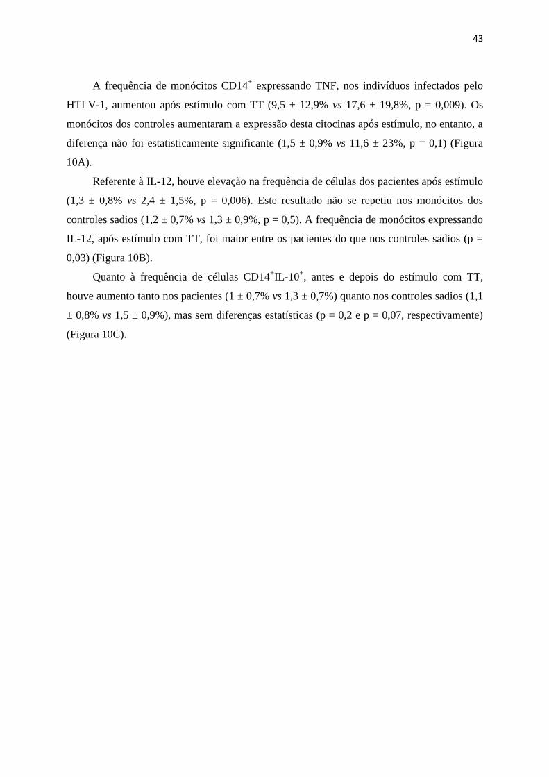
A frequência de monócitos CD14+ expressando TNF, nos indivíduos infectados pelo
HTLV-1, aumentou após estímulo com TT (9,5 ± 12,9% vs 17,6 ± 19,8%, p = 0,009). Os
monócitos dos controles aumentaram a expressão desta citocinas após estímulo, no entanto, a
diferença não foi estatisticamente significante (1,5 ± 0,9% vs 11,6 ± 23%, p = 0,1) (Figura
10A).
Referente à IL-12, houve elevação na frequência de células dos pacientes após estímulo
(1,3 ± 0,8% vs 2,4 ± 1,5%, p = 0,006). Este resultado não se repetiu nos monócitos dos
controles sadios (1,2 ± 0,7% vs 1,3 ± 0,9%, p = 0,5). A frequência de monócitos expressando
IL-12, após estímulo com TT, foi maior entre os pacientes do que nos controles sadios (p =
0,03) (Figura 10B).
Quanto à frequência de células CD14+IL-10
+, antes e depois do estímulo com TT,
houve aumento tanto nos pacientes (1 ± 0,7% vs 1,3 ± 0,7%) quanto nos controles sadios (1,1
± 0,8% vs 1,5 ± 0,9%), mas sem diferenças estatísticas (p = 0,2 e p = 0,07, respectivamente)
(Figura 10C).

44
Figura 10. Expressão de TNF (A), IL-12 (B) e IL-10 (C) em monócitos CD14+ de
indivíduos infectados pelo HTLV-1 e de controles sadios não estimulados ou estimulados
com TT (0,5 Lf/mL). Análises estatísticas realizadas pelo teste de Wilcoxon e Mann-
Whitney.
Frequência de células CD14 +TNF+
0
10
20
30
40 p = 0,009
HTLV-1(n = 14)
Controles sadios (n = 12)
% d
e c
élu
las
Frequência de células CD14 +IL-12+
0
1
2
3
4
5
p = 0,006
p = 0,03
HTLV-1(n = 14)
Controles sadios (n = 12)
s/ estímulo
TT
% d
e c
élu
las
Frequência de células CD14 +IL-10+
0.0
0.5
1.0
1.5
2.0
2.5
HTLV-1(n = 14)
Controles sadios (n = 12)
% d
e c
élu
las
A
B
C
45
DISCUSSÃO
Há poucos relatos na literatura sobre a resposta imune humoral de indivíduos infectados
pelo HTLV-1 durante rotina de imunização. No presente estudo foi demonstrado que os
portadores assintomáticos do HTLV-1 são capazes de aumentar a produção de anticorpo anti-
TT após vacinação, no entanto, os títulos de IgG foram significativamente menores quando
comparados com os dos controles sadios. A sorologia positiva após imunização com TT já foi
observada em estudos anteriores (Jarvis, Janoff et al., 2005; Biasutti, Moraes-Pinto et al.,
2008). No que se refere aos menores títulos de anticorpos encontrados nos pacientes, tem-se
documentado que, em indivíduos infectados pelo HTLV-2, os níveis de anticorpos anti-TT
são menores do que os de controles sadios não infectados pelo vírus após imunização, porém,
neste trabalho não foi encontrada diferença estatística (Jarvis, Janoff et al., 2005). A
anormalidade na produção de IgG após vacinação de reforço também já foi identificado em
indivíduos infectados pelo HIV, em que a concentração de IgG total anti-TT foi maior em
controles sadios do que nos indivíduos infectados pelo vírus após vacinação (Kroon, Van Tol
et al., 1999). A disfunção na produção de anticorpo contra antígeno de memória poderia estar
relacionada à carga proviral, visto que indivíduos infectados pelo HIV-1 apresentaram um
aumento da carga viral após imunização com vírus influenza (O'brien, Grovit-Ferbas et al.,
1995; Rosok, Voltersvik et al., 1996). No entanto, no presente estudo foi possível observar
que não houve variação da carga proviral nos indivíduos infectados pelo HTLV-1 antes e
após imunização (dado não mostrado).
Paralelamente à produção de anticorpos anti-TT após imunização, observou-se a
produção de citocinas por CMSPs estimuladas com TT. As células mononucleares dos
pacientes diminuíram a produção de IFN- após imunização, porém, sem significância
estatística. Aumento da produção desta citocina pode ser observado nas CMSPs dos
controles, no entanto, sem diferença estatisticamente significante. Quanto à produção de TNF
e IL-10, não houve alteração da produção em ambos os grupos de indivíduos. Apesar destes
resultados, observou-se que não ocorreu aumento da produção de IFN- , após estímulo com
TT, por células dos pacientes pós-imunização. Tal aumento foi observado em controles
sadios, porém, sem diferença estatisticamente significante. Este resultado sugere que há uma
anormalidade na produção desta citocina em indivíduos infectados pelo HTLV-1, visto que a
resposta imune contra TT, além da produção de anticorpo, está relacionada com reação de
hipersensibilidade tardia, com produção de IFN- e IL-2 (Parronchi, Macchia et al., 1991;

46
Fernandez, Andersson et al., 1994). Referente à produção de TNF pelos pacientes, os
resultados estavam de acordo com o encontrado em controles sadios, nos quais ocorreu
aumento da produção destas moléculas quando as células foram estimuladas com TT,
corroborando com dados anteriores em que o aumento da produção de TNF por células
mononucleares de controles sadios também é observado após estimulação com TT (Nielsen,
Galdiers et al., 2009). Quanto à citocina IL-10, o presente estudo observou o aumento da
expressão após estímulo com TT, contrariando o que já foi reportado (Nielsen, Galdiers et al.,
2009).
Adicionalmente, realizou-se a caracterização da resposta imune celular quanto à ação
dos linfócitos T dos pacientes e dos controles sadios após estímulo com TT, antes e após
imunização. A frequência de linfócitos T CD4+ expressando IFN- , TNF e IL-10 não
aumentou após estímulo com TT e pós-imunização, o que foi observado nas células dos
controles sadios. Nos resultados pós-imunização, a frequência dos linfócitos T CD4+ dos
controles sadios expressando estas citocinas era maior quando comparados com os pacientes,
evidenciando que há uma falha na resposta imune celular dos pacientes contra TT. A
detecção de IFN- por citometria de fluxo tem se mostrado uma técnica sensível para avaliar
a produção desta citocina em indivíduos que foram imunizados com toxoide tetânico
(Tassignon, Burny et al., 2005). No entanto, foi demonstrado aqui que os linfócitos T dos
indivíduos infectados pelo HTLV-1 não apresentaram a resposta imune do tipo IV quando
estimulados com TT. Uma das causas para a ausência dessa reação seria a produção da
citocina regulatória IL-10 (Sabin, Araujo et al., 1996; Mascarenhas, Brodskyn et al., 2006).
Porém, a expressão desta citocina não aumentou, após estímulo com TT, tanto em linfócitos
T CD4+ quanto em linfócitos T CD8
+ dos pacientes depois da imunização. Os linfócitos dos
controles sadios expressaram níveis maiores de IL-10 após estímulo com TT antes e após
imunização. Isso pode ser explicado devido à regulação normal da resposta imune induzida
por esta citocina (Yssel, De Waal Malefyt et al., 1992). Uma alta expressão de IL-10 era
esperada neste estudo, visto que portadores assintomáticos produzem, espontaneamente,
níveis significativos de IL-10 (Carvalho, Bacellar et al., 2001).
Após imunização, os linfócitos T CD8+ dos pacientes e controles aumentaram a
expressão de TNF na presença de TT. Tal resultado não pode ser observado em linfócitos T
CD4+ de ambos os grupos de indivíduos. Visto que TNF também é produzido por células TH1
(Parronchi, Macchia et al., 1991), este achado sugere que esta citocina é normalmente
produzida após estímulo com TT em indivíduos infectados pelo HTLV-1.

47
Devido à resposta imune humoral e celular estar diminuída nos indivíduos infectados
pelo HTLV-1 e estes não responderem adequadamente ao TT após imunização, procurou-se
observar se estas anormalidades poderiam estar relacionadas às células apresentadoras de
antígeno. Portanto, executou-se a avaliação dos monócitos dos pacientes estimulados ou não
com TT. Visto que, teoricamente, os monócitos não são considerados uma população celular
que se diferenciam para células de memórias, avaliou-se as expressões de moléculas
coestimulatórias e citocinas apenas antes da imunização com TT.
Uma das hipóteses para a não resposta a antígenos de memória em indivíduos
infectados pelo HTLV-1 seria uma disfunção nas células apresentadoras de antígeno (Suzuki,
Dezzutti et al., 1999). Os monócitos dos pacientes não aumentaram a expressão de HLA-DR
após estímulo com TT, ao contrário do que foi observado nos controles sadios. Este resultado
se repetiu quando os monócitos foram estimulados com LPS (dados não mostrados). Os
monócitos são células capazes de apresentar antígenos virais, pois também podem ser
infectados pelo HTLV-1 (De Revel, Mabondzo et al., 1993). No que se refere às células
apresentadoras de antígeno (APC) e sua função, poucos estudos têm focalizado na atuação
dessas células na infecção pelo HTLV-1. Sabe-se que, além de infectar, preferencialmente, os
linfócitos T CD4+, o HTLV-1 também infecta monócitos e células dendríticas (Macatonia,
Cruickshank et al., 1992; De Revel, Mabondzo et al., 1993). A infecção viral pode
influenciar na função dessas células e, como apresentado neste estudo, o não aumento da
expressão de HLA-DR em monócitos, após estímulo com TT, corrobora com achados
semelhantes da literatura. Monócitos de indivíduos infectados pelo HTLV-1 com ATLL não
amadurecem completamente, in vitro, para células dendríticas e apresentam uma baixa
expressão de CD14 e HLA-DR (Makino, Wakamatsu et al., 2000). Adicionalmente,
monócitos de pacientes com HAM/TSP são incapazes de se diferenciarem em células
dendríticas devido à baixa expressão de CD1a e HLA-DR, mesmo quando estimulados com
TNF (Nascimento, Lima et al., 2011). Mais recentemente, um grupo de pesquisa do Japão
demonstrou que macrófagos do leite materno infectados com HTLV-1, quando cocultivados
com monócitos não infectados, impedem estas células de se diferenciarem em células
dendríticas, principalmente devido ao bloqueio da expressão de CD1a e HLA-DR (Inagaki,
Takahashi et al., 2012). Em contrapartida, a proteína Tax mostrou ser eficaz na diferenciação
e ativação de células dendríticas derivadas de monócitos de pacientes com ATLL, induzindo
o aumento da apresentação antigênica (expressão elevada de HLA-DR) e coestimulação de
linfócitos T (Jain, Ahuja et al., 2007; Manuel, Schell et al., 2009). No entanto, tais estudos
48
foram voltados para as células dos indivíduos infectados pelo HTLV-1 com sintomas
neurológicos e com ATLL, o que diferencia do apresentado aqui, em que se utilizou células
de portadores assintomáticos do vírus.
A expressão de CD80 e CD86 em monócitos estimulados ou não com TT foi similar
entre os pacientes e os controles sadios. O discreto aumento da expressão de CD80 após
estímulo com TT não alcançou significância estatística. A expressão de CD86 não foi
alterada após estímulo em ambos os grupos de indivíduos. Interessantemente, a expressão de
CD86 diminuiu após estímulo com LPS em ambos os grupos (dados não mostrados). Tal
resultado pode ser explicado de acordo com estudo anterior afirmando que, em células
tolerantes à endotoxina, a expressão de CD86 diminui com diferentes tempos de exposição ao
LPS e com concentrações variadas desta molécula (Wolk, Docke et al., 2000). Os dados da
literatura acerca das moléculas CD80 e CD86 em monócitos de indivíduos infectados pelo
HTLV-1 são escassos. Estas moléculas são bem documentadas em linfócitos T CD4+, em que
já foi observado que há expressão normal em indivíduos infectados pelo HTLV-1 com ATLL
(Takamoto, Makino et al., 1997).
Referente à expressão das citocinas em monócitos dos dois grupos de estudo, houve o
aumento significante da porcentagem de células expressando TNF após estímulo com TT nos
pacientes, o que não ocorreu nos monócitos dos controles sadios apesar da evidente elevação
da expressão desta citocina. A porcentagem de monócitos expressando IL-12 aumentou após
os dois tipos de estímulo apenas no grupo dos pacientes. Quanto à expressão de IL-10, houve
aumento nos dois grupos de indivíduos quando os monócitos foram estimulados com TT,
porém, não houve diferença estatisticamente significante. Poucos estudos são encontrados
acerca da expressão de citocinas em monócitos de indivíduos infectados pelo HTLV-1.
Porém, sabe-se que monócitos de indivíduos sadios são capazes de produzir IL-10 e TNF
após estímulo com TT (Fernandez, Andersson et al., 1994; Nielsen, Galdiers et al., 2009).
Adicionalmente, estudos focados em células dendríticas derivadas de monócitos
demonstraram que a proteína Tax é capaz de induzir a secreção de IL-12, TNF e quimiocinas
da resposta TH1 por estas células (Ahuja, Kampani et al., 2006; Ahuja, Lepoutre et al., 2007).
Os dados do aumento da frequência de monócitos expressando TNF e IL-12, após
estímulo com TT, em indivíduos infectados pelo HTLV-1 e não nos controles podem estar
relacionados com a constante ativação das células dos pacientes. Faz-se necessário
investigação aprofundada deste mecanismo imunológico.
49
Quanto aos monócitos dos pacientes estudados aqui, pode-se observar que há uma
disfunção na expressão de HLA-DR após estímulo com TT, o que pode prejudicar na
apresentação de antígenos virais ou outros antígenos não relacionados ao vírus. Tal situação
pode permitir a predisposição dos indivíduos infectados pelo HTLV-1 a contraírem doenças
infecciosas como tuberculose (Verdonck, Gonzalez et al., 2008; De Lourdes Bastos,
Osterbauer et al., 2009; Bastos, Santos et al., 2012), sarna norueguesa (Brites, Weyll et al.,
2002) e estrongiloidíase (Porto, Alcantara et al., 2005). Contudo, a expressão de moléculas
coestimulatórias (CD80 e CD86) e citocinas (TNF, IL-10 e IL-12) não é prejudicada após
estímulo antigênico, sugerindo que não há disfunção dos monócitos quanto a essas vias de
apresentação antigênica e ativação celular.
Alguns aspectos da resposta imune não foram abordados neste trabalho. Acerca da
resposta imune humoral, faz-se necessária a dosagem de subclasses de anticorpos IgG (IgG1,
IgG2, IgG3 e IgG4) e IgE antes e após imunização com toxoide tetânico. Tais detecções
poderiam explicar se a resposta imune contra TT está direcionada para via TH1 ou TH2.
Atualmente, são reconhecidas mais de uma classe de monócitos, de acordo com a
expressão dos marcadores de superfície, sendo eles: monócitos clássicos (CD14+CD16
-),
inflamatórios (CD14+CD16
+) e não clássicos (CD14
lowCD16
++) (Tacke e Randolph, 2006;
Ingersoll, Spanbroek et al., 2010). Como este estudo iniciou-se antes destes conhecimentos
estarem bem estabelecidos, torna-se como perspectiva deste trabalho a realização da
caracterização destas subpopulações de monócitos em indivíduos infectados pelo HTLV-1,
principalmente no que se refere à resposta a antígenos de memória.
Outro aspecto na apresentação antigênica em indivíduos infectados pelo HTLV-1, que
não foi explorado no presente estudo, é a função de células dendríticas na ativação dos
linfócitos T após apresentação de antígeno de memória.
Visto que a resposta imune de linfócitos contra TT estava diminuída nos pacientes, faz-
se necessária a avaliação de células T de memória nestes indivíduos. Além disso, é
importante, também, avaliar o estudo funcional das células T regulatórias
CD4+CD25
highCD127
low frente a antígenos de memórias (Iwashiro, Messer et al., 2001;
Michaelsson, Barbosa et al., 2008).
Foi possível mostrar, no presente estudo, que a resposta imune humoral e celular contra
TT está diminuída em indivíduos infectados pelo HTLV-1, mesmo quando estes pacientes
são imunizados com esta proteína. Uma das causas da diminuição destas respostas poderia ser
a função dos monócitos, porém, estes apenas mostraram-se anormais na apresentação

50
antigênica via HLA-DR. Estudos futuros devem ser conduzidos com o objetivo de esclarecer
o papel de monócitos e de suas subpopulações em indivíduos infectados pelo HTLV-1 e após
desafio com antígenos de memória.

51
PROPOSTAS DE ESTUDO
Com objetivo de enriquecer os resultados encontrados neste estudo, a proposta será de
avaliar o perfil de monócitos, das células T e da resposta imune humoral contra antígenos de
memória em indivíduos infectados pelo HTLV-1.
O primeiro objetivo específico é avaliar a frequência de subpopulações de monócitos
em indivíduos infectados pelo HTLV-1. Como mencionado anteriormente, sabe-se que há
três subclasses de monócitos: clássicos, inflamatórios e não clássicos (Tacke e Randolph,
2006; Ingersoll, Spanbroek et al., 2010). A hipótese para este objetivo é de que os indivíduos
infectados pelo HTLV-1 com HAM/TSP tem uma maior frequência de monócitos
inflamatórios, com elevada expressão de TNF, quando comparados com indivíduos com
bexiga hiperativa e portadores assintomáticos do vírus.
O segundo objetivo é avaliar, funcionalmente, os linfócitos T de indivíduos infectados
pelo HTLV-1 quando estimulados com antígeno de memória. A hipótese é que as células T
regulatórias, efetoras e de memória dos pacientes influenciam na anormalidade da resposta
imune celular contra antígeno de memória. A população de estudo será a mesma do objetivo
anterior, além dos controles sadios. Serão avaliadas a frequência de células T regulatórias, de
memória e naïve, tanto ex-vivo quanto em cultura após estímulo com TT e PPD, neste caso
para expressão de IFN- antes e depois do estímulo.
O terceiro e último objetivo é caracterizar a resposta imune humoral de indivíduos
infectados pelo HTLV-1 contra toxoide tetânico. A diminuição da resposta imune humoral
contra TT, após imunização, pode estar relacionada com produção de títulos elevados de
anticorpos anti-TT relacionados à resposta do tipo TH1 (IgG2 e IgG3). Serão avaliados os
mesmos indivíduos do presente estudo (portadores assintomáticos e controles sadios), antes e
após imunização. Através do ELISA, as subclasses de IgG (IgG1, IgG2, IgG3 e IgG4)
poderão ser detectadas nos soros dos indivíduos de ambos os grupos. Adicionalmente, espera-
se aumentar o número amostral, caso sejam identificados mais indivíduos com sorologia
negativa para TT.

52
RESUMO DOS RESULTADOS
1. Apesar de apresentarem sorologia positiva para TT após imunização, os indivíduos
infectados pelo HTLV-1 apresentaram menores títulos de IgG anti-TT quando comparados
com controles sadios, sugerindo que há uma anormalidade na resposta imune humoral dos
pacientes contra este antígeno de memória.
2. Os linfócitos T CD4+ e T CD8
+ dos indivíduos infectados pelo HTLV-1 não aumentaram a
expressão de citocinas quando estimulados com TT, sugerindo que há uma diminuição e,
até mesmo, anergia da resposta imune celular contra este antígeno mesmo após
imunização dos pacientes.
3. Os monócitos dos indivíduos infectados pelo HTLV-1 apresentaram uma disfunção na
apresentação antigênica, devido ao não aumento da expressão de HLA-DR após estímulo
com TT. No entanto, o segundo sinal de apresentação antigênica e expressão de citocinas
não se mostraram alterados.

53
CONCLUSÃO
As falhas nos mecanismos da resposta imune, evidenciadas pela diminuição da resposta
imune humoral, anergia dos linfócitos T CD4+ e T CD8
+ e o não aumento da expressão de
HLA-DR por monócitos após estímulo com antígeno de memória podem levar ao aumento da
susceptibilidade a doenças infecciosas nos indivíduos infectados pelo HTLV-1.
54
SUMMARY
DECREASE OF THE IMMUNE RESPONSE AGAINST TETANUS TOXOID IN
HTLV-1 INFECTED INDIVIDUALS. HTLV-1 is an etiologic agent of Adult T-cell
leukemia/lymphoma (ATLL) and HTLV-1-associated myelopathy/tropical spastic paraparesis
(HAM/TSP). It was shown that peripheral blood mononuclear cells (PBMCs) from HTLV-1
infected subjects do not proliferate after stimulation with recall antigens such as purified
protein derivate (PPD) from Mycobacterium tuberculosis and tetanus toxoid (TT). Some
factors may be involved in this abnormality: regulatory T cell function and dysfunctional
antigen-presenting cells (APC). Objective: To evaluate the immune response against TT in
HTLV-1 infected subjects. Materials and Methods: It was selected HTLV-1 carriers with low
IFN- production and healthy controls. It was performed serology for TT. The seronegative
individuals were immunized with TT. Before and after immunization, it was made serology
for TT and evaluation of the cytokine (IFN- , TNF and IL-10) expression by CD4+ and CD8
+
T cells stimulated with TT. The monocytes, stimulated with TT, from patients and healthy
controls were evaluated to HLA-DR, CD80, CD86, TNF, IL-12 and IL-10 before the
immunization. Results: After immunization, patients had low IgG anti-TT production when
compared with healthy controls (p = 0.007). Mononuclear cells from the patients, stimulated
with TT, did not increase the IFN- , TNF and IL-10 production after immunization.
Frequency of CD4+ T cells expressing IFN- , TNF and IL-10, after stimulus, were lower in
patients than the controls after immunization (p = 0.01, p = 0.04 and p = 0.01, respectively).
Monocytes from patients did not increase the HLA-DR expression after stimulation with TT.
TNF and IL-12 expression by monocytes from patients increased after stimulation (p = 0.009
and p = 0.006, respectively). Conclusions: HTLV-1 infected subjects, after vaccination,
presented a decreasing of the humoral and cellular immune response against TT. Monocytes
from the patients showed an impairment of antigen presentation through the HLA-DR
expression mechanisms. However, the second signal (CD80 and CD86 expression) and
cytokine expression did not show abnormalities. These results suggest that these
immunological mechanisms may participate of the increasing susceptibility of HTLV-1
infected subjects to acquire others infectious diseases.
Key words: 1. HTLV-1; 2. Lymphocytes; 3. Monocytes; 4. Immunology.
55
REFERÊNCIAS BIBLIOGRÁFICAS
Ahuja, J., K. Kampani, et al. Use of human antigen presenting cell gene array profiling to
examine the effect of human T-cell leukemia virus type 1 Tax on primary human dendritic
cells. J Neurovirol, v.12, n.1, Feb, p.47-59. 2006.
Ahuja, J., V. Lepoutre, et al. Induction of pro-inflammatory cytokines by human T-cell
leukemia virus type-1 Tax protein as determined by multiplexed cytokine protein array
analyses of human dendritic cells. Biomed Pharmacother, v.61, n.4, May, p.201-8. 2007.
Ballard, D. W., E. Bohnlein, et al. HTLV-I tax induces cellular proteins that activate the
kappa B element in the IL-2 receptor alpha gene. Science, v.241, n.4873, Sep 23, p.1652-5.
1988.
Bangham, C. R. HTLV-1 infections. J Clin Pathol, v.53, n.8, Aug, p.581-6. 2000.
Basbous, J., C. Arpin, et al. The HBZ factor of human T-cell leukemia virus type I dimerizes
with transcription factors JunB and c-Jun and modulates their transcriptional activity. J Biol
Chem, v.278, n.44, Oct 31, p.43620-7. 2003.
Bastos, M. D., S. Santos, et al. Influence of HTLV-1 on the clinical, microbiologic and
immunologic presentation of tuberculosis. BMC Infect Dis, v.12, n.1, Aug 28, p.199. 2012.
Biasutti, C., M. I. Moraes-Pinto, et al. Antibody response after vaccination with tetanus and
diphtheria toxoids in human T-cell lymphotropic virus type 1 asymptomatic carriers.
Vaccine, v.26, n.23, Jun 2, p.2808-10. 2008.
Biddison, W. E., R. Kubota, et al. Human T cell leukemia virus type I (HTLV-I)-specific
CD8+ CTL clones from patients with HTLV-I-associated neurologic disease secrete
proinflammatory cytokines, chemokines, and matrix metalloproteinase. J Immunol, v.159,
n.4, Aug 15, p.2018-25. 1997.
Brites, C., M. Weyll, et al. Severe and Norwegian scabies are strongly associated with
retroviral (HIV-1/HTLV-1) infection in Bahia, Brazil. Aids, v.16, n.9, Jun 14, p.1292-3.
2002.
Brito-Melo, G. E., V. Peruhype-Magalhaes, et al. IL-10 produced by CD4+ and CD8+ T cells
emerge as a putative immunoregulatory mechanism to counterbalance the monocyte-derived
TNF-alpha and guarantee asymptomatic clinical status during chronic HTLV-I infection. Clin
Exp Immunol, v.147, n.1, Jan, p.35-44. 2007.
Carod-Artal, F. J., H. M. Mesquita, et al. [Neurological symptoms and disability in HTLV-1
associated myelopathy]. Neurologia, v.23, n.2, Mar, p.78-84. 2008.
Carvalho, E. M., O. Bacellar, et al. Cytokine profile and immunomodulation in asymptomatic
human T-lymphotropic virus type 1-infected blood donors. J Acquir Immune Defic Syndr,
v.27, n.1, May 1, p.1-6. 2001.
56
Carvalho, E. M. e A. Da Fonseca Porto. Epidemiological and clinical interaction between
HTLV-1 and Strongyloides stercoralis. Parasite Immunol, v.26, n.11-12, Nov-Dec, p.487-97.
2004.
Caskey, M. F., D. J. Morgan, et al. Clinical manifestations associated with HTLV type I
infection: a cross-sectional study. AIDS Res Hum Retroviruses, v.23, n.3, Mar, p.365-71.
2007.
Castro, N. M., D. M. Freitas, et al. Urodynamic features of the voiding dysfunction in HTLV-
1 infected individuals. Int Braz J Urol, v.33, n.2, Mar-Apr, p.238-44; discussion 244-5. 2007.
Castro, N. M., W. Rodrigues, Jr., et al. Urinary symptoms associated with human T-cell
lymphotropic virus type I infection: evidence of urinary manifestations in large group of
HTLV-I carriers. Urology, v.69, n.5, May, p.813-8. 2007.
Catalan-Soares, B., A. B. Carneiro-Proietti, et al. Heterogeneous geographic distribution of
human T-cell lymphotropic viruses I and II (HTLV-I/II): serological screening prevalence
rates in blood donors from large urban areas in Brazil. Cad Saude Publica, v.21, n.3, May-
Jun, p.926-31. 2005.
Chen, I. S., S. G. Quan, et al. Human T-cell leukemia virus type II transforms normal human
lymphocytes. Proc Natl Acad Sci U S A, v.80, n.22, Nov, p.7006-9. 1983.
Costa, D. T., A. L. Santos, et al. Neurological symptoms and signs in HTLV-1 patients with
overactive bladder syndrome. Arq Neuropsiquiatr, v.70, n.4, Apr, p.252-6. 2012.
Datta, A., U. Sinha-Datta, et al. The HTLV-I p30 interferes with TLR4 signaling and
modulates the release of pro- and anti-inflammatory cytokines from human macrophages. J
Biol Chem, v.281, n.33, Aug 18, p.23414-24. 2006.
David, J. R., M. A. Vadas, et al. Enhanced helminthotoxic capacity of eosinophils from
patients with eosinophilia. N Engl J Med, v.303, n.20, Nov 13, p.1147-52. 1980.
De Lourdes Bastos, M., B. Osterbauer, et al. Prevalence of human T-cell lymphotropic virus
type 1 infection in hospitalized patients with tuberculosis. Int J Tuberc Lung Dis, v.13, n.12,
Dec, p.1519-23. 2009.
De Revel, T., A. Mabondzo, et al. In vitro infection of human macrophages with human T-
cell leukemia virus type 1. Blood, v.81, n.6, Mar 15, p.1598-606. 1993.
Dourado, I., L. C. Alcantara, et al. HTLV-I in the general population of Salvador, Brazil: a
city with African ethnic and sociodemographic characteristics. J Acquir Immune Defic
Syndr, v.34, n.5, Dec 15, p.527-31. 2003.
Edlich, R. F., L. G. Hill, et al. Global epidemic of human T-cell lymphotrophic virus type-I
(HTLV-I): an update. J Long Term Eff Med Implants, v.13, n.2, p.127-40. 2003.
Fernandez, V., J. Andersson, et al. Cytokine synthesis analyzed at the single-cell level before
and after revaccination with tetanus toxoid. Eur J Immunol, v.24, n.8, Aug, p.1808-15. 1994.
57
Galvao-Castro, B., L. Loures, et al. Distribution of human T-lymphotropic virus type I
among blood donors: a nationwide Brazilian study. Transfusion, v.37, n.2, Feb, p.242-3.
1997.
Garlet, G. P., S. P. Giozza, et al. Association of human T lymphotropic virus 1 amplification
of periodontitis severity with altered cytokine expression in response to a standard
periodontopathogen infection. Clin Infect Dis, v.50, n.3, Feb 1, p.e11-8. 2010.
Gaudray, G., F. Gachon, et al. The complementary strand of the human T-cell leukemia virus
type 1 RNA genome encodes a bZIP transcription factor that down-regulates viral
transcription. J Virol, v.76, n.24, Dec, p.12813-22. 2002.
Gessain, A., F. Barin, et al. Antibodies to human T-lymphotropic virus type-I in patients with
tropical spastic paraparesis. Lancet, v.2, n.8452, Aug 24, p.407-10. 1985.
Gessain, A. e G. De The. What is the situation of human T cell lymphotropic virus type II
(HTLV-II) in Africa? Origin and dissemination of genomic subtypes. J Acquir Immune Defic
Syndr Hum Retrovirol, v.13 Suppl 1, p.S228-35. 1996.
Giozza, S. P., S. B. Santos, et al. [Salivary and lacrymal gland disorders and HTLV-1
infection]. Rev Stomatol Chir Maxillofac, v.109, n.3, Jun, p.153-7. 2008.
Goon, P. K., T. Igakura, et al. High circulating frequencies of tumor necrosis factor alpha-
and interleukin-2-secreting human T-lymphotropic virus type 1 (HTLV-1)-specific CD4+ T
cells in patients with HTLV-1-associated neurological disease. J Virol, v.77, n.17, Sep,
p.9716-22. 2003.
Gotuzzo, E., A. Terashima, et al. Strongyloides stercoralis hyperinfection associated with
human T cell lymphotropic virus type-1 infection in Peru. Am J Trop Med Hyg, v.60, n.1,
Jan, p.146-9. 1999.
Guerreiro, J. B., S. B. Santos, et al. Levels of serum chemokines discriminate clinical
myelopathy associated with human T lymphotropic virus type 1 (HTLV-1)/tropical spastic
paraparesis (HAM/TSP) disease from HTLV-1 carrier state. Clin Exp Immunol, v.145, n.2,
Aug, p.296-301. 2006.
Hanon, E., S. Hall, et al. Abundant tax protein expression in CD4+ T cells infected with
human T-cell lymphotropic virus type I (HTLV-I) is prevented by cytotoxic T lymphocytes.
Blood, v.95, n.4, Feb 15, p.1386-92. 2000.
Higuchi, M. e M. Fujii. Distinct functions of HTLV-1 Tax1 from HTLV-2 Tax2 contribute
key roles to viral pathogenesis. Retrovirology, v.6, p.117. 2009.
Inagaki, S., M. Takahashi, et al. HTLV-I-infected breast milk macrophages inhibit monocyte
differentiation to dendritic cells. Viral Immunol, v.25, n.2, Apr, p.106-16. 2012.
Ingersoll, M. A., R. Spanbroek, et al. Comparison of gene expression profiles between human
and mouse monocyte subsets. Blood, v.115, n.3, Jan 21, p.e10-9. 2010.
58
Iwashiro, M., R. J. Messer, et al. Immunosuppression by CD4+ regulatory T cells induced by
chronic retroviral infection. Proc Natl Acad Sci U S A, v.98, n.16, Jul 31, p.9226-30. 2001.
Jacobson, S., A. Gupta, et al. Immunological studies in tropical spastic paraparesis. Ann
Neurol, v.27, n.2, Feb, p.149-56. 1990.
Jacobson, S., V. Zaninovic, et al. Immunological findings in neurological diseases associated
with antibodies to HTLV-I: activated lymphocytes in tropical spastic paraparesis. Ann
Neurol, v.23 Suppl, p.S196-200. 1988.
Jain, P., J. Ahuja, et al. Modulation of dendritic cell maturation and function by the Tax
protein of human T cell leukemia virus type 1. J Leukoc Biol, v.82, n.1, Jul, p.44-56. 2007.
Jarvis, G. A., E. N. Janoff, et al. Human T lymphotropic virus type II infection and humoral
responses to pneumococcal polysaccharide and tetanus toxoid vaccines. J Infect Dis, v.191,
n.8, Apr 15, p.1239-44. 2005.
Jones, K. S., C. Petrow-Sadowski, et al. Cell-free HTLV-1 infects dendritic cells leading to
transmission and transformation of CD4(+) T cells. Nat Med, v.14, n.4, Apr, p.429-36. 2008.
Kinoshita, K., S. Hino, et al. Demonstration of adult T-cell leukemia virus antigen in milk
from three sero-positive mothers. Gann, v.75, n.2, Feb, p.103-5. 1984.
Kiyokawa, T., M. Seiki, et al. Identification of a protein (p40x) encoded by a unique
sequence pX of human T-cell leukemia virus type I. Gann, v.75, n.9, Sep, p.747-51. 1984.
Knight, S. C., S. E. Macatonia, et al. Dendritic cells in HIV-1 and HTLV-1 infection. Adv
Exp Med Biol, v.329, p.545-9. 1993.
Kramer, A., S. Jacobson, et al. Spontaneous lymphocyte proliferation in symptom-free
HTLV-I positive Jamaicans. Lancet, v.2, n.8668, Oct 14, p.923-4. 1989.
Kroon, F. P., M. J. Van Tol, et al. Immunoglobulin G (IgG) subclass distribution and IgG1
avidity of antibodies in human immunodeficiency virus-infected individuals after
revaccination with tetanus toxoid. Clin Diagn Lab Immunol, v.6, n.3, May, p.352-5. 1999.
Kubota, R., T. Kawanishi, et al. Demonstration of human T lymphotropic virus type I
(HTLV-I) tax-specific CD8+ lymphocytes directly in peripheral blood of HTLV-I-associated
myelopathy/tropical spastic paraparesis patients by intracellular cytokine detection. J
Immunol, v.161, n.1, Jul 1, p.482-8. 1998.
Lagrenade, L., B. Hanchard, et al. Infective dermatitis of Jamaican children: a marker for
HTLV-I infection. Lancet, v.336, n.8727, Dec 1, p.1345-7. 1990.
Lee, T. H., J. E. Coligan, et al. Antigens encoded by the 3'-terminal region of human T-cell
leukemia virus: evidence for a functional gene. Science, v.226, n.4670, Oct 5, p.57-61. 1984.

59
Macatonia, S. E., J. K. Cruickshank, et al. Dendritic cells from patients with tropical spastic
paraparesis are infected with HTLV-1 and stimulate autologous lymphocyte proliferation.
AIDS Res Hum Retroviruses, v.8, n.9, Sep, p.1699-706. 1992.
Macnamara, A., A. Rowan, et al. HLA class I binding of HBZ determines outcome in HTLV-
1 infection. PLoS Pathog, v.6, n.9, p.e1001117. 2010.
Makino, M., S. Wakamatsu, et al. Production of functionally deficient dendritic cells from
HTLV-I-infected monocytes: implications for the dendritic cell defect in adult T cell
leukemia. Virology, v.274, n.1, Aug 15, p.140-8. 2000.
Maloney, E. M., F. R. Cleghorn, et al. Incidence of HTLV-I-associated myelopathy/tropical
spastic paraparesis (HAM/TSP) in Jamaica and Trinidad. J Acquir Immune Defic Syndr Hum
Retrovirol, v.17, n.2, Feb 1, p.167-70. 1998.
Maloney, E. M., E. L. Murphy, et al. Human T-lymphotropic virus type I (HTLV-I)
seroprevalence in Jamaica. II. Geographic and ecologic determinants. Am J Epidemiol,
v.133, n.11, Jun 1, p.1125-34. 1991.
Mani, K. S., A. J. Mani, et al. A spastic paraplegic syndrome in South India. J Neurol Sci,
v.9, n.1, Jul-Aug, p.179-99. 1969.
Manns, A., R. J. Wilks, et al. A prospective study of transmission by transfusion of HTLV-I
and risk factors associated with seroconversion. Int J Cancer, v.51, n.6, Jul 30, p.886-91.
1992.
Manuel, S. L., T. D. Schell, et al. Presentation of human T cell leukemia virus type 1 (HTLV-
1) Tax protein by dendritic cells: the underlying mechanism of HTLV-1-associated
neuroinflammatory disease. J Leukoc Biol, v.86, n.5, Nov, p.1205-16. 2009.
Mascarenhas, R. E., C. Brodskyn, et al. Peripheral blood mononuclear cells from individuals
infected with human T-cell lymphotropic virus type 1 have a reduced capacity to respond to
recall antigens. Clin Vaccine Immunol, v.13, n.5, May, p.547-52. 2006.
Matsuoka, M. Human T-cell leukemia virus type I (HTLV-I) infection and the onset of adult
T-cell leukemia (ATL). Retrovirology, v.2, p.27. 2005.
Matsuzaki, T., M. Nakagawa, et al. HTLV-I proviral load correlates with progression of
motor disability in HAM/TSP: analysis of 239 HAM/TSP patients including 64 patients
followed up for 10 years. J Neurovirol, v.7, n.3, Jun, p.228-34. 2001.
Mesnard, J. M., B. Barbeau, et al. HBZ, a new important player in the mystery of adult T-cell
leukemia. Blood, v.108, n.13, Dec 15, p.3979-82. 2006.
Michaelsson, J., H. M. Barbosa, et al. The frequency of CD127low expressing
CD4+CD25high T regulatory cells is inversely correlated with human T lymphotrophic virus
type-1 (HTLV-1) proviral load in HTLV-1-infection and HTLV-1-associated
myelopathy/tropical spastic paraparesis. BMC Immunol, v.9, p.41. 2008.

60
Mochizuki, M., T. Watanabe, et al. Uveitis associated with human T-cell lymphotropic virus
type I. Am J Ophthalmol, v.114, n.2, Aug 15, p.123-9. 1992.
Mueller, N., A. Okayama, et al. Findings from the Miyazaki Cohort Study. J Acquir Immune
Defic Syndr Hum Retrovirol, v.13 Suppl 1, p.S2-7. 1996.
Nagai, M., K. Usuku, et al. Analysis of HTLV-I proviral load in 202 HAM/TSP patients and
243 asymptomatic HTLV-I carriers: high proviral load strongly predisposes to HAM/TSP. J
Neurovirol, v.4, n.6, Dec, p.586-93. 1998.
Nakamura, S., A. Ikebe-Hiroki, et al. An association between salivary gland disease and
serological abnormalities in Sjogren's syndrome. J Oral Pathol Med, v.26, n.9, Oct, p.426-30.
1997.
Narikawa, K., K. Fujihara, et al. CSF-chemokines in HTLV-I-associated myelopathy:
CXCL10 up-regulation and therapeutic effect of interferon-alpha. J Neuroimmunol, v.159,
n.1-2, Feb, p.177-82. 2005.
Nascimento, C. R., M. A. Lima, et al. Monocytes from HTLV-1-infected patients are unable
to fully mature into dendritic cells. Blood, v.117, n.2, Jan 13, p.489-99. 2011.
Nielsen, C. H., M. P. Galdiers, et al. The self-antigen, thyroglobulin, induces antigen-
experienced CD4+ T cells from healthy donors to proliferate and promote production of the
regulatory cytokine, interleukin-10, by monocytes. Immunology, v.129, n.2, Feb, p.291-9.
2009.
Nishioka, K., I. Maruyama, et al. Chronic inflammatory arthropathy associated with HTLV-I.
Lancet, v.1, n.8635, Feb 25, p.441. 1989.
Nishiura, Y., T. Nakamura, et al. Increased production of inflammatory cytokines in cultured
CD4+ cells from patients with HTLV-I-associated myelopathy. Tohoku J Exp Med, v.179,
n.4, Aug, p.227-33. 1996.
O'brien, W. A., K. Grovit-Ferbas, et al. Human immunodeficiency virus-type 1 replication
can be increased in peripheral blood of seropositive patients after influenza vaccination.
Blood, v.86, n.3, Aug 1, p.1082-9. 1995.
Olindo, S., A. Lezin, et al. HTLV-1 proviral load in peripheral blood mononuclear cells
quantified in 100 HAM/TSP patients: a marker of disease progression. J Neurol Sci, v.237,
n.1-2, Oct 15, p.53-9. 2005.
Oliveira, P., N. M. Castro, et al. Prevalence of erectile dysfunction in HTLV-1-infected
patients and its association with overactive bladder. Urology, v.75, n.5, May, p.1100-3. 2010.
Osame, M., K. Usuku, et al. HTLV-I associated myelopathy, a new clinical entity. Lancet,
v.1, n.8488, May 3, p.1031-2. 1986.
61
Parronchi, P., D. Macchia, et al. Allergen- and bacterial antigen-specific T-cell clones
established from atopic donors show a different profile of cytokine production. Proc Natl
Acad Sci U S A, v.88, n.10, May 15, p.4538-42. 1991.
Pedral-Sampaio, D. B., E. Martins Netto, et al. Co-Infection of Tuberculosis and HIV/HTLV
Retroviruses: Frequency and Prognosis Among Patients Admitted in a Brazilian Hospital.
Braz J Infect Dis, v.1, n.1, Mar, p.31-35. 1997.
Poetker, S. K., A. F. Porto, et al. Clinical manifestations in individuals with recent diagnosis
of HTLV type I infection. J Clin Virol, v.51, n.1, May, p.54-8. 2011.
Poiesz, B. J., F. W. Ruscetti, et al. Detection and isolation of type C retrovirus particles from
fresh and cultured lymphocytes of a patient with cutaneous T-cell lymphoma. Proc Natl Acad
Sci U S A, v.77, n.12, Dec, p.7415-9. 1980.
Popovic, M., N. Flomenberg, et al. Alteration of T-cell functions by infection with HTLV-I
or HTLV-II. Science, v.226, n.4673, Oct 26, p.459-62. 1984.
Porto, M. A., L. M. Alcantara, et al. Atypical clinical presentation of strongyloidiasis in a
patient co-infected with human T cell lymphotrophic virus type I. Am J Trop Med Hyg, v.72,
n.2, Feb, p.124-5. 2005.
Ribas, J. G. e G. C. Melo. [Human T-cell lymphotropic virus type 1(HTLV-1)-associated
myelopathy]. Rev Soc Bras Med Trop, v.35, n.4, Jul-Aug, p.377-84. 2002.
Richardson, J. H., A. J. Edwards, et al. In vivo cellular tropism of human T-cell leukemia
virus type 1. J Virol, v.64, n.11, Nov, p.5682-7. 1990.
Rosok, B., P. Voltersvik, et al. Dynamics of HIV-1 replication following influenza
vaccination of HIV+ individuals. Clin Exp Immunol, v.104, n.2, May, p.203-7. 1996.
Sabin, E. A., M. I. Araujo, et al. Impairment of tetanus toxoid-specific Th1-like immune
responses in humans infected with Schistosoma mansoni. J Infect Dis, v.173, n.1, Jan, p.269-
72. 1996.
Sabouri, A. H., K. Usuku, et al. Impaired function of human T-lymphotropic virus type 1
(HTLV-1)-specific CD8+ T cells in HTLV-1-associated neurologic disease. Blood, v.112,
n.6, Sep 15, p.2411-20. 2008.
Saito, M., T. Matsuzaki, et al. In vivo expression of the HBZ gene of HTLV-1 correlates with
proviral load, inflammatory markers and disease severity in HTLV-1 associated
myelopathy/tropical spastic paraparesis (HAM/TSP). Retrovirology, v.6, p.19. 2009.
Santos, S. B., P. Oliveira, et al. Immunological and viral features in patients with overactive
bladder associated with human T-cell lymphotropic virus type 1 infection. J Med Virol, v.84,
n.11, Nov, p.1809-17. 2012.

62
Santos, S. B., A. F. Porto, et al. Exacerbated inflammatory cellular immune response
characteristics of HAM/TSP is observed in a large proportion of HTLV-I asymptomatic
carriers. BMC Infect Dis, v.4, n.1, Mar 2, p.7. 2004.
______. Modulation of T cell responses in HTLV-1 carriers and in patients with myelopathy
associated with HTLV-1. Neuroimmunomodulation, v.13, n.3, p.145-51. 2006.
Schachter, D., L. Cartier, et al. Osteoporosis in HTLV-I-associated myelopathy/tropical
spastic paraparesis (HAM/TSP). Bone, v.33, n.2, Aug, p.192-6. 2003.
Seiki, M., S. Hattori, et al. Human adult T-cell leukemia virus: complete nucleotide sequence
of the provirus genome integrated in leukemia cell DNA. Proc Natl Acad Sci U S A, v.80,
n.12, Jun, p.3618-22. 1983.
Shembade, N. e E. W. Harhaj. Role of post-translational modifications of HTLV-1 Tax in
NF-kappaB activation. World J Biol Chem, v.1, n.1, Jan 26, p.13-20. 2010.
Silva, M. T., F. Coutinho, et al. Isolated bladder dysfunction in human T lymphotropic virus
type 1 infection. Clin Infect Dis, v.48, n.3, Feb 1, p.e34-6. 2009.
Suzuki, M., C. S. Dezzutti, et al. Modulation of T-cell responses to a recall antigen in human
T-cell leukemia virus type 1-infected individuals. Clin Diagn Lab Immunol, v.6, n.5, Sep,
p.713-7. 1999.
Tachibana, N., A. Okayama, et al. Suppression of tuberculin skin reaction in healthy HTLV-I
carriers from Japan. Int J Cancer, v.42, n.6, Dec 15, p.829-31. 1988.
Tacke, F. e G. J. Randolph. Migratory fate and differentiation of blood monocyte subsets.
Immunobiology, v.211, n.6-8, p.609-18. 2006.
Takamori, A., A. Hasegawa, et al. Functional impairment of Tax-specific but not
cytomegalovirus-specific CD8+ T lymphocytes in a minor population of asymptomatic
human T-cell leukemia virus type 1-carriers. Retrovirology, v.8, p.100. 2011.
Takamoto, T., M. Makino, et al. HTLV-I-infected T cells activate autologous CD4+ T cells
susceptible to HTLV-I infection in a costimulatory molecule-dependent fashion. Eur J
Immunol, v.27, n.6, Jun, p.1427-32. 1997.
Takatsuki, K. Discovery of adult T-cell leukemia. Retrovirology, v.2, p.16. 2005.
Tassignon, J., W. Burny, et al. Monitoring of cellular responses after vaccination against
tetanus toxoid: comparison of the measurement of IFN-gamma production by ELISA,
ELISPOT, flow cytometry and real-time PCR. J Immunol Methods, v.305, n.2, Oct 30,
p.188-98. 2005.
Uchiyama, T., J. Yodoi, et al. Adult T-cell leukemia: clinical and hematologic features of 16
cases. Blood, v.50, n.3, Sep, p.481-92. 1977.

63
Ureta-Vidal, A., C. Angelin-Duclos, et al. Mother-to-child transmission of human T-cell-
leukemia/lymphoma virus type I: implication of high antiviral antibody titer and high proviral
load in carrier mothers. Int J Cancer, v.82, n.6, Sep 9, p.832-6. 1999.
Verdonck, K., E. Gonzalez, et al. HTLV-1 infection is associated with a history of active
tuberculosis among family members of HTLV-1-infected patients in Peru. Epidemiol Infect,
v.136, n.8, Aug, p.1076-83. 2008.
White, J. D., S. L. Zaknoen, et al. Infectious complications and immunodeficiency in patients
with human T-cell lymphotropic virus I-associated adult T-cell leukemia/lymphoma. Cancer,
v.75, n.7, Apr 1, p.1598-607. 1995.
Wolk, K., W. D. Docke, et al. Impaired antigen presentation by human monocytes during
endotoxin tolerance. Blood, v.96, n.1, Jul 1, p.218-23. 2000.
Yamano, Y., C. J. Cohen, et al. Increased expression of human T lymphocyte virus type I
(HTLV-I) Tax11-19 peptide-human histocompatibility leukocyte antigen A*201 complexes
on CD4+ CD25+ T Cells detected by peptide-specific, major histocompatibility complex-
restricted antibodies in patients with HTLV-I-associated neurologic disease. J Exp Med,
v.199, n.10, May 17, p.1367-77. 2004.
Yoshida, M. Multiple viral strategies of HTLV-1 for dysregulation of cell growth control.
Annu Rev Immunol, v.19, p.475-96. 2001.
Younis, I. e P. L. Green. The human T-cell leukemia virus Rex protein. Front Biosci, v.10,
Jan 1, p.431-45. 2005.
Yssel, H., R. De Waal Malefyt, et al. IL-10 is produced by subsets of human CD4+ T cell
clones and peripheral blood T cells. J Immunol, v.149, n.7, Oct 1, p.2378-84. 1992.

64
ANEXOS

ANEXO 1
Termo de Consentimento Livre e Esclarecido


ANEXO 2
Ofício do Comitê de Ética em Pesquisa da MCO - UFBA


ANEXO 3
“Decrease of the immune response against tetanus toxoid in HTLV-1 infected
subjects”. Retrovirology (em processo de submissão, vide Normas de Publicação
no ANEXO 4).
- 1 -
Decrease of the immune response against tetanus toxoid in HTLV-1
infected subjects
Anselmo Souza1,3*
, Silvane Santos1,2,3*
, Lucas P. Carvalho1,3*
, Edgar M. Carvalho1,3*§
1Serviço de Imunologia, Hospital Professor Edgard Santos, Universidade Federal da
Bahia, Salvador, Bahia, Brazil.
2Departamento de Ciências Biológicas, Universidade Estadual de Feira de Santana,
Feira de Santana, Bahia, Brazil
3Instituto Nacional de Ciência e Tecnologia – Doenças Tropicais (INCT-DT)
*These authors contributed equally to this work
§Corresponding author
Email addresses:
LPC: [email protected]
EMC: [email protected]
- 2 -
Abstract
Background: HTLV-1 is an etiologic agent of Adult T-cell leukemia/lymphoma
(ATLL) and HTLV-1-associated myelopathy/tropical spastic paraparesis
(HAM/TSP). It was shown that peripheral blood mononuclear cells (PBMCs) from
HTLV-1 infected subjects do not proliferate after stimulation with recall antigens
such as purified protein derivate (PPD) from Mycobacterium tuberculosis and tetanus
toxoid (TT). Some factors may be involved in this abnormality: regulatory T cell
function and dysfunctional antigen-presenting cells (APC). The aim of this study was
To evaluate the immune response against TT in HTLV-1 infected subjects.
Results: It was selected 14HTLV-1 carriers with low IFN- production and 12
healthy controls (HC). It was performed serology for TT. The seronegative
individuals were immunized with TT. After immunization, patients had low IgG anti-
TT production when compared with healthy controls (p = 0.007). Frequency of CD4+
T cells expressing IFN-, TNF and IL-10, after stimulus, were lower in patients than
the controls after immunization (p = 0.01, p = 0.04 and p = 0.01, respectively).
Monocytes from patients did not increase the HLA-DR expression after stimulation
with TT. TNF and IL-12 expression by monocytes from patients increased after
stimulation (p = 0.009 and p = 0.006, respectively). Conclusions: HTLV-1 infected
subjects, after vaccination, presented a decreasing of the humoral and cellular immune
response against TT. Monocytes from the patients showed an impairment of antigen
presentation through the HLA-DR expression mechanisms. However, the second
signal (CD80 and CD86 expression) and cytokine expression did not show
abnormalities. These results suggest that these immunological mechanisms may
- 3 -
participate of the increasing susceptibility of HTLV-1 infected subjects to acquire
others infectious diseases.
Key words: HTLV-1, antibody, lymphocytes, monocytes.

- 4 -
Background
The human T cell lymphotropic virus type 1 (HTLV-1) is a retrovirus that
infects 10 to 20 million people worldwide [1]. Adult T-cell leukemia/lymphoma
(ATLL) [2] and HTLV-1-associated myelopathy/tropical spastic paraparesis
(HAM/TSP) [3] are the main diseases associated with the virus. However, a large
percentage of infected individuals remain only as HTLV-1 carriers (HC) [4].
HTLV-1 infects predominantly CD4+ T cells leading to spontaneous
lymphocyte proliferation and cytokine production in the absence of stimulus [5]. Both
CD4+ and CD8
+ cells produce proinflammatory cytokines, such as interferon-gamma
(IFN-γ), tumor necrosis factor-alpha (TNF-α) and interleukin-6 (IL-6) [6-8]. In vitro
studies have shown that HC have a higher cytokine production when compared with
healthy subjects (HS) [9], and that HAM/TSP patients produce more IFN-γ and TNF-
α than HC [10].
It is known that HTLV-1-infected individuals are more susceptible to infectious
diseases. Subjects infected with HTLV-1 and Strongyloides stercoralis develop more
frequently recurrent and disseminated strongyloidiasis due to an impaired
immunological response to worm antigens [11, 12]. Another example is the
association between tuberculosis and HTLV-1 infection. In Peru, individuals infected
with HTLV-1 have a two-fold increased chance of acquiring tuberculosis [13]. In
Salvador, Bahia, an endemic area for tuberculosis and HTLV-1, individuals infected
with HTLV-1 have a 2.6 times greater risk of acquiring an infection with
Mycobacterium tuberculosis [14].
Regarding immune response, the frequency of HTLV-1 carriers who have
negative tuberculin skin test (TST) is higher than in seronegative controls [15].
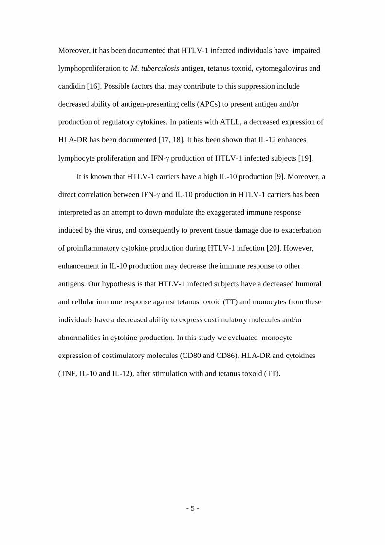
- 5 -
Moreover, it has been documented that HTLV-1 infected individuals have impaired
lymphoproliferation to M. tuberculosis antigen, tetanus toxoid, cytomegalovirus and
candidin [16]. Possible factors that may contribute to this suppression include
decreased ability of antigen-presenting cells (APCs) to present antigen and/or
production of regulatory cytokines. In patients with ATLL, a decreased expression of
HLA-DR has been documented [17, 18]. It has been shown that IL-12 enhances
lymphocyte proliferation and IFN- production of HTLV-1 infected subjects [19].
It is known that HTLV-1 carriers have a high IL-10 production [9]. Moreover, a
direct correlation between IFN-γ and IL-10 production in HTLV-1 carriers has been
interpreted as an attempt to down-modulate the exaggerated immune response
induced by the virus, and consequently to prevent tissue damage due to exacerbation
of proinflammatory cytokine production during HTLV-1 infection [20]. However,
enhancement in IL-10 production may decrease the immune response to other
antigens. Our hypothesis is that HTLV-1 infected subjects have a decreased humoral
and cellular immune response against tetanus toxoid (TT) and monocytes from these
individuals have a decreased ability to express costimulatory molecules and/or
abnormalities in cytokine production. In this study we evaluated monocyte
expression of costimulatory molecules (CD80 and CD86), HLA-DR and cytokines
(TNF, IL-10 and IL-12), after stimulation with and tetanus toxoid (TT).

- 6 -
Results
Humoral and cellular immune response against TT
The production of antigen specific IgG after immunization with TT in HTLV-1
infected subjects and controls is shown in figure 1. The median of the observances in
healthy subjects (0.800) ranging from 0.52 to 1.04 was higher (P=0.007) than in
HTLV-1 infected subjects who had median of 0.48 ranging from 0.23 to 1.1. There
was no correlation between antibody titers and production of IFN-γ (Figure 1).
The frequency of CD4 and CD8 T cells displaying cytokines in cultures
stimulated with TT were also different among HTLV-1 infected individuals and
healthy subjects. The median of CD4 T cells expressing IFN-γ in healthy subjects was
0.3 ranging from 0.2 to 14 while in HTLV-1 infected subjects was 0.15 ranging from
0.1 to 0,4 (P=0.01). The frequency of CD4+ T cells expressing IL-10 was also higher
(P=0.01) in healthy subjects 0.4 ranging from 0.2 to 13 than in HTLV-1 infected
subjects median of 0.2 ranging from 0.1 to 0.4. Indeed the frequency of CD8 T cells
expressing TNF-α in healthy subjects was also higher in healthy subjects than in
HTLV-1 infected subjects (P=0.04) (Figure 2).
HLA-DR, CD80, CD86 and cytokines expression by monocytes
The phenotypic characteristics of monocytes in HTLV-1 infected subjects and
in healthy controls were also different. Specifically the intensity of the expression of
HLA-DR in CD14 cells was higher healthy subjects median of 552 ranging from 112
to 4013 than in HTLV-1 infected subjects 504 ranging from 68 to 2349. There was no
difference regarding the expression of CD80 and CD86 between HTLV-1 infected
individuals and healthy subjects (Figure 3).

- 7 -
Both HTLV-1 infected individuals and healthy subjects increased the frequency
of cells expressing TNF-α, IL-12 and IL-10 after stimulation with TT but significant
increasing was only observed in HTLV-1 infected individuals (Figure 4).
Discussion
The association of other infectious diseases with HTLV-1 infection is well
documented and severe factors may participate to increase the susceptibility of
HTLV-1 infected subjects to have an abnormal immune response to a biased antigen.
Here in we showed that antibodies production as well as the frequency of cells
expressing cytokines upon stimulation with TT after immunization with this antigen
were higher in healthy subjects than in HTLV-1 infected individuals indicating an
impairment in both antibody production and cell mediated immunity of HTLV-1
infected subjects in develop an immune response to a biased antigen.
During HTLV-1 infection immunologic abnormalities have been documented in
both antigen presenting cells and in T cells which could impair immunologic response
to other antigens. The increased susceptibility to Strongyloides stercoralis in HTLV-1
infected individuals have been associated with a decrease in the Th2 type of immune
against parasite antigens [21] and also due to an increasing in regulatory T cells [22].
It has been shown in patients with HTLV-1 and strongiloidiasis a negative correlation
between IFN-γ with IL-5, and IFN-γ with total and parasite specific IgE.
In the present study because the majority of subjects infected with HTLV-1
have a high production of cytokines and an increased lymphocyte proliferative
response even in unstimulated culture we decide to only include subjects who had low
production of IFN-γ and TNF-α. These cytokines are determined in all participants at

- 8 -
the admission in the HTLV-1 clinic. However when we determine the production of
cytokines in unstimulated cultures previous to the immunization about 50% of
infected subjects had higher levels of IFN-γ. However even having a limited number
of subjects with high production of IFN-γ we showed that there was no correlation
between IFN-γ productions an unstimulated cultures and production of IgG to tetanus
toxoid after immunization with this antigen. IFN-γ contribute to IgG production [23]
but it can down modulate IgG1 response. We did not evaluate IgG subclasses but the
observation indicates that high levels of IFN-γ may have little influence in antigen
specific IgG levels.
As cytokine production and frequency of cells expressing these cytokines are
increased in HTLV-1 it is difficult sometimes to evaluate the influence of antigen
stimulation in cytokine production and in the frequency of cells expressing cytokines.
However we showed that the frequency of CD4 T cells expressing IFN-γ and IL-10 as
well as the frequency of CD8 T cells expressing TNF-α was higher in healthy subjects
than in HTLV-1 infected subjects in cultures stimulated with TT. This indicates that
in HTLV-1 infection there is a decrease in the production of both pro-inflammatory
and regulatory cytokines after immunization with TT.
The impairment in T cells in produce cytokine and produce antibody after
immunization with TT antigen may be due to defect in antigen presenting cells.
Because HTLV-1 infected predominantly T cells very little attention have be given to
antigen present cells. However HTLV-1 may infected monocytes/macrophages as
well as dendritic cells [24, 25]. Moreover it has been shown that dendritic cells from
HTLV-1 infected subjects express less HLA-DR [17] and that there is impairment on
the differentiation of macrophages to dendritic cells in HTLV-1 infected subjects. It is
relevant to pointed out that these previous studies included patients with HAM/TSP,
- 9 -
whose are known to present more immunologic abnormalities, since lymphocyte
proliferation and IFN-γ production in unstimulated cultures are higher in HAM/TSP
than in HTLV-1 carriers. Here all participants of the study were HTLV-1 carriers and
the majority of them had little or no evidence of lymphocyte activation measured by
secretion of IFN-γ. So even in HTLV-1 carriers without evidence of great T cell
activation we showed an impairment in the expression of HLA-DR molecules that are
important to the generation of an immune response. Therefore it is possible that the
decreasing in the insensitivity of expression of HLA-DR by impair antigen
presentation to T cells had contributed to the decreasing in antibody and in cell
mediated immunity in HTLV-1 infected subjects after immunization with TT.
The cytokine production by antigen presenting cells such as IL-12 and IL-10
may influence the immunologic responses to other antigens in HTLV-1 infected
subjects. We have previously shown that compared to uninfected controls HTLV-1
carriers produce more IL-10 the most important cytokine that down modulate the
immunologic response in humans [9]. It was also shown that levels of IL-12 is
reduced in supernatants of HTLV-1 infected subjects with tuberculosis in cultures
stimulated with PPD [26] and that exogenous addition of IL-12 to lymphocyte
cultures of HTLV-1 infected subjects enhance lymphocyte proliferation to PPD [19].
However our data did not show impairment in IL-12 neither higher expression of IL-
10 by monocytes. Therefore while we cannot ruled out a role for regulatory cytokines
in the impairment of the immune response in HTLV-1 infected subjects, as our studies
were performed only monocytes we show that decreasing expression of HLA-DR
may play an important role in the immune response to a biased antigen in HTLV-1
infection and may participate in the increasing susceptibility of HTLV-1 infected
subjects to develop other infectious disease.

- 10 -
Conclusions
This study showed that HTLV-1 infected subjects, after vaccination, presented a
decreasing of the humoral and cellular immune response against TT. Monocytes from
the patients showed an impairment of antigen presentation through the HLA-DR
expression mechanisms. However, the second signal (CD80 and CD86 expression)
and cytokine expression did not show abnormalities. These results suggest that these
immunological mechanisms may participate of the increasing susceptibility of HTLV-
1 infected subjects to acquire others infectious diseases

- 11 -
Methods
Subjects
HTLV-1 carriers (HTLV, n = 14) were selected from the HTLV-1 clinic at the
Hospital Universitario Professor Edgard Santos, Federal University of Bahia, Brazil.
The diagnosis of HTLV-1 was performed by ELISA (Murex Biotech Limited,
Dartford, UK) and confirmed by Western Blot (HTLV blot 2.4, Genelabs, Singapore).
Exclusion criteria included the use of antiviral drugs or immunomodulators in the
previous 90 days, helminthic infection, co-infection with HIV, HCV, HBV or
tuberculosis and presence of diseases. Motor dysfunction was determined by Osame’s
Motor Disability Score (OMDS) [3] and the degree of neurological involvement was
assessed by the Expanded Disability Status Scale (EDSS) [27]. Patients with OMDS =
0 and EDSS = 0 were selected.
HTLV-1 seronegative healthy subjects (HS, n = 12) were employees from the
Immunology Service, Hospital Universitario Professor Edgard Santos. The Ethical
Committee of the Maternidade Climério de Oliveira approved this study (number
154/2009) and informed consent was obtained from all prospectively enrolled
subjects.
Serology for tetanus toxoid and immunization
The serology for TT consisted of ELISA. A 96-wells plate was coated with TT
(0.1 Lf/mL), overnight, 4 oC. The sera from patients and HS were diluted (1:100) and
incubated during 1 hour, 37 oC. Anti-human IgG alkaline phosphatase conjugate
(Sigma Chemicals, St. Louis, MO, USA) was diluted (1:1,000) and incubated for 1
hour, 37 oC. 1 mg/mL of pNPP (Sigma Chemicals, St. Louis, MO, USA) was used to

- 12 -
reveal the reaction. The plate was read at 405 nm. Serum of immunized individuals
was used as positive control. Serum of non-immunized individuals (more than 10
years of the last immunization) was used as negative controls. It was considered
seronegative to TT if the optical density (O.D.) was lower than the cut-off. The cut-off
was calculated with the mean of the O.D. from the negative controls plus two standard
deviation (mean + 2 x S.D.).
Patients and HS with negative serology to TT were immunized. After two
vaccine doses, they returned to collect the blood and serum. Immunological
evaluations were performed before and after immunization.
PBMC isolation and Flow cytometry
PBMC from HTLV-1 and HS were obtained from heparinized venous blood
samples by Ficoll-Hypaque density gradient centrifugation. Cultures of 4 x 10⁵ PBMC
were prepared in RPMI 1640 plus 5% heat inactivated human AB serum (Sigma
Chemical Co. St. Louis, MO), antibiotics and glutamine.
To evaluate the cytokine expression by monocytes, PBMC were cultured during
20 hours with TT (0,5 Lf/mL). The last four hours it was added Brefeldin A (1
µg/mL). The cells were labeled with anti-CD4 and anti-CD8 and fixed with
formaldehyde. After, cells were labeled with anti-IFN-, anti-TNF and anti-IL-10 for
30 minutes at room temperature.
To evaluate costimulatory molecules and HLA-DR expression in monocytes,
PBMC were cultured during 6 hours without stimulus, or were stimulated with
tetanus toxoid (0.5 Lf/mL). Cells were then incubated with FITC, PE or PE-Cy5-
labeled monoclonal antibodies (Ig controls, CD14, HLA-DR, CD80 and CD86) for 20
minutes at 4oC. All mentioned reagents were from BD Pharmingen, Franklin Lakes,

- 13 -
NJ. After staining, preparations were washed with phosphate-buffer saline (PBS),
fixed with 2% formaldehyde in PBS and kept at 4 oC until data acquisition using a
FACScalibur (Becton Dickinson, San Jose, CA). To evaluate cytokine expression, the
fixed cells were permeabilized with a solution of Saponin and stained, for 30 minutes
at 4 oC, using anti-cytokine monoclonal antibodies (TNF-α, IL-12, IL-10) conjugated
with PE (BD Pharmingen).
In all cases, 100,000 gated events were acquired for later analysis in FACScanto
II cytometer. Analysis was performed using FlowJo Software version 7.6.1.
Statistical analysis
Wilcoxon signed rank test was used to compare the cytokine and surface
molecule expression between cells with or without stimulation. Mann-Whitney test
was used to compare results between the HS group and the HTLV-1 group. An alpha
of 5% (p < 0.05) was considered for statistical significance. Analyses were performed
using Graphpad Prism version 5.0.
Competing interests
The author(s) have no competing interests.
Authors' contributions
AS, SS, LPC and EMC participated equally in the study design.
AS and EMC drafted the manuscript.
AS and SS performed PBMC isolation.

- 14 -
AS and LPC performed flow cytometry.
All the authors read and approved the final manuscript.
Acknowledgements
We are grateful to Elbe Myrtes Silva and Lúcia Reis for their secretarial
assistance.
This work was supported by the CNPq, and the NIH Training Grants R01
AI079238A (EMC). AS, SS, LPC and EMC are funded by the CNPq.
- 15 -
References
1. Edlich RF, Arnette JA, Williams FM: Global epidemic of human T-cell
lymphotropic virus type-I (HTLV-I). J Emerg Med 2000, 18:109-119.
2. Poiesz BJ, Ruscetti FW, Gazdar AF, Bunn PA, Minna JD, Gallo RC:
Detection and isolation of type C retrovirus particles from fresh and
cultured lymphocytes of a patient with cutaneous T-cell lymphoma. Proc
Natl Acad Sci U S A 1980, 77:7415-7419.
3. Osame M, Usuku K, Izumo S, Ijichi N, Amitani H, Igata A, Matsumoto M,
Tara M: HTLV-I associated myelopathy, a new clinical entity. Lancet 1986,
1:1031-1032.
4. Hollsberg P, Hafler DA: Seminars in medicine of the Beth Israel Hospital,
Boston. Pathogenesis of diseases induced by human lymphotropic virus
type I infection. N Engl J Med 1993, 328:1173-1182.
5. Prince H, Kleinman S, Doyle M, Lee H, Swanson P: Spontaneous
lymphocyte proliferation in vitro characterizes both HTLV-I and HTLV-
II infection. J Acquir Immune Defic Syndr 1990, 3:1199-1200.
6. Kubota R, Kawanishi T, Matsubara H, Manns A, Jacobson S: Demonstration
of human T lymphotropic virus type I (HTLV-I) tax-specific CD8+
lymphocytes directly in peripheral blood of HTLV-I-associated
myelopathy/tropical spastic paraparesis patients by intracellular cytokine
detection. J Immunol 1998, 161:482-488.
7. Nishimoto N, Yoshizaki K, Eiraku N, Machigashira K, Tagoh H, Ogata A,
Kuritani T, Osame M, Kishimoto T: Elevated levels of interleukin-6 in
serum and cerebrospinal fluid of HTLV-I-associated myelopathy/tropical
spastic paraparesis. J Neurol Sci 1990, 97:183-193.
8. Osame M: Pathological mechanisms of human T-cell lymphotropic virus
type I-associated myelopathy (HAM/TSP). J Neurovirol 2002, 8:359-364.
9. Carvalho EM, Bacellar O, Porto AF, Braga S, Galvao-Castro B, Neva F:
Cytokine profile and immunomodulation in asymptomatic human T-
lymphotropic virus type 1-infected blood donors. J Acquir Immune Defic
Syndr 2001, 27:1-6.
10. Santos SB, Porto AF, Muniz AL, de Jesus AR, Magalhaes E, Melo A, Dutra
WO, Gollob KJ, Carvalho EM: Exacerbated inflammatory cellular immune
response characteristics of HAM/TSP is observed in a large proportion of
HTLV-I asymptomatic carriers. BMC Infect Dis 2004, 4:7.
11. Porto MA, Alcantara LM, Leal M, Castro N, Carvalho EM: Atypical clinical
presentation of strongyloidiasis in a patient co-infected with human T cell
lymphotrophic virus type I. Am J Trop Med Hyg 2005, 72:124-125.
12. Porto AF, Santos SB, Muniz AL, Basilio V, Rodrigues W, Jr., Neva FA, Dutra
WO, Gollob KJ, Jacobson S, Carvalho EM: Helminthic infection down-
regulates type 1 immune responses in human T cell lymphotropic virus
type 1 (HTLV-1) carriers and is more prevalent in HTLV-1 carriers than
in patients with HTLV-1-associated myelopathy/tropical spastic
paraparesis. J Infect Dis 2005, 191:612-618.
13. Verdonck K, Gonzalez E, Schrooten W, Vanham G, Gotuzzo E: HTLV-1
infection is associated with a history of active tuberculosis among family
members of HTLV-1-infected patients in Peru. Epidemiol Infect 2008,
136:1076-1083.
- 16 -
14. de Lourdes Bastos M, Osterbauer O, Mesquita DL, Carrera CA, Albuquerque
MJ, Silva L, Pereira DN, Riley L, Carvalho EM: Prevalence of human T-cell
lymphotropic virus type 1 infection in hospitalized patients with
tuberculosis. Int J Tuberc Lung Dis 2009, 13(12):1519-1523.
15. Tachibana N, Okayama A, Ishizaki J, Yokota T, Shishime E, Murai K, Shioiri
S, Tsuda K, Essex M, Mueller N: Suppression of tuberculin skin reaction in
healthy HTLV-I carriers from Japan. Int J Cancer 1988, 42:829-831.
16. Mascarenhas RE, Brodskyn C, Barbosa G, Clarencio J, Andrade-Filho AS,
Figueiroa F, Galvao-Castro B, Grassi F: Peripheral blood mononuclear cells
from individuals infected with human T-cell lymphotropic virus type 1
have a reduced capacity to respond to recall antigens. Clin Vaccine
Immunol 2006, 13:547-552.
17. Makino M, Wakamatsu S, Shimokubo S, Arima N, Baba M: Production of
functionally deficient dendritic cells from HTLV-I-infected monocytes:
implications for the dendritic cell defect in adult T cell leukemia. Virology
2000, 274:140-148.
18. Nascimento CR, Lima MA, de Andrada Serpa MJ, Espindola O, Leite AC,
Echevarria-Lima J: Monocytes from HTLV-1-infected patients are unable
to fully mature into dendritic cells. Blood 2011, 117:489-499.
19. Suzuki M, Dezzutti CS, Okayama A, Tachibana N, Tsubouchi H, Mueller N,
Lal RB: Modulation of T-cell responses to a recall antigen in human T-cell
leukemia virus type 1-infected individuals. Clin Diagn Lab Immunol 1999,
6:713-717.
20. Santos SB, Porto AF, Muniz AL, Luna T, Nascimento MC, Guerreiro JB,
Oliveira-Filho J, Morgan DJ, Carvalho EM: Modulation of T cell responses
in HTLV-1 carriers and in patients with myelopathy associated with
HTLV-1. Neuroimmunomodulation 2006, 13:145-151.
21. Porto AF, Santos SB, Alcantara L, Guerreiro JB, Passos J, Gonzalez T, Neva
F, Gonzalez D, Ho JL, Carvalho EM: HTLV-1 modifies the clinical and
immunological response to schistosomiasis. Clin Exp Immunol 2004,
137:424-429.
22. Montes M, Sanchez C, Verdonck K, Lake JE, Gonzalez E, Lopez G,
Terashima A, Nolan T, Lewis DE, Gotuzzo E, White AC, Jr.: Regulatory T
cell expansion in HTLV-1 and strongyloidiasis co-infection is associated
with reduced IL-5 responses to Strongyloides stercoralis antigen. PLoS
Negl Trop Dis 2009, 3:e456.
23. Kawano Y, Noma T, Yata J: Regulation of human IgG subclass production
by cytokines. IFN-gamma and IL-6 act antagonistically in the induction of
human IgG1 but additively in the induction of IgG2. J Immunol 1994,
153:4948-4958.
24. de Revel T, Mabondzo A, Gras G, Delord B, Roques P, Boussin F, Neveux Y,
Bahuau M, Fleury HJ, Dormont D: In vitro infection of human
macrophages with human T-cell leukemia virus type 1. Blood 1993,
81:1598-1606.
25. Knight SC, Macatonia SE, Cruickshank K, Rudge P, Patterson S: Dendritic
cells in HIV-1 and HTLV-1 infection. Adv Exp Med Biol 1993, 329:545-
549.
26. Bastos MD, Santos S, Souza A, Finkmoore B, Bispo O, Barreto T, Cardoso I,
Bispo I, Bastos F, Pereira D, et al: Influence of HTLV-1 on the clinical,
- 17 -
microbiologic and immunologic presentation of tuberculosis. BMC Infect
Dis 2012, 12:199.
27. Kurtzke JF: Rating neurologic impairment in multiple sclerosis: an
expanded disability status scale (EDSS). Neurology 1983, 33:1444-1452.

- 18 -
Figure Legends
Figure 1. Serology for TT from HTLV-1 infected individuals and healthy subjects
after immunization for TT. The dashed line represents the cut-off (O.D. = 0,238).
Statistical analysis was performed using Mann-Whitney test.
Figure 2. Expression of cytokines from CD4+ and CD8
+ T cell, stimulated with TT,
from HTLV-1 infected individuals and HS before and after immunization. Statistical
analysis was performed using Wilcoxon test.
Figure 3. HLA-DR (A), CD80 (B) and CD86 (C) expression by CD14+ monocytes
from HTLV-1 infected individuals and HS with or without TT stimulation. Statistical
analysis was performed using Wilcoxon test. MFI = mean of fluorescence intensity.
Figure 4. TNF (A), IL-12 (B) and IL-10 (C) expression by CD14+ monocytes from
HTLV-1 infected individuals and HS with or without TT stimulation. Statistical
analysis was performed using Wilcoxon and Mann-Whitney tests.

- 19 -
FIGURE 1
HTLV-1 Healthy Subjects0.0
0.4
0.8
1.2p = 0,007
O.D
. 405 n
m
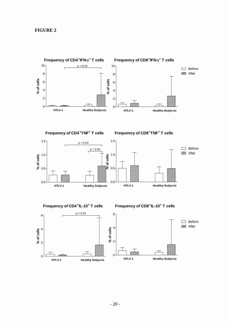
- 20 -
FIGURE 2
Frequency of CD4+IFN-+ T cells
0
2
4
6
8
10
HTLV-1 Healthy Subjects
p = 0,01
% o
f cell
s
Frequency of CD8+IFN-+ T cells
0
2
4
6
8
10Before
After
HTLV-1 Healthy Subjects%
of
cell
s
Frequency of CD4+TNF+ T cells
0.0
0.5
1.0
1.5
p = 0,04
p = 0,04
HTLV-1 Healthy Subjects
% o
f cell
s
Frequency of CD8+TNF+ T cells
0.0
0.5
1.0
1.5
Before
After
HTLV-1 Healthy Subjects
% o
f cell
s
Frequency of CD4 +IL-10+ T cells
0
2
4
6p = 0,01
HTLV-1 Healthy Subjects
% o
f cell
s
Frequency of CD8+IL-10+ T cells
0
2
4
6
Before
After
HTLV-1 Healthy Subjects
% o
f cell
s

- 21 -
FIGURE 3
HLA-DR expression by monocytes
0
500
1000
1500
2000
2500
HTLV-1 Healthy Subjects
p = 0,005
MFI
CD80 expression by monocytes
0
1000
2000
3000
4000
5000
HTLV-1 Healthy Subjects
MFI
CD86 expression by monocytes
0
2000
4000
6000
8000
10000w/ stimulus
TT
HTLV-1 Healthy Subjects
MFI

- 22 -
FIGURE 4
Frequency of CD14 +TNF+ T cells
0
10
20
30
40 p = 0,009
HTLV-1 Healthy Subjects
% o
f cell
s
Frequency of CD14+IL-12+ T cells
0
1
2
3
4
5
p = 0,006
p = 0,03
HTLV-1 Healthy Subjects
% o
f cell
s
Frequency of CD14+IL-10+ T cells
0.0
0.5
1.0
1.5
2.0
2.5w/ stimulus
TT
HTLV-1 Healthy Subjects
% o
f cell
s

ANEXO 4
Normas de publicação da revista Retrovirology
1/29/13 Retrovirology | Instructions for Authors | Research Articles
www.retrovirology.com/authors/instructions/research 1/6
6.47
Instructions for authors
Research Articles
Submission process | Preparing main manuscript text | Preparing illustrations and figures | Preparing tables | Preparing additional files | Style and language
See 'About this journal' for descriptions of different article types and information about policies and the refereeing process.
Submission process
Manuscripts must be submitted by one of the authors of the manuscript, and should not be submitted by anyone on their behalf. The submitting author takes
responsibility for the article during submission and peer review.
Please note that Retrovirology levies an article-processing charge on all accepted Research Articles; if the submitting author's institution is a BioMed Central member
the cost of the article-processing charge may be covered by the membership (see About page for detail). Please note that the membership is only automatically
recognised on submission if the submitting author is based at the member institution.
To facilitate rapid publication and to minimize administrative costs, Retrovirology accepts only online submission.
Files can be submitted as a batch, or one by one. The submission process can be interrupted at any time; when users return to the site, they can carry on where they
left off.
See below for examples of word processor and graphics file formats that can be accepted for the main manuscript document by the online submission system.
Additional files of any type, such as movies, animations, or original data files, can also be submitted as part of the manuscript.
During submission you will be asked to provide a cover letter. Use this to explain why your manuscript should be published in the journal, to elaborate on any issues
relating to our editorial policies in the 'About Retrovirology' page, and to declare any potential competing interests. You will be also asked to provide the contact details
(including email addresses) of potential peer reviewers for your manuscript. These should be experts in their field, who will be able to provide an objective assessment
of the manuscript. Any suggested peer reviewers should not have published with any of the authors of the manuscript within the past five years, should not be current
collaborators, and should not be members of the same research institution. Suggested reviewers will be considered alongside potential reviewers recommended by the
Editor-in-Chief and/or Editorial Board members.
Assistance with the process of manuscript preparation and submission is available from BioMed Central customer support team.
We also provide a collection of links to useful tools and resources for scientific authors on our Useful Tools page.
File formats
The following word processor file formats are acceptable for the main manuscript document:
Microsoft word (DOC, DOCX)
Rich text format (RTF)
Portable document format (PDF)
TeX/LaTeX (use )
DeVice Independent format (DVI)
Users of other word processing packages should save or convert their files to RTF before uploading. Many free tools are available which ease this process.
TeX/LaTeX users: We recommend using BioMed Central's TeX template and BibTeX stylefile. If you use this standard format, you can submit your manuscript in TeX
format. If you have used another template for your manuscript, or if you do not wish to use BibTeX, then please submit your manuscript as a DVI file. We do not
recommend converting to RTF.
Note that figures must be submitted as separate image files, not as part of the submitted manuscript file.
Publishing Datasets
Through a special arrangement with LabArchives, LLC, authors submitting manuscripts to Retrovirology can obtain a complimentary subscription to LabArchives with
an allotment of 100MB of storage. LabArchives is an Electronic Laboratory Notebook which will enable scientists to share and publish data files in situ; you can then link
your paper to these data. Data files linked to published articles are assigned digital object identifiers (DOIs) and will remain available in perpetuity. Use of LabArchives
or similar data publishing services does not replace preexisting data deposition requirements, such as for nucleic acid sequences, protein sequences and atomic
coordinates.
Instructions on assigning DOIs to datasets, so they can be permanently linked to publications, can be found on the LabArchives website. Use of LabArchives’ software
has no influence on the editorial decision to accept or reject a manuscript.
Authors linking datasets to their publications should include an Availability of supporting data section in their manuscript and cite the dataset in their reference list.
Preparing main manuscript text
General guidelines of the journal's style and language are given below.
Overview of manuscript sections for Research Articles
Manuscripts for Research Articles submitted to Retrovirology should be divided into the following sections (in this order):
(if any)
BioMed Central's TeX template
Title page
Abstract
Keywords
Background
Results and discussion
Conclusions
Methods
Availability of supporting data
List of abbreviations used
Competing interests
Authors' contributions
Authors' information

1/29/13 Retrovirology | Instructions for Authors | Research Articles
www.retrovirology.com/authors/instructions/research 2/6
(if any)
The Accession Numbers of any nucleic acid sequences, protein sequences or atomic coordinates cited in the manuscript should be provided, in square brackets and
include the corresponding database name; for example, [EMBL:AB026295, EMBL:AC137000, DDBJ:AE000812, GenBank:U49845, PDB:1BFM, Swiss-Prot:Q96KQ7,
PIR:S66116].
The databases for which we can provide direct links are: EMBL Nucleotide Sequence Database (EMBL), DNA Data Bank of Japan (DDBJ), GenBank at the NCBI
(GenBank), Protein Data Bank (PDB), Protein Information Resource (PIR) and the Swiss-Prot Protein Database (Swiss-Prot).
You can download a template (Mac and Windows compatible; Microsoft Word 98/2000) for your article.
For reporting standards please see the information in the About section.
Title page
The title page should:
provide the title of the article
list the full names, institutional addresses and email addresses for all authors
indicate the corresponding author
Please note:
abbreviations within the title should be avoided
Abstract
The Abstract of the manuscript should not exceed 350 words and must be structured into separate sections: Background, the context and purpose of the study;
Results, the main findings; Conclusions, brief summary and potential implications. Please minimize the use of abbreviations and do not cite references in the
abstract.
Keywords
Three to ten keywords representing the main content of the article.
Background
The Background section should be written in a way that is accessible to researchers without specialist knowledge in that area and must clearly state - and, if helpful,
illustrate - the background to the research and its aims. The section should end with a brief statement of what is being reported in the article.
Results and discussion
The Results and discussion may be combined into a single section or presented separately. The Results and discussion sections may also be broken into subsections
with short, informative headings.
Conclusions
This should state clearly the main conclusions of the research and give a clear explanation of their importance and relevance. Summary illustrations may be included.
Methods
The methods section should include the design of the study, the type of materials involved, a clear description of all comparisons, and the type of analysis used, to
enable replication.
Availability of supporting data
Retrovirology requires authors to deposit the data set(s) supporting the results reported in submitted manuscripts in a publicly-accessible data repository. This section
should only be included when supporting data are available and must include the name of the repository and the permanent identifier or accession number and
persistent hyperlink(s) for the data set(s). The following format is required:
"The data set(s) supporting the results of this article is(are) available in the [repository name] repository, [unique persistent identifier and hyperlink to dataset(s) in
http:// format]."
We also recommend that the data set(s) be cited, where appropriate in the manuscript, and included in the reference list.
A list of available scientific research data repositories can be found here. A list of all BioMed Central journals that require or encourage this section to be included in
research articles can be found here.
List of abbreviations
If abbreviations are used in the text they should be defined in the text at first use, and a list of abbreviations can be provided, which should precede the competing
interests and authors' contributions.
Competing interests
A competing interest exists when your interpretation of data or presentation of information may be influenced by your personal or financial relationship with other
people or organizations. Authors must disclose any financial competing interests; they should also reveal any non-financial competing interests that may cause them
embarrassment were they to become public after the publication of the manuscript.
Authors are required to complete a declaration of competing interests. All competing interests that are declared will be listed at the end of published articles. Where an
author gives no competing interests, the listing will read 'The author(s) declare that they have no competing interests'.
When completing your declaration, please consider the following questions:
Financial competing interests
In the past five years have you received reimbursements, fees, funding, or salary from an organization that may in any way gain or lose financially from the
publication of this manuscript, either now or in the future? Is such an organization financing this manuscript (including the article-processing charge)? If so,
please specify.
Do you hold any stocks or shares in an organization that may in any way gain or lose financially from the publication of this manuscript, either now or in the
future? If so, please specify.
Do you hold or are you currently applying for any patents relating to the content of the manuscript? Have you received reimbursements, fees, funding, or salary
from an organization that holds or has applied for patents relating to the content of the manuscript? If so, please specify.
Do you have any other financial competing interests? If so, please specify.
Acknowledgements
Endnotes
References
Illustrations and figures
Tables and captions
Preparing additional files

1/29/13 Retrovirology | Instructions for Authors | Research Articles
www.retrovirology.com/authors/instructions/research 3/6
Non-financial competing interests
Are there any non-financial competing interests (political, personal, religious, ideological, academic, intellectual, commercial or any other) to declare in relation to this
manuscript? If so, please specify.
If you are unsure as to whether you, or one your co-authors, has a competing interest please discuss it with the editorial office.
Authors' contributions
In order to give appropriate credit to each author of a paper, the individual contributions of authors to the manuscript should be specified in this section.
An 'author' is generally considered to be someone who has made substantive intellectual contributions to a published study. To qualify as an author one should 1) have
made substantial contributions to conception and design, or acquisition of data, or analysis and interpretation of data; 2) have been involved in drafting the manuscript
or revising it critically for important intellectual content; and 3) have given final approval of the version to be published. Each author should have participated
sufficiently in the work to take public responsibility for appropriate portions of the content. Acquisition of funding, collection of data, or general supervision of the
research group, alone, does not justify authorship.
We suggest the following kind of format (please use initials to refer to each author's contribution): AB carried out the molecular genetic studies, participated in the
sequence alignment and drafted the manuscript. JY carried out the immunoassays. MT participated in the sequence alignment. ES participated in the design of the
study and performed the statistical analysis. FG conceived of the study, and participated in its design and coordination and helped to draft the manuscript. All authors
read and approved the final manuscript.
All contributors who do not meet the criteria for authorship should be listed in an acknowledgements section. Examples of those who might be acknowledged include a
person who provided purely technical help , writing assistance, or a department chair who provided only general support.
Authors' information
You may choose to use this section to include any relevant information about the author(s) that may aid the reader's interpretation of the article, and understand the
standpoint of the author(s). This may include details about the authors' qualifications, current positions they hold at institutions or societies, or any other relevant
background information. Please refer to authors using their initials. Note this section should not be used to describe any competing interests.
Acknowledgements
Please acknowledge anyone who contributed towards the article by making substantial contributions to conception, design, acquisition of data, or analysis and
interpretation of data, or who was involved in drafting the manuscript or revising it critically for important intellectual content, but who does not meet the criteria for
authorship. Please also include the source(s) of funding for each author, and for the manuscript preparation. Authors must describe the role of the funding body, if
any, in design, in the collection, analysis, and interpretation of data; in the writing of the manuscript; and in the decision to submit the manuscript for publication.
Please also acknowledge anyone who contributed materials essential for the study. If a language editor has made significant revision of the manuscript, we
recommend that you acknowledge the editor by name, where possible.
Authors should obtain permission to acknowledge from all those mentioned in the Acknowledgements section.
Endnotes
Endnotes should be designated within the text using a superscript lowercase letter and all notes (along with their corresponding letter) should be included in the
Endnotes section. Please format this section in a paragraph rather than a list.
References
All references, including URLs, must be numbered consecutively, in square brackets, in the order in which they are cited in the text, followed by any in tables or
legends. Each reference must have an individual reference number. Please avoid excessive referencing. If automatic numbering systems are used, the reference
numbers must be finalized and the bibliography must be fully formatted before submission.
Only articles, datasets and abstracts that have been published or are in press, or are available through public e-print/preprint servers, may be cited; unpublished
abstracts, unpublished data and personal communications should not be included in the reference list, but may be included in the text and referred to as "unpublished
observations" or "personal communications" giving the names of the involved researchers. Obtaining permission to quote personal communications and unpublished
data from the cited colleagues is the responsibility of the author. Footnotes are not allowed, but endnotes are permitted. Journal abbreviations follow Index
Medicus/MEDLINE. Citations in the reference list should include all named authors, up to the first 30 before adding 'et al.'.
Any in press articles cited within the references and necessary for the reviewers' assessment of the manuscript should be made available if requested by the editorial
office.
Style files are available for use with popular bibliographic management software:
Examples of the Retrovirology reference style are shown below. Please ensure that the reference style is followed precisely; if the references are not in the correct
style they may have to be retyped and carefully proofread.
All web links and URLs, including links to the authors' own websites, should be given a reference number and included in the reference list rather than within the text of
the manuscript. They should be provided in full, including both the title of the site and the URL, in the following format: The Mouse Tumor Biology Database
[http://tumor.informatics.jax.org/mtbwi/index.do]. If an author or group of authors can clearly be associated with a web link, such as for weblogs, then they should be
included in the reference.
Examples of the Retrovirology reference style
Article within a journal
Koonin EV, Altschul SF, Bork P: BRCA1 protein products: functional motifs. Nat Genet 1996, 13:266-267.
Article within a journal supplement
Orengo CA, Bray JE, Hubbard T, LoConte L, Sillitoe I: Analysis and assessment of ab initio three-dimensional prediction, secondary structure, and
contacts prediction. Proteins 1999, 43(Suppl 3):149-170.
In press article
Kharitonov SA, Barnes PJ: Clinical aspects of exhaled nitric oxide. Eur Respir J, in press.
Published abstract
Zvaifler NJ, Burger JA, Marinova-Mutafchieva L, Taylor P, Maini RN: Mesenchymal cells, stromal derived factor-1 and rheumatoid arthritis [abstract].
Arthritis Rheum 1999, 42:s250.
Article within conference proceedings
Jones X: Zeolites and synthetic mechanisms. In Proceedings of the First National Conference on Porous Sieves: 27-30 June 1996; Baltimore. Edited by Smith Y.
Stoneham: Butterworth-Heinemann; 1996:16-27.
Book chapter, or article within a book
Schnepf E: From prey via endosymbiont to plastids: comparative studies in dinoflagellates. In Origins of Plastids. Volume 2. 2nd edition. Edited by Lewin
RA. New York: Chapman and Hall; 1993:53-76.
Whole issue of journal
Ponder B, Johnston S, Chodosh L (Eds): Innovative oncology. In Breast Cancer Res 1998, 10:1-72.
BibTeX
EndNote style file
Reference Manager
Zotero

1/29/13 Retrovirology | Instructions for Authors | Research Articles
www.retrovirology.com/authors/instructions/research 4/6
Whole conference proceedings
Smith Y (Ed): Proceedings of the First National Conference on Porous Sieves: 27-30 June 1996; Baltimore. Stoneham: Butterworth-Heinemann; 1996.
Complete book
Margulis L: Origin of Eukaryotic Cells. New Haven: Yale University Press; 1970.
Monograph or book in a series
Hunninghake GW, Gadek JE: The alveolar macrophage. In Cultured Human Cells and Tissues. Edited by Harris TJR. New York: Academic Press; 1995:54-56.
[Stoner G (Series Editor): Methods and Perspectives in Cell Biology, vol 1.]
Book with institutional author
Advisory Committee on Genetic Modification: Annual Report. London; 1999.
PhD thesis
Kohavi R: Wrappers for performance enhancement and oblivious decision graphs. PhD thesis. Stanford University, Computer Science Department; 1995.
Link / URL
The Mouse Tumor Biology Database [http://tumor.informatics.jax.org/mtbwi/index.do]
Link / URL with author(s)
Neylon C: Open Research Computation: an ordinary journal with extraordinary aims.
[http://blogs.openaccesscentral.com/blogs/bmcblog/entry/open_research_computation_an_ordinary]
Dataset with persistent identifier
Zheng, L-Y; Guo, X-S; He, B; Sun, L-J; Peng, Y; Dong, S-S; Liu, T-F; Jiang, S; Ramachandran, S; Liu, C-M; Jing, H-C (2011): Genome data from sweet and grain
sorghum (Sorghum bicolor). GigaScience. http://dx.doi.org/10.5524/100012.
Preparing illustrations and figures
Illustrations should be provided as separate files, not embedded in the text file. Each figure should include a single illustration and should fit on a single page in portrait
format. If a figure consists of separate parts, it is important that a single composite illustration file be submitted which contains all parts of the figure. There is no
charge for the use of color figures.
Please read our figure preparation guidelines for detailed instructions on maximising the quality of your figures.
Formats
The following file formats can be accepted:
PDF (preferred format for diagrams)
DOCX/DOC (single page only)
PPTX/PPT (single slide only)
EPS
PNG (preferred format for photos or images)
TIFF
JPEG
BMP
Figure legends
The legends should be included in the main manuscript text file at the end of the document, rather than being a part of the figure file. For each figure, the following
information should be provided: Figure number (in sequence, using Arabic numerals - i.e. Figure 1, 2, 3 etc); short title of figure (maximum 15 words); detailed
legend, up to 300 words.
Please note that it is the responsibility of the author(s) to obtain permission from the copyright holder to reproduce figures or tables that have
previously been published elsewhere.
Preparing a personal cover page
If you wish to do so, you may submit an image which, in the event of publication, will be used to create a cover page for the PDF version of your article. The cover
page will also display the journal logo, article title and citation details. The image may either be a figure from your manuscript or another relevant image. You must
have permission from the copyright to reproduce the image. Images that do not meet our requirements will not be used.
Images must be 300dpi and 155mm square (1831 x 1831 pixels for a raster image).
Allowable formats - EPS, PDF (for line drawings), PNG, TIFF (for photographs and screen dumps), JPEG, BMP, DOC, PPT, CDX, TGF (ISIS/Draw).
Preparing tables
Each table should be numbered and cited in sequence using Arabic numerals (i.e. Table 1, 2, 3 etc.). Tables should also have a title (above the table) that summarizes
the whole table; it should be no longer than 15 words. Detailed legends may then follow, but they should be concise. Tables should always be cited in text in
consecutive numerical order.
Smaller tables considered to be integral to the manuscript can be pasted into the end of the document text file, in A4 portrait or landscape format. These will be
typeset and displayed in the final published form of the article. Such tables should be formatted using the 'Table object' in a word processing program to ensure that
columns of data are kept aligned when the file is sent electronically for review; this will not always be the case if columns are generated by simply using tabs to
separate text. Columns and rows of data should be made visibly distinct by ensuring that the borders of each cell display as black lines. Commas should not be used
to indicate numerical values. Color and shading may not be used; parts of the table can be highlighted using symbols or bold text, the meaning of which should be
explained in a table legend. Tables should not be embedded as figures or spreadsheet files.
Larger datasets or tables too wide for a landscape page can be uploaded separately as additional files. Additional files will not be displayed in the final, laid-out PDF of
the article, but a link will be provided to the files as supplied by the author.
Tabular data provided as additional files can be uploaded as an Excel spreadsheet (.xls ) or comma separated values (.csv). As with all files, please use the standard
file extensions.
Preparing additional files
Although Retrovirology does not restrict the length and quantity of data included in an article, we encourage authors to provide datasets, tables, movies, or other
information as additional files.
Please note: All Additional files will be published along with the article. Do not include files such as patient consent forms, certificates of language editing, or revised
versions of the main manuscript document with tracked changes. Such files should be sent by email to [email protected], quoting the Manuscript ID number.
Results that would otherwise be indicated as "data not shown" can and should be included as additional files. Since many weblinks and URLs rapidly become broken,
Retrovirology requires that supporting data are included as additional files, or deposited in a recognized repository . Please do not link to data on a
personal/departmental website. The maximum file size for additional files is 20 MB each, and files will be virus-scanned on submission.

1/29/13 Retrovirology | Instructions for Authors | Research Articles
www.retrovirology.com/authors/instructions/research 5/6
Additional files can be in any format, and will be downloadable from the final published article as supplied by the author. reuse. e.g. We recommend CSV rather than
PDF for tabular data.
Certain supported files formats are recognized and can be displayed to the user in the browser. These include most movie formats (for users with the Quicktime
plugin), mini-websites prepared according to our guidelines, chemical structure files (MOL, PDB), geographic data files (KML).
If additional material is provided, please list the following information in a separate section of the manuscript text:
File name (e.g. Additional file 1)
File format including the correct file extension for example .pdf, .xls, .txt, .pptx (including name and a URL of an appropriate viewer if format is unusual)
Title of data
Description of data
Additional files should be named "Additional file 1" and so on and should be referenced explicitly by file name within the body of the article, e.g. 'An additional movie
file shows this in more detail [see Additional file 1]'.
Additional file formats
Ideally, file formats for additional files should not be platform-specific, and should be viewable using free or widely available tools. The following are examples of
suitable formats.
Additional documentation
PDF (Adode Acrobat)
Animations
SWF (Shockwave Flash)
Movies
MP4 (MPEG 4)
MOV (Quicktime)
Tabular data
XLS, XLSX (Excel Spreadsheet)
CSV (Comma separated values)
As with figure files, files should be given the standard file extensions.
Mini-websites
Small self-contained websites can be submitted as additional files, in such a way that they will be browsable from within the full text HTML version of the article. In
order to do this, please follow these instructions:
Create a folder containing a starting file called index.html (or index.htm) in the root.
Put all files necessary for viewing the mini-website within the folder, or sub-folders.
Ensure that all links are relative (ie "images/picture.jpg" rather than "/images/picture.jpg" or "http://yourdomain.net/images/picture.jpg" or "C:\Documents and
Settings\username\My Documents\mini-website\images\picture.jpg") and no link is longer than 255 characters.
Access the index.html file and browse around the mini-website, to ensure that the most commonly used browsers (Internet Explorer and Firefox) are able to
view all parts of the mini-website without problems, it is ideal to check this on a different machine.
Compress the folder into a ZIP, check the file size is under 20 MB, ensure that index.html is in the root of the ZIP, and that the file has .zip extension, then
submit as an additional file with your article.
Style and language
General
Currently, Retrovirology can only accept manuscripts written in English. Spelling should be US English or British English, but not a mixture.
There is no explicit limit on the length of articles submitted, but authors are encouraged to be concise.
Retrovirology will not edit submitted manuscripts for style or language; reviewers may advise rejection of a manuscript if it is compromised by grammatical errors.
Authors are advised to write clearly and simply, and to have their article checked by colleagues before submission. In-house copyediting will be minimal. Non-native
speakers of English may choose to make use of a copyediting service.
Help and advice on scientific writing
The abstract is one of the most important parts of a manuscript. For guidance, please visit our page on Writing titles and abstracts for scientific articles.
Tim Albert has produced for BioMed Central a list of tips for writing a scientific manuscript. American Scientist also provides a list of resources for science writing. For
more detailed guidance on preparing a manuscript and writing in English, please visit the BioMed Central author academy.
Abbreviations
Abbreviations should be used as sparingly as possible. They should be defined when first used and a list of abbreviations can be provided following the main
manuscript text.
Typography
Please use double line spacing.
Type the text unjustified, without hyphenating words at line breaks.
Use hard returns only to end headings and paragraphs, not to rearrange lines.
Capitalize only the first word, and proper nouns, in the title.
All pages should be numbered.
Use the Retrovirology .
Footnotes are not allowed, but endnotes are permitted.
Please do not format the text in multiple columns.
Greek and other special characters may be included. If you are unable to reproduce a particular special character, please type out the name of the symbol in
full. Please ensure that all special characters used are embedded in the text, otherwise they will be lost during conversion to PDF.
Genes, mutations, genotypes, and alleles should be indicated in italics, and authors are required to use approved gene symbols, names, and formatting. Protein
products should be in plain type.
Units
reference format

1/29/13 Retrovirology | Instructions for Authors | Research Articles
www.retrovirology.com/authors/instructions/research 6/6
SI units should be used throughout ( liter and molar are permitted, however).
ANEXO 5
Publicação científica no período de 2009-2012.

61Gaz. méd. Bahia 2009;79:1(Jan-Dez):61-67 HTLV e Coinfecções
www.gmbahia.ufba.br
Recebido em 29/06/2009 Aceito em 30/10/2009Endereço para correspondência: Dr. Edgar M. Carvalho - Serviço deImunologia, 5o andar, Complexo Hospitalar Universitário ProfessorEdgard Santos, Rua João das Botas s/n, Canela, 40110-160, Salvador,BA, Brasil. Phone (55.71) 3237-7353, Fax: (55.71) 3245-7110. E-mail: [email protected] and [email protected] de financiamento: Este trabalho teve o suporte do NationalInstitute of Health projeto R01 AI079238A.
Gazeta Médica da Bahia 2009;79:1(Jan-Dez):61-67© 2009 Gazeta Médica da Bahia. Todos os direitos reservados.
INFLUÊNCIA DO HTLINFLUÊNCIA DO HTLINFLUÊNCIA DO HTLINFLUÊNCIA DO HTLINFLUÊNCIA DO HTLVVVVV-I NA INCIDÊNCIA, RESPOST-I NA INCIDÊNCIA, RESPOST-I NA INCIDÊNCIA, RESPOST-I NA INCIDÊNCIA, RESPOST-I NA INCIDÊNCIA, RESPOSTA IMUNE E MANIFESTA IMUNE E MANIFESTA IMUNE E MANIFESTA IMUNE E MANIFESTA IMUNE E MANIFESTAÇÕES CLÍNICAS DEAÇÕES CLÍNICAS DEAÇÕES CLÍNICAS DEAÇÕES CLÍNICAS DEAÇÕES CLÍNICAS DEOUTRAS DOENÇAS INFECCIOSASOUTRAS DOENÇAS INFECCIOSASOUTRAS DOENÇAS INFECCIOSASOUTRAS DOENÇAS INFECCIOSASOUTRAS DOENÇAS INFECCIOSAS
INFLUENCE OF HTLV-I IN THE INCIDENCE, IMMUNE RESPONSE AND CLINICAL MANIFESTATIONS OF OTHERINFECTIOUS DISEASES
Anselmo Souza1, Aurélia Porto2, Silvane B. Santos1, Maria de Lourdes Bastos3, Edgar M. Carvalho1
1Serviço de Imunologia, Complexo Hospitalar Universitário Prof. Edgard Santos, Universidade Federal da Bahia, Salvador, BA;2Universidade Federal de Sergipe, Aracaju, SE; 3Hospital Especializado Octávio Mangabeira, Salvador, BA; Brasil
O vírus linfotrópico de células T humanas do tipo 1 (HTLV-I) é o agente causal da mielopatia associada ao HTLV-I(HAM/TSP) e da leucemia/linfoma de células T do adulto (ATLL) e é prevalente no Brasil, África, América Central eJapão. O vírus infecta células T e tem sido observado que a infecção pelo HTLV-I pode interferir na incidência,expressão de doença e resposta imune de outras doenças infecciosas, tais como estrongiloidíase, sarna norueguesa,tuberculose e esquistossomose. Neste trabalho, foi revisado o que há na literatura sobre a associação entre HTLV-I eoutras doenças infecciosas, enfatizando a tuberculose, outras doenças bacterianas, helmintíases, infecções virais esarna norueguesa. Além disso, foram adicionados dados ainda não publicados de pesquisa que vem sendo desenvolvidano Serviço de Imunologia do Complexo Hospitalar Universitário Professor Edgard Santos, UFBA.Palavras chave: HTLV-I e estrongiloidíase, tuberculose e HTLV-I, escabiose e HTLV-I, esquistossomose e HTLV-I.
Human T cell lymphotropic virus type I (HTLV-I) is causal agent of HTLV-I-associated myelopathy (HAM/TSP) and adultT cell leukemia/lymphoma (ATLL) and its prevalent in Brazil, Africa and Central America. The virus infects predominantlyCD4+ T cells and it has been observed that HTLV-I infection may influence the incidence, clinical manifestations andimmune response of other diseases like strongyloidiasis, scabies and schistosomiasis. Here we revised publications aboutassociation between HTLV-I and others infectious diseases, with emphasis in tuberculosis, helminthiasis, scabies and viraland bacterial infections. Moreover, we added data generated in the Immunology Service of the Complexo HospitalarUniversitário Prof. Edgard Santos.Key words: HTLV-I and strongyloidiasis, tuberculosis in HTLV-I, scabies and HTLV-I, schistosomiasis and HTLV-I.
O vírus linfotrópico de células T humanas tipo I (HTLV-I)tem alta prevalência no Brasil, América Central e África, regiõescom elevada incidência de doenças infecto-parasitárias,fazendo com que a associação entre o HTLV-I e doençasinfecciosas sejam comuns nestas áreas(7 18). O HTLV-I é o agentecausal da mielopatia associada ao vírus ou paraparesiaespástica tropical (HAM/TSP) (28) e da leucemia de células Tdo adulto (ATLL)(57). O vírus infecta, predominantemente,células T, induzindo ativação e proliferação celular comprodução de concentrações elevadas de citocinas associadascom a resposta Th1, como Interleucina (IL)-2, fator de necrosetumoral alfa (TNF-α) e interferon-gama (IFN-γ)(8 47). Essasalterações são observadas em portadores assintomáticos dovírus, mas são mais marcantes em pacientes com HAM/TSP(47).Como a resposta imune desempenha papel importante napatogênese das doenças infecciosas, é esperado que as
alterações imunológicas induzidas pelo HTLV-I possaminterferir na incidência, resposta imune e manifestações clínicasdas doenças infecciosas em pacientes coinfectados.
Estudos de corte transversal tem identificado quepacientes infectados pelo HTLV-I tem mais doençasparasitárias, doenças virais, doenças causadas por bactériasextra e intracelulares do que indivíduos soronegativos paraeste vírus(23 33 42 43). Investigadores do Serviço de Imunologiado Complexo Hospitalar Universitário Professor EdgardSantos (SIM-HUPES) acompanham, há cerca de 10 anos,indivíduos infectados pelo HTLV-I e o papel da infecção peloHTLV-I em modificar a resposta imune e manifestações dedoenças infecciosas tem sido estudado. No presente trabalhofoi feita uma revisão sobre a associação entre HTLV-I e outrosagentes infecciosos assim como é mostrada a experiência destegrupo nestas coinfecções.
Material e MétodosForam revisados artigos que tem como foco a infecção
pelo HTLV-I associada com outras doenças infecciosas como:Tuberculose, Estrongiloidíase, Esquistossomose, infecçõesvirais, infecções bacterianas e sarna norueguesa. Os dadosgerados no SIM-HUPES ainda não publicados foram obtidosatravés de estudo aprovado pelo Comitê de Ética em Pesquisada Maternidade Climério de Oliveira aprovado em 20/06/2006.
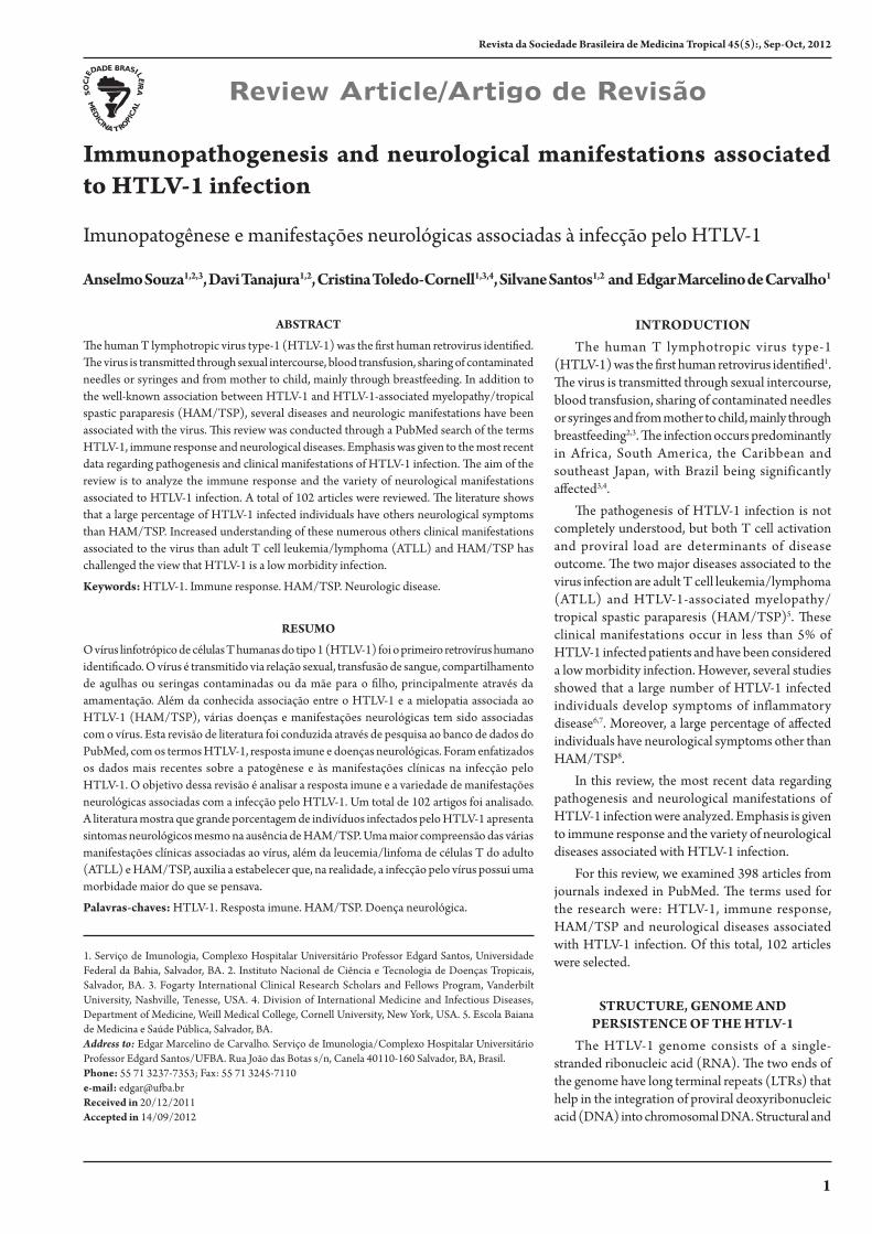
1
Revista da Sociedade Brasileira de Medicina Tropical 45(5):, Sep-Oct, 2012
Review Article/Artigo de Revisão
1. Serviço de Imunologia, Complexo Hospitalar Universitário Professor Edgard Santos, Universidade Federal da Bahia, Salvador, BA. 2. Instituto Nacional de Ciência e Tecnologia de Doenças Tropicais, Salvador, BA. 3. Fogarty International Clinical Research Scholars and Fellows Program, Vanderbilt University, Nashville, Tenesse, USA. 4. Division of International Medicine and Infectious Diseases, Department of Medicine, Weill Medical College, Cornell University, New York, USA. 5. Escola Baiana de Medicina e Saúde Pública, Salvador, BA.Address to: Edgar Marcelino de Carvalho. Serviço de Imunologia/Complexo Hospitalar Universitário Professor Edgard Santos/UFBA. Rua João das Botas s/n, Canela 40110-160 Salvador, BA, Brasil.Phone: 55 71 3237-7353; Fax: 55 71 3245-7110e-mail: [email protected] in 20/12/2011Accepted in 14/09/2012
Immunopathogenesis and neurological manifestations associated to HTLV-1 infection
Imunopatogênese e manifestações neurológicas associadas à infecção pelo HTLV-1
Anselmo Souza1,2,3, Davi Tanajura1,2, Cristina Toledo-Cornell1,3,4, Silvane Santos1,2 and Edgar Marcelino de Carvalho1
ABSTRACT
The human T lymphotropic virus type-1 (HTLV-1) was the first human retrovirus identified. The virus is transmitted through sexual intercourse, blood transfusion, sharing of contaminated needles or syringes and from mother to child, mainly through breastfeeding. In addition to the well-known association between HTLV-1 and HTLV-1-associated myelopathy/tropical spastic paraparesis (HAM/TSP), several diseases and neurologic manifestations have been associated with the virus. This review was conducted through a PubMed search of the terms HTLV-1, immune response and neurological diseases. Emphasis was given to the most recent data regarding pathogenesis and clinical manifestations of HTLV-1 infection. The aim of the review is to analyze the immune response and the variety of neurological manifestations associated to HTLV-1 infection. A total of 102 articles were reviewed. The literature shows that a large percentage of HTLV-1 infected individuals have others neurological symptoms than HAM/TSP. Increased understanding of these numerous others clinical manifestations associated to the virus than adult T cell leukemia/lymphoma (ATLL) and HAM/TSP has challenged the view that HTLV-1 is a low morbidity infection.
Keywords: HTLV-1. Immune response. HAM/TSP. Neurologic disease.
RESUMO
O vírus linfotrópico de células T humanas do tipo 1 (HTLV-1) foi o primeiro retrovírus humano identificado. O vírus é transmitido via relação sexual, transfusão de sangue, compartilhamento de agulhas ou seringas contaminadas ou da mãe para o filho, principalmente através da amamentação. Além da conhecida associação entre o HTLV-1 e a mielopatia associada ao HTLV-1 (HAM/TSP), várias doenças e manifestações neurológicas tem sido associadas com o vírus. Esta revisão de literatura foi conduzida através de pesquisa ao banco de dados do PubMed, com os termos HTLV-1, resposta imune e doenças neurológicas. Foram enfatizados os dados mais recentes sobre a patogênese e às manifestações clínicas na infecção pelo HTLV-1. O objetivo dessa revisão é analisar a resposta imune e a variedade de manifestações neurológicas associadas com a infecção pelo HTLV-1. Um total de 102 artigos foi analisado. A literatura mostra que grande porcentagem de indivíduos infectados pelo HTLV-1 apresenta sintomas neurológicos mesmo na ausência de HAM/TSP. Uma maior compreensão das várias manifestações clínicas associadas ao vírus, além da leucemia/linfoma de células T do adulto (ATLL) e HAM/TSP, auxilia a estabelecer que, na realidade, a infecção pelo vírus possui uma morbidade maior do que se pensava.
Palavras-chaves: HTLV-1. Resposta imune. HAM/TSP. Doença neurológica.
INTRODUCTIONThe human T lymphotropic virus type-1
(HTLV-1) was the first human retrovirus identified1. The virus is transmitted through sexual intercourse, blood transfusion, sharing of contaminated needles or syringes and from mother to child, mainly through breastfeeding2,3. The infection occurs predominantly in Africa, South America, the Caribbean and southeast Japan, with Brazil being significantly affected3,4.
The pathogenesis of HTLV-1 infection is not completely understood, but both T cell activation and proviral load are determinants of disease outcome. The two major diseases associated to the virus infection are adult T cell leukemia/lymphoma (ATLL) and HTLV-1-associated myelopathy/tropical spastic paraparesis (HAM/TSP)5. These clinical manifestations occur in less than 5% of HTLV-1 infected patients and have been considered a low morbidity infection. However, several studies showed that a large number of HTLV-1 infected individuals develop symptoms of inflammatory disease6,7. Moreover, a large percentage of affected individuals have neurological symptoms other than HAM/TSP8.
In this review, the most recent data regarding pathogenesis and neurological manifestations of HTLV-1 infection were analyzed. Emphasis is given to immune response and the variety of neurological diseases associated with HTLV-1 infection.
For this review, we examined 398 articles from journals indexed in PubMed. The terms used for the research were: HTLV-1, immune response, HAM/TSP and neurological diseases associated with HTLV-1 infection. Of this total, 102 articles were selected.
STRUCTURE, GENOME AND PERSISTENCE OF THE HTLV-1
The HTLV-1 genome consists of a single-stranded ribonucleic acid (RNA). The two ends of the genome have long terminal repeats (LTRs) that help in the integration of proviral deoxyribonucleic acid (DNA) into chromosomal DNA. Structural and

Journal of Medical Virology 84:1809–1817 (2012)
Immunological and Viral Features in Patients WithOveractive Bladder Associated With Human T-CellLymphotropic Virus Type 1 Infection
Silvane Braga Santos,1,2 Paulo Oliveira,1 Tania Luna,1 Anselmo Souza,1,2
Marcia Nascimento,1 Isadora Siqueira,1 Davi Tanajura,1,2 Andre Luiz Muniz,1,2
Marshall J. Glesby,3 and Edgar M. Carvalho1,2*1Immunology Service, Professor Edgard Santos University Hospital, Federal University of Bahia, Salvador,Bahia, Brazil2National Institute of Science and Technology of Tropical Diseases (CNPq/MCT), Salvador, Bahia, Brazil3Weill Cornell Medical College, New York, New York
The majority of patients infected with humanT-cell lymphotropic virus-type 1 (HTLV-1) areconsidered carriers, but a high frequency ofurinary symptoms of overactive bladder,common in HTLV-1 associated myelopathy/tropical spastic paraparesis (HAM/TSP) havebeen documented in these patients. The aim ofthis study was to determine if immunologicaland viral factors that are seen in HAM/TSP arealso observed in these patients. Participantswere classified as HTLV-1 carriers (n ¼ 45),HTLV-1 patients suffering from overactivebladder (n ¼ 45) and HAM/TSP (n ¼ 45). Cellsfrom HTLV-1 overactive bladder patients pro-duced spontaneously more proinflammatorycytokines than carriers. TNF-a and IL-17 levelswere similar in HAM/TSP and HTLV-1overactive bladder patients. High proviral loadwas found in patients with overactive bladderand HAM/TSP and correlated with proinflam-matory cytokines. In contrast with findings inpatients with HAM/TSP, serum levels of Th1chemokines were similar in HTLV-1 overactivebladder and carriers. Exogenous addition ofregulatory cytokines decreased spontaneousIFN-g production in cell cultures from HTLV-1overactive bladder patients. The results showthat HTLV-1 overactive bladder and HAM/TSPpatients have in common some immunologicalfeatures as well as similar proviral load profile.The data show that HTLV-1 overactive bladderpatients are still able to down regulate theirinflammatory immune response. In addition,these patients express levels of chemokinessimilar to carriers, which may explain why theyhave yet to develop the same degree of spinalcord damage as seen in patients with HAM/TSP. These patients present symptoms of
overactive bladder, which may be an early signof HAM/TSP. J. Med. Virol. 84:1809–1817,2012. � 2012 Wiley Periodicals, Inc.
KEY WORDS: HTLV-1; immune response;cytokines; chemokines; pro-viral load
INTRODUCTION
Human T-cell lymphotropic virus type 1 (HTLV-1)is a complex retrovirus belonging to the Deltaretrovi-rus family. It is associated etiologically with adult Tcell leukemia/lymphoma (ATLL) and HTLV-1 associ-ated myelopathy/tropical spastic paraparesis (HAM/TSP) [Uchiyama, 1997]. The HTLV-1 infection hasbeen considered an infection with low morbidity.However, a cross-sectional study showed a higher fre-quency of neurological symptoms, erectile dysfunction,and urinary disturbances in HTLV-1 carriers than inuninfected healthy controls, suggesting that the spec-trum of disease may be broader [Caskey et al., 2007].
Grant sponsor: National Institute of Health; Grant numbers:AI079238; K24 AI078884; Grant sponsor: Brazilian NationalResearch Council (CNPq).
Conflicts of interest: None.
*Correspondence to: Edgar M. Carvalho, Servico deImunologia, Complexo Hospitalar Universitario ProfessorEdgard Santos, Universidade Federal da Bahia (UFBA), RuaJoao das Botas s/n, Canela, 40110-160, BA, Brazil.E-mail: [email protected], [email protected]
Accepted 24 May 2012
DOI 10.1002/jmv.23341Published online in Wiley Online Library(wileyonlinelibrary.com).
� 2012 WILEY PERIODICALS, INC.
Bastos et al. BMC Infectious Diseases 2012, 12:199http://www.biomedcentral.com/1471-2334/12/199
RESEARCH ARTICLE Open Access
Influence of HTLV-1 on the clinical, microbiologicand immunologic presentation of tuberculosisMaria de Lourdes Bastos1,2,3, Silvane B Santos1, Anselmo Souza1, Brooke Finkmoore4, Ohana Bispo1,2,Tasso Barreto1,2, Ingrid Cardoso1,2, Iana Bispo1,2, Flávia Bastos1,2, Daniele Pereira1,2, Lee Riley4
and Edgar M Carvalho1,2,5,6*
Abstract
Background: HTLV-1 is associated with increased susceptibility to Mycobacterium tuberculosis infection and severityof tuberculosis. Although previous studies have shown that HTLV-1 infected individuals have a low frequency ofpositive tuberculin skin test (TST) and decreasing in lymphoproliferative responses compared to HTLV-1 uninfectedpersons, these studies were not performed in individuals with history of tuberculosis or evidence of M. tuberculosisinfection. Therefore the reasons why HTLV-1 infection increases susceptibility to infection and severity oftuberculosis are not understood.The aim of this study was to evaluate how HTLV-1 may influence the clinical,bacteriologic and immunologic presentation of tuberculosis.
Methods: The study prospectively enrolled and followed 13 new cases of tuberculosis associated with HTLV-1(cases) and 25 patients with tuberculosis without HTLV-1 infection (controls). Clinical findings, bacterial load in thesputum, x-rays, immunological response and death were compared in the two groups.
Results: There were no differences in the demographic, clinical and TST response between the two study groups.IFN-γ and TNF-α production was higher in unstimulated cultures of mononuclear cells of case than in controlpatients (p < 0.01). While there was no difference in IFN-γ production in PPD stimulated cultures, TNF-α levels werelower in cases than in controls (p = 0.01). There was no difference in the bacterial load among the groups butsputum smear microscopy results became negative faster in cases than in controls. Death only occurred in twoco-infected patients.
Conclusion: While the increased susceptibility for tuberculosis infection in HTLV-1 infected subjects may be relatedto impairment in TNF-α production, the severity of tuberculosis in co-infected patients may be due to theenhancement of the Th1 inflammatory response, rather than in their decreased ability to control bacterial growth.
Keywords: HTLV-1, Tuberculosis, Mycobacterium tuberculosis
BackgroundThe human T cell lymphotropic virus type 1 (HTLV-1)has a worldwide distribution; it is most prevalent inCentral and South America, Central Africa and south-western Japan [1]. HTLV-1 infects predominantly CD4 Tcells inducing cell activation and proliferation of bothCD4 and CD8 T cells [2,3]. Moreover, the high produc-tion of pro-inflammatory cytokines (TNF-α and IFN-γ)
* Correspondence: [email protected]ço de Imunologia do Complexo Hospitalar Universitário ProfessorEdgard Santos, Universidade Federal da Bahia, Salvador, BA, Brazil2Escola Bahiana de Medicina e Saúde Pública, Salvador, BA, BrazilFull list of author information is available at the end of the article
© 2012 Bastos et al.; licensee BioMed CentralCommons Attribution License (http://creativecreproduction in any medium, provided the or
has been associated with central nervous system (CNS)damage and the development of HTLV-1 associatedmyelopathy (HAM) [2,3].There is evidence that HTLV-1 infection increases se-
verity and susceptibility to strongyloidiasis, scabies andtuberculosis [4-9]. In patients co-infected with HTLV-1and Strongyloides stercoralis the exaggerated Th1 im-mune response and increased regulatory T cell responsedecrease the Th2 immune response, which plays a piv-otal role in host defense against helminthes [9-12].HTLV-1 infected individuals have two to four-foldincreased chance to acquire tuberculosis [6,13-15]. Add-itionally, in one retrospective study, it was observed that
Ltd. This is an Open Access article distributed under the terms of the Creativeommons.org/licenses/by/2.0), which permits unrestricted use, distribution, andiginal work is properly cited.