CASE REPORT Open Access Peculiar type 1 congenital pyloric ...
4
CASE REPORT Open Access Peculiar type 1 congenital pyloric atresia: a case report Enrico Zecca 1* , Mirta Corsello 1 , Claudio Pintus 2 , Lorenzo Nanni 2 , Susanna Zecca 3 Abstract Pyloric atresia (PA) is a very rare condition. Its incidence is approximately 1 in 100,000 newborns and constitutes about 1% of all intestinal atresias. We describe the neonatal course of a peculiar case of type 1 pyloric atresia, in which the pyloric membrane was connected to a second duodenal membrane through a virtual duodenal lumen in a premature newborn. The atypical variant required an unusual side to side gastroduodenostomy. We emphasize the importance of a prompt diagnosis to avoid potentially fatal complications and to warrant a good outcome even in the presence of a strange form of PA in the neonatal period. Background Pyloric atresia (PA) is a very rare condition. Its incidence is approximately 1 in 100,000 newborns and constitutes about 1% of all intestinal atresias [1,2]. Sometimes it occurs with genetic disorders like epidermolysis bullosa and aplasia cutis congenital [3-6] or in association with other atresias of the gastrointestinal tract [7-9]. The pre- sence of associated anomalies is a contributing factor for the reported high mortality [10]. There are 3 recognized anatomic varieties of pyloric atresia: type 1, pyloric mem- brane (57%); type 2, pyloric canal replaced by solid tissue (34%); type 3, atretic pylorus with a gap between stomach and duodenum (9%) [10]. We describe the neonatal course of a peculiar case of type 1 PA connected to a sec- ond duodenal membrane through a virtual lumen in a premature newborn with visualization of polyhydramnios on prenatal ultrasonography. We emphasize the impor- tance of a prompt diagnosis to avoid potentially fatal complications and to warrant a good outcome. Case Presentation Case report A girl was born by emergency cesarean section for abruptio placentae at 34 weeks’ gestation and weighted 2130 grams. Apgar scores were 8 and 9 at 1 and 5 min- utes, respectively. Pregnancy was complicated by gesta- tional diabetes and polyhydramnios. Physical examination at birth revealed a good-looking, with normal vital data and no signs of sepsis. Routine hema- tological examinations showed normal values. Nonpro- jectile and nonbilious vomiting began on the second day of life and persisted for the next day, with staunching > 4 cc/kg after feeding. Feeding was suspended but emesis persisted and the abdomen was mildly distended. A high intestinal obstruction was suspected and an abdominal roentgenogram showed a single gastric bubble with no air in the small and large intestine (Figure 1, left). The baby was referred for a surgical opinion on day 3. Con- trast study with barium meal confirmed complete obstruction at the pyloric region (Figure 1, right). At laparatomy she was found to have a moderately dis- tended stomach and a narrowed duodenal tract. A first membrane was found at the pylorus and a second one was found 2 cm forward, with a virtual duodenal lumen between them. Partial resection of the gastric antrum and of the first duodenal tract was performed, followed by a two layers, side to side gastroduodenostomy. The child had an uneventful postoperative course. Ten days postoperatively, a contrast study documented the patency of the entire gastrointestinal tract (Figure 2). She was treated with total intravenous nutrition for 20 days, associated with oral feeding in postoperative day 7 without problems. She was discharged home at 30 days of age on full oral feeding and remains well at 12 months of life. Discussion Pyloric atresia is a very rare condition. Its incidence is approximately 1 in 100.000 newborns and constitutes * Correspondence: [email protected] 1 Department of Pediatrics, Institute of Pediatrics, Division of Neonatology, Catholic University of the Sacred Heart, Rome, Italy Zecca et al. Italian Journal of Pediatrics 2010, 36:3 http://www.ijponline.net/content/36/1/3 ITALIAN JOURNAL OF PEDIATRICS © 2010 Zecca et al; licensee BioMed Central Ltd. This is an Open Access article distributed under the terms of the Creative Commons Attribution License (http://creativecommons.org/licenses/by/2.0), which permits unrestricted use, distribution, and reproduction in any medium, provided the original work is properly cited.
Transcript of CASE REPORT Open Access Peculiar type 1 congenital pyloric ...
CASE REPORT Open Access
Peculiar type 1 congenital pyloric atresia: a casereportEnrico Zecca1*, Mirta Corsello1, Claudio Pintus2, Lorenzo Nanni2, Susanna Zecca3
Abstract
Pyloric atresia (PA) is a very rare condition. Its incidence is approximately 1 in 100,000 newborns and constitutesabout 1% of all intestinal atresias. We describe the neonatal course of a peculiar case of type 1 pyloric atresia, inwhich the pyloric membrane was connected to a second duodenal membrane through a virtual duodenal lumenin a premature newborn. The atypical variant required an unusual side to side gastroduodenostomy. We emphasizethe importance of a prompt diagnosis to avoid potentially fatal complications and to warrant a good outcomeeven in the presence of a strange form of PA in the neonatal period.
BackgroundPyloric atresia (PA) is a very rare condition. Its incidenceis approximately 1 in 100,000 newborns and constitutesabout 1% of all intestinal atresias [1,2]. Sometimes itoccurs with genetic disorders like epidermolysis bullosaand aplasia cutis congenital [3-6] or in association withother atresias of the gastrointestinal tract [7-9]. The pre-sence of associated anomalies is a contributing factor forthe reported high mortality [10]. There are 3 recognizedanatomic varieties of pyloric atresia: type 1, pyloric mem-brane (57%); type 2, pyloric canal replaced by solid tissue(34%); type 3, atretic pylorus with a gap between stomachand duodenum (9%) [10]. We describe the neonatalcourse of a peculiar case of type 1 PA connected to a sec-ond duodenal membrane through a virtual lumen in apremature newborn with visualization of polyhydramnioson prenatal ultrasonography. We emphasize the impor-tance of a prompt diagnosis to avoid potentially fatalcomplications and to warrant a good outcome.
Case PresentationCase reportA girl was born by emergency cesarean section forabruptio placentae at 34 weeks’ gestation and weighted2130 grams. Apgar scores were 8 and 9 at 1 and 5 min-utes, respectively. Pregnancy was complicated by gesta-tional diabetes and polyhydramnios. Physicalexamination at birth revealed a good-looking, with
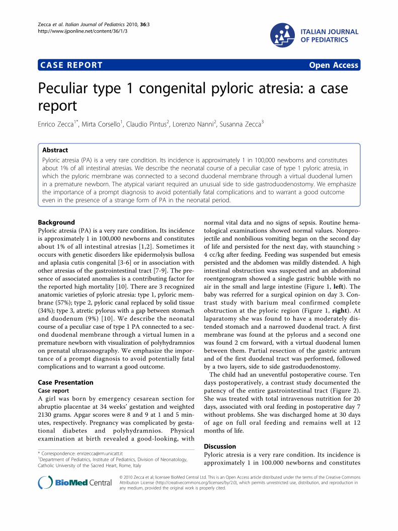
normal vital data and no signs of sepsis. Routine hema-tological examinations showed normal values. Nonpro-jectile and nonbilious vomiting began on the second dayof life and persisted for the next day, with staunching >4 cc/kg after feeding. Feeding was suspended but emesispersisted and the abdomen was mildly distended. A highintestinal obstruction was suspected and an abdominalroentgenogram showed a single gastric bubble with noair in the small and large intestine (Figure 1, left). Thebaby was referred for a surgical opinion on day 3. Con-trast study with barium meal confirmed completeobstruction at the pyloric region (Figure 1, right). Atlaparatomy she was found to have a moderately dis-tended stomach and a narrowed duodenal tract. A firstmembrane was found at the pylorus and a second onewas found 2 cm forward, with a virtual duodenal lumenbetween them. Partial resection of the gastric antrumand of the first duodenal tract was performed, followedby a two layers, side to side gastroduodenostomy.The child had an uneventful postoperative course. Ten
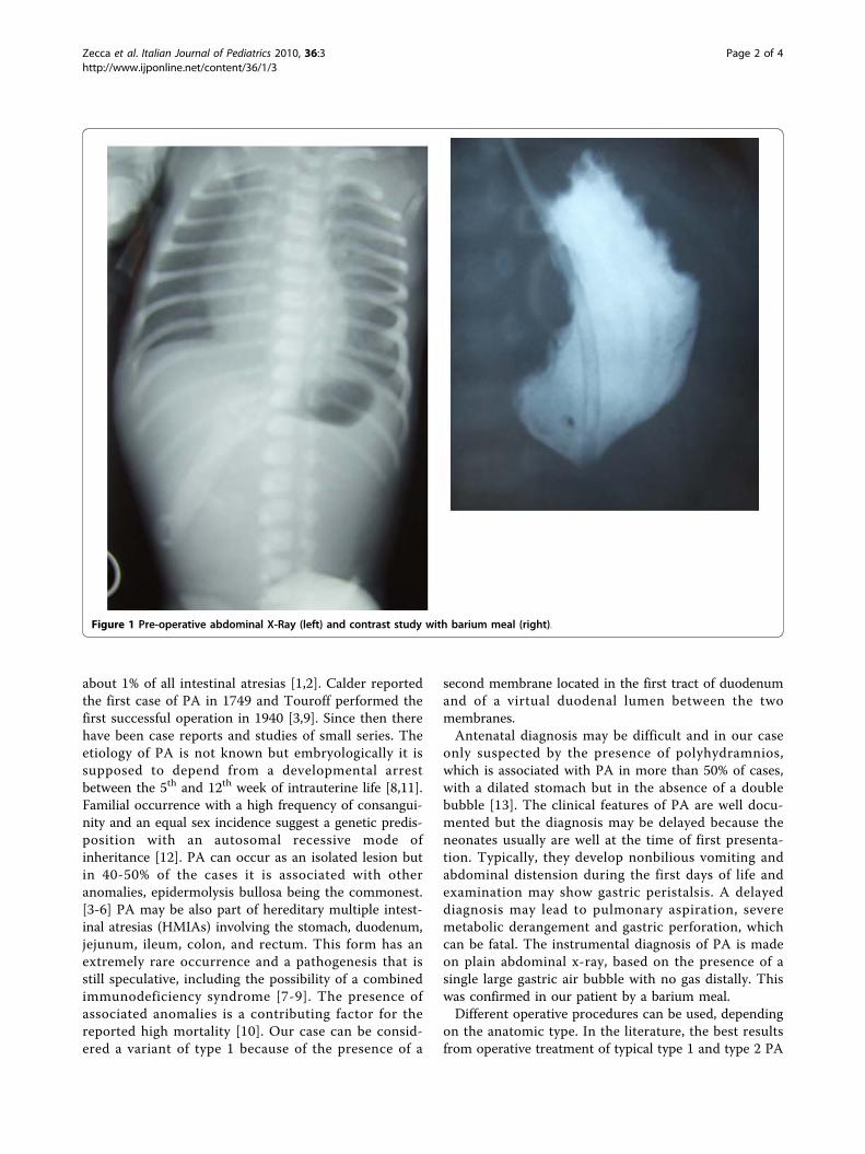
days postoperatively, a contrast study documented thepatency of the entire gastrointestinal tract (Figure 2).She was treated with total intravenous nutrition for 20days, associated with oral feeding in postoperative day 7without problems. She was discharged home at 30 daysof age on full oral feeding and remains well at 12months of life.
DiscussionPyloric atresia is a very rare condition. Its incidence isapproximately 1 in 100.000 newborns and constitutes
* Correspondence: [email protected] of Pediatrics, Institute of Pediatrics, Division of Neonatology,Catholic University of the Sacred Heart, Rome, Italy
Zecca et al. Italian Journal of Pediatrics 2010, 36:3http://www.ijponline.net/content/36/1/3 ITALIAN JOURNAL
OF PEDIATRICS
© 2010 Zecca et al; licensee BioMed Central Ltd. This is an Open Access article distributed under the terms of the Creative CommonsAttribution License (http://creativecommons.org/licenses/by/2.0), which permits unrestricted use, distribution, and reproduction inany medium, provided the original work is properly cited.
about 1% of all intestinal atresias [1,2]. Calder reportedthe first case of PA in 1749 and Touroff performed thefirst successful operation in 1940 [3,9]. Since then therehave been case reports and studies of small series. Theetiology of PA is not known but embryologically it issupposed to depend from a developmental arrestbetween the 5th and 12th week of intrauterine life [8,11].Familial occurrence with a high frequency of consangui-nity and an equal sex incidence suggest a genetic predis-position with an autosomal recessive mode ofinheritance [12]. PA can occur as an isolated lesion butin 40-50% of the cases it is associated with otheranomalies, epidermolysis bullosa being the commonest.[3-6] PA may be also part of hereditary multiple intest-inal atresias (HMIAs) involving the stomach, duodenum,jejunum, ileum, colon, and rectum. This form has anextremely rare occurrence and a pathogenesis that isstill speculative, including the possibility of a combinedimmunodeficiency syndrome [7-9]. The presence ofassociated anomalies is a contributing factor for thereported high mortality [10]. Our case can be consid-ered a variant of type 1 because of the presence of a
second membrane located in the first tract of duodenumand of a virtual duodenal lumen between the twomembranes.Antenatal diagnosis may be difficult and in our case
only suspected by the presence of polyhydramnios,which is associated with PA in more than 50% of cases,with a dilated stomach but in the absence of a doublebubble [13]. The clinical features of PA are well docu-mented but the diagnosis may be delayed because theneonates usually are well at the time of first presenta-tion. Typically, they develop nonbilious vomiting andabdominal distension during the first days of life andexamination may show gastric peristalsis. A delayeddiagnosis may lead to pulmonary aspiration, severemetabolic derangement and gastric perforation, whichcan be fatal. The instrumental diagnosis of PA is madeon plain abdominal x-ray, based on the presence of asingle large gastric air bubble with no gas distally. Thiswas confirmed in our patient by a barium meal.Different operative procedures can be used, depending
on the anatomic type. In the literature, the best resultsfrom operative treatment of typical type 1 and type 2 PA
Figure 1 Pre-operative abdominal X-Ray (left) and contrast study with barium meal (right).
Zecca et al. Italian Journal of Pediatrics 2010, 36:3http://www.ijponline.net/content/36/1/3
Page 2 of 4
were obtained by excision of the membrane and pyloro-plasty according to Heineke-Mikulicz or Finney [2,14].Pyloro-duodenostomy is the treatment of choice in case oftype 3 PA. In our patient, the association of a duodenalmembrane connected with the pyloric one by a virtuallumen required an unusual side to side gastroduodenost-omy. The prognosis of PA is variable. The overall mortal-ity is very high exceeding 50% but it is due to the highincidence of severe and often fatal associated anomalies[6]. Isolated PA and PA associated with other intestinalatresias can be managed successfully. Early diagnosis andsurgery, together with current neonatal supportive care,
have significantly improved the survival rate in thesepatients.
ConclusionOur case demonstrates that a prompt diagnosis is cru-cial to obtain a good outcome, even in the presence of astrange form of PA in the neonatal period.
ConsentWritten informed consent was obtained from thepatient for publication of this case report and anyaccompanying images. A copy of the written consent is
Figure 2 Postoperative abdominal X-Ray with barium meal.
Zecca et al. Italian Journal of Pediatrics 2010, 36:3http://www.ijponline.net/content/36/1/3
Page 3 of 4
available for review by the Editor-in-Chief of thisjournal.”
Author details1Department of Pediatrics, Institute of Pediatrics, Division of Neonatology,Catholic University of the Sacred Heart, Rome, Italy. 2Department ofPediatrics, Institute of Surgical Pathology, Section of Pediatric Surgery,Catholic University of the Sacred Heart, Rome, Italy. 3Department ofPediatrics, Tor Vergata University, Rome, Italy.
Authors’ contributionsEZ took care of the general presentation and the final version of themanuscriptMC got all the information about the case and wrote the first draft of themanuscriptCP and LN were the surgeons involved, they gave the pictures and alldetails concerning the surgical proceduresSZ made an extensive literature review and revised the English version.We declare that all authors read and approved the final manuscript
Competing interestsThe authors declare that they have no competing interests.
Received: 11 November 2009Accepted: 14 January 2010 Published: 14 January 2010
References1. Gester BC, Aberdeen SD: Prepyloric diaphragm, an unusual abnormality.
Arch Surg 1965, 90:472-475.2. Muller M, Morger R, Engert J: Pyloric atresia: report of two cases and
review of literature. Pediatr Surg Int 1990, 5:276-279.3. Nawaz A, Matta H, Jacobsz A, Al-Salem A: Congenital pyloric atresia and
junctional epidermolysis bullosa: a report of two cases. Pediatr Surg Int2000, 16:206-208.
4. Tomà P, Mengozzi E, Dell’Acqua A, Mattioli G, Pieroni G, Fabrizzi G: Pyloricatresia: report of two cases (one associated with epidermolysis bullosaand one associated with multiple intestinal atresias). Pediatr Radiol 2002,32:552-555.
5. Carmi R, Sofer S, Karphus M, Ben-Yakar Y, Mahler D, Zirkin H, Bar-Ziv J:Aplasia cutis congenita in two sibs discordant for pyloric atresia. Am JMed Genet 1982, 11:319-328.
6. Achiron R, Hamiel-Pinchas O, Engelberg S, Baraki G, Reichman B,Mashiach S: Aplasia cutis congenital associated with epydermolisisbullosa and pyloric atresia: the diagnostic role of prenatalultrasonography. Prenatal Diagn 1992, 12:765-771.
7. Al-Salem AH: Pyloric atresia associated with jejunal and duodenal atresiaand duplication. Pediatr Surg Int 1999, 15:512-514.
8. Snyder CL, Mancini ML, Kennedy AP, Amoury RA: Multiple gastrointestinalatresias with cystic dilatation of the biliary ducts. Pediatr Surg Int 2000,16:211-213.
9. Sencan A, Mir E, Karace I, Günşar C, Sencan A, Topçu K: Pyloric atresiaassociated with multiple intestinal atresias and pylorocholedochalfistula. J Pediatr Surg 2002, 37:1223-1224.
10. Ilce BZ, Erdogan E, Kara C, Celayir S, Sarimurat N, Snyuz OF, Yeker D: Pyloricatresia: 15-year review from a single institution. J Pediatr Surg 2003,38:1581-1584.
11. Bronsther B, Nadeau MR, Abrams MW: Congenital pyloric atresia: a reportof three cases and review of literature. Surgery 1971, 69:130-136.
12. Bar-Maor JA, Nissan S, Nevo S: Pyloric atresia: a hereditary congenitalanomaly with autosomal recessive transmission. J Med Genet 1972, 9:70-72.
13. Peled Y, Hod M, Friedman S, Mashiach R, Greenberg N, Ovadia J: Prenataldiagnosis of congenital pyloric atresia. Prenat Diag 1992, 12:151-154.
14. Moore CCM: Congenital gastric outlet obstruction. J Ped Surg 1989,24:1241-1246.
doi:10.1186/1824-7288-36-3Cite this article as: Zecca et al.: Peculiar type 1 congenital pyloricatresia: a case report. Italian Journal of Pediatrics 2010 36:3.
Submit your next manuscript to BioMed Centraland take full advantage of:
• Convenient online submission
• Thorough peer review
• No space constraints or color figure charges
• Immediate publication on acceptance
• Inclusion in PubMed, CAS, Scopus and Google Scholar
• Research which is freely available for redistribution
Submit your manuscript at www.biomedcentral.com/submit
Zecca et al. Italian Journal of Pediatrics 2010, 36:3http://www.ijponline.net/content/36/1/3
Page 4 of 4


![[dig2012] All type e tipografia na web](https://static.fdocumentos.tips/doc/165x107/555ad749d8b42a024a8b4cb6/dig2012-all-type-e-tipografia-na-web.jpg)

![[Type text] [Type text] [Type text] de Gestao Rede... · [Type text] [Type text] [Type text] MANUAL DE GESTÃO REDE E-TEC BRASIL E PROFUNCIONÁRIO 05 de Maio de 2016 Brasília –](https://static.fdocumentos.tips/doc/165x107/5c5edb4109d3f20b6b8c6676/type-text-type-text-type-text-de-gestao-rede-type-text-type-text.jpg)


![SiMN 2014 + matrix14 on tour - EMBAP · Web view[Type text][Type text][Type text] SiMN 2014 + matrix14 on tour2º Simpósio Internacional de Música Nova e Computação Musical EXPERIMENTALSTUDIO](https://static.fdocumentos.tips/doc/165x107/60ebf1cd7291ba1a963ee2f2/simn-2014-matrix14-on-tour-web-view-type-texttype-texttype-text-simn-2014.jpg)