Anti TNFalpha immunomodulators and susceptibility to ...Anti TNFalpha immunomodulators and...
37
2015/2016 Lúcia Helena Carvalho Boavista Samouco Anti TNFalpha immunomodulators and susceptibility to infectious diseases março, 2016
Transcript of Anti TNFalpha immunomodulators and susceptibility to ...Anti TNFalpha immunomodulators and...
2015/2016
Lúcia Helena Carvalho Boavista Samouco
Anti TNFalpha immunomodulators
and susceptibility to infectious
diseases
março, 2016
Mestrado Integrado em Medicina
Área: Doenças Infecciosas
Tipologia: Monografia
Trabalho efetuado sob a Orientação de:
Doutor António Carlos Megre Eugénio Sarmento
Trabalho organizado de acordo com as normas da revista:
European Journal of Clinical Microbiology & Infectious
Diseases
Lúcia Helena Carvalho Boavista Samouco
Anti TNFalpha immunomodulators
and susceptibility to infectious
diseases
março, 2016
Authors
1. Lúcia Helena Carvalho Boavista Samouco
2. António Carlos Megre Eugénio Sarmento
Title: Anti TNFalpha immunomodulators and susceptibility to infectious diseases
Affiliations
1. Lúcia Samouco
Faculty of Medicine of the University of Porto, Alameda Professor Hernâni Monteiro, 4200-309 Porto, Portugal
2. António Sarmento
Faculty of Medicine of the University of Porto, Alameda Professor Hernâni Monteiro, 4200-309 Porto, Portugal
Infectious Disease Service, Centro Hospitalar de S. João, Alameda Professor Hernâni Monteiro, 4202-451
Porto, Portugal
Corresponding Author
1. Lúcia Samouco
e-mail adress: [email protected]
Telephone no.: +351 918586471
Fax no.: +351 22 551 21 00
1
Anti TNFalpha immunomodulators and susceptibility to infectious diseases
Abstract
Tumor necrosis factor-α is a pleiotropic cytokine involved in a variety of immunological processes. It
plays a major role in protective immunity, intervening in the control of infections, especially in the
defense against intracellular microorganisms. However, it can also be implicated in a number of
pathological processes, as is the case of autoimmune diseases.
The introduction of anti-TNF-α biological therapy constituted a major progress in the treatment of many
autoimmune diseases. However, as the usage of these drugs expanded several reports of infections have
been increasingly reported in patients taking these medications. Nonetheless, the current evidence does
not offer undoubtable proof of a causal relationship between pharmacologic TNF-α’s inactivation and the
emergence of different infections.
In this work, the authors review TNF-α’s general biological functions and mechanisms of action, and
summarize the evidences found in literature regarding the role and importance of TNF in the immune
defense against different microorganisms, particularly Mycobacterium tuberculosis, Streptococcus
pneumoniae, Listeria monocitogenis, Histoplasma capsulatum, Candida, Leishmania, Hepatitis B virus
and Herpes simplex virus.
Key Words
Biological Therapy; Drug-Related Side Effects and Adverse Reactions; Immunotherapy; Infection;
Tumor Necrosis Factor-alpha
Introduction
Tumor Necrosis Factor-α (TNF-α) is a cytokine with multiple effects of importance in inflammation
and immune functions.[1] In fact, it has been found to contribute to the immune defense against several
pathogens, including virus (e.g. Herpes virus, Hepatitis virus), bacteria (such as mycobacteria and
intracellular bacteria, like Listeria), fungi (e.g. Candida, Aspergillus, Blastomyces and Histoplasma) and
parasites.[2] Moreover, due to its strong pro-inflammatory and immunostimulatory activities, TNF is, in
general, an important mediator of progression of many autoimmune diseases, including rheumatoid
arthritis and inflammatory bowel disease; significant clinical improvement can be achieved in these
contexts when patients are treated with TNF-α neutralizing agents.[3]
Development of TNF inhibitors and its introduction in clinical practice was a significant milestone for
the treatment of selected autoimmune diseases. Currently, there are five approved anti-TNFα agents,
including infliximab, etanercept, adalimumab, certolizumab pegol and golimumab.[4,5] All of the
approved anti-TNF agents, except for eternacept, are full-length bivalent IgG monoclonal antibodies or
monovalent Fab antibody fragments. Etanercept is a genetically engineered fusion protein consisting of
an Fc fragment of human IgG1 fused to a dimer of the extracellular part of human TNF-receptor (TNFR)
2. Infliximab is a chimeric protein, 25% of which are constituted by mouse-derived amino acids, with
human-derived amino acids forming the remaining 75%. Certolizumab is a humanized protein, containing
amino acid sequences derived from a mouse’s anti-TNF monoclonal antibody, inserted into human VH
2
and VL domains; the hinge region of certolizumab is covalently linked to polyethileno glycol, which
ensures better solubility and half-life prolongation; additionally, since it is a Fab fragment and has no Fc
region, it lacks effector functions. Adalimumab and golimumab are fully human monoclonal antibodies.
TNF inhibitors revolutionized the treatment of many autoimmune conditions, being especially
advantageous to the patients resistant to first-line treatments. Due to their ability to decrease the
inflammatory parameters associated with the autoimmune disease, anti-TNF drugs allowed these patients,
who otherwise would have a more active disease, to have a beter quality of life.
However, ever since the introduction of anti-TNF drugs, several reports have claimed an increased
incidence of infectious diseases in these patients, suggesting a probable association of this findings to the
therapy in question; among the reported, are included opportunist infections, such as Mycobacterium
tuberculosis, Listeria monocytogenes, Leishmania, Toxoplasma gondii, Histoplasma capsulatum,
Candida and Aspergillus infections. [2,3,6]
Therefore, in order to prevent these secondary infections, it is essential to improve our knowledge of
TNF-α’s actions and the mechanisms through which it influences the immune responses to different
pathogens. Several experimental studies have already been performed in this area, mostly using animal
and in vitro models, in order to understand the link between TNF and infection. This review of the
literature aims to comprehensibly summarize the mechanisms through which TNF contributes to the
immunological defense against different pathogens, as well as to revise the consequences of its inhibition
using biologic anti-TNF drugs.
Overview of TNF’s functions
TNF is a pleiotropic factor synthetized mainly by activated macrophages in response to various
stimuli (bacteria, viruses, complement factors, ischemia, hypoxia, injury and cytokines, such as IL-1, IL-
17, TNF, GM-CSF or IFN-ƴ)[7], but also by a broad variety of other tissues, including lymphoid cells,
mast cells, endothelial cells, fibroblasts and neuronal tissue. [7,8,3] It functions as a key regulator of
leukocyte trafficking and its effects include increased vascular permeability, migration and maturation of
dendritic cells, antigen processing, activation of macrophages and nitric oxide production, induction of its
own secretion and upregulation of other molecules, such as: proinflammatory cytokines (e.g., INF-ƴ in T-
lymphocytes and NK-cells), chemokines, prostaglandins, adhesion molecules and major
histocompatibility complex molecules. [9,10] The proteins induced by TNF include IL-6, IL-8, GM-CSF,
M-CSF, plasminogen’s activator inhibitor and platelet activating factor; among new antigens appearing
on the endothelial membrane are the adhesion molecule E-selectine, membrane-bound IL-1 and
enhancement of lymphocyte adhesion molecule ICAM-1 and VCAM-1; the enzymes inducible nitric
oxide synthase (iNOS) and COX-2 are also activated.[11,12]
TNF directs leucocyte movement through several mechanisms, including its action on the vascular
endothelium and its ability to establish gradients for chemokines, such as CCL2 (MCP1), CCL3 (MIP1a),
CCL4 (MIP1b), CCL5 (RANTES), CXCL10, and CXCL13.[13] Furthermore, its signal results in
leukocytes’ cytoskeletal re-organization, filopodium formation and macropinocitosis, ultimately enabling
the process of leukocyte migration. [9]
3
TNF has a cytotoxic action and causes necrosis and lysis of the target cell; nonetheless, this cytokine
can also induce cell death through other pathways. In fact, in some cell types, TNF’s action leads to
apoptosis. The induction of apoptosis by TNF-α is mainly mediated by activation of TNFR1, which
results in activation of pro-caspase 8. [14,3] Additionally, on a number of cells, especially primary
fibroblasts, TNF exerts a mitogenic activity, presumably through induction of proteins that promote cell
cycle. [14,12] IL-10 is induced by TNF-α and, in turn, also inhibits its synthesis, therefore being involved
in a negative feedback loop.[4]
There are two biological forms of TNF: transmembrane-TNF and soluble-TNF; both forms function
physiologically by interacting with the TNF-receptors 1 and 2. Membrane-bound TNF may be cleaved by
tumor necrosis factor alpha-converting enzyme, a matrix metalloprotease, which leads to the release of
soluble TNF.[8] TNF-α binds as homotrimers to TNF receptor 1 (p55) and TNF receptor 2 (p75). [15]
TNF-receptors are present on nearly all cell types, with a few exceptions (for example, erythrocytes);
TNFR1 seems to be ubiquitous and occurs, among others, on epithelial cells and fibroblasts; on the other
hand, TNFR2 seems to be more restricted to immune system cells (for example, it is strongly expressed
upon T-cells’ induction). [12] When TNF binds to TNFR1, the receptor’s conformation is changed in
order that its death domain can interact with TNFR-associated factor containing death domains
(TRADD), which in turn recruit TNFR-associated factors (TRAFs), including TRAF2 and TRAF5, as
well as the cellular inhibitor of apoptosis proteins 1 and 2 (c-IAP1/2), in order to form the TNF-receptor
signaling complex (TNF-RSC). Subsequent steps lead to translocation of NF-kB to the nucleus, where it
initiates the transcription of more than 200 NF-kB-dependent genes, including cell-survival genes, pro-
inflammatory cytokines, chemokines, growth-factors and TNF-α itself; TNF signalling also activates a
heterodimer called Activation Protein 1 (AP-1), which is a critical transcription factor itself and has
similar functions to NF-kB. [14,3] TNFR2 does not contain a death domain, but is able to form a complex
with TRAF2 and TRAF5 upon stimulation with TNF, leading to activation of NF-kB and AP-1. [14,3]
Therefore, there is a complex interaction and cross-talk between TNFR1 and TNFR2 signaling.[16]
TNF and granuloma formation/ defense against Micobacterium tuberculosis
TNF is believed to be essential in the maintenance of granulomas.[17,18] In fact, as both experimental
and observational studies have shown, its downregulation could lead to an increased susceptibility to
granulomatous infections, such as tuberculosis, histoplasmosis or listeriosis. [17,18]
TNF has been shown to be fundamentally involved in cellular immunity against intracellular
pathogens, as exemplified by its role in regulating immunity against mycobacterium species, particularly
in tuberculosis by Micobacterium tuberculosis (M. tuberculosis). [19]
The first contact of the host with M. tuberculosis can have three possible outcomes: the person
becomes infected and develops symptoms of the disease (primary infection); the immune system is
capable of controlling the bacteria, eliminating it from the host; the immune system can partially control
the pathogen and M. tuberculosis becomes dormant (latent infection), although symptoms may possibly
develop sometime in the future (secondary infection). [20] Not everyone with a latent infection will
manifest the clinical disease, and only about 10% of them will have a reactivation of tuberculosis. [20]
4
Infection by M. tuberculosis results in the recruitment of host macrophages, which phagocyte the
bacteria and then transit into deeper tissues. There, the infected macrophages initiate the inflammatory
response, which includes production of TNF, IL-12, IL-1, IL-6 and various chemokines [21], along with
recruitment of additional macrophages and other immune cells (e.g. monocytes, neutrophils and dendritic
cells). [22] At this point, the bacilli are likely taken up by dendritic cells, which then traffic to lung-
draining lymph nodes and prime naive T-cells. [22] Effector T-cells move to the lungs to help forming
tightly aggregated immune structures, called granulomas; this is one of the TNF-α major
functions.[16,20] On the effector side, TNF supports apoptosis of infected macrophages, activated
cytotoxic T-cell are able to kill the bacilli through phagocytosis, and there is an induction of reactive
nitrogen intermediates’ (RNI) production.[16,13]
The downregulation of the immune system can play a part in the M. tuberculosis’s reactivation, as
probably does the inhibitory action of an array of cytokines essential in tuberculosis control. Patients
taking anti-TNF drugs have increased risk of reactivating latent tuberculosis or developing a tuberculosis
infection de novo. [23]
Early experimental works evidenced TNF-α’s importance in the defense against Mycobacterium
infection by showing that experimental models of TNF’s inhibition lead to greater susceptibility to
infection by this pathogen. Mice lacking TNF-α have been shown to have a much higher susceptibility to
M. tuberculosis and to succumb to infection, with higher loads of bacteria, when compared to wild-type
mice.[24,25] Furthermore, the cellular recruitment has been shown to be significantly delayed in the
TNF-negative mice, with higher inflammatory cell levels and appearance of granulomas at a later time
during the infectious process, in comparison to wild-type mice.[25,24] The induction of chemokines
(MIP-1bβ, MIP-2, MCP-1 and eotaxin) has also been shown to follow a similar pattern: at first it is
delayed in mice lacking TNF, but after some time it peaks, reaching higher levels than in wild-type mice.
[25]
Nevertheless, although some experimental studies [24,25] showed that formation of granulomas in
TNF-depleted mice occurs in equal numbers to control mice, their characteristics differed from those in
the latter group: the cells did not form tight clusters; and the granulomas were less organized and lacked
differentiated cells (e.g. epithelioid cells). Furthermore, some studies found that granulomas formed
without TNF signaling displayed a larger size, which was thought to be caused by an increase in
macrophages, possibly lead by the higher bacterial burden observed in this context. [26]
Other in vivo studies that followed have led to the prevailing view that TNF is responsible for
granuloma formation and maintenance. [26] This hypothesis is consistent with its well-known role in
orchestrating macrophage trafficking and leukocytes movement during inflammation. However,
subsequent experimentation showed that TNF is likely unnecessary for the formation of granulomas;
additionally, TNF’s absence was shown to be responsible for the loss of previously formed granuloma, as
it resulted in increased granuloma expansion and accelerated necrosis of participating macrophages due to
higher intracellular bacterial burdens.[26]
There have also been studies evaluating the production of nitric oxide and RNI, considering this is a
known mechanism by which macrophages control mycobacteria. [24] iNOS seems to be induced by INF-
ƴ, while TNF-α exerts a co-stimulatory activation.[20,16] When macrophages lacking TNF-receptor are
5
stimulated with TNF and IFN- ƴ, they do not evidence an increase in the production of nitric oxide, as
opposed to wild-type macrophages that respond to this stimuli. [24] While wild-type macrophages
produce substantial spontaneous RNI upon infection, macrophages lacking TNF-receptor do not do so in
the absence of alternative stimulation.[24,16] Nevertheless, a few days after the beginning of infection,
TNF-negative mice and control-mice produced almost equivalent amounts of spontaneous RNI. [24]
Furthermore, other experimental works also found no difference in expression of iNOS or RNI between
knock-out and control groups. [16]
Classically, granulomas are thought to preform three basic functions: they are a structure that
physically gathers pathogens and immune cells together, facilitating the interaction of immune cells in
order to kill the bacteria; they form a barrier that prevents the spread of bacilli to other areas; and they
also prevent the spreading of inflammation. [22,21]
However, contradicting these theories, granulomas have recently been hypothesized to contribute to
the spreading of bacteria and growth of bacterial burden.[27,26] Indeed it seems that bacillus benefit from
this structure, which appears to work as a “shelter” until conditions are favorable for growth and
reactivation. In fact, although a large portion of the bacilli within the granuloma can likely be killed, a
few are able to survive and under certain conditions they may reactivate.[21] In the absence of TNF,
studies have shown an increased death of granuloma macrophages by a non-apoptotic way, which was
associated with a higher mycobacterial burden. Regardless of whether increased macrophage death is a
primary or secondary effect of the loss of TNF signaling, it confers a further growth advantage to the
bacteria by rendering them extracellular. Additionally, bacteria released from dead granuloma
macrophages can subsequently be ingested by other macrophages, thus perpetuating the infection. [26]
Rather than being a cytokine responsible for starting and upregulating the immune response against
intracellular pathogens, experiments with mycobacterium have suggested that TNF is actually important
in its downregulation; this suggests that the lack of TNF would ultimately lead to an increased Th1
immune response, with higher levels of IFN-ƴ and IL-12. [16,28] In an experimental model of
mycobacterial infection, it was found that TNF-deficient mice had an immune response characterized by
expansion and activation of CD4+ and CD8
+ T-cells, overproduction of IFN-ƴ and IL-12, and
simultaneous disintegration and degradation of granuloma and lung structure.[29] TNF was concluded to
be a critical negative regulator of cellular immunity, at least in part by suppressing T-cell proliferation
during intracellular mycobacterial infection. It became clear that most of the pathology stemmed not from
the increased number of mycobacteria but most likely from a deregulation of the immune response to
infection.[30]
Regarding latent infection, some studies evidenced that TNF-α is expressed in Mycobacterium
tuberculosis infected tissues throughout the quiescent phase of infection. [31] TNF has not only been
involved in response to acute mycobacterium infection, but has also been shown to play a role in chronic
persistent tuberculosis and its reactivation.[31] On TNF-α-deficient mice with chronic persistent
tuberculosis, receiving anti-tuberculosis drugs, the drugs’ cessation resulted in a massive spontaneous
reactivation of the infection, with necrotic pneumonia and death; contrary to that, wild-type mice
displayed mild subclinical reactivation. [31]. Experiments using anti-TNF antibodies or soluble TNF-
receptor were able to reactivate latent infection. Disease recrudescence was associated with: moderately
6
increased bacterial burden and 100% mortality; altered levels of specific genes in the lungs (increased IL-
10 and decreased iNOS expression); and severe pulmonary infiltration of inflammatory cells. [16,31]
TNF-α’s role in other bacterial infections
Listeria monocytogenes is a facultative intracellular coccobacillus that is particularly prevalent in
immunocompromised patients; immunity in this context is due to rapid activation of neutrophils and
macrophages, followed by activation of specific CD4+ and CD8
+ T-cells. [8] TNF has an essential
protective role in Listeria infections, since TNF-depleted mice are unable to control the growth of
Listeria.[8] Transmembrane TNF is sufficient to establish protective immunity against a primary low-
dose Listeria infection (despite increased hepatic inflammation); however, soluble TNF is required for
optimal control of cellular inflammation and resistance to a primary high-dose infection; moreover, in the
case of a high-dose infection and lack of soluble TNF, a delay in leukocytes infiltration and, more
specifically, a delayed T-cell response, is observed. By contrast, membrane TNF alone is sufficient for
resolution of a secondary high-dose infection and for transfer of protective immunity, from mice immune
to Listeria to TNF-deficient mice, through memory T-cells. [8]
Host defense against Streptococcus pneumoniae is also dependent on TNF-α’s action. In TNF-α
knock-out mice infected with Streptococcus pneumoniae, it was observed an early and high mortality,
along with increased bacterial counts in blood and lungs.[32] Similar observations had previously been
made in TNF-α-depleted mice infected with a lethal Streptococcus pneumoniae strain (serotype 3).[33] In
both these studies, the knock-out mice’s lung histopathology did not significantly differ from the control
group’s.[32,33] On the other hand, histopathology of the knock-out mice’s spleen revealed a severe white
pulp depletion and increased apoptosis, in comparison to wild-type mice; mice infected with a lethal
strain showed exacerbated liver damage in the context of disseminated infection. [33] In this animal
model, the absence of the cytokine influenced the extrapulmonary pathology to a greater extent than the
pulmonary pathology, and sepsis and systemic organ damage were probably the cause of death in these
animals. [32] Cases of exacerbated Legionella infection have been reported in patients taking anti-TNF
drugs, constituting one of infectious diseases probably linked to this therapy.
TNF-α’s role in viral infections
TNF-α is also implied in the defense against virus. Viral components can be recognized by the innate
immune system, leading to release of TNF-α; this is the case of Herpes simplex virus (HSV) type 1, when
recognized by Toll-like receptors 2 and 3. [35] Anti-TNF therapy is frequently associated with worse
clinical manifestations, especially in chronic infections (including latent viral infections, such as Varicella
zoster virus or HSV, which are often reactivated in this context). [9] One of TNF-α’s properties is its
ability to inhibit replication of a number of RNA and DNA virus. [1,34,15] Several authors have shown
that, when both TNF and IFN-ƴ are present simultaneously, they induce a synergistic response which
inhibits viral-gene expression and lowers viral titers to a greater extent than treatment with each cytokine
alone; this synergistic antiviral activity state has been shown to be effective against a diverse array of
viruses, including HSV1, HSV2, murine Cytomegalovirus and Adenovirus. [35] HSV2’s replication is
inhibited after co-stimulation with IFN-ƴ and TNF-α; it has been evidenced that TNF alone is unable to
7
inhibit HSV2’s replication, but rather acts to enhance IFN-ƴ activity in this context. These cytokines act
together by inducing indoleamine 2,3-dioxygenase, which cleaves the essential amino acid L-tryptophan
to kyurenine, causing L-tryptophan’s availability for protein synthesis to decrease and, therefore, limiting
HSV2’s replication.[36] Other mechanisms proposed to explain the synergic effect of these two cytokines
in virus control include: increased TNF-receptor expression, as well as an increased binding of TNF to the
cell surface, both occurring after exposure to INF-ƴ; and TNF-induced secretion of INF-β, which then
acts along with INF-ƴ. [35] Treatment with TNF has been shown to prolong the survival of mice acutely
infected with HSV [1], and decreased levels of TNF-α have been associated with HVS1 reactivation from
latency in humans; this could possibly be explained by a shift to a Th2-immune response. [7]
One of the most compelling evidences for an antiviral role for TNF is the demonstration that different
virus families encode factors which target TNF-dependent activities: Adenovirus have been shown to
encode multiple genes that can block TNF’s cytotoxic effects; several of Poxviruses encode soluble
versions of TNF-receptors and disruption of the viral TNFR gene in Myxoma virus resulted in reduced
virulence in vivo; overexpression of viral genes encoding homologues of death effector domains, involved
in p55-mediated signaling, inhibited TNF-induced apoptosis, same as occurs in the genome of Molluscum
contagiosum [37]
In the pulmonary intersticium, Influenza virus infects macrophages and stimulates these cells to
release a number of cytokines, including TNF-α. An experiment in vitro has shown that TNF-α has a
dose-dependent antiviral effect in influenza virus’ replication.[1] In this study, it was demonstrated that
TNF decreases influenza’s viral proteins, which appears to occur at an early stage of the virus’ replication
(namely during the translation phase).[1]
Hepatitis B can be controlled by the action of cytotoxic T-cells that target the infected hepatocytes and
induce their death. [38,39] Moreover, cytotoxic T-cells can also control the viral infection through the
secretion of different cytokines, namely IFN-ƴ and TNF-α. [38] These two cytokines contribute to the
destruction of the viral genome and proteins hosted within the hepatocytes, doing so without cytolysis and
therefore contributing to the process of virus elimination from the host hepatic cells. [40,41,38,39] These
cytokines are also thought to be involved in the unspecific immune response that develops before T-cell
cytotoxic response. [42,43]
Although IFN-ƴ and cytotoxic T-cells are normally seen as the major factors responsible for the
destruction of infected hepatocytes and viral DNA, different studies have confirmed that TNF-α plays a
role in controlling Hepatitis B virus’ (HBV) infection in the liver. [38,40,41]
One of TNF-α’s non-cytotoxic roles is the downregulation of HBV-nucleocapside levels in the
cytoplasm of infected hepatocytes.[43] Additionally under TNFα’s influence, HBV-DNA levels in the
nucleus have been shown to be decreased, which could be a result to of possible DNA translocation from
cytoplasmic nucleocapsides into the nucleus. These effects could be mediated through the activation of
NF-kB by TNF and the different products of the subsequent NF-kB-induced transcription[42,43] The
HBV-nucleocapside downregulation does not appear to be mediated by a reduction in core proteins,
suggesting that TNF does not degrade core proteins and rather compromises nucleocapside assembly
and/or stability. [42,43] Nonetheless, the HVB-mRNA levels do not appear to be significantly influenced
8
by TNF. Therefore, HVB-mRNA reduction does not constitute a suitable cause for the diminution of DNA
viral replication and capsid levels. [42]
Alongside IFN-ƴ, TNF-α was also found to be responsible for the deamination of covalently closed
circular HBV-DNA (cccDNA). Apurinic/apyrimidinic sites in the cccDNA (introduced through cleavage
of uracils by uracil-DNA glycosylase) are recognized and digested by endonucleases, one of which, A3B
endonuclease, was found to be upregulated in infected hepatocytes under TNF stimulation. Furthermore,
DNA deamination by TNF-α was found to be almost completely inhibited when this endonuclease is
neutralized. [40]
TNF-α may also induce a negative regulation of HBV replication in hepatocytes through another
molecular pathway. In fact this cytokine was shown to increase the expression of p22-FLIP in cells
infected with HBV. [41] p22-FLIP was proven to exert an antiviral effect in vitro, suppressing HBV
replication; furthermore, its inhibition blocks the antiviral effects of TNF-α.[41] This anti-viral effect is
thought to be mediated by the suppression of viral enhancers and copromoter (Enh 1 and Enh 11/Cp),
which in turn occurs through the induction of their inhibitor HNF3β and reduction of their stimulator
HNF4α.[41]
The control of hepatitis B by TNF can be summarized to two major effects: intervention on innate and
adaptive immunity. Due to its involvement in innate immunity, TNF-α may be responsible for the
activation of T-cells and, therefore, the development of adaptive immunity. Confirming this effect, TNF-
α’s blockade with antibodies in the context of an HBV infection model was shown to result in an impaired
T-cell activation. [44] In this model, the TNF inactivation also resulted in an impaired HBV clearance,
inducing an elevation of serum HBV viral-load and a sustained HBV viral antigen expression. The
sustained elevation of serum HBsAg and HBV-DNA levels varied in a dose-dependent response, with
higher anti-TNF dosages also being shown to correlate with an increased T-cell exhaustion. [44]
Furthermore, early TNF blockade led to persistent HBV clearance with enhanced T-cell exhaustion and
maintained high HBV viral load.[44]
These experiments confirmed that both IFN-ƴ and TNF-α are key factors in non-cytolytic inhibition of
HBV and, most importantly, are able to trigger nuclear HBV-DNA degradation in an additive fashion,
with IFN-ƴ playing the dominant role in this context.[41] Alternative mechanisms under TNF influence
and yet to be discovered may be involved in HBV elimination; nevertheless, as opposed to IFN-ƴ, TNF
does not appear to exert is effects through iNOS activity.[42]
Cytokines other than these two have been implicated in decreasing HBV replication; IL-2, for
example, was shown to decrease HBV replication, in part by inducing the production of TNF.[42]
TNF-α’s role in fungal infections
Histoplasma capsulatum fungus’ infection is associated with a prompt and vigorous release of TNF,
which does not necessarily mean that TNF is implicated in the immune response against this agent.
Nevertheless, when TNF is neutralized, this infection has a fatal outcome, with a markedly increased
burden of Histoplasma capsulatum evidenced in different animal models, regardless of it being a primary
or secondary infection. [2,45] Therefore, TNF-α is necessary for protection in the context of both primary
and secondary Histoplasma infections. [45,46] In primary infections, TNF appears to be essential in the
9
first 5 days; in fact, experiments show that neutralization of TNF-α on the seventh day of infection
dampened the host response transiently, but did not lead to uncontrolled infection. [46]
In infected mice, T-cells were found to constitute the largest proportion of TNF-α-producing
cells.[46] After TNF-α’s blockade there wasn’t an alteration in the number of T-cells expressing IFN-ƴ;
on the other hand, T-cells expressing a memory phenotype appear to be decreased in this context;
nevertheless, the number of activated T-cells and T-cell expansion do not seem to be influenced. [46]
During secondary infections, TNF’s blockade appears to be associated with an upregulation of IL-10
and IL-4, both of which are known to diminish protective immunity to Histoplasma capsulatum; in
primary infection, TNF’s blockade correlates with a decrease in nitric oxide, which may be related to the
impaired immunity.[2,45] In both primary and secondary infections, T-cells from infected mice given
neutralizing anti-TNF antibodies are unable to transfer protective immunity, what may be explained by an
increase in regulatory T-cells.[46] Mice receiving anti-TNF antibodies also showed an increased
population of CD4+CD25
+ T-cells in both primary and secondary infections, an effect which was not
otherwise demonstrated in mice lacking other important regulatory cytokines (such as IFN-ƴ and GM-
CSF), or receiving higher inoculum of Histoplasma. However, despite that, the presence of cells
expressing natural regulatory T-cell markers didn’t differ from the control group. The population of
CD4+CD25
+ T-cells treated with anti-TNF antibodies dampened the proliferation of Histoplasma-specific
T-cells when in presence of antigenic extract of Histoplasma in vitro, and the suppressive effect was
reversed with anti-IL-10 antibodies; similar results were found in vivo after transferring these cells.
Therefore, IL-10 can be mediate the dampened proliferation of Histoplasma-specific T-cells.
Furthermore, the neutralization of CD25 (and thus CD25 expressing cells) improved the survival and
diminished fungal burden of anti-TNF treated mice, particularly in primary infections. [47]
Protective immunity against other fungi, such as occurs in candida infections and aspergillosis, can be
related to TNF-α’s function. For example, when Candida albicans yeasts were treated with TNF-α in
vitro, this cytokine lead to impaired morphological transformation to the hyphal form of the fungus, even
at adequate CO2 conditions. To be noted that the hypha is characteristically the invasive and resistant
form of C. albicans, and phagocytes (neutrophils and macrophages) easily ingest the yeast form and short
filaments of Candida. [48] TNF-α synthesis is increased during fungal infection, and TNF-knock-out
mice or mice receiving antibodies against this cytokine show diminished survival when challenged with
C. albicans intravenously.[6] Additionally, maximal endothelial expression of VCAM-1, E-selectin
(leukocyte adhesion molecules) and IL-8 (neutrophilic chemoattractant) in response to C. albicans in
vitro requires TNF-α.[6] Therefore, TNF probably stimulates recruitment of polymorphonuclear
leukocytes and, moreover, enhances phagocytic activity and oxidative burst during candida infection.
[48,6] Concerning immunity to Candida, TNF-receptor 1 signaling has been shown to be the principal
mediator of TNF-α stimulation and is likely to mediate the neutrophilic augmentation of fungus
phagocytosis and elimination.[6,48]
TNF-α’s role in defense against Leishmania
Leishmania, an intracellular parasite, is also controlled by the immune system in a TNF-dependent
manner. [49,50] While there are many Leshmania species, TNF seems to play a role transversal to them;
10
nonetheless, it also appears that different regulation mechanisms might be involved in the protective
immune response against the different species.[51] Upon infection, a specific immune response is
mounted after the transition of dendritic cells from peripheral tissue to draining lymph nodes, and the
presence of TNF at the site of challenge is essential to achieve an effective migration of sufficient number
of antigen-presenting dendritic cells. [50] After that, the T-cells are activated and produce IFN-ƴ, which
in turn is synergistically supported by TNF-α, and drives macrophages to upregulate the enzyme iNOS to
produce large amounts of the effector molecule, nitric oxide, essential for the resolution of Leishmania
major infections in vivo.[49] For example, after infection with a Leishmania major substrain, TNF-
negative mice were unable to mount a protective adaptive immune response. [50] In one study, the
authors correlate this deficient protective response with: a reduction in the dermal infiltrate of
inflammatory cells and a consequent diminution of inflammatory cytokines (such as IFN-ƴ); a lack of
upregulation of chemokine CCL21, known to be involved in proinflammatory-cells migration (namely the
orchestration of mature CCR7+
dendritic-cells migration to draining lymph nodes). [50] The authors
hypothesize this findings could be related to a previously found involvement of TNF-α in dendritic cells
maturation and, thus, CCR7 expression. Therefore, TNF and CCL21 locally expressed in the skin would
enhance specific antigen presentation by increasing the number of dendritic cells that migrate to local
lymph nodes.[50] In another study, with dendritic-cells cultured in vitro, the authors found them to have a
differential activation, distinguishing cells infected by Leishmania braziliensis from bystander dendritic
cells. In fact, bystander-cells were shown to be activated in this context (contrary to the infected cells),
producing IL-12, TNF-α and class II CD80 and CD86 surface markers, and thus being capable of
initiating a T-cell response. These findings were replicated with other Leishmania species (major and
mexicana). [51] The inhibition of TNF prevented bystander dendritic cells’ activation; therefore, TNF
was shown to be essential for their activation to the mature form, which in turn permits antigen
presentation and T-cell activation. [51] However, while the lack of T-cell activation seems to be a
suitable mechanism to prevent Leishmania immune resistance, a recent work with mice models infected
with Leishmania major demonstrated that, in the presence of a strong T-helper 1 response, along with
significantly elevated IFN-ƴ expression in lymph nodes of TNF-null mice, and relatively strong
expression of iNOS in the draining lymph nodes, TNF’s absence prevented an effective immune
response. [49] In this study, TNF-receptor 1 was proven the most important receptor in the protective
immune response, while wild-type mice and mice expressing only the membrane-TNF form, controlled
the infection and showed a comparable course. On the other hand, TNF-receptor 2-deficient mice
developed a large skin lesion, similarly to what was evidenced by TNF-receptor 1-deficient mice;
nevertheless, as opposed to the latter group, they were ultimately able to control the infection. [49]
Conclusion
Biological treatment with anti-TNF-α drugs seems to be related to the emergence of infectious
diseases. After the introduction of these drugs, multiple reports have emerged of infections concomitant
to the anti-TNF-α therapy.[52] Nonetheless, so far, tuberculosis was the only infection shown to present a
high correlation with anti-TNF-α therapy, and the existence of a cause-effect for other infections is still
dubious. [53] Additionally, there are many confounding factors potentially influencing these
11
observations: generally, the patients taking these drugs are already immunologically suppressed with
other agents; moreover, their own pathology could influence the immunological system, and thus the
immune control of infections.[54] Furthermore, it is hard to find a control population for these
individuals, as the emergence of infections is greatly related to the patient’s background: if an infectious
disease has a low frequency in a specific population, individuals from that population (including the
patients with neutralized-TNF-α) will consequently have a low probability of catching said infection; thus
making it difficult to prove statistically significant increases in infections in patients treated with anti-
TNF-α.
Our research has returned a significant amount of articles reporting various infections in patients
taking TNF-α suppressing drugs. In fact, respiratory infections are amongst the most common reported
adverse effects of TNF-α’s inhibitory therapy; virus like Influenza and Adenovirus may be frequently
involved as well. [9] Nonetheless, although most results show a higher incidence of infections among
these patients, it does not seem to be a clear consensus in literature regarding the existence of a significant
increase in susceptibility to infection as a consequence of anti-TNF-α therapy. Despite that, doctors
usually approach this problem in clinical practice, and several guidelines include recommendations to
apply preventive measures prior to initiating a patient on anti-TNFα therapy, namely screening for
tuberculosis and HBV, along with subsequent treatment if necessary.[55]
Ever since TNF-α was made available, many experimental studies have correlated this cytokine with
immune protection against an array of pathogens. Nevertheless, considering clinical studies’ limitations,
they cannot provide definite evidence that the increasing incidence of infections results from TNF-α’s
neutralization; experimental studies on this subject are therefore essential to gather further evidence on
the importance of TNF-α in immune protection. However, the experimental models used so far in this
context present some limitations when extrapolating the findings to human physiology. Nonetheless,
experiments show that TNF’s presence is strongly correlated with the inhibition of some microorganisms;
furthermore, TNF’s inhibition was associated with an increased burden of microorganisms; these findings
suggest that this cytokine is very important for the regulation of the immune system and, additionally, that
it is likely of great importance in controlling pathogens in human hosts. Supporting these hypothesis,
several studies using human subjects evidenced that susceptibility to different infections is significantly
influenced by whether the individual carries a TNF-α polymorphism.
The authors of this paper proposed themselves to collect the experimental evidence in favor of TNF-
α’s role in infection control and to comprehensibly summarize its pathways and biological actions.
However, in view of the large amount of information collected on the subject over the last decades, and
the fact that, for methodological reasons, the authors only considered works published after 1995, it is
possible that many important works were excluded, and this article may thus reflect only a fraction of the
information already established on TNF-α’s functions in infection control. Nevertheless, this review
includes information concerning several experimental models in which TNF-α exerts an essential role,
and the authors attempted to list all the pathogens in regard to which there seemed to be evidence
supporting TNF-α’s intervention.
Nonetheless, there is still much to be clarified regarding TNF-α’s mechanisms and functions. In fact,
its signaling appears to result in hundreds of different outcomes, probably dependent of cellular factors
12
and kinetic characteristics; additionally, in some cases, TNF seems to favor the proliferation of
microorganisms, rather than controlling the infection (e.g. in HIV infection).[56] Moreover, the pathways
through which it controls infections and whether they are cytotoxic, are not yet fully understood.
The comprehension on how TNF exerts its effects could be of great value, potentially offering new
ways to improve infections’ control; furthermore, assuming TNF’s inhibition does increase susceptibility
to infections, this knowledge could be helpful in preventing infections in individuals taking anti-TNF-α
drugs.
Conflict of Interest: The authors declare that they have no conflict of interest.
Ethical approval: This article does not contain any studies with human participants or animals
performed by any of the authors.
References
1. Campen HV (1994) Influenza A virus replication is inhibited by tumor necrosis factor in vitro.
Arch Virol 136:439-446
2. Deepe GS, Jr. (2007) Tumor necrosis factor-alpha and host resistance to the pathogenic fungus,
Histoplasma capsulatum. J Investig Dermatol Symp Proc 12 (1):34-37. doi:10.1038/sj.jidsymp.5650026
3. Wajant H, Pfizenmaier K, Scheurich P (2003) Tumor necrosis factor signaling. Cell Death and
Differentiation 10:45-65. doi:10.1038/
4. Cessak G, Kuzawinska O, Burda A, Lis K, Wojnar M, Mirowska-Guzel D, Balkowiec-Iskra E
(2014) TNF inhibitors - Mechanisms of action, approved and off-label indications. Pharmacol Rep 66
(5):836-844. doi:10.1016/j.pharep.2014.05.004
5. Jinesh S (2015) Pharmaceutical aspects of anti-inflammatory TNF-blocking drugs.
Inflammopharmacology 23 (2-3):71-77. doi:10.1007/s10787-015-0229-0
6. Filler SG, Yeaman MR, Sheppard DC (2005) Tumor Necrosis Factor Inhibition and Invasive
Fungal Infections. Clin Infect Dis 41 (S):208-212
7. Motamedifar M, Sarvari J, Ebrahimpour A, Emami A (2015) Symptomatic Reactivation of HSV
Infection Correlates with Decreased Serum Levels of TNF-α. Iran J Immunol 12 (1):27-34
8. Musicki K, Briscoe H, Tran S, Britton WJ, Saunders BM (2006) Differential requirements for
soluble and transmembrane tumor necrosis factor in the immunological control of primary and secondary
Listeria monocytogenes infection. Infect Immun 74 (6):3180-3189. doi:10.1128/IAI.02004-05
9. Sedger LM, McDermott MF (2014) TNF and TNF-receptors: From mediators of cell death and
inflammation to therapeutic giants - past, present and future. Cytokine Growth Factor Rev 25 (4):453-
472. doi:10.1016/j.cytogfr.2014.07.016
10. Li J, Wasmuth S, Bauer D, Baehler H, Hennig M, Heiligenhaus A (2008) Subconjunctival
antisense oligonucleotides targeting TNF-alpha influence immunopathology and viral replication in
murine HSV-1 retinitis. Graefes Arch Clin Exp Ophthalmol 246 (9):1265-1273. doi:10.1007/s00417-008-
0839-y
11. Palladino MA, Bahjat FR, Theodorakis EA, Moldawer LL (2003) Anti-TNF-alpha therapies: the
next generation. Nat Rev Drug Discov 2 (9):736-746. doi:10.1038/nrd1175

13
12. Fiers W (1991) Tumor necrosis factor Characterization at the molecular, cellular and in vivo level.
FEBS Lett 285 (2):199-212
13. Stenger S (2005) Immunological control of tuberculosis: role of tumour necrosis factor and more.
Ann Rheum Dis 64 Suppl 4:iv24-28. doi:10.1136/ard.2005.042531
14. Chu WM (2013) Tumor necrosis factor. Cancer Lett 328 (2):222-225.
doi:10.1016/j.canlet.2012.10.014
15. Lundberg P, Welander PV, Edwards CK, 3rd, van Rooijen N, Cantin E (2007) Tumor necrosis
factor (TNF) protects resistant C57BL/6 mice against herpes simplex virus-induced encephalitis
independently of signaling via TNF receptor 1 or 2. J Virol 81 (3):1451-1460. doi:10.1128/JVI.02243-06
16. Jacobs M, Togbe D, Fremond C, Samarina A, Allie N, Botha T, Carlos D, Parida SK, Grivennikov
S, Nedospasov S, Monteiro A, Le Bert M, Quesniaux V, Ryffel B (2007) Tumor necrosis factor is critical
to control tuberculosis infection. Microbes Infect 9 (5):623-628. doi:10.1016/j.micinf.2007.02.002
17. Furst DE, Wallis R, Broder M, Beenhouwer DO (2006) Tumor necrosis factor antagonists:
different kinetics and/or mechanisms of action may explain differences in the risk for developing
granulomatous infection. Semin Arthritis Rheum 36 (3):159-167. doi:10.1016/j.semarthrit.2006.02.001
18. Wallis RS, Broder M, Wong J, Lee A, Hoq L (2005) Reactivation of Latent Granulomatous
Infections by Infliximab. Clin Infect Dis 41 (S3):194-198
19. Xie X, Li F, Chen JW, Wang J (2014) Risk of tuberculosis infection in anti-TNF-alpha biological
therapy: from bench to bedside. J Microbiol Immunol Infect 47 (4):268-274.
doi:10.1016/j.jmii.2013.03.005
20. Yasui K (2014) Immunity against Mycobacterium tuberculosis and the risk of biologic anti-TNF-α
reagents. Pediatr Rheumatol Online J 12 (45)
21. Lin PL, Plessner HL, Voitenok NN, Flynn JL (2007) Tumor necrosis factor and tuberculosis. J
Investig Dermatol Symp Proc 12 (1):22-25. doi:10.1038/sj.jidsymp.5650027
22. Algood HMS, Lin PL, Flynn JL (2005) Tumor Necrosis Factor and Chemokine Interactions in the
Formation and Maintenance of Granulomas in Tuberculosis. Clin Infect Dis 41 (S):189–193
23. Ali T, Kaitha S, Mahmood S, Ftesi A, Stone J, Bronze MS (2013) Clinical use of anti-TNF
therapy and increased risk of infections. Drug Healthc Patient Saf 5:79-99. doi:10.2147/DHPS.S28801
24. Flynn JL, Goldstein MM, Chan J, Triebold KJ, Pfeffersps K, Lowensteln CJ, Schreiber R, Mak
TW, Bloom BR (1995) Tumor Necrosis Factor-alpha Is Required in the Protective Immune Response
Against Mycobacterium tuberculosis in Mice. Immunity 2 (6):561-572
25. Roach DR, Bean AGD, Demangel C, France MP, Briscoe H, Britton WJ (2002) TNF Regulates
Chemokine Induction Essential for Cell Recruitment, Granuloma Formation, and Clearance of
Mycobacterial Infection. The Journal of Immunology 168 (9):4620-4627.
doi:10.4049/jimmunol.168.9.4620
26. Clay H, Volkman HE, Ramakrishnan L (2008) Tumor necrosis factor signaling mediates
resistance to mycobacteria by inhibiting bacterial growth and macrophage death. Immunity 29 (2):283-
294. doi:10.1016/j.immuni.2008.06.011
27. Ramakrishnan L (2012) Revisiting the role of the granuloma in tuberculosis. Nat Rev Immunol 12
(5):352-366. doi:10.1038/nri3211
14
28. Di Paolo NC, Shafiani S, Day T, Papayannoupoulou T, Russell DW, Iwakura Y, Sherman D,
Urdahl K, Shayakhmetov DM (2015) Interdependence between Interleukin-1 and Tumor Necrosis Factor
Regulates TNF-Dependent Control of Mycobacterium tuberculosis Infection. Immunity 43 (6):1125-
1136. doi:10.1016/j.immuni.2015.11.016
29. Zganiacz A, Santosuosso M, Wang J, Yang T, Chen L, Anzulovic M, Alexander S, Gicquel B,
Wan Y, Bramson J, Inman M, Xing Z (2004) TNF-α is a critical negative regulator of type 1 immune
activation during intracellular bacterial infection. Journal of Clinical Investigation 113 (3):401-413.
doi:10.1172/jci18991
30. Florido M, Appelberg R (2007) Characterization of the Deregulated Immune Activation Occurring
at Late Stages of Mycobacterial Infection in TNF-Deficient Mice. The Journal of Immunology 179
(11):7702-7708. doi:10.4049/jimmunol.179.11.7702
31. Mohan VP, Scanga CA, Yu K, Scott HM, Tanaka KE, Tsang E, Tsai MM, Flynn JL, Chan J
(2001) Effects of tumor necrosis factor alpha on host immune response in chronic persistent tuberculosis:
possible role for limiting pathology. Infect Immun 69 (3):1847-1855. doi:10.1128/IAI.69.3.1847-
1855.2001
32. Jeong DG, Seo JH, Heo SH, Choi YK, Jeong ES (2015) Tumor necrosis factor-alpha deficiency
impairs host defense against Streptococcus pneumoniae. Lab Anim Res 31 (2):78-85.
doi:10.5625/lar.2015.31.2.78
33. Kirby AC, Raynes JG, Kayea PM (2005) The Role Played by Tumor Necrosis Factor during
Localized and Systemic Infection with Streptococcus pneumoniae. J Infect Dis 191:1538–1547
34. Sergerie Y, Rivest S, Boivin G (2007) Tumor necrosis factor-alpha and interleukin-1 beta play a
critical role in the resistance against lethal herpes simplex virus encephalitis. J Infect Dis 196 (6):853-860.
doi:10.1086/520094
35. Bartee E, Mohamed MR, McFadden G (2008) Tumor necrosis factor and interferon: cytokines in
harmony. Curr Opin Microbiol 11 (4):378-383. doi:10.1016/j.mib.2008.05.015
36. Adams O, Besken K, Oberdorfer C, MacKenzie CR, Russing D, Daubener W (2004) Inhibition of
human herpes simplex virus type 2 by interferon gamma and tumor necrosis factor alpha is mediated by
indoleamine 2,3-dioxygenase. Microbes Infect 6 (9):806-812. doi:10.1016/j.micinf.2004.04.007
37. Ruby J, Bluethmann H, Peschon JJ (1997) Antiviral Activity of Tumor Necrosis Factor (TNF) Is
Mediated via p55 and p75 TNF Receptors. J Exp Med 186 (9):1591–1596
38. Guidotti LG, Ishikawa T, Hobbs MV, Matzke B, Schreiber R, Chisari FV (1996) Intracellular
Inactivation of the Hepatitis B Virus by Cytotoxic T Lymphocytes. Immunity 4:25-36
39. Cuidotti LC, Rochford R, Chung J, Shapiro M, Purcell R, Chisari FV (1999) Viral Clearance
Without Destruction of Infected Cells During Acute HBV Infection. Science 284 (5415):825-829
40. Xia Y, Stadler D, Lucifora J, Reisinger F, Webb D, Hosel M, Michler T, Wisskirchen K, Cheng
X, Zhang K, Chou WM, Wettengel JM, Malo A, Bohne F, Hoffmann D, Eyer F, Thimme R, Falk CS,
Thasler WE, Heikenwalder M, Protzer U (2016) Interferon-gamma and Tumor Necrosis Factor-alpha
Produced by T Cells Reduce the HBV Persistence Form, cccDNA, Without Cytolysis. Gastroenterology
150 (1):194-205. doi:10.1053/j.gastro.2015.09.026
15
41. Park YK, Park ES, Kim DH, Ahn SH, Park SH, Lee AR, Park S, Kang HS, Lee JH, Kim JM, Lee
SK, Lim KH, Isorce N, Tong S, Zoulim F, Kim KH (2015) Cleaved c-FLIP mediates the antiviral effect
of TNF-alpha against hepatitis B virus by dysregulating hepatocyte nuclear factors. J Hepatol.
doi:10.1016/j.jhep.2015.09.012
42. Puro R, Schneider RJ (2007) Tumor necrosis factor activates a conserved innate antiviral response
to hepatitis B virus that destabilizes nucleocapsids and reduces nuclear viral DNA. J Virol 81 (14):7351-
7362. doi:10.1128/JVI.00554-07
43. Biermer M, Puro R, Schneider RJ (2003) Tumor Necrosis Factor Alpha Inhibition of Hepatitis B
Virus Replication Involves Disruption of Capsid Integrity through Activation of NF- B. Journal of
Virology 77 (7):4033-4042. doi:10.1128/jvi.77.7.4033-4042.2003
44. Chyuan IT, Tsai HF, Tzeng HT, Sung CC, Wu CS, Chen PJ, Hsu PN (2015) Tumor necrosis
factor-alpha blockage therapy impairs hepatitis B viral clearance and enhances T-cell exhaustion in a
mouse model. Cell Mol Immunol 12 (3):317-325. doi:10.1038/cmi.2015.01
45. George S. Deepe J (2005) Modulation of Infection with Histoplasma capsulatum by Inhibition of
Tumor Necrosis Factor–a Activity. Clin Infect Dis 41 (S):204–207
46. Jr. GSD, Gibbons RS (2006) T Cells Require Tumor Necrosis Factor-α to Provide Protective
Immunity in Mice Infected with Histoplasma capsulatum. J Infect Dis 193 (2):322-330
47. Jr. GSD, Gibbons RS (2008) TNF-α Antagonism Generates a Population of Antigen-Specific
CD4+CD25+T Cells That Inhibit Protective Immunity in Murine Histoplasmosis. 180 (2):1088–1097
48. Ohta H, Tanimoto T, Taniai M, Taniguchi M, Ariyasu T, Arai S, Ohta T, Fukuda S (2007)
Regulation of Candida albicans Morphogenesis by Tumor Necrosis Factor-alpha and Potential for
Treatment of Oral Candidiasis. in vivo 21:25-32
49. Fromm PD, Kling JC, Remke A, Bogdan C, Korner H (2015) Fatal Leishmaniasis in the Absence
of TNF Despite a Strong Th1 Response. Front Microbiol 6:1520. doi:10.3389/fmicb.2015.01520
50. Ritter U, Lechner A, Scharl K, Kiafard Z, Zwirner J, Korner H (2008) TNF controls the
infiltration of dendritic cells into the site of Leishmania major infection. Med Microbiol Immunol 197
(1):29-37. doi:10.1007/s00430-007-0056-z
51. Carvalho LP, Pearce EJ, Scott P (2008) Functional dichotomy of dendritic cells following
interaction with Leishmania braziliensis: Infected cells produce high levels of TNFalpha, while bystander
dendritic cells are activated to promote T cell responses. J Immunol 181 (9):6473–6480
52. Nacci F, Matucci-Cerinic M (2011) Tuberculosis and other infections in the anti-tumour necrosis
factor-alpha (anti-TNF-alpha) era. Best Pract Res Clin Rheumatol 25 (3):375-388.
doi:10.1016/j.berh.2011.06.001
53. Morisco F, Castiglione F, Rispo A, Stroffolini T, Sansone S, Vitale R, Guarino M, Biancone L,
Caruso A, D'Inca R, Marmo R, Orlando A, Riegler G, Donnarumma L, Camera S, Zorzi F, Renna S,
Bove V, Tontini G, Vecchi M, Caporaso N (2013) Effect of immunosuppressive therapy on patients with
inflammatory bowel diseases and hepatitis B or C virus infection. J Viral Hepat 20 (3):200-208.
doi:10.1111/j.1365-2893.2012.01643.x
16
54. Goh L, Jewell T, Laversuch C, Samanta A (2013) A systematic review of the influence of anti-
TNF on infection rates in patients with rheumatoid arthritis. Rev Bras Reumatol 53 (6):501-515.
doi:10.1016/j.rbr.2012.12.001
55. Rahier JF, Magro F, Abreu C, Armuzzi A, Ben-Horin S, Chowers Y, Cottone M, de Ridder L,
Doherty G, Ehehalt R, Esteve M, Katsanos K, Lees CW, Macmahon E, Moreels T, Reinisch W, Tilg H,
Tremblay L, Veereman-Wauters G, Viget N, Yazdanpanah Y, Eliakim R, Colombel JF, European Cs,
Colitis O (2014) Second European evidence-based consensus on the prevention, diagnosis and
management of opportunistic infections in inflammatory bowel disease. J Crohns Colitis 8 (6):443-468.
doi:10.1016/j.crohns.2013.12.013
56. Gallitano SM, McDermott L, Brar K, Lowenstein E (2016) Use of tumor necrosis factor (TNF)
inhibitors in patients with HIV/AIDS. J Am Acad Dermatol. doi:10.1016/j.jaad.2015.11.043

ANEXOS

European Journal of Clinical Microbiology & Infectious Diseases
Editor-in-Chief: Alex Van Belkum
ISSN: 0934-9723 (print version)
ISSN: 1435-4373 (electronic version)
Journal no. 10096
Instructions for Authors
Length of an original article should be approximately 3,500 to 4.500 words (excluding references).
Types of papers
The journal publishes Invited Editorials, Reviews, and Original Articles. To facilitate rapid publication
manuscripts should be prepared carefully in accordance with the following requirements.
Editorials should consist of the
– title page,
– text,
– references,
– tables and/or figures (if any).
Reviews should consist of the
– title page
– abstract
– text
– references
– tables and/or figures (if any).
Original Articles should contain the following sections in the order shown:
– title page
– abstract
– keywords
– introduction
– materials and methods
In the section on materials and methods, sufficient detail should be given so that experiments can be
repeated; a reference suffices for commonly used materials and methods.
– results
The authors’ results only, without interpretation, should be presented in the results section; results
presented in the tables and figures should not be repeated unnecessarily in the text. The discussion
section should provide an interpretation of the results in relation to findings of other investigators;
conclusions should be stated clearly.
– discussion
– acknowledgement (if any)
– references
– tables and/or figures (if any)
Manuscripts from all countries will be considered for publication. EJCMID will only consider original
manuscripts that have not been published previously or been submitted simultaneously for consideration
elsewhere in the same or substantially the same form in English or in any other language. As a rule, the opinion
of at least two reviewers will be sought before determining whether a manuscript should be accepted, rejected
or returned to the authors for revision. However, submissions that are outside the scope of the journal, too
preliminary in nature or of regional value only, may be rejected immediately by editorial decision without
review.
Authors submitting a manuscript should suggest at least two potential reviewers who are acknowledged to have
expertise in the subject of the work. These persons must not be or have been members of the institution(s) of
the authors or have been associated with them. The address, fax number, e-mail address and area of expertise
must be provided for each person suggested. EJCMID will use such recommended reviewers at its own
discretion.
EJCMID has adopted the following guidelines on authorship drawn up by the International Committee of
Medical Journal Editors: Each author should have participated sufficiently in the study to take public
responsibility for the content. This participation must include conception and design, or analysis and
interpretation of data, or both; drafting the article or revising it for critically important intellectual content; and
final approval of the version to be published. Participation solely in the collection of data does not justify
authorship.
The language of EJCMID is English. Authors whose native language is not English must ensure that the
language in the manuscript is checked before submission for correctness of grammar, syntax and usage. This
must be done by a native speaker of English who has knowledge of the subject matter. Poor quality of the
language used may result in immediate rejection of the manuscript.
Manuscript Submission
Manuscript Submission
Submission of a manuscript implies: that the work described has not been published before; that it is not under
consideration for publication anywhere else; that its publication has been approved by all co-authors, if any, as
well as by the responsible authorities – tacitly or explicitly – at the institute where the work has been carried
out. The publisher will not be held legally responsible should there be any claims for compensation.
Permissions
Authors wishing to include figures, tables, or text passages that have already been published elsewhere are
required to obtain permission from the copyright owner(s) for both the print and online format and to include
evidence that such permission has been granted when submitting their papers. Any material received without
such evidence will be assumed to originate from the authors.
Online Submission
Please follow the hyperlink “Submit online” on the right and upload all of your manuscript files following the
instructions given on the screen.
Title page
Title Page
The title page should include:
The name(s) of the author(s)
A concise and informative title
The affiliation(s) and address(es) of the author(s)
The e-mail address, telephone and fax numbers of the corresponding author
Abstract
Please provide a structured abstract of 150 to 250 words which should be divided into the following sections:
Purpose (stating the main purposes and research question)
Methods
Results
Conclusions
Keywords
Please provide 4 to 6 keywords which can be used for indexing purposes.
Text
Text Formatting
Manuscripts should be submitted in Word.
Use a normal, plain font (e.g., 10-point Times Roman) for text.
Use italics for emphasis.
Use the automatic page numbering function to number the pages.
Do not use field functions.
Use tab stops or other commands for indents, not the space bar.
Use the table function, not spreadsheets, to make tables.
Use the equation editor or MathType for equations.
Save your file in docx format (Word 2007 or higher) or doc format (older Word versions).
Manuscripts with mathematical content can also be submitted in LaTeX.
LaTeX macro package (zip, 182 kB)
Headings
Please use no more than three levels of displayed headings.
Abbreviations
Abbreviations should be defined at first mention and used consistently thereafter.
Footnotes
Footnotes can be used to give additional information, which may include the citation of a reference included in
the reference list. They should not consist solely of a reference citation, and they should never include the
bibliographic details of a reference. They should also not contain any figures or tables.
Footnotes to the text are numbered consecutively; those to tables should be indicated by superscript lower-case
letters (or asterisks for significance values and other statistical data). Footnotes to the title or the authors of the
article are not given reference symbols.
Always use footnotes instead of endnotes.
Acknowledgments
Acknowledgments of people, grants, funds, etc. should be placed in a separate section on the title page. The
names of funding organizations should be written in full.
Important note on Formatting:
Please use the automatic line numbering function (continuous numbering).
Scientific style
Please always use internationally accepted signs and symbols for units (SI units).
Generic names of drugs and pesticides are preferred; if trade names are used, the generic name should
be given at first mention.
References
Citation
Reference citations in the text should be identified by numbers in square brackets. Some examples:
1. Negotiation research spans many disciplines [3].
2. This result was later contradicted by Becker and Seligman [5].
3. This effect has been widely studied [1-3, 7].
Reference list
The list of references should only include works that are cited in the text and that have been published or
accepted for publication. Personal communications and unpublished works should only be mentioned in the
text. Do not use footnotes or endnotes as a substitute for a reference list.
The entries in the list should be numbered consecutively.
Journal article
Gamelin FX, Baquet G, Berthoin S, Thevenet D, Nourry C, Nottin S, Bosquet L (2009) Effect of high
intensity intermittent training on heart rate variability in prepubescent children. Eur J Appl Physiol
105:731-738. doi: 10.1007/s00421-008-0955-8
Ideally, the names of all authors should be provided, but the usage of “et al” in long author lists will
also be accepted:
Smith J, Jones M Jr, Houghton L et al (1999) Future of health insurance. N Engl J Med 965:325–329
Article by DOI
Slifka MK, Whitton JL (2000) Clinical implications of dysregulated cytokine production. J Mol Med.
doi:10.1007/s001090000086
Book
South J, Blass B (2001) The future of modern genomics. Blackwell, London
Book chapter
Brown B, Aaron M (2001) The politics of nature. In: Smith J (ed) The rise of modern genomics, 3rd
edn. Wiley, New York, pp 230-257
Online document
Cartwright J (2007) Big stars have weather too. IOP Publishing PhysicsWeb.
http://physicsweb.org/articles/news/11/6/16/1. Accessed 26 June 2007
Dissertation
Trent JW (1975) Experimental acute renal failure. Dissertation, University of California
Always use the standard abbreviation of a journal’s name according to the ISSN List of Title Word
Abbreviations, see
ISSN.org LTWA
If you are unsure, please use the full journal title.
For authors using EndNote, Springer provides an output style that supports the formatting of in-text citations
and reference list.
EndNote style (zip, 2 kB)
Authors preparing their manuscript in LaTeX can use the bibtex file spbasic.bst which is included in Springer’s
LaTeX macro package.
Tables
All tables are to be numbered using Arabic numerals.
Tables should always be cited in text in consecutive numerical order.
For each table, please supply a table caption (title) explaining the components of the table.
Identify any previously published material by giving the original source in the form of a reference at
the end of the table caption.
Footnotes to tables should be indicated by superscript lower-case letters (or asterisks for significance
values and other statistical data) and included beneath the table body.
General remark
Please use graphs instead of tables, whenever possible.
Artwork and Illustrations Guidelines
Electronic Figure Submission
Supply all figures electronically.
Indicate what graphics program was used to create the artwork.
For vector graphics, the preferred format is EPS; for halftones, please use TIFF format. MSOffice files
are also acceptable.
Vector graphics containing fonts must have the fonts embedded in the files.
Name your figure files with "Fig" and the figure number, e.g., Fig1.eps.
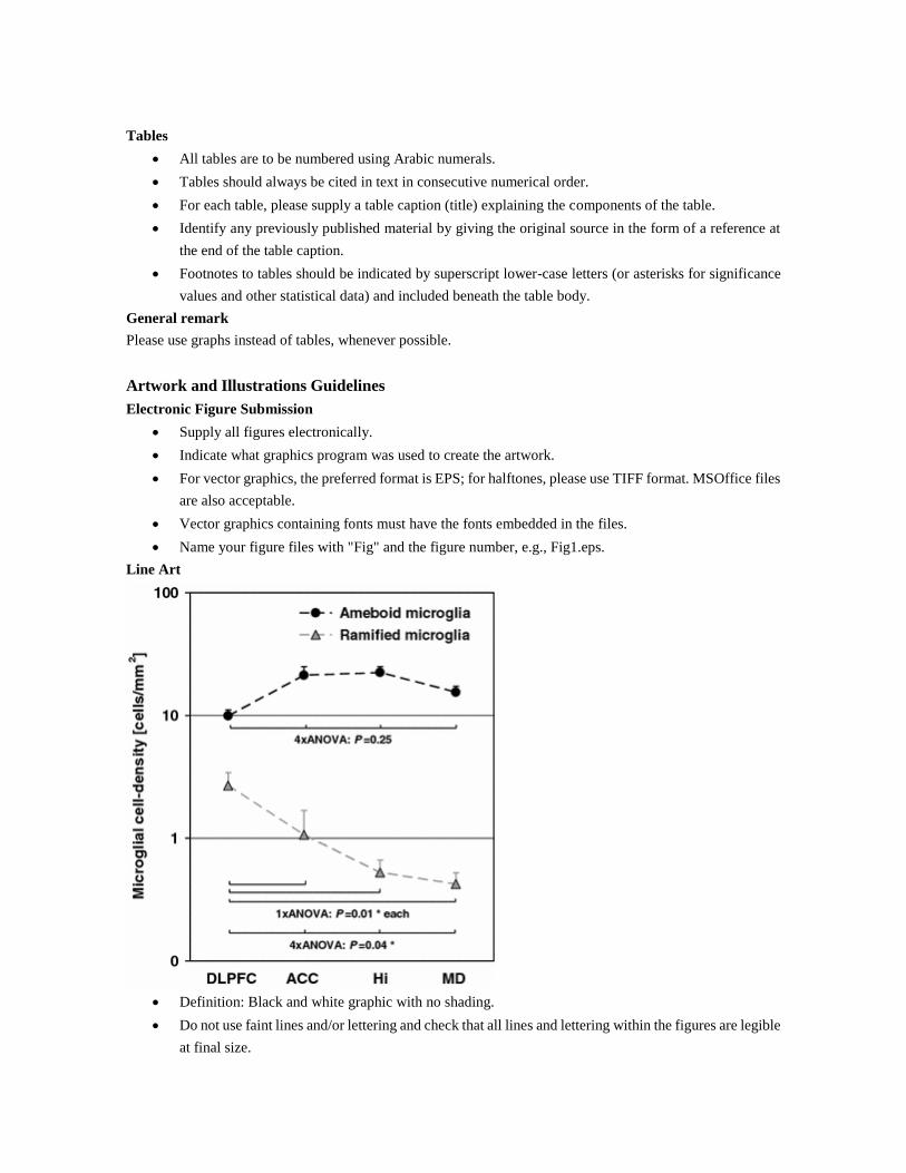
Line Art
Definition: Black and white graphic with no shading.
Do not use faint lines and/or lettering and check that all lines and lettering within the figures are legible
at final size.
All lines should be at least 0.1 mm (0.3 pt) wide.
Scanned line drawings and line drawings in bitmap format should have a minimum resolution of 1200
dpi.
Vector graphics containing fonts must have the fonts embedded in the files.
Halftone Art
Definition: Photographs, drawings, or paintings with fine shading, etc.
If any magnification is used in the photographs, indicate this by using scale bars within the figures
themselves.
Halftones should have a minimum resolution of 300 dpi.
Combination Art
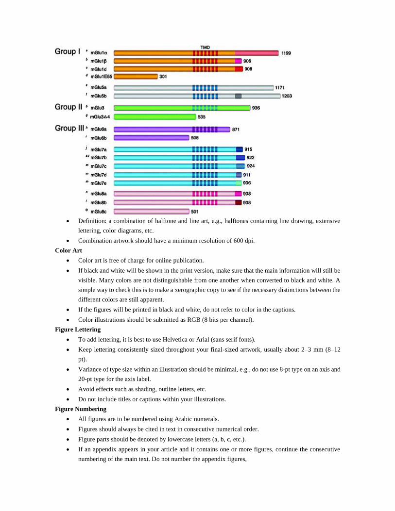
Definition: a combination of halftone and line art, e.g., halftones containing line drawing, extensive
lettering, color diagrams, etc.
Combination artwork should have a minimum resolution of 600 dpi.
Color Art
Color art is free of charge for online publication.
If black and white will be shown in the print version, make sure that the main information will still be
visible. Many colors are not distinguishable from one another when converted to black and white. A
simple way to check this is to make a xerographic copy to see if the necessary distinctions between the
different colors are still apparent.
If the figures will be printed in black and white, do not refer to color in the captions.
Color illustrations should be submitted as RGB (8 bits per channel).
Figure Lettering
To add lettering, it is best to use Helvetica or Arial (sans serif fonts).
Keep lettering consistently sized throughout your final-sized artwork, usually about 2–3 mm (8–12
pt).
Variance of type size within an illustration should be minimal, e.g., do not use 8-pt type on an axis and
20-pt type for the axis label.
Avoid effects such as shading, outline letters, etc.
Do not include titles or captions within your illustrations.
Figure Numbering
All figures are to be numbered using Arabic numerals.
Figures should always be cited in text in consecutive numerical order.
Figure parts should be denoted by lowercase letters (a, b, c, etc.).
If an appendix appears in your article and it contains one or more figures, continue the consecutive
numbering of the main text. Do not number the appendix figures,
"A1, A2, A3, etc." Figures in online appendices (Electronic Supplementary Material) should, however,
be numbered separately.
Figure Captions
Each figure should have a concise caption describing accurately what the figure depicts. Include the
captions in the text file of the manuscript, not in the figure file.
Figure captions begin with the term Fig. in bold type, followed by the figure number, also in bold type.
No punctuation is to be included after the number, nor is any punctuation to be placed at the end of the
caption.
Identify all elements found in the figure in the figure caption; and use boxes, circles, etc., as coordinate
points in graphs.
Identify previously published material by giving the original source in the form of a reference citation
at the end of the figure caption.
Figure Placement and Size
Figures should be submitted separately from the text, if possible.
When preparing your figures, size figures to fit in the column width.
For most journals the figures should be 39 mm, 84 mm, 129 mm, or 174 mm wide and not higher than
234 mm.
For books and book-sized journals, the figures should be 80 mm or 122 mm wide and not higher than
198 mm.
Permissions
If you include figures that have already been published elsewhere, you must obtain permission from the
copyright owner(s) for both the print and online format. Please be aware that some publishers do not grant
electronic rights for free and that Springer will not be able to refund any costs that may have occurred to receive
these permissions. In such cases, material from other sources should be used.
Accessibility
In order to give people of all abilities and disabilities access to the content of your figures, please make sure
that
All figures have descriptive captions (blind users could then use a text-to-speech software or a text-to-
Braille hardware)
Patterns are used instead of or in addition to colors for conveying information (colorblind users would
then be able to distinguish the visual elements)
Any figure lettering has a contrast ratio of at least 4.5:1
Electronic Supplementary Material
Springer accepts electronic multimedia files (animations, movies, audio, etc.) and other supplementary files to
be published online along with an article or a book chapter. This feature can add dimension to the author's
article, as certain information cannot be printed or is more convenient in electronic form.
Submission
Supply all supplementary material in standard file formats.
Please include in each file the following information: article title, journal name, author names;
affiliation and e-mail address of the corresponding author.
To accommodate user downloads, please keep in mind that larger-sized files may require very long
download times and that some users may experience other problems during downloading.
Audio, Video, and Animations
Aspect ratio: 16:9 or 4:3
Maximum file size: 25 GB
Minimum video duration: 1 sec
Supported file formats: avi, wmv, mp4, mov, m2p, mp2, mpg, mpeg, flv, mxf, mts, m4v, 3gp
Text and Presentations
Submit your material in PDF format; .doc or .ppt files are not suitable for long-term viability.
A collection of figures may also be combined in a PDF file.
Spreadsheets
Spreadsheets should be converted to PDF if no interaction with the data is intended.
If the readers should be encouraged to make their own calculations, spreadsheets should be submitted
as .xls files (MS Excel).
Specialized Formats
Specialized format such as .pdb (chemical), .wrl (VRML), .nb (Mathematica notebook), and .tex can
also be supplied.
Collecting Multiple Files
It is possible to collect multiple files in a .zip or .gz file.
Numbering
If supplying any supplementary material, the text must make specific mention of the material as a
citation, similar to that of figures and tables.
Refer to the supplementary files as “Online Resource”, e.g., "... as shown in the animation (Online
Resource 3)", “... additional data are given in Online Resource 4”.
Name the files consecutively, e.g. “ESM_3.mpg”, “ESM_4.pdf”.
Captions
For each supplementary material, please supply a concise caption describing the content of the file.
Processing of supplementary files
Electronic supplementary material will be published as received from the author without any
conversion, editing, or reformatting.
Accessibility
In order to give people of all abilities and disabilities access to the content of your supplementary files, please
make sure that
The manuscript contains a descriptive caption for each supplementary material
Video files do not contain anything that flashes more than three times per second (so that users prone
to seizures caused by such effects are not put at risk)
Ethical Responsibilities of Authors
This journal is committed to upholding the integrity of the scientific record. As a member of the Committee on
Publication Ethics (COPE) the journal will follow the COPE guidelines on how to deal with potential acts of
misconduct.

Authors should refrain from misrepresenting research results which could damage the trust in the journal, the
professionalism of scientific authorship, and ultimately the entire scientific endeavour. Maintaining integrity of
the research and its presentation can be achieved by following the rules of good scientific practice, which
include:
The manuscript has not been submitted to more than one journal for simultaneous consideration.
The manuscript has not been published previously (partly or in full), unless the new work concerns an
expansion of previous work (please provide transparency on the re-use of material to avoid the hint of
text-recycling (“self-plagiarism”)).
A single study is not split up into several parts to increase the quantity of submissions and submitted
to various journals or to one journal over time (e.g. “salami-publishing”).
No data have been fabricated or manipulated (including images) to support your conclusions
No data, text, or theories by others are presented as if they were the author’s own (“plagiarism”).
Proper acknowledgements to other works must be given (this includes material that is closely copied
(near verbatim), summarized and/or paraphrased), quotation marks are used for verbatim copying of
material, and permissions are secured for material that is copyrighted.
Important note: the journal may use software to screen for plagiarism.
Consent to submit has been received explicitly from all co-authors, as well as from the responsible
authorities - tacitly or explicitly - at the institute/organization where the work has been carried out,
before the work is submitted.
Authors whose names appear on the submission have contributed sufficiently to the scientific work
and therefore share collective responsibility and accountability for the results.
In addition:
Changes of authorship or in the order of authors are not accepted after acceptance of a manuscript.
Requesting to add or delete authors at revision stage, proof stage, or after publication is a serious matter
and may be considered when justifiably warranted. Justification for changes in authorship must be
compelling and may be considered only after receipt of written approval from all authors and a
convincing, detailed explanation about the role/deletion of the new/deleted author. In case of changes
at revision stage, a letter must accompany the revised manuscript. In case of changes after acceptance
or publication, the request and documentation must be sent via the Publisher to the Editor-in-Chief. In
all cases, further documentation may be required to support your request. The decision on accepting
the change rests with the Editor-in-Chief of the journal and may be turned down. Therefore authors
are strongly advised to ensure the correct author group, corresponding author, and order of authors at
submission.
Upon request authors should be prepared to send relevant documentation or data in order to verify the
validity of the results. This could be in the form of raw data, samples, records, etc.
If there is a suspicion of misconduct, the journal will carry out an investigation following the COPE guidelines.
If, after investigation, the allegation seems to raise valid concerns, the accused author will be contacted and
given an opportunity to address the issue. If misconduct has been established beyond reasonable doubt, this
may result in the Editor-in-Chief’s implementation of the following measures, including, but not limited to:
If the article is still under consideration, it may be rejected and returned to the author.
If the article has already been published online, depending on the nature and severity of the infraction,
either an erratum will be placed with the article or in severe cases complete retraction of the article
will occur. The reason must be given in the published erratum or retraction note.
The author’s institution may be informed.
Compliance with Ethical Standards
To ensure objectivity and transparency in research and to ensure that accepted principles of ethical and
professional conduct have been followed, authors should include information regarding sources of funding,
potential conflicts of interest (financial or non-financial), informed consent if the research involved human
participants, and a statement on welfare of animals if the research involved animals.
Authors should include the following statements (if applicable) in a separate section entitled “Compliance with
Ethical Standards” when submitting a paper:
Disclosure of potential conflicts of interest
Research involving Human Participants and/or Animals
Informed consent
Please note that standards could vary slightly per journal dependent on their peer review policies (i.e. single or
double blind peer review) as well as per journal subject discipline. Before submitting your article check the
instructions following this section carefully.
The corresponding author should be prepared to collect documentation of compliance with ethical standards
and send if requested during peer review or after publication.
The Editors reserve the right to reject manuscripts that do not comply with the above-mentioned guidelines.
The author will be held responsible for false statements or failure to fulfill the above-mentioned guidelines.
Disclosure of potential conflicts of interest
Authors must disclose all relationships or interests that could have direct or potential influence or impart bias
on the work. Although an author may not feel there is any conflict, disclosure of relationships and interests
provides a more complete and transparent process, leading to an accurate and objective assessment of the work.
Awareness of a real or perceived conflicts of interest is a perspective to which the readers are entitled. This is
not meant to imply that a financial relationship with an organization that sponsored the research or
compensation received for consultancy work is inappropriate. Examples of potential conflicts of interests that
are directly or indirectly related to the research may include but are not limited to the following:
Research grants from funding agencies (please give the research funder and the grant number)
Honoraria for speaking at symposia
Financial support for attending symposia
Financial support for educational programs
Employment or consultation
Support from a project sponsor
Position on advisory board or board of directors or other type of management relationships
Multiple affiliations
Financial relationships, for example equity ownership or investment interest
Intellectual property rights (e.g. patents, copyrights and royalties from such rights)
Holdings of spouse and/or children that may have financial interest in the work
In addition, interests that go beyond financial interests and compensation (non-financial interests) that may be
important to readers should be disclosed. These may include but are not limited to personal relationships or
competing interests directly or indirectly tied to this research, or professional interests or personal beliefs that
may influence your research.
The corresponding author collects the conflict of interest disclosure forms from all authors. In author
collaborations where formal agreements for representation allow it, it is sufficient for the corresponding author
to sign the disclosure form on behalf of all authors. Examples of forms can be found
here:
The corresponding author will include a summary statement in the text of the manuscript in a separate section
before the reference list, that reflects what is recorded in the potential conflict of interest disclosure form(s).
See below examples of disclosures:
Funding: This study was funded by X (grant number X).
Conflict of Interest: Author A has received research grants from Company A. Author B has received a speaker
honorarium from Company X and owns stock in Company Y. Author C is a member of committee Z.
If no conflict exists, the authors should state:
Conflict of Interest: The authors declare that they have no conflict of interest.
Research involving human participants and/or animals
1) Statement of human rights
When reporting studies that involve human participants, authors should include a statement that the studies have
been approved by the appropriate institutional and/or national research ethics committee and have been
performed in accordance with the ethical standards as laid down in the 1964 Declaration of Helsinki and its
later amendments or comparable ethical standards.
If doubt exists whether the research was conducted in accordance with the 1964 Helsinki Declaration or
comparable standards, the authors must explain the reasons for their approach, and demonstrate that the
independent ethics committee or institutional review board explicitly approved the doubtful aspects of the study.
The following statements should be included in the text before the References section:
Ethical approval: “All procedures performed in studies involving human participants were in accordance with
the ethical standards of the institutional and/or national research committee and with the 1964 Helsinki
declaration and its later amendments or comparable ethical standards.”
For retrospective studies, please add the following sentence:
“For this type of study formal consent is not required.”
2) Statement on the welfare of animals
The welfare of animals used for research must be respected. When reporting experiments on animals, authors
should indicate whether the international, national, and/or institutional guidelines for the care and use of animals
have been followed, and that the studies have been approved by a research ethics committee at the institution
or practice at which the studies were conducted (where such a committee exists).
For studies with animals, the following statement should be included in the text before the References section:
Ethical approval: “All applicable international, national, and/or institutional guidelines for the care and use of
animals were followed.”
If applicable (where such a committee exists): “All procedures performed in studies involving animals were in
accordance with the ethical standards of the institution or practice at which the studies were conducted.”
If articles do not contain studies with human participants or animals by any of the authors, please select one of
the following statements:
“This article does not contain any studies with human participants performed by any of the authors.”
“This article does not contain any studies with animals performed by any of the authors.”
“This article does not contain any studies with human participants or animals performed by any of the authors.”
Informed consent
All individuals have individual rights that are not to be infringed. Individual participants in studies have, for
example, the right to decide what happens to the (identifiable) personal data gathered, to what they have said
during a study or an interview, as well as to any photograph that was taken. Hence it is important that all
participants gave their informed consent in writing prior to inclusion in the study. Identifying details (names,
dates of birth, identity numbers and other information) of the participants that were studied should not be
published in written descriptions, photographs, and genetic profiles unless the information is essential for
scientific purposes and the participant (or parent or guardian if the participant is incapable) gave written
informed consent for publication. Complete anonymity is difficult to achieve in some cases, and informed
consent should be obtained if there is any doubt. For example, masking the eye region in photographs of
participants is inadequate protection of anonymity. If identifying characteristics are altered to protect
anonymity, such as in genetic profiles, authors should provide assurance that alterations do not distort scientific
meaning.
The following statement should be included:
Informed consent: “Informed consent was obtained from all individual participants included in the study.”
If identifying information about participants is available in the article, the following statement should be
included:
“Additional informed consent was obtained from all individual participants for whom identifying information
is included in this article.”
After acceptance
Upon acceptance of your article you will receive a link to the special Author Query Application at Springer’s
web page where you can sign the Copyright Transfer Statement online and indicate whether you wish to order
OpenChoice, offprints, or printing of figures in color.
Once the Author Query Application has been completed, your article will be processed and you will receive the
proofs.
Open Choice
In addition to the normal publication process (whereby an article is submitted to the journal and access to that
article is granted to customers who have purchased a subscription), Springer provides an alternative publishing
option: Springer Open Choice. A Springer Open Choice article receives all the benefits of a regular subscription-
based article, but in addition is made available publicly through Creative Commons Attribution-
NonCommercial 4.0 International License.
Springer Open Choice
Copyright transfer
Authors will be asked to transfer copyright of the article to the Publisher (or grant the Publisher exclusive
publication and dissemination rights). This will ensure the widest possible protection and dissemination of
information under copyright laws.
Open Choice articles do not require transfer of copyright as the copyright remains with the author. In opting for
open access, the author(s) agree to publish the article under the Creative Commons Attribution License.
Creative Commons Attribution-NonCommercial 4.0 International License
Offprints
Offprints can be ordered by the corresponding author.
Color illustrations
Online publication of color illustrations is free of charge. For color in the print version, authors will be expected
to make a contribution towards the extra costs.
Proof reading
The purpose of the proof is to check for typesetting or conversion errors and the completeness and accuracy of
the text, tables and figures. Substantial changes in content, e.g., new results, corrected values, title and
authorship, are not allowed without the approval of the Editor.
After online publication, further changes can only be made in the form of an Erratum, which will be hyperlinked
to the article.
Online First
The article will be published online after receipt of the corrected proofs. This is the official first publication
citable with the DOI. After release of the printed version, the paper can also be cited by issue and page numbers.
Does Springer provide English language support?
Manuscripts that are accepted for publication will be checked by our copyeditors for spelling and formal style.
This may not be sufficient if English is not your native language and substantial editing would be required. In
that case, you may want to have your manuscript edited by a native speaker prior to submission. A clear and
concise language will help editors and reviewers concentrate on the scientific content of your paper and thus
smooth the peer review process.
The following editing service provides language editing for scientific articles in all areas Springer
publishes in:
Edanz English editing for scientists
Use of an editing service is neither a requirement nor a guarantee of acceptance for publication.
Please contact the editing service directly to make arrangements for editing and payment.
For Authors from China
文 章在投稿前进行专业的语言润色将对作者的投稿进程有所帮助。作者可自愿选择使用Springer推荐
的编辑服务,使用与否并不作为判断文章是否被录用的依 据。提高文章的语言质量将有助于审稿人理
解文章的内容,通过对学术内容的判断来决定文章的取舍,而不会因为语言问题导致直接退稿。作者
需自行联系 Springer推荐的编辑服务公司,协商编辑事宜。
理文编辑
For Authors from Japan
ジャーナルに論文を投稿する前に、ネイティブ・スピーカーによる英文校閲を希望されている方に
は、Edanz社をご紹介しています。サービス内容、料金および申込方法など、日本語による詳しい
説明はエダンズグループジャパン株式会社の下記サイトをご覧ください。
エダンズグループジャパン
For Authors from Korea
영어 논문 투고에 앞서 원어민에게 영문 교정을 받고자 하시는 분들께 Edanz 회사를 소개해 드립니다.
서비스 내용, 가격 및

신청 방법 등에 대한 자세한 사항은 저희 Edanz Editing Global 웹사이트를 참조해 주시면
감사하겠습니다.
Edanz Editing Global