Amanda Bergmann da Fonseca -...
53
Amanda Bergmann da Fonseca Prótese oculopalpebral- Relato de caso clínico Brasília 2015
Transcript of Amanda Bergmann da Fonseca -...

Amanda Bergmann da Fonseca
Prótese oculopalpebral- Relato de caso clínico
Brasília
2015

Amanda Bergmann da Fonseca
Prótese oculopalpebral- Relato de caso clínico
Trabalho de Conclusão de Curso apresentado ao
Departamento de Odontologia da Faculdade de
Ciências da Saúde da Universidade de Brasília,
como requisito parcial para a conclusão do curso de
Graduação em Odontologia.
Orientadora: Profa. Dra. Aline Úrsula R. Fernandes
Brasília
2015

À minha família e amigos.

AGRADECIMENTOS
A Deus, por ter me dado saúde e força para superar as
dificuldades e alcançar objetivos.
Aos meus pais, Emídio Prata e Simone Bergmann, e meu irmão
Victor Bergmann, pelo amor, incentivo e apoio incondicional,
dados a mim até aqui.
Aos meus avós, Maximino Bergmann, Myrian Nunes, Murilo
Prata e Jupira Fonseca, por seus exemplos de força,
simplicidade e amor, que me tornam uma pessoa mais humana e
perseverante.
Aos meus amigos, Clovis Moura, Isabela Fernandes, Fabiana
Dantas, Marina Assunção, Ana Bauer, Gabriela Vilmondes, Paula
Araújo e Camila Wells, que sempre me apoiaram e despertaram
em mim coragem para seguir em frente, me ensinando a focar,
dedicando tempo e esforços ao que eu realmente gosto.
Agradeço também a minha professora e orientadora Aline Úrsula,
por toda paciência e dedicação para me acompanhar durante
todo o curso e em especial durante a realização deste trabalho.

EPÍGRAFE
“Que os vossos esforços desafiem as impossibilidades,
lembrai-vos de que as grandes coisas do homem foram
conquistadas do que parecia impossível”.
Charles Chaplin

RESUMO
FONSECA, Amanda Bergmann da. Prótese oculopalpebral-
Relato de caso clínico. 2015. Trabalho de Conclusão de Curso
(Graduação em Odontologia) – Departamento de Odontologia da
Faculdade de Ciências da Saúde da Universidade de Brasília.
A reabilitação por prótese oculopalpebral envolve questões
físicas e psicológicas. Com a perda de estrutura da região orbital,
o paciente sofre por comprometimento de função, desfiguração
facial e transtornos psicológicos. Este trabalho apresenta o caso
clínico da reabilitação facial de uma paciente acometida por
carcinoma em região orbital esquerda, submetida à ressecção
cirúrgica do bulbo ocular e estruturas adjacentes, com posterior
enxerto epitelial. Após período de cicatrização dos procedimentos
cirúrgicos, foi realizada uma moldagem facial e obtida uma
máscara em gesso. O padrão em cera da prótese oculopalpebral
foi esculpido e analisado em posição quanto a aspectos estéticos
e adaptação às margens do defeito cirúrgico. A prótese foi
pigmentada, em silicone industrial, e caracterizada para
aproximar-se à coloração e textura facial da paciente,
Concomitante, foi confeccionada prótese ocular em resina
acrílica, para que completasse a região. A reabilitação facial, por
meio de prótese oculopalpebral, foi responsável pelo
restabelecimento da harmonia e estética almejadas, e pela
melhoria na qualidade de vida da paciente, reinserida em
sociedade. A protetização facial é a melhor opção para
reabilitação de pacientes mutilados, contra-indicados para a
cirurgia plástica reconstrutiva, alcançando resultados bastante
satisfatórios.
ABSTRACT
FONSECA, Amanda Bergmann da. Oculoplpebral Prosthesis: A
Clinic Report. 2015. Undergraduate Course Final Monograph
(Undergraduate Course in Dentistry) – Department of Dentistry,
School of Health Sciences, University of Brasília.
Rehabilitation through oculopalpebral prosthesis involves physical
and psychological issues. With the loss of the orbital region
structure, the patient suffers function impairment, facial
disfigurement and psychological disorders. This paper presents a
case of facial rehabilitation of a patient affected by carcinoma in
the left orbital region , underwent surgical resection of the eyeball
and adjacent structures , with subsequent epithelial graft. After
healing period of surgical procedures, facial molding was
performed and obtained a mask in plaster. The standard wax
oculopalpebral prosthesis has been carved and analyzed in
position on aesthetics and adaptation to the banks of the surgical
defect. The prosthesis was pigmented in industrial silicone, and
characterized to approach the color and texture of facial patient,
Concurrent, was made ocular prosthesis in acrylic resin, to
complete the region. The facial rehabilitation through
oculopalpebral prosthesis, was responsible for the restoration of
harmony and aesthetics, and the improvement in patient quality of
life, reinserted in society. The facial prosthesis is the best option
for rehabilitation of mutilated patients contraindicated for
reconstructive plastic surgery, achieving satisfactory results.

SUMÁRIO
Artigo Científico ........................................................................... 09 Folha de Título ........................................................................ 10 Resumo ................................................................................... 11 Abstract ................................................................................... 12 Introdução ............................................................................... 14 Relato de caso ........................................................................ 21 Discussão ................................................................................ 31 Conclusão ............................................................................... 34 Referências ............................................................................. 35
Anexos ......................................................................................... 36
Normas da Revista .................................................................. 36

9
ARTIGO CIENTÍFICO
Este trabalho de Conclusão de Curso é baseado no artigo
científico:
FONSECA, Amanda Bergmann da; FERNANDES, Aline Úrsula
Rocha. Prótese oculopalpebral- Relato de caso clínico.
Apresentado sob as normas de publicação do National Journal of Maxillofacial Surgery

10
FOLHA DE TÍTULO
Prótese oculopalpebral- Relato de caso clínico
Oculopalpebral Prosthesis: A Clinic Report
Amanda Bergmann da Fonseca1
Aline Úrsula Rocha Fernandes2
1 Aluna de Graduação em Odontologia da Universidade de
Brasília. 2 Professora Adjunta de Prótese Dentária da Universidade de
Brasília (UnB).
Correspondência: Profa. Dra. Aline Úrsula Rocha Fernandes
Campus Universitário Darcy Ribeiro - UnB - Faculdade de
Ciências da Saúde - Departamento de Odontologia - 70910-900 -
Asa Norte - Brasília - DF
E-mail: [email protected] / [email protected] Telefone: (61) 31071811

11
RESUMO
Prótese oculopalpebral- Relato de caso clínico
Resumo
A reabilitação por prótese oculopalpebral envolve questões
físicas e psicológicas. Com a perda de estrutura da região orbital,
o paciente sofre por comprometimento de função, desfiguração
facial e transtornos psicológicos. Este trabalho apresenta o caso
clínico da reabilitação facial de uma paciente acometida por
carcinoma basocelualr em região orbital esquerda, submetida à
ressecção cirúrgica do bulbo ocular e estruturas adjacentes, com
posterior enxerto epitelial. Após período de cicatrização dos
procedimentos cirúrgicos, foi realizada uma moldagem facial e
obtida uma máscara em gesso. O padrão em cera da prótese
oculopalpebral foi esculpido e analisado em posição quanto a
aspectos estéticos e adaptação às margens do defeito cirúrgico.
A prótese foi pigmentada, em silicone industrial, e caracterizada
para aproximar-se à coloração e textura facial da paciente,
Concomitante, foi confeccionada prótese ocular em resina
acrílica, para que completasse a região. A reabilitação facial, por
meio de prótese oculopalpebral, foi responsável pelo
restabelecimento da harmonia e estética almejadas, e pela
melhoria na qualidade de vida da paciente, reinserida em
sociedade. A protetização facial é a melhor opção para
reabilitação de pacientes mutilados, contraindicados para a
cirurgia plástica reconstrutiva, alcançando resultados bastante
satisfatórios.
Palavras-chave
Carcinoma Basocelular; Prótese maxilofacial; Olho artificial
12
ABSTRACT
Oculopalpebral Prosthesis: A Clinic Report
Abstract
Rehabilitation through oculopalpebral prosthesis involves
physical and psychological issues. With the loss of the
orbital region structure, the patient suffers function
impairment, facial disfigurement and psychological
disorders. This paper presents a case of facial rehabilitation
of a patient affected by carcinoma in the left orbital region,
underwent surgical resection of the eyeball and adjacent
structures, with subsequent epithelial graft, after healing
period of surgical procedures, facial molding was performed
and obtained a mask in plaster. The standard
oculopalpebral prosthesis has been carved and analyzed in
position on aesthetics and adaptation to the banks of the
surgical defect. The prosthesis was pigmented in industrial
silicone, and characterized to approach the color and
texture of facial patient, Concurrent, was made ocular
prosthesis in acrylic resin, to complete the region. The
facial rehabilitation through oculopalpebral prosthesis, was
responsible for the restoration of harmony and aesthetics,
and the improvement in patient quality of life, reinserted in
society. The facial prosthesis is the best option for
rehabilitation of mutilated patients contraindicated for
reconstructive plastic surgery, achieving satisfactory
results.

13
Keywords
Carcinoma, Basal Cell; Maxillofacial Prosthesis; Eye, artificial

14
INTRODUÇÃO
Câncer de cabeça e pescoço é um termo genérico
que se refere a um grupo de tumores malignos que
ocorrem em regiões anatômicas da cabeça e do pescoço.
Os tumores palpebrais são divididos em benignos e
malignos, os benignos são os mais comuns, não possuem
ulcerações nem sangramento, e possuem um crescimento
lento, dentre eles o mais comum é o papiloma, já os
tumores malignos possuem ulcerações, sangramento,
vasos sanguíneos em suas extremidades, podem causar a
perda dos cílios na região acometida, sendo o carcinoma
basocelular o mais comum dentre eles, ocorrendo em
cerca de 90% dos casos relatados, segundo estudos.
Podendo acometer a pálpebra inferior, canto medial,
pálpebra superior e canto lateral, respectivamente. A
15
prevalência dos carcinomas basocelulares ocorre entre
pacientes do sexo feminino, de 50 a 70 anos e de pele
branca. (6) Os fatores de risco envolvem a exposição
excessiva a radiação ultravioleta, histórico familiar
positivo, ingestão deficiente de vitamina, dieta rica em
gorduras e genodermatoses, como o albinismo. (7) O
diagnóstico do carcinoma basocelular é dado a partir de
análise clínica, e anatomopatológica, podendo ser
classificada clinicamente, em nodular, nódulo ulcerativo,
pigmentado e infiltrativo, apesar de não causar metástase
estes tumores podendo causar infiltração local, portanto
a detecção precoce aumenta as chances de cura. (6) O
tratamento pode ser escolhido de acordo com a extensão
da lesão compreendendo cirurgias com excisão total de
tumor com margens de segurança, curetagem e

16
cauterização (tumores pequenos), terapia fotodinâmica, e
radioterapia. (7)
As mutilações faciais são, na maioria das vezes,
de etiologia patológica ou acidental. No caso de
neoplasias malignas envolvendo a face, muitas vezes a
reabilitação completa não é possível apenas com
recursos cirúrgicos, sendo necessária a utilização de
recursos protéticos. A prótese bucomaxilofacial exerce o
papel de reabilitar esses pacientes, tornando-os mais
aptos ao convívio social. Indivíduos que após a
mutilação frequentemente eram acometidos por
sentimentos de vergonha e depressão que os levavam ao
isolamento social, muitas vezes conseguem uma
adequada reabilitação com o uso de próteses. (2)
O tratamento cirúrgico é necessário para a cura
do tumor, porém a reconstrução facial é condicionada ao

17
estado fisiológico e patológico dos pacientes. Alguns
trabalhos demonstram que a satisfação dos pacientes
submetidos apenas ao tratamento cirúrgico após
rinectomia é baixa. A maioria desses pacientes não estão
satisfeitos com sua aparência e todos eles têm alguma
reclamação estética a respeito da área acometida. (1)
Para a escolha entre a opção cirúrgica e a
protética, no caso de mutilações faciais, devem ser
considerados também os seguintes aspectos: quantidade
de tecido de suporte remanescente, número, condição e
posição dos dentes remanescentes, idade e estado de
saúde do paciente, achados patológicos e habilidades
disponíveis tanto para a reconstrução cirúrgica quanto
para a protética. A reconstrução cirúrgica e a reabilitação
protética podem ser usadas em conjunto quando
nenhuma das duas opções isoladas alcança a máxima

18
estética e função. O tratamento e a reabilitação de
pacientes com neoplasias da face devem ser planejados
por uma equipe antes da cirurgia de retirada do tumor.
Isso determinará a escolha entre reabilitação cirúrgica ou
protética e permitirá a otimização dos resultados
funcionais e estéticos. (3)
A decisão deve considerar também os desejos do
paciente e de sua família. Se o defeito é pequeno, a
reconstrução cirúrgica é preferível. No entanto, é
praticamente impossível reconstruir cirurgicamente as
regiões anatômicas com um aspecto estético tão bom
quanto de uma prótese. A reconstrução é um desafio
técnico muito grande e requer múltiplas cirurgias. A
restauração da função do órgão através de cirurgia é
muitas vezes limitada e imprevisível. No caso da prótese,
normalmente ocorre uma boa aceitação por parte do

19
paciente quando sua família apoia e aceita essa opção. O
paciente deve ser avaliado em relação aos fatores sociais
e psicológicos, ao passo que deve estar consciente de que
sua aparência mudou permanentemente. (3)
Muitas vezes a cirurgia plástica não é capaz de
restaurar o volume total do nariz. Nesses casos uma
prótese nasal é estética e permite uma respiração
adequada. As próteses, além de permitirem ao clínico
observar a cura ou recorrência da doença, possuem um
grau de complexidade diferente das cirurgias
reconstrutivas e podem apresentar menor custo em
relação as mesmas. Existem três opções para retenção de
próteses faciais: suporte mecânico, adesivo ou
ancoragem em implantes crânio- faciais. (1)
Para a confecção das próteses bucomaxilofacias
podem ser utilizados materiais como

20
polimetilmetacrilato, polivinil, poliuretano e silicone. O
silicone é o material mais usado, devido a suas
propriedades superiores. (2) A reabilitação do paciente
com deformidade facial objetiva não somente a
restauração da estética, bem como seu conforto físico,
psicológico e social.
O objetivo deste trabalho foi relatar a reabilitação
facial de paciente com perda oculopalpebral, por meio de
próteses maxilofaciais.

21
RELATO DE CASO
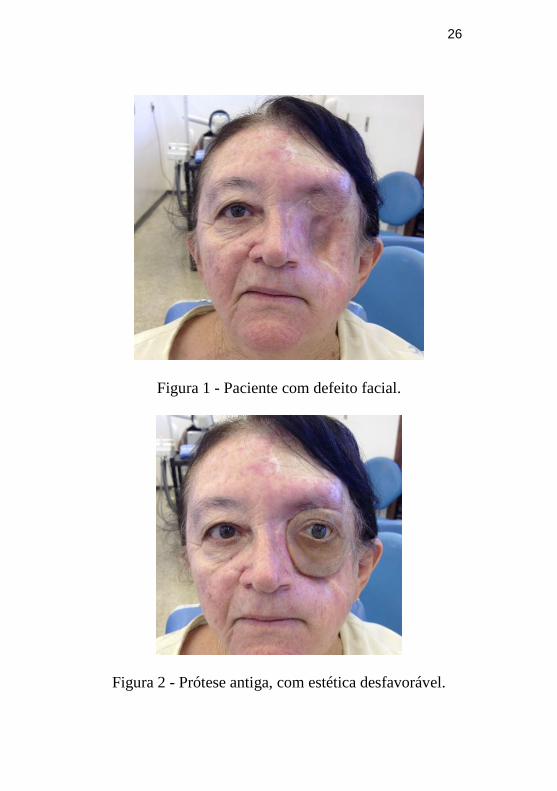
Paciente feminino, M.A.C, 66 anos, que
compareceu à Clínica Odontológica do HUB, com o
objetivo de obter reabilitação facial estética. A paciente
relatou ter sido acometida por Carcinoma Basocelular,
sendo submetida a sessões de radioterapia e alguns
procedimentos cirúrgicos, nos quais o tumor foi
exenterado e enxerto autógeno de tecido abdominal foi
posicionado para fechar a cavidade, localizada na região
orbital esquerda com ressecção de bulbo ocular e
estruturas adjacentes (Figura 1). Além do
comprometimento estético, a paciente apresentava um
quadro de baixa autoestima e dificuldades na
socialização. A paciente faz o uso de prótese
oculopalpebral desde 2003, sendo a última usada há 2
anos (Figura 2).

22
Em vista do caso e da impossibilidade em
restabelecimento estético por meio cirúrgico, foi
proposto como plano de tratamento a confecção de
prótese oculopalpebral em silicone (2), que estaria retida
à face por meio de retenção adesiva.
Diante do proposto, realizou-se inicialmente a
moldagem da face da paciente com hidrocolóide
irreversível (Jeltrate Plus; Dentsply Ind. Com. Ltda,
Brasil), utilizando uma moldeira individual de gesso
comum (Gesso-Rio; Brasil). A partir do molde, obteve-
se uma máscara facial com gesso pedra tipo III, sobre o
qual foi realizado todo trabalho de escultura da peça
protética (Figura 3). Essa foi confeccionada em cera rosa
n° 7 (Wilson Polidental Ind. Com. Ltda, Brasil), sendo
esculpidas as formas anatômicas de interesse.

23
A prova do padrão em cera foi realizada
observando-se todos os requisitos estéticos e funcionais,
avaliando a boa adaptação de bordas, estética e harmonia
faciais. A prótese palpebral em cera foi incluída em
mufla, e seu molde preenchido com silicone incolor
(Silastic, Dow Corning do Brasil) pigmentado com
pigmentos de cerâmica e pós de maquiagem (Figura 4).
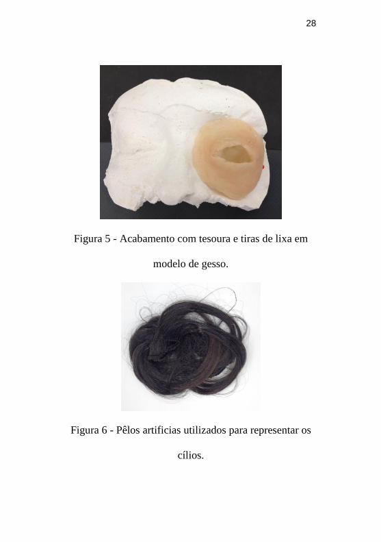
Após 24 horas, a prótese foi retirada da mufla, sofreu
acabamento com tesoura e tiras de lixa (Figura 5).
A prótese ocular foi confeccionada
simultaneamente, em processo separado (2, 3). A esclera
artificial foi preparada em resina acrílica
termopolimerizável branca (n. 1, Produtos
Odontológicos Clássico Ltda, Brasil). A íris artificial foi
pintada sobre disco de cartolina preta, com tintas a óleo
(Gato Preto, Brasil), e colada sobre a esclera artificial.

24
Após caracterização com fios de lã e pigmentos
resinosos, foi depositada camada de resina acrílica
incolor termopolimerizável (Produtos Odontológicos
Clássico Ltda, Brasil) sobre a face estética da prótese. O
acabamento e polimento foram obtidos por broca de
tungstênio e lixas de diferentes granulações. A fixação
da prótese ocular à palpebral foi realizada com o mesmo
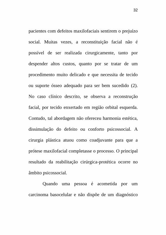
silicone industrial da confecção da última. Pêlos
artificiais foram costurados, para representar os cílios
(Figura 6).
No momento da instalação da prótese
oculopalpebral, esta foi fixada por meio de adesivo
químico (Pros-Aide Adhesive, EUA) (Figura 7).
Após a instalação da prótese, testes funcionais
foram realizados e observou-se que a retenção e
estabilidade estavam adequadas, além de uma adaptação

25
satisfatória das bordas da prótese facial com a face da
paciente (Figura 8). A reabilitação facial por meio de
prótese oculopalpebral foi responsável pelo
restabelecimento da harmonia e estética almejadas, e
pela melhoria na qualidade de vida da paciente,
reinserida em sociedade (Figura 9).
26
Figura 1 - Paciente com defeito facial.
Figura 2 - Prótese antiga, com estética desfavorável.

27
Figura 3 - Modelo em gesso para confecção de prótese
oculopalpebral.
Figura 4 - Silicone incolor e pigmentos.
28
Figura 5 - Acabamento com tesoura e tiras de lixa em
modelo de gesso.
Figura 6 - Pêlos artificias utilizados para representar os
cílios.

29
Figura 7 - Adesivo químico para fixação da prótese.
Figura 8 - Prova e ajustes finais da prótese.

30
Figura 9 - Paciente com prótese instalada.

31
DISCUSSÃO
Segundo Rezende (4), prótese oculopalpebral
trata-se de uma restauração aloplásica do olho e da
região palpebral, se diferencia da prótese oftálmica por
não substituir apenas o globo ocular e sim o conjunto
oculopalpebral, normalmente com etiologia patológica
que levam a exéreses oncocirúrgicas, deixando assim um
aspecto de mutilação, gerando a necessidade de uma
intervenção terapêutica para proteção de tecidos
adjacentes bem como a reinclusão do paciente na
sociedade.
Pacientes que possuem essas condições relatam
que a aceitação social é muito difícil, principalmente por
parte das crianças que enxergam a deformidade como
uma aberração. Os olhares e comentários
discriminatórios causam desconforto e permitem aos
32
pacientes com defeitos maxilofaciais sentirem o prejuízo
social. Muitas vezes, a reconstituição facial não é
possível de ser realizada cirurgicamente, tanto por
despender altos custos, quanto por se tratar de um
procedimento muito delicado e que necessita de tecido
ou suporte ósseo adequado para ser bem sucedido (2).
No caso clínico descrito, se observa a reconstrução
facial, por tecido enxertado em região orbital esquerda.
Contudo, tal abordagem não ofereceu harmonia estética,
dissimulação do defeito ou conforto psicossocial. A
cirurgia plástica atuou como coadjuvante para que a
prótese maxilofacial completasse o processo. O principal
resultado da reabilitação cirúrgica-protética ocorre no
âmbito psicossocial.
Quando uma pessoa é acometida por um
carcinoma basocelular e não dispõe de um diagnóstico

33
precoce, compromete muito sua estética, devido às
proporções que a lesão pode tomar (6), uma vez que o
tratamento indicado para esse tipo de lesão é a exérese
total do tumor com margem de segurança de 3 a 5 mm
em toda sua extensão (7), a mesma deve passar por um
intensivo acompanhamento psicológico para que tenha
consciência de que sua aparência permanecerá alterada
para o resto de sua vida (3), porém este processo requer
muito esforço e tolerância. O profissional de saúde, em
sua possibilidade de integração multidisciplinar, tem
fundamental importância no processo de resgate desses
pacientes.

34
CONCLUSÃO
A reabilitação com o uso de próteses
oculopalpebrais é de extrema importância para uma
reintegração social, melhoria na qualidade de vida e
restabelecimento psicológico.
35
Referências
1. Ciocca L, Maremonti P, Bianchi B, Scotti R.
Maxillofacial rehabilitation after rhinectomy using two different treatment options: clinical reports. J Oral Rehabil. 2007; 34(4):311-5.
2. Goiato MC, Fernandes AÚR, dos Santos DM, Barão
VAR. Positioning Magnets on a Multiple/Sectional
Maxillofacial Prosthesis. J Contemp Dent Pract 2007
November; (8)7:101-107.
3. Sharma N, Thakral GK, Mohapatra A, Seth J, Vashisht
PA. simplified technique for fabrication of orbital
prosthesis. J Clin Diagn Res. 2014 Jun;8(6):ZD10-2.
4. Rezende JRV. Fundamentos da prótese bucomaxilofacial. 1. ed. São Paulo: Editora Sarvier, 1997.
5. Veerareddy C, Nair C, Reddy GR. Simplified technique for fabrication of orbital prosthesis. Jornal of prosthodontics (21) 2012 561-568.
6. Narikawa S, Padovani CR, Schellini SA. Frequency of occurrence of eyelid basal cell carcinoma in the centralwest region of São Paulo State and carriers characteristics. Arq Bras Oftalmol 2011; 74 (4):245-7.
7. Brooke R. Basal cell carcinoma. Clin Med. 2005;5(6):551-4.
8. Soares EH, Belo CV, Reis AK, Nunes RR, Mason EM. Tumores malignos da pálpebra. Arq Bras Oftalmol. 2001;64(4):287-9.

36
ANEXOS
NORMAS DA REVISTA
Instructions to the Authors General | About Journal | Permissions | The Editorial Process | Clinical trial registry | Authorship Criteria | Conflicts of Interest/ Competing Interests | Submission of Manuscripts| Preparation of Manuscripts| Reprint and Proofs| Copyrights || Contributors' form
General There are no page charges for submissions to the journal however author is required to subscribe to the journal before his/her article is put under consideration for publication. Please check http://www.njms.in/contributors.asp for details. All manuscripts must be submitted online at www.journalonweb.com/njms.. It is mandatory for all authors and co-authors of the article to subscribe to the journal atleast for one year as a prerequisite for their article to be considered for publication.
About Journal National Journal of Maxillofacial Surgery (ISSN: Print - 0975-5950) is peer-reviewed journal. The Journal is Official Publication of Maxillofacial Society of India. The journal publishes articles on the subject of maxillofacial surgery, oral surgery, plastic surgery, radiology, pathology and related subjects. The Journal is published in January and July. It is mandatory for all authors and co-authors of the article to subscribe to the journal atleast for one year as a prerequisite for their article to be considered for publication.
The Editorial Process A manuscript will be reviewed for possible publication with the understanding that it is being submitted to National Journal of

37
Maxillofacial Surgery (NJMS) alone at that point in time and has not been published anywhere, simultaneously submitted, or already accepted for publication elsewhere. The journal expects that authors would authorize one of them to correspond with the Journal for all matters related to the manuscript. All manuscripts received are duly acknowledged. On submission, editor review all submitted manuscripts initially for suitability for formal review. Manuscripts with insufficient originality, serious scientific or technical flaws, or lack of a significant message are rejected before proceeding for formal peer-review. Manuscripts that are unlikely to be of interest to the National Journal of Maxillofacial Surgery (NJMS) readers are also liable to be rejected at this stage itself. Manuscripts that are found suitable for publication in National Journal of Maxillofacial Surgery (NJMS) are sent to two or more expert reviewers. During submission, the contributor is requested to provide names of two or three qualified reviewers who have had experience in the subject of the submitted manuscript, but this is not mandatory. The reviewers should not be affiliated with the same institutes as the contributor/s. However, the selection of these reviewers is at the sole discretion of the editor. The journal follows a double-blind review process, wherein the reviewers and authors are unaware of each other‟s identity. Every manuscript is finally reviewed by the Editor of the journal, who based on the comments from the reviewers takes a final decision on the manuscript. The comments and suggestions (acceptance/ rejection/ amendments in manuscript) received from reviewers are conveyed to the corresponding author. If required, the author is requested to provide a point by point response to reviewers‟ comments and submit a revised version of the manuscript. This process is repeated till reviewers and editors are satisfied with the manuscript. Manuscripts accepted for publication are copy edited for grammar, punctuation, print style, and format. Page proofs are sent to the corresponding author. The corresponding author is expected to return the corrected proofs within three days. It may not be possible to incorporate corrections received after that period. The whole process of submission of the manuscript to final decision and sending and receiving proofs is completed online. To achieve faster and greater dissemination of knowledge and information, the journal publishes articles online as „Ahead of Print‟ immediately on acceptance.
Clinical trial registry National Journal of Maxillofacial Surgery (NJMS) favors registration of clinical trials and is a signatory to the Statement on publishing

38
clinical trials in Indian research journals. Registration in the following trial registers is acceptable: http://www.ctri.in/ The trials conducted outside India may be registered with any other clinical trial registry.
Authorship Criteria Authorship credit should be based only on substantial contributions to each of the three components mentioned below: Concept and design of study or acquisition of data or analysis and interpretation of data; Drafting the article or revising it critically for important intellectual content; and Final approval of the version to be published. Participation solely in the acquisition of funding or the collection of data does not justify authorship. General supervision of the research group is not sufficient for authorship. Each contributor should have participated sufficiently in the work to take public responsibility for appropriate portions of the content of the manuscript. The order of naming the contributors should be based on the relative contribution of the contributor towards the study and writing the manuscript. Once submitted the order cannot be changed without written consent of all the contributors. The journal prescribes a maximum number of six (06) authors for manuscripts depending upon the type of manuscript, its scope and number of institutions involved (vide infra). The authors should provide a justification, if the number of authors exceeds these limits.
Conflicts of Interest/ Competing Interests All authors of submitting manuscript to the journal must disclose any and all conflicts of interest they may have with publication of the manuscript or an institution or product that is mentioned in the manuscript and/or is important to the outcome of the study presented. Authors should also disclose conflict of interest with products that compete with those mentioned in their manuscript.
Submission of Manuscripts: All manuscripts must be submitted on-line through the website http://www.journalonweb.com/njms. First time users will have to register at this site. Registration is free but mandatory. Registered authors can keep track of their articles after logging into the site using their user name and password. The submitted manuscripts that are not as per the “Instructions to Authors” would be returned to the authors for technical correction, before they undergo editorial/ peer-review. Generally, the manuscript should be submitted in the form of two separate files: [1] Title Page/First Page File/covering letter:
39
This file should provide a. The type of manuscript (original article, case report, review
article, short communications, Ethics Forum, Education Forum, Letter to editor, Images, etc.) title of the manuscript, running title, names of all authors/ contributors (with their highest academic degrees, designation and affiliations) and name(s) of department(s) and/ or institution(s) to which the work should be credited. All information which can reveal your identity should be here. Use text/rtf/doc files. Do not zip the files.
b. The total number of pages, total number of photographs and word counts separately for abstract and for the text (excluding the references, tables and abstract), word counts for introduction + discussion in case of an original article;
c. Source(s) of support in the form of grants, equipment, drugs, or all of these;
d. Acknowledgement, if any. One or more statements should specify 1) contributions that need acknowledging but do not justify authorship, such as general support by a departmental chair; 2) acknowledgments of technical help; and 3) acknowledgments of financial and material support, which should specify the nature of the support. This should be included in the title page of the manuscript and not in the main article file.
e. If the manuscript was presented as part at a meeting, the organization, place, and exact date on which it was read. A full statement to the editor about all submissions and previous reports that might be regarded as redundant publication of the same or very similar work. Any such work should be referred to specifically, and referenced in the new paper. Copies of such material should be included with the submitted paper, to help the editor decide how to handle the matter.
f. Registration number in case of a clinical trial and where it is registered (name of the registry and its URL)
g. Conflicts of Interest of each author/ contributor. A statement of financial or other relationships that might lead to a conflict of interest, if that information is not included in the manuscript itself or in an authors' form
h. Criteria for inclusion in the authors‟/ contributors‟ list i. A statement that the manuscript has been read and
approved by all the authors, that the requirements for authorship as stated earlier in this document have been met,

40
and that each author believes that the manuscript represents honest work, if that information is not provided in another form (see below); and
j. The name, address, e-mail, and telephone number of the corresponding author, who is responsible for communicating with the other authors about revisions and final approval of the proofs, if that information is not included on the manuscript itself.
[2] Blinded Article file: The manuscript must not contain any mention of the authors' names or initials or the institution at which the study was done or acknowledgements. Page headers/running title can include the title but not the authors' names. Manuscripts not in compliance with The Journal's blinding policy will be returned to the corresponding author. The main text of the article, beginning from Abstract till References (including tables) should be in this file. Use rtf/doc files. Do not zip the files. Do not incorporate images in the file. If file size is large, graphs can be submitted as images separately without incorporating them in the article file to reduce the size of the file. The pages should be numbered consecutively, beginning with the first page of the blinded article file. [3] Images: Submit good quality color images. Each image should be less than 1024 kb (1 MB) in size. Size of the image can be reduced by decreasing the actual height and width of the images (keep up to 1240 x 800 pixels or 5-6 inches). Images can be submitted as jpeg files. Do not zip the files. Legends for the figures/images should be included at the end of the article file. [4] The contributors' / copyright transfer form (template provided below) has to be submitted in original with the signatures of all the contributors within two weeks of submission via courier, fax or email editor @ NJMS . org as a scanned image. Print ready hard copies of the images (one set) or digital images should be sent to the journal office at the time of submitting revised manuscript. High resolution images (up to 5 MB each) can be sent by email on editor @ NJMS . org The hard copies of the Contributors‟ form / copyright transfer form may be sent to the following addresses or submitted online from the authors‟ area on http://www.journalonweb.com/njms
Preparation of Manuscripts Manuscripts must be prepared in accordance with "Uniform requirements for Manuscripts submitted to Biomedical Journals" developed by the International Committee of Medical Journal Editors (October 2006). The uniform requirements and specific requirement of National Journal of Maxillofacial Surgery (NJMS) are summarized below. Before submitting a manuscript, contributors are requested to

41
check for the latest instructions available. Instructions are also available from the website of the journal http://www.njms.in and from the manuscript submission site (http://www.journalonweb.com/njms). The manuscript should be typed, double-spaced on standard-sized-paper (8.5" x 11") with 1" margins on all sides. Times New Roman font 12 pt should be used. The fonts used in the text as well as graphics should be restricted to Times New Roman, Symbol and Zapf Dingbats. Title: Should be in Title Case; the first character in each word in the title has to be capitalized. A research paper typically should include the following in the order given below: Abstract Keywords Introduction Materials and Methods Results including Tables and/or Figures Discussion Conclusion Acknowledgements (if any) References Appendixes (if necessary) Abbreviations used (if necessary) Abstract Should be structured and limited to 250 Words. A brief summary of the research should be given under the subheadings Introduction, Methods, Results, and Conclusions. Key words No more than six keywords are needed. Words appearing in the title should not be given as keywords. It is desirable to include the alternative words, if any under keywords e.g. the word „famotidine‟. They should be written left aligned, arranged alphabetically in 12pt Times Roman, and the line must begin with the words Keywords boldfaced. A 12pt space should separate the keywords from the affiliations. Introduction Description of the research area, pertinent background information, and the hypotheses tested in the study should be included under this section. The introduction should provide sufficient background information such that a scientifically literate reader can understand and appreciate the work to be described. A detailed review of literature is not at all required under this section. The specific aims of the project should be identified along with rationale for the specific
42
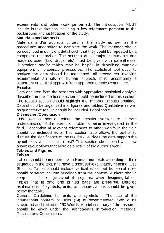
experiments and other work performed. The introduction MUST include in-text citations including a few references pertinent to the background and justification for the study. Materials and Methods Materials and/or subjects utilized in the study as well as the procedures undertaken to complete the work. The methods should be described in sufficient detail such that they could be repeated by a competent researcher. The sources of all major instruments and reagents used (kits, drugs, etc) must be given with parentheses. Illustrations and/or tables may be helpful in describing complex equipment or elaborate procedures. The statistical tool used to analyze the data should be mentioned. All procedures involving experimental animals or human subjects must accompany a statement on ethical approval from appropriate ethics committee. Results Data acquired from the research with appropriate statistical analysis described in the methods section should be included in this section. The results section should highlight the important results obtained. Data should be organized into figures and tables. Qualitative as well as quantitative results should be included if applicable. Discussion/Conclusion This section should relate the results section to current understanding of the scientific problems being investigated in the field. Description of relevant references to other work/s in the field should be included here. This section also allows the author to discuss the significance of the results - i.e. does the data support the hypotheses you set out to test? This section should end with new answers/questions that arise as a result of the author‟s work. Tables and Figures Tables Tables should be numbered with Roman numerals according to their sequence in the text, and have a short self-explanatory heading. Use SI units. Tables should include vertical rules, but horizontal rules should separate column headings from the content. Authors should keep in mind the page layout of the journal when designing tables. Tables that fit onto one printed page are preferred. Detailed explanations of symbols, units, and abbreviations should be given below the table. General Guidelines for units and symbols - The use of the international System of Units (SI) is recommended. Should be structured and limited to 250 Words. A brief summary of the research should be given under the subheadings Introduction, Methods, Results, and Conclusions.
43
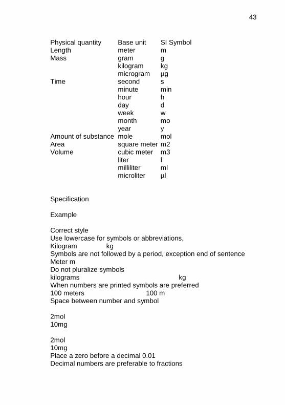
Physical quantity Base unit SI Symbol Length meter m Mass gram g kilogram kg microgram µg Time second s minute min hour h day d week w month mo year y Amount of substance mole mol Area square meter m2 Volume cubic meter m3 liter l milliliter ml microliter µl
Specification Example Correct style Use lowercase for symbols or abbreviations, Kilogram kg Symbols are not followed by a period, exception end of sentence Meter m Do not pluralize symbols kilograms kg When numbers are printed symbols are preferred 100 meters 100 m Space between number and symbol 2mol 10mg 2mol 10mg Place a zero before a decimal 0.01 Decimal numbers are preferable to fractions 0.75

44
Space used to separate long number exception four-digit numbers 1 500 000 1000
Chemical terminology - The chemical nomenclature used must be in accordance with that used in the Chemical Abstracts. Symbols and abbreviations - In vitro, in vivo, in situ, ex vivo, ad libitum, et al. and so on are two words each and should be written in italics. None of the above is a hyphenated word. All foreign language (other than English) names and words shall be in italics as a general rule. Words such as carrageenan-induced inflammation, paracetamol-induced hepatotoxicity, isoproterenol-induced myocardial necrosis, dose-dependent manner are all hyphenated. Biological nomenclature - Names of plants, animals and bacteria should be in italics. Enzyme nomenclature - The trivial names recommended by the IUPAC-IUB Commission should be used. When the enzyme is the main subject of a paper, its code number and systematic name should be stated at its first citation in the paper. Spelling - These should be as in the Concise Oxford Dictionary of Current English. Illustrations (Figures)
1. Upload the images in JPEG format. The file size should be within 4 Mb in size while uploading. Send sharp, glossy, un-mounted, color photographic prints, with height of 4 inches and width of 6 inches at the time of submitting the revised manuscript.
2. Figures should be numbered consecutively according to the order in which they have been first cited in the text.
3. Labels, numbers, and symbols should be clear and of uniform size. The lettering for figures should be large enough to be legible after reduction to fit the width of a printed column.
4. Symbols, arrows, or letters used in photomicrographs should contrast with the background and should mark neatly with transfer type or by tissue overlay and not by pen.
5. Titles and detailed explanations belong in the legends for illustrations not on the illustrations themselves.
6. When graphs, scatter-grams or histograms are submitted the numerical data on which they are based should also be supplied.
7. The photographs and figures should be trimmed to remove all the unwanted areas.

45
8. If photographs of people are used, either the subjects must not be identifiable or their pictures must be accompanied by written permission to use the photograph.
9. If a figure has been published elsewhere, acknowledge the original source and submit written permission from the copyright holder to reproduce the material. A credit line should appear in the legend for such figures.
10. Legends for illustrations: Type or print out legends (maximum 40 words, excluding the credit line) for illustrations using double spacing, with Arabic numerals corresponding to the illustrations. When symbols, arrows, numbers, or letters are used to identify parts of the illustrations, identify and explain each one in the legend. Explain the internal scale (magnification) and identify the method of staining in photomicrographs.
11. Final figures for print production: Print outs of digital photographs are not acceptable. If digital images are the only source of images, ensure that the image has minimum resolution of 300 dpi or 1800 x 1600 pixels in TIFF format. Send the images on a CD. Each figure should have a label pasted (avoid use of liquid gum for pasting) on its back indicating the number of the figure, the running title, top of the figure and the legends of the figure. Do not write the contributor/s' name/s. Do not write on the back of figures, scratch, or mark them by using paper clips.
12. The Journal reserves the right to crop, rotate, reduce, or enlarge the photographs to an acceptable size.
Table and Figure captions Figure captions/legends should include a statement at the end of each caption/legend about reproduction size (e.g. at full page width, at column width). They should be double spaced and typed in the journal format. Explanations should be brief and authors should keep in mind that captions/legends will be placed below figures. Acknowledgements Those who have helped the authors carry out the study and/or prepare the manuscript but have not made significant intellectual contribution to deserve authorship must be acknowledged. Mention all applicable grants and other funding that supported the work. Review Articles: It is expected that these articles would be written by individuals who have done substantial work on the subject or are considered experts in the field. A short summary of the work done by the contributor(s) in the field of review should accompany the manuscript.
46
The prescribed word count is up to 3000 words excluding tables, references and abstract. The manuscript may have about 90 references. The manuscript should have an unstructured Abstract (250 words) representing an accurate summary of the article. The section titles would depend upon the topic reviewed. Authors submitting review article should include a section describing the methods used for locating, selecting, extracting, and synthesizing data. These methods should also be summarized in the abstract. The journal expects the contributors to give post-publication updates on the subject of review. The update should be brief, covering the advances in the field after the publication of the article and should be sent as a letter to editor, as and when major development occurs in the field. Case reports: New, interesting and rare cases can be reported. They should be unique, describing a great diagnostic or therapeutic challenge and providing a learning point for the readers. Cases with clinical significance or implications will be given priority. These communications could be of up to 1000 words (excluding Abstract and references) and should have the following headings: Abstract (unstructured), Key-words, Introduction, Case report, Discussion, Reference, and Tables and Legends in that order. The manuscript could be of up to 1000 words (excluding references and abstract) and could be supported with up to 10 references. Case Reports could be authored by up to four authors. Letter to the Editor: These should be short and decisive observations. They should preferably be related to articles previously published in the Journal or views expressed in the journal. They should not be preliminary observations that need a later paper for validation. The letter could have up to 500 words and 5 references. It could be generally authored by not more than four authors. Clinicopathologic Conference (CPC): Manuscripts that present interesting, challenging, or unusual cases. The presentation should simulate clinical work-up, including the formulation of a detailed and well thought out differential diagnosis. The complete diagnostic evaluation, management, and follow-up must be included. CPC articles must be organized into six parts:
1. Title: Provide a descriptive clinical title that does not reveal the final diagnosis.
2. Clinical presentation: Describe the clinical and imaging characteristics of the lesion. Use clinical photographs and radiographs as appropriate.
47
3. Differential diagnosis: List and discuss lesions to be considered as reasonable diagnostic possibilities. The authors are reminded that the most important part of the CPC manuscript is the clinical differential diagnosis, where the authors guide the readership through their own diagnostic thought process. This will require the formulation of a list of the most probable diagnostic possibilities (ideally at least 5-6 entities) based on the clinical presentation, medical history, and/or radiographic studies.
4. Diagnosis: Histopathologic findings illustrated with appropriate photomicrographs.
5. Management: Describe the treatment of the patient and response to treatment.
6. Discussion: Concentrate on the most interesting aspect(s) of the case. No abstract is needed for CPC manuscripts.
Other: Editorial, Guest Editorial, and Commentary are solicited by the editorial board. Page layout & styles Page size Letter Portrait 8 ½ X 11 Margins All Margins, 1cm Page number Numbered at bottom right Footer/Headers None Title 14 pt Times New Roman, bold, centered. Author and co-authors 12 pt Times New Roman centered, bold - author and all co-authors names in one line. The corresponding author should include an asterisk*. Authors address Author for Correspondence: 10pt Times New roman centered - giving a valid e-mail of the corresponding (main) author is a must. It should be indicated as* followed by two line spacing. Abstract 12 pt Times New roman, full justification Normal - maximum 250 words Text 12 pt Times New roman, full justification – double lines spacing between paragraphs. No indentation Heading Major headings (ABSTRACT, KEYWORDS, INTRODUCTION, MATERIALS AND METHODS, RESULTS AND DISCUSSION, ACKNOWLEDGEMENTS, REFERENCES) in upper case left-justified, 12 pt bold, Intermediate headings should be in italics, sentence case, left justified, 12 pt Tables To be incorporated at the end of Manuscript Correct “Table 1: Serum enzyme levels………” Incorrect “Table No. 1: Serum enzyme levels………”

48
Figures /Graphs Figures may be embedded in your word document but they should be created with a program that allows you to save them as gif, jpg or tiff format. Figures, tables or other materials copied verbatim or adopted from previously published materials, the author must have written permission from the copyright holder of that material (publisher and/or authors) for reproduction in your article. A copy of the permission release must be submitted with the manuscript. It is the author's responsibility to obtain permission. To be incorporated at the end of the manuscript with proper labeling Correct “Figure 1: Serum enzyme levels………” Incorrect “Figure No. 1: Serum enzyme levels………” Graphs To be included from excel and it should be editable. Non–editable graphs will not be accepted. All text should be fully justified. Please put all primary section titles in UPPER CASE letters and subheading in both Upper and Lower Case letters. Do not number your titles (for example, 1.0 Introduction; 2.0 Background). Do not use the tab key to indent blocks of text such as paragraphs of quotes or lists because the page layout program overrides the left margin with its own, and the tabs end up in mid-sentence. References In-text citation Correct / Acceptable Format Respiratory tract infection is one of the most important infectious diseases worldwide. This infection is the leading cause of morbidity and mortality in critically ill patients in developing countries. [1-3] Respiratory tract infections (RTIs), which involve the upper or lower respiratory tract, frequently occurs after birth.[4] RTIs, such as sore thoat, earache, laryngitis, common cold, otitis media, sinusitis, and mastoiditis, are the most frequently-occurred infections of all human diseases and have been frequently documented. [4, 5] Incorrect / Not accepted Respiratory tract infection is one of the most important infectious diseases worldwide. This infection is the leading cause of morbidity and mortality in critically ill patients in developing countries [1, 2, 3]. Respiratory tract infections (RTIs), which involve the upper or lower respiratory tract, frequently occurs after birth [4]. RTIs, such as sore thoat, earache, laryngitis, common cold, otitis media, sinusitis, and mastoiditis, are the most frequently-occurred infections of all human diseases and have been frequently documented [4, 5].

49
Reference List: Author/Authors JOURNAL REFERENCES Single/Multiple Authors Halpern SD, Ubel PA, Caplan AL. Solid-organ transplantation in HIV-infected patients. N Engl J Med. 2002 Jul 25; 347(4): 284-7. More than six authors Rose ME, Huerbin MB, Melick J, Marion DW, Palmer AM, Schiding JK, et al. Regulation of interstitial excitatory amino acid concentrations after cortical contusion injury. Brain Res. 2002; 935(1-2): 40-6. Organization as Author Diabetes Prevention Program Research Group. Hypertension, insulin, and proinsulin in participants with impaired glucose tolerance. Hypertension. 2002; 40(5): 679-86. Unknown Author 21st century heart solution may have a sting in the tail. BMJ. 2002; 325(7357): 184-5. Journal article on the Internet Abood S. Quality improvement initiative in nursing homes: the ANA acts in an advisory role. Am J Nurs [serial on the Internet]. 2002 Jun [cited 2002 Aug 12]; 102(6): [about 3 p.]. Available from: http://www.nursingworld.org/AJN/2002/june/Wawatch.htm Note: Plant/Micro organisms, in-vivo, in-vitro should be in italics. Personal author(s) Murray PR, Rosenthal KS, Kobayashi GS, Paller MA. Medical microbiology. 4th ed. St. Louis: Mosby; 2002. Editor(s), compiler(s) as author Gilstrap LC 3rd, Cunningham FG, VanDorsten JP, editors. Operative obstetrics. 2nd ed. New York: McGraw-Hill; 2002. Author(s) and editor(s) Breedlove GK, Schorfheide AM. Adolescent pregnancy. 2nd ed. Wieczorek RR, editor. White Plains (NY): March of Dimes Education Services; 2001. Organization(s) as author Royal Adelaide Hospital; University of Adelaide, Department of Clinical Nursing. Compendium of nursing research and practice development, 1999-2000. Adelaide (Australia): Adelaide University; 2001. Chapter in a book Meltzer PS, Kallioniemi A, Trent JM. Chromosome alterations in human solid tumors. In: Vogelstein B, Kinzler KW, editors. The genetic basis of human cancer. New York: McGraw-Hill; 2002. p. 93-113. Conference proceedings

50
Harnden P, Joffe JK, Jones WG, editors. Germ cell tumors V. Proceedings of the 5th Germ Cell Tumour Conference; 2001 Sep 13-15; Leeds, UK. New York: Springer; 2002. Thesis N. Khoshakhlagh. The compositions of volatile fractions of Peganum harmala seeds and its smoke. Pharm. D. Thesis, Faculty of Pharmacy, Tehran University of Medical Sciences, Tehran, Iran. (2002). WEBSITES Website information Cancer-Pain.org [homepage on the Internet]. New York: Association of Cancer Online Resources, Inc.; c2000-01 [updated 2002 May 16; cited 2002 Jul 9]. Available from: http://www.cancer-pain.org/. Acknowledgements All messages and reviews sent electronically will be acknowledged automatically upon receipt. Protection of Patients' Rights to Privacy Identifying information should not be published in written descriptions, photographs, sonograms, CT scans, etc., and pedigrees unless the information is essential for scientific purposes and the patient (or parent or guardian, wherever applicable) gives written informed consent for publication. Authors should remove patients' names from figures unless they have obtained written informed consent from the patients. When informed consent has been obtained, it should be indicated in the article and copy of the consent should be attached with the covering letter. Sending a revised manuscript The revised version of the manuscript should be submitted online in a manner similar to that used for submission of the manuscript for the first time. However, there is no need to submit the “First Page” or “Covering Letter” file while submitting a revised version. When submitting a revised manuscript, contributors are requested to include, the „referees‟ remarks along with point to point clarification at the beginning in the revised file itself. In addition, they are expected to mark the changes as underlined or colored text in the article.
Reprints and proofs Journal provides no free printed reprints. Authors can purchase reprints, payment for which should be done at the time of submitting the proofs.
Copyrights The entire contents of the National Journal of Maxillofacial Surgery (NJMS) are protected under Indian and international copyrights. The Journal, however, grants to all users a free, irrevocable, worldwide, perpetual right of access to, and a license to copy, use, distribute, perform and display the work publicly and to make and distribute

51
derivative works in any digital medium for any reasonable non-commercial purpose, subject to proper attribution of authorship and ownership of the rights. Checklist Covering letter Signed by all contributors Previous publication / presentations mentioned Source of funding mentioned Conflicts of interest disclosed Authors Last name and given name provided along with Middle name initials (where applicable) Author for correspondence, with e-mail address provided Number of contributors restricted as per the instructions Identity not revealed in paper except title page (e.g. name of the institute in Methods, citing previous study as 'our study', names on figure labels, name of institute in photographs, etc.) Presentation and format Double spacing Margins 1 cm from all four sides Page numbers included at bottom Title page contains all the desired information Running title provided (not more than 50 characters) Abstract page contains the full title of the manuscript Abstract provided (structured abstract of 250 words for original articles, unstructured abstracts of about 150 words for all other manuscripts excluding letters to the Editor) Key words provided (three or more) Introduction of 75-100 words Headings in title case (not ALL CAPITALS) The references cited in the text should be before punctuation marks, with square bracket. References according to the journal's instructions, punctuation marks checked Send the article file without „Track Changes‟ Language and grammar Uniformly American English Write the full term for each abbreviation at its first use in the title, abstract, keywords and text separately unless it is a standard unit of measure. Numerals from 1 to 10 spelt out Numerals at the beginning of the sentence spelt out Check the manuscript for spelling, grammar and punctuation errors If a brand name is cited, supply the manufacturer's name and

52
address (city and state/country). Species names should be in italics Tables and figures No repetition of data in tables and graphs and in text Actual numbers from which graphs drawn, provided Figures necessary and of good quality (colour) Table and figure numbers in Arabic letters (not Roman) Labels pasted on back of the photographs (no names written) Figure legends provided (not more than 40 words) Patients' privacy maintained (if not permission taken) Credit note for borrowed figures/tables provided Write the full term for each abbreviation used in the table as a footnote
Contributors' form (to be modified as applicable and one signed copy attached with the manuscript) Manuscript Title: ______________________________________________________________________________ I/we certify that I/we have participated sufficiently in contributing to the intellectual content, concept and design of this work or the analysis and interpretation of the data (when applicable), as well as writing of the manuscript, to take public responsibility for it and have agreed to have my/our name listed as a contributor. I/we believe that the manuscript represents valid work. Neither this manuscript nor one with substantially similar content under my/our authorship has been published or is being considered for publication elsewhere, except as described in the covering letter. I/we certify that all the data collected during the study is presented in this manuscript and no data from the study has been or will be published separately. I/we attest that, if requested by the editors, I/we will provide the data/information or will cooperate fully in obtaining and providing the data/information on which the manuscript is based, for examination by the editors or their assignees. Financial interests, direct or indirect, that exist or may be perceived to exist for individual contributors in connection with the content of this paper have been disclosed in the cover letter. Sources of outside support of the project are named in the covering letter. I/We hereby transfer(s), assign(s), or otherwise convey(s) all copyright ownership, including any and all rights incidental thereto, exclusively to the National Journal of Maxillofacial Surgery (NJMS), in the event that such work is published by the National Journal of

53
Maxillofacial Surgery (NJMS). The National Journal of Maxillofacial Surgery (NJMS) shall own the work, including Copyright; The right to grant permission to republish the article in whole or in part, with or without fee; The right to produce preprints or reprints and translate into languages other than English for sale or free distribution; and The right to republish the work in a collection of articles in any other mechanical or electronic format. We give the rights to the corresponding author to make necessary changes as per the request of the journal, do the rest of the correspondence on our behalf and he/she will act as the guarantor for the manuscript on our behalf. All persons who have made substantial contributions to the work reported in the manuscript, but who are not contributors, are named in the Acknowledgment and have given me/us their written permission to be named. If I/we do not include an Acknowledgment that means I/we have not received substantial contributions from non-contributors and no contributor has been omitted.
Name Signature Date signed 1 --------------- --------------- --------------- 2 --------------- --------------- --------------- 3 --------------- --------------- --------------- 4 --------------- --------------- --------------- (up to 4 contributors for case report/ images/ review) 5 --------------- --------------- --------------- 6 --------------- --------------- --------------- (up to 6 contributors for original studies)