


Línguas
Páginas
Legal

UNIVERSIDADE DE BRASÍLIA
INSTITUTO DE GEOCIÊNCIAS
ÁREA DE CONCENTRAÇÃO: GEOLOGIA REGIONAL
DISSERTAÇÃO DE MESTRADO NO 312
UM NOVO PODOCNEMÍDEO FÓSSIL DE GRANDE PORTE DA
FORMAÇÃO SOLIMÕES (MIOCENO-PLIOCENO), ACRE,
BRASIL E AS RELAÇÕES FILOGENÉTICAS ENTRE OS
PODOCNEMIDAE
Marcos Vitor Dumont Júnior
Orientador: Prof. Dr. Rodrigo Miloni Santucci
Co-orientadora: Profa. Dra. Caroline Thaís Martinho
Brasília-DF
Abril de 2013

Banca Examinadora:
Orientador:
Rodrigo Miloni Santucci
Examinadores Internos:
Ricardo Lourenço Pinto – IG/UnB
Renato Caparroz – IB/UnB (Suplente)
Examinadores Externos:
Max Cardoso Langer – USP/Ribeirão Preto
Gustavo Ribeiro de Oliveira – UF/RPE (Suplente)

À minha companheira, que me ensinou o real significado desta palavra.
Agradecimentos
Agradeço ao CNPq, pela concessão da bolsa de mestrado para o
desenvolvimento deste trabalho.
Ao programa de pós-graduação em Geologia do Instituto de Geologia da UnB e
ao Laboratório de Pesquisas Paleontológicas da UFAC pela estrutura e material para a
execução deste trabalho.
Ao meu orientador Dr. Rodrigo Miloni Santucci, pelo apoio, incentivo,
disponibilização de material bibliográfico, tempo e paciência, desde a graduação, assim
como pelas contribuições e críticas essenciais para esse trabalho.
À Dra. Caroline Thaís Martinho, pelo apoio, em especial durante meu período de
seleção e início na pós-graduação da Geologia da UnB.
Ao Dr. Édson Guilherme da Silva, pelo auxílio logístico em Rio Branco, bem
como suas inúmeras contribuições durante as visitas ao LPP-UFAC. Bem como pela
amizade.
Ao Dr. Jonas Pereira de Souza Filho, por abrir as portas do LPP-UFAC para
mim. Bem como à equipe do laboratório.
Ao Dr. Ricardo Lourenço Pinto, pelo apoio e pelo espaço cedido para as
reuniões com meu orientador.
Agradeço também a meus pais, Marcos e Déa, por compreenderem minha
ausência durante esse período, bem como pelo apoio financeiro.
Aos meus sogros, Ana Maria e Célio, pelo incentivo à busca de meus sonhos
acadêmicos.
Aos amigos, pelos momentos de descontração, ainda que infelizmente raros
durante esse período.
E, por último e mais importante, à minha companheira Ana Carolina Vieira
Ribeiro. Por ter sido um pouco de tudo, desde uma rocha firme onde pude me apoiar em
horas difíceis até a melhor companhia para celebrar momentos felizes, uma fonte sem
fim de incentivo e apoio desde o dia em que nos conhecemos.

ÍNDICE
1. Resumo........................................................................................................1
2. Introdução...................................................................................................2
3. Artigo ..........................................................................................................5
4. Conclusões ................................................................................................59
5. Referências Bibliográficas.......................................................................61
6. Tabelas e Figuras......................................................................................66

1
1. Resumo
A família Podocnemidae é representada hoje por oito espécies viventes distribuídas na
América do Sul e em Madagascar. A família também possui um rico registro fóssil,
encontrado em quase todo o hemisfério sul, com uma grande variedade de formas e
tamanhos, assim como uma história paleobiogeográfica complexa. Aqui nós
descrevemos uma nova espécie de Podocnemidae fóssil do Mioceno-Plioceno da
Formação Solimões do Brasil, Podocnemis manchineri sp. nov., baseada em um casco
quase completo, uma carapaça e plastrão fragmentários e uma carapaça fragmentária,
compreendendo três diferentes indivíduos. Nós analisamos esse novo táxon e outros
cinco táxons conhecidos apenas por material pós-craniano em uma análise de
parcimônia usando uma matriz de caracteres extraída da literatura, além de novos
caracteres criados nesse estudo baseados na observação da carapaça e plastrão de vários
podocnemídeos viventes e extintos. Os resultados indicam que P. manchineri está
aninhada dentro do gênero Podocnemis. Nossa análise também valida a posição de
outros táxons baseados em carapaças como membros do gênero Podocnemis (e. g. P.
negrii¸ P. medemi e P. pritchardi), que têm sido referidos como incertae sedis dentro de
Podocnemidae em outros estudos. Além disso, essa análise resolveu o táxon
Stupendemys geographicus como mais relacionado ao clado que inclui Bairdemys que o
clado que inclui Peltocephalus, dentro de Erymnochelyinae. Adicionalmente, pela
primeira vez táxons fósseis foram recuperados (Kenyemys, apenas conhecido por
cascos, e Turkanemys) entre a associação comum de Erymnochelys e Peltocephalus,
constante em análises filogenéticas com base em dados morfológicos. Comparações
morfológicas de material fragmentário da Formação Solimões sugerem que pelo menos
um terceiro táxon de Podoncmidae fóssil poderia estar presente na Amazônia Sul-
ocidental. Os resultados do presente trabalho foram submetidos ao Journal of
Systematic Paleontology, apresentamos aqui o trabalho na forma em que foi submetido
ao periódico.

2
2. Introdução
A Formação Solimões (Mioceno-Plioceno) têm revelado muitos fósseis de
vertebrados ao longo dos anos, representando uma grande variedade de peixes,
crocodilianos, tartarugas e mamíferos (Barbosa-Rodrigues 1892; Price 1964; Sill 1970;
Campos 1977; Campos & Broin 1981; Gasparini 1985; Bocquentin & Rancy 1987;
Bocquentin & Santos 1989; Bocquentin & Souza-Filho 1989, 1990; Souza-Filho &
Bocquentin 1989, 1991; Broin et al. 1993; Souza-Filho et al. 1993; Latrubesse et al.
1997; Bocquentin-Villanueva et al. 1997; Gaffney et al. 1998; Souza-Filho 1998;
Bocquentin & Guilherme 1999; Negri & Ferigolo 1999; Bocquentin et al. 2001;
Carvalho et al. 2002; Bocquentin & Melo 2006; Kay & Cozzuol 2006; Hsiou et al.
2007; Meylan et al. 2009; Hsiou 2010; Riff et al. 2010).
Os Testudines encontrados na Formação Solimões, em sua maioria, pertencem ao
grupo Pleurodira, típico do hemisfério sul, facilmente diferenciados do subgrupo
Cryptodira pelo modo de retração da cabeça. Em Pleurodira a retração é lateral e em
Cryptodira ela ocorre para trás, com a cabeça se encaixando entre os ombros (Figura 9).
Os Pleurodira são atualmente representados por três famílias: Podocnemidae, da
América do Sul e Madagascar, com 8 espécies de água doce, de tamanho pequeno até o
maior Pleurodira atual, Podocnemis expansa, que alcança 90cm de comprimento;
Pelomedusidae, da África, Madagascar e Ilhas Seychelles, pequenos, com cerca de 18
espécies de água doce; e Chelidae, de porte pequeno a grande, com cerca de 40 espécies
de água doce, presentes na América do Sul, Austrália e Nova Guiné (Rueda-Almonacid
et al. 2007; Pough et al., 2008). Embora hoje apenas ocorram em ambiente dulcícola
nos continentes do Hemisfério Sul, os Pleurodira ocuparam também ambientes de água
salobra e ambientes próximos à costa da maioria dos continentes desde o Eocretáceo,
sendo bem mais diversificado no registro fóssil (Gaffney et al., 2006).
Até agora, os Podocnemidae conhecidos da Formação Solimões incluem Caninemys
tridentata (Meylan et al. 2009), “Stupendemys” souzai (Bocquentin & Melo 2006), e
Podocnemis negrii (Carvalho et al. 2002), sendo C. tridentata o único táxon bem aceito.
A atribuição de S. souzai ao gênero referido é criticada e S. souzai pode não representar
um único táxon (Meylan et al. 2009; Gaffney et al. 2011) e P. negrii é considerada
incertae sedis dentro de Podocnemidae (Meylan et al. 2009; Gaffney et al. 2011).
Apenas C. tridentata foi incluída em uma análise filogenética até hoje. Essa ausência

3
em estudos cladísticos provavelmente ocorre devido ao comportamento ambíguo que
espécies conhecidas apenas por carapaças e plastrões apresentam nesse tipo de análise.
A maior parte dos caracteres usados na filogenia de podocnemídeos são retirados do
crânio e mandíbula, enquanto os caracteres pós cranianos são escassos (e. g. Meylan et
al. 2009; Cadena et al. 2010, 2012; Gaffney et al., 2011). Interessantemente, devido à
complexidade do casco dos Testudines, onde tanto o plastrão quanto a carapaça sendo
compostos por uma associação de ossos superpostos por escudos dérmicos, pode se
esperar que vários caracteres possam ser retirados dessa associação. Ao invés disso, os
caracteres de carapaça geralmente representam menos de 25% dos caracteres usados em
estudos cladísticos referentes a Podocnemidae fósseis (Tabela 6). Isso contrasta
fortemente com o grande número de espécies de cágados fósseis conhecidos apenas pela
carapaça. Aqui nós descrevemos uma nova espécie de podocnemídeo, proveniente da
Formação Solimões, Acre, Brasil (Fig. 2), e a comparamos a outras espécies
relacionadas, fósseis e atuais.
Adicionalmente, nós executamos uma análise cladística, incluindo espécies fósseis e
viventes de podocnemídeos, usando uma compilação de caracteres da literatura e novos
caracteres do casco. Que forneceram uma hipótese filogenética que inclui alguns táxons
baseados apenas em cascos, comumente deixados de fora desse tipo de estudo (e. g.
Stupendemys geographicus, Podocnemis negrii, Podocnemis medemi, Podocnemis
pritchardi, and Kenyemys williamsi). A Sistemática Filogenética tenta descobrir o grau
de relação de parentesco entre os diferentes táxons em determinado grupo de
organismos, com base em caracteres derivados compartilhados (sinapomorfias).
Podendo ser representado por um cladograma (Hennig, 1965). Assim, dadas três
espécies A, B e C: a espécie A pode ser mais aparentada a B do que a C; a espécie A
pode ser mais aparentada a C do que a B; ou as espécies B e C podem ser mais
aparentadas entre si do que qualquer uma delas com A (Figura 10). A escola
desenvolvida por Hennig defende que o grau de relação filogenética não pode ser
inferido com base apenas em semelhanças morfológicas. As relações devem ser
evidenciadas através da observação de características derivadas compartilhadas, isto é,
por sinapomorfias. Isso ocorre porque caracteres podem manter-se inalterados por muito
tempo durante a evolução. Assim sendo, a relação de parentesco entre espécies pode ser
demonstrada apenas pela posse, em comum, de sinapomorfias (Hennig, 1965) ou, em

4
outras palavras, a presença de sinapomorfias é a única evidência de uma origem
evolutiva comum entre dois ou mais táxons.
Desta forma, o presente estudo teve como objetivos; 1 - descrever o material
fóssil inédito (UFAC-1000), que consiste de carapaça quase completa, plastrão
incompleto e cintura pélvica. Provavelmente relacionado ao gênero Podocnemis (Rancy
& Bocquentin, 1987). Além de rever a classificação de outros espécimes. O que poderá
aumentar de maneira significativa a resolução das relações dentro do gênero
Podocnemis. 2 - avaliar mais acuradamente as relações filogenéticas dos
Podocnemidae da Formação Solimões, ainda não incluídos nesse tipo de trabalhos sobre
o grupo (UFAC-1000, P. negrii). Comparando-os com táxons em outras coleções e, se
possível, incluir novos caracteres para análise, na tentativa de aumentar a resolução dos
cladogramas.
5
3. Artigo
A new large podocnemid turtle from the upper Miocene Solimões Formation,
Acre, Brazil and the phylogenetic relationships within Podocnemidae
Marcos Vitor Dumont Júniora, Rodrigo Miloni Santucci
b*, Caroline Thaís Martinho
c,
Édson Guilherme da Silvad
aInstituto de Geociências, Universidade de Brasília, Campus Universitário Darcy Ribeiro, Asa Norte,
70910-900, Brasilia, DF, Brazil; bFaculdade UnB Planaltina, Universidade de Brasília, Área
Universitária 1, Vila Nossa Senhora de Fátima, 73345-010, Brasilia, DF, Brazil; cPontifícia
Universidade Católica do Rio Grande do Sul, Instituto do Meio Ambiente e Recursos Naturais, Av.
Ipiranga, 6681, Prédio 96J, Partenon, 90619-900, Porto Alegre, RS, Brazil; dUniversidade Federal do
Acre, Laboratório de Pesquisas Paleontológicas, CCBN, BR-364, KM 4, Distrito Industrial, 69915-900,
Rio Branco, AC, Brazil
*Corresponding author. Email: [email protected]

6
Abstract
The family Podocnemidae is represented today by eight moderate-sized extant species
distributed in South America and Madagascar. They also have a rich fossil record,
which is found over almost all the southern hemisphere, with a wide variety of forms
and sizes, as well as a complex palaeobiogeographical history. Here we describe a new
species of fossil Podocnemidae from the Miocene-Pliocene Solimões Formation of
Brazil, Podocnemis manchineri sp. nov., based on an almost complete large shell, a
fragmentary carapace and plastron, and a fragmentary carapace, comprising three
different individuals. We analysed this new taxon and other five fossil taxa known only
by shell material in a parsimony analysis using a morphological character matrix with
characters extracted from the literature, as well as new characters created in this study
based on observation of the carapace and plastron of several extinct and extant
podocnemid taxa. The results indicate that P. manchineri is nested within the genus
Podocnemis. Our analysis also validated the position of several shell taxa to the genus
Podocnemis (e. g. P. negrii, P. medemi, and P. pritchardi), which have been referred as
incertae sedis within Podocnemidae in previous studies. Furthermore, this analysis
resolved Stupendemys geographicus as more closed related to the clade that includes
Bairdemys than to the clade that includes Peltocephalus within Erymnochelyinae.
Additionally, for the first time it is recovered the presence of fossil taxa (namely
Kenyemys (also shell based) and Turkanemys ) between the common association of
Erymnochelys and Peltocephalus, commonly recovered in morphological phylogenetic
approaches. Morphological comparisons of fragmentary material from the Solimões
Formation also suggest that at least a third taxon of a large sized fossil podocnemid
could have been present in South-western Amazonia.
Key-words: Podocnemidae, Acre, Solimões Formation, Podocnemis, Phylogenetic
Analysis.
Introduction

7
The Brazilian Solimões Formation (Miocene-Pliocene) has yielded many
vertebrate fossils over the years, representing a great variety of fishes, crocodilians,
turtles, and mammals (Barbosa-Rodrigues 1892; Price 1964; Sill 1970; Campos 1977;
Campos & Broin 1981; Gasparini 1985; Bocquentin & Rancy 1987; Bocquentin &
Santos 1989; Bocquentin & Souza-Filho 1989, 1990; Souza-Filho & Bocquentin 1989,
1991; Broin et al. 1993; Souza-Filho et al. 1993; Latrubesse et al. 1997; Bocquentin-
Villanueva et al. 1997; Gaffney et al. 1998; Souza-Filho 1998; Bocquentin &
Guilherme 1999; Negri & Ferigolo 1999; Bocquentin et al. 2001; Carvalho et al. 2002;
Bocquentin & Melo 2006; Kay & Cozzuol 2006; Hsiou et al. 2007; Meylan et al. 2009;
Hsiou 2010; Riff et al. 2010). Concerning turtles, so far, the known record of
Podocnemidae from the Solimões Formation includes Caninemys tridentata (Meylan et
al. 2009), “Stupendemys” souzai (Bocquentin & Melo 2006), and Podocnemis negrii
(Carvalho et al. 2002), with the only well accepted taxon being C. tridentata. S. souzai
is criticized and may not represent a single taxon (Meylan et al. 2009; Gaffney et al.
2011) and P. negrii is considered to be incertae sedis within the Podocnemidae (Meylan
et al. 2009; Gaffney et al. 2011). Also, only C. tridentata has been included in a
phylogenetic analysis to date. This absence in cladistics approaches is likely due to the
ambiguous behaviour that species known only by shell material show in this type of
analysis. Most characters used in podocnemid phylogeny are taken from the skull and
jaws, while the postcranial characters are scarce (e. g. Meylan et al. 2009; Cadena et al.
2010, 2012; Gaffney et al., 2011). Interestingly, due to the complexity of the turtle
shell, with both carapace and plastron being composed by an association of bones
superposed by an association of scales, it might be expected that several characters
could be extracted from this association. Instead, shell characters generally represent
less than 25% of the characters used in cladistic studies regarding fossil turtles. This
strongly contrasts with the large number of fossil turtle species only based in shell
materials. Here we describe a large new podocnemidid turtle uncovered from the
Solimões Formation deposits, Acre, Brazil (Fig. 1), and compare it to other close related
fossil and extant species. Additionally, we performed a cladistics analysis including
both extant and fossil podocnemid species by using a compilation of characters in the
literature and new shell characters which furnished a phylogenetic hypothesis that also
includes few shell based taxa commonly not present in this kind of work (e. g.

8
Stupendemys geographicus, Podocnemis negrii, Podocnemis medemi, Podocnemis
pritchardi, and Kenyemys williamsi).
Abbreviations
LACM – Natural History Museum of Los Angeles County; UFAC – Universidade
Federal do Acre, Laboratório de Pesquisas Paleontológicas; UCMP – University of
California, Museum of Paleontology, Berkeley, California.
Geological setting
The Solimões Formation, located in north-western South America, consists of
claystones, sandstones and siltstones intercalated with a few lignite and limestone layers
(Radambrasil 1977; Hoorn 1993). The Solimões Formation has lateral continuity on
neighbour basins, receiving different names. In Pastaza, Marañón and Madre de Dios
basins from Peru the Solimões Formation is known as Pebas, Ipururo, and Nauta
formations. In Colombia, on Amazonas and Putumayo basins, it receives the
denominations of Amazonic Tertiary and La Tagua layers (Hoorn 1993, 1994a;
Campbell et al. 2001; Roddaz et al. 2005; Rebata et al. 2006).
Palynological data suggest an age range from Miocene to Pliocene for the
Solimões Formation in the Amazonas State of Brazil (Cruz 1984). Many
biostratigraphic works based on palynomorphs were carried out in central and north-
western Amazonia (Hoorn 1993, 1994a, b, 1995). However, most vertebrate fossils
from the Solimões Formation come from the south-western of Amazonia and no
relation between the north and south sediments has been established yet (Campbell et
al. 2000, 2001, 2006).
Other biostratigraphic propositions have been made, using mainly fossil
mammals. To the south-western Brazilian Amazonia a Huayquerian, possibly reaching
Montehermosian age has been suggested (upper Miocene to Pliocene) (Campbell et al.
1985; Frailey 1986; Latrubesse 1992; Latrubesse et al. 1997, 2007).
There are different palaeoenvironmental interpretations for the Solimões
Formation as well, it could have been either a shallow lake, or lake system (Campbell &
Frailey 1984; Frailey et al. 1988; Vonhof et al. 1998; Wesselingh et al. 2002); a great

9
fluvial megafan complex (Latrubesse et al. 1997, 2007); a marginal marine influence
area (Hoorn 1993, 1994a, b, 1995); or an internal sea (Räsänen et al. 1995; Hovikoski et
al. 2005).
Fossil reptiles are abundant, with a great variety of extinct crocodilians, being
represented by Crocodylidae (Charactosuchus), Alligatoridae (Caiman, Purussaurus,
Mourasuchus), and Gavialidae (Brasilosuchus, Gryposuchus, and Hesperogavialis)
(Barbosa-Rodrigues 1892; Price 1964; Sill 1970; Gasparini 1985; Bocquentin & Souza-
Filho 1989, 1990; Souza-Filho 1998; Souza-Filho & Bocquentin 1989, 1991; Souza-
Filho et al. 1993; Riff et al. 2010). The chelonians are represented by the Podocnemidae
(Podocnemis and Stupendemys), Chelidae (Chelus), and Testudinidae (Chelonoidis)
(Barbosa-Rodrigues 1892; Campos 1977; Campos & Broin 1981; Broin et al. 1993;
Bocquentin & Rancy 1987; Bocquentin & Santos 1989; Gaffney et al. 1998;
Bocquentin et al. 2001; Carvalho et al. 2002; Bocquentin & Melo 2006; Riff et al.
2010). There are also records of Squamata, represented by snakes (Aniliidae, Boidae
and Colubridae) and lizards (Teiidae) (Hsiou et al. 2007; Hsiou 2010). All these
findings tend to support a continental water body palaeoenvironmental interpretation.
Historical background
In past years various phylogenetic analyses, with both molecular and
morphological data, have been contributing to the understanding of the Podocnemidae
inter-relationships (Georges et al. 1998; França & Langer 2006; Gaffney et al. 2006;
Noonan & Chippindale 2006; Vargas-Ramírez et al. 2008; Meylan et al. 2009; Cadena
et al. 2010, 2012; Gaffney et al. 2011). However, there are still many problems to be
solved in order to understand podocnemid phylogeny.
The morphological data results conflict with the molecular ones. The
relationships among the living genera Podocnemis, Peltocephalus, and Erymnochelys
are different when the two datasets are considered. For instance, the molecular data
suggests a closer relation between Podocnemis and Erymnochelys (Georges et al. 1998;
Noonan & Chippindale 2006; Vargas-Ramírez et al. 2008), while the morphological
data suggests that Erymnochelys and Peltocephalus are sister-taxa (França & Langer
2006; Meylan et al. 2009; Cadena et al. 2010, 2012; Gaffney et al. 2011). In any case,

10
those results may be of great palaeobiogeographic importance, since Erymnochelys is
from Madagascar while the other two genera are from South America.
The Podocnemidae shell is known as very conservative, with many shell based
taxa being first referred initially as Podocnemis and later, with skull discoveries, revised
and reassigned to other genera (Gaffney et al. 2006, 2011). This led to a major focus on
bringing new cranial characters for phylogenetic analyses, while new postcranial
characters have been much rarer. Usually more than 75% of the characters in a
phylogenetic analysis are from the skull and jaws (Meylan et al. 2009; Cadena et al.
2010, 2012; Gaffney et al. 2011). Consequently, shell based taxa, such as Stupendemys
and Kenyemys, hardly produce well resolved cladograms in this context.
So far, the only Solimões Formation podocnemid included in a phylogenetic
analysis is the skull based taxon Caninemys tridentate, whereas the shell based taxa
from the Solimões Formation (“Stupendemys” souzai and Podocnemis negrii) have
never been included in a cladistics study. Both shell taxa are controversial.
“Stupendemys” souzai assignment to the genus Stupendemys has been criticized and the
referred material may not represent a single taxon (Meylan et al. 2009; Gaffney et al.
2011). Podocnemis negrii has been considered as incertae sedis within the
Podocnemidae, due to the lack of synapomorphies to support its suggested relationship
as a sister-taxon to the extant P. sextuberculata (Carvalho et al. 2002; Gaffney et al.
2011).
There are many other indeterminate Podocnemidae fossil remains from the
Solimões Formation in the UFAC collection. Among them, there is UFAC-1000 and
UFAC-1001, considered until now to be an indeterminate podocnemid (Rancy &
Bocquentin-Villanueva, 1987). Here we review these specimens, in addition to a third
non described specimen (UFAC-1559) in order to clarify their status within the
Podocnemidae.
Systematic palaeontology
Testudines Linnaeus, 1758
Pleurodira Cope,1864
Pelomedusoides Cope, 1868
Podocnemidinura Cope, 1868
Podocnemidae Cope, 1868
11
Podocnemis Wagler, 1830
Type species: Emys expansa Schweigger, 1812.
Included species: Podocnemis expansa, P. vogli, P. unifilis, P. erythrocephala, P.
lewyana, P. sextuberculata, and P. bassleri.
Diagnosis: As in Gaffney et al. (2011).
Podocnemis manchineri sp. nov.
(Figs. 2 and 3)
Etymology: Named after the indigenous Manchineri tribe, living near the collection site
of the holotype.
Holotype: UFAC-1000, a nearly complete carapace and plastron.
Referred materials: UFAC-1001, incomplete carapace and plastron; UFAC-1559, a
fragmentary carapace.
Locality and horizon: UFAC-1000, Acre River, Seringal Bélgica, Assis Brasil, Acre
State, Brazil; UFAC-1001, Juruá River, Cruzeiro do Sul, Acre State, Brazil; UFAC-
1559, Upper Purus River, Purus 6 UFAC locality, AM (Fig. 1). Solimões Formation,
upper Miocene (Huayqueriense) (Campbell et al. 1985; Frailey 1986; Latrubesse 1992;
Latrubesse et al. 1997, 2007). All materials are deposited in the UFAC fossil vertebrate
collection.
Diagnosis: A very large sized podocnemid turtle (more than a meter long shell) with the
following combination of characters: vase-like shaped vertebral scutes, especially
vertebral two; vertebral one significantly larger than other vertebral scutes; rounded
cranial margin of the shell, without any kind of embayment; gular scales reaching the
entoplastra.

12
Description
UFAC-1000 Carapace
The UFAC-1000 (Figs. 2A and 3A, C, and D) specimen is represented by an
almost complete carapace and plastron. The carapace of the UFAC-1000 is uniformly
arched, without any irregularities, ornamentations, keels or depressions. The carapace
bones are regularly articulated, not presenting significant alterations due to diagenetic
processes. This indicates that the dorsoventral flattening of the shell represents a feature
present in the living animal, more easily seen in frontal view. The plastral bridge goes
outwards, almost parallel to the dorsoventral axis. The scars left by the dermic scutes
are easily seen and identifiable. The shell interior is still filled with the matrix and,
therefore, not accessible. The presence or absence of ducts, such as axillary musk duct
is uncertain, due to the poor preservation state of the regions where they may have been
placed, such as the joining region between the plastral bridge and the peripheral bones.
Nuchal bone. It is slightly wider than long. Its widest portion is the region where it meets
the first costal and the first peripherals, reaching the minimum width at the end of the
first cranial quarter of the bone. The lateral edges are sinuous, while the cranial edge is
curved without any embayment or notch. This gives the bone a general bulbous shape
from a dorsal view.
Neural series. The carapace has seven neural bones. The first neural is oval and
elongated, connected only to costals one, neural two, and the nuchal bone. All other
neurals connect to two pairs of costals, the ones with the same number of the neural and
the proceeding ones. The neurals two to five have six sides, with an elongated
hexagonal shape, resembling a coffin. The neural six is shorter, showing a more regular
hexagonal shape, and the neural seven has five sides, resembling a gem.
Suprapygal. It is wider than long and is skirt shaped. It is caudally formed by three
arches, where it articulates to the pygal and the peripherals eleven. The suprapygal do
not contact the neural bones.
Pygal. The caudal edge is broken, but the bone is trapezoidal in shape and wider
caudally.
Costals. There are eight pairs of costals. Only the seventh and eighth pairs connect each
other medially, in a slight asymmetrical way. All the others are medially connected to a

13
neural bone. The first one is longer than the others, with a rounded anterior edge,
connected to the nuchal and the peripheral bones one to four. The costals two and three
are slightly arched caudally. The costal four is almost straight, and the remaining ones
are sequentially more arched cranially than the anterior costal bone.
Peripherals. From the eleven pairs of peripheral bones, only the second, third and fourth
are completely preserved on the right side of the shell. The others show different
degrees of preservation. However, it is possible to infer the probable shell outline by the
combination of the information on both sides of the shell. It would have been oval in
shape, with the maximum width at the seventh or eighth peripheral. The peripherals
strongly vary in shape with the peripheral one being larger than the others. The size of
peripherals decreases progressively until the peripheral six, which is the smallest. From
the peripheral seven onward the size gradually increases until the last bone of the series.
Vertebral scutes. The first vertebral scute is clearly wider than the others, with a different
and more rounded shape. The second and third scutes are very similar, showing a vase-
like shape. All the six edges are curved, arched towards the center, with the cranial edge
being larger than the caudal one. The maximum width is reached where the cranial and
caudal lateral edges meet. The fourth scute is similar to vertebrals two and three, being
only thinner than these elements and with the caudal end arched slightly caudally, while
in vertebrals two and three it is arched cranially. The fifth and last vertebral scute is
wider than the others, being roughly triangular in shape, resembling a fish tail.
Pleural and marginal scutes. There are four pairs of pleural scutes and twelve pairs of
marginal scutes. None of the marginal scutes reach any of the costal bones. The first
marginal pair is proportionally small and roughly rectangular in shape.
Plastron
The plastron of UFAC-1000 (Fig. 3B) is almost complete. The cranial and
caudal extremities are broken. The epiplastra is also broken, almost not preserved.
Entoplastra. The maximum width is reached about in the middle of the bone, despite the
broken cranial edge, the different shortening of the cranial and caudal portions of the
bone suggest a shorter caudal and a longer cranial portion.
Hioplastra and hipoplastra are well preserved. A small lateral mesoplastra is
present. The xifiplastra caudal edge is not preserved. The caudal lobe of the plastron has
an almost straight edge.

14
Scales. The region that would be covered by the gular and intergular scales is not
preserved. The humeral scales contact each other in the midline, covering the
entoplastra, what is left from the epiplastra almost completely, and a small portion of
the hioplastra. The pectoral scales covers a big portion of the hioplastra, cranially it
reaches the epiplastra and the entoplastra. The abdominal scales cover portions of the
hioplastra, the hipoplastra, and the mesoplastra almost completely, which is also
covered only by marginal scutes.
Pelvic girdle
Only the right pelvic girdle is accessible for study (Figs. 2D and 3E). It is not
completely preserved, with the pubic and ischiatic processes broken. Furthermore, the
ilium is broken, so that the pelvic girdle can be detached from the carapace, where the
other half of the ilium is still attached to the shell. However, the pelvic girdle bones are
still articulated to each other. They have a slender structure, being considerably flat. The
ilium is slightly more robust than the other bones, but flattens anterocaudally, forming a
small crest in its lateral portion. The acetabulum has the shape of a slightly arched drop,
with the tip placed in the pubis.
UFAC-1001
The UFAC-1001 specimen (Fig. 2B) preserves the cranial right portion of the
carapace, articulated to the plastron, which keeps its cranial lobe at right side. The scute
scars are easily seen. Additionally, as in UFAC-1000, the region where the axillary
musk ducts may have existed is not well preserved.
In the carapace of the UFAC-1001 the nuchal and costals one to three are
partially preserved. Peripherals one to four are completely preserved and the fifth is
almost completely preserved. The nuchal has a bulbous contour where it meets the
peripheral one. The axillary buttress is preserved in the visceral portion of the costal one
reaching the peripheral three. The costal two is also present but only partially.
The plastron of UFAC-1001 is represented only by its cranial portion.
Epiplastra. Only the right epiplastra is completely preserved, the left is partially
preserved. They arch smoothly, delimitating the cranial edge of the plastron.
Entoplastra. It has the same shape seen in UFAC-1000 entoplastra. Such as in UFAC-
1000, the caudal part of the bone is shortened abruptly, resulting in a caudal portion

15
smaller than the cranial one. Both right hioplastra and mesoplastra are partially
preserved.
Scales. The intergular scale completely separates the gulars, covering the cranial margin
of the entoplastra. The gulars reach the entoplastra.
The humeral scales meet each other in the midline, covering the entoplastra
almost completely, a large portion of the epiplastra, and a small portion of the
hioplastra. The pectoral scales cover a large portion of what is preserved from the
hioplastra, and reaches the epiplastra, and the entoplastra. The abdominal scale covers
the rest of the preserved portion of the hioplastra and mesoplastra.
UFAC-1559
UFAC-1559 (Fig.2 C) consists of a carapace fragment. None of the bones is
completely preserved, however it is possible to identify and delimitate them. The neural
one is broken, but its caudal portion has an elongated oval shape. The neurals two to
five are coffin-shaped. The axillary buttress is partially preserved in costal one. The
costals two and three are partially preserved. The second vertebral scute is easily
identifiable and has a vase-like shape as in UFAC-1000. It is only slightly more arched
medially on the contact with pleural one than in the vertebral two of the UFAC-1000.
This slight difference is the only feature that is not identical when comparing the
overlapping parts of UFAC-1000, UFAC-1001, and UFAC-1559.
Comparison
Here we highlight the main differences seen in the P. manchineri when
compared to other closely related fossil taxa. Podocnemis manchineri differs from P.
pritchardi by having seven rather than six neurals, as well as by having a less extreme
dorsoventral shell flattening and by not possessing an almost rectangular mesoplastra. It
differs from P. medemi in the shape of the entoplastra, which is proportionally shorter
caudally and by the shape of the plastron lobes which are shorter and more rounded in
P. medemi. Also, the gular scales do not touch the entoplastra in P. medemi and only
half of the entoplastra is covered by the humeral scales while in the P. manchineri the
entoplastra is almost completely covered by humeral scales. Stupendemys geographicus

16
has no connection between the last pairs of vertebrals. It is also much larger in size than
P. manchineri, the shell has a nuchal notch, and is medially depressed. Because these
differences, we rule out the possibility that the materials described here and S.
geographicus belong to the same taxon. P. manchineri can also be compared to
Cerrejonemys wayuunaiki, from the Palaeocene of Colombia, the shell of C. wayuunaiki
is thicker, despite the preserved bones having a similar length and width when
compared to P. manchineri shells. Considering that, and also their different ages, it does
not seem reasonable to consider both as the same taxa (Table 1).
According to Meylan et al. (2009), using skull-shell ratios of recent podocnemid
species, it is hypothesized that the shell of Caninemys tridentata would be less than 1.2-
1.5 meters, compatible with the P. manchineri shell size (1.2 meters). The skull of P.
bassleri is slightly shorter (15.7 cm) than that of C. tridentata (16.5 – 17.0 cm) (Meylan
et al. 2009) and the Carbonemys cofrinii holotype skull is even larger (21 cm) than C.
tridentata (Cadena et al. 2012). Therefore all these taxa could also be compatible in size
with the shell of P. manchineri.
The extant species of Podocnemis are much smaller than P. manchineri. The
larger extant member of the genus P. expansa can reach a maximum total length of
about 90 cm (Rueda-Almonacid et al. 2007), while the shell of P. manchineri is much
larger (1.2 meters). Due to the massive difference in size, as well as some
morphological differences, such as the axillary buttress reaching peripheral 3 in P.
manchineri, instead of peripheral 2 in all other Podocnemis extant species except in P.
vogli, we discard the idea of P. manchineri belonging to any extant species (Table 2).
Phylogenetic analysis
To examine the phylogenetic relationships of P. manchineri, , we included
several podocnemid taxa for which the skull is known, as well as some shell based taxa,
commonly excluded from the analyses due to missing data, such as Stupendemys
geographicus, P. medemi, P. negrii, P. pritchardi, and Kenyemys williamsi.
We analyzed a data matrix with 45 taxa and 122 morphological characters. 109
characters were extracted or modified from published works (Lapparent de Broin 2000;
De la Fuente 2003; Gaffney & Forster 2003; França & Langer 2006; Gaffney et al.

17
2006, 2011; Meylan et al. 2009; Cadena et al. 2010; Aquentin 2012), and 13 are new.
These new characters regard carapace and plastron and where based on first-hand
observation of some taxa, as well as from literature review of fossil and recent
podocnemid species.
All taxa were coded on species level except by the outgroups Chelidae (Chelus
fimbriatus, Phrynops geoffroanus), Pelomedusidae (Pelomedusa, Pelusios),
Bothremydidae (Kurmademys, Cearachelys, Foxemys) and the podocnemids Neochelys
(N. fajumensis and N. zamorensis), and Stereogenys (S. cromeri and S. libyca). The
coding was based on direct observation and on photographs of fossil and recent species
of podocnemids, as well as from literature (Wood & Díaz de Gamero 1971; Wood
1976, 1983, 1997, 2003; Lapparent de Broin 2000; De la Fuente 2003; Gaffney &
Forster 2003; França & Langer 2006; Meylan 2006; Gaffney et al. 2006, 2011; Meylan
et al. 2009; Cadena et al. 2010, 2012). The outgroups were chosen based on Gaffney et
al. (2006).
The character matrix was constructed using NEXUS Data Editor and analyzed
using PAUP 4.0 beta 10 (Swofford 2000). The following protocol was used in the
analyses: random addition sequence with 100,000 replicates as addition sequence
method, Three Bisection and Reconnection (TBR) as swapping algorithm. The branches
were also collapsed if the minimum branch length is zero, and synapomorphies for the
nodes follow ACCTRAN character optimization. No topological constrains were used.
A second analysis without P. bassleri was conducted, since this taxon has been
constantly referred as problematic or a wildcard in phylogenetic analyses, being
commonly excluded (e. g. Meylan et al. 2009; Gaffney et al. 2011; Cadena et al. 2012).
Additionally, it is the only taxa assigned to the genus Podocnemis known only by skull
material. We also conducted a test by coding the postcranial characters of P. manchineri
in two skull only taxa, P. bassleri and Caninemys tridentata, to compare changes in the
first cladogram and look for possible relations.
Results
From the 122 characters (Appendix 1), 11 were parsimony uninformative
(characters 1, 2, 14, 50, 61, 78, 81, 83, 85, 88 and 97), and character 47 was constant

18
(Appendix 2). None of the new characters were uninformative or constant. The analysis
of 45 taxa yielded 134 most parsimonious trees of 331 steps (CI excluding
uninformative characters = 0.5266; RI = 0.7615; RC = 0.4141). The analysis without P.
bassleri (44 taxa) resulted in 12 most parsimonious trees of 331 steps (CI excluding
uninformative characters = 0.5266; RI = 0.7588; RC = 0.4126). The strict consensus in
both cases is very similar (Figs. 4 and 5). The only difference is within the Podocnemis
genus, which is less resolved when P. bassleri is included in the analysis (Fig. 4). The
results using only the holotype of P. manchineri or the information available from all
referred specimens were the same. The combination of P. manchineri and P. bassleri
data does not change the position of any other taxa in the analysis, and this combination
of taxa takes the position of P. manchineri in the cladogram without P. bassleri. On the
other hand, the combination of P. manchineri and C. tridentata resulted in a large
polytomy within the subfamily Erymnochelyinae.
Our analysis yielded trees with similar topologies to the trees from other works,
which are also based on morphology (e. g. Meylan et al. 2009; Cadena et al. 2010,
2012; Gaffney et al. 2011). Among these previous works, the tree found in our study is
more similar to that of Gaffney et al. (2011). The topology for the taxa outside
Podocnemidae is the same (Chelidae (Pelomedusidae + Araripemys) (Euraxemydidae
(Bothremydidae (Brasilemys (Hamadachelys + Portezueloemys + Podocnemidae)))))),
as well as the topology of the clade that includes Cretaceous taxa, such as: (Bauruemys
(Peiropemys (Pricemys + Lapparentemys))). The position of Cerrejonemys is the same
as in Cadena et al. (2010, 2012), being the sister-taxa of the genus Podocnemis.
Discussion
Taxonomy and phylogenetic relationships of P. manchineri
None of the extant podocnemid taxa compares with the proportions of the P.
manchineri shell. From the extinct taxa, the ones that could be as large as the P.
manchineri are Podocnemis pritchardi and Podocnemis medemi, from the middle
Miocene of Colombia, Cerrejonemys wayuunaiki and Carbonemys cofrinii, from the
middle-upper Palaeocene of Colombia, Stupendemys and Caninemys tridentata, from

19
the late Miocene of Amazonia, and Podocnemis bassleri, from the Mio-Pliocene of
Peru. All comparable taxa are different from the P. manchineri.
In any case, our analysis support a Podocnemis monophyly that includes all the
recent species (Podocnemis expansa, P. vogli, P. unifilis, P. erythrocephala, P.
lewyana, and P. sextuberculata) plus the fossil species, both skull (P. bassleri) and shell
based (P. manchineri, P. negrii, P. pritchardi ,and P. medemi), which is supported by
the following unambiguous synapomorphies: prefrontal with an interorbital sulcus at the
sutural area between both prefrontals (character 7) and dentary with accessory ridges
(character 77) (Fig. 4).
Phylogenetic analysis
Here we resolved the position of three fossil Podocnemidae previously regarded
as incertae sedis (P. negrii, P. medemi, and P. pritchardi) (Fig. 4). Since the most basal
taxon within the genus Podocnemis in our analysis (excluding P. bassleri) is the recent
P. erythrocephala, we assume that the incertae sedis taxa analysed here actually belong
to the genus Podocnemis (Fig. 5). From those, P. negrii is the sister-taxon of P.
sextuberculata, as has been already suggested by Carvalho et al. 2002. This close
relationship is supported by the following unambiguous synapomorphies: carapace with
keeled neurals, 92 (01) and carapace with the second vertebral scute hexagonal in
shape, 103 (21). P. pritchardi and P. medemi relations are less cleared, but both
belong to the Podocnemis clade (Fig. 4).
In the subfamily Erymnochelyinae our topology for Caninemys, Dacquemys and
the unnamed taxon UCMP-42008 is the same as in Gaffney et al. (2011). An interesting
new result is the relationships of Peltocephalus, Turkanemys, Kenyemys, and
Erymnochelys, which comprise a monophyletic clade, (Peltocephalus (Kenyemys
(Turkanemys + Erymnochelys))). In all morphological cladistic analyses Peltocephalus
and Erymnochelys are depicted as sister-taxa (e. g. França & Langer 2006; Meylan et al.
2009; Cadena et al. 2010, 2012; Gaffney et al. 2011), this more common topology
would represent a huge gap in the known fossil record, since both species are living
representatives and considering that Peltocephalus is from South America and
Erymnochelys from Madagascar. In this case we would have two possibilities to explain
their relationship: first, somehow they, or their ancestral, managed to overcome huge
biogeographical barriers: second, they have a vicariant origin dating prior to the broke

20
up of the Gondwana, with fossil specimens yet to be found, either on continental Africa
or Antarctica. Anyhow, both explanations would need more evidence to be adequately
supported (like saltwater tolerance in recent species, or new fossil findings). In our
analysis, however, the presence of African species between Peltocephalus and
Erymnochelys furnishes new information to subsequent studies on biogeographical
history of these two extant taxa (Fig. 4).
Although we had a polytomy comprising Stupendemys, all the Bairdemys
species, Cordichelys, Latentemys, Mogharemys, Papoulemys, Neochelys and
Brontochelys (Lemurchelys (Shweboemys (Stereogenys))), we provide for the first time
a phylogenetic proposition to Stupendemys. However, the relationships among those
taxa are not well established. In any case, our topology within the genus Bairdemys, and
the topology of the clade comprising Brontochelys, Lemurchelys, Shweboemys, and
Stereogenys agree with that of Gaffney et al. (2011).
In our study we observed a clear division within Podocnemidae with one
clade leading to the genus Podocnemis and the other one leading to the clade that
includes the other extant Podonemidae and Bairdemys. These two clades have been
previously regarded as the subfamilies Podocneminae and Erymnochelyinae. Here we
propose the phylogenetic definitions for the two clades as follows: Erymnochelyinae
would be the stem-based clade defined as all Podocnemidae more related to
Erymnochelys madagascariensis than to Podocnemis expansa. However, due to the
conflict between morphological and molecular datasets we propose a new name to the
Erymnochelyinae subfamily, Peltocephalinae. The morphological data yield a clear
division within Podocnemidae, where Erymnochelys madagascariensis is more related
to Peltocephalus dumeriliana than to Podocnemis expansa. However, the molecular
data shows a closer relationship between E. madagascariensis and P. expansa. That
would leave P. dumeriliana in a third subfamily, not supported by morphological data.
Therefore, we prefer to use P. dumeriliana as the type species for the subfemily,
Peltocephalinae. Being the stem-based clade defined as all Podocnemidae more related
to Peltocephalus dumerilianus than to Podocnemis expansa and Podocneminae is the
stem-based clade defined as all Podocnemidae more related to Podocnemis expansa
than to Peltocephalus dumerilianus.
This arrangement sustains the division of the Podocnemidae in two subgroups,
observed in morphological dataset cladograms, without the use of E. madagascariensis

21
which is depicted as more related to Podocnemis expansa than to Peltocephalus
dumerilianus in molecular dataset cladograms, that would leave Peltocephalus
dumerilianus in a third subfamily, not well recognizable in morphological analyses, if
E. madagascariensis is used to define a clade. So, this division works well with both
current molecular and morphological datasets.
Large podocnemid fossil taxa in western Amazonia
This new species confirms that there is at least two very large podocnemids in
the late Miocene of Amazonia (Lapparent de Broin et al. 1993; Gaffney et al. 1998;
Meylan et al. 2009). Since Stupendemys is a shell-based taxon, it could not be compared
to the skull based taxa like Caninemys and P. bassleri. Our analysis suggests that P.
manchineri and Stupendemys are two distinct large podocnemid shell based taxa from
the late Miocene of Acre mainly based on the differences observed in overall shell
morphology.
Additionally, we consider the possibility that there are more than two species of
large Podocnemidae in the late Miocene of Amazonia. There are some odd bones such
as UFAC-901, UFAC-637, and UFAC-933, as well as the possibility that the fossils
assigned to “Stupendemys” souzai may actually belong to more than one taxa (Meylan
et al. 2009).
Due to the fragmentary nature of some bones from the Miocene of Acre, they are
hard to classify. However, there is a large, upwardly curved, broken nuchal bone
numbered UFAC-901 that would likely be a podocnemid, or a testudinid, due to the
occurrence of those families in the region (Barbosa-Rodrigues 1892; Campos 1977;
Campos & Broin 1981; Broin et al. 1993; Gaffney et al. 1998; Carvalho et al. 2002;
Bocquentin & Melo 2006; Riff et al. 2010) and the absence of a cervical scute. It is
clearly different from the nuchals of P. manchineri and the LACM-131946 material,
assigned to “Stupendemys” souzai, which is strongly upturned (Gaffney et al. 1998;
Bocquentin & Melo 2006). There is also a coffin shaped, flat neural bone (UFAC-637),
which is almost identical in shape, length and width to some of the neurals in UFAC-
1000 specimen. The neural UFAC-637 and the nuchal UFAC-901 share a similar
feature, both are very thick (up to 31 and 25mm, respectively). Their thickness is neither
compatible with Stupendemys nor P. manchineri shells, either when compared to bones

22
similar in length and width, or with the overall shell thickness. Those are also
incompatible with the testudinids already known from the Solimões Formation. Because
of that, they could represent another large taxon of either a testudinid or a podocnemid
with a thicker shell, like Cerrejonemys (Fig. 6, 7 and Tables 4, 5).
There are some comparable fossil cervical vertebrae showing again that there
may have been more than two large podocnemids in the Miocene of south-western
Amazonia. Unfortunately, one of them is missing from the collection of University of
Acre. Only a cast from the UFAC-1542 has left. However, it still can provide some
valuable information. The vertebra is typically podocnemid in shape, with a saddle
shaped centra. It strongly resembles the cervical vertebrae of the living Podocnemis
species, except for its greater size. This vertebra is clearly different from the other large
sized vertebrae assigned to “Stupendemys” souzai (e. g. UFAC-1163, UFAC-1553,
UFAC-1554, and UFAC-5275), being smaller and more elongated. They likely
represent the same taxon as the LACM-131949, also from the late Miocene of
Amazonia (Gaffney et al. 1998). All the vertebrae assigned to S. souzai are considered
to be from the caudal part of the cervical series, each one being probably the eighth or
seventh vertebra of the series (Negri & Bocquentin 1998; Bocquentin & Melo 2006).
From these, we highlight UFAC-5275 where the ventral surface is much more arched
and the neural channel is considerably smaller than all the other vertebrae. Also, the
UFAC-5275 has a more robust constitution. These different features indicate that the
UFAC-5275 might represent a third taxon, different from S. souzai and UFAC-1542
(Fig. 8).
Conclusions
A new podocnemid, Podocnemis manchineri, from the Solimões Formation
from Brazil is described. It is distinguished from other Podocnemis by the following
association of characteristics: vase-like shaped vertebral scutes, especially vertebral
two; vertebral one significantly larger than other vertebral scutes; rounded cranial
margin of the shell, without any kind of embayment; and gular scales reaching the
entoplastra.
23
Here we could differentiate it from all other species with overlapping parts, as
well as include P. manchineri and several shell based taxa in a phylogenetic analysis.
Moreover, it is very likely that a third large sized podocnemid lived in the Miocene-
Pliocene of South-western Amazonia.
We also conclude that the presence of fossil species between the common
association of Peltocephalus and Erymnochelys showed here could help to explain the
relationship and biogeographic history of those extant taxa.
We also provide phylogenetic definitions for the subfamilies Erymnochelyinae
and Podocneminae. Erymnochelyinae is the stem-based clade defined as all
Podocnemidae more related to Peltocephalus dumerilianus than to Podocnemis expansa
and Podocneminae is the stem-based clade defined as all Podocnemidae more related to
Podocnemis expansa than to Peltocephalus dumerilianus.
After the inclusion of new characters regarding carapace and plastron in the data
matrix and according to our analysis, we conclude that although the podocnemid shell
morphology is generally referred as conservative it can provide important phylogenetic
information. Our phylogenetic analysis supports the assignment of P. negrii, P. medemi,
and P. pritchardi to the genus Podocnemis. The analysis also depicted a better idea of
the phylogenetic position of Stupendemys geographicus, regarded as a member of
Erymnochelyinae. Also, one of the main problematic taxa in several phylogenetic
analyses, P. bassleri, known only from skull material is recovered as a Podocnemis as
well. Interestingly, in contrast with other phylogenetic analyses based on morphological
data, the resolution of the analysis within Podocnemis decreased with the inclusion of P.
bassleri, while the analysis with shell based fossil taxa resulted in a more resolved
topology. Since we included new shell character in the analysis, it may suggest that they
can be as important as skull characters in cladistics studies concerning chelonians.
Acknowledgements
The authors thank staff of the Laboratory of Paleontological Research of
University of Acre for their help during the study of the specimen. Scholarship for the
first author was provided by CNPq.
24
References
Anquetin, J. 2012. Reassessment of the phylogenetic interrelationships of basal turtles
(Testudinata). Journal of Systematic Palaeontology, 10, 3–45.
Barbosa-Rodrigues, J. B. 1892. Les reptiles de la vallée de l’Amazone. Vellosia, 2,
41–60.
Bocquentin, J. & E. Guilherme. 1999. As preguiças Mylodontinae (Mammalia,
Xenarthra, Mylodontidae) do Neógeno do Sítio Niterói, Acre, Brasil. Acta Geologica
Leopoldina, 22, 57–67.
Bocquetin-Villanueva, J., Jégu, M. & Brito, P. M. 1997. “An extinct
Phractocephalus species (Siluriformes, Pimelodidae) from the Mio-Pliocene Solimões
Formation of Acre State, Brazil”. In Abstracts of International Symposium on
Phylogeny and Classification of Neotropical Fishes, Museu de Ciências e Tecnologia.
PUCRS, 1997. p. 56. Porto Alegre.
Bocquentin, J. V. & Rancy, A. 1987. “Presença de Chelus lewisi Wood, 1976
(Testudinata, Pleurodira) no Neógeno do Estado do Acre, Brasil”. In 4th Congresso
Latino-Americano de Paleontologia, Santa Cruz de la Sierra, 1987, Edited by:
Asociacion Boliviana de Paleontologia. 566–573. Santa Cruz de la Sierra.
Bocquentin, J. V., Santos, J. C. R. 1989. “Ocorrência de Chelus colombianus
(Chelonii, Chelidae) no Mioceno Superior do Acre, Brasil”. In Resumos do 11th
Congresso Brasileiro de Paleontologia, Curitiba, 1989, Edited by: Sociedade Brasileira
de Paleontologia. 104–105. Curitiba.
Bocquentin, J. V. & Souza-Filho, J. P. 1989. “Nova interpretação do gênero
Purussaurus (Crocodylia, Alligatoridae)”. In Resumos do 11th Congresso Brasileiro de
Paleontologia, Curitiba, 1989, Edited by: Sociedade Brasileira de Paleontologia. 427–
438. Curitiba.

25
Bocquentin, J. V.., Souza-Filho, J. P. 1990. O crocodiliano Sul-Americano
Carandaisuchus como sinonímia de Mourasuchus (Nettosuchidae). Revista Brasileira
de Geociências, 20, 230–233.
Bocquentin, J. V., Guilherme, E. & Negri, F. R. 2001. Duas espécies do gênero
Chelus (Pleurodira, Chelidae) no Mioceno Superior-Plioceno Inferior da Amazônia Sul-
Ocidental. Revista Universidade Guarulhos, 6, 50–55.
Bocquentin, J. V. & Melo, J. 2006. Stupendemys souzai sp. nov. (Pleurodira,
Podocnemididae) from the Miocene-Pliocene of the Solimões Formation, Brazil.
Revista Brasileira de Paleontologia, 9, 187–192.
Broin, F., Bocquentin, J. & Negri, F. R. 1993. Gigant turtles (Pleurodira,
Podocnemididae) from the late Miocene-early Pliocene of South-Western Amazon.
Bulletin de l’Institut Français d’Études Andines, 22, 657–670.
Cadena, E. A., Bloch, J. I. & Jaramillo, C. A. 2010. New podocnemidid turtle
(Testudines: Pleurodira) from the Middle-Upper Paleocene of South America. Journal
of Vertebrate Paleontology, 30, 367–382.
Cadena, E. A., Ksepka, D. T., Jaramillo, C. A. & Bloch, J. I. 2012. New
pelomedusoid turtles from the late Palaeocene Cerrejón Formation of Colombia and
their implications for phylogeny and body size evolution. Journal of Systematic
Paleontology, 10, 313–331.
Campbell Jr., K. E. & Frailey, C. D. 1984. Holecene flooding and species diversity in
southwestern Amazônia. Quaternary Research, 21, 369–375.
Campbell Jr., K. E., Frailey, C. D. & López, J. A. 1985. The geology of the Rio Beni:
further evidence for Holocene flooding in Amazonia. Contributions in Science, Natural
History Museum, 364: 1–18.

26
Campbell Jr., K. E., Heizler, M., Frailey, C. D., Romero-Pitman, L. & Prothero, D.
R. 2001. Upper Cenozoic chronostratigraphy of the southwestern Amazon Basin.
Geology, 29, 595–598.
Campbell Jr., K. E., Frailey, C. D. & Romero-Pitman, L. 2000. The Late Miocene
gomphothere Amahuacatherium peruvium (Proboscidea: Gomphotheriidae) from
Amazonian Peru: implications for the Great American Faunal Interchange. Instituto
Geológico Minero y Metalúrgico, Série D, Estudios Regionales, 23, 1–152.
Campbell Jr., K .E., Frailey, C. D., Romero-Pitman, L. 2006. The Pan-Amazonian
Ucayali Peneplain, late Neogene sedimentation in Amazônia, and the birth of the
modern Amazon River system. Palaeogeography, Palaeoclimatology, Palaeoecology,
239, 166–219.
Campos, D. A. 1977. Tartarugas fósseis do Brasil. Unpublished MSC Dissertation,
Universidade Federal do Rio de Janeiro, 101 pp.
Campos, D. A. & Broin, F. 1981. Tartarugas fósseis do Brasil. Anais da Academia
Brasileira de Ciências, 53, 210–211.
Carvalho, P., Bocquentin, J. & Broin, F. L. 2002. Une nouvelle espèce de
Podocnemis (Pleurodira, Podocnemididae) provenant du Néogène de la Formação
Solimões, Acre, Brésil. Geobios, 35, 677–686.
Cruz. N. M. C. 1984. “Palinologia do linhito do Solimões no Estado do Amazonas”. In
Anais do 2nd Simpósio de Geologia da Amazônia, Manaus, 1984. 473–480. Manaus.
De la Fuente, M. 2003. Two new pleurodiran turtles from the Portezuelo Formation
(Upper Cretaceous) of northern Patagonia, Argentina. Journal of Paleontology, 77,
559–575.

27
Frailey, C. D. 1986. Late Miocene and Holocene Mammals, exclusive of the
Notoungulata, of the rio Acre region, Western Amazonia. Contributions in Science,
Natural History Museum, 374, 1–46.
Frailey, C. D., Lavina, E., Rancy, A. & Souza-Filho, J. A. 1988. Proposed
Pleistocene/Holocene lake in the Amazon Basin and its significance to Amazonian
geology and biogeography. Acta Amazonica, 18, 119–143.
Gaffney, E. S., Campbel, K. E. & Wood, R. C. 1998. Pelomedusoid side-necked
turtles from Late Miocene sediments in South-western Amazonia. American Museum
Novitates, 3245, 1–11.
Gaffney, E. S. & R. Wood. 2002. Bairdemys, a new side-necked turtle
(Pelomedusoides: Podocnemididae) from the Miocene of the Caribbean. American
Museum Novitates, 3359, 1–28.
Gaffney, E. S. & Forster, C. A. 2003. Side-necked turtle lower jaws (Podocnemididae,
Bothremydidae) from the Late Cretaceous Maevarano Formation of Madagascar.
American Museum Novitates, 3397, 1–13.
Gaffney, E. S., Tong, H. & Meylan, P.A. 2006. Evolution of the side-necked turtles:
the families Bothremydidae, Euraxemydidae, and Araripemydidae. Bulletin of the
American Museum of Natural History, 300, 1–698.
Gaffney, E. S., Meylan, P. A., Wood, R. C., Simons, E. & Campos, D. A. 2011.
Evolution of the side-necked turtles: the family Podocnemididae. Bulletin of the
American Museum of Natural History, 350, 1–37.
Gasparini, Z. 1985. Un nuevo cocodrilo (Eusuchia) Cenozóico de América del Sur.
Coletânea de Trabalhos Paleontológicos, Série Geologia, 27, 51–53.
Georges, A., Birrell, J., Saint, K. M., Mccord, W. & Donnellan, S. C. 1998. A
phylogeny for side-necked turtles (Chelonia: Pleurodira) based on mitochondrial and
28
nuclear gene sequence variation. Biological Journal of the Linnean Society, 67, 213–
246.
Hoorn, C. 1993. Marine incursions and the influence of Andean tectonics on the
Miocene depositional history of northwestern Amazônia: results of a
palynostratigraphic study. Palaeogeography Palaeoclimatology Palaeoecology, 105,
267–309.
Hoorn, C. 1994a. Fluvial palaeoenvironments in the Amazonas Basin (Early Miocene -
early Middle Miocene, Colombia). Palaeogeography, Palaeoclimatology,
Palaeoecology, 109, 1–54.
Hoorn, C. 1994b. An environmental reconstruction of the palaeo-Amazon river system
(Middle–Late Miocene, NW Amazonia). Palaeogeography, Palaeoclimatology,
Palaeoecology, 112, 187–238.
Hoorn, C. 1995.Comment on Late Miocene tidal deposits in the Amazonian foreland
basin by Räsänen, M., Linna, A.M., Santos, J.C.R., Negri, F.R. Science, 273, 122–123.
Hovikoski, J., Räsänen, M., Roddaz, M., Brusset, S., Hermoza, W., Pittman, L. &
Lertola, K. 2005. Miocene semidiurnal tidal rhythmites in Madre de Dios, Peru.
Geology, 33, 177–180.
Hsiou, A. S. 2010. Os lagartos e serpentes (Lepidosauria, Squamata) do Mioceno
médio-superior da região norte da América do Sul. PhD Thesis, Universidade Federal
do Rio Grande do Sul, 236 pp.
Hsiou, A.S., Ferigolo, J. & Albino, A. 2007. Sobre os Squamata (Lepidosauria) da
Formação Solimões, Mioceno da Amazônia Sul-Ocidental, Brasil. Ameghiniana
(Supplement), 44, 23R.
Kay, R. F. & Cozzuol, M. A. 2006. New platyrrhine monkeys from the Solimões
Formation (late Miocene, Acre State, Brazil). Journal of Human Evolution, 50, 1–14.

29
Lapparent de Broin, F. 2000. The oldest pre-podocnemidid turtle (Chelonii,
Pleurodira), from the Early Cretaceous, Ceará State, Brasil, and its environment.
Treeballs del Museu de Geología de Barcelona, 9, 43–95.
Lapparent de Broin, F., Bocquentin, J. & Negri, F. R. 1993. Gigantic turtles
(Pleurodira, Podocnemididae) from the late Miocene–early Pliocene of south western
Amazon. Bulletin de l’Institut Francais d’Etudes Andines, 22, 657–670.
Latrubesse, E. M. 1992. El cuaternario fuvial de la cuenca del Purus en el estado de
Acre, Brasil. Unpublished PhD Thesis, Universidad Nacional de San Luis, 214 pp.
Latrubesse, E. M., Bocquentin, J., Santos, C. R. & Ramonell, C. G. 1997.
Paleoenvironmental model for the late Cenozoic southwestern Amazonia: paleontology
and geology. Acta Amazonica, 27, 103–118.
Latrubesse, E. M., Silva, S. A. F., Cozzuol, M. A. & Absy, M. L. 2007. Late Miocene
continental sedimentation in southwestern Amazônia and its regional significance:
Biotic and geological evidence. Journal of South American Earth Sciences, 23, 61–80.
Meylan, P. A. 1996. Skeletal morphology and relationships of the Early Cretaceous
sidenecked turtle, Araripemys barretoi (Testudines: Pelomedusoides: Araripemydidae),
from the Santana Formation of Brazil. Journal of Vertebrate Paleontology, 16, 20–33.
Meylan, P. A., Gaffney, E. S. & Campos, D. A. 2009. Caninemys, a new side-necked
turtle (Pelomedusoides: Podocnemididae) from the Miocene of Brazil. American
Museum Novitates, 3639, 1–26.
Negri, F. R. & Bocquentin, J. 1998. Vértebras cervicais e xifiplastrão de Stupendemys
sp. (Chelonii, Podocnemididae, Podocnemidinae) no Mio-Plioceno do Estado do Acre e
da região frontereiriça Brasil-Peru. Boletim do Museu Paraense Emilio Goeldi, 10, 17–
27.

30
Negri, F. R. & Ferigolo, J. 1999. Anatomia craniana de Neoepiblema ambrosettianus
(Ameghino, 1889) (Rodentia, Caviomorpha, Neoepiblemidae) do Mioceno Superior-
Plioceno, Estado do Acre, Brasil, e revisão das espécies do gênero. Boletim do Museu
Paraense Emílio Goeldi Série Ciências da Terra, 11, 1–80.
Noonan, B. P. & Chippindale, P. T. 2006. Vicariant origin of Malagasy reptiles
supports Late Cretaceous Antarctic land bridge. American Naturalist, 168, 730–741.
Price, L. I. 1964. Sobre o crânio de um grande crocodilídeo extinto do Alto Rio Juruá,
Estado do Acre. Anais da Academia Brasiliera de Ciências, 36, 59–66.
Radambrasil. 1977. Geologia. Levantamento de recursos naturais. Ministério de Minas
e Energia, DNPM, 14, 49–66.
Rancy, A. & Bocquentin, J. V. 1987. “Dois quelônios do Neógeno do Acre, Brasil”. In
Anais do 10th Congresso Brasileiro de Paleontologia, Rio de Janeiro, 1987, Edited by:
Sociedade Brasileira de Paleontologia. 181–187. Rio de Janeiro.
Räsänen, M. E., Linna, M. A., Santos, J. C. & Negri, F. R. 1995. Late Miocene tidal
deposits in the Amazonian Foreland Basin. Science, 269, 386–390.
Rebata-H., L. A., Gingras, M. Y. K., Räsänen, M. E. & Barberi, M. 2006. Tidal
channel deposits on a delta plain from the Upper Miocene Nauta Formation, Marañón
Foreland Sub-basin, Peru. Sedimentology, 53, 971–1013.
Rueda-Almonacid, J. V., Carr, J. L., Mittermeier, R. A., Rodríguez-M., J. V.,
Mast, R. B., Vogt, R. C., Rhodin, A. G. J., De La Ossa-Velázquez, J., Rueda, J. N.
& Mittermeier, C. G. 2007. Las tortugas y los cocodrilianos de los países andinos del
trópico. Serie de guías tropicales de campo Nº 6. Editorial Panamericana, Bogotá, 538
pp.
Riff, D., Romano, P. S. R., Oliveira, G. R. & Aguilera, O. A. 2010. Neogene
crocodile and turtle fauna in northern South America. Pp. 259–280 in C. Hoorn & F.

31
P.Wesselingh, (eds) Amazonia: landscape and species evolution - a look into the past.
Blackwell Publishing Ltd., Chichester.
Roddaz, M., Baby, P., Brusset, S., Hermoza, W. & Darrozes, J. M. 2005. Forebulge
dynamics and environmental control in western Amazônia: the case study of the Arch of
Iquitos (Peru). Tectonophysics, 399, 87–108.
Souza-Filho, J. P. 1998. Novas formas fósseis de Crocodylia (Alligatoridae e
Gavialidae) da Formação Solimões, Cenozóico do Estado do Acre-Brasil,
representadas por materiais cranianos e mandibulares. Unpublished PhD Thesis,
Universidade Federal do Rio Grande do Sul, 194 pp.
Souza-Filho, J. P. & Bocquentin, J. 1989. “Brasilosuchus mendesi, n.g. n.sp. um novo
representante da Família Gavialidae do Neógeno do Estado do Acre, Brasil”. In 11th
Congresso Brasileiro de Paleontologia, Curitiba, 1989, Edited by: Sociedade Brasileira
de Paleontologia. p. 139. Curitiba.
Souza-Filho, J. P. & Bocquentin, J. 1991. “Caiman niteroiensis sp. nov.
(Alligatoridae, Crocodylia) do Neógeno do Estado do Acre, Brasil”. In 12th Congresso
Brasileiro de Paleontologia, São Paulo, 1991, Edited by: Sociedade Brasileira de
Paleontologia. p. 126. São Paulo.
Sill, W. D. 1970. Nota preliminar sobre un nuevo gavial del Plioceno de Venezuela y
una discusión de los gaviales sud-americanos. Ameghiniana, 7, 151–159.
Swofford, D. 2002. PAUP ∗. Phylogenetic Analysis Using Parsimony (∗and other
methods). Version 4.0b10. Sinauer Associates, Sunderland.
Vonhof, H. B., Wesseling, F. P. & Ganssen, G. M. 1998. Reconstruction of the
Miocene western Amazonian aquatic system using molluscan isotopic signature.
Palaeogeography, Palaeoclimatology, Palaeoecology, 141, 85–93.
32
Vargas-Ramírez, M., O. V. Castaño-Mora, & U. Fritz. 2008. Molecular phylogeny
and divergence times of ancient South American and Malagasy river turtles (Testudines:
Pleurodira: Podocnemididae). Organisms, Diversity, and Evolution, 8, 388–398.
Wesselingh, F. P., Räsänen, M. E., Irion, G., Vonhof, H. B., Kaandorp, R.,
Renema, W., Romero-Pittman, L. & Gingras, M. 2002. Lake Pebas: a palaeo-
ecological reconstruction of a Miocene long-lived lake complex in Western Amazônia.
Cainozoic Research, 1, 35– 81.
Wood, R. C. 1976. Stupendemys geographicus, the world’s largest turtle. Breviora,
436, 1–31.
Wood, R. C. 1983. Kenyemys williamsi, a fossil pelomedusid turtle from the Pliocene
of Kenya. Pp. 74–85 in G. J. Rhodin & K. Miyata (eds) Advances in herpetology and
evolutionary biology. Cambridge, Massachusetts.
Wood, R. C. 1997. Turtles. Pp. 155–170 in R. F. Kay, R. H. Madden, R. L. Cifelli & J.
J. Flynn (eds) Vertebrate paleontology in the Neotropics. Smithsonian Institution Press,
Washington.
Wood, R. C. 2003. Fossil turtles from Lothagam. Pp. 115–136 in M.G. Leakey & J. M.
Harris (eds) Lothagam: the dawn of humanity in eastern Africa. Columbia University
Press, New York.
Wood, R. C. & Díaz de Gamero, M. L. 1971. Podocnemis venezuelensis, a new fossil
pelomedusid (Testudines, Pleurodira) from the Pliocene of Venezuela and a review of
the history of Podocnemis in South America. Breviora, 376, 1–23.
33
Appendix 1: Character list
1. Nasals: (0) present; (1) absent. (Gaffney et al., 2006).
2. Prefrontals meet on midline: (0) absent; (1) present. (Gaffney et al., 2006).
3. Prefrontal, anterior overhang onto apertura narium externa: (0) shaped by the
nasals ; (1) by the prefrontals, covering a small portion of the posterior part of the
apertura, ending in acute medial tip; (2) by the prefrontals, completely covering the
apertura, ending in a straight to convex edge. (Cadena et al., 2010).
4. Frontal, orbital position: (0) Facing laterally/anterolaterally; (1) dorsolaterally;
(2) dorsally. (Cadena et al., 2010).
5. Frontal, interorbital groove: (0) absent; (1) present. (Gaffney et al., 2011).
6. Prefrontal/frontal: (0) flat or slight convex; (1) strongly convex dorsally.
(Gaffney et al., 2011).
7. Prefrontal, interorbital sulcus at the sutural area between both prefrontals: (0)
absent; (1) present. (Broin, 2000).
8. Prefrontal at the interorbital space: (0) wide; (1) narrow. (Cadena et al., 2010).
9. Parietal, quadratojugal-parietal contact: (0) absent; (1) short contact; (2) long
contact. (Gaffney et al., 2011).
10. Parietal, parietal-pterygoid contact in septum orbitotemporale: (0) absent; (1)
present and wider; (2) present and narrower. (Gaffney et al., 2011).
11. Parietal, temporal emargination (Gaffney et al, 2011): (0) moderate to absent;
(1) extreme, as in Pelusios; (2) shallow, cheek emargination extensive; (3) emargination
absent due to expanded parietal/supraoccipital. (Gaffney et al., 2011).

34
12. Temporal emargination, secondary roofing of the fossa temporalis in dorsal
view: (0) not advanced and highly concave allowing the complete exposure of the otic
chamber roof; (1) medially advanced with posteriorly expanded posterolateral temporal
emargination of the parietals and quadratojugal with concave margins, covering
partially or almost totally the otic chamber roof; (2) very advanced with convex to
straight tapering margins completely covering the roof of the otic chamber. (Broin,
2000).
13. Parietal, interparietal scale: (0) absent; (1) equilateral triangle; (2) elongate
triangle; (3) parallel sided; (4) broad posteriorly. (Gaffney et al., 2011).
14. Parietal, interparietal scale, anterior margin: (0) anterior to the frontal parietal
suture; (1) posterior to the frontal parietal suture. (Cadena et al., 2010).
15. Jugal-parietal contact: (0) absent; (1) present. (De la Fuente, 2003).
16. Jugal-quadrate contact: (0) absent; (1) present. (Gaffney et al., 2011).
17. Jugal, cheek emargination: (0) slight; (1) reaches level of orbit; (2) reaches
above level of orbit; (3) reaches above quadrate. (Gaffney et al., 2011).
18. Squamosal, ventral vertical flangea: (0) absent; (1) present. (Gaffney et al.,
2011).
19. Squamosal-parietal contact: (0) present; (1) absent. (Gaffney et al., 2006).
20. Postorbital, size: (0) equal to orbit; (1) smaller than orbit. (Gaffney et al., 2011).
21. Premaxillae, reach apertura narium interna: (0) no; (1) yes. (Gaffney et al.,
2011).

35
22. Premaxillae, pinched snout: (0) absent; (1) concave outline near premaxilla-
maxilla contact, snout not elongated; (2) concave outline posterior to premaxilla-maxilla
contact, snout elongated. (Gaffney et al., 2011).
23. Premaxillae, one or two accessory ridges on the ventral surface of the
premaxilla: (0) absent; (1) present. (Cadena et al., 2010).
24. Premaxillae, foramen prepalatinum in suture with maxilla (Meylan et al, 2009):
(0) in premaxilla only; (1) in premaxillamaxillary suture; (2) absent. (Meylan et al.,
2009).
25. Premaxillae, foramen prepalatinum relative to triturating ridge: (0) on flat
surface; (1) under triturating ridge; (2) absent. (Meylan et al., 2009).
26. Maxilla, medial expansion of triturating surface (Gaffney et al, 2011): (0)
absent; (1) present, forming median maxillary ridge; (2) secondary palate with midline
cleft. (Gaffney et al., 2011).
27. Maxilla, secondary palate long: (0) no; (1) yes. (Gaffney et al., 2011).
28. Maxilla, triturating surface convexity: (0) absent or shallow; (1) deep. (Gaffney
et al., 2011).
29. Maxilla, labial ridge: (0) high and narrow; (1) low and thick. (Gaffney et al.,
2011).
30. Maxilla, accessory ridges: (0) absent; (1) one or two. (Gaffney et al., 2011).
31. Maxilla, meet broadly on midline: (0) no; (1) yes. (Gaffney et al., 2011).
32. Maxilla, median maxillary ridge: (0) absent; (1) present. (Meylan et al., 2009).
33. Vomer: (0) present; (1) absent. (Gaffney et al., 2006).

36
34. Palatine, medial edges of palatal cleft: (0) absent; (1) parallel; (2) curved.
(Gaffney et al., 2011).
35. Palatine, palatine extent in triturating surface: (0) narrow or absent; (1)
moderate, but much less than maxilla extent; (2) large, equal or slightly less than
maxilla extent. (Gaffney et al., 2011).
36. Palatine, dorsal process of palatine contacts parietal in septum orbitotemporale:
(0) no; (1) yes. (Gaffney et al., 2011).
37. Palatine, dorsal process reaches frontal: (0) no; (1) yes. (Gaffney et al., 2011).
38. Palatine, fossa orbitalis posterior pocket: (0) absent; (1) present in septum
orbitotemporale. (Gaffney et al., 2011).
39. Palatine-basisphenoid contact separates pterygoids: (0) no; (1) yes. (Gaffney et
al., 2011).
40. Palatine, second palate: (0) absent ; (1) present. (Gaffney et al., 2006).
41. Palatine, foramen palatinum posterius: (0) present; (1) absent. (Meylan et al.,
2009).
42. Quadrate, antrum postoticum: (0) large; (1) smaller; (2) smallest and slitlike.
(Gaffney et al., 2011).
43. Quadrate, fossa precolumellaris: (0) very small to absent; (1) present but
shallow; (2) deep and well defined. (Gaffney et al., 2006).
44. Quadrate, eustachian tube separated from fenestrapostotica; (0) no ; (1) yes.
(Gaffney & Wood, 2002).

37
45. Quadrate, incisura columellae auris: (0) no posterior bony restrictions; (1)
eustachian tube separated from stapes by bone or narrow fissure; (2) eustachian tube
and stapes enclosed or nearly enclosed by bone. (Gaffney et al., 2011).
46. Quadrate, quadrate-basioccipital contact: (0) absent ; (1) present. (Gaffney et al.,
2006).
47. Quadrate, medial process reaches braincase: (0) absent ; (1) present. (Gaffney et
al., 2011).
48. Quadrate, ventral projection: (0) very short, condylus mandibularis very close to
the cavum tympani region; (1) short, condylus mandibularis slightly separated from the
cavum tympani region; (2) long, condylus mandibularis considerably separated from the
cavum tympani region. (Cadena et al., 2010).
49. Quadrate, condylus mandibularis shape: (0) much wider than long, with anterior
and posterior edges straight to concave making it shorter at midline ; (1) slightly wider
than long, kidney shaped, with anterior edge straight to concave and posterior edge
convex. (Cadena et al., 2010).
50. Quadratojugal: (0) absent; (1) present. (Gaffney et al., 2006).
51. Pterygoid, cavum pterygoidei: (0) absent ; (1) partial ; (2) complete. (Gaffney et
al., 2011).
52. Pterygoid, anterior opening of cavum pterygoidei: (0) absent; (1) small opening;
(2) moderate opening; (3) large opening with foramen cavernosum inroof. (Gaffney et
al., 2011).
53. Pterygoid flange around cavum pterygoidei: (0) absent or very small; (1) partial;
(2) complete. (Gaffney et al., 2011).

38
54. Pterygoid, processus trochlearis pterygoidei: (0) absent; (1) oblique; (2) right
angle. (Gaffney et al., 2011).
55. Supraoccipital, on roof exposure: (0) absent or slight; (1) present, small; (2)
present, very large. (Gaffney et al., 2011).
56. Supraoccipital, horizontal plate along ventral edge of cristasupraoccipitalis: (0)
absent ; (1) present. (Gaffney et al., 2011).
57. Supraoccipital, crista supraoccipitalis: (0) very short to absent; (1) long,
ventrally wider with uniform width from the anteriorto the posterior aspect, ending in an
acute tip in dorsal view; (2) short, wider posteroventrally than anteroventrally, ending in
a bulbous shape in dorsal view. (Cadena et al., 2010).
58. Exoccipital, occipital condyle: (0) basioccipitals plus exoccipitals; (1)
exoccipitals only. (Gaffney et al., 2011).
59. Exoccipital, foramen jugulare posterius: (0) closed partially; (1) closed
completely. (Gaffney et al., 2011).
60. Exoccipital, foramen nervi hypoglossi: (0) separated; (1) combined and recessed.
(Gaffney et al., 2011).
61. Exoccipital-quadrate contact: (0) absent ; (1) extensive ; (2) narrow. (Cadena et
al., 2010).
62. Basioccipital very short: (0) no ; (1) yes. (Gaffney et al., 2011).
63. Basiocipital tubera width: (0) closer to median; (1) farther from median.
(Gaffney et al., 2011).
64. Basioccipital, horizontal occipital shelf: (0) absent; (1) present. (Gaffney et al.,
2011).
39
65. Basioccipital-opisthotic contact: (0) present ; (1) absent. (Gaffney et al., 2006).
66. Prootic, ventral exposure: (0) completely exposed; (1) at least partially covered.
(Gaffney et al., 2011).
67. Prootic, pterygoid covers prootic: (0) no ; (1) at least in part. (Gaffney et al.,
2011).
68. Prootic, foramen posterius canalis carotici interni: (0) in prootic ; (1) in
basisphenoid within cavum pterygoidei; (2) variably in pterygoid, quadrate,
basisphenoid ; (3) pterygoid and basisphenoid. (Gaffney et al., 2011).
69. Prootic-quadrate contact: (0) present ; (1) absent. (Gaffney et al., 2006).
70. Opisthotic, processus interfenestralis ventral exposure: (0) exposed ; (1) covered.
(Gaffney et al., 2011).
71. Opisthotic, fenestra postotica: (0) open; (1) at least partially closed. (Gaffney et
al., 2011).
72. Opisthotic, processus paroccipitalis: (0) small and flat, does not project beyond
the squamosal ; (1) narrow medially and elongated, projects beyond the squamosal
ending in a prominent tip. (Cadena et al., 2010).
73. Basisphenoid, foramen nervi abducentis: (0) small; (1) moderate to large.
(Gaffney et al., 2011).
74. Basisphenoid-quadrate contact: (0) present; (1) absent. (Gaffney et al., 2011).
75. Dentary, symphyseal contact: (0) fused; (1) sutured. (Gaffney et al., 2011).

40
76. Dentary, internal angle between rami: (0) acute, between 40 and 90 degrees; (1)
obtuse, over 90 degrees; (2) very acute, less than 40 degrees. (Cadena et al., 2010).
77. Dentary, accessory ridges: (0) absent; (1) present. (Gaffney & Forster, 2003)
78. Dentary, narrow and elongated ridge, located in the medial margin on the ventral
surface: (0) absent; (1) present. (Cadena et al., 2010).
79. Articular, processus retroarticularis: (0) very short ; (1) short, projected
posteriorly ; (2) short, projected ventrally ; (3) long, projected posteriorly. (Cadena et
al., 2010).
80. Articular, chorda tympani enclosed in processus retroarticularis: (0) no ; (1) yes.
(Gaffney et al., 2011).
81. Coronoid, wide lateral exposure (Gaffney et al, 2006): (0) absent ; (1) present.
(Gaffney et al., 2006).
82. Surangular, well-extended anteriorly: (0) absent ; (1) present. (Gaffney et al.,
2006).
83. Splenial: (0) present ; (1) absent. (Gaffney et al., 2011).
84. Vertebrae, cervical centra saddle shaped: (0) absent, procoelous; (1) completely
heterocoelic as in Podocnemis; (2) wide as in Erymnochelys. (Gaffney et al., 2011).
85. Vertebrae, second cervical biconvex: (0) no; (1) yes. (Gaffney et al., 2011).
86. Vertebrae, cervical zygapophyses: (0) none fused; (1) some fused. (Gaffney et
al., 2011).
87. Vertebrae (cervical), Ventral keel at the posterior condyle: (0) protuberant ; (1)
reduced almost absent. (Broin, 2000).

41
88. Carapace, cervical scale: (0) present ; (1) absent. (Gaffney et al., 2006).
89. Carapace, nuchal bone width: (0) width 2 or more times length ; (1) width
greater than length but less than 2 times; (2) width equals length; (3) width less than
length; (4) nuchal retracted, not contacting peripheral bones (Araripemys condition).
(Gaffney et al., 2011).
90. Carapace, neural series extent: (0) to suprapygal; (1) to costals 8; (2) to costals 7;
(3) to costals 6; (4) neurals discontinuous or absent. (Gaffney et al., 2011).
91. Carapace, neural number: (0) 8 or more; (1) 7; (2) 6; (3) 5 or fewer. (Gaffney et
al., 2011).
92. Carapace, keeled neurals: (0) none; (1) some. (Gaffney et al., 2011).
93. Carapace, four-sided neural: (0) first neural; (1) second neural; (2) third neural;
(3) neurals absent or discontinuous; (4) four-sided neural absent, neurals. (Gaffney et
al., 2011).
94. Carapace, costal two anterior edge thickened near buttress: (0) no; (1) yes.
(Gaffney et al., 2011).
95. Carapace, axillary buttress: (0) reaches peripheral 3; (1) reaches peripheral 2.
(Gaffney et al., 2011).
96. Carapace, Neural 2: (0) present but not contacting costal 1; (1) present and
contacting costal 1; (2) absent. (França & Langer, 2006).
97. Carapace, Lateral thickness of the shell: (0) less or equal to 20 mm; (1) more
than 20 mm. (França & Langer, 2006).

42
98. Carapace, medial embayment on contact between vertebrals 3 and 4: (0) cranial;
(1) caudal ; (2) absent. Character modified from Aquentin (2012).
99. Carapace, shell nuchal embayment: (0) absent UFAC-1000; (1) small
Podocnemis expansa; (2) large Stupendemys. (Gaffney et al., 2006).
100. Carapace, peripheral 6 exterior edge: (0) medially inclined towards cranial edge;
(1) almost parallel to cranio-caudal axis; (2) medially inclined towards caudal edge.
(New character).
101. Carapace, last neural connects to: (0) 2 pairs of costals; (1) 3 pairs of costals; (2)
1 pair of costals. (New character).
102. Carapace, first vertebral scute width: (0) about the same width as vertebral 2; (1)
larger than vertebral 2; (2) smaller than vertebral 2. (New character).
103. Carapace, second vertebral scute shape.: (0) quadrangular; (1) hexagonal; (2)
vase; (3) trapezoidal. (New character).
104. Carapace, first marginal scute shape: (0) trapezoidal; (1) rectangular; (2)
triangular. (New character).
105. Carapace, supra-pygal - peripheral 11 contact: (0) large; (1) small. (New
character).
106. Carapace, pygal shape: (0) rectangular; (1) trapezoidal. (New character).
107. Carapace, vertebral scutes 2 to 4 longer than wide: (0) no; (1) yes. (New
character)
108. Plastron, axillary musk duct (1): (0) in buttress; (1) absent in buttress. (Gaffney
et al., 2011).

43
109. Plastron, axillary musk duct (2): (0) not in bridge; (1) one opening in bridge; (2)
three in bridge; (3) four in bridge. (Gaffney et al., 2011).
110. Plastron, mesoplastra: (0) absent ; (1) small and lateral. (Gaffney et al., 2011).
111. Plastron, pectoral scales contact mesoplastra: (0) yes ; (1) no. (Gaffney et al.,
2011).
112. Plastron, pectoral scales contact entoplastron: (0) no; (1) yes. (Gaffney et al.,
2011).
113. Plastron, pectoral scales contact epiplastra: (0) no ; (1) yes. (Gaffney et al.,
2011).
114. Plastron, Plastral bridge: (0) short; (1) elongated. (De la Fuente, 2003).
115. Plastron, Intergular scale: (0) medium, covering the cranial margin of
entoplastron, separating only the gulars ; (1) small, restricted between the gulars,
lacking contact with entoplastron ; (2) large, separating the gulars and the humerals.
Character modified from Gaffney et al., (2006).
116. Plastron, epiplastra exterior outline: (0) smooth curve; (1) angular; (2) straight
line. (New character).
117. Plastron, entoplastra shape: (0) cranial and caudal portions about the same
length. (diamond shaped); (1) caudal part smaller; (2) caudal part longer, projecting
caudally; (3) pentagonal. (New character).
118. Plastron, caudal lobe lateral outline: (0) almost straight lined; (1) smoothly
curved; (2) sinuous. (New character).
119. Plastron, plastral bridge height very short: (0) no; (1) yes. (New character).

44
120. Plastron, gular scales reach entoplastra: (0) yes; (1) no. (New character).
121. Coracoid, shape: (0) slightly curved longitudinally and much wider distally ; (1)
narrow, almost straight longitudinally and slightly wider distally. (Cadena et al, 2010).
122. Coracoid, dorsal longitudinal ridge: (0) absent ; (1) present. (França & Langer,
2006).

45
Appendix 2: Data matrix
Chelidae
000(01)000(01)00 2000003000 0001000000 1000000000 0020201000 0001100010
0000000000 0000(01)00000 00000000(012)(024) (0123)0(023)0(01)00(012)01
0100000??0 ?1100012?1 00
Pelomedusidae
1111000100 1010001010 1002200000 0010(01)00000 0020201001 0001000110
0001000000 0000(01)00000 00101001(12)(12) (0123)00000001(01) ?0(12)???(01)??1
0001?????? 00
Araripemys barretoi
1111000100 100?001010 1000000000 0010000000 0020001001 00010000(01)0
0000100000 0000100000 1010110140 0030010020 2222010??0 ?000?2?0?? ??
Euraxemydidae
1111000110 000?0010(01)0 0000000000 0000000000 0010001001 0001100000
2000110301 1(01)00100001 0010110110 0000000?01 0??010???1 00010000?1 ??
Bothremydidae
11(12)(012)000(01)(01)0 0(01)000(01)00(01)0 0(01)00000000 0000(01)00(01)00
0(01)(02)0111001 000(12)000(01)00 1010110211 10010(01)(01)03(01)
0(01)101(01)01(012)(023) (123)0(014)00(01)0(02)(01)(01) (01)011(01)(01)0??1
0(01)0(01)0(01)0(01)01 00
Podocnemis negrii
?????????? ?????????? ?????????? ?????????? ?????????? ?????????? ??????????
?????????? ?????????2 11?1?101?0 0?1?111??? 111?001??1 ??
Podocnemis expansa
1111101122 0020101011 0011110001 1110100000 0110211011 2222111010
0011011111 1111001020 0111101112 1001110010 0020111111 1111001211 11
Podocnemis unifilis
1111101120 0020101010 0010110001 1110100000 0120211011 2122111010
0011011111 1111001020 0111101112 1101110010 0020011121 111100?01? 11
Podocnemis erythrocephala
1111101120 0010101011 1010110001 0110100000 0120211011 2222111010
0011011111 1111001020 0111101122 100?110211 ?011??0021 111100?11? 11
Podocnemis sextuberculata
1111101122 0020101011 1011110001 0110100000 0110211011 2222111010
0011011111 1111001020 0111101112 1101110010 ?210??1121 111100?01? 11
Podocnemis lewyana
1111101122 0020101011 0010110001 0110100000 0120211011 2122111010
46
0011011111 1111001020 0111101102 1001110010 ?020??1?31 111100?21? 11
Podocnemis vogli
1111101120 0010101011 0010110001 0100100000 0120211011 2122111010
0011011111 1111001020 0111101112 1001010010 ?020??1131 111100?01? 11
Portezueloemys patagonica
?1?1?0??(12)? 00?????01? ??0???00?? ??000???0? ???0?11??1 1112?????? ?00?0111?1
11?1?????? ???????1?2 204??(01)00?1 123???0??1 1101000?0? ??
Brasilemys josai
?111000000 00??0??010 ??0??00000 0??000?000 ?010011001 11110?0?00 0000010011
11?10?0011 01?0?0?121 204??0020? 1101?????? ?????????? ??
Hamadachelys escuilliei
1111000110 0020001010 ?00??00000 000000?000 0120211001 111200?000 0001011111
110100001? 011??????? ?????????? ?????????? ?????????? ??
Bauruemys elegans
1112000110 0010001010 0000100000 0000100000 0120211001 2122101000
0001011111 1101?00010 0110?00122 2011000001 1101010??1 11111010?0 00
Pricemys caiera
???1?????? 0?3??02??? ?????00000 ???010??0? ?110211??? 212211?010 ?011?111?1
1?11?????? ?????????? ?????????? ?????????? ?????????? ??
Peiropemys mezzalirai
1??100??20 0?1?0020?0 00???00000 0?0010?00? ?120211??? 212211?110 ?011?111?1
1??1?????? ?????????? ?????????? ?????????? ?????????? ??
Lapparentemys vilavilensis
1??100??20 0?1?0020?0 00???00000 0?0010?00? ?110211??? 2122?1?010 ?01??111?1
1?010???(12)0 ???1?0?112 100?01?101 1001000??1 0???1000?1 ??
Turkanemys pattersoni
1??000??2? 0?1?0000?0 10???10001 0?101??00? ?110211??? 2?220??010 ?011??11?1
???10???(12)1 ??1210?113 200?110011 0021010??1 1111(01)0(03)100 ??
Erymnochelys madagascariensis
1120000020 0120010010 1000110001 0110100000 0120211011 2322001010
0010011111 1101000011 0112100113 2001110001 00210?0001 11111001?0 10
Peltocephalus dumerilianus
1120000020 0140010010 1102210001 0110100000 0110211011 2322001(01)10
0010011111 1101000011 0111100112 1001010211 0010010001 110100(03)001 10
Neochelys
1120(01)00021 0110000010 1000110000 0010100000 0110211001 232201(01)010
0010011111 11?1000011 01?????11(23) (12)00?0(01)?001 00200(01)0??1

47
(01)111(12)(01)0(01)?0 ??
Papoulemys laurenti
1??000??2? 0?1?0000?0 ?0???10000 0??01??00? ?110211??? 2?2201?110 ?010?111?1
1??1?????? ?????????? ?????????? ?????????? ?????????? ??
Dacquemys paleomorpha
112000002? 3111000000 0002210001 111010?000 0110211001 2?22211010
0011011111 1101?????? ?????????? ?????????? ?????????? ?????????? ??
UCMP 42008
1??000??2? 3???010??0 ?0???10001 ???010?00? ???0211??? 2?222??0?0 ?0???11??1
1??1?????? ?????????? ?????????? ?????????? ?????????? ??
Mogharemys blanckenhorni
???000???1 ?????????0 ?????100?1 ???010100? ?100211??? 2322???011 ?010?111?1
1??1?????? ?????????? ?????????? ?????????? ?????????? ??
Brontochelys gaffneyi
1??000???1 ?????????? ?????20010 0?1220?10? ?????11??? 2322????11 ?010?111?1
??01?????? ?????????? ?????????? ?????????? ?????????? ??
Lemurchelys diasphax
1??000???? ?????????0 11???20010 0?1120110? ?2?0211??? 23220???10 ?010?111?1
1??1?????? ?????????? ?????????? ?????????? ?????????? ??
Shweboemys pilgrimi
11200000(12)1 ?1?00??010 1100021010 0011211?11 1????11101 2?22????11
011?011?1? ?1???????? ?????????? ?????????? ?????????? ?????????? ??
Stereogenys
112000?021 0?30??0??0 ?102221010 0011211111 120021110? 232201?011 0110011111
11010???(12)1 ???????111 1?3??1?220 0211000??1 11112121?1 ??
Latentemys plowdeni
???000???1 ???????0?0 ?????20000 0?1220110? ?1?1211??? 23220??011 ?010?111?1
1?01?????? ?????????? ?????????? ?????????? ?????????? ??
Cordichelys antiqua
???000??21 0?3?0010?0 10???20000 0?122011?? ?100211??? 232201?011 ?010?111?1
1??1?????? ???????113 200??1?222 0100010??1 111?1010?0 ??
Bairdemys hartsteini
1120010021 013000?110 100??20100 0?1220?101 ?201211201 23220?2011 0010011111
10?1?????? ???1?????? ?????????? ?????????? ?????????? ??
Bairdemys venezuelensis
1120010021 0130001110 100??20100 0?12201101 ?201211201 2322012011
0010011111 10?101?0(12)? ????????14 3031120?00 ????01???1 1111?000?? ??

48
Bairdemys sanchezi
1120010021 0???001110 100??20000 0??220?101 ?201211201 2??201?010 ?0??0???1?
?0???10?2? 01???????? ?????????? ?????????? ????0????1 ??
Bairdemys winklerae
112000002? 01???01110 120??20?00 0??22???01 ??01211201 2??2??2??? 0010011?11
?0???10?1? 01???????? ?????????? ?????????? ?????????? ??
Stupendemys geographicus
?????????? ?????????? ?????????? ?????????? ?????????? ?????????? ??????????
?????????? ???1???110 100??1?02? 010??11??1 1????????? ??
Kenyemys williamsi
?????????? ?????????? ?????????? ?????????? ?????????? ?????????? ??????????
?????????? ???????122 210?010111 1000010??1 1111110100 ??
Cerrejonemys wayuunaiki
111???01(12)? ?0?01???1? ??????0??? ?????????0 ????21?0?1 2?2???10?? 0???0???1?
?1??02012? 01?1??111? ??0?011?0? ?011??0??1 1????????? 11
Podocnemis bassleri
11111?11(12)? ?0?01???1? ??1???0??? ??0??????0 ??2021?011 2?2???10?? 0???0???1?
?1???????? ?????????? ?????????? ?????????? ?????????? ??
Podocnemis manchineri
?????????? ?????????? ?????????? ?????????? ?????????? ?????????? ??????????
?????????? ???????112 100001000? 0121011??1 1111001010 ??
Caninemys tridentata
111100002? 0220000010 0000110001 0100100000 0200211001 2?22?1?010
0011011111 11?1?????? ?????????? ?????????? ?????????? ?????????? ??
Podocnemis pritchardi
?????????? ?????????? ?????????? ?????????? ?????????? ?????????? ??????????
?????????? ???????113 2?0??1?000 0120011??1 1111001211 ??
Podocnemis medemi
?????????? ?????????? ?????????? ?????????? ?????????? ?????????? ??????????
?????????? ???????11? ??0??1??0? ?121??1??1 11110001?1 ??

49
Tables
Table 1. Comparison among P. manchineri and other large sized fossil Podocnemidae.
Taxon Neural
number
Shorter
caudal
portion of
entoplastra
Plastron
lobes short
and rounded
Carapace
bones very
thick
Vase shaped
vertebral
scutes
Gular
scales
reach
entoplastra
P. manchineri 7 yes no no yes yes
P. medemi ? no yes no yes no
P. pritchardi 6 yes no no yes no
S. geographicus 7 ? ? no no ?
C. wayuunaiki ? ? ? yes no ?
Table 2. Postcranial characters of the genus Podocnemis, according to Gaffney et al.
(2011), preserved in P. manchineri. *The paper says costal 8, but is actually costal 7 in
all Podocnemis species.
Taxon
Nuchal bone
wider than
long
Seven neurals
reaching costal
7*
Axillary
buttress
reaching
peripheral 2
Pectoral
scales reach
mesoplastra
Pectoral
scales reach
entoplastra
and epiplastra
P. manchineri yes yes no no yes
P. expansa yes yes yes no yes
P. unifilis yes yes yes no yes
P. lewyana yes yes yes no yes
P.
erythrocephala yes yes yes no yes
P.
sextuberculata yes yes yes no yes
P. vogli yes yes no no yes

50
Table 3. Measurements of nuchal, neural bones, and vertebral scutes for UFAC-
1000 and UFAC-1559 specimens. Asterisk indicates an estimated measure.
Bone/Scute UFAC-1000 UFAC-1559
Length (mm) Width (mm) Length (mm) Width (mm)
Nuchal 205 210 - -
Neural 1 151 82 - -
Neural 2 92 96 90 86
Neural 3 100 88 99 92
Neural 4 91 83 86 83*
Neural 5 89 85 - -
Neural 6 65 77 - -
Neural 7 56 70 - -
Vertebral 1 224 260 199 242*
Vertebral 2 193 188 - -
Vertebral 3 188 150 - -
Vertebral 4 210 152 - -
Vertebral 5 225 325 - -
Table 4. Measurements of podocnemid fossil neural bones from the UFAC
collection. Asterisk indicates an estimated measure. N2, N3, and N4 stands for
neural 2, 3, and 4 respectively.
Specimen number Midline length
(mm)
Maximum width
(mm)
Thickness
(mm)
UFAC-1559 N2 90 86 17
UFAC-1559 N3 99 92 15
UFAC-1559 N4 86 83* 14
UFAC-637 91 70 25
UFAC-860 49 46 10
UFAC-884 71 60* 15
UFAC-915 180* 100 17
UFAC-917 72 61 17
UFAC-929 60 52 13
UFAC-933 83 76 13

51
Table 5. Measurements of podocnemid fossil and recent neural bones
from the UFAC collection. Asterisk indicates an estimated measure. R
represents recent species (P. expansa and P. unifilis).
Specimen number Anterior width (mm) Thickness (mm)
R-061 54 8.4
R-084 57 6.3
R-190 27 4.9
UFAC-1000 120 23
UFAC-1947 9 1.1
UFAC-1948 17 2.4
UFAC-2127 21 2.5
UFAC-2307 23 2.7
UFAC-603 77 15
UFAC-901 88 31

52
Figures
Figure 1. Location map showing P. manchineri findings. 1, UFAC-1000; 2, UFAC-
1001; 3, UFAC-1559. Modified from Hsiou (2010).

53
Figure 2. Podocnemis manchineri, holotype and referred materials. A, UFAC-1000 in
dorsal view; B, UFAC-1001 in ventral view; C, UFAC-1559 in dorsal view; D, UFAC-
1000 right pelvic girdle in lateral view.
60cm
10cm A
B
C
D

54
Figure 3. Podocnemis manchineri reconstruction. Based on UFAC-1000 and UFAC-
1001. A, shell in dorsal view; B, plastron in ventral view; C, shell in lateral view; D,
shell in frontal view; E, right pelvic girdle in lateral view. B and E modified from
Rancy & Bocquentin-Villanueva (1987). Striped areas are inferred from the probable
shell outline and comparison with other Podocnemis species.
50cm
10cm
A B
C D
E

55
Chelidae
Pelomedusidae
Araripemys
Euraxemydidae
Bothremydidae
Brasilemys
Portezueloemys
Hamadachelys
Bauruemys
Cerrejonemys wayuunaiki
Podocnemis erythrocephala
Podocnemis medemi
Podocnemis manchinerii
Podocnemis pritchardi
Podocnemis vogli
Podocnemis bassleri
Podocnemis unifilis
Podocnemis lewyana
Podocnemis expansa
Podocnemis negrii
Podocnemis sextuberculata
Caninemys
Peltocephalus
Kenyemys
Turkanemys
Erymnochelys
Neochelys
Papoulemys
Mogharemys
Stupendemys
Latentemys
Cordichelys
Brontochelys
Lemurchelys
Shweboemys
Stereogenys
Bairdemys winklerae
Bairdemys sanchezi
Bairdemys hartsteini
Bairdemys venezuelensis
Dacquemys
UCMP 42008
Peiropemys
Pricemys
Lapparentemys
Po
do
cnem
ida
e
Pe
lom
edu
soid
es
Pa
np
od
oc
nem
ida
e
Po
do
cnem
oid
ea
Po
do
cnem
oid
ae
Pe
ltoce
ph
alin
ae
Po
do
cnem
ina
e
70
%/2
70
%/3
63%
/1
79%
/3
60
%/2
58
%/2
1
1
1
1
1
1
1
1
1
1
2 66
%/2
1
1
54
%/2
1
1
1
62
%/1 62%
/1 81%
/1
1
68%
/1
1
Figure 4. Strict consensus of 134 most parsimonious trees (45 taxa and 122 characters)
depicting the phylogenetic position of Podocnemis manchineri and the phylogenetic
relationships within Podocnemidae. Numbers associated to certain clades refer to
bootstrap and Bremer support values.

56
Chelidae
Pelomedusidae
Araripemys
Euraxemydidae
Bothremydidae
Brasilemys
Portezueloemys
Hamadachelys
Bauruemys
Cerrejonemys wayuunaiki
Podocnemis erythrocephala
Podocnemis medemi
Podocnemis manchinerii
Podocnemis pritchardi
Podocnemis vogli
Podocnemis unifilis
Podocnemis lewyana
Podocnemis expansa
Podocnemis negrii
Podocnemis sextuberculata
Caninemys
Peltocephalus
Kenyemys
Turkanemys
Erymnochelys
Neochelys
Papoulemys
Mogharemys
Stupendemys
Latentemys
Cordichelys
Brontochelys
Lemurchelys
Shweboemys
Stereogenys
Bairdemys winklerae
Bairdemys sanchezi
Bairdemys hartsteini
Bairdemys venezuelensis
Dacquemys
UCMP 42008
Peiropemys
Pricemys
Lapparentemys
Figure 5. Strict consensus of 12 most parsimonious trees (44 taxa and 122 characters)
depicting the phylogenetic position of Podocnemis manchineri and the phylogenetic
relationships within Podocnemidae after the exclusion of the skull based taxon
Podocnemis bassleri.

57
Figure 6. Possible podocnemid taxa with thick shell bones. A, B, UFAC-901 in lateral
and dorsal views; C, D, UFAC-637 in lateral and dorsal views. Thinner podocnemid
neural bone with comparable proportions. E, F, UFAC-933 in lateral and dorsal views.
Figure 7. A, maximum width versus thickness of fossil neural bones. B, anterior width
versus thickness of fossil and recent nuchal bones. Numbers refers to the specimen
catalogue number from the UFAC collection, n2= neural 2, n3= neural 3, n4= neural 4.
A
B
C
D
E
F
10 cm
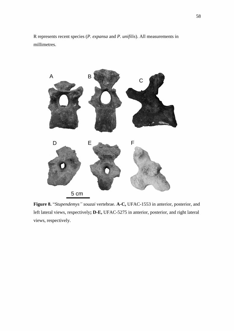
58
R represents recent species (P. expansa and P. unifilis). All measurements in
millimetres.
Figure 8. “Stupendemys” souzai vertebrae. A-C, UFAC-1553 in anterior, posterior, and
left lateral views, respectively; D-E, UFAC-5275 in anterior, posterior, and right lateral
views, respectively.
A B C
D E F
5 cm

59
4. Conclusões
Um novo podocnemídeo, Podocnemis manchineri, da Formação Solimões é
descrito. Ela se distingue de outras Podocnemis pela associação das características a
seguir: escudos vertebrais em forma de vaso, especialmente o vertebral dois; vertebral
um significantemente maior que outros escudos vertebrais; margem cranial da carapaça
arredondada, sem nenhum tipo de reentrância; e escudos gulares alcançando o
entoplastrão.
Aqui pudemos diferenciá-la de todas as outras espécies com partes coincidentes,
assim como incluir P. manchineri e vários outros táxons baseados em carapaças em
uma análise filogenética. Além disso, é bem provável que um terceiro táxon de
podocnemídeo de grande porte tenha vivido no Mioceno-Plioceno da Amazônia Sul-
ocidental.
Nós também concluímos que a presença de espécies fósseis entre a associação
comum de Peltocephalus e Erymnochelys mostrada aqui poderia ajudar a explicar as
relações e história biogeográfica desses táxons viventes.
Nós também provemos uma definição filogenética para as subfamílias
Erymnochelyinae e Podocneminae, sendo que renomeamos Erymnochelyinae como
Peltocephalinae. Devido às diferenças entre os dados morfológicos e moleculares. Os
dados morfológicos provêm uma divisão clara entre a família Podocnemidae, onde o
táxon Erymnochelys madagascariensis é mais relacionado à Peltocephalus dumeriliana
do que a Podocnemis expansa. Entretanto os dados moleculares mostram uma relação
mais próxima entre E. madagascariensis e P. expansa. Isso deixaria P. dumeriliana em
uma terceira subfamília, não suportada pelos dados morfológicos. Sendo assim,
preferimos aqui utilizar P. dumeriliana como a espécie tipo da subfamília. Desta forma,
Peltocephalinae é o clado “stem-based” definido como todos os Podocnemidae mais
relacionados à Peltocephalus dumerilianus do que à Podocnemis expansa e
Podocneminae é o clado “stem-based” definido como todos os Podocnemidae mais
relacionados à Podocnemis expansa do que à Peltocephalus dumerilianus.
Após a inclusão de novos caracteres referentes à carapaça e o plastrão na matriz
de dados e de acordo com nossa análise, nós concluímos que embora a morfologia do
casco dos Podocnemidae seja geralmente referida como conservativa ela pode prover
informações filogenéticas importantes. Nossa análise filogenética suporta a inclusão de

60
P. negrii, P. medemi, e P. pritchardi ao gênero Podocnemis. A análise também proveu
uma ideia melhor da posição de Stupendemys geographicus, como membro de
Erymnochelyinae. Além disso, um dos principais táxons problemáticos em várias
análises filogenéticas, P. bassleri, conhecida apenas por material do crânio é também
recuperada como uma Podocnemis. De maneira interessante, em contraste com outras
análises filogenéticas baseadas em caracteres morfológicos, a resolução da análise
dentro de Podocnemis caiu com a inclusão de P. bassleri, enquanto a análise com os
táxons fósseis baseados na carapaça resultaram em uma topologia mais resolvida. Já que
incluímos caracteres da carapaça e plastrão na análise, isso pode sugerir que eles podem
ser tão importantes quanto os caracteres cranianos em estudos cladísticos a respeito de
podocnemídeos.

61
5. Referências Bibliográficas
BARBOSA-RODRIGUES, J. B. 1892. Les reptiles de la vallée de l’Amazone. Vellosia,
2, 41–60.
BOCQUENTIN, J. & E. GUILHERME. 1999. As preguiças Mylodontinae (Mammalia,
Xenarthra, Mylodontidae) do Neógeno do Sítio Niterói, Acre, Brasil. Acta Geologica
Leopoldina, 22, 57–67.
BOCQUETIN-VILLANUEVA, J., JÉGU, M. & BRITO, P. M. 1997. “An extinct
Phractocephalus species (Siluriformes, Pimelodidae) from the Mio-Pliocene Solimões
Formation of Acre State, Brazil”. Em Abstracts of International Symposium on
Phylogeny and Classification of Neotropical Fishes, Museu de Ciências e Tecnologia.
PUCRS, 1997. p. 56. Porto Alegre.
BOCQUENTIN, J. V. & RANCY, A. 1987. “Presença de Chelus lewisi Wood, 1976
(Testudinata, Pleurodira) no Neógeno do Estado do Acre, Brasil”. Em 4o Congresso
Latino-Americano de Paleontologia, Santa Cruz de la Sierra, 1987, Editado por:
Asociacion Boliviana de Paleontologia. 566–573. Santa Cruz de la Sierra.
BOCQUENTIN, J. V., SANTOS, J. C. R. 1989. “Ocorrência de Chelus colombianus
(Chelonii, Chelidae) no Mioceno Superior do Acre, Brasil”. Em Resumos do 11o
Congresso Brasileiro de Paleontologia, Curitiba, 1989, Editado por: Sociedade
Brasileira de Paleontologia. 104–105. Curitiba.
BOCQUENTIN, J. V. & SOUZA-FILHO, J. P. 1989. “Nova interpretação do gênero
Purussaurus (Crocodylia, Alligatoridae)”. In Resumos do 11th Congresso Brasileiro de
Paleontologia, Curitiba, 1989, Editado por: Sociedade Brasileira de Paleontologia.
427–438. Curitiba.
BOCQUENTIN, J. V.., SOUZA-FILHO, J. P. 1990. O crocodiliano Sul-Americano
Carandaisuchus como sinonímia de Mourasuchus (Nettosuchidae). Revista Brasileira
de Geociências, 20, 230–233.

62
BOCQUENTIN, J. V., GUILHERME, E. & NEGRI, F. R. 2001. Duas espécies do
gênero Chelus (Pleurodira, Chelidae) no Mioceno Superior-Plioceno Inferior da
Amazônia Sul-Ocidental. Revista Universidade Guarulhos, 6, 50–55.
BOCQUENTIN, J. V. & MELO, J. 2006. Stupendemys souzai sp. nov. (Pleurodira,
Podocnemididae) from the Miocene-Pliocene of the Solimões Formation, Brazil.
Revista Brasileira de Paleontologia, 9, 187–192.
BROIN, F., BOCQUENTIN, J. & NEGRI, F. R. 1993. Gigant turtles (Pleurodira,
Podocnemididae) from the late Miocene-early Pliocene of South-Western Amazon.
Bulletin de l’Institut Français d’Études Andines, 22, 657–670.
CADENA, E. A., BLOCH, J. I. & JARAMILLO, C. A. 2010. New podocnemidid turtle
(Testudines: Pleurodira) from the Middle-Upper Paleocene of South America. Journal
of Vertebrate Paleontology, 30, 367–382.
CADENA, E. A., KSEPKA, D. T., JARAMILLO, C. A. & BLOCH, J. I. 2012. New
pelomedusoid turtles from the late Palaeocene Cerrejón Formation of Colombia and
their implications for phylogeny and body size evolution. Journal of Systematic
Paleontology, 10, 313–331.
CAMPOS, D. A. 1977. Tartarugas fósseis do Brasil. Dissertação de mestrado,
Universidade Federal do Rio de Janeiro, 101 pp.
CAMPOS, D. A. & BROIN, F. 1981. Tartarugas fósseis do Brasil. Anais da Academia
Brasileira de Ciências, 53, 210–211.
CARVALHO, P., BOCQUENTIN, J. & BROIN, F. L. 2002. Une nouvelle espèce de
Podocnemis (Pleurodira, Podocnemididae) provenant du Néogène de la Formação
Solimões, Acre, Brésil. Geobios, 35, 677–686.

63
GAFFNEY, E. S., CAMPBEL, K. E. & WOOD, R. C. 1998. Pelomedusoid side-necked
turtles from Late Miocene sediments in South-western Amazonia. American Museum
Novitates, 3245, 1–11.
GAFFNEY, E. S., TONG, H. & MEYLAN, P.A. 2006. Evolution of the side-necked
turtles: the families Bothremydidae, Euraxemydidae, and Araripemydidae. Bulletin of
the American Museum of Natural History, 300, 1–698.
GAFFNEY, E. S., MEYLAN, P. A., WOOD, R. C., SIMONS, E. & CAMPOS, D. A.
2011. Evolution of the side-necked turtles: the family Podocnemididae. Bulletin of the
American Museum of Natural History, 350, 1–37.
GASPARINI, Z. 1985. Un nuevo cocodrilo (Eusuchia) Cenozóico de América del Sur.
Coletânea de Trabalhos Paleontológicos, Série Geologia, 27, 51–53.
HENNIG, W. 1965. Phylogenetic systematics. Annual Review of Entomology, 10: 97-
116.
HSIOU, A. S. 2010. Os lagartos e serpentes (Lepidosauria, Squamata) do Mioceno
médio-superior da região norte da América do Sul. Tese de doutorado, Universidade
Federal do Rio Grande do Sul, 236 pp.
HSIOU, A.S., FERIGOLO, J. & ALBINO, A. 2007. Sobre os Squamata (Lepidosauria)
da Formação Solimões, Mioceno da Amazônia Sul-Ocidental, Brasil. Ameghiniana
(Suplemento), 44, 23R.
LAPPARENT DE BROIN, F., BOCQUENTIN, J. & NEGRI, F. R. 1993. Gigantic
turtles (Pleurodira, Podocnemididae) from the late Miocene–early Pliocene of south
western Amazon. Bulletin de l’Institut Francais d’Etudes Andines, 22, 657–670.
LATRUBESSE, E. M., BOCQUENTIN, J., SANTOS, C. R. & RAMONELL, C. G.
1997. Paleoenvironmental model for the late Cenozoic southwestern Amazonia:
paleontology and geology. Acta Amazonica, 27, 103–118.

64
MEYLAN, P. A., GAFFNEY, E. S. & CAMPOS, D. A. 2009. Caninemys, a new side-
necked turtle (Pelomedusoides: Podocnemididae) from the Miocene of Brazil. American
Museum Novitates, 3639, 1–26.
NEGRI, F. R. & FERIGOLO, J. 1999. Anatomia craniana de Neoepiblema
ambrosettianus (Ameghino, 1889) (Rodentia, Caviomorpha, Neoepiblemidae) do
Mioceno Superior-Plioceno, Estado do Acre, Brasil, e revisão das espécies do gênero.
Boletim do Museu Paraense Emílio Goeldi Série Ciências da Terra, 11, 1–80.
POUGH, F.H. JANIS, C.M. & HEISER, J.B. 2008. A Vida dos Vertebrados. Atheneu
Editora São Paulo Ltda. 4ªed.
PRICE, L. I. 1964. Sobre o crânio de um grande crocodilídeo extinto do Alto Rio Juruá,
Estado do Acre. Anais da Academia Brasiliera de Ciências, 36, 59–66.
RANCY, A. & BOCQUENTIN, J. V. 1987. “Dois quelônios do Neógeno do Acre,
Brasil”. In Anais do 10th Congresso Brasileiro de Paleontologia, Rio de Janeiro, 1987,
Editado por: Sociedade Brasileira de Paleontologia. 181–187. Rio de Janeiro.
RUEDA-ALMONACID, J. V., CARR, J. L., MITTERMEIER, R. A., RODRÍGUEZ-
M., J. V., MAST, R. B., VOGT, R. C., RHODIN, A. G. J., DE LA OSSA-
VELÁZQUEZ, J., RUEDA, J. N. & MITTERMEIER, C. G. 2007. Las tortugas y los
cocodrilianos de los países andinos del trópico. Serie de guías tropicales de campo Nº
6. Editorial Panamericana, Bogotá, 538 pp.
RIFF, D., ROMANO, P. S. R., OLIVEIRA, G. R. & AGUILERA, O. A. 2010. Neogene
crocodile and turtle fauna in northern South America. Pp. 259–280 in C. Hoorn & F.
P.Wesselingh, (eds) Amazonia: landscape and species evolution - a look into the past.
Blackwell Publishing Ltd., Chichester.
SOUZA-FILHO, J. P. 1998. Novas formas fósseis de Crocodylia (Alligatoridae e
Gavialidae) da Formação Solimões, Cenozóico do Estado do Acre-Brasil,

65
representadas por materiais cranianos e mandibulares. Tese de doutorado,
Universidade Federal do Rio Grande do Sul, 194 pp.
SOUZA-FILHO, J. P. & BOCQUENTIN, J. 1989. “Brasilosuchus mendesi, n.g. n.sp.
um novo representante da Família Gavialidae do Neógeno do Estado do Acre, Brasil”.
In 11th Congresso Brasileiro de Paleontologia, Curitiba, 1989, Editado por: Sociedade
Brasileira de Paleontologia. p. 139. Curitiba.
SOUZA-FILHO, J. P. & BOCQUENTIN, J. 1991. “Caiman niteroiensis sp. nov.
(Alligatoridae, Crocodylia) do Neógeno do Estado do Acre, Brasil”. In 12th Congresso
Brasileiro de Paleontologia, São Paulo, 1991, Editado por: Sociedade Brasileira de
Paleontologia. p. 126. São Paulo.
SILL, W. D. 1970. Nota preliminar sobre un nuevo gavial del Plioceno de Venezuela y
una discusión de los gaviales sud-americanos. Ameghiniana, 7, 151–159.
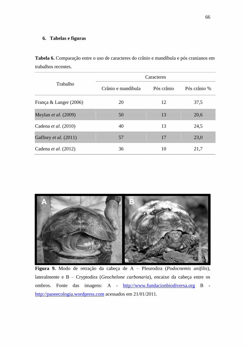
66
6. Tabelas e figuras
Tabela 6. Comparação entre o uso de caracteres do crânio e mandíbula e pós cranianos em
trabalhos recentes.
Trabalho
Caracteres
Crânio e mandíbula Pós crânio Pós crânio %
França & Langer (2006) 20 12 37,5
Meylan et al. (2009) 50 13 20,6
Cadena et al. (2010) 40 13 24,5
Gaffney et al. (2011) 57 17 23,0
Cadena et al. (2012) 36 10 21,7
Figura 9. Modo de retração da cabeça de A – Pleurodira (Podocnemis unifilis),
lateralmente e B – Cryptodira (Geochelone carbonaria), encaixe da cabeça entre os
ombros. Fonte das imagens: A - http://www.fundacionbiodiversa.org B -
http://paoeecologia.wordpress.com acessados em 21/01/2011.
A B

67
Figura 10. Possíveis relações entre os táxons A, B e C. (Baseado em
Hennig, 1987)
A C B A B C
C A B Sinapomorfia de A e B
x’
x
x – Caractere plesiomórfico
(primitivo)
x’ – Caractere apomórfico
(derivado)
Top Related
