
Línguas
Páginas
Legal

Gabriela Fernandes Henriques
Licenciada em Biologia Celular e Molecular
Study of the role of wall teichoic acids in
the localization Staphylococcus aureus cell
wall synthesis protein PBP4
Orientador: Doutora Mariana G. Pinho, Investigador Auxiliar, ITQB AX-UNL
Outubro de 2013

Gabriela Fernandes Henriques
Licenciada em Biologia Celular e Molecular
Study of the role of wall teichoic acids in
the localization Staphylococcus aureus cell
wall synthesis protein PBP4
Dissertação para obtenção do Grau de Mestre em Genética Molecular e Biomedicina
Orientador: Doutora Mariana G. Pinho, Investigador Auxiliar, ITQB AX-UNL
Júri:
Presidente: Prof. José Paulo Sampaio
Arguente (s): Doutora Rita Sobral
Vogal (ais): Doutora Mariana G. Pinho
Outubro de 2013


Study of the role of wall teichoic acids in the localization Staphylococcus aureus cell wall
synthesis protein PBP4
Copyright Gabriela Fernandes Henriques, FCT/UNL, UNL
A Faculdade de Ciências e Tecnologia e a Universidade Nova de Lisboa têm o direito, perpétuo e sem limites
geográficos, de arquivar e publicar esta dissertação através de exemplares impressos reproduzidos em papel ou
de forma digital, ou por qualquer outro meio conhecido ou que venha a ser inventado, e de a divulgar através de
repositórios científicos e de admitir a sua cópia e distribuição com objectivos educacionais ou de investigação, não
comerciais, desde que seja dado crédito ao autor e editor


I
Acknowledgments
The work obtained during this journey would not be possible without the help and support of an
amazing group of people that I want to acknowledge.
Firstly, I want to thank my supervisor Dr. Mariana Pinho, for the opportunity to develop my
master's thesis at this great institute (ITQB AX-UNL), in her amazing group, for everything she taught
me during this year, for sharing her vision and knowledge, for all of her support and encouragement at
such an important and difficult moment of my life.
To my internal supervisor Dr. Ana Madalena Ludovice, thank you for helping me when I needed
it most, for all the concern and advices during the writing of my work.
I would like to thank Dr. Sérgio Filipe, for his help with difficult questions and challenging discussions
in the lab meetings.
I would also like to acknowledge my faculty, Faculdade de Ciências e Tecnologia da
Universidade Nova de Lisboa, and to Professor José Paulo Sampaio, coordinator of the Molecular
Genetics and Biomedicine Master degree, for the opportunity to grow professionally and for
contributing to my development as a scientist.
I would like to thank Dr. Patricia Reed for teaching me and sharing her knowledge during this
year, which contributed not only to this work, but for my professional development.
A special thanks to all my lab colleagues, for the amazing environment, lovely friendship and
for all the help, support and encouragement. To Helena Veiga and Pedro Matos, for all the helpful work
discussions, help and for believing in me and my work when I most needed. To Pedro Fernandes, my
fellow master student, for sharing this journey. To Teresa Ferreira, for sharing this special year with me,
thank you for all the moments, for the laughs and tears you shared with me and for your friendship.
To my dear family – Agradeço à minha família por me fazerem de mim a pessoa mais sortuda
e feliz, por me apoiarem incondicionalmente e porque ser tão difícil encontrar uma família como a nossa,
enorme e que está presente nos momentos mais importantes. Claro não posso deixar de agradecer
especialmente à minha Mãe por todo o esforço dos últimos anos, por acreditar em mim e nos meus
sonhos, por me apoiar incondicionalmente e principalmente por ser a minha amiga e companheira para
a vida; Ao meu pai por todo o suporte e apoio incondicional, por todo o carinho e otimismo que só um
pai sabe transmitir, que foram bastante importantes para voltar a acreditar em mim em muitos momentos
de frustração. Às minhas segundas mães, Tia Ângela e Tia Milu, e aos irmãos de coração, Catarina
Lopes e Cláudio Lopes, por todo o apoio, conselhos, conversas e carinho que foram tão importantes
nesta nova fase tão importante da minha vida. E aos novos membros da nossa família, os meus
priminhos, que com as suas brincadeiras e conquistas enchem a minha vida de alegria.
Este ano não teria sido possível sem a presença, o suporte, o amor, carinho e apoio e
incondicional do Diogo, que é acima de tudo o meu melhor amigo. Deste modo agradeço-lhe por toda a

II
paciência que teve comigo (mesmo sem perceber nada de bactérias passou horas exaustivas a olhar para
elas comigo), e por ter estado do meu lado mais uma jornada tão importante.
Por último, e não menos importante, um enorme obrigado, aos meus amigos (“a minha segunda
família”), por todos os momentos especiais que partilhámos, por me apoiarem neste percurso e nas
minhas decisões, mesmo que isso implique ouvir algum conselho mais difícil, por me aturarem, pelas
visitas a casa quando não podia sair ou pelos resgates para ir tomar café, resumindo por serem tão
importantes e terem tornado este ano muito melhor. Um especial obrigado à Ana Sofia Santana e à Joana
Pereira, por me terem feito companhia em muitos fins-de-semana passados no laboratório, assim como
nos momentos mais críticos e stressantes, mas principalmente por me apoiarem incondicionalmente e
pela nossa amizade tão especial. À Carolina Cassona e à Joana Viana, que mesmo longe foram amigas
tão especiais e presentes, que não deixaram de me apoiar e partilhar todas as minhas alegrias e
frustrações. À Patrícia Apura pela nossa amizade, pelo seu apoio, carinho e por todas as visitas diárias,
que como tantas gargalhadas tornaram muitos dos meus dias melhores.
III
Resumo
A parede celular de Staphylococcus aureus é uma rede extremamente complexa composta
maioritariamente por peptidoglicano (PG) com alto teor em pontes interpeptidicas e ácidos teicóicos
(TAs), ambos importantes para a manutenção da integridade e viabilidade celular da bactéria. As
proteínas de ligação à penicilina (PBP), que catalisam a fase final da biossíntese do PG, são alvos dos
antibióticos β –lactâmicos e como tal têm sido um dos principais focos da investigação antibacteriana.
S. aureus tem quatro PBPs nativas, PBP1 – 4, que estão presentes quer nas estirpes sensíveis á meticilina
(MSSA), quer nas resistentes (MRSA). PBP4 cataliza a formação de ligações interpetidicas do
peptidoglicano e, como demonstrado recentemente, é essencial para a expressão da resistência aos
antibióticos β - lactâmicos em estirpes adquiridas na comunidade (CA-MRSA). Esta proteína, em S.
aureus, localiza-se no septo celular, localização esta que parece ser espacialmente e temporalmente
regulada por um intermediário, ainda não identificado, da biossíntese dos ácidos teicoícos da parede
(WTA). Neste sentido, se a síntese dos WTA é comprometida, a PBP4 perde a sua localização septal e
surge dispersa por toda a membrana celular. O objetivo deste projeto foi identificar o precursor da síntese
dos WTA responsável pelo recrutamento septal da PBP4. Foram construídos mutantes indutíveis de dois
genes essenciais para esta via de síntese, o tarB e tarL, utilizando a estirpe NCTCPBP4 – YFP (que
expressa um derivado fluorescente da PBP4), o que nos permite estudar a localização da PBP4 na
presença e ausência da expressão destes genes. Em conclusão, com este trabalho, fomos capazes de
mostrar que a ausência destas duas proteínas, TarB e TarL, levam à deslocalização da PBP4, indicando
que provavelmente a proteína TarL ou uma proteína ou precursores da síntese WTA dependente de
TarL, é responsáveis pelo recrutamento de PBP4.
Palavras-chave: Staphylococcus aureus; Parede celular; Resistência aos antibióticos β –
lactâmicos; Biossíntese dos ácidos teicoícos; Proteínas de ligação à penicilina; localização de proteínas.

IV

V
Abstract
The cell wall of Staphylococcus aureus is a highly complex network mainly composed of highly
cross-linked peptidoglycan (PG) and teichoic acids (TAs), both important for the maintenance of the
integrity and viability of bacteria. The penicillin binding proteins (PBPs), which catalyse the final stage
of PG biosynthesis, are targets of β-lactam antibiotics and have been a key focus of antibacterial
research. S. aureus has four native PBPs, PBP1-4 carried by both methicillin-sensitive (MSSA) and –
resistant (MRSA) strains. PBP4 is required for the synthesis of the highly cross-linked PG and, as shown
in recent studies, is essential for the expression of β-lactam resistance in community-acquired strains
(CA-MRSA). This protein has a septal localization that seems to be spatially and temporally regulated
by an unknown intermediate of the wall teichoic acids (WTA) biosynthesis pathway. Therefore, if WTA
synthesis is compromised, PBP4 becomes dispersed throughout the entire cell membrane. The aim of
this project was to identify the WTA precursor responsible for the septal recruitment of PBP4. In order
to do so, inducible mutants of tarB and tarL genes in the background of NCTCPBP4-YFP were
constructed allowing for the study of PBP4 localization in the presence and absence of these specific tar
genes.With this work we were able to show that the absence of TarB or TarL leads to the delocalization
of PBP4, indicating that TarL or a protein/WTA precursor whose localization/synthesis is dependent on
TarL is responsible for the recruitment of PBP4.
Keywords: Staphylococcus aureus; cell wall; β-lactam resistance; wall teichoic acids
biosynthesis; penicillin-binding proteins, protein localization

VI

VII
Contents Introduction ........................................................................................................................................... 1
Staphylococcus aureus as an antibiotic resistant pathogen. ................................................................ 1
Cell wall biosynthesis and β-lactam resistance. .................................................................................. 2
Wall teichoic acid biosynthesis and β-lactam resistance. .................................................................... 5
Connection between WTA and PG biosynthesis in S. aureus ........................................................... 11
Materials and Methods ....................................................................................................................... 13
Bacterial strains and growth conditions ............................................................................................ 13
General procedures ............................................................................................................................ 13
Mutant construction ........................................................................................................................... 16
Growth analysis of S. aureus strains ................................................................................................. 19
Fluorescence Microscopy .................................................................................................................. 19
Analysis of the expression of fluorescent proteins in S. aureus ........................................................ 20
Western blot analysis......................................................................................................................... 20
Results .................................................................................................................................................. 21
Construction of TarB and TarL inducible mutants ............................................................................ 21
Deletion of tarB or tarL leads to delocalization of PBP4. ................................................................ 26
Depletion of TarB causes delocalization of PBP4......................................................................... 26
Depletion of TarL causes delocalization of PBP4. ........................................................................ 29
Statistical analysis. ........................................................................................................................ 31
The PBP4-YFP fusion is not cleaved. ............................................................................................... 31
Delocalization of PBP4 in the absence of TarL or Tar B is not due to cell death. ............................ 33
Discussion ............................................................................................................................................. 39
Bibliography ........................................................................................................................................ 43

VIII

IX
Figures and Tables Index
Chapter 1- Introduction
Figure 1.1. Cell wall biosynthesis in S. aureus…………………………………………………………4 Figure 1.3. Chemical structure of wall teichoic acids (WTAs) in Staphylococcus aureus…….......……6
Figure 1.4. Genes and proteins involved in the primary Staphylococcus aureus WTA biosynthetic
pathway…………………………………………………………………………………………….8
Figure 1.5. Staphylococcus aureus WTA biosynthetic pathway, with potential antibiotic targets…….9
Figure 1.6. Model for the role of teichoic acids synthesis in PBP4 recruitment to the septum…….....12
Chapter 2- Material and Methods
Table 2.1. Bacterial strains used and constructed in this study……………………......………………15
Table 2.2. Plasmids used and constructed in this study……………….………………...…………….16
Table 2.3. Primers used in this study…………………………………...………………...…………...16
Chapter 3- Results
Figure 3.1. Schematic representation of the inducible mutant constructs.............................................21
Figure 3.2. Schematic representation of the spa gene replacement by an gene of interest...................22
Figure 3.3. Growth of S. aureus in the presence or in the absence of TarB and TarL..........................23
Figure 3.4. Growth analysis of NCTCΔspa::tarBi................................................................................24
Figure 3.5. Growth analysis of NCTCΔspa::tarLi……………………………………………………25
Figure 3.6. Septal localization of PBP4 is lost in absence of TarB in the NCTCΔspa::tarBi inducible
strain...............................................................................................................................................27
Figure 3.7. Septal localization of PBP4 is lost in absence of TarL in the NCTCΔspa::tarLi inducible
strain...............................................................................................................................................29
Figure 3.8. The PBP4-YFP fusion is not cleaved in NCTCPBP4-YFP, NCTCΔspa::tarBi and
NCTCΔspa::tarLi strains................................................................................................................32
Figure 3.9. Strains NCTCΔspa::tarBΔtarBi EzrA-cfp and NCTCΔspa::tarLΔtarLi EzrA-cfp form
aggregates.......................................................................................................................................34
Figure 3.10. The presence of Erythromycin does not affect the formation of cell aggregates in tarB
and tarL inducible strains...............................................................................................................35
Figure 3.11. Morphology of NCTCEzrA-CFP cells.............................................................................36
Figure 3.12. Septal localization of EzrA is not lost in the absence of TarB or TarL………............…37

X

Chapter 1 – Introduction
1
Introduction
Staphylococcus aureus as an antibiotic resistant pathogen.
The gram-positive cocci Staphylococcus aureus is a common commensal organism of the skin
and mucosal surfaces, but it is also an important opportunistic pathogen responsible for a wide range of
nosocomial and community-acquired infections, such as skin and ocular infections, pneumonia,
septicemia, endocarditis and osteomyelitis (Archer, 1998; Diekema et al., 2001). This organism lives as
a persistent commensal on 20% of the human population, preferentially on the skin and nasopharynx,
and it is intermittently carried by a further 60% of individuals (Edwards et al., 2012; Foster, 2005).
Colonization is normally asymptomatic, but clearly increases the risk for subsequent infection, as if the
skin barrier or the mucous membranes are breached S. aureus can enter into the soft tissues and establish
an invasive infection. Colonization also allows the transmission of S. aureus by skin-to-skin contact
between individuals or contaminated objects (Archer, 1998; Miller and Diep, 2008; Wertheim et al.,
2005). The success of S. aureus as a virulent pathogen and its ability to cause a large spectrum of
infections are due to the expression of several virulence factors, such as surface-attached proteins and
secreted enzymes, that allow the adherence to and invasion of human tissues, impart resistance to innate
immune defences and act as toxins (Archer, 1998; Edwards et al., 2012; Gordon and Lowy, 2008).
Antibiotic resistance in S. aureus is also a serious health-care problem due to its remarkable
ability to develop new mechanisms to resist the effects of antimicrobial agents. The introduction of the
β-lactam penicillin in the early 1940s, the first effective drug against S. aureus, produced in 1928 by the
Scottish microbiologist Alexander Fleming, dramatically improved the prognostic of patients with
staphylococcal infections (Plord and Sherris, 1974). However, in 1942, as a consequence of the
remarkable adaptive efficiency of S. aureus, penicillin-resistant staphylococci were recognized, first in
hospitals and then in the community. By the late 1960s, more than 80 % of both community- and
hospital-acquired staphylococcal isolates were resistant to penicillin (Lowy, 2003; Swoboda et al., 2010;
Swoboda et al., 2009; Szweda et al., 2012). The resistance of these strains was conferred by the presence
of a plasmid containing the blaZ gene that encodes a β-lactamase (called first penicillinase), an
extracellular enzyme synthetized when staphylococci are exposed to β-lactam antibiotics. The enzyme
functions to hydrolyse the β-lactam ring of penicillin, thus rendering the antibiotic inactive (Lowy,
2003). In the sixties, a semisynthetic β-lactamase-resistant penicillin called methicillin was developed
to treat the infections caused by these penicillin-resistant S. aureus strains (Barber, 1961; Parker and
Hewitt, 1970). However, soon after methicillin therapy in hospitals began, methicillin resistant
Staphylococcus aureus (MRSA) strains were isolated, initially from patients in a hospital in Colindale,
UK. Through the late 1960s and early 1970s, MRSA strains were reported, with increasing frequency,
in others countries all over the world, such as Australia, Belgium, Denmark, France, India, Poland,
Switzerland and United States of America (Chambers, 1988; Jevons et al., 1963; Lyon and Skurray,

Chapter 1 – Introduction
2
1987; Szweda et al., 2012). Nowadays, MRSA strains are one of the leading causes of nosocomial
infections worldwide (Chambers and Deleo, 2009). Recent studies show that in the United States the
number of deaths caused by MRSA infections is higher than those related to HIV/AIDS and tuberculosis
combined (Boucher and Corey, 2008). Reports from The European Centre of Disease Prevention and
Control (ECDC) show that in recent years the percentage of methicillin-resistant S. aureus isolates has
increased dramatically. For example in Portugal more than 50% of isolates are now resistant to
methicillin.
For the first three decades after their appearance, MRSA strains were known only as hospital-
acquired pathogens (HA-MRSA). Then, in the early nineties, with an unpredicted epidemiological turn,
MRSA strains also began to appear in the community among healthy people, who had no symptoms or
risk factors for such infections. These strains, called community-acquired MRSA (CA-MRSA) (Okuma
et al., 2002; Rice, 2006; Saravolatz et al., 1982), are less resistant to most antibiotics, other than β-
lactams, but exhibit a major virulence potential, and are consequently capable of causing infections in
healthy individuals (Szweda et al., 2012). The spread of such a dangerous pathogen to the community
is recognized as a disturbing reality and a huge concern in many countries. It also highlights the
requirement for an increase in our knowledge about the resistance mechanisms in S. aureus to aid in the
development of new therapies against these infections.
Cell wall biosynthesis and β-lactam resistance.
The cell wall, the external layer of bacterial cells, is very important for the integrity and viability
of bacteria, as it provides physical protection, determines the cell shape and is the principal stress-
bearing element, which makes it an ideal target for antibiotics (Scheffers and Pinho, 2005). In Gram-
positive bacteria such as S. aureus the cell wall is composed of surface proteins, teichoic acids and a
thick layer of peptidoglycan (PG). Peptidoglycan, also called murein, is a heteropolymer composed of
long glycan chains, made up of alternating β-1,4-linked N-acetylglucosamine (GlcNAc) and N-
acetylmuramic acid (MurNAc) subunits, which are cross-linked by flexible peptide bridges to form a
strong but flexible structure (Beeby et al., 2013; Scheffers and Pinho, 2005; Schleifer and Kandler,
1972; Szweda et al., 2012). Peptidoglycan is present in almost all bacteria, except in Mycoplasma and
a few other species that lack detectable cell walls. Attached to the carboxyl group of each MurNAc
residue are stem peptides that, unlike glycan chains, have varying composition between different
species. In S. aureus the stem peptides are composed of the sequentially added L-Alanine (L-Ala), D-
Glutamic acid (D-Glu), L-Lysine (L-Lys), D-Alanine (D-Ala), D-Ala amino acids. The interpeptide
bridges, created by the addition of five glycine residues to the L-Lys residue, allow for the cross-linking
between different layers of PG (Kopp et al., 1996; Schleifer and Kandler, 1972; Vollmer et al., 2008).
Peptidoglycan synthesis is a major target of some of the most successful classes of antibiotics,
including the β-lactams such as penicillin or methicillin (Popham, 2013). The biosynthesis of PG can be
divided into three different stages, as shown in Figure1.1 (Heijenoort, 1998, 2001). The first stage

Chapter 1 – Introduction
3
involves the cytoplasmic synthesis of the nucleotide sugar-linked precursors UDP-N-acetylmuramyl-
pentapeptide (UDP-MurNAc-pentapeptide) and UDP-N-acetylglucosamine (UDP-GlcNAc). In the
second stage, which takes place at the inner side of the cytoplasmic membrane, MraY transfers the
phospho-MurNAc-pentapeptide moiety of UDP-MurNAc-pentapeptide to the membrane acceptor
bactoprenol, generating lipid I [MurNAc-(pentapeptide)-pyrophosphoryl-undecaprenol]. MurG then
promotes the β-1,4 linkage between UDP-GlcNAc and lipid I, yielding the final PG precursor, lipid II
[GlcNAc-β- (1,4)-MurNAc- (pentapeptide)-pyrophosphoryl-undecaprenol]. Before its translocation to
the outer side of the cytoplasmic membrane, the lipid II is modified by a family of peptidyltransferases
(FemX, FemA and FemB), which promote the sequential addition of five glycines to the L-Lys residue,
creating a pentaglycine bridge peptide for the cross-linking of PG in the cell wall. It has been proposed
that the export of the fully modified PG lipid II precursor is catalyzed by a flippase (Roemer et al., 2013;
Typas et al., 2012) . The third and final stage of PG biosynthesis, that takes place at the outer surface of
the cytoplasmic membrane, consists on the polymerization of the newly synthesized disaccharide–
peptide units and its incorporation into the growing PG, by elongation (transglycosylation) and peptide
cross-linking (transpeptidation) between glycan strands (Heijenoort, 1998, 2001; Llarrull et al., 2009;
Scheffers and Pinho, 2005; Typas et al., 2012; Vollmer et al., 2008). These reactions, which occur
mainly at the division septum of S. aureus, are catalyzed by the penicillin‐binding proteins (PBPs) and
monofunctional transglycosylases (Pinho and Errington, 2003). PBPs are membrane-associated
proteins, anchored to the cytoplasmic membrane facing the extracellular surface, which can be classified
as low-molecular-weight (LMW) and high-molecular-weight (HMW) proteins (Ghuysen, 1991; Goffin
and Ghuysen, 1998). LMW PBPs are enzymes that only have a penicillin binding domain, that exhibit
a DD- carboxypeptidase leading to the removal of terminal D-aminoacids from the PG muropeptides or
transpeptidase activity leading to the formation of the cross‐links between the peptides strands of PG.
HMW PBPs are enzymes composed of two modules located on the outer side of cytoplasm membrane
and an N-terminal anchored to the cytoplasmic membrane. The C‐terminal is the penicillin binding
domain, with transpetpidasse (TP) activity responsible for the cross-linking of the PG peptides. The N-
terminal domain allows, depending on its primary structure and catalytic activity, the classification of
HMW PBPs into two major classes: A and B (Ghuysen, 1991; Goffin and Ghuysen, 1998). The N-
terminal domain of class A PBPs has a glycosyltransferase activity, catalyzing the elongation of glycan
strands. The N-terminal domain of HMW class B PBPs have a non-penicillin-binding domain of
unknown function, that has been suggested to have a role in cell morphogenesis (Scheffers and Pinho,
2005).

Chapter 1 – Introduction
4
Since their discovery as targets of β-lactam antibiotics, PBPs have been a key focus of
antibacterial research. β-lactam antibiotics bind irreversibly to the transpeptidase active site of PBPs.
Through the formation of an acyl-enzyme complex, they act as pseudosubstrates causing the inhibition
of synthesis and cross-linking of PG, resulting in the weakening of the cell wall and leading to eventual
cell lysis (Llarrull et al., 2009; Zapun et al., 2008). S. aureus have four native PBPs, PBP1-4 carried by
both methicillin-sensitive and –resistant strains, to which most β-lactam antibiotics bind (Pereira et al.,
2009; Pinho et al., 1998; Zapun et al., 2008). The first three are HMW PBPs, while PBP4, a non-
essential protein, is a LMW PBP that has transpeptidase activity performing secondary cross-linking of
the PG and therefore leading to the high degree of cross‐linking characteristic of the S. aureus PG (Leski
and Tomasz, 2005; Memmi et al., 2008). Recent studies have also shown that PBP4, is essential for the
expression of β-lactam resistance in CA-MRSA (Memmi et al., 2008). MRSA strains encode an
Figure 1.1. Cell wall biosynthesis in S. aureus. The image represents the three stages of cell wall synthesis:
(i) cytoplasmic synthesis of the UDP-MurNAc-pentapeptide and the UDP-GlcNAc; (ii) inner membrane
biosynthesis of the lipid II precursor and (iii) outer membrane polymerization of glycan chains and peptide
crosslinking. The chemical structure of a muropeptide and the enzymes which catalyze each biosynthetic step
are also represented (reproduced from Pinho (2008)).

Chapter 1 – Introduction
5
additional PBP, PBP2A, the expression of which is responsible for the resistance of these strains to β-
lactam antibiotics. This enzyme is encoded by the mecA gene that is situated in the chromosome in a
genomic island designated staphylococcal cassette chromosome mec (SCCmec) (Berger-Bächi et al.,
1992; de Lencastre et al., 2007; de Lencastre and Tomasz, 1994; Verghese et al., 2012). The mecA gene
is not native to S. aureus, but was acquired by lateral transfer, possibly from others related organisms,
like Staphylococcus sciuri or Staphylococcus fleurettii (Couto et al., 1996; Crisostomo et al., 2001; de
Lencastre et al., 2007). PBP2A has a remarkably low affinity for all β-lactams, and in their presence
performs all of the transpeptidase activity, in cooperation with the glycosyltransferase activity of PBP2,
ensuring continued cell wall synthesis (Pinho et al., 2001a; Pinho et al., 2001b; Pinho et al., 1997).
Wall teichoic acid biosynthesis and β-lactam resistance.
In addition to peptidoglycan, an important class of cell surface glycopolymers in Gram‐positive
bacteria are the phosphate rich teichoic acids (TAs). These molecules play a role in a large variety of
functions, such as in maintaining the physicochemical properties of the cell surface, cation homeostasis,
resistance to antimicrobial peptides and lytic enzymes, acting as phage receptors, in cell division, biofilm
formation and host adhesion (Figure 1.2). There are two types of TAs, distinguished by the way they
are covalently linked to the surface, the lipo- teichoic acids (LTAs), which are anchored to the
cytoplasmic membrane, extending from the cell into the peptidoglycan layer, and the wall teichoic acids
(WTAs), which are covalently attached to the peptidoglycan layers and extend beyond them (Figure
1.2). Together, the LTAs and the WTAs, create a negative gradient that goes from the bacterial cell
surface until the outer most layers of the PG (Morath et al., 2005; Pasquina et al., 2013; Swoboda et al.,
2010; Weidenmaier and Peschel, 2008).
Figure 1.2. Simplified illustration of Gram-positive bacterial cell envelope and the TAs functions. A)
Representation of the Gram-positive bacterial cell wall. This image does not show proteins, which are also an
important element of the cell wall, in order to simplify the scheme. LTA: lipo-teichoic acid; WTA: wall teichoic
acid. (Adapted from Swoboda et al. 2010); B) Representation of the functions of teichoic acid, which are involved
in cell division, charge homeostasis and infection. (Adapted from Pasquina et al. 2013).
B A

Chapter 1 – Introduction
6
It has been shown that the expression of WTAs is critical for the pathogenicity of S. aureus
strains, so a detailed study of WTA biosynthesis is important for a better understanding of their roles in
bacterial physiology and to evaluate their potential as antibacterial targets (Weidenmaier et al., 2005).
The chemical structure of WTAs vary among Gram‐positive bacteria, but the most common structures
are composed of a β‐(1,4)‐linked N‐acetylmannosamine (ManNAc) and N‐ acetylglucosamine
(GlcNAc), attached by a phosphodiester linkage to the C6 hydroxyl of MurNAc residue of PG, followed
by two glycerol phosphate units which are linked to a chain of glycerol- or ribitol phosphate repeats
(Lazarevic et al., 2002; Sanderson et al., 1962). S. aureus WTAs contain polyribitol phosphate (poly‐
RboP) units with GlcNAc and cationic D‐alanine esters substituents at their hydroxyl group (Figure 1.3)
(Brown et al., 2010; Weidenmaier and Peschel, 2008)
The biosynthesis of WTAs (shown in Figure 1.4) in S. aureus is catalysed by the tar genes (for teichoic
acid ribitol) whose function has been established based mostly on sequence homology to the tag genes
(for teichoic acid glycerol) involved in the production of WTAs of the well-studied model organism
Bacillus subtilis (Lazarevic et al., 2002; Qian et al., 2006). This biosynthesis pathway begins in the
cytoplasm, at the wall-membrane interface, with the transfer of GlcNAc-1-P from UDP‐GlcNAc to the
membrane-anchored undecaprenyl phosphate carrier lipid, an intermediate also used in the PG
biosynthesis. This first step is a reversible reaction catalysed by TarO, which is a N‐acetylglucosamine‐
1‐phosphate transferase that belongs to the glycosyltransferase family, which also includes the enzyme
MraY, required for PG biosynthesis (Anderson et al., 1978; Brown et al., 2008; Soldo et al., 2002). The
first irreversible step in WTA biosynthesis is catalysed by an N‐acetylmannosaminyl transferase, TarA,
that transfers a ManNAc residue from the UDP‐ManNAc to the C4 hydroxyl of GlcNAc forming a β‐
linked disaccharide (Yokoyama et al., 1989; Zhang et al., 2006). Following the formation of the
ManNAc(β1-4)GlcNAc disaccharide, the synthesis continues with the addition of two glycerol‐3‐
phosphate units, by TarB and TarF glycerolphosphate transferases (Brown et al., 2008). The glycerol‐
Figure 1.3. Chemical structure of wall teichoic acids (WTAs) in Staphylococcus
aureus. RboP: ribitol-phosphate; y = 1–2, z = 20–40 (Adapted from Brown et al. 2010).
Chapter 1 – Introduction
7
3‐phosphate derived from CDP‐glycerol is a nucleotide‐activated precursor of TarD, a
cytidylyltransferase (Park et al., 1993). In S. aureus the assembly of the WTA main chain (a poly-ribitol-
5-P chain), requires a bi‐functional poly‐ribitol primase/polymerase, TarL, which transfers a single
ribitol phosphate residue to the linkage unit and then attaches more than forty ribitol-5-P units to
complete the polymer (Brown et al., 2008; Meredith et al., 2008). The ribitol-5-P is derived from CDP-
Ribitol, in a reaction performed by the combined action of TarI, a cytidylyltransferase, and TarJ, an
alcohol dehydrogenase (Pereira and Brown, 2004). All S. aureus strains contain an apparent duplication
of the chromosomal region containing the tarIJL genes, this second set of genes is designated tarI’J’K.
The significance of these duplications is still unclear, and it was already shown that the tarK gene is
highly homologous to the tarL gene and consequently their encoded enzymes have similar functions.
TarL has a polymerase function that catalyses the formation of a primary TarL-directed WTA polymer
(L-WTA) while TarK it’s a primase makes a secondary TarK-directed WTA polymer (K-WTA)
(Meredith et al., 2008; Pereira et al., 2008; Swoboda et al., 2010). The WTA glycosylation occurs in
the cytoplasm, following polymer synthesis, through the addition of α‐GlcNAc, by TarM, and β‐
GlcNAc, by TarS (Brown et al., 2012; Xia et al., 2010). The WTA polymer is then translocated to the
external side of the membrane by the ABC transporter complex composed of TarH and TarG. This WTA
transporter consists of an ATPase domain, the TarH, which provides the necessary energy to catalyse a
conformational change in the transmembrane component, and a transmembrane domain, the TarG, a
channel that facilitates the translocation across the membrane (Schirner et al., 2011; Seeger and van
Veen, 2009). Once the WTA polymer is outside of the cell, it has to be incorporated into the PG, by a
phosphodiester linkage between the polymer and the C6 hydroxyl of the PG MurNAc residue. This
reaction is catalysed by unknown proteins, presumably homologous to the TagTUV enzymes (Brown et
al., 2013). The D‐alanylation of WTAs is another important mechanism, because it allows bacteria
modulate their surface charge. This process, which occurs outside the cell, involves the attachment of
D-alanine esters to WTAs and is catalysed by four enzymes encoded in the dltABCD operon (Kovacs et
al., 2006). Although this reaction is not completely understood, it is believed that the DltA, an D‐alanyl
carrier protein ligase, activates D‐alanine as an AMP ester and then, with the help of the membrane‐
anchored DltD protein, transfers the aminoacyl adenylate to the carrier protein DltC (Heaton and
Neuhaus, 1992, 1994). The DltB protein is an uncharacterized transmembrane protein of the membrane-
bound-O-acetyltransferase (MBOAT) family, that has been suggested to be involved in the translocation
of the D‐alanine-charged DltC across the cytoplasmic membrane, where D‐alanine is then transferred
to the WTA backbone (Brown et al., 2013). These final steps of the synthesis pathway are illustrated in
Figure 1.5.
WTAs are not essential for S. aureus viability, since tarO and tarA can be deleted and the mutant
strains survive (although their growth and virulence are impaired) (D'Elia et al., 2006a). In contrast, the
deletion of genes involved in downstream reactions of the WTAs biosynthesis pathway results in a lethal
phenotype, indicating that these are conditionally essential genes. The lethal phenotype can be rescued

Chapter 1 – Introduction
8
in a ΔtarO or ΔtarA background, suggesting that lethality can be due to the accumulation of toxic
intermediates in the cell or depletion of cellular undecaprenyl phosphate, an intermediate shared with
the PG biosynthesis (D'Elia et al., 2006b; Swoboda et al., 2010).
The role of WTA in β-lactam resistance of MRSA strains has remained elusive for a long time.
In 1994, Maki et al identified the llm gene, through transposon insertional inactivation as playing an
important role in methicillin resistance of MRSA strains. Although its molecular function was
unknown, llm mutants had a profoundly restored β-lactam susceptibility in a wide range of MRSA
clinical isolates studied (Maki et al., 1994). Recent studies showed, by sequence comparison, that llm is
the same as tarO, the gene encoding the first enzyme in wall teichoic acid (WTA) biosynthesis pathway
in S. aureus (Campbell et al., 2010).
A.
B.
Figure 1.4. Genes and proteins involved in the primary Staphylococcus aureus WTA biosynthetic
pathway. A) Genetic organization of wall teichoic acid biosynthetic genes in S. aureus; tar: teichoic acid
ribitol (//: number of nucleic acids between genes if >120 base pairs); B) Depiction of the primary S. aureus
WTA biosynthetic (L-WTA) pathway. After the intracellular production, the poly-ribitol-phosphate polymer
is translocated to the outside of the membrane by a two-component ABC transporter, TarGH, and then
incorporated into the PG. The green section represents the non-essential WTA pathway enzymes.
Conditionally essential enzymes are coloured red, whose deletion is lethal in a wild-type background but
permitted in a ΔtarO or ΔtarA background. Adapted from Swoboda et al. 2009 and Swoboda et al. 2010.

Chapter 1 – Introduction
9
Figure 1.5. Staphylococcus aureus WTA biosynthetic pathway, with potential antibiotic targets. The image
shows, in boxes with different colours, the three possible types of antibacterial targets in the S. aureus WTA
pathway: traditional antibiotic targets (Brown), β-lactam potentiators (blue) and antivirulence antimicrobial targets
(green). The three chemical structures represented are small molecules known to inhibit the WTA enzymes TarO,
TarG, and DltA; GlcNAc: N-acetylglucosamine; ManNAc: N-acetylmannosamine; TFA: trifluoroacetic acid
(Brown et al., 2013).
Chapter 1 – Introduction
10
The role of WTA in expression of β-lactam resistance was confirmed with the identification of
drugs that targets WTA synthesis and have a synergistic effect with β-lactams. One of these drugs is
tunicamycin, a naturally produced inhibitor of a family of enzymes that, in S. aureus, includes the TarO
and MraY, an essential enzyme involved in PG biosynthesis (Campbell et al., 2010; Campbell et al.,
2012). Although tunicamycin inhibits both enzymes, TarO is inhibited at much lower concentrations
(Campbell et al., 2010). The use of tunicamycin in conditions that specifically inhibit TarO has shown
that the absence of WTAs caused MRSA strains to become more susceptible to β-lactams.
Unfortunately, this compound is highly cytotoxic to mammals because it inhibits GPT, an essential
phosphotransferase involved in eukaryotic N-linked glycan biosynthesis (Price and Tsvetanova, 2007;
Roemer et al., 2013).
A second drug that targets WTA synthesis is targocil, a synthetic small molecule that, through
drug resistant mutant isolation, was shown to inhibit TarG, an essential subunit of the WTA ABC
transporter (Swoboda et al., 2009; Wang et al., 2013). Resistance to targocil is achieved by loss-of-
function mutations in tarO or tarA, given that in these conditions WTAs become dispensable, and the
frequency of resistance (FOR) is high. However, when targocil is used in combination with oxacillin,
β-lactam resistance of MRSA strains is impaired and the FOR for targocil mutants is greatly reduced
(Campbell et al., 2010; Lee et al., 2010).These findings suggest that WTA inhibitors could work as β-
lactam combination agents against MRSA (Roemer et al., 2013; Wang et al., 2013).Given that β-lactams
are broad spectrum and safe and the most used class of antibiotics, the study and development of new
therapeutic agents that restore β-lactam sensitivity to resistant microorganisms is of great importance
(Brown et al., 2013).
The WTA biosynthetic pathway is thus an important target for new antibacterial drugs to treat
MRSA infections, given that different Tar enzymes can be considered antivirulence targets, essential
targets and β-lactam potentiator targets (Figure 1.5) (Brown et al., 2013). Antivirulence targets do not
affect essential genes but disturb the pathogenicity of the cell. The enzymes of the dlt operon are an
example of such targets, as strains without teichoic acid D-alanine esters are strongly attenuated in
animal infection models and yet show minimal growth defects under laboratory growth conditions. In
2005, the 5’-O-[N- (D-alanyl)-sulfamoyl] adenosine molecule, was described as a DltA inhibitor, but
remains to be optimized and is likely not specific (Brown et al., 2013; May et al., 2005).
Chapter 1 – Introduction
11
Connection between WTA and PG biosynthesis in S. aureus
In 2010, J. Campbell and colleagues, showed that tunicamycin, which blocks the first and non-
essential step in the WTA pathway, caused profound morphological defects, even though it did not
significantly affect growth rates and had only a modest effect on gene expression (Campbell et al., 2010;
Campbell et al., 2012). The morphological defects included aberrations in septal placement, a high
frequency of duplicate septa and an inability to separate daughter cells following the completion of new
septa. These defects demonstrate that WTAs play a fundamentally important role for properly
coordinated cell division and suggest a link between PG and WTA biosynthesis (Campbell et al., 2010).
In 2010 M. Atilano and colleagues discovered that WTAs modulate the degree of PG cross-
linking by temporally and spatially regulating the recruitment of PBP4 to the site of cell-wall synthesis,
the division septum (Atilano et al., 2010). PBP4, the enzyme responsible for the high degree of PG
cross-linking in S. aureus, localizes to the septum in wild type strains. However, in ΔtarO mutants, in
which the level of PG cross-linking was shown to be severely decreased, the PBP4 protein no longer
accumulates specifically at the septum, but instead is dispersed over the entire cell membrane. These
observations suggested that the septal recruitment of PBP4 was dependent upon the synthesis of WTAs
(Atilano et al., 2010). The recruitment of PBP4 was shown not to occur via direct protein-protein
interaction with TarO, which reinforces the idea that this recruitment is dependent of the septal synthesis
of WTA. A delocalized PBP4 is unable to perform its function, a fact that may be due to the substrate
being found only at the septum or to the lateral PG exhibiting a different structure to the septal PG,
which may not allow the addition of further cross-links between the glycan strands (Atilano et al., 2010).
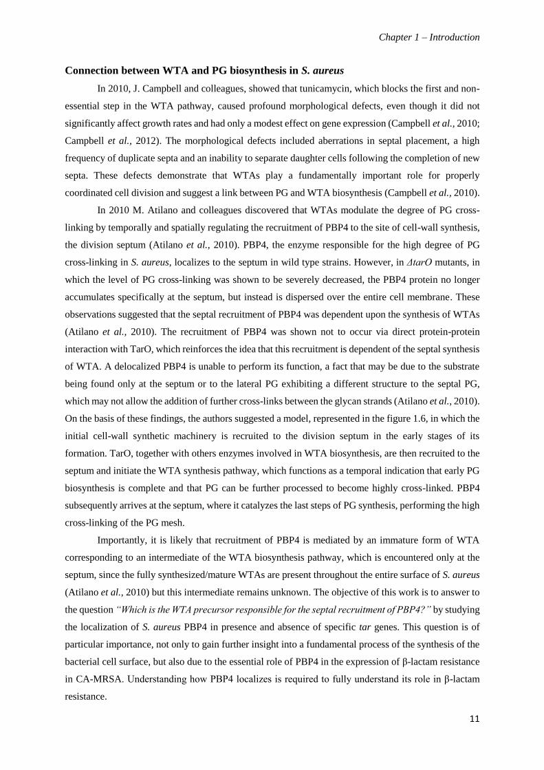
On the basis of these findings, the authors suggested a model, represented in the figure 1.6, in which the
initial cell-wall synthetic machinery is recruited to the division septum in the early stages of its
formation. TarO, together with others enzymes involved in WTA biosynthesis, are then recruited to the
septum and initiate the WTA synthesis pathway, which functions as a temporal indication that early PG
biosynthesis is complete and that PG can be further processed to become highly cross-linked. PBP4
subsequently arrives at the septum, where it catalyzes the last steps of PG synthesis, performing the high
cross-linking of the PG mesh.
Importantly, it is likely that recruitment of PBP4 is mediated by an immature form of WTA
corresponding to an intermediate of the WTA biosynthesis pathway, which is encountered only at the
septum, since the fully synthesized/mature WTAs are present throughout the entire surface of S. aureus
(Atilano et al., 2010) but this intermediate remains unknown. The objective of this work is to answer to
the question “Which is the WTA precursor responsible for the septal recruitment of PBP4?” by studying
the localization of S. aureus PBP4 in presence and absence of specific tar genes. This question is of
particular importance, not only to gain further insight into a fundamental process of the synthesis of the
bacterial cell surface, but also due to the essential role of PBP4 in the expression of β-lactam resistance
in CA-MRSA. Understanding how PBP4 localizes is required to fully understand its role in β-lactam
resistance.

Chapter 1 – Introduction
12
Figure 1.6. Model for the role of teichoic acids synthesis in PBP4 recruitment to the septum. The initial
cell-wall synthetic machinery arrives to the division site, leading to the synthesis of new PG, with low levels of
crosslinking (Left). TagO, and the remaining enzymes involved in WTA biosynthesis, are recruited to the
septum, by an unknown mechanism, and there initiate the synthesis of intermediate molecules in TA
biosynthesis (Centre). These intermediates (or another cellular components dependent on TA biosynthesis)
function as a temporal and spatial cue for PBP4 recruitment to the division septum, allowing the synthesis of
highly cross-linked PG to occur in a regulated manner (Right) (Atilano et al., 2010)

Chapter2- Materials and Methods
13
Materials and Methods
Bacterial strains and growth conditions
The bacterial strains and plasmids used and constructed during this study are listed in Tables
2.1 and 2.2. E. coli strain Dc10B was grown on Luria-Bertani agar (LA; Difco) or Luria-Bertani broth
(LB; Difco) medium, supplemented with ampicillin (100 µg/ml) as required. S. aureus strains were
grown at 37 ºC, with aeration, in tryptic soy broth medium (TSB; Difco) or in tryptic soy agar (TSA;
Difco). The medium was supplemented, when required, with erythromycin 10µg/ml (Ery10; Sigma)
and/or chloramphenicol 10 µg/ml (Cm10; Sigma), 5-bromo-4-chloro-3-indolyl-β-D-galactopyranoside
100 µg/ml (X-Gal; Apollo Scientific) and isopropyl-D-thiogalactopyranoside (IPTG; Apollo Scientic).
General procedures
DNA purification and manipulation. In order to obtain S. aureus genomic DNA cells were incubated
overnight on TSA plates at 37 ºC. Cells were scraped from confluent growth and re-suspended in 100
µl of 50 mM Ethylenediaminetetraacetic acid (EDTA). Lysostaphin 10 µg/mL (Sigma) and RNase 20
µg/mL (Sigma) were added to degrade the cell wall and RNA respectively, followed by 30 minutes
incubation at 37ºC. 400 µL of 50 mM EDTA and 500 µl of Nuclei Lysis Solution (Promega) were added
to cells and samples were incubated for 5 minutes at 80 ºC. The samples were then cooled to room
temperature before the addition of 200 µl of Protein Precipitation Solution (Promega). Samples were
vortexed vigorously then incubated on ice for 10 minutes. DNA was precipitated with isopropanol,
washed with 70% ethanol and re-suspended in sterile water. Purified genomic DNA was used as
template for the amplification of genes of interest via PCR reactions, using Phusion polymerase
(Finnyzymes- Thermo Scientific Molecular Biology), following the manufacturer’s instructions.
Plasmid DNA was purified from E. coli DC10B using the Wizard SV Plus Miniprep kit
(Promega) according to the manufacturers protocols. All DNA digests were performed with fast
restriction enzymes acquired from Fermentas- Thermo Scientific Molecular Biology, following the
manufacturer’s guidelines. DNA ligations were performed following standard molecular biology
techniques using T4 DNA ligase (Fermentas). PCR colony screening was performed using GoTaq
polymerase (Promega) and all clones were sequenced (Macrogen). All primers used are listed in Table
2.2.
E. coli transformation. E. coli competent cells were prepared according to the Rubidium Chloride
protocol as previously described (Sambrook 1989). In order to propagate the plasmid DNA of interest,
10 µl of ligated DNA or 1 µl of extracted plasmid DNA, was added to 50 µl of competent cells, incubated
on ice for 15 minutes, incubated for 1 minute at 42 ºC, returned to ice for more 5 minutes and rescued

Chapter2- Materials and Methods
14
in 1 ml of LB. After 60 minutes incubation at 37 ºC with aeration, the cells were spreaded on LA plates
containing ampicillin (100 µg/ml). Positive clones were identified by PCR colony screening. Plasmids
were extracted and the insert sequenced.
S. aureus transformation. RN4220 electro-competent cells were prepared as previously described
(Kraemer & Iandolo, 1990). For transformation, 0.5µg of purified DNA were mixed with 50µl of
RN4220 competent cells, transferred to a 0.2 cm BioRad Gene Pulser cuvette and incubated on ice for
5 minutes. Electroporation of the cells was performed in a gene pulser xcell (Bio-Rad) using the
following conditions: 2.5 kV; 25 µF and 100Ω. Immediately after electroporation cells were rescued in
1 ml of TSB and incubated at 30 ºC for 2 hours with aeration, before plating on TSA supplemented with
Ery10 (Sigma).
S. aureus transduction. Transductions were performed using phage 80α as previously described
(Oshida and Tomasz, 1992). In order to prepare the phage lysates, cells of the donor strain were scraped
from plates and re-suspended in 1 ml of TSB containing 5 mM of CaCl2. Serial dilutions of 80α phage
to 10-7 were made in Phage Buffer (MgSO4 1mM, CaCl2 4 mM, Tris-HCl 50 mM pH 7.8, NaCl 5.9 g/L,
gelatin 1 g/L). CaCl2 was added to a final concentration of 5 mM to phage top agar (casamino acids 3
g/l, Difco; yeast extract 3 g/L, Difco; sodium chloride 5.9 g/L, Sigma; agar 5 g/L, Difco; pH 7.8) that
was kept in the water-bath for 60 minutes at 45 ºC before being mixed with 10 µl of donor strain and 10
µl of each phage dilution. The mixtures were poured onto previously prepared plates of phage bottom
agar (the same composition as the phage top agar but containing 15 g/L of agar) containing CaCl2 5 mM
and incubated at 30 ºC overnight. To the plates showing confluent lysis phage buffer was added (3-4
ml) and incubated for 1 hour at 4 ºC, for the phage to be transferred to the phage buffer. The top agar
and phage buffer were then collected to a 50 mL centrifuge tube, vortexed to disrupt the phage top agar
and incubated at 4 ºC for 1 hour. The tubes were then centrifuged at 3000 rpm for 15 minutes at 4 ºC.
The supernatant was recovered and filtered with a 0.45 µm sterile filter.
For transduction the cells of the recipient strain were scraped from confluent growth and re-
suspended in 1 ml of TSB containing CaCl2 5mM. A volume of 100 µl of this cell suspension was mixed
with a range of different volumes of phage lysate (0.1 µl, 1 µl, 10 µl, 100 µl) and 100 µl of phage buffer
containing CaCl2 5 mM. A control sample in which no phage lysate was added was also prepared. The
samples were incubated for 20 minutes at 30 ºC. The mixtures were then added to the 0.3 GL top agar
(casaminoacids 3 g/L; yeast extract 3 g/L; NaCl 5.9 g/L; sodium lactate 60% syrup 3.3 ml/L, Sigma;
glycerol 50%, 2 ml/L, Sigma; Tri-sodium citrate 0.5 g/L, Sigma; agar 7,5 g/L; pH 7.8) previously left
in the water-bath for 60 minutes at 45 ºC. These samples were poured onto pre-prepared plates (used
within an hour of preparation) containing a 10 mL layer of 0.3 GL bottom agar (the same as the 0.3 GL
top agar but containing 15 g/L of agar) supplemented with 30 µg/mL of appropriate antibiotic and a 20
mL layer of 0.3 GL bottom agar without antibiotic. The plates were incubated for 48 hours at 30 ºC.
When needed, the medium was supplemented with IPTG.

Chapter2- Materials and Methods
15
Table 2.1. Bacterial strains used and constructed in this study
Name Relevant characteristics Source or
reference
E. coli
DC10B
E. coli cloning strain, chromosomal genotype: F-mcrA Δ (mrr-
hsdRMS-mcrBC) Φ80dlacZΔM15 ΔlacX74 endA1 recA1 deoR Δ
(ara, leu) 7697 araD139 galU galK nupG rpsL λ-
Lab stock
S. aureus
RN4220 MSSA strain. Restriction-deficient derivative of S. aureus
NCTC8325-4, which accepts foreign DNA. R. Novick
RNpEzrA-CFP RN4220 with integrated pEzrA-CFP plasmid encoding C-terminal
EzrA-CFP fusion; Eryr
(Pereira et
al., 2007)
NCTC8325-4 MSSA strain R. Novick
NCTCPBP4-YFP NCTC8325-4 with integrated pMad plasmid encoding a pbp4-yfp C-
terminal fusion; Lab stock
NCTCΔpbp4 NCTC8325-4 pbp4 null mutant Lab stock
NCTCΔspa::tarB NCTC8325-4 pbp4::pbp4-YFPΔspa::Pspac-tarB-lacI This study
NCTCΔspa::tarL NCTC8325-4 pbp4::pbp4-YFPΔspa::Pspac-tarL-lacI This study
NCTCΔspa::tarBΔtarB NCTC8325-4 pbp4::pbp4-YFPΔspa::Pspac-tarB-lacIΔtarB This study
NCTCΔspa::tarLΔtarL NCTC8325-4 pbp4::pbp4-YFPΔspa::Pspac-tarL-lacIΔtarL This study
NCTCΔspa::tarBi NCTC8325-4 pbp4::pbp4-YFPΔspa::Pspac-tarB-lacIΔtarB lacImC;
Cmr This study
NCTCΔspa::tarLi NCTC8325-4 pbp4::pbp4-YFPΔspa::Pspac-tarL-lacIΔtarL lacImC ;
Cmr This study
NCTCEzrA-CFP NCTC8325-4 with with integrated pEzrA-CFP plasmid encoding C-
terminal EzrA-CFP fusion; Eryr Lab stock
NCTCΔspa::tarBi EzrA-
cfp
NCTC8325-4 pbp4::pbp4-YFPΔspa::Pspac-tarB-lacIΔtarB lacImC
ezrA::ezrA-cfp; Cmr; Eryr This study
NCTCΔspa::tarLi EzrA-
cfp
NCTC8325-4 pbp4::pbp4-YFPΔspa::Pspac-tarL-lacIΔtarL lacImC
ezrA::ezrA-cfp ; Cmr ; EryR This study
abbreviations: Eryr – Erythromycin resistant; Cmr – Chloramphenicol resistant; lacI mc – cells expressing multiple copies of the lacI gene
(encoded by pMGPII);

Chapter2- Materials and Methods
16
Table 2.2. Plasmids used and constructed in this study
Name Relevant characteristics Source or
reference
pMAD E. coli – S. aureus shuttle vector with a thermosensitive origin of
replication for Gram-positive bacteria; Ampr; Eryr; LacZ+
(Arnaud et al.,
2004)
pBCB13 pMAD derivative with up- and downstream regions of spa gene and
Pspac-lacI region from pDH88; Ampr, Eryr
(Pereira et al.,
2010)
pMGPII Plasmid encoding lacI gene; Cmr (Pinho et al.,
2001)
pEzrA-CFP Plasmid encoding C-terminal EzrA-CFP fusion; Ampr Eryr (Pereira et al.,
2010)
pBCB13tarB pBCB13 derivative containing Pspac-tarB-lacI This study
pBCB13tarL pBCB13 derivative containing Pspac-tarL-lacI This study
pMADtarBKO pMAD derivative containing the up-and downstream regions of tarB This study
pMADtarLKO pMAD derivative containing the up-and downstream regions of tarL This study
abbreviations: Ampr – Ampicillin resistant; Eryr – Erythromycin resistant; Cmr – Chloramphenicol resistant; lacI mc – cells expressing
multiple copies of the lacI gene (encoded by pMGPII);
Mutant construction
To investigate the localization of S. aureus PBP4 in presence and absence of specific tar genes,
we constructed inducible mutants of these genes in the background of NCTC8325-4 PBP4-YFP. In
order to construct an inducible mutant, a full copy of the interest gene was first placed in the spa locus
under the control of the Pspac promoter and, subsequently, while in the presence of IPTG, was deleted
from its native chromosomal locus. Sequences of the primers used in this study are listed in Table 2.3.
Table 2.3. Primers used in this study
Primer Name Primer Sequence (5’- 3’)*
pSpaTarB3-P1 TACCCGGGACATATTAAGTTGGTG
pSpaTarB-P2 TACTCGAGTCAGTAGAACCACCATC
pTarB-KO-P1 ACGAGAATTCAGTGTGGTTTAATGGAATG
pTarB-KO-P2 GTCACCATCTTATCTATATAAATACACCAACTTAATATG
pTarB-KO-P3 AGTTGGTGTATTTATATAGATAAGATGGTGAC
pTarB-KO-P4 ACTGGATCCGCAGTTTATGGTCATCAATG
pTarB-KO-P5 ATGACGAAACCCCGCTAACC
pTarB-KO-P6 TGTCGTGTGCGTTACTGCTGGGTG
tarBchrom TCAGAGTGGGTGTTTTGACAC
pSpaTarL-P1 ATTACCCGGGTGAAGCAGACCTGTC
pSpaTarL-P2 ATACTCGAGTACCTCTCCCACTTTGAC
pTarL-KO-P1 ACGAGAATTCAGTTGAATGGAGGAAG

Chapter2- Materials and Methods
17
Primer Name Primer Sequence (5’- 3’)*
pTarL-KO-P2 TGACTACTATATAAACCGTTAATTCATCC
pTarL-KO-P3 AGGATGAATTAACGGTTTATATAGTAGTCAAAGTGGGAGAG
pTarL-KO-P4 TCGCA GGATCC TCATGTTGGCTCACAATG
pTarL-KO-P5 TCACCAGAAGGAAGCATTGCACTG
pTarL-KO-P6 ACGCCACATTTCTAGGTTTACCTGG
tarLchrom AGAAGATGGACAAGCGTCACAACG
pMADI CTCCTCCGTAACAAATTGAGG
pMADII CGTCATCTACCTGCCTGGAC
Spa_p1_BamHI TGAGGATCCCCAGCTTGTTGTTGTCTTC
Spa_p4_NcoI TGCAGTCCATGGTTGAAAAAGAAAAACATTTATTC
Pspac_p1_pDH88EcoRI GCTGAATTCTTCTACACAGCCCAGTCCAGAC
* Underlined sequences correspond to restriction sites
Construction of a tarB inducible mutant. To clone the tarB gene, in the ectopic spa locus of S. aureus
strain NCTCPBP4-YFP, under the control of the IPTG inducible/lacI-repressible Pspac promoter
(Yansura and Henner, 1984), the entire tarB gene, including the RBS sequence, was amplified by PCR
from NCTC8325-4 genomic DNA using the primers pSpaTarB3-P1 and pSpaTarB-P2. The resulting
PCR product was digested with SmaI and XhoI fast restriction enzymes and ligated into pBCB13
plasmid digested with the same enzymes, giving rise to pBCB13 tarB. E. coli DC10B competent cells
were then transformed with this plasmid and after its purification, the insert in pBCB13tarB was
confirmed by enzymatic digestion and sequencing. The plasmid pBCB13tarB was transferred to
RN4220 by electroporation (selection with erythromycin) and subsequently transduced to NCTCPBP4-
YFP using phage 80α as previously described (Oshida and Tomasz, 1992).
In order to integrate the pBCB13tarB plasmid into the chromosome, an erythromycin resistant
colony was inoculated into fresh TSB containing Ery10 and incubated at 30 ºC overnight. The overnight
culture was diluted 1:1000 into fresh TSB with Ery10, incubated at 30 ºC for 8 hours, then diluted again
into the same media and incubated overnight at 43 ºC, a non-permissive temperature that prevents the
plasmid replication due to the thermosensitive origin of replication and allows, in presence of
erythromycin, the selection of recombinants in which the plasmid had integrated into the chromosome.
The overnight culture was serially diluted and 100 µL of each of the 10-4, 10-5 and 10-6 dilutions were
plated on TSA containing Ery10 and X-GAL 100 µg/mL at 43 ºC. Several light blue colonies were
chosen and re-streaked in the same conditions. The integration of pBCB13tarB plasmid into the
chromosome can occur via the upstream or downstream regions of the gene encoded in the plasmid, so
the integration by upstream region was confirmed by PCR using primers pMADII and spa_p4_NcoI,
while the downstream region was confirmed using primers spa_p1_BamHI and pMADI. Two clones
with the plasmid integrated into the chromosome, via the up and downstream regions, were inoculated
Chapter2- Materials and Methods
18
in TSB at 30 ºC overnight. The overnight culture was diluted 1:500 in the same conditions, incubated at
30 ºC for 8 hours, serially diluted (10-4, 10-5 and 10-6) and then plated on TSA containing X-GAL 100
µg/mL at 43 ºC. White colonies that represent candidates for the loss of the plasmid, were chosen and
re-streaked on TSA X-GAL 100 µg/mL and TSA Ery10 X-GAL 100 µg/mL through replica plating.
The white and erythromycin sensitive colonies were screened by PCR, to confirm the substitution of the
spa gene by tarB using primers Pspac_p1_pDH88EcoRI and pSpaTarB-P2 and for the wild type
phenotype (presence of spa gene in spa locus) using primers Spa_p1_BamHI and Spa_p4_NocI. The
resulting strain, which has two copies of tarB gene, one in the native locus and the other in the spa locus
under the control of Pspac promoter was named NCTCΔspa::tarB.
Subsequently, to delete tarB from its normal locus in the background of strain NCTCΔspa::tarB,
a PCR fragment containing the upstream and downstream regions of the sequence, approximately 1 Kb
each, were amplified from NCTC8325-4 genomic DNA, in two sequential PCR steps. First, the
upstream region, that contains the upstream region of tarB until the start codon, as amplified using
primers pTarB-KO-P1 and pTarB-KO-P2, and the downstream region, containing the downstream
region of tarB including the 3´end, was amplified using the primers pTarB-KO-P3 and pTarB-KO-P4.
These two amplified products were then purified and joined by an overlap PCR reaction, using primers
pTarB-KO-P1 and pTarB-KO-P4. The final PCR product was digested with EcoRI and BamHI and
cloned into pMAD plasmid, giving rise to pMADtarBKO. The presence of the cloned insert was verified
by enzymatic digestion and sequencing. The pMADtarBKO plasmid was electroporated into RN4220
(selection with erythromycin), transduced to NCTCΔspa::tarB by phage transduction and subsequently,
integrated and excised, as described above. The deletion of the tarB gene from the native locus was
confirmed by PCR using primers pTarB-KO-P5 and pTarB-KO-P6, resulting in NCTCΔspa::tarBΔtarB
strain.
The pMGPII plasmid (Pinho et al., 2001), which encodes the lacI gene, was also transduced into
NCTCΔspa::tarBΔtarB, to ensure tight regulation of tarB expression. The resultant strain was named
NCTCΔspa::tarBi. As a control, we also transduced pEzrA-CFP into this strain, which resulted in
NCTCΔspa::tarBi EzrA-cfp strain.
Construction of a tarL inducible mutant. The construction of this inducible mutant was performed as
described above for the construction of tarB inducible mutant. The entire tarL gene, including the RBS
sequence, was amplified by PCR from NCTC8325-4 genomic DNA using the primers pSpaTarL-P1 and
pSpaTarL-P2, digested with SmaI and XhoI fast restriction enzymes and cloned into pBCB13 plasmid,
giving rise to pBCB13tarL. The insert in pBCB13tarL was confirmed by enzymatic digestion and
sequencing. The plasmid pBCB13tarL was electroporated into RN4220 (selection with erythromycin)
and subsequently transduced to NCTCPBP4-YFP. The integration and excision of the plasmid into the
chromosome was performed as described above, to check the integration by upstream region we made
a PCR using primers pMADII and spa_p4_NcoI, while the downstream region was confirmed using

Chapter2- Materials and Methods
19
primers spa_p1_BamHI and pMADI. Substitution of the spa gene by tarL was confirmed by PCR colony
screening using primers Pspac_p1_pDH88EcoRI and pSpaTarL-P2 and for the wild type phenotype
using primers Spa_p1_BamHI and Spa_p4_NocI. The resulting strain, which has two copies of tarL
gene, one in the native locus and the other in the ectopic spa locus under the control of Pspac promoter
was named NCTCΔspa::tarL.
Subsequently, to delete tarL from its native locus in the NCTCΔspa::tarL background, the
pMADtarLKO plasmid was transduced into this strain and, after an integration and excision events, the
gene deletion was confirmed by PCR using primers pTarL-KO-P5 and pTarL-KO-P6, resulting in
NCTCΔspa::tarLΔtarL strain, expressing a single copy of tarL from the spa locus, under the control of
Pspac promoter.
In order to ensure tight regulation of tarL expression the pMGPII plasmid, which expresses the
Pspac repressor lacI, was also transduced into NCTCΔspa::tarLΔtarL strain, giving rise to a new strain
named NCTCΔspa::tarLi. As a control, we also transduced pEzrA-CFP into this last strain which
resulted in NCTCΔspa::tarLi EzrA-cfp strain.
Growth analysis of S. aureus strains
The growth of the S. aureus strains was analyzed by measuring, at regular intervals, the optical
density at 600nm (OD600nm) of the liquids cultures. For that, an overnight culture of parental strain
NCTCPBP4-YFP was diluted (1:200) into fresh TSB media and incubated at 37 ᵒC with aeration,
while the inducible mutants were grown overnight, in the same conditions, in TSB medium
supplemented with 10 μg/ml of chloramphenicol (Cm10) and 0.5mM of IPTG, then the overnight
cultures were harvested, washed three times with fresh TSB and re-inoculated (with a 1:200 dilution) in
media with and without IPTG. The inducible mutants were also tested on solid media (TSA)
supplemented with chloramphenicol 10 µg/ml (Cm10) with or without 0.5 mM IPTG.
Fluorescence Microscopy
S. aureus strains were grown overnight, in TSB at 37 ºC, with appropriate antibiotic selection
and, the next day, were diluted (1:400) in 50 ml of fresh TSB supplemented with 0.5 mM IPTG and
grown until OD600nm 0.2. Cultures were then harvested, washed three times with fresh TSB and split
into two 25ml cultures of fresh TSB with and without IPTG. To visualize the localization of PBP4 and
EzrA, cultures were incubated for at least one hour after the washes, and thereafter at regulated intervals
we took the samples to be observed by fluorescence microscopy. For that the samples were centrifuged,
re-suspended in 20 µl of 1X Phosphate Buffered Saline (PBS) and 1 µl was placed on a thin film of 1%
agarose in 1X PBS. Fluorescence microscopy was performed using a Zeiss Axio Observer.Z1
microscope equipped with a Photometrics CoolSNAP HQ2 camera (Roper Scientific), using
Metamorph software (Molecular devices). Analysis of fluorescence images was performed using
Metamorph and ImageJ software.
Chapter2- Materials and Methods
20
Analysis of the expression of fluorescent proteins in S. aureus
In order to confirm whether the pbp4-YFP fusion protein was being cleaved in strains
NCTCPBP4-YFP, NCTCΔspa::tarBi and NCTCΔspa::tarLi the length of the band relative to YFP was
analysed by SDS-PAGE using a Fuji FLA 5100 laser scanner (Fuji Photo Film) to detect the fluorescent
protein. For that purpose, the strains were grown overnight in TSB medium supplemented with
appropriate antibiotics and 0.5 mM IPTG, when required. To prepare total protein extracts from each
strain, the overnight cultures were diluted 1:200 into fresh TSB (supplemented with the same antibiotics)
incubated at 37 oC until an O.D600nm of 0.8. Cells were harvested by centrifugation, re-suspended in 1X
PBS and disrupted with 250 µl glass beads in a Fast Prep FP120 (Thermo Electro Corporation). The
protein extracts were separated from glass beads by centrifugation (4200 x g, 1 minute at 4 ºC). The
total protein content of the extracts was quantified by the Bradford method, using bovine serum albumin
as a standard (BCA protein assay kit, Pierce) and equal amounts of protein, from each sample, were
loaded in a 10% SDS-PAGE gel and separated at 120V. Gel images were acquired on a Fuji FLA 5100
laser scanner (Fuji Photo Film) using 473 nm laser for YFP.
Western blot analysis
To analyze if the pbp4-YFP fusion was being cleaved, western blots were performed using a
polyclonal anti-PBP4 and anti-GFP antibody. The protein extracts of NCTC8325-4, NCTCΔpbp4,
NCTCPBP4-YFP, NCTCΔspa::tarBi and NCTCΔspa::tarLi strains and the quantification of total
protein content of the extracts were performed as described above. Equal amounts of protein, from each
sample, were heated to 100 ºC for 5 minutes, loaded onto a 10% SDS-PAGE gel and separated at 120V.
Proteins were then transferred to a Hybond-P Polyvinylidene fluoride (PVDF) membrane (GE
Healthcare) using a semidry transfer cell (Bio-Rad) according to standard western blotting techniques
(Burnette,W.N., 1980). The membranes were blocked with blocking buffer (PBS, 5% milk, 5% Tween
20), as previously described (Jonhson, D.A. et al, 1984), for 1 hour and, after washed three times the
membranes with 0.5% of Tween 20 in PBS, were incubated with a polyclonal anti-PBP4 antibody (1/100
dilution in blocking buffer) or an anti-GFP antibody (1/500 dilution in blocking buffer) overnight at 4
ºC. The following day membranes were washed three times with 1 x PBS-T and incubated with
secondary antibodies diluted 1/100000 in blocking buffer. The detection was performed using ECL Plus
Western blotting detection system from Amersham according to the manufacturers guidelines.

Chapter3- Results
21
Results
Construction of TarB and TarL inducible mutants
In order to study the localization of S. aureus PBP4 in the presence and absence of specific tar
genes we constructed inducible mutants of these genes in the background of NCTCPBP4-YFP strain
(Figure 3.1). For that purpose we replaced the spa gene by a full copy of the gene of interest, under the
control of IPTG inducible / LacI repressible Pspac promoter, and subsequently, while in the presence of
IPTG, deleted the gene from its native chromosomal locus (Yansura and Henner, 1984).
Figure 3.1. Schematic representation of the inducible mutant constructs. A. S. aureus strain with inducible
tarB gene NCTCΔspa::tarBi; B. S. aureus strain with inducible tarL gene NCTCΔspa::tarLi; The tarB and tarL
genes were cloned at the ectopic spa locus, under the control of the Pspac promoter, and were subsequently deleted
from their native loci. The pMGPII plasmid, encoding the LacI repressor protein was transduced into these strains
in order to ensure tight regulation from Pspac.
Most of the tar genes, involved in WTA biosynthesis, can not be deleted in a wild type S.
aureus strain and are encoded within operons, as shown in the figure 1.4.A. Therefore deletion of genes
such as tarB or tarL, can have lethal effects and their placement under the control of an inducible
promoter at the wild type locus can have deleterious polar effects on downstream essential genes
(Swoboda et al., 2010). These facts were taken in account during the construction of the inducible
mutant strains, NCTCΔspa::tarBi and NCTCΔspa::tarLi. For the construction of these strains, a copy
of the tarB or tarL gene was placed in the spa locus under the control of Pspac.The lacI gene, encoding
the repressor protein LacI was also placed at the spa locus, to repress the Pspac promoter. The tarB or
tarL genes were then deleted from their native chromosomal locus. The process for placing the tar genes
in the spa locus is shown in Figure 3.2. A similar process was used for their deletion from the native
chromosomal locus, using the pMAD vector containing only the up and downstream regions of the gene
of interest. Importantly, deletion of tar genes was performed in the presence of IPTG to induce
expression of the essential gene from the Pspac promoter, at the spa locus, and thus avoid cell damage
or the appearance of suppressor mutations.

Chapter3- Results
22
Figure 3.2. Schematic representation of the spa gene replacement by an gene of interest. This process, to
place tarB or tarL under the control of Pspac promoter in the spa locus, involves the integration and excision of
a plasmid encoding the gene of interest and lacI between the up- and downstream regions of the spa gene, by
homologous recombination, into the parental strain NCTCPBP4-YFP; A. Integration through the homologous
region 1; B. Integration through the homologous region 2.

Chapter3- Results
23
Although the inducible mutants have a copy of lacI in the spa locus, we transduced into the
mutants the multicopy pMGPII plasmid (Pinho et al., 2001b), which encodes the lacI gene, to ensure
tight regulation of expression of the tar genes from the Pspac promoter. It has been previously shown
that, in S. aureus, expression of the lacI gene from a multicopy plasmid is required for the tight
regulation of genes under the control of the Pspac promoter (Jana et al., 2000). The resulting strains
NCTCΔspa::tarBi and NCTCΔspa::tarLi strains allowed for the study of the localization of PBP4 in the
presence and absence of tarB and tarL, by growing them with and without IPTG, respectively. When
the strains were plated on TSA in the presence of IPTG (and therefore in the presence of the tar gene)
both strains displayed normal growth. In contrast, in the absence of IPTG, and thus the absence of TarB
or TarL, cells failed to grow indicating the essentiality of these gene products for viability (Figure 3.3).
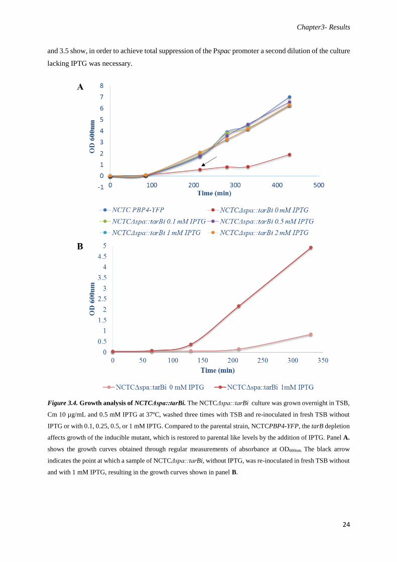
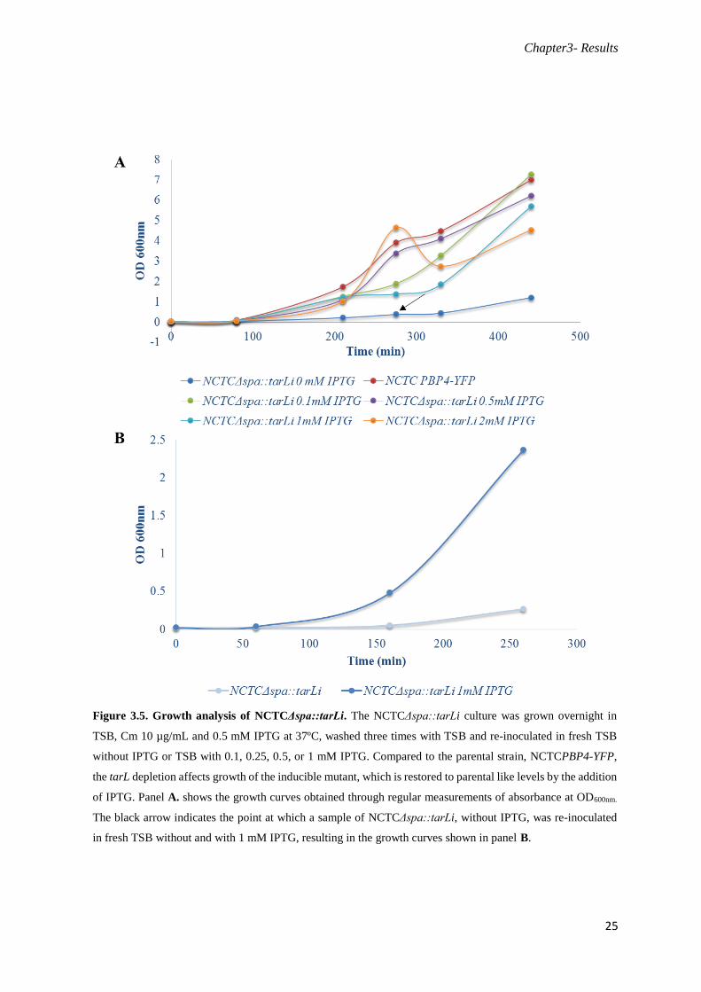
Growth of the inducible tar mutants was also analysed in liquid culture in the presence and
absence of IPTG and compared with the parental strain NCTCPBP4-YFP, as shown in Figures 3.4 and
3.5. In the absence of IPTG, the NCTCΔspa::tarBi and NCTCΔspa::tarLi strains grow slower than the
parental strain, NCTCPBP4-YFP. However in the presence of IPTG, even at low concentrations such
0.1 mM, the growth rates are like the parental strain. These observations show that the ectopic expression
of tarB or tarL from the spa locus, in the presence of IPTG, enabled cells to recover the levels of these
proteins and grow like the wild-type cells (Figure 3.4 and 3.5). As the results presented in Figures 3.4
Figure 3.3. Growth of S. aureus in the presence or in the absence of TarB and TarL. A)
NCTCΔspa::tarBΔtarBi; B) NCTCΔspa::tarLΔtarLi; The strains with tarB and tarL under control of
theIPTG incudible Pspac promoter were grown overnight at 37ºC on TSA plates with
chloramphenicol (10 µg/mL) supplemented (left plate) or not (right plate) with 0.5 mM IPTG.
Chapter3- Results
24
and 3.5 show, in order to achieve total suppression of the Pspac promoter a second dilution of the culture
lacking IPTG was necessary.
Figure 3.4. Growth analysis of NCTCΔspa::tarBi. The NCTCΔspa::tarBi culture was grown overnight in TSB,
Cm 10 µg/mL and 0.5 mM IPTG at 37ºC, washed three times with TSB and re-inoculated in fresh TSB without
IPTG or with 0.1, 0.25, 0.5, or 1 mM IPTG. Compared to the parental strain, NCTCPBP4-YFP, the tarB depletion
affects growth of the inducible mutant, which is restored to parental like levels by the addition of IPTG. Panel A.
shows the growth curves obtained through regular measurements of absorbance at OD600nm. The black arrow
indicates the point at which a sample of NCTCΔspa::tarBi, without IPTG, was re-inoculated in fresh TSB without
and with 1 mM IPTG, resulting in the growth curves shown in panel B.
Chapter3- Results
25
Figure 3.5. Growth analysis of NCTCΔspa::tarLi. The NCTCΔspa::tarLi culture was grown overnight in
TSB, Cm 10 µg/mL and 0.5 mM IPTG at 37ºC, washed three times with TSB and re-inoculated in fresh TSB
without IPTG or TSB with 0.1, 0.25, 0.5, or 1 mM IPTG. Compared to the parental strain, NCTCPBP4-YFP,
the tarL depletion affects growth of the inducible mutant, which is restored to parental like levels by the addition
of IPTG. Panel A. shows the growth curves obtained through regular measurements of absorbance at OD600nm.
The black arrow indicates the point at which a sample of NCTCΔspa::tarLi, without IPTG, was re-inoculated
in fresh TSB without and with 1 mM IPTG, resulting in the growth curves shown in panel B.

Chapter3- Results
26
Then we grew the S. aureus strains (NCTCPBP4-YFP, NCTCΔspa::tarBi or NCTCΔspa::tarLi without
and with several IPTG conditions) in liquid medium the cells tend to form clusters at cell densities
corresponding to OD600nm values of 1 or 2. This results in inaccurate OD600nm values and, consequently,
in fluctuations in the growth curves, as can be seen in figure 3.5.A. However this phenomenon did not
affect our downstream experiments as cultures used for microscopy analysis hadOD600nm values lower
than 1.
Deletion of tarB or tarL leads to delocalization of PBP4.
In 2010 M. Atilano and colleagues discovered that the deletion of the tarO gene, the first gene
in the WTAs synthesis pathway in S. aureus, resulted in the delocalization of PBP4. The recruitment of
PBP4 to its normal septal location was shown not to occur via direct interaction between these two
proteins, implying that PBP4 is likely recruited by an intermediate in WTA biosynthesis. Using the tarB
and tarL inducible mutants described above, we wanted to test if PBP4 localization was dependent on
intermediates that are downstream, in the WTA biosynthesis pathway, the reaction catalyzed by TarB
and TarL.
Depletion of TarB causes delocalization of PBP4.
When the PBP4–YFP fusion was expressed in the NCTC parental strain NCTCPBP4-YFP and
in the tarB inducible strain NCTCΔspa::tarBi in the presence of IPTG, it localized to the division septum
(Figure 3.7) where cell-wall synthesis has been reported to take place in S. aureus (Atilano et al., 2010;
Pinho and Errington, 2003). However, when the same fusion was expressed in NCTCΔspa::tarBi in the
absence of IPTG and thus depleted for TarB, PBP4 became delocalized, appearing all around the cellular
membrane, with no specific accumulation at the division septum (Figure 3.6). To quantify the
delocalization of PBP4 in the absence of the TarB protein, we calculated the ratio of fluorescence
measured at the septum versus the fluorescence measured at the “lateral” wall. If the fluorescent protein
is specifically accumulated at the division septum (which contains two membranes) then the
fluorescence ratio should be higher than two, however if it is delocalized and homogeneously dispersed
over the entire cell membrane, the intensity of the fluorescent signal at the septum should be
approximately twice the fluorescence at the lateral membrane. When this ratio was calculated for PBP4–
YFP in the parental strain NCTCPBP4-YFP we obtained an average value of 3.2±0.98 and a value of
3.3±1.07 for the NCTCΔspa::tarBi plus IPTG, a condition that allows the mutant strain to recover and
grow like the parental strain. A value of 1.9±0.47 was obtained for the tarB inducible mutant
NCTCΔspa::tarBi grown in the absence of IPTG, indicating that the absence of the TarB protein leads
to a loss of the specific accumulation of PBP4 at the septum.

Chapter3- Results
27

Chapter3- Results
28
Figure 3.6. Septal localization of PBP4 is lost in absence of TarB in the NCTCΔspa::tarBi inducible
strain. A) Microscopy images showing cells of NCTCPBP4-YFP and NCTCΔspa::tarBi, grown in the
presence or absence of IPTG. Panels on the left show the phase-contrast image and panels on the right show
the localization PBP4-YFP fusion; B) Quantification of septum (S) versus lateral membrane (L)
fluorescence (fluorescence ratio, FR) of PBP4–YFP in a parental background (NCTCPBP4YFP), in a tarB
inducible background (NCTCΔspa::tarBi) with or without IPTG. Quantification was performed in 200
cells displaying closed septa for each strain/condition. Horizontal lines correspond to average FR values.
FR values above 2 indicate septal localization, whereas FR values equal to or under 2 indicate that the
protein is distributed over the entire cell surface. P< 0.001. All images are false‐coloured. Scale bar: 1µm.

Chapter3- Results
29
Depletion of TarL causes delocalization of PBP4.
When the PBP4–YFP fusion was expressed in the NCTC parental strain, NCTCPBP4-YFP, and
in the tarL inducible strain, NCTCΔspa::tarLi, in the presence of IPTG, it localized to the division
septum (Figure 3.7) as expected (Atilano et al., 2010; Pinho and Errington, 2003). However, when the
same fusion was expressed in the strain NCTCΔspa::tarLi in the absence of IPTG (and therefore of
TarL), PBP4 is delocalized, appearing all around the cellular membrane with no specific accumulation
at the division septum (Figure 3.7). To quantify the delocalization of PBP4 in the absence of the TarL
protein, we calculated the fluorescence ratio as descrived above, and obtained an average value of
3.1±0.98 for NCTCPBP4-YFP and of 3.7±1.39 for the NCTCΔspa::tarLi plus IPTG, whereas a value
of 1.8±0.30 was obtained for the tarL inducible mutant NCTCΔspa::tarLi. These results indicate that
the absence of the TarL protein leads to the delocalization of PBP4 from the septum.

Chapter3- Results
30
Figure 3.7. Septal localization of PBP4 is lost in absence of TarL in the NCTCΔspa::tarLi inducible
strain. A) Microscopy images showing cells of NCTCPBP4-YFP and NCTCΔspa::tarLi, grown in the
presence or absence of IPTG. Panels on the left show the phase-contrast image and panels on the right show
the localization PBP4-YFP fusion; B) Quantification of septum (S) versus lateral membrane (L)
fluorescence (fluorescence ratio, FR) of PBP4–YFP in a parental background (NCTCPBP4YFP), in a tarL
inducible background (NCTCΔspa::tarLi) with or without IPTG. Quantification was performed in 200 cells
displaying closed septa for each strain/condition. Horizontal lines correspond to average FR values. FR
values above 2 indicate septal localization, whereas FR values equal to or under 2 indicate that the protein
is distributed over the entire cell surface. P< 0.001. All images are false‐coloured. Scale bar: 1µm.

Chapter3- Results
31
Statistical analysis.
Statistical analysis was performed to assess the significance of the differences between PBP4
localization in the parental strain NCTCPBP4-YFP and in the inducible strains NCTCΔspa::tarBi and
NCTCΔspa::tarLi grown in the presence of IPTG, and between these two strains grown in the presence
or in the absence of IPTG. For that purpose, we performed the statistical significance tests Kruskal-
Wallis and Dunn's Multiple Comparison tests, and calculated the p-value, with a confidence level of
0.001. The results obtained for the TarB mutant, presented in Figure 3.6, showed no significant
difference between PBP4 localization in the parental strain and in the inducible strain grown in the
presence of IPTG, however a significant difference was observed between tarB inducible mutant grown
in the absence and in the presence of IPTG, indicating that lack of tarB causes delocalization of PBP4.
Regarding TarL, a significant difference was also noticed between the inducible strain lacking tarL and
the parental strain, indicating that lack of tarL also causes delocalization of PBP4 (Figure 3.7). However,
in this case, a difference was also observed between the parental strain NCTCPBP4-YFP and the
inducible strain NCTCΔspa::tarLi grown with IPTG, which surprisingly has a higher value for PBP4-
YFP fluorescence in the septum than the parental strain.
The PBP4-YFP fusion is not cleaved.
In some of the microscopy images showing PBP4-YFP fluorescence many cells show a greater
degree of cytoplasmic signal than that previously observed in the NCTCPBP4-YFP strain (Atilano et
al., 2010). One possible explanation for this signal would be the cleavage of the PBP4-YFP fusion. In
order to address this and to be sure that the PBP4-YFP fusion is not degraded in our mutant strains we
analysed the presence of the PBP4-YFP fusion in the NCTCΔspa::tarBi and NCTCΔspa::tarLi mutants
strains by SDS-PAGE followed by imaging in a fluorescent image analyzer and western blotting, using
a polyclonal anti-PBP4 antibody, as described in the materials and methods. The cell extracts of the wild
type strain NCTC8325-4, the parental strain NCTCPBP4-YFP and the null mutant NCTCΔpbp4 were
used as controls. The results, shown in Figure 3.8, show that the PBP4-YFP fusion is not cleaved in the
tar inducible strains, because only one band is present, corresponding to the molecular weight of this
fusion (Figure 3.8 A) instead of one band with lower weight which would result from cleavage of the
fusion protein. The western blots (Figure 3.8 B), show the presence of the PBP4 band only in the wild-
type strain NCTC8325-4 as expected. Additionally, one band corresponding to the PBP4-YFP fusion
was observed for the NCTCΔspa::tarBi and NCTCΔspa::tarLi mutants strains.

Chapter3- Results
32
Figure 3.8. The PBP4-YFP fusion is not cleaved in NCTCPBP4-YFP, NCTCΔspa::tarBi and
NCTCΔspa::tarLi strains. A) Analysis of protein bands by fluorescence imaging detected intact PBP4-
YFP fusion in strains NCTCPBP4-YFP, NCTCΔspa::tarBΔtarBi and NCTCΔspa::tarLΔtarLi. B) Western
blot analysis, using an anti-PBP4 specific antibody, of NCTC8325-4, NCTCΔpbp4, NCTCPBP4-YFP,
NCTCΔspa::tarBΔtarBi and NCTCΔspa::tarLΔtarLi (from left to right). The band that appear in all strains,
between the PBP4 band and PBP4-YFP band, is a nonspecific band, as it appears in the NCTCΔpbp4 strain
lacking PBP4.
Chapter3- Results
33
Delocalization of PBP4 in the absence of TarL or Tar B is not due to cell death.
Bacterial cell division is a highly regulated process during which cells undergo a series of
temporally and spatially controlled events that result in the generation of two identical daughter cells
(Adams and Errington, 2009; Jorge et al., 2011). In almost all bacteria, this process begins with the
polymerization of a tubulin-like protein, FtsZ, into a ring-like structure located at the future division
septum, which serves as a scaffold for the recruitment of other proteins that together form a multi-protein
complex called the divisome (Adams and Errington, 2009). One of these proteins is EzrA, a
transmembrane protein that acts as a negative regulator of Z-ring assembly, preventing FtsZ assembly
at inappropriate locations different from the mid-cell (Jorge et al., 2011). In S. aureus, EzrA localizes
to the division septum in dividing cells (Jorge et al., 2011).
When cells are unhealthy and begin to die, the divisome is not assembled and consequently there
is a loss of septal localization of proteins involved in its formation (Jorge et al., 2011). PBP4 normally
has a septal localization, so in order to ensure that its delocalization observed in the mutant strains,
NCTCΔspa::tarBi and NCTCΔspa::tarLi, is due to the absence of the Tar proteins and not due to cell
death, we localized EzrA in the same strains. EzrA has previously been shown to delocalize from its
normal septal location prior to cell death (Jorge et al., 2011). For that purpose we transduced an
integrative plasmid encoding EzrA-CFP to the tarB and tarL inducible mutants, resulting in the strains
NCTCΔspa::tarBi ezrA-cfp and NCTCΔspa::tarLi ezrA-cfp. These new mutant strains allowed us to
quantify the PBP4 and EzrA localization in the same cells and to determine if the delocalization of PBP4
was part of general protein delocalization upon cell death or if it was specifically due to lack of the TarL
and TarB proteins. However, these strains displayed a distinct phenotype, even in the presence of IPTG
and therefore of the Tar proteins forming clusters (shown in Figure 3.9) not previously seen in the strains
lacking the EzrA-cfp fusion. This phenomenon limited our ability to correctly quantify the localization
of PBP4 as in the cell aggregates it was difficult to measure the PBP4-YFP fluorescence at the septum
and “lateral” wall. In contrast to the initial mutant strains, NCTCΔspa::tarBi and NCTCΔspa::tarLi,
these strains have an additional erythromycin resistant marker. We studied the effect of expression of
erythromycin resistance on the strains to determine whether it was responsible for the observed
phenotype, perhaps interfering with cell growth and causing the formation of cell aggregates. As shown
in figure 3.11 the presence or absence of erythromycin (10 ug/mL) had no effect upon the formation of
cell aggregates in strains carrying the EzrA-CFP fusion. Given that the control strain, NCTCEzrA-CFP,
does not show as many aggregates, this phenotype may be caused by the co-expression of the two fusion
proteins (PBP4-YFP and EzrA-CFP) in the same cells. We therefore decided to analyse separately the
effect of TarB and TarL depletion on ErzA and PBP4 localization, instead of doing the analysis in the
same cells, as initially planned.

Chapter3- Results
34
Figure 3.9. Strains NCTCΔspa::tarBΔtarBi EzrA-cfp and NCTCΔspa::tarLΔtarLi EzrA-cfp form
aggregates. The microscopy images show cell aggregates of NCTCΔspa::tarBΔtarBi EzrA-cfp and
NCTCΔspa::tarLΔtarLi EzrA-cfpin the presence of IPTG. Panels from top to the bottom show phase-
contrast image, PBP4-YFP fusion fluorescence, EzrA-CFP fusion fluorescence and the overlay between
PBP4-YFP and EzrA-CFP images. Scale bars 1 µm. All images are false‐colored.
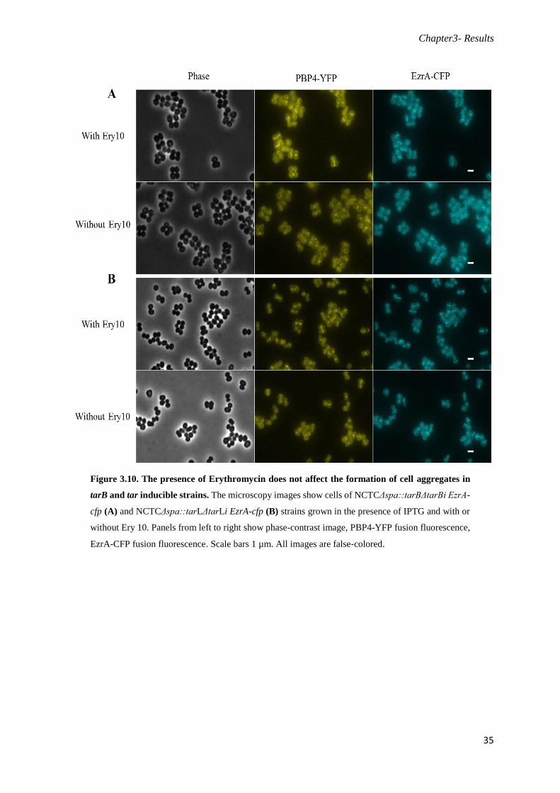
Chapter3- Results
35
Figure 3.10. The presence of Erythromycin does not affect the formation of cell aggregates in
tarB and tar inducible strains. The microscopy images show cells of NCTCΔspa::tarBΔtarBi EzrA-
cfp (A) and NCTCΔspa::tarLΔtarLi EzrA-cfp (B) strains grown in the presence of IPTG and with or
without Ery 10. Panels from left to right show phase-contrast image, PBP4-YFP fusion fluorescence,
EzrA-CFP fusion fluorescence. Scale bars 1 µm. All images are false‐colored.

Chapter3- Results
36
When the EzrA-CFP fusion was expressed in the NCTCΔspa::tarBi and NCTCΔspa::tarLi
strains, with and without IPTG the protein localized to the division septum (Figure 3.12) as expected
(Jorge et al., 2011). However, as stated above, when the PBP4-YFP fusion was expressed in the same
background causing the formation of cell clusters we could not accurately quantify the localization of
this protein. Therefore we used the strains NCTCΔspa::tarBi EzrA-cfp and NCTCΔspa::tarLi EzrA-cfp
to quantify the localization of EzrA at the same time points at which we analysed PBP4-YFP localization
in strains NCTCΔspa::tarBi and NCTCΔspa::tarLi. In this way we were able to determine whether
delocalization of PBP4 occurs before or simultaneously with the delocalization of EzrA, the later of
which would indicate that PBP4 could be delocalizing, not specifically due to lack of TarB or TarL, but
rather as part of general protein delocalization in cells dying because of the lack of essential Tar proteins.
To quantify the localization of EzrA in the presence and absence of the TarB and TarL proteins, we
calculated the fluorescence ratio, obtaining average values of 3.7±0.98 and 3.9±1.08 for
NCTCΔspa::tarBi EzrA-cfp strain in the presence and absence of IPTG, respectively, and average values
of 4.2±1.28 and 4.2±1.34 for the NCTCΔspa::tarLi EzrA-cfp strain in the same conditions. These results
indicate that, in the absence of the TarB or TarL proteins, EzrA remains localized to the septum,
indicating that the divisome is correctly assembled and there is no general protein delocalization.
Therefore, the observed delocalization of PBP4 was most likely due to lack of TarB and TarL.
Figure 3.11. Morphology of NCTCEzrA-CFP cells. The microscopy images show cells of
NCTCEzrA-CFP which do not form large clusters. Scale bars 1 µm. All images are false‐colored.

Chapter3- Results
37

Chapter3- Results
38
Statistical analyses were performed to assess if the differences between the quantifications of
EzrA-CFP localization in the strains NCTCΔspa::tarBi EzrA-cfp and NCTCΔspa::tarLi EzrA-cfp, with
and without IPTG were significant. Mann Whitney tests were performed and the p-values calculated,
with a confidence level of 99.9%. The results obtained for the TarB and TarL mutants (Figures 3.11 and
3.12) showed that a significant difference did not exist indicating that EzrA-Cfp localization remained
unchanged in upon depletion of either of the Tar proteins.
Figure 3.12. Septal localization of EzrA is not lost in the absence of TarB or TarL. Quantification of
septum (S) versus lateral membrane (L) fluorescence (fluorescence ratio, FR) of EzrA in A)
NCTCΔspa::tarBΔtarBi EzrA-cfp or B) NCTCΔspa::tarLΔtarLi EzrA-cfp strains in the presence or
absence of IPTG, with the respective microscopy images. Quantification was performed in 100 cells
displaying closed septa for each strain. Horizontal lines correspond to average FR values. FR values above
2 indicate a preferential septal localization, whereas FR values equal to or under 2 indicate that a protein is
distributed over the entire cell surface. P< 0.001. All images are false‐colored. Scale bar: 1µm.
Chapter4- Discussion
39
Discussion
The cell wall is very important for the integrity and viability of bacteria, as it provides physical
protection, determines the cell shape and is the principal stress-bearing element, which makes it an ideal
target for antibiotics (Scheffers and Pinho, 2005). In Gram-positive bacteria, such as S. aureus, the cell
wall is composed of surface proteins, teichoic acids and a thick layer of peptidoglycan (PG), whose
synthesis is a major target of some of the most successful classes of antibiotics, including the β-lactams
such as penicillin or methicillin (Popham, 2013). The peptidoglycan requires a complex process of
synthesis that involves the elongation (transglycosylation) and peptide cross-linking (transpeptidation)
of glycan strands, which occurs mainly at the division septum of S. aureus and is catalyzed by the
penicillin binding proteins (PBPs) (Scheffers and Pinho, 2005). Recent studies have shown that the
PBP4 is essential for the expression of β-lactam resistance in CA-MRSA strains (Memmi et al., 2008).
This protein has a septal localization and is responsible for the high degree of PG cross-linking notable
in S. aureus. The localization of PBP4 has recently been shown to be spatially and temporally regulated
by an unknown intermediate of the WTA biosynthesis pathway (Atilano et al., 2010). In this work we
aimed to identify the WTA precursor responsible for the septal recruitment of PBP4. We constructed
inducible mutants of specific tar genes in the background of NCTCPBP4-YFP, a strain expressing a
fluorescent derivative of PBP4, that allowed us to study its localization in the presence and absence of
tarB and tarL genes.
Deletion of tarO and the use of tunicamycin, which blocks the first and non-essential step in the
WTA pathway by the inhibiting TarO, have been shown to cause profound morphological defects in S.
aureus, such as aberrations in septal placement, a high frequency of double septa and an inability to
separate daughter cells following the completion of new septa (Atilano et al., 2010; Campbell et al.,
2010; Campbell et al., 2012). In both the absence of TarO and in the presence of tunicamycin, the PBP4
protein is dispersed over the entire cell membrane instead of displaying its normal septal localization,
indicating that PBP4 is recruited by the TarO protein or the product of a downstream reaction in the
WTA biosynthesis pathway (Atilano et al., 2010; Roemer et al., 2013). However, Atilano et al have
shown that the recruitment of PBP4 does not occur via direct protein–protein interaction with TarO
because these two proteins did not interact in a bacterial two-hybrid screening, did not colocalize in 49%
of the cells in the early stages of septum synthesis and PBP4 did not retain its septal localization in the
presence of an inactive TarO protein properly localized at the septum (Atilano et al., 2010). Therefore
it is more likely that PBP4 is recruited by a WTA synthesis intermediate. The use of a second drug,
targocil, which inhibits the TarG WTA ABC transporter, demonstrated that the PBP4 is recruited by a
precursor of the WTA biosynthesis pathway present inside the membrane. In the presence of this drug
PBP4 remains specifically at the division septum which shows that the steps after the WTA translocation
to the outside membrane are not involved in the recruitment of PBP4 (Roemer et al., 2013). Based upon
Chapter4- Discussion
40
these results we chose to study the tarB and tarL genes, which catalyze the addition of one glycerol‐3‐
phosphate unit and the addition of a ribitol phosphate residue chain, respectively (Figure 1.4).
In this work, we have shown that in parental strain NCTCPBP4-YFP, PBP4 can be found at the
septum of S. aureus. However, in the inducible tar mutant strains depleted for TarB and TarL (when
grown without IPTG), PBP4 no longer accumulates specifically at the division septum, but instead is
dispersed over the entire cell membrane (Figures 3.6 and 3.7). There is no statistically significant
difference in the PBP4-YFP localization between parental NCTCPBP4-YFP PBP4 strain and the
NCTCΔspa::tarBi strain plus IPTG, which means the inducible strain behaves as expected in the
presence of IPTG, i.e. the expression of tarB from the spa locus or from its native locus is similar for
the purpose of PBP4 localization. However, in the case of NCTCΔspa::tarLi there was a significant
difference between PBP4 localization in this strain, in the presence of IPTG, and in the parental strain
with the former having a higher fluorescence ratio for PBP4 localization, i.e., more PBP4 protein
localized at the septum. This observation can be explained by two hypotheses (1) expression of tarL in
the conditions used to grow the inducible strain, NCTCΔspa::tarLi, in the presence of IPTG, could lead
to an overexpression of TarL with a consequent increase in the number of precursors that recruit the
PBP4 and therefore an increase in the septal signal of the PBP4; (2) by cloning the tarL in the spa locus,
separating it from its operon, we could be changing the regulation network of the teichoic acids
synthesis, which could also affect the pathways for cell wall synthesis, including PBP4 production, given
that the two pathways have common substrates. Overexpression of PBP4 could therefore be the reason
for the increased fluorescence ratio observed in the TarL inducible strain in the presence of IPTG.
We determined PBP4 localization on cells depleted for TarB and TarL for 1 hour and 45
minutes. However, TarB and TarL depletion eventually leads to cell death, which can be accompanied
by general protein delocalization. It was therefore important to determine if PBP4 delocalization was
part of general protein delocalization in cells approaching death, or if PBP4 was specifically delocalizing
in the absence of TarB and TarL, in conditions where other proteins remain properly localized. Given
that PBP4 localizes to the septum, we used a divisome protein, EzrA, as a control and determined the
effect of TarB and TarL depletion of EzrA septal localization. For that purpose we used a EzrA-CFP
fusion as the fluorescence emitted by CFP does not overlap the fluorescence emitted by YFP fused to
PBP4, i.e., the emission maxima of the two fluorophores are sufficiently apart to be separated using
appropriate filters (Pereira et al., 2010). The strains simultaneously expressing PBP4-YFP and EzrA-
CFP, with either tarB or tarL under the control of the inducible promoter Pspac, NCTCΔspa::tarBi EzrA-
cfp and NCTCΔspa::tarLi EzrA-cfp, should have enabled us to quantify the localization of PBP4 and
EzrA in the same cells and confirm if the delocalization of PBP4 is accompanied or not by changes in
the localization of EzrA. Unfortunately these strains formed cell clusters which did not permit the correct
quantification of PBP4-YFP localization, as we cannot correctly measure the fluorescence at the septum
and at the “lateral” wall for calculation of the fluorescence ratio. As an alternative, we quantified EzrA
and PBP4 localization in separate strains but under the same TarB/TarL depletion conditions. The

Chapter4- Discussion
41
obtained results shown in Figure 3.12 confirm that PBP4 delocalization did not occur as part of general
protein delocalization, as EzrA-CFP remained localized at the septum in the absence of TarB/TarL while
PBP4-YFP was dispersed throughout the cell membrane.
In this work we were able to determine that in the absence of TarB or TarL, PBP4 loses
its normal septal localization and becomes dispersed all around the cell membrane. With these results
we can suggest two hypotheses:
(1) The TarL protein itself recruits PBP4 to the division septa. In this case, once the TarL
catalyses one of the last steps of the WTA biosynthesis occurring in the inner side of the
cytoplasmic membrane (Figure 1.4 and 1.5 in the introduction) and the absence of TarO,
the first protein in this synthesis pathway, also leads to PBP4 delocalization, then TarL
localization should be dependent on the substrate. Therefore, blocking TarO protein would
deplete subsequent intermediates in WTA synthesis, which would in turn affect TarL and
PBP4 localization;
(2) PBP4 is recruited by a substrate of the WTA synthetic pathway whose presence is dependent
on TarL. In this case, the absence of an earlier protein in the WTA biosynthetic pathway,
such as TarO or TarB, would deplete the substrate for TarL, which would therefore be
unable to make its product.
In conclusion, these results indicate that the molecule responsible for PBP4 recruitment is
probably one involved in the last steps in WTA synthesis pathway at the inner side of the
membrane. To prove that hypothesis and identify the intermediate responsible for PBP4
localization, we look for PBP4 interaction partners by a bacterial two-hybrid screening, namely
to see if the PBP4 interacts with TarL, TarH and/or TarG. We will also study of the localization
of S. aureus PBP4 in the absence of TarH and TarG by using tarH and tarG inducible mutants.
Although PBP4 maintains its septal localization in the presence of the antibiotic targocil, which
blocks the activity of TarG, we do not know if TarH or TarG physically interact with PBP4 to
recruit it to the septa. Uncovering the mode of recruitment of PBP4 is not only important to gain
knowledge into the fundamental process of bacterial cell wall synthesis, but also into the
essential role of PBP4 in the expression of β-lactam resistance in CA-MRSA strains. For this
purpose we have already started to construct the strains to reproduce our studies in the CA-
MRSA strain MW2, an understanding of how PBP4 localizes is required to fully understand its
role in β-lactam resistance.

Chapter4- Discussion
42
Chapter 5 – Bibliography
43
Bibliography
Adams, D.W., and Errington, J. (2009). Bacterial cell division: assembly, maintenance and disassembly
of the Z ring. Nature Reviews Microbiology 7: 642-653.
Anderson, A.J., Green, R.S., Sturman, A.J., and Archibald, A.R. (1978). Cell wall assembly in Bacillus
subtilis: location of wall material incorporated during pulsed release of phosphate limitation, its
accessibility to bacteriophages and concanavalin A, and its susceptibility to turnover. Journal of
bacteriology 136: 886-899.
Archer, G.L. (1998). Staphylococcus aureus: a well-armed pathogen. Clinical infectious diseases : an
official publication of the Infectious Diseases Society of America 26: 1179-1181.
Atilano, M.L., Pereira, P.M., Yates, J., Reed, P., Veiga, H., Pinho, M.G., and Filipe, S.R. (2010).
Teichoic acids are temporal and spatial regulators of peptidoglycan cross-linking in
Staphylococcus aureus. Proceedings of the National Academy of Sciences of the United States of
America 107: 18991-18996.
Barber, M. (1961). Methicillin-resistant staphylococci. Journal of clinical phatology 14: 385-393.
Beeby, M., Gumbart, J.C., Roux, B., and Jensen, G.J. (2013). Architecture and assembly of the Gram-
positive cell wall. Molecular Microbiology 88: 664-672.
Berger-Bächi, B., Strässle, A., Gustafson, J.E., and Kayser, F.H. (1992). Mapping and characterization
of Multiple chromosomal factors involved in methicillin resistance in Staphylococcus aureus.
Antimicrobial agents and chemotherapy 36: 1367-1373.
Boucher, H.W., and Corey, G.R. (2008). Epidemiology of methicillin-resistant Staphylococcus aureus.
Clinical infectious diseases : an official publication of the Infectious Diseases Society of America
46: Suppl 5, S344-349.
Brown, S., Meredith, T., Swoboda, J., and Walker, S. (2010). Staphylococcus aureus and Bacillus
subtilis W23 make polyribitol wall teichoic acids using different enzymatic pathways. Chemistry
& biology 17: 1101-1110.
Brown, S., Santa Maria, J.P., Jr., and Walker, S. (2013). Wall teichoic acids of gram-positive bacteria.
Annual review of microbiology 67: 313-336.
Brown, S., Xia, G., Luhachack, L.G., Campbell, J., Meredith, T.C., Chen, C., Winstel, V., Gekeler, C.,
Irazoqui, J.E., Peschel, A., et al. (2012). Methicillin resistance in Staphylococcus aureus requires
glycosylated wall teichoic acids. Proceedings of the National Academy of Sciences of the USA
109: 18909-18914.
Brown, S., Zhang, Y.H., and Walker, S. (2008). A revised pathway proposed for Staphylococcus aureus
wall teichoic acid biosynthesis based on in vitro reconstitution of the intracellular steps.
Chemistry & biology 15: 12-21.

Chapter 5 – Bibliography
44
Campbell, J., Singh, A.K., Santa Maria, J., Kim, S.J., Brown, S., Swoboda, J., Mylonakis, E., Wilkinson,
B.J., and Walker, S. (2010). Synthetic lethal compound combinations reveal a fundamental
connection between Wall Teichoic Acid and Peptidoglycan Biosyntheses in Staphylococcus
aureus. ACS chemical biology 6: 106-116.
Campbell, J., Singh, A.K., Swoboda, J.G., Gilmore, M.S., Wilkinson, B.J., and Walker, S. (2012). An
antibiotic that inhibits a late step in wall teichoic acid biosynthesis induces the cell wall stress
stimulon in Staphylococcus aureus. Antimicrobial agents and chemotherapy 56: 1810-1820.
Chambers, H.F. (1988). Methicillin-Resistant Staphylococci. Clinical Microbiology Reviews 1: 173-
186.
Chambers, H.F., and Deleo, F.R. (2009). Waves of resistance: Staphylococcus aureus in the antibiotic
era. Nature Reviews Microbiology 7: 629-641.
Couto, I., De Lencastre, H., Severina, E., Kloos, W., Webster, J.A., Hubner, R.J., Sanches, I.S., and
Tomasz, A. (1996). Ubiquitous presence of a mecA homologue in natural isolates os
Staphylococcus sciuri. Microbial Drug Resistance 2: 377-391.
Crisostomo, M.I., Westh, H., Tomasz, A., Chung, M., Oliveira, D.C., and de Lencastre, H. (2001). The
evolution of methicillin resistance in Staphylococcus aureus: similarity of genetic backgrounds
in historically early methicillin-susceptible and -resistant isolates and contemporary epidemic
clones. Proceedings of the National Academy of Sciences of the USA 98: 9865-9870.
D'Elia, M.A., Millar, K.E., Beveridge, T.J., and Brown, E.D. (2006a). Wall teichoic acid polymers are
dispensable for cell viability in Bacillus subtilis. Journal of bacteriology 188: 8313-8316.
D'Elia, M.A., Pereira, M.P., Chung, Y.S., Zhao, W., Chau, A., Kenney, T.J., Sulavik, M.C., Black, T.A.,
and Brown, E.D. (2006b). Lesions in teichoic acid biosynthesis in Staphylococcus aureus lead to
a lethal gain of function in the otherwise dispensable pathway. Journal of bacteriology 188: 4183-
4189.
de Lencastre, H., Oliveira, D., and Tomasz, A. (2007). Antibiotic resistant Staphylococcus aureus: a
paradigm of adaptive power. Current opinion in microbiology 10: 428-435.
de Lencastre, H., and Tomasz, A. (1994). Reassessment of the number of auxiliary genes essencial for
expression of high-level Methicillin Resistance in Staphylococcus aureus. Antimicrobial agents
and chemotherapy 38: 2590-2598.
Diekema, D.J., Pfaller, M.A., Schmitz, F.J., Smayevsky, J., Bell, J., Jones, R.N., and Beach, M. (2001).
Survey of infections due to Staphylococcus species: frequency of occurence and antimicrobial
susceptibility of isolates collected in the United States, Canada, Latin America, Europe, and the
Western Pacific region for the SENTRY Antimicrobial Surveillance Program, 1997-1999 .
Clinical infectious diseases : an official publication of the Infectious Diseases Society of America
32: S114-132.
Edwards, A.M., Massey, R.C., and Clarke, S.R. (2012). Molecular mechanisms of Staphylococcus
aureus nasopharyngeal colonization. Molecular oral microbiology 27: 1-10.

Chapter 5 – Bibliography
45
Foster, T.J. (2005). Immune evasion by staphylococci. Nature Reviews Microbiology 3: 948-958.
Ghuysen, J.M. (1991). Serine β-lactamases and Penicillin-Binding proteins. Annual Review
Microbiology 45: 37-67.
Goffin, C., and Ghuysen, J.M. (1998). Multimodular Penicillin-Binding Proteins: An enigmatic family
of orthologs and paralogs. Microbiology and Molecular Biology Reviews 62: 1079-1093.
Gordon, R.J., and Lowy, F.D. (2008). Pathogenesis of methicillin-resistant Staphylococcus aureus
infection. Clinical infectious diseases : an official publication of the Infectious Diseases Society
of America 46: Suppl 5, S350-359.
Heaton, M.P., and Neuhaus, F.C. (1992). Biosynthesis ofD-alanyl-lipoteichoic acid: cloning, nucleotide
sequence, and expression of the Lactobacillus caseigene for the D-alanine-activating enzyme.
Journal of bacteriology 174: 4707-4717.
Heaton, M.P., and Neuhaus, F.C. (1994). Role of the D-alanyl carrier protein in the biosynthesis of D-
alanyllipoteichoic acid. Journal of bacteriology 176: 681-690.
Heijenoort, J.V. (1998). Assembly of the monomer unit of bacterial peptidoglycan. CMLS Cellular and
Molecular Life Sciences 54: 300-304.
Heijenoort, J.V. (2001). Formation of the glycan chains in the synthesis of bacterial peptidoglycan.
Glycobiology 11: 25-36.
Jana, M., Luong, T.T., Komatsuzawa, H., Shigeta, M., and Lee, C.Y. (2000). A method for
demonstrating gene essentiality in Staphylococcus aureus. Plasmid 44: 100-104.
Jevons, M.P., Coe, A.W., and Parker, M.T. (1963). Methicillin resistance in staphylococci. Lancet 1:
904-907.
Jorge, A.M., Hoiczyk, E., Gomes, J.P., and Pinho, M.G. (2011). EzrA contributes to the regulation of
cell size in Staphylococcus aureus. PloS one 6: e27542.
Kopp, U., Roos, M., Wecke, J., and Labischinski, H. (1996). Staphylococcal peptidoglycan interpeptide
bridge biosynthesis: a novel antistaphylococcal target? Microbial Drug Resistance 2: 29-41.
Kovacs, M., Halfmann, A., Fedtke, I., Heintz, M., Peschel, A., Vollmer, W., Hakenbeck, R., and
Bruckner, R. (2006). A functional dlt operon, encoding proteins required for incorporation of d-
alanine in teichoic acids in gram-positive bacteria, confers resistance to cationic antimicrobial
peptides in Streptococcus pneumoniae. Journal of bacteriology 188: 5797-5805.
Lazarevic, V., Abellan, F.X., Möller, S.B., Karamata, D., and Mauël, C. (2002). Comparison of ribitol
and glycerol teichoic acid genes in Bacillus subtilis W23 and 168: identical function, similar
divergent organization, but different regulation. Microbiology 148: 815-824.
Lee, K., Campbell, J., Swoboda, J.G., Cuny, G.D., and Walker, S. (2010). Development of improved
inhibitors of wall teichoic acid biosynthesis with potent activity against Staphylococcus aureus.
Bioorganic & Medicinal Chemistry letters 20: 1767-1770.
Chapter 5 – Bibliography
46
Leski, T.A., and Tomasz, A. (2005). Role of penicillin-binding protein 2 (PBP2) in the antibiotic
susceptibility and cell wall cross-linking of Staphylococcus aureus: evidence for the cooperative
functioning of PBP2, PBP4, and PBP2A. Journal of bacteriology 187: 1815-1824.
Llarrull, L.I., Fisher, J.F., and Mobashery, S. (2009). Molecular basis and phenotype of methicillin
resistance in Staphylococcus aureus and insights into new beta-lactams that meet the challenge.
Antimicrobial agents and chemotherapy 53: 4051-4063.
Lowy, F.D. (2003). Antimicrobial resistance: the example of Staphylococcus aureus. Journal of Clinical
Investigation 111: 1265-1273.
Lyon, B.R., and Skurray, R. (1987). Antimicrobial Resistence of Staphylococcus aureus: Genetic Basis.
FEMS Microbiology Reviews 51: 88-134.
Maki, H., Yamaguchi, T., and Murakami, K. (1994). Cloning and characterization of a gene affecting
the methicillin resistance level and the autolysis rate in Staphylococcus aureus. Journal of
bacteriology 176: 4993-5000.
May, J.J., Finking, R., Wiegeshoff, F., Weber, T.T., Bandur, N., Koert, U., and Marahiel, M.A. (2005).
Inhibition of the D-alanine:D-alanyl carrier protein ligase from Bacillus subtilis increases the
bacterium's susceptibility to antibiotics that target the cell wall. FEMS Microbiology Reviews
272: 2993-3003.
Memmi, G., Filipe, S.R., Pinho, M.G., Fu, Z., and Cheung, A. (2008). Staphylococcus aureus PBP4 is
essential for beta-lactam resistance in community-acquired methicillin-resistant strains.
Antimicrobial agents and chemotherapy 52: 3955-3966.
Meredith, T.C., Swoboda, J.G., and Walker, S. (2008). Late-stage polyribitol phosphate wall teichoic
acid biosynthesis in Staphylococcus aureus. Journal of bacteriology 190: 3046-3056.
Miller, L.G., and Diep, B.A. (2008). Clinical practice: colonization, fomites, and virulence: rethinking
the pathogenesis of community-associated methicillin-resistant Staphylococcus aureus infection.
Clinical infectious diseases : an official publication of the Infectious Diseases Society of America
46: 752-760.
Morath, S., von Aulock, S., and Hartung, T. (2005). Structure/function relationships of lipoteichoic
acids. Journal of Endotoxin Research 11: 348-356.
Okuma, K., Iwakawa, K., Turnidge, J.D., Grubb, W.B., Bell, J.M., O'Brien, F.G., Coombs, G.W.,
Pearman, J.W., Tenover, F.C., Kapi, M., et al. (2002). Dissemination of New Methicillin-
Resistant Staphylococcus aureus Clones in the Community. Journal of Clinical Microbiology 40:
4289-4294.
Oshida, T., and Tomasz, A. (1992). Isolation and characterization of a Tn551-autolysis mutant of
Staphylococcus aureus. Journal of bacteriology 174: 4952-4959.
Park, Y.S., Sweitzer, T.D., Dixon, J.E., and Kent, C. (1993). Expression, Purification, and
Characterization of CTP: Glycerol-3-phosphate cytidylyltransferase from Bacillus subtilis.
Journal of Biological Chemistry 268: 16648-16654.

Chapter 5 – Bibliography
47
Parker, M.T., and Hewitt, J.H. (1970). Methicillin resistence in Staphylococcus aureus. Lancet 800-804.
Pasquina, L.W., Santa Maria, J.P., and Walker, S. (2013). Teichoic acid biosynthesis as an antibiotic
target. Current opinion in microbiology.
Pereira, M.P., and Brown, E.D. (2004). Bifunctional catalysis by CDP‐ribitol synthase: convergent
recruitment of reductase and cytidylyltransferase activities in Haemophilus influenzae and
Staphylococcus aureus. . Biochemistry 43: 11802-11812.
Pereira, M.P., D'Elia, M.A., Troczynska, J., and Brown, E.D. (2008). Duplication of teichoic acid
biosynthetic genes in Staphylococcus aureus leads to functionally redundant poly(ribitol
phosphate) polymerases. Journal of bacteriology 190: 5642-5649.
Pereira, P.M., Veiga, H., Jorge, A.M., and Pinho, M.G. (2010). Fluorescent reporters for studies of
cellular localization of proteins in Staphylococcus aureus. Applied and environmental
microbiology 76: 4346-4353.
Pereira, S.F., Henriques, A.O., Pinho, M.G., de Lencastre, H., and Tomasz, A. (2007). Role of PBP1 in
cell division of Staphylococcus aureus. Journal of Bacteriology 189: 3525-3531.
Pereira, S.F., Henriques, A.O., Pinho, M.G., de Lencastre, H., and Tomasz, A. (2009). Evidence for a
dual role of PBP1 in the cell division and cell separation of Staphylococcus aureus. Molecular
Microbiology 72: 895-904.
Pinho, M.G. (2008). Mechanisms of β-Lactam and Glycopeptide Resistance in Staphylococcus aureus.
In Staphylococcus molecular genetics. Caister Academic Press Edited by: Jodi Lindsay,
Department of Cellular and Molecular Medicine, St George's, University of London.
Pinho, M.G., De Lencastre, H., and Tomasz, A. (1998). Transcriptional Analysis of the Staphylococcus
aureus Penicillin Binding Protein 2 gene. Journal of Bacteriology 180: 6077-6081.
Pinho, M.G., de Lencastre, H., and Tomasz, A. (2001a). An acquired and a native penicillin-binding
protein cooperate in building the cell wall of drug-resistant staphylococci. Proceedings of the
National Academy of Sciences of the United States of America 98: 10886-10891.
Pinho, M.G., and Errington, J. (2003). Dispersed mode of Staphylococcus aureus cell wall synthesis in
the absence of the division machinery. Molecular Microbiology 50: 871-881.
Pinho, M.G., Filipe, S.R., de Lencastre, H., and Tomasz, A. (2001b). Complementation of the essential
peptidoglycan transpeptidase function of penicillin-binding protein 2 (PBP2) by the drug
resistance protein PBP2A in Staphylococcus aureus. Journal of Bacteriology 183: 6525-6531.
Pinho, M.G., Ludovice, A.M., Wu, S., and De Lencastre, H. (1997). Massive Redution in Methicilin
Resistance by transposon Inactivation of the Normal PBP2 in a Methicillin-Resistant strain of
Staphylococcus aureus. Microbial Drug Resistance 3: 409-413.
Plord, J.J., and Sherris, J.C. (1974). Staphylococcal resistance to antibiotics: origin, measurement and
epidemiology. ANNALS of the NY Academy of Sciences 236: 413-434.
Popham, D.L. (2013). Visualizing the production and arrangement of peptidoglycan in Gram-positive
cells. Molecular Microbiology 88: 645-649.

Chapter 5 – Bibliography
48
Price, N.P.J., and Tsvetanova, B. (2007). Biosynthesis of the Tunicamycins: A Review. Journal of
Antibiotics 60: 485-491.
Qian, Z., Yin, Y., Zhang, Y., Lu, L., Li, Y., and Jiang, Y. (2006). Genomic characterization of ribitol
teichoic acid synthesis in Staphylococcus aureus: genes, genomic organization and gene
duplication. BMC genomics 7: 74.
Rice, L.B. (2006). Antimicrobial resistance in gram-positive bacteria. American Journal of Infection
Control 34: S11-19; discussion S64-73.
Roemer, T., Schneider, T., and Pinho, M.G. (2013). Auxiliary factors: a chink in the armor of MRSA
resistance to beta-lactam antibiotics. Current opinion in microbiology.
Sanderson, A.R., Strominger, J.L., and Nathenson, S.G. (1962). Chemical Struture of Teichoic Acid
from Staphylococcus aureus, strain Copenhagen. Journal of Biological Chemistry 237: 3603-
3613.
Saravolatz, L.D., Markowitz, N., Arking, L., Pohlod, D., and Fisher, E. (1982). Methicillin-Resistant
Staphylococcus aureus. Epidemiologic observations during a community-acquired outbreak.
ANNALS Internal Medicine 96: 11-16.
Scheffers, D.J., and Pinho, M.G. (2005). Bacterial cell wall synthesis: new insights from localization
studies. Microbiology and molecular biology reviews 69: 585-607.
Schirner, K., Stone, L.K., and Walker, S. (2011). ABC transporters required for export of wall teichoic
acids do not discriminate between different main chain polymers. ACS chemical biology 6, 407-
412.
Schleifer, K.H., and Kandler, O. (1972). Peptidoglycan types of bacterial cell walls and their taxonomic
implications. Bacteriology Reviews 36: 407-477.
Seeger, M.A., and van Veen, H.W. (2009). Molecular basis of multidrug transport by ABC transporters.
Biochimica et biophysica acta 1794: 725-737.
Soldo, B., Lazarevic, V., and Karamata, D. (2002). tagO is involved in the synthesis of all anionic cell-
wall polymers in Bacillus subtilis 168. Microbiology 148: 2079-2087.
Swoboda, J.G., Campbell, J., Meredith, T.C., and Walker, S. (2010). Wall teichoic acid function,
biosynthesis, and inhibition. Chembiochem : a European journal of chemical biology 11: 35-45.
Swoboda, J.G., Meredith, T.C., Campbell, J., Brown, S., Suzuki, T., Bollenbach, T., Malhowski, A.J.,
Kishony, R., Gilmore, M.S., and Walker, S. (2009). Discovery of a Small Molecule that Blocks
Wall Teichoic Acid Biosynthesis in Staphylococcus aureus. ACS chemical biology 4: 875-883.
Szweda, P., Schielmann, M., Kotlowski, R., Gorczyca, G., Zalewska, M., and Milewski, S. (2012).
Peptidoglycan hydrolases-potential weapons against Staphylococcus aureus. Applied
microbiology and biotechnology 96: 1157-1174.
Typas, A., Banzhaf, M., Gross, C.A., and Vollmer, W. (2012). From the regulation of peptidoglycan
synthesis to bacterial growth and morphology. Nature Reviews Microbiology 10: 123-136.

Chapter 5 – Bibliography
49
Verghese, B., Schwalm, N.D., 3rd, Dudley, E.G., and Knabel, S.J. (2012). A combined multi-virulence-
locus sequence typing and Staphylococcal Cassette Chromosome mec typing scheme possesses
enhanced discriminatory power for genotyping MRSA. Infection, genetics and evolution : journal
of molecular epidemiology and evolutionary genetics in infectious diseases 12: 1816-1821.
Vollmer, W., Blanot, D., and de Pedro, M.A. (2008). Peptidoglycan structure and architecture. FEMS
microbiology reviews 32: 149-167.
Wang, H., Gill, C.J., Lee, S.H., Mann, P., Zuck, P., Meredith, T.C., Murgolo, N., She, X., Kales, S.,
Liang, L., et al. (2013). Discovery of wall teichoic acid inhibitors as potential anti-MRSA beta-
lactam combination agents. Chemistry & Biology 20: 272-284.
Weidenmaier, C., and Peschel, A. (2008). Teichoic acids and related cell-wall glycopolymers in Gram-
positive physiology and host interactions. Nature Reviews Microbiology 6: 276-287.
Weidenmaier, C., Peschel, A., Xiong, Y.-Q., Kristian, S.A., Dietz, K., Yeaman, M.R., and Bayer, A.S.
(2005). Lack of Wall Teichoic Acids in Staphylococcus aureus leads to reduced interactions with
endothelial cells and to attenuated virulence in a rabbit model of endocarditis. Journal of
Infectious Diseases 191: 1771-1777.
Wertheim, H., Melles, D., Vos, M., Vanleeuwen, W., Vanbelkum, A., Verbrugh, H., and Nouwen, J.
(2005). The role of nasal carriage in Staphylococcus aureus infections. The Lancet Infectious
Diseases 5: 751-762.
Xia, G., Maier, L., Sanchez-Carballo, P., Li, M., Otto, M., Holst, O., and Peschel, A. (2010).
Glycosylation of wall teichoic acid in Staphylococcus aureus by TarM. J Biol Chem 285, 13405-
13415.
Yansura, D.G., and Henner, D.J. (1984). Use of the Escherichia coli lac repressor and operator to control
gene expression in Bacillus subtilis. Proceedings of the National Academy of Sciences of the
United States of America 81: 439-443.
Yokoyama, K., Mizuguchi, H., Araki, Y., Kaya, S., and Ito, E. (1989). Biosynthesis of linkage units for
teichoic acids in gram‐positive bacteria: distribution of related enzymes and their specificities for
UDP‐sugars and lipid‐linked intermediates. Journal of Bacteriology 171: 940-946.
Zapun, A., Contreras-Martel, C., and Vernet, T. (2008). Penicillin-binding proteins and beta-lactam
resistance. FEMS Microbiology Reviews 32: 361-385.
Zhang, Y.H., Ginsberg, C., Yuan, Y., and Walker, S. (2006). Acceptor substrate selectivity and kinetic
mechanism of Bacillus subtilis TagA. Biochemistry 45: 10895-10904.
Top Related