


Línguas
Páginas
Legal

RAPHAEL AFONSO DE MATOS
INFLUÊNCIA DA MELATONINA SOBRE A NEUROGÊNESE E
NEUROINFLAMAÇÃO EM RATOS ESPONTANEAMENTE
DIABÉTICOS DO TIPO 2 (GOTO-KAKIZAKI)
Tese apresentada ao Programa de Pós-
Graduação em Fisiologia Humana do Instituto
de Ciências Biomédicas da Universidade de
São Paulo, para obtenção do título de Doutor
em Ciências.
São Paulo
2019
RESUMO
MATOS, R.A. Influência da melatonina sobre a neurogênese e neuroinflamação em ratos
espontaneamente diabéticos do tipo 2 (Goto-kakizaki). 2019. 109 f. Tese de doutorado
(Fisiologia Humana) – Instituto de Ciências Biomédicas, Universidade de São Paulo, São
Paulo, 2019.
A glândula pineal é responsável pela produção circadiana do hormônio melatonina, o qual
possui além da bem documentada propriedade cronobiótica, ações relacionadas à
neurogênese e atividade anti-inflamatória. A prevalência do distúrbio do metabolismo dos
carboidratos conhecido como diabetes mellitus afeta cada vez mais a população mundial em
decorrência do aumento da expectativa de vida, alimentação inadequada, obesidade e
detrimento de atividade física regular, caracterizando além dos sintomas clássicos como
hiperglicemia, polifagia, polidipsia e glicosúria, problemas relacionados a plasticidade
cerebral, neuroinflamação e maior probabilidade do desenvolvimento de doenças
neurodegenerativas. A hiperglicemia crônica influencia negativamente a síntese e secreção
de melatonina, fato que contribui ainda mais para o agravamento dos sintomas do quadro de
diabetes, formando um ciclo de retroalimentação positiva. Tendo em vista as recentes
pesquisas que mostram que a molécula de melatonina atua como agente anti-inflamatório,
neuroprotetor e indutor da neurogênese, nos propomos a investigar, com este trabalho, se a
suplementação via oral de melatonina em ratos modelo de diabetes tipo 2 (Goto-Kakizaki) é
capaz de reverter ou amenizar os danos cerebrais associados ao quadro diabético. Nossos
resultados demonstram que a suplementação de melatonina não foi capaz de alterar a
glicemia de jejum dos animais, tampouco torná-los mais sensíveis à insulina mediante teste
de tolerância à glicose. Entretanto, a mesma atuou sobre o peso corporal dos animais
diabéticos, fato ainda a ser elucidado do ponto de vista molecular. Foi possível demonstrar
que alguns dos fatores fisiopatológicos associados ao diabetes tipo 2, aparentemente,
dependem mais do quadro hiperglicêmico do que da redução da produção de melatonina
pineal. No entanto, a alteração patológica de outros parâmetros, parecem estar mais
diretamente dependentes da redução da produção de melatonina nos animais diabéticos,
como a expressão da caspase e consequente processo de regulação de morte celular
hipocampale, expressão de VEGF e a conseqüentevasogênese anômala neural. Demonstrou-
se, ainda, que os animais Goto-Kakizaki não apresentam processos inflamatórios no
hipocampo e hipotálamo.
Palavras-chave: Melatonina; Diabetes tipo 2; Neurogênese; Neuroinflamação.
ABSTRACT
MATOS, R.A. Influence of melatonin on neurogenesis and neuroinflammation of
spontaneously type 2 diabetic rats (Goto-kakizaki). 2019. 109 p. PhD thesis (Human
Physiology) - Instituto de Ciências Biomédicas, Universidade de São Paulo, São Paulo, 2019.
The pineal gland is responsible for the circadian production of the hormone melatonin, which
has besides well documented chronobiotic properties, actions related to neurogenesis and anti-
inflammatory activity. The prevalence of carbohydrate metabolism disorder known as
diabetes mellitus increasingly affects the world population due to increased life expectancy,
inadequate diet, obesity, and the detriment of regular physical activity, characterizing in
addition to classic symptoms such as hyperglycemia, polyphagia, polydipsia and glycosuria,
problems related to brain plasticity, neuroinflammation, and increased likelihood of
developing neurodegenerative diseases. Chronic hyperglycemia negatively influences the
synthesis and secretion of melatonin, a fact that contributes even more to the worsening
symptoms of diabetes, forming a positive feedback loop. Given the recent research showing
that the melatonin molecule acts as an anti-inflammatory, neuroprotective and neurogenesis
inducer, we propose to investigate whether oral supplementation of melatonin in type 2
diabetes mellitus (Goto-Kakizaki) is able to reverse or ameliorate the brain damage associated
with the diabetic condition. Our results demonstrate that melatonin supplementation was not
able to alter the fasting glycemia of the animals, nor did it make them more sensitive to insulin
through a glucose tolerance test. However, it acted on the body weight of diabetic animals, a
fact still to be elucidated from the molecular point of view. It was possible to demonstrate that
some of the pathophysiological factors associated with type 2 diabetes apparently depend
more on the hyperglycemic picture than on the reduction of pineal melatonin production.
However, the pathological alteration of other parameters seems to be more directly dependent
on the reduction of melatonin production in diabetic animals, such as the expression of
caspase and consequent regulation of hippocampal cell death and VEGF expression and
consequent anomalous neural vasogenesis . It was also demonstrated that Goto-Kakizaki
animals do not present inflammatory processes in the hippocampus and hypothalamus.
Keywords: Melatonin; Type 2 diabetes; Neurogenesis; Neuroinflammation.
4
1. Introdução
1.1 Neurogênese
A palavra neurogênese refere-se ao surgimento de novos neurônios no encéfalo adulto,
tal fenômeno tem início com a multiplicação de células tronco neurais (CTNs), fato seguido
pela diferenciação destas, bem como sua maturação e possível integração aos circuitos neurais
(KEMPERMANN, 2011).
Este processo fisiológico nem sempre foi aceito no meio científico, tendo em vista a
declaração dada pelo famoso histologista espanhol Santiago Ramón y Cajal na década de 20,
na qual este afirmava que o sistema nervoso central (SNC) não possuía capacidade de se
regenerar, consequentemente, este tecido só apresentaria apoptose celular no decorrer da vida,
sendo impossível sua regeneração como é observado em tecidos como o músculo esquelético.
Essa afirmação feita por um cientista de renome à época tornou-se um grande dogma.
Tal ideia foi mantida até os albores dos anos 60, período onde tivemos o
desenvolvimento de drogas que se incorporam a célula durante seu processo mitótico, o que
permitia realizar marcações posteriores pela técnica de autorradiografia. Diante disso, pouco
tempo depois, foram publicados trabalhos relatando a presença de novos neurônios em
roedores, em diferentes estruturas encefálicas (SMART, 1961; ALTMAN, 1962, 1966, 1969;
ALTMAN e DAS, 1965). Em decorrência do axioma acerca da incapacidade de regeneração
do SNC, estes relatos não foram alçados a algo de fato relevante.
No final dos anos 70 e início dos anos 80, outros trabalhos surgiram relatando a
marcação de células que supostamente teriam passado pelo processo de divisão no SNC, mais
especificamente no hipocampo e bulbo olfatório de roedores, ainda, relatando que estas
células possuíam características de neurônios, fato que novamente não foi levado em
consideração pela comunidade científica da época (KAPLAN, 1977; KAPLAN e BELL,
1983)
O olhar sobre a neurogênese passou a ser diferente somente na década de 90 com a
utilização da droga conhecida como BrdU. Esta é incorporada às células durante a fase S do
ciclo celular, sendo, portanto, um indicador de proliferação.
Diferentemente das células marcadas com outras drogas de décadas passadas, as
células marcadas com BrdU não necessitavam de autorradiografia para serem visualizadas,
podendo ser empregada a técnica de imunohistoquímica, fato que permitia que fossem feitas
duplas marcações com anticorpos específicos para proteínas expressas unicamente em
5
neurônios (DCX, Nestina, NeuN) ou células da glia (GFAP). Diante disso o dogma acerca da
imutabilidade do SNC foi desfeito, confirmando-se o surgimento de novas células neurais e
gliais em diversos mamíferos (CAMERON et al., 1993; SEKI e ARAI,1993; GOULD et al.,
1997; KUHN et al., 1997; RIKSSON et al., 1998; GOULD et al., 1999; KORNACK e
RAKIC, 1999).
1.1.2 Regiões onde a neurogênese é observada
Com relação aos locais onde a neurogênese ocorre, existem dois nichos clássicos
amplamente descritos na literatura, o bulbo olfatório e o giro denteado hipocampal. Contudo,
existem trabalhos que relatam a marcação de novos neurônios também em outras estruturas
encefálicas (BERNIER et al., 2002; FOWLER et al., 2002; GOULD, 2007; OKUDA et al.,
2009; LEE, BLACKSHAW, 2012; CHENG, 2013).
Na zona subventricular dos ventrículos laterais (ZSV) observa-se CTNs, as quais após
alguns processos, são capazes de migrar por uma via bem descrita chamada corrente
migratória rostral (CMR), e se diferenciam em neurônios no bulbo olfatório. Já no hipocampo,
as CTNs se encontram na zona subgranular do giro denteado (ZSG) que se constitui de uma
estrutura dividida em três camadas distintas: a camada molecular, a camada granular e a
camada polimórfica. Nessa estrutura, os neurônios novos migram para dentro da camada de
células granulares aonde tem início a projeção do axônio e dos dendritos para a área CA3 e
camada molecular (GOULD, 2007; KEMPERMANN, 2011).
6
1.1.3 Fatores que podem influenciar este processo
A neurogênese definitivamente não é um processo plasticamente rígido, podendo
sofrer modulações por diversos fatores intrínsecos e extrínsecos.
Fatores que modulam positivamente este fenômeno: interação social e enriquecimento
ambiental (LEAL-GALICIA et al., 2007; OKUDA et al., 2009), exercício físico forçado e
numa magnitude ainda maior ao feito espontâneamente (voluntário) (VAN PRAAG et al,
1999; UDA et al., 2006), neurotrofinas (ALBERG at al., 2000; PENCEA et al., 2001; JIN et
al., 2002), hormônios sexuais e insulina (TANAPAT et al., 1997; BRANNVALL et al., 2005),
etc.
Fatores que modulam negativamente este fenômeno: isolamento social, stress
(CAMERON e GOULD, 1994; SCACCIANOCE et al., 2006; LIEBERWIRTH et al., 2012),
dieta hipercalórica (LINDQVIST et al., 2006) e diabetes (HO; SOMMERS; LUCKI, 2013)
são vistos como os principais.
1.2 Processo inflamatório no cérebro (Neuroinflamação)
O processo inflamatório é uma resposta de defesa que acontece após determinada
injúria tecidual, seja por agentes químicos, físicos ou imunológicos (ABBAS e JANEWAY,
2000). A reação inflamatória momentânea é definida por uma sequência de acontecimentos
dentre os quais observamos: elevação do fluxo sanguíneo na região, permeabilidade vascular
com consequente edema e migração de células do sistema imunológico para a área lesada.
Estas células especializadas realizam a fagocitose dos elementos que estão originando o
processo inflamatório e produzem mediadores químicos, dentre os quais estão as citocinas,
as quimiocinas, a bradicinina, as prostaglandinas e leucotrienos. O que se observa
posteriormente é a reparação do dano que desencadeou essa cascata de eventos bioquímicos
(WHITNEY et al., 2009).
A resposta inflamatória possui a cooperação de diversos tipos celulares como os
neutrófilos, macrófagos, mastócitos, linfócitos, plaquetas, células dentríticas, células
endoteliais e fibroblastos (ABBAS e JANEWAY, 2000; WYSS-CORAY e MUCKE, 2002).
Essas respostas imunológicas mediante um insulto, normalmente são benéficas ao organismo,
contudo, um processo inflamatório exacerbado ou crônico pode ter efeito adverso e ser
extremamente deletério ao indivíduo.
7
No sistema nervoso central, em decorrência das suas características inerentes, sua
resposta inflamatória difere de outros tecidos. Principalmente pela presença da barreira
hematoencefálica (BHE), a penetração de moléculas de tamanho acentuado e células
circulantes é escassa e justamente por isso, durante muito tempo esse tecido ganhou a alcunha
de imunologicamente privilegiado.
Após injúria tecidual de qualquer espécie, um processo inflamatório é iniciado
porativação da microglia (que é a menor célula glial e responsável pela defesa do sistema
nervoso central) eastrócitos residentes, este processo acarreta um aumento das fenestrações da
BHE, culminando em infiltração de macrófagos e linfócitos periféricos, fato que culmina com
a liberação de uma série de substâncias por estas células como: citocinas anti e pró-
inflamatórias, quimiocinas, neurotransmissores e espécies reativas de oxigênio (KEMPURAJ
et al., 2016).
O aumento do processo inflamatório atrai uma quantidade maior de células do
sistema imune para a região acometida, criando um feedback positivo no processo, o que, caso
não seja controlado, invariavelmente suscita dano ao sistema nervoso com morte neuronal
(DAS e BASU, 2008; WANG e JIN, 2015), fato este que está atrelado a diversas patologias
neurodegenerativas como a doença de Alzheimer, doença de Parkinson, doença de Huntigton
(WHITNEY et al., 2009) e patologias de ordem metabólica como o diabetes mellitus do tipo
2, quando a estrutura afetada pelo processo inflamatório é o hipotálamo (CALEGARI et al.,
2011; ARRUDA et al., 2011; MILANSKI et al., 2012).
1.3 Diabetes mellitus
O diabetes mellitus é uma patologia caracterizada pela hiperglicemia crônica em
decorrência de um distúrbio metabólico dos carboidratos. Este se deve ou a ausência na
produção de insulina pelo pâncreas endócrino ou quando as células do organismo não
conseguem utilizar apropriadamente a insulina produzida por este órgão, ou, ainda, ambos.
Desta forma temos prejudicada a incorporação de glicose sanguínea pelas células,
circunstância que está associada a alterações degenerativas em diversos sistemas do
organismo (SILVERTHORN, 2010; GUYTON e HALL, 2011), dentre eles o sistema
nervoso central. Observa-se neuroinflamação e comprometimento no processo de
neurogênese em modelos de animais diabéticos (JACKSON-GUILFORD, LEANDER,
NISENBAUM, 2000; HO, SOMMERS, LUCKI, 2013; HAN et al., 2016). O diabetes
constitui um problema de saúde mundial, acarretando gastos exacerbados aos governos para
8
tratamento populacional em larga escala (INTERNATIONAL DIABETES FEDERATION,
2015).
O diabetes tipo 1 está diretamente veiculado a destruição das células β pancreáticas
(responsáveis pela síntese e secreção da insulina), por meio de mecanismos autoimunes.
Nesta patologia, invariavelmente, existe imprescindibilidade de insulinoterapia
(ANDERSSON etal., 2001; GUYTON e HALL, 2011; NATHAN, 2015).
O diabetes tipo 2 (responsável por cerca de 95% dos casos da doença), caracteriza-se
pelo decréscimo da ação da insulina nas células do organismo, o que é chamado de
resistência insulínica. A médio/longo prazo observa-se, por consequência, redução da
secreção de insulina em decorrência da falência das células β pancreáticas. A maior
ocorrência é em indivíduos adultos, contudo, podemos observar estes eventos ocorrendo
cada vez mais cedo (infância ou adolescência) (GUYTON e HALL, 2011). Existem
alguns tipos específicos de diabetes mellitus como o MODY (Maturity-
Onset Diabetes of the Young) e o diabetes gestacional, que não serão abordados.
Um modelo animal de diabetes experimental do tipo 2 é a linhagem de ratos Goto-
Kakizaki (GK). Estes animais são um modelo de diabetes tipo 2, sem obesidade, obtido por
seleção genética feita por reprodução seletiva e repetitiva de ratos Wistar com intolerância a
glicose (GOTO, KAKISAKI, MASAKI, 1976; AKASH, REHMAN, CHEN, 2013). Nesta
linhagem de animais observamos níveis moderadamente elevados de glicemia (120mg/dL)
após oitosemanas de vida (BISBIS et al., 1993).
Alguns trabalhos mostram que roedores diabéticos possuem uma redução
significativa na produção hormonal de melatonina (FRESE et al., 2009; AMARAL et al,
2014). Importante salientar que essa queda da síntese e secreção hormonal não ocorre por
apoptose dos pinealócitos (células da glândula pineal, a qual produz melatonina e está
descrita no capítulo adiante) o que temos na realidade é um desarranjo metabólico da
glândula em decorrência da hiperglicemia com consequente nível reduzido de melatonina
circulante. Proporcionando, desta forma, um cenário de elevação do estresse oxidativo,
ruptura dos ritmos biológicos e atenuação da ação da insulina, contribuindo ainda mais para
o agravamento dos sintomas típicos da doença (AMARAL et al, 2014; RAMOS-LOBO;
BUONFIGLIO; CIPOLLA-NETO, 2015; SHARMA et al., 2015). A insulina intensifica a
síntese e secreção de melatonina em ratos através da modulação de enzimas envolvidas na
sua via de síntese (GARCIA et al., 2008; 2010), sendo, portanto, fundamental sua presença
para que tenhamos íntegra a produção do hormônio pineal.
9
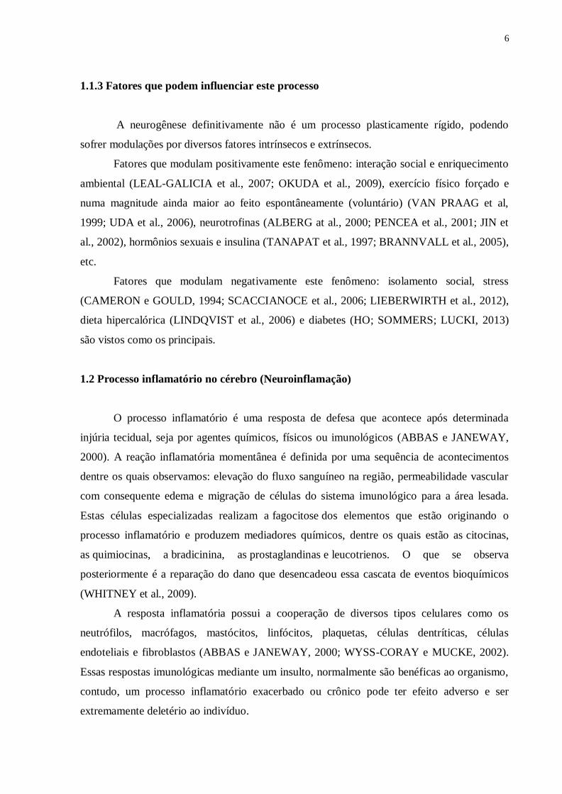
1.4 Glândula pineal e melatonina
Durante muito tempo a glândula pineal foi assinalada somente como um órgão
vestigial sem função específica. No século XVII a mesma foi descrita por René Descartes
como a estrutura incumbida de distribuir os fluidos do encéfalo para os músculos. Em 1958
o dermatologista Aaron Lerner identificou e isolou um hormônio produzido na pineal,
chamando-o de melatonina (ARENDT, 1995).
A gênese embriológica desta glândula se dá como uma evaginação dorsal do teto do
terceiro ventrículo (EKSTROM e MEISSL, 2003) sendo uma pequena estrutura epitalâmica
estabelecida dorsalmente à região caudal do diencéfalo, composta por tecido glandular
detentor de células características denominadas pinealócitos. Em roedores, a pineal não é
diretamente fotossensível e possui três porções distintas: a pineal profunda, o pedúnculo
pineal e a pineal superficial (MOLLER, 1992) (Figura 1).
Figura 1. Localização anatômica da glândula pineal de rato apresentando suas três porções distintas: pineal
superficial, pedúnculo pineal e pineal profunda (Modificado de Swanson, L.W. 1998).
10
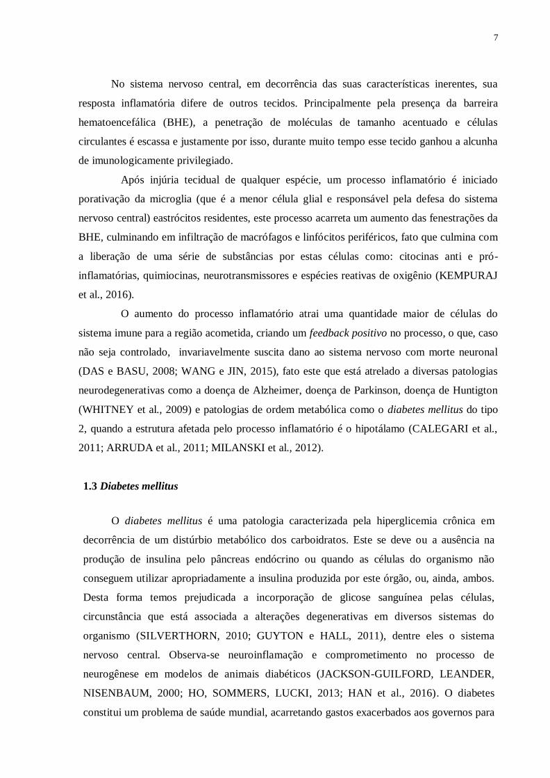
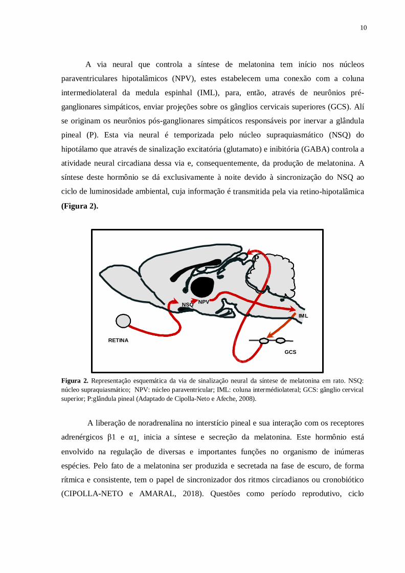
A via neural que controla a síntese de melatonina tem início nos núcleos
paraventriculares hipotalâmicos (NPV), estes estabelecem uma conexão com a coluna
intermediolateral da medula espinhal (IML), para, então, através de neurônios pré-
ganglionares simpáticos, enviar projeções sobre os gânglios cervicais superiores (GCS). Alí
se originam os neurônios pós-ganglionares simpáticos responsáveis por inervar a glândula
pineal (P). Esta via neural é temporizada pelo núcleo supraquiasmático (NSQ) do
hipotálamo que através de sinalização excitatória (glutamato) e inibitória (GABA) controla a
atividade neural circadiana dessa via e, consequentemente, da produção de melatonina. A
síntese deste hormônio se dá exclusivamente à noite devido à sincronização do NSQ ao
ciclo de luminosidade ambiental, cuja informação é transmitida pela via retino-hipotalâmica
(Figura 2).
P
NSQ NPV
IML
RETINA
GCS
Figura 2. Representação esquemática da via de sinalização neural da síntese de melatonina em rato. NSQ:
núcleo supraquiasmático; NPV: núcleo paraventricular; IML: coluna intermédiolateral; GCS: gânglio cervical
superior; P:glândula pineal (Adaptado de Cipolla-Neto e Afeche, 2008).
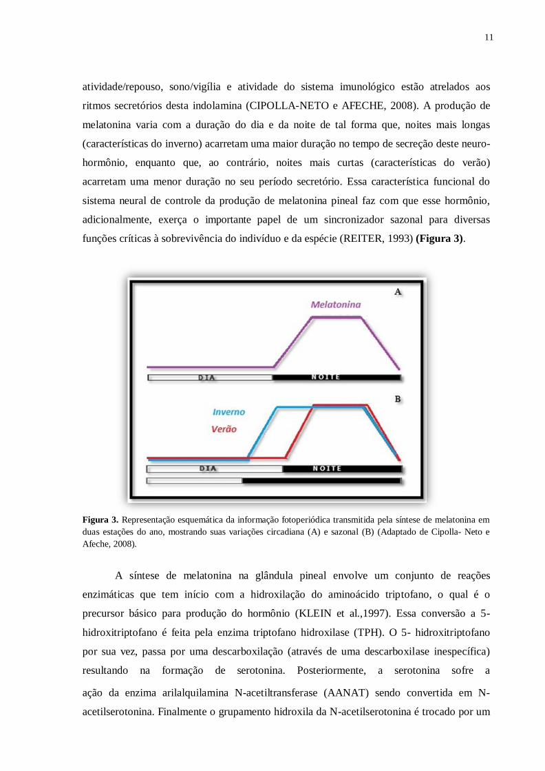
A liberação de noradrenalina no interstício pineal e sua interação com os receptores
adrenérgicos β1 e α1, inicia a síntese e secreção da melatonina. Este hormônio está
envolvido na regulação de diversas e importantes funções no organismo de inúmeras
espécies. Pelo fato de a melatonina ser produzida e secretada na fase de escuro, de forma
rítmica e consistente, tem o papel de sincronizador dos ritmos circadianos ou cronobiótico
(CIPOLLA-NETO e AMARAL, 2018). Questões como período reprodutivo, ciclo
11
atividade/repouso, sono/vigília e atividade do sistema imunológico estão atrelados aos
ritmos secretórios desta indolamina (CIPOLLA-NETO e AFECHE, 2008). A produção de
melatonina varia com a duração do dia e da noite de tal forma que, noites mais longas
(características do inverno) acarretam uma maior duração no tempo de secreção deste neuro-
hormônio, enquanto que, ao contrário, noites mais curtas (características do verão)
acarretam uma menor duração no seu período secretório. Essa característica funcional do
sistema neural de controle da produção de melatonina pineal faz com que esse hormônio,
adicionalmente, exerça o importante papel de um sincronizador sazonal para diversas
funções críticas à sobrevivência do indivíduo e da espécie (REITER, 1993) (Figura 3).
Figura 3. Representação esquemática da informação fotoperiódica transmitida pela síntese de melatonina em
duas estações do ano, mostrando suas variações circadiana (A) e sazonal (B) (Adaptado de Cipolla- Neto e
Afeche, 2008).
A síntese de melatonina na glândula pineal envolve um conjunto de reações
enzimáticas que tem início com a hidroxilação do aminoácido triptofano, o qual é o
precursor básico para produção do hormônio (KLEIN et al.,1997). Essa conversão a 5-
hidroxitriptofano é feita pela enzima triptofano hidroxilase (TPH). O 5- hidroxitriptofano
por sua vez, passa por uma descarboxilação (através de uma descarboxilase inespecífica)
resultando na formação de serotonina. Posteriormente, a serotonina sofre a
ação da enzima arilalquilamina N-acetiltransferase (AANAT) sendo convertida em N-
acetilserotonina. Finalmente o grupamento hidroxila da N-acetilserotonina é trocado por um
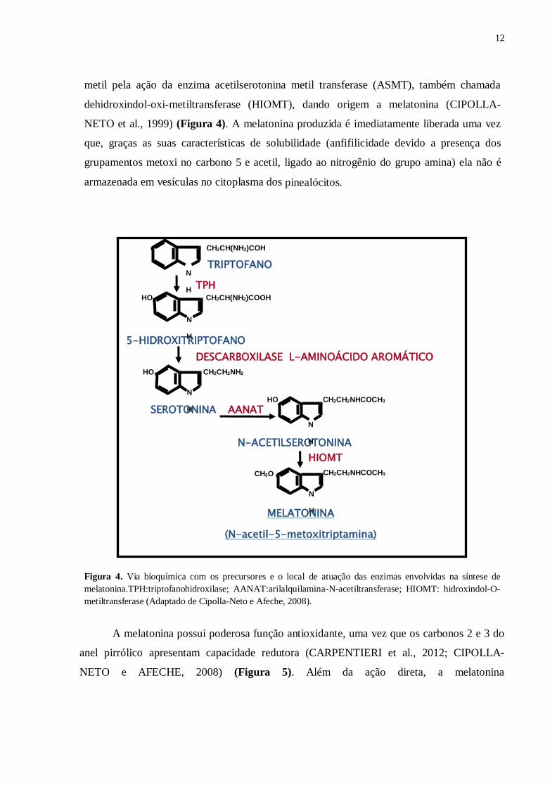
12
metil pela ação da enzima acetilserotonina metil transferase (ASMT), também chamada
dehidroxindol-oxi-metiltransferase (HIOMT), dando origem a melatonina (CIPOLLA-
NETO et al., 1999) (Figura 4). A melatonina produzida é imediatamente liberada uma vez
que, graças as suas características de solubilidade (anfifilicidade devido a presença dos
grupamentos metoxi no carbono 5 e acetil, ligado ao nitrogênio do grupo amina) ela não é
armazenada em vesículas no citoplasma dos pinealócitos.
Figura 4. Via bioquímica com os precursores e o local de atuação das enzimas envolvidas na síntese de
melatonina.TPH:triptofanohidroxilase; AANAT:arilalquilamina-N-acetiltransferase; HIOMT: hidroxindol-O-
metiltransferase (Adaptado de Cipolla-Neto e Afeche, 2008).
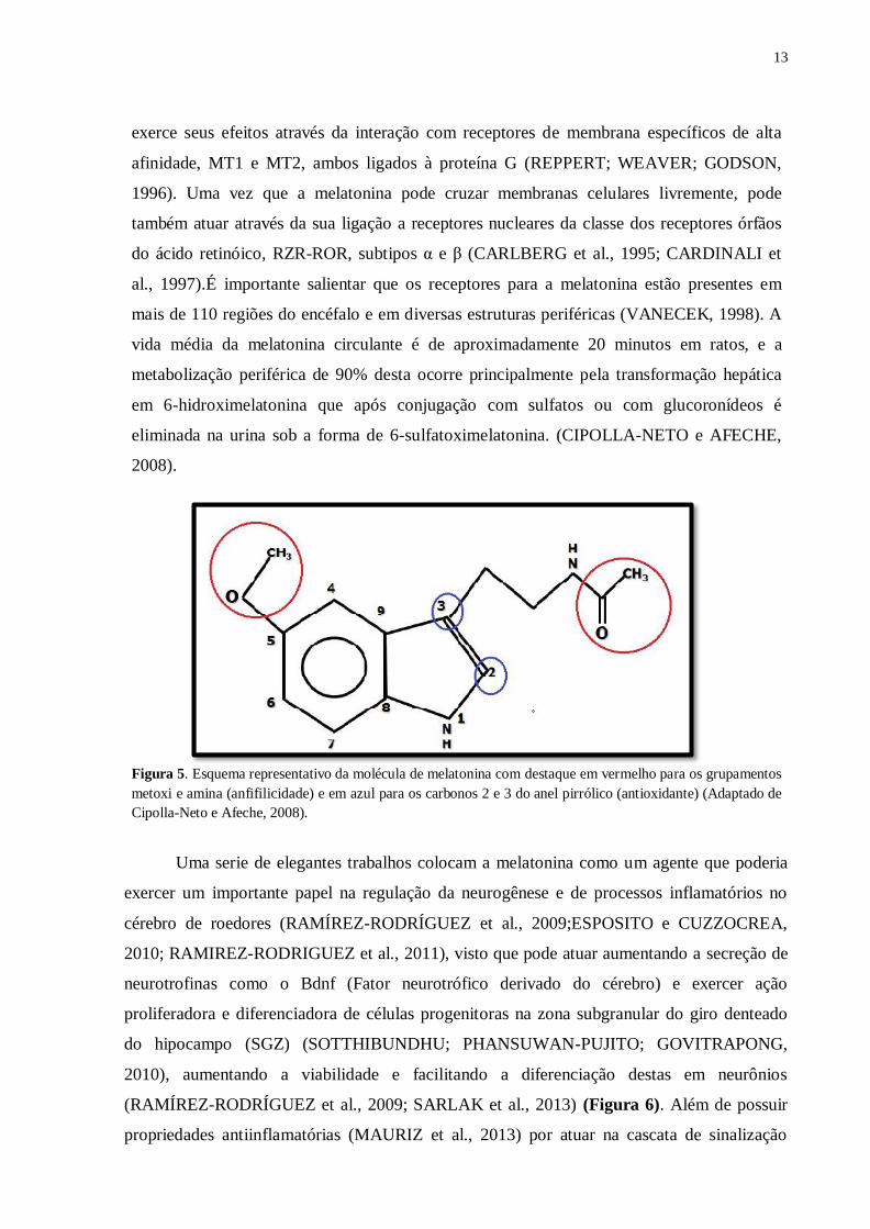
A melatonina possui poderosa função antioxidante, uma vez que os carbonos 2 e 3 do
anel pirrólico apresentam capacidade redutora (CARPENTIERI et al., 2012; CIPOLLA-
NETO e AFECHE, 2008) (Figura 5). Além da ação direta, a melatonina
5-HIDROXITRIPTOFANO
AANAT
N
H
CH2CH(NH2)COH
CH2CH2NHCOCH3 CH3O
CH2CH2NH2 HO
N
H
CH2CH(NH2)COOH HO
HO
SEROTONINA
N-ACETILSEROTONINA
TPH
N
H
N
H
N
H
HIOMT
MELATONINA
(N-acetil-5-metoxitriptamina)
CH2CH2NHCOCH3
TRIPTOFANO
DESCARBOXILASE L-AMINOÁCIDO AROMÁTICO
13
exerce seus efeitos através da interação com receptores de membrana específicos de alta
afinidade, MT1 e MT2, ambos ligados à proteína G (REPPERT; WEAVER; GODSON,
1996). Uma vez que a melatonina pode cruzar membranas celulares livremente, pode
também atuar através da sua ligação a receptores nucleares da classe dos receptores órfãos
do ácido retinóico, RZR-ROR, subtipos α e β (CARLBERG et al., 1995; CARDINALI et
al., 1997).É importante salientar que os receptores para a melatonina estão presentes em
mais de 110 regiões do encéfalo e em diversas estruturas periféricas (VANECEK, 1998). A
vida média da melatonina circulante é de aproximadamente 20 minutos em ratos, e a
metabolização periférica de 90% desta ocorre principalmente pela transformação hepática
em 6-hidroximelatonina que após conjugação com sulfatos ou com glucoronídeos é
eliminada na urina sob a forma de 6-sulfatoximelatonina. (CIPOLLA-NETO e AFECHE,
2008).
Figura 5. Esquema representativo da molécula de melatonina com destaque em vermelho para os grupamentos
metoxi e amina (anfifilicidade) e em azul para os carbonos 2 e 3 do anel pirrólico (antioxidante) (Adaptado de
Cipolla-Neto e Afeche, 2008).
Uma serie de elegantes trabalhos colocam a melatonina como um agente que poderia
exercer um importante papel na regulação da neurogênese e de processos inflamatórios no
cérebro de roedores (RAMÍREZ-RODRÍGUEZ et al., 2009;ESPOSITO e CUZZOCREA,
2010; RAMIREZ-RODRIGUEZ et al., 2011), visto que pode atuar aumentando a secreção de
neurotrofinas como o Bdnf (Fator neurotrófico derivado do cérebro) e exercer ação
proliferadora e diferenciadora de células progenitoras na zona subgranular do giro denteado
do hipocampo (SGZ) (SOTTHIBUNDHU; PHANSUWAN-PUJITO; GOVITRAPONG,
2010), aumentando a viabilidade e facilitando a diferenciação destas em neurônios
(RAMÍREZ-RODRÍGUEZ et al., 2009; SARLAK et al., 2013) (Figura 6). Além de possuir
propriedades antiinflamatórias (MAURIZ et al., 2013) por atuar na cascata de sinalização
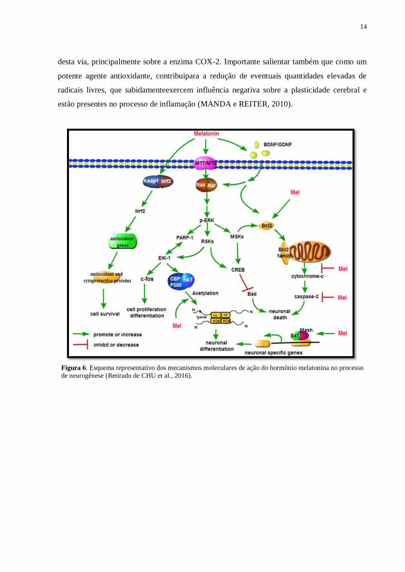
14
desta via, principalmente sobre a enzima COX-2. Importante salientar também que como um
potente agente antioxidante, contribuipara a redução de eventuais quantidades elevadas de
radicais livres, que sabidamenteexercem influência negativa sobre a plasticidade cerebral e
estão presentes no processo de inflamação (MANDA e REITER, 2010).
Figura 6. Esquema representativo dos mecanismos moleculares de ação do hormônio melatonina no processo
de neurogênese (Retirado de CHU et al., 2016).

15
7 Conclusão
Este estudo concluiu que o quadro de diabetes tipo 2 característico da linhagem de
animais Goto-Kakizaki promove alterações na expressão gênica e proteica de fatores
envolvidos na neurogênese e que isso se dá provavelmente como mecanismo compensatório
ao insulto hiperglicêmico com suas comorbidades características e que a melatonina é capaz,
em determinados momentos, de amenizar tais alterações. Conclui-se também que o diabetes
observado nos animais GK não é decorrente de processos inflamatórios no hipotálamo, ainda,
a doença em questão não parece promover quaisquer indícios de inflamação hipocampal.
16
Referências
ABBAS, A.K. ; JANEWAY, C.A. Jr. Immunology: improving on nature in the twenty-
first century. Cell. v. 100 (1), p. 129-138. 2000. Review.
ABDELSAID, M.; COUCHA, M.; HAFEZ, S.; YASIR, A.; JOHNSON, M.H.; ERGUL, A.
Enhanced VEGF signalling mediates cerebral neovascularisation via downregulation of
guidance protein ROBO4 in a rat model of diabetes. Diabetologia. v. 60, p. 740-750. 2017.
AKASH, M.S.; REHMAN, K.; CHEN, S. Goto-Kakizaki rats: as non- obese diabetic animal
model for spontaneous type 2diabetes mellitus. Curr Diabetes Rev. v.9 (5), p.387-396. 2013.
Review.
ALBERG, M.A.; ABERG, N.D.; HEDBACKER, H.; OSCARSSON, J.; ERIKSSON, P.S.
Peripheral infusion of IGF-I selectively induces neurogenesis in the adult rat hippocampus. J
Neurosci. v. 15, p.:2896-2903. 2000.
ALTMAN, J. Are New Neurons Formed in the brain. January, 1962.
ALTMAN, J. Autoradiographic and histological studies of postnatal neurogenesis. A
longitudinal investigation of the kinetics, migration and transformation of cells incorporating
tritiated thymidine in infant rats, with special reference to postnatal neurogenesis. The
Journal of Comparative Neurology, v. 128, n. 4, p. 431–473, 1966.
ALTMAN, J. Autoradiographic and histological studies of postnatal neurogenesis. Cell
proliferation and migration in the anterior forebrain, with special reference to persisting
neurogenesis in the olfactory bulb. The Journal of Comparative Neurology, v. 137, n. 4, p.
433–457, 1969.
ALTMAN, J.; DAS, G. D. Autoradiographic and histological evidence of postnatal
hippocampal neurogenesis in rats. The Journal of Comparative Neurology, v. 124, n. 3, p.
319–335, 1965.
ALVAREZ-GARCÍA, V.; GONZÁLEZ, A.; ALONSO-GONZÁLEZ, C.; MARTÍNEZ-
CAMPA, C.; COS, S. Regulation of vascular endothelial growth factor by melatonin in
human breast cancer cells. J. Pineal Res. v.54, p. 373-380. 2013.
AMARAL, F.G.; TURATI, A. O.; BARONE, M.; SCIALFA, J.H.; CARMO-
BUONFIGLIO, D.; PERES, R.; GARCIA, R.A.P.; AFECHE, S.C.; LIMA, L.; SCAVONE,
C.; BORDIN, S.; REITER, R. J.; MENNA-BARRETO, L.; CIPOLLA-NETO, J. Melatonin
synthesis impairmentas anewdeleterious outcomeofdiabetes derived hyperglycemia.J. Pineal
Res., v.57, p. 67-79, 2014.
AMBROGINI, P.; LATTANZI, D.; CIUFFOLI, S.; AGOSTINI, D.; BERTINI, L.;
STOCCHI, V.; SANTI, S.; CUPPINI, R. Morpho-functional characterization of neuronal cells
at different stages of maturation in granule cell layer of adult rat dentate gyrus. Brain
research, v. 1017, n. 1-2, p. 21–31, 2004.
17
ANDERSSON, A.K.; SANDLER, S. Melatonin protects against streptozotocin, but not
interleukin-1[beta]-induced damage ofrodent pancreatic [beta]-cells. J. Pineal Res., v. 30,
p.157-165, 2001.
ARENDT, J. Melatonin and the mammalian pineal gland. London: Chapman & Hall,
1995.
ARRUDA, A.P.; MILANSKI, M.; COOPE, A.; TORSONI, A.S.; ROPOLLE, E.;
CARVALHO, D.P.; CARVALHEIRA, J.B.; VELLOSO, L.A. Low-grade hypothalamic
inflammation leads to defective thermogenesis, insulin resistance, and impaired insulin
secretion. Endocrinology. v. 152 (4), p.1314-1326. 2011.
BARTELS, A.L.; LEENDERS, K.L. Cyclooxygenase and neuroinflammation in Parkinson's
disease neurodegeneration. Curr Neuropharmacol. v. 8(1), p.62-68. 2010.
BEAUQUIS, J.; HOMO-DELARCHE, F.; GIROIX, M.H.; EHSES, J.; COULAUD, J.;
ROIG, P.; PORTHA, B.; DE NICOLA, A.F.; SARAVIA, F. Hippocampal neurovascular and
hypothalamic–pituitary–adrenal axis alterations in spontaneously type 2 diabetic GK rats.
Experimental Neurology. v. 222, p. 125-134. 2010.
BERNIER, P. J.; BEDARD, A.; VINET, J.; LEVESQUE, M.; PARENT, A. Newly generated
neurons in the amygdala and adjoining cortex of adult primates. Proceedings of the National
Academy of Sciences of the United States of America, v. 99, n. 17, p. 11464 9, 2002.
BISBIS, S.; BAILBE, D.; TORMO, M.A.; PICAREL - BLANCHOT, F.; DEROUET, M.
;SIMON, J.; PORTHA, B. Insulin resistance in the GK rat : decreased receptor number but
normal kinase activity in liver. Am J Physiol., 1993.
BORGES, L.S.; DERMARGOS, A.; SILVA JÚNIOR, E.P.; WEIMANN, E.;
LAMBERTUCCI, R.H.; HATANAKA, E. Melatonin decreases muscular oxidative stress and
inflammation induced by strenuous exercise and stimulates growth factor synthesis. J. Pineal
Res.v. 58, p. 166-172. 2015.
BOYUK, B.; DEGIRMENCIOGLU, S.; ATALAY, H.; GUZEL, S.; ACAR, A.; CELEBI, A.;
EKIZOGLU, I.; SIMSEK, C. Relationship between levels of brain-derived neurotrophic
factor and metabolic parameters in patients with type 2 diabetes mellitus. J Diabetes Res.
2014.
BRANDT, M. D.; JESSBERGER, S.; STEINER, B.; KRONENBERG, G.; REUTER, K.;
BICK-SANDER, A.; BEHRENS, W. von der; KEMPERMANN, G. Transient calretinin
expression defines early postmitotic step of neuronal differentiation in adult hippocampal
neurogenesis of mice. Molecular and Cellular Neuroscience, v. 24, n. 3, p. 603–613, 2003.
BRANNVALL, K.; BOGDANOVIC, N.; KORHONEN, L.; LINDHOLM, D. 19-
Nortestosterone influences neural stem cell proliferation and neurogenesis in the rat brain.
Eur J Neurosci. v. 21(4), p. 871-878. 2005.
18
BUONFIGLIO, D.; PARTHIMOS, R.; DANTAS, R.; SILVA, R.C.; GOMES, G.;
ANDRADE-SILVA, J.; RAMOS-LOBO, A.; AMARAL, F.G.; MATOS, R.; SINÉSIO, J.;
MOTTA-TEIXEIRA, L.C.; DONATO JR., J.; REITER, R.; CIPOLLA-NETO, J. Melatonin
absence leads to long-term leptin resistance and overweight in rats. Frontiers in
Endocrinology. 2018.
CALEGARI, V.C.; TORSONI, A.S.; VANZELA, E.C.; ARAÚJO, E.P.; MORARI, J.;
ZOPPI, C.C.; SBRAGIA, L.; BOSCHERO, A.C.; VELLOSO, L.A. Inflammation of the
hypothalamus leads to defective pancreatic islet function. J Biol Chem. v. 286 (15), p. 12870-
12880. 2011.
CAMERON, H. A.; GOULD, E. Adult neurogenesis is regulated by adrenal steroids in the
dentate gyrus. Neuroscience, v. 61, n. 2, p. 203–209, 1994.
CAPPELLANO, G.; CARECCHIO, M.; FLEETWOOD, T.; MAGISTRELLI, L.;
CANTELLO, R.; DIANZANI, U.; COMI, C. Immunity and inflammationin
neurodegenerative diseases. Am J Neurodegener Dis.v. 2 (2), p.89-107. 2013.
CARDINALI, D. P.; GOLOMBEK, D.A.; ROSENSTEIN, R. E.; CUTRERA, R.A.;
ESQUIFIN, A.I. Melatonin site and mechanism ofaction: single or multiple? J Pineal Res.,
v. 263,p. 32-39, 1997.
CARPENTIERI, A.; DÍAZ DE BARBOSA, G.; ARECOA, V.; LÓPEZA, M.P.;
TALAMONI, N.T. New perspectives in melatonin uses. Pharmacological Research, v. 65,
p.437–444, 2012.
CARLBERG, C.; WIESENBERG, I. The orphan receptor family RZR / ROR,
melatonin and 5-lipoxygenase: expected relationship. J. Pineal Res.,v.4,p.171-178, Review,
1995. CHENG, M.-F. Hypothalamic neurogenesis in the adult brain. Frontiers in
Neuroendocrinology, v. 34, n. 3, p. 167–178, 2013. CHU, J.; TU, Y.; CHEN, J.; TAN, D.; LIU, X.; PI, R. Effects of melatonin and its analogues
on neural stem cells. Molecular and cellular endocrinology. v. 420, p. 169–179, 2016.
CIPOLLA-NETO, J.; AMARAL, F. Melatonin as a hormone: New physiological and clinical
insights. Endocr Rev. v. 39. p. 990-1028. 2018.
CIPOLLA-NETO, J.; SKORUPA, A.L.; RIBEIRO-BARBOSA, E. R.; BARTOL, I.; MOTA,
S. R.; AFECHE, S. C.; DELAGRANGE, P.; GUARDIOLA-LEMAITRE, B.; CANTERAS,
N.S. The role of the retrochiasmatic area in the control of pineal metabolism.
Neuroendocrinology, v. 69,p. 97-104, 1999.
CIPOLLA-NETO, J.; AFECHE, S. C. Glândulapineal. In: AIRES,M. M. (Ed.). Fisiologia. 3.
ed. Rio de Janeiro: Guanabara-Koogan, 2008. p. 981-990.
19
CIVELEK, S.; KONUKOGLU, D.; ERDENEN, F.; UZUN, H.Serum neurotrophic factor
levels in patients with type 2 diabetes mellitus: relationship to metabolic syndrome
components. Clin Lab. v. 59 (3-4), p. 369-374. 2013.
CORDEIRA, J.; RIOS, M. Weighing in the role of BDNF in the central control of eating
behavior. Mol Neurobiol. v. 44(3), p.441-448. 2011. Review.
DAS, S.; BASU, A. Inflammation: a new candidate in modulating adult neurogenesis. J
Neurosci Res.v. 86, p.1199-1208. 2008. Review.
DUAN, X.; KANG, E.; LIU, C. Y.; MING, G.-L.; SONG, H. Development of neural stem
cell in the adult brain. Current opinion in neurobiology, v. 18, n. 1, p. 108 – 15, 2008.
EKSTROM, P.; MEISSL, H. Evolution of photosensory pineal organs in new light: the fate
of neuroendocrine photoreceptors. Philos.Trans.R.Soc.Lond.B.Biol.Sci.,v.358, p.1679-
1700, 2003.
ENCINAS, J. M.; ENIKOLOPOV, G. Identifying and quantitating neural stem and progenitor
cells in the adult brain. Methods in cell biology, v. 85, p. 243–72, 2008.
ENCINAS, J. M.; SIERRA, A. Neural stem cell deforestation as the main force driving the
age-related decline in adult hippocampal neurogenesis. Behaviour Brain Research, v. 227, n.
2, p. 433–9, 2012.
ERIKSSON, P. S.; PERFILIEVA, E.; BJÖRK-ERIKSSON, T.; ALBORN, a M.;
NORDBORG, C.; PETERSON, D.; GAGE, F. H. Neurogenesis in the adult human
hippocampus. Nature medicine, v. 4, n. 11, p. 1313–1317, 1998.
ESPOSITO, E.; CUZZOCREA, S. Antiinflammatory activity of melatonin in central nervous
system. Curr Neuropharmacol. v. 8 (3), p. 228-242. 2010.
FISCHER, C.; MUELLER, T.; PFEFFER, M.; WICHT, H.; VON GALL, C.; KORF, H.W.
Melatonin Receptor 1-Deficiency Affects Feeding Dynamics and Pro-Opiomelanocortin
Expression in the Arcuate Nucleus and Pituitary of Mice. Neuroendocrinology. 2016.
FOWLER, C. D.; LIU, Y.; OUIMET, C.; WANG, Z. The effects of social environment on
adult neurogenesis in the female prairie vole. Journal of Neurobiology, v. 51, n. 2, p. 115–
128, 2002.
FREDRICH, M.; CHRIST, E.; DEROUICHE, A.; KORF, H.W. Impact of melatonin on
zeitgeber time-dependent changes in cell proliferation and apoptosis in the adult murine
hypothalamic-hypophyseal system. Neuroendocrinology. v. 102, p. 311-326. 2015.
FREDRICH, M.; HAMPEL, M.; SEIDEL, K.; CHRIST, E.; KORF, H.W. Impact of
Melatonin Receptor-Signaling on Zeitgeber Time-Dependent Changes in Cell Proliferation
and Apoptosis in the Adult Murine Hippocampus. Hippocampus. v. 27, p. 495-506. 2017.
FRESE,T.; BACH, A.G.; MUHLBAUER, E.; PONICKE, K.; BROMME, H.J.; WELP, A.;
PESCHKE, E. Pineal melatonin synthesis is decreased in type 2 diabetic Goto-Kakizaki rats.
Life Sci.v. 85, p. 526-533.2009.
20
FRUHBEIS, C.; FROHLICH, D.; KUO, W. P.; KRAMER-ALBERS, E. M. Extracellular
vesicles as mediators of neuron-glia communication. Front. Cell. Neurosci.v. 7, p. 182.
2013.
GAGE, F.H. Mammalian neural stem cells. Science, v. 287, p. 1433–1438, 2000.
GARCIA, R.A.P.; AFECHE, S.C.; SCIALFA, J.H.; AMARAL ,F.G.; SANTOS,
S.H.J.;LIMA, F.B.; YOUNG, M.E.; CIPOLLA-NETO, J. Insulin modulates norepinephrine-
mediates melatonin synthesis in culture rat pineal gland. Life Sciences.v.82, p.108–114,
2008.
GARCIA, R.A.P.; MARÇAL, A.C.; SILVA, J.A.; CARMO-BUONFIGLIO, D.; AMARAL,
F.G.; AFECHE, S.C.; CIPOLLA-NETO, J.; CARVALHO, C.R.O. Insulin temporal
sensitivity and its signaling pathway in the rat pineal gland. Life Sciences. v.87, p.169–174,
2010.
GIRAULT, F.M.; SONNAY, S.; GRUETTER, R.; DUARTE, J.M.N. Alterations of brain
energy metabolism in type 2 diabetic goto-kakizaki rats measured in vivo by 13C magnetic
resonance spectroscopy. Neurotox Res. 2017.
GOTO,Y.; KAKIZAKI, M.; MASAKI, N. Production of spontaneous diabetic rats by
repetition of selective breeding.Tohoku J Exp Med.v. 119(1), p.85-90. 1976.
GOULD, E.; MCEWEN, B. S.; TANAPAT, P.; GALEA, L.; FUCHS, E. Neurogenesis in the
dentate gyrus of the adult tree shrew is regulated by psychosocial stress and NMDA receptor
activation. The Journal of neuroscience: the official journal of the Society for
Neuroscience, v. 17, n. 7, p. 2492–2498, 1997.
GOULD, E.; REEVES, A.J.; FALLAH, M.; TANAPAT, P.; GROSS, C.G.; FUCHS, E.
Hippocampal neurogenesis in adult Old World primates.Proc.Natl.Acad.Sci. v. 96, p. 5263–
5267, 1999.
GOULD, E. How widespread is adult neurogenesis in mammals? Nat Rev Neurosci, v. 8, n.
June, p. 481–488, 2007.
GUYTON, A.C.; HALL, J. E. Tratado de Fisiologia Médica. 12ª edição. Editora: Elsevier,
São Paulo. 2011.
HAKIM, Z.S.; PATEL, B.K.; GOYAL, R.K. Effects of chronic ramipril treatment in
stresptozotocin-induced diabetic rats. Indian J Pharmacol. v.41, p.353-360, 1997.
HAN, C.; RICE, M.W.; CAI, D. Neuroinflammatory and autonomic mechanisms in diabetes
and hypertension. Am J Physiol Endocrinol Metab. v. 311(1), p. 32-41. 2016. Review.
21
HAN, W.; SONG, X.; HE, R.; LI, T.; CHENG, L.; XIE, L.; CHEN, H.; JIANG, L. VEGF
regulates hippocampal neurogenesis and reverses cognitive déficits in immature rats after
status epilepticus through the VEGF R2 signaling pathway. Epilepsy & Behavior. v. 68, p.
159-167. 2017.
HO, N.; SOMMERS, M.S.; LUCKI, I. Effects of diabetes on hippocampal neurogenesis: links
to cognition and depression. Neuro Sci Biobehav Rev. v. 37(8), p. 1346-1362. Review. 2013.
HUSSAIN, S.; MANSOURI, S.; SJOHOLM, A.; PATRONE, C.; DARSALIA, V. Evidence
for Cortical Neuronal Loss in Male Type 2 Diabetic Goto-Kakizaki Rats. Journal of
Alzheimer´s Disease. v. 41, p. 551-560. 2014.
INTERNATIONAL DIABETES FEDERATION. The Diabetes Atlas. Third Edition.
Brussels : International Diabetes Federation. 2015.
JACKSON-GUILFORD, J.; LEANDER, J.D.; NISENBAUM, L.K. The effect of
streptozotocin-induced diabetes on cell proliferation in the rat dentate gyrus. Neurosci Lett.
v. 293, p.91-94, 2000.
JIN, K.; ZHU, Y.; SUN, Y.; MAO, X.O.; XIE, L.; GREENBERG, D.A.Vascular endothelial
growth factor (VEGF) stimulates neurogenesis in vitro and in vivo.Proc Natl Acad Sci. v. 99,
p. 11946-11950. 2002.
JUNOD, A.; LAMBERT, A.E.; ORCI, L.; PICTET, R.; GONET, A.E.; RENOLD, A.E.
Studies of the diabetogenic action of streptozotocin. Proc Soc Exp Biol Med.v.126 (1), p.
201-205. 1967.
JUNOD, A.; LAMBERT, A.E.; STAUFFACHER, W.; RENOLD, A.E. Diabetogenic
action of streptozotocin: relationship of dose to metabolic response. J Clin Invest.v. 48(11),
p. 2129-2139. 1969.
KANDEMIR, Y.B.; KONUK, E.; BEHRAM, M.; SINDEL, M. Effect of melatonin on the
expression of VEGF-A and on the degeneration of follicle reserve in rat ovary. Eurasian J
Med. v. 50, p. 160-163. 2018.
KAPLAN, M. S.; HINDS, J.W. Neurogenesis in the adult rat: Electron microscopic analysis
of light radioautographs. Science, v. 197, n. 4308, p. 1092–1094, 1977.
KAPLAN, M. S.; BELL, D. H. Neuronal proliferation in the 9-month-old
rodentradioautographic study of granule cells in the hippocampus. Experimental brain
research, v. 52, n. 1, p. 1–5, 1983.
KARAASLAN, C.; SUZEN, S. Antioxidant properties of melatonin and its potential action
in diseases. Curr Top Med Chem.v. 15(9), p.894-903. 2015. Review.
KAUR, C.; LING, E.A. Effects of melatonin on macrophages/microglia in postnatal rat brain.
J Pineal Res. v. 26, p. 158-168. 1999.
22
KEE, N.; SIVALINGAM, S.; BOONSTRA, R.; WOJTOWICZ, J.M. The utility of Ki-67 and
BrdU as proliferative markers of adult neurogenesis. Journal of Neuroscience Methods. v.
115, p. 97-105. 2002.
KEMPERMANN, G.; JESSBERGER, S.; STEINER, B.; KRONENBERG, G. Milestones of
neuronal development in the adult hippocampus. Trends in Neurosciences, v. 27, n. 8, p.
447–452, 2004.
KEMPERMANN, G. Adult neurogenesis: stem cells and neuronal development in the adult
brain. 1. ed. [s.l.] Oxford University Press, 2005.
KEMPERMANN, G. Adult Neurogenesis: stem cells and neuronal development in the
adult brain. 2nd ed. USA: Oxford University Press, 2010.
KEMPERMANN, G. Adult neurogenesis. 2. ed. [s.l.] Oxford University Press, 2011.
KEMPURAJ, D.; THANGAVEL, R.; NATTERU, P.A. SELVAKUMAR, G.P.; SAEED, D.;
ZAHOOR, H.; ZAHEER, S.; IYER, S.S.; ZAHEER, A. Neuroinflammation Induces
Neurodegeneration. J Neurol Neurosurg Spine. v. 1(1). 2016.
KIREEV, R.A.; VARA, E.; TRESGUERRES, J.A.F. Growth hormone and melatonin prevent
age-related alteration in apoptosis processes in the dentate gyrus of male rats.
Biogerontology. v.14, p-431-442. 2013.
KLEIN, D.C.; COON, S.L.; ROSEBOOM, P.H.; WELLER, J.L.; BERNARD, M.; GASTEL,
J.A.; ZATZ, M.; IUVONE, P.M.; RODRIGUEZ, I.R.; BÉGAY, V.; FALCÓN, J.; CAHILL,
G.M.; CASSONE, V.M.; BALER, R. The melatonin rhythm-generation enzyme: molecular
regulation of serotonin N-acetyltransferase in the pineal gland. Rec. Prog. inHorm.Res., v.
52, p. 307-358, 1997.
KONG, X.; LI, X.; CAI, Z.; YANG, N.; LIU, Y.; SHU, J.; PAN, L.; ZUO, P. Melatonin
regulates the viability and differentiation of rat midbrain neural stem cells. Cell Mol.
Neurobiol., v. 28, p. 569-579, 2008.
KORNACK, D. R.; RAKIC, P. Continuation of neurogenesis in the hippocampus of the adult
macaque monkey. Proceedings of the National Academy of Sciences of the United States
of America, v. 96, n. 10, p. 5768–5773, 1999.
KRONENBERG, G.; REUTER, K.; STEINER, B.; BRANDT, M. D.; JESSBERGER, S.;
YAMAGUCHI, M.; KEMPERMANN, G. Subpopulations of proliferating cells of the adult
hippocampus respond differently to physiologic neurogenic stimuli. The Journal of
Comparative Neurology, v. 467, n. 4, p. 455–463, 2003.
KUHN, H. G.; WINKLER, J.; KEMPERMANN, G.; THAL, L. J.; GAGE, F. H. Epidermal
growth factor and fibroblast growth factor-2 have different effects on neural progenitors in the
adult rat brain. The Journal of neuroscience: the official journal of the Society for
Neuroscience, v. 17, n. 15, p. 5820–5829, 1997.
23
KUMAR, B.; GUPTA, S.K.; SRINIVASAN, B.P.; NAG, T.C.; SRIVASTAVA, S.;
SAXENA, R.; JHA, K.A. Hesperetin rescues retinal oxidative stress, neuroinflammation and
apoptosis in diabetic rats. Microvasc Res. p. 65-74. 2013.
KUWABARA, W.M.T.; PANVELOSKI-COSTA, A.C.; NAOMI, C.; YOKOTA, F.;
PEREIRA, J.N.B.; FILHO, J.M.; TORRES, R.P.; HIRABARA, S.M.; CURI, R.; ALBA-
LOUREIRO, T.C. Comparison of Goto-Kakizaki rats and high fat diet-induced obese rats:
Are they reliable models to study Type 2 Diabetes mellitus? PLoS One. v.12. 2017.
LANG, B.T.; YAN,Y.; DEMPSEY, R.J.; VEMUGANTI, R. Impaired neurogenesis in adult
type-2 diabetic rats. Brain Res. v. 33, p. 25-33. 2009.
LEAL-GALICIA, P.; SALDÍVAR-GONZÁLEZ, A.; MORIMOTO, S.; ARIAS, C. Exposure
to environmental enrichment elicits differential hippocampal cell proliferation: Role of
individual responsiveness to anxiety. Developmental Neurobiology, v. 67, n. 4, p. 395–405,
2007.
LEE, D.; BLACKSHAW, S. Functional implications of hypothalamic neurogenesis in the
adult mammalian brain. International Journal of Developmental. v. 30, n. 8, p. 615–621,
2012.
LENZ, K.M.; MCCARTHY, M.M. A starring role for microglia in brain sex differences.
Neuroscientist. v. 21, p. 306-321. 2015.
LEZOUALC´H, F.; SKUTELLA, T.; WIDMANN, M.; BEHL, C. Melatonin prevents
oxidative stress-induced cell death in hippocampal cells. Neuro Report. v. 7, p. 2071-2077.
1996.
LIEBERWIRTH, C.; LIU, Y.; JIA, X.; WANG, Z. Social isolation impairs adult neurogenesis
in the limbic system and alters behaviors in female prairie voles. Hormones and behavior, v.
62, n. 4, p. 357–66, 2012.
LINDQVIST, A.; MOHAPEL, P.; BOUTER, B.; FRIELINGSDORF, H.; PIZZO, D.;
BRUNDIN, P.; ERLANSON-ALBERTSSON, C. High-fat diet impairs hippocampal
neurogenesis in male rats. Eur J Neurol. v. 13(12), p. 1385-1388. 2006.
LIU, W.; HAN, X.; ZHOU, X.; ZHANG, S.; CAI, X.; ZHANG, L.; LI, Y.; LI, M.; GONG, S.;
JI, L. Brain derived neurotrophic factor in newly diagnosed diabetes and prediabetes. Mol
Cell Endocrinol.v. 429, p.106-113. 2016.
LOUISSAINT, A.; RAO, S.; LEVENTHAL, C.; GOLDMAN, S. Coordinated interaction of
neurogenesis and angiogenesis in the adult songbird brain. Neuron. v. 34, p. 945-960. 2002.
MANDA, K.; UENO, M.; ANZAI, K. Cranial irradiation-induced inhibition of neurogenesis
in hippocampal dentate gyrus of adult mice: attenuation by melatonin pre treatment. J Pineal
Res., v. 46, p. 71–78, 2008.
MANDA, K.; REITER, R.J. Melatonin maintains adult hippocampal neurogenesis and
cognitive functions after irradiation. Progressin Neurobiology, v. 90, p. 60-68, 2010.
24
MAURIZ, J.L.; COLLADO, P.S.; VENEROSO, C.; REITER, R.J.; GONZÁLEZ-
GALLEGO, J. Are view of the molecular aspects of melatonin's anti-inflammatory actions:
recent insights and new perspectives. J Pineal Res.v. 54(1), p.1-14. 2013. Review.
MILANSKI, M.; ARRUDA, A.P.; COOPE, A.; IGNACIO-SOUZA, L.M.; NUNEZ, C.E.;
ROMAN, E.A.; ROMANATTO, T.; PASCOAL, L.B.; CARICILLI, A.M.; TORSONI,
M.A.; PRADA, P.O.; SAAD, M.J.;VELLOSO,L.A. Inhibition of hypothalamic
inflammation reverses diet-induced insulin resistance in the liver. Diabetes.v. 61, p.1455-
1462. 2012.
MOLLER,M. Fine structure of the pineal innervation of the mammalian pineal gland.
Microsc. Res. Tech., v. 3, p. 188-204, Review, 1992.
MONTALBANO, G.; MANIA, M.; ABBATE, F.; NAVARRA, M.; GUERRERA, M.C.;
LAURA, R.; VEGA, J.A.; LEVANTI, M.; GERMANÀ, A. Melatonin treatment suppresses
appetite genes and improves adipose tissue plasticity in diet-induced obese zebrafish.
Endocrine. v. 62, p. 381-393. 2018.
MORARI, J.; ANHÊ, G.;NASCIMENTO, L.; MOURA, R.F.; RAZOLLI, D.; SOLON, C.;
GUADAGNINI, D.; SOUZA, G.; MATTOS, A.H.; TOBAR, N.; RAMOS, C.D.; PASCOAL,
V.D.; SAAD, M.J.; LOPES-CENDES, I.; MORAES, J.C.; VELLOSO, L.A. Fractalkine
(CX3CL1) is involved in the early activation of hypothalamic inflammation in experimental
obesity. Diabetes. v. 63, p. 3770-3784. 2014.
MORIYA, T.; HORIE, N.; MITOME, M.; SHINOHARA ,K. Melatonin influences the
proliferative and differentiative activity of neural stem cells. J Pineal Res. v.42, p.411–
418,2007.
MUELLER, A. D.; MEAR, R. J.; MISTLBERGER, R. E. Inhibition of hippocampal
neurogenesis by sleep deprivation is independent of circadian disruption and melatonin
suppression. Neuroscience, v. 193, p. 170-181, 2011.
NASCIMENTO, L.F.; SOUZA, G.F.; MORARI, J.; BARBOSA, G.O.; SOLON, C.;
MOURA, R.F.; VOCTÓRIO, S.C.; IGNÁCIO-SOUZA, L.M.; RAZOLLI, D.S.;
CARVALHO, H.F.; VELLOSO, L.A. n-3 Fatty Acids Induce Neurogenesis of Predominantly
POMC-Expressing Cells in the Hypothalamus. Diabetes. v. 65(3), p. 673-686. 2016.
NATHAN, D.M. Diabetes: Advances in Diagnosis and Treatment. JAMA. v.10, p.1052-
1062. 2015. Review.
NIWA, A.; NISHIBORI, M.; HAMASAKI, S.; KOBORI, T.; LIU, K.; WAKE, H.; MORI,
S.; YOSHINO, T.; TAKAHASHI, H. Voluntary exercise induces
neurogenesis in the hypothalamus and pendymallining of the third ventricle. Brain Struct
Funct. p. 1-14. 2015.
25
OKUDA, H.; TATSUMI, K.; MAKINODAN, M.; YAMAUCHI, T.; KISHIMOTO, T.;
WANAKA, A. Environmental enrichment stimulates progenitor cell proliferation in the
amygdala. Journal of Neuroscience Research, v. 87, n. 16, p. 3546 – 3553, 2009.
PÉREZ-MARTÍN, M.; CIFUENTES, M.; GRONDONA, J.M.; LÓPEZ-AVALOS, M.D.;
GÓMEZ-PINEDO, U.; GARCÍA-VERDUGO, J.M.; FERNÁNDEZ-LLEBREZ, P. IGF-I
stimulates neurogenesis in the hypothalamus of adult rats. Eur J Neurosci.v. 31(9), p.1533-
1548. 2010.
PENCEA, V.; BINGAMAN, K.D.; WIEGAND, S.J.; LUSKIN, M.B. Infusion of brain-
derived neurotrophic factor into the lateral ventricle of the adult rat leads to new neurons in
the parenchyma of the striatum, septum, thalamus, and hypothalamus. J Neurosci. v. 21, p.
6706-6717. 2001.
PIERCE, A.A.; XU, A.W. De novo neurogenesis in adult hypothalamus as a compensatory
mechanism to regulate energy balance. J Neurosci.v.30(2), p.723-730. 2010.
PRAKASH, R.; SOMANATH, P.R.; EL-REMESSY, A.B.; KELLY-COBBS, A.; STERN,
J.E.; DORE-DUFFY, P.; JOHNSON, M.; FAGAN, S.C.; ERGUL, A. Enhanced cerebral but
not peripheral angiogenesis in the Goto-Kakizaki model of type 2 diabetes involves VEGF
and peroxynitrite signaling. Diabetes. v. 61(6), p. 1533-1542. 2012.
PRAKASH, R.; JOHNSON, M.; FAGAN, S.C.; ERGUL, A.
Cerebral neovascularization and remodeling patterns in two different models of type 2 diabets.
PLoS One. 2013.
RAJKOVSKA, G.; MIGUEL-HIDALGO, J. J. Gliogenesis and glial pathology in
depression. CNS Neurol. Disord.v. 6, p.219–233. 2007.
RAKIETEN, N.; RAKIETEN, M.L.; NADKARNI, M.V. Studies on the diabetogenic action
of streptozotocin. Cancer Chemother Rep. v.29, p.91-108, 1963.
RANA, I.; BADOER, E.; ALAHMADI, E.; LEO, C.H.; WOODMAN, O.L.; STEBBING,
M.J. Microglia are selectively activated in endocrine and cardiovascular control centres in
streptozotocin-induced diabetic rats. J Neuroendocrinol. v. 26(7), p. 413-425. 2014.
RAMIREZ-RODRÍGUEZ, G.; KLEMPIN, F.; BABU, H.; KING, G.B.; KEMPERMANN, G.
Melatonin modulates cell survival of new neurons in the hippocampus of adult mice.
Neuropsychopharmacology.v. 34, p. 2180–2191, 2009.
26
RAMIREZ-RODRÍGUEZ, G.; LOPEZ, O.; ALONSO, A.D.; KING, G.A.B.;
KEMPERMANN, G. Chronic treatment with melatonin stimulates dendrite maturation
and complexity in adult hippocampal neurogenesis of mice. J Pineal Res.v.37,p.50-29,
2011.
RAMIREZ-RODRÍGUEZ, G.; VEGA-RIVERA, N.M.; BENÍTEZ-KING, G.;
CASTRO-GARCÍA, M.; ORTÍZ-LOPEZ, L. Melatonin supplementation delays the
decline of adult hippocampal neurogenesis during normal aging of mice. Neuroscience
Letters. v. 530, p. 53-58, 2012.
RAMOS-LOBO, A.M.; BUONFIGLIO,D.C.; CIPOLLA-NETO, J. Streptozotocin-
induced diabetes disrupts the body temperature daily rhythm in rats. Diabetol Metab
Syndr.v. 29, p.7:39, 2015.
RANA,I.;BADOER, E.; ALAHMADI, E.; LEO, C.H.; WOODMAN,O.L.; STEBBING
,M.J. Microglia are selectively activate endocrine and cardiovascular control centres in
streptozotocin-induced diabetic rats. J Neuroendocrinol.v.26(7), p.413-425.2014.
REITER, R.J. Melatonin: the chemical expression of darkness. Mol Cell Endocrinol.,
v. 79, p.153–158, 1991.
REITER, R.J. The melatonin rhythm- botha clock and calendar .Experientia. v.49, p.
654-664, 1993.
RENNIE, K.; BUTTE, M.; PAPAS, B.A. Melatonin promotes neurogenesis in dentate
gyrus in the pinealectomized rat. J Pineal Res. v. 47, p. 313–317, 2009.
REPPERT, S.M.; WEAVER, D.R.; GODSON, C. Melatonin receptors step into the
light: cloning and classification of subtypes. Trends Pharmacol Sci. v.17, p.100-102,
1996.
RIBAK, C. E.; KORN, M. J.; SHAN, Z.; OBENAUS, A. Dendritic growth cones and
recurrent basal dendrites are typical features of newly generated dentate granule cells in
the adult hippocampus. Brain research, v. 1000, n. 1-2, p. 195–9, 2004.
SARLAK, G.; JENWITHEESUK, A.; CHETSAWANG, B.; GOVITRAPONG, P.
Effects of melatonin on nervous system aging: neurogenesis and neurodegeneration.
Journal of pharmacological sciences. v. 123, n. 1, p. 9–24, 2013.
SCACCIANOCE, S.; DEL BIANCO, P.; PAOLONE, G.; CAPRIOLI, D.;
MODAFFERI, A. M. E.; NENCINI, P.; BADIANI, A. Social isolation selectively
reduces hippocampal brain-derived neurotrophic factor without altering plasma
corticosterone. Behavioural brain research, v. 168, n. 2, p. 323–5, 2006.
SEKI, T.; ARAI, Y. Highly polysialylated neural cell adhesion molecule (NCAM-H) is
expressed by newly generated granule cells in the dentate gyrus of the adult rat. The
Journal of neuroscience: the official journal of the Society for Neuroscience, v.13,
p.2351-2358, 1993.
27
SERI, B.; GARCÍA-VERDUGO, J. M.; COLLADO-MORENTE, L.; MCEWEN, B. S.;
ALVAREZ-BUYLLA, A. Cell types, lineage, and architecture of the germinal zone in
the adult dentate gyrus. The Journal of Comparative Neurology, v. 478, n. 4, p. 359 –
378, 2004.
SHARMA, S.; SINGH, H.; AHMAD, N.; MISHRA, P.; TIWARI, A. The role of
melatonin in diabetes: therapeutic implications. Arch Endocrinol Metab. v.59 (5),
p.391-399. 2015.
SHIVRAJ SOHUR, U.; EMSLEY, J. G.; MITCHELL, B. D.; MACKLIS, J. D. Adult
neurogenesis and cellular brain repair with neural progenitors, precursors and stem
cells. Philosophical Transactions of the Royal Society B: Biological Sciences, v. 361,
n. 1473, p. 1477–1497, 2006.
SHU, X.; ZHANG, Y.; XU, H.; KANG, K.; CAI, D. Brain-derived neurotrophic
factor inhibits glucose intolerance after cerebral ischemia. Neural Regen Res. v. 8 (25),
p. 2370-2378. 2013.
SILVERTHORN, D.U. Fisiologia Humana. Uma Abordagem Integrada. 5ªedição,
Editora Artmed, São Paulo. 2010.
SMART, I. The subependymal layer of the mouse brain and its cell production as
shown by radioautography after thymidine-H3 injection. The Journal of Comparative
Neurology, v. 116, n. 3, p. 325–347, 1961.
SOMPOL, P.; LIU, X.; BABA, K.; PAUL, K.N.; TOSINI, G.; IUVONE, P.M.; YE,K.
N-acetylserotonin promotes hippocampal neuroprogenitor cell proliferation in sleep-
deprived mice. Proc Natl Acad Sci USA. v. 108, p. 8844-8849, 2011.
SOUMIER, A.; BANASR, M.; LORTET, S.; MASMEJEAN, F.; BERNARD, N.;
KERKERIAN-LE-GOFF, L.; GABRIEL, C.; MILLAN, M. J.; MOCAER, E.;
DASZUTA, A. Mechanisms contributing to the phase-dependent regulation of
neurogenesis by the novel antidepressant agomelatine in the adult rat hippocampus.
Neuropsychopharmacology, v. 11, p. 2390-2403, 2009.
SOTTHIBUNDHU, A.; PHANSUWAN-PUJITO, P.; GOVITRAPONG, P. Melatonin
increases proliferation of culture neural stem cells obtained from adult mouse
subventricular zone. J Pineal Res. v. 49, p. 291-300, 2010.
SZKUDELSKI, T. The mechanism of alloxan and streptozotocin action in B cells of
the rat pancreas. Physiol Res.v.50, p.537-546, 2001.
TANAPAT, P.; GOULD, E. EGF stimulates proliferation of granule cell precursors in
the dentate gyrus. Soc Neurosci Abstr. v. 23, p. 317. 1997.
28
TANG, Y.; CAI, B.; YUAN, F.; HE, X.; LIN, X.; WANG, J.; WANG, Y.; YANG, G.Y.
Melatonin pretreatment improves the survival and function of transplanted
mesenchymal stem cells after focal cerebral ischemia. Cell Transplantation. v. 23, p.
1279-1291. 2014.
TOCHARUS, C.; PURIBORIBOON, Y.; JUNMANEE, T.; TOCHARUS, J.;
EKTHUWAPRANEE, K.; GOVITRAPONG, P. Melatonin enhances adult rat
hippocampal progenitor cell proliferation via ERK signaling pathway through melatonin
receptor. Neuroscience. v.275, p. 314-321. 2014.
TOZUKA, Y.; FUKUDA, S.; NAMBA, T.; SEKI, T.; HISATSUNE, T. GABAergic
excitation promotes neuronal differentiation in adult hippocampal progenitor cells.
Neuron, v. 47, n. 6, p. 803–15, 2005. UDA, M.; ISHIDO, M.; KAMI, K.; MASUHARA, M. Effects of chronic treadmill
running on neurogenesis in the dentate gyrus of the hippocampus of adult rat.Brain
Res. n. 1104(1) p:64-72. 2006.
VANECEK, J. Cellular mechanisms of melatonin action. Physiol Rev. v.3, p.687-721,
Review, 1998.
VAN PRAAG, H.; KEMPERMANN, G.; GAGE, F.H. Running increases cell
proliferation and neurogenesis in the adult mouse dentate gyrus. Nat Neurosci. V 3. p.
266-270.1999
WANG, B.; JIN, K. Current perspectives on the link between neuroinflammation and
neurogenesis. Metab Brain Dis.v.30(2), p.355-365. 2015.
WINNER, B.; WINKLER ,J. Adult neurogenesis in neurodegenerative diseases. Cold
Spring Harb Perspect Biol. v. 7(4). 2015.Review.
WHITNEY, N.P.; EIDEM, T.M.; PENG, H.; HUANG, Y.; ZHENG, J.C.
Inflammation mediates varying effects in neurogenesis: relevance to the pathogenesis of
brain injury and neurodegenerative disorders. J Neurochem. v. 108(6), p.1343-1359.
Review. 2009.
WYSS-CORAY, T.; MUCKE, L. Inflammation in neurodegenerative disease--a double-
edged sword. Neuron. v.35(3), p.419-432. Review. 2002.
XIANG, Q.; ZHANG, J.; LI, C.Y.; WANG, Y.; ZENG, M.J.; CAI, Z.X.; TIAN, R.B.
JIA, W.; LI, X.H. Insulin resistance-induced hyperglycemia decreased the activation of
Akt/CREB in hippocampus neurons: Molecular evidence for mechanism of diabetes-
induced cognitive dysfunction. Neuropeptides. v. 54, p. 9-15. 2015.
XUE, B.; SUKUMARAN, S.; NIE, J.; JUSKO, W.J.; DUBOIS, D.C.; ALMON, R.R.
Adipose tissue deficiency and chronic inflammation in diabetic Goto-Kakizaki rats.
PLoS One. v.6(2). 2011.
29
YUAN, H.; CHEN, R.; WU, L.; CHEN, Q.; HU, A.; ZHANG, T.; WANG, Z.The
Regulatory Mechanism of Neurogenesis by IGF-1 in Adult Mice. Mol
Neurobiol. 2014.
YUAN, T.F.; GU, S.; SHAN ,C.; MARCHADO ,S.; ARIAS-CARRIÓN, O. Oxidative
Stress and Adult Neurogenesis. Stem Cell Rev. v. 11(5), p.706-709. 2015.
ZHANG, W.J.; TAN,Y.F.; YUE,J.T.; VRANIC,M.; WOJTOWICZ, J.M. Impairment
of hippocampal neurogenesis in streptozotocin-treated diabetic rats. Acta Neurol
Scand.v.3, p.205-210, 2008.
ZIEGLER, A.N.; LEVISON, S.W.; WOOD, T.L. Insulin and IGF receptor signaling in
neural-stem-cell homeostasis. Nat Rev Endocrinol.v.11, p. 161-170. 2015.
Top Related