


Línguas
Páginas
Legal

PONTIFÍCIA UNIVERSIDADE CATÓLICA DO RIO GRANDE DO SUL
FACULDADE DE MEDICINA
LUCIANO DA SILVA SELISTRE
NEFROPATIA INDUZIDA POR CONTRASTE APÓS TOMOGRAFIA
COMPUTADORIZADA.
PORTO ALEGRE
2009
2
LUCIANO DA SILVA SELISTRE
NEFROPATIA INDUZIDA POR CONTRASTE APÓS TOMOGRAFIA
COMPUTADORIZADA.
Dissertação apresentada como requisito
para a obtenção do grau de Mestre em
Clínica Médica, pelo programa de Pós-
Graduação da em Medicina e Ciências da
Saúde Faculdade de Medicina da Pontifícia
Universidade Católica do Rio Grande do
Sul.
ORIENTADOR: Professor Doutor David Saitovitch
PORTO ALEGRE
2009
3
FONTES FINANCIADORAS
Pontifícia Católica do Rio Grande do Sul (PUCRS) através do programa Probolsa
para o Mestrando Luciano da Silva Selistre
Este trabalho foi desenvolvido na Pontifícia Católica do Rio Grande do Sul
(PUCRS) no Serviço de Radiologia do Hospital São Lucas da PUCRS e enfermarias do
mesmo nosocômio.
4
DADOS INTERNACIONAIS DE CATALOGAÇÃO NA PUBLICAÇÃO (CIP)
Rosária Maria Lúcia Prenna Geremia Bibliotecária CRB 10/196
S465n Selistre, Luciano da Silva
Nefropatia induzida por contraste após tomografia computadorizada /
Luciano da Silva Selistre. Porto Alegre: PUCRS, 2009.
100 p.: il. tab.
Orientador: Prof. Dr. David Saitovich.
Dissertação (Mestrado) - Pontifícia Universidade Católica do Rio Grande
do Sul. Faculdade de Medicina. Programa de Pós-Graduação em Medicina e
Ciências da Saúde. Área de concentração: Nefrologia.
1. NEFROPATIAS/induzido quimicamente. 2. MEIOS DE CONTRASTE
/toxicidade. 3. TOMOGRAFIA COMPUTADORIZADA/efeitos adversos. 4.
INSUFICIÊNCIA RENAL. 5. INCIDÊNCIA. 6. FATORES DE RISCO. 7.
HOSPITALIZAÇÃO. 8. ESTUDOS DE COORTE. I. Saitovich, David. II. Título.
C.D.D. 616.61 C.D.U. 616.61-073(043.3)
N.L.M. WJ 342

5
FOLHA DE APROVAÇÃO
LUCIANO DA SILVA SELISTRE
NEFROPATIA INDUZIDA POR CONTRASTE APÓS TOMOGRAFIA
COMPUTADORIZADA.
Dissertação apresentada como requisito
para a obtenção do grau de Mestre em
Clínica Médica, pelo programa de Pós-
Graduação da Faculdade de Medicina da
Pontifícia Universidade Católica do Rio
Grande do Sul.
Orientador: DAVID SAITOVITCH
Aprovada em 11 de janeiro de 2010.
BANCA EXAMINADORA
______________________________________
IVAN ANTONELLO - PUCRS
______________________________________
VINICIUS DUVAL DA SILVA - PUCRS
______________________________________
CLOTILDE DUCK GARCIA - UFSCMPA
______________________________________
ELIZETE KEITEL – UFSCMPA
6
AGRADECIMENTOS
Aos meus pais pelo maior bem: a vida.
Ao meu orientador David Saitovitch e co-autor Luciano Passiani Diogo pelo processo de
aprendizado.
A minha esposa Vandrea Carla de Souza pela paciência e ajuda no trabalho de digitação e
correção.
Ao Doutor Mário Wagner, pelos frutíferos debates a respeito da metodologia empregada e
da análise estatística.
Ao serviço de Nefrologia da PUCRS que acreditou na minha capacidade de implementar
esse projeto.
7
EPÍGRAFE
―Une expérience scientifique est une expérience qui contredit l'expérience
commune‖
―Uma experiência científica é um experiência que contradiz a experiência
comum.”
Gaston Bachelard (La Formation de l'esprit scientifique)

8
SUMÁRIO
Lista de ilustrações----------------------------------------------------------------------------------09
Considerações sobre a dissertação---------------------------------------------------------------10
Introdução ao projeto------------------------------------------------------------------------------11
Bibliografia ------------------------------------------------------------------------------------------24
Capítulo 1 – Artigo Revisão--- -------------------------------------------------------------------28
Capítulo 2 – Artigo Original ---------------------------------------------------------------------42
Capítulo 3 – Artigo Original (Versão Inglês)--------------------------------------------------69
Anexos-------------------------------------------------------------------------------------------------95
9
LISTA DE ILUSTRAÇÕES
Tabela 1 – Dados demográficos --------------------------------------------------------------------61
Tabela 2 – Volume de contraste e Função Renal-------------------------------------------------62
Tabela 3 – Impacto na variação da creatinina em porcentagem por 100 ml de contraste---63
Tabela 4 – Impacto na variação da creatinina em mg/dl por 100 ml de contraste ----------63
Tabela 5 – Impacto na variação da D.C.E estimada (C.G) por 100 ml de contraste---------64
Tabela 6 – Impacto na variação da D.C.E estimada (MDRD) a cada 100 ml de contraste-64
Tabela 7 – Fatores de risco por análise multivariada para NIC (CrS aumenta>0,5 mg/dl)-65
Tabela 8 – Fatores de risco por análise multivariada para NIC (CrS aumenta >25%)------65
Gráfico 1 – Variação no aumento da creatinina após uso de contraste endovenoso--------66
Gráfico 2 – Variação na redução da taxa de filtração glomerular estimada por MDRD após
uso de contraste endovenoso------------------------------------------------------------------------67
Gráfico 3 – Variação na redução da taxa de filtração glomerular estimada por Cockroft-
Gault após uso de contraste endovenoso----------------------------------------------------------68
10
CONSIDERAÇÕES SOBRE A DISSERTAÇÃO
O programa de Pós-graduação em Medicina e Ciências da Saúde –
FAMED/PUCRS não exige um formato específico para apresentação da dissertação de
mestrado. Assim, o formato segue a preferência do autor, sendo a mesma escrita conforme
as recomendações de Spector (1)
. As referências bibliográficas seguem as normas de
Vancouver e as citações indicadas no texto seguiram o sistema de citações em sequência
(2).
O desenvolvimento de insuficiência renal aguda intra-hospitalar implica em
aumento da morbi-mortalidade do paciente, sendo que os meios de contraste contribuem
como fator de risco para esse desfecho. Não há trabalhos no nosso Brasil que avaliem a
tomografia computadorizada como fator de nefropatia por contraste, com escassos
trabalhos na literatura que avaliem esse parâmetro nos pacientes submetidos à tomografia
computatorizada.
Objetivando à publicação dos resultados, realizaram-se três artigos sobre o tema de
nefropatia por contraste com aplicação endovenosa. O primeiro manuscrito foi o artigo
original que deu o escopo para o tema, sendo redigido conforme as normas da revista
internacional Radiology para a qual foi submetido à apreciação. Os dois artigos de revisão
foram encaminhados para a Revista Radiologia Brasileira (ISSN 0100-3984) que versa
sobre os assuntos relacionados à nefropatia por contraste, como medidas preventivas
(artigo 2) e o uso de contraste endovenoso (artigo 3).
11
INTRODUÇÃO
NEFROPATIA POR CONTRASTE
A administração de radiocontraste aumenta risco de insuficiência renal aguda,
sendo a terceira causa de perda da função renal em pacientes hospitalizados (3-8)
. Estudos
mostram fortes indícios de necrose tubular aguda (NTA), com mecanismos
fisiopatológicos pouco esclarecidos (11-15)
. Entretanto, as duas principais teorias são
vasoconstrição renal possivelmente mediada por alterações de óxido nítrico, endotelina e
adenosina, com efeitos citotóxicos pelo contraste (11-30).
, resultando em hipoxemia medular.
A NTA ocasionada por contraste leva à recuperação na função renal rapidamente,
quando comparada as outras causas de NTA. Há pelo menos duas possibilidades para
explicar estas conclusões (1-5)
:
Grau de ruptura do citoesqueleto tubular é muito menos grave;
Função tubular é pouco prejudicada;
A vasoconstrição renal é um achado comum em nefropatia contraste; sendo
mediada por liberação de endotelina e de adenosina devido à hiperosmolalidade do
contraste (15-18)
. Concomitantemente, a redução no fluxo sanguíneo medular pode ocorrer
devido a efeitos de viscosidade, sendo alterada pela hiperosmalidade dos contrastes (16)
.
O diabetes mellitus e a insuficiência cardíaca aumentam o risco de insuficiência
renal induzida por contraste em seres humanos associados principalmente com a redução
do óxido nítrico glomerular (19)
. A lesão tubular decorreria dos efeitos citotóxicos pela
geração de radicais livres de oxigênio (14-19)
, descritos preferencialmente em modelos
12
animais nos quais a diminuição da atividade protetora das enzimas antioxidantes
descreveria a lesão pelo contraste na hipovolemia (19)
.
INCIDÊNCIA
As publicações sobre a incidência de nefropatia por contraste são heterogêneas, com
séries que demonstram incidências de zero a mais de 50 por cento. Esta amplitude de
resultados deve-se (2-10)
:
Às diferentes definições de nefropatia induzida por contraste;
À quantidade e tipo de agente administrado;
Aos estudos prospectivos e retrospectivos;
Ao tipo de procedimento radiológico;
Ao fato da maioria dos estudos não excluir outras causas de insuficiência renal
aguda que possam estar associadas ao procedimento radiológico.
São comuns estudos prospectivos que encontram discreto aumento na concentração
plasmática de creatinina (média 0,2 mg/dl) após o uso de contraste endovenoso (5)
.
A incidência de perda da função renal cresce nos pacientes com múltiplos fatores de
risco, quando se utiliza como critério de nefropatia por contraste o aumento superior a 50%
ou a elevação de 1 mg nos valores basais da creatinina sérica (4,11)
. A chance de nefropatia
aumenta em 40 % em portadores de disfunção renal prévia, hipovolemia, insuficiência
cardíaca grave, ou múltiplos estudos no período de 72 horas. Alguns estudos demonstram
que 9 à 38% dos pacientes diabéticos com disfunção renal levemente reduzida submetidos
a contraste pioram a creatinina sérica (3,7)
.
Pacientes submetidos à intervenção coronária percutânea (ICP) para a doença
coronariana representam outro grupo de alto risco (21-22)
. Em uma revisão de mais de 7500
desses pacientes, foram relatadas as seguintes conclusões (21)
:
13
1. Insuficiência renal aguda, definida como um aumento da creatinina plasmática
basal em mais de 0,5 mg/dl, que ocorreu em 3,3%; e, nos indivíduos que possuiam
creatinina basal acima de 2 mg/dl, 25 % apresentaram piora da função renal.
2. Insuficiência renal aguda foi associada a aumentos significativos nas taxas de
mortalidade intra-hospitalar (22 versus 1,4 por cento sem insuficiência renal).
3. Similar taxa de insuficiência renal aguda após PCI associada ao aumento da
mortalidade intra-hospitalar foram observadas em duas outras grandes séries (28,29)
.
No entanto, esses estudos não estabeleceram as causas da insuficiência renal.
A taxa de insuficiência renal aguda que exige diálise, após administração contraste,
parece ser muito baixa. Em uma revisão retrospectiva de 58.000 procedimentos
coronarianos, apenas 10 e 49 pacientes necessitaram de diálise no período entre a primeira
e quarta semana da administração contraste (menos de 0,1 %) (29)
.
FATORES DE RISCO
A diminuição mais acentuada na taxa de filtração glomerular ocorre principalmente em
pacientes com um ou mais dos seguintes fatores de risco (3-30)
:
1. Subjacente à insuficiência renal, com a creatinina plasmática superior a 1,5 mg/ a
taxa de filtração glomerular inferior a 60 ml/min por 1,73 m2;
2. Nefropatia diabética com insuficiência renal;
3. Insuficiência cardíaca avançada ou outras causas de diminuição de perfusão renal
(tais como hipovolemia);
4. Intervenção coronária percutânea;
5. Mieloma múltiplo em idosos;
6. Altas doses de contraste.
14
CARACTERÍSTICAS CLÍNICAS
A insuficiência renal induzida por contraste inicia entre 12 e 24 horas após a infusão do
contraste. A insuficiência renal é não-oligúrica para a grande maioria dos doentes. Em
quase todos os casos, a diminuição da função renal é ligeira e transitória, com a
recuperação da função renal geralmente dentro de três a cinco dias (3-10)
.
Os fatores de risco para nefrotoxicidade do radiocontraste incluem: insuficiência renal
subjacente, com risco aumentado progressivamente para creatinina ≥ 1,5 mg/dl; nefropatia
diabética com insuficiência renal, insuficiência cardíaca avançada ou outras causas de
diminuição de perfusão renal (tais como hipovolemia); dose total e a osmolalidade do
agente; mieloma múltiplo e cateterismo cardíaco ou de intervenção (1-28)
.
A necessidade de diálise em nefropatia por contraste foi melhor descrita em pacientes
submetidos a procedimentos coronarianos. Esses pacientes representam um grupo de
doentes com uma elevada taxa de mortalidade. De <1 a 12 por cento dos doentes
sucumbem à diálise, a vasta gama provavelmente influenciada por função renal
previamente reduzida (23,24)
. Os melhores dados provêm de uma revisão de mais de 1800
pacientes consecutivos que sofreram uma intervenção coronária, que exigiu a utilização de
contraste (23)
. A incidência de insuficiência renal aguda foi 14,4 % e de diálise ocorreu em
0,8%. A mortalidade intra-hospitalar no grupo de diálise foi significativamente maior do
que naqueles sem insuficiência renal aguda (36% versus 1%, respectivamente). A
sobrevida em dois anos no grupo de diálise foi de apenas 19 %.
15
OBJETIVOS
1.0 - Geral
Analisar fatores de risco para desenvolvimento de Nefropatia Induzida por
Contraste após tomografia computatorizada.
1.1 - Específicos:
Analisar os seguintes fatores de risco com à nefropatia induzida por contraste:
1. Hipertensão arterial sistêmica;
2. Diabetes;
3. Dislipidemia;
4. Doença Cardiovascular;
5. Neoplasia;
6. Obesidade;
7. Uso prévio de drogas anti-hipertensivas e AINES.
16
PACIENTES E MÉTODOS
3.1 Definição de Termos
Variáveis de confusão: aquelas cujas modificações durante o período do estudo
pudessem interferir na associação.
Nefropatia por contraste: é definida com aumento da creatinina sérica (Cs) no
mínimo de 0,5 mg/dl, ou 25% da Cs basal nas primeiras 48 horas(2,3)
.
Idoso: O Instituto Brasileiro de Geografia e Estatística (IBGE) considera idosas as
pessoas com 60 anos ou mais, sendo o mesmo limite de idade considerado pela
Organização Mundial da Saúde (OMS) para os países em desenvolvimento. Devido as
características especificas do estado do Rio Grande do Sul, segundo o IBGE, definiu-se a
idade acima de 65 anos como fator de risco (31)
.
Pressão arterial média (PAM): A PAM, fornecida pelo aparelho, correspondeu ao
resultado da fórmula de Poiseuille PAM=PAD+[(PAS-PAD)/3], em que PAS e PAD
significam pressão arterial sistólica e diastólica, respectivamente. Foi avaliada por
esfigmomanometria digital através da artéria braquial (direita ou esquerda) colocando o
manguito firmemente cerca de 2 cm a 3 cm acima da fossa antecubital, centralizando a
bolsa de borracha sobre a artéria braquial. A largura da bolsa de borracha do manguito
correspondeu a 40% da circunferência do braço e seu comprimento, envolvendo pelo
menos 80% do braço.
17
Índice de Massa Corpórea (IMC): classificado pela seguinte fórmula peso=
(kg)/(altura)2 (m
2)
Desnutrição: IMC menor que 18 kg/m2.
Saudável: IMC=19-24,9 kg/m2.
Sobrepeso: IMC=25-29,9 kg/m2.
Obesidade:
o Grau I: IMC=30-34,9 kg/m2.
o Grau II: IMC=35-39,9 kg/m2.
o Grau III: IMC= acima de 40 kg/m2.
Infecções: qualquer quadro infeccioso bacteriano, viral, fúngico ou parasitário,
independente de sua gravidade, que necessitou de tratamento sistêmico.
Disfunção renal Prévia: definimos como uma creatinina maior ou igual a 1,5
mg/dl.
Diabetes: utilizamos o conceito e classificação do ―Consenso Brasileiro de
Diabetes 2009 (32)
.
Hidratação Preventiva: uso de qualquer tipo de solução hidrossalina endovenosa
antes da infusão do contraste.
Doença cardiovascular: doença cardíaca isquêmica, doença vascular cerebral e/ou
doença vascular periférica.
18
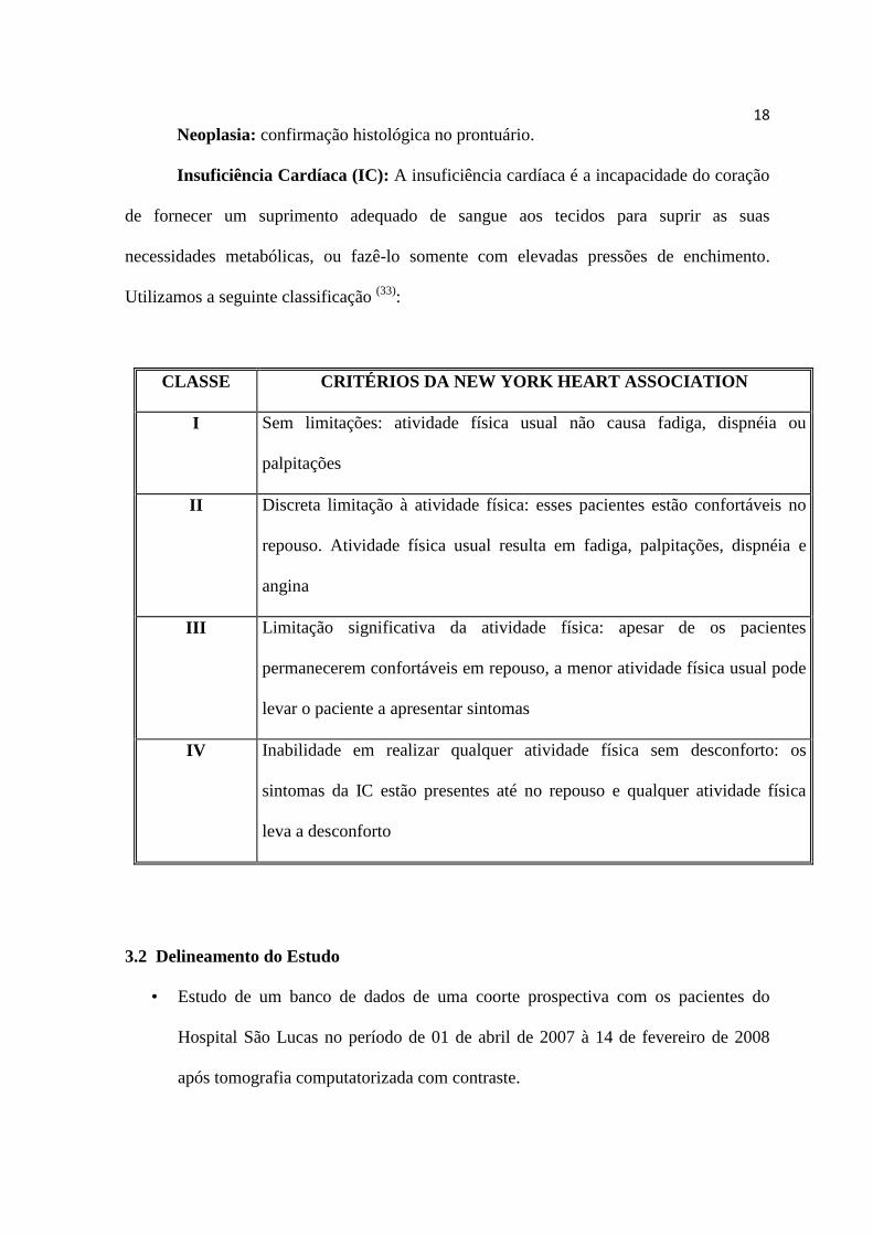
Neoplasia: confirmação histológica no prontuário.
Insuficiência Cardíaca (IC): A insuficiência cardíaca é a incapacidade do coração
de fornecer um suprimento adequado de sangue aos tecidos para suprir as suas
necessidades metabólicas, ou fazê-lo somente com elevadas pressões de enchimento.
Utilizamos a seguinte classificação (33)
:
CLASSE CRITÉRIOS DA NEW YORK HEART ASSOCIATION
I Sem limitações: atividade física usual não causa fadiga, dispnéia ou
palpitações
II Discreta limitação à atividade física: esses pacientes estão confortáveis no
repouso. Atividade física usual resulta em fadiga, palpitações, dispnéia e
angina
III Limitação significativa da atividade física: apesar de os pacientes
permanecerem confortáveis em repouso, a menor atividade física usual pode
levar o paciente a apresentar sintomas
IV Inabilidade em realizar qualquer atividade física sem desconforto: os
sintomas da IC estão presentes até no repouso e qualquer atividade física
leva a desconforto
3.2 Delineamento do Estudo
• Estudo de um banco de dados de uma coorte prospectiva com os pacientes do
Hospital São Lucas no período de 01 de abril de 2007 à 14 de fevereiro de 2008
após tomografia computatorizada com contraste.
19
3.3 Amostra
A amostra do estudo foi constituída por um banco de dados de 400 indivíduos que
realizaram TC com contraste endovenoso internados no Hospital São Lucas de Porto
Alegre no período supracitado.
3.3.1 Análise Estatísica
Analisou-se o banco de dados de uma coorte prospectiva no serviço de radiologia do
Hospital São Lucas (PUCRS) que preencheram os critérios de inclusão durante o período
de 01 de janeiro de 2007 à 31 de março de 2008.
O banco de dados sofreu processo de dupla digitação e posterior processamento das
incongruências.
Utilizou-se o teste de Kolmogorov-Smirnov para avaliar a normalização dos dados
quantitativos e a logaritimização dos dados quantitativos, quando esses não apresentavam
distribuição numa curva normal.
Os dados são apresentados em porcentagens, medianas e médias. As comparações
entre médias foram realizadas pelo uso de teste ―t‖ de Student bicaudal ou em casos de
múltiplas comparações pelo teste ―ANOVA one way‖, entre as medianas pelo teste de
Mann-Whitney ou Fisher.
Considerou-se como potencias riscos para NIC a diabete melito (DM), neoplasia,
anemia, insuficiência cardíaca (IC), acidente vascular encefálico (AVE), obesidade, baixo-
peso, insuficiência renal ou não, sexo feminino, hipotensão arterial no dia do exame.
Optou-se pela dicotomização dos seguintes fatores: idoso (>65 versus <65 anos), obeso ou
não-obeso (IMC>30 ou <30 kg/m2), baixo peso ou não (IMC<18,5 ou <18,5 kg/m
2),
20
insuficiência função renal pré-exame (CrS>1,5 mg/dl versus <1,5 mg/dl), hipotensão
arterial ou não (PAM<80 mmHg ou >80 mmHg), anemia ou não (Ht<36 ou > 36%).
Foi utilizada análise multivariada com modelo de regressão logística (backward
stepwise) associando a nova variável aos previamente registrados.
Considerou-se como significativos os valores de p < 0,05.
Usou-se o programa SPSS versão 17 para Windows.
3.3 Período de Realização do Estudo
1. Início da coleta de dados : 04/2007
2. Término da coleta de dados: 02/2008
3. Análise dos dados: 03/2008 a 12/2008
4. Confecção dos artigos para publicação: 01/2009 a 10/2009.
3.4 Critérios de Inclusão
1. Submetidos à tomografia computatorizada;
2. Ambos os gêneros;
3. Idade igual ou superior a 18 anos;
4. Consentimento informado assinado pelo paciente;
3.5 Critérios de Exclusão
Paciente com alta programada anterior a 48 horas;
21
3.6 Desfechos de Interesse
Função Renal;
Hemodiálise;
Anemia;
3.7 Variáveis Analisadas
Dados
• Idade;
• Gênero;
• Índice de Massa Corporal;
• Hipertensão Arterial Sistêmica;
• Insuficiência Cardíaca;
• Desidratação;
• Diabetes;
• Volume de Contraste;
• Disfunção renal Prévia – (creatinina maior ou igual a 1,5 mg/dl);
• Neoplasia;
• Idade acima de 65 anos;
• Hidratação Preventiva;
• Pressão arterial média no momento do exame contrastado;
• Creatinina sérica no dia do exame e 48 horas após;
• Hematimetria;
22
3.8 Acompanhamento dos Pacientes
Os pacientes foram alocados no estudo sendo acompanhados segundo o
cronograma.
Procedimentos Basal 48 horas
Consentimento informado assinado X
Dados Clínicos* X X
Creatinina sérica X X
Hemograma. X X
* Dados demográficos, doença básica, tipo de hidratação, presença de diabete, doença
cardiovascular prévia, medicações, índice de massa corporal.
3.9 Tamanho da População
Pacientes que foram submetidos à tomografia computatorizada no Hospital São
Lucas, totalizando 400 indivíduos.
3.10 Custos do Projeto e fonte pagadora
Os custos foram divididos em:
Fotocópias: R$ 200,00
Epidemiologista: R$ 3.000,00
Transporte: R$ 2.000,00
Tradução e revisão ortográfica: 1.000,00
Total de: R$ 6.200,00
23
3.12 Aspectos Éticos
O estudo encontra-se dentro das normas e diretrizes regulamentadoras de pesquisas
envolvendo seres humanos, conforme a Resolução no 196/96, do Conselho Nacional de
Saúde.
Considera-se que toda pesquisa com seres humanos envolve risco, podendo causar
dano eventual imediato ou tardio, comprometendo o indivíduo ou a coletividade. Não
obstante, os riscos potenciais do presente estudo são admissíveis, pois o estudo oferece elevada
possibilidade de gerar conhecimento para entender, prevenir ou aliviar um problema que afeta
o bem-estar dos sujeitos da pesquisa e de outros indivíduos.
O estudo já se encontra aprovado pelo comitê de ética em pesquisa da Pontifícia
Universidade Católica do Rio Grande do Sul (anexos).
RESULTADOS
A pesquisa gerou dois artigos que foram encaminhados para publicação. Segue em
anexo, os artigos.
24
BIBLIOGRAFIA
1. Spector, N. Manual para a redação de teses, projetos de esquisa e artigos
científicos. 2ª edição. Rio de Janeiro: Guanabara Koogan, p. 150, 2001.
2. Uniform Requirements for Manuscripts Submitted to Biomedical Journals:Writing
and Editing for Biomedical Publication. Bethesda; NLM; 2004. [updated 2003 nov;
cited 2004 jun 12]. Available from: http://www.icmje.org
3. Parfrey, PS, Griffiths, SM, Barrett, BJ, et al. Contrast material-induced renal failure
in patients with diabetes mellitus, renal insufficiency, or both. A prospective
controlled study. N Engl J Med 1989; 320:143.
4. Rudnick, MR, Goldfarb, S, Wexler, L, et al. Nephrotoxicity of ionic and nonionic
contrast media in 1196 patients: A randomized trial. Kidney Int 1995; 47:254.
5. Davidson, CJ, Hlatky, M, Morris, KG, et al. Cardiovascular and renal toxicity of a
nonionic radiographic contrast agent after cardiac catheterization. A prospective
trial. Ann Intern Med 1989; 110:119.
6. Cigarroa, RG, Lange, RA, Williams, RH, Hillis, LD. Dosing of contrast material to
prevent contrast nephropathy in patients with renal disease. Am J Med 1989;
86:649.
7. Rudnick, MR, Berns, JS, Cohen, RM, Goldfarb, S. Nephrotoxic risks of renal
angiography: Contrast-media associated nephrotoxicity and atheroembolism — A
critical review. Am J Kidney Dis 1994; 24:713.
8. Barrett, BJ. Contrast nephrotoxicity. J Am Soc Nephrol 1994; 5:125.
9. Solomon, R. Contrast-medium-induced acute renal failure. Kidney Int 1998;
53:230.

25
10. Weisbord, SD, Palevsky, PM. Radiocontrast-induced acute renal failure. J
Intensive Care Med 2005; 20:63.
11. Aspelin, P, Aubry, P, Fransson, SG, et al. Nephrotoxic effects in high-risk patients
undergoing angiography. N Engl J Med 2003; 348:491.
12. Sandler, CM. Contrast-agent-induced acute renal dysfunction--is iodixanol the
answer?. N Engl J Med 2003; 348:551.
13. Detrenis, S, Meschi, M, Musini, S, Savazzi, G. Lights and shadows on the
pathogenesis of contrast-induced nephropathy: state of the art. Nephrol Dial
Transplant 2005; 20:1542.
14. Persson, PB, Hansell, P, Liss, P. Pathophysiology of contrast medium-induced
nephropathy. Kidney Int 2005; 68:14.
15. Heyman, SN, Rosenberger, C, Rosen, S. Regional alterations in renal
haemodynamics and oxygenation: a role in contrast medium-induced nephropathy.
Nephrol Dial Transplant 2005; 20 Suppl 1:i6.
16. Agmon, Y, Peleg, H, Greenfield, Z, et al. Nitric oxide and prostanoids protect the
renal outer medulla from radiocontrast toxicity in the rat. J Clin Invest 1994;
94:1069.
17. Weisberg, LS, Kurnik, PB, Kurnik, BR. Radiocontrast-induced nephropathy in
humans. Role of renal vasoconstriction. Kidney Int 1992; 41:1408.
18. Cantley, LG, Clark, BA, et al. Role of endothelin and prostaglandins in
radiocontrast-induced renal artery constriction. Kidney Int 1993; 44:1217.
19. Goodman, AI, Olszanecki, R, Yang, LM, et al. Heme oxygenase-1 protects against
radiocontrast-induced acute kidney injury by regulating anti-apoptotic proteins.
Kidney Int 2007; 72:945.
26
20. Pflueger, A, Larson, TS, Nath, KA, et al. Role of adenosine in contrast media-
induced acute renal failure in diabetes mellitus. Mayo Clin Proc 2000; 75:1275.
21. Rihal, CS, Textor, SC, Grill, DE, et al. Incidence and prognostic importance of
acute renal failure after percutaneous coronary intervention. Circulation 2002;
105:2259.
22. Nikolsky, E, Mehran, R, Lasic, Z, et al. Low hematocrit predicts contrast-induced
nephropathy after percutaneous coronary interventions. Kidney Int 2005; 67:706.
23. McCullough, PA, Wolyn, R, Rocher, LL, et al. Acute renal failure after coronary
intervention: Incidence, risk factors, and relationship to mortality. Am J Med 1997;
103:368.
24. Weisbord, SD, Palevsky, PM. Prevention of contrast-induced nephropathy with
volume expansion. Clin J Am Soc Nephrol 2008; 3:273.
25. Wang, A, Holcslaw, T, Bashore, TM, et al. Exacerbation of radiocontrast
nephrotoxicity by endothelin receptor antagonism. Kidney Int 2000; 57:1675.
26. Heyman, SN, Rosen, S, Rosenberger, C. Renal parenchymal hypoxia, hypoxia
adaptation, and the pathogenesis of radiocontrast nephropathy. Clin J Am Soc
Nephrol 2008; 3:288.
27. Goodman, AI, Olszanecki, R, Yang, LM, et al. Heme oxygenase-1 protects
againstradiocontrast-induced acute kidney injury by regulating anti-apoptotic
proteins. Kidney Int 2007; 72:945.
28. Mehran, R, Aymong, ED, Nikolsky, E, et al. A simple risk score for prediction of
contrast-induced nephropathy after percutaneous coronary intervention:
development and initial validation. J Am Coll Cardiol 2004; 44:1393.
27
29. Liss, P, Persson, PB, Hansell, P, Lagerqvist, B. Renal failure in 57 925 patients
undergoing coronaryprocedures using iso-osmolar or low-osmolar contrast media.
Kidney Int 2006; 70:1811.
30. Gruberg, L, Mintz, G, Mehran, R, et al. The prognostic implications of further renal
function deterioration within 48 h of interventional coronary procedures in patients
with pre-existent chronic renal insufficiency. J Am Coll Cardiol 2000; 36:1542.
31. www.ibge.gov.br/home/mapa_site/mapa_site.php#populacao
32. Sociedade Brasileira de Diabetes. Consenso Brasileiro sobre diabetes. Diagnóstico
e classificação do diabetes melito e tratamento do diabetes melito tipo 2. Rio de
Janeiro: Diagraphic; 2003.
33. Swedberg K, Cleland J, Dargie H, Drexler H, Follath F, Komajda M, et al.
Guidelines for the diagnosis and treatment of chronic heart failure: executive
summary (update 2005): The Task Force for the Diagnosis and Treatment of
Chronic Heart Failure of the European Society of Cardiology. Eur Heart J. 2005;
26: 1115-40.
28
ARTIGO DE REVISÃO
29
NEFROPATIA DO CONTRASTE: MANEJO PREVENTIVO
CONTRAST NEPHROPATHY: PREVENTIVE MANAGEMENT
Luciano da Silva SelistreI; Laura Fuchs Bahlis
IV, Cinthia Fonseca O'Keeffe
IV , Vandrea
Carla de SouzaIII
; Mário Bernardes WagnerII; João Rubião Hoefel Filho
II; David
SaitovitchII; Luciano Passamani Diogo
II.
I Mestrando em Clínica Médica pelo programa de Pós-Graduação da Faculdade de
Medicina da Pontifícia Universidade Católica do Rio Grande do Sul (PUCRS) Porto
Alegre, RS, Brasil. Professor da Universidade de Caxias do Sul, RS.
II Doutor, professor da Faculdade de Medicina da Pontifícia Universidade Católica do Rio
Grande do Sul (PUCRS), Porto Alegre, RS, Brasil.
III Médica Nefrologista Pediátrica no Serviço de Nefrologia do Hospital Geral de Caxias do
Sul (HGCS), Caxias do Sul, RS, Brasil.
Instituição onde o trabalho foi desenvolvido:
Hospital São Lucas da PUCRS
Autor responsável para correspondência:
Luciano da Silva Selistre
Rua Adelino Roldo, 310 – CEP 95052-020 – Caxias do Sul - RS
Telefone: (54) 3202-2540/ 9122-4798/ 3218-7200 ramal 277.
Correio eletrônico: [email protected]
30
RESUMO
A nefropatia por contraste (NIC) é definida como uma piora na função renal que se
segue à administração de contraste parenteral, tendo sido excluídas outras causas. A NIC
manifesta-se, usualmente, como uma insuficiência renal aguda não oligúrica. Por
definição, deve haver um aumento na creatinina basal de 25-50% ou um aumento superior
a 0,5 mg/dl de 24 a 48 horas após o uso vascular de contraste. As medidas de prevenção
são baseadas na correção dos fatores que levam ao desenvolvimento da NIC e dividem-se
em: escolha de agentes de contrastes menos nefrotóxicos (não-iônicos) e utilização de
doses menores; melhora no estado clínico do paciente com hidratação; uso de drogas que
reduzam vasoconstrição renal e estresse oxidativo; e suspensão temporária de drogas com
potencial nefrotóxico ou prejudiciais, no caso de diminuição da filtração glomerular. Essa
revisão tem o objetivo de examinar as principais recomendações de manejo para prevenir
sua ocorrência.
Unitermos: Insuficiência Renal Aguda, Nefropatia por contraste, prevenção.
31
ABSTRACT
Contrast-induced nephropathy (CIN) is defined as worsening in renal function after
administration of parenteral contrast and exclusiona of other causes. CIN is usually
manifested as acute non-oliguric renal failures. Cases of CIN are usually defined by a fixed
(0.5 mg/dl) or proportionate (25%) rise in serum creatinine levels 24-48 hours after
exposure to the contrast medium. Prevention strategies are based on the correction of
factors leading to the development of CIN and are divided into choice of less nephrotoxic
contrast (non-ionic); improvement in the patient’s clinical status through hydration; use of
drugs that reduce renal vasoconstriction and oxidative stress; and temporary suspension of
drugs with nephrotoxic potential or that are hamful in case of reduced glomerular filtration.
This review aims on the management recommendations to prevent its occurrence.
Keywords: Acute renal failure, contrast-induced nephropathy, prevention.
32
Introdução
A disfunção renal causada por radiocontraste ou nefropatia do contraste (NIC) é
uma complicação potencialmente séria das angiografias e cateterismo cardíaco, sejam
exames diagnósticos ou intervenções terapêuticas 1-3
. A sua definição, embora varie na
literatura, é mais comumente aceita como o aumento de 25 a 50% da medida basal ou
aumento absoluto de 0,5 a 1 mg/dl da creatinina sérica. Estima-se, de maneira geral, que
até 5% dos pacientes submetidos à cineangiocoronariografia possam ter, ao menos,
elevação transitória da creatinina 3.
Usualmente, esta disfunção tem evolução benigna, não oligúrica e o aumento de
escórias nitrogenadas se dá em 24 a 48 horas e retorna ao normal em 7 a 14 dias 2.
Entretanto, esta incidência varia de acordo com as condições clínicas e os fatores de risco
preexistentes, havendo nitidamente subgrupos de maior risco para esta complicação 3.
Ainda mais, para os pacientes que evoluem com lesão permanente dos rins, os efeitos
podem ser catastróficos. Está comprovada morbimortalidade elevada nos indivíduos com
lesão renal após o procedimento, de até cinco vezes mais que a esperada, quando
comparada ao grupo que mantém sua função renal intacta 3.
Entretanto, no nosso serviço, apenas 23% dos pacientes submetidos à TC com
contraste realizaram alguma medida preventiva (dados ainda não publicados).
Visamos nesse artigo revisar os principais tratamentos para prevenção de NIC nos
principais serviços de radiologia convencional.
33
Medidas não-farmacológicas para prevenção de NIC
Quando a insuficiência renal aguda desenvolve, a conduta deve ser como por
qualquer outra causa de necrose tubular aguda, no foco na manutenção equilíbrio
hidroeletrolítico. A melhor conduta para disfunção renal induzida por contraste é a
prevenção 1-3
.
Uma série de medidas preventivas pode reduzir o risco de contraste nefropatia 2:
Uso de doses mais baixas de contraste;
Evitar repetir estudos com contraste, respeitando o intervalo mínimo de 48
horas entre infusões;
Evitar depleção de volume;
A administração intravenosa de soro fisiológico ou de bicarbonato de sódio;
Não uso concomitantemente antiinflamatórios não-esteróides, metformina ou
outras drogas com potencial nefrotóxico;
O uso contrastes com baixa ou iso-osmolalidade;
Tratamento Farmacológico
Diuréticos
Vários ensaios pequenos têm investigado o efeito dos diuréticos. Em um deles, 78
pacientes com insuficiência renal crônica estável (média de concentração plasmática de
creatinina 2,1 mg/dl) foram submetidos a angiografia coronária e distribuídos
aleatoriamente em três regimes 4:
1. Isotônica (0,45 % de sódio) salina a uma taxa de 1 ml / kg por hora para 12
horas antes e 12 horas após a angiografias;
34
2. Isotônica (0,45 % de sódio) com 25 g de manitol infusão intravenosa durante
o procedimento;
3. Isotônica (0,45 % de sódio) com 80 mg de furosemida intravenosa antes das
angiografias;
A NIC foi menor no grupo tratado apenas com soro fisiológico. A infusão de
manitol não ocasionou benefício adicional. Já o uso de furosemida aumentou ligeiramente
o risco.
Outro estudo avaliou 50 pacientes (24 dos quais eram diabéticos) com insuficiência
renal crônica moderada submetidos à coronariografia (creatinina sérica média de 2,5
mg/dl) que comparou o uso de dopamina, peptídio natriurético atrial, manitol, 5. A
administração de solução salina foi associada a uma incidência de 40 % na disfunção renal,
contra 75% a 83% com o uso dopamina, peptídeo natriurético e manitol. O trabalho
indicou apenas o uso de solução salina.
N-acetilcisteína
A N-acetilcisteína é um composto com propriedades antioxidantes e vasodilatador.
Embora não seja bem compreendido o seu mecanismo na prevenção da nefropatia do
contraste, essa medicação apresenta a capacidade de evitar a vasoconstrição e geração
radicais livres de oxigênio 6-13
.
Existe uma grande heterogeneidade e resultados conflitantes disponíveis em ensaios
clínicos e meta-análises que examinam a eficácia dos acetilcisteina na prevenção da
nefropatia contraste 8-12
.
Duas metas-análise sugerem um benefício substancial, com reduções no risco de até
50% 10,11
.
35
A maior meta-análise que utilizou dados de vinte ensaios randomizados do uso
oral acetilcisteína totalizando uma população de 2.195 doentes encontrou redução em 27%
(OR 0,73, IC 95% 0,52-1,0) para desenvolvimento de nefropatia. Infelizmente esses dados
não obtiveram significado estatístico 12
.
Outras drogas
A redução na taxa de filtração glomerular aguda induzida por contraste pode ser
teoricamente minimizada ou evitada em alguns pacientes com teofilina ou aminofilina,
presumivelmente por inibição do efeito de adenosina. Em 2005 uma meta-análise de nove
ensaios controlados com 585 pacientes em uso de teofilina constatou que essa droga pode
trazer alguns benefícios 13
.
Um ensaio randomizado prospectivo avaliou a eficácia do fenoldopam em 315
doentes. Não houve redução da incidência de nefropatia com contraste no grupo
fenoldopam com placebo (34 versus 30%) 14
.
A possível importância da endotelina na indução de vasoconstrição renal levou à
avaliação de um receptor antagonista não-seletivo em um estudo multicêntrico, duplo-cego
e randomizado em modelo animal submetidos à angiografia coronária 15
. Esse trabalho
descreveu redução na nefropatia do contraste quando comparada ao uso de placebo (56
versus 29 %).
Nenhum benefício foi observado em estudos multicêntricos, prospectivos, duplo-
cego, controlados por placebo e randomizados no uso de peptídeo natriurético atrial,
estatina e ácido ascórbico16-18
.
Finalmente o uso de trimetazidina, um celular agente antiisquêmico, ofereceu
proteção adicional com a solução salina em um pequeno estudo prospectivo
36
randomizado19
. Estudo adicional em um número maior de pacientes é necessário para
caracterizar melhor a eficácia deste agente.
Hemofiltração e hemodiálise
A eficácia do hemofiltração em pacientes de alto risco para nefropatia do contraste
após a administração desse agente foi descrita no mesmo grupo europeu 20
. A comparação
entre solução salina isotônica e hemofiltração demonstrou redução na elevação da
creatinina: hemofiltração com 5% versus 50% no grupo solução. A necessidade de diálise
após o contraste foi de 3 % para hemofiltração versus 25% no grupo salino. Outro trabalho
do mesmo grupo descobriu que hemofiltração após administração contraste trouxe
resultados intermediários entre hemofiltração profilática e solução salina isotônica 21
.
Outro método profilático proposto para grupos com alto risco para NIC é
hemodiálise profilática após o contraste. Uma revisão de oito estudos não houve qualquer
benefício e com tendência a complicações com essa terapia 22
.
Soluções hidrossalinas
Existem inúmeras combinações para hidratação endovenosa na prevenção de NIC.
Em alguns ensaios clínicos randomizados, o bicarbonato de sódio demonstrou
superioridade à solução salina quando comparada ao com cloreto de sódio à 0,9%23,24
. No
entanto todos esses estudos são limitados pelo pequeno número de pacientes.
Em pacientes com insuficiência renal leve, um único bolus intravenoso de
bicarbonato de sódio pode ajudar a prevenir a NIC durante intervenção coronária
percutânea (ICP) ou arteriografia coronária, de acordo com pesquisa recente 25
. Nesse
37
estudo, 144 pacientes que estavam sendo submetidos a um procedimento coronário
eletivo foram randomizados para receber hidratação padrão de cloreto de sódio com ou
sem bolus de bicarbonato de sódio (20meq) antes da exposição ao contraste. Todos tinham
níveis séricos de creatinina entre >1,1 e <2,0 mg/dL. O grupo do bicarbonato teve menor
tendência a desenvolver NIC (1,4% versus. 12,5%, p = 0, 017), definida como um aumento
da creatinina sérica maior que 25% ou 0,5 mg/dL até três dias após o procedimento. Não
houve diferença significativa nos eventos adversos (incluindo insuficiência renal aguda
com necessidade de diálise, edema pulmonar agudo e morte até sete dias após o
procedimento) entre os grupos bicarbonato e controle.
Entretanto, em publicação em outra população com um número de pacientes maior
não confirmou o benefício da associação de bicarbonato contra a solução salina para
prevenção da NIC 26
.
Conclusão
A nefropatia induzida pelo radiocontraste pode ser complicação grave, levando
inclusive a maior mortalidade nos pacientes de risco. Nos últimos anos, reforçou-se a
necessidade de hidratação e limitação do volume de contraste como principais medidas
preventivas e se consolidou a superioridade dos contrastes de baixa osmolaridade. As
propostas mais recentes de uso de bicarbonato devem ser mais difundidas para sua maior
aplicação, pelo impacto positivo que demonstraram nos estudos.
38
Referências
1. Pannu, N, Wiebe, N, Tonelli, M. Prophylaxis strategies for contrast-induced
nephropathy. JAMA 2006; 295:2765.
2. Asif, A, Epstein, M. Prevention of radiocontrast-induced nephropathy. Am J
Kidney Dis 2004; 44:12.
3. Rudnick, MR, Goldfarb, S, Wexler, L, et al. Nephrotoxicity of ionic and nonionic
contrast media in 1196 patients: A randomized trial. Kidney Int 1995; 47:254.
4. Solomon, R, Werner, C, Mann, D, et al. Effects of saline, mannitol, and furosemide
on acute decreases in renal function induced by radiocontrast agents. N Engl J Med
1994; 331:1416
5. Briguori, C, Airoldi, F, D'Andrea, D, et al. Renal Insufficiency Following Contrast
Media Administration Trial (REMEDIAL): a randomized comparison of 3
preventive strategies. Circulation 2007; 115:1211.
6. Recio-Mayoral, A, Chaparro, M, Prado, B, et al. The reno-protective effect of
hydration with sodium bicarbonate plus N-acetylcysteine in patients undergoing
emergency percutaneous coronary intervention: the RENO Study. J Am Coll
Cardiol 2007; 49:1283.
7. Ozcan, EE, Guneri, S, Akdeniz, B, et al. Sodium bicarbonate, N-acetylcysteine, and
saline for prevention of radiocontrast-induced nephropathy. A comparison of 3
regimens for protecting contrast-induced nephropathy in patients undergoing
coronary procedures. A single-center prospective controlled trial. Am Heart J 2007;
154:539.
8. Kshirsagar, AV, Poole, C, Mottl, A, et al. N-acetylcysteine for the prevention of
radiocontrast induced nephropathy: a meta-analysis of prospective controlled trials.
J Am Soc Nephrol 2004; 15:761.
39
9. Fishbane, S. N-acetylcysteine in the prevention of contrast-induced nephropathy.
Clin J Am Soc Nephrol 2008; 3:281.
10. Alonso, A, Lau, J, Jaber, BL, Weintraub, A. Prevention of radiocontrast
nephropathy with N-acetylcysteine in patients with chronic kidney disease: a meta-
analysis of randomized, controlled trials. Am J Kidney Dis 2004; 43:1.
11. Birck, R, Krzossok, S, Markowetz, F, Schnulle, P. Acetylcysteine for prevention of
contrast nephropathy: Meta-analysis. Lancet 2003; 362:598.
12. Zagler, A, Azadpour, M, Mercado, C, Hennekens, CH. N-acetylcysteine and
contrast-induced nephropathy: a meta-analysis of 13 randomized trials. Am Heart J
2006; 151:140.
13. Bagshaw, SM, Ghali, WA. Theophylline for prevention of contrast-induced
nephropathy: a systematic review and meta-analysis. Arch Intern Med 2005;
165:1087.
14. Teirstein, PS, Price, MJ, Mathur, VS, et al. Differential effects between intravenous
and targeted renal delivery of fenoldopam on renal function and blood pressure in
patients undergoing cardiac catheterization. Am J Cardiol 2006; 97:1076.
15. Margulies, KB, McKinley, LJ, Cavero, PG, Burnett, JC Jr. Induction and
prevention of radiocontrast-induced nephropathy in dogs with heart failure. Kidney
Int 1990; 38:1101.
16. Kurnik, BR, Allgren, RL, Genter, FC, et al. Prospective study of atrial natriuretic
peptide for the prevention of radiocontrast-induced nephropathy. Am J Kidney Dis
1998; 31:674.
17. Khanal, S, Attallah, N, Smith, DE, et al. Statin therapy reduces contrast-induced
nephropathy: an analysis of contemporary percutaneous interventions. Am J Med
2005; 118:843.
40
18. Spargias, K, Alexopoulos, E, Kyrzopoulos, S, et al. Ascorbic acid prevents
contrast-mediated nephropathy in patients with renal dysfunction undergoing
coronary angiography or intervention. Circulation 2004; 110:2837.
19. Onbasili, AO, Yeniceriglu, Y, Agaoglu, P, et al. Trimetazidine in the prevention of
contrast-induced nephropathy after coronary procedures. Heart 2007; 93:698.
20. Marenzi, G, Lauri, G, Campodonico, J, et al. Comparison of two hemofiltration
protocols for prevention of contrast-induced nephropathy in high-risk patients. Am
J Med 2006; 119:155.
21. Cruz, DN, Perazella, MA, Bellomo, R, et al. Extracorporeal blood purification
therapies for prevention of radiocontrast-induced nephropathy: a systematic review.
Am J Kidney Dis 2006; 48:361.
22. Lee, PT, Chou, KJ, Liu, CP, et al. Renal protection for coronary angiography in
advanced renal failure patients by prophylactic hemodialysis. A randomized
controlled trial. J Am Coll Cardiol 2007; 50:1015.
23. Merten, GJ, Burgess, WP, Gray, LV, et al. Prevention of contrast-induced
nephropathy with sodium bicarbonate: a randomized controlled trial. JAMA 2004;
291:2328.
24. Weisbord, SD, Palevsky, PM. Prevention of contrast-induced nephropathy with
volume expansion. Clin J Am Soc Nephrol 2008; 3:273.
25. Tamura A, Goto Y, Miyamoto K, Naono S, Kawano Y, Kotoku M, Watanabe T,
Kadota J. Efficacy of single-bolus administration of sodium bicarbonate to prevent
contrast-induced nephropathy in patients with mild renal insufficiency undergoing
an elective coronary procedure. Am J Cardiol. 2009 Oct 1;104(7):921-5.
26. Vasheghani-Farahani A, Sadigh G, Kassaian SE, Khatami SM, Fotouhi A, Razavi
SA et al. Sodium bicarbonate plus isotonic saline versus saline for prevention of
41
contrast-induced nephropathy in patients undergoing coronary angiography: a
randomized controlled trial. Am J Kidney Dis. 2009 Oct;54 (4):610-8.
42
ARTIGO ORIGINAL – VERSÃO EM PORTUGUÊS
43
CONTRASTE ENDOVENOSO EM TOMOGRAFIA COMPUTATORIZADA E
DESENVOLVIMENTO DE NEFROPATIA DO CONTRASTE.
Luciano da Silva SelistreI; Vandrea Carla de Souza
III; Mário Bernardes Wagner
II; João
Rubião Hoefel Filho II
; David SaitovitchII; Luciano Passamani Diogo
II.
I Mestrando em Clínica Médica pelo programa de Pós-Graduação da Faculdade de
Medicina da Pontifícia Universidade Católica do Rio Grande do Sul (PUCRS) Porto
Alegre, RS, Brasil. Professor da Universidade de Caxias do Sul, RS.
II Doutor, professor da Faculdade de Medicina da PUCRS, Porto Alegre, RS, Brasil.
III Médica Nefrologista Pediátrica no Serviço de Nefrologia do Hospital Geral de Caxias do
Sul (HGCS), Caxias do Sul, RS, Brasil Instituição onde o trabalho foi desenvolvido:
Hospital São Lucas da PUCRS
IVAcadêmicas de Medicina da PUCRS
Autor responsável para correspondência:
Luciano da Silva Selistre
Rua Adelino Roldo, 310 – CEP 95052-020 – Caxias do Sul - RS
Telefone: (54) 3202-2540/ 9122-4798/ 3218-7200 ramal 277.
Correio eletrônico: [email protected]
44
RESUMO
Introdução: Nefropatia induzida por contraste (NIC) é a terceira causa de insuficiência
renal em pacientes hospitalizados. A incidência e fatores de risco para NIC após a
tomografia computatorizada (CT) são parcamente estudados.
Métodos e Resultados: Estudou-se 400 pacientes consecutivos que se submeteram à TC
no Hospital Universitário São Lucas de janeiro de 2007 a fevereiro 2008. A incidência de
NIC definida como: um aumento absoluto da creatinina sérica (CRs) ≥ 0,5 mg / dl; um
aumento relativo ≥ 25% em CRs; ou utilizando os dois critérios foi de 4,0%, 13,8% e
13,9%, respectivamente. Modelos multivariados de logística revelaram que a insuficiência
renal preexistente, diabetes, insuficiência cardíaca e uso de contraste de alto volume foram
independentemente associados com um aumento absoluto na CRs ≥ 0,5 mg / dl. Gênero
feminino, baixo peso e alto volume de uso de contraste não foram independentemente
associados com um aumento relativo ≥ 25% nem com um aumento absoluto ou relativo na
CRs.
Conclusões: Embora a incidência e fatores de risco para NIC após TC tenham variado na
população estudada de acordo com a definição de NIC, uma atenção cuidadosa deve ser
dada aos pacientes que apresentarem os fatores de risco identificados para cada definição
de NIC.
Palavras-chave: tomografia; nefropatia induzida por contraste, fatores de risco;
45
ABSTRACT
Purpose: Contrast-induced nephropathy (CIN) is the third leading cause of all hospital-
acquired renal failure. The incidence of and risk factors for CIN after tomography are,
however, unknown at present.
Material and Methods: We performed a prospective of 400 consecutive patients
hospitalized who underwent computed tomography (CT) with intravenous contrast at the
São Lucas University Hospital from January 2007 to February 2008. The incidence of CIN
defined as: first, an absolute increase in serum creatinine (SCr) ≥0.5 mg/dl, second, a
relative ≥25% increase in SCr, and finally, both after CT.
Results: Four hundred patients were followed. There were 61 (13%) who fulfilled the
criteria for CIN. Multivariate logistic models revealed that preexisting renal insufficiency,
diabetes, cardiac insufficiency and the age were independently associated with an absolute
increase in CIN. However, gender, underweight, and high-volume contrast usage were not
independently associated with a relative ≥25% increase and either an absolute.
Conclusions: Our results suggest that in hospitalized patients, especially preexisting renal
insufficiency, diabetes, cardiac insufficiency and the age, the risk of CIN is substantial.
Key Words: Tomography; Contrast-induced nephropathy; Risk factors.
46
INTRODUÇÃO
A administração de radiocontraste é a terceira causa de insuficiência renal aguda
em paciente hospitalizados2-5, 19-22, 27-28, 32-35
.A incidência da nefropatia induzida por
contraste endovenoso (NIC), principalmente na tomografia computatorizada (TC), não está
totalmente estabelecida25, 36
. Menos de 2% das pesquisas sobre NIC abrangem o uso de
contraste endovenoso1-2, 25, 28, 30, 37
.
Vários fatores de risco estão associados com da infusão arterial de contraste como
diabetes, grandes doses de contraste, idosos, insuficiência renal crônica, sexo feminino,
mieloma múltiplo, hepatopatia, insuficiência renal ou cardíaca, local de administração, uso
de antiinflamatórios não-hormonais37-41
Entretanto, esses fatores não estão bem
caracterizados no uso de contraste endovenoso.1, 25
Várias medidas preventivas contra NIC em TC tem sido tentadas, como uso de
contrastes com baixa osmolaridade, teofilina, acetilcesteína, manitol, com resultados pífios
e com pouco significado clinico2, 15, 20, 25, 31, 42-43
Esse estudo visa prospectivamente, em único centro, avaliar os pacientes
hospitalizados que se submeteram à TC com contraste endovenoso identificando a
incidência de NIC e os fatores de risco clássicos como diabetes, insuficiência cardíaca,
acidente vascular encefálico, gênero e obesidade. Concomitantemente, visa estabelecer o
volume do contraste e a variação na função renal nesses grupos de indivíduos, seja
utilizando a creatinina sérica como a taxa de filtração glomerular (TFG), seja utilizando as
fórmulas de Cockroft-Gault e MDRD.
47
MATERIAL E MÉTODOS
Critérios de Seleção e população de pacientes.
O estudo é embasado no bande de dados de uma coorte prospectiva que alocou um
total de 400 pacientes no período de 01 janeiro de 2007 a 31 de março de 2008 num único
centro de referência (Hospital São Lucas PUCRS) com indivíduos hospitalizados
submetidos à TC com contraste tipo Telebrix 30 do laboratório Guebert (ioxitalâmico
59,285 g, meglumina 15,1 g/100 ml, teor de iodo de 300 mg/ml, osmolalidade de 1650
mOsm/Kg H2O).
Os critérios de inclusão foram: Ambos os gêneros, idade superior aos 18 anos,
institucionalizados sem previsão de alta hospitalar após 48 horas do contraste, capacidade
de decidir e assinar o termo de consentimento. Não houve nenhuma perda no período do
estudo.
O estudo seguiu os princípios da declaração de Helsinki, estando aprovado pela
comissão de ética da PUCRS. Todos os pacientes assinaram consentimento antes da
entrada no estudo.
Condução do Estudo
Os pacientes eram submetidos à entrevista no dia da TC com coleta de exames
laboratoriais. Após 48 horas, repetia-se a investigação laboratorial. Os dados eram
anotados por 2 pesquisadores distintos.
As variáveis registradas foram: idade, (estratificada para análise em maior ou menor
que 65 anos), gênero, índice de massa corporal, superfície corporal, creatinina sérica, taxa
de filtração glomerular estimada pelas fórmulas Cockroft-Gault e MDRD22
, diabete melito,
pressão arterial média no momento do exame, creatinina de base maior que 1,5 mg/dl,
48
história de insuficiência cardíaca pelos critérios de NY, neoplasia, volume de contraste.
Os demais dados eram coletados do prontuário do paciente durante sua estadia no hospital.
O processo de coleta era realizado por dois entrevistadores, três revisores do
prontuário, dois digitadores e um revisor final. Após a dupla digitação, sendo averiguado a
discordância entre os digitadores, ocorria verificação pelo revisor final das fichas.
Desfechos
O desfecho primário de interesse era averiguar o aumento absoluto da creatinina
sérica >0,5 mg/dl (>44.2 mmol/L) e aumento em > 25 % e redução da TFG em 25% 48
horas após a infusão do contraste endovenoso. O desfecho secundário era avaliar a
associação entre diabetes, insuficiência cardíaca, obesidade (IMC>30), baixo peso
(IMC<18,5), gênero feminino, neoplasia, anemia (hematócrito <36%), creatinina sérica
acima de 1,5 mg/dl e idade acima de 65 anos para incidência de NIC.
Análise Estatística
Analisou-se o banco de dados de uma coorte prospectiva no serviço de radiologia do
Hospital São Lucas (PUCRS) cujos dados preenchessem os critérios de inclusão durante o
período de 01 de janeiro de 2007 a 31 de março de 2008.
O banco de dados sofreu processo de dupla digitação e posterior processamento das
incongruências.
Utilizou-se o teste de Kolmogorov-Smirnov para avaliar a normalização dos dados
quantitativos e, quando estes não apresentavam distribuição numa curva normal, optou-se
pela sua logaritimização.
Todos os dados são apresentados em porcentagens e como médias. As comparações
entre médias foram realizadas pelo uso de teste ―t‖ de Student bicaudal ou em casos de
49
múltiplas comparações pelo teste ANOVA one way, entre as medianas pelo teste de
Mann-Whitney ou Fisher.
Consideramos como potenciais riscos para NIC a diabete melito (DM), neoplasia,
anemia, insuficiência cardíaca (IC), acidente vascular encefálico (AVE), obesidade, baixo-
peso, insuficiência renal, gênero feminino, hipotensão arterial no dia do exame. Optou-se
pela dicotomização dos seguintes fatores: idoso (>65 verus <65 anos), obeso ou não-obeso
(IMC>30 ou <30 kg/m2), baixo peso ou não (IMC<18,5 ou <18,5 kg/m
2), insuficiência
renal ou não (CrS>1,5 mg/dl versus <1,5 mg/dl), hipotensão arterial ou não (PAM<80
mmHg ou >80 mmHg), anemia ou não (Ht<36 ou > 36%).
Foi utilizada análise multivariada com modelo de regressão logística (backward
stepwise) associando a nova variável aos previamente registrados.
Consideraram-se como significativos os valores de p < 0,05. Nós usamos o
programa SPSS versão 17 para Windows.
50
RESULTADOS
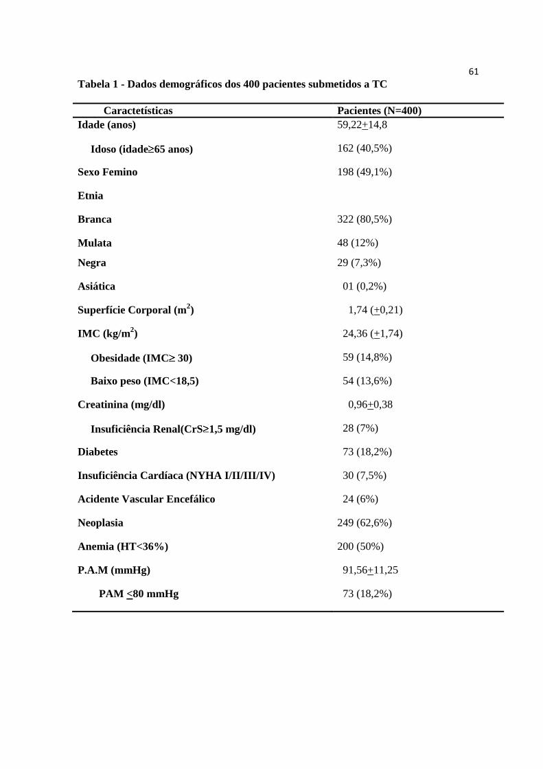
Características Clínicas de Base
As características clínicas basais dos 400 pacientes são listadas na tabela 1. A série
possuía idade média de 59,21+14,8 anos (tabela 1), com uma proporção de idosos e gênero
masculino de 40,2% e 50,4% dos casos, com predominância da etnia branca 80,5% (tabela
1). A média de IMC, percentagem de obesidade e baixo-peso foi de 24,36 +1,74, 14,8% e
13,6% respectivamente.
A prevalência de neoplasia, anemia, diabetes insuficiência cardíaca e insuficiência
renal prévia foi 62,6%, 50%, 18,2%, 7,5%, 7%, respectivamente. Quanto à pressão arterial
média no momento do exame encontrou-se uma média de 91,56+11,25 mmHg, e em
18,2% dos pacientes pressão igual ou menor do que 80 mmHg.
Procedimentos e alteração na função renal
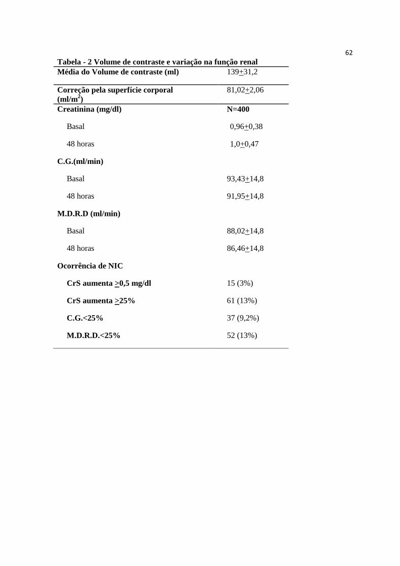
O volume médio de contraste foi 139,1+31,2 ml. Após correção pela superfície
corporal, chegamos ao valor de 81,02+2,06 ml/ m2.
Nessa população, observamos a elevação de 25% da creatinina basal em 13% (61)
dos casos e o aumento absoluto de 0,5 mg/dl em apenas 3% (15) dos pacientes (tabela 2).
Quando verificamos o decréscimo em 25% da T.F.G estimada pelas fórmulas C.G.
e M.D.R.D, encontramos uma incidência de 9,2% e 13%, respectivamente, após o
contraste.
Análise univariada e impacto na flutuação da função renal
A fim de avaliar o impacto do volume de contraste corrigido pela superfície
corporal do paciente com a variação absoluta da creatinina endógena, verificamos nessa
51
população um aumento percentual da creatinina em 13% a cada 100 ml de contraste
endovenoso por m2 de S.C (p=0,046). A diabetes e a insuficiência cardíaca demonstraram
elevação em 14% (p<0,001) e 11% (p=0,04) respectivamente. Entretanto, nos pacientes
com função renal acima de 1,5 mg/dl e em idosos, não se demonstrou esse impacto com a
dose de contraste (p=0,52) (Tabela 3).
Verificamos uma tendência à significância estatística (p=0,07) para o aumento
absoluto de 0,1 mg/dl da creatinina sérica a cada 100 ml de contraste endovenoso injetado
para toda a população. Porém, os pacientes portadores de diabetes e insuficiência cardíaca
persistiram, estatisticamente, como fatores de impacto para nefrotoxicidade por contraste,
com variação de 0,13 mg/dl (p<0,001), 0,11 mg/dl (p=0,02) (tabela 4).
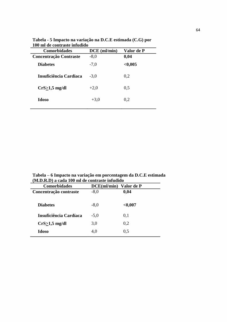
Notamos que tanto a estimação da TFG (tabela 5) com CG quanto com MDRD
(tabela 6) possuíram diferença estatística com uma redução de 8 ml (p<0,04) após o uso de
contraste em 48 horas. Quanto aos fatores de risco, somente a diabetes demonstrou uma
queda significativa nas fórmulas CG de 7 ml (p<0,005) e MDRD de 8 ml (p<0,007).
Análise Multivariada
Correlacionamos o aumento absoluto da CrS >0,5 mg/dl (tabela 7) em 25% (tabela
8) após o uso de contraste endovenoso na TC com os seguintes fatores: pacientes idosos,
diabetes, gênero feminino, obesidade, magreza, insuficiência cardíaca, insuficiência renal
prévia, neoplasia, anemia e pressão arterial média <80 mmHg.
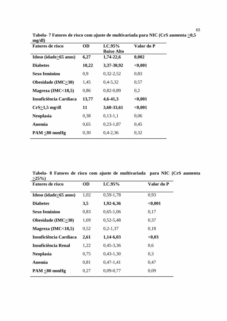
A análise multivariada revelou associação do aumento absoluto da CrS >0,5 mg/dl
com: pacientes idosos (OR 6,27; IC 95% 1,74-22,57; p=0,002), portadores de diabetes (OR
10,22; IC 95% 3,37-30,92; p<0,001), insuficiência cardíaca (OR 13,77; IC 95% 4,6-41,3;
p<0,001) e com insuficiência renal (OR 11; IC 95% 3,60-33,61; p<0,001) (Tabela 7).
52
A variação da CrS em 25% após a TC apresentou relação com: pacientes
portadores de diabetes (OR 3,5; IC 95% 1,92-6,36; p<0,001), insuficiência cardíaca (OR
2,61; IC 95% 1,14-6,03; p<0,031). Entretanto, não se evidenciou alteração estatisticamente
significativa para pacientes idosos e com insuficiência renal prévia (Tabela 8).
Apesar de relatos na literatura, não se encontrou diferenças quanto a: gênero
feminino, obesidade, baixo peso, neoplasia, PAM<80 mmHg, anemia para qualquer umas
das medidas clássicas de NIC (Tabelas 7 e 8).
DISCUSSÃO
O uso de contraste tem sido apontado como uma das causas mais comum de
disfunção renal aguda. A literatura aborda ricamente a incidência de NIC, como atestam
artigos publicados desde a década de 50 28
.
A análise da NIC provém, inequivocamente de estudos de angiográficos,
perfazendo um percentual superior a 98%2, 6, 8, 17
. Existem poucas pesquisas que analisam a
NIC com contraste endovenoso.
A literatura orienta que os fatores de risco para o grupo de pacientes submetidos à
TC sejam os mesmos do grupo em estudos angiográficos. Há dois estudos iniciais que
foram realizados com grupos de controle para flutuações nos valores de creatinina sérica 14
.
Ambos os artigos claramente não demonstram conclusões relevantes sobre o uso de
contrastes endovenosos.
Estimativas inflam o risco de NIC se originar a partir de estudos em pacientes que
se submeteram a cateterismo cardíaco. O cateterismo cardíaco pode impor eventos
embólicos. Além disso, hematomas após o cateterismo podem se formar no retroperitônio,
produzindo hipoperfusão renal. 2-8, 16-17, 27, 29-40
Mesmo dentre os radiologistas, não há
consenso sobre incidência, fatores de risco e medidas preventivas para NIC em TC2, 16, 41
.
53
Sugere-se que o metabolismo hepático do contraste endovenoso produza
proteção contra a NIC, reduzindo a ação principalmente na liberação de endotelina 16, 42
.8
Trabalho recente demonstra que pacientes portadores de cirrose possuem maior incidência
de NIC quando submetidos à TC10
.
Apesar da gravidade do estado clínico do paciente, retrospectivamente, algumas
séries de estudos descrevem incidência relativamente baixa de NIC em pacientes
hospitalizados, além disso, provou-se o risco de NIC em TC com flutuação da creatinina
sérica5-6, 8, 11, 16
. Outro trabalho confirmou essa variação, tanto na dosagem de creatinina
sérica como na taxa de filtração estimada pelas fórmulas43
. Alguns fatores avaliados, como
internação em UTI, traumatismo, acidente vascular encefálico, tromboembolismo
pulmonar, neoplasia, são relacionados a aumento significativo de NIC 5, 9-10, 18, 26, 30-32, 37-39
.
Um número restrito de trabalhos demonstrou associação entre volume de contraste
endovenoso e elevação da creatinina sérica 3, 16
. Nosso trabalho consegue demonstrar, seja
através da creatinina sérica seja pelo uso de fórmulas, o risco de perda da função renal
associado ao uso de contraste endovenoso, principalmente em pacientes portadores de
diabetes, insuficiência renal e insuficiência cardíaca.
A maioria dos estudos como o nosso foi realizada em pacientes internados. Estas
pesquisas descreveram a associação de NIC com os vários níveis de coomorbidades em
indivíduos hospitalizados, tais como aterosclerose, hipertensão, diabetes, uso de drogas
nefrotóxicas. Estes fatores são sabidamente conhecidos como inerentes na associação da
NIC.
Encontramos uma incidência de NIC de 13% para variação percentual da CrS. Na
literatura, os dados mais recentes reportam uma incidência variável de 1% até mais de 30%
para pacientes submetidos à tomografia.
54
Entre as limitações do nosso estudo, está não só o fato de ser ele realizado em um
único centro, mas também a impossibilidade de acompanhamento dos pacientes após as 48
horas do exame, para que se pudesse averiguar outros desfechos, como a mortalidade.
Outro fator é o grande número de pacientes com neoplasias e anemias.
55
CONCLUSÃO
Apesar das dificuldades devidas à variabilidade desta população, este é um dentre
os poucos estudos prospectivos que demonstrou a associação do uso de contraste
endovenoso como fator de variação na função renal. Essa circunstância foi mais expressiva
em pacientes diabéticos e com insuficiência cardíaca. Além desses pacientes, os idosos e
aqueles com função renal alterada no dia do exame possuem maior risco para NIC.
56
Referências
1. Parfrey PS, Griffiths SM, Barrett BJ, et al. Contrast material-induced renal failure
in patients with diabetes mellitus, renal insufficiency, or both. A prospective controlled
study. N Engl J Med 1989;320:143-9.
2. Reddan D, Fishman EK. Radiologists' knowledge and perceptions of the impact of
contrast-induced nephropathy and its risk factors when performing computed tomography
examinations: a survey of European radiologists. Eur J Radiol 2008;66:235-45.
3. Shetty AN, Bis KG, Vyas AR, Kumar A, Anderson A, Balasubramaniam M.
Contrast volume reduction with superior vena cava catheter-directed coronary CT
angiography: comparison with peripheral i.v. contrast enhancement in a swine model. AJR
Am J Roentgenol 2008;190:W247-54.
4. Thomsen HS, Morcos SK, Erley CM, et al. The ACTIVE Trial: comparison of the
effects on renal function of iomeprol-400 and iodixanol-320 in patients with chronic
kidney disease undergoing abdominal computed tomography. Invest Radiol 2008;43:170-
8.
5. Abujudeh HH, Gee MS, Kaewlai R. In emergency situations, should serum
creatinine be checked in all patients before performing second contrast CT examinations
within 24 hours? J Am Coll Radiol 2009;6:268-73.
6. Bruce RJ, Djamali A, Shinki K, Michel SJ, Fine JP, Pozniak MA. Background
fluctuation of kidney function versus contrast-induced nephrotoxicity. AJR Am J
Roentgenol 2009;192:711-8.
7. Goldfarb S, McCullough PA, McDermott J, Gay SB. Contrast-induced acute kidney
injury: specialty-specific protocols for interventional radiology, diagnostic computed
tomography radiology, and interventional cardiology. Mayo Clin Proc 2009;84:170-9.

57
8. Heinrich MC, Haberle L, Muller V, Bautz W, Uder M. Nephrotoxicity of iso-
osmolar iodixanol compared with nonionic low-osmolar contrast media: meta-analysis of
randomized controlled trials. Radiology 2009;250:68-86.
9. Holmquist F, Hansson K, Pasquariello F, Bjork J, Nyman U. Minimizing contrast
medium doses to diagnose pulmonary embolism with 80-kVp multidetector computed
tomography in azotemic patients. Acta Radiol 2009;50:181-93.
10. Lodhia N, Kader M, Mayes T, Mantry P, Maliakkal B. Risk of contrast-induced
nephropathy in hospitalized patients with cirrhosis. World J Gastroenterol 2009;15:1459-
64.
11. Thomsen HS, Morcos SK. Risk of contrast-medium-induced nephropathy in high-
risk patients undergoing MDCT--a pooled analysis of two randomized trials. Eur Radiol
2009;19:891-7.
12. Band RA, Gaieski DF, Mills AM, et al. Discordance between serum creatinine and
creatinine clearance for identification of ED patients with abdominal pain at risk for
contrast-induced nephropathy. Am J Emerg Med 2007;25:268-72.
13. Anto HR, Chou SY, Porush JG, Shapiro WB. Infusion intravenous pyelography and
renal function. Effect of hypertonic mannitol in patients with chronic renal insufficiency.
Arch Intern Med 1981;141:1652-6.
14. Cramer BC, Parfrey PS, Hutchinson TA, et al. Renal function following infusion of
radiologic contrast material. A prospective controlled study. Arch Intern Med
1985;145:87-9.
15. Heller CA, Knapp J, Halliday J, O'Connell D, Heller RF. Failure to demonstrate
contrast nephrotoxicity. Med J Aust 1991;155:329-32.
16. Katzberg RW, Lamba R. Contrast-induced nephropathy after intravenous
administration: fact or fiction? Radiol Clin North Am 2009;47:789-800, v.
58
17. Rao QA, Newhouse JH. Risk of nephropathy after intravenous administration of
contrast material: a critical literature analysis. Radiology 2006;239:392-7.
18. Haveman JW, Gansevoort RT, Bongaerts AH, Nijsten MW. Low incidence of
nephropathy in surgical ICU patients receiving intravenous contrast: a retrospective
analysis. Intensive Care Med 2006;32:1199-205.
19. Katzberg RW, Haller C. Contrast-induced nephrotoxicity: clinical landscape.
Kidney Int Suppl 2006:S3-7.
20. Lee JK, Warshauer DM, Bush WH, Jr., McClennan BL, Choyke PL. Determination
of serum creatinine level before intravenous administration of iodinated contrast medium.
A survey. Invest Radiol 1995;30:700-5.
21. Rudnick MR, Goldfarb S, Wexler L, et al. Nephrotoxicity of ionic and nonionic
contrast media in 1196 patients: a randomized trial. The Iohexol Cooperative Study.
Kidney Int 1995;47:254-61.
22. K/DOQI clinical practice guidelines for chronic kidney disease: evaluation,
classification, and stratification. Am J Kidney Dis 2002;39:S1-266.
23. Najjar M, Hamad A, Salameh M, Agarwal A, Feinfeld DA. The risk of
radiocontrast nephropathy in patients with cirrhosis. Ren Fail 2002;24:11-8.
24. Dorio PJ, Lee FT, Jr., Henseler KP, et al. Using a saline chaser to decrease contrast
media in abdominal CT. AJR Am J Roentgenol 2003;180:929-34.
25. Nyman U, Almen T, Aspelin P, Hellstrom M, Kristiansson M, Sterner G. Contrast-
medium-Induced nephropathy correlated to the ratio between dose in gram iodine and
estimated GFR in ml/min. Acta Radiol 2005;46:830-42.
26. Cheruvu B, Henning K, Mulligan J, et al. Iodixanol: risk of subsequent contrast
nephropathy in cancer patients with underlying renal insufficiency undergoing diagnostic
computed tomography examinations. J Comput Assist Tomogr 2007;31:493-8.
59
27. Kuhn MJ, Chen N, Sahani DV, et al. The PREDICT study: a randomized double-
blind comparison of contrast-induced nephropathy after low- or isoosmolar contrast agent
exposure. AJR Am J Roentgenol 2008;191:151-7.
28. Bartels ED, Brun GC, Gammeltoft A, Gjorup PA. Acute anuria following
intravenous pyelography in a patient with myelomatosis. Acta Med Scand 1954;150:297-
302.
29. Detrenis S, Meschi M, del Mar Jordana Sanchez M, Savazzi G. Contrast medium
induced nephropathy in urological practice. J Urol 2007;178:1164-70.
30. Krol AL, Dzialowski I, Roy J, et al. Incidence of radiocontrast nephropathy in
patients undergoing acute stroke computed tomography angiography. Stroke
2007;38:2364-6.
31. Lameire N. Contrast-induced nephropathy in the critically-ill patient: focus on
emergency screening and prevention. Acta Clin Belg Suppl 2007:346-52.
32. Mitchell AM, Kline JA. Contrast nephropathy following computed tomography
angiography of the chest for pulmonary embolism in the emergency department. J Thromb
Haemost 2007;5:50-4.
33. Sandstede JJ, Roth A, Machann W, Kaupert C, Hahn D. Evaluation of the
nephrotoxicity of iodixanol in patients with predisposing factors to contrast medium
induced nephropathy referred for contrast enhanced computed tomography. Eur J Radiol
2007;63:120-3.
34. From AM, Bartholmai BJ, Williams AW, McDonald FS. Iodixanol compared to
iohexol for contrast procedures: a case-matched retrospective cohort study. Acta Radiol
2008;49:409-14.
35. Heiken JP. Contrast safety in the cancer patient: preventing contrast-induced
nephropathy. Cancer Imaging 2008;8 Suppl A:S124-7.
60
36. Herts BR, Schneider E, Poggio ED, Obuchowski NA, Baker ME. Identifying
outpatients with renal insufficiency before contrast-enhanced CT by using estimated
glomerular filtration rates versus serum creatinine levels. Radiology 2008;248:106-13.
37. Hipp A, Desai S, Lopez C, Sinert R. The incidence of contrast-induced nephropathy
in trauma patients. Eur J Emerg Med 2008;15:134-9.
38. Langner S, Stumpe S, Kirsch M, Petrik M, Hosten N. No increased risk for
contrast-induced nephropathy after multiple CT perfusion studies of the brain with a
nonionic, dimeric, iso-osmolal contrast medium. AJNR Am J Neuroradiol 2008;29:1525-9.
39. Mehdiratta M, Schlaug G, Kumar S, Caplan LR, Selim M. Reducing the delay in
thrombolysis: is it necessary to await the results of renal function tests before computed
tomography perfusion and angiography in patients with code stroke? J Stroke Cerebrovasc
Dis 2008;17:273-5.
40. Nyman U, Elmstahl B, Leander P, Almen T. Iodine contrast media doses equal-
attenuating with gadolinium chelates at CT-aortography may have less risk of contrast-
induced nephropathy and no risk of nephrogenic systemic fibrosis in azotaemic patients!
Eur Radiol 2008;18:2013-4.
41. Elicker BM, Cypel YS, Weinreb JC. IV contrast administration for CT: a survey of
practices for the screening and prevention of contrast nephropathy. AJR Am J Roentgenol
2006;186:1651-8.
42. Gleeson TG, Bulugahapitiya S. Contrast-induced nephropathy. AJR Am J
Roentgenol 2004;183:1673-89.
43. Herts BR, Schneider E, Obuchowski N, Poggio E, Jain A, Baker ME. Probability of
reduced renal function after contrast-enhanced CT: a model based on serum creatinine
level, patient age, and estimated glomerular filtration rate. AJR Am J Roentgenol
2009;193:494-500.
61
Tabela 1 - Dados demográficos dos 400 pacientes submetidos a TC
Caractetísticas Pacientes (N=400)
Idade (anos) 59,22+14,8
Idoso (idade 65 anos) 162 (40,5%)
Sexo Femino 198 (49,1%)
Etnia
Branca 322 (80,5%)
Mulata 48 (12%)
Negra 29 (7,3%)
Asiática 01 (0,2%)
Superfície Corporal (m2) 1,74 (+0,21)
IMC (kg/m2) 24,36 (+1,74)
Obesidade (IMC 30) 59 (14,8%)
Baixo peso (IMC<18,5) 54 (13,6%)
Creatinina (mg/dl) 0,96+0,38
Insuficiência Renal(CrS 1,5 mg/dl) 28 (7%)
Diabetes 73 (18,2%)
Insuficiência Cardíaca (NYHA I/II/III/IV) 30 (7,5%)
Acidente Vascular Encefálico 24 (6%)
Neoplasia 249 (62,6%)
Anemia (HT<36%) 200 (50%)
P.A.M (mmHg) 91,56+11,25
PAM <80 mmHg 73 (18,2%)
62
Tabela - 2 Volume de contraste e variação na função renal
Média do Volume de contraste (ml) 139+31,2
Correção pela superfície corporal
(ml/m2)
81,02+2,06
Creatinina (mg/dl) N=400
Basal 0,96+0,38
48 horas 1,0+0,47
C.G.(ml/min)
Basal 93,43+14,8
48 horas 91,95+14,8
M.D.R.D (ml/min)
Basal 88,02+14,8
48 horas 86,46+14,8
Ocorrência de NIC
CrS aumenta >0,5 mg/dl 15 (3%)
CrS aumenta >25% 61 (13%)
C.G.<25% 37 (9,2%)
M.D.R.D.<25% 52 (13%)

63
Tabela - 3 Impacto na variação creatinina sérica em porcentagem
por 100 ml de contraste infudido
Comorbidades Percentagem
Valor de P
Concentração contraste +1,0% 0,04
Diabetes +14% <0,001
Insuficiência Cardiaca +12% 0,04
CrS>1,5 mg/dl -5,0% 0,4
Idoso -3,0% 0,2
Tabela - 4 Impacto na variação da creatinina sérica em mg/dl
por 100 ml de contraste infudido
Comorbidades CrS (mg/dl) Valor de P
Concentração contraste +0,1 0,07
Diabetes +0,13 <0,001
Insuficiência
Cardiaca
+0,12 0,01
CrS>1,5 mg/dl +0,02 0,7
Idoso -0,02 0,5
64
Tabela - 5 Impacto na variação na D.C.E estimada (C.G) por
100 ml de contraste infudido
Comorbidades DCE (ml/min) Valor de P
Concentração Contraste -8,0 0,04
Diabetes -7,0 <0,005
Insuficiência Cardiaca -3,0 0,2
CrS>1,5 mg/dl +2,0 0,5
Idoso +3,0 0,2
Tabela – 6 Impacto na variação em porcentagem da D.C.E estimada
(M.D.R.D) a cada 100 ml de contraste infudido
Comorbidades DCE(ml/min) Valor de P
Concentração contraste -8,0 0,04
Diabetes -8,0 <0,007
Insuficiência Cardíaca -5,0 0,1
CrS>1,5 mg/dl 3,0 0,2
Idoso 4,0 0,5
65
Tabela- 7 Fatores de risco com ajuste de multivariada para NIC (CrS aumenta >0,5
mg/dl)
Fatores de risco OD I.C.95%
Baixo Alto
Valor do P
Idoso (idade>65 anos) 6,27 1,74-22,6 0,002
Diabetes 10,22 3,37-30,92 <0,001
Sexo feminino 0,9 0,32-2,52 0,83
Obesidade (IMC>30) 1,45 0,4-5,32 0,57
Magreza (IMC<18,5) 0,86 0,82-0,89 0,2
Insuficiência Cardiaca 13,77 4,6-41,3 <0,001
CrS>1,5 mg/dl 11 3,60-33,61 <0,001
Neoplasia 0,38 0,13-1,1 0,06
Anemia 0,65 0,23-1,87 0,45
PAM <80 mmHg 0,30 0,4-2,36 0,32
Tabela- 8 Fatores de risco com ajuste de multivariada para NIC (CrS aumenta
>25%)
Fatores de risco OD I.C.95%
Valor do P
Idoso (idade>65 anos) 1,02 0,59-1,78 0,93
Diabetes 3,5 1,92-6,36 <0,001
Sexo feminino 0,83 0,65-1,06 0,17
Obesidade (IMC>30) 1,69 0,52-5,48 0,37
Magreza (IMC<18,5) 0,52 0,2-1,37 0,18
Insuficiência Cardiaca 2,61 1,14-6,03 <0,03
Insuficiência Renal 1,22 0,45-3,36 0,6
Neoplasia 0,75 0,43-1,30 0,3
Anemia 0,81 0,47-1,41 0,47
PAM <80 mmHg 0,27 0,09-0,77 0,09

66
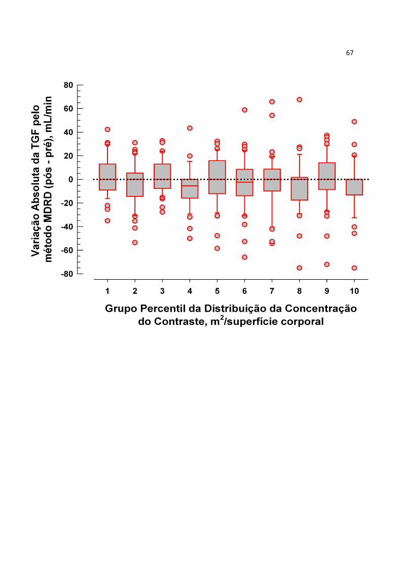
67
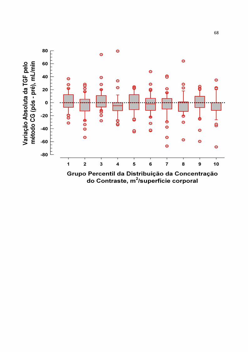
68
69
ARTIGO ORIGINAL – VERSÃO EM INGLÊS
70
INTRAVENOUS CONTRAST IN COMPUTERIZED TOMOGRAPHY AND IN THE
DEVELOPMENT OF CONTRAST-INDUCED NEPHROPATHY
Luciano da Silva SelistreI; Laura Fuchs Bahlis
IV, Cinthia Fonseca O'Keeffe
IV ,Vandrea
Carla de SouzaIII
; Mário Bernardes WagnerII; João Rubião Hoefel Filho
II; David
SaitovitchII; Luciano Passamani Diogo
II
71
ABSTRACT
Background: Contrast-induced nephropathy (CIN) is the third leading cause of all hospital-
acquired renal insufficiency. The incidence of and risk factors for CIN after tomography
are, however, unknown at present. Methods and Results: The 400 consecutive patients who
underwent tomography at São Lucas University Hospital from January 2007 to February
2008 were prospectively examined. The incidence of CIN defined as an absolute increase
in serum creatinine (SCr) ≥0.5 mg/dl, a relative ≥25% increase in SCr, and either an
absolute or relative increase after CT were 4.0%, 13.8%, and 13.9%, respectively.
Multivariate logistic models revealed that preexisting renal insufficiency, diabetes, cardiac
insufficiency and the use of high-volume contrast were independently associated with an
absolute increase in SCr ≥0.5 mg/dl. Female gender, underweight, and high-volume
contrast usage were not independently associated with a relative ≥25% increase and either
an absolute or relative increase in SCr. Conclusions: Although the incidence of and risk
factors for CIN after TC varied in the study population according to the definition of CIN,
careful attention should be paid to patients who have risk factors identified by each
definition of CIN.
Key Words: Tomography; Contrast-induced nephropathy; Risk factors;
72
BACKGROUND
The administration of radiocontrast is the third leading cause of acute renal
insufficiency in hospitalized patients 1-14
. The incidence of intravenous contrast-induced
nephropathy (CIN), specifically in computed tomography (CT), is not fully set 15-16.
Less
than 2% of the CIN research includes the use of intravenous contrast.8, 11, 16-19
Several risk factors are associated with arterial infusion of contrast as diabetes, high
doses of contrast, the elderly, chronic renal failure, female gender, multiple myeloma, liver
diseases, kidney or heart failure, the spot of administration, use of nonsteroidal
antiinflammatory drugs,19-23
. However, these factors are not well characterized in the use
of intravenous contrast. 16-17
Several ways of preventing CIN in CT have been tried, such as the usage of low
osmolarity contrast, theophylline, acetylcysteine, mannitol, with poor results and no
clinical significance 4, 8, 15-19
.
This study aimed to prospectively evaluate, in a single center, hospitalized patients
who underwent CT with intravenous contrast to identify the incidence of CIN and the
classic risk factors such as diabetes, heart failure, stroke, obesity and gender. At the same
time, it aimed to establish the volume of contrast and variation of renal function in those
groups of individuals, either by using serum creatinine and the glomerular filtration rate
(GFR) or formulas Crockoft-Gault and MDRD.
73
METHODS
Patients’ Population and Selection Criteria
This is a Prospective Cohort Study which has allocated 400 patients at a single
reference center (Hospital São Lucas PUCRS) with hospitalized individuals who
underwent CT, which used contrast type Telebrix 30 Laboratory Guebert (ioxitalamic
59.285 g, meglumine 15.1 g/100 ml, iodine content of 300 mg / ml, osmolality of 1650
mOsm / kg H2O), from January 01, 2007 to March 31, 2008.
The inclusion criteria for this study were: both genders over 18 years old,
institutionalized patients whose hospital discharge was not predicted after 48 hours of
contrast, and the ability to decide and sign the consent form. There was no loss during the
study period. The study followed the principles in the Declaration of Helsinki and was
approved by the Ethics Committee at PUCRS. All patients had signed consent forms
before entering the study.
Carrying on the study
Patients were submitted to interviews and laboratory testing on the same day when
they underwent CT. After 48 hours, laboratory investigations were repeated. Data were
recorded by 2 different researchers.
There were recorded variables such as: age (which had been divided in two: over or
under 65), gender, body mass index, body surface area, serum creatinine level, glomerular
filtration rate that is estimated by the formulas Crockoft-Gault and MDRD 22,
diabetes
mellitus, mean arterial pressure measured during the examination, baseline serum
creatinine higher than 1.5 mg / dl, history of heart failure according to NY criteria,
neoplasia, contrast volume. Other data were collected from the patient's record during his
stay in hospital.
74
The process of gathering data was carried on by two interviewers, three
reviewers of medical records, two typists and a final reviewer. After the doubled entry, and
in case of discrepancy between data entries, there was a new verification by the final
reviewer.
Outcomes
The primary outcome which interested us was to check the absolute increase in
serum creatinine > 0.5 mg / dl (> 44.2 mol / L) and the increase of > 25% and the reduction
in eGFR to 25% in 48 hours after the infusion of intravenous contrast. Secondary outcomes
were to evaluate the association between diabetes, heart failure, obesity (BMI >30),
underweight (BMI < 18.5), female gender, neoplasia, anemia (hematocrit <36%), serum
creatinine over 1.5 mg / dl and age over 65 years old for incidence of CIN.
Statistical analysis
We analyzed the database of a prospective cohort study in the radiology department
of Hospital São Lucas (PUCRS) which met the inclusion criteria, from January 01, 2007 to
March 31, 2008. The database has undergone the process of double entry and then the
processing of inconsistencies.
We used the Kolmogorov-Smirnov test to evaluate the normalization of quantitative
data, but when such distribution was not a normal curve it was carried out a
logarithmization of data.
All data are presented as percentages and averages.Comparisons between means were
performed by using two-tailed T test or, in cases of multiple comparisons, by the "One-
way ANOVA, and by Mann-Whitney or Fisher for comparisons between medians.
75
We have considered potential risks for CIN: diabetes mellitus (DM), neoplasia,
anemia, heart failure (HF), stroke, obesity, underweight, renal insufficiency, female
gender, and low blood pressure on examination. We have made the choice for
dichotomization of the following factors: elderliness( > 65 versus <65 years), obese or
non-obese (BMI> 30 or <30 kg / m2),
underweight or not (BMI <18.5 or <18.5 kg /
m 2),
renal function or not (SCr >1.5 mg / dl versus <1.5 mg / dl), low blood pressure or not
(MAP <80 mmHg or > 80 mmHg), anemia or not (Ht <36 or > 36%).
We used multivariate analysis with backward stepwise, referring the new variable to
those previously reported. It was considered a significant value if p <0.05. We used the
software SPSS version 17 for Windows.
76
RESULTS
Baseline Clinical Characteristics
The baseline clinical characteristics of those 400 patients are listed in the table. This
series had an average age of 59.21 + 14.8 (Table 1), its proportion of elderly patients and
male gender was 40.2% and 50.4% of all cases, predominantly Caucasian ethnicity at
80.5% (Table 1). The mean BMI, the obesity and underweight percentages were
24.36 + 1.74, 14.8% and 13.6%, respectively.
The prevalence of neoplasia, anemia, diabetes, heart failure and renal insufficiency
was 62.6%, 50%, 18.2%, 7.5%, 7%, respectively. The mean arterial pressure at the
moment of examination was found at average of 91.56 + 11.25 mmHg, and in 18.2% of
patients it was found less than or equal to 80 mmHg.
Procedures and variation in renal function
The average volume of contrast was 139.1 + 31.2 ml. After correction according
body surface area, we obtained 81.02 + 2.06 ml / m 2. In this population, we noticed a 25%
increase of baseline serum creatinine in 13% (61) of cases and an absolute increase of 0.5
mg / dl in only 3% (15) of patients (Table 2).
When we verified a decrease in 25% of estimated GFR by CG and MDRD
formulas, we also found an incidence of 9.2% and 13%, respectively, after the contrast.
Univariate analysis and the impact on renal function fluctuation
In order to assess the impact of contrast volume, which was corrected according to
patient’s body surface area and absolute variation of endogenous creatinine, in this
population, we verified an increase in creatinine up to 13% per 100 ml of intravenous

77
contrast agent per m 2 SC (p = 0.046). Diabetes and heart failure showed an increase of
14% (p <0.001) and 11% (p = 0.04), respectively. However, in patients with renal function
greater than 1.5 mg / dl and among the elderly, there has not been shown such an impact
with that dose of contrast (p = 0.52) (Table 3).
We have found statistical significance (p = 0.07), for the whole population, towards
the absolute increase of 0.1 mg / dl serum creatinine per 100 ml of contrast agent
injected. But patients with diabetes and heart failure statistically remained as impact
factors for contrast nephrotoxicity which ranged from 0.13 mg / dl (p <0.001), to 0.11 mg /
dl (p = 0.02) (table 4).
We noticed that both GFR estimative with CG (Table 5) and MDRD (Table 6)
possessed significant difference with a reduction of 8 ml (p <0.04) after the use of contrast
in 48 hours. Regarding risk factors, only diabetes has shown a significant drop in the
formulas CG 7 ml (p <0.005) and MDRD 8 ml (p <0.007).
Multivariate Analysis
After using intravenous contrast in CT, we related the absolute increase in Cr> 0.5
mg / dl (Table 7) and 25% (Table 8) to the following factors: the elderly, diabetes, female
gender, obesity, thinness, heart failure, renal insufficiency, neoplasia, anemia and mean
arterial pressure at <80 mmHg.
Multivariate analysis has revealed a relation to the absolute increase in Cr> 0.5 mg /
dl among the elderly (OR 6.27, CI 95% 1.74-22.57, p = 0.002), patients with diabetes (OR
10.22 95% CI 3.37-30.92, p <0.001), heart failure (OR 13.77, CI 95% 4.6-41.3, p <0.001),
and renal failure (OR 11 CI 95% 3.60-33.61, p <0.001) ((Table 7).
The variation of Cr, in 25% after CT, was related to patients with diabetes (OR 3.5,
CI 95% 1.92-6.36, p <0.001), heart failure (OR 2.61, 95 1.14-6.03%, p <0.031). However,
78
no statistically significant change has been found among the elderly and the ones with
renal insufficiency (Table 8).
Regardless of reports in medical literature, no differences were found concerning
female gender, obesity, low weight, neoplasia, MAP <80 mmHg, and anemia in any of the
classical measures of NIC (Tables 7 and 8).
79
DISCUSSION
Using contrast has been considered one of the most common causes of acute renal
failure. Medical literature richly discusses the incidence of CIN, as it has been evidenced
by articles published since the decade of 1950s 28
. The analysis of NIC mainly comes from
angiographic studies, amounting to a percentage higher than 98% 2, 6, 8, 17.
Nevertheless,
there are few studies that analyze the NIC with intravenous contrast.
Medical literature advises that risk factors for the group of patients undergoing CT
are the same as those in angiographic studies. There are two early studies which were
carried on with control groups for observing fluctuations in serum creatinine 14.
Both
articles clearly show no relevant findings on the use of intravenous contrast.
Estimates inflate the risk of CIN if they had been originated from studies in patients
who have undergone cardiac catheterization. Cardiac catheterization may have imposed
embolic events. In addition, after catheterization, hematomas may be formed in the
retroperitoneum, producing renal hypoperfusion. 2-8, 16-17, 27, 29-40
Even among the
radiologist there is no consensus on the incidence, risk factors, and preventive measures for
CIN in CT 2, 16, 41.
It has been suggested that the hepatic metabolism of intravenous contrast would
produce protection against CIN, mainly reducing the action of releasing endothelin 16, 42.
8 A recent study shows that patients with cirrhosis have a higher incidence of CIN when
undergoing CT 10.
Some series of former studies have described a relatively low incidence of CIN in
hospitalized patients, independently from the severity of patient’s clinical retrospect, after
using contrast in CT. Nevertheless, even when considering the severity of patient's clinical
retrospect there has been a risk of CIN in CT with fluctuating serum creatinine 5-6, 8, 11,
80 16.
Another study has confirmed that variation not only in the dosage of serum creatinine,
but also in the filtration rate estimated by the formulas 43
. Some of the analyzed factors,
such as required ICU treatment, trauma, stroke, pulmonary thromboembolism, neoplasia,
have been related to a significant increase in CIN 5, 9-10, 18, 26, 30-32, 37-39.
A limited number of studies have shown the association between volume of
intravenous contrast and high serum creatinine 3, 16
. Our study can demonstrate the risk of
loss in renal function, either by checking serum creatinine or using formulas with
intravenous contrast, especially among patients with diabetes, kidney failure and heart
failure.
Most studies, including this one, have been performed in hospitalized patients.
They describe an association of CIN to several levels of comorbidities in hospitalized
individuals such as atherosclerosis, hypertension, diabetes, and the use of nephrotoxic
drugs. Those factors are well-known as being inherent to NIC. We have found incidence of
CIN at 13% for the percentage variation in Cr. In medical literature, recent data have
evaluated incidence ranging from 1% to over 30% for patients undergoing CT.
The limitations of our study are mainly related to the fact of being performed at a
single center as well as the impossibility of monitoring these patients 48 hours after the
examination to determine other possible outcomes such as mortality. Another factor is the
large number of patients with neoplasia and anemia.
81
CONCLUSION
Despite the difficulties due to the variability of this population, this study is one of
the few prospective studies that have shown the use of intravenous contrast as a variation
factor to renal function. This condition is stronger in patients with diabetes and heart
failure. Besides that, the elderly and those presenting impaired renal function, on the
examination day, are at higher risk for CIN.
82
References
1. Parfrey PS, Griffiths SM, Barrett BJ, et al. Contrast material-induced renal failure
in patients with diabetes mellitus, renal insufficiency, or both. A prospective controlled
study. N Engl J Med 1989;320:143-9.
2. Reddan D, Fishman EK. Radiologists' knowledge and perceptions of the impact of
contrast-induced nephropathy and its risk factors when performing computed tomography
examinations: a survey of European radiologists. Eur J Radiol 2008;66:235-45.
3. Shetty AN, Bis KG, Vyas AR, Kumar A, Anderson A, Balasubramaniam M.
Contrast volume reduction with superior vena cava catheter-directed coronary CT
angiography: comparison with peripheral i.v. contrast enhancement in a swine model. AJR
Am J Roentgenol 2008;190:W247-54.
4. Thomsen HS, Morcos SK, Erley CM, et al. The ACTIVE Trial: comparison of the
effects on renal function of iomeprol-400 and iodixanol-320 in patients with chronic
kidney disease undergoing abdominal computed tomography. Invest Radiol 2008;43:170-
8.
5. Abujudeh HH, Gee MS, Kaewlai R. In emergency situations, should serum
creatinine be checked in all patients before performing second contrast CT examinations
within 24 hours? J Am Coll Radiol 2009;6:268-73.
6. Bruce RJ, Djamali A, Shinki K, Michel SJ, Fine JP, Pozniak MA. Background
fluctuation of kidney function versus contrast-induced nephrotoxicity. AJR Am J
Roentgenol 2009;192:711-8.
7. Goldfarb S, McCullough PA, McDermott J, Gay SB. Contrast-induced acute kidney
injury: specialty-specific protocols for interventional radiology, diagnostic computed
tomography radiology, and interventional cardiology. Mayo Clin Proc 2009;84:170-9.
83
8. Heinrich MC, Haberle L, Muller V, Bautz W, Uder M. Nephrotoxicity of iso-
osmolar iodixanol compared with nonionic low-osmolar contrast media: meta-analysis of
randomized controlled trials. Radiology 2009;250:68-86.
9. Holmquist F, Hansson K, Pasquariello F, Bjork J, Nyman U. Minimizing contrast
medium doses to diagnose pulmonary embolism with 80-kVp multidetector computed
tomography in azotemic patients. Acta Radiol 2009;50:181-93.
10. Lodhia N, Kader M, Mayes T, Mantry P, Maliakkal B. Risk of contrast-induced
nephropathy in hospitalized patients with cirrhosis. World J Gastroenterol 2009;15:1459-
64.
11. Thomsen HS, Morcos SK. Risk of contrast-medium-induced nephropathy in high-
risk patients undergoing MDCT--a pooled analysis of two randomized trials. Eur Radiol
2009;19:891-7.
12. Band RA, Gaieski DF, Mills AM, et al. Discordance between serum creatinine and
creatinine clearance for identification of ED patients with abdominal pain at risk for
contrast-induced nephropathy. Am J Emerg Med 2007;25:268-72.
13. Anto HR, Chou SY, Porush JG, Shapiro WB. Infusion intravenous pyelography and
renal function. Effect of hypertonic mannitol in patients with chronic renal insufficiency.
Arch Intern Med 1981;141:1652-6.
14. Cramer BC, Parfrey PS, Hutchinson TA, et al. Renal function following infusion of
radiologic contrast material. A prospective controlled study. Arch Intern Med
1985;145:87-9.
15. Heller CA, Knapp J, Halliday J, O'Connell D, Heller RF. Failure to demonstrate
contrast nephrotoxicity. Med J Aust 1991;155:329-32.
16. Katzberg RW, Lamba R. Contrast-induced nephropathy after intravenous
administration: fact or fiction? Radiol Clin North Am 2009;47:789-800, v.
84
17. Rao QA, Newhouse JH. Risk of nephropathy after intravenous administration of
contrast material: a critical literature analysis. Radiology 2006;239:392-7.
18. Haveman JW, Gansevoort RT, Bongaerts AH, Nijsten MW. Low incidence of
nephropathy in surgical ICU patients receiving intravenous contrast: a retrospective
analysis. Intensive Care Med 2006;32:1199-205.
19. Katzberg RW, Haller C. Contrast-induced nephrotoxicity: clinical landscape.
Kidney Int Suppl 2006:S3-7.
20. Lee JK, Warshauer DM, Bush WH, Jr., McClennan BL, Choyke PL. Determination
of serum creatinine level before intravenous administration of iodinated contrast medium.
A survey. Invest Radiol 1995;30:700-5.
21. Rudnick MR, Goldfarb S, Wexler L, et al. Nephrotoxicity of ionic and nonionic
contrast media in 1196 patients: a randomized trial. The Iohexol Cooperative Study.
Kidney Int 1995;47:254-61.
22. K/DOQI clinical practice guidelines for chronic kidney disease: evaluation,
classification, and stratification. Am J Kidney Dis 2002;39:S1-266.
23. Najjar M, Hamad A, Salameh M, Agarwal A, Feinfeld DA. The risk of
radiocontrast nephropathy in patients with cirrhosis. Ren Fail 2002;24:11-8.
24. Dorio PJ, Lee FT, Jr., Henseler KP, et al. Using a saline chaser to decrease contrast
media in abdominal CT. AJR Am J Roentgenol 2003;180:929-34.
25. Nyman U, Almen T, Aspelin P, Hellstrom M, Kristiansson M, Sterner G. Contrast-
medium-Induced nephropathy correlated to the ratio between dose in gram iodine and
estimated GFR in ml/min. Acta Radiol 2005;46:830-42.
26. Cheruvu B, Henning K, Mulligan J, et al. Iodixanol: risk of subsequent contrast
nephropathy in cancer patients with underlying renal insufficiency undergoing diagnostic
computed tomography examinations. J Comput Assist Tomogr 2007;31:493-8.
85
27. Kuhn MJ, Chen N, Sahani DV, et al. The PREDICT study: a randomized double-
blind comparison of contrast-induced nephropathy after low- or isoosmolar contrast agent
exposure. AJR Am J Roentgenol 2008;191:151-7.
28. Bartels ED, Brun GC, Gammeltoft A, Gjorup PA. Acute anuria following
intravenous pyelography in a patient with myelomatosis. Acta Med Scand 1954;150:297-
302.
29. Detrenis S, Meschi M, del Mar Jordana Sanchez M, Savazzi G. Contrast medium
induced nephropathy in urological practice. J Urol 2007;178:1164-70.
30. Krol AL, Dzialowski I, Roy J, et al. Incidence of radiocontrast nephropathy in
patients undergoing acute stroke computed tomography angiography. Stroke
2007;38:2364-6.
31. Lameire N. Contrast-induced nephropathy in the critically-ill patient: focus on
emergency screening and prevention. Acta Clin Belg Suppl 2007:346-52.
32. Mitchell AM, Kline JA. Contrast nephropathy following computed tomography
angiography of the chest for pulmonary embolism in the emergency department. J Thromb
Haemost 2007;5:50-4.
33. Sandstede JJ, Roth A, Machann W, Kaupert C, Hahn D. Evaluation of the
nephrotoxicity of iodixanol in patients with predisposing factors to contrast medium
induced nephropathy referred for contrast enhanced computed tomography. Eur J Radiol
2007;63:120-3.
34. From AM, Bartholmai BJ, Williams AW, McDonald FS. Iodixanol compared to
iohexol for contrast procedures: a case-matched retrospective cohort study. Acta Radiol
2008;49:409-14.
35. Heiken JP. Contrast safety in the cancer patient: preventing contrast-induced
nephropathy. Cancer Imaging 2008;8 Suppl A:S124-7.

86
36. Herts BR, Schneider E, Poggio ED, Obuchowski NA, Baker ME. Identifying
outpatients with renal insufficiency before contrast-enhanced CT by using estimated
glomerular filtration rates versus serum creatinine levels. Radiology 2008;248:106-13.
37. Hipp A, Desai S, Lopez C, Sinert R. The incidence of contrast-induced nephropathy
in trauma patients. Eur J Emerg Med 2008;15:134-9.
38. Langner S, Stumpe S, Kirsch M, Petrik M, Hosten N. No increased risk for
contrast-induced nephropathy after multiple CT perfusion studies of the brain with a
nonionic, dimeric, iso-osmolal contrast medium. AJNR Am J Neuroradiol 2008;29:1525-9.
39. Mehdiratta M, Schlaug G, Kumar S, Caplan LR, Selim M. Reducing the delay in
thrombolysis: is it necessary to await the results of renal function tests before computed
tomography perfusion and angiography in patients with code stroke? J Stroke Cerebrovasc
Dis 2008;17:273-5.
40. Nyman U, Elmstahl B, Leander P, Almen T. Iodine contrast media doses equal-
attenuating with gadolinium chelates at CT-aortography may have less risk of contrast-
induced nephropathy and no risk of nephrogenic systemic fibrosis in azotaemic patients!
Eur Radiol 2008;18:2013-4.
41. Elicker BM, Cypel YS, Weinreb JC. IV contrast administration for CT: a survey of
practices for the screening and prevention of contrast nephropathy. AJR Am J Roentgenol
2006;186:1651-8.
42. Gleeson TG, Bulugahapitiya S. Contrast-induced nephropathy. AJR Am J
Roentgenol 2004;183:1673-89.
43. Herts BR, Schneider E, Obuchowski N, Poggio E, Jain A, Baker ME. Probability of
reduced renal function after contrast-enhanced CT: a model based on serum creatinine
level, patient age, and estimated glomerular filtration rate. AJR Am J Roentgenol
2009;193:494-500.
87
Table 1 – Demographic data for 400 patients undergone CT
Characteristics Patients (N=400)
Age (years) 59.22+14.8
Elderly (age 65 years) 162 (40.5%)
Female gender 198 (49.1%)
Ethnicity
White 322 (80.5%)
Mulata1 48 (12%)
Afro-Brazilian 29 (7.3%)
Asian-Brazilian 01 (0.2%)
Body Mass Surface (m2) 1.74 (+0.21)
BMI (kg/m2) 24.36 (+1.74)
Obesity (BMI 30) 59 (14.8%)
Underweight (BMI<18.5) 54 (13.6%)
Creatinine (mg/dl) 0.96+0.38
Renal insufficiency (SCr 1.5 mg/dl) 28 (7%)
Diabetes 73 (18.2%)
Heart Failure (NYHA I/II/III/IV) 30 (7.5%)
Stroke 24 (6%)
Neoplasia 249 (62.6%)
Anemia (Ht < 36%) 200 (50%)
MAP (mmHg) 91.56+11.25
MAP < 80 mmHg 73 (18.2%)
1 Mulata is a woman who belongs to mixed ethnicity that results from the meeting between Afro and
European descendents. (TN)
88
Table - 2 Contrast Volume and Renal Function
Contrast volume (ml) 139+31.2
Correction by body surface area (ml/m2) 81.02+2.06
Creatinine (mg/dl) N=400
Baseline 0.96+0.38
48 hours 1.0+0.47
C.G.(ml/min)
Baseline 93.43+14.8
48 hours 91.95+14.8
M.D.R.D (ml/min)
Baseline 88.02+14.8
48 hours 86.46+14.8
Occurrence of CIN
SCr increases >0,5 mg/dl 15 (3%)
SCr increases >25% 61 (13%)
C.G.<25% 37 (9.2%)
M.D.R.D.<25% 52 (13%)
89
Table - 3 Impact on serum creatinine variation (percentage per
100 ml of contrast)
Comorbidities Percentage
P
Contrast concentration +1.0% 0.04
Diabetes +14% <0.001
Heart Failure +12% 0.04
SCr >1.5 mg/dl -5.0% 0.4
Elderly -3.0% 0.2
Table - 4 Impact on serum creatinine variation in mg/dl
per 100 ml of contrast
Comorbidities SCr (mg/dl) P
Contrast concentration +0.1 0.07
Diabetes +0.13 <0.001
Heart failure +0.12 0.01
SCr>1.5 mg/dl +0.02 0.7
Elderly -0.02 0.5
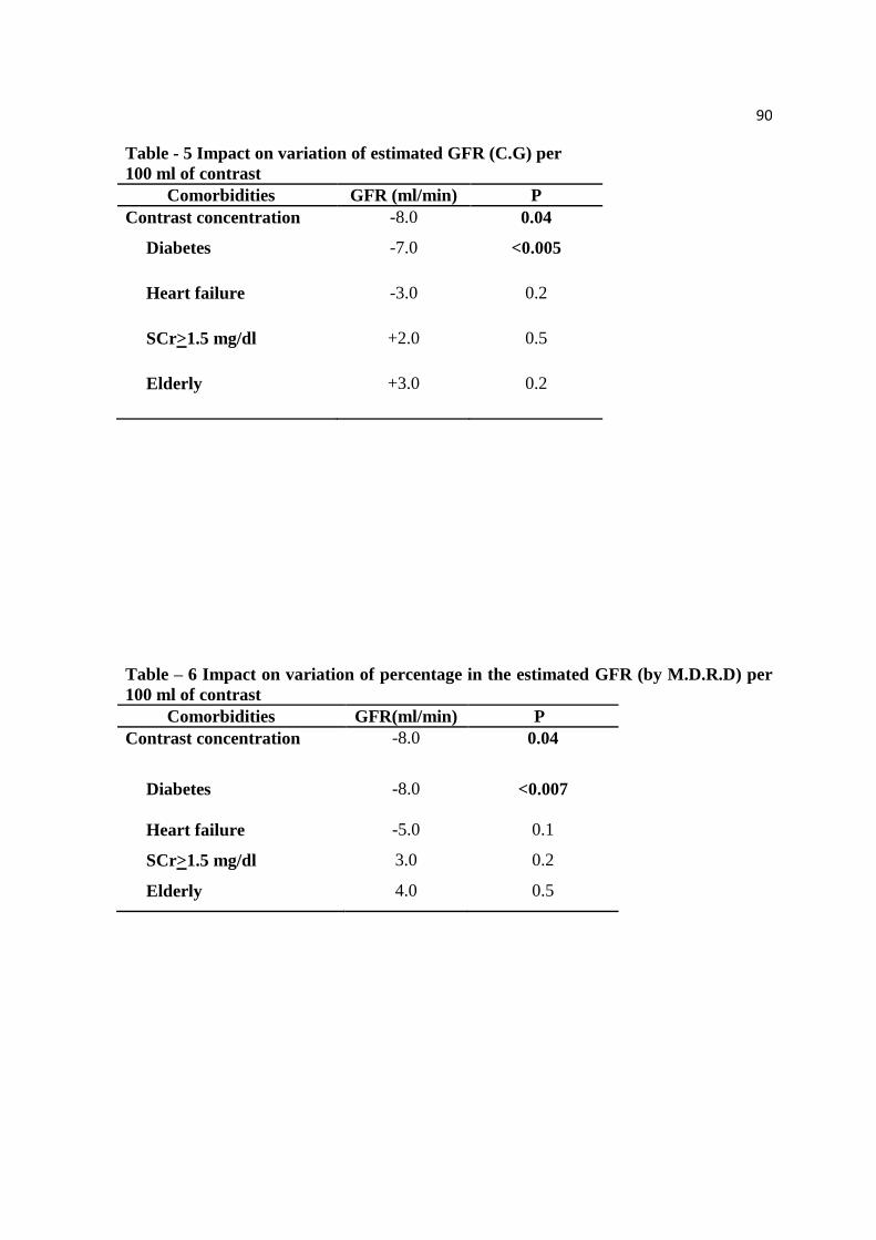
90
Table - 5 Impact on variation of estimated GFR (C.G) per
100 ml of contrast
Comorbidities GFR (ml/min) P
Contrast concentration -8.0 0.04
Diabetes -7.0 <0.005
Heart failure -3.0 0.2
SCr>1.5 mg/dl +2.0 0.5
Elderly +3.0 0.2
Table – 6 Impact on variation of percentage in the estimated GFR (by M.D.R.D) per
100 ml of contrast
Comorbidities GFR(ml/min) P
Contrast concentration -8.0 0.04
Diabetes -8.0 <0.007
Heart failure -5.0 0.1
SCr>1.5 mg/dl 3.0 0.2
Elderly 4.0 0.5

91
Table- 7 Risk factors with multivariate adjustment to NIC (SCr increases >0.5
mg/dl)
Risk factors OR CI 95%
Low High
P
Elderly (age >65 years) 6.27 1.74-22.6 0.002
Diabetes 10.22 3.37-30.92 <0.001
Females 0.9 0.32-2.52 0.83
Obesity (BMI>30) 1.45 0.4-5.32 0.57
Thinness (BMI<18.5) 0.86 0.82-0.89 0.2
Heart failure 13.77 4.6-41.3 <0.001
SCr >1.5 mg/dl 11 3.60-33.61 <0.001
Neoplasia 0.38 0.13-1.1 0.06
Anemia 0.65 0.23-1.87 0.45
MAP <80 mmHg 0.30 0.4-2.36 0.32
Table- 8 Risk factors with multivariate adjustment to NIC (SCr increases >25%)
Risk factors OR CI 95%
P
Elderly (age >65 years) 1.02 0.59-1.78 0.93
Diabetes 3.5 1.92-6.36 <0.001
Female gender 0.83 0.65-1.06 0.17
Obesity (BMI >30) 1.69 0.52-5.48 0.37
Underweight (BMI<18.5) 0.52 0.2-1.37 0.18
Heart failure 2.61 1.14-6.03 <0.03
Renal insufficiency 1.22 0.45-3.36 0.6
Neoplasia 0.75 0.43-1.30 0.3
Anemia 0.81 0.47-1.41 0.47
MAP < 80 mmHg 0.27 0.09-0.77 0.09
2
2 Y axis – Absolute Variation of Creatinine (after – before) mg/dl
X axis – Percentage Group of Contrast Concentration Distribution, m2/body surface
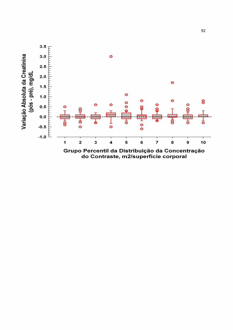
92
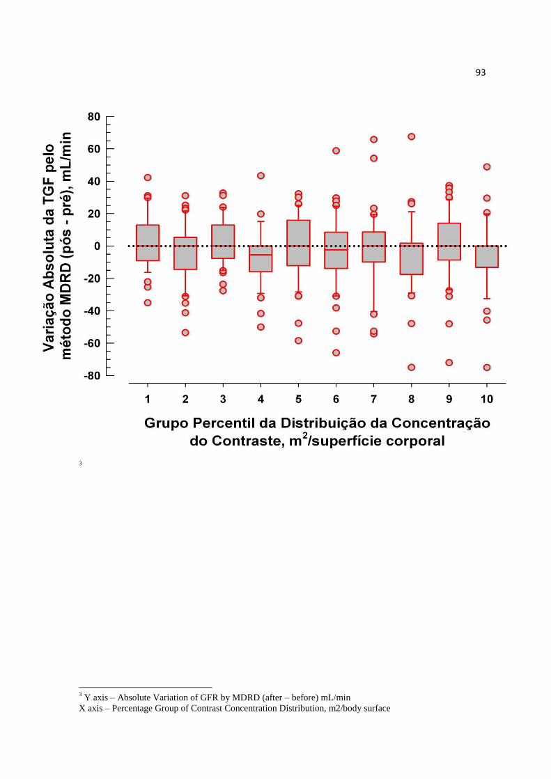
93
3
3 Y axis – Absolute Variation of GFR by MDRD (after – before) mL/min
X axis – Percentage Group of Contrast Concentration Distribution, m2/body surface
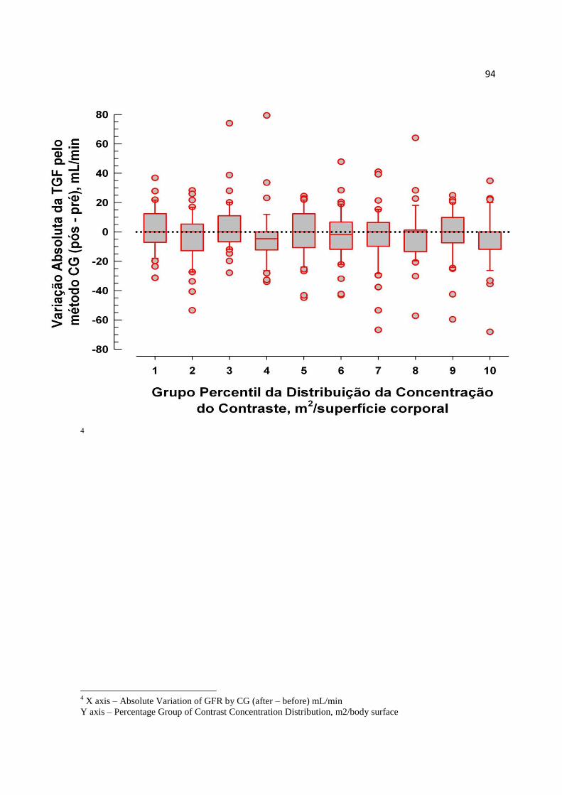
94
4
4 X axis – Absolute Variation of GFR by CG (after – before) mL/min
Y axis – Percentage Group of Contrast Concentration Distribution, m2/body surface
95
ANEXOS
96
Dear Hebbert Y. Kressel, MISQ Editor, Radiology
Prudential Tower
800 Boylston St, 15th fl oor
Boston, MA 02199
e-mail: [email protected]
617-236-7376
EDITORIAL OFFICE
I would like to submit the attached manuscript, "Intravenous Contrast in Computerized
Ttomography and in the development of Contrast-induced Nephropathy," for consideration for possible
publication in the Research Articles section of Radiology.
The authors have no conflict of interest with any of the authors of the paper being
submitted.
This paper (or closely related research) has not been published or accepted for publication.
It is not under consideration at another journal or at Radiology. No other papers using the
same data set have been published. (NOTE: If other papers using the data set have been
published, please indicate the differences between the submitted paper and the other
published papers.)
Sincerely,
Luciano da Silva Selistre Adelino Roldo, 310 Universidade de Caxias do Sul Caxias do Sul, RS 95052-020 Brazil phone: 55 54 91224798 fax: 55 54 32022540 e-mail: [email protected]
97
RADIOLOGY Transfer of Copyright and Certification Agreement
Manuscript No. RAD-10-0825 Complete copyright to the article entitled: INTRAVENOUS CONTRAST IN COMPUTERIZED TOMOGRAPHY AND IN THE DEVELOPMENT OF CONTRAST-INDUCED NEPHROPATHY
is hereby transferred to the Radiological Society of North America, effective if and when the article is accepted for publication in Radiology, printed or electronic format. (In the case of authors who are officers or employees of the U.S. government, RSNA recognizes that works prepared by them as part of their official duties are in the public domain.) For authors whose research was funded by the National Institutes of Health, the RSNA grants the authors permission to provide a copy of the accepted manuscript to NIH and encourages, but does not require, them to specify a release date of 12 months after the publication date.
Authors reserve the following rights: 1. Other than copyright, all proprietary rights including patent rights relating to the subject matter of the article. 2. The license for personal use in lectures, lecture notes, and exhibits.
Please return this form as soon as possible. The signed statement must be received by the Editor before the manuscript can be processed for publication. 1. All authors certify that this material or similar material has not been and will not be submitted by them or by colleagues in their institution to any other publication prior to its appearance in Radiology. 2. All authors certify that they have read the Publication Information for Authors found in the most recently published issue of Radiology at the time the manuscript is submitted. They further certify their understanding of and compliance with the various requirements found therein, in particular those relating to authorship and redundant publication and potential sanctions for redundant publication. 3. All authors certify that they have submitted all required permissions and that all required statements have been inserted in the text where appropriate. 4. All authors certify that all individuals who have met the ICMJE criteria for authorship of this manuscript have been listed as authors. All authors further certify that they have read the final version
of the manuscript being submitted for peer review and approve it for submission and possible publication. All authors agree with placement of authors in the author listing. 5. All authors certify that the Editor may correspond with the designated corresponding author regarding acceptance, rejection, revision of the manuscript and that the corresponding author is responsible for informing all other authors of the same. 6. All authors certify that responses, revisions received by the Editor from the designated corresponding author will be treated as if from all authors and that the corresponding author is responsible for informing all other authors of the same. 7. All authors certify that: (must check one) (Please see Publication Information for Authors for explanation.)
they have no direct or indirect financial interest in the products under investigation or subject matter discussed in this manuscript, or
one or more (please name) authors do have a direct or indirect financial interest in the products under investigation or subject matter discussed in this manuscript. A separate statement is below that details this proprietary interest, and the authors hereby grant permission to publish this information or an appropriate summary thereof with the manuscript if the manuscript is published.
Insert statement here:
By clicking here you agree to the terms and conditions detailed above.
Please type your full name: LUCIANO DA SILVA SELISTRE
98
99
Radiologia Brasileira
Colégio Brasileiro de Radiologia
Av. Paulista, 37 - 7º andar - conjunto 71
Bela Vista - São Paulo - SP 01311-000
Tel.: (55 11) 3372-4544
São Paulo, sábado, 19 de dezembro de 2009
Ilmo(a) Sr.(a)
Prof(a), Dr(a) LUCIANO DA SILVA SELISTRE
Referente ao código de fluxo: 829
Classificação: Artigos de Revisão
Informamos que recebemos a CORREÇÃO do manuscrito epígrafe, que será reanalisado pelos revisores com vistas a publicação na
Radiologia Brasileira. Por favor, para qualquer comunicação sobre este manuscrito, cite o número de referência indicado acima.
Obrigado por submeter seu trabalho na Radiologia Brasileira.
Atenciosamente,
Edson Marchiori / Giovanni Guido Cerri Editores da Radiologia Brasileira

100
Brasil - São Paulo, 5/5/2010
RB Radiologia Brasileira
Cód Fluxo: 829
Título: NEFROPATIA DO CONTRASTE: MANEJO PREVENTIVO
O(s) autor(es) do artigo, como aqui especificado, por este meio, transfere a Radiologia Brasileira (RB) todos os direitos autorais, título e interesses que o autor tenha, ou possa vir a ter pelo artigo e qualquer revisão ou versões dele, incluindo, mas não limitado, o direito exclusivo para imprimir,
publicar e vender o artigo em todo o mundo, em todos os idiomas e em todas as mídias.
Este acordo será considerado efetivo e válido se e quando o artigo for aceito para publicação. Se o artigo contiver qualquer material protegido por direito autoral de terceiros, o(s) autor(es) entregará(ão) a RB permissão, por escrito, do titular dos direitos autorais para reproduzir tal material no
artigo. O(s) autor(es) garante ser o detentor da titularidade do artigo; não ter concedido ou cedido qualquer direito do artigo para qualquer outra
pessoa ou entidade; ser o artigo passível de requisição de direitos autorais, por seu autor; não infringir qualquer direito autoral, marca registrada ou
patente; não invadir o direito de privacidade ou publicidade de qualquer pessoa ou entidade; não conter qualquer assunto difamatório; serem
verdadeiras as declarações afirmadas como fatos ou estarem baseadas em pesquisa razoável para atingir precisão; e, finalmente, até onde é de seu conhecimento, que nenhuma fórmula, procedimento, ou prescrição contidas no artigo causarão dano se usados ou seguidos conforme advertências
e/ou instruções contidas no artigo.
O(s) autor(es) indenizará a RB contra qualquer custo, despesas ou danos que a RB possa incorrer ou para os quais a RB possa se tornar sujeita como resultado de eventuais omissões destas garantias. Estas representações e garantias poderão ser estendidas a terceiros pela RB.
LUCIANO DA SILVA SELISTRE CPF: 66678803000 Cargo: PROFESSOR AUXILIAR DA FACULDADE DE MEDICINA DA UNIVERSIDADE DE CAXIAS DO SUL (UCS), CHEFE DO SERVIÇO DE NEFROLOGIA
DO HOSPITAL GERAL DE CAXIAS DO SUL
JOÃO RUBIÃO HOEFEL FILHO CPF: Cargo: PROFESSOR ASSISTENTE DA FACULDADE DE MEDICINA DA PONTIFÍCIA DA UNIVERSIDADE CATÓLICA DO RIO GRANDE DO SUL (PUCRS),
MÉDICO-RADIOLOGISTA DO HOSPITAL SÃO LUCAS DE PORTO ALEGRE DA PUCRS
LUCIANO PASSAMANI DIOGO CPF: Cargo: PROFESSOR ADJUNTO DA FACULDADE DE MEDICINA PONTÍFICIA CATÓLICA DO RIO GRANDE DO SUL (PUCRS), CHEFE DA EMERGÊNCIA DO
HOSPITAL SÃO LUCAS DA PUCRS.
VANDREA CARLA DE SOUZA CPF: Cargo: MÉDICA NEFROLOGISTA PEDIÁTRICA DO HOSPITAL GERAL DE CAXIAS DO SUL (UCS)
DAVID SAITOVITCH CPF: Cargo: PROFESSOR ADJUNTO NA FACULDADE DE MEDICINA DA PONTIFICIA CATÓLICA DO RIO GRANDE DO SUL (PUCRS), MEDICO-NEFROLOGISTA
DO HOSPITAL SÃO LUCAS DA PUCRS.
Top Related