


Línguas
Páginas
Legal

I
Cláudia Sofia da Cunha Mesquita Rodrigues Vieira dos Santos
Estudo in vitro da biocompatibilidade dos
cimentos de obturação endodônticos
Porto, 2012

II
III
Estudo in vitro da biocompatibilidade dos
cimentos de obturação endodônticos
Orientador:
Prof. Doutor José António Macedo Carvalho Capelas
Co-orientadora:
Prof. Doutora Maria Helena Raposo Fernandes

IV

V
Dissertação de candidatura ao grau de Doutor em Medicina Dentária,
submetida à Faculdade de Medicina Dentária, Universidade do Porto.

VI
VII
Conselho Científico da Faculdade de Medicina Dentária, Universidade do Porto
Prof. Doutor Afonso Manuel Pinhão Ferreira (Prof. Catedrático)
Prof. Doutor Américo dos Santos Afonso (Prof. Associado c/agregação)
Prof. Doutor António Cabral Campos Felino (Prof. Catedrático)
Prof. Doutor César Fernando Coelho Leal Silva (Prof. Associado c/agregação)
Prof. Doutor Germano Neves Pinto Rocha (Prof. Associado)
Prof. Doutora Irene Graça Azevedo Pina Vaz (Prof. Associado)
Prof. Doutora Inês Alexandra Costa Morais Caldas (Prof. Auxiliar)
Prof. Doutor João Carlos Antunes Sampaio Fernandes (Prof. Catedrático)
Prof. Doutor João Carlos Gonçalves Ferreira de Pinho (Prof. Associado c/agregação)
Prof. Doutor João Fernando Costa Carvalho (Prof. Catedrático)
Prof. Doutor Jorge Manuel Carvalho Dias Lopes (Prof. Catedrático)
Prof. Doutor José António Macedo Carvalho Capelas (Prof. Associado c/agregação)
Prof. Doutor José Carlos Reis Campos (Prof. Auxiliar c/ agregação)
Prof. Doutor José Mário Castro Rocha (Prof. Auxiliar)
Prof. Douto Manuel José Fontes de Carvalho (Prof. Associado)
Prof. Doutora Maria Cristina P. C. M. Figueiredo Pollmann (Prof. Associado)
Prof. Doutora Maria Helena Guimarães Figueiral da Silva (Prof. Associada c/agregação)
Prof. Doutora Maria Helena Raposo Fernandes (Prof. Catedrático)
Prof. Doutora Maria Lurdes Ferreira Lobo Pereira (Prof. Auxiliar)
Prof. Doutor Mário Augusto Pires Vaz (Prof. Associado - personalidade convidada)
Prof. Doutor Mário Jorge Rebolho Fernandes Silva (Prof. Catedrático)
Prof. Doutor Mário Ramalho Vasconcelos (Prof. Associado c/agregação)

VIII
Prof. Doutor Miguel Fernando Silva Gonçalves Pinto (Prof. Catedrático)
Prof. Doutor Paulo Rui Galrão Ribeiro Melo (Prof. Associado c/ agregação)
Prof. Doutor Ricardo Manuel Lobo Faria Almeida (Prof. Associado c/ agregação)
IX
Docentes Jubilados
Prof. Doutor Adão Fernando Pereira (Prof. Catedrático)
Prof. Doutor Amilcar Almeida Oliveira (Prof. Associado)
Prof. Doutor António Manuel Machado Capelas (Prof. Associado )
Dr. António Ulisses Matos dos Santos (Assistente Convidado)
Prof. Doutor Durval Manuel Belo Moreira (Prof. Associado c/Agregação)
Prof. Doutor Francisco António Rebelo Morais Caldas (Prof. Catedrático)
Dr. José Maria Vaz Osório (Assistente Convidado)
Prof. Doutor José Serra Silva Campos Neves (Prof. Catedrático)
Prof. Doutor Manuel Desport Marques (Prof. Associado Convidado )
Prof. Doutor Manuel Guedes de Figueiredo (Prof. Associado)
Docentes Aposentados
Prof. Doutor António Manuel Guerra Capelas (Prof. Auxiliar)
Prof. Dr. Artur Manuel Osório de Araújo (Prof. Associado Convidado)
Prof. Doutor Fernando Jorge Morais Branco (Prof. Catedrático)
Prof. Doutor Fernando José Brandão Martins Peres (Prof. Catedrático )
Prof. Doutor José Albertino Cruz Lordelo (Prof. Associado c/ agregação)
Prof. Doutor José Carlos Pina Almeida Rebelo (Prof. Catedrático)
Prof. Doutor Manuel Pedro da Fonseca Paulo (Prof. Catedrático)
Prof. Doutora Maria Adelaide Macedo Carvalho Capelas (Prof. Associada )

X
Prof. Doutora Maria Purificação Valenzuela Sampaio Tavares (Prof. Catedrática)
Prof. Doutor Rogério Serapião Martins Aguiar Branco (Prof. Catedrático)

XI
Ao meu marido,
José Carlos
Aos meus tesourinhos,
Matilde e João (e bebé)

XII

XIII
Aos meus super pais
Ao meu irmão

XIV

XV
Índice

XVI
XVII
Agradecimentos………………………………………………XIX
Resumo………………………………………………………XXV
Abstract…………………………………………………… .XXXI
Enquadramento…………………………………………..XXXVII
Introdução………………………………………………………43
Capítulo I
Osteoblastic cytocompatibility of endodontic sealers extracts prepared
according to ISO standards and a root-dipping technique ......................81
Capítulo II
Long-term dose and time-dependent cytotoxicity profile of endodontic
sealers in human in vitro osteoclastogenesis…………………………97
Capítulo III
Behaviour of co-cultured human osteoclastic and osteoblastic cells exposed
to endodontic sealers..……………………………………………121
Discussão...…………………………………………………….143
Conclusões….....………………………………………………157

XVIII

XIX
Agradecimentos

XX
XXI
Durante o tempo de realização deste trabalho, comecei a apelidar esta tese de meu
“fantasma”, porque mesmo nos períodos em que me dediquei menos à sua elaboração, a
sua sombra estava sempre presente, e como tal acompanhou a minha vida pessoal e
profissional. O “fantasma” viu a minha filha crescer, deixar de ser bebé e tornar-se uma
menina, aprender as letras e os números e perceber que a mamã tinha um trabalho
importante para fazer; viu o meu filho nascer, dar os primeiros passos e dizer as
primeiras palavras; viu os meus pacientes entrar e sair do consultório; viu os meus
alunos crescerem na arte da endodontia; viu as mudanças na faculdade e no
departamento…enfim, fez parte da minha vida. E como tal, para além do conteúdo
científico, esta tese tem um grande conteúdo emocional. E só foi possível a sua
elaboração, porque muitas pessoas contribuíram com a sua sabedoria e, sobretudo, com
a sua amizade.
Ao Professor Doutor José António Macedo de Carvalho Capelas, meu orientador,
agradeço ter continuado a orientação desta tese aquando da aposentação do Prof. Doutor
Manuel Paulo; obrigada pelos ensinamentos ao longo destes anos, obrigada pelo
exemplo e pelo estímulo na prática da Endodontia e sobretudo, obrigada pelo carinho e
amizade com que sempre me tratou.
À Professora Doutora Maria Helena Raposo Fernandes, co-orientadora deste trabalho,
um agradecimento muito especial, por partilhar os seus conhecimentos, por dispor de
tempo e sobretudo de paciência para orientar uma médica dentista, pouco habituada ao
meio da investigação. Muito obrigada pelo exemplo de dinamismo e trabalho, e por ter
disponibilizado os meios técnicos e humanos do Laboratório de Farmacologia e
Biocompatibilidade Celular da Faculdade de Medicina Dentária da Universidade do
Porto, tornando possível a execução de todo o trabalho experimental.
Ao Professor Doutor Manuel da Fonseca Paulo, professor aposentado da FMDUP e que
foi o primeiro orientador desta tese de doutoramento, um muito, muito obrigada, porque
me abriu as portas para a endodontia e para a docência, e porque me continua a
acompanhar com enorme amizade e carinho.
À Professora Doutora Irene Graça Azevedo Pina Vaz e ao Professor Doutor Manuel
José Fontes de Carvalho, que me acolheram e acompanharam de perto a minha evolução
e o meu trabalho, agradeço a amizade, a disponibilidade e o exemplo de dedicação ao
ensino e à endodontia. Muito têm contribuído para o crescimento e afirmação do
XXII
Departamento de Endodontia da Faculdade de Medicina Dentária da Universidade do
Porto, dentro e fora da instituição.
Ao Professor Doutor João Miguel da Costa Rodrigues, do Laboratório de Farmacologia
e Biocompatibilidade Celular da Faculdade de Medicina Dentária da Universidade do
Porto, um agradecimento sincero pelo tempo que dispensou para realizar comigo toda a
parte laboratorial desta tese, pela disponibilidade, pelo dinamismo e pela simpatia que
sempre mostrou. E por toda a paciência…
À Professora Doutora Maria de Lurdes Ferreira Lobo Pereira, agradeço com muito
carinho, porque me ajudou na fase inicial do trabalho e se disponibilizou em todos os
momentos, mas agradeço principalmente a amizade e os incentivos constantes.
A todos os meus colegas do Departamento, em especial à Dra. Joana Barros, ao Dr.
Miguel Martins, à Dra. Rita Noites e à Dra. Eva Salgueirinho, agradeço todo o apoio e
todo o carinho. É um privilégio trabalhar num ambiente tão “familiar”.
À Mestre Ana Cláudia Morais de Moura Teles, minha colega, minha amiga e minha
comadre. Obrigada por estar sempre presente, obrigada por todas as palavras de
incentivo. Temos caminhado sempre lado a lado e tenho a convicção de que iremos
continuar assim…
Ao meu marido, José Carlos, obrigada pelo seu amor e apoio ao longo de todos estes
anos, por crescer comigo e permitir a realização de tantos sonhos, sempre com os pés
bem assentes na terra. Juntos, já temos uma Vida.
Aos meus filhos, obrigada por serem meus. Espero que quando mais tarde lerem estas
palavras, me perdoem por algumas ausências e de alguma forma isso seja compensado
pelo exemplo que lhes possa estar a dar. São os sorrisos, as gargalhadas, os beijos e
abraços ao final do dia que dão significado a cada segundo da minha vida.
Ao meu irmão, obrigado por fazer parte da nossa vida. Porque não é preciso estar
presente em todos os momentos nem falar a toda a hora para ser indispensável…
Por fim, e porque é o agradecimento mais difícil, obrigada, muito, muito obrigada aos
meus pais. É difícil agradecer porque não há palavras que traduzam a minha gratidão.
Na minha tese de Mestrado escrevi nos agradecimentos: “Devo-lhes a vida, a educação,
a instrução, a orientação, o exemplo... Enfim, tudo! Que este trabalho seja uma forma de

XXIII
demonstrar que mereci tudo o que me deram.” Agora, subscrevo e acrescento: obrigada
por serem o prolongamento dos meus braços, das minhas pernas e sobretudo do meu
coração. Obrigado pela dedicação incondicional a mim e aos meus filhos, obrigada por
serem os melhores pais e avós do mundo, obrigada por nunca estarem cansados, nunca
estarem tristes, nunca estarem indisponíveis…Esta tese é o resultado do meu trabalho,
apoiada por todas as pessoas a quem já agradeci e outras aqui não referidas, mas só foi
possível porque os meus pais estiveram sempre ao meu lado. Por isso, esta tese é
deles…

XXIV

XXV
Resumo

XXVI
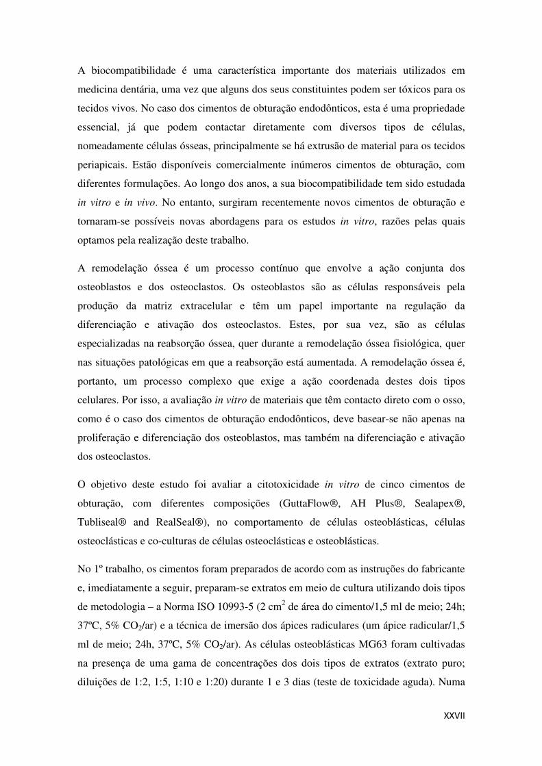
XXVII
A biocompatibilidade é uma característica importante dos materiais utilizados em
medicina dentária, uma vez que alguns dos seus constituintes podem ser tóxicos para os
tecidos vivos. No caso dos cimentos de obturação endodônticos, esta é uma propriedade
essencial, já que podem contactar diretamente com diversos tipos de células,
nomeadamente células ósseas, principalmente se há extrusão de material para os tecidos
periapicais. Estão disponíveis comercialmente inúmeros cimentos de obturação, com
diferentes formulações. Ao longo dos anos, a sua biocompatibilidade tem sido estudada
in vitro e in vivo. No entanto, surgiram recentemente novos cimentos de obturação e
tornaram-se possíveis novas abordagens para os estudos in vitro, razões pelas quais
optamos pela realização deste trabalho.
A remodelação óssea é um processo contínuo que envolve a ação conjunta dos
osteoblastos e dos osteoclastos. Os osteoblastos são as células responsáveis pela
produção da matriz extracelular e têm um papel importante na regulação da
diferenciação e ativação dos osteoclastos. Estes, por sua vez, são as células
especializadas na reabsorção óssea, quer durante a remodelação óssea fisiológica, quer
nas situações patológicas em que a reabsorção está aumentada. A remodelação óssea é,
portanto, um processo complexo que exige a ação coordenada destes dois tipos
celulares. Por isso, a avaliação in vitro de materiais que têm contacto direto com o osso,
como é o caso dos cimentos de obturação endodônticos, deve basear-se não apenas na
proliferação e diferenciação dos osteoblastos, mas também na diferenciação e ativação
dos osteoclastos.
O objetivo deste estudo foi avaliar a citotoxicidade in vitro de cinco cimentos de
obturação, com diferentes composições (GuttaFlow®, AH Plus®, Sealapex®,
Tubliseal® and RealSeal®), no comportamento de células osteoblásticas, células
osteoclásticas e co-culturas de células osteoclásticas e osteoblásticas.
No 1º trabalho, os cimentos foram preparados de acordo com as instruções do fabricante
e, imediatamente a seguir, preparam-se extratos em meio de cultura utilizando dois tipos
de metodologia – a Norma ISO 10993-5 (2 cm2 de área do cimento/1,5 ml de meio; 24h;
37ºC, 5% CO2/ar) e a técnica de imersão dos ápices radiculares (um ápice radicular/1,5
ml de meio; 24h, 37ºC, 5% CO2/ar). As células osteoblásticas MG63 foram cultivadas
na presença de uma gama de concentrações dos dois tipos de extratos (extrato puro;
diluições de 1:2, 1:5, 1:10 e 1:20) durante 1 e 3 dias (teste de toxicidade aguda). Numa
XXVIII
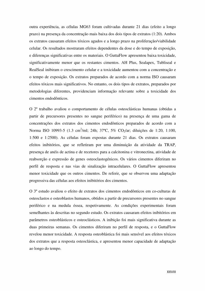
outra experiência, as células MG63 foram cultivadas durante 21 dias (efeito a longo
prazo) na presença da concentração mais baixa dos dois tipos de extratos (1:20). Ambos
os extratos causaram efeitos tóxicos agudos e a longo prazo na proliferação/viabilidade
celular. Os resultados mostraram efeitos dependentes da dose e do tempo de exposição,
e diferenças significativas entre os materiais. O GuttaFlow apresentou baixa toxicidade,
significativamente menor que os restantes cimentos. AH Plus, Sealapex, Tubliseal e
RealSeal inibiram o crescimento celular e a toxicidade aumentou com a concentração e
o tempo de exposição. Os extratos preparados de acordo com a norma ISO causaram
efeitos tóxicos mais significativos. No entanto, os dois tipos de extratos, preparados por
metodologias diferentes, providenciam informação relevante sobre a toxicidade dos
cimentos endodônticos.
O 2º trabalho avaliou o comportamento de células osteoclásticas humanas (obtidas a
partir de precursores presentes no sangue periférico) na presença de uma gama de
concentrações dos extratos dos cimentos endodônticos preparados de acordo com a
Norma ISO 10993-5 (1.3 cm2/ml; 24h; 37ºC, 5% CO2/ar; diluições de 1:20, 1:100,
1:500 e 1:2500). As células foram expostas durante 21 dias. Os extratos causaram
efeitos inibitórios, que se refletiram por uma diminuição da atividade da TRAP,
presença de anéis de actina e de recetores para a calcitonina e vitronectina, atividade de
reabsorção e expressão de genes osteoclastogénicos. Os vários cimentos diferiram no
perfil de resposta e nas vias de sinalização intracelulares. O GuttaFlow apresentou
menor toxicidade que os outros cimentos. De referir, que se observou uma adaptação
progressiva das células aos efeitos inibitórios dos cimentos.
O 3º estudo avaliou o efeito de extratos dos cimentos endodônticos em co-culturas de
osteoclastos e osteoblastos humanos, obtidos a partir de precursores presentes no sangue
periférico e na medula óssea, respetivamente. As condições experimentais foram
semelhantes às descritas no segundo estudo. Os extratos causaram efeitos inibitórios em
parâmetros osteoblásticos e osteoclásticos. A inibição foi mais significativa durante as
duas primeiras semanas. Os cimentos diferiram no perfil de resposta, e o GuttaFlow
revelou menor toxicidade. A resposta osteoblástica foi mais sensível aos efeitos tóxicos
dos extratos que a resposta osteoclástica, e apresentou menor capacidade de adaptação
ao longo do tempo.
XXIX
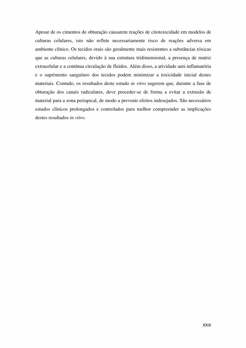
Apesar de os cimentos de obturação causarem reações de citotoxicidade em modelos de
culturas celulares, isto não reflete necessariamente risco de reações adversa em
ambiente clínico. Os tecidos orais são geralmente mais resistentes a substâncias tóxicas
que as culturas celulares, devido à sua estrutura tridimensional, a presença de matriz
extracelular e a contínua circulação de fluidos. Além disso, a atividade anti-inflamatória
e o suprimento sanguíneo dos tecidos podem minimizar a toxicidade inicial destes
materiais. Contudo, os resultados deste estudo in vitro sugerem que, durante a fase de
obturação dos canais radiculares, deve proceder-se de forma a evitar a extrusão de
material para a zona periapical, de modo a prevenir efeitos indesejados. São necessários
estudos clínicos prolongados e controlados para melhor compreender as implicações
destes resultados in vitro.

XXX

XXXI
Abstract

XXXII
XXXIII
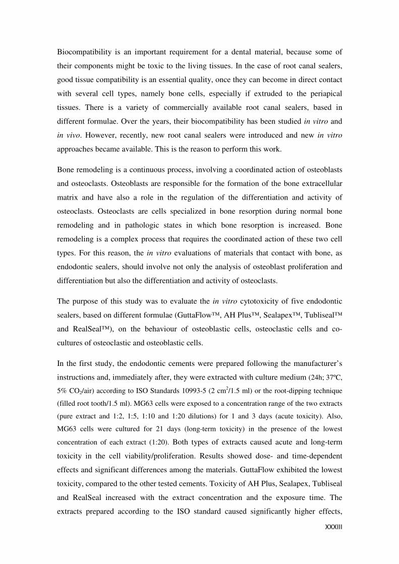
Biocompatibility is an important requirement for a dental material, because some of
their components might be toxic to the living tissues. In the case of root canal sealers,
good tissue compatibility is an essential quality, once they can become in direct contact
with several cell types, namely bone cells, especially if extruded to the periapical
tissues. There is a variety of commercially available root canal sealers, based in
different formulae. Over the years, their biocompatibility has been studied in vitro and
in vivo. However, recently, new root canal sealers were introduced and new in vitro
approaches became available. This is the reason to perform this work.
Bone remodeling is a continuous process, involving a coordinated action of osteoblasts
and osteoclasts. Osteoblasts are responsible for the formation of the bone extracellular
matrix and have also a role in the regulation of the differentiation and activity of
osteoclasts. Osteoclasts are cells specialized in bone resorption during normal bone
remodeling and in pathologic states in which bone resorption is increased. Bone
remodeling is a complex process that requires the coordinated action of these two cell
types. For this reason, the in vitro evaluations of materials that contact with bone, as
endodontic sealers, should involve not only the analysis of osteoblast proliferation and
differentiation but also the differentiation and activity of osteoclasts.
The purpose of this study was to evaluate the in vitro cytotoxicity of five endodontic
sealers, based on different formulae (GuttaFlow™, AH Plus™, Sealapex™, Tubliseal™
and RealSeal™), on the behaviour of osteoblastic cells, osteoclastic cells and co-
cultures of osteoclastic and osteoblastic cells.
In the first study, the endodontic cements were prepared following the manufacturer’s
instructions and, immediately after, they were extracted with culture medium (24h; 37ºC,
5% CO2/air) according to ISO Standards 10993-5 (2 cm2/1.5 ml) or the root-dipping technique
(filled root tooth/1.5 ml). MG63 cells were exposed to a concentration range of the two extracts
(pure extract and 1:2, 1:5, 1:10 and 1:20 dilutions) for 1 and 3 days (acute toxicity). Also,
MG63 cells were cultured for 21 days (long-term toxicity) in the presence of the lowest
concentration of each extract (1:20). Both types of extracts caused acute and long-term
toxicity in the cell viability/proliferation. Results showed dose- and time-dependent
effects and significant differences among the materials. GuttaFlow exhibited the lowest
toxicity, compared to the other tested cements. Toxicity of AH Plus, Sealapex, Tubliseal
and RealSeal increased with the extract concentration and the exposure time. The
extracts prepared according to the ISO standard caused significantly higher effects,
XXXIV
compared to those prepared by the root dipping technique. However, both types of
extracts provided relevant information regarding the cytotoxicity of the endodontic
sealers.
The second study addressed the behavior of human osteoclastic cells (from
mononuclear precursors present in the peripheral blood) in the presence of a
concentration range of the cements’ extracts, prepared according to ISO Standards
10993-5 (1.3 cm2/ml; 24h; 37ºC, 5% CO2/air; dilutions of 1:20, 1:100, 1:500 and
1:2500), Cells were exposed during 21 days. The extracts caused inhibitory effects,
reflected by a decrease in TRAP activity, presence of actin rings and receptors for
calcitonin and vitronectin, resorption activity and expression of osteoclastogenic genes.
The tested cements presented differences in the cell response profile and in the
intracellular signaling pathways. GuttaFlow presented the lowest cytotoxicity. Results
also showed a progressive adaptation of the cell response to the inhibitory effects of the
extracts.
The third study evaluated the effects of the cements’ extracts in co-cultures of human
osteoclasts and osteoblasts, obtained from precursors present in the peripheral blood and
bone marrow, respectively. The experimental conditions were similar to those used in
the second study. The extracts caused inhibitory effects in osteoblastic and osteoclastic
parameters. The inhibition was more significant during the two first weeks. The
cements differed in the elicited cell response, and GuttaFlow revealed the lowest
toxicity. In the co-culture system, the osteoblastic response was more sensitive to the
extracts’ cytotoxicity, and presented a lower ability of adaptation during a long
exposure, compared to the osteoclastic cells.
Although endodontic sealers elicit cytotoxic responses in cell culture models, this does
not necessarily reflect long-term risk for adverse effects in the clinical setting. Oral
tissues are generally more resistant to toxic substances than cultured cells, due to the
tridimensional structure, the presence of an extracellular matrix and the continuous fluid
flow. In addition,the inflammatory activity together with intact blood supply in tissue
repair process could minimize the initial toxicity of these materials. However, the in
vitro results of this study suggest that caution is needed in root canal obturation, to
prevent that material is extruded in periapical tissues, in order to avoid apex reactions.

XXXV
Long-term, controlled clinical studies are needed to better understand the clinical
implications of these in vitro effects.

XXXVI

XXXVII
ENQUADRAMENTO

XXXVIII
XXXIX
A Endodontia tem assumido, nos últimos anos, uma importância crescente no panorama
da medicina dentária, na perspetiva do profissional, do paciente e dos fabricantes de
material dentário.
Para nós, médicos dentistas, a endodontia tem vindo a ocupar uma percentagem cada
vez maior do tempo de trabalho, porque estamos empenhados em manter a dentição do
paciente e porque dispomos de técnicas, materiais e aparelhos cada vez mais eficientes e
diversificados, permitindo maior sucesso no tratamento endodôntico. Exemplos disso
são os localizadores eletrónicos do ápice, os instrumentos rotatórios de níquel-titanio,
vários sistemas de irrigação e técnicas de obturação de maior facilidade de execução e
de maior previsibilidade, novas fórmulas de cimentos e a utilização do microscópio e de
aparelhos laser.
Os pacientes de hoje, mais exigentes consigo e com os profissionais, esperam manter os
seus dentes naturais em função por mais tempo, e estão dispostos a investir mais tempo
e dinheiro em tratamentos que preservem a função mastigatória e a estética facial.
A indústria e comércio de material dentário têm investido muito em inovações na área
da endodontia, e temos assistido na última década a consideráveis melhorias nos
materiais e equipamentos direcionados para estes tratamentos. Este interesse crescente é
uma resposta às necessidades do mercado e, também, uma das causas do maior
investimento dos clínicos nesta área de tratamento, porque têm a possibilidade de obter
melhores resultados com menores dificuldades e riscos.
As taxas de sucesso do tratamento endodôntico têm vindo a subir ao longo dos anos,
mas ainda existem casos de insucesso no tratamento e retratamento dos canais
radiculares. Há vários estudos de controlo dos tratamentos endodônticos, por exemplo,
Grossman et al1 (1964), Sjögrenet al2 (1990), Eriksen3 (1991), Friedman4 (1998), Imura
et al 5 (2007). A maior parte dos estudos avalia os resultados dos tratamentos
endodônticos realizados por especialistas5 ou em clínicas universitárias6,7 , sendo as
taxas de sucesso normalmente superiores (mais de 90%) às que se observam em estudos
com os clínicos generalistas 8 , 9 (65-75%). É muito difícil comparar as taxas de
sucesso/insucesso, uma vez que os estudos utilizam amostras e metodologias muito
diversas, no entanto, uma revisão extensiva da literatura, de Torabinejad et al10, em
2007, mostrou que as taxas de sucesso variam de 80 a 98%, e uma revisão similar de
XL
Eleman RF11, em 2010, refere taxas de sucesso de 86%. Julgamos fundamental perceber
as causas dos insucessos.
O tratamento endodôntico radical é um procedimento com várias fases, desde o
diagnóstico até à restauração do dente, e o insucesso pode dever-se à técnica e/ou aos
materiais utilizados em qualquer uma dessas fases. O rigor das técnicas e a utilização de
materiais biocompatíveis podem, portanto, aumentar a taxa de sucesso.
Após a preparação biomecânica dos canais radiculares, procede-se à sua obturação,
quase sempre utilizando um material central sólido (guta-percha ou resilon) associado a
um cimento de obturação. Os cimentos de obturação dos canais radiculares devem
possuir uma série de propriedades físicas e químicas que permitam a sua utilização
segura como material obturador e, portanto, são submetidos a testes tecnológicos que
caracterizam e garantem vários aspetos: consistência, manipulação, tempo de trabalho,
caracterização física após a mistura e presa. Podem também antecipar o comportamento
clínico do material. Segundo a norma ISO standard 6876-200112, os testes avaliam a
consistência, o tempo de trabalho, o tempo de presa, a radiopacidade, a solubilidade e a
desintegração e, ainda, as alterações dimensionais após a presa. Depois desta fase, são necessários novos testes que incidem no comportamento
biológico (testes biológicos, em culturas celulares, por exemplo) e clínico (utilização
clínica em animais). Um outro aspeto importante é a avaliação da capacidade anti-
bacteriana do material.
Há, portanto, várias abordagens para testar a biocompatibilidade dos materiais
endodônticos, nomeadamente os cimentos: culturas celulares, implantes subcutâneos,
implantes intra-ósseos, estudos in vivo da tolerância dos tecidos (utilização dos
materiais em tratamentos endodônticos de dentes em animais)13,14,15.
Há diversos cimentos de obturação disponíveis comercialmente, alguns utilizados há
décadas (por exemplo, à base de óxido de zinco eugenol), sofrendo apenas algumas
alterações na sua composição, outros de introdução mais recente (por exemplo, à base
de resina de metacrilato), ainda com poucas provas dadas clinicamente. A
biocompatibilidade do cimento é extremamente importante, uma vez que este material
pode contactar diretamente os tecidos vivos da região periapical, nomeadamente o
tecido ósseo do alvéolo que aloja o dente, e desta forma pode ou não permitir a
XLI
resolução de uma lesão óssea pré-existente, ou pode originar uma reação inflamatória
em tecido ósseo saudável.
Assim, aproveitando os excelentes recursos existentes no Laboratório de Farmacologia
e Biocompatibilidade Celular da Faculdade de Medicina Dentária da Universidade do
Porto, desenvolvemos este estudo em culturas celulares, com o objetivo de comparar a
citotoxicidade de diversos cimentos de obturação disponíveis comercialmente e avaliar
os seus efeitos nas células do tecido ósseo, osteoblastos e osteoclastos. Analisou-se a
resposta celular a uma gama de concentrações de extratos dos cimentos e durante
diferentes tempos de exposição, de modo a obter informação sobre o perfil de
toxicidade de cimentos endodônticos de diferentes composições.
XLII
Bibliografia
1Grossman LI et al. Roentgenological and clinical evaluation of endodontically treated teeth. Oral Surg, Oral Med and Oral Pathol 1964; 17: 368-74 2Sjögren U et al. Factors affecting the long-term results of endodontic treatment. J Endodon 1990; 16: 498-504. 3Eriksen HM, Bjertness E. Prevalence of apical periodontitis and results of endodontic treatment in middleaged adults in Norway. Endodon and Dental Traumatol 1991; 7: 1-4. 4Friedman S. Treatment outcome and prognosis of endodontic therapy. In: Örstavik D, Pitt Ford TR, eds. Essential Endodontology: Prevention and Treatment of Apical Periodontitis, 1st ed. 1998; Oxford, UK: Blackwell Science Ltd, 367-401. 5Imura N et al. The outcome of endodontic treatment: a retrospective study of 2000 cases performed by a specialist. J Endodon 2007; 33: 1278-82. 6Benenati F et al. A radiographic recall evaluation of 894 endodontic cases treated in a dental scholl setting. J Endodon 2002; 28: 391- 95. 7Dammaschke T et al. Long-term survival of root-canal-treated teeth: a retrospective study over 10 years. J Endodon 2003; 29: 638-43. 8 De Moor RJG et al. Periapical health related to the quality of root canal treatment in a Belgian population. Int Endodon J 2000; 33: 113- 20. 9Gilbert GH et al. Outcomes of root canal treatment in Dental PBRN practices. Gen Dent 2010; 58:28. 10Torabinejad M et al. Outecomes of root canal treatment and restauration, implant-suported single crowns, fixed partial dentures, and extraction without replacement: a systematic review. J Prost Dentist 2007; 98: 285-311. 11Eleman RF, Pretty I. Comparision of the success rate of endodontic treatment and implant treatment. Int Scholary Reseach Network 2011; article ID 640509. 12International Standard ISO 6876:2001 Dental root canal sealing materials. 13Bernáth M, Szabó J. Tissue reaction initiated by different sealers. Int Endodon J 2003; 36: 256-61. 14Ingle JI et al. The obturation of radicular space. In: Ingle, Bakland and Baumgartner edt. Ingle´s Endodontics 6th ed 2008; BC Becker Inc. 1053- 87. 15Hauman CHJ and Love RM. Biocompatibility of dental material used in contemporary endodontic therapy: a review. Part 2. Root canal filling materials. Int Endod J 2003; 36: 147-60.

INTRODUÇÃO

44
INTRODUÇÃO
45
O princípio da obturação do espaço do canal radicular é genérica e naturalmente aceite.
No entanto, tem surgido a discussão sobre a importância e até sobre a necessidade de
obturar os canais radiculares. Há atualmente alguma controvérsia sobre a influência da
obturação versus restauração no sucesso/insucesso do tratamento endodôntico1-13. De
acordo com alguns autores2,3,4, ambos são importantes para o sucesso, posição com que
concordamos.
O sucesso ou insucesso do tratamento endodôntico radical baseia-se tradicionalmente na
desinfeção do sistema de canais e na necessidade de obter um selamento o mais
hermético possível, para preservar o saneamento obtido durante o preparo canalar ou
manter as condições assépticas previamente existentes, como é o caso dos dentes com
vitalidade. Um estudo recente de Sabeti et al1 comprovou que o sucesso do tratamento
endodôntico depende da eliminação dos microrganismos, da resposta do hospedeiro e
do selamento coronal dos canais radiculares. Segundo estes autores, é mais importante a
restauração coronal para impedir a penetração de microrganismos, que a obturação
canalar, sendo que esta continua a ser recomendada porque reduz o espaço e a nutrição
para a multiplicação de microrganismos que eventualmente tenham permanecido no
canal. Saunders et al5 (1994) e Ray et al6 (1995) também concluíram que a qualidade da
restauração coronal é significativamente mais importante que a do tratamento
endodôntico. Pelo contrário, Riccucci et al7 referem que a restauração não é crítica para
o resultado do tratamento endodôntico. Numa posição intermédia, vários autores
defendem que a combinação de um tratamento endodôntico e restauração adequados
permitem uma elevada taxa de sucesso, mas consideram que o fator mais importante
para o sucesso é a qualidade do tratamento endodôntico8 , 9 , 10 , 11 . Dentes com uma
obturação deficiente, independentemente da qualidade da restauração, apresentavam
uma prevalência significativamente mais elevada de periodontite apical que os dentes
obturados adequadamente. No entanto, a qualidade da restauração também influencia o
resultado do tratamento, possivelmente prevenindo a re-infeção do canal radicular.
Chugal et al12 e Ng et al13, em revisões extensivas da literatura sobre os fatores clínicos
que influenciam o resultado do tratamento endodôntico apontaram a obturação do canal
(limite apical e condensação) e a restauração pós tratamento como fatores que afetam
significativamente o resultado, sendo o outro fator a presença ou ausência de lesão
apical pré-operatória.
INTRODUÇÃO
46
Independentemente das teorias, parece ser fundamental para uma maior taxa de sucesso
a obturação do canal com um material densamente compactado, até ao limite apical da
preparação, sem extrusão do material obturador para os tecidos periapicais e prevenindo
a re-infeção com uma restauração coronal de boa qualidade.
Depois de um correto diagnóstico, os canais radiculares são preparados
biomecanicamente, através da instrumentação, irrigação e, se necessário, medicação
intracanal. O clínico estabelece assim a forma e tamanho adequados do canal radicular,
para depois proceder à sua obturação. O objetivo da obturação é prevenir a re-infeção
do canal radicular, utilizando materiais e técnicas que possibilitem o total
preenchimento de todo o sistema de canais e promovam um selamento que impeça a
penetração de fluidos desde o segmento apical do canal até à embocadura dos canais14.
Sundqvist e Figdor15 atribuíram três funções primárias à obturação: selamento contra a
penetração de bactérias provenientes da cavidade oral; isolamento dos microrganismos
remanescentes; obturação completa que previna a infiltração de fluidos que podem
servir de nutrientes para as bactérias.
A técnica clássica de obturação associa um cimento obturador com um material central
sólido. O material central funciona como um núcleo para o cimento. Este cimento deve
preencher os espaços vazios e aderir às paredes da dentina. Na obturação existe o risco
de que o cimento extrua e contacte com os tecidos vivos da região periapical.
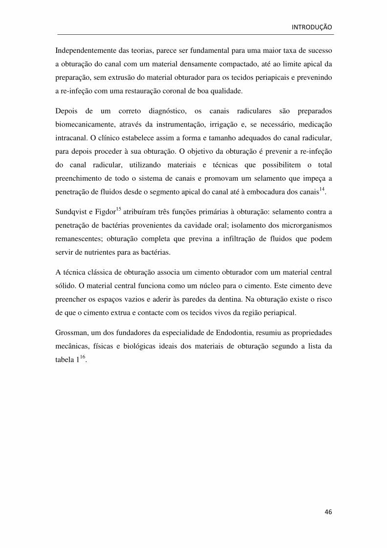
Grossman, um dos fundadores da especialidade de Endodontia, resumiu as propriedades
mecânicas, físicas e biológicas ideais dos materiais de obturação segundo a lista da
tabela 116.
INTRODUÇÃO
47
Tabela 1: Propriedades ideais de um material de obturação segundo Grossman
Deve ser fácil de introduzir no sistema de canais
Deve selar o canal lateral e apicalmente
Não deve contrair depois de endurecer
Deve ser impermeável aos fluidos
Deve ser bacteriostático (ou, pelo menos, não promover o crescimento bacteriano)
Deve ser radiopaco
Não deve pigmentar a estrutura dentária
Não deve ser irritante para os tecidos periapicais
Deve ser estéril ou fácil e rapidamente esterilizável imediatamente antes de ser utilizado
Deve ser fácil de remover do canal se necessário
Alguns autores foram acrescentando outras propriedades, nomeadamente:
- Não desencadear resposta imune nos tecidos periapicais;
- Não deve ser mutagénico nem carcinogénico17,18.
Material central sólido
Guta-percha
É, sem dúvida, o núcleo sólido universalmente mais utilizado.
Em 1867, Bowman19 introduziu o uso da guta-percha em Endodontia. A guta-percha é
um polímero do metilbutadieno ou isopreno (1,4 poliisopreno), sendo assim um isómero
da borracha, mas mais dura, mais quebradiça e menos elástica do que esta. Enquanto a
borracha natural é um cis-poliisopreno, possuindo agrupamentos CH2 do mesmo lado da
ligação dupla, a guta-percha é um trans-poliisopreno, o qual apresenta os seus
agrupamentos CH2 em lados opostos da ligação dupla. A guta-percha é obtida a partir
da coagulação do látex de árvores da Malásia, dos géneros Payena ou Palaquium, da
família Sapotaceae.
Em 1942, C.W. Bunn20 descreveu que a guta–pecha pode apresentar quimicamente duas
formas cristalinas distintas: alfa e beta. A forma alfa é a que é naturalmente extraída da
árvore. A maioria das gutas disponíveis comercialmente encontra-se na forma beta. A
forma alfa é quebradiça à temperatura ambiente e quando aquecida torna-se pegajosa,
INTRODUÇÃO
48
aderente e com maior escoamento. A temperatura de fusão é 65ºC, sendo esta a guta
utilizada nas técnicas termoplásticas. A forma beta é estável e flexível à temperatura
ambiente e quando aquecida não tem adesividade e tem menor escoamento que a forma
alfa. A sua temperatura de fusão é de 56ºC.
A forma comercial da guta-percha mais comum é em cones, que podem ser calibrados
ou não. Os cones calibrados têm diâmetros e conicidades determinados. O diâmetro da
ponta de um cone de guta é denominado D0. É um diâmetro virtual que consiste na
projeção da conicidade do cone até à sua extremidade. Os diâmetros em D0, expressos
em centésimas de milímetro, correspondem aos números padronizados (ISO) e variam
entre 15 e 140. O diâmetro dos cones aumenta 0,05mm até ao nº 60 e 0,10mm a partir
daqui até ao nº 140. No entanto, existem pequenas variações nos diâmetros de ponta,
inerentes ao processo de fabrico. As conicidades podem ser de 0.02, 0.04 e 0.06.
A composição básica dos cones de guta-percha inclui guta-percha (19 a 20%), óxido de
zinco (60 a 75%), substâncias para conferir radiopacidade como o sulfato de bário (1,5 a
17%) e outras substâncias como resinas, ceras e corantes (1 a 4%). Alguns fabricantes
adicionam ainda agentes anti-microbianos como o hidróxido de cálcio21, clorohexidina22
ou iodoformio23.
A guta-percha tem uma aceitável biocompatibilidade e baixa toxicidade24,25. A guta-
percha pura não é citotóxica, no entanto, os cones de guta-percha apresentam alguma
citotoxicidade, provavelmente devido aos outros componentes adicionados,
nomeadamente o óxido de zinco que é libertado ao longo do tempo26,27.
A obturação com guta-percha obriga ao uso de cimentos seladores, e ainda que a guta
possa ter alguma toxicidade, os cimentos são normalmente o elemento mais tóxico da
obturação.
As vantagens e as desvantagens dos cones de guta-percha são apresentadas na tabela 2.
INTRODUÇÃO
49
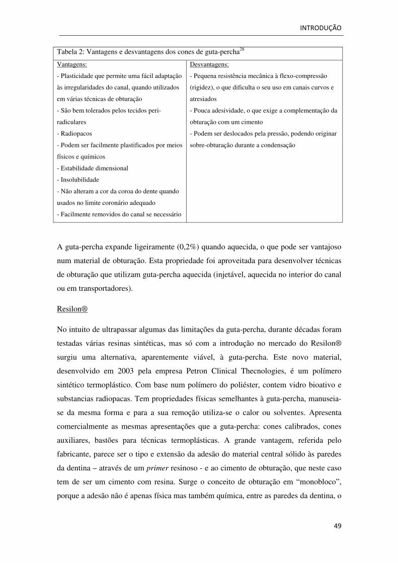
Tabela 2: Vantagens e desvantagens dos cones de guta-percha28
Vantagens:
- Plasticidade que permite uma fácil adaptação
às irregularidades do canal, quando utilizados
em várias técnicas de obturação
- São bem tolerados pelos tecidos peri-
radiculares
- Radiopacos
- Podem ser facilmente plastificados por meios
físicos e químicos
- Estabilidade dimensional
- Insolubilidade
- Não alteram a cor da coroa do dente quando
usados no limite coronário adequado
- Facilmente removidos do canal se necessário
Desvantagens:
- Pequena resistência mecânica à flexo-compressão
(rigidez), o que dificulta o seu uso em canais curvos e
atresiados
- Pouca adesividade, o que exige a complementação da
obturação com um cimento
- Podem ser deslocados pela pressão, podendo originar
sobre-obturação durante a condensação
A guta-percha expande ligeiramente (0,2%) quando aquecida, o que pode ser vantajoso
num material de obturação. Esta propriedade foi aproveitada para desenvolver técnicas
de obturação que utilizam guta-percha aquecida (injetável, aquecida no interior do canal
ou em transportadores).
Resilon®
No intuito de ultrapassar algumas das limitações da guta-percha, durante décadas foram
testadas várias resinas sintéticas, mas só com a introdução no mercado do Resilon®
surgiu uma alternativa, aparentemente viável, à guta-percha. Este novo material,
desenvolvido em 2003 pela empresa Petron Clinical Thecnologies, é um polímero
sintético termoplástico. Com base num polímero do poliéster, contem vidro bioativo e
substancias radiopacas. Tem propriedades físicas semelhantes à guta-percha, manuseia-
se da mesma forma e para a sua remoção utiliza-se o calor ou solventes. Apresenta
comercialmente as mesmas apresentações que a guta-percha: cones calibrados, cones
auxiliares, bastões para técnicas termoplásticas. A grande vantagem, referida pelo
fabricante, parece ser o tipo e extensão da adesão do material central sólido às paredes
da dentina – através de um primer resinoso - e ao cimento de obturação, que neste caso
tem de ser um cimento com resina. Surge o conceito de obturação em “monobloco”,
porque a adesão não é apenas física mas também química, entre as paredes da dentina, o
INTRODUÇÃO
50
Resilon® e o cimento. As vantagens desta obturação em “monobloco” seriam um
melhor selamento apical e coronal e o isolamento de bactérias residuais29.
Cimentos de obturação
Os cimentos são responsáveis pelas principais funções da obturação: selar
hermeticamente o sistema de canais radiculares, isolar as bactérias remanescentes e
preencher as irregularidades do canal preparado, nomeadamente os espaços entre a
superfície da dentina e o material central sólido. Tradicionalmente, o que se esperava do
cimento era que aderisse quer à dentina quer à guta-percha, no entanto os novos
cimentos são desenvolvidos com o objetivo de que penetrem nos túbulos dentinários e
estabeleçam uma melhor adesão à dentina e ao material central sólido.
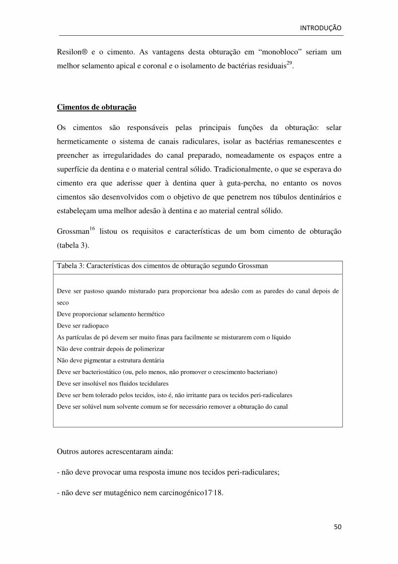
Grossman16 listou os requisitos e características de um bom cimento de obturação
(tabela 3).
Tabela 3: Características dos cimentos de obturação segundo Grossman
Deve ser pastoso quando misturado para proporcionar boa adesão com as paredes do canal depois de
seco
Deve proporcionar selamento hermético
Deve ser radiopaco
As partículas de pó devem ser muito finas para facilmente se misturarem com o líquido
Não deve contrair depois de polimerizar
Não deve pigmentar a estrutura dentária
Deve ser bacteriostático (ou, pelo menos, não promover o crescimento bacteriano)
Deve ser insolúvel nos fluidos tecidulares
Deve ser bem tolerado pelos tecidos, isto é, não irritante para os tecidos peri-radiculares
Deve ser solúvel num solvente comum se for necessário remover a obturação do canal
Outros autores acrescentaram ainda:
- não deve provocar uma resposta imune nos tecidos peri-radiculares;
- não deve ser mutagénico nem carcinogénico17,18.
INTRODUÇÃO
51
Ingle e Taintor20 afirmaram que a reação inicial dos tecidos periapicais a qualquer
cimento de obturação é inflamatória, no entanto há uma regeneração celular quando o
cimento polimeriza, a menos que continue a desintegrar-se, libertando componentes
tóxicos.
Têm sido utilizadas várias fórmulas químicas nos cimentos de obturação.
Classicamente, os cimentos são classificados, de acordo com a sua constituição, em:
-cimentos à base de óxido de zinco-eugenol (Roth, Kerr PCS, Procosol, Endometasona,
Tubliseal, etc)
- cimentos à base de hidróxido de cálcio (Sealapex, Apexit)
- cimentos á base de resina (AH Plus, Epiphany, Endorez, Acroseal)
- cimentos à base de silicone (RoekoSeal, GuttaFlow)
- cimentos à base de ionómero de vidro (KetacEndo)
Cimentos à base de óxido de zinco eugenol
Há muitos anos que os cimentos à base de óxido de zinco eugenol, na sua composição
mais básica (óxido de zinco no pó e eugenol como líquido) ou noutras formulações
(adição de resinas, bário, paraformaldeído, corticoides, etc) são dos mais utilizados na
obturação dos canais radiculares. A grande vantagem destes cimentos é o facto de não
contraírem e preencherem adequadamente todos os espaços. São ótimos selantes
marginais. A desvantagem é a solubilidade nos fluidos tecidulares e alguma
toxicidade30.
O endurecimento ou presa do cimento dá-se por reação de ácido-base, em que o óxido
de zinco atua como base e o eugenol como ácido, formando um sal quelato de
eugenelato de zinco e água28. A água é essencial e funciona como um acelerador da
reação de presa. Após a presa do material, 5% da quantidade original de eugenol
permanece livre.
O eugenol possui atividade anti-bacteriana, efeito anestésico e anti-inflamatório. É
também bactericida em concentrações relativamente altas (10-2 a 10-3mol/L). Os iões de
INTRODUÇÃO
52
zinco (Zn2+) também podem inibir o crescimento bacteriano, uma vez que em
concentrações elevadas são inibidores enzimáticos e interferem no metabolismo
bacteriano. No entanto, quer o eugenol quer os iões de zinco podem ter efeito
citotóxico31,32. Os efeitos biológicos do eugenol variam com a sua concentração. Doses
elevadas de eugenol inibem o crescimento e a respiração celular, podendo levar à morte;
promovem alterações vasculares, como a vasodilatação; são neurotóxicas. No entanto,
baixas concentrações têm propriedades farmacológicas desejáveis, como ação anti-
inflamatória e analgésica33. Alguns autores sugeriram a substituição do eugenol por
ácidos gordos para prevenir os seus efeitos citotóxicos34. Parece, no entanto, que a
quantidade de eugenol libertada de um cimento de óxido de zinco eugenol para além do
ápice é muito pequena (na gama das concentrações com efeitos benéficos) e diminui ao
longo do tempo35. Contudo, a quantidade libertada vai depender também da técnica de
obturação utilizada e do limite da obturação. As técnicas termoplásticas, por exemplo,
por um lado impedem a libertação de eugenol porque proporcionam um selamento mais
hermético, mas por outro lado são mais propícias a que ocorra extravazamento de
material obturador, e portanto aumenta a área de contacto do cimento com os tecidos
periapicais.
A quantidade de eugenol que é libertado e a sua toxicidade parecem ser maiores nos
cimentos que utilizam a mistura convencional de óxido de zinco eugenol que nos
cimentos que utilizam pó de oxido de zinco reforçado36.
Por outro lado, o eugenol produz uma expansão do volume da guta-percha.
Aumentando a proporção do eugenol no cimento, aumenta a expansão do volume da
guta-percha37, o que pode traduzir-se numa melhor capacidade seladora, mas aumentar
os riscos de efeitos indesejáveis do eugenol.
O eugenol tem ainda uma desvantagem, ainda que de menor importância: inibe a reação
de polimerização das resinas, o que pode condicionar o tipo de restauração 38 . No
entanto, este efeito pode ser minimizado adotando os procedimentos adequados.
Há ainda outros componentes que podem ser citotóxicos, como a resina adicionada para
aumentar a adesão à dentina, paraformaldeído para efeito anti-microbiano e
mumificante, germicidas para ação antiséptica e corticoides para inibir a inflamação.
INTRODUÇÃO
53
Um estudo de Sunzel et al 39 mostrou que a incorporação de zinco reduz
significativamente a toxicidade das resinas e resinas ácidas (que são anti-microbianas)
proporcionalmente ao aumento da concentração de zinco.
O formaldeído e/ou paraformaldeído, mesmo em pequenas quantidades podem ser
irritantes e impedir ou retardar o processo regenerativo. Alguns cimentos de obturação
contêm estas substâncias ou, mesmo que não esteja presente nas suas formulações,
podem libertar moléculas de formaldeído durante a reação química de polimerização40.
Além do efeito tóxico per si, parece potenciar o efeito tóxico do eugenol.
O formaldeído é classificado (pela International Agency for Research on Cancer) como
carcinogénico para os animais, mas a evidência do efeito carcinogénico no Homem é
limitada41. No entanto, os elevados teores de paraformaldeído em alguns cimentos de
óxido de zinco eugenol pode ser motivo de preocupação42. Já foi demonstrado que o
efeito desinfetante do canal, a longo prazo, promovido pelo formaldeído é pouco
significativo 43 , com o risco de originar reações adversas, nomeadamente
neurotoxicidade 44 , parestesia do nervo alveolar inferior 45 , 46 , urticária 47 e reações
anafiláticas48,49,50.
Cimentos à base de hidróxido de cálcio
O hidróxido de cálcio foi introduzido na endodontia por Herman em 1920 pela sua
capacidade de regeneração pulpar51. Devido aos efeitos biológicos benéficos que lhe são
atribuídos, passou a ser utilizado em endodontia para recobrimento pulpar, medicação
intra-canal, em técnicas de apexificação e na composição dos cimentos para obturação
definitiva dos canais radiculares. As duas principais razões para usar o hidróxido de
cálcio como material de obturação são a estimulação dos tecidos periapicais para se
manterem sãos ou para promover a sua regeneração, e pelos seus efeitos anti-
bacterianos (ainda que inferiores a outros cimentos, nomeadamente à base de óxido de
zinco-eugenol ou à base de resina)52.
Os mecanismos de ação exatos são desconhecidos, mas foram propostos os seguintes:
1. O hidróxido de cálcio é anti-bacteriano devido à libertação de iões hidroxil53. O
seu elevado pH promove a reparação e calcificação dos tecidos. Inicialmente há
INTRODUÇÃO
54
uma resposta degenerativa na zona circundante, que é depois seguida por
mineralização e ossificação54;
2. O pH alcalino do hidróxido de cálcio neutraliza o ácido láctico dos osteoclastos
e previne a dissolução dos componentes minerais do dente. Este pH também
ativa a fosfatase alcalina, que tem um importante papel na formação de tecido
ósseo55;
3. O hidróxido de cálcio desnatura as proteínas encontradas no canal, tornando-as
menos tóxicas;
4. O hidróxido de cálcio ativa a reação da adenosina trifosfatase cálcio dependente,
associada à formação de tecido duro55 ,56;
5. O hidróxido de cálcio difunde através dos túbulos dentinários e pode atingir o
ligamento periodontal, detendo a reabsorção radicular externa e acelerando a
cicatrização53,57.
Para ter um efeito terapêutico, o hidróxido de cálcio tem que ser dissociado em iões
cálcio (Ca2+) e iões hidroxil (OH). Portanto, para um cimento endodôntico à base de
hidróxido de cálcio ser terapêutico, tem de haver libertação destes iões, o que pode
afetar a integridade estrutural do cimento e comprometer o selamento a longo prazo58.
Vários estudos in vitro e in vivo comprovam que este fenómeno ocorre e pode mesmo
induzir uma resposta inflamatória dos tecidos periapicais52,59,60. No entanto, há também
evidências de que a dissolução do cimento de hidróxido de cálcio não é superior a
outros tipos de cimento61. A reação de polimerização destes cimentos é complexa e
pouco homogénea; o contacto com a humidade origina uma superfície dura, mas a zona
mais profunda da mistura pode permanecer com menor consistência.
Estes cimentos são geralmente caracterizados como tendo uma boa
citocompatibilidade62,63, mas a falta de dureza física é motivo de preocupação.
Há algumas controvérsias quanto à biocompatibilidade dos cimentos à base de
hidróxido de cálcio, que poderá ser atribuída aos métodos de avaliação, no entanto, a
maior parte dos estudos conclui que estes cimentos apresentam resultados aceitáveis
comparativamente a outros cimentos64.
INTRODUÇÃO
55
Cimentos à base de resina
Resinas epóxicas
Dentro deste grupo, os cimentos que atingiram mais sucesso foram os da série AH. O
protótipo foi desenvolvido há mais de 50 anos por Andre Schroeder na Suiça65, e é uma
resina bis-fenol que usa metamina para polimerização (AH26). Como a metanima
liberta formaldeído durante a reação de polimerização43, passou a usar-se como
substituto uma mistura de aminas que permitia a polimerização sem a formação de
formaldeído, sendo o produto comercializado como AH Plus. Um estudo de Cohen et
al66 comparou a libertação de formaldeído pelos dois cimentos e mostrou que o AH Plus
liberta uma quantidade mínima comparativamente ao AH 26 (3,9 ppm/1347 ppm). Um
estudo mais recente de Evcil et al67, não detetou formaldeído no AH Plus. Além disso, o
AH Plus, ao contrário do AH 26, parece não ter efeito estrogénico, ainda que ambos
tenham na sua constituição bisGMA68 . Outras vantagens do AH Plus são a rápida
polimerização, maior radiopacidade, maior facilidade de remoção e baixa
solubilidade69,70.
A popularidade deste tipo de cimento deveu-se em grande parte ao facto de não conter
eugenol e portanto não interferir com as restaurações em resina.
Uma outra fórmula com resina, até há pouco amplamente utilizada em todo o mundo, é
o tipo resorcinol-formaldeído (Tratamento Spad, por exemplo)71 ,72 . Este cimento é
altamente anti-bacteriano, mas contrai e deixa uma tonalidade avermelhada na estrutura
dentária envolvente. Como se preconizava a sua utilização sem cones de guta-percha e
polimerizava numa massa muito dura e insolúvel, o re-tratamento destes casos era e é
extremamente difícil. Além disso, como já referimos, o formaldeído é um componente
citotóxico e que pode difundir-se para os tecidos periapicais, ainda que alguns autores
considerem que a quantidade mínima de formaldeído nos cimentos de obturação é
irrelevante sob o ponto de vista toxicológico, atendendo à elevada exposição e
tolerância dos mamíferos a este componente43. No entanto, o formaldeído libertado dos
cimentos de obturação, principalmente quando há extrusão do material para a região
periapical, pode originar uma reação inflamatória periapical ou contribuir para a
manutenção de uma lesão periapical pré-existente73.
INTRODUÇÃO
56
Resinas de metacrilato
Estes cimentos, extensivamente estudados nos últimos anos, vão já na quarta geração e
o objetivo seria conseguir uma adesão física e química às paredes da dentina e ao
material de obturação central29.
O EndoRez® faz parte deste grupo de cimentos. Baseado em uretanodimetacrilato
(UDMA), semelhante a muitas resinas utilizadas em dentisteria para restauração. Tem
algumas propriedades hidrofílicas que parecem melhorar o seu comportamento, mesmo
na presença de humidade. Recentemente, o EndoRez® foi comercializado juntamente
com cones de guta-percha revestidos com resina 74 , que por ligação ao cimento,
proporcionariam melhor adesão e selamento. Este é o mesmo conceito dos novos
produtos Ephiphany/Resilon® e RealSeal®75. Com este tipo de cimento, é aplicado um
primer à superfície da dentina depois de esta ter sido limpa com um ácido para remover
a smear-layer. De seguida, um cimento de dupla polimerização baseado em BisGMA,
UDMA e metacrilatos hidrofílicos e com partículas radiopacas, liga-se às paredes de
dentina através do primer. Para completar o preenchimento, são inseridos cones de
Resilon®, em lugar da guta-percha. Então, o cimento liga-se à dentina via primer e há
uma ligação química do cimento ao cone, o que originou o conceito de obturação
homogénea, “monobloco”76. Um estudo de revisão recente77 concluiu não haver, para
já, vantagem na utilização destes cimentos.
Cimentos à base de silicone
O Lee Endo-Fill® foi o primeiro cimento a testar as propriedades do silicone
(amplamente utilizado noutras áreas da medicina dentária), nomeadamente a capacidade
hidrofóbica, a estabilidade química e propriedades adesivas, como material de
endodontia65. Apresentava alguma toxicidade, que aumentava com o tempo,
provavelmente devido ao prolongado período de polimerização78.
O RoekoSeal Automix® é uma formulação mais recente deste tipo de cimento. O
cimento é colocado por meio de um aplicador de câmara dupla, que mistura o cimento
na dosagem apropriada. O material polimeriza por completo, sem contração,
independentemente da humidade e temperatura28, apresentando bons resultados79.
INTRODUÇÃO
57
O GuttaFlow® surgiu da tentativa de incorporar no cimento de obturação as qualidades
da guta-percha. A guta-percha foi triturada em partículas muito pequenas e misturada
nos componentes de um cimento à base de silicone. Uma das vantagens advogada é a
sua capacidade de expandir ligeiramente (0,2%) durante a presa 80 , melhorando a
capacidade seladora. Estudos recentes comprovam a boa capacidade seladora deste
cimento, mas alertam para a possibilidade de extrusão para além do foramen apical81,
pelo que o potencial risco para os tecidos periapicais tem de ser estudado.
Cimentos à base de ionómero de vidro
Com pouca representação comercial (o Ketac-Endo® é o mais conhecido), os cimentos
de ionómero de vidro tradicionais são constituídos por alumina, sílica e ácido
polialcenóico e são autopolimerizáveis. Foram introduzidos no mercado há mais de 20
anos, e foram muito utilizados82, ainda que laboratorialmente tenham mostrado alguma
infiltração e desintegração83. Mostraram propriedades anti-microbianas84,85e capacidade
de adesão à estrutura dentária, mas parecem ativar a libertação de prostaglandinas nos
tecidos periapicais86.
Estudos em culturas celulares com estes cimentos mostraram elevada citotoxicidade
imediatamente após a mistura (provavelmente devido à libertação de alumínio), no
entanto, após a presa, a toxicidade era baixa ou nula87.
Novos materiais
Nos últimos anos, tem surgido na literatura referência a estudos com novos materiais:
- cimentos à base de fosfato de cálcio88,89;
- cimentos biocerâmicos (à base de cimento de Portland, que tem na sua constituição
silicato de cálcio) e cuja principal vantagem é a sua elevada atividade anti-microbiana,
apresentando boa capacidade seladora90 , bioatividade (capacidade para formar uma
camada de apatite tipo osso, estabelecendo uma ligação química com os tecidos vivos) e
biocompatibilidade91,92. Resultam da tentativa de aproveitar os excelentes resultados do
Agregado Trióxido Mineral (MTA), material de reparação tão amplamente utilizado nos
últimos anos na Endodontia, ultrapassando algumas das suas limitações (dificuldade de
manipulação, reduzido tempo de trabalho, dificuldade de remoção);
INTRODUÇÃO
58
- adição de hidroxiapatite nano-estruturada aos cimentos de obturação93;
- a utilização de quitosano tem sido objeto de pesquisa nas Unidades Curriculares de
Endodontia da Faculdade de Medicina Dentária da Universidade do Porto,
nomeadamente em trabalhos de doutoramento, pelo que esperamos num futuro próximo
obter resultados relativos à sua utilização como material de obturação de canais.
Avaliação do comportamento biológico dos cimentos endodônticos
Em 1930, Grove estabeleceu o limite apical da instrumentação e da obturação na junção
cimento-dentinária (limite CDC). Mais tarde, Kuttler94 estudou mais de 400 ápices e os
seus estudos foram fundamentais para esclarecer a anatomia do canal radicular.
Concluiu que a terminação apical do canal é formada por duas zonas cónicas: uma
dentinária, com a base no orifício camaral do canal e o vértice na junção cimento-
dentinária, e a outra cimentária, com o vértice na junção cimento-dentinária e a base no
foramen apical. A união destas duas zonas é o local mais estreito do canal radicular -
constrição apical - considerada como o ponto ideal para a terminação da preparação e
obturação do canal.
Todos os materiais de obturação têm algum efeito irritante para os tecidos vivos,
nomeadamente para os tecidos periapicais. Ainda que o objetivo durante a obturação
dos canais radiculares seja que o material obturador fique confinado ao interior dos
canais, há sempre o risco de extravasamento, maior ou menor conforme as técnicas de
instrumentação e/ou obturação. Muitas vezes o material ultrapassa o foramen apical e
entra em contacto direto com os tecidos periapicais, mesmo quando essa sobre-
obturação não é visível na radiografia pós-operatória. Esse risco está aumentado nos
casos de lesão apical crónica pré-operatória, uma vez que pode haver reabsorção do
cimento apical, alterando a anatomia do ápice radicular, nomeadamente foramen apical
e constrição apical. Portanto, o que importa saber acerca dos materiais de obturação,
principalmente os cimentos de obturação, não é se são irritantes mas qual o grau de
irritação que causam e durante quanto tempo.
Vários estudos 95 , 96 , 97 , 98 referem que a resposta dos tecidos aos diferentes tipos de
cimentos de obturação varia consideravelmente e concluíram que a quantidade de
cimento colocado no canal, a área de contacto entre o material e os tecidos e a interação
INTRODUÇÃO
59
entre a dentina e produtos libertados dos cimentos, influenciam significativamente a
resposta do tecido ao material obturador. Esta reação varia também com o tipo de
substancia presente ou libertada, a quantidade libertada e a velocidade de absorção.
Segundo Williams (1987)99, a biocompatibilidade é definida como a capacidade do
material promover uma resposta apropriada numa aplicação específica, com o mínimo
de reações alérgicas, inflamatórias ou tóxicas, quando em contacto com os tecidos vivos
ou fluidos orgânicos.
Um material biocompatível deve estimular a cicatrização dos tecidos lesados sem causar
efeitos adversos. Quase todos os cimentos endodônticos têm uma composição química
complexa e são vários os aditivos que podem afetar a sua biocompatibilidade.
A biocompatibilidade dos cimentos foi caracterizada em vários parâmetros, tais como
genotoxicidade, mutagenicidade, carcinogenicidade, citotoxicidade, compatibilidade
dos tecidos e efeitos microbiológicos. No entanto, é impossível avaliar os materiais
utilizando apenas um tipo de teste. As suas propriedades devem ser investigadas por
uma bateria de testes in vitro e in vivo, seguindo uma abordagem estruturada58.
Ainda que os testes in vivo sejam de grande interesse, a utilização de animais coloca
problemas éticos e está atualmente sob grande discussão pública. Além disso, este tipo
de experiência é cara, demorada e difícil de controlar100, pelo que a citotoxicidade é
normalmente testada em células de cultura.
Os testes em culturas celulares, utilizados há mais de 30 anos na investigação da
citotoxicidade dos materiais endodônticos, são mais simples, mais rápidos e mais
económicos que outros métodos, e permitem testar um grande número de materiais
utilizando as mesmas células e sob as mesmas condições. São fiáveis e facilmente
reprodutíveis. Os testes in vitro têm a vantagem de utilizar um meio de cultura com
composição standard, ambiente de incubação definido e condições de trabalho estéreis,
e permitem uma avaliação qualitativa e quantitativa precisa98. No entanto, os resultados
não podem ser extrapolados para a situação clínica. Os métodos que utilizam culturas
celulares são muitas vezes mais sensíveis que os métodos in vivo, mas devem ser
avaliados dentro dos limites de um teste de toxicidade aguda101.
A utilização de testes in vitro permite estudar os efeitos da libertação de componentes
do material no sistema celular102. Podem ser utilizadas linhas celulares (por exemplo
INTRODUÇÃO
60
HeLa, 3T3 ou L929) ou células humanas primárias (por exemplo fibroblastos ou células
da polpa), sendo que estas últimas são consideradas mais relevantes para os estudos de
biocompatibilidade103. As células primárias são caracterizadas por apresentarem uma
grande capacidade de diferenciação, e ainda que sejam menos homogéneas e mais
sensíveis que as linhas celulares, o seu comportamento torna-as mais facilmente
comparáveis à mucosa oral104,105. Com este tipo de estudo podem avaliar-se vários
aspetos biológicos: inibição do crescimento, determinação da dose efetiva (DE50),
integridade da membrana, síntese de DNA, RNA ou proteínas e/ou alterações da
morfologia celular por microscopia103,106,107,108,109.
Culturas celulares provenientes de diferentes órgãos podem apresentar respostas
metabólicas diferentes que afetam a sua suscetibilidade a agentes tóxicos exógenos110.
A genotoxicidade resulta da presença de componentes DNA reativos que podem
provocar alterações mutagénicas e carcinogénicas42. Os testes in vitro para avaliação da
genotoxicidade podem ser diferenciados em estudos procaróticos/bacterianos (por
exemplo: teste de Ames, teste umu) e eucarióticos (por exemplo: teste de inibição da
síntese do DNA)111. Como vários materiais endodônticos têm uma elevada citoxicidade,
é importante que os testes de genotoxicidade quantifiquem simultaneamente a
citotoxicidade, para evitar interpretações erradas dos resultados. Por outro lado, como
muitos dos materiais têm uma elevada atividade anti-bacteriana, é necessário associar
testes bacterianos com testes eucarióticos para obter resultados fiáveis. Ao contrário do
efeito citotóxico, que provoca morte celular, a genotoxicidade não provoca
obrigatoriamente a morte celular ou qualquer outro efeito imediato, mas a lesão do
genoma celular pode diminuir significativamente a capacidade reparativa do tecido ou, a
longo prazo, originar alterações neoplásicas112.
A exposição das células a agentes citotóxicos pode causar necrose ou apoptose. Durante
a necrose, a célula começa por dilatar, a membrana plasmática colapsa e as células
sofrem lise 113 . Portanto, a necrose é uma morte celular patológica. A apoptose é
geralmente caracterizada por um colapso interno dos organelos, uma desintegração da
membrana plasmática em corpos vesiculares apoptóticos e a destruição do material
genético114. A eliminação celular das células por apoptose não é detetada pelo sistema
imunológico, enquanto a eliminação do conteúdo intracelular das células necróticas para
o espaço extracelular origina uma resposta inflamatória115. A apoptose é essencialmente
INTRODUÇÃO
61
uma morte celular programada (“suicídio celular”) – auto-destruição das células, não
afetando as células vizinhas116.
Neste trabalho experimental, utilizamos uma linha celular permanente MG63 (células
“osteoblast-like”, provenientes de osteossarcoma humano) e também células humanas
primárias - células mononucleares de sangue periférico e células mesenquimais de
medula óssea, respetivamente para o estabelecimento de culturas de osteoclastos e
osteoblastos.
Uma vez que avaliamos os efeitos dos cimentos de obturação em células ósseas, parece-
nos relevante fazer uma revisão sobre o tecido ósseo e seu metabolismo.
Tecido ósseo/Metabolismo ósseo
O tecido ósseo é um tipo especializado de tecido conjuntivo, formado por células e
material extracelular calcificado, a matriz óssea. As células, responsáveis pela
formação, reabsorção e manutenção da arquitetura óssea, são: os osteoblastos, que
sintetizam a parte orgânica da matriz e se localizam na sua periferia; os osteócitos, que
se situam em cavidades ou lacunas no interior da matriz; os osteoclastos, células
gigantes, móveis e multinucleadas, que reabsorvem o tecido ósseo, participando dos
processos de remodelação dos ossos.
Para além das funções básicas de suporte, proteção e locomoção, os ossos constituem
um importante reservatório de minerais. Sistemicamente, este mecanismo é controlado
por fatores hormonais; localmente é controlado por forças mecânicas (incluindo os
movimentos dentários), fatores de crescimento e citocinas.
Matriz óssea
É constituída por uma parte mineral e uma parte orgânica.
A parte mineral ou inorgânica representa cerca de 67% do peso da matriz óssea. Os iões
mais encontrados são o fosfato e o cálcio (fosfato de cálcio, organizado na forma de
hidroxiapatite - Ca10(PO4)6(OH)2), mas existem também pequenas quantidades de
bicarbonato, magnésio, potássio, sódio e citrato.
INTRODUÇÃO
62
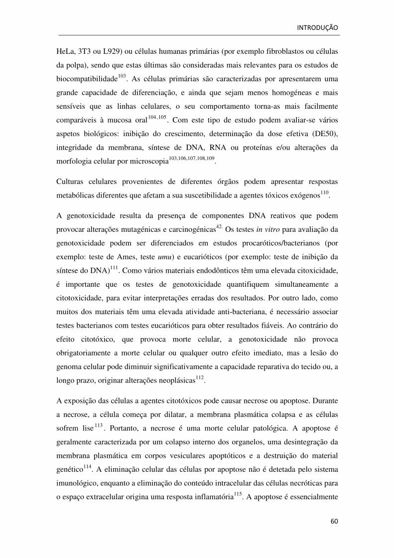
A parte orgânica da matriz (33%) é formada por fibras de colagénio tipo I (28%) e
pequenas quantidades (5%) de proteoglicanos e glicoproteínas (osteocalcina,
sialoproteína óssea, osteopontina, fosfoproteína, proteína específica do osso). As
glicoproteínas do osso parecem estar envolvidas na mineralização da matriz.
Figura 1. Esquema da ossificação intra-membranosa. Osteoblastos sintetizam a matriz orgânica que forma uma faixa – osteóide – que mineraliza, aprisionando alguns osteoblastos que se diferenciam em osteócitos (adaptado de Junqueira e Carneiro – Histologia básica)
Periósteo e Endósteo
As superfícies internas e externas dos ossos são recobertas por células osteogénicas e
tecido conjuntivo, que constituem o endósteo e periósteo, respetivamente.
A camada mais superficial do periósteo contém principalmente fibras colagéneas e
fibroblastos. Na zona mais profunda, o periósteo é mais celular e apresenta células
osteoprogenitoras. Estas multiplicam-se por mitose e diferenciam-se em osteoblastos,
desempenhando um papel importante no crescimento dos ossos e reparação de defeitos
ou fraturas ósseas.
O endósteo é geralmente constituído por uma camada de células osteogénicas achatadas,
que revestem as cavidades do osso esponjoso, o canal medular, os canais de Havers e os
de Volkmann.
INTRODUÇÃO
63
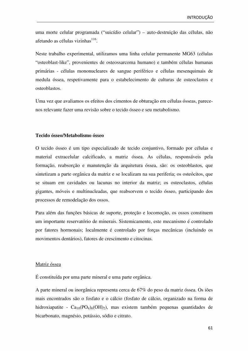
Figura 2. Esquema da parede da diáfise dos ossos longos. Aparecem três tipos de tecido ósseo lamelar: os sistemas de Havers e as lamelas circunferenciais externas e internas (adaptado de Junqueira e Carneiro – Histologia básica)
As principais funções do periósteo e endósteo são a nutrição do tecido ósseo e
fornecimento de novos osteoblastos, para o crescimento e recuperação do osso.
Tipos de tecido ósseo
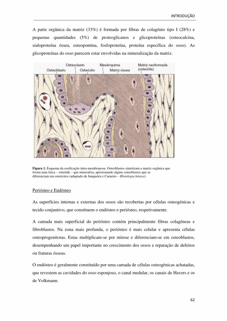
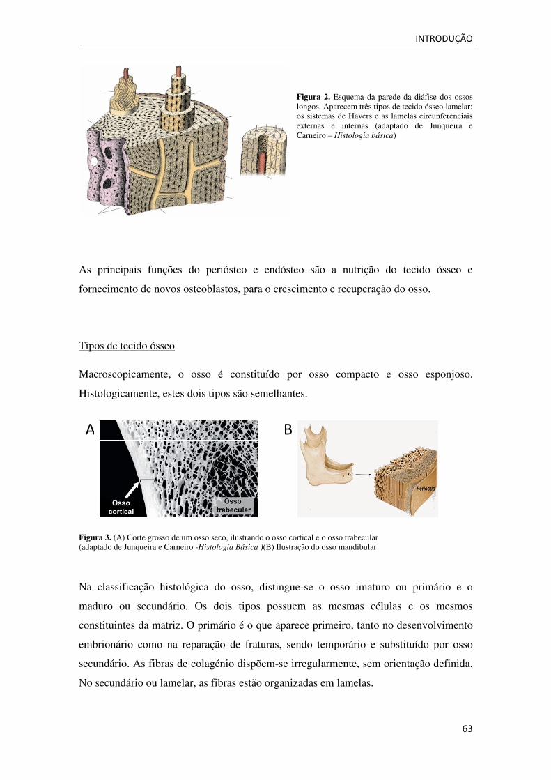
Macroscopicamente, o osso é constituído por osso compacto e osso esponjoso.
Histologicamente, estes dois tipos são semelhantes.
Figura 3. (A) Corte grosso de um osso seco, ilustrando o osso cortical e o osso trabecular (adaptado de Junqueira e Carneiro -Histologia Básica )(B) Ilustração do osso mandibular
Na classificação histológica do osso, distingue-se o osso imaturo ou primário e o
maduro ou secundário. Os dois tipos possuem as mesmas células e os mesmos
constituintes da matriz. O primário é o que aparece primeiro, tanto no desenvolvimento
embrionário como na reparação de fraturas, sendo temporário e substituído por osso
secundário. As fibras de colagénio dispõem-se irregularmente, sem orientação definida.
No secundário ou lamelar, as fibras estão organizadas em lamelas.
A B
INTRODUÇÃO
64
Osteoblastos
São as células que sintetizam a parte orgânicada matriz óssea - osteóide (composto
essencialmente por colagénio tipo I, proteoglicanos e glicoproteínas). São capazes de
concentrar fosfato de cálcio, participando na mineralização da matriz extracelular. Uma
vez aprisionado na matriz recém-sintetizada, o osteoblasto passa a ser chamado
osteócito.
Os osteoblastos derivam de células mesenquimatosas multipotentes (mesenquimal stem
cells) do estroma da medula óssea, que também originam condroblastos, fibroblastos,
adipócitos e miócitos. A sua célula precursora é denominada pré-osteoblasto, uma
célula não funcional, que posteriormente se diferencia em osteoblasto maduro, capaz de
formar osso.
Os osteoblastos dispõem-se como uma camada celular na superfície do osso em
formação, originando uma membrana que controla o fluxo de iões para dentro e para
fora do osso. Quando termina a formação do osso, os osteoblastos ficam inativos e
denominam-se “lining cells”, que parecem manter as junções comunicantes com os
osteócitos, formando uma rede que controla a homeostasia mineral e assegura a
vitalidade do osso117.
Os pré-osteoblastos e os osteoblastos apresentam níveis elevados de fosfatase alcalina
na superfície externa da membrana plasmática. Esta enzima, utilizada
experimentalmente como marcador citoquímico, distingue os osteoblastos dos
fibroblastos. Funcionalmente, parece clivar o fosfato dos compostos orgânicos. O
fosfato libertado provavelmente contribui para iniciar e continuar o crescimento dos
cristais minerais do osso.
Os osteoblastos produzem diversas moléculas importantes para o metabolismo ósseo,
nomeadamente precursores do colagénio tipo I, osteocalcina (proteína não colagénica
mais abundante na matriz óssea), sialoproteína óssea, proteoglicanos e segregam várias
citocinas. Estas, que incluem fatores de crescimento, ajudam a regular o metabolismo
celular. Os osteoblastos segregam ainda vários membros da superfamília de proteínas
morfogenéticas do osso (bone morphogenetic proteins -BMP), incluindo BMP2, BMP7
e o fator de crescimento transformante beta (transforming growth factor beta - TGF-β),
os fatores de crescimento semelhante à insulina I e II (insulin-like growth factors- IGF-I
INTRODUÇÃO
65
e IGF-II), o fator de crescimento derivado das plaquetas (platelet-derived growth factor
- PDGF-AA) e o fator de crescimento dos fibroblastos beta (fibroblast growth factor
beta - FGF-β). A presença destes fatores de crescimento locais, produzidos pelos
próprios osteoblastos, pode estimular a formação óssea. As BMP e os TGF são
sintetizados pelos osteoblastos, sob a forma de precursores inativos. A concentração
local de TGF-β é regulada por hormonas, como a paratormona e os estrogénios e por
proteases osteoclásticas, que clivam as formas precursoras desses fatores de
crescimento. Os TGF-β estimulam a proliferação dos precursores dos osteoblastos e a
síntese de proteínas da matriz óssea pelo osteoblasto maduro. Além disso, os TGF-β que
são libertados durante o processo de reabsorção óssea, participam na apoptose dos
osteoclastos, constituindo um sinal para o fim da reabsorção e recrutam osteoblastos
para a lacuna de reabsorção, permitindo iniciar o processo de formação de novo osso118.
As BMP, estrutural e funcionalmente muito semelhantes aos TGF-β, são reconhecidas
por recetores diferentes, mas atuam na indução da diferenciação osteoblástica,
aumentando a expressão do fator de transcrição Cbfa1 específico dos osteoblastos119.
Outros fatores importantes, como IGF, PDGF e FGF, produzidos pelos osteoblastos,
atuam no microambiente ósseo, estimulando a proliferação e a diferenciação dos
precursores osteoblásticos ou aumentando a capacidade de síntese de novo material
ósseo pelos osteoblastos maduros120.
Apesar de o “timing” da secreção e a complexa interação destes fatores de crescimento
não estarem completamente esclarecidos, a combinação de IGF-I e TGF-β e o fator de
crescimento derivado das plaquetas (PDGF-BB) aumenta consideravelmente a
velocidade de formação e reparação óssea e será provavelmente muito importante no
futuro da medicina dentária. Por exemplo, estas combinações poderão ser utilizadas
para acelerar a cicatrização e crescimento ósseo após cirurgia periodontal ou para
prevenir a doença periodontal pelo tratamento precoce de bolsas periodontais. Poderá
ainda ser utilizada para melhorar a osteointegração após colocação de implantes.
Em condições fisiológicas que promovem a reabsorção, os osteoblastos podem ser
estimulados pelas linfocinas (ex: interleucina 1, fator de necrose tumoral alfa) e pelas
prostraglandinas para produzir interleucina 6. Os osteoblastos sob o estímulo da
interleucina 6 também produzem as suas próprias enzimas hidroliticas que participam
na destruição e modificação da matriz desmineralizada ou cobertura pericelular,
libertando os osteoblastos da sua própria matriz. Esta separação pode ser crítica durante
INTRODUÇÃO
66
as fases iniciais do metabolismo ósseo, quando os osteoblastos têm de migrar e
proliferar117. Portanto, estímulos como a atividade física ou movimentos dentários,
afetam o metabolismo ósseo, atuando a nível dos osteoblastos121.
Geneticamente, o osteoblasto pode ser vistos como um “fibroblasto sofisticado”, uma
vez que todos os genes expressos nos fibroblastos podem ser encontrados nos
osteoblastos. Apenas dois fatores de transcrição específicos dos osteoblastos foram
identificados: o fator de transcrição Cbfa1 (core binding factor a1) e o fator de
transcrição para a calcitonina, uma molécula que inibe a função osteoclástica. O Cbfa1
desempenha um papel central na diferenciação osteoblástica. Este fator de transcrição é
o marcador mais precoce e específico da osteogénese, pois induz a diferenciação dos
osteoblastos, controla a formação de material ósseo pelos osteoblastos diferenciados e
regula a expressão da osteocalcina 122 . Ratos deficientes em Cbfa1 apresentam um
esqueleto apenas formado por cartilagem, pois a diferenciação osteoblástica está
comprometida 123 . Estes ratos são igualmente desprovidos de osteoclastos, o que
demonstra a relação dinâmica que existe entre estes dois tipos de células vitais para o
tecido ósseo, evidenciando a necessidade da presença de osteoblastos para que ocorra a
diferenciação dos osteoclastos. Atualmente não são ainda conhecidos os detalhes
moleculares que controlam a expressão do Cbfa1. Sabe-se, no entanto, que algumas
BMP podem induzir a expressão de Cbfa1 in vitro122. Foi também demonstrado que o
TGF-β controla a diferenciação dos osteoblastos e modula a expressão do gene que
codifica para o Cbfa1, e que outros fatores, como o FGF, também são importantes na
diferenciação dos osteoblastos.
Osteócitos
À medida que os osteoblastos produzem a matriz óssea, alguns ficam isolados no seu
interior e são chamados osteócitos. São, portanto, as células encontradas no interior da
matriz óssea, ocupando lacunas das quais partem canalículos. Cada lacuna tem apenas
um osteócito. Dentro dos canalículos, os prolongamentos dos osteócitos estabelecem
contactos entre os osteócitos e com os osteoblastos da superfície do osso. Através destas
junções comunicantes, podem passar pequenas moléculas e iões de uma célula para
outra. Este complexo osteoblasto-osteócito é essencial para a manutenção e vitalidade
da matriz óssea, apesar da pequena atividade de síntese, sendo que a morte dos
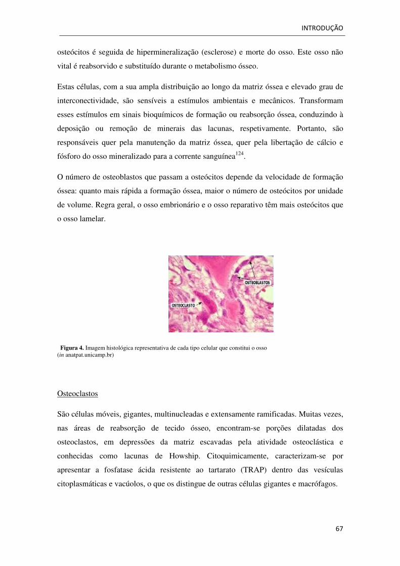
osteócitos é seguida de hipermineralização (esclerose) e morte do osso. Este osso n
vital é reabsorvido e substituído durante o metabolismo ósseo.
Estas células, com a sua ampla distribuição ao longo da matriz óssea e elevado grau de
interconectividade, são sensíveis a estímulos ambientais e mecânicos. Transformam
esses estímulos em sinais bioquímicos de formação ou reabsorção óssea, conduzindo à
deposição ou remoção de minerais das lacunas, respetivamente. Portanto, são
responsáveis quer pela manutenção da matriz óssea, quer pela libertação de cálcio e
fósforo do osso mineralizado para
O número de osteoblastos que passam a osteócitos depende da velocidade de formação
óssea: quanto mais rápida a formação óssea, maior o número de osteócitos por unidade
de volume. Regra geral, o osso embrionário e o osso reparativo t
o osso lamelar.
Figura 4. Imagem histológica representativa de cada tipo celular que constitui o osso(in anatpat.unicamp.br)
Osteoclastos
São células móveis, gigantes, multinucleadas e extensamente
nas áreas de reabsorção de tecido ósseo, encontram
osteoclastos, em depressões da matriz escavadas pela atividade osteoclástica e
conhecidas como lacunas de Howship. Citoquimicamente, caracterizam
apresentar a fosfatase ácida resistente
citoplasmáticas e vacúolos, o que os distingue de outras células gigantes e macrófagos.
osteócitos é seguida de hipermineralização (esclerose) e morte do osso. Este osso n
vital é reabsorvido e substituído durante o metabolismo ósseo.
Estas células, com a sua ampla distribuição ao longo da matriz óssea e elevado grau de
interconectividade, são sensíveis a estímulos ambientais e mecânicos. Transformam
nais bioquímicos de formação ou reabsorção óssea, conduzindo à
deposição ou remoção de minerais das lacunas, respetivamente. Portanto, são
responsáveis quer pela manutenção da matriz óssea, quer pela libertação de cálcio e
fósforo do osso mineralizado para a corrente sanguínea124.
O número de osteoblastos que passam a osteócitos depende da velocidade de formação
óssea: quanto mais rápida a formação óssea, maior o número de osteócitos por unidade
de volume. Regra geral, o osso embrionário e o osso reparativo têm mais osteócitos que
Imagem histológica representativa de cada tipo celular que constitui o osso
São células móveis, gigantes, multinucleadas e extensamente ramificadas. Muitas vezes,
nas áreas de reabsorção de tecido ósseo, encontram-se porções dilatadas dos
osteoclastos, em depressões da matriz escavadas pela atividade osteoclástica e
conhecidas como lacunas de Howship. Citoquimicamente, caracterizam
apresentar a fosfatase ácida resistente ao tartarato (TRAP) dentro das vesículas
citoplasmáticas e vacúolos, o que os distingue de outras células gigantes e macrófagos.
INTRODUÇÃO
67
osteócitos é seguida de hipermineralização (esclerose) e morte do osso. Este osso não
Estas células, com a sua ampla distribuição ao longo da matriz óssea e elevado grau de
interconectividade, são sensíveis a estímulos ambientais e mecânicos. Transformam
nais bioquímicos de formação ou reabsorção óssea, conduzindo à
deposição ou remoção de minerais das lacunas, respetivamente. Portanto, são
responsáveis quer pela manutenção da matriz óssea, quer pela libertação de cálcio e
O número de osteoblastos que passam a osteócitos depende da velocidade de formação
óssea: quanto mais rápida a formação óssea, maior o número de osteócitos por unidade
êm mais osteócitos que
ramificadas. Muitas vezes,
se porções dilatadas dos
osteoclastos, em depressões da matriz escavadas pela atividade osteoclástica e
conhecidas como lacunas de Howship. Citoquimicamente, caracterizam-se por
ao tartarato (TRAP) dentro das vesículas
citoplasmáticas e vacúolos, o que os distingue de outras células gigantes e macrófagos.
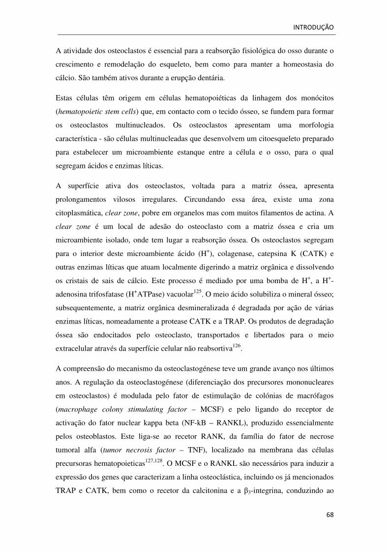
INTRODUÇÃO
68
A atividade dos osteoclastos é essencial para a reabsorção fisiológica do osso durante o
crescimento e remodelação do esqueleto, bem como para manter a homeostasia do
cálcio. São também ativos durante a erupção dentária.
Estas células têm origem em células hematopoiéticas da linhagem dos monócitos
(hematopoietic stem cells) que, em contacto com o tecido ósseo, se fundem para formar
os osteoclastos multinucleados. Os osteoclastos apresentam uma morfologia
característica - são células multinucleadas que desenvolvem um citoesqueleto preparado
para estabelecer um microambiente estanque entre a célula e o osso, para o qual
segregam ácidos e enzimas líticas.
A superfície ativa dos osteoclastos, voltada para a matriz óssea, apresenta
prolongamentos vilosos irregulares. Circundando essa área, existe uma zona
citoplasmática, clear zone, pobre em organelos mas com muitos filamentos de actina. A
clear zone é um local de adesão do osteoclasto com a matriz óssea e cria um
microambiente isolado, onde tem lugar a reabsorção óssea. Os osteoclastos segregam
para o interior deste microambiente ácido (H+), colagenase, catepsina K (CATK) e
outras enzimas líticas que atuam localmente digerindo a matriz orgânica e dissolvendo
os cristais de sais de cálcio. Este processo é mediado por uma bomba de H+, a H+-
adenosina trifosfatase (H+ATPase) vacuolar125. O meio ácido solubiliza o mineral ósseo;
subsequentemente, a matriz orgânica desmineralizada é degradada por ação de várias
enzimas líticas, nomeadamente a protease CATK e a TRAP. Os produtos de degradação
óssea são endocitados pelo osteoclasto, transportados e libertados para o meio
extracelular através da superfície celular não reabsortiva126.
A compreensão do mecanismo da osteoclastogénese teve um grande avanço nos últimos
anos. A regulação da osteoclastogénese (diferenciação dos precursores mononucleares
em osteoclastos) é modulada pelo fator de estimulação de colónias de macrófagos
(macrophage colony stimulating factor – MCSF) e pelo ligando do receptor de
activação do fator nuclear kappa beta (NF-kB – RANKL), produzido essencialmente
pelos osteoblastos. Este liga-se ao recetor RANK, da família do fator de necrose
tumoral alfa (tumor necrosis factor – TNF), localizado na membrana das células
precursoras hematopoieticas127,128. O MCSF e o RANKL são necessários para induzir a
expressão dos genes que caracterizam a linha osteoclástica, incluindo os já mencionados
TRAP e CATK, bem como o recetor da calcitonina e a β3-integrina, conduzindo ao
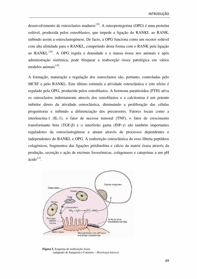
INTRODUÇÃO
69
desenvolvimento de osteoclastos maduros129. A osteoprotegerina (OPG) é uma proteína
solúvel, produzida pelos osteoblastos, que impede a ligação do RANKL ao RANK,
inibindo assim a osteoclastogénese. De facto, a OPG funciona como um recetor solúvel
com alta afinidade para o RANKL, competindo desta forma com o RANK pela ligação
ao RANKL 130 . A OPG regula a densidade e a massa óssea nos animais e após
administração sistémica, pode bloquear a reabsorção óssea patológica em vários
modelos animais129.
A formação, maturação e regulação dos osteoclastos são, portanto, controladas pelo
MCSF e pelo RANKL. Este último estimula a atividade osteoclástica e este efeito é
regulado pela OPG, produzida pelos osteoblastos. A hormona paratiroidea (PTH) ativa
os osteoclastos indiretamente através dos osteoblastos e a calcitonina é um potente
inibidor direto da atividade osteoclástica, diminuindo a proliferação das células
progenitoras e inibindo a diferenciação dos precursores. Fatores locais como a
interleucina-1 (IL-1), o fator de necrose tumoral (TNF), o fator de crescimento
transformante beta (TGF-β) e o interferão gama (INF-γ) são também importantes
reguladores da osteoclastogénese e atuam através de processos dependentes e
independentes do RANKL e OPG. A reabsorção osteoclástica do osso liberta peptídeos
colagénicos, fragmentos das ligações piridinolina e cálcio da matriz óssea através da
produção, secreção e ação de enzimas lisossómicas, colagenases e catepsinas a um pH
ácido131.
Figura 5. Esquema de reabsorção óssea (adaptado de Junqueira e Carneiro – Histologia básica)
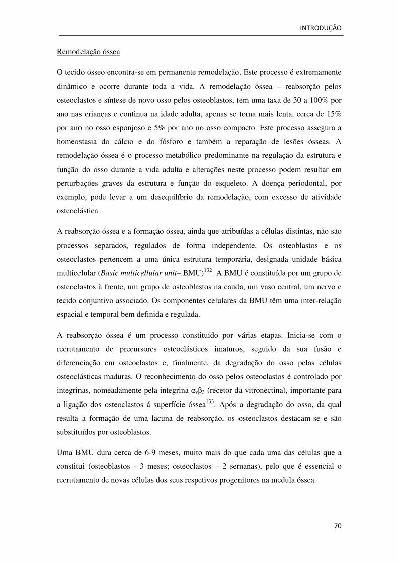
INTRODUÇÃO
70
Remodelação óssea
O tecido ósseo encontra-se em permanente remodelação. Este processo é extremamente
dinâmico e ocorre durante toda a vida. A remodelação óssea – reabsorção pelos
osteoclastos e síntese de novo osso pelos osteoblastos, tem uma taxa de 30 a 100% por
ano nas crianças e continua na idade adulta, apenas se torna mais lenta, cerca de 15%
por ano no osso esponjoso e 5% por ano no osso compacto. Este processo assegura a
homeostasia do cálcio e do fósforo e também a reparação de lesões ósseas. A
remodelação óssea é o processo metabólico predominante na regulação da estrutura e
função do osso durante a vida adulta e alterações neste processo podem resultar em
perturbações graves da estrutura e função do esqueleto. A doença periodontal, por
exemplo, pode levar a um desequilíbrio da remodelação, com excesso de atividade
osteoclástica.
A reabsorção óssea e a formação óssea, ainda que atribuídas a células distintas, não são
processos separados, regulados de forma independente. Os osteoblastos e os
osteoclastos pertencem a uma única estrutura temporária, designada unidade básica
multicelular (Basic multicellular unit– BMU)132. A BMU é constituída por um grupo de
osteoclastos à frente, um grupo de osteoblastos na cauda, um vaso central, um nervo e
tecido conjuntivo associado. Os componentes celulares da BMU têm uma inter-relação
espacial e temporal bem definida e regulada.
A reabsorção óssea é um processo constituído por várias etapas. Inicia-se com o
recrutamento de precursores osteoclásticos imaturos, seguido da sua fusão e
diferenciação em osteoclastos e, finalmente, da degradação do osso pelas células
osteoclásticas maduras. O reconhecimento do osso pelos osteoclastos é controlado por
integrinas, nomeadamente pela integrina αvβ3 (recetor da vitronectina), importante para
a ligação dos osteoclastos á superfície óssea133. Após a degradação do osso, da qual
resulta a formação de uma lacuna de reabsorção, os osteoclastos destacam-se e são
substituídos por osteoblastos.
Uma BMU dura cerca de 6-9 meses, muito mais do que cada uma das células que a
constitui (osteoblastos - 3 meses; osteoclastos – 2 semanas), pelo que é essencial o
recrutamento de novas células dos seus respetivos progenitores na medula óssea.
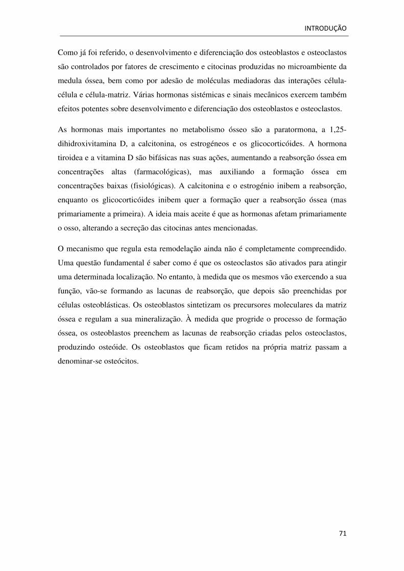
INTRODUÇÃO
71
Como já foi referido, o desenvolvimento e diferenciação dos osteoblastos e osteoclastos
são controlados por fatores de crescimento e citocinas produzidas no microambiente da
medula óssea, bem como por adesão de moléculas mediadoras das interações célula-
célula e célula-matriz. Várias hormonas sistémicas e sinais mecânicos exercem também
efeitos potentes sobre desenvolvimento e diferenciação dos osteoblastos e osteoclastos.
As hormonas mais importantes no metabolismo ósseo são a paratormona, a 1,25-
dihidroxivitamina D, a calcitonina, os estrogéneos e os glicocorticóides. A hormona
tiroidea e a vitamina D são bifásicas nas suas ações, aumentando a reabsorção óssea em
concentrações altas (farmacológicas), mas auxiliando a formação óssea em
concentrações baixas (fisiológicas). A calcitonina e o estrogénio inibem a reabsorção,
enquanto os glicocorticóides inibem quer a formação quer a reabsorção óssea (mas
primariamente a primeira). A ideia mais aceite é que as hormonas afetam primariamente
o osso, alterando a secreção das citocinas antes mencionadas.
O mecanismo que regula esta remodelação ainda não é completamente compreendido.
Uma questão fundamental é saber como é que os osteoclastos são ativados para atingir
uma determinada localização. No entanto, à medida que os mesmos vão exercendo a sua
função, vão-se formando as lacunas de reabsorção, que depois são preenchidas por
células osteoblásticas. Os osteoblastos sintetizam os precursores moleculares da matriz
óssea e regulam a sua mineralização. À medida que progride o processo de formação
óssea, os osteoblastos preenchem as lacunas de reabsorção criadas pelos osteoclastos,
produzindo osteóide. Os osteoblastos que ficam retidos na própria matriz passam a
denominar-se osteócitos.
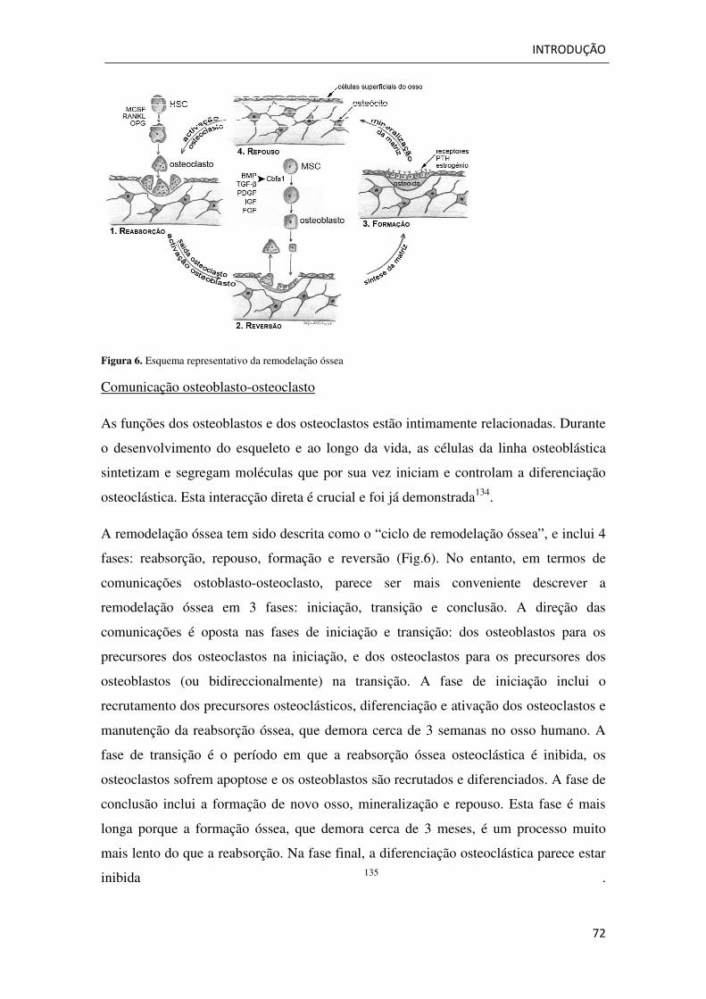
INTRODUÇÃO
72
Figura 6. Esquema representativo da remodelação óssea
Comunicação osteoblasto-osteoclasto
As funções dos osteoblastos e dos osteoclastos estão intimamente relacionadas. Durante
o desenvolvimento do esqueleto e ao longo da vida, as células da linha osteoblástica
sintetizam e segregam moléculas que por sua vez iniciam e controlam a diferenciação
osteoclástica. Esta interacção direta é crucial e foi já demonstrada134.
A remodelação óssea tem sido descrita como o “ciclo de remodelação óssea”, e inclui 4
fases: reabsorção, repouso, formação e reversão (Fig.6). No entanto, em termos de
comunicações ostoblasto-osteoclasto, parece ser mais conveniente descrever a
remodelação óssea em 3 fases: iniciação, transição e conclusão. A direção das
comunicações é oposta nas fases de iniciação e transição: dos osteoblastos para os
precursores dos osteoclastos na iniciação, e dos osteoclastos para os precursores dos
osteoblastos (ou bidireccionalmente) na transição. A fase de iniciação inclui o
recrutamento dos precursores osteoclásticos, diferenciação e ativação dos osteoclastos e
manutenção da reabsorção óssea, que demora cerca de 3 semanas no osso humano. A
fase de transição é o período em que a reabsorção óssea osteoclástica é inibida, os
osteoclastos sofrem apoptose e os osteoblastos são recrutados e diferenciados. A fase de
conclusão inclui a formação de novo osso, mineralização e repouso. Esta fase é mais
longa porque a formação óssea, que demora cerca de 3 meses, é um processo muito
mais lento do que a reabsorção. Na fase final, a diferenciação osteoclástica parece estar
inibida 135 .
73
Bibliografia
1Sabeti MA et al. Healing of Apical Periodontitis after Endodontic Treatment with and without obturation in dogs. J Endodon 2006; 32: 628-33. 2Tavares PBL et al. Prevalence of apical periodontitis in root canal treated teeth from an urban French population: influence of quality of root canal fillings and coronal restauration. J Endodon 2009; 35: 810-13. 3Kirkvang LL et al. Periapical status and quality of root fillings and coronal restorations in a Danish population. Int Endod J 2000; 33: 509-15. 4Hommez GMG, Coppens CRM, De Moor RJG. Periapical health related to the quality of coronal restorations and root fillings. Int Endod J 2002; 35:680-9. 5Saunders WP, Saunders EM. Coronal leakage as a cause of failure in root-canal therapy: a review. Endod Dent Traumatol 1994;10: 105- 8. 6Ray HA, Trope M. Periapical status of endodontically treated teeth in relation to the technical quality of the root filling and the coronal restoration. Int Endod J 1995; 28: 12-18. 7Riccucci et al. Periapical status of root-filled teeth exposed to the oral environment by loss of restoration or caries. Oral Surg Oral Med Oral Pathol 2000; 90: 354- 59. 8Tronstad L et al. Influence of coronal restorations on the periapical health of endodontically treated teeth. Endod Dent Traumatol 2000; 16: 218- 21. 9Kayahan MB et al. Periapical health related to the type of coronal restorations and quality of root canal fillings in a Turkish subpopulation. Oral Surg Oral Med Oral Pathl Oral Radiol Endodon 2008; 105: e58-e62. 10Segura-Egea JJ et al. Periapical status and quality of root canal fillings and coronal restorations in an adult Spanish population. Int Endod J 2004;37:525-30. 11Siqueira JF Jr et al. Periapical status related to the quality of coronal restorations and root canal fillings in a Brazilian population. Oral Surg Oral Med Oral Pathol Oral Radiol Endod 2005; 100:369-74. 12Chugal NM et al. Endodontic treatment outcome: effect of the permanent restoration. Oral Surg Oral Med Oral Pathl Oral Radiol Endodon 2007; 104: 576-82. 13Ng YL et al. Outcome of primary root canal treatment: systematic review of the literature – part 2. Influence of clinical factors. Int Endod J 2008; 41: 6-31. 14American Association of Endodontists. Obturation of root canal Systems. Endodontics: Colleagues for Excellence Fall 2009. 15Sundqvist G, Figdor D. Endodontic treatment of apical periodontitis. In: Ørstavik D, Pitt Ford TR, eds. Essencial Endodontology. Prevention and Treatment of Apical Periodontitis. Oxford: Blackwell, 1998. 16Grossman L. Endodontic Practic.Philadelphia, Lea &Febiger 1981. 17Harnden DG. Tests for carcinogenicity and mutagenicity. Int Endod J 1981;14:35. 18Lewis BB, Chestner SB. Formaldehyde in dentistry: a review of the mutagenic and carcinogenic potential. J Am Dent Assoc 1981;103:429-34. 19Grossman LI. A brief history of endodontics. J Endod 1982; 8: S36.
74
20 Ingle JI et al. The obturation of radicular space. In: Ingle, Bakland and Baumgartner edt. Ingle´s Endodontics 6th ed 2008; BC Becker Inc. 1053- 87. 21Lohbauer U et al. Calcium release and pH characteristics of calcium hydroxide plus points. Int Endod J 2005; 38: 683-689. 22 Lui JN et al. In vitro antimicrobial effect of clorohexidine impregnated guttapercha points on Entericoccus faecalis. Int Endod J 2004; 37: 105-117. 23Chogle S. et al. An in vitro assessment of iodoform gutta-percha. J Endod 2005; 31: 814-816. 24Spångberg L, Langeland K. Biological effects of dental materials: 1 Toxicity of root filling materials on HeLa cells in vitro. Oral Surg 1973; 35:402-14. 25Wolfson EM, Seltzer S. Reaction of rat connective tissue to some gutta-percha formulations. J Endodon 1975; 1: 395-402. 26Szep S et al. In vitro cytotoxicity of medicated and nonmedicated gutta-percha points in cultures of gingival fibroblasts. J Endodon 2003; 29: 36-40. 27 Pascon EA, Sångberg LS. In vitro cytotoxicity of root canal filling materials: 1. Gutta-percha. J Endodon 1990; 16:429-33. 28Siqueira Jr JF, Rôças IN and Lopes HP. Materiais obturadores. In: Gomes HP e Siqueira Jr JF eds. Endodontia – Biologia e Técnica. 2º ed. 2004; Editora Guanabara Koogan S.A.619-644. 29Schwartz RS. Adhesive dentistry and endodontics. Part 2: bonding in root canal system – the promise and the problems: a review. J Endodon 2006; 12: 1125-134. 30Tronstad L. Endodontic Instruments. In: Tronstad L ed. Clinical Endodontics 3thed 2009; Thieme, 158- 66. 31Guigand M et al. Evaluation of the cytocompatibility of three endodontic materials. J Endodon 1999; 25:419-23 32Segura JJ, Jiménez-Rubio A. Effect of eugenol on macrophage adhesion in vitro to plastic surfaces. Endod Dent Traumatol 1998; 14: 72-74. 33Markowitz K et al. Biological properties of eugenol and zinc oxide-eugenol. A clinical oriented review. Oral Surg 1992; 73: 729-37. 34Araki K et al. Reduced citotoxicity of a root canal sealer through eugenol substitution. J Endodon 1993; 19: 554-7. 35Hashieh IA et al. Concentration of eugenol apically released from zinc oxide-eugenol based sealers. J Endodon 1999; 25: 713-15. 36Meryon SD, Johnson SG and Smith AJ. Eugenol release and the cytotoxicity of different zinc oxide-eugenol combinations. J Dentist 1988; 16: 66-70. 37Chandrasekhar V et al. Expansion of gutta-percha in contact with various concentrations of zinc oxide-eugenol sealers: a three-dimensional volumetric study. J Endodon 2011; 37: 697-700. 38Macchi RL et al. Influence of endodontic materials on the bonding of composite resin to dentin. Endod Dent Traumatol 1992; 8: 26-9. 39Sunzel et al. The protective effect of zinc on rosin and resin acid toxicity in human polymorphonuclear leucocytes and human gingival fibroblasts in vitro. J Biomed Mater Res 1997; 37: 20-8.

75
40Leonardo MR et al. Release of formaldehyde by 4 endodontic sealers. Oral Surg Oral Med Oral Pathol 1999; 88: 221-25. 41WHO Publications Centre, 1999. IARC Monographs on the evaluation of carcinogenic risk of chemicals to man. Vol.71: reevaluation of some organic chemicals, hydrazine and hydrogen peroxide. Albany, NY. 42Geurtsen W, Leyhausen G. Biological aspects of root canal filling materials – histocompatibility, cytotoxicity and mutagenicity. Clin Oral Invest 1997; 1: 5-11. 43Spangberg LS, Barbosa SV, Lavigne GD. AH 26 releases formaldehyde. J Endodon 1993; 19: 596-98. 44Serper A et al. Comparative neurotoxic effects of root canal filling materials on rat sciatic nerve. J Endodon 1998; 24: 592-94. 45Erisen R, Yucel T, Kucukay S. Endomethasone root canal filling material in the mandibular canal: a case report. Oral Surg Oral Med Oral Pathol 1989; 63: 343- 5. 46Köseoğlu BG et al. Anesthesia following overfilling of a root canal sealer into the mandibular canal: a case report. Oral Surg Oral Med Oral Pathol Oral Radiol Endod 2006;101: 803-6. 47Oda K et al. Urticaria due to formaldehyde in a root canal dental-paste. Skin Reseach 2004; 3: 271- 74. 48Braun JJ et al. Anaphylactic reactions to formaldehyde in root canal sealant after endodontic treatment: four cases of anaphylactic shock and three of generalized urticaria. Allergy 2003; 58: 1210-5. 49Haïkel Y et al. Anaphylactic shock during endodontic treatment due to allergy to formaldehyde in a root canal sealant. J Endodon 2000; 26: 529-31. 50Modre B and Kränke. Anaphylactic reaction to formaldehyde. Allergy 2001; 56: 263- 64. 51Desai S and Chandler N. Calcium Hydroxide- based root canal sealers: a review. J Endodon 2009; 35: 475-80. 52Tronstad L, Barnett F, Flax M. Solubility and biocompatibility of calcium hydroxide-containing root canal sealers. Endodontics and Dental Traumatology 1988; 4: 152-9 53Brystom A, Claesson R, Sundqvist G. The antimicrobial effect of camphorated paramonochlorophenol, camphorated phenol and calcium hydroxide in treatment of infected root canals. Endod Dent Traumatol 1985; 1: 170- 5. 54Manhart M. The calcium hydroxide method of endodontic sealing. Oral Surg Oral Med Oral Pathol 1982; 54: 219- 24. 55Stock C. Calcium hydroxide: root resorption and perio-endo lesions. Br Dent J 1985; 158: 325- 34. 56Torneck C, Moe H and Howley T. The effect of calcium hydroxide on porcine pulp fibroblasts in vitro. J Endodon 1983; 9: 131- 6. 57Tronstad I et al. pH changes in dental tissue after root canal filling with calcium hydroxide. J Endodon 1981; 7: 17- 21. 58Hauman CHJ and Love RM. Biocompatibility of dental material used in contemporary endodontic therapy: a review. Part 1. Intracanal drugs and substances. Int Endod J 2003; 36: 75-85. 59Staehle HJ et al. Effect of root canal filling materials containing calcium hydroxide on the alkalinity of root dentin. Endodon and Dental Traumatol 1995; 11: 163-8. 60 Soares et al. Periapical tissue response to two calcium hydroxide-containing endodontic sealer. J Endodon 1990; 16: 166-9.
76
61Sleder FS et al. Long-term sealing ability of a calcium hydroxide sealer. J Endodon 1991; 17: 541- 43. 62Ersev H et al. Cytotoxic and mutagenic potencies of various root canal filling materials in eukaryotic and prokaryotic cells in vitro. J Endodon 1999; 25: 359-63. 63Osorio RM et al. Cytotoxicity of endodontic materials. J Endodon 1998; 24: 91-6. 64Mohammadi Z, Dummer PMH. Properties and applications of calcium hydroxide in endodontics and dental traumatology. Int Endodon J 2011; 44: 697-730. 65 Ørstavik D. Materials used for root canal obturation: technical, biological and clinical testing. Endodontic Topics 2005; 12: 25-38. 66Cohen BI et al. Formaldehyde evaluation from endodontic materials. Oral Health 1998; 88: 37- 39. 67Eveil et al. Release of formaldehyde from endodontic sealers. Asian J Chemis 2009; 21: 2091- 98. 68Pulgar R et al. The effect of AH 26 and AH Plus on MCF-7 breast cancer cell proliferation in vitro. Int Endon J 2002; 35: 551- 56. 69Witworth JM, Boursin EM. Dissolution of root canal sealers cements in volatile solvents. Int Endodon J 2000; 33: 19- 24. 70De Almeida WA et al. Evaluation of apical sealing of three endodontic sealers. Int Endodon J 2000; 33: 25- 7. 71 Schwant NW, Gound TG. Resorcinol-formaldehyde resin “Russian Red” endodontic therapy. J Endodon 2003; 29: 435-37. 72Gambrel MG et al. The effect of endodontic solutions on Resorcinol-Formalin paste in teeth. J Endodon 2005; 31: 25-29. 73Ho YC, Huang M and Chang YC. Cytotoxicity of formaldehyde on human osteoblastic cells is related to intracellular glutathione levels. J Biomed Mater Res Part B: Appl Biomat 2007; 83: 340- 44. 74 Tay FR et al. Effectiveness of resin-coated gutta-percha cones and a dual-cured, hydrophilic methacrylate resin-based sealer in obturating root canals. J Endodon 2005; 31: 659-64. 75Shipper G et al. An evaluation of microbial leakage in roots filled with a thermoplastic synthetic polymer-based root canal filling material (Resilon). J Endodon 2004; 30: 342-47. 76Heffernan ML et al. Clinical performance of Resilon and Gutta-Percha at 3 and 6 months. J Endodon 2004; 30: 342-47. 77Kim YK et al. Critical review on methacrylate resin-based root canal sealers. J Endodon 2010; 36: 383-99. 78Brisenõ BM, Willerhausen B. Root canal sealer cytotoxicity on human gingival fibroblasts: II. Silicone and resin based sealers. J Endodon 1991; 17: 537-40. 79Huumonen S et al. Healing of apical periodontitis after endodontic treatment: a comparison between a silicone-based and a zinc oxide-eugenol-based sealer. Int Endodon J 2003; 36: 296-301. 80 Ørstavik D, Nordahl I, Tibballs JE. Dimensional change following setting of root canal sealers materials. Dent Mater 2001; 17: 512- 519. 81Wu D et al. The sealing ability of a new silicone-based root canal filling material (GuttaFlow): an in vitro study using the percentage of gutta-percha-filled area. Dent Mat J 2011; 30: 569- 75.
77
82Friedman S et al. Evaluation of success and failure after endodontic therapy using a glass ionomer cement sealer. J Endodon 1995; 21: 384- 90. 83Schafer E, Zandbiglari T. Solubility of root canal sealers in water and artificial saliva. Int Endodon J 2003; 36: 660- 69. 84Perez CR, Hirata R Jr, Sergio PP. Evaluation of antimicrobial activity of fluoride releasing dental materials using a new in vitro method. Quintessence Int 2003; 34: 473-7. 85Coogan MM, Creeaven PJ. Antibacterial properties of eight dental cements. Int Endodon J 1993; 26: 355- 61. 86Willershausen B et al. Cytotoxicity of root canal filling material to three different human cell lines. J Endodon 2000; 26: 703- 7. 87Dahl J. E. Toxicity of endodontic filling materials. Endodontics topics 2005; 12:39-43. 88Khashaba RM, Chutkan NB, Borke JL. Comparative study of biocompatibility of newly developed calcium fosfate-based root canal sealers on fibroblasts derived from primary human gengiva and a mouse L929 cell line. Int Endodon J 2009; 42: 711-18. 89Khashaba RM et al. The response of subcutaneous connective tissue to newly developed calcium phosphate-based root canal sealers. Int Endodon J 2011; 44: 342-52. 90Zoufan K et al. Cytotoxicity evaluation of Gutta Flow and Endo Sequence BC sealers. Oral Surg Oral Med Oral Pathol Oral Radiol Endod 2011;112: 657-61. 91Chen CC et al. In vitro bioactivity and biocompatibility of dicalcium silicate cements for endodontic use. J Endodon 2009; 35: 1554- 57. 92Bryan TE et al. In vitro osteogenic potencial of an experimental calcium silicate-based root canal sealer. J Endodon 2010; 36: 1163- 69. 93Collares FM et al. Nanostructured hydroxyapatite as filler for methacrylate-based root canal sealers. Int Endodon J 2012;45:63- 7. 94Kuttler Y. Microscopic investigation of root apexes. J Am Dent Assoc 1955; 50: 544- 52. 95Figueiredo JA et al. The histological effects of four endodontic sealers implanted in the oral mucosa: submucous injection versus implant in polyethylene tubes. Int Endodon J 2001; 34: 377–85. 96Huang FM et al. Cytotoxicity of resin-, zinc oxide-eugenol-, and calcium hydroxide-based root canal sealers on human periodontal ligament cells and permanent V79 cells. Int Endodon J 2002; 35: 153–8. 97 Kaplan AE et al. Rheological properties and biocompatibility of endodontic sealers. Int Endodon J 2003; 36: 527–32. 98Miletić I, et al. The cytotoxicity of RoekoSeal and AH Plus compared during different setting periods. J Endodon 2005; 31: 307–9. 99Williams DF. Definitions in Biomaterials. 1987. Oxford: Elsevier. 100 Schmalz G. Concepts in biocompatibility testing of dental-restorative material. Clinical Oral Investigation 1997; 1: 182-5. 101Granchi D et al. Endodontic cements induce alterations in the cycle of in vitro cultured osteoblasts. Oral Surg Oral Med Oral Pathol Oral Radiol and Endodon 1995; 79: 359-66.
78
102 Pertot WJ et al. Model for quantitative immunohistochemical assessment of pulpar response to biomaterials. J Biomed Mater Res 1997; 15: 457-62. 103Matsumoto K, Inoue K, Matsumoto A. The effect of newly developed root canal sealers on rat dental pulp cells in primary cultures. J Endodon 1989; 15:60-7. 104Scmalz G. Use of cell cultures for cytotoxicity testing of dental materials – advantages and limitations. J Dentistry 1994; 22 (Suppl. 2): S6-11. 105Tiozzo R et al. Study of the potential cytotoxicity of dental impression materials. Toxicology in vitro 2003; 17:657-62. 106Al-Nazhan S, Spångberg L. Morphological cell changes due to chemical toxicity of a dental material: electron microscopic study on human periodontal ligament fibroblasts and L929 cells. J Endodon 1990; 16: 129-34. 107McNamara JR, Heithersay GS, WiebkinOW. Cell response to Hydron by a new in vitro method. Int Endodon J 1992; 25: 205-12. 108Barbosa SV, Burkard DH, Spångberg LSW. Cytotoxic effects of gutta-percha solvents. J Endodon 1994; 20: 6-8. 109Beltes P et al. In vitro evaluation of the cytotoxicity of calcium hydroxide-based root canal sealers. Endodon and Dent Traumatol 1995; 11: 245-9. 110Pelkonen O, Raunio H. Metabolic activation of toxins: tissue-specific expression and metabolism in target organs. Envir Health Perspect 1997; 105(Suppl); 767- 74. 111Heil J et al. Genotoxicity of dental materials. Mutation Research 1996; 368:181-94. 112Brzovic V et al. In vitro genotoxicity of root canal sealers. Int Endodon J 2009; 42: 253-63. 113Proskuryakov SA, Konoplyannikov AG, Gabai VL. Necrosis: a specific form of cell death? Experim Cell Research 2003; 1: 1-16. 114Aslan JE, Yhomas G. Death committee: organellar trafficking and communication in apoptosis. Traffic 2009; 10: 1390- 404. 115Guimarães CA, Linden R. Programmed cell death. Apoptosis and alternative death styles. Europ J Biochem 2004; 271: 1638-50. 116 Satchell PG, Gutmann JL, Witherspoon DE. Apoptosis: an introduction for the endodontist. Int Endodon J 2003; 36: 237- 45. 117Cate RT. Oral Histology – Development, struture and function. Chapter 7: Bone. 118Manolagas, SC. Birth and death of bone cells: basic regulatory mechanisms and implications for the pathogenesis and treatment of osteoporosis. Endocr Rev. 2000; 21:115-37. 119Wan M, Cao X. BMP signaling in skeletal development. Biochem Biophys Res Commun 2005; 328(3): 651-57. 120Chaudhary LR, Hofmeister AM, Hruska KA. Differential growth factor control of bone formation through osteoprogenitor differentiation. Bone.2004; 34(3): 402-11. 121Lehtonen-Veromaa M et al. A 1-year prospective study on the relationship between physical activity, markers of bone metabolism, and bone acquisition. J Clin Endocrinol Metab 2000; 85(10): 3726-32.
79
122Ducy P, Schinke T, Karsenty. The osteoblast: a sophisticated fibroblast under central surveillance. Science 2000; 289: 1501-4. 123Thomas T, Lafage-Proust MH. Contribution of genetically modified mouse models to the elucidation of bone physiology. Rev Rhum Engl 1999; 66 (12): 728-35. 124Lanyon, LE. Osteocytes, strain detection, bone modeling and remodeling. Calcif Tissue Int. 1993; 53 (sup.):102-7. 125Smith AN, Jouret F, Bord S et al. Vacuolar H+ ATPase d2 subunit: molecular characterization, developmental regulation, and localization to specialized proton pumps in kidney and bone. J Am Soc Nephrol. 2005; 16(5): 1245-56. 126Stenbeck G, Horton MA. Endocytic trafficking in actively resorbing osteoclasts. J Cell Sci 2004; 117(6): 827-36. 127Teitelbaum SL. Bone resorption by osteoclasts. Science 2000; 289: 1504-8. 128Karst M et al. Roles of stromal cell RANKL, OPG, and M-CSF expression in biphasic TGF-beta regulation of osteoclast differentiation. J Cell Physiol. 2004; 200(1): 99-106. 129Boyle WJ, Simonet WS and Lacey DL. Osteoclast differentiation and activation. Nature 2003; 423: 337- 42. 130Lacey DL et al. Osteoprotegerin ligand is a cytokine that regulates osteoclast differentiation and activation. Cell 1998; 93(2):165-76. 131Baynes JW, Dominiczak MH. In: Medical Biochemistry. Capítulo 24, Elsevier Mosby 2ª edição , 2005. 132Parfitt AM. Osteonal and hemi-osteonal remodeling: the spatial and temporal framework for signal traffic in adult human bone. J Cell Biochem 1994; 55: 273–86 133Faccio R et al. Activation of alpha beta 3 integrin on human osteoclast-like cells stimulates adhesion and migration in response to osteopontin. Biochem Biophys Res Commun 1998; 249(2): 522-5. 134Tortelli F et al. Osteoblast and osteoclast differentiation in an in vitro three-dimensional model of bone. Tissue Engenier 2009;15: 2373-83. 135Matsuo K, Irie N. Osteoclast-osteobalst communication. Arch Biochem Bioph 2008; 473: 201- 9.

80

CAPÍTULO I
Osteoblastic cytocompatibility of endodontic sealers extracts prepared according
to ISO standards and a root-dipping technique

82
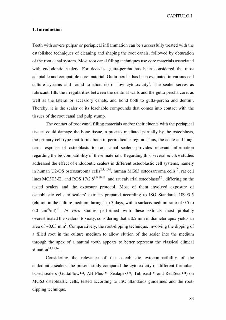
CAPÍTULO I
83
1. Introduction
Teeth with severe pulpar or periapical inflammation can be successfully treated with the
established techniques of cleaning and shaping the root canals, followed by obturation
of the root canal system. Most root canal filling techniques use core materials associated
with endodontic sealers. For decades, gutta-percha has been considered the most
adaptable and compatible core material. Gutta-percha has been evaluated in various cell
culture systems and found to elicit no or low cytotoxicity1. The sealer serves as
lubricant, fills the irregularities between the dentinal walls and the gutta-percha core, as
well as the lateral or accessory canals, and bond both to gutta-percha and dentin1.
Thereby, it is the sealer or its leachable compounds that comes into contact with the
tissues of the root canal and pulp stump.
The contact of root canal filling materials and/or their eluents with the periapical
tissues could damage the bone tissue, a process mediated partially by the osteoblasts,
the primary cell type that forms bone in periradicular region. Thus, the acute and long-
term response of osteoblasts to root canal sealers provides relevant information
regarding the biocompatibility of these materials. Regarding this, several in vitro studies
addressed the effect of endodontic sealers in different osteoblastic cell systems, namely
in human U2-OS osteosarcoma cells2,3,4,5,6, human MG63 osteosarcoma cells 7, rat cell
lines MC3T3-E1 and ROS 17/2.88,9,10,11 and rat calvarial osteoblasts12 , differing on the
tested sealers and the exposure protocol. Most of them involved exposure of
osteoblastic cells to sealers’ extracts prepared according to ISO Standards 10993-5
(elution in the culture medium during 1 to 3 days, with a surface/medium ratio of 0.5 to
6.0 cm2/ml)13. In vitro studies performed with these extracts most probably
overestimated the sealers’ toxicity, considering that a 0.2 mm in diameter apex yields an
area of ~0.03 mm2. Comparatively, the root-dipping technique, involving the dipping of
a filled root in the culture medium to allow elution of the sealer into the medium
through the apex of a natural tooth appears to better represent the classical clinical
situation14,15,16.
Considering the relevance of the osteoblastic cytocompatibility of the
endodontic sealers, the present study compared the cytotoxicity of different formulae-
based sealers (GuttaFlow™, AH Plus™, Sealapex™, Tubliseal™ and RealSeal™) on
MG63 osteoblastic cells, tested according to ISO Standards guidelines and the root-
dipping technique.
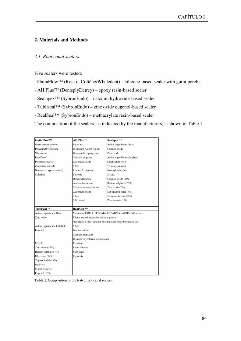
CAPÍTULO I
84
2. Materials and Methods
2.1. Root canal sealers
Five sealers were tested:
- GuttaFlow™ (Roeko, Colténe/Whaledent) – silicone-based sealer with gutta-percha
- AH Plus™ (DentsplyDetrey) – epoxy resin-based sealer
- Sealapex™ (SybronEndo) – calcium hydroxide-based sealer
- Tubliseal™ (SybronEndo) – zinc oxide eugenol-based sealer
- RealSeal™ (SybronEndo) – methacrylate resin-based sealer
The composition of the sealers, as indicated by the manufacturers, is shown in Table 1.
GuttaFlow™ AH Plus ™ Sealapex ™
Gutta-percha powder Paste A: Active ingredients- Base:
Polydimethylsiloxane Bisphenol-A epoxy resin Calcium oxide
Silicone oil Bisphenol-F epoxy resin Zinc oxide
Paraffin oil Calcium tungstate Active ingredients- Catalyst:
Platinum catalyst Zirconium oxide Disalicylate resin
Zirconium dioxide Silica Trisalicylate resin
Nano-silver (preservative) Iron oxide pigments Isobutyl salicylate
Coloring Paste B: Mixed:
Dibenzyldiamine Calcium oxide (24%)
Aminoadamantane Barium sulphate (20%)
Tricyclodecane-diamine Zinc oxide (7%)
Zirconium oxide Sub-micron silica (4%)
Silica Titanium dioxide (2%)
Silicone oil Zinc stearate (1%)
Tubliseal ™ RealSeal ™
Active ingredients- Base: Mixture of UDMA, PEGDMA, EBPADMA and BISGMA resins
Zinc oxide Silane-treated barlumborosilicate glasses *
*(contains a small amount of aluminium oxide barium sulfate)
Active ingredients- Catalyst: Silica
Eugenol Barium sulfate
Calcium hidroxide
Bismuth oxychloride with amines
Mixed: Peroxide
Zinc oxide (59%) Photo initator
Barium sulphate (4%) Stabilizers
Oleo resin (14%) Pigments
Thymol iodide (3%)
Oil 8(%)
Modifiers (2%)
Eugenol (10%) Table 1. Composition of the tested root canal sealers.
CAPÍTULO I
85
2.2. Preparation of the sealers’ extracts
ISO Standard technique
The sealers were mixed according to the manufacturer’s instructions under aseptic
conditions. With an insulin syringe, 0.3 ml of each sealer was placed at the bottom of
the well of a 24-well plate (Orange Scientific, Belgium) and the surface of the material
was smoothed. Immediately after, 1.5 ml of culture medium was added to each well.
The medium consisted of α-Minimal Essencial Medium (α-MEM, Gibco, UK), 10%
foetal bovine serum, 100 IU/ml penicillin, 2.5 µg/ml streptomycin, 2.5 µg/ml
amphotericin B and 50 µg/ml ascorbic acid. The plates were kept for 24h at 37ºC in a
humidified 5% CO2/air. Following, culture medium was collected, diluted (1:2, 1:5,
1:10 and 1:20), aliquoted and frozen at -20oC.
Root-dipping technique
Fifteen intact monoradicular teeth, freshly extracted, were used in this study. The
crowns were removed at the cementodentinal junction with a diamond disk under water
coolant. The root canals were prepared by the same operator at the cementodentinal
junction with X-Smart device (Maillefer) using Protaper files, following the sequence:
Sx, S1, S2; F1, F2 and F3. A 15 K-file was used between each protaper to verify the
apex patency. The irrigant was 3% NaOCl delivered with a needle, 2 ml between each
file size. The teeth were then sterilized at 135ºC for 35 min. After sterilization, the teeth
were randomly divided into 5 groups of 3 teeth and filled using the single cone
obturation technique. Four groups were filled with guta-percha cones corresponding to
F3 protaper file (Maillefer), and the sealer, namely Sealapex, Tubliseal, AHPlus or
GuttaFlow. The other group was filled with Resilon cones corresponding to F3 protaper
file, using RealSeal as a sealer. This was performed under sterile conditions in a laminar
flow hood. The apex of the roots was immediately dipped into 1.5 ml of culture medium
for 24h. The extracts were then prepared as in ISO standard technique.
2.3. Exposure of osteoblastic cells to the sealers’ extracts
Human MG63 osteoblastic cells were cultured (37ºC, 5% CO2 humidified atmosphere)
in α-MEM containing 10% foetal bovine serum, 100 IU/ml penicillin, 2.5 µg/ml
streptomycin, 2.5 µg/ml amphotericin B and 50 µg/ml ascorbic acid. At about 70-80%

CAPÍTULO I
86
confluence, cells were enzymatically detached (0.05% trypsin and 0.5 mM EDTA) and
re-suspended in culture medium. The cell suspension was used for the cytocompatibility
studies.
Cells were seeded (104 cells/cm2) into 96-well plates (Orange Scientific,
Belgium), and cultures were incubated for 24h, for cell adhesion. Following, the culture
medium was removed and replaced by one containing the sealers’ extracts (pure extract
and 1:2, 1:5, 1:10 and 1:20 dilutions) obtained by the two techniques. Culture medium
(containing the extracts) was replaced daily and cultures were characterized at days 1
and 3 for cell viability/proliferation.
In another set of experiments, cells were seeded at 103 cells/cm2 and, after the
24h adhesion period, cultures were exposed to the lowest concentration (1:20) of the
sealers’ extracts obtained by the two techniques, and maintained for 21 days. Culture
medium (containing the extracts) was replaced once a week and cultures were
characterized at days 7, 14 and 21 for cell viability/proliferation and alkaline
phosphatase (ALP) activity.
In both experiments, pH of the culture medium was assessed at each time-point.
Cultures performed in the absence of the extracts were used as control.
2.4. Cell viability/proliferation
Cell viability/proliferation was evaluated by the reduction of the tetrazolium salt MTT
[3-(4,5-dimethylthiazol-2-yl)-2,5-diphenyltetrazolium bromide], (0.5 mg/ml), by viable
cells, to form a purple formazan product, after 4h of incubation. The absorbance (A)
was measured at 600 nm in an ELISA plate reader (Synergy HT, Biotek), after crystals
solubilisation in DMSO.
2.5. ALP activity
Cell layers were solubilized (0.1% Triton X-100, 5 min), and ALP activity was
evaluated in cell lysates by the hydrolysis of p-nitrophenyl phosphate in alkaline buffer
solution (pH~10.3; 30 min, 37 ºC) and colorimetric determination of the product (p-
nitrophenol) at 400 nm in an ELISA plate reader (Synergy HT, Biotek). ALP activity
was normalized to total protein content (quantified by Bradford’s method) and
expressed as nmol/min/mgprotein-1.
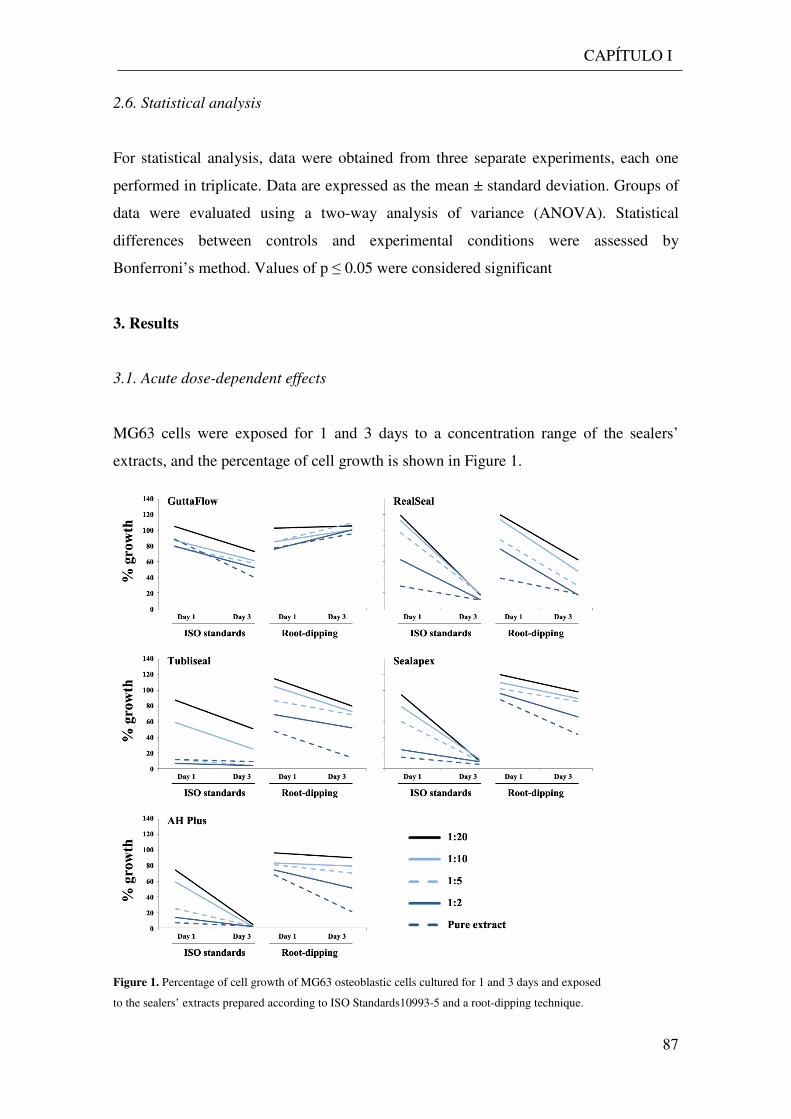
CAPÍTULO I
87
2.6. Statistical analysis
For statistical analysis, data were obtained from three separate experiments, each one
performed in triplicate. Data are expressed as the mean ± standard deviation. Groups of
data were evaluated using a two-way analysis of variance (ANOVA). Statistical
differences between controls and experimental conditions were assessed by
Bonferroni’s method. Values of p ≤ 0.05 were considered significant
3. Results
3.1. Acute dose-dependent effects
MG63 cells were exposed for 1 and 3 days to a concentration range of the sealers’
extracts, and the percentage of cell growth is shown in Figure 1.
Figure 1. Percentage of cell growth of MG63 osteoblastic cells cultured for 1 and 3 days and exposed
to the sealers’ extracts prepared according to ISO Standards10993-5 and a root-dipping technique.
CAPÍTULO I
88
3.1.1. Extracts prepared according to ISO Standards.
In the cultures exposed to GuttaFlow, cell growth was little affected at day 1 (~80%, at
levels ≥ 1:2), but decreased significantly at day 3 (~73% to 40%). AH Plus and
Sealapex caused a dose-dependent decrease on cell growth at day 1, and almost a total
inhibition at day 3. RealSeal elicited a small stimulation on cell proliferation at day 1 in
the presence of the lower concentrations (1:10 and 1:20, ~10 to 20%) and a dose-
dependent decrease at higher levels; at day 3, cell growth was very low within the tested
concentration range. In the presence of Tubliseal, toxicity also increased from day 1 to
day 3, and at levels ≥ 1:5 the extract caused rapid cell death. The pH of the culture
medium varied between 7.7 and 8.3 at day 1, except for the Sealapex extracts which
presented a pH ~9.5. Afterwards, pH decreased rapidly and, at day 3, pH varied
between 7.4 and 7.7, considering all the sealers.
3.1.2. Extracts prepared by the root-dipping technique.
GuttaFlow caused an initial inhibition (~20% at day 1, levels ≥ 1:2) but, afterwards, the
inhibitory effect decreased and, at day 3, values were similar to control. AH Plus caused
a dose-dependent decrease in the cell growth, and the inhibition was similar at days 1
and 3, except in the presence of the pure extract (inhibition higher at day 3). Sealapex,
Tubliseal and RealSeal elicited a slight increase in the cell growth at day 1, in the
presence of the lower concentrations (~10 to 20%, at 1:10 and 1:20), and a dose-
dependent decrease at higher levels; inhibition was higher at day 3, compared to day 1.
The pH of the culture medium remained around 7.4 during the 21-day culture
time.
3.2. Long-term exposure to low levels of the sealers’ extracts
Osteoblastic cell cultures were exposed for 21 days to the lowest tested concentration
(1:20) of the extracts, and were evaluated for cell viability/proliferation and ALP
activity, Figure 2. The sealers’ extracts prepared according to ISO Standards caused an
inhibition of the cell growth. The inhibition was total with AH Plus and partial with the
other sealers. At day 21, the percentages of inhibition were 12% (GuttaFlow), 18%
(Tubliseal), 29% (Sealapex) and 25% (RealSeal). Regarding ALP activity, a similar
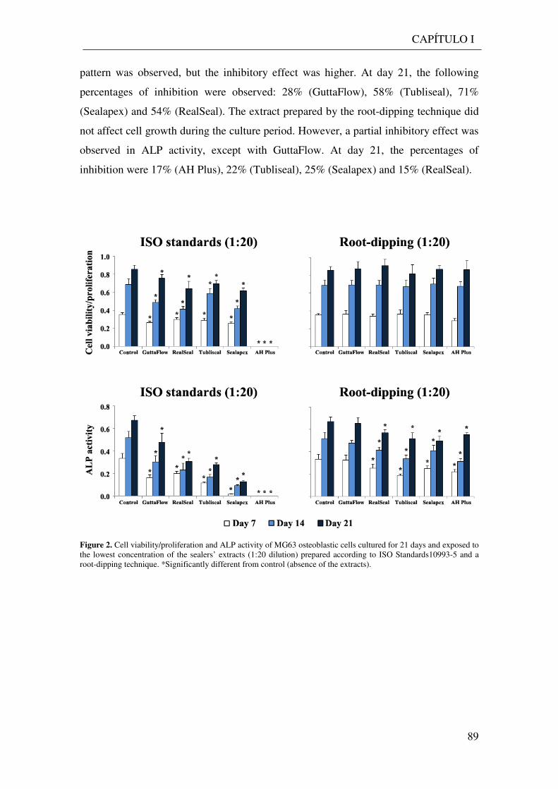
CAPÍTULO I
89
pattern was observed, but the inhibitory effect was higher. At day 21, the following
percentages of inhibition were observed: 28% (GuttaFlow), 58% (Tubliseal), 71%
(Sealapex) and 54% (RealSeal). The extract prepared by the root-dipping technique did
not affect cell growth during the culture period. However, a partial inhibitory effect was
observed in ALP activity, except with GuttaFlow. At day 21, the percentages of
inhibition were 17% (AH Plus), 22% (Tubliseal), 25% (Sealapex) and 15% (RealSeal).
Figure 2. Cell viability/proliferation and ALP activity of MG63 osteoblastic cells cultured for 21 days and exposed to the lowest concentration of the sealers’ extracts (1:20 dilution) prepared according to ISO Standards10993-5 and a root-dipping technique. *Significantly different from control (absence of the extracts).
CAPÍTULO I
90
4. Discussion
The contact of endodontic sealers and/or their leachable compounds with the periapical
tissues might cause deleterious effects in the local bone metabolism, and several in vitro
studies suggest that osteoblastic cells are a relevant target cell type for these effects1.
This work compared the behaviour of MG63 osteoblastic cells in the presence of the
sealers’ extracts prepared according to ISO Standards 10993-5 and by the root-dipping
technique14,15,16. The two methodologies provide distinct information regarding the
biological profile of endodontic sealers, as the extracts are obtained using a different
surface area/volume of culture medium. The extracts were prepared by incubating the
freshly mixed sealers with culture medium as, clinically, the sealer is applied after
mixing and the unset material/leachable compounds might contact immediately with the
local tissues.
During the first days after the sealer’ application, the local concentration of the
degradation products/leachable compounds is expected to be high, due to the favorable
concentration gradient. However, levels of these compounds decrease progressively due
to the continuous fluid flow and clearance, and, after a variable time-period, their
concentration is expected to be very low1. This is a relevant issue, as the endodontic
sealers remain in the local of application for many years. Thus, the characterization of
the cell response in the presence of low levels of the sealers’ degradation
products/leachable compounds for long periods might provide useful information in
predicting the long-term clinical performance of these materials. Considering this, two
set of experiments were performed. In the first one, osteoblastic cells were exposed to
high concentrations of the extracts, for 1 and 3 days, aiming to compare the dose-
dependent acute cell response to the extracts obtained by the two methodologies.
Following, the osteoblastic cell response was evaluated in cultures exposed for 21 days
to the lowest concentration of the extracts obtained by the two techniques.
Regarding the acute cell response, the sealers’ extracts, prepared according to
ISO Standards or by the root-dipping technique, caused significant cytotoxic effects.
GuttaFlow exhibited the lowest toxicity compared to the other sealers. Evident dose-
dependent inhibitory effects on cell viability/proliferation were observed at day 1 for
most of the sealers. The small stimulatory effect seen with low concentrations of
RealSeal (ISO 10993-5) or Sealapex, Tubliseal and RealSeal (root-dipping technique)
reflects most probably an adaptive response to the presence of toxic compounds.
CAPÍTULO I
91
Cytotoxicity was time-dependent, as the inhibition was higher at day 3, compared to
that on day 1. These results suggest that the continuous exposure to the sealers leachable
compounds caused cumulative toxic effects. The only exception was the extract from
GuttaFlow prepared by the root-dipping technique. In this case, cells were able to
recover from the initial deleterious effects and, at day 3, cell response was similar to
control.
In the long-term cell response to the lowest concentration of the two extracts,
significant differences were noticed. Cell viability/proliferation and, to a more extent,
ALP activity were significantly decreased in the cultures exposed to the extract prepared
according to ISO Standards. The extract prepared by the root-dipping technique did not
affect cell viability/proliferation, but decreased partially ALP activity. Thus, synthesis
of ALP was more sensitive to the deleterious effects of the sealers’ extracts than cell
viability/proliferation. This is a relevant issue, as ALP is an early marker of osteogenic
differentiation, and has a determinant role in the bone tissue, by providing phosphate
ions that, with calcium ions, are used in the formation of the cell-mediated calcium
phosphate mineralized matrix17.
In spite of the similarities in the pattern of the cell response to the two types of
extracts, those prepared according to ISO 10993-5 caused significantly higher
quantitative cytotoxic effects. This behaviour is expected considering that the ratio
surface area/volume of culture medium was ~2 cm2/1.5 ml and ~0.03 mm2/1.5 ml,
respectively in the ISO 10993-5 and the root-dipping technique and, thus, the
percentage of the leachable compounds in the extract is expected to be significantly
different in the two extracts.
Few studies were performed in osteoblastic cells involving the sealers tested in
the present work. AH Plus, an epoxy resin-based sealer apparently “formaldehyde-free”,
appeared to be cytotoxic for human U2-OS osteosarcoma cells2,3. Another study,
performed in the osteoblast rat cell lines MC3T3-E1 and ROS 17/2.8, showed that the
toxicity of AH Plus decreased with the aging of the sealer11 and that these cell lines
responded differently to this sealer10. It was also shown that this sealer exhibits a
moderate to severe toxicity immediately after mixing (the conditions used in the present
work) in fibroblasts10,18,19,20,21,22,23,24,25 and human periodontal ligament cells 26. It has
been associated to the release of low levels of formaldehyde, the presence of amines
added to accelerate the epoxy polymerization and the epoxy resin content20,27. Extracts
of Sealapex, a calcium hydroxide based sealer, were toxic to rat calvarial osteoblasts12
CAPÍTULO I
92
and MC3T3-E1 osteoblast cells8, and also to fibroblasts23,28 and human periodontal
ligament cells26,29; its toxicity might involve the increase in the pH of the culture
medium resulting from the sealer dissolution20,30,31. In the present work, a partial
contribution of this effect might be hypothesized to explain the deleterious effect
observed with the extracts prepared according to ISO Standards. However, the pH
remained constant throughout the culture time in the extracts obtained by the root-
dipping technique, thus other components are involved in the observed cytotoxicity, and
polyresin, zinc oxide and Ba2+ have also been suggested as potential toxic compounds31.
Realseal was also evaluated in osteoblast cells. Disks of RealSeal, after setting, were
placed in direct contact with ROS 17/2.8 cells for 5 succeeding weeks after immersion
in simulated body fluid, and results showed that toxicity decreased gradually over time9.
In another study, RealSeal extracts showed initial cytotoxicity effect on MG63 cells
which disappeared after few days of culture7. In addition, this sealer was shown to be
rather toxic in fibroblasts10,16,18,19,21,28,32,33,34 and human dental pulp cells34. Toxicity of
this dual-cured hydrophilic multi-methacrylate resin-based sealer appears to be caused
by the leaching of unreacted monomers as a result of incomplete polymerization, and
also to filler particles due to the degradation of the sealer. The toxicity of Tubliseal, a
zinc oxide eugenol-based sealer, was demonstrated in fibroblasts24 and human
periodontal ligament cells29, and has been attributed to free eugenol release, and also
several additives, namely resins used for greater dentin adhesion31. The low cytotoxicity
of GuttaFlow observed in the present work is in line with studies involving
fibroblasts18,19,24,25,33. This is a silicone-based sealer manufactured by adding gutta-
percha powder to the silicone matrix (and containing nanosilver particles as a
preservative). Toxicity might result from the release of nanosilver particles, as they have
been associated with dose-dependent cytotoxic effects31. Also, the presence of small
voids within the core of GuttaFlow favors the release of unreacted compounds that
accumulates in such pores35.
The studies referenced above differed in the target cell type, preparation of the
extracts, tested concentrations and time of exposure, which is reflected by a great results
discrepancy on cytotoxicity patterns or the effects on a specific cell type. Most of them
involved fibroblastic cells, short exposures and high levels of the sealers extracts.
Nevertheless, the results of the present work are in line with the reported studies, which
showed mostly in vitro cytotoxicity of the endodontic sealers. However, to the best of
our knowledge, this is the first study to compare the acute and long-term osteoblastic
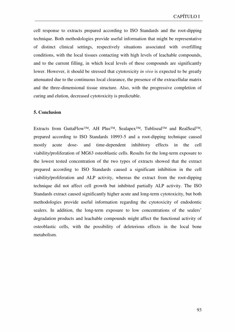
CAPÍTULO I
93
cell response to extracts prepared according to ISO Standards and the root-dipping
technique. Both methodologies provide useful information that might be representative
of distinct clinical settings, respectively situations associated with overfilling
conditions, with the local tissues contacting with high levels of leachable compounds,
and to the current filling, in which local levels of these compounds are significantly
lower. However, it should be stressed that cytotoxicity in vivo is expected to be greatly
attenuated due to the continuous local clearance, the presence of the extracellular matrix
and the three-dimensional tissue structure. Also, with the progressive completion of
curing and elution, decreased cytotoxicity is predictable.
5. Conclusion
Extracts from GuttaFlow™, AH Plus™, Sealapex™, Tubliseal™ and RealSeal™,
prepared according to ISO Standards 10993-5 and a root-dipping technique caused
mostly acute dose- and time-dependent inhibitory effects in the cell
viability/proliferation of MG63 osteoblastic cells. Results for the long-term exposure to
the lowest tested concentration of the two types of extracts showed that the extract
prepared according to ISO Standards caused a significant inhibition in the cell
viability/proliferation and ALP activity, whereas the extract from the root-dipping
technique did not affect cell growth but inhibited partially ALP activity. The ISO
Standards extract caused significantly higher acute and long-term cytotoxicity, but both
methodologies provide useful information regarding the cytotoxicity of endodontic
sealers. In addition, the long-term exposure to low concentrations of the sealers’
degradation products and leachable compounds might affect the functional activity of
osteoblastic cells, with the possibility of deleterious effects in the local bone
metabolism.
94
References
1Dahl JE. Toxicity of endodontic filling materials. Endodontic topics 2005; 12:39-43. 2Huang FM, Chou MY and Chang YC. Induction of cyclooxygenase-2 mRNA and protein expression by epoxy resin and zinc oxide-eugenol based root canal sealers in human osteoblastic cells. Biomater 2003; 24: 1869-75. 3Huang FM et al. Induction of interleukin-6 and interleukin-8 gene expression by root canal sealers in human osteoblastic cells. J Endodon 2005; 31: 679-83. 4Huang FM, Yang S-F and Chang Y-C. Up-regulation of gelatinases and tissue type plasminogen activator by root canal sealers in human osteoblastic cells. J Endodon 2008; 34: 291-4. 5Huang FM et al. Up-regulation of receptor activator nuclear factor-kappa B ligand expression by root canal sealers in human osteoblastic cells. J Endodon 2009; 35: 363–6. 6Huang FM, Yang S-F and Chang Y-C. Effects of root canal sealers on alkaline phosphatase in human osteoblastic cells. J Endodon 2010; 36: 1230-3. 7Xu P et al. Cytotoxicity of RealSeal on human osteoblast like MG63 cells. J Endodon 2010;36: 40- 4. 8Lee DH et al. Mechanisms of root canal sealers cytotoxicity on osteoblastic cell line MC3T3-E1. Oral Surg Oral Med Oral Pathol Oral Radiol and Endodon 2007;104: 717- 21. 9Ames JM et al. Contemporary methacrylate resin-based root canal sealers exhibit different degrees of ex vivo cytotoxicity when cured in their self-cured mode. J Endodon 2009; 35: 225- 8. 10Brackett MG et al. Cytotoxic response of three cell lines exposed in vitro to dental endodontic sealers. J Biomed Mater Research B: Applied Biomaterials 2010; 95B: 380- 6. 11Bryan TE et al. In vitro osteogenic potencial of an experimental calcium silicate-based root canal sealer. J Endodon 2010, 36: 1163-9. 12Al-Awadhi S et al. Cultured primary osteoblasts viability and apoptosis in the presence of root canal sealers. J Endodon 2004;30: 527- 33. 13ISO standards 10993-5. Biological evaluation of medical devices. Tests for cytotoxicity: in vitro methods. 14Camps J, About I. Cytotoxicity testing of endodontic sealers: a new method. J Endodon 2003; 29: 583-6. 15Schwarze T, Leyhausen G, Geurtsen W. Long-term cytocompatibility of various endodontic sealers using a new root canal model. J Endodon 2002; 28: 749-53. 16Susini G et al. Cytotoxicity of Epiphany and Resilon with a root model. Int Endodon J 2006; 39: 940-4. 17Aubin J. “Mesenchymal stem cell and osteoblast differentiation”, in Principles of bone biology, eds. Bilezikian JP, Raisz LG, Martin TJ, Academic Press 2008; 85-107. 18Bouillaguet S et al. Initial in vitro biological response to contemporary endodontic sealers. J Endodon 2006; 32: 989-92. 19Brackett MG et al. Cytotoxicity of endodontic materials over 6-weeks ex vivo. Int Endodon J 2008; 41: 1072-8. 20Eldeniz AU et al. Evaluation of pH and ion release of Acroseal sealer in comparison with Apexit and Sealapex sealers. Oral Surg Oral Med Oral Pathol Oral Radiol and Endodon 2007;103(3): e86- e91.
95
21Lodienè G et al. Toxicity evaluation of root canal sealers in vitro. Int Endodon J 2008; 41: 72-7. 22Miletić I et al. The cytotoxicity of RoekoSeal and AH Plus Compared during different setting periods. J Endodon 2005; 31: 307-9. 23Silva EJ et al. Evaluation of cytotoxicity and up-regulation of gelatinases in human fibroblast cells by four root canal sealers. Int Endodon J 2012; 45: 49-56. 24Zoufan K et al. Cytotoxicity evaluation of GuttaFlow and Endo Sequence BC sealers. Oral Surg Oral Med Oral Pathol Oral Radiol and Endodon 2011;112: 657-61. 25Willershausen I et al. In vitro analysis of the cytotoxicity and the antimicrobial effect of four endodontic sealers. Head & Face Medicine 2011; 7: 15-23. 26Huang FM et al. Cytotoxicity of resin-, zinc oxide eugenol-, and calcium hydroxide-based root canal sealers on human periodontal ligament cells and permanent V79 cells. Int Endodon J 2002; 35: 153- 8. 27Merdad K et al. Short term cytotoxicity assessment of components of the Epiphany resin-percha obturating system by indirect and direct Millipore filter assays. J Endodon 2007;33: 24- 7. 28Key JE, Rahemtulla FG, Eleazer PD. Cytotoxicity of new root canal filling material on human gingival fibroblasts. J Endodon 2006; 32: 756-8. 29Chang MC et al. Comparative cytotoxicity of five root canal sealers on cultured human periodontal ligament fibroblast. Int Endodon J 2010; 43: 251-7. 30Duarte MA et al. Evaluation of pH and calcium ion release of three root canal sealers. J Endodon 2000; 26: 389-90. 31Hauman CH, Love RM. Biocompatibility of dental materials used in contemporary endodontic therapy: a review. Part 2. Root canal filling materials. Int Endodon J 2003; 36: 147-60. 32Donadio M et al. Cytotoxicity evaluation of Activ GP and Resilon sealers in vitro. Oral Surg Oral Med Oral Pathol Oral Radiol and Endodon 2009; 107: e74- e78. 33Eldeniz AU et al. Cytotoxicity of new resin-, calcium hydroxide-, and silicone-based root canal sealers on fibroblasts derived from human gingival and L929 cell lines. Int Endodon J 2007; 40: 329-37. 34Karapinar-Kazandağ M et al. Cytotoxicity of 5 endodontic sealers on L929 cell line and human dental pulp cells. Int Endodon J 2011; 44: 626- 34. 35Elayouti A et al. Homogeneity and adaptation of a new gutta-percha paste to root canal walls. J Endodon 2005; 31: 687-90.

96

CAPÍTULO II
Long-term dose and time-dependent cytotoxicity profile of endodontic sealers in
human in vitro osteoclastogenesis

98
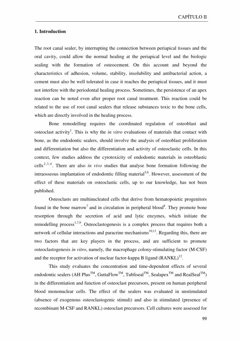
CAPÍTULO II
99
1. Introduction
The root canal sealer, by interrupting the connection between periapical tissues and the
oral cavity, could allow the normal healing at the periapical level and the biologic
sealing with the formation of osteocement. On this account and beyond the
characteristics of adhesion, volume, stability, insolubility and antibacterial action, a
cement must also be well tolerated in case it reaches the periapical tissues, and it must
not interfere with the periodontal healing process. Sometimes, the persistence of an apex
reaction can be noted even after proper root canal treatment. This reaction could be
related to the use of root canal sealers that release substances toxic to the bone cells,
which are directly involved in the healing process.
Bone remodelling requires the coordinated regulation of osteoblast and
osteoclast activity1. This is why the in vitro evaluations of materials that contact with
bone, as the endodontic sealers, should involve the analysis of osteoblast proliferation
and differentiation but also the differentiation and activity of osteoclastic cells. In this
context, few studies address the cytotoxicity of endodontic materials in osteoblastic
cells2,3 ,4 . There are also in vivo studies that analyse bone formation following the
intraosseous implantation of endodontic filling material5,6. However, assessment of the
effect of these materials on osteoclastic cells, up to our knowledge, has not been
published.
Osteoclasts are multinucleated cells that derive from hematopoietic progenitors
found in the bone marrow7 and in circulation in peripheral blood8. They promote bone
resorption through the secretion of acid and lytic enzymes, which initiate the
remodelling process1,7,9. Osteoclastogenesis is a complex process that requires both a
network of cellular interactions and paracrine mechanisms10,11. Regarding this, there are
two factors that are key players in the process, and are sufficient to promote
osteoclastogenesis in vitro, namely, the macrophage colony-stimulating factor (M-CSF)
and the receptor for activation of nuclear factor-kappa B ligand (RANKL)12.
This study evaluates the concentration and time-dependent effects of several
endodontic sealers (AH PlusTM, GuttaFlowTM, TublisealTM, SealapexTM and RealSealTM)
in the differentiation and function of osteoclast precursors, present on human peripheral
blood mononuclear cells. The effect of the sealers was evaluated in unstimulated
(absence of exogenous osteoclastogenic stimuli) and also in stimulated (presence of
recombinant M-CSF and RANKL) osteoclast precursors. Cell cultures were assessed for
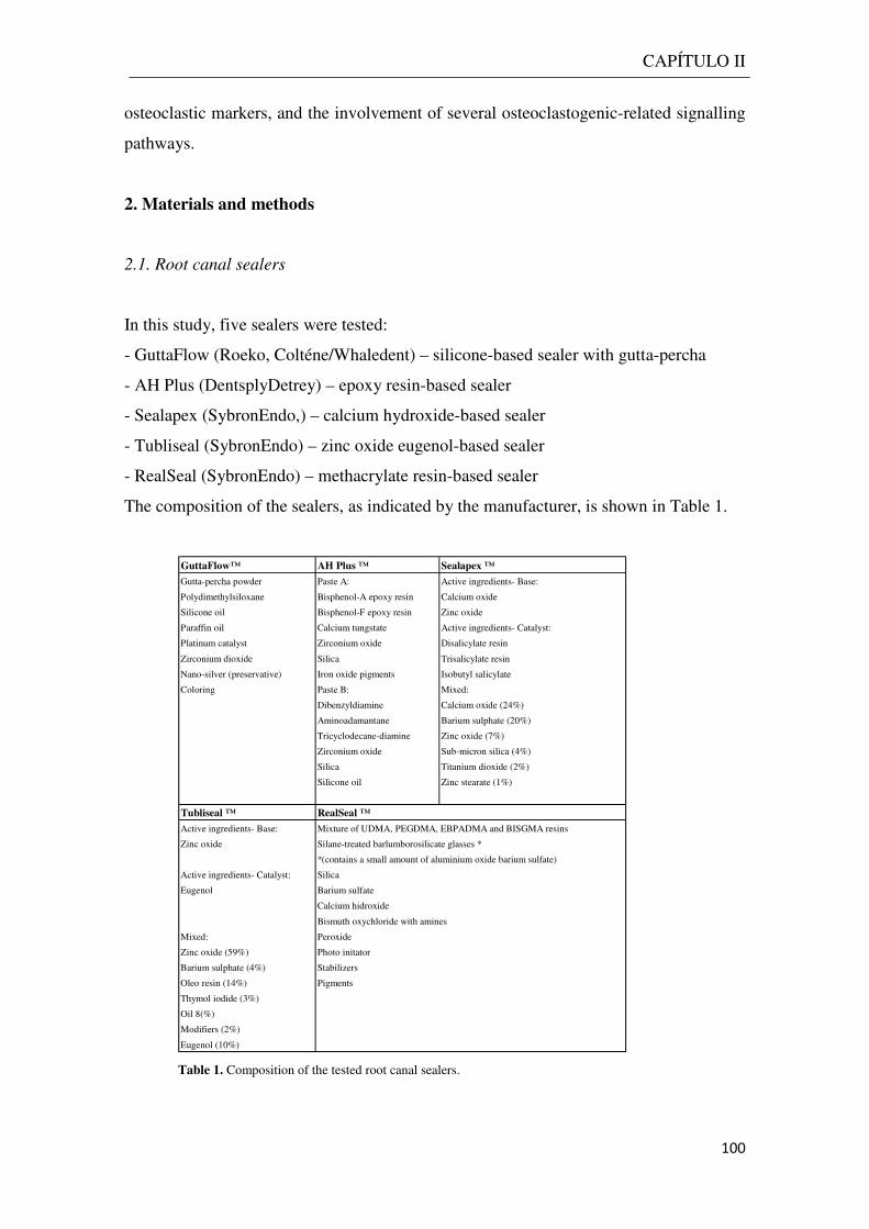
CAPÍTULO II
100
osteoclastic markers, and the involvement of several osteoclastogenic-related signalling
pathways.
2. Materials and methods
2.1. Root canal sealers
In this study, five sealers were tested:
- GuttaFlow (Roeko, Colténe/Whaledent) – silicone-based sealer with gutta-percha
- AH Plus (DentsplyDetrey) – epoxy resin-based sealer
- Sealapex (SybronEndo,) – calcium hydroxide-based sealer
- Tubliseal (SybronEndo) – zinc oxide eugenol-based sealer
- RealSeal (SybronEndo) – methacrylate resin-based sealer
The composition of the sealers, as indicated by the manufacturer, is shown in Table 1.
Table 1. Composition of the tested root canal sealers.
GuttaFlow™ AH Plus ™ Sealapex ™
Gutta-percha powder Paste A: Active ingredients- Base:
Polydimethylsiloxane Bisphenol-A epoxy resin Calcium oxide
Silicone oil Bisphenol-F epoxy resin Zinc oxide
Paraffin oil Calcium tungstate Active ingredients- Catalyst:
Platinum catalyst Zirconium oxide Disalicylate resin
Zirconium dioxide Silica Trisalicylate resin
Nano-silver (preservative) Iron oxide pigments Isobutyl salicylate
Coloring Paste B: Mixed:
Dibenzyldiamine Calcium oxide (24%)
Aminoadamantane Barium sulphate (20%)
Tricyclodecane-diamine Zinc oxide (7%)
Zirconium oxide Sub-micron silica (4%)
Silica Titanium dioxide (2%)
Silicone oil Zinc stearate (1%)
Tubliseal ™ RealSeal ™
Active ingredients- Base: Mixture of UDMA, PEGDMA, EBPADMA and BISGMA resins
Zinc oxide Silane-treated barlumborosilicate glasses *
*(contains a small amount of aluminium oxide barium sulfate)
Active ingredients- Catalyst: Silica
Eugenol Barium sulfate
Calcium hidroxide
Bismuth oxychloride with amines
Mixed: Peroxide
Zinc oxide (59%) Photo initator
Barium sulphate (4%) Stabilizers
Oleo resin (14%) Pigments
Thymol iodide (3%)
Oil 8(%)
Modifiers (2%)
Eugenol (10%)
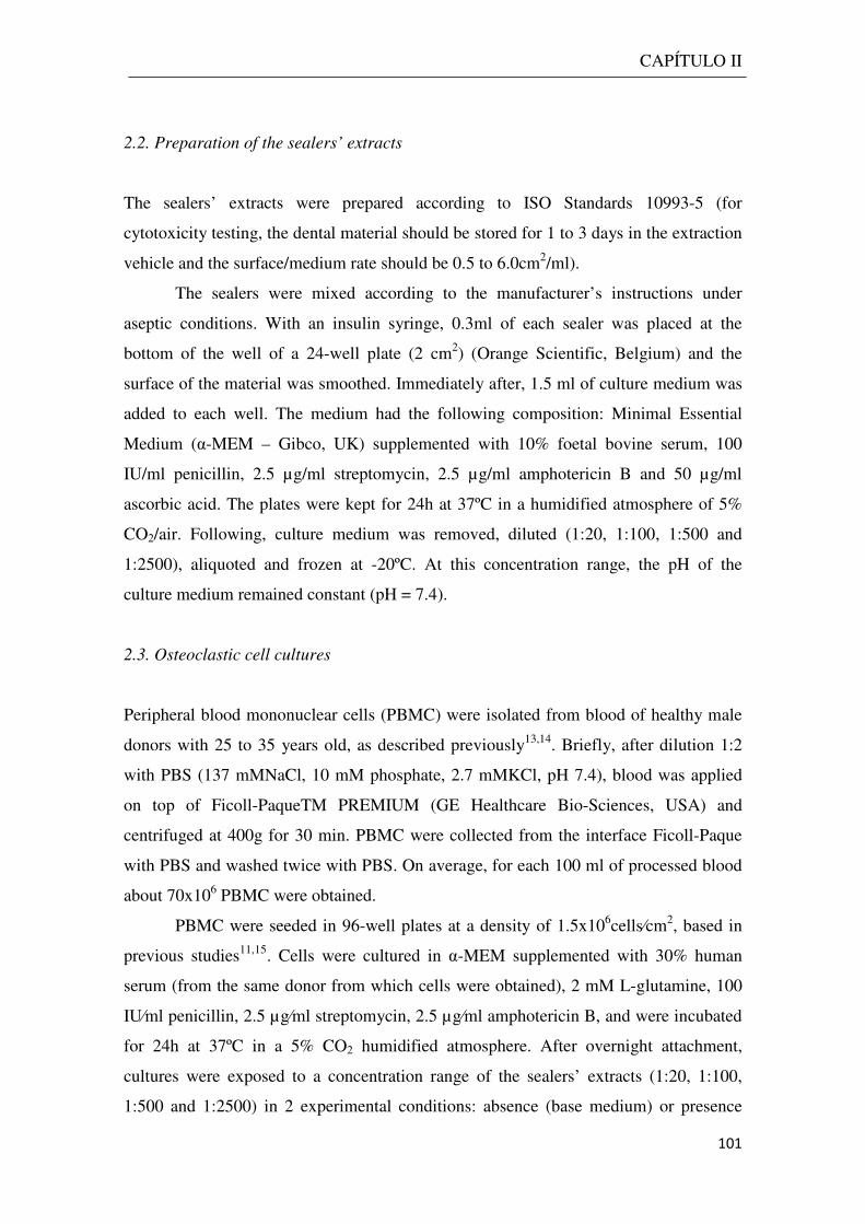
CAPÍTULO II
101
2.2. Preparation of the sealers’ extracts
The sealers’ extracts were prepared according to ISO Standards 10993-5 (for
cytotoxicity testing, the dental material should be stored for 1 to 3 days in the extraction
vehicle and the surface/medium rate should be 0.5 to 6.0cm2/ml).
The sealers were mixed according to the manufacturer’s instructions under
aseptic conditions. With an insulin syringe, 0.3ml of each sealer was placed at the
bottom of the well of a 24-well plate (2 cm2) (Orange Scientific, Belgium) and the
surface of the material was smoothed. Immediately after, 1.5 ml of culture medium was
added to each well. The medium had the following composition: Minimal Essential
Medium (α-MEM – Gibco, UK) supplemented with 10% foetal bovine serum, 100
IU/ml penicillin, 2.5 µg/ml streptomycin, 2.5 µg/ml amphotericin B and 50 µg/ml
ascorbic acid. The plates were kept for 24h at 37ºC in a humidified atmosphere of 5%
CO2/air. Following, culture medium was removed, diluted (1:20, 1:100, 1:500 and
1:2500), aliquoted and frozen at -20ºC. At this concentration range, the pH of the
culture medium remained constant (pH = 7.4).
2.3. Osteoclastic cell cultures
Peripheral blood mononuclear cells (PBMC) were isolated from blood of healthy male
donors with 25 to 35 years old, as described previously13,14. Briefly, after dilution 1:2
with PBS (137 mMNaCl, 10 mM phosphate, 2.7 mMKCl, pH 7.4), blood was applied
on top of Ficoll-PaqueTM PREMIUM (GE Healthcare Bio-Sciences, USA) and
centrifuged at 400g for 30 min. PBMC were collected from the interface Ficoll-Paque
with PBS and washed twice with PBS. On average, for each 100 ml of processed blood
about 70x106 PBMC were obtained.
PBMC were seeded in 96-well plates at a density of 1.5x10⁶cells⁄cm2, based in
previous studies11,15. Cells were cultured in α-MEM supplemented with 30% human
serum (from the same donor from which cells were obtained), 2 mM L-glutamine, 100
IU⁄ml penicillin, 2.5 µg⁄ml streptomycin, 2.5 µg⁄ml amphotericin B, and were incubated
for 24h at 37ºC in a 5% CO2 humidified atmosphere. After overnight attachment,
cultures were exposed to a concentration range of the sealers’ extracts (1:20, 1:100,
1:500 and 1:2500) in 2 experimental conditions: absence (base medium) or presence
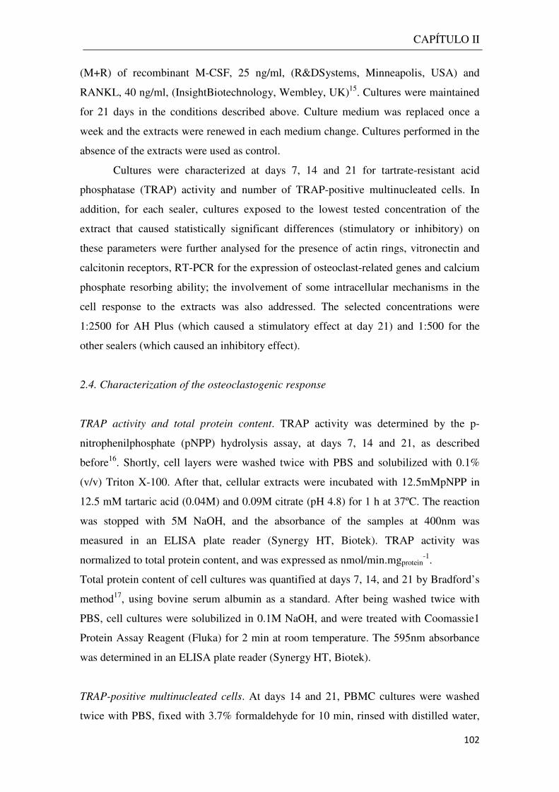
CAPÍTULO II
102
(M+R) of recombinant M-CSF, 25 ng/ml, (R&DSystems, Minneapolis, USA) and
RANKL, 40 ng/ml, (InsightBiotechnology, Wembley, UK)15. Cultures were maintained
for 21 days in the conditions described above. Culture medium was replaced once a
week and the extracts were renewed in each medium change. Cultures performed in the
absence of the extracts were used as control.
Cultures were characterized at days 7, 14 and 21 for tartrate-resistant acid
phosphatase (TRAP) activity and number of TRAP-positive multinucleated cells. In
addition, for each sealer, cultures exposed to the lowest tested concentration of the
extract that caused statistically significant differences (stimulatory or inhibitory) on
these parameters were further analysed for the presence of actin rings, vitronectin and
calcitonin receptors, RT-PCR for the expression of osteoclast-related genes and calcium
phosphate resorbing ability; the involvement of some intracellular mechanisms in the
cell response to the extracts was also addressed. The selected concentrations were
1:2500 for AH Plus (which caused a stimulatory effect at day 21) and 1:500 for the
other sealers (which caused an inhibitory effect).
2.4. Characterization of the osteoclastogenic response
TRAP activity and total protein content. TRAP activity was determined by the p-
nitrophenilphosphate (pNPP) hydrolysis assay, at days 7, 14 and 21, as described
before16. Shortly, cell layers were washed twice with PBS and solubilized with 0.1%
(v/v) Triton X-100. After that, cellular extracts were incubated with 12.5mMpNPP in
12.5 mM tartaric acid (0.04M) and 0.09M citrate (pH 4.8) for 1 h at 37ºC. The reaction
was stopped with 5M NaOH, and the absorbance of the samples at 400nm was
measured in an ELISA plate reader (Synergy HT, Biotek). TRAP activity was
normalized to total protein content, and was expressed as nmol/min.mgprotein-1.
Total protein content of cell cultures was quantified at days 7, 14, and 21 by Bradford’s
method17, using bovine serum albumin as a standard. After being washed twice with
PBS, cell cultures were solubilized in 0.1M NaOH, and were treated with Coomassie1
Protein Assay Reagent (Fluka) for 2 min at room temperature. The 595nm absorbance
was determined in an ELISA plate reader (Synergy HT, Biotek).
TRAP-positive multinucleated cells. At days 14 and 21, PBMC cultures were washed
twice with PBS, fixed with 3.7% formaldehyde for 10 min, rinsed with distilled water,
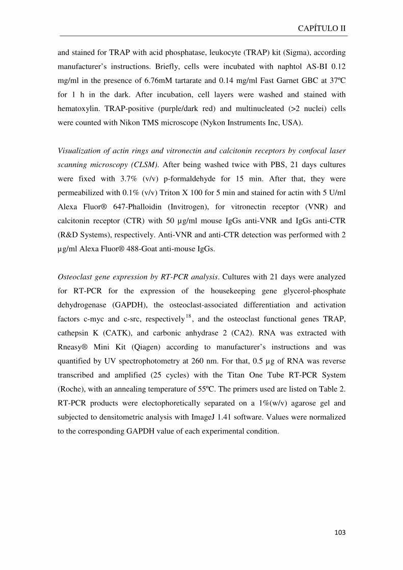
CAPÍTULO II
103
and stained for TRAP with acid phosphatase, leukocyte (TRAP) kit (Sigma), according
manufacturer’s instructions. Briefly, cells were incubated with naphtol AS-BI 0.12
mg/ml in the presence of 6.76mM tartarate and 0.14 mg/ml Fast Garnet GBC at 37ºC
for 1 h in the dark. After incubation, cell layers were washed and stained with
hematoxylin. TRAP-positive (purple/dark red) and multinucleated (>2 nuclei) cells
were counted with Nikon TMS microscope (Nykon Instruments Inc, USA).
Visualization of actin rings and vitronectin and calcitonin receptors by confocal laser
scanning microscopy (CLSM). After being washed twice with PBS, 21 days cultures
were fixed with 3.7% (v/v) p-formaldehyde for 15 min. After that, they were
permeabilized with 0.1% (v/v) Triton X 100 for 5 min and stained for actin with 5 U/ml
Alexa Fluor® 647-Phalloidin (Invitrogen), for vitronectin receptor (VNR) and
calcitonin receptor (CTR) with 50 µg/ml mouse IgGs anti-VNR and IgGs anti-CTR
(R&D Systems), respectively. Anti-VNR and anti-CTR detection was performed with 2
µg/ml Alexa Fluor® 488-Goat anti-mouse IgGs.
Osteoclast gene expression by RT-PCR analysis. Cultures with 21 days were analyzed
for RT-PCR for the expression of the housekeeping gene glycerol-phosphate
dehydrogenase (GAPDH), the osteoclast-associated differentiation and activation
factors c-myc and c-src, respectively18 , and the osteoclast functional genes TRAP,
cathepsin K (CATK), and carbonic anhydrase 2 (CA2). RNA was extracted with
Rneasy® Mini Kit (Qiagen) according to manufacturer’s instructions and was
quantified by UV spectrophotometry at 260 nm. For that, 0.5 µg of RNA was reverse
transcribed and amplified (25 cycles) with the Titan One Tube RT-PCR System
(Roche), with an annealing temperature of 55ºC. The primers used are listed on Table 2.
RT-PCR products were electophoretically separated on a 1%(w/v) agarose gel and
subjected to densitometric analysis with ImageJ 1.41 software. Values were normalized
to the corresponding GAPDH value of each experimental condition.
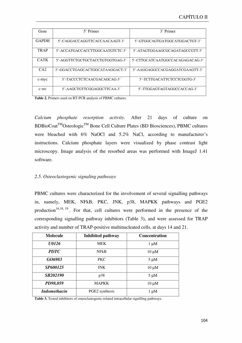
CAPÍTULO II
104
Gene 5’ Primer 3’ Primer
GAPDH 5’-CAGGACCAGGTTCACCAACAAGT-3’ 5’-GTGGCAGTGATGGCATGGACTGT-3’
TRAP 5’-ACCATGACCACCTTGGCAATGTCTC-3’ 5’-ATAGTGGAAGCGCAGATAGCCGTT-3’
CATK 5’-AGGTTCTGCTGCTACCTGTGGTGAG-3’ 5’-CTTGCATCAATGGCCACAGAGACAG-3’
CA2 5’-GGACCTGAGCACTGGCATAAGGACT-3’ 5’-AAGGAGGCCACGAGGATCGAAGTT-3’
c-myc 5’-TACCCTCTCAACGACAGCAG-3’ 5’-TCTTGACATTCTCCTCGGTG-3’
c-src 5’-AAGCTGTTCGGAGGCTTCAA-3’ 5’-TTGGAGTAGTAGGCCACCAG-3’
Table 2. Primers used on RT-PCR analysis of PBMC cultures.
Calcium phosphate resorption activity. After 21 days of culture on
BDBioCoatTMOsteologicTM Bone Cell Culture Plates (BD Biosciences), PBMC cultures
were bleached with 6% NaOCl and 5.2% NaCl, according to manufacturer’s
instructions. Calcium phosphate layers were visualized by phase contrast light
microscopy. Image analysis of the resorbed areas was performed with ImageJ 1.41
software.
2.5. Osteoclastogenic signaling pathways
PBMC cultures were characterized for the involvement of several signalling pathways
in, namely, MEK, NFkB, PKC, JNK, p38, MAPKK pathways and PGE2
production16,18, 19 . For that, cell cultures were performed in the presence of the
corresponding signalling pathway inhibitors (Table 3), and were assessed for TRAP
activity and number of TRAP-positive multinucleated cells, at days 14 and 21.
Molecule Inhibited pathway Concentration
U0126 MEK 1 µM
PDTC NFkB 10 µM
GO6983 PKC 5 µM
SP600125 JNK 10 µM
SB202190 p38 5 µM
PD98,059 MAPKK 10 µM
Indomethacin PGE2 synthesis 1 µM
Table 3. Tested inhibitors of osteoclastogenic-related intracellular signilling pathways.
CAPÍTULO II
105
2.6. Statistical analysis
Data were obtained from three separate experiments, each one performed in triplicate,
using cell cultures from different donors. Data are expressed as the mean ± standard
deviation. Groups of data were evaluated using a two-way analysis of variance
(ANOVA) and no significant differences in the pattern of the cell behavior were found.
Statistical differences between controls and experimental conditions were assessed by
Bonferroni’s method. Values of p ≤ 0.05 were considered significant.
3. Results
3.1. TRAP activity and number of TRAP-positive multinucleated cells
Unstimulated PBMC
Control PBMC cultures performed in base medium displayed low values of TRAP
activity (Fig. 1), which increased slightly during the culture period. AH Plus extract, at
1:2500 and 1:500, caused a stimulatory effect at day 21 (~25%); higher concentrations
resulted in dose-dependent inhibitory effects. The other cements inhibited TRAP
activity with time- and dose-dependent effects, at extract concentrations similar to and
higher than 1:500. GuttaFlow and Tubliseal exhibited a low toxicity (slightly higher in
the case of Tubliseal). Sealapex showed an evident dose-dependent inhibitory effect,
whereas RealSeal exhibited the highest toxicity, with a significant deleterious effect at
all concentrations, particularly between days 7 and 14. At day 21, the inhibitory effect
observed in the cultures exposed to the highest extract concentration (1:20) was ~25%
(GuttaFlow), ~45% (Tubliseal), ~57% (Sealapex) and ~75% (AH Plus and RealSeal).
The effect of the cements extracts in the number of TRAP-positive multinucleated cells
followed a pattern similar to that of TRAP activity (Fig. 1).
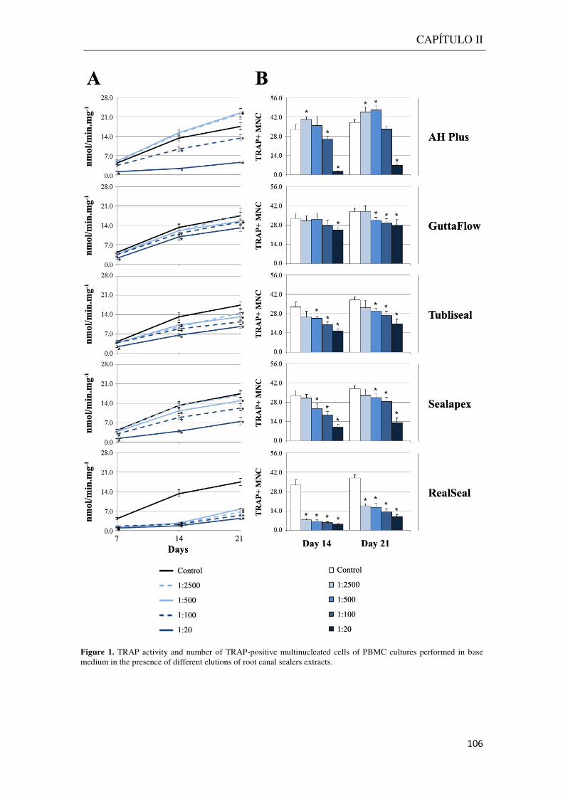
CAPÍTULO II
106
Figure 1. TRAP activity and number of TRAP-positive multinucleated cells of PBMC cultures performed in base medium in the presence of different elutions of root canal sealers extracts.
CAPÍTULO II
107
Stimulated PBMC
At control conditions, PBMC cultures supplemented with M-CSF and RANKL
displayed high values of TRAP activity (Fig. 2). Enzymatic activity increased
significantly from days 7 to 14, and tended to stabilize from days 14 to 21. As observed
in unstimulated PBMC, AHPlus extract, at 1:2500 and 1:500, caused a slight
stimulatory effect at day 21 (~15%), and inhibitory effects at higher levels. The other
cements caused an inhibitory effect, however, with a different pattern of inhibition.
Extracts from GuttaFlow and Tubliseal (levels ≥ 1:500) and AH Plus (levels ≥ 1:100)
presented a dose-dependent inhibitory effect on TRAP activity, and the pattern of cell
growth was similar to that of control, i.e. TRAP activity increased from days 7 to 14 and
showed a tendency for stabilization in the last week. Thus, similar inhibitory effects
were seen at days 14 and 21 in the cultures exposed to the extracts of these sealers; at
the higher concentration (1:20) the percentages of inhibition at days 14 and 21 were,
respectively, ~36% and ~30% (GuttaFlow), ~58% and ~50% (Tubliseal) and ~93% and
87% (AH Plus). However, the inhibitory effect of Sealapex and RealSeal showed a
distinct profile; cytotoxicity was particularly evident from days 7 to 14 but, afterwards,
cells tended to recover and, at day 21, the inhibitory effect was much lower than that
seen on day 14. Thus, at days 14 and 21, the percentage of inhibition observed in the
cultures exposed to the highest extract concentration (1:20) was, respectively, ~85% and
~55% (Sealapex) and ~70% and ~41% (RealSeal).
Control PBMC cultures presented a high and similar number of TRAP-positive
multinucleated cells at days 14 and 21, and the effect of the sealers’ extracts was
identical to that observed on TRAP activity (Fig. 2).
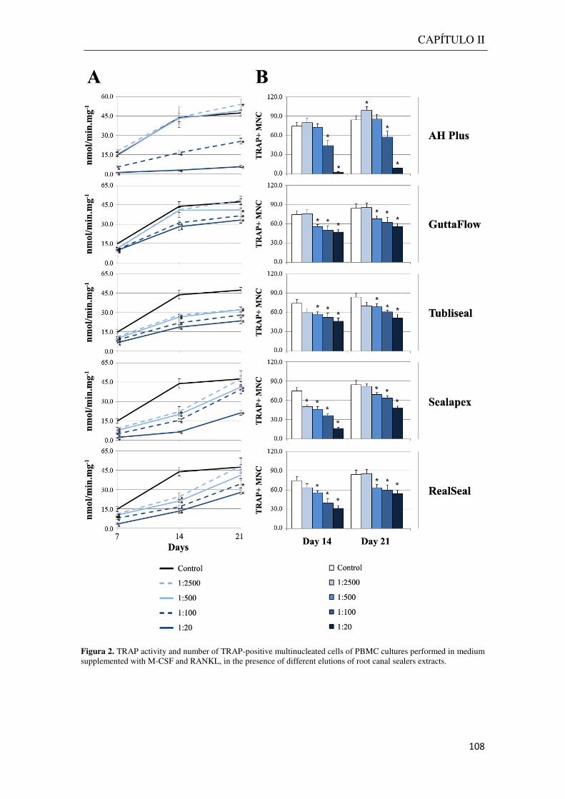
CAPÍTULO II
108
Figura 2. TRAP activity and number of TRAP-positive multinucleated cells of PBMC cultures performed in medium supplemented with M-CSF and RANKL, in the presence of different elutions of root canal sealers extracts.
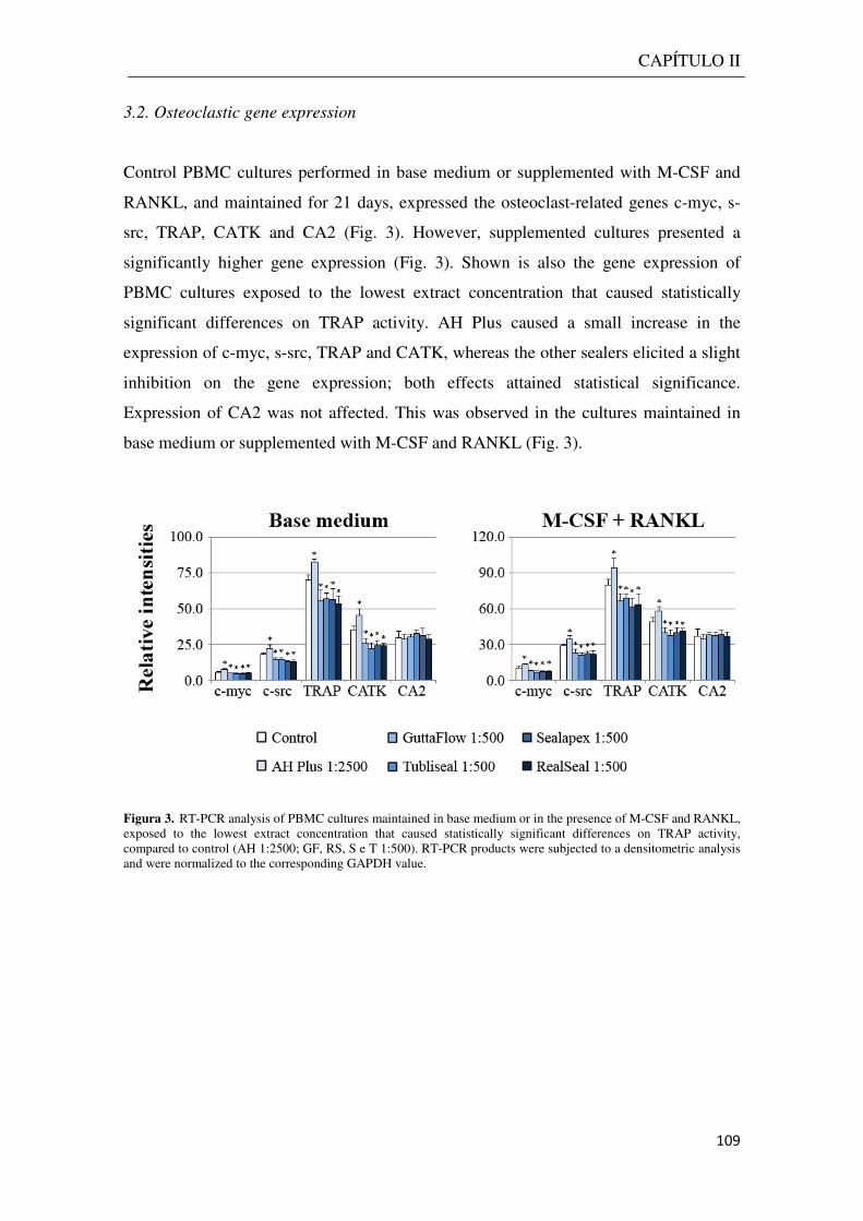
CAPÍTULO II
109
3.2. Osteoclastic gene expression
Control PBMC cultures performed in base medium or supplemented with M-CSF and
RANKL, and maintained for 21 days, expressed the osteoclast-related genes c-myc, s-
src, TRAP, CATK and CA2 (Fig. 3). However, supplemented cultures presented a
significantly higher gene expression (Fig. 3). Shown is also the gene expression of
PBMC cultures exposed to the lowest extract concentration that caused statistically
significant differences on TRAP activity. AH Plus caused a small increase in the
expression of c-myc, s-src, TRAP and CATK, whereas the other sealers elicited a slight
inhibition on the gene expression; both effects attained statistical significance.
Expression of CA2 was not affected. This was observed in the cultures maintained in
base medium or supplemented with M-CSF and RANKL (Fig. 3).
Figura 3. RT-PCR analysis of PBMC cultures maintained in base medium or in the presence of M-CSF and RANKL, exposed to the lowest extract concentration that caused statistically significant differences on TRAP activity, compared to control (AH 1:2500; GF, RS, S e T 1:500). RT-PCR products were subjected to a densitometric analysis and were normalized to the corresponding GAPDH value.
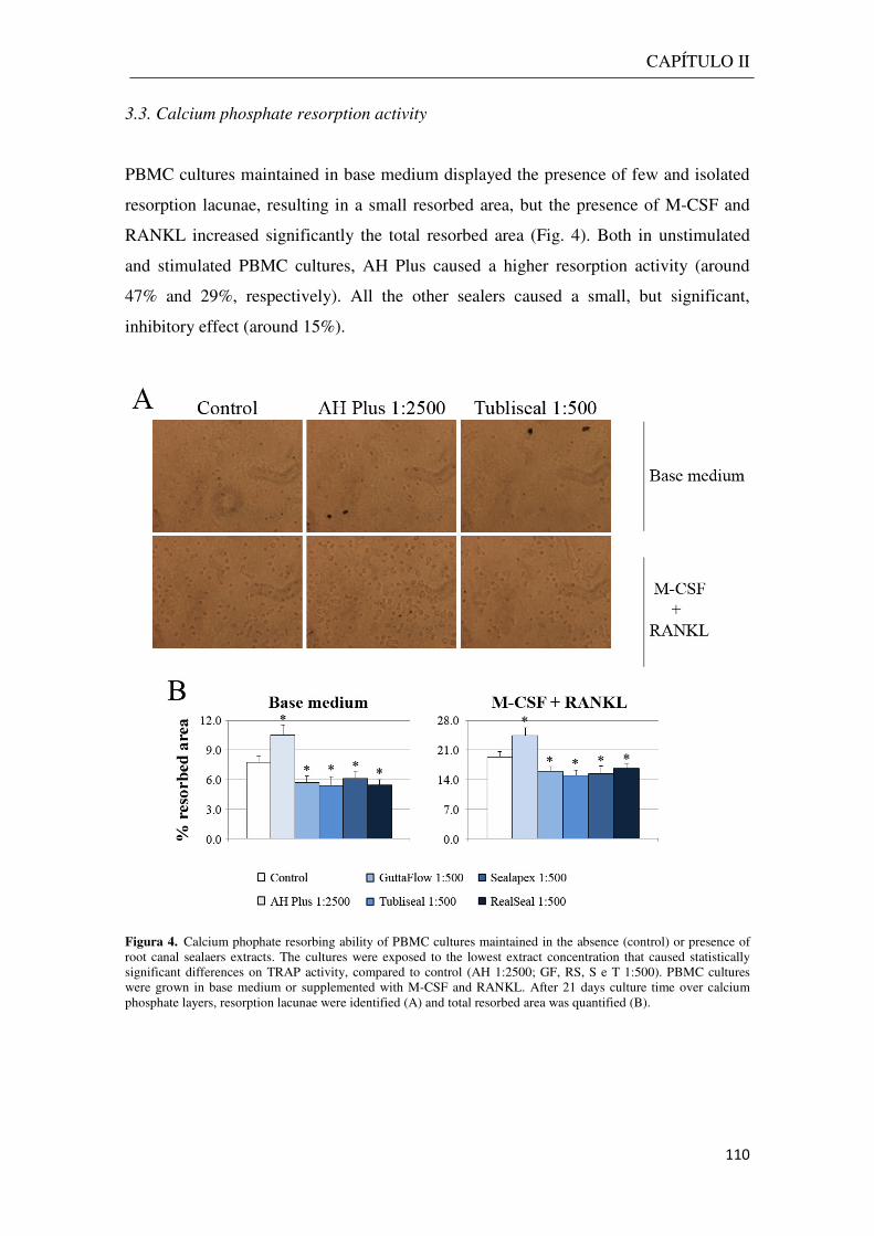
CAPÍTULO II
110
3.3. Calcium phosphate resorption activity
PBMC cultures maintained in base medium displayed the presence of few and isolated
resorption lacunae, resulting in a small resorbed area, but the presence of M-CSF and
RANKL increased significantly the total resorbed area (Fig. 4). Both in unstimulated
and stimulated PBMC cultures, AH Plus caused a higher resorption activity (around
47% and 29%, respectively). All the other sealers caused a small, but significant,
inhibitory effect (around 15%).
Figura 4. Calcium phophate resorbing ability of PBMC cultures maintained in the absence (control) or presence of root canal sealaers extracts. The cultures were exposed to the lowest extract concentration that caused statistically significant differences on TRAP activity, compared to control (AH 1:2500; GF, RS, S e T 1:500). PBMC cultures were grown in base medium or supplemented with M-CSF and RANKL. After 21 days culture time over calcium phosphate layers, resorption lacunae were identified (A) and total resorbed area was quantified (B).
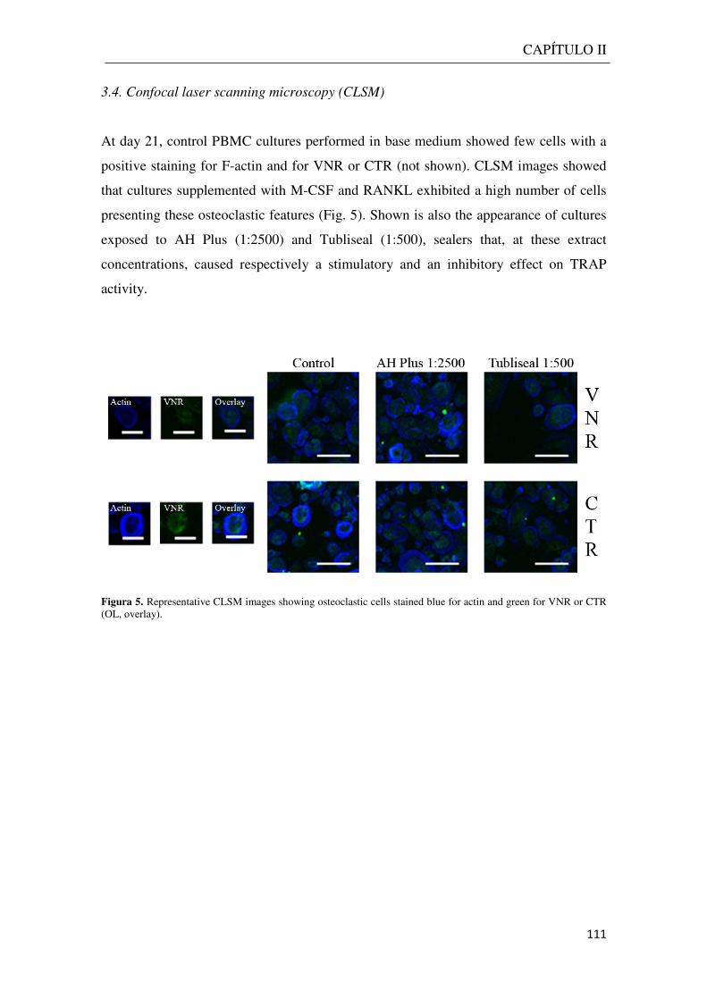
CAPÍTULO II
111
3.4. Confocal laser scanning microscopy (CLSM)
At day 21, control PBMC cultures performed in base medium showed few cells with a
positive staining for F-actin and for VNR or CTR (not shown). CLSM images showed
that cultures supplemented with M-CSF and RANKL exhibited a high number of cells
presenting these osteoclastic features (Fig. 5). Shown is also the appearance of cultures
exposed to AH Plus (1:2500) and Tubliseal (1:500), sealers that, at these extract
concentrations, caused respectively a stimulatory and an inhibitory effect on TRAP
activity.
Figura 5. Representative CLSM images showing osteoclastic cells stained blue for actin and green for VNR or CTR (OL, overlay).
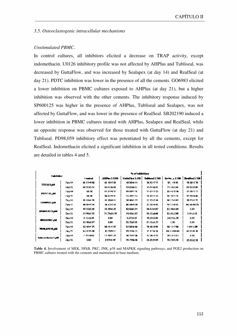
3.5. Osteoclastogenic intracellular mechanisms
Unstimulated PBMC.
In control cultures, all inhibitors elicited a decrease on TRAP activity, except
indomethacin. U0126 inhibitory profile was not affected by AHPlus and Tubliseal, was
decreased by GuttaFlow, and was increased by Sealapex (at day 14) and RealSeal (at
day 21). PDTC inhibition was low
a lower inhibition on PBMC cultures exposed to AHPlus (at day 21), but a higher
inhibition was observed with the other cements. The inhibitory response induced by
SP600125 was higher in the presence of
affected by GuttaFlow, and was lower in the presence of RealSeal. SB202190 induced a
lower inhibition in PBMC cultures treated with AHPlus, Sealapex and RealSeal, while
an opposite response was observed for those tr
Tubliseal. PD98,059 inhibitory effect was potentiated by all the cements,
RealSeal. Indomethacin elicited a significant inhibition in all tested conditions.
are detailed in tables 4 and 5
Table 4. Involvement of MEK, NFkB, PKC, JNK, p38 and MAPKK signaling pathways, and PGE2 production on PBMC cultures treated with the cements and maintained in base medium
Osteoclastogenic intracellular mechanisms
inhibitors elicited a decrease on TRAP activity, except
indomethacin. U0126 inhibitory profile was not affected by AHPlus and Tubliseal, was
decreased by GuttaFlow, and was increased by Sealapex (at day 14) and RealSeal (at
day 21). PDTC inhibition was lower in the presence of all the cements. GO6983 elicited
a lower inhibition on PBMC cultures exposed to AHPlus (at day 21), but a higher
inhibition was observed with the other cements. The inhibitory response induced by
SP600125 was higher in the presence of AHPlus, Tubliseal and Sealapex, was not
affected by GuttaFlow, and was lower in the presence of RealSeal. SB202190 induced a
lower inhibition in PBMC cultures treated with AHPlus, Sealapex and RealSeal, while
an opposite response was observed for those treated with GuttaFlow (at day 21) and
Tubliseal. PD98,059 inhibitory effect was potentiated by all the cements,
ndomethacin elicited a significant inhibition in all tested conditions.
4 and 5.
Involvement of MEK, NFkB, PKC, JNK, p38 and MAPKK signaling pathways, and PGE2 production on PBMC cultures treated with the cements and maintained in base medium.
CAPÍTULO II
112
inhibitors elicited a decrease on TRAP activity, except
indomethacin. U0126 inhibitory profile was not affected by AHPlus and Tubliseal, was
decreased by GuttaFlow, and was increased by Sealapex (at day 14) and RealSeal (at
er in the presence of all the cements. GO6983 elicited
a lower inhibition on PBMC cultures exposed to AHPlus (at day 21), but a higher
inhibition was observed with the other cements. The inhibitory response induced by
AHPlus, Tubliseal and Sealapex, was not
affected by GuttaFlow, and was lower in the presence of RealSeal. SB202190 induced a
lower inhibition in PBMC cultures treated with AHPlus, Sealapex and RealSeal, while
eated with GuttaFlow (at day 21) and
Tubliseal. PD98,059 inhibitory effect was potentiated by all the cements, except for
ndomethacin elicited a significant inhibition in all tested conditions. Results
Involvement of MEK, NFkB, PKC, JNK, p38 and MAPKK signaling pathways, and PGE2 production on
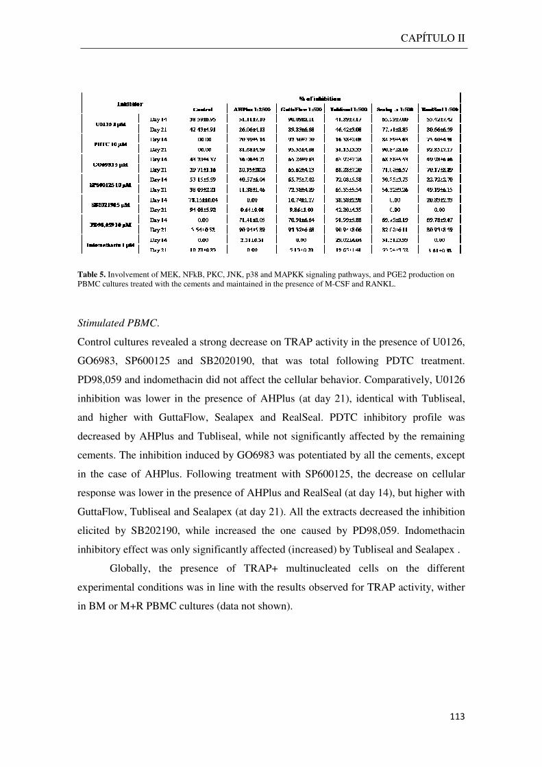
Table 5. Involvement of MEK, NFkB, PKC, JNK, p38 and MAPKK signaling pathways, and PGE2 production on PBMC cultures treated with the cements and maintained in the presence of M
Stimulated PBMC.
Control cultures revealed a strong decrease on TRAP activity in the presence of U0126,
GO6983, SP600125 and SB2020190, that was total following PDTC treatment.
PD98,059 and indomethacin did not affect the cellular behavior. Comparative
inhibition was lower in the
and higher with GuttaFlow
decreased by AHPlus and Tubliseal, while not significantly affected by the remaining
cements. The inhibition induced by
in the case of AHPlus. Following treatment with SP600125, the decrease on cellular
response was lower in the presence of AHPlus and RealSeal (at day 14), but higher
GuttaFlow, Tubliseal and Sealapex (
elicited by SB202190, while increased the one caused by PD98,059. Indomethacin
inhibitory effect was only significantly affected (increased) by Tubliseal and Sealapex .
Globally, the presence of TRAP+
experimental conditions was in line with the results observed for TRAP activity,
in BM or M+R PBMC cultures
NFkB, PKC, JNK, p38 and MAPKK signaling pathways, and PGE2 production on PBMC cultures treated with the cements and maintained in the presence of M-CSF and RANKL.
revealed a strong decrease on TRAP activity in the presence of U0126,
GO6983, SP600125 and SB2020190, that was total following PDTC treatment.
PD98,059 and indomethacin did not affect the cellular behavior. Comparative
n the presence of AHPlus (at day 21), identical
GuttaFlow, Sealapex and RealSeal. PDTC inhibitory profile was
decreased by AHPlus and Tubliseal, while not significantly affected by the remaining
cements. The inhibition induced by GO6983 was potentiated by all the cements, except
in the case of AHPlus. Following treatment with SP600125, the decrease on cellular
response was lower in the presence of AHPlus and RealSeal (at day 14), but higher
GuttaFlow, Tubliseal and Sealapex (at day 21). All the extracts decreased the inhibition
elicited by SB202190, while increased the one caused by PD98,059. Indomethacin
inhibitory effect was only significantly affected (increased) by Tubliseal and Sealapex .
Globally, the presence of TRAP+ multinucleated cells on the different
experimental conditions was in line with the results observed for TRAP activity,
in BM or M+R PBMC cultures (data not shown).
CAPÍTULO II
113
NFkB, PKC, JNK, p38 and MAPKK signaling pathways, and PGE2 production on CSF and RANKL.
revealed a strong decrease on TRAP activity in the presence of U0126,
GO6983, SP600125 and SB2020190, that was total following PDTC treatment.
PD98,059 and indomethacin did not affect the cellular behavior. Comparatively, U0126
presence of AHPlus (at day 21), identical with Tubliseal,
Sealapex and RealSeal. PDTC inhibitory profile was
decreased by AHPlus and Tubliseal, while not significantly affected by the remaining
GO6983 was potentiated by all the cements, except
in the case of AHPlus. Following treatment with SP600125, the decrease on cellular
response was lower in the presence of AHPlus and RealSeal (at day 14), but higher with
at day 21). All the extracts decreased the inhibition
elicited by SB202190, while increased the one caused by PD98,059. Indomethacin
inhibitory effect was only significantly affected (increased) by Tubliseal and Sealapex .
multinucleated cells on the different
experimental conditions was in line with the results observed for TRAP activity, wither
CAPÍTULO II
114
4. Discussion
In this work, differentiation and function of human peripheral osteoclast precursors
were evaluated in the presence of a concentration range of the extracts of five
endodontic sealers, based on distinct compositions. Clinically, the sealers are applied as
freshly mixed materials, and the unset material might contact with apical and periapical
tissues. Thus, the extracts were obtained by incubating the freshly mixed material with
culture medium, and tested for time and dose-dependent effects.
In physiological conditions, osteoclastogenesis is a complex process in which
paracrine mechanisms play a significant role. Among the wide variety of regulator
factors, M-CSF and RANKL are particularly relevant. M-CSF is important for the
earlier steps of osteoclastogenesis, mainly increasing osteoclastic precursor survival 20
and promoting RANK expression on those precursors, in order to initiate RANKL-
mediated oteoclastic differentiation 21 , 22 . RANKL is a factor essential for the later
osteoclastogenic steps, mainly for osteoclastic differentiation22, but also for the survival
of mature osteoclasts 22. Accordingly, the present results showed that in unstimulated
conditions (absence of M-CSF and RANKL), PBMC cultures exhibited low osteoclastic
features, i.e. regarding TRAP activity, number of TRAP-positive multinucleated cells,
expression of osteoclastic genes and resorption activity. The presence of M-CSF and
RANKL increased significantly osteoclast differentiation and function, which is
expected considering the role of these factors on osteoclastogenesis. Considering this,
the effects of the sealers’ extracts were studied in unstimulated and stimulated
conditions (absence or presence of M-CSF and RANKL), aiming to get information on
eventual differences of the sealers’ toxicity profile regarding immature/undifferentiated
precursors and also on precursors committed and undergoing osteoclastogenic
differentiation.
PBMC cultures were significantly affected by the sealers’ extracts, as shown by the
results regarding TRAP activity and number of TRAP-positive multinucleated cells.
Both in unstimulated and stimulated PBMC cultures, AHPlus, at the lower
concentrations, caused a small stimulatory effect, reflecting most probably an adaptive
cell response to cope with the presence of toxic compounds; however, higher levels
caused significant toxic effects. All the other sealers exhibited inhibitory effects, with
differences on the dose- and time-dependent effects. AHPlus (at the two higher
concentrations), GuttaFlow and Tubliseal exhibited a similar pattern, reflected as an

CAPÍTULO II
115
inhibition on TRAP activity during the first two weeks, and a relative stabilization of
the cell behavior afterwards. Sealapex and RealSeal also caused an inhibition until day
14, but afterwards cells were able to recover completely or partially from the inflicted
toxicity. This suggests that cells were able to trigger adaptive mechanisms to the
presence of the sealers, apparently, more efficient in the case of Sealapex and RealSeal.
In the clinical setting, direct contact with the root filling materials and the apical
and periapical tissues involves a small contact area, and the leachable cytotoxic
compounds are continually cleared by the extracellular fluids. Also, these materials
remain in contact with the surrounding tissues for years or decades. Thus, in terms of
predicting the long-term clinical effects of the sealers, exposure of bone cells to low
concentrations of the sealers’ extracts appears to be a more representative situation. In
this context, PBMC cultures were exposed to the lowest tested concentration that
elicited statistically significant effects on TRAP activity, and were further characterized
for a set of osteoclast parameters. Results showed that these concentrations were 1:2500
for AHPlus, which caused small stimulatory effects and 1:500 for the other sealers,
which was associated with an inhibition of the cell response. In these conditions, the
sealers’ extracts affected the expression of the differentiation and activation genes c-
myc and c-src and the functional genes TRAP and CATK, that was in line with those
observed for TRAP activity. The same was found for the presence of actin rings,
vitronectin and calcitonin receptors, and calcium phosphate resorbing activity. In
addition, the relative contribution of some of the osteoclastogenic-related signaling
pathways was sharply affected by the cements’ extracts. In unstimulated PBMC, NFkB
pathway was significantly down regulated by AHPlus, GuttaFlow and Tubliseal. JNK
pathway appeared to have no significant relevance in PBMC cultures treated with
RealSeal, but in the presence of Tubliseal JNK and p38 pathways seemed to be
especially important. MAPKK pathway and PGE2 production appeared to be
particularly involved in the osteoclastogenic response modulated by GuttaFlow and
Sealapex. In stimulated PBMC, GuttaFlow elicited a significant increase in the
relevance of MEK pathway. Tubliseal caused a down regulation of NFkB pathway. All
the cements except AHPlus induced an increase on the involvement of PKC pathway.
p38 appeared to be significantly less involved in the osteoclastogenic response in the
presence of the cements, while the opposite situation was observed for MAPKK
pathway. PGE2 production seemed to be important only following Sealapex treatment.
CAPÍTULO II
116
Several aspects have to be considered in the assessment of the endodontic
sealers cytocompatibility, i.e. composition and leachable components, setting
characteristics, stability of the set sealer and the contact between the sealer and the
surrounding tissues. A wide variety of studies have been performed involving many
sealers, but differing in the target cell type, preparation of the extracts, tested
concentrations and time of exposure, which is reflected by a great discrepancy of results
regarding the establishment of cytotoxicity patterns or the effects on a specific cell type.
However, the results of the present work agree with most of the reported studies, which
also showed inhibitory effects of endodontic sealers in several cell types. GuttaFlow is a
silicone-based sealer manufactured by adding gutta-percha powder to the silicone
matrix, and containing nanosilver particles as a preservative. The low cytotoxicity
observed in the present work is in line with a variety of studies involving
fibroblasts23,24,25,26,27,28, and might result from the release of nanosilver particles as they
have been associated with dose-dependent cytotoxic effects29. It is also reported the
presence of small voids within the core of GuttaFlow, which favour the release of
unreacted compounds that accumulates in such pores30. Regarding AHPlus, an epoxy
resin-based sealer apparently “formaldehyde-free”, is reported to exhibit a moderate to
severe toxicity immediately after mixing (the conditions used in the present work) in
fibroblasts23,24,25,27,28, 31 , 32 , 33 , 34 , 35 , 36 , 37 , human periodontal ligament cells33, 38 and
osteoblasts36, 39,40,. The toxicity of this sealer has been mostly associated to the release of
low levels of formaldehyde, the presence of amines added to accelerate the epoxy
polymerization and the epoxy resin content24,41. The toxicity of Tubliseal, a zinc oxide
eugenol-based sealer was demonstrated in fibroblasts27, 42 and human periodontal
ligament cells 43 , and has been attributed to free eugenol release, and also several
additives, namely resins used for greater dentin adhesion29. Sealapex is a calcium
hydroxide based sealer and its cytotoxicity is widely reported, namely in
fibroblasts37,44,45, human periodontal ligament cells38,43 and osteoblasts46,47. It has been
attributed to an increase in the pH of the culture medium resulting from the sealer
dissolution29,48,49. In the present work, the pH remained constant throughout the culture
time, due to the low tested levels, and other components such as polyresin, zinc oxide
and Ba2+ have been suggested as potential toxic compounds50. Most of the studies
performed with RealSeal reported a rather cytotoxic effect of this dual-cured
hydrophilic multi-methacrylate resin-based sealer, namely in fibroblasts, 23,24,25,35,
36,45,51,52,53, human dental pulp cells53 and osteoblasts36,54,55 , suggesting that it is caused
CAPÍTULO II
117
by the leaching of unreacted monomers as a result of incomplete polymerization , and
also filler particles due to the degradation of the sealer.
Regarding bone cells, few studies were performed, and all involved osteoblastic
cells. Considering the sealers studied in the present work, AHPlus has been tested in
human U2-OS osteosarcoma cells39,56 and in the rat cell lines MC3T3-E1 and ROS
17/2.8 36,40, whereas Sealapex was addressed in rat calvarial osteoblasts46 and MC3T3-
E1 cells3 and RealSeal was studied in MG63 cells55 and MC3T3-E1 and ROS 17/2.8
cells36,54. To the best of our knowledge, no studies have been described addressing the
effects of the sealers on osteoclast cells, which is of utmost relevance considering the
intimate relationship between the two cells types in bone metabolism. In the present
work, addressing long-term effects of different sealers’ extracts on osteoclastogenesis,
results observed until day 14 might be considered as reflecting acute/short-term effects,
and showed dose-dependent inhibitory effects, whereas data collected on day 21
provides information regarding a longer exposure time, evidencing the ability of
adaptive cell responses to the sealers’ extracts. Due to the differences on the time-
dependent effects of the different sealers, it is not easy to establish an order of toxicity.
However, evident results were the low toxicity of GuttaFlow (both in unstimulated and
stimulated PBMC cultures), the high deleterious effect of RealSeal in unsttimulated
cultures, and the severe cytotoxicity of the highest concentration of AHPlus in both
unstimulated and stimulated conditions.
5. Conclusion
The sealers’ extracts caused inhibitory effects on human osteoclast differentiation and
function. The sealers differed on the dose- and time-dependent profile, and also in the
involved intracellular signaling pathways. However, following long-term exposure an
adaptive cell response was noticed. This observation, together with the lower toxicity
expected in in vivo conditions due to the continuous fluids flow and sealers’ clearance,
suggests that after a variable time-period the cytotoxicity of the sealers eluates on
osteoclast cells might be greatly minimized.
118
References
1Datta HK et al. The cell biology of bone metabolism. J Clin Pathol 2008; 61: 577–87. 2Awadhi et al. Cultured primary osteoblast viability and apoptosis in the presence of root canal sealers. J Endodon 2004;30: 527- 33. 3Lee DH et al. Mechanism of root canal sealers cytotoxicity on osteoblastic cell line MC3T3-E1. Oral Surg Oral Med Oral Pathol Oral Radiol Endodon 2007;104: 717- 21. 4Xu P et al. Cytotoxicity of RealSeal on human osteoblast-like MG63 cells. J Endodon 2010; 36: 40-44. 5Pertot WJ et al. Comparision of the intraosseous biocompatibility of dyract and super EBA. J Endodon 1997;23: 315- 19. 6Torabinejad M et al. Tissue reaction to implanted root-end filling materials in the tibia and mandible of Guinea pigs. J Endodon 1998;24: 468-71. 7Matsuo K, Irie N. Osteoclast-osteobalst communication. Arch Biochem Bioph 2008;473: 201- 9. 8Vaananen, HK. and Laitala-Leinonen T, Osteoclast lineage and function. Arch Biochem Biophys, 2008; 473: 132-8. 9Zaidi, M. Skeletal remodeling in health and disease. Nat Med 2007; 13(7): 791-801. 10Costa-Rodrigues J et al. Characterization of the osteoclastogenic potential of human osteoblastic and fibroblastic conditioned media. J Cell Biochem 2010; 109(1): 205-16. 11Costa-Rodrigues J, Fernandes MH. Paracrine-mediated differentiation and activation of human hematopoietic osteoclast precursor cells by skin and gingival fibroblasts. Cell Prolif 2011; 44: 264–73. 12Asagiri M, Takayanagi H. The molecular understanding of osteoclast differentiation. Bone 2007;40: 251- 64. 13Costa-Rodrigues J, Fernandes A, Fernandes MH. Reciprocal osteoblastic and osteoclastic modulation in co-cultured MG63 osteosarcoma cells and human osteoclast precursors. J Cell Biochem 2011;112: 3704- 13. 14 Costa-Rodrigues J et al. Hydroxyapatite surface roughness: Complex modulation of the osteoclastogenesis of human precursor cells. Acta Biomater, 2012;8:1137-45. 15Costa-Rodrigues J, Fernandes A, Fernandes MH. Spontaneous and induced osteoclastogenic behaviour of human peripheral blood mononuclear cells and their CD14(+) and CD14(-) cell fractions. Cell Prolif 2011;44:410-19. 16Costa-Rodrigues, J et al. Variability of the paracrine-induced osteoclastogenesis by human breast cancer cell lines. J Cell Biochem, 2012; 113:1069-79. 17Bradford, M. M. A rapid and sensitive method for the quantitation of microgram quantities of protein utilizing the principle of protein-dye binding. Analyt. Biochem 1976;72: 248–54. 18Zhao Q et al. Osteoclast differentiation and gene regulation. Front Biosci 2007; 12: 2519–29. 19Teitelbaum SL and Ross FP. Genetic regulation of osteoclast development and function. Nat Rev Genet, 2003; 4(8): 638-49. 20Pixley FJ, Stanley ER. CSF-1 regulation of the wandering macrophage: complexity in action. Trends Cell Biol 2004; 14: 628-638.
119
21Boyle WJ, Simonet WS, Lacey DL. Osteoclast differentiation and activation. Nature 2003; 423: 337–342. 22Boyce BF, Xing L. Functions of RANKL ⁄RANK⁄ OPG in bone modeling and remodeling. Arch.
Biochem. Biophys 2008; 473: 139–146. 23Bouillaguet S et al. Initial in vitro biological response to contemporary endodontic sealers. J Endodon 2006;32: 989- 92. 24Eldeniz AU et al. Cytotoxicity of new resin-, calcium hydroxide-, and silicone-based root canal sealers on fibroblasts derived from human gingival and L929 cell lines. Int Endodon J 2007; 40: 329- 37. 25Brackett MG et al. Cytotoxicity of endodontic materials over 6-weeks ex vivo. Int Endodon J 2008;41: 1072- 78. 26Ashraf H, Taherian A and Kerdar AN. Evaluation of cytotoxicity of two root canal filling materials by MTT assay. Aust Endod J 2010;36: 24- 28. 27Zoufan K et al. Cytotoxicity evaluation of GuttaFlow and Endo Sequence BC sealers. Oral Surg Oral Med Oral Pathol Oral Radiol Endodon 2011;112:657-61. 28Willershausen I et al. In vitro analysis of the cytotoxicity and the antimicrobial effect of four endodontic sealers. Head & Face Med 2011;7: 15. 29Hauman CH, Love RM. Biocompatibility of dental materials used in contemporary endodontic therapy: a review. Part 2.Root canal filling materials. Int Endod J 2003; 36:147-60. 30Elayouti A et al. Homogeneity and adaptation of a new gutta-percha paste to root canal walls. J Endod 2005; 31: 687-90. 31Cohen BI et al. An in vitro study of the cytotoxicity of two root canal sealers. J Endodon 2000; 28: 228-29. 32Miletić I et al. Cytotoxic effect of four root filling materials. Endod Dent Traumatol 2000; 16: 287- 90. 33Schwarze T et al. The cellular compatibility of five endodontic sealers during the setting period. J Endodon 2002; 28: 784- 86. 34Miletić I et al. The cytotoxicity of RoekoSeal and AH Plus compared during different setting periods. J Endodon 2005;31: 307- 9. 35Lodienè G et al. Toxicity evaluation of root canal sealers in vitro. Int Endodon J 2008; 41: 72-7. 36Brackett MG et al. Cytotoxic response of three cell lines exposed in vitro to dental endodontic sealers. J Biomed Mat Res B: Appl Biomater 2010; 95B:380- 86. 37Silva EJNL et al. Evaluation of cytotoxicity and up-regulation of gelatinases in human fibroblast cells by four root canal sealers. Int Endodon J 2012; 45: 49- 56. 38Huang FM et al. Cytotoxicity of resin-, zinc oxide eugenol-, and calcium hydroxide-based root canal sealers on human periodontal ligament cells and permanent V79 cells. Int Endodon J 2002;35: 153- 58. 39Huang FM, Chou MY and Chang YC. Induction of cyclooxygenase-2 mRNA and protein expression by epoxy resin and zinc oxide-eugenol based root canal sealers in human osteoblastic cells. Biomaterials 2003;24: 1869- 75. 40Bryan TE et al. In Vitro osteogenic potencial of an experimental calcium silicate-based root canal sealer. J Endodon 2010; 36: 1163-69.

120
41Merdad K et al. Short term cytotoxicity assessement of components of the Epiphany resin-percha obturating system by indirect and direct Millipore filter assays. J Endodon 2007;33: 24- 7. 42 Ersev H et al. Cytotoxic and mutagenic potencies of various root canal filling materials in eukaryotic and prokaryotic cells in vitro. J Endodon 1999;25: 358-63. 43Chang MC et al. Comparative cytotoxicity of five root canal sealers on cultured human periodontal ligament fibroblast. Int Endodon J 2010;43: 251- 57. 44Beltes P et al. In Vitro evaluation of the cytotoxicity of calcium hydroxide-based root canal sealers. Endod Dent Traumatol 1995;11: 245-249. 45Key JE, Rahemtulla FG and Eleazer PD. Cytotoxicity of new root canal filling material on human gingival fibroblasts. J Endodon 2006;32: 756- 58. 46Al-Awadhi S et al. Cultured primary osteoblasts viability and apoptosisi in the presence of root canal sealers. J Endodon 2004;30: 527- 33. 47Lee DH et al. Mechanisms of root canal sealers cytotoxicity on osteoblastic cell line MC3T3-E1. Oral Surg Oral Med Oral Pathol Oral Radiol Endodon 2007;104: 717- 21. 48Duarte MAH et al. Evaluation of pH and calcium ion release of three root canal sealers. J Endodon 2000;26: 389- 90. 49Eldeniz AU et al. Evaluation of pH and ion release of Acroseal sealer in comparision with Apexit and Sealapex sealers. Oral Surg Oral Med Oral Pathol Oral Radiol Endodon 2007; 103: e86- e91. 50Takahara K, Onodera A, Matsumoto K. Toxicity of root canal sealers on rat bone cells in primary culture. Endod Dent Traumatol 1990;6:200-7. 51Susini G et al. Cytotoxicity of Epiphany and Resilon with a root model. Int End J 2006; 39: 940- 44. 52 Donadio M et al. Cytotoxicity evaluation of Activ GP and Resilon sealers in vitro. Oral Surg Oral Med Oral Pathol Oral RadiolEndodon 2009; 107: e74- e78. 53Karapinar-Kazandağ M et al. Cytotoxicity of 5 endodontic sealers on L929 cell line and human dental pulp cells. Int Endodon J 2011; 44: 626- 34. 54Ames JM et al. Contemporary methacrylate resin-based root canal sealers exhibit different degrees of ex vivo cytotoxicity when cured in their self cured mode. J Endodon 2009; 35: 225- 28. 55 Xu P et al. Cytotoxicity of RealSeal on human osteoblast like MG63 cells. J Endodon 2010;36: 40- 44. 56Huang FM et al. Induction of interleukin-6 and interleukin-8 gene expression by root canal sealers in human osteoblastic cells. J Endodon 2005; 31: 679-683.

CAPÍTULO III
Behaviour of co-cultured human osteoclastic and osteoblastic cells exposed to
endodontic sealers

122
CAPÍTULO III
123
1. Introduction
Root canal sealers are widely used worldwide, in endodontic procedures. Although they
are intended to be contained within the root canal space, they may extrude through the
apical constriction, or eluents from the sealers may come in contact with periradicular
tissues. This may cause irritation and delay in wound healing1. Moreover, the contact of
root canal filling materials with periapical tissues can also affect the normal bone
metabolic activities by direct effects on osteoblastic and osteoclastic cells, which might
impair local bone regeneration and remodeling. Regarding this, several in vitro studies
addressed the effect of endodontic sealers in different osteoblastic cell systems, namely
in human U2-OS osteosarcoma cells2,3,4,5,6 , human MG63 osteosarcoma cells7, rat cell
lines MC3T3-E1 and ROS 17/2.85,8,9,10,11 and rat calvarial osteoblasts12, differing on the
tested sealers and the exposure protocol. Most of them involved short-term exposures
and reported inhibitory effects in the viability/proliferation of osteoblastic cells and in
functional parameters such as the alkaline phosphatase activity. Some studies suggested
that the sealers eluents might interfere with the production of molecules, such as COX-
22, IL-6 and IL-83, gelatinases4 and RANKL5, which are involved in signaling pathways
and cellular crosstalks within the bone environment.
Bone tissue is constantly being moulded and shaped by the coordinated action of
bone-resorbing osteoclasts and bone-synthesizing osteoblasts 13 . Both cell types are
involved in reciprocal crosstalks that modulate the differentiation and function of each
other13,14 , which are essential to maintain an adequate bone mass and/or structure.
Therefore, for in vitro bone-related research, osteoclast/osteoblast co-cultures appear as
attractive experimental models, because they are one step closer to natural conditions
and allow elucidation of some aspects of the complex interactions between bone-
building and bone-resorbing cells. Accordingly, some recent reports stressed the
relevance of these co-culture systems, as more representative models to address in vitro
bone metabolism studies14,15,16. Despite the high relevance of this issue, to the best of
our knowledge, the effects of endodontic sealers on human osteoclastic and osteoblastic
cells in a co-culture system have not been reported.
In this context, this study addresses the long-term effects of five root canal
sealers’ extracts, based on distinct compositions (AH PlusTM, GuttaFlowTM,
TublisealTM, SealapexTM and RealSealTM), on co-cultures of human osteoclastic and
osteoblastic cells. These cultures were established from human peripheral blood
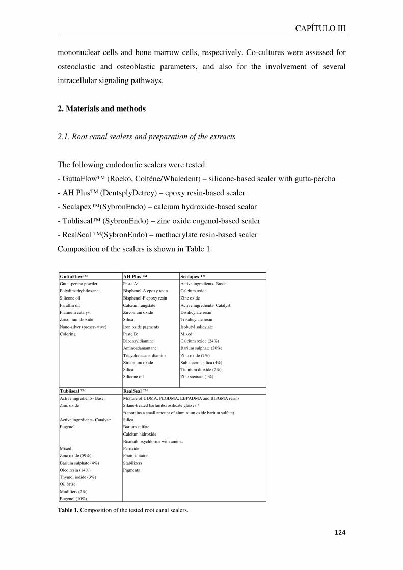
CAPÍTULO III
124
mononuclear cells and bone marrow cells, respectively. Co-cultures were assessed for
osteoclastic and osteoblastic parameters, and also for the involvement of several
intracellular signaling pathways.
2. Materials and methods
2.1. Root canal sealers and preparation of the extracts
The following endodontic sealers were tested:
- GuttaFlow™ (Roeko, Colténe/Whaledent) – silicone-based sealer with gutta-percha
- AH Plus™ (DentsplyDetrey) – epoxy resin-based sealer
- Sealapex™(SybronEndo) – calcium hydroxide-based sealar
- Tubliseal™ (SybronEndo) – zinc oxide eugenol-based sealer
- RealSeal ™(SybronEndo) – methacrylate resin-based sealer
Composition of the sealers is shown in Table 1.
Table 1. Composition of the tested root canal sealers.
GuttaFlow™ AH Plus ™ Sealapex ™
Gutta-percha powder Paste A: Active ingredients- Base:
Polydimethylsiloxane Bisphenol-A epoxy resin Calcium oxide
Silicone oil Bisphenol-F epoxy resin Zinc oxide
Paraffin oil Calcium tungstate Active ingredients- Catalyst:
Platinum catalyst Zirconium oxide Disalicylate resin
Zirconium dioxide Silica Trisalicylate resin
Nano-silver (preservative) Iron oxide pigments Isobutyl salicylate
Coloring Paste B: Mixed:
Dibenzyldiamine Calcium oxide (24%)
Aminoadamantane Barium sulphate (20%)
Tricyclodecane-diamine Zinc oxide (7%)
Zirconium oxide Sub-micron silica (4%)
Silica Titanium dioxide (2%)
Silicone oil Zinc stearate (1%)
Tubliseal ™ RealSeal ™
Active ingredients- Base: Mixture of UDMA, PEGDMA, EBPADMA and BISGMA resins
Zinc oxide Silane-treated barlumborosilicate glasses *
*(contains a small amount of aluminium oxide barium sulfate)
Active ingredients- Catalyst: Silica
Eugenol Barium sulfate
Calcium hidroxide
Bismuth oxychloride with amines
Mixed: Peroxide
Zinc oxide (59%) Photo initator
Barium sulphate (4%) Stabilizers
Oleo resin (14%) Pigments
Thymol iodide (3%)
Oil 8(%)
Modifiers (2%)
Eugenol (10%)
CAPÍTULO III
125
The sealers’ extracts were prepared according to ISO Standards 10993-5
(surface/medium rate: 0.5 - 6.0cm2/ml; extraction time: 1- 3 days). The sealers were
mixed according to the manufacturer’s instructions under aseptic conditions. With an
insulin syringe, 0.3ml of each sealer was placed at the bottom of the well of a 24-well
plate (Orange Scientific, Belgium) and the surface of the material was smoothed.
Immediately after, 1.5ml of culture medium was added to each well. The medium
consisted of α-Minimal Essential Medium (α-MEM) supplemented with 10% fetal
bovine serum, 100 IU/ml penicillin, 2.5 µg/ml streptomycin, 2.5 µg/ml amphotericin B
and with 50 µg/ml ascorbic acid. The plates were kept for 24h at 37ºC in a humidified
5% CO2/air. Following, culture medium was collected, diluted (1:20, 1:100, 1:500,
1:2500), aliquoted and frozen at -20ºC. In this concentration range, the pH of the culture
medium remained constant (pH ~7.4).
2.2. Co-cultures of osteoclastic and osteoblastic cells. Exposure to the extracts.
Osteoclast cell cultures were established from precursors present in peripheral blood
mononuclear cells (PBMCs), isolated from blood of healthy male donors (25–35 years
old), after informed consent, as described before17. Briefly, blood was diluted with
phosphate buffered saline (PBS) (1:1) and applied on top of Ficoll-Paque™ PREMIUM
(GE Healthcare Bio Sciences). Samples were centrifuged at 400g for 30 min. PBMCs
were collected at the interface between Ficoll-Paque and PBS and washed twice with
PBS. On average, for each 100 ml of processed blood, ~70 x 106 PBMCs were obtained.
Osteoblast cell cultures were established from bone marrow obtained from
patients (25–35 years old) undergoing orthopedic surgery procedures, after informed
consent18. Human bone marrow cells (hBMCs) were cultured in α-MEM containing
10% fetal bovine serum, 100 IU/ml penicillin, 2.5 µg/ml streptomycin, 2.5 µg/ml
amphotericin B and 50 µg/ml ascorbic acid. At about 70-80% confluence, cells were
enzymatically detached with 0.05% trypsin and 0.5mM EDTA and seeded as described
below.
For the co-culture, hBMCs were seeded (1x10³ cells/cm2) and cultured for 24
hours in the conditions used for the culture of osteoblastic cells. Following, PBMC were
added (1.5x10⁶ cells/cm2) and co-cultures were maintained in the conditions described
for PBMC cultures. Seeding densities and culture conditions were selected based in
previous studies19 . After 24 h, culture medium was removed and replaced by one
CAPÍTULO III
126
containing the sealers’ extracts (1:2500, 1:500, 1:100 and 1:20). Co-cultures performed
in the absence of the extracts were used as control. Cultures were maintained for 21
days at 37ºC in a 5% CO2 humidified atmosphere. Culture medium was replaced once a
week and the extracts were renewed in every medium change. Co-cultures were
characterized throughout the culture time for the osteoclastic and the osteoblastic
response, as described previously19.
2.2.1. Osteoclastogenic response
Co-cultures were characterized at days 7, 14 and 21 for tartrate-resistant acid
phosphatase (TRAP) activity and number of TRAP-positive multinucleated cells, at all
extracts’ concentrations (1:2500 - 1:20). In addition, for each sealer, co-cultures
exposed to the lowest extract concentration that caused statistically significant
differences on cellular response, at day 21, were further analysed for more specific
osteoclastogenic parameters. These concentrations were 1:2500 for AH Plus (which
caused a stimulatory effect) and 1:500 for the other sealers (which caused an inhibitory
effect).
TRAP activity. TRAP activity was determined by the hydrolysis of p-
nitrophenilphosphate (pNPP), at days 7, 14 and 21. Cell layers were solubilized (0.1%
Triton X-100, 5 min), and cellular lysates were incubated with 12.5 mM pNPP in 12.5
mM tartaric acid and 0.09 M citrate (pH 4.8) for 1h at 37ºC. The reaction was stopped
with 5M NaOH, and the absorbance was measured at 400nm in an ELISA plate reader
(Synergy HT, Biotek). TRAP activity was normalized to total protein content
(quantified by Bradford’s method) and expressed as nmol/min/mgprotein-1.
TRAP-positive multinucleated cells. At days 14 and 21, co-cultures were fixed
(3.7% formaldehyde, 10 min) rinsed with distilled water, and stained for TRAP with
acid phosphatase, leukocyte (TRAP) kit (Sigma), according to manufacturer’s
instructions. Briefly, cells were incubated with naphtol AS-BI 0.12 mg/ml in the
presence of 6.76 mM tartarate and 0.14 mg/ml Fast Garnet GBC (37ºC, 1h in the dark).
After, cell layers were washed and stained with hematoxylin. TRAP-positive
(purple/dark red) and multinucleated (>2 nuclei) cells were counted with Nikon TMS
microscope (Nykon Instruments Inc, USA).
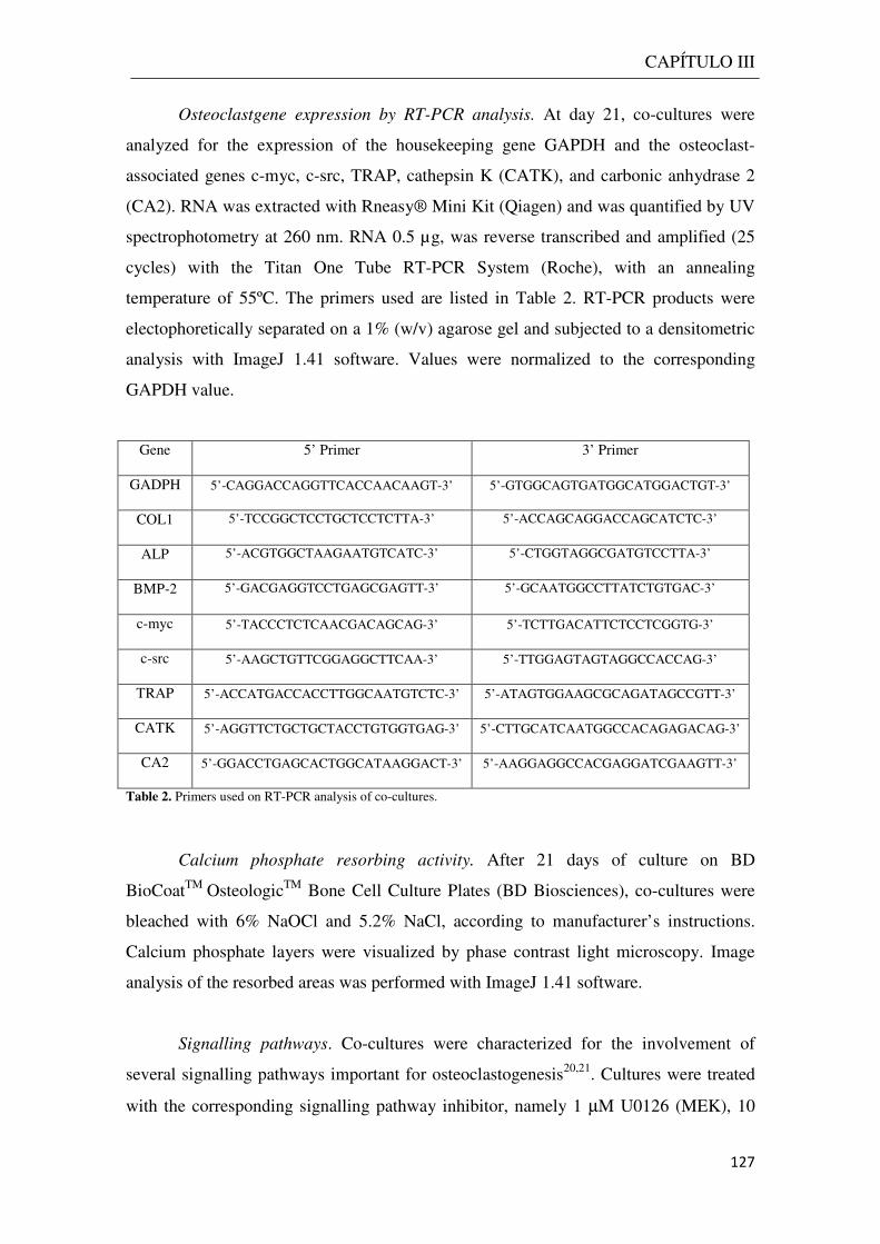
CAPÍTULO III
127
Osteoclastgene expression by RT-PCR analysis. At day 21, co-cultures were
analyzed for the expression of the housekeeping gene GAPDH and the osteoclast-
associated genes c-myc, c-src, TRAP, cathepsin K (CATK), and carbonic anhydrase 2
(CA2). RNA was extracted with Rneasy® Mini Kit (Qiagen) and was quantified by UV
spectrophotometry at 260 nm. RNA 0.5 µg, was reverse transcribed and amplified (25
cycles) with the Titan One Tube RT-PCR System (Roche), with an annealing
temperature of 55ºC. The primers used are listed in Table 2. RT-PCR products were
electophoretically separated on a 1% (w/v) agarose gel and subjected to a densitometric
analysis with ImageJ 1.41 software. Values were normalized to the corresponding
GAPDH value.
Gene 5’ Primer 3’ Primer
GADPH 5’-CAGGACCAGGTTCACCAACAAGT-3’ 5’-GTGGCAGTGATGGCATGGACTGT-3’
COL1 5’-TCCGGCTCCTGCTCCTCTTA-3’ 5’-ACCAGCAGGACCAGCATCTC-3’
ALP 5’-ACGTGGCTAAGAATGTCATC-3’ 5’-CTGGTAGGCGATGTCCTTA-3’
BMP-2 5’-GACGAGGTCCTGAGCGAGTT-3’ 5’-GCAATGGCCTTATCTGTGAC-3’
c-myc 5’-TACCCTCTCAACGACAGCAG-3’ 5’-TCTTGACATTCTCCTCGGTG-3’
c-src 5’-AAGCTGTTCGGAGGCTTCAA-3’ 5’-TTGGAGTAGTAGGCCACCAG-3’
TRAP 5’-ACCATGACCACCTTGGCAATGTCTC-3’ 5’-ATAGTGGAAGCGCAGATAGCCGTT-3’
CATK 5’-AGGTTCTGCTGCTACCTGTGGTGAG-3’ 5’-CTTGCATCAATGGCCACAGAGACAG-3’
CA2 5’-GGACCTGAGCACTGGCATAAGGACT-3’ 5’-AAGGAGGCCACGAGGATCGAAGTT-3’
Table 2. Primers used on RT-PCR analysis of co-cultures.
Calcium phosphate resorbing activity. After 21 days of culture on BD
BioCoatTM OsteologicTM Bone Cell Culture Plates (BD Biosciences), co-cultures were
bleached with 6% NaOCl and 5.2% NaCl, according to manufacturer’s instructions.
Calcium phosphate layers were visualized by phase contrast light microscopy. Image
analysis of the resorbed areas was performed with ImageJ 1.41 software.
Signalling pathways. Co-cultures were characterized for the involvement of
several signalling pathways important for osteoclastogenesis20,21. Cultures were treated
with the corresponding signalling pathway inhibitor, namely 1 µM U0126 (MEK), 10
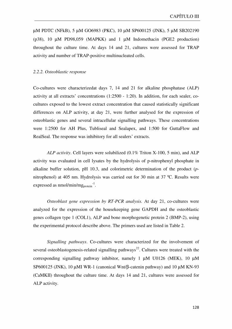
CAPÍTULO III
128
µM PDTC (NFkB), 5 µM GO6983 (PKC), 10 µM SP600125 (JNK), 5 µM SB202190
(p38), 10 µM PD98,059 (MAPKK) and 1 µM Indomethacin (PGE2 production)
throughout the culture time. At days 14 and 21, cultures were assessed for TRAP
activity and number of TRAP-positive multinucleated cells.
2.2.2. Osteoblastic response
Co-cultures were characterizedat days 7, 14 and 21 for alkaline phosphatase (ALP)
activity at all extracts’ concentrations (1:2500 - 1:20). In addition, for each sealer, co-
cultures exposed to the lowest extract concentration that caused statistically significant
differences on ALP activity, at day 21, were further analysed for the expression of
osteoblastic genes and several intracellular signalling pathways. These concentrations
were 1:2500 for AH Plus, Tubliseal and Sealapex, and 1:500 for GuttaFlow and
RealSeal. The response was inhibitory for all sealers’ extracts.
ALP activity. Cell layers were solubilized (0.1% Triton X-100, 5 min), and ALP
activity was evaluated in cell lysates by the hydrolysis of p-nitrophenyl phosphate in
alkaline buffer solution, pH 10.3, and colorimetric determination of the product (p-
nitrophenol) at 405 nm. Hydrolysis was carried out for 30 min at 37 ºC. Results were
expressed as nmol/min/mgprotein-1.
Osteoblast gene expression by RT-PCR analysis. At day 21, co-cultures were
analyzed for the expression of the housekeeping gene GAPDH and the osteoblastic
genes collagen type 1 (COL1), ALP and bone morphogenetic protein 2 (BMP-2), using
the experimental protocol describe above. The primers used are listed in Table 2.
Signalling pathways. Co-cultures were characterized for the involvement of
several osteoblastogenesis-related signalling pathways22. Cultures were treated with the
corresponding signalling pathway inhibitor, namely 1 µM U0126 (MEK), 10 µM
SP600125 (JNK), 10 µMI WR-1 (canonical Wnt/β-catenin pathway) and 10 µM KN-93
(CaMKII) throughout the culture time. At days 14 and 21, cultures were assessed for
ALP activity.
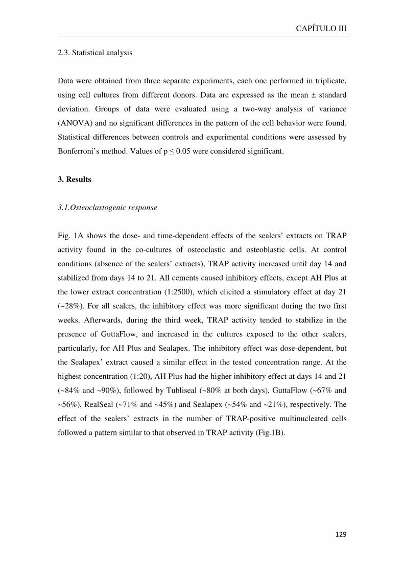
CAPÍTULO III
129
2.3. Statistical analysis
Data were obtained from three separate experiments, each one performed in triplicate,
using cell cultures from different donors. Data are expressed as the mean ± standard
deviation. Groups of data were evaluated using a two-way analysis of variance
(ANOVA) and no significant differences in the pattern of the cell behavior were found.
Statistical differences between controls and experimental conditions were assessed by
Bonferroni’s method. Values of p ≤ 0.05 were considered significant.
3. Results
3.1.Osteoclastogenic response
Fig. 1A shows the dose- and time-dependent effects of the sealers’ extracts on TRAP
activity found in the co-cultures of osteoclastic and osteoblastic cells. At control
conditions (absence of the sealers’ extracts), TRAP activity increased until day 14 and
stabilized from days 14 to 21. All cements caused inhibitory effects, except AH Plus at
the lower extract concentration (1:2500), which elicited a stimulatory effect at day 21
(~28%). For all sealers, the inhibitory effect was more significant during the two first
weeks. Afterwards, during the third week, TRAP activity tended to stabilize in the
presence of GuttaFlow, and increased in the cultures exposed to the other sealers,
particularly, for AH Plus and Sealapex. The inhibitory effect was dose-dependent, but
the Sealapex’ extract caused a similar effect in the tested concentration range. At the
highest concentration (1:20), AH Plus had the higher inhibitory effect at days 14 and 21
(~84% and ~90%), followed by Tubliseal (~80% at both days), GuttaFlow (~67% and
~56%), RealSeal (~71% and ~45%) and Sealapex (~54% and ~21%), respectively. The
effect of the sealers’ extracts in the number of TRAP-positive multinucleated cells
followed a pattern similar to that observed in TRAP activity (Fig.1B).
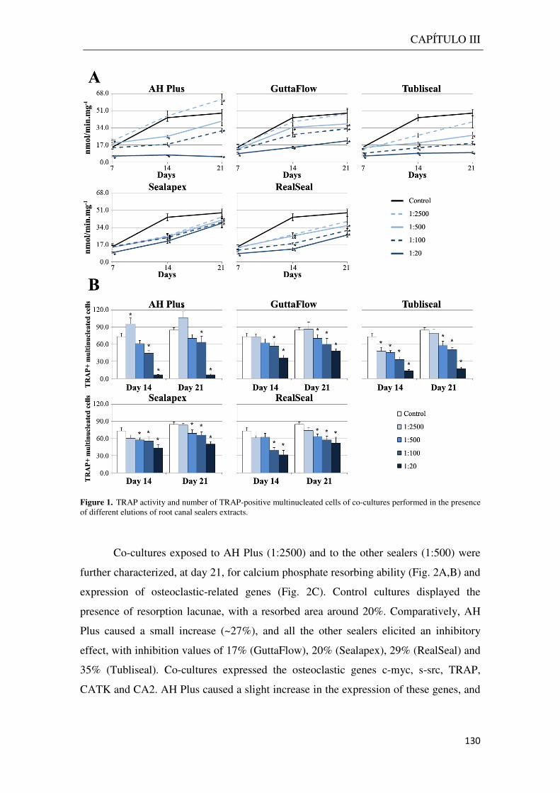
CAPÍTULO III
130
Figure 1. TRAP activity and number of TRAP-positive multinucleated cells of co-cultures performed in the presence of different elutions of root canal sealers extracts.
Co-cultures exposed to AH Plus (1:2500) and to the other sealers (1:500) were
further characterized, at day 21, for calcium phosphate resorbing ability (Fig. 2A,B) and
expression of osteoclastic-related genes (Fig. 2C). Control cultures displayed the
presence of resorption lacunae, with a resorbed area around 20%. Comparatively, AH
Plus caused a small increase (~27%), and all the other sealers elicited an inhibitory
effect, with inhibition values of 17% (GuttaFlow), 20% (Sealapex), 29% (RealSeal) and
35% (Tubliseal). Co-cultures expressed the osteoclastic genes c-myc, s-src, TRAP,
CATK and CA2. AH Plus caused a slight increase in the expression of these genes, and
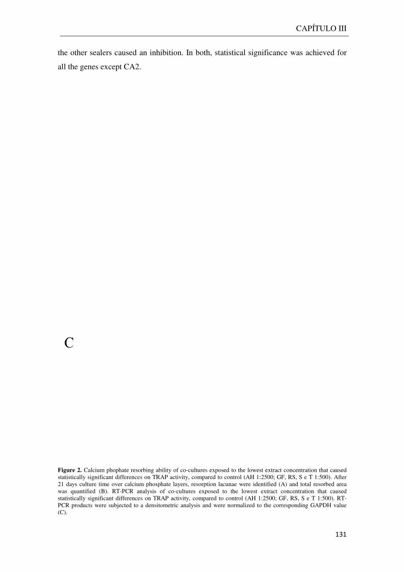
the other sealers caused an
all the genes except CA2.
Figure 2. Calcium phophate resorbing ability of costatistically significant differences on TRAP activity, compared to control (A21 days culture time over calcium phosphate layers, resorption lacunae were identified (A) and total resorbed area was quantified (B). RT-PCR analysis of costatistically significant differences on TRAP activity, compared to control (AH 1:2500; GF, RS, S e T 1:500). RTPCR products were subjected to a densitometric analysis and were normalized to the corr(C).
n inhibition. In both, statistical significance was achieved for
Calcium phophate resorbing ability of co-cultures exposed to the lowest extract concentration that caused differences on TRAP activity, compared to control (AH 1:2500; GF, RS, S e T 1:500).
21 days culture time over calcium phosphate layers, resorption lacunae were identified (A) and total resorbed area PCR analysis of co-cultures exposed to the lowest extract concentration that caused
statistically significant differences on TRAP activity, compared to control (AH 1:2500; GF, RS, S e T 1:500). RTPCR products were subjected to a densitometric analysis and were normalized to the corresponding GAPDH value
CAPÍTULO III
131
In both, statistical significance was achieved for
cultures exposed to the lowest extract concentration that caused H 1:2500; GF, RS, S e T 1:500). After
21 days culture time over calcium phosphate layers, resorption lacunae were identified (A) and total resorbed area exposed to the lowest extract concentration that caused
statistically significant differences on TRAP activity, compared to control (AH 1:2500; GF, RS, S e T 1:500). RT-esponding GAPDH value
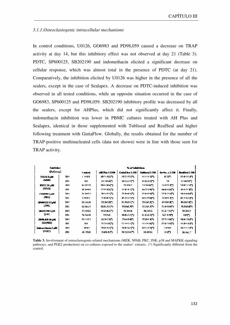
3.1.1.Osteoclastogenic intracellular mechanisms
In control conditions, U0126, GO6983 and PD98,059 cau
activity at day 14, but this inhibitory effect was not observed at day 21 (
PDTC, SP600125, SB202190 and
cellular response, which was almost total in the presence of PDTC (at day 21).
Comparatively, the inhibition elicited by U0126 was higher in the presence of all the
sealers, except in the case of Sealapex. A decrease on PDTC
observed in all tested conditions, while an opposite situation occurred in the case of
GO6983, SP600125 and PD98,059. SB202190 inhibitory profile was decreased by all
the sealers, except for AHPlus, which did not significantly
indomethacin inhibition was lower in PBMC cultures treated with AH
Sealapex, identical in those supplemen
following treatment with GuttaFlow.
TRAP-positive multinucleated cells
TRAP activity.
Table 3. Involvement of osteoclastogenicpathways, and PGE2 production) on cocontrol.
Osteoclastogenic intracellular mechanisms
In control conditions, U0126, GO6983 and PD98,059 caused a decrease on TRAP
at day 14, but this inhibitory effect was not observed at day 21 (
PDTC, SP600125, SB202190 and indomethacin elicited a significant decrease on
cellular response, which was almost total in the presence of PDTC (at day 21).
Comparatively, the inhibition elicited by U0126 was higher in the presence of all the
cept in the case of Sealapex. A decrease on PDTC-induced inhibition was
observed in all tested conditions, while an opposite situation occurred in the case of
GO6983, SP600125 and PD98,059. SB202190 inhibitory profile was decreased by all
pt for AHPlus, which did not significantly affect
indomethacin inhibition was lower in PBMC cultures treated with AH
identical in those supplemented with Tubliseal and RealSeal and
following treatment with GuttaFlow. Globally, the results obtained for the
positive multinucleated cells (data not shown) were in line with those
Involvement of osteoclastogenic-related mechanisms (MEK, NFkB, PKC, JNK, p38 and MAPKK signaling pathways, and PGE2 production) on co-cultures exposed to the sealers’ extracts. (*) Significantly different from the
CAPÍTULO III
132
sed a decrease on TRAP
at day 14, but this inhibitory effect was not observed at day 21 (Table 3).
elicited a significant decrease on
cellular response, which was almost total in the presence of PDTC (at day 21).
Comparatively, the inhibition elicited by U0126 was higher in the presence of all the
induced inhibition was
observed in all tested conditions, while an opposite situation occurred in the case of
GO6983, SP600125 and PD98,059. SB202190 inhibitory profile was decreased by all
affect it. Finally,
indomethacin inhibition was lower in PBMC cultures treated with AH Plus and
ted with Tubliseal and RealSeal and higher
Globally, the results obtained for the number of
were in line with those seen for
JNK, p38 and MAPKK signaling
(*) Significantly different from the
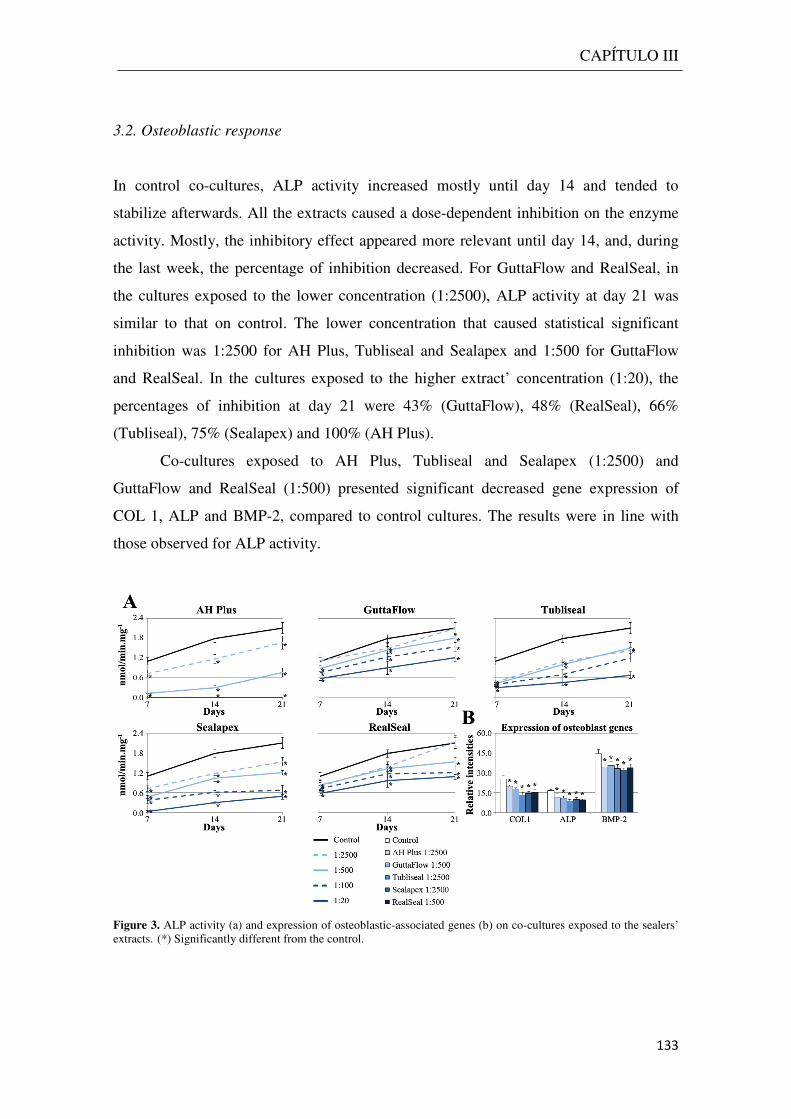
CAPÍTULO III
133
3.2. Osteoblastic response
In control co-cultures, ALP activity increased mostly until day 14 and tended to
stabilize afterwards. All the extracts caused a dose-dependent inhibition on the enzyme
activity. Mostly, the inhibitory effect appeared more relevant until day 14, and, during
the last week, the percentage of inhibition decreased. For GuttaFlow and RealSeal, in
the cultures exposed to the lower concentration (1:2500), ALP activity at day 21 was
similar to that on control. The lower concentration that caused statistical significant
inhibition was 1:2500 for AH Plus, Tubliseal and Sealapex and 1:500 for GuttaFlow
and RealSeal. In the cultures exposed to the higher extract’ concentration (1:20), the
percentages of inhibition at day 21 were 43% (GuttaFlow), 48% (RealSeal), 66%
(Tubliseal), 75% (Sealapex) and 100% (AH Plus).
Co-cultures exposed to AH Plus, Tubliseal and Sealapex (1:2500) and
GuttaFlow and RealSeal (1:500) presented significant decreased gene expression of
COL 1, ALP and BMP-2, compared to control cultures. The results were in line with
those observed for ALP activity.
Figure 3. ALP activity (a) and expression of osteoblastic-associated genes (b) on co-cultures exposed to the sealers’ extracts. (*) Significantly different from the control.
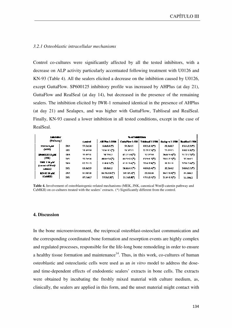
3.2.1 Osteoblastic intracellular mechanisms
Control co-cultures were significantly affected by all the tested inhibitors, with a
decrease on ALP activity particularly accentuated following treatment with U0126 and
KN-93 (Table 4). All the sealers
except GuttaFlow. SP600125 inhibitory profile was increased
GuttaFlow and RealSeal (at day 14), but decreased in the presence of the remaining
sealers. The inhibition elicited by IWR
(at day 21) and Sealapex, and was higher
Finally, KN-93 caused a lower inhibition in all tested conditions, except in the case of
RealSeal.
Table 4. Involvement of osteoblastogenicCaMKII) on co-cultures treated with the sealers’ extracts.
4. Discussion
In the bone microenvironment, the reciprocal osteoblast
the corresponding coordinated bone formation and resorption events are highly complex
and regulated processes, responsible for the life
a healthy tissue formation and
osteoblastic and osteoclastic cells were used as an
and time-dependent effects of endodontic sealers’ extracts
were obtained by incubating the freshly mixed material with culture medium, as,
clinically, the sealers are applied in this form, and the uns
intracellular mechanisms
cultures were significantly affected by all the tested inhibitors, with a
decrease on ALP activity particularly accentuated following treatment with U0126 and
sealers elicited a decrease on the inhibition caused by
except GuttaFlow. SP600125 inhibitory profile was increased by AHPlus (at day 21),
GuttaFlow and RealSeal (at day 14), but decreased in the presence of the remaining
cited by IWR-1 remained identical in the presence of AHPlus
(at day 21) and Sealapex, and was higher with GuttaFlow, Tubliseal and RealSeal.
93 caused a lower inhibition in all tested conditions, except in the case of
Involvement of osteoblastogenic-related mechanisms (MEK, JNK, canonical Wnt/β-catenincultures treated with the sealers’ extracts. (*) Significantly different from the control.
microenvironment, the reciprocal osteoblast-osteoclast communication and
corresponding coordinated bone formation and resorption events are highly complex
responsible for the life-long bone remodeling in order to ensure
formation and maintenance14. Thus, in this work, co-cultures of
osteoblastic and osteoclastic cells were used as an in vitro model to address
dependent effects of endodontic sealers’ extracts in bone cells.
were obtained by incubating the freshly mixed material with culture medium, as,
clinically, the sealers are applied in this form, and the unset material might contact with
CAPÍTULO III
134
cultures were significantly affected by all the tested inhibitors, with a
decrease on ALP activity particularly accentuated following treatment with U0126 and
elicited a decrease on the inhibition caused by U0126,
AHPlus (at day 21),
GuttaFlow and RealSeal (at day 14), but decreased in the presence of the remaining
1 remained identical in the presence of AHPlus
GuttaFlow, Tubliseal and RealSeal.
93 caused a lower inhibition in all tested conditions, except in the case of
catenin pathway and
(*) Significantly different from the control.
osteoclast communication and
corresponding coordinated bone formation and resorption events are highly complex
long bone remodeling in order to ensure
cultures of human
model to address the dose-
bone cells. The extracts
were obtained by incubating the freshly mixed material with culture medium, as,
et material might contact with
CAPÍTULO III
135
apical and periapical tissues. The sealers’ surface/medium rate was 1.3cm2/ml, which is
in agreement to ISO Standards 10993-5 for the testing of dental materials.
It is known that osteoblasts synthesize a variety of molecules important in the
recruitment and survival of osteoclast precursors and also engaged with the later steps
of osteoclastogenesis, namely M-CSF and RANKL, which are sufficient to promote in
vitro osteoclastogenesis13. Also, some factors produced by osteoclasts seem to stimulate
osteogenesis14. In the present work, the protocol used allowed the establishment of co-
cultures of osteoclastic and osteoblastic cells displaying phenotype features of both cell
types, in the absence of any exogenous differentiation factors, as observed
previously16,19. Osteoclast parameters included a high TRAP activity and number of
TRAP-positive multinucleated cells, expression of osteoclastic-related genes and
calcium phosphate resorbing ability. Co-cultures also showed high ALP levels and gene
expression of ALP, COL 1 and BMP-2, which are typical osteoblastic markers13. The
differentiation of osteoclastic and osteoblastic cells was evident by the progressive
increase in TRAP and ALP activities, respectively, which occurred mostly during the
two first weeks; values stabilized in the last week, suggesting that differentiated/mature
osteoclasts and osteoblasts were present from day 14 onwards. Thus, this co-culture
system appeared as a useful model to analyze the effects of endodontic sealers in
interacting osteoclastic and osteoblastic cells.
The results on TRAP and ALP activities observed in the co-cultures showed that
the sealers’ extracts caused dose-dependent inhibitory effects on osteoclastic and
osteoblastic behavior. The only exception was the low stimulatory effect on TRAP
activity seen in the presence of the lowest concentration of AH Plus, reflecting most
probably an adaptive cell response to cope with the presence of toxic compounds.
Differences were noted for the effects on the two cell types, and also on the profile of
each sealer. Regarding TRAP activity and number of TRAP-positive multinucleated
cells, inhibition was mainly observed during the two first weeks, i.e. during osteoclastic
differentiation13,23; afterwards, values stabilized (as for GuttaFlow) or increased (as for
the other sealers). The lower concentration to cause inhibitory effects was 1:100 for AH
Plus and 1:500 for the other sealers. Evident dose-dependent inhibitory effects were
observed for AH Plus, GuttaFlow, Tubliseal and RealSeal, but Sealapex caused a
similar inhibition in the tested concentration range (1:2500 - 1:20) and cells recovered
almost completely from the initial deleterious effects. Compared to the osteoclastic
cells, osteoblastic cells appeared more sensitive to the toxic effects of the sealers. This is
CAPÍTULO III
136
suggested by the results regarding the lower concentration that caused statistical
significant decrease on ALP activity (i.e. 1:2500 for AH Plus, Tubliseal and Sealapex)
and also by lower ability to recover from the inflicted initial toxicity. The later
observation was especially evident for AH Plus (in which the two higher concentrations
were lethal) and Sealapex (that caused dose-dependent inhibition in the tested
concentration range). As observed for the osteoclastic behavior, inhibition on ALP
activity occurred mainly during the first two weeks, i.e during the osteoblastic
differentiation13,18.
In the clinical setting, the contact of the endodontic sealer with the apical and
periapical tissues involves a small area; in addition, the potential leachable cytotoxic
compounds are continually cleared by the extracellular fluids, suggesting the presence
of low local levels after the initial contact. Thus, in predicting the long-term effects of
the sealers, exposure of bone cells to low concentrations of the sealers’ extracts appears
to be a more representative situation. Thus, co-cultures exposed to the lowest tested
concentrations that elicited statistically significant effects on TRAP and ALP activities
were further characterized for other phenotype parameters. Regarding the osteoclastic
behavior, the extracts affected the expression of the differentiation and activation genes
c-myc and c-src and the functional genes TRAP and CATK, and the calcium phosphate
resorbing activity. The sealers also decreased the expression of the osteoblastic genes
COL 1, ALP and BMP-2. The results were in line with those observed for TRAP and
ALP activities, respectively.
Osteoclast and osteoblast differentiation are complex processes that involve
multiple overlapping intracellular mechanisms that, at the end, direct the precursors or
the immature cells towards the formation of mature and functional bone cells20,22. In this
context, co-cultures were further characterized for the involvement of several signalling
pathways on the observed cellular responses. It is important to note that although very
important, none of them is exclusive of osteoclastogenesis or osteoblastogenesis, and
two of the analyzed pathways (MEK and JNK) have an important role in both
processes. The sealers differed significantly on the relative contribution of the signalling
pathways, which might be related with the different nature of the toxic compounds
associated with each sealer’ toxicity. Some differences were particularly evident. In the
case of the osteoclastogenic response, MEK pathway appeared to be significantly up
regulated following treatment with AHPlus, GuttaFlow and Tubliseal, while NFkB
seemed to be less involved in the presence of Tubliseal, Sealapex and Realseal. All the
CAPÍTULO III
137
sealers elicited an increase on the involvement of PKC, JNK and MAPKK pathways.
p38 pathway seemed to be down regulated by Sealapex and RealSeal. Finally, PGE2
production had no significant contribution in PBMC cultures supplemented with
Sealapex. Regarding the osteoblastic response, MEK pathway had a little role in the
presence of RealSeal. GuttaFlow and Tubliseal appeared to induce an up regulation of
the canonical Wnt/β-catenin pathway, while Ca2+/calmodulin-dependent protein kinase
II appeared to be down regulated by AHPlus, GuttaFlow and Sealapex.
Many studies have reported on the cellular effects of endodontic sealers, but a
great results discrepancy is observed regarding cytotoxicity patterns or the effects on a
specific cell type, which is expected considering the variety of experimental conditions
regarding the tested cell type (mainly fibroblasts), the preparation of the extracts, and
the concentration range and time of exposure. GuttaFlow, a silicone-based sealer
manufactured by adding gutta-percha powder to the silicone matrix (and containing
nanosilver particles as a preservative) was previously tested in fibroblasts24,25,26,27,28; its
toxicity might result from the release of nanosilver particles as they have been
associated with dose-dependent cytotoxic effects1. It is also reported the presence of
small voids within the core of GuttaFlow, which favor the release of unreacted
compounds that accumulates in such pores29. Effects of AH Plus were evaluated in
fibroblasts10,24-28,30,31,32, human periodontal ligament cells33 and osteoblasts2,3,10,11. Most
of these studies suggested that this epoxy resin-based sealer, apparently “formaldehyde-
free”, exhibited a moderate to severe toxicity immediately after mixing (the conditions
used in the present work), due to the release of low levels of formaldehyde, the presence
of amines added to accelerate the epoxy polymerization and the epoxy resin content34,35.
The toxicity of Tubliseal, a zinc oxide eugenol-based sealer has been attributed to free
eugenol release, and also to several additives, namely the resins used for greater dentin
adhesion1, and was demonstrated in fibroblasts28 and human periodontal ligament
cells36. Sealapex was tested in fibroblasts32,37, human periodontal ligament cells33,36 and
osteoblasts8,12, and its toxicity has been associated to an increase in the pH of the culture
medium resulting from the sealer dissolution1,34,38. In the present work, the pH remained
constant throughout the culture time, due to the low tested levels, and other components
such as polyresin, zinc oxide and Ba2+ have been suggested as potential toxic
compounds1. RealSeal is a dual-cured hydrophilic multi-methacrylate resin-based
sealer, and its toxic effects were shown in fibroblasts10,24,26,31,34,37,39,40,41, human dental
pulp cells41 and osteoblasts7,9,10, and it is caused by the leaching of unreacted monomers
CAPÍTULO III
138
as a result of incomplete polymerization, and also to filler particles due to the
degradation of the sealer.
Regarding bone cells, few studies were performed in osteoblastic cells involving
the sealers studied in the present work. AH Plus was tested in human U2-OS
osteosarcoma cells and authors reported that the activation of COX-2 expression2 and
IL-6 and IL-8 mRNA gene expression3 might be contributing mechanisms in the
toxicity of this sealer. Studies performed in the rat cell lines MC3T3-E1 and ROS
17/2.8 showed that the toxicity of AH Plus decreased with the aging of the sealer11 and
that these cell lines responded differently to this sealer10. Viability and apoptosis was
analyzed in rat calvarial osteoblasts exposed to conditioned medium from Sealapex12.
MC3T3-E1 cells were also exposed to Sealapex extracts, and authors suggested that
oxidative stress is not involved in the cytotoxicity of this sealer8. Disks of RealSeal,
after setting, were placed in direct contact with ROS 17/2.8 cells and for 5 succeeding
weeks after immersion in simulated body fluid, and results showed that toxicity
decreased gradually over time9. RealSeal extracts showed initial cytotoxicity effect on
MG63 cells which disappeared after few days of culture7. Considering other sealers, a
study performed with extracts of AH26, Canals and N2 showed an up-regulation on the
expression RANKL in U2-OS cells5. As RANKL plays a critical role in
osteoclastogenesis, authors suggested that this might be a contributing mechanism in the
pathogenesis of periapical bone destruction induced by root canal sealers5. An indirect
effect on osteoclastic activity might also be hypothesized in the studies referred above
showing that AH Plus caused an induction on the expression of COX-22 and IL-6 and
IL-83 in U2-OS cells, as these molecules might have a role in osteoclastic activity13.
These studies suggest that toxicity of these sealers in the bone tissue might involve an
alteration of the normal interaction between osteoblasts and osteoclasts in the bone
microenvironment. However, to the best of our knowledge, the present study in the first
to address the effects of endodontic sealers in osteoblastic and osteoclastic cells in a co-
culture system.
5. Conclusion
Results of the present work showed that extracts from freshly mixed sealers
caused mostly inhibitory effects in the osteoclastic and the osteoblastic behavior of co-
cultures of human peripheral blood mononuclear cells and bone marrow cells. In co-
CAPÍTULO III
139
cultured cells, the sealers caused a dose-dependent decrease on TRAP and ALP
activities, respectively an osteoclastic and an osteoblastic marker. The inhibitory effect
was more significant during the two first weeks, the culture period mainly engaged with
the osteoclastic and osteoblastic differentiation, in the experimental conditions used.
Afterwards, adaptive cell responses were observed, as reflected by stabilization or
increase on TRAP and ALP activities. Osteoblastic cells were more sensitive to the
toxic effects of the sealers’ extracts, compared to the osteoclastic cells, as suggested by
the cytotoxic profile in the tested concentration range and the lower ability to recover
from the initial deleterious effects. Other phenotype parameters, as the resorbing ability
of the osteoclastic cells and the expression of osteoclastic and osteoblastic genes were
also affected, and results were in line with those observed on TRAP and ALP activities.
The sealers differed on the dose- and time-dependent profile in co-cultured osteoclastic
and osteoblastic cells, and also in the modulation of intracellular signaling pathways
important for osteoclastogenesis and osteoblastogenesis. Although in vivo, a lower
cytotoxicity is expected due to the continuous fluids flow and the presence of the
extracellular matrix, the higher sensitivity of the osteoblastic cells and the better
adaptive response of the osteoclastic cells, suggested in the present work, might disrupt
the highly regulated interaction between these cell types, compromising the local bone
metabolism.
140
References
1Hauman CH, Love RM. Biocompatibility of dental materials used in contemporary endodontic therapy: a review. Part 2.Root canal filling materials. Int Endod J 2003; 36:147-60. 2Huang FM, Chou MY and Chang YC. Induction of cyclooxygenase-2 mRNA and protein expression by epoxy resin and zinc oxide-eugenol based root canal sealers in human osteoblastic cells. Biomaterials 2003; 24: 1869-75. 3Huang FM, Tsai C-H, Yang S-F, Chang Y-C. Induction of interleukin-6 and interleukin-8 gene expression by root canal sealers in human osteoblastic cells. J Endod 2005;31:679-683. 4Huang FM, Yang S-F, Chang Y-C. Up-regulation of gelatinases and tissue type plasminogen activator by root canal sealers in human osteoblastic cells. J Endod 2008;34:291-4. 5Huang FM, Lee SS, Yang SF, Chang YC. Up-regulation of receptor activator nuclear factor-kappaB ligand expression by root canal sealers in human osteoblastic cells. J Endod 2009;35:363–6. 6Huang FM, Yang S-F, Chang Y-C. Effects of root canal sealers on alkaline phosphatase in human osteoblastic cells. J Endod 2010;36:1230-3. 7Xu P et al. Cytotoxicity of RealSeal on human osteoblast like MG63 cells. J Endod 2010;36:40- 44. 8Lee DH et al. Mechanisms of root canal sealers cytotoxicity on osteoblastic cell line MC3T3-E1. Oral Surg Oral Med Oral Pathol Oral Radiol Endod 2007;104:717- 21. 9Ames JM et al. Contemporary methacrylate resin-based root canal sealers exhibit different degrees of ex vivo cytotoxicity when cured in their self-cured mode. J Endod 2009;35:225- 28. 10Brackett MG et al. Cytotoxic response of three cell lines exposed in vitro to dental endodontic sealers. J Biomed Mat Res B: Appl Biomater 2010; 95B:380- 86. 11Bryan TE et al. In vitro osteogenic potencial of an experimental calcium silicate-based root canal sealer. J Endod 2010;36:1163-69. 12Al-Awadhi S et al. Cultured primary osteoblasts viability and apoptosis in the presence of root canal sealers. J Endod 2004;30:527- 33. 13Datta HK et al. The cell biology of bone metabolism. J Clin Pathol 2008;61:577-87. 14Matsuo K, Irie N, Osteoclast-osteoblast communication. Arch Biochem Biophys 2008; 473: 201-209. 15Heinemann C et al. Development of an osteoblast/osteoclast co-culture derived by human bone marrow stromal cells and human monocytes for biomaterial testing. Eur Cell Mater 2011; 21: 80- 93. 16Costa-Rodrigues J, Fernandes A, Fernandes MH. Reciprocal osteoblastic and osteoclastic modulation in co-cultured MG63 osteosarcoma cells and human osteoclast precursors. J Cell Biochem 2011;112:3704-13. 17Costa-Rodrigues J, Fernandes MH. Paracrine-mediated differentiation and activation of human hematopoietic osteoclast precursor cells by skin and gingival fibroblasts. Cell Prolifer 2011; 44: 264-73. 18Coelho MJ, Fernandes MH. Human bone cell cultures in biocompatibility testing. Part II: effect of ascorbic acid, b-glycerophosphate and dexamethasone on osteoblastic differentiation. Biomaterials 2000; 21: 1095-1102. 19Costa-Rodrigues J et al. Hydroxyapatite surface roughness: Complex modulation of the osteoclastogenesis of human precursor cells. Acta Biomaterials 2012; 8:1137-45.
141
20Zhao Q et al.Osteoclast differentiation and gene regulation. Front Biosci 2007; 12, 2519–2529. 21Vaananen HK, Laitala-Leinonen T. Osteoclast lineage and function. Arch Biochem Biophys 2008;473: 32–8. 22Lian JB et al. Networks and hubs for the transcriptional control of osteoblastogenesis. Rev Endocr Metab Disord 2006;7: 1-16. 23Costa-Rodrigues J, Fernandes A, Fernandes MH. Spontaneous and induced osteoclastogenic behaviour of human peripheral blood mononuclear cells and their CD14+ and CD14- cell fractions. Cell Prolifer 2011; 44: 410-419. 24Bouillaguet S et al. Initial in vitro biological response to contemporary endodontic sealers. J Endod 2006;32: 989- 92. 25Eldeniz AU et al. Cytotoxicity of new resin-, calcium hydroxide-, and silicone-based root canal sealers on fibroblasts derived from human gingival and L929 cell lines. Int Endod J 2007; 40: 329- 37. 26Brackett MG et al. Cytotoxicity of endodontic materials over 6-weeks ex vivo. Int Endod J 2008; 41: 1072- 78. 27Willershausen I et al. In vitro analysis of the cytotoxicity and the antimicrobial effect of four endodontic sealers. Head & Face Med 2011; 7: 15. 28Zoufan K et al. Cytotoxicity evaluation of GuttaFlow and Endo Sequence BC sealers. Oral Surg Oral Med Oral Pathol Oral Radiol Endod 2011; 112:657-61. 29Elayouti A et al. Homogeneity and adaptation of a new gutta-percha paste to root canal walls. J Endod 2005; 31: 687-90. 30Miletić I et al. The cytotoxicity of RoekoSeal and AH Plus Compared during different setting periods. J Endod 2005; 31: 307- 9. 31Lodienè G et al. Toxicity evaluation of root canal sealers in vitro. Int Endod J 2008; 41: 72-7. 32Silva EJ et al. Evaluation of cytotoxicity and up-regulation of gelatinases in human fibroblast cells by four root canal sealers. Int Endod J 2012; 45: 49- 56. 33Huang FM et al. Cytotoxicity of resin-, zinc oxide eugenol-, and calcium hydroxide-based root canal sealers on human periodontal ligament cells and permanent V79 cells. Int Endod J 2002;35:153- 8. 34Eldeniz AU et al. Evaluation of pH and ion release of Acroseal sealer in comparision with Apexit and Sealapex sealers.Oral Surg Oral Med Oral Pathol Oral RadiolEndod 2005; 103: e86- e91. 35Merdad K et al. Short term cytotoxicity assessement of components of the Epiphany resin-percha obturating system by indirect and direct Millipore filter assays. J Endod 2007; 33: 24- 7. 36Chang MC et al Comparative cytotoxicity of five root canal sealers on cultured human periodontal ligament fibroblast. Int Endod J 2010; 43: 251- 7. 37Key JE, Rahemtulla FG, Eleazer PD. Cytotoxicity of new root canal filling material on human gingival fibroblasts. J Endod 2006; 32: 756- 8. 38Duarte MA et al. Evaluation of pH and calcium ion release of three root canal sealers. J Endod 2000; 26: 389- 90. 39Susini G et al. Cytotoxicity of Epiphany and Resilon with a root model. Int End J 2006; 39: 940- 44.

142
40Donadio M et al. Cytotoxicity evaluation of Activ GP and Resilon sealers in vitro. Oral Surg Oral Med Oral Pathol Oral Radiol Endod 2009; 107: e74- e78. 41Karapinar-Kazandağ M et al. Cytotoxicity of 5 endodontic sealers on L929 cell line and human dental pulp cells. Int Endod J 2011; 44: 626- 34.

DISCUSSÃO

144
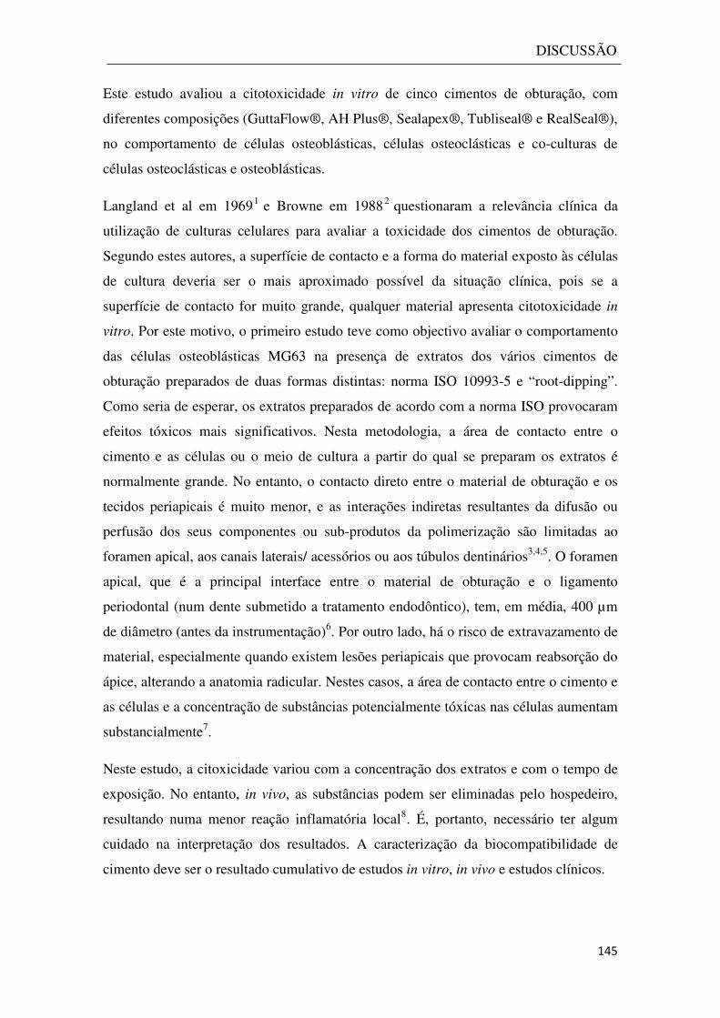
DISCUSSÃO
145
Este estudo avaliou a citotoxicidade in vitro de cinco cimentos de obturação, com
diferentes composições (GuttaFlow®, AH Plus®, Sealapex®, Tubliseal® e RealSeal®),
no comportamento de células osteoblásticas, células osteoclásticas e co-culturas de
células osteoclásticas e osteoblásticas.
Langland et al em 19691 e Browne em 19882 questionaram a relevância clínica da
utilização de culturas celulares para avaliar a toxicidade dos cimentos de obturação.
Segundo estes autores, a superfície de contacto e a forma do material exposto às células
de cultura deveria ser o mais aproximado possível da situação clínica, pois se a
superfície de contacto for muito grande, qualquer material apresenta citotoxicidade in
vitro. Por este motivo, o primeiro estudo teve como objectivo avaliar o comportamento
das células osteoblásticas MG63 na presença de extratos dos vários cimentos de
obturação preparados de duas formas distintas: norma ISO 10993-5 e “root-dipping”.
Como seria de esperar, os extratos preparados de acordo com a norma ISO provocaram
efeitos tóxicos mais significativos. Nesta metodologia, a área de contacto entre o
cimento e as células ou o meio de cultura a partir do qual se preparam os extratos é
normalmente grande. No entanto, o contacto direto entre o material de obturação e os
tecidos periapicais é muito menor, e as interações indiretas resultantes da difusão ou
perfusão dos seus componentes ou sub-produtos da polimerização são limitadas ao
foramen apical, aos canais laterais/ acessórios ou aos túbulos dentinários3,4,5. O foramen
apical, que é a principal interface entre o material de obturação e o ligamento
periodontal (num dente submetido a tratamento endodôntico), tem, em média, 400 µm
de diâmetro (antes da instrumentação)6. Por outro lado, há o risco de extravazamento de
material, especialmente quando existem lesões periapicais que provocam reabsorção do
ápice, alterando a anatomia radicular. Nestes casos, a área de contacto entre o cimento e
as células e a concentração de substâncias potencialmente tóxicas nas células aumentam
substancialmente7.
Neste estudo, a citoxicidade variou com a concentração dos extratos e com o tempo de
exposição. No entanto, in vivo, as substâncias podem ser eliminadas pelo hospedeiro,
resultando numa menor reação inflamatória local8. É, portanto, necessário ter algum
cuidado na interpretação dos resultados. A caracterização da biocompatibilidade de
cimento deve ser o resultado cumulativo de estudos in vitro, in vivo e estudos clínicos.

DISCUSSÃO
146
Num estudo recente, Brackett e colaboradores9 sugerem que a avaliação dos cimentos in
vitro deve incluir mais que um tipo celular. Neste trabalho avaliou-se o efeito de
extratos de cimentos de obturação em células osteoblásticas MG63, células
osteoclásticas e co-culturas de células osteoblásticas e osteoclásticas. As co-culturas de
osteoblastos e osteoclastos começaram por ser utilizadas para estudar a relação entre os
dois tipos celulares e a importância do RANKL na diferenciação dos osteoclastos. No
entanto, este conhecimento começou a ser aproveitado para outras aplicações práticas,
nomeadamente a utilização deste tipo de co-cultura em estudos de biocompatibilidade
de materiais.
Os cimentos de obturação utilizados neste estudo foram colocados em contacto com o
meio de cultura imediatamente após a mistura, uma vez que é o que acontece na
situação clínica – o cimento é colocado no interior do canal imediatamente após a sua
espatulação. Antes da polimerização completa, os cimentos podem libertar sub-produtos
citotóxicos que provocam irritação tecidular. As reações de polimerização podem causar
a degradação de alguns químicos ou produzir novas combinações que não estavam
presentes nas formulações originais.
Os cimentos à base de óxido de zinco eugenol são amplamente utilizados há muitos
anos, mas libertam eugenol. A libertação do eugenol por hidrólise é uma causa
demonstrada de toxicidade in vitro10,11,12,13 e in vivo
14,15,16. A libertação de eugenol
parece, no entanto, diminuir com o tempo, devido à progressiva hidrólise da superfície
do cimento 17 , pelo que a toxicidade dos cimentos diminui ao longo do tempo,
proporcionalmente à concentração de eugenol 18 . Parece, no entanto, que outros
componentes dos cimentos à base de óxido de zinco eugenol, como iões zinco (Zn2+) ou
óxido de zinco (ZnO), podem influenciar a sua toxicidade19,20.
O Tubliseal, cimento à base de óxido de zinco eugenol utilizado neste trabalho,
apresenta na sua constituição, e segundo o fabricante, 14% de uma resina oleosa.
Alguns componentes das resinas oleosas extraídas de plantas mostraram efeito
antibacteriano e promoveram a cicatrização de feridas21,22, mas também demonstraram
ser potencialmente citotóxicos 23 . A citotoxicidade do Tubliseal pode também ser
causada pelo seu teor de eugenol, no entanto a contribuição do iodeto de timol, resinas
oleosas e outros componentes do cimento necessita de mais investigação24. O Tubliseal
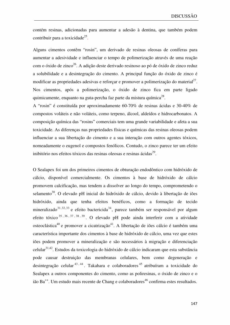
DISCUSSÃO
147
contém resinas, adicionadas para aumentar a adesão à dentina, que também podem
contribuir para a toxicidade25.
Alguns cimentos contêm “rosin”, um derivado de resinas oleosas de coníferas para
aumentar a adesividade e influenciar o tempo de polimerização através de uma reação
com o óxido de zinco26. A adição deste derivado resinoso ao pó de óxido de zinco reduz
a solubilidade e a desintegração do cimento. A principal função do óxido de zinco é
modificar as propriedades adesivas e reforçar e promover a polimerização do material27.
Nos cimentos, após a polimerização, o óxido de zinco fica em parte ligado
quimicamente, enquanto na guta-percha faz parte da mistura química28.
A “rosin” é constituída por aproximadamente 60-70% de resinas ácidas e 30-40% de
compostos voláteis e não voláteis, como terpeno, álcool, aldeídos e hidrocarbonatos. A
composição química das “rosins” comerciais tem uma grande variabilidade e afeta a sua
toxicidade. As diferenças nas propriedades físicas e químicas das resinas oleosas podem
influenciar a sua libertação do cimento e a sua interação com outros agentes tóxicos,
nomeadamente o eugenol e compostos fenólicos. Contudo, o zinco parece ter um efeito
inibitório nos efeitos tóxicos das resinas oleosas e resinas ácidas29.
O Sealapex foi um dos primeiros cimentos de obturação endodôntico com hidróxido de
cálcio, disponível comercialmente. Os cimentos à base de hidróxido de cálcio
promovem calcificação, mas tendem a dissolver ao longo do tempo, comprometendo o
selamento30. O elevado pH inicial do hidróxido de cálcio, devido à libertação de iões
hidróxido, ainda que tenha efeitos benéficos, como a formação de tecido
mineralizado31,32,33 e efeito bactericida34, parece também ser responsável por algum
efeito tóxico 35 , 36 , 37 , 38 , 39 . O elevado pH pode ainda interferir com a atividade
osteoclástica40 e promover a cicatrização41. A libertação de iões cálcio é também uma
característica importante dos cimentos à base de hidróxido de cálcio, uma vez que estes
iões podem promover a mineralização e são necessários à migração e diferenciação
celular31,42. Estudos da toxicologia do hidróxido de cálcio indicaram que esta substância
pode causar destruição das membranas celulares, bem como degeneração e
desintegração celular 43 , 44 . Takahara e colaboradores 45 atribuíram a toxicidade do
Sealapex a outros componentes do cimento, como as poliresinas, o óxido de zinco e o
ião Ba++. Um estudo mais recente de Chang e colaboradores46 confirma estes resultados.
DISCUSSÃO
148
O cimento AH Plus é o sucessor do cimento AH26, com a vantagem de, segundo o
fabricante, não libertar formaldeído durante a reação de polimerização. No entanto,
vários estudos referem que o AH Plus liberta uma pequena quantidade de formaldeído47,
48,49. A toxicidade do AH26 pode estar relacionada com outros constituintes do material,
além do formaldeído, como aminas ou resinas. Schweikl e colaboradores50 atribuíram a
citotoxicidade do AH26 à resina epoxi bisfenol, que também está presente na
constituição do AH Plus, justificando assim a sua toxicidade47,49, 51 . As aminas,
presentes para acelerar a polimerização também podem ser causa de toxicidade52. Em
resumo, a toxicidade inicial do AH Plus pode ser atribuída à libertação de uma pequena
quantidade de formaldeído, às aminas adicionadas para acelerar a polimerização ou à
resina epoxi.
O AH Plus tem uma composição química complexa, pelo que são várias as substâncias
que podem libertar-se durante a reação de polimerização e causar efeito tóxico. É um
sistema de duas pastas, cuja polimerização resulta da reação resina epoxi-aminas.
Este cimento tem efeito antibacteriano, o que também pode originar efeito tóxico53.
O tunguestato de cálcio, presente na constituição do AH Plus, é muito utilizado para
aumentar a radiopacidade dos cimentos de obturação54.
A presença de porosidades nos cimentos espatulados à mão (como é o caso do AH Plus)
pode favorecer a libertação de componentes que não reagem quimicamente, os quais
podem ser responsáveis por alguma toxicidade55.
O RealSeal é um cimento à base de resina de metacrilato, de última geração, que utiliza
cones de Resilon em substituição da guta-percha e cuja principal vantagem parece ser a
adesão química ao cone e às paredes da dentina, originando assim um “monobloco”. O
cimento de metacrilato necessita da temperatura corporal e de total ausência de ar para
polimerizar. Polimeriza em 30 minutos num ambiente anaeróbio, mas na presença de ar
demora muito mais tempo, e permanece uma camada superficial por polimerizar. Este
facto influencia significativamente as propriedades biológicas do material e tem sido
associado à sua toxicidade48.
A toxicidade dos cimentos de resina pode dever-se à presença de monómeros
hidrofílicos não polimerizados (BisGMA, UDMA, EBPADMA e trietilenoglicol-di-
metacrilato)56,57,58,59,60. A extrusão deste material para além do foramen apical poderá
DISCUSSÃO
149
originar uma camada não polimerizada durante longos períodos de tempo61,62. Outra
causa de toxicidade pode ser a libertação de partículas por degradação do material ao
longo do tempo63.
O RealSeal tem na sua constituição hidróxido de cálcio, originando um aumento do pH
no meio circundante, o que pode favorecer o processo reparativo42, tal como acontece
com os cimentos à base de hidróxido de cálcio. No entanto, é lógico pensar que se este
factor é apontado como causa de toxicidade nos cimentos de hidróxido de cálcio,
também o poderá ser para os cimentos de metacrilato, ainda que não haja estudos a
comprova-lo.
O GuttaFlow é um cimento à base de silicone, apresentado na forma de cápsulas
unidose e é injetado após a mistura (num vibrador de amálgama). Contem pó de guta-
percha e nanopartículas de prata como conservante. O pó de guta-percha pode ter efeito
irritante nos tecidos64 e a prata demonstrou alguma toxicidade65,66. No entanto, este tipo
de cimento mostra baixa toxicidade, antes e depois da polimerização67,68. É possível,
porém, que haja libertação de componentes não polimerizados em porosidades do
cimento69.
Relativamente à resposta aguda em culturas celulares osteoblásticas, o GuttaFlow foi o
menos citotóxico, verificando-se mesmo uma recuperação do crescimento celular no dia
3, na técnica “root-dipping”, atingindo valores semelhantes ao controlo. O RealSeal
(técnica ISO), o Sealapex, Tubliseal e RealSeal (técnica “root dipping”), originaram
uma pequena estimulação das culturas celulares ao dia 1, o que pode traduzir uma
resposta celular adaptativa à agressão, no entanto os efeitos tóxicos foram cumulativos,
uma vez que a inibição do crescimento celular foi maior no dia 3.
Quanto à resposta a longo prazo, nas mesmas culturas celulares, verificou-se que todos
os cimentos afetaram significativamente a viabilidade/proliferação celular e a atividade
enzimática da ALP, quando se utilizou a técnica ISO. Com a técnica “root dipping”,
apenas foi afetada a atividade enzimática da ALP. A ALP é uma enzima muito
importante no metabolismo ósseo, uma vez que hidrolisa fosfatos orgânicos a iões
fosfato, que com os iões cálcio, são utilizados na síntese da matriz óssea mineralizada.
As culturas celulares de osteoclastos foram significativamente afetadas pela presença
dos extratos dos cimentos. Todos os cimentos exibiram efeitos tóxicos dependentes da
DISCUSSÃO
150
dose e do tempo de exposição, no entanto verificaram-se diferenças entre os cimentos.
O AHPlus (nas duas concentrações mais altas), o GuttaFlow e o Tubliseal inibiram a
atividade da TRAP nas duas primeiras semanas, mas o efeito estabilizou depois desse
período. O Sealapex e o RealSeal provocaram também inibição até ao dia 14, no entanto
as células recuperaram parcial ou totalmente da agressão após esse período. Quanto aos
parâmetros osteoclásticos, os extratos dos cimentos afetaram a diferenciação e ativação
dos genes c-myc, c-src, TRAP e CATK, a presença de anéis de actina, e receptores para
vitronectina e calcitonina e a atividade de reabsorção. Influenciaram ainda algumas das
vias de sinalização osteoclastogénicas. De uma forma geral, os resultados obtidos até ao
dia 14 refletem os efeitos agudos dos cimentos de obturação e traduzem-se por efeitos
inibitórios dependentes da dose e do tempo. Os resultados obtidos no dia 21 podem ser
interpretados como efeitos mais a longo prazo e evidenciam a resposta celular
adaptativa à presença dos extratos dos cimentos.
Nas co-culturas de células osteoclásticas e osteoblásticas, foi possível analisar os efeitos
dos cimentos de obturação endodônticos nas interações entre os dois tipos celulares.
Foram analisados parâmetros osteoclásticos (atividade da TRAP e número de células
multinucleadas TRAP-positivas, expressão de genes osteoclásticos e capacidade de
reabsoção do fosfato de cálcio) e osteoblásticos (atividade da ALP e expressão dos
genes da ALP, COL 1 e BMP-2), e ainda diversas vias de sinalização, ainda que
nenhuma exclusiva da osteoclastogénese ou da osteoblastogénese.
Verificou-se um efeito inibitório dose e tempo dependente na atividade da TRAP e da
ALP. Os resultados sugerem que as células osteoblásticas são mais sensíveis aos efeitos
nocivos dos cimentos que as células osteoclásticas. Ainda assim, tal como no estudo
anterior, os extratos dos cimentos afetaram alguns parâmetros osteoclásticos.
Observaram-se também diferenças entre os cimentos. Foi evidente um efeito inibitório
dependente da dose para o AHPlus, GuttaFlow, Tubliseal e RealSeal. O Sealapex não
mostrou grande variação dose-dependente e as células conseguiram recuperar dos
efeitos nocivos iniciais. Os resultados da expressão génica foram concordantes com
estes resultados. Quanto às vias de sinalização, os cimentos diferiram significativamente
entre si, o que pode dever-se à diversidade de compostos tóxicos responsáveis pela
toxicidade de cada cimento.
DISCUSSÃO
151
A comparação dos resultados deste trabalho com outros estudos in vitro não é fácil, uma
vez que os estudos apresentam uma grande variabilidade entre si, diferindo nos
materiais utilizados, nas técnicas de avaliação, nas culturas celulares utilizadas, nos
tempos de exposição e concentrações dos cimentos. Ainda que todos os cimentos
utilizados neste trabalho já tenham sido objecto de estudos de biocompatibilidade, são
poucos os que utilizam células ósseas e todos eles utilizam osteoblastos. Não se
encontra na literatura referência aos efeitos dos cimentos em culturas celulares de
osteoclastos ou em co-culturas de osteoblastos e osteoclastos. Uma vez que os cimentos
de obturação podem contactar directamente o tecido ósseo da região periapical, parece
lógico estudar os seus possíveis efeitos em todas as células ósseas e avaliar a sua
influência no metabolismo ósseo.
A aplicabilidade dos modelos e dos resultados in vitro no comportamento clínico dos
materiais é sempre uma questão central na avaliação da biocompatibilidade. Este
trabalho não é exceção. Tentamos inserir algumas variáveis clinicamente relevantes,
nomeadamente a utilização dos cimentos imediatamente após a mistura e a diluição dos
extratos, no entanto, há sempre questões que só poderão ser esclarecidas com estudos
clínicos controlados e a longo prazo.
152
Bibliografia
1Langland K et al. Methods in the study of biologic responses to endodontic materials. Oral Surg 1969; 27: 522- 42. 2Browne R. The in vitro assessement of the cytotoxicity of dental materials, does it have a role? Int Endon J 1988; 21: 50-8. 3Schmalz G et al. Permeability characteristics of bovine and human dentin under different pretreatment conditions. J Endodon 2001; 27: 23–30. 4Schwarze T et al. The cellular compatibility of five endodontic sealers during the setting period. J Endodon 2002; 28: 784–6. 5Susini G et al. Cytotoxicity of Epiphany and Resilon with a root model. Int Endodon J 2006; 39: 940–4. 6Kutler Y. Microscopic investigation of root apexes. J Am Dent Assoc 1955; 50: 544- 52. 7Siqueira JF Jr. Reaction of periradicular tissues to root canal treatment: benefits and drawbacks. EndodonTopics 2005; 10: 123–47. 8Donadio M et al. Cytotoxicity evaluation of Activ GP and Resilon sealers in vitro. Oral Surg Oral Med Oral Pathol Oral Radiol Endodon 2009; 107: e74–8. 9Brackett et al. Cytotoxicity responses of three cell lines exposed in vitro to dental endodontic sealers. J Biomed Mater Research 2010; 95B: 380- 86. 10Koulaouzidou E et al. Cytotoxicity of three resin-based root canal sealers: an in vitro evaluation. End Dent Traumatol 1998; 14: 182- 5. 11Gulati N et al. Cytotoxicity of eugenol in sealers containing zinc oxide. End Dent Traumatol 1991; 7: 181- 85. 12Abou H et al. Eugenol diffusion through dentin related to dentin hydraulic conductance. Dent Mater 1998; 14: 229-36. 13Stea S et al. Mutagenic potential of root canal sealers: evaluation through Ames testing. J Biomed Mater Res 1994; 28:319- 28. 14Persson G and Thilander H. Experimental studies of surgical packs. I. In vitro experiments on antimicrobial effects. II. Tissue reactions to various packs. Odontol. Tidskr 1968; 76:147-162. 15Koch G, Magnussen B and Nyquist G. Contact allergy to medicaments and materials used in dentistry. II. Sensitivity to eugenol and colophony. Odontol Revy 1971; 22: 275-89. 16 Wetter L et al. Effects of zinc oxide in an occlusive, adhesive dressing on the granulation tissue formation. Scand J Plast Reconstr Surg 1986; 20:165-72. 17Becker RM, Hume WR, Wolinsky. Release of eugenol from mixture of zinc oxide and eugenol in vitro. J Periodontol 1983; 8: 71. 18Abou Hashieh I, Pommel L, Camps J. Concentration of eugenol apically release from zinc oxide-based sealers. J Endodon 1999; 25: 713- 5. 19Maseki T et al. Lack of correlation between the amount of eugenol release from zinc oxide-eugenol sealers and cytotoxicity of the sealer. J Endodon 1991; 17: 76-9. 20Nakamura H et al. Study on the citotoxicity of root canal filling materials. J Endodon 1996; 14: 156- 60.
153
21Paiva LA, et al. Investigation on the wound healing of oleo-resin from Copaifera langsdorffi in rats. Phytotherapy Research 2004; 16, 737–9. 22Rahman MM, Garvey M, Piddock LJ, Gibbons S. Antibacterial terpenes from the oleo-resin of Commiphora molmol. Phytotherapy Research 2008; 22, 1356–60. 23Costa-Lotufo LV et al. The cytotoxic and embryotoxic effects of kaurenoic acid, a diterpene isolated from Copaifera landsdorffii oleo-resin. Toxicon 2002; 40, 1231–4. 24Chang et al. Comparative cytotoxicity of five root canal sealers on cultured human periodontal ligament fibroblasts. Int Endodon J 2010; 43: 251- 57. 25Hauman CHJ and Love RM. Biocompatibility of dental material used in contemporary endodontic therapy: a review. Part 2. Root canal filling materials. Int Endod J 2003; 36: 147-60. 26Grossman LI. The effect of pH of rosin on setting time of root canal cements. J Endodon 1982; 8: 326–27. 27Grove G. How zinc oxide functions in rubber-based adhesives. Adhesives Age 1971; June: 39–42. 28Moorer WR and Genet JM. Antibacterial activity of gutta-percha cones attributed to the zinc oxide component. Oral Surg 1982; 53: 508–17. 29 Sunzel et al. The protective effect of zinc on rosin and resin acid toxicity in human polymorphonuclearleucocites and human gingival fibroblasts in vitro. J Biomed Mater Res 1997; 37: 20-8. 30 Zmener 0. Evaluation of the apical seal obtained with two calcium hydroxide based endodontic sealers. Int Endod J 1987;20: 87-90. 31da Silva LA et al. Calcium hydroxide root canal sealers: evaluation of pH, calcium ion concentration and conductivity. Int Endod J 1997;30:205-9. 32Holland et al. Calcium salts deposition in rat connective tissue after the implantation of calcium hydroxide-containing sealers. J Endod 2002; 28:173-6. 33Eldeniz AU et al. Evaluation of pH and calcium ion release of Acroseal sealer in comparison with apexit and Sealapex sealers. Oral Surg Oral Med Oral Pathol Oral Radiol Endod 2007;103:e86-91. 34Hosoya N et al. Calcium concentration and pH of the periapical environment after applying calcium hydroxide into root canals in vitro. J Endod 2001; 27: 343-6. 35Ranade MP, Kamra AI. A comparative tissue toxicity evaluation of four endodontic materials. Endodontology 2003;15: 7- 13. 36Silva LAB et al. Inflammatory response to calcium hydroxide based root canal sealers. J Endodon 1997; 23:86-90. 37Zmener 0, Gigliemotti MB, Cabrini RL. Tissue response to an experimental calcium hydroxide-based endodontic sealer: a quantitative study in subcutaneous connective tissue of the rat. Endod Dent Traumatol 1990;6: 66- 72. 38Huang TH et al. Root canal sealers induce cytotoxicity and necrosis. J Mat Scien 2004; 15: 767- 71. 39Beltes P et al. In vitro evaluation of the cytotoxicity of calcium hydroxide-based root canal sealers. Endodon and Dent Traumatol 1995; 11: 245-9.
154
40Tronstad L et al. pH changes in dental tissues after root canal filling with calcium hydroxide. J Endod 1981;7:17-21. 41Lengheden A, Jansson L. pH effects on experimental wound healing of human fibroblasts in vitro. Eur J Oral Sci 1995;103:148-55. 42Seux D et al. Odontoblast-like cytodifferentiation of human dental pulp cells in vitro in the presence of a calcium hydroxide-containing cement. Arch Oral Biol 1991;36:117-28. 43Das S. Effect of certain dental materials on human pulp tissue culture. Oral Surg Oral Med Oral Pathol 1981;52:76–84. 44Kawahara H, Yamagami A, Nakamura M. Biological testing of dental materials by means of tissue culture. Int Dent J 1968; 18: 443–67. 45Takahara K, Onodera A, Matsumoto K. Toxicity of root canal sealers on rat bone cells in primary culture. Endodon Dent Traumatol 1990; 6: 200- 7. 46Chang MC et al. Comparative cytotoxicity of five root canal sealers on cultures human periodontal ligament fibroblasts. Int Endodon J 2010; 43: 251- 57. 47Cohen BI et al. An in vitro study of the cytotoxicity of two root canal sealers. J Endodon 2000; 26: 228- 29. 48Merdad et al. Short-term cytotoxicity assessement of components of the Epiphany resin-percha obturation system by indirect and direct contact millipore filter assay. J Endodon 2007; 33: 24- 27. 49Leonardo MR et al. Release of formaldehyde by 4 endodontic sealers. Oral Surg Oral Med Oral Pathol 1999; 88: 221-25. 50Schweikl H et al. Mutagenicity of AH26 in na in vitro mammalian cell mutation assay. J Endodon 1995; 21: 407- 10. 51Huang TH et al. Evaluation of the genotoxicity of zinc oxide eugenol-based, calcium hydroxide-based and epoxy resin-based root canal sealers by comet assay. J Endodon 2001; 27: 744- 48. 52Zmener O et al. Sealing properties of a new epoxy resin-based root canal sealer. Int Endodon J 1997; 30: 332- 4. 53Kayaoglu G et al. Short-term antibacterial activity of root canal sealers towards Enterococcus faecalis. Int Endodon J 2005; 38: 483- 8. 54Gomes Cornellio AL et al. Cytotoxicity of Portland cement with different radiopacifying agents: a cell death study. J Endodon 2011; 37. 203- 10. 55Mutal L, Gani O. Presence of pores and vacuoles in set endodontic sealers. Int Endodon J 2005; 38: 690- 6. 56Susini G et al. Cytotoxicity of Epiphany and Resilon with root model. Int Endodon J 2006; 39: 940- 44. 57Hikage S et al. Cytotoxicity of dental resin monomers in the presence of S9 mix enzymes. Dent Mater J 1999; 18: 76–86. 58Sideridou ID, Achilias DS. Elution study of unreacted Bis-GMA, TEGDMA, UDMA, and Bis-EMA from light-cured dental resins and resin composites using HPLC. J Biomed Mat Research. Part B: Applied Biomaterials 2005; 74: 617–26. 59Donnelly A et al. Water sorption and solubility of methacrylate resin-based root canal sealers. J Endodon 2007; 33: 990–4.
155
60Kim YK et al. Critical review on methacrylate resin-based root canal sealers. J Endodon 2010; 36: 383-99. 61Bouillaguet S et al. Initial in vitro biological response to contemporary endodontic sealers. J Endodon 2006; 32: 989- 92. 62Nielsen BA et al. Setting times of Resilon and other sealers in aerobic and anaerobic environments. J Endod 2006;32:130–2. 63Versiani MA et al. A comparative study of physicochemical properties of AH Plus and Epiphany root canal sealants. Int Endodon J 2006; 39: 464–71.
64Sjögren U, Sundqvist G, Nair PNR . Tissue reaction to gutta-percha particles of various sizes when implanted subcutaneously in guinea pigs. Europ J Science 1995;103: 313–21. 65Zhang FQ, She WJ, Fu YF. Comparison of the cytotoxicity in vitro among six types of nano-silver base inorganic antibacterial agents. Zhonghua Kou Giang Yi Xue Za Zhi 2005; 40: 504–7. 66Baker C et al. Synthesis and antibacterial properties of silver nano particles. J Nanosci Nanotechnol 2005; 5: 244- 9. 67Eldeniz AU et al Cytotoxicity of new resin-, calcium hydroxide- and silicone-based root canal sealers on fibroblasts derived from human gingiva and L929 cell lines. Int Endodon J 2007; 40: 329- 37. 68Zoufan K et al. Cytotoxicity evaluation of Gutta Flow and Endo Sequence BC sealers. Oral Surg Oral Med Oral Pathol Oral Radiol Endod 2011;112:675-61. 69Elayouti A et al. Homogeneity and adaptation of new gutta-percha paste to root canal walls. J Endodon 2005, 31: 687- 90.

156

CONCLUSÕES

158
CONCLUSÕES
159
Com este trabalho in vitro avaliamos a citotoxicidade de cinco cimentos de obturação,
com diferentes composições (GuttaFlow®, AH Plus®, Sealapex®, Tubliseal® e
RealSeal®), no comportamento de diferentes tipos celulares: células osteoblásticas,
células osteoclásticas e co-culturas de células osteoclásticas e osteoblásticas.
Os extratos preparados a partir dos cimentos endodônticos segundo a norma ISO 10993-
5 e a técnica “root-dipping” originaram uma resposta aguda, com efeitos inibitórios na
viabilidade/proliferação das células osteoblásticas MG63, dependentes da dose e do
tempo de exposição. Relativamente à exposição prolongada a uma concentração baixa,
os extratos preparados de acordo com a norma ISO causaram uma inibição significativa
na viabilidade/proliferação celular e na atividade da fosfatase alcalina, enquanto os
extratos preparados pela técnica “root-dipping” não afetaram a viabilidade/proliferação
celular, mas inibiram parcialmente a atividade desta enzima. Os extratos preparados
segundo a norma ISO causaram uma citotoxicidade aguda e a longo prazo
significativamente maior que os extratos preparados pela técnica “root-dipping”.
Nas culturas de células osteoclásticas humanas, os extratos causaram efeitos inibitórios
na diferenciação e função osteoclástica. Os cimentos apresentaram diferenças no perfil
de resposta em função da dose e do tempo de exposição e também nas vias de
sinalização intracelulares. No entanto, para tempos de exposição prolongados, parece
haver uma resposta celular adaptativa, o que associado às capacidades de resposta in
vivo (fluxo contínuo de fluidos), sugere que após um período de tempo variável, a
citotoxicidade dos cimentos nas células osteoclásticas pode estar minimizada.
Nas co-culturas, os extratos dos cimentos provocaram efeitos inibitórios nas células
osteoclásticas e osteoblásticas. Verificou-se uma diminuição dependente da dose na
atividade da TRAP (marcador osteoclástico) e na ALP (marcador osteoblástico). O
efeito inibitório foi mais evidente nas duas primeiras semanas, tendo-se depois
verificado uma estabilização ou aumento da atividade da TRAP e da ALP, o que sugere
uma resposta celular adaptativa. Outros parâmetros foram afetados de forma
semelhante, nomeadamente a capacidade de reabsorção dos osteoclastos e a expressão
de genes osteoblásticos e osteoclásticos. Tal como nas situações experimentais
anteriores, os cimentos mostraram diferenças em função da dose e do tempo de
exposição e também na modulação de vias de sinalização importantes na
osteoclastogénese e na osteoblastogénese. Os resultados mostraram ainda uma maior
CONCLUSÕES
160
sensibilidade dos osteoblastos aos efeitos tóxicos dos extratos e uma menor capacidade
de adaptação durante a exposição prolongada, o que pode sugerir uma alteração na
interação entre os dois tipos celulares, comprometendo o metabolismo ósseo local.
A interpretação e comparação dos resultados deste estudo não são uma tarefa fácil, dada
a grande variação dos efeitos de cada cimento e de cada dose dos extratos em cada um
dos parâmetros analisados e em cada tipo celular. Nenhum dos cimentos estudados é
totalmente biocompatível, todos apresentando algum grau de citotoxicidade, o que é
expectável atendendo a outras propriedades exigidas para estes materiais,
nomeadamente o efeito antibacteriano. Uma vez que a área de contacto entre o material
e os tecidos vivos da região periapical, em situação clínica normal, é muito reduzido, e
como in vivo existem mecanismos de depuração e neutralização das substâncias tóxicas,
os resultados deste tipo de estudo de contacto directo devem ser interpretados com
cautela e idealmente deveriam ser comparados com os de estudos clínicos a longo
prazo.
Top Related