

Línguas
Páginas
Legal

Danilo Lopes Ferreira Lima
PERFIL PERIODONTAL DE PACIENTES PORTADORES DE ALTERAÇÕES ENDÓCRINO-METABÓLICAS
Natal-RN
2007

Danilo Lopes Ferreira Lima
PERFIL PERIODONTAL DE PACIENTES PORTADORES DE ALTERAÇÕES ENDÓCRINO-METABÓLICAS
Tese apresentada ao programa de Pós-
graduação em Ciências da Saúde da
Universidade Federal do Rio Grande do Norte,
como requisito para obtenção do título de
Doutor em Ciências da Saúde
Orientadora: Profa Dra Delane Maria Rêgo
Natal-RN
2007

Universidade Federal do Rio Grande do Norte
Centro de Ciências da Saúde
Programa de Pós-graduação em Ciências da Saúde
Coordenador do Programa de Pós-graduação em Ciências da Saúde:
Prof. Dr. Aldo da Cunha Medeiros
iii

Danilo Lopes Ferreira Lima
PERFIL PERIODONTAL DE PACIENTES PORTADORES DE ALTERAÇÕES ENDÓCRINO-METABÓLICAS
Presidente da banca: Profa Dra Delane Maria Rêgo
Banca examinadora
Prof. Dr. Marcos Aurélio Rabelo Lima Verde
Prof Dr José Brandão Neto
Profa Dra Hébel Cavalcanti Galvão
Prof Dr Carlos Antônio Bruno da Silva
iv

Dedicatória
Dedico este trabalho ao Sr. José Baquit, por ter sido uma mão paterna no
início de minha profissão e à paciente Bruna Lopes Frazão (in memoriam) pela
determinação que teve em viver.
v

Agradecimentos
Deus, na pessoa de Jesus Cristo, por ser a razão da minha existência;
a minha mãe, Elma, e todos os demais familiares pela torcida que empreendem
em todos os meus projetos;
a minha avó, Helena, pelo amor dispensado durante toda minha vida;
a minha orientadora, Profa Dra Delane Maria Rêgo, pela amizade fraterna e por
acreditar em mim;
os amigos, Gilberto Oliveira Filho e José Wilde Freire Júnior, pelo imenso apoio
dado em terras potiguares;
os médicos Dr. Renan Magalhães Montenegro Júnior, Mônica Fiterman Albano,
Virgínia Oliveira Fernandes e Antônio Iran de Souza Barros, pelo apoio a mim
concedido para realizar minha pesquisa no Hospital Universitário Walter Cantídio,
em Fortaleza;
e os pacientes, meu povo carente, sempre me recebendo com o sorriso moleque
do cearense, apesar das circunstâncias, minha eterna gratidão.
vi
Resumo
Objetivo- Com a convicção de que o periodonto, muitas vezes, pode ser um
sinalizador de desequilíbrios da saúde humana, os presentes estudos tiveram como
objetivo investigar a situação periodontal de pacientes com distúrbios endócrino-
metabólicos, destacando-se a Síndrome de Berardinelli-Seip, hipertireoidismo,
hipotireoidismo e acromegalia.
Métodos- Foram avaliados 8 pacientes com Síndrome de Berardinelli-Seip, 30
hipertireóideos, 30 hipotireóideos e 16 acromegálicos, além de 35 pacientes que
fizeram parte do grupo controle. Perda de inserção clínica, profundidade de
sondagem, índice de sangramento gengival, aumento gengival e índice de dentes
cariados, perdidos e obturados foram os parâmetros investigados em cada paciente.
Todos os aspectos éticos foram rigidamente observados, sendo o estudo conduzido
após a aprovação pelo Comitê de Ética em Pesquisa da Universidade de Fortaleza.
Resultados- A presença de periodontite foi marcante nos pacientes hipertireóideos
e naqueles com Síndrome de Berardinelli-Seip. Os pacientes hipotireóideos
mostrarm pouca presença de periodontite, enquanto todos os acromegálicos
apresentaram ausência de periodontite.
Conclusão- O efeito protetor dos acromegálicos em relação à periodontite é um
novo achado cujos mecanismos ainda não estão claros, mas podem estar
relacionados com os efeitos anabólicos do hormônio de crescimento. A presença de
periodontite na Síndrome de Berardinelli-Seip pode ocorrer devido à precoce
presença de diabetes. Nos hipertireóideos, a alta prevalência de periodontite pode
estar ligada aos efeitos dos hormônios tireoideanos no osso, explicando a menor
prevalência nos hipotireóideos.
vii
Sumário
Dedicatória...................................................................................................................v
Agradecimentos...........................................................................................................vi
Resumo......................................................................................................................vii
1.INTRODUÇÃO .........................................................................................................1
2.REVISÃO DA LITERATURA.....................................................................................3
3.ANEXAÇÃO DO ARTIGO PUBLICADO...................................................................9
4. ANEXAÇÃO DO ARTIGO ENVIADO PARA PUBLICAÇÃO .................................23
5. COMENTÁRIOS, CRÍTICAS E CONCLUSÕES ...................................................37
6. REFERÊNCIAS......................................................................................................42
7. ANEXOS.................................................. ..............................................................47
7.1 Anexo 1- Parecer do Comitê de Ética em Pesquisa da Universidade de Fortaleza
7.2 Anexo 2- Ficha de exames
7.3 Anexo 3- Termo de consentimento livre e esclarecido
7.4 Anexo 4- Aspectos faciais dos pacientes portadores de Síndrome de Berardinelli-
Seip
7.5 Anexo 5- Aspectos dentários dos pacientes portadores de Síndrome de
Berardinelli-Seip
7.6 Anexo 6- Aceite para publicação do Journal of the International Academy of
Periodontology.
7.7 Anexo 7- Publicação de artigos, capítulos de livros e resumos em anais de
congressos.
Abstract
viii
1 INTRODUÇÃO
Nos anos 90 um novo modelo inspirou, no meio acadêmico, o surgimento de
um novo ciclo na Periodontia. O marco foi o trabalho intitulado ¨Associação entre
saúde dentária e infarto agudo do miocárdio¨(1). Através de métodos científicos, este
estudo reintroduziu a associação entre a infecção oral e outras enfermidades.
Seguindo esse raciocínio, pesquisadores realizaram estudos relacionando as
doenças periodontais com várias alterações sistêmicas como doenças
cardiovasculares isquêmicas(2,3), doenças pulmonares(4,5), nascimento de bebês
prematuros e com baixo peso(6,7), osteoporose(8,9) e diabetes(10-13). Assim foi
consolidada uma nova linha de pesquisa, fazendo com que, durante o Workshop
Mundial de Periodontia realizado em 1996, fosse introduzido o termo “Medicina
periodontal¨ como uma área de estudo que tivesse como foco a plausibilidade
biológica dessas relações.
Durante os últimos anos esse tema tem sido exaustivamente explorado,
transformando a Periodontia na especialidade odontológica com maior proximidade
com as demais áreas da saúde. Desde então, vários trabalhos são realizados com a
participação de profissionais diversos e publicados em periódicos de Medicina (1,2,4) e
Odontologia (5,6,8,9). A busca de evidências em torno dessa idéia veio também
fortalecer a transdiciplinaridade na busca de uma compreensão da complexidade,
que vai além da ciência exclusivamente centrada na especialidade.
Assim, a própria Academia Americana de Periodontologia (AAP) incorporou o
pensamento e introduziu, na sua classificação das doenças periodontais(14), a nova
visão das relações sistêmicas com as gengivites e periodontites, tornando
necessária uma modificação na classificação das doenças periodontais, que

levasse em consideração todos os fatores que tivessem influências na saúde
periodontal, não somente os microorganismos.
Tal classificação refere às doenças periodontais que afetam somente o tecido
gengival como sendo modificadas por fatores sistêmicos. São incluídas nesse tópico
as gengivites associadas à puberdade, ciclo menstrual, gravidez e diabetes mellitus
como doenças gengivais associadas ao sistema endócrino. Porém, algumas
disfunções endócrinas e metabólicas não são estudadas com maior profundidade e
a observação da relação destas disfunções com as patologias periodontais mais
avançadas, como as periodontites, é escassa na literatura, com exceção do diabetes
mellitus.
Duas alterações estão diretamente associadas tanto a distúrbios endócrino-
metabólicos quanto à doença periodontal: o diabetes mellitus e a osteoporose.
Desde a descrição de Seiffert, em 1862, que pesquisadores emitem as mais
diversas opiniões sobre a associação entre o diabetes e alterações na cavidade
bucal. A partir daí, os mais variados estudos foram realizados relacionando o
diabetes às patologias bucais, principalmente, com a doença periodontal. Nesta
perspectiva surgiu a intenção de pesquisar a situação periodontal de pacientes com
hipertireoidismo, hipotireoidismo, acromegalia e Síndrome de Berardinelli-Seip.
Este trabalho, com sua característica interdisciplinar, envolvendo dentistas e
médicos, periodontistas e endocrinologistas, pediatras e odontopediatras gerou em
todos um entusiasmo intenso e a convicção de que o periodonto, muitas vezes, pode
ser um sinalizador de desequilíbrios da saúde humana.

2 REVISÃO DA LITERATURA
A doença periodontal é caracterizada por uma resposta inflamatória e
imunológica da gengiva e estruturas subjacentes, como o osso alveolar, ao acúmulo
de bactérias sobre a superfície do dente que se organizam em colônias complexas
de microorganismos denominadas de biofilme dentário ou placa bacteriana(15,16).
Essas respostas inflamatórias abrangem duas categorias clínicas: a gengivite e a
periodontite, com suas subdivisões(14).
A gengivite é comum e é manifestada clinicamente com o sangramento dos
tecidos gengivais, sem evidência de perda de inserção do dente ao osso alveolar ou
perda óssea. A periodontite ou doença periodontal destrutiva ocorre quando a
resposta inflamatória, induzida pelo biofilme nos tecidos, resulta na perda de
inserção conjuntiva do dente ao osso, na reabsorção de osso alveolar e na eventual
perda do dente(16). Os sinais e sintomas clínicos que caracterizam as doenças
periodontais são: tendência ao sangramento gengival acentuado, alteração do
formato e da textura gengival, presença de mobilidade dentária indolor, volume
gengival aumentado, gengiva vermelha e lisa, halitose e presença de cálculo(17).
É estimado que cerca de 600 diferentes espécies de bactérias são capazes
de colonizar a boca e um indivíduo pode abrigar 150 ou mais espécies. Embora
existam tantas espécies presentes somente um grupo relativamente pequeno de
patógenos, atuando isoladamente ou em combinação são realmente relevantes(18).
Tais espécies na sua maioria são bactérias anaeróbias gram-negativas das quais se
incluem: Actinobacillus actinomycetemcomitans(19), Tannerella forsythensis (20),
Porphyromonas gingivalis(21) e Prevotella intermédia(22).

O modelo focalizado no biofilme foi considerado o ponto de partida na
construção dos modelos conceituais em Periodontia. A mudança na microflora
observada pelos pesquisadores demonstrou a ação bacteriana sobre o tecido
gengival, definindo o primeiro período do pensamento periodontal moderno,
denominado de Teoria da Placa Inespecífica. Com estudos microbiológicos,
realizados notadamente na década de 70, foi demonstrado que somente a presença
bacteriana não era suficiente para provocar as periodontites, e sim determinados
tipos bacterianos eram mais patogênicos. Este segundo ciclo ficou conhecido como
a era da Teoria da Placa Específica(23).
Entretanto, algo incomodava os pesquisadores. Determinados sujeitos com
rigoroso controle de placa desenvolviam o tipo mais agressivo de doença
periodontal, conhecida como periodontite juvenil. Sendo essa alteração a chave das
pesquisas voltadas para o doente, e não para a doença, começa a ganhar destaque,
na década de 80, os estudos voltados para os chamados fatores de risco, pois
evidenciou-se que a resposta do hospedeiro determina a instalação e/ou progressão
da doença(24,25).
Tal compreensão permitiu que eles fossem categorizados como fatores de
risco congênitos ( raça, sexo, fatores genéticos/hereditários, imunodeficiências
congênitas, disfunção fagocítica e síndromes como a Síndrome de Down) e fatores
de risco adquiridos e ambientais ( higiene oral precária, idade, uso de fenitoína,
nifedipina, ciclosporina, antiinflamatórios não-esteroidais, tabagismo,
imunodeficiência adquirida além da presença de doenças sistêmicas e situações de
estresse)(26).
Os tecidos periodontais, por sua dinâmica, refletem muitas alterações com
causas sistêmicas que vão desde deficiências nutricionais, como a deficiência de
vitaminas A, B e C(27), até doenças hematológicas como a leucemia(28) ou as
desordens de imunodeficiência secundárias caracterizada por distúrbios
leucocitários(29).
A noção de que infecções periodontais ou orais podem ter influência na saúde
sistêmica não é um assunto tão atual. Escritos cuneiformes datados de 650 aC
contam que o rei Anapper-Essa ficou curado de uma patologia sistêmica após a
exodontia de dentes infectados. Os estudos de William Hunter relacionando doenças
sépticas às infecções de origem oral introduziram o termo ¨sepsis oral¨. Contudo,
quem introduziu a terminologia Infecção focal foi Frank Billing no ano de 1916(30).
O conceito clássico sobre infecção focal é de que ¨os germes, em nível dos
focos, vivem em estado de relativa "latência", produzindo sintomatologia local
discreta ou mesmo não a produzindo, mas invadindo o organismo
intermitentemente, de modo quase imperceptível, através de pequenos surtos de
bacteriemia ou de toxemia, acarretando, em conseqüência, quadro sintomático
infeccioso sistêmico, traduzido por astenia, adinamia, febricula vesperal, com
freqüente dores musculares esparsas, por vezes metástases infecciosas em
determinados departamentos orgânicos¨ (31).
A perspectiva de que as periodontopatias podem ser fatores de risco para
alterações sistêmicas fez com que extensivas pesquisas começassem a ser
realizadas a partir do final dos anos 80. O trabalho realizado em 1989(1) por
pesquisadores finlandeses relacionando as más condições da saúde dentária ao
infarto agudo do miocárdio foi o marco do retorno da teoria da infecção focal às
pesquisas na Odontologia. Posteriormente, em 1996, duas novas possibilidades de
associação das doenças periodontais às alterações gerais foram postuladas.
A primeira tinha como objeto de estudo a pneumonia bacteriana. Os autores
já haviam realizado em 1992 um trabalho prévio(4) em que observaram a colonização
da placa bacteriana de indivíduos internados em unidades de tratamento intensivo
por patógenos respiratórios. Então fizeram, em 1996, o caminho contrário e
investigaram a doença periodontal numa relação direta com a pneumonia(5). O
segundo trabalho contemplando a Medicina Periodontal ocorrido em 1996 versava
sobre a possibilidade da infecção periodontal ser um fator de risco para o
nascimento de bebês prematuros e com baixo peso(6).
Duas outras alterações completam o que eu chamo “quinteto central da
Periodontia Médica¨. A osteoporose e sua ação sobre o osso alveolar(8) e o diabetes
em uma relação bidirecional com as doenças do periodonto(32). A ação do diabetes
na cavidade bucal vem de longa data, contudo os atuais estudos estão mais
direcionados para a ação do processo infeccioso periodontal no controle metabólico
do diabetes (10-13,32).
Quanto à osteoporose ocorre o mesmo. Trabalhos realizados desde a década
de 50 vinham, lentamente, sinalizando para uma provável relação até que, em 1998,
um estudo colocando a osteoporose como um possível fator modificador na perda
óssea oral(8) incrementou a pesquisa nessa área.
O diabetes sendo uma alteração metabólica e a osteoporose como
conseqüência de diversos distúrbios endócrinos fez com que objetivássemos nosso
estudo na compreensão de que distúrbios endócrino-metabólicos tivessem uma
possível associação às alterações nas estruturas periodontais.
Modificações hormonais que ocorrem na puberdade e na gravidez também
produzem respostas no periodonto fazendo com que alguns trabalhos envolvendo
hormônios esteróides fosses realizados e, não por menos, fossem contemplados na

classificação da AAP de 1999(14). Todavia, raros estudos envolvendo outras
alterações que induzem ao diabetes, como a Síndrome de Berardinelli e a
acromegalia, ou modificam o metabolismo ósseo, como o hipertireoidismo e o
hipoteireoidismo foram descritos(33,34).
Tais desordens metabólicas não têm seus perfis dentários e periodontais
definidos. Acromegalia e Síndrome de Berardinelli são alterações raras que
necessitam de um estudo mais aprofundado das condições bucais daqueles das
quais são portadores.
A acromegalia é uma doença crônica resultante da hipersecreção de
hormônio de crescimento (GH), na quase totalidade dos pacientes, devido a um
adenoma benigno na glândula pituitária que apresenta, em geral, um tamanho maior
que 10mm(35). Essa doença tem uma incidência anual estimada de 3-4 casos por
milhão com uma prevalência de 40-60 casos por milhão podendo atingir até 90
casos por milhão(35,36).
A acromegalia apresenta várias características orais sendo as mais comuns o
prognatismo mandibular, presença de espaços interdentais, mobilidade dentária,
macroglossia, lábios evertidos e edemaciados e perdas dentárias, tendo sido
descrito também a presença de hiperplasia gengival(33,37). Contudo, as
manifestações orais nunca foram bem descritas na literatura e as doenças
periodontais que afetam grande parte da população, poderiam estar presentes de
forma prevalente nos indivíduos portadores desta patologia levando-os a uma perda
dentária precoce.
A lipodistrofia generalizada congênita ou Síndrome de Berardinelli-Seip
(BSCL) é uma desordem autossômica recessiva humana rara com severas
conseqüências metabólicas adversas. Há aproximadamente 250 casos relatados no

mundo. Ela é caracterizada pela quase ausência de adipose tecidual desde o
nascimento ou infância precoce e resistência à insulina. É acompanhada por outros
achados como: acantose nigricans, organomegalia (hepatomegalia),
hipertrigliceridemia, hiperandrogenismo e diabetes mellitus. Ainda podem ser
observadas uma aparente hipertrofia muscular e uma reduzida tolerância ao
exercício. Estenose pilórica hipertrófica infantil, veias proeminentes (flebomegalia),
distúrbio do ritmo cardíaco e cardiomiopatia foram observados na musculatura não-
esquelética(38).
A consagüineidade também é tida como uma das causas dessa alteração.
Recentemente dois loci têm sido identificados por abrigar as mutações causando
esta desordem: BSCL1 mapeado no cromossomo humano 9q34 e BSCL2 mapeado
do cromossomo humano 11q13. BSCL2 parece ser uma desordem mais severa que
BSCL1 com uma incidência mais alta de morte prematura e mais baixa prevalência
de lipodistrofia parcial e/ou de início retardado(39). As anormalidades bioquímicas
encontradas nesta síndrome têm, como um mecanismo básico, uma resposta
anormal à ação da insulina e a incapacidade de produção de tecido adiposo(40).
Baseados em todas essas considerações nos envolvemos neste amplo
projeto buscando compreender melhor as manifestações periodontais dos pacientes
acometidos por tais alterações metabólicas e encontrarmos novas pistas para uma
integração concreta entre a Periodontia e a Medicina.

3. ANEXAÇÃO DO ARTIGO ACEITO PARA PUBLICAÇÃO
Title: Dental and periodontal alterations in Berardinelli-Seip Syndrome
Authors:
Danilo Lopes Ferreira Lima 1,3
Renan Magalhães Montenegro Júnior2
Virginia Oliveira Fernandes2
Antonio Iran de Sousa Barros2
Delane Maria Rêgo3
1-Dentistry Course, Universidade de Fortaleza (Unifor), Ceará, Brazil
2-Endocrinology and Diabetes Center, Walter Cantídio University Hospital,
Universidade Federal do Ceará, Ceará, Brazil.
3-Programa de Pós-Graduação em Ciências da Saúde da Universidade Federal do
Rio Grande do Norte- PPGCSA-UFRN, Rio Grande do Norte, Brazil
Corresponding author
Danilo Lopes Ferreira Lima
Av Norte 2930 Ap-203A CEP 60813-670
Bairro- Água Fria Fortaleza-Ceará-Brazil
Tel- 85-32347592
85-88827592
E-mail- [email protected]

Abstract
Background: Congenital generalized lipodystrophy (CGL), or Berardinelli-
Seip congenital lipodystrophy (BSCL), is a rare disorder, characterized by the
absence of body fat and resistance to insulin. Other manifestations, such as
acanthosis nigricans, hepatomegaly, hyperandrogenism, muscular hypertrophy, and
diabetes mellitus may be seen. Although the general clinical picture of this disease
has already been characterized, there has yet been no description of the dental
manifestations of individuals with this disease. Methods: Eight patients with BSCL
were assessed (4 adults and 4 children/adolescents), with ages ranging between 4
and 35 years, of which 6 had diabetes mellitus. Gingival bleeding index (GBI) was
measured and periodontal probing, caries detection and visual examination of
gingival volume were performed. Results: GBI was variable and was related to poor
oral hygiene. There was a marked presence of caries and no alteration in gum tissue
growth was observed. Important anomalies were detected in the periodontal
examination. The patients over the age of 30 years had an aggressive and
generalized form of periodontal disease, uncommon for this age. The etiology of
these alterations may be the diabetes mellitus present in these patients since
childhood. Conclusion: We concluded that a predisposition to caries must be
reviewed and the periodontal disease must be systematically investigated in
individuals with BSCL, since periodontitis may be seen along with the numerous
systemic complications.
Key words- Berardinelli-Seip, lipodystrophy, periodontal, dental, alterations

Introduction
Congenital generalized lipodystrophy (CGL) or Berardinelli-Seip congenital
lipodystrophy (BSCL) [Online Mendelian Inheritance in Man (OMIM) n° 269700] is a
rare human autosomal recessive disorder (Berardinelli, 1954; Seip, 1959) with
severe adverse metabolic consequences. Approximately 250 cases from the various
ethnic origins have been reported throughout the world and it is estimated that this
alteration appears in 1 out of every 10 million individuals (Garg, 2004). Brazil exhibits
one of the largest indices, especially in the northeast region of the country.(Gomes et
al.,2004)
The principal characteristic of BSCL is the almost total absence of adipose
tissue from birth or early childhood (Figure 1), accompanied by other findings,
including resistance to insulin, acanthosis nigricans, organomegaly (hepatomegaly),
hypertriglyceridemia, hyperandrogenism and diabetes mellitus (Figueiredo-Filho et
al., 2004). Apparent muscular hypertrophy can be observed as well as reduced
tolerance to exercise and myxedema to percussion. Infantile hypertrophic pyloric
stenosis, prominent veins (phlebomegaly), cardiac rhythm disturbances and
cardiomyopathy have been observed in the non-skeletal musculature (Bjornstad et
al., 1996; Rajab et al., 2002; van Maldergem et al., 2003; Santos et al., 2005). The
severe myocardial hypertrophy encountered signifies that cardiac involvement
becomes the greatest influence in the life expectancy of patients with this syndrome
(Rajab et al., 2002).
Two loci that host the mutations responsible for this disorder have recently
been identified: BSCL1 mapped from the human chromosome 9q34 (Garg et al.
1999) and BSCL2 mapped from human chromosome 11q13. BSCL2 seems to be a

more severe disorder than BSCL1 with a higher incidence of premature death and
lower prevalence of partial and/or delayed onset lipodystrophy ( Magre et al., 2001;
Heathcote et al., 2002; Rajab et al., 2002; van Maldergem et al., 2003; Raygada and
Rennert, 2005). Consanguineous sibships were observed (Heathcote et al., 2002;
Rajab et al., 2002; Gomes et al., 2004).
More recently mutations in the 1-acylglycerol-3-phosphate-O-acyltranferase 2
(AGPAT2) gene linked to chromosome 9q34 and mutations in the Seipin gene linked
to chromosome 11q13 were reported. A lack of metabolically active adipose
characterizes both subtypes although Seipin gene also presents a paucity of
mechanical adipose tissue in the palms, soles, orbits, scalp, and periarticular regions
(Simha and Garg, 2003). Seipin gene has an unknown function (Capeau et al.,
2005). It is suggested that a reduction in AGPAT2 enzymatic activity causes the loss
of adipose tissue in BSCL. Lysophosphatidic acid is catalyzed to phosphatidic acid
by AGPAT2 at the sn-2 position during the biosynthesis of glycerophospholipids and
triglycerides from glycerol-3-phosphate. (Haque et al., 2005; Agarwal et al., 2004).
Although the general manifestations of the patient with BSCL have been described in
the literature, dental alterations have never been reported.
Periodontal disease is an inflammatory, infectious condition characterized by
the reaction of periodontal tissues to microorganisms that accumulate on dental
surfaces in organized and densely populated colonies called bacterial plaque or
dental biofilm. On penetrating the gingival sulcus, these microorganisms, the majority
of which are anaerobic gram-negative, may cause two categories of disease in the
periodontium: gingivitis, if only the gingival tissue is affected, or periodontitis, if the
alveolar bone, periodontal ligament and cementum are compromised. Periodontal

disease may also be influenced by systemic conditions (Christersson et al., 1991;
Loesche, 1993).
The periodontium, because of its dynamics, is a great indicator of possible
systemic alterations. Since the 1990s a new concept has dominated the study of
periodontal diseases, with the advent of Periodontal Medicine. Evidence has led to
the belief that periodontopathies may influence the course of diabetes (Nunn, 2000;
Soskolne and Klinger, 2001), creating a bidirectional relationship (Taylor, 2001) .
Individuals with Berardinelli-Seip syndrome develop diabetes mellitus at a very
early age. Aware of the association between periodontopathies and diabetes, we
consider it important to report possible dental and periodontal alterations in patients
with this pathology.
Methodology
In this transversal descriptive study, 8 patients with BSCL, treated at the
Endocrinology and Diabetes Center of Walter Cantídio University Hospital,
Universidade Federal do Ceará, Brazil, were assessed. The diagnosis was based on
clinical features and family history, being evident at birth or immediately afterwards
due to the absence of fat, the essential criterion for its diagnosis. Of these, 4 were
adults and 4 children/adolescents (3 males and 5 females). Ages ranged between 4
and 35 years (mean = 20.4 ± 12.8). Dental assessment occurred in the Dentistry
Course of Universidade de Fortaleza (UNIFOR) after approval of the Research Ethics
Committee and the signing of a consent form where patients were informed of the
purpose of the present study.
Periodontal and dental examinations were performed. To detect carious tissue
mouth mirrors and exploratory probes were used. The periodontal examination
consisted of the Gingival Bleeding Index (GBI) and periodontal probing. GBI is used
to analyze the oral hygiene conditions of the patient indirectly by observing the
presence of inflammatory alterations in the peridontium through bleeding on probing
(BOP). It is a simple examination consisting of probing the gingival margin at 4 points
located on the labial/buccal, lingual, mesial and distal surfaces, in order to observe
the amount of bleeding surfaces. The index is obtained in percentages, through the
relation between the bleeding surfaces and all the surfaces examined. We used the
OMS probe for this purpose.
Periodontal probing, using UNC-15 periodontal probe, was performed at 6
different points on each tooth. Probing depths (PD) from 4mm were recorded and
classified from 4 to 6mm and greater than 6mm (>6mm). Clinical attachment loss
(CAL) was evaluated from 5 to 7mm and greater than 7mm (>7mm). The degree of
gingival overgrowth was scored and the criteria used was: Score 0 = no gingival
overgrowth; Score 1 = mild gingival overgrowth; thickening of the marginal gingiva;
lobular granulation of the gingival pocket; overgrowth covering the gingival third of
the crown or less; Score 2 = moderate gingival overgrowth; overgrowth extending to
the middle of the crown; and Score 3 = severe gingival overgrowth; overgrowth
covering two thirds of the crown or the whole attached gingiva was affected.(Pernu et
al., 1992)
Results
In this group 6 had diabetes mellitus, 2 hyperinsulinemia, 4 were glucose-
intolerant and 2 had dyslipidemia. Acanthosis nigricans was observed in half of the
patients, six had hepatomegaly and of these 4 had steatosis detected by abdominal
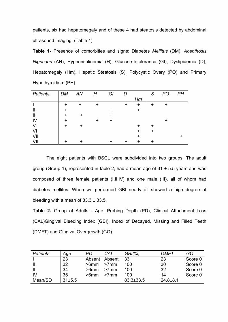
ultrasound imaging. (Table 1)
Table 1- Presence of comorbities and signs: Diabetes Mellitus (DM), Acanthosis
Nigricans (AN), Hyperinsulinemia (H), Glucose-Intolerance (GI), Dyslipidemia (D),
Hepatomegaly (Hm), Hepatic Steatosis (S), Polycystic Ovary (PO) and Primary
Hypothyroidism (PH).
Patients DM AN H GI D Hm
S PO PH
IIIIIIIVVVIVIIVIII
+ + + + +
+
+
+
+
+
+
+
+ + +
+
+
+
+ +
+ + + +
+
+ +
+
+
+
+
The eight patients with BSCL were subdivided into two groups. The adult
group (Group 1), represented in table 2, had a mean age of 31 ± 5.5 years and was
composed of three female patients (I,II,IV) and one male (III), all of whom had
diabetes mellitus. When we performed GBI nearly all showed a high degree of
bleeding with a mean of 83.3 ± 33.5.
Table 2- Group of Adults - Age, Probing Depth (PD), Clinical Attachment Loss
(CAL)Gingival Bleeding Index (GBI), Index of Decayed, Missing and Filled Teeth
(DMFT) and Gingival Overgrowth (GO).
Patients Age PD CAL GBI(%) DMFT GO
IIIIIIIVMean/SD
2332343531±5.5
Absent>6mm>6mm>6mm
Absent>7mm>7mm>7mm
3310010010083.3±33,5
2330321424.8±8.1
Score 0Score 0Score 0Score 0
Periodontal probing revealed dental pockets in three patients (II, III, IV).
Patients II, III and IV had advanced periodontitis with CAL>7mm and PD>6mm. The
youngest patient (I) wore a full upper prosthesis. The periodontitis was classified as
periodontitis associated with systemic conditions. (Armitage, 1999)
Decayed, missing and filled teeth were numerous, demonstrated by the high
DMFT index of 24.8 ± 8.1. Patient I, who had the lowest GBI, reported that she
frequently developed carious lesions, despite a meticulous oral hygiene and regular
dental visits. Patient II showed an indication for extraction in all of her remaining
teeth, which were destroyed by carious lesions and periodontal disease. Patient IV,
who had the lowest DMFT, was at the beginning of periodontal treatment. Patient III,
also with advanced periodontitis and high DMFT, is not undergoing any treatment.
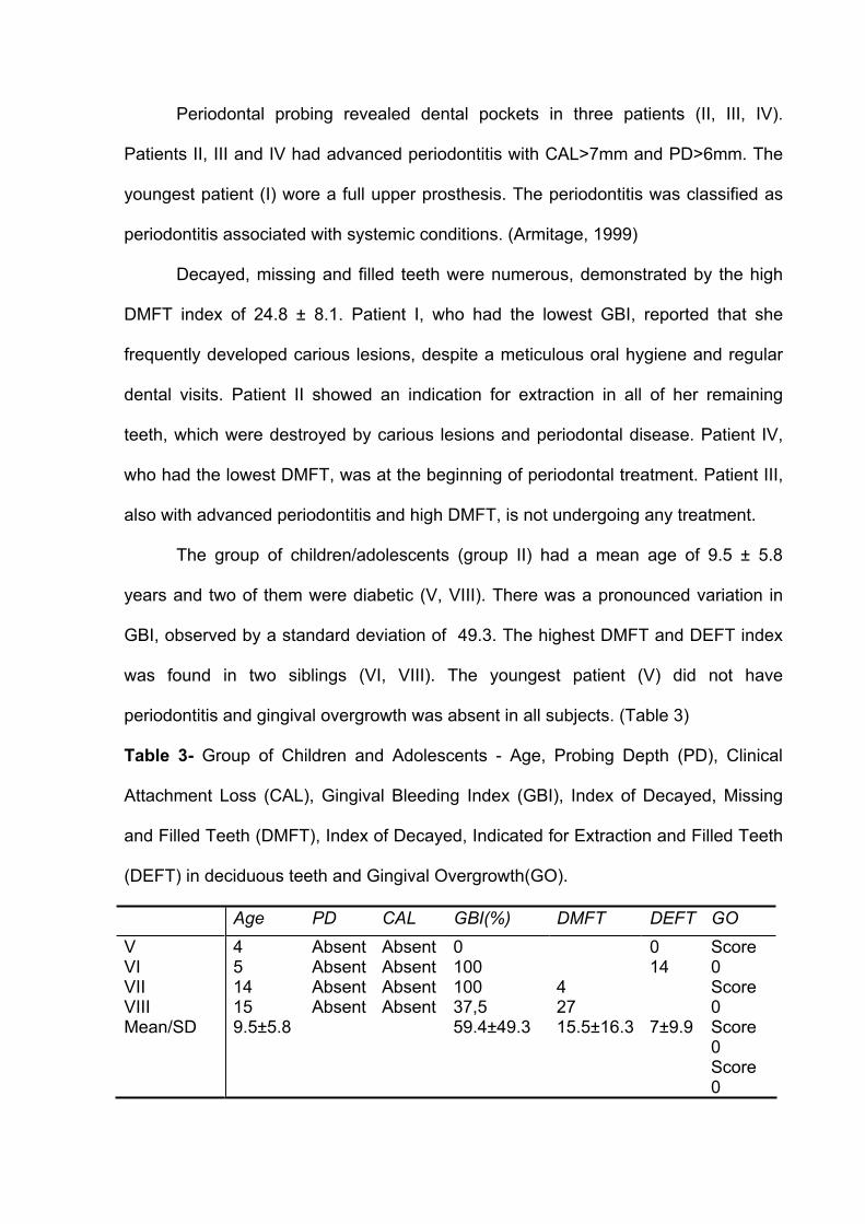
The group of children/adolescents (group II) had a mean age of 9.5 ± 5.8
years and two of them were diabetic (V, VIII). There was a pronounced variation in
GBI, observed by a standard deviation of 49.3. The highest DMFT and DEFT index
was found in two siblings (VI, VIII). The youngest patient (V) did not have
periodontitis and gingival overgrowth was absent in all subjects. (Table 3)
Table 3- Group of Children and Adolescents - Age, Probing Depth (PD), Clinical
Attachment Loss (CAL), Gingival Bleeding Index (GBI), Index of Decayed, Missing
and Filled Teeth (DMFT), Index of Decayed, Indicated for Extraction and Filled Teeth
(DEFT) in deciduous teeth and Gingival Overgrowth(GO).
Age PD CAL GBI(%) DMFT DEFT GO
VVIVIIVIIIMean/SD
4514159.5±5.8
AbsentAbsentAbsentAbsent
AbsentAbsentAbsentAbsent
010010037,559.4±49.3
42715.5±16.3
014
7±9.9
Score0Score0Score0Score0
Discussion
Dental caries and periodontal disease presented prematurely and in a very
destructive form in the patients with BSCL. High caries activity was verified in all age
groups, with the exception of the youngest (4 years). The high caries incidence in two
members of the same family requires an investigation of the oral conditions of the
entire family in order to compare those with and without BSCL.
One factor that could lead to the high prevalence of caries is the type of diet
that the patient with lipodystrophy follows. Due to insufficient caloric reserves, caused
by the absence of adipose tissue, the patient is limited in the energy substrate
metabolism of lipids. It is supposed that the patient adopts a hypercaloric diet as a
form of compensation.
It is known that constant food ingestion modifies salivary pH levels, favoring
bacterial action and consequently the cariogenic process. In order to exercise
bacterial control, the patient would require effective mechanical removal of dental
plaque. This effectiveness was not demonstrated by the study group, verified by the
high GBI levels observed.
Authors have reported cases of generalized congenital lipodystrophy that
showed evidence of intellectual compromise. They have emphasized both the
existence of mental retardation and pronounced learning disabilities, compromised
sociability along with aggressive behavior and motor inability (Quiñonero et al., 1992;
Reis et al., 2004). Patients with this syndrome may have difficulty in learning
toothbrushing and oral hygiene habits, because of this possible intellectual and motor
deficiency.
An important finding was the presence of advanced periodontitis in the adult
group, affecting 2 out of the 4 adults examined.

We believe that the main cause of this periodontal destruction lies not with oral
hygiene, but rather with diabetes mellitus, of which they suffered. Insulin resistance
syndrome develops from childhood in individuals with BSCL. The cumulative effect of
this pathology and the relation existing between diabetes and periodontal disease
may be the key for explaining the prevalence in the patients studied (Nunn, 2000;
Soskolne and Klinger, 2001; Taylor, 2001). Xerostomia caused by diabetes may also
facilitate increased caries indices (Costa et al., 2004).
There are areas in Brazil, such as the Seridó region in the state of Rio Grande
do Norte (Fu et al., 2004), where the prevalence of BSCL is among the highest in the
world. Complementary studies investigating other populations must be performed to
obtain more evidence about the presence of advanced periodontitis. This is vital for
prevention, early diagnosis and correct treatment, aiming at improved glycemic
control of diabetes and optimized oral health conditions. The monitoring of oral health
in patients with this syndrome plays an important role in the sociability and
acceptance of the individual affected by a series of physical and biological
alterations.
References
1. Agarwal, A.K., Barnes, R.I., Garg, A. Genetic basis of congenital generalized
lipodystrophy. International Journal of Obesity and Related Metabolic
Disorders 2004;28(2):336-9.
2. Armitage, G.C. Development of a classification system for periodontal
diseases and conditions. Annals of Periodontology 1999;4:1-6.
3. Berardinelli, W. An undiagnosed endocrinometabolic syndrome: report of 2
case. Journal of Clinical Endocrinology and Metabolism 1954; 14:193-204.

4. Bjornstad, P.G., Foerster, A., Ihlen, H. Cardiac findings in generalized
lipodistrophy. Acta Paediatrica Supplement 1996; 413:39-43.
5. BRASIL. Ministério da Saúde. Projeto SB 2000: Condições de saúde bucal da
população brasileira. Relatório Final. Brasília, DF, 2004. Avaiable in:
www.saude.gov.br
6. Capeau, J., Magre, J., Lascols, O., et al. Diseases of adipose tissue: genetic
and acquired lipodystrophies. Biochemical Society Transaction 2005;33(Pt
5):1073-77.
7. Christersson, L.A., Zambon, J.J., Genco, R.J. Dental bacterial plaques. Nature
and role in periodontal disease. Journal of Periodontology 1991; 18(6): 441-6.
8. Costa, C.C., Resende, G.B., Souza, J.M., et al. Study of the oral
manifestations in diabetic children and their correlation variables. Arquivos
Brasileiros de Endocrinologia e Metabologia 2004;48(3):374-378.
9. Figueiredo-Filho, P.P., Val, A.C., Diamante, R., Cunha, C.F., Norton, R.C.,
Lamounier, J.A., Leão, E. Lipodistrofia generalizada congênita. Jornal de
Pediatria 2004;80(4):333-6.
10. Fu, M., Kazlauskaite, R., Baracho, M.F.P., et al. Mutations in Gng31g
AGPAT2 in Berardinelli-Seip Congenital Lipodystrophy and Brunzell
Syndrome: Phenotype Variability Suggests Important Modifier Effects. Journal
of Clinical Endocrinology and Metabolism 2004;89:2916-2922
11. Garg, A., Wilson, R., Barnes, R. A gene for congenital generalized
lipodistrophy maps to human chromosome 9q34. Journal of Clinical
Endocrinology and Metabolism 1999;84(9):3390-4.
12. Garg, A. Acquired and Inherited Lipodystrophies. New England Journal of
Medicine 2004;350: 1220- 34.

13. Gomes, K.B., Fernandes, A.P., Ferreira, A.C., et al. Mutations in the seipin
and AGPAT2 genes clustering in consanguineous families with Berardinelli-
Seip congenital lipodystrophy from two separate geographical regions of
Brazil. Journal of Clinical Endocrinology and Metabolism 2004;89(1):357-61.
14. Haque, W., Garg, A., Agarwal, A.K. Enzymatic activity of naturally occurring 1-
acylglycerol-3-phosphate-O-acyltransferase 2 mutants associated with
congenital generalized lipodystrophy. Biochemical and Biophysical Research
Communications 2005;327(2):446-53.
15. Heathcote, K., Rajab, A., Magre, J., et al. Molecular analysis of Berardinelli-
Seip congenital lipodystrophy in Oman: evidence for multiple loci. Diabetes.
2002; 51(4):1291-3.
16. Loesche, W.J. Bacterial Mediators in Periodontal Disease. Clinical Infectious
Diseases. 1993;16(4):203-10.
17. Magre, J., Delepine, M., Khallouf, E., et al.. Identification of the gene altered in
Berardinelli-Seip congenital lipodystrophy on chromosome 11q13. Nature
Genetics. 2001; 28(4):365-70.
18. Nunn, M.E. Understanding the etiology of periodontitis: an overview of
periodontal risk factors. Periodontology 2000 2003; 32:11-23.
19. Quiñonero, M.A., Leon, R.G., Ortigosa, M.A.G., et al. Lipodistrofia
Generalizada Congênita. Anales Espanoles de Pediatria 1992;37(2):173-74.
20. Pernu, H.E., Pernu, L.M.H., Huttunen, K.R.H., Nieminen, P.A., Knuuttila,
M.L.E. Gingival overgrowth among renal transplant recipients related to
immunosuppressive medication and possible local background factors.
Journal of Periodontology 1992; 63:548-53.
21. Rajab, A., Heathcote, K., Joshi, S., Jeffery, S., Patton, M. Heterogeneity for
congenital generalized lipodystrophy in seventeen patients from Oman.
American Journal of Medical Genetics. 2002; 110(3):219-25.
22. Raygada, M., Rennert, O. Congenital generalized lipodystrophy: profile of the
disease and gender differences in two siblings. Clinical Genetics 2005;
67(1):98-101.
23. Reis, O.L.L., Pedroso, E.R.P., Campos, A.R., et al. Síndrome de Berardinelli-
Descrição de um caso. Revista Médica de Minas Gerais 2004;4(4):51-4.
24. Santos, M.G.N., Medeiros, T.M.D., Baracho, M.F.P., Brandão-Neto, J., Gurgel,
A.M.C., Silva, A.S.C. Lipodistrofia generalizada congênita: correlação com
leptina e outros aspectos bioquímicos. Acta Cirúrgica Brasileira 2005;
20(suppl 1):190-5.
25. Seip, M. Lipodistrophy and gigantism with associated endocrine manifestation:
a new diencephalic syndrome? Acta Paediatrica 1959;48:555-74
26. Simha, V. Garg, A. Phenotypic heterogeneity in body fat distribution in patients
with congenital generalized lipodystrophy caused by mutations in the AGPAT2
or seipin genes. Journal of Clinical Endocrinology and Metabolism
2003;88(11):5433-7.
27. Soskolne, W.A., Klinger, A. The Relationship Between Periodontal Diseases
and Diabetes: An Overview. Annals of Periodontology 2001; 6(1):91-8.
28. Taylor, G.W. Bidirectional Interrelashionships Between Diabetes and
periodontal Diseases: An Epidemiologic Perspective. Annals of
Periodontology 2001; 6(1):99-112

29. Van Maldergem, L., Magre, J., Khallouf, T.E., et al. Genotype-phenotype
relationships in Berardinelli-Seip congenital lipodystrophy. Journal of Medical
Genetics. 2003; 40(2):150.
30. Viégas, R.F.M., Diniz, R.V.Z., Viégas, T.M.R.F., Lira-Filho, E.B., Almeida, D.R.
Cardiac Involvement in Total Generalized Lipodystrophy (Berardinelli- Seip
Syndrome). Arquivos Brasileiros de Cardiologia 2000; 5(3):246-8.
4. ANEXAÇÃO DE ARTIGO ENVIADO PARA PUBLICAÇÃO
Title: Absence of periodontitis in acromegalic patients: a protective effect of
growth hormone excess?
Abstract
Acromegaly is a rare metabolic disorder caused by increased growth hormone (GH)
secretion. Common oral features are prognatism, increased interdental spaces,
macroglosia, and dental mobility. However, not much is known about the periodontal
status of acromegalics, the aim of this study. The periodontal status of sixteen
acromegalic subjects was investigated and compared with twenty controls (similar
socio-economic profile and age). They were selected among thirty patients attending
the Neuroendocrinology Clinic of the Endocrinology and Diabetes Service at the
Federal University of Ceará (UFC). Periodontal probing, followed by the assessment
of gingival overgrowth, tooth loss and mobility, and maloclusion was performed.
Acromegalic patients’ didn´t presented periodontitis and all of them had complete
absence of periodontal pockets, while 50% of the control group presented
periodontitis. All acromegalic patients presented dental mobility degree 1, mainly in
the anterior inferior teeth. Maloclusion (100%) and diastemas (93,75%) were also
present in these patients. The protective effect of acromegaly on periodontitis is a
new finding, whose mechanisms are not yet elucidated, but may be related to the
anabolic effects of GH and IGF-I on bone.
Introduction
Acromegaly is a chronic disease due to growth hormone (GH) hypersecretion,
mostly caused by a benign adenoma of the pituitary gland. This adenoma usually
measures over 10mm 1. It is a rare condition, with an estimated annual incidence of
3-4 cases per million and a prevalence of 40-60 cases per million worldwide 2,3. The
GH hypersecretion increase insulin-like growth factor (IGF-I), which affects bone
metabolism 4.
The change in appearance, characterized by enlargement of the feet, hands
and mandible, is remarkable, but cardiovascular, metabolic, and respiratory
complications as well as neoplasias are also present in patients with this disorder3.
These systemic problems are responsible for the reduced quality of life. In these
patients, life expectancy is shortened by an average of 10 years. When diabetes or
heart disease are already present at the time of diagnosis the life expectancy is even
shorter5.
Acromegaly exhibits various oral characteristics, most commonly prognathism,
macroglossy, everted and edematous lips, tooth mobility, interdental spaces
(diastemas) and tooth loss6,7, and more rarely, gingival overgrowth8. Diastemas, and
more significantly tooth mobility, are oral clinical manifestations that suggest the
presence of periodontitis. Although these alterations are often observed in
acromegalic patients, there is a lack of information regarding the periodontal status of
these patients in the literature.
Periodontal diseases are the second major cause of tooth loss and this
disease is a common finding in some systemic diseases9. Evidence suggests that
periodontitis may be a risk factor to ischemic cardiovascular disease, pulmonary
disorders, preterm births and low birth weight10,11,12. Risk factors associated to
periodontal disease include tobacco use, traumatic occlusion and diabetes mellitus13.
The last one affects 19 to 56% of acromegalic patients.14 Thus, the aim of the
present study was to assess the periodontal status of patients with acromegaly
attended at a reference hospital in the state of Ceará, Brazil.
Materials and Methods
This investigation is a cross-sectional study of 16 acromegalic patients,
selected among thirty patients attending the Neuroendocrinology Clinic of the
Endocrinology and Diabetes Service at the Federal University of Ceará (UFC). The
control group consisted of 20 individuals, presenting similar age and socioeconomical
status, seeking treatment at the University of Fortaleza (UNIFOR) dental clinic
facilities.
All ethical aspects were rigidly observed, being the study conducted after its
approval by the UNIFOR Research Ethics Committee. All patients gave informed
consent to participate in this study.
Patients’ clinical data were obtained using their hospital charts, and dental
examinations were performed at UNIFOR dental clinic. Glucose tolerance status of
all patients was evaluated to diagnose the presence of diabetes mellitus. They were
also questioned about tobacco use.
Diagnosis of acromegaly was performed through basal determinations of GH
(Quimiluminescence), and insulin-like growth factor, IGF-I (IRMA), and GH level after
oral glucose tolerance test (OGTT)- 75 grams of glucose orally (15). It was
considered diagnostic a GH nadir greater than 1 ng/L during OGTT, which was
indicated when IGF-1 was above the reference range for sex and age and/or GH>0,4
ng/L, except for patients with diabetes, where only basal values were obtained15.
Glucose tolerance of acromegalic patients was evaluated using diagnostic criteria
from the American Diabetes Association16.
All dental examinations were performed by the same experienced periodontist.
Periodontal pockets depth were evaluated using a UNC -15 periodontal probe at six
different points of each tooth. Individuals with probing depth (PD) < 4mm with no

clinical attachment loss (CAL) were deemed healthy; those with PD between 4 and
6mm and CAL of up to 4mm as having chronic periodontitis with slight to moderate
loss of periodontal support17 and those with PD 6mm and CAL greater than 4mm as
having chronic periodontitis with advanced loss of periodontal support18. Using
clinical criteria, subjects were also classified as having or not aggressive chronic
periodontitis19.
Bleeding on probing (BOP), considered an objective inflammatory parameter
in periodontitis establishment, was measured using Gingival Bleeding Index (GBI).
GBI 0,1 was considered as low risk for the development of periodontitis20. Tooth
mobility levels were evaluated and classified as degrees 1, 2 and 321. Presence of
diastemas and malocclusion were also assessed.
Results were described using mean ± standard deviation for continuous
variables, and proportions for cathegoric variables. Significance level was set at
p=0,05. Independent t test was used to compare periodontal status between the
acromegalic and the control group. The statistical software SPSS 12.0 (SPSS inc,
Chicago, IL, USA) was used for statistical analysis.
Results
A total of 16 acromegalic patients were assessed, eight (50%) male and eight
(50%) female, with mean age of 45.8 ± 11.8 years, being 13 above 40 year of age.
Two (12,5%) patients of this group were tobacco users. The mean age at diagnosis
was 41.4 ± 10.3 years (Table 1). Control group mean age was 41.5 ± 10.5 years,
ranging from 23 to 65 years, and consisting of 11 (55%) females and 9 (45%) males.
None of the patients were tobacco users.
On magnetic resonance imaging (MRI), 13 (81.25%) of the acromegalic
patients presented pituitary macroadenomas, and 3 (18.75%) pituitary

microadenomas (6.25%). Seven patients (43.75%) were diabetics, four (25%)
presented impaired glucose tolerance (pre-diabetes) and five (31,25%) normal
glucose tolerance; 10 patients also presented hypertension (Table 1). No patients in
the control group presented metabolic disorders, including diabetes.
Transsphenoidal hypophysectomy (surgical adenoma removal) was performed
on 10 (62.5%) patients, four (25%) underwent radiotherapy and 13 (81.25%) used or
were using 10 to 30 mg of octreotide (Sandostatin-LAR®) per month. Octreotide is a
synthetic form of a brain hormone, somatostatin, which stops GH production (Pless,
2005). Four patients (25%) used all three treatment modalities, five (31.25%) up to
two treatment modalities and only two (12.5%), recently diagnosed patients, had not
received any treatment at that time of this investigation.
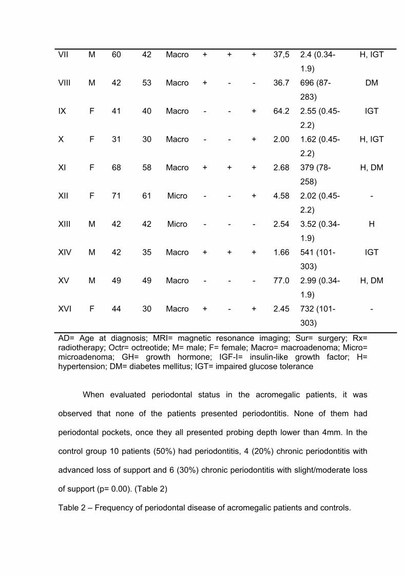
Table 1- Clinical presentation at the time of evaluation of the sixteen acromegalic
patients studied.
Patie
nt
Se
x
Age AD MRI Therapeutics GH IGF-I Comorbidi
ties
(year
s)
(year
s)
Sur Rx Octr (ng/L) (ng/ml)
I M 41 38 Macro + - + 0.30 0.67 (0.34-
1.9)
H, DM
II F 41 40 Macro + - + 0.10 0.8 (0.45-
2.2)
H
III F 44 37 Micro + + + 0.89 2.05 (0.45-
2.2)
DM
IV F 30 25 Macro + - + 2.96 1.23 (0.45-
2.2)
H, DM
V M 51 50 Macro - - + 3.46 534 (80-
283)
H, DM
VI M 35 33 Macro + - + 0.33 1.12 (0.34-
1.9)
H
VII M 60 42 Macro + + + 37,5 2.4 (0.34-
1.9)
H, IGT
VIII M 42 53 Macro + - - 36.7 696 (87-
283)
DM
IX F 41 40 Macro - - + 64.2 2.55 (0.45-
2.2)
IGT
X F 31 30 Macro - - + 2.00 1.62 (0.45-
2.2)
H, IGT
XI F 68 58 Macro + + + 2.68 379 (78-
258)
H, DM
XII F 71 61 Micro - - + 4.58 2.02 (0.45-
2.2)
-
XIII M 42 42 Micro - - - 2.54 3.52 (0.34-
1.9)
H
XIV M 42 35 Macro + + + 1.66 541 (101-
303)
IGT
XV M 49 49 Macro - - - 77.0 2.99 (0.34-
1.9)
H, DM
XVI F 44 30 Macro + - + 2.45 732 (101-
303)
-
AD= Age at diagnosis; MRI= magnetic resonance imaging; Sur= surgery; Rx= radiotherapy; Octr= octreotide; M= male; F= female; Macro= macroadenoma; Micro= microadenoma; GH= growth hormone; IGF-I= insulin-like growth factor; H= hypertension; DM= diabetes mellitus; IGT= impaired glucose tolerance
When evaluated periodontal status in the acromegalic patients, it was
observed that none of the patients presented periodontitis. None of them had
periodontal pockets, once they all presented probing depth lower than 4mm. In the
control group 10 patients (50%) had periodontitis, 4 (20%) chronic periodontitis with
advanced loss of support and 6 (30%) chronic periodontitis with slight/moderate loss
of support (p= 0.00). (Table 2)
Table 2 – Frequency of periodontal disease of acromegalic patients and controls.

Control AcromegalicPeriodontaldisease n° % n° %
Absent 10 50.0 16 100.0Chronicperiodontitis(Slight to moderate)
6 30.0 - -
Chronicperiodontitis(Advanced)
4 20.0 - -
Total 20 100.0 16 100.0
Chi-square. p=0.00.
All 16 acromegalic patients presented tooth mobility (degree I), notably in the
lower anterior teeth. Malocclusion, showing Angle’s class III or crossbite were also
noted in all these patients. Diastemas were found in 15 (93.75%) of the 16 patients,
while gingival recession was observed in eight (50%) patients. However, this gingival
recession was presented in very localized areas, and with less than 4 mm of CAL.
Bleeding on probing with GBI > 0,1 was observed in nine (56,2%) of the 16 patients.
(Table 3).
Table 3 – Periodontal manifestations and tobacco use of the sixteen evaluated acromegalic patients.
Patient PP Mob Diastemas Malocclusion Recession GBI Tobacco
I - + + + - 0,62 -
II - + + + + 1,0 -
III - + - + + 0,0 +
IV - + + + + 0,13 -
V - + + + - 0,05 -
VI - + + + + 0,37 -
VII - + + + + 0,15 -
VIII - + + + + 0,13 +
IX - + + + - 0,0 -
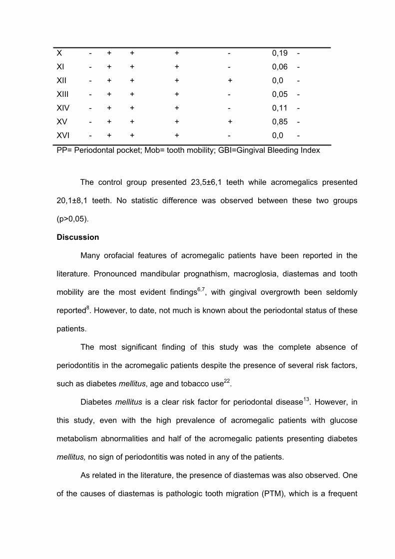
X - + + + - 0,19 -
XI - + + + - 0,06 -
XII - + + + + 0,0 -
XIII - + + + - 0,05 -
XIV - + + + - 0,11 -
XV - + + + + 0,85 -
XVI - + + + - 0,0 -
PP= Periodontal pocket; Mob= tooth mobility; GBI=Gingival Bleeding Index
The control group presented 23,5±6,1 teeth while acromegalics presented
20,1±8,1 teeth. No statistic difference was observed between these two groups
(p>0,05).
Discussion
Many orofacial features of acromegalic patients have been reported in the
literature. Pronounced mandibular prognathism, macroglosia, diastemas and tooth
mobility are the most evident findings6,7, with gingival overgrowth been seldomly
reported8. However, to date, not much is known about the periodontal status of these
patients.
The most significant finding of this study was the complete absence of
periodontitis in the acromegalic patients despite the presence of several risk factors,
such as diabetes mellitus, age and tobacco use22.
Diabetes mellitus is a clear risk factor for periodontal disease13. However, in
this study, even with the high prevalence of acromegalic patients with glucose
metabolism abnormalities and half of the acromegalic patients presenting diabetes
mellitus, no sign of periodontitis was noted in any of the patients.
As related in the literature, the presence of diastemas was also observed. One
of the causes of diastemas is pathologic tooth migration (PTM), which is a frequent
finding in periodontitis. Studies have demonstrated a variation in PTM between
30.03% and 55.8% in the lower anterior sextant of patients with periodontitis23,24.
Many factors, such as periodontal tissue support loss, malocclusion, periapical
inflammation and lip and tongue pressure may influence the onset of PTM25. The
results of this study suggest that the presence of diastemas in the acromegalic
patients is not related to periodontitis. In this case, one factor that may favor
diastema is the increased mandibular volume found in most of these patients6.
Macroglosia, commonly found in acromegalic patients, may also promote the onset
of interdental spaces and dental mobility through the pressure of the tongue on the
teeth26.
Tooth mobility is also a common dental finding in acromegalics. All patients
studied exhibited class-1 mobility, found mainly in the lower incisor region, and nearly
all presented it with diastemas, confirming literature data7. However, when probing
depth was performed to verify the presence of periodontitis, no acromegalic patient
presented periodontal pocket depth over 4mm.
Severe occlusal trauma, due to malocclusions, which, in these patients, varied
from simple crossbites to Angle’s class III may be yet another component that favors
dental mobility. The significant masticatory force resultant from exaggerated
mandible enlargement and muscle hypertrophy is an additional factor. Existing
occlusal trauma may be one causal factor of gingival recession, seen in 50% of the
acromegalics patients examined in this study. This recession was observed mainly
close to edentulous regions27.
The destructive action on the alveolar bone caused by periodontopathies has
made periodontal esthetic and functional reconstruction one of the main objectives of
periodontics. Several biomaterials and proteins have been tested over the last 20
years for the preservation of periodontal structures and regeneration of lost support
tissue. Guided tissue regeneration and growth factors such as fibroblast growth
factor (FGF), platelet-derived growth factor (PDGF), transforming growth factor
(TGF), bone morphogenic proteins (BMP) and insulin-like growth factor (IGF-I) are
among the materials tested28.
The GH is a hormone with anabolic effects on bone tissue that promotes IGF-I
secretion29. GH increases bone formation via a direct interaction with GH receptors
on osteoblasts and via local production of IGF-I (autocrine/paracrine action) 30. Thus,
in the presence of GH excess, a high alveolar bone formation may occur31,32. IGF-I
plays an important role in skeletal development by promoting chondrocyte
proliferation and maturation, while inhibiting apoptosis to form bone of appropriate
size and strength33, being like this an important growth factor for the skeleton.
Additionally, a possible beneficial role of IGF-I combined with PDGF in the treatment
of periodontal bone defects and IGF-I impact on specific cells, such as periodontal
ligament fibroblasts, have been recently postulated, which may explain the apparent
protective effect for periodontitis in the acromegalic patients seen in this study34,35.
The protective effect of periodontitis in acromegalic patients is a new finding,
whose mechanisms are not yet clear, but may be related to the anabolic effects of
GH and IGF-I on bone. As acromegaly diagnosis generally occur very late, the
changing of oral aspects can make the dentist or the periodontist an important health
professional in the early detection of this disease, through early detection of these
alterations. Improvement in the treatment and the control of acromegaly can happen
when the disease is diagnosed early, further improving the life of these patients.

References
1. Melmed S. Acromegaly. N Engl J Med 1990 322: 966-977.
2. Etxabe J, Gaztambide S, Latorre P, et al. Acromegaly: an epidemiological
study. J Endocrinol Invest 1993 16: 181–187.
3. Colao A, Ferone D, Marzullo P, et al. Systemic complications of acromegaly:
epidemiology, patogénesis and management. Endocr Rev 2004 25: 102-52.
4. Agnusdei D, Gentilella R. GH and IGF-I as therapeutic agents for
osteoporosis. J Endocrinol Invest 2005 28: 32-6.
5. Kleinberg DL. New roles and challenges in neurosurgery of acromegaly. J
Endocrinol Invest 2005 28: 88-91.
6. Dostalova S, Sonka K, Smahel Z, et al. Cephalometric assessment of cranial
abnormalities in patients with acromegaly. J Craniomaxillofac Surg 2003 31:
80-7.
7. Feelders RA, Delwel EJ, de Baat C. Acromegaly.Treatment of the casual
factor and the oral sequelae. Ned Tijdschr Tandheelkd 2004 111: 20-2.
8. Capoglu I, Yilmaz AB, Unuvar N, et al. Gingival enlargement in acromegaly.
Endocrine 2002 18: 207-210.
9. Armitage GC. Development of a classification system for periodontal diseases
and conditions. Ann Periodontol 1999 4: 1-6.
10.Mattila KJ, Pussinen PJ, Paju S. Dental infections and cardiovascular
diseases: a review. J Periodontol 2005 76: 2085-8.
11.Scannapieco FA, Wang B, Shiau HJ. Oral bacteria and respiratory infection:
effects on respiratory pathogen adhesion and epithelial cell proinflammatory
cytokine production. Ann Periodontol 2001 6: 78-86.
12.Offenbacher S, Katz V, Fertik G, et al. Periodontal infections as a possible risk
factor for preterm low birth weight. J Periodontol(supp) 1996 64: 1103-13.
13. Soskolne WA, Klinger A. The Relationship Between Periodontal Diseases and
Diabetes: An Overview. Ann Periodontol 2001 6: 91-8.
14. Kreze A, Kreze-Spirova E, Mikulecky M. Risk factors for glucose intolerance in
active acromegaly. Braz J Med Biol Res 2001 34: 1429-33.
15.Giustina A, Barkan A, Casanueva FF, et al. Criteria for cure of acromegaly: a
consensus statement. J Clin Endocrinol Metabol 2000 85: 526-9.
16. The Expert Committee on the diagnosis and classification of diabetes mellitus.
Report of the Expert Committee on the diagnosis and classification of diabetes
mellitus. Diabetes Care 1997 20: 1183-97.
17. American Academy of Periodontology-Position paper. Parameter on chronic
periodontitis with slight to moderate loss of periodontal support. J Periodontol
2000 71: 853-855.
18. American Academy of Periodontology-Position paper. Parameter on chronic
periodontitis with advanced loss of periodontal support. J Periodontol 2000 71:
856-858.
19. American Academy of Periodontology-Position paper. Parameter on
aggressive periodontitis. J Periodontol 2000 71: 867-869.
20. Lang NP, Adler R, Joss A, et al. Absence of bleeding on probing. An indicator
of periodontal stability. J Clin Periodontol 1990 17: 714-721.
21. Carranza FA. Diagnóstico clínico. In: Newman MG, Takei HH, Carranza FA.
Periodontia Clínica. 9th ed. Rio de Janeiro: Editora Guanabara Koogan AS,
2004, 384-402.
22. Albandar JM. Epidemiology and risk factors of periodontal diseases. Dent Clin
North Am 2005 49: 517-532,v-vi.

23. Martinez-Canut P, Carrasquer A, Magan R, et al. A study on factors
associated with pathologic tooth migration. J Clin Periodontol 1997 24:492-
497.
24. Towfighi P, Brunsvold M, Storey A, et al. Pathologic migration of anterior teeth
in patients with moderate to severe periodontitis. J Periodontol 1997 68: 967-
972.
25. Brunsvold MA. Pathologic tooth migration. J Periodontol 2005 76: 859-866.
26. Proffit W. Equilibrium theory revisited:Factors influencing position of the teeth.
Angle Orthod 1978 48: 75-186.
27. Gher ME. Changing concepts. The effects of occlusion on periodontitis. Dent
Clin North Am 1998 42: 285-99.
28.Franceschi RT. Biological approaches to bone regeneration by gene therapy.
J Dent Research 2005 84: 1093-103.
29. Ueland T. GH/IGF-I and bone resorption in vivo and in vitro. Eur J Endocrinol
2005 152: 327-332.
30. Isaksson OG, Ohlsson C, Bengtsson BA, et al. GH and bone--experimental
and clinical studies. Endocr J 2000 47: S9-16.
31. Li H, Bartold PM, Young WG, et al. Growth hormone induces bone
morphogenetic proteins and bone-related proteins in the developing rat
periodontium. J Bone Miner Res 2001 16: 1068-76.
32. Haase HR, Ivanovski S, Waters MJ, et al. Growth hormone regulates
osteogenic marker mRNA expression in human periodontal fibroblasts and
alveolar bone-derived cells. J Periodontal Res 2003 38: 366-374.
33. Wang Y, Nishida S, Sakata T, et al. Insulin-Like Growth Factor-I Is Essential
For Embryonic Bone Development. Endocrinology 2006 147: 4753-61.

34. Han X, Amar S. IGF-1 signaling enhances cell survival in periodontal ligament
fibroblasts vs.gingival fibroblasts. J Dent Research 2003 82: 454-9.
35. Chen FM, Zhao YM, Wu H, et al. Enhancement of periodontal tissue
regeneration by locally controlled delivery of insulin-like growth factor-I from
dextran-co-gelatin microspheres. J Control Release 2006 114: 209-22.

5 COMENTÁRIOS, CRÍTICAS E SUGESTÕES
A idéia da formação de um homem integral remonta desde a antiga Grécia e a
integração dos saberes foi marcante em vários momentos históricos, passando pelo
Renascimento e pelo Iluminismo. Porém, com o pensamento cartesiano (René
Descartes, 1596-1650) e o uso disciplinado da razão, é iniciado um modelo que
influenciou profundamente o pensamento científico pelos séculos seguintes. A
fragmentação do conhecimento gerada pelo modelo cartesiano foi a base para a
consolidação das especializações e o nascimento da disciplinaridade (41).
As ciências sociais, ambientais e filosóficas foram as primeiras a dar um
passo adiante na busca de uma integração dos conhecimentos. Somente no final do
século passado, as ciências da saúde começaram a vislumbrar uma saúde mais
holística, onde o saber científico não fosse tão dividido. Começa então, a surgir na
nossa área, a idéia da interdisciplinaridade.
A interdisciplinaridade é considerada uma inter-relação e interação das
disciplinas a fim de atingir um objetivo comum. É a reunião de diferentes disciplinas
articuladas em torno de uma mesma temática com diferentes níveis de integração.
Ocorre aí, uma exploração das potencialidades de cada disciplina de forma
interdependente na busca de um novo conhecimento. (41,42)
Surgem também os conceitos de multidisciplinaridade e transdisciplinaridade.
A multidisciplinaridade ou pluridisciplinaridade, como o conjunto de disciplinas que
se agrupam em torno de um dado tema desenvolvendo investigações e análises
isoladas por diferentes especialistas, poderia ser considerada um avanço já que esta
abordagem ultrapassa as disciplinas, porém com características cartesianas já que
não são estabelecidas relações conceituais ou metodológicas entre as disciplinas se

inscrevendo no quadro da pesquisa disciplinar. Já a transdisciplinaridade pode ser
entendida como uma radicalização do conceito de interdisciplinaridade, pois várias
disciplinas são participantes de um mesmo pressuposto teórico e operacional, tendo
o mesmo campo de atuação. A transdiciplinaridade se situa, ao mesmo tempo, entre
as disciplinas, através destas e além de toda e qualquer disciplina. É a presença de
vários níveis de realidade que interessa à pesquisa transdiciplinar (41, 43).
Na construção do conhecimento estão situadas a disciplinaridade, a
multidisciplinaridade, a interdisciplinaridade e a transdisciplinaridade. É na visão
interdisciplinar que surge nosso estudo, indo ao encontro do novo paradigma
existente na Periodontia, chamado de Medicina Periodontal. A compreensão e
disponibilidade da equipe do Setor de Endocrinologia e Diabetes do Hospital
Universitário Walter Cantídio, da Universidade Federal do Ceará, foi a chave para a
realização do presente trabalho. Todos os pacientes que se propuseram em
participar do nosso estudo eram atendidos no referido setor. Para início da coleta de
dados obteve-se uma aprovação pelo Comitê de Ética em Pesquisa da Universidade
de Fortaleza (UNIFOR) sob o parecer nº181/2005 (ANEXO 1). Também, fez-se
necessária a assinatura de um Termo de Consentimento Livre e Esclarecido
(ANEXO 3) por parte do sujeito pesquisado ou por algum responsável devidamente
identificado, no caso de menores ou por aquele que detenha a curatela no caso de
pessoas com incapacidades que os tornem sem discernimento necessário para os
atos da vida civil.
Para realização dos exames todo o material foi autoclavado e esterilizado de
acordo com as regras de biossegurança. O respeito pelos princípios éticos e
bioéticos e o entendimento de que esses aspectos devem ser rigidamente
observados fez com que os pacientes examinados pudessem ser encaminhados

para os serviços de atendimento odontológico da UNIFOR e para meu consultório
privado para a realização de procedimentos odontológicos. Restaurações,
exodontias, profilaxias e próteses foram realizadas obedecendo às regras da
beneficência que a pesquisa deve gerar.
Foram avaliados 8 pacientes portadores da Síndrome de Berardinelli, 16
acromegálicos, 30 hipotireóideos e 30 hipertireóideos, além de 35 pacientes que
fizeram parte do grupo controle. A exposição do que é a doença periodontal e sua
influência na saúde sistêmica dos pacientes foi explanada para os médicos
residentes que participaram ativamente dos trabalhos, bem como para cada
indivíduo participante deste estudo. Os indivíduos selecionados como grupo controle
foram catalogados no setor de triagem do curso de Odontologia da UNIFOR de
forma aleatória. Como em todo estudo dificuldades existiram, mas foram
contornadas porque prevaleceu o espírito de cooperação.
O primeiro grupo examinado foi o de portadores da Síndrome de Berardinelli
(ANEXOS 4 e 5). Os dados coletados geraram dois artigos, tendo um sido aceito e o
segundo enviado para análise. O estudo com o grupo portador de acromegalia gerou
um artigo enviado para publicação e ainda em análise. Os grupos hiper e
hipotireóideos tem um artigo ainda a ser finalizado.
Ao todo 12 resumos em anais de congressos na América do Sul e Europa, 4
capítulos de livros e 3 artigos foram publicados em conjunto com minha orientadora
(ANEXO 7). Dos 3 artigos, dois versam sobre outros temas. Existem dois artigos em
avaliação e um terceiro a ser finalizado. Essa produção não teria tal expressividade
se não fosse o olhar atento da Professora Doutora Delane Maria Rêgo, que além de
uma dedicada orientadora, foi amiga, irmã, mãe, no momento em que precisou,

como também o seu amor pela pesquisa e pela Odontologia foi inspiração freqüente
na realização de cada trabalho.
Poder perceber as várias opiniões emitidas nos trabalhos científicos que
versam sobre a relação sistêmica da Periodontia me deixou alguns
questionamentos. A cada artigo publicado confirmando associações, aparecem
outros negando. E, após o cansaço causado pela discussão das probabilidades vêm
as revisões sistemáticas. Certamente, as publicações ajudaram na realização do
meu trabalho, porém pude perceber que muitos cientistas estão mais preocupados
com dados estatísticos que comprovem associações do que com aquilo que o
organismo pode responder. A estatística com seus infinitos testes, p-valor e
variáveis separadas terminam valendo mais do que o corpo que trabalha de forma
conjunta e inseparável. Muitas vezes na tentativa de querer provar que seu
pensamento é certo, o cientista esquece da moderação e de que biologicamente
tudo é possível. Temos que ser criteriosos sim, mas não dogmáticos. Se nos
tornamos dogmáticos somos levados a um fundamentalismo que terá como único
propósito as nossas crenças, fazendo com que nos tornemos pesquisadores de
mentes fechadas.
Participar deste programa trouxe-me um crescimento profissional e humano
pouco experimentado antes. Não é só o prazer que Arquimedes sentiu ao gritar
Eureka quando concebeu a Teoria do Empuxo na tentativa de responder ao Rei de
Siracusa, que pude experimentar. O contato com o próximo, participar de seus
desejos, suas angústias, suas alegrias. Poder perceber um sorriso que não existia,
lidar com a perda, com a reabilitação, com a gratidão. Essas sensações vão muito
mais além do que a obtenção de um título, vão bem mais além que o prazer de
receber um sim por uma revista internacional, com seu corpo editorial cheio de

grandes doutores do conhecimento científico, mas muitas vezes analfabetos no lidar
com o que há de mais precioso: o ser humano. Essas sensações são únicas,
pessoais e intransferíveis.
Aprendi muito. O significado de ter participado desse Programa de Doutorado
ensinou-me não só conhecimentos técnicos, e no seu objetivo da
transdisciplinaridade atingiu muito mais do que o conceito de multi, inter ou
transdisciplinaridade se propõe. Na minha experiência pessoal não tenho o que
criticar. Tenho a agradecer e se algo tenho a sugerir, sugiro que continue assim,
objetivo, humano, atendendo às exigências das instituições sem esquecer aquele
pela qual as instituições existem, o homem.
Finalizo com uma frase de Olga Pombo: “Só há interdisciplinaridade se somos
capazes de partilhar o nosso pequeno domínio do saber, se temos a coragem
necessária para abandonar o conforto da nossa linguagem técnica para nos
aventurarmos num domínio que é de todos e de que ninguém é proprietário
exclusivo. Para arriscar fazer interdisciplinaridade é necessário perceber que a
nossa liberdade só começa quando começa a liberdade do outro. Ou seja, temos
que dar as mãos e caminhar juntos”
6 REFERÊNCIAS BIBLIOGRÁFICAS
1. Mattila KJ, Nieminen MS, Valtonen VV, Kesänieme YA,Syajälä SL, Jungell PS et
al. Association between dental health and acute myocardial infarction. Br Med J.
1989; 298:779-82.
2. Pussinen PJ, Alfthan G, Tuomilehto J, Asikainen S, Jousilahti P. High serum
antibody levels to Porphyromonas gingivalis predict myocardial infarction. Eur J
Cardiovasc Prev Rehabil. 2004;11(5):408-11.
3. Lima DLF, Moreira, MMSM, Saba-Chujfi E, Pereira, SLS, Soares Filho, WA.
Análise Epidemiológica da Doença Periodontal em Pacientes Cardiopatas
Isquêmicos no Hospital de Messejana, na cidade de Fortaleza-Ceará. Periodontia.
2004; 14(2):17-21.
4. Scannapieco FA, Stewart EM, Mylotte JM. Colonization of dental plaque by
respiratory pathogens in medical intensive care patients. Crit Care Med.
1992;20:740-5
5. Scannapieco FA, Mylotte JM. Relationships between periodontal disease and
bacterial pneumonia. J Periodontol. 1996;67:1114-22.
6. Offenbacher S, Katz V, Fertik G, Collins J, Boyd D, Maynor G, McKaig R, Beck J.
Periodontal infection as a possible risk factor for preterm low birth weight. J
Periodontol. 1996;67(10 Suppl):1103-13.
7. Azevedo, ID. Periodontal Disease and low birth weight infants: a case control
study [dissertation]. Natal(RN): Universidade Federal do Rio Grande do Norte; 2002.
8. Jeffcoat MK. Osteoporosis: A possible modifying factor in oral bone loss. Ann
Periodontol. 1998; 3(1):312-21.

9. Yoshihara A, Seida Y, Hanada N, Miyazaki H. A longitudinal study of the
relationship between periodontal disease and bone mineral density in community-
dwelling older adults. J Clin Periodontol. 2004;31(8):680-4.
10. Nishimura F, Takahashi K, Kurihara M. Periodontal disease as a complication of
diabetes mellitus. Ann Periodontol.1998;3(1):20-29.
11. Taylor GW. Bidirectional interrelationships between diabetes and periodontal
diseases: An Epidemiologic Perspective. Ann Periodontol. 2001;6(1):99-112.
12. Nishimura F, Soga Y, Iwamoto Y, Kudo C, Murayama Y. Periodontal disease as
part of the insulin resistance syndrome in diabetic patients. J Int Acad Periodontol.
2005;7(1):16-20.
13. Graves DT, Al-Mashat H, Liu R. Evidence that diabetes mellitus aggravates
periodontal diseases and modifies the response to an oral pathogen in animal
models. Compend Contin Educ Dent. 2004;25(7 Suppl 1):38-45.
14. Armitage GC. Development of a classification System for periodontal Diseases
and conditions. Ann Periodontol. 1999; 4:1-6.
15. Takahashi K, Ohyama H, Kitanaka M, Sawa T, Mineshiba J, Nishimura F, Arai H,
Takashiba S, Murayama Y. Heterogeneity of host immunological risk factors in
patients with aggressive periodontitis. J Periodontol. 2001;72(4):425-37.
16. Lovegrove JM. Dental plaque revisited: bacteria associated with periodontal
disease. J N Z Soc Periodontol. 2004;(87):7-21.
17. Loesche WJ. Bacterial mediators in periodontal disease. Clin Infect Dis. 1993;16:
203-10.
18. Haffajee AD, Socransky SS, Lindhe J. Clinical risk indicators for periodontal
attachment loss. J Clin Periodontol. 1991;18(2):117-25.
19. Yang HW, Huang YF, Chan Y, Chou MY. Relationship of Actinobacillus
actinomycetemcomitans serotypes to periodontal condition: prevalence and
proportions in subgingival plaque. Eur J Oral Sci. 2005;113(1):28-33.
20. Boutaga K, Savelkoul PH, Winkel EG, van Winkelhoff AJ Comparison of
subgingival bacterial sampling with oral lavage for detection and quantification of
periodontal pathogens by real-time polymerase chain reaction. J Periodontol.
2007;78(1):79-86.
21. Noiri Y, Li L, Yoshimura F, Ebisu S. Localization of Porphyromonas gingivalis-
carrying fimbriae in situ in human periodontal pockets. J Dent Res. 2004;83(12):941-
5.
22. Lopez NJ, Socransky SS, Da Silva I, Japlit MR, Haffajee AD. Subgingival
microbiota of chilean patients with chronic periodontitis. J Periodontol. 2004
May;75(5):717-25.
23. Kornman K. The microbiologic etiology of periodontal disease. Compend Contin
Educ Dent. 1986;Suppl 7:S173-5, S178.
24. Löe H, Anerud A, Boysen H, Morrison E. Natural history of periodontal disease in
man. Rapid, moderate and no loss of attachment in Sri Lankan laborers 14 to 46
years of age. J Clin Periodontol. 1986; 13:431-40.
25. Salvi GE, Lawrence HP, Offenbacher S, Beck JD. Influence of risk factors on the
pathogenesis of periodontitis. Periodontol. 2000 1997; 14:173-201.
26. Williams RC. Periodontal Disease: The emergence of a new paradigm. Compend
Contin Educ Dent. 2001; 22(2):.3-6.
27. Pussinen PJ, Laatikainen T, Alfthan G, Asikainen S, Jousilahti P. Periodontitis is
associated with a low concentration of vitamin C in plasma. Clin Diagn Lab Immunol.
2003;10(5):897-902.

28. Viera NT, Rojas-de-Morales T, Navas RM, Zambrano OR, Paz-de-Gudino M.
Gingivitis and anti-neutrophil cytoplasmic antibodies in children and adolescents
suffering from Leukemia. Med Oral Patol Oral Cir Bucal. 2004;9(5):399-402; 396-8.
29. Defraia E, Marinelli A.Oral manifestations of congenital neutropenia or Kostmann
syndrome. J Clin Pediatr Dent. 2001;26(1):99-102.
30. Azevedo-Gamas A. A Infecção Focal e as suas vicissitudes. Rev Sal Am Lus.
1996; 3:7-13.
31. Hungria H. Infecção focal. Evolução do conceito. a bacteride. Rev Bras
Otorrinolaringol. 1993;59(3): 176-80.
32. Nishimura F, Iwamoto Y, Mineshiba J, Shimizu A, Soga Y, Murayama Y.
Periodontal disease and diabetes mellitus: the role of tumor necrosis factor-alpha in a
2-way relationship.J Periodontol. 2003;74(1):97-102.
33. Feelders RA, Delwel EJ, de Baat C. Acromegaly.Treatment of the casual factor
and the oral sequelae. Ned Tijdschr Tandheelkd. 2004;111:20-2.
34. Pinto A, Glick M. Management of patients with thyroid disease. J Am Dent Assoc.
2002;133(7)- 849-58.
35. Colao A, Ferone D, Marzullo P, Lombardi G. Systemic complications of
acromegaly: epidemiology, pathogenesis, and management. Endocrine Reviews.
2004; 25:102-152.
36. Udave NAC, Estrada SAC, Rodriguez AO, Pérez HET, Martinez PMG.
Acromegalia: remisión posterior a intervención quirúrgica. Méd Int Mex. 2005; 21:32-
37.
37. Capoglu I, Yilmaz AB, Unuvar N, Orbak R, Aksoy H, Yesilyurt H. Gingival
enlargment in acromegaly. Endocrine. 2002;18(3):207-10.

38. Santos MGN, Medeiros TMD, Baracho MFP, Brandão-Neto J, Gurgel AMC, Silva
ASC. Lipodistrofia generalizada congênita: correlação com leptina e outros aspectos
bioquímicos. Acta Cir Bras. 2005;20(suppl 1):190-5.
39. Heathcote K, Rajab A, Magre J, Syrris P, Besti M, Patton M, et al. Molecular
analysis of Berardinelli-Seip congenital lipodystrophy in Oman: evidence for multiple
loci. Diabetes. 2002;51(4):1291-3
40. Garg A. Acquired and Inherited Lipodystrophies. N Engl J Med. 2004; 350:1220-
34.
41. Vilela EM, Mendes IJM. Interdisciplinaridade e saúde: um estudo bibliográfico.
Rev Latino-Am Enfermagem. 2003;11(4):525-531.
42. Porto MFS, Almeida GES. Significados e limites das estratégias de integração
disciplinar: uma reflexão sobre as contribuições da saúde do trabalhador. Ciência &
Saúde Coletiva. 2002;7(2):335-47.
43. Nicolescu B. Rumo a uma educação transdiciplinar. IX Colóquio Internacional da
AFIRSE. Disponível em: http://www.ppgcsa.com.br/leitura1.html. 2007.

7.ANEXOS
ANEXO 1- Parecer do Comitê de Ética em Pesquisa da Universidade de Fortaleza
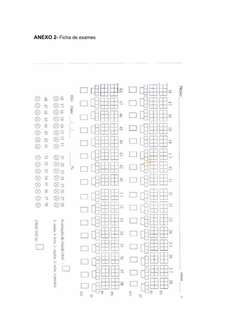
ANEXO 2- Ficha de exames

ANEXO 3- Termo de consentimento livre e esclarecido
- TERMO DE CONSENTIMENTO LIVRE E ESCLARECIDO -
Pelo presente instrumento que atende às exigências legais, O Sr. (a) _____________
_________________________________________, portador(a) da cédula de identidade __ __________________________, após leitura minuciosa da CARTA DE INFORMAÇÃO AO PACIENTE, devidamente explicada pelos profissionais em seus mínimos detalhes, ciente dos serviços e procedimentos aos quais será submetido, não restando quaisquer dúvidas a respeito do lido e explicado, firma seu CONSENTIMENTO LIVRE E ESCLARECIDO concordando em participar da pesquisa proposta intitulada Perfil Periodontal de Pacientes Portadores de Alterações Metabólicas, além da permissão de publicação de fotos e de seus dados coletados no exame.
Fica claro que o paciente e/ou seu representante legal pode a qualquer momento retirar seu CONSENTIMENTO LIVRE E ESCLARECIDO e deixar de participar desta pesquisa e ciente de que todas as informações prestadas tornaram-se confidenciais e guardadas por força de sigilo profissional (art. 9 .do Código de Ética Odontológica).
O responsável pela pesquisa, Prof Danilo Lopes Ferreira Lima, residente nesta cidade à Av Norte 2930-203/A, tel 85-34591949, RG 690539-83 SSP-Ce, está à disposição para qualquer esclarecimento.
E, por estarem de acordo, assinam o presente termo.
Fortaleza- Ce.,_______ de _______________________ de ___________.
______________________________________________________ Assinatura do paciente ou Assinatura do Pesquisador
Representante Legal
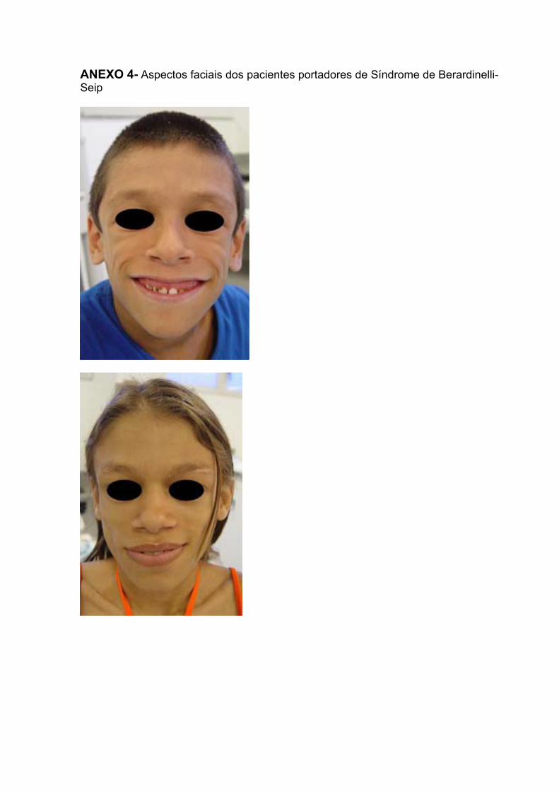
ANEXO 4- Aspectos faciais dos pacientes portadores de Síndrome de Berardinelli-Seip
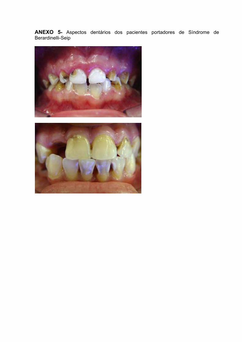
ANEXO 5- Aspectos dentários dos pacientes portadores de Síndrome de Berardinelli-Seip

ANEXO 6- Aceite para publicação do Journal of the International Academy of Periodontology.

ANEXO 7- Publicação de artigos, capítulos de livros e resumos em anais de congressos.
ARTIGOS
1. LIMA, D. L. F., RÊGO, Delane Maria, DIAS, Aldo Angelim, SOUSA, Andréa Félix, FROTA, Raphaelle Menezes da, MENEZES, Ticiane Jucá A de. Avaliação da margem gengival dos dentes anteriores superiores como guia para cirurgias estéticas periodontais. Revista Periodontia. , v.16, p.58 - 64, 2006.
2. PINHEIRO, Ana Ceci Cordeiro, SANDRES, Débora Lopes, OLIVEIRA, Giselle C de, LIMA, D. L. F., NUTO, Sharmênia de A Soares, RÊGO, Delane Maria Tratamento periodontal e bem-estar: um estudo qualitativo. Revista Brasileira em Promoção da Saúde (UNIFOR). , v.19, p.68 - 73, 2006.
CAPÍTULOS DE LIVROS
1. LIMA, D. L. F., RÊGO, Delane Maria, DIAS, Aldo Angelim Epidemiologia e História da Doença Periodontal e sua Importância para a Saúde Pública In: Saúde Bucal Coletiva: Metodologia de Trabalho e Práticas.1 ed.São Paulo : Editora Santos, 2006, v.1, p. 75-94.
2. DIAS, Aldo Angelim, SAMPAIO, José Jackson Coelho, RÊGO, Delane Maria, LIMA, D. L. F., DALCICO, Roberta. Políticas Públicas e Epidemiologia do Câncer de Boca In: Saúde Bucal Coletiva: Metodologia de Trabalho e Práticas.1 ed.São Paulo : Editora Santos, 2006, v.1, p. 297-314.
3. RÊGO, Delane Maria, DIAS, Aldo Angelim, LIMA, D. L. F. A doença periodontal In: Saúde Bucal Coletiva-conhecer para atuar.1 ed.Natal : EDUFRN, 2005, v.1, p. 103-117.
4. REGO, D.M., LIMA, D. L. F., DIAS, Aldo Angelim, FERREIRA, Maria Ângela Fernandes. Periodontites agressivas: etiologia, diagnóstico e tratamento. In: Periodontia: A Atuação Clínica Baseada em Evidências Científicas.1ª ed.São Paulo - SP : Artes Médicas Ltda, 2005, v.1, p. 191-207.
RESUMOS EM ANAIS DE CONGRESSOS OU PERIÓDICOS
1. LIMA, D. L. F., ALBANO, Mônica Fiterman, QUIDUTE, Ana Rosa Pinto, QUEZADO, Rosana, FONTENELE, Eveline Gadelha Pereira, RÊGO, Delane Maria. Ausência de doença periodontal em pacientes acromegálicos: efeito protetor do excesso de GH? In: Simpósio Internacional de Neuroendocrinologia, 2006, Porto Alegre.Arquivos Brasileiros de Endocrinologia & Metabologia. , 2006. v.50. p.S31 - S31
2. LIMA, D. L. F., MONTENEGRO JÚNIOR, R.M., FERNANDES, Virgínia, BARROS, A.I.S., REGO, D.M., MAGALHÃES, Renan. Anormalidades Dentárias na Síndrome
de Berardinelli-Seip In: Arquivos Brasileiros de Endocrinologia & Metabologia. São Paulo-SP: , 2005. v.49. p.S-405 - S-405
3. LIMA, D. L. F., SOUSA, Andréa Félix, FROTA, Raphaelle Menezes da, MENEZES, Ticiane Jucá A de, RÊGO, Delane Maria. Avaliação da margem gengival dos dentes anteriores superiores como guia para cirurgias estéticas periodontais In: XI Encontro de Iniciação à Pesquisa da UNIFOR, 2005, Fortaleza-Ceará.
4. LIMA, D. L. F., REGO, D.M., DIAS, Aldo Angelim. Avaliação da margem gengival dos dentes anteriores superiores como guia para cirurgias estéticas periodontais In: IX Congresso Nacional de Cirurgia Experimental, 2005, Natal- Rio Grande do Norte. Acta Cirurgica Brasileira. São Paulo - SP: , 2005. v.20. p.311 - 311
5. LIMA, D. L. F., ALBANO, Mônica Siterman, MONTENEGRO JÚNIOR, Renan Magalhães, MONTENEGRO, Renan M, RÊGO, Delane Maria. Avaliação do status periodontal de pacientes portadores de acromegalia In: XI Encontro de Iniciação à Pesquisa da UNIFOR, 2005, Fortaleza-Ceará.
6. MOTA, Hugo César Novais, RÊGO, Delane Maria, SILVA, Edna Maria da, LIMA, D. L. F. Clinical Evaluation of the Effects of propolis on Biofilm and Chronic Gingivitis In: 10th International Biennial Congress-International Academy of Periodontology, 2005, p.24 – 24, Salvador-Bahia
7. LIMA, D. L. F., RÊGO, Delane Maria, MONTENEGRO JÚNIOR, Renan Magalhães, FERNANDES, Virgínia Oliveira, BARROS, Antônio Iran de Sousa, MOURA JÚNIOR, José Edson de. Periodontal and Dental Status in Berardinelli-Seip Syndrome In: 10th International Biennial Congress-International Academy of Periodontology, 2005, p.23 - 24
8. LIMA, D. L. F., MONTENEGRO JÚNIOR, R.M., CARDOSO NETO, Antonio Mário, MATOS, Leonardo, GONÇALVES, Renata Duarte, SILVA, C.A.B., FERNANDES, Virgínia, MAGALHÃES, R. A., REGO, D.M., MAGALHÃES, Renan Status periodontal de Pacientes Portadores de Hipertireoidismo In: Arquivos Brasileiros de Endocrinologia & Metabologia. São Paulo-SP: , 2005. v.49. p.S-384 - 384
9. LIMA, D. L. F., FERNANDES, Virgínia Oliveira, BARROS, Antônio Iran de Sousa, MONTENEGRO JÚNIOR, Renan Magalhães, RÊGO, Delane Maria. Alterações dentárias e periodontais na Síndrome de Berardinelli-Seip In: XI Encontro de Iniciação à Pesquisa da UNIFOR, 2005, Fortaleza-Ceará-Brasil.
10.RÊGO, D. M. ; LIMA, D L F ; MONTENEGRO JR, R M ; ALBANO, M S . Investigation of periodontal status in acromegalic patients. Journal of Clinical Periodontology, Espanha, v. 33, p. 146-146, 2006.

11.RÊGO, D. M. ; MONTENEGRO JR, R ; LIMA, D L F ; BARROS, A I S . Glycemic control and periodontal disease: a case report of berardinelli-seip syndrome. Journal of Clinical Periodontology, Espanha, v. 33, p. 156-156, 2006.
12.RÊGO, D. M. ; LIMA, D ; SANTOS, G ; TATAKIS, D . Designing Brazilian smiles: Experiences from periodontology.. Journal of Clinical Periodontology, Espanha, v. 33, p. 165-165, 2006.

Abstract
Objective- Convinced that periodontium, many times, can show alterations in human
health, the aim of these studies was to investigate the periodontal situation in
patients with endocrine-metabolic disorders such as, Berardinelli-Seip Syndrome,
hyperthyroidism, hypothyroidism and acromegaly.
Methods- Eight patients with Berardinelli-Seip Syndrome, 16 acromegalics, 30
hypothyroids, 30 hyperthyroids, and a control group with 35 patients were evaluated.
Clinical attachment loss, probing depth, gingival bleeding index, gingival overgrowth
and Index of Decayed, Missing and Filled Teeth were measured in each patient. All
ethical aspects were rigidly observed, being the study conducted after its approval by
the University of Fortaleza Research Ethics Committee.
Results- The presence of periodontitis was marked in hyperthyroids and in patients
with Berardinelli-Seip Syndrome. Hypothyroids showed not much presence of
periodontitis, while all acromegalics presented absence of periodontitis.
Conclusions- The protective effect of periodontitis in acromegalic patients is a new
finding, whose mechanisms are not yet clear, but may be related to the anabolic
effects of growth hormone. The presence of periodontitis in Berardinelli-Seip
Syndrome may occur due the early onset of diabetes. In hyperthyroids, the high
prevalence of periodontitis could be linked to thyroid hormones effects on bone,
explaining also the minor prevalence in hypothyroids.
Top Related