


Línguas
Páginas
Legal


PONTIFÍCIA UNIVERSIDADE CATÓLICA DO RIO GRANDE DO SUL
PRÓ-REITORIA DE PESQUISA E PÓS-GRADUAÇÃO
FACULDADE DE MEDICINA
PROGRAMA DE PÓS-GRADUAÇÃO EM PEDIATRIA
DOUTORADO EM PEDIATRIA E SAÚDE DA CRIANÇA
CÉLULAS-TRONCO DE CORDÃO
UMBILICAL EM MODELO EXPERIMENTAL
DE ASFIXIA NEONATAL EM SUÍNOS
Davi de Paula
Tese de Doutorado apresentada à Faculdade de medicina da PUC-RS para a obtenção do título de Doutor em Pediatria e Saúde da Criança
Orientador: Prof. Dr. Jaderson Costa da Costa Co-orientador: Prof. Dr. Humberto Holmer Fiori
Porto Alegre, 2010
ii
Dados Internacionais de Catalogação na Publicação ( CIP)
Ficha Catalográfica elaborada por Nívea Bezerra Vasconcelos e Silva CRB 10/1255
P324e Paula, Davi de Células-tronco de cordão umbilical em modelo experimental de
asfixia neonatal em suínos / Davi de Paula. – Porto Alegre, 2010. 63 f.: il. gráf. tab. Tese (Doutorado) – Pontifícia Universidade Católica do Rio
Grande do Sul. Faculdade de Medicina. Programa de Pós-graduação em Pediatria e Saúde da Criança. Doutorado em Pediatria e Saúde da Criança.
Orientador: Prof. Dr. Jaderson Costa da Costa. Co-orientador: Prof. Dr. Humberto Holmer Fiori.
1. CÉLULAS-TRONCO. 2. ASFIXIA NEONATAL. 3. CORDÃO UMBILICAL. 4. HIPÓXIA-ISQUEMIA ENCEFÁLICA. 5. MODELOS ANIMAIS. 6. ANIMAIS RECÉM-NASCIDOS. 7. SUÍNOS. 8. HUMANOS. I. Costa, Jaderson Costa da. II. Fiori, Humberto Holmer. III. Título.
CDD 574.87 NLM QU 325

iii
Dedicatória
À minha querida esposa Alice, e aos meus filhos Júlio e Raquel pelo amor,
confiança e incentivo. Aos meus amados pais Mário e Paula pelo
exemplo de vida e dedicação.
iv
Agradecimentos
Ao meu orientador, Dr. Jaderson Costa da Costa pela oportunidade,
ensinamentos, e apoio ao longo de todo o curso.
Ao meu Co-orientador Humberto Holmer Fiori pelos ensinamentos,
participação, amizade e coleguismo na realização do estudo.
Ao Mestrando Daniel Marinowic pela dedicação, idealismo, perseverança e
amizade durante todos os momentos do trabalho.
Aos professores do programa de Pós-graduação em Pediatria da PUC-RS
em especial ao querido Mestre Dr. Renato Machado Fiori, pela confiança e
oportunidade.
À minha irmã Simone de Paula pelo apoio, amizade durante a realização
dos experimentos e redação da tese.
Ao Dr. Affonso Vittola, às acadêmicas Cristiane e Thaís e às biólogas Ana
Cláudia e Anna Siebel pelo auxilio na realização dos experimentos.
Aos colegas do Laboratório de Neurociências em especial ao Ricardo
Breda, Zuzete Pires, Simone Salamoni e Daniela Abreu.
Á Drª Denise Cantarelli Machado e aos Biólogos Christian e Jeremiah do
Centro de Terapia Celular do instituto de Pesquisas Biomédicas da PUC-RS, pelo
auxilio na separação das células.
Ao professor Léder Xavier, pelo auxilio nas avaliações histológicas.
Ao Dr. Mário Wagner, pelos ensinamentos metodológicos e pelo auxilio na
análise estatística.
Ao Professor Dr. Ivo Wentz e às pós-graduandas Cristiana, Andréa e Gisele
da Faculdade de Veterinária da UFRGS, pelo apoio na realização do experimento e
nos cuidados pós-operatórios dos animais da pesquisa.
Ao funcionário Tiago Giuliani Lopes, do laboratório de Anatomia Patológica
do Hospital São Lucas da PUC-RS, pela ajuda na preparação histológica e imuno-
histoquímica.
À Drª. Silvia Azevedo, ao Dr. Eduardo Cruz e ao Sr. Amarildo Costa da
empresa Cryopraxis pelo apoio financeiro na fase inicial do trabalho e pelo
fornecimento de material de coleta para sangue de cordão umbilical.
Ás secretárias Vanessa, Iones, Carla, Ana e Nelcy, e a funcionária Marlene
pela disposição em sempre ajudar.

v
Às enfermeiras Cristiane, Jociane, Larissa e Simone pela
participação na coleta de material.
Aos funcionários Gilberto e Luiz da Cooperativa Languiru pelo apoio no
fornecimento dos animais.
À CAPES, pelo fornecimento de bolsa de estudo fundamental para a
realização desta pesquisa.
À minha família pelo apoio incondicional e dedicação.
vi
SUMÁRIO
Lista de Figuras ....................................................................................................................vii
Lista de Tabelas .................................................................................................................. viii
Lista de abreviaturas..............................................................................................................ix
Resumo .................................................................................................................................xi
Abstract ................................................................................................................................xii
CAPÍTULO I ......................................... .....................................................................13
1. INTRODUÇÃO E JUSTIFICATIVA ...................... .................................................14
1.1. Anóxia neonatal ............................................................................................................ 14
1.2. Modelos animais de anóxia neonatal ............................................................................ 15
1.3. Terapia com células-tronco........................................................................................... 16
1.4. Células-tronco e doenças neurológicas ........................................................................ 17
1.5. Aspectos Éticos ............................................................................................................ 17
1.6. Justificativa ................................................................................................................... 18
1.7.Objetivos........................................................................................................................ 19
1.8. Bibliografia .................................................................................................................... 20
CAPÍTULO II ........................................ .....................................................................23
2. DEVELOPMENT OF AN EXPERIMENTAL PIGLET MODEL OF N EONATAL ASPHYXIA WITH 21-DAY SURVIVAL ...................... ...............................................24
METHODS........................................................................................................................... 25
RESULTS............................................................................................................................ 28
DISCUSSION ...................................................................................................................... 33
CAPÍTULO III ....................................... .....................................................................39
3. EFFECTS OF THE UMBILICAL CORD STEM CELL TRANSPLA NT IN A PIGLET MODEL OF NEONATAL ASPHYXIA ......................... ..............................................40
METHODS........................................................................................................................... 42
RESULTS............................................................................................................................ 48
DISCUSSION ...................................................................................................................... 52
ANEXOS ...................................................................................................................61

vii
Lista de Figuras
Capítulo II
Figure1 - Photograph illustrating the brain taken from a piglet of Group III with severe
brain damage.----------------------------------------------------------------------------------------- 29
Figure 2- Photomicrographs of the ischemic core to marginal zone with Nissl staining.
.-------------------------------------------------------------------------------------------------------------29
Figure 3– Neurological score at four moments.------------------------------------------------32
Figure 4- Brain volume and final body weight of Group III, compared with the other
two groups. ---------------------------------------------------------------------------------------------32
Figure 5 - Scatter plot of brain volume (ordinate) versus body weight (abscissa) at the
end of the study.---------------------------------------------------------------------------------------33
Capítulo III
Figure 1- Piglet brain -------------------------------------------------------------------------------- 46
Figure 2 - Immunophenotyping of the mononuclear fraction of umbilical cord blood-----
------------------------------------------------------------------------------------------------------------------------------------49
Figure 3 - Agarose gel with the result of the amplification of different brain regions in
two piglets -----------------------------------------------------------------------------------------------50
Figure 4 - Scatter plot of brain volume (ordinate) versus body weight (abscissa) at
the end of the study --------------------------------------------------------------------------------- 52

viii
Lista de Tabelas
Capítulo II
Table 1 -Neurobehavioral Scoring Tool for piglets------------------------------------------- 27
Table 2 -Physiological variables in the pre-operative period--------------------------------30
Table 3-Variables of Arterial Pressure and Arterial Blood Gas from Group III-------------
------------------------------------------------------------------------------------------------------------ -30
Table 4-Duration of the hypoxic-ischemic insult (HI), Neurological findings at four
different periods (2nd, 7th, 14th and 21st days) , Brain Volume, and Survival period.------
--------------------------------------------------------------------------------------------------------------31
Capítulo III
Table 1- Neurobehavioral Scoring Tool for piglets -------------------------------------------45
Table 2-Primers used, reagents and amplification conditions---------------------------- -47
Table 3 - Body weight and Physiological variables before H-I induction-----------------48
Table 4- Neurological scores in four different periods ---------------------------------------50
Table 5 - Result of the PCR for presence of the human β-globin in two animals of
group IV------------------------------------------------------------------------------------------------- 51

ix
Lista de abreviaturas
BDNF Brain-derived neurotrophic factor
BE Basic excess
BLAST Basic Local Alignment Search Tool
bp Base pairs
bpm Breaths per minute
CNS Central nervous system
CO2T Total CO2
CVA Cerebrovascular Accident
DBPS Disinfection by products
DNA Deoxyribonucleic acid
FSC Fluxo sangüíneo cerebral
HCO3 Bicarbonate
H-I Hypoxic-ischemic insult
HLA Human leukocyte antigen system
HR Heart rate
HUCSC Human umbilical cord stem cells
IV Intravenous
MAP Mean arterial pressure
mM Millimolar
mm 3 Cubic millimeter

x
mm Hg Millimeter of mercury
NGF Nerve growth factor
NT3 Neurotrophin-3
PBS Public broadcasting service
PCR Polymerase Chain Reaction
pCO2 Partial CO2 pressure
pO2 Partial O2 pressure
PUC-RS Pontifícia Universidade Católica do Rio Grande do Sul
SNC Sistema nervoso central
RPM Rotations per minute
µm Micrometer
SA Sum of the areas
Sat O2 Saturation
SD Standard deviation
T Distance between the analyzed sections
V(est) Volume estimation

xi
Resumo
EFEITOS DO TRANSPLANTE DE CÉLULAS-TRONCO DE CORDÃO UMBILICAL EM MODELO EXPERIMENTAL
DE ASFIXIA NEONATAL EM SUÍNOS
Introdução : A asfixia neonatal é a principal causa de lesão cerebral no período perinatal, tendo como conseqüências
alta mortalidade e grande número de seqüelas neurológicas. Atualmente, várias estratégias neuroprotetoras estão
sendo avaliadas em modelos animais na tentativa de reduzir a morte celular e melhorar os desfechos neuro-
comportamentais dos recém nascidos, mas os resultados são pouco expressivos. Estudos sugerem que o
transplante de células-tronco limitaria a expansão de lesões e facilitaria o reparo de tecidos lesados, podendo se
constituir numa opção terapêutica em casos de asfixia. Os pesquisadores optaram pelo uso de um modelo em
suínos recém-nascidos devido ao fácil manejo, baixo custo e similaridade de peso e tamanho em relação aos bebês.
Objetivo: O objetivo deste estudo é analisar de que forma células-tronco de cordão umbilical humano, infundidas via
intra-arterial entram no cérebro, sobrevivem neste micro ambiente, e promovem a recuperação da função
neurológica após insulto hipóxico-isquêmico, usando dois tipos diferentes de acessos arteriais.
Materiais e métodos: Foram utilizados 36 suínos com até dois dias de vida divididos em 4 grupos: Grupo I (Sham),
Grupo II de controle, Grupo III tratado com células-tronco via artéria umbilical, e Grupo IV com células-tronco
injetadas pela artéria carótida comum.
Para a indução da asfixia utilizou-se a associação simultânea de procedimentos que causavam hipóxia e isquemia.
As células-tronco foram obtidas a partir de sangue umbilical humano.
Com 2, 7, 14 e 21 dias de vida os animais eram examinados e era aplicado um escore neurológico. O tecido cerebral
de animais tratados com células tronco que morreram antes de completar 21 dias foi utilizado para pesquisa de PCR
para DNA humano. Aos 21 dias os animais sobreviventes eram novamente levados a sala cirúrgica, anestesiados
profundamente a fim de serem sacrificados e realizar-se uma perfusão trans-cardíaca com paraformaldeído para a
extração dos encéfalos. Logo após, era aplicada a técnica histológica de Nissl e realizada a estimativa de volume
encefálico para avaliação do grau de lesão cerebral.
Resultados: . Aos 21 dias houve diferença entre a média dos escores do grupo que recebeu células pela carótida
quando comparada as dos grupos controle e o que recebeu células pela artéria umbilical. Na pesquisa através de
PCR em animais do grupo das células-tronco pela artéria carótida comum foi possível a visualização da banda
correspondente ao gene β-globina humano em dois dos quatro animais em diversos pontos de tecido cerebral em
amostras obtidas 15 e 24 horas após o procedimento de asfixia. Não se identificou PCR positivo nas coletas
realizadas 7 dias e 15 dias deste mesmo grupo bem como em nenhuma das amostras dos animais do outro grupo
pesquisado . Não houve diferença entre as médias dos volumes encefálicos nos quatro grupos. O volume cerebral e
o peso final dos animais apresentaram uma correlação positiva moderada.
Conclusão: Os resultados deste estudo sugerem que a administração de células-tronco de cordão umbilical humano
via artéria carótida comum em modelo de hipóxia-isquemia em suínos está associada a presença de PCR positivo
para o gene da β-grobina humana e a uma melhora na função neurológica com 3 semanas embora sem evidência de
diminuição da área de lesão.
Descritores : 1. Células-tronco, 2. Asfixia Neonatal, 3. Cordão Umbilical, 4. Hipóxia-isquemia encefálica, 5. Modelos
Animais, 6. Animais recém-nascido,. 7. Suínos, 8.Humanos .

xii
Abstract
EFFECTS OF THE UMBILICAL CORD STEM CELL TRANSPLANT IN A PIGLET MODEL OF
NEONATAL ASPHYXIA
Introduction – Neonatal asphyxia is the main cause of brain damage in the perinatal period. Studies
suggest that the stem cell transplant would curb the expansion of damages and facilitate the repair of
damaged tissues, and could thus become a therapeutic option in cases of asphyxia.
Objective : In the present study we tested whether intra-arterialy infused human umbilical cord stem
cells enter brain and survive in the brain microenvironment, and improve neurological functional
recovery after hypoxic-ischemic insult using two two different arterial access.
Materials and methods: Thirty-six healthy piglets not older than two days were divided into four
groups: Group I (Sham), Group II, which was the control group, Group III, treated with stem cells
infused through the umbilical artery, and Group IV, treated with stem cells injected via the common
carotid artery. Stem cells were obtained from human umbilical cord blood.
For induced asphyxia, a simultaneous association of procedures that caused hypoxia and ischemia
was used. The brain tissue of treated animals that died before completing the twenty-one days was
used for PCR research for human DNA. At two, seven, fourteen, and twenty-one days after the
procedures, the animals a neurologic score was applied. After twenty-one days, the survivors were
taken to the surgery room again, deeply anesthetized and a transcardiac perfusion was performed in
order to be sacrificed. After this, the animal brains were slowly extracted and the Nissl histological
staining technique was used to assess the degree of brain damage.
Results : At 21 days there were differences among the average scores of group treated via carotid,
when compared to those of control group and treated via umbilical artery. At other assessment
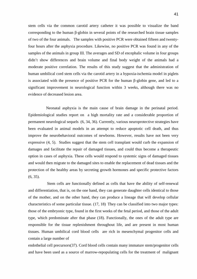
moments no differences were found. In the PCR research of animals that received stem cells via
the common carotid artery catheter it was possible to visualize the band corresponding to the
human β-globin in several points of the researched brain tissue samples of two of the four animals.
The samples with positive PCR were obtained fifteen and twenty-four hours after the asphyxia
procedure. Likewise, no positive PCR was found in any of the samples of the animals in group III.
The averages and SD of encephalic volume in four groups didn’t show differences and brain volume
and final body weight of the animals had a moderate positive correlation
Conclusion : The results of this study suggest that the administration of human umbilical cord stem
cells via the carotid artery in a hypoxia-ischemia model in piglets is associated with the presence of
positive PCR for the human β-globin gene, and led to a significant improvement in neurological
function with 3 weeks, although there was no evidence of decreased lesion area.
Key-words: 1.Stem cells, 2.Neonatal asphyxia, 3.Umbilical cord blood, 4. Hypoxic-ischemic brain
injury, 5.Animal models, 6.Animal newborns, 7.Piglets, 8. Humans.

13
CAPÍTULO I

14
1. INTRODUÇÃO E JUSTIFICATIVA
Esta tese foi redigida sob forma de dois artigos originais conforme as normas
do programa de Pós-Graduação em Pediatria. O primeiro é apresentado no capítulo
II sob o título “DEVELOPMENT OF AN EXPERIMENTAL PIGLET MODEL OF NEONATAL
ASPHYXIA WITH 21-DAY SURVIVAL”, e descreve o desenvolvimento de um modelo
experimental de asfixia neonatal em suínos em que os animais apresentam
sobrevida por um período longo. Já no capítulo III, descrevemos o segundo artigo,
intitulado “ EFFECTS OF THE UMBILICAL CORD STEM CELL TRANSPLANT IN A
PIGLET MODEL OF NEONATAL ASPHYXIA” que tem como objetivo principal
analisar de que forma células-tronco de cordão umbilical humano, infundidas pela
corrente sanguínea se instalam no cérebro, sobrevivem neste micro ambiente, e
promovem a recuperação da função neurológica após insulto hipóxico-isquêmico,
utilizando-se dois tipos diferentes de acessos intra-arteriais no modelo experimental
desenvolvido no estudo anterior. Os artigos foram redigidos no formato da revista
Pediatric Research.
1.1. Anóxia neonatal
De acordo com a American Academy of Pediatrics e o American College of
Obstetrician and Gynecologists, anóxia ou asfixia neonatal é definida como uma
agressão hipóxico-isquêmica grave ao feto ou ao recém-nascido que tem como
resultado um percentual elevado de danos neurofisiológicos permanentes e alta
mortalidade(1). Asfixia é conseqüência do bloqueio da troca gasosa que leva a três
efeitos bioquímicos: hipoxemia, hipercapnia, e acidose metabólica. As condições a
seguir caracterizam a asfixia neonatal:
• Evidência de acidose no sangue de cordão umbilical obtido no
parto;
• Escore de Apgar de 0-3 por 5 minutos ou mais;
• Evidência de seqüela neurológica e em um ou mais dos
sistemas orgânicos a seguir: cardiovascular, gastrintestinal, hematológico,
pulmonar, hepático ou renal.

15
Os recém-nascidos são particularmente vulneráveis a asfixia durante o parto
ou imediatamente após este. Quando ela começa no útero, pouco antes ou durante
o trabalho de parto, decorre geralmente de comprometimento do fluxo sanguíneo da
placenta ou do cordão umbilical e após este período muito provavelmente se origine
de problemas na passagem do ar pelas vias aéreas. (2, 3).
A suscetibilidade do cérebro imaturo à asfixia perinatal depende do estado
temporal e regional do processo de desenvolvimento, bem como, da proliferação,
migração, diferenciação, mielinização, morte programada de células e da regulação
no fluxo sangüíneo cerebral e metabolismo(3).
Atualmente, várias estratégias neuroprotetoras estão sendo avaliadas em
modelos animais na tentativa de reduzir a morte celular apoptótica e, assim,
melhorar os desfechos comportamentais. Dentre os novos tratamentos propostos
para recuperar o tecido cerebral lesado pelos efeitos da anóxia neonatal inclui-se:
inibidores de aminoácidos excitatórios e radicais livres, óxido nítrico, caspases,
topiramato e hipotermia(4, 5). Mais recentemente o transplante com células-tronco
também tem sido considerado como alternativa terapêutica(6).
1.2. Modelos animais de anóxia neonatal
Nenhum modelo de anóxia perinatal é considerado ideal, apesar das
pesquisas em animais têm sido de grande importância neste campo. Modelos com
porcos e ovelhas parecem ser os mais apropriados para os estudos de curto prazo
(até aproximadamente uma semana) e com ratos mais apropriados para estudos
mais longos (com duração de várias semanas). O modelo suíno tem se mostrado
bom para estudos de fluxo sangüíneo cerebral (FSC) e metabolismo, sendo que
atualmente está bem padronizado. Os estudos em curto prazo têm nos ajudado a
entender a fisiopatologia da asfixia, mas estudos de maior prazo têm maior
possibilidade de oferecer evidências histopatológicas de lesão cerebral. Nestes
últimos é possível também avaliar achados clínicos e neurológicos(7-10).

16
Lê blanc et al.(11, 12) desenvolveram um modelo suíno com sobrevivência de
prazo relativamente longo utilizando a combinação de oclusão de vasos cerebrais,
seguidos por um período de hipotensão hemorrágica e hipóxia. Neste modelo a
mortalidade foi de 30% e aproximadamente 70 a 80% dos animais sobreviventes
apresentaram déficits neurológicos. Munnkeby et al(13, 14) desenvolveram dois
modelos agudos de asfixia utilizando mistura gasosa de oxigênio a 8%. Em um deles
a manutenção da hipóxia durou até a pressão sanguínea média alcançar 15 mm Hg
ou o excesso de base alcançar -20 mM. No outro modelo deste autor os animais
foram submetidos a 30 minutos de hipóxia simultâneo a um clampeamento bilateral
das carótidas comuns. Nossa idéia foi fazer uma associação entre os estudos destes
dois autores e desenvolver um modelo suíno de prazo maior, em que pudéssemos
avaliar os efeitos da infusão de células tronco. Fatores como o peso e tamanho dos
suínos recém-nascidos que são parecidos com o de bebês, o fácil manejo e o baixo
custo, fazem com que este modelo seja muito atrativo e factível. Além disso, existe
uma quantidade considerável de literatura relacionada a metabolismo e FSC em
suínos(7, 15, 16).
1.3. Terapia com células-tronco
As células-tronco são definidas funcionalmente como células que tem a
capacidade de auto-renovação associada a habilidade de gerar diferentes células,
ou seja, elas podem gerar células filhas idênticas à mãe (auto-renovação), além de
produzir linhagem com potencial mais restrito (células diferenciadas) (17, 18).
Pode-se classificá-las em dois tipos principais: as do tipo embrionário
existente nas primeiras semanas do período fetal, e as do tipo adulto que
predominam após esta fase. Funcionalmente, as do tipo adulto são responsáveis
pelo reabastecimento tecidual ao longo da vida e estão presentes na maioria dos
tecidos humanos, tais como, o sangue, a pele, o fígado, o coração e o cérebro. Há
muitos anos doenças hematológicas malignas têm sido tratadas através do
transplante de células-tronco tipo adulto de medula óssea ou de sangue de cordão
umbilical e atualmente estudos em diversas doenças de variados órgãos e sistemas
vêm testando esta terapia(18-25).

17
1.4. Células-tronco e doenças neurológicas
Nos últimos 30 anos ocorreram grandes avanços no campo do transplante
neural, e muitos estudos clínicos vem sendo propostos. Em boa parte dos trabalhos
células embrionárias são transplantadas no cérebro de pacientes com doenças
neurológicas, incluindo doença de Parkinson e Huntington e, apesar de alguns
resultados controversos, existe uma concordância geral de que esta terapia trouxe
benefícios aos pacientes (26-29). Todavia, a aplicação do transplante de células
embrionárias em terapia de larga escala encontra séria resistência relacionada a
aspectos éticos e metodológicos, uma vez que se utiliza material abortivo.
Conseqüentemente, um grande esforço tem sido devotado para encontrar fontes
doadoras alternativas, dentre as quais as células-tronco do tipo adulto provenientes
da placenta e cordão umbilical (4, 20, 28, 30-32).
1.5. Aspectos Éticos
Na coleta de sangue de cordão umbilical, embora se trate de material de
descarte, optamos por solicitar o consentimento informado de todas as gestantes
cujo material da placenta tenha sido utilizado.
O presente trabalho baseia-se no princípio de valorizar a vida animal,
considerando sua sensibilidade e procurando sempre reduzir ou evitar sofrimentos
desnecessários. Russell et al l(33) conseguiram sintetizar com 3 palavras o Princípio
Humanitário da experimentação animal, o que ficou definido como o princípio dos 3
Rs devido a sua grafia em inglês.
Replasements, ou seja, Alternativas, indicando que sempre que possível
devemos usar, no lugar de animais vivos, materiais sem sensibilidade. No caso do
presente trabalho, que busca uma avaliação de aspectos clínicos e terapêuticos
embora haja uma fundamentação in vitro bem estabelecida, a complexidade dos
processos envolvidos e a impossibilidade de avaliação dos resultados em seres
humanos, não deixam alternativas senão a experimentação em animais. Trata-se,
portanto, de um indispensável estudo pré-clínico, a fim de que se obtenha
indicações prévias sobre a possibilidade das células-tronco apresentarem efeito
terapêutico e proporcionarem segurança.

18
Reduction, quer dizer, o número utilizado deverá ser o menor possível,
baseado em um cálculo amostral e o minimamente suficiente para que se alcance
resultados confiáveis pelos métodos estatísticos disponíveis.
Refinement, aprimoramento, refere-se a técnicas menos invasivas ou ao
manejo de animais somente por pessoas treinadas. No presente estudo, os
protocolos experimentais utilizados seguirão as normas internacionais de
experimentação animal. Os procedimentos anestésicos, sedativos, e as técnicas
utilizadas estarão de acordo com a prática veterinária correntemente aceita,
evitando-se ao máximo a dor e o sofrimento. Durante a fase de recuperação os
animais ficarão em local apropriado onde pessoas treinadas serão encarregadas da
alimentação, recuperação dos ferimentos e cuidados com a temperatura e a higiene.
1.6. Justificativa
O sistema nervoso central (SNC) é um dos principais sistemas acometidos
por lesão tecidual no período perinatal, e a anóxia neonatal é a causa mais
importante de dano neurológico ocorrendo em aproximadamente 2-4:1000 nascidos
vivos a termo. (6, 34). Recentemente, muitas pesquisas estão avaliando a aplicação
de células-tronco nas mais diversas doenças, em especial no campo da neurologia.
A terapia celular poderia facilitar o reparo de tecidos lesados e exercer efeito
protetor, limitando a expansão de lesões. A capacidade potencial destas células em
responder a sinais sistêmicos de tecidos lesados, de migrar para estas regiões, de
substituir tecidos mortos ou de proporcionar proteção por secreção de hormônios de
crescimento e fatores de proteção específicos, são consideradas características
desejáveis às necessidades terapêuticas da medicina perinatal. Há também algumas
particularidades dos recém nascidos poderiam oferecer vantagens. As dimensões
relativamente pequenas, a perspectiva de futuro desenvolvimento do bebê e a
disponibilidade de material da placenta, que é um grande reservatório de sangue
fetal, favorecem a obtenção, a aplicação e os efeitos do tratamento(6, 28, 35).
Existe, porém, um número menor de estudos nesta faixa etária. Além disso,
várias questões carecem de respostas mesmo considerando os bons resultados em
estudos com modelos adultos. Exatamente que mecanismos moleculares, celulares,

19
fisiológicos estariam implicados? Quais os tecidos seriam mais suscetíveis à sua
utilização? Qual a duração dos possíveis efeitos? Que via de administração e
momento de aplicação seriam mais adequados (2, 6, 17, 20, 22, 23)?
Um estudo pré-clínico visando contribuir na elucidação de algumas destas
questões é de fundamental importância, especialmente quando levamos em
consideração o uso desta terapia durante o período perinatal.
1.7.Objetivos
Objetivo geral: Avaliar os efeitos da injeção de células-tronco de cordão umbilical humano em
um modelo experimental em suínos de anóxia neonatal.
Objetivos específicos:
1. Testar um modelo experimental de asfixia neonatal em suínos recém-
nascidos;
2. Comparar as alterações histológicas, e neurocomportamentais entre
animais não tratados e tratados com células-tronco administradas por
duas vias arteriais diferentes, após serem submetidos à asfixia neonatal;
3. Detectar a migração das células-tronco transplantadas nas regiões
encefálicas acometidas pelo insulto hipóxico-isquêmico através de PCR
humano.

20
1.8. Bibliografia
1. Dios JG. Definición de asfixia perinatal en la bibliografia médica: necesidad de
um consenso. Rev Neurol 2002;35(7):628-634.
2. Alonso-Spilsbury M, Mota-Rojas D, Villanueva-García D, J. M-B, Orozco H,
Ramírez-Necoechea R, et al. Perinatal asphyxia pathophysiology in pig and
human: A review. Animal Reproduction Science 2005(90):1-30.
3. Vexler ZS, Ferriero DM. Molecular and biochemical mechanisms of perinatal
brain injury. Semin Neonatol 2001;6(2):99-108.
4. Johnston MV, Trescher WH, Ishida A, Nakajima W. Novel treatments after
experimental brain injury. Semin Neonatol 2000;5(1):75-86.
5. Lee SR, Kim SP, Kim JE. Protective effect of topiramate against hippocampal
neuronal damage after global ischemia in the gerbils. Neurosci Lett
2000;281(2-3):183-6.
6. Santner-Nanan B, Peek MJ, McCullagh P, Nanan R. Therapeutic potential of
stem cells in perinatal medicine. Aust N Z J Obstet Gynaecol 2005;45(2):102-
7.
7. Raju TNK. Some Animal Models for the Study of Perinatal Asphyxia. Biol
Neonate 1992(62):202-214.
8. Roohey T, Raju TNK, Moustagiannis AN. Animal models for the study of
perinatal hypoxic-ischemic encephalopathy: a critical analysis. Early Human
Development 1997(47):115-146.
9. Thorosen M, Haaland K, Loberg EM, Whitelaw A, Apricena F, Hanko E, et al. A
piglet model of posthypoxic encephalopathy. Pediatric Res 1996;40(5):738-48.
10. Yager JY. Animal models of hypoxic-ischemic brain damage in the newborn.
Semin Pediatr Neurol 2004;11(1):31-46.
11. LeBlanc MH, Vig V, Smith B, Parker CC, Evans OC, Smith EE. MK-801 Does
Not Protect Against Hypoxic-Ischemic Brain Injury in Piglets. Stroke
1991(22):1270-1275.
12. LeBlanc MH, Li XQ, Huang M, Patel DM, Smith EE. AMPA Antagonist
LY293558 Does Not Affect the Severity of Hypoxic-Ischemic Injury in Newborn
Pigs. Stroke 1995(26):1908-1915.
13. Munkeby BH, Borke WB. Resuscitation with 100% O2 Incrases Cerebral Injury
in Hypoxemic Piglets. Pediatric Research 2004;56(5):783-790.

21
14. Munkeby BH, Lyng K, Froen FJ, Winther-larssen EH, Rosland JH, Smith H-J,
et al. Morphological and Hemodynamic magnetic Resonance Assessment of
Early neonatal Brain Injury in a Piglet Model. Journal of magnetic Resonance
Imaging 2004 20:8-15.
15. Shum-Tim D, Nagashima M, Shinoka T, Nollert G. Postischemic hyperthermia
exacerbats neurologic injury after deep hypothermic circulatory arrest. The
Journal of thoracic and Cardiovascular Surgery 1998;116(5):780-792.
16. Agnew MD, Koehler RC, Guerguerian A, Shaffner DH. Hypothermia for
24hours after Asphyxic Cardiac arrest in Piglets Provides Striatal
Neuroprotetion That is Sustained 10 Days after Rewarming. Pediatric Res
2003;54(2):253-262.
17. Melton DA, Cowan C. "Stemness": Definitions, Criteria, and Standards. In:
Lanza R, editor. Handbook of Stem Cells. San Diego: Elsevier Inc, 2004.
18. Li L, Xie T. Stem Cell Niche: Structure and Function. Annu Rev Cell Dev Biol
2005.
19. Daley GQ, Goodell MA, Snyder EY. Realistic prospects for stem cell
therapeutics. Hematology (Am Soc Hematol Educ Program) 2003:398-418.
20. Haas S, Weidner N, Winkler J. Adult stem cell therapy in stroke. Curr Opin
Neurol 2005;18(1):59-64.
21. Rice CM, Scolding NJ. Adult stem cells--reprogramming neurological repair?
Lancet 2004;364(9429):193-9.
22. Nash RA. Allogeneic HSCT for autoimmune diseases: conventional
conditioning regimens. Bone Marrow Transplant 2003;32 Suppl 1:S77-80.
23. Muller P, Pfeiffer P, Koglin J, Schafers HJ, Seeland U, Janzen I, et al.
Cardiomyocytes of noncardiac origin in myocardial biopsies of human
transplanted hearts. Circulation 2002;106(1):31-5.
24. Horwitz EM, Prockop DJ, Fitzpatrick LA, Koo WW, Gordon PL, Neel M, et al.
Transplantability and therapeutic effects of bone marrow-derived
mesenchymal cells in children with osteogenesis imperfecta. Nat Med
1999;5(3):309-13.
25. Theise ND, Nimmakayalu M, Gardner R, Illei PB, Morgan G, Teperman L, et
al. Liver from bone marrow in humans. Hepatology 2000;32(1):11-6.
26. Langston JW. The promise of stem cells in Parkinson disease. J Clin Invest
2005;115(1):23-5.

22
27. Dunnett SB, Rosser AE. Cell therapy in Huntington's disease. NeuroRx
2004;1(4):394-405.
28. Rossi F, Cattaneo E. Neurologic Diseases. In: Lanza R, editor. Handbook of
Stem Cells. San Diego: Elsevier Inc, 2004:695-702.
29. Korbling M, Estrov Z. Adult stem cells for tissue repair - a new therapeutic
concept? N Engl J Med 2003;349(6):570-82.
30. Hayashi T, Iwai M, Ikeda T, Jin G, Deguchi K, Nagotani S, et al. Neural
precursor cells division and migration in neonatal rat brain after
ischemic/hypoxic injury. Brain Res 2005;1038(1):41-9.
31. Koda M, Okada S, Nakayama T, Koshizuka S, Kamada T, Nishio Y, et al.
Hematopoietic stem cell and marrow stromal cell for spinal cord injury in mice.
Neuroreport 2005;16(16):1763-1767.
32. Kohyama J, Abe H, Shimazaki T, Koizumi A, Nakashima K, Gojo S, et al. Brain
from bone: efficient "meta-differentiation" of marrow stroma-derived mature
osteoblasts to neurons with Noggin or a demethylating agent. Differentiation
2001;68(4-5):235-44.
33. Rivera EAB. Ética na Experimentação Animal. In: Andrade A, Pinto SC,
Oliveira RS, editors. Animais de Laboratório, Criação e Experimentação. Rio
de Janeiro: Fiocruz, 2002.
34. Vannucci SJ, Hagberg H. Hypoxia-ischemia in the immature brain. J Exp Biol
2004; 207(Pt 18):3149-54.
35. Mayhall EA, Paffett-Lugassy N, Zon LI. The clinical potential of stem cells. Curr
Opin Cell Biol 2004;16(6):713-20.

23
CAPÍTULO II

24
2. DEVELOPMENT OF AN EXPERIMENTAL PIGLET MODEL OF NEONATAL
ASPHYXIA WITH 21-DAY SURVIVAL
DAVI DE PAULA, DANIEL MARINOWIK, SIMONE DE PAULA, LÉDER LEAL
XAVIER, HUMBERTO HOLMER FIORI, JADERSON COSTA DA COSTA
Laboratório de Neurociências [D.P.,D.M., S.P, L.L.X., J.C.D.], Departamento de Pediatria [H.H.F.], Pontifícia Universidade Católica do Rio Grande do Sul, Porto Alegre, RS 90610-000, Brazil
Correspondence: Jaderson Costa Dacosta, M.D., Ph.D., Laboratório de Neurociências, Instituto de Pesquisas Biomédicas e Instituto do Cérebro, PUCRS, Av. Ipiranga 6690/220, 90610-000, Porto Alegre, RS, Brazil; e-mail: [email protected]
Supported by grants from CAPES, FUNPAR, and Pandurata
ABSTRACT: Perinatal Asphyxia is the most frequent cause of neurological damage in newborn.
The purpose of this study was to develop a swine model of Neonatal Asphyxia in which the animals
remain alive for a period up to twenty-one days, keeping evidence of damage throughout the study.
We used combined hypoxia and ischemia induction techniques in two different time regimes. Twenty
animals were divided into three groups: Group I (n-5) or Sham , Group II(n-5), submitted to hypoxia
and ischemia for forty-five minutes, and Group III(n-10), submitted to hypoxia and ischemia for
variable periods of time, based on a combination of hypotension and acidosis. At 2, 7, 14, and 21 days
after the procedures, the animals were examined ,any abnormalities found were recorded, and a
neurological score was performed. After twenty-one days, the piglets were deeply anesthetized and a
transcardiac perfusion was performed to the brain extraction . To estimate the degree of brain damage,
we applied a brain volume method. Most part of animals in Group II presented changes forty-eight
hours after the procedure but seven days later, such changes were still found in just one of the animals.
In Group III, just one of survivors did not present any kind of abnormality after forty-eight hours, and
after the twenty-one days, four of the five survivors did not demonstrate effective sucking. There was
a significant difference in the neurological score mean of Group III, if compared with the other groups,
in the four evaluations. Brain volume and final body weight of Group III were significantly higher,
compared with the other two groups. In conclusion, this model of neonatal asphyxia with association
of hypoxia and ischemia was useful in a more prolonged follow-up, thus enabling the identification of
neurological changes throughout the twenty-one days of the study, mainly when the time of the insult
was based on parameters of hypotension and acidosis. The brain damage estimation based on brain
volume was not effective to determine damage.
The central nervous system (CNS) is one of the main systems affected by tissue injury in the
perinatal period, and Perinatal Asphyxia is the most frequent cause of neurological damage. It occurs

25
in approximately 2-4 out of every 1,000 living infants born at full term. The damages caused by
Asphyxia to the newborn infant are the result of the association between hypoxia and ischemia,
affecting the organism in a generalized manner, and oftentimes leading to a severe neurological
damage, difficult to recover from, and in many instances related to long-term repercussions (1-3).
According to the American Academy of Pediatrics and the American College of Obstetricians and
Gynecologists, neonatal anoxia or asphyxia is defined as a severe hypoxic-ischemic aggression to the
fetus or newborn, which results in a high percentage of permanent neurophysiological damage and
high mortality rate (4) (5) .
Although research conducted with animals has been of great importance in this field, no
model of perinatal asphyxia is considered ideal. Models with pigs and sheep have been more widely
used in short-term studies (of approximately up to one week), whereas rats are used in longer studies
(lasting several weeks). The short-term studies are used to study the physiopathology of asphyxia, and
the more prolonged ones are used to assess clinical aspects, and these are more favorable to the
detection of histopathologic evidence of brain damage (6-10). In this study, we chose a model in
swine. Several characteristics are interesting in this model. The gyroenchephalic anatomy and the
brain vascularization of the swine are similar to those of humans, and there is a considerable amount
of literature concerning metabolism and brain blood flow (6, 8, 11-13) (14). Due to the size and body
weight of the animals, the swine models enable easy obtaining of vascular accesses, as well as
cardiovascular and respiratory monitoring, thus also allowing the use of neonatal intensive treatment
equipment. Nevertheless, the number of longer-term studies is relatively small.
The objective of this study is to use a swine model of Neonatal Asphyxia in which the
animals remain alive for a period up to twenty-one days, keeping evidence of damage throughout the
study. For this purpose, we planned to use combined hypoxia and ischemia induction techniques in
two different time regimes.
METHODS
Twenty-nine healthy piglets of the Sus scrofa race, obtained from a local farm and not older
than two days old, were used in this study. Initially, a pilot study was conducted, in which nine
animals were subjected to the hypoxia and ischemia association, with different durations. Based on the
results of the pilot, the duration of the hypoxia and ischemia was established for one of the groups in
the subsequent phase of the study. In the next stage, twenty animals were divided into three groups:
Group I (Sham), Group II, submitted to hypoxia and ischemia for forty-five minutes, and Group III,
submitted to hypoxia and ischemia for variable periods of time, based on a combination of
hypotension and acidosis. The first two groups were composed of five animals, whereas the third
group had ten animals. The protocol of this study was approved by the Research Ethics Committee of
the Medical School of PUC-RS, under the following registration number: CEP-PUC 06/03425.
Anaesthesia, ventilation and monitorin of physiological variables .After the stabilization
period, the animals were subjected to anesthetic induction with inhalational halothane (3%),
26
endotracheal intubation, and umbilical cord vessel catheterization. For anesthetic maintenance,
ketamine(15-20 mg/kg IV or IM) and xilazine (2mg/Kg IV or IM) was used every two hours.
After intubation the animals were kept on a mechanical ventilator (BP 400, Pró Médico,São
Paulo,Brasil) with respiratory rate of 20 breaths per minute(bpm), PIP of 15 mmHg and PEEP
of 3 mmHg and oxygen concentration of 21%.
The heart rate, rectal temperature, and saturation of all animals were monitored by
transcutaneous monitoring(Ohmeda 3800,GE,Helsinki,Finland). In the group III the mean
arterial pressure was obtained via catheter installed through an umbilical artery and connected
to a pressure monitoring device(Kananda 2, Belo Horizonte, Brasil) . For the same catheter
samples were collected for determination of arterial blood gases.
Protocol for hypoxic-ischemic insult and experimental groups. For induced asphyxia,
a simultaneous association of procedures that caused hypoxia and ischemia was used.
Hypoxia was obtained by administering an inhalational mixture of 8% O2 and 92% nitrogen
through an endotracheal tube connected to a mechanical ventilator, and ischemia was induced
by clamping both common carotid arteries. The procedure was completed with the reversion
of the arterial occlusion and the replacement of the hypoxic mixture with ambient air.
In Group I, the common carotid arteries of the animals were dissected, but not
occluded. The animals in Group II were subjected to the inhalation of a hypoxic mixture
associated with the bilateral occlusion of the common carotid artery for a fixed time of forty-
five minutes. And the animals in Group III were submitted to hypoxia and occlusion of the
carotid arteries until their mean arterial pressure (MAP) reached less than 30 mm Hg,
associated with an arterial pH of 7.28 or below. All animals in Group III had one of their
umbilical arteries dissected and catheters were introduced into them for the monitoring of the
arterial pressure. Arterial blood gas samples were taken immediately before asphyxia was
induced and at the moment the animals’ mean arterial pressure reached 30 or below.
After the surgical procedures, the animals remained under mechanical ventilation until
they regained spontaneous breathing and were fit to be transported to a shelter. In the shelter,
the animals stayed in incubators, warmed by radiant heat and gavage-fed bovine milk until
they were able to suck effectively to drink the milk directly from the bottle.
After twenty-one days, the survivors were taken to the surgery room again, deeply
anesthetized with thiopental 50 mg/kg administered intraperitoneally for them to be
sacrificed, and so that a transcardiac perfusion with physiological serum and 4%
paraformaldehyde could be performed.
Neurological Evaluation. At 2, 7, 14, and 21 days after the procedures , the animals

27
were examined and any abnormalities found were recorded, such as gait changes, palsies,
difficulty sucking, hypoactivity, and abnormal movements. A neurological score previously
used for swine models was adapted and included the assessment of consciousness level (0 to
15 points), brainstem function (0 to 22 points), sensory response ( 0 to 20 points), muscle
tone (0 to 8 points),postural reflexes (0 to 8 points), mobility ( 0 to 30), spatial orientation (0
to 20 points), activity (0 to 16 points) and seizures (0 to 10 points) (Table 1) (12).The results
were recorded and scored from 0 to 149
Table 1- Neurobehavioral Scoring Tool for piglets
Item Scoring code
Consciousness 0=Normal 5=clouded 10=stupor 15=coma Brainstem function
Respiration 0=Normal 5=Present but abnormal 10=Absent Pupilar light reflex 0=Normal 2=Present but abnormal 4=Absent
Corneal reflex 0=Normal 2=Present but abnormal 4=Absent Gag reflex 0=Normal 2=Present but abnormal 4=Absent
Sensory responses Olfaction 0=Normal 2=Present but abnormal 4=Absent
Visual threat/orienting 0=Normal 2=Present but abnormal 4=Absent Auditory startle/arousal 0=Normal 2=Present but abnormal 4=Absent
Pain withdrawal 0=Normal 2=Present but abnormal 4=Absent Tactile localization 0=Normal 2=Present but abnormal 4=Absent
Muscle tone Muscle tone, trunk 0=Normal 2=Present but abnormal 4=Flaccid Muscle tone, limbs 0=Normal 2=Present but abnormal 4=Flaccid Postural reflexes Extensor thrust 0=Normal 2=Present but abnormal 4=Absent Wheelbarrow 0=Normal 2=Present but abnormal 4=Absent
Mobility 0=Normal postural righting and gait 5=Ataxic, walk, but walks without falling 10=Ataxic, walks but falls frequently 15=Can't walk, but stands without assistance 20=Stands only with assistance 25=Cannot stand, but attempts to right head &trunk 30=Unable to right head, no purposeful movement
Spatial orientation During locomotion 0=Normal 4=Abnormal 8=Absent
With sniffing 0=Normal 2=Abnormal 8=Absent Toward depth 0=Present 4=Absent
Activity Appetite 0=Present 2=Abnormal 4=Absent
Vocalization 0=Present 2=Abnormal 4=Absent Psychomotor activity 0=Present 2=Abnormal 4=Absent Social Interativiness 0=Normal(seeks contact) 2=Abnormal(aggressive)
Withdrawn) 4=Absent (no social responsiveness) Seizures
Stimulus-induced myoclonus 0=Absent 5=Present
Clonic or tonic or seizures 0=Absent 10=Present Total Range 0 (no deficit) – 149(maximum deficit)

28
Brain Volume Estimation and histology. Brain volume estimation was chosen to
assess the degree of brain damage. Brain damage is associated with the loss of brain tissue
and the consequent decrease of brain volume.
After being extracted, the brains were photographed and stored for twenty-four
hours in 4% paraformaldehyde, and then embedded in paraffin. 20- µm thick coronal sections
at 1,200-µm intervals were obtained by microtome. The Nissl histological staining technique
(cresyl violet method) was used. Images of the histological sections were obtained through a
video camera installed on an Olympos (BX40) microscope, and later analyzed with the aid of
the software Image Pro-Plus 6. 1. The brain volume estimation was determined using the
Cavalieri principle, according to the following equation: V(est) = T. AS, where V(est) =
volume estimation; T = distance between the analyzed sections; SA = sum of the areas (15).
Data analysis. Normal distribution variables were presented as mean and standard
deviation. Comparisons between the groups were analyzed using one-way analysis of
variance (ANOVA), followed by the Tukey test. In the neurologic evaluation results, when
appropriate, the Kruskal-Wallis non-parametric analysis and post-hoc Mann-Whitney tests,
followed by the Finner-Bonferroni correction for multiple testing were performed.
Differences were considered significant at P_< 0.05.
RESULTS
A total of twenty-nine animals were used. Nine of them were used in the pilot study,
Group I had five animals, Group II had also five animals, and the remaining ten piglets were
in Group III. In the pilot study, none of the animals submitted to hypoxia and ischemia (H-I)
for less than forty-five minutes presented any neurological changes forty-eight hours after the
procedure, whereas no animal submitted to the procedure for one hour or more survived for
over three days.

29
Figure 1. Photograph illustrating the brain taken from a piglet of Group III with severe brain
damage.
Figure 2 - Photomicrographs of the ischemic core to marginal zone with Nissl staining. The
right picture shows a hemisphesric brain damage in place indicated by red arrow, and the left one the
same region in a normal brain.
Physiologic variables before and during the hypoxic-ischemic insult. The groups did
not demonstrate any differences concerning body weight, age, temperature, heart rate, and arterial
saturation obtained in the pre-operative period (table 2)

30
Table 2 – Physiological variables in the pre-operative period
GROUPS Age (h) Initial body
weight (g) HR (bpm) Temp. (ºC) Sat
I (n-5) 10,6 + 4,1 1430 + 327,1 132,6 + 29,72 36,54 + 0,68 93,6 + 9,86
II (n-5) 13,67 + 7 1560 + 243,41 156 + 12,94 36,8 + 0,8 98,2 + 1,8
III (n-10) 13,6 + 8,5 1635 + 270,85 125,8 + 26,55 35,9 + 1,19 97,6 + 1,57
Values are presented as means ± S.D.
HR = Heart rate MAP = Mean Arterial Pressure PaO2 = Partial oxygen pressure PaO2 = Partial carbon dioxide
pressure Sat –O2. saturation SD = Standard deviation
In the comparison between the mean arterial pressures and the variables of the
arterial blood gas samples calculated immediately before the induction and with those
conducted at the end of the hypoxia and ischemia mean values for the Group III was
significantly lower (p< 0.001), as shown in table 3.
Table 3 - Variables of Arterial Pressure and Arterial Blood Gas of piglets from Group III
Values are presented as means ± S.D. MAP = Mean Arterial Pressure pO2=partial O2 pressure pCO2=partial CO2
pressure HCO3=Bicarbonate CO2T= Total CO2 Sat – saturation BE= Basic excess
Survival and posthypoxic neurological examination. Signs of neurological
impairment and longer survival were only identified in those animals whose pre-established
procedure time was forty-five minutes. For this reason, a fixed period of forty-five minutes of
H-I was established for Group II in the subsequent stage. All the five animals in Group II
survived, and four of them presented changes forty-eight hours after the procedure. However,
seven days later, such changes were still found in just one of the animals.
In Group III, all of them presented some sort of change forty-eight hours after the
procedure, and out of the animals that survived until the seventh day, just one did not show
any changes. Out of the ten animals, five survived. Nonetheless, just one of them did not
present any kind of abnormality after forty-eight hours, and after the twenty-one days, four of the
MAP pH pO2 pCO2 HCO3 CO2T Sat BE
Pre-
Induction 57,8 + 8,5 7,53 + 0,1 81,8 + 14,1 34 + 3,1 27,7 + 2,8 28,4 + 3,1 96,2 + 2 5,1 + 2,6
Final 27,8 + 8,5 7,15 + 0 17,5 + 6,8 61,7 + 3,3 20,5 + 3,3 22,4 + 3,5 16,5 + 11,8 -8,8 + 3,4

31
five survivors did not demonstrate effective sucking, thus requiring the introduction of a gastric
feeding tube (Table 4).
Table 4- Duration of the hypoxic-ischemic insult (HI), Neurological findings at four
different periods (2nd, 7th, 14th and 21st days, Brain Volume, and Survival period.
Group Animal number
Duration of HI insult(min) 2 days 7 days 14 days 21 days Brain Vol.
(mm 3) Survival period after
HI insult (days)
10 0 NA NA NA NA 21
4 0 NA NA NA NA 21
I 1 0 NA NA NA NA 2700 21
6 0 NA NA NA NA 2956 21
7 0 NA NA NA NA 2481,6 21
2 45 Normal Normal Normal Normal 2560,8 21
9 45 Unilateral facial
palsy Ataxia Normal Normal 2286,38 21
Lethargy
II ataxia
11 45 ataxia Normal Normal Normal 2459,18 21
12 45 bilateral facial
palsy Normal Normal Normal
21
ataxia
13 45 bilateral facial
palsy Normal Normal Normal 21
ataxia
19 107 Unilateral facial
palsy; Unilateral facial
palsy;
12
Lethargy Lethargy
Ataxia
Tremulousness
poor sucking
25 60 coma 1
30 47 coma Bilateral facial
palsy Ataxia poor sucking 3190,36 21
Seizure Ataxia Lethargy Ataxia
Lethargy poor
sucking Lethargy
poor sucking
41 35 Lethargy 2
Not stand
poor sucking
42 50 Lethargy Normal Normal Normal 3912 21
Ataxia
III poor sucking
43 45 Unilateral facial
palsy Lethargy poor sucking
poor sucking 4128 21
Lethargy poor sucking
Ataxia
poor sucking
45 49 Lethargy poor sucking poor
sucking poor
sucking 3639,98 21
Ataxia
poor sucking
46 65 Coma 2
49 40 Lethargy Lethargy poor
sucking poor
sucking 4001,56 21
Ataxia poor sucking
poor sucking
57 100 Coma 2

32
When the neurological score was applied, there was a significant difference in the mean
score of Group III, if compared with the other groups, in the four evaluations (figure 3).
Figure 3 – Graph shows neurological score o on the second, seventh, fourteenth, and
twenty-first days.
Assessment of Brain Volume. The means of brain volume and final body weight of
the three groups are shown in figure 4.
Figure 4 – Brain volume and final body weight of Group III, compared with the other two groups.
Values are presented as means + S.D.* P< 0,0001 Group III vs. I and II. Error bars are mean ±
SEM.
By using the correlation test between the body weight verified at the end of the
follow-up period and the brain volume of all the animals, a positive correlation of 0.92 (p <
0.001) was found (figure 5).

33
Figure 5 – Scatter plot of brain volume (ordinate) versus body weight (abscissa) at the end of the study. There was a significant correlation of 0.92 (p< 0.001).
DISCUSSION
We have developed a model of hypoxic-ischemic brain damage in piglets , in which
the animals survived for a prolonged period of time. This model is suitable for examining
mechanisms of damage and evaluation of potential protective therapies after birth asphyxia.
The animals whose duration of the asphyxia was based on the presence of acidosis and
hypotension presented longer-lasting neurological manifestations. Moreover, mortality rate,
final mean body weight, and brain volume were significantly higher in the group where the
number of neurological changes was larger.
Finding an ideal model to study Neonatal Asphyxia remains a great challenge for
researchers.
Models with rats are well standardized. Such animals are available in large numbers
and are widely used in testing new therapeutic strategies. However, they become limited when
physiological monitoring is important (3, 6, 7, 16, 17). These models also present difficulty
reproducing neurological changes in the long run. Pigs are relatively available and allow the
access to physiological monitoring. Throughout this work it was possible to utilize equipment
used in neonatal Intensive Care Units for various functions, such as heating, mechanical
ventilation, and umbilical catheterization. This fact contributed to the performance of the
experiment and to the maintenance care.
For the purpose of sham neonatal asphyxia, our choice was for newborn animals. In
this experiment, notwithstanding the severe insult, several animals recovered completely in a
short time. The damage and repair mechanisms vary according to the stage of neuronal
maturation (18,19). Differently from the adult brain, in the immature brain we find neuronal

34
proliferation, myelination process and apoptosis, low ratio of glial cells, low metabolic need,
dependence on different substrate, and different enzymatic activity (8). Therefore, the choice
was for animals not older than two days old. Research suggests that although in immature
animals the groups of neurons are more vulnerable to damage, they are in general more
resistant to hypoxia and ischemia, and present low rates of energy use, which makes it
difficult to study sequelae in the long run. All these aspects explain why it is difficult to
develop a neonatal asphyxia model with a longer observation period.
An association between hypoxia and ischemia techniques has been used in models of
different species (14, 20-24). . Munkeby et al(21, 22) developed a swine model of short-term
asphyxia by using a gaseous mixture of 8% oxygen. In one of them, the maintenance of
hypoxia lasted until the mean blood pressure reached 15 mm Hg, or until the basic excess
reached -20mM. In another one, the animals were submitted to thirty minutes of hypoxia
simultaneously with the bilateral clamping of the common carotid arteries. In these studies,
the animals were maintained during a maximum period of forty-eight hours. Lê blanc et al(23-
25) developed a model in piglets with a relatively long-term survival by using a combination
of occluded brain vessels, followed by a period of hemorrhagic hypotension and hypoxia with
a concentration of 6% O2. In this model, the mortality rate was approximately 30%, and 70%
to 80% of the surviving animals demonstrated neurological deficits. He kept the animals alive
for three days. However, we do not know whether these animals would have sustained
damage if they had been kept alive for a longer period of time.
Nevertheless, some authors claim that the use of vascular occlusion with regional
ischemia would represent an important limitation, due to the fact that it does not occur in
human neonatal asphyxia (8). The animal diffusely exposed to aggression, with subsequent
impairment of organs such as kidneys, heart, and intestines would reproduce more faithfully
the actual conditions. Nevertheless, for the objective of this study, this proposal would pose
additional difficulties, since a larger number of severely affected systems would represent a
higher level of complication to keep the survival of the animals, and consequently the
continuation of the neurological manifestations.
In this model, the challenge lies in provoking a sufficiently severe damage that may
lead to prolonged sequelae, but not so strong as to impede the survival of the animal. When it
comes to balance, there is no doubt that the duration of the hypoxic-ischemic insult is an
important variable. At the other end of the scale, factors like the use of mechanical ventilation
equipment and the anesthetic regimen employed may influence the survival time (14). Based
on this work, a suggestion is that, in addition to the above-mentioned items, two other highly

35
useful items be also included. The use of an umbilical catheter for hydration and anesthetic
maintenance, especially during the immediate post-operative period, as well as the use of an
orogastric tube until the animals are able to suck, which may take several days. It was
observed that animals initially in a more severe condition needed the gastric tube for a longer
period of time, which is a fact that also occurs with human newborns.
Through the analysis of neurological manifestations, it was observed that the fixed
time model was capable of provoking briefer changes, but the model in which MAP and
acidosis as a reference were used led to more prolonged changes, despite its higher mortality
rate. The scale used was effective in detecting conspicuous changes, although it may not have
been sensitive to detect subtle signs. New studies are necessary to develop a more sensitive
and specific scale for this model.
Brain volume determination is a widely used method in brain damage models in rats
(17, 26-29). Contrary to what we thought, the results of this study showed that the brain
volume in the group with more neurological changes was significantly larger than that of the
other ones. Furthermore, the mean body weight of these animals was also greater at the end of
the experiment. The animals with a higher level of impairment stayed inactive or hypoactive
for longer periods of time, and were often tube-fed, which would explain a greater weight
gain in this group. The comparison between the brain volume and the final body weight of all
the animals included in the study showed that there was a positive correlation, which suggests
that the brain volume in newborns of this species has a direct relation to the animal's
nutritional state. Our hypothesis is that the volume gain brought about by better nutrition may
have compensated for the possible decrease in volume caused by the loss of brain mass.
In conclusion, this model of neonatal asphyxia obtained through the association of
hypoxia and ischemia was useful in a more prolonged follow-up, thus enabling the
identification of neurological changes throughout the twenty-one days of the study, mainly
when the time of the insult was based on parameters of hypotension and acidosis.
The brain volume of newborn piglets is related to the animals’ nutritional state
throughout the experiment. In this study, brain damage estimation based on brain volume was
not effective to determine damage. We suggest that further research be conducted to evaluate
the use of this brain damage estimation method in experimental models with piglets.

36
REFERENCES
1. Vannucci SJ, Hagberg H 2004 Hypoxia-ischemia in the immature brain. J Exp Biol
207:3149-3154.
2. Santner-Nanan B, Peek MJ, McCullagh P, Nanan R 2005 Therapeutic potential of
stem cells in perinatal medicine. Aust N Z J Obstet Gynaecol 45:102-107.
3. Weitzdoerfer R, Pollak A, Lubec B 2004 Perinatal asphyxia in the rat has lifelong
effects on morphology, cognitive functions, and behavior. Semin Perinatol 28:249-
256.
4. Dios JGd 2002 Definición de asfixia perinatal en la bibliografia médica: necesidad de
un consenso. Rev Neurol 35:628-634.
5. Alonso-Spilsbury M, Mota-Rojas D, Villanueva-García D, J. M-B, Orozco H,
Ramírez-Necoechea R, Mayagoitia AL, Trujillo ME 2005 Perinatal asphyxia
pathophysiology in pig and human: A review. Animal Reproduction Science:1-30.
6. Raju TNK 1992 Some Animal Models for the Study of Perinatal Asphyxia. Biol
Neonate:202-214.
7. Roohey T, Raju TNK, Moustagiannis AN 1997 Animal models for the study of
perinatal hypoxic-ischemic encephalopathy: a critical analysis. Early Human
Development:115-146.
8. Thorosen M, Haaland K, Loberg EM, Whitelaw A, Apricena F, Hanko E, Seen PA
1996 A piglet model of posthypoxic encephalopathy. Pediatric Res 40:738-748.
9. Yager JY 2004 Animal models of hypoxic-ischemic brain damage in the newborn.
Semin Pediatr Neurol 11:31-46.
10. Lingwood BE, Dunster KR, Healy GN, Ward LC, Colditz PB 2003 Cerebral
impedance and neurological outcome following a mild or severe hypoxic/ischemic
episode in neonatal piglets Brain Research 969:160-167.
11. Shum-Tim D, Nagashima M, Shinoka T, Nollert G 1998 Postischemic hyperthermia
exacerbats neurologic injury after deep hypothermic circulatory arrest. The Journal of
thoracic and Cardiovascular Surgery 116:780-792.
12. Agnew MD, Koehler RC, Guerguerian A, Shaffner DH 2003 Hypothermia for
24hours after Asphyxic Cardiac arrest in Piglets Provides Striatal Neuroprotetion That
is Sustained 10 Days after Rewarming. Pediatric Res 54:253-262.
13. Temesvári, P, Karg E, Bódi I, Németh I, Pintér S, Lazics K, Domoki F, Bari F 2001
Impaired early neurologic outcome in newborn piglets reoxygenated with 100%

37
oxygen compared with room air after pneumothorax-induced asphyxia. . Pediatric
research 49 812-819.
14. McCulloch M. K, 2005 Developing a long-term surviving piglet model of neonatal
hypoxic-ischemic encephalopathy. neurological Research 27:16-21.
15. Spain WJ, Schwindt PC, Crill WE 1987 Anomalous rectification in neurons from cat
sensorimotor cortex in vitro. J Neurophysiol 57:1555-1576.
16. Lee SR, Kim SP, Kim JE 2000 Protective effect of topiramate against hippocampal
neuronal damage after global ischemia in the gerbils. Neurosci Lett 281:183-186.
17. Jatana M, Singh I, Singh AK, D J 2006 Combination of systemic hypothermia and N-
acetylcysteine attenuates hypoxic-ischemic brain injury in neonatal rats. . Pediatric
research 59 684-689
18. Bartley J, Soltau T, Wimborne H, Kim S, Martin-Studdard A, Hess D, Hill W, Waller
J, Carroll J 2005 BrdU-positive cells in the neonatal mouse hippocampus following
hypoxic-ischemic brain injury. BMC Neurosci 6:15.
19. Missios S HB, Simoni MK,Dodge CP,Costine BA,Quebada PB,Hillier SC,Adams
LB,Duhaime AC,Lee YL 2009 Scaled cortical impact in immature swine: effect of age
and gender on lesion volume. . Journal of neurotrauma.
20. Robertson N, Iwata O, 2007 Bench to bedside strategies for optimizing
neuroprotection following perinatal hypoxia-ischaemia in high and low resource
settings. Early human development 83 : 801-811.
21. Munkeby BH, Borke WB 2004 Resuscitation with 100% O2 Incrases Cerebral Injury
in Hypoxemic Piglets. Pediatric Research 56:783-790.
22. Munkeby BH, Lyng K, Froen FJ, Winther-larssen EH, Rosland JH, Smith H-J,
Saugstad OD, Bjornerud A 2004 Morphological and Hemodynamic magnetic
Resonance Assessment of Early neonatal Brain Injury in a Piglet Model. Journal of
magnetic Resonance Imaging 20:8-15.
23. LeBlanc MH, Vig V, Smith B, Parker CC, Evans OC, Smith EE 1991 MK-801 Does
Not Protect Against Hypoxic-Ischemic Brain Injury in Piglets. Stroke:1270-1275.
24. LeBlanc MH, Li XQ, Huang M, Patel DM, Smith EE 1995 AMPA Antagonist
LY293558 Does Not Affect the Severity of Hypoxic-Ischemic Injury in Newborn
Pigs. Stroke:1908-1915.
25. LeBlanc MH, Huang M, Vig V, Patel D, Smith EE 1993 Glucose affects the severity
of hypoxic-ischemic brain injury in newborn pigs. Stroke 24:1055-1062.

38
26. Salgado, AV, Jones, SC, Furlan A, Korfali, E, Marshall S, Little J 1989 Bimodal
treatment with nimodipine and low-molecular-body weight dextran for focal cerebral
ischemia in the rat . Annals of neurology 26: 621-627
27. Nedelcu J, Klein M, Aguzzi, A, Martin E 2000 Resuscitative hypothermia protects the
neonatal rat brain from hypoxic-ischemic injury. Brain pathology 10 61-71.
28. Carlsson Y, Leverin A, Hedtjärn M, Wang, X, Mallard, C, Hagberg, H 2009 Role of
mixed lineage kinase inhibition in neonatal hypoxia-ischemia. Developmental
neuroscience 31 :420-426.
29. de Paula, S, Vitola A, Greggio S, de Paula D, Mello P, Lubianca, JM Xavier L, Fiori
H, Dacosta, JC 2009 Hemispheric brain injury and behavioral deficits induced by
severe neonatal hypoxia-ischemia in rats are not attenuated by intravenous
administration of human umbilical cord blood cells. Pediatric research 65 631-635.

39
CAPÍTULO III

40
3. EFFECTS OF THE UMBILICAL CORD STEM CELL TRANSPL ANT IN A PIGLET
MODEL OF NEONATAL ASPHYXIA
DAVI DE PAULA, DANIEL MARINOWIK, SIMONE DE PAULA, AFFONSO
VITTOLA, HUMBERTO HOLMER FIORI, JADERSON COSTA DA COSTA
Laboratório de Neurociências [D.P.,D.M., S.P, A.V., J.C.D.], Departamento de Pediatria [H.H.F.], Pontifícia Universidade Católica do Rio Grande do Sul, Porto Alegre, RS 90610-000, Brazil
Correspondence: Jaderson Costa Dacosta, M.D., Ph.D., Laboratório de Neurociências, Instituto de Pesquisas Biomédicas e Instituto do Cérebro, PUCRS, Av. Ipiranga 6690/220, 90610-000, Porto Alegre, RS, Brazil; e-mail: [email protected]
Supported by grants from CAPES, FUNPAR, and Pandurata
ABSTRACT : Neonatal asphyxia is the main cause of brain damage in the perinatal period.
Studies suggest that the stem cell transplant would curb the expansion of damages and
facilitate the repair of damaged tissues, and could thus become a therapeutic option in cases
of asphyxia. In the present study we tested whether intra-arterialy infused human
umbilical cord stem cells enter brain and survive in the brain microenvironment, and
improve neurological functional recovery after hypoxic-ischemic insult using two two
different arterial access. Thirty-six healthy piglets not older than two days old were used in
this study. The animals were divided into four groups: Group I (Sham), Group II, which
was the control group, Group III, treated with stem cells infused through the umbilical
artery, and Group IV, treated with stem cells injected via the common carotid artery. Stem
cells were obtained from human umbilical cord blood. For induced asphyxia, a
simultaneous association of procedures that caused hypoxia and ischemia was used. The
brain tissue of treated animals that died before completing the twenty-one days was used
for PCR research for human DNA. At two, seven, fourteen, and twenty-one days after the
procedures, the animals a neurological score was applied. After twenty-one days, the
survivors were taken to the surgery room again, deeply anesthetized and a transcardiac
perfusion was performed in order to be sacrificed. After this, the animal brains were slowly
extracted and the Nissl histological staining technique was used to assess the degree of brain
damage. At 21 days there were differences among the average scores of group treated via
carotid, when compared to those of control group and treated via umbilical artery. At other
assessment moments no differences were found. In the PCR research of animals that received
41
stem cells via the common carotid artery catheter it was possible to visualize the band
corresponding to the human β-globin in several points of the researched brain tissue samples
of two of the four animals. The samples with positive PCR were obtained fifteen and twenty-
four hours after the asphyxia procedure. Likewise, no positive PCR was found in any of the
samples of the animals in group III. The averages and SD of encephalic volume in four groups
didn’t show differences and brain volume and final body weight of the animals had a
moderate positive correlation. The results of this study suggest that the administration of
human umbilical cord stem cells via the carotid artery in a hypoxia-ischemia model in piglets
is associated with the presence of positive PCR for the human β-globin gene, and led to a
significant improvement in neurological function within 3 weeks, although there was no
evidence of decreased lesion area.
Neonatal asphyxia is the main cause of brain damage in the perinatal period.
Epidemiological studies report on a high mortality rate and a considerable proportion of
permanent neurological sequels (6, 34, 36). Currently, various neuroprotective strategies have
been evaluated in animal models in an attempt to reduce apoptotic cell death, and thus
improve the neurobehavioral outcomes of newborns. However, results have not been very
expressive (4, 5). Studies suggest that the stem cell transplant would curb the expansion of
damages and facilitate the repair of damaged tissues, and could thus become a therapeutic
option in cases of asphyxia. These cells would respond to systemic signs of damaged tissues
and would then migrate to the damaged sites to enable the replacement of dead tissues and the
protection of the healthy areas by secreting growth hormones and specific protective factors
(6, 35).
Stem cells are functionally defined as cells that have the ability of self-renewal
and differentiation, that is, on the one hand, they can generate daughter cells identical to those
of the mother, and on the other hand, they can produce a lineage that will develop cellular
characteristics of some particular tissue. (17, 18) They can be classified into two major types:
those of the embryonic type, found in the first weeks of the fetal period, and those of the adult
type, which predominate after that phase (18). Functionally, the ones of the adult type are
responsible for the tissue replenishment throughout life, and are present in most human
tissues. Human umbilical cord blood cells are rich in mesenchymal progenitor cells and
contain a large number of
endothelial cell precursors(37). Cord blood cells contain many immature stem/progenitor cells
and have been used as a source of marrow-repopulating cells for the treatment of malignant

42
hematologic diseases. More recently, studies of several diseases affecting various organs and
systems have been testing this therapy(19-25, 38, 39). Parkinson's disease, Huntington’s
disease, Multiple Sclerosis, and Cerebrovascular Accident (CVA), are some of the
pathologies studied, also in clinical trials. Based on these studies, there is a certain agreement
that such techniques are beneficial to patients, in spite of some controversial results(26-29,
40).
Some particular characteristics of newborns could offer some advantages. The
relatively small dimensions, the perspective of future development (6, 35), and the availability
of the material in the placenta, which is a large reservoir of fetal blood (37), favor the
obtaining, the application, and the effects of the treatment.(37). However, the number of
studies covering this age range is smaller. Regarding to the potential therapeutic use of
infused Human umbilical cord stem cells (HUCSC) in newborns with hypoxic-ischemic insult
several questions needed to be answered What exactly would be the molecular, cellular, and
physiologic mechanisms involved? Which tissues would be more susceptible to its use? What
would be the duration of the possible effects? What would be the most adequate mode of
administration and optimum moment for the application of the therapy(6, 28, 35, 37, 41, 42)?
These are questions that need to be answered, especially when we take into consideration the
use of this treatment during the perinatal period.
We opted for the use of newborn piglets as an experimental model for hypoxic-
ischemic insult due to their easy handling, low cost, and to the fact that their weight and size
are similar to those of human babies (7-10).
In the present study we tested whether intra-arterialy infused Human umbilical
cord stem cells enter brain and survive in the brain microenvironment, and improve
neurological functional recovery after hypoxic-ischemic insult using two two different
arterial access.
METHODS
Thirty-six healthy piglets of the Sus scrofa race, obtained from a local farm and
not older than two days old were used in this study. The animals were divided into four
groups: Group I (Sham), Group II, which was the control group, Group III, treated with stem
cells infused through the umbilical artery, and Group IV, treated with stem cells injected via
the common carotid artery.
Anesthesia, ventilation and monitoring of physiological variables. After the
stabilization period, the animals were subjected to anesthetic induction with inhalational
halothane (3%), endotracheal intubation, and umbilical cord vessel catheterization. For

43
anesthetic maintenance, ketamine (15-20 mg/kg IV or IM) and xilazine (2mg/Kg IV or IM)
was used every two hours. The heart rate, rectal temperature, and saturation of all animals
were monitored.
Protocol for hypoxic-ischemic insult and experimental groups. For induced
asphyxia, a simultaneous association of procedures that caused hypoxia and ischemia was
used. In Group I, the common carotid arteries of the animals were dissected, but not occluded.
In the other groups, hypoxia was obtained by administering an inhalational mixture of 8% O2
and 92% nitrogen through an endotracheal tube connected to a mechanical ventilator, and
ischemia was induced by reversible clamping of both common carotid arteries. The procedure
was completed when the mean arterial pressure (MAP) reached less than 30 mm Hg,
associated with an arterial pH of 7.28 or below. All animals in Groups II, III and IV had one
of their umbilical arteries dissected and catheters were introduced into them for the
monitoring of the arterial pressure. Arterial blood gas samples were taken immediately before
asphyxia was induced and at the moment the animals’ mean arterial pressure reached 30 or
below.
Sources and Preparation of HUCSC. Stem cells were obtained from human
umbilical cord blood of placentas of newborn females. Although this material disposal, we
decided to seek the informed consent of all pregnant women whose placental material has
been used. The collection of umbilical cord blood was done not later than twenty hours prior
to the procedure whereby the stem cells were administered to the animals in the experiment.
The material was kept under a temperature of 4°C during the transport and storage, until the
moment the cells were separated. For the collection, a closed system was used, which was
constituted of a collection bag connected to a puncture needle by a catheter(43). After the
baby’s birth, immediately prior to or following the delivery of the placenta, local asepsis was
performed and the needle was inserted into the vessel of larger caliber of the distal portion of
the umbilical cord. The bag was placed at a lower position in relation to the site of the
puncture, so the blood could run down by gravity until its flow stopped completely. Finally,
the bag was homogenized and stored in a thermal container with recyclable ice and a properly
adjusted thermo recorder. Immediately after the collection of approximately 40 ml of blood,
the material was sent for preparation. For the separation of the stem cells, the blood obtained
from the human umbilical cord was diluted in RPMI 1640 medium (1:1) (Gibco®, USA):
This suspension was fractionated in a density gradient generated by centrifugation over

44
Histopaque® with a density of 1.077 g/L (Sigma-Aldrich®, USA), at 400 g for 30 minutes at
25°C. The mononuclear fraction located on the interface with Histopaque® was collected and
washed twice with 0.9% sterile saline solution. Cellular viability was assessed by the
exclusion method with Trypan Blue Stain 0.4%. Cells were filtered through a 100-µm 3M©
Steri-Dual filter.
Flow cytometric analysis. We performed flow cytometry on one sample of umbilical
cord blood that was used in the transplantation of some animals. Immunophenotyping surface
of the mononuclear fraction of umbilical cord blood was performed using the technique of
flow cytometry using FACS Calibur cytometer (Becton Dickinson). We used the following
markers for analysis: anti-CD45, anti-CD135, anti-CD34, anti-CD117 and corresponding
isotype control. Samples containing 104 cells were incubated with antibodies to 5 ° C for 20
minutes in the dark. Was added 3 ml of DPBS and centrifuged at 500g for 5 minutes. The
supernatant was discarded and the precipitate was dissolved in 1 ml of paraformaldehyde
3.6%. The analysis was performed two days after incubation in buffer cytometry (DBPS +
sodium azide 0.2%).
HUCSC administration. The animals in group I were not treated. Approximately
twenty-four hours following the procedure, the animals in group II were submitted to the
infusion of 5 ml of physiological serum via the arterial umbilical catheter. Groups III and IV
received an injection of 1 x 10⁸ mononuclear cells from the human umbilical cord diluted in
saline. In group III, the injection was through an arterial umbilical catheter approximately
twenty-four hours following the asphyxia, whereas in group IV it was via a puncture into the
common carotid artery between three and four hours after the asphyxia.
Maintenance Care. After the surgical procedures, the animals remained under
mechanical ventilation until they regained spontaneous breathing and were fit to be
transported to a shelter. In the shelter, the animals stayed in incubators, warmed by radiant
heat and gavage-fed bovine milk until they were able to suck effectively to drink the milk
directly from the bottle. After twenty-one days, the survivors were taken to the surgery room
again, deeply anesthetized with thiopental 50 mg/kg administered intraperitoneally, and a
transcardiac perfusion with physiological serum and 4% paraphormaldehyde was performed
in order to be sacrificed. Immediately after this, the animal brains were slowly extracted.
Neurological Evaluation. At two, seven, fourteen, and twenty-one days after the
procedures, the animals were examined and a neurologic score previously used for swine
models was applied, which included the assessment of consciousness level (0 to 15 points),
brainstem function (0 to 22 points), sensory response ( 0 to 20 points), muscle tone (0 to 8

45
points),postural reflexes (0 to 8 points), mobility ( 0 to 30), spatial orientation (0 to 20
points), activity (0 to 16 points) and seizures (0 to 10 points) (Table 1) (12).The results were
recorded and scored from 0 to 149. The completing form of score was based, in part, on
observations of animal caregivers which had been instructed to observe and record every
abnormalities found and completed by assessment of examiners. As higher score number, as
worse neurologic function (Table 1).
Table 1- Neurobehavioral Scoring Tool for piglets
Item Scoring code
Consciousness 0=Normal 5=clouded 10=stupor 15=coma
Brainstem function
Respiration 0=Normal 5=Present but abnormal 10=Absent
Pupillary light reflex 0=Normal 2=Present but abnormal 4=Absent
Corneal reflex 0=Normal 2=Present but abnormal 4=Absent
Gag reflex 0=Normal 2=Present but abnormal 4=Absent
Sensory responses
Olfaction 0=Normal 2=Present but abnormal 4=Absent
Visual threat/orienting 0=Normal 2=Present but abnormal 4=Absent
Auditory startle/arousal 0=Normal 2=Present but abnormal 4=Absent
Pain withdrawal 0=Normal 2=Present but abnormal 4=Absent
Tactile localization 0=Normal 2=Present but abnormal 4=Absent
Motor function
Muscle tone, trunk 0=Normal 2=Present but abnormal 4=Flaccid
Muscle tone, limbs 0=Normal 2=Present but abnormal 4=Flaccid
Postural reflexes
Extensor thrust 0=Normal 2=Present but abnormal 4=Absent
Wheelbarrow 0=Normal 2=Present but abnormal 4=Absent
Mobility 0=Normal postural righting and gait
5=Ataxic, walk, but walks without falling
10=Ataxic, walks but falls frequently
15=Can't walk, but stands without assistance
20=Stands only with assistance
25=Cannot stand, but attempts to right head &trunk
30=Unable to right head, no purposeful movement
Spatial orientation
During locomotion 0=Normal 4=Abnormal 8=Absent
With sniffing 0=Normal 4=Abnormal 8=Absent
Toward depth 0=Present 4=Absent
Activity
Appetite 0=Present 2=Abnormal 4=Absent
Vocalization 0=Present 2=Abnormal 4=Absent
Psychomotor activity 0=Present 2=Abnormal 4=Absent
Social Interativiness 0=Normal(seeks contact) 2=Abnormal(aggressive, Withdrawn)
4=Absent (no social responsiveness)
Seizures
Stimulus-induced myoclonus 0=Absent 5=Present
Clonic or tonic or seizures 0=Absent 5=Present
Total Range 0 (no deficit) – 149(maximum deficit)

46
PCR research of human cord blood stem cells. The brain tissue of treated animals that
died before completing the twenty-one days was used for PCR research for human DNA. The
samples were taken from many places of brain according showed in figure 1. The group III
ones were collected twelve and twenty-four hours, four and ten days after the procedure; and
the group IV ones fifteen and twenty-four hours, seven and fifteen days after the cellular
infusion.
Figure 1 – Piglet brain. A) Top view. B) Left and right hemispheres. Numbers identify brain regions used as sample. The points 11, 12, 23 and 24 were used as polled tissue macerated from all brain areas.
DNA extraction was performed with phenol / chlorophorm based on the method
described by Isola et al. (44). The material collected was macerated in the microtubes
containing the 300 µl of PBS buffer. 600 µl of Brazol (LGC Biotecnologia®) and 120µl of
chlorophorm were added. The contents were homogenized with the use of vortex (Genie™)
and centrifuged at 4000g during ten minutes (Eppendorf 5417C™). The supernatant was
preserved and isopropyl alcohol was added to it at a ratio of 70% of its volume. The contents
were resuspended and stored at -20°C overnight. The material was centrifuged again at 4000g
during ten minutes and the supernatant was discarded. The pellet formed was resuspendend in
40 µl of ultrapure water. The DNA of the samples was quantified spectrophotometrically, and
presented results between 3.05 ng/ µl and 13.53ng/µl (FEMTO 700®).
Human DNA detection. For the identification of the human DNA (derived from the
stem cell administration) the Polymerase Chain Reaction (PCR) was used. This technique was
devised by Kay Mullis and enables the amplification of a specific DNA region, which can be
viewed by agarose gel electrophoresis (45). The amplification was performed with the use of
A B

47
a thermocycler (PTC-200/MJ Research™), by employing complementary primers in the DNA
sequence of the human ß-globin gene.
As a positive control of each reaction, DNA samples obtained from human peripheral
blood were tested together with each battery of DNA of samples of transplanted animals. The
specific primers for the human ß-globin gene were designed so as to amplify specifically the
desired gene. Proper care was taken to prevent the amplification of homologous swine
regions, which was confirmed by the alignment using the data bank of the Basic Local
Alignment Search Tool (BLAST), on the NCBI website (http:// www.ncbi.nlm.nih.gov/blast).
The conditions for the polymerase chain reaction, the primers, and the concentration
of the reagents are described in the Table 2.
Table 2 - Primers used, reagents and amplification conditions
Primers Reagents PCR Conditions
F - 5’-caacttcatccacgttcacc-3” 36µl - H2O 95°C - 5 min.
R - 5’-gaagagccaaggacaggtac-3” 5µl – Buffer (10x) 95°C - 1 min.
1,5µl - MgCl2 (1,5mM) 52°C - 40 sec.
2µl - dNTP (200µM) 72°C - 30 sec
1µl - Pf (10pmol) 72°C - 5 min
1µl Pr - (10pmol) 4°C - Maintenance.
0,5µl - Taq (2,5U)}
5µl - DNA
The products generated by the PCR were subjected to 2% agarose gel electrophoresis
containing ethidium bromide in 1x TBE buffer during thirty minutes, at a voltage 100 V and
amperage of 400 mA. Gels were visualized with an ultraviolet transilluminator (3UV™) and
the images were captured from the agarose gels resulting from the work using
photodocumentation equipment through the software Quantity One™. The product of
amplification of the polymerase chain reaction with the use of complementary primers in the
human ß-globin gene sequence generates a fragment of 276 base pairs (bp).
Brain Volume Estimation. Brain volume estimation was chosen to assess the degree of
brain damage. The presence of damage is associated with the loss of brain tissue and the
consequent decrease of brain volume. After being extracted, the brains were photographed
and stored for twenty-four hours in 4% paraformaldehyde, and then embedded in paraffin. 20
µm thick coronal sections at 1,200 µm intervals were obtained by microtome. The Nissl

48
histological staining technique (cresyl violet method) was used. Images of the histological
sections were obtained through a video camera installed on an Olympus microscope (BX40),
and later analyzed with the aid of the software Image Pro-Plus 6. The brain volume estimation
was determined using the Cavalieri principle, according to the following equation: V(est) = T.
AS, where V(est) = volume estimation; T = distance between the analyzed sections; SA = sum
of the areas (30).
Data analysis. Normal distribution variables were presented as mean and standard
deviation. Comparisons between the groups were analyzed using one-way analysis of
variance (ANOVA), followed by the Tukey test. In the neurologic evaluation results, when
appropriate, the Kruskal-Wallis non-parametric analysis and post-hoc Mann-Whitney tests,
followed by the Finner-Bonferroni correction for multiple testing were performed. .
Differences were considered significant at P_< 0.05.
RESULTS
A total of thirty-six animals were used. Five of them were in Group I (SHAM); ten were
in Group II, eleven in Group III, and ten in Group IV. The groups were not different
concerning weight, temperature, heart rate, and arterial saturation obtained in the pre-
operative period (Table 3).
Table 3 – Body weight and Physiological variables before H-I induction
Values are presented as means ± S.D.
HR = Heart rate Temp. = Temperature Sat –O2. Saturation MAP = Mean Arterial Pressure
There was no difference regarding the time of asphyxia, mean arterial pressure (MAP),
or the arterial blood gas data during the asphyxia among groups II, III, and IV. The averages
and SD of pH and MAP at the end of procedure in groups II, III and IV were respectively 7,15
+ 0,05, 7,11 + 0,11 and 7,11+ 0,14, and 27,8 + 5,6, 29,9 + 4,3 and 29,6 + 5,4.
GROUPS Initial body weight (g)
HR (bpm) Temp. (ºC) sat MAP
I (n-5) 1430 + 327,1 132,6 + 29,72 36,54 + 0,68 93,6 + 9,86 -
II (n-10) 1635 + 270,8 125,8 + 26,55 35,9 + 0,8 97,6 + 1,5 57,8 + 8,56
III (n-11) 1615 + 252,9 123,18+ 18,24
35,44 + 1,2 95,8 + 5,8 52,09 + 9,7
IV (n-10) 1564 + 296,2 123 + 18,4 35,87 + 1 97,5 + 1,7 58,9 + 8,8

49
At the end of study the body weight in Groups I, II, III and IV had average and SD of
2700 g + 771,3, 5233 g + 550,7, 3270 +1803g and 3191g + 770. There was statistically
significant differences among the final weight averages of groups III and IV (p=0.009).
Flow cytometric analysis. The major cell population of the mononuclear fraction of
umbilical cord blood cells sample was CD 45 +, reaching 34.35% of the population. CD34 +
had 2,43%, CD 105+ 0,38% and CD 117 + 0,06% (Figure 2).
Figure 2 - Immunophenotyping of the mononuclear fraction of umbilical cord blood. Monoclonal
antibodies to CD34/45 FITC and PE CD117/105. The reading was done with 100,000 events.
Fraction indicates negative cells not marked.
Neurological Evaluation. The comparison among the neurologic scores in the four
proposed moments for the groups subjected to asphyxia showed that, the three hypoxic-
ischemic (H-I) groups were different from the SHAM at first assessment. At 21 days there
were differences among the average scores of group IV, when compared to those of groups II
and III (p=0.02). At other assessment moments no differences were found among H-I groups
(Table 4).

50
Table 4 - Neurological scores in four different periods
Values are presented as means ± S.D. * p < 0, 05 vs. Group II and III
PCR Research of human cord blood stem cells. In the PCR research of
animals that received stem cells via the common carotid artery catheter it was possible to
visualize the band corresponding to the human β-globin in several points of the
researched brain tissue samples of two of the four animals. (Figure 3)
Figure 3 – Agarose gel with the result of the amplification of different brain regions in two piglets.
Representation of the presence of the band corresponding to the gene human β-globin gene (276bp) in the swine
brain tissue sample treated with stem cells injected via carotid artery at 15(E 15) and 24(E 24) hours .
Molecular weight marker (100bp) positive control (C +) negative control (C-)
2 days 7 days 14 days 21 days
Group II 77 + 37,4 17,5 + 27 10,4 + 16,7 9 + 13,6
Group III 55,33 + 36 28 + 38 21 + 20 16,8 + 18,1
Group IV 44,9 + 35 4,3 + 7 2,6 + 4,4 0 *
P value 0,23 0,29 0,13 0,02

51
The samples with positive PCR were obtained fifteen and twenty-four hours after the
asphyxia procedure (Table 5). No positive PCR was identified in the samples collected on
days seven and fifteen in group IV. Likewise, no positive PCR was found in any of the
samples of the animals in group III.
Table 5 – Result of the PCR for presence of the human β-globin in two animals of
group IV
R01 R03 R04 R06 R09 R10 R11 R12 R13 R14 R15 R16 R17 R21 R23
A1 X X X X X X X X X
A2 X X X X X X X X
A1 = 15 hours after infusion; A2= 24 hours after infusion. X: positive results. Regions 2, 5, 7, 8, 18, 19, 20, 22, and 24 were negative.
Estimation of brain volume. The averages and SD of encephalic volume in four groups
were 2826 + 194 mm3 in Group I, 3145 + 310 mm3 in Group II, 2859 + 896 mm3 in Group III
and 2936 + 378 mm3 in Group IV (p=0,19). The brain volume and final body weight of the
animals showed a moderate positive correlation (Pearson= 0,629 p=0.021).

52
Figure 4 – Scatter plot of brain volume (ordinate) versus body weight (abscissa) at the end of the study. There was a significant correlation of 0.629 (p< 0.021).
DISCUSSION
According to the results of this study in the comparison of the average scores of the
neurologic evaluation at the end of the twenty-one days of observation, the group that
received stem cells administered via common carotid artery showed a better result than the
control group or the group that received stem cells via the umbilical artery. Additionally, it
was also possible to identify through the PCR the presence of human β-globin gene in two of
the four animals tested in the group that was treated via the carotid artery, which was not
evidenced in any sample of the group treated through the umbilical cord artery.
The positive PCR suggests that there was cell migration from the carotid artery to the
damaged site. Probably, the pattern diffuse of cells distribution was because there was a
generalized hypoxic- ischemic damage of this experiment. These findings is compatible with
many studies showing that adult stem cells could find their way to the damaged area in
appropriate circumstances, and then differentiate themselves into neurons, astrocytes, and
oligodendrocytes (46-51). However, we observed that the identification of the human β-
globin gene was positive just until twenty-four hours after the administration, and negative in
the samples collected later. Other authors described this evolution, for example, Keimpema et
al.(52). They administered stem cells in a model of ischemia in adult rats through the common
carotid artery, and observed that six hours after the injection the cells started being detected
around the lesion, increasing in the first twelve hours, but suddenly decreasing after twenty-
four hours. Two weeks after the lesion, no transplanted cell was detected. Also, studies
reporting improvement in animal models show a relative lack of correlation between these
results and the migration and differentiation of the donated cells. These dates suggest the

53
implication of additional mechanisms. Perhaps the action of this therapy is based more on
protection than regeneration. The transplanted cells could secrete neurotransmitters such as
dopamine, acetylcholine, or GABA, or even produce or stimulate the production of
neurotrophic factors or neuroprotectors, which would stop degeneration or promote
endogenous regeneration. Studies have demonstrated that umbilical cord blood contains
growth factors similar to the ones that exert neurotrophic effects. Neurotrophins such as brain-
derived neurotrophic factor (BDNF), neurotrophin-3 (NT3), and nerve growth factor (NGF)
have been found in the umbilical cord. These neurotrophins are widely known for their great
effects on brain plasticity. Particularly, studies with BDNF suggest that this neurotrophin
plays a critical role in the development and maturation in the axonal and dendritic
connections. Therefore, the effects that we observed in this study might have been partially
mediated by the secretion of neurotrophins(6, 21, 28, 39). Alternatively, recent studies have
attributed beneficial effects of stem cells by their modulation of inflammatory and immune
responses, apparently by alternative activation of microglia and/or macrophages. The fate of
the noncommitted neural stem cells and its differentiation potency are often under strict
regulation, and these proinflammatory mediators seem to disrupt this critical balance and
finely tune the neurogenesis pattern in the two niches of neurogenesis, the subventricular zone
and the subgranular zone of the hippocampus. Moreover, the migration ability of these stem
cells, which is important for localization to the proper site, is also affected in a major way by
the chemokines released following inflammation(53-56).
We compare 2 different kinds of arterial access to stem cell administration, one of
them central (carotid) and another peripheral (umbilical). The animals treated with stem cells
through carotid had the better performance in the end of the observation. There is little
literature comparing the infusion through the different kinds of administration in newborn
animals(57). Procedures using the injection of intraparenchymal stem cells are associated with
tissue lesion and high surgical risk. Most studies use intravenous stem cell infusion(40). The
umbilical vases are often used in newborns to infusion of drugs, access of laboratorial samples
and get invasive monitorization. In this study, umbilical artery didn’t seem an effective way.
It’s probable that stem cells injected by peripheral administration couldn’t get through several
barriers in the body, due to having to do a longer path. Lundberg et al(58) analyzed the
amount of mesenchymal cells transplanted after the administration via internal carotid artery,
compared with intravenous administration in an experimental model of brain trauma in rats,
and showed that the administration via carotid artery is significantly more efficient. We
speculate that the administration of cells via the common carotid artery enabled the migration

54
of a larger number of cells toward the damaged site, and, consequently, a better performance
on the neurologic score. According to our observation, no complication seems to be related
clearly to the infusion through the carotid artery.
In the first 14 days of follow-up found no difference in neurological performance of
the groups that received stem cells compared to control. The use of cells from cord blood
could explain a apparently slower response to treatment as happened in this experiment. In
umbilical cord stem cells transplant, the engraftment of some cells, such as neutrophils and
platelets, is delayed. Furthermore, the limiting number of stem-progenitor cells in one single
cord blood collection, the immature nature of these cells, the difficulty in programming
themselves toward differentiation, or all of these factors combined may be responsible for the
relatively delayed blood cell engraftment(37, 39). On the other hand, some of umbilical cells
features may have contributed to the favorable outcome at 21 days. These are young cells,
which, therefore, have not been exposed to the main harmful environmental factors yet and
demonstrate a low incidence of graft-host disease, thus allowing the use of cord blood with a
greater HLA-disparity than is usually acceptable for bone marrow transplantation(38, 39).
One of our biggest challenges was to develop an experimental model in large animal
that can be monitored for a longer period. This model is based in an association of hypoxia
and ischemia. Although in some animals the pH hadn’t achieved a significant reduction of pH
(maximum 7.28) on average in all groups the pH was approximately 7.15, and has always
been associated with hypotension (MAP less than 30 ) and reversible ischemia of the common
carotid by an average of 45 min. Most of the experimental hypoxia and ischemia studies
followed by stem cell infusion in newborns use rats and mice. Compared with swine, models
with rats are better standardized and the animals are available in large numbers, which might
facilitate the detection of clinical signs. Nonetheless, small animal models also present
difficulty in reproducing neurological changes in the long run(5, 7, 8, 36, 59). Piglets are
relatively available and allow the access to physiological monitoring, reproducing more
accurately the environment of a neonatal Intensive Care Unit(60-63).
An additional difficulty was to find a method that would allow a proper assessment of
the neurological findings. The test used was able to detect gross abnormalities, such as
consciousness level, motor activity and appetite, but many times we observed that animals
had different degree difficulty, and the score provided only the framework in presence or
absence of the clinical signal. Considering the peculiar difficulties of the assessment of
newborns, the availability of a more sensitive technique would be useful to the comparison of
neurological outcome in this study(64, 65). Several authors suggest that although in immature

55
animals some groups of neurons are more vulnerable to damage, they are in general relatively
more resistant to hypoxia and ischemia, and present low rates of energy use, which makes it
difficult to study sequelae in the long run (9). Unfortunately, well-standardized assessment
tools for this model are practically nonexistent, since piglets are mainly used for shorter
studies.
A further limitation of our study is the fact of the neurologic score evaluators weren’t
blinded to the groups. However, it should be considered, that the data were strongly based on
observation of the animal caregivers who were not aware of the treatment previously applied
and the most part of score items were objectives and easily recognized.
Regarding the estimation of damaged brain area, no differences were found in the
average brain volume among the groups. Therefore, it was not possible to determine whether
there was a greater neuronal replacement in the treated groups. A positive correlation between
the brain volume and the weight at the end of the study suggests that the nutritional state
could influence this variable. Estimation of brain damage, therefore, showed not to be the
most appropriate method for this model in swine. Possibly, a neuronal counting method
would be more appropriate.
In conclusion, the results of this study suggest that the administration of human
umbilical cord stem cells via the carotid artery in a hypoxia-ischemia model in piglets is
associated with the presence of positive PCR for the human β-globin gene, and led to a
significant improvement in neurological function within 3 weeks, although there was no
evidence of decreased lesion area.
REFERENCES
1. Dios JGd 2002 Definición de asfixia perinatal en la bibliografia médica: necesidad de
um consenso. Rev Neurol 35:628-634.
2. Alonso-Spilsbury M, Mota-Rojas D, Villanueva-García D, J. M-B, Orozco H,
Ramírez-Necoechea R, Mayagoitia AL, Trujillo ME 2005 Perinatal asphyxia
pathophysiology in pig and human: A review. Animal Reproduction Science:1-30.
3. Vexler ZS, Ferriero DM 2001 Molecular and biochemical mechanisms of perinatal
brain injury. Semin Neonatol 6:99-108.

56
4. Johnston MV, Trescher WH, Ishida A, Nakajima W 2000 Novel treatments after
experimental brain injury. Semin Neonatol 5:75-86.
5. Lee SR, Kim SP, Kim JE 2000 Protective effect of topiramate against hippocampal
neuronal damage after global ischemia in the gerbils. Neurosci Lett 281:183-186.
6. Santner-Nanan B, Peek MJ, McCullagh P, Nanan R 2005 Therapeutic potential of
stem cells in perinatal medicine. Aust N Z J Obstet Gynaecol 45:102-107.
7. Raju TNK 1992 Some Animal Models for the Study of Perinatal Asphyxia. Biol
Neonate:202-214.
8. Roohey T, Raju TNK, Moustagiannis AN 1997 Animal models for the study of
perinatal hypoxic-ischemic encephalopathy: a critical analysis. Early Human
Development:115-146.
9. Thorosen M, Haaland K, Loberg EM, Whitelaw A, Apricena F, Hanko E, Seen PA
1996 A piglet model of posthypoxic encephalopathy. Pediatric Res 40:738-748.
10. Yager JY 2004 Animal models of hypoxic-ischemic brain damage in the newborn.
Semin Pediatr Neurol 11:31-46.
11. LeBlanc MH, Vig V, Smith B, Parker CC, Evans OC, Smith EE 1991 MK-801 Does
Not Protect Against Hypoxic-Ischemic Brain Injury in Piglets. Stroke:1270-1275.
12. LeBlanc MH, Li XQ, Huang M, Patel DM, Smith EE 1995 AMPA Antagonist
LY293558 Does Not Affect the Severity of Hypoxic-Ischemic Injury in Newborn
Pigs. Stroke:1908-1915.
13. Munkeby BH, Borke WB 2004 Resuscitation with 100% O2 Incrases Cerebral Injury
in Hypoxemic Piglets. Pediatric Research 56:783-790.
14. Munkeby BH, Lyng K, Froen FJ, Winther-larssen EH, Rosland JH, Smith H-J,
Saugstad OD, Bjornerud A 2004 Morphological and Hemodynamic magnetic
Resonance Assessment of Early neonatal Brain Injury in a Piglet Model. Journal of
magnetic Resonance Imaging 20:8-15.
15. Shum-Tim D, Nagashima M, Shinoka T, Nollert G 1998 Postischemic hyperthermia
exacerbats neurologic injury after deep hypothermic circulatory arrest. The Journal of
thoracic and Cardiovascular Surgery 116:780-792.
16. Agnew MD, Koehler RC, Guerguerian A, Shaffner DH 2003 Hypothermia for
24hours after Asphyxic Cardiac arrest in Piglets Provides Striatal Neuroprotetion That
is Sustained 10 Days after Rewarming. Pediatric Res 54:253-262.
17. Melton DA, Cowan C 2004 "Stemness": Definitions, Criteria, and Standards. In Lanza
R (ed) Handbook of Stem Cells. Elsevier Inc, San Diego.

57
18. Li L, Xie T 2005 Stem Cell Niche: Structure and Function. Annu Rev Cell Dev Biol.
19. Daley GQ, Goodell MA, Snyder EY 2003 Realistic prospects for stem cell
therapeutics. Hematology (Am Soc Hematol Educ Program):398-418.
20. Haas S, Weidner N, Winkler J 2005 Adult stem cell therapy in stroke. Curr Opin
Neurol 18:59-64.
21. Rice CM, Scolding NJ 2004 Adult stem cells--reprogramming neurological repair?
Lancet 364:193-199.
22. Nash RA 2003 Allogeneic HSCT for autoimmune diseases: conventional conditioning
regimens. Bone Marrow Transplant 32 Suppl 1:S77-80.
23. Muller P, Pfeiffer P, Koglin J, Schafers HJ, Seeland U, Janzen I, Urbschat S, Bohm M
2002 Cardiomyocytes of noncardiac origin in myocardial biopsies of human
transplanted hearts. Circulation 106:31-35.
24. Horwitz EM, Prockop DJ, Fitzpatrick LA, Koo WW, Gordon PL, Neel M, Sussman
M, Orchard P, Marx JC, Pyeritz RE, Brenner MK 1999 Transplantability and
therapeutic effects of bone marrow-derived mesenchymal cells in children with
osteogenesis imperfecta. Nat Med 5:309-313.
25. Theise ND, Nimmakayalu M, Gardner R, Illei PB, Morgan G, Teperman L, Henegariu
O, Krause DS 2000 Liver from bone marrow in humans. Hepatology 32:11-16.
26. Langston JW 2005 The promise of stem cells in Parkinson disease. J Clin Invest
115:23-25.
27. Dunnett SB, Rosser AE 2004 Cell therapy in Huntington's disease. NeuroRx 1:394-
405.
28. Rossi F, Cattaneo E 2004 Neurologic Diseases. In Lanza R (ed) Handbook of Stem
Cells. Elsevier Inc, San Diego, pp 695-702.
29. Korbling M, Estrov Z 2003 Adult stem cells for tissue repair - a new therapeutic
concept? N Engl J Med 349:570-582.
30. Hayashi T, Iwai M, Ikeda T, Jin G, Deguchi K, Nagotani S, Zhang H, Sehara Y,
Nagano I, Shoji M, Ikenoue T, Abe K 2005 Neural precursor cells division and
migration in neonatal rat brain after ischemic/hypoxic injury. Brain Res 1038:41-49.
31. Koda M, Okada S, Nakayama T, Koshizuka S, Kamada T, Nishio Y, Someya Y,
Yoshinaga K, Okawa A, Moriya H, Yamazaki M 2005 Hematopoietic stem cell and
marrow stromal cell for spinal cord injury in mice. Neuroreport 16:1763-1767.
32. Kohyama J, Abe H, Shimazaki T, Koizumi A, Nakashima K, Gojo S, Taga T, Okano
H, Hata J, Umezawa A 2001 Brain from bone: efficient "meta-differentiation" of
58
marrow stroma-derived mature osteoblasts to neurons with Noggin or a demethylating
agent. Differentiation 68:235-244.
33. Rivera EAB 2002 Ética na Experimentação Animal. In Andrade A, Pinto SC, Oliveira
RS (eds) Animais de Laboratório, Criação e Experimentação. Fiocruz, Rio de Janeiro.
34. Vannucci SJ, Hagberg H 2004 Hypoxia-ischemia in the immature brain. J Exp Biol
207:3149-3154.
35. Mayhall EA, Paffett-Lugassy N, Zon LI 2004 The clinical potential of stem cells. Curr
Opin Cell Biol 16:713-720.
36. Weitzdoerfer R, Pollak A, Lubec B 2004 Perinatal asphyxia in the rat has lifelong
effects on morphology, cognitive functions, and behavior. Semin Perinatol 28:249-
256.
37. Sanberg PR, Willing AE, Garbuzova-Davis S, Saporta S, Liu G, Sanberg CD,
Bickford PC, Klasko SK, El-badri NS 2005 Umbilical Cord Blood–Derived Stem
Cells and
Brain Repair. Ann. N.Y. Acad. Sci.:67–83.
38. Broxmeyer HE 2004 Stem and Progenitor Cells Isolated from Cord Blood. In Lanza R
(ed) Handbook of Stem Cells. Elsevier Inc, San Diego, pp 181-190.
39. Newman MB, Davis CD, Kuzmin-Nichols N, Sanberg PR 2003 Human umbilical cord
blood (HUCB) cells for central nervous system repair. Neurotox Res 5:355-368.
40. Janowski M, Walczak P, Date I 2010 Intravenous Route of Cell Delivery for
Treatment of Neurological Disorders: A Meta-Analysis of Preclinical Results. Stem
Cells and Development Volume 19 5-16.
41. Kennea NL, Mehmet H 2004 Perinatal applications of neural stem cells. Best Pract
Res Clin Obstet Gynaecol 18:977-994.
42. Kirschstein R 2001 Stem cells: scientific progress and future research directions. In
Health NIo (ed).
43. Solves P, Mirabet V, Larrea L 2003 Comparison between two cord blood collection
strategies. Acta Obstet Gynecol Scand:439-442
44. Isola J, Devries S, Chu L, Ghazvini S, Waldman F 1994 Analysis of changes in DNA
sequence copy number by comparative genomic hybridization in archival paraffin-
embedded tumor samples. Am J Pathol 145:1301-1308. .
45. Gibbs RA 1990 DNA amplification by the polymerase chain reaction. . Analytical
Chemistry 62 1202-1214. .

59
46. Chen J, Samberg PR, Li Y, L W, Lu M, Willing AE, Sanchez-Ramos J, Chopp, M
2001 Intravenous Administration of Human Umbilical Cord Bood Reduces Behavioral
Deficits After Stroke in Rats.Stroke:2682-2688.
47. Lu D, Sanberg PR, Mahmood A 2002 Intravenous Administration of umbilical cord
blood reduces neurological deficit in the rat after traumatic brain injury. Cell
Transplant:275-281.
48. Nan Z, Andrew G, Sanberg CD, Sanberg PR, Low W 2005 Infusion of Human
Umbilical Cord Blood Ameliorates Neurologic Deficits in Rats with Hemorragic
Brain Injury. Ann. N. Y. Acad Sci 84-96.
49. Newman MB, Willing AE, Manresa JJ, Davis-Sanberg C, Samberg PR 2005 Stroke-
induced Migration of Human Umbilical Cord Blood Cells: Time Course and
Cytokines.Stem Cells and Development:576-558.
50. Vendrame M, Cassady J, Newcomb J 2004 Infusion of Human umbilical cord blood
cells in a rat model of stroke dose-dependently rescues behavioral defictis and reduces
infarct volume. Stroke:2390-2395.
51. Vendrame M, Gemma C, Mesquita D, Collier L, Bickford PC, Sanberg CD, Sanberg
PR, Pennypacker KR, Willing AE 2005 Anti-inflamatory Effects of Human Cord
Blood Cells in a Rat model of Stroke. 40:90-150.
52. Keimpema EF, MR Nagy, Z Agoston, V Luiten, PG Nyakas, C Boddeke, HW Copray,
JC 2009 Early transient presence of implanted bone marrow stem cells reduces lesion
size after cerebral ischaemia in adult rats. Neuropathol Appl Neurobiol. 35:89-102.
53. Ekdahl CT, Claasen J-H, Bonde S, Kokaia Z, Lindvall O 2003 Inflammation is
detrimental for neurogenesis in adult brain. PNAS 100:13632–13637.
54. Ohtaki H, Ylostalo JH, Foraker JE, Robinson AP, Reger RL, Shioda S, Prockop DJ
2008 Stem/progenitor cells from bone marrow decrease neuronal death in global
ischemia by modulation of inflammatory/immune responses. PNAS 105:14638–
14643.
55. Richardson JD, Vasko MR 2002 Celular Mechanisms of Neurogenic Inflammation.
The Journal of Pharmacology and Experimental Therapeutics 302:839–845.
56. Das S, Basu A 2008 Inflammation: A New Candidate in Modulating Adult
Neurogenesis Journal of Neuroscience Research:1199–1208.
57. Willing AE, Lixian J, Milliken M 2003 Intravenous versus intrastrial cord blood
administration in a rodent model of stroke. J. Neurosci. Res.:296-307.

60
58. Lundberg J, Le Blanc, K, Söderman M, Andersson T, Holmin S 2009 Endovascular
transplantation of stem cells to the injured rat CNS. Neuroradiology 51:661-667.
59. Jatana M, Singh I, Singh AK, D J 2006 Combination of systemic hypothermia and N-
acetylcysteine attenuates hypoxic-ischemic brain injury in neonatal rats. . Pediatric
research 59 684-689
60. Mayoral SR OG, Penn AA 2009 Sex differences in a hypoxia model of preterm brain
damage. . Pediatric research 66 48-53
61. Sullivan SM BS, Miller SM,Colditz PB,Pow DV 2010 Structural remodeling of gray
matter astrocytes in the neonatal pig brain after hypoxia/ischemia. . Glia 58: 181-194.
62. Robertson N, Iwata O, 2007 Bench to bedside strategies for optimizing
neuroprotection following perinatal hypoxia-ischaemia in high and low resource
settings. Early human development 83 : 801-811.
63. McCulloch M. K, 2005 Developing a long-term surviving piglet model of neonatal
hypoxic-ischemic encefalopathy. neurological Research 27:16-21.
64. Bartley J, Soltau T, Wimborne H, Kim S, Martin-Studdard A, Hess D, Hill W, Waller
J, Carroll J 2005 BrdU-positive cells in the neonatal mouse hippocampus following
hypoxic-ischemic brain injury. BMC Neurosci 6:15.
65. Missios S HB, Simoni MK,Dodge CP,Costine BA,Quebada PB,Hillier SC,Adams
LB,Duhaime AC,Lee YL 2009 Scaled cortical impact in immature swine: effect of age
and gender on lesion volume. Journal of neurotrauma 26:1943-1951.

61
ANEXOS

62
ANEXO I TERMO DE CONSENTIMENTO LIVRE E ESCLARECIDO
CONSENTIMENTO PARA DOAÇÃO DE SANGUE DE CORDÃO UMBILICAL PARA REALIZAÇÃO DO PROJETO DE PESQUISA:
“AVALIAÇÃO DO POTENCIAL TERAPÊUTICO DAS CÉLULAS-TRO NCO EM LESÃO CEREBRAL POR ANÓXIA NEONATAL EXPERIMENTAL”
A – PROPOSTA DO PROGRAMA:
Você está sendo convidado a participar do projeto de pesquisa: “Efeitos da injeção de células-tronco de cordão
umbilical humano em modelo experimental de anóxia neonatal”
B – OBJETIVOS DO PROGRAMA:
Durante a gravidez, o oxigênio e nutrientes essenciais passam do sangue materno para o bebê através da placenta
e do cordão umbilical. Após o parto, o sangue que permanece no cordão umbilical e na placenta é geralmente
descartado. Este sangue contém um grande número de células-tronco, que são células jovens, que conseguem se
reproduzir em células de seus respectivos tecidos. Pesquisas em andamento buscam utilizar essas células na
regeneração de órgãos, como o coração e o cérebro, pois estas células podem se transformar em diversas outras
células, tais como, células sangüíneas, musculares e nervosas (neurônios).
Você está sendo convidada a participar deste projeto de pesquisa porque acaba de dar a luz a um bebê. A coleta
ocorrerá após o nascimento do seu bebê e não afetará de nenhuma maneira o parto ou os cuidados dispensados
ao seu filho.
O objetivo deste trabalho é avaliar se as células-tronco de cordão umbilical humano podem tratar a lesão cerebral
de animais que sofreram asfixia no período do nascimento.
Com a sua permissão, o sangue do cordão umbilical de seu filho, poderá ser usado para os objetivos citados
anteriormente. Porém, o sangue do cordão umbilical de seu filho não será usado para doação para pacientes que
precisam de transplante, mas apenas para pesquisa.
C – RISCOS E DESCONFORTOS POTENCIAIS:
Não existe qualquer tipo de risco na participação do programa. O sangue coletado da placenta não é necessário
para o seu bebê após o cordão umbilical ter sido cortado, uma vez que este material seria descartado.
Todas as informações coletadas serão mantidas confidenciais. Nem você e nem seu bebê serão identificados em
qualquer publicação dos dados que seja realizada.
D – BENEFÍCIOS:
O único benefício em participar deste programa é que, doando o sangue da placenta de seu bebê para pesquisa,
você poderá ajudar-nos a conhecer o tratamento de lesões no cérebro de crianças com o uso de células-tronco de
cordão umbilical.
E – CUSTOS:
Não haverá, para o participante, nenhum custo com a realização da pesquisa proposta. Caso você decida não
participar do programa, não sofrerá nenhum prejuízo no atendimento dispensado à você e a seu filho. A decisão
de não participar do programa não irá afetar o seu atendimento no hospital.
F – TERMO DE PARTICIPAÇÃO:
Assuntos de importância para você serão esclarecidos antes de o consentimento ser assinado.

63
G – ALTERNATIVAS PARA PARTICIPAÇÃO:
Você pode decidir não doar o sangue da placenta de seu filho para o projeto. Caso decida não participar do
programa, o sangue da placenta não será coletado, ou se já coletado, será desprezado, não sendo utilizado na
pesquisa.
CONSENTIMENTO PARA DOAÇÃO DE SANGUE DE PLACENTA
Declaro que li as informações anteriormente descritas a respeito do projeto de pesquisa “Efeitos da injeção de
células-tronco de cordão umbilical humano em modelo experimental de anóxia neonatal” e tive a oportunidade
de esclarecer todas as minhas dúvidas.
Aceito participar da doação de sangue de placenta para o projeto proposto, com o objetivo de pesquisa, não
sendo usado para doação.
______________________ ______________________ ___/___/___
Assinatura da mãe Nome da mãe
______________________ ______________________ ___/___/___
Assinatura do Pesquisador Nome do Pesquisador
Este formulário foi lido para _______________________________ (paciente) em ____/____/____ pelo
_______________________ (pesquisador) enquanto eu estava presente.
______________________ ______________________ ___/___/___
Assinatura do testemunha Nome da testemunha
AFIRMAÇÃO DO INVESTIGADOR: foi oferecido ao participante a oportunidade de tirar dúvidas a respeito
dos riscos e desconfortos que podem se associar à participação no programa. Todas as respostas foram
devidamente respondidas.
______________________ ___/___/___
Assinatura do Pesquisador
Telefones de contato do Pesquisador: (51) 3320-3000 ramal 2173 ou (51) 99883365
Aprovado pelo Comitê de Ética em Pesquisa do Hospital em ___/___/___
Número: ________
Top Related