


Línguas
Páginas
Legal

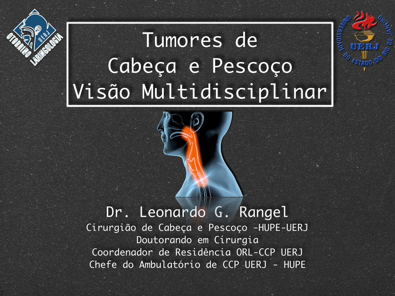
Tumores de Cabeça e Pescoço
Visão Multidisciplinar
Dr. Leonardo G. RangelCirurgião de Cabeça e Pescoço -HUPE-UERJ
Doutorando em CirurgiaCoordenador de Residência ORL-CCP UERJChefe do Ambulatório de CCP UERJ - HUPE

"Experiência não é Igual a Competência"


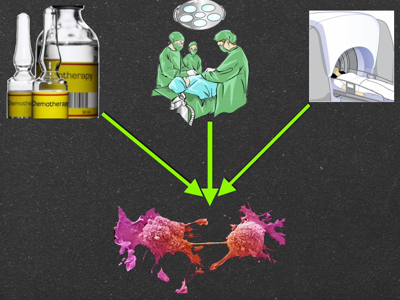
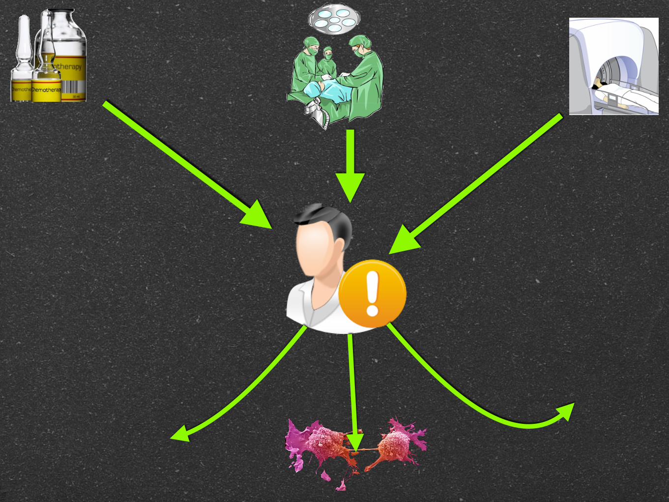
O Problema
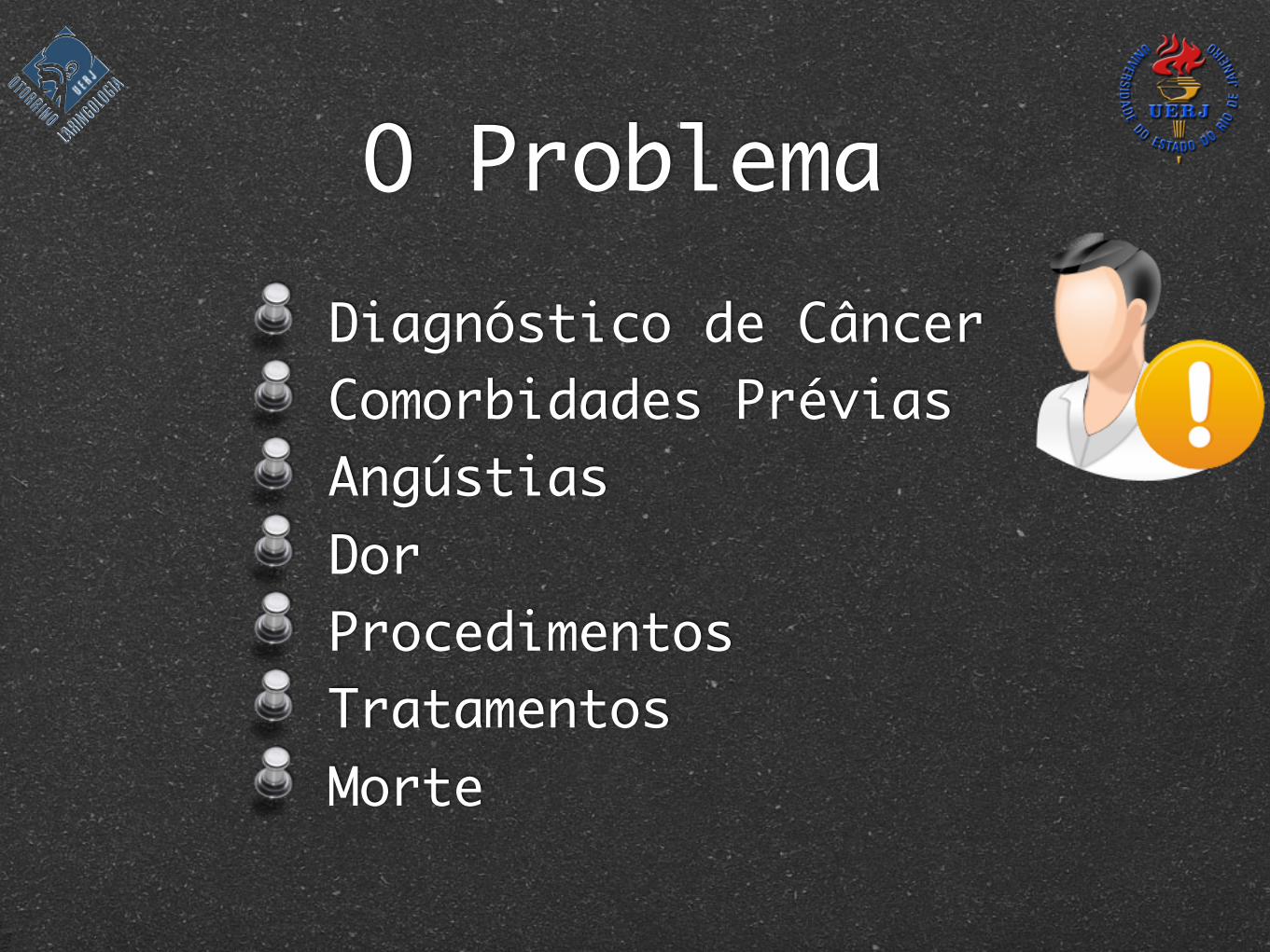
Diagnóstico de Câncer Comorbidades Prévias Angústias Dor Procedimentos Tratamentos Morte
O Problema
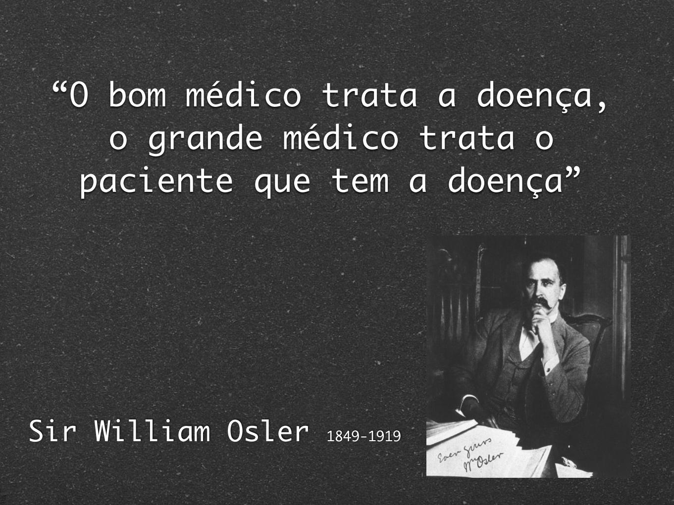
“O bom médico trata a doença, o grande médico trata o
paciente que tem a doença”
Sir William Osler 1849-1919
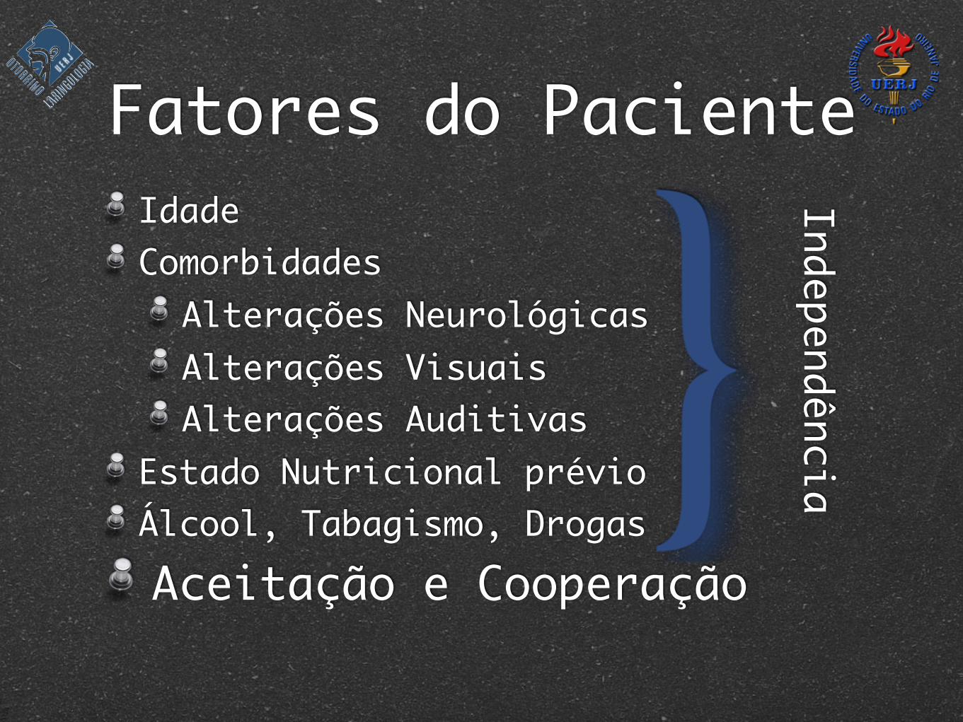
Fatores do PacienteIdadeComorbidades
Alterações NeurológicasAlterações VisuaisAlterações Auditivas
Estado Nutricional prévioÁlcool, Tabagismo, DrogasAceitação e Cooperação
Independência
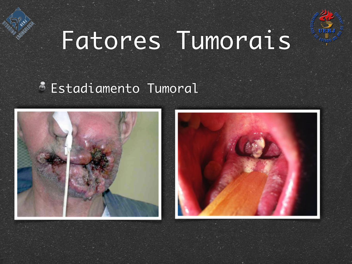
Fatores Tumorais
Biologia Tumoral
Sítio Tumoral
Estadiamento
Características Histopatológicas
Fatores TumoraisEstadiamento Tumoral
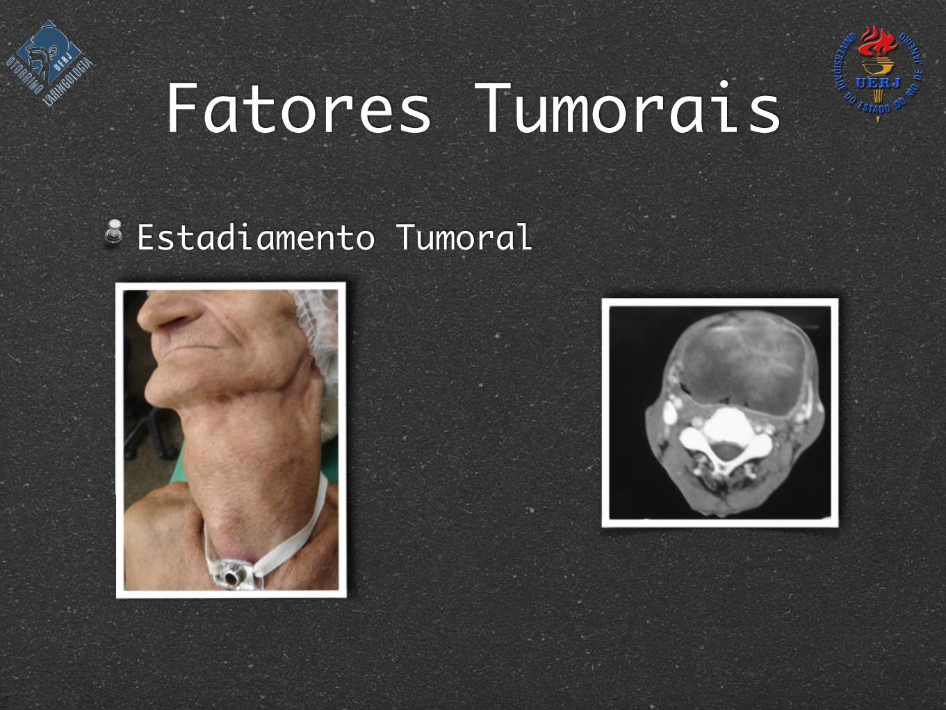
Fatores TumoraisEstadiamento Tumoral
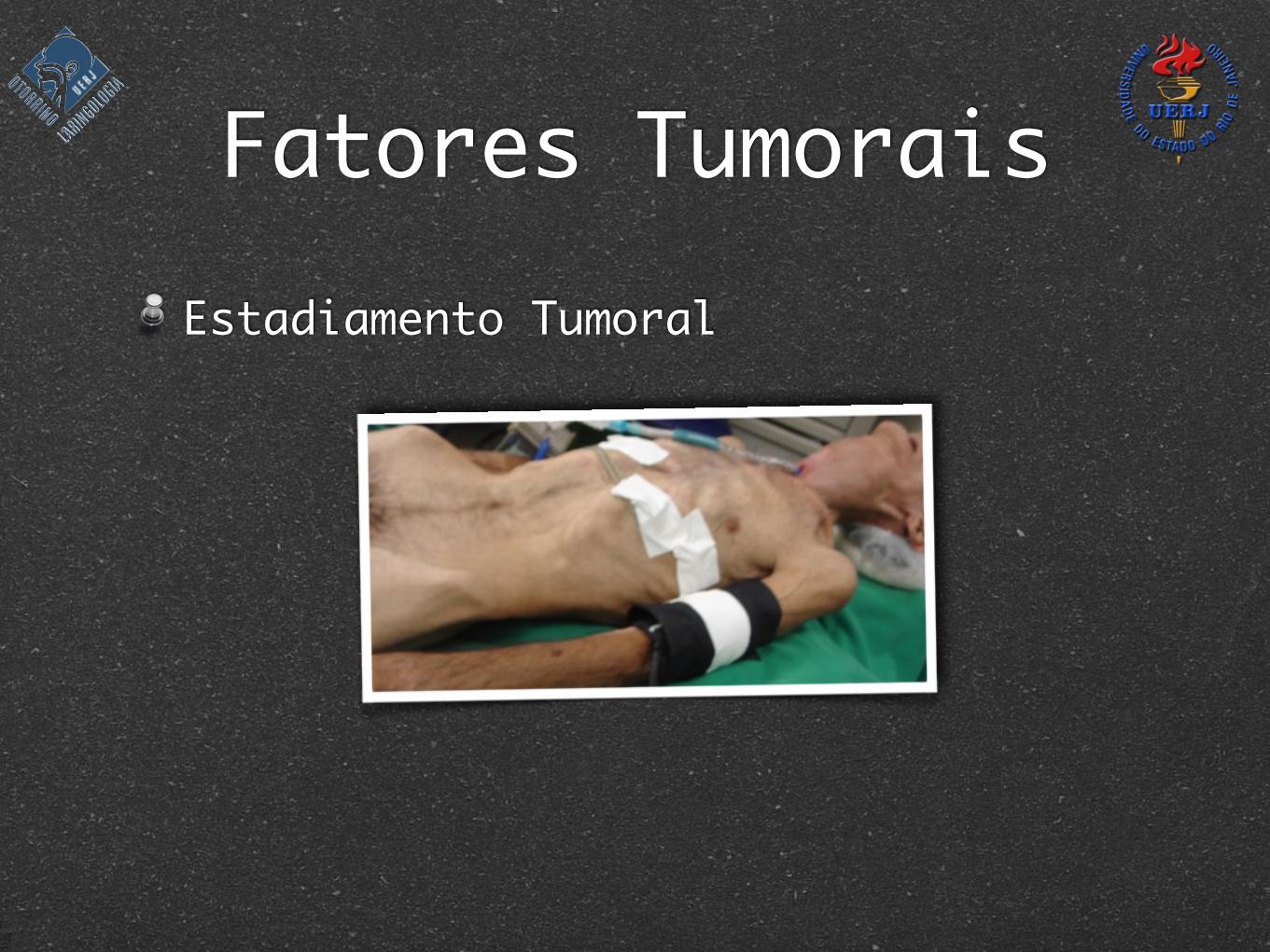
Fatores TumoraisEstadiamento Tumoral
Fatores TumoraisEstadiamento Tumoral
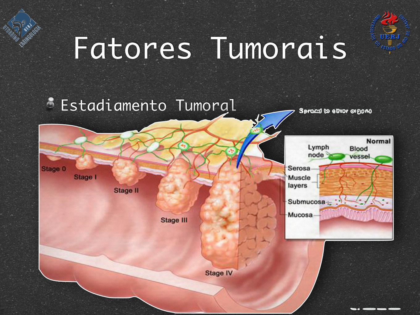
Fatores TumoraisEstadiamento Tumoral
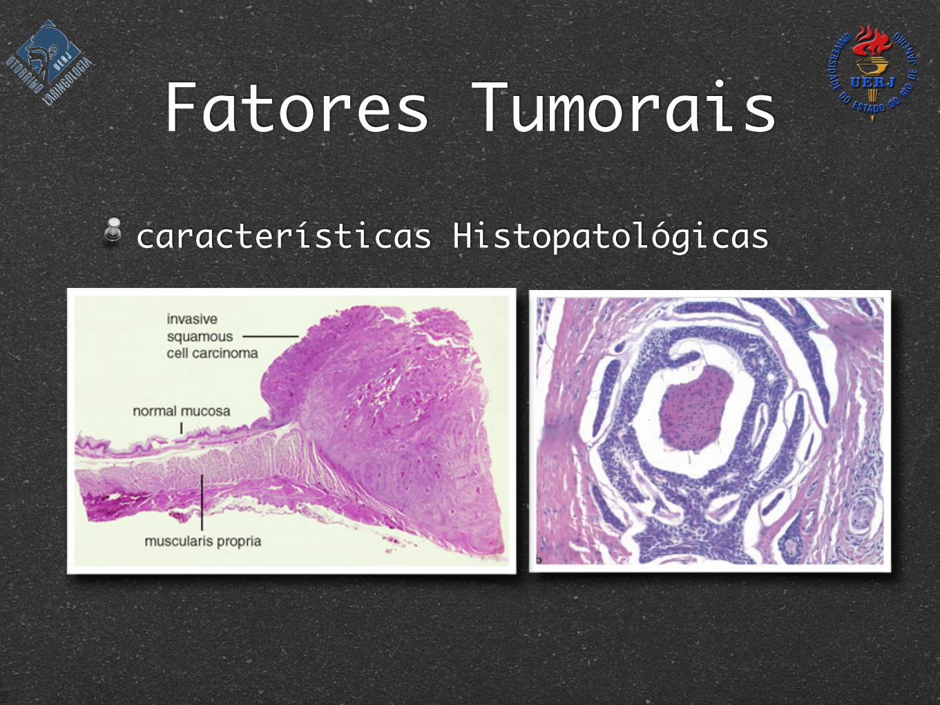
Fatores Tumoraiscaracterísticas Histopatológicas
epithelial membrane antigen and are negative for muscle-specific actin and vimentin. They may or may not express S-100 protein [469]. Recently adenoid cystic carcinomas have been shown to be positive for MUC3 [14, 204, 229, 406]. They may also be estrogen and progesterone recep-tor positive although not in all cases [182]. Approximately 90% are c-kit (CD117) positive [142].
Differential Diagnosis
Included in the differential diagnosis of adenoid cystic carcinoma are basaloid squamous cell carcinoma, basal cell adenocarcinoma, basal cell adenoma, cellular pleo-morphic adenoma, polymorphous low-grade adeno-carcinoma, and the basal cell and plexiform subtypes of ameloblastoma. The staining pattern with p63 is useful in distinguishing basaloid squamous cell carcinoma from adenoid cystic carcinoma. Basaloid squamous cell car-cinomas consistently display diffuse staining of nearly 100% of the tumor cells with p63. Adenoid cystic carci-nomas, on the other hand, show staining of a single pe-ripheral layer of cells or compartmentalized staining with surrounding or interspersed p63 negative cells [463].
Basal cell adenomas, unlike adenoid cystic carcino-mas, are characterized by peripheral palisading, a deli-cate fibrovascular stroma, a circumscribed rather than infiltrating growth pattern and lack of perineural inva-sion. Rarely, however, they may show trabecular and solid
cribriform growth patterns reminiscent of adenoid cystic carcinoma [250]. Basal cell adenocarcinomas show areas of invasive growth and perineural invasion, features in common with adenoid cystic carcinoma, but otherwise resemble basal cell adenomas.
Cellular pleomorphic adenomas can resemble adenoid cystic carcinomas, however, careful examination of the junction of the cellular elements with the stroma aids in the distinction. In pleomorphic adenomas the myoepithe-lial cells spin off the epithelial elements and blend into the stroma. By contrast, there is a sharp demarcation between the cellular components of adenoid cystic carcinomas and the surrounding, often hyalinized, stroma. In addition, perineural invasion is not present in pleomorphic adeno-mas. Pleomorphic adenomas are also GFAP positive and adenoid cystic carcinomas are GFAP negative.
Perineural invasion occurs as often in polymorphous low-grade adenocarcinoma (PLGA) as in adenoid cystic carcinoma. However, the cells in PLGA are cuboidal to columnar with eosinophilic or clear cytoplasm and vesic-ular nuclei. The classic hyperchromatic angulated nucleus of adenoid cystic carcinoma is not present. Expression of c-kit may also be helpful as it is positive in virtually 100% of adenoid cystic carcinomas and in only approximately 50–60% of PLGA [268, 474, 546]. In addition, it has also been reported that where as 90% of tumor cells in PLGA are positive for epithelial membrane antigen, only the epithelial cells lining true lumens stain in adenoid cystic carcinoma [474].
Fig. 3.11: Adenoid cystic carcinoma. a Cribriform growth pattern. Cells with dense angular nuclei and scant clear cytoplasm sur-round spaces producing a classic Swiss cheese pattern (H&E, 200×). b Perineural invasion (H&E, 200×)
61Pathology of Salivary Gland Disease Chapter 3
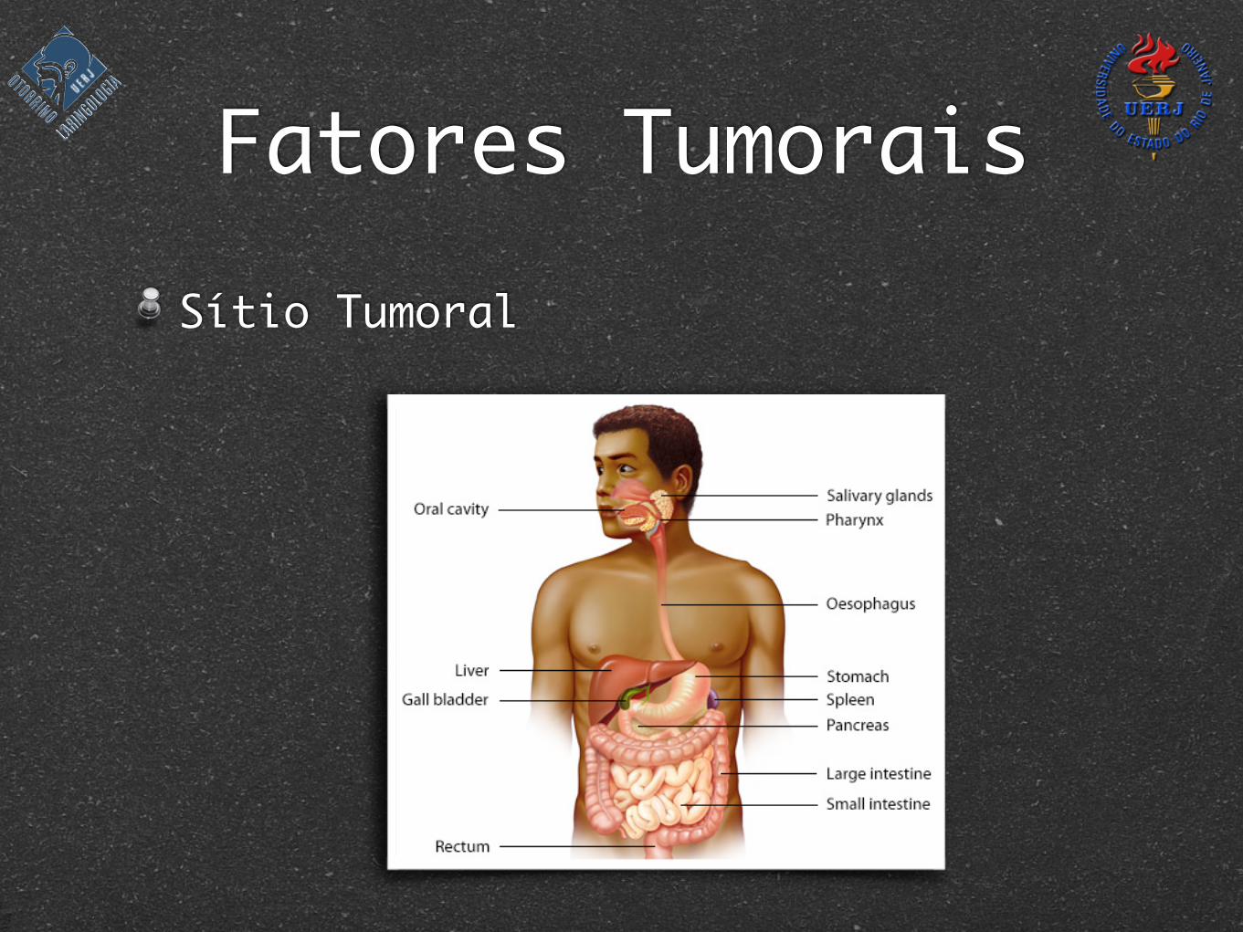
Fatores TumoraisSítio Tumoral
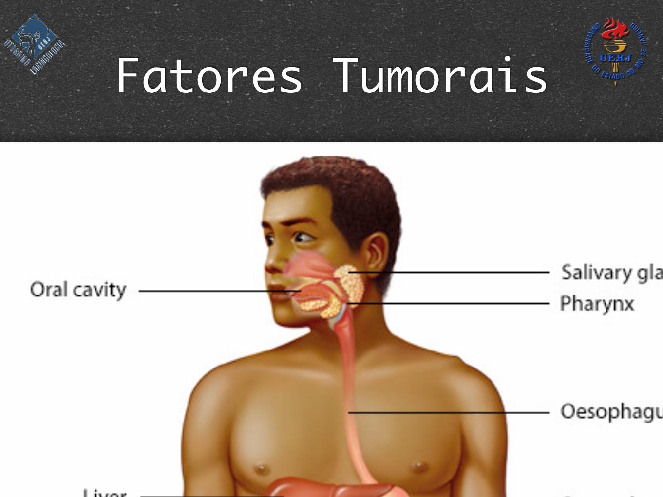
Fatores TumoraisSítio Tumoral
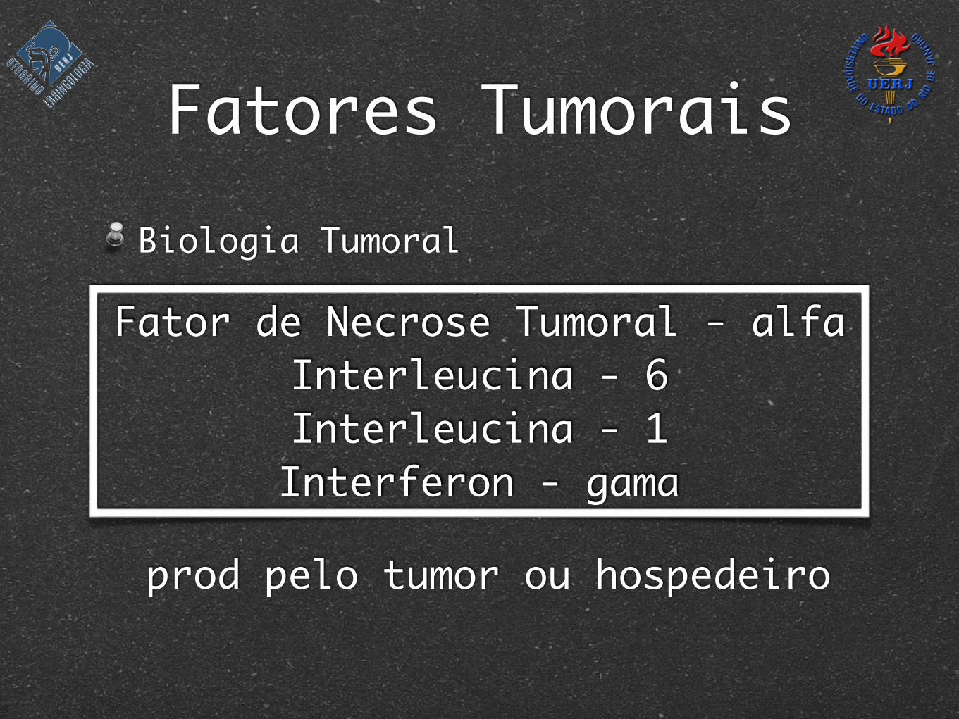
Biologia Tumoral
Fatores Tumorais
Fator de Necrose Tumoral - alfaInterleucina - 6Interleucina - 1Interferon - gama
prod pelo tumor ou hospedeiro
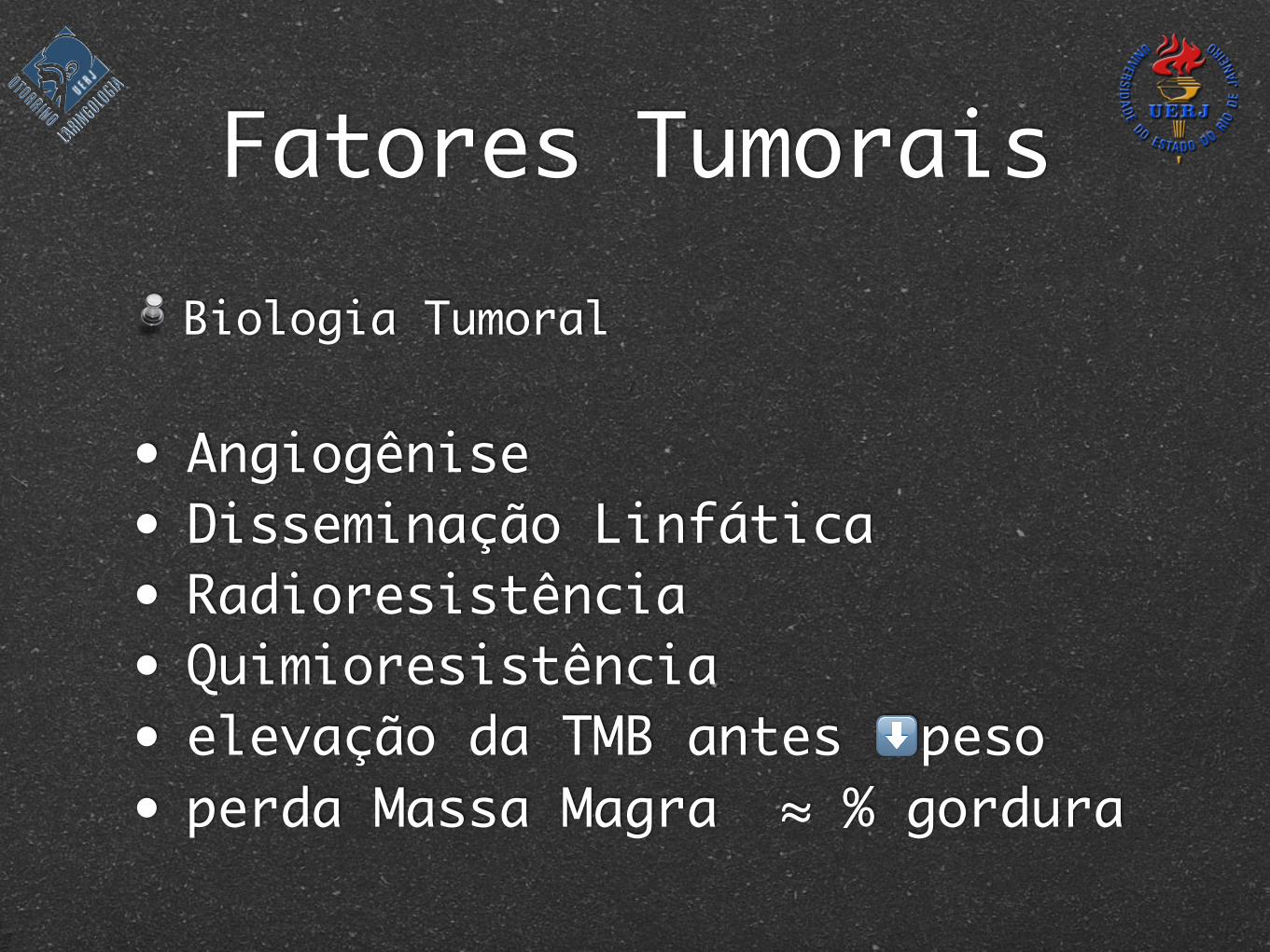
Fatores TumoraisBiologia Tumoral
• Angiogênise • Disseminação Linfática• Radioresistência• Quimioresistência• elevação da TMB antes ⬇ peso• perda Massa Magra ≈ % gordura
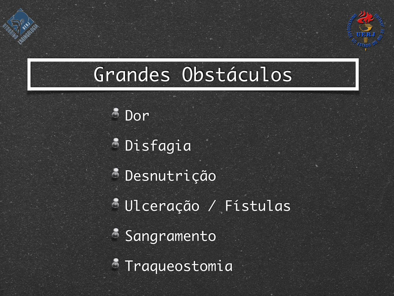
Dor
Disfagia
Desnutrição
Ulceração / Fístulas
Sangramento
Traqueostomia
Grandes Obstáculos
Caquexia associada a 20% das mortes
Caquexia ≠ Inaniçãonão reverte com Calorias extras
Anorexia 15-40%
Apetite e Habilidade em comerprincipais fatores de Qualidade de Vida
Disfagia≈Desnutrição≈Caquexia
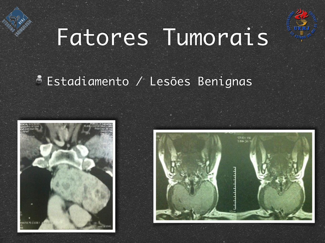
Fatores TumoraisEstadiamento / Lesões Benignas
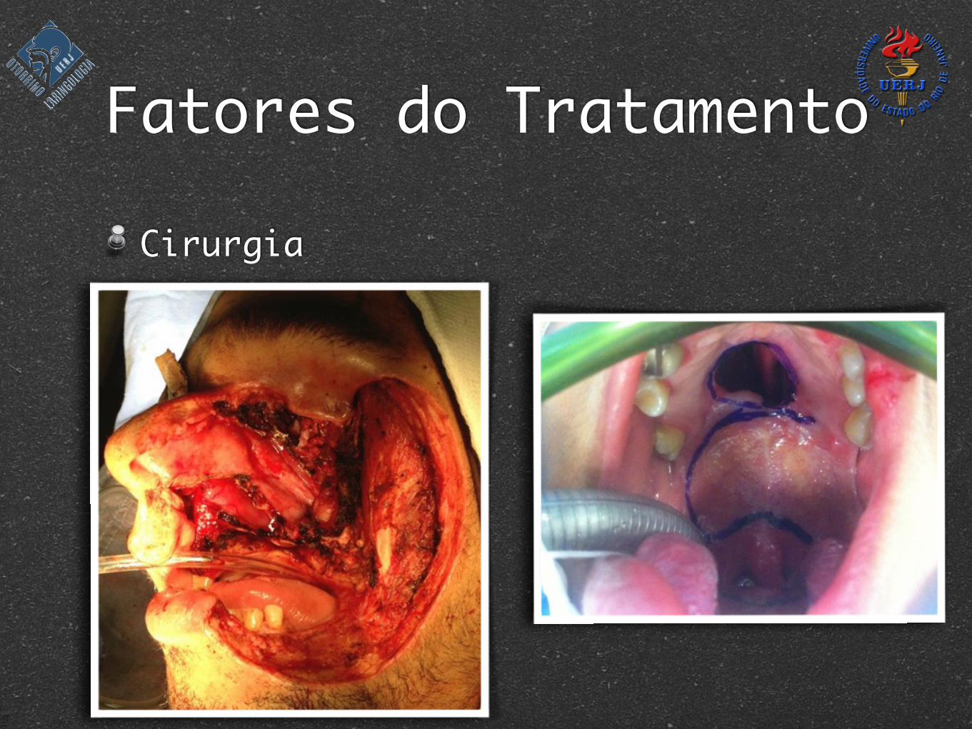
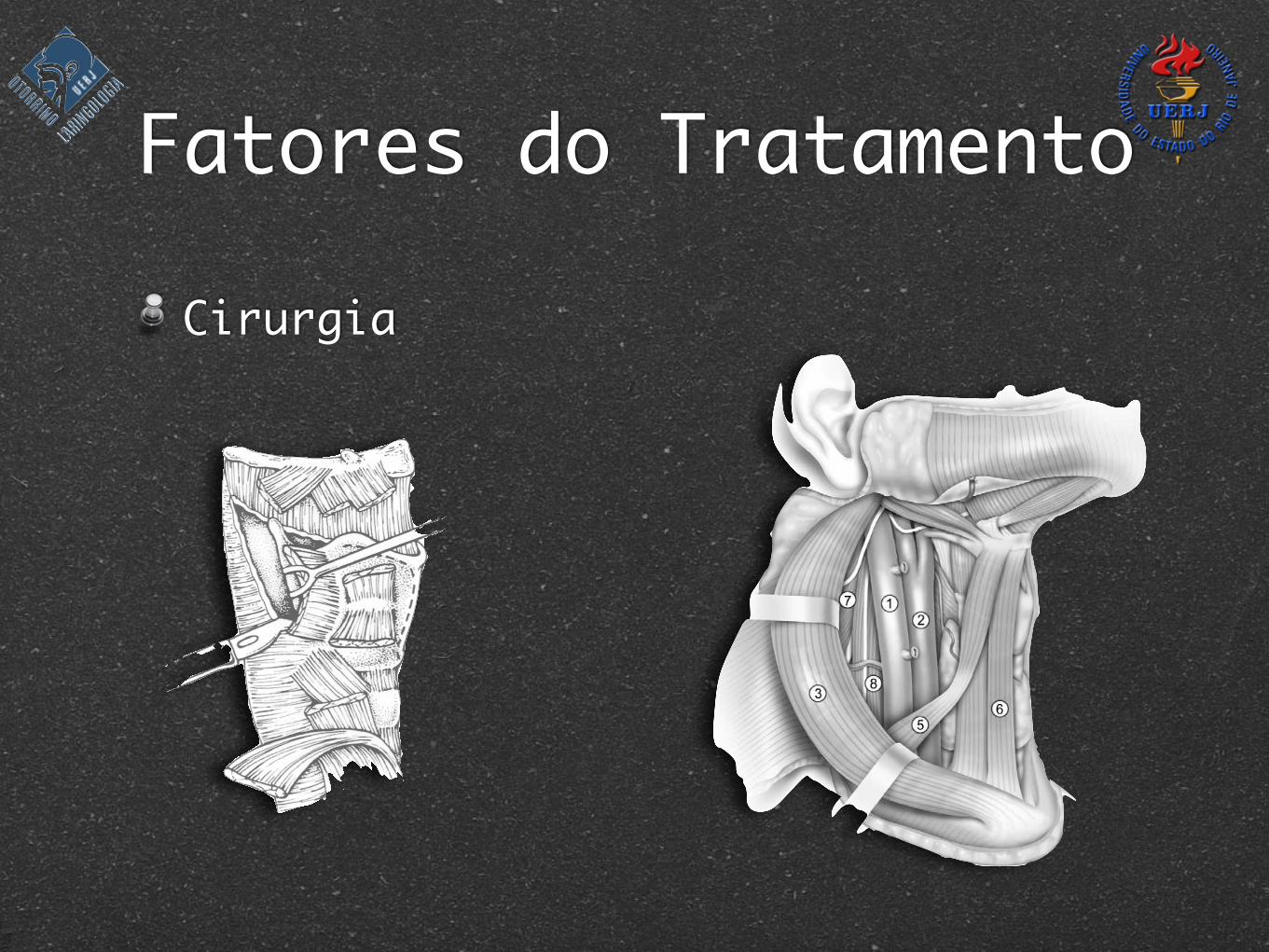
Fatores do Tratamento
Cirurgia
should be clearly identified, ligated, and divided to com-plete the isolation of the internal jugular vein. Othersmaller branches can be cauterized, by means of bipolarcautery.
The dissection of the carotid sheath has 2 danger points,one at each end—upper and lower—of the dissection. Atthese 2 points the traction exerted to facilitate the dissectionof the fascial envelope produces a folding of the wall of theinternal jugular vein that can be easily sectioned at the touchof the scalpel blade. The surgeon must be extremely cau-tious to avoid injuring the vein at these points.
Lower in the neck, the terminal portion of the thoracicduct on the left side, and the right lymphatic duct—whenpresent—also are within the boundaries of the dissectionand must be preserved. Once the internal jugular vein isreleased from its covering fascia, the dissection continuesmedially over the carotid artery. The specimen is now com-pletely separated from the great vessels and remains at-tached only to the strap muscles
Dissection of the strap muscles
Although this is described as the last step of theoperation (Figure 10), it may be performed in a differentorder according to the needs of the surgery and thelocation of the primary tumor. The midline constitutesthe medial border of the dissection for unilateral opera-tions. Thus, a midline cut is made in the superficial layerof the cervical fascia from the upper border of the sur-gical field to the sternal notch. The anterior jugular vein
is identified, ligated, and divided at both ends of thesurgical field. The fascia is now dissected from the un-derlying strap muscles. The dissection starts at the upperpart of the surgical field and continues in a lateral andinferior direction. The sternohyoid and omohyoid mus-cles are completely freed from their fascial covering.
The superior thyroid artery can be identified coursingin an inferomedial direction toward the thyroid gland.Depending on the resection of the primary tumor, the
Figure 10 The strap muscles are released from their fascialcovering. (1) Strap muscles, (2) thyroid cartilage, (3) thyroidgland, (4) fascia of the strap muscles, (5) stylohyoid muscle, (6)digastric muscle, (7) anterior facial vein, and (8) submandibulargland optionally preserved.
Figure 11 Anatomical boundaries of the central compartment ofthe neck. (1) Carotid artery, (2) internal jugular vein (3) hyoidbone, (4) suprasternal notch, and (5) thyroid gland
Figure 12 The neck after a right functional neck dissection forsupraglottic cancer of the larynx. (1) Internal jugular vein, (2)carotid artery, (3) sternocleidomastoid muscle, (4) submandibulargland, (5) omohyoid muscle, (6) sternohyoid muscle, (7) levatorscapulae muscle, and (8) anterior scalene muscle.
174 Operative Techniques in Otolaryngology, Vol 15, No 3, September 2004
F@lJRE 1. Transection of the strap muscles: Along the superior border of the thyroid cartilage, the stemohyoid, omohyoid and tlqrohyoid muscles are cut. The sternothyroid muscle is also transected. This is performed bilaterally.
FIGURE 3. Disarticulation of the cricothyroid joint: A Freer ele- vator is placed carefully between the inferior thyroid comu and the cricoid cartilage so that the recurrent laryngeal nerve is not damaged. The nerve is not identified during the dissection.
FIGURE 2. Transection of the constrictor muscles: The inferior pharyngeal constrictor muscles and the thyroid perichondrium are transected with a No. 15 blade along the posterolateral and superolateral borders of the thyroid cartilage.
brane, and the periosteum of the inferior hyoid bone is incised. A Freer elevator is then used to dissect the preepi- glottic space from the inferior and posterior aspect of the hyoid bone. The larynx is now entered through a small transvallecular pharyngotomy, just wide enough to visu- alize the epiglottis. It is grasped with an Allis clamp and pulled externally. The surgeon now moves to the head of the bed, and further tumor cuts can be made under direct visualization (Fig 5). Using scissors, incisions are made so that the entire preepiglottic space is resected, but the cuts are made medial to the main trunk of the internal branch of the superior laryngeal nerve.
Further tumor cuts are now made on the non-tumor bearing side. The scissors are advanced anterior to the previously released pyriform sinus. Precise cuts are made through the aryepiglottic fold and down to the level of the false cord. The false cord is transected just anterior to the arytenoids; the vocal process and true cords are transected just posterior to the ventricle. It is essential that the aryte- noid cartilage be preserved on the non-tumor bearing side of the larynx. In addition, it is important not to enter the cricoarytenoid joint inadvertently so that postoperative ankylosis may be avoided. Incisions are now made con- necting these prearytenoid cuts to the cricothyroidotomy. The cricothyroid and lateral cricoarytenoid muscles are transected along the superior border of the cricoid carti- lage.
Complete visualization of the tumor bearing side is necessary. The surgeon takes both thyroid ala in her/his hands and cracks the cartilage down the middle. It is akin to opening a book. The resection along the tumor bearing
DUANE SEWELL 29
Fatores do Tratamento
Cirurgia
Fatores do Tratamento
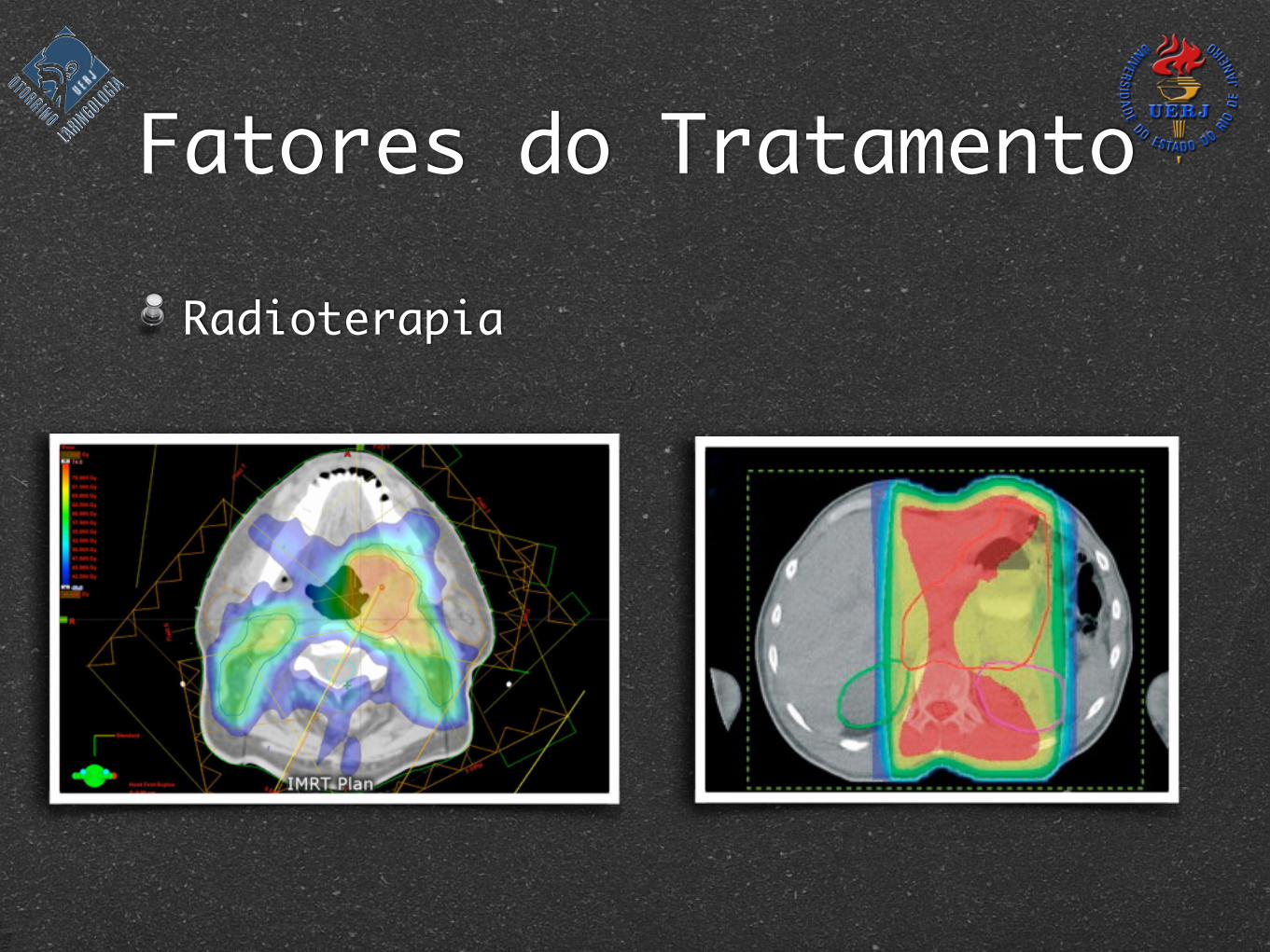
Radioterapia
Fatores do Tratamento
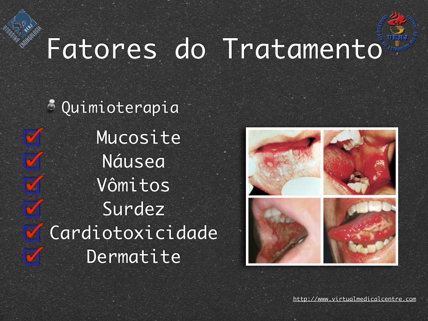
Quimioterapia
http://www.virtualmedicalcentre.com
MucositeNáuseaVômitosSurdez
CardiotoxicidadeDermatite
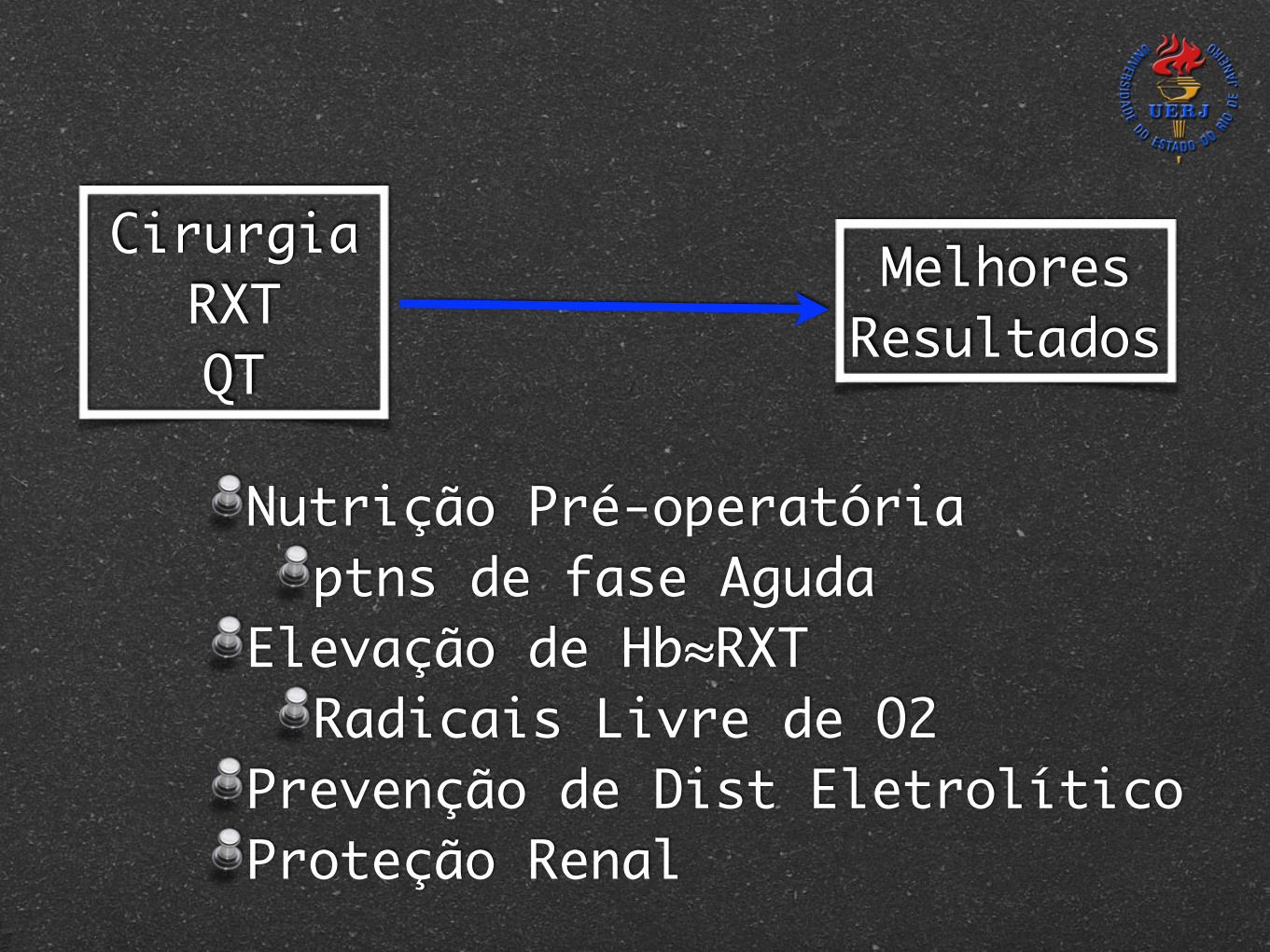
CirurgiaRXTQT
Melhores Resultados
Nutrição Pré-operatóriaptns de fase Aguda
Elevação de Hb≈RXTRadicais Livre de O2
Prevenção de Dist EletrolíticoProteção Renal

"Good judgement comes from experience;
and experience comes from bad judgement!"
Ashok Shara