


Línguas
Páginas
Legal

UNIVERSIDADE FEDERAL DO PARANÁ
ANA CAROLINA DOS SANTOS LOURENÇO
EFEITOS DO PLASTIFICANTE DIBUTIL FTALATO (DBP) EM MODELOS IN VIVO
E IN VITRO DE HIPERSENSIBILIDADE DE CONTATO
CURITIBA
2015
UNIVERSIDADE FEDERAL DO PARANÁ
ANA CAROLINA DOS SANTOS LOURENÇO
EFEITOS DO PLASTIFICANTE DIBUTIL FTALATO (DBP) EM MODELOS IN VIVO
E IN VITRO DE HIPERSENSIBILIDADE DE CONTATO
Tese apresentada como requisito parcial à obtenção do grau
de Doutor em Farmacologia, Curso de Pós-Graduação em
Farmacologia, Setor de Ciências Biológicas da Universidade
Federal do Paraná.
Orientador: Prof. Dr. Anderson Joel Martino Andrade
Co-orientador: Prof. Dr. Michel Fleith Otuki
CURITIBA
2015

Dedico esse trabalho ao meu maior fã
e meu maior ídolo, meu avô Virgílio,
que apesar de não estar presente em
corpo para ver a minha conclusão do
Doutorado, sei que de alguma forma
e de algum lugar sempre me
acompanha e me protege...
AGRADECIMENTOS
A Deus por todas as bênçãos durante meu caminho.
À minha família pelo apoio incondicional.
Ao Prof. Dr. Francisco J. R. Paumgartten, por me acolher em seu
Laboratório de Toxicologia na FIOCRUZ, e a Rose Carvalho por toda a ajuda
prestada quando meus experimentos eram lá. Ao seu Ivan do biotério da FIOCRUZ,
e aos funcionários da Casa Amarela, que sempre me trataram tão bem.
Aos professores, funcionários e colegas do Departamento de Farmacologia
da Universidade Federal do Paraná.
A Francislaine Lívero e a Prof. Dr. Alexandra Acco pela parceria nas
dosagens de estresse oxidativo.
Aos meus amigos italianos da Università Degli Studi di Milano. Valentina,
Angela, Maria Serena, Natalia, Fiona, Elena, Daniele, Nicolò e Francesco, obrigada
pelo ano esplêndido, pela paciência durante meu aprendizado com as células e
também com a língua italiana. Já dizia Dante: não podemos ter uma vida perfeita
sem amigos. Obrigada pela amizade! Sinto saudade de vocês todos os dias! E
gostaria de deixar aqui essa mensagem em italiano. “Ai miei amici italiani
dell'Università degli Studi di Milano. Valentina, Angela, Maria Serena, Natalia, Fiona,
Elena, Daniele, Nicolò e Francesco, grazie per lo splendido anno e per la pazienza
durante il mio apprendimento con le cellule e anche della lingua italiana. Già lo
diceva Dante: “Noi non potemo avere perfetta vita senza amici”. Grazie dell'amicizia!
Ho saudade di voi tutti giorni!”
Aos meus amigos do laboratório, Caroline, Leonardo, Fabíola, Ana Cláudia,
Juliane, Emerson, Inês, as Natálias, Marina, e Noruê, que durante essa jornada me
ajudaram de alguma forma. Obrigada pelos auxílios nos experimentos, pelo
companheirismo e pelos almoços no “massinha”.
Ao meu amigo Arthur Prudente, por me acompanhar desde o início dos
experimentos, por me ensinar o modelo, por discutir resultados comigo e por nossas
viagens juntos durante o tempo de Doutorado na Europa.
À minha estagiária, companheira, braço direito e amiga Renata Mercer Zaia
por estar a cada minuto desse experimento ao meu lado. Nos tratamentos dos
animais, nas análises dos dados, na discussão de resultados e redação de artigos.
Meu muito, muito obrigada!
À minha orientadora italiana, Prof. Dr. Emanuela Corsini, que depositou sua
confiança em mim e me permitiu passar um ano em seu laboratório. Muito obrigada
professora! “Alla Prof.ssa Dott.ssa Emanuela Corsini, che ha messo la sua fiducia in
me e mi ha permesso di trascorrere un anno nel suo laboratorio. Grazie mille prof!”
Ao Prof. Dr. Michel Otuki pela coorientação.
Ao meu orientador Prof. Dr. Anderson Joel Martino Andrade por todos esses
anos de orientação. Você é um profissional em quem todos deveríamos nos inspirar,
além de ser uma pessoa sensacional. Obrigada por tudo.
À banca, por aceitar avaliar esse trabalho, muito obrigada. Espero que seja
uma leitura agradável.

“O conhecimento torna a alma jovem e diminui a amargura da velhice.
Colhe, pois, a sabedoria. Armazena suavidade para amanhã.”
Leonardo da Vinci
RESUMO
A prevalência de doenças alérgicas tais como dermatite, asma e rinite, tem aumentado, e isso poderia ser devido à presença de substâncias químicas ambientais com potencial adjuvante, ou seja, capazes de exacerbar a resposta imune. Ésteres de ftalatos, plastificantes amplamente encontrados no ambiente, têm sido reportados como participantes no desenvolvimento de doenças alérgicas agindo como adjuvantes. A hipersensibilidade de contato (HSC) é uma reação alérgica que resulta de contatos repetidos da pele com substâncias químicas chamadas haptenos, e consiste em duas fases: sensibilização e elicitação. O plastificante dibutil ftalato (DBP) é capaz de aumentar a resposta inflamatória em HSC induzida por alérgeno quando associado em ambas as fases da HSC, mas em doses muito mais altas que aquelas potencialmente relevantes para a exposição humana. Além disso, os mecanismos moleculares envolvidos nesse efeito adjuvante não estão completamente elucidados. O presente estudo pretendeu investigar os efeitos do DBP na fase de elicitação da HSC induzida por oxazolona em modelo animal de edema de orelha, usando camundongos BALB/c e doses mais baixas de DBP. Ainda, investigamos alguns aspectos relacionados aos mecanismos envolvidos nesse efeito adjuvante. Para isso, avaliamos os efeitos do DBP isoladamente ou associado a alérgenos em dois ensaios in vitro, o ensaio de ativação de NCTC 2544 (representativo de ativação de queratinócitos) e o ensaio de ativação de THP-1 (substituto de ativação de células dendríticas). Foi também investigado se a indução de estresse oxidativo poderia ser parte do mecanismo molecular responsável pelo efeito adjuvante induzido pelo DBP. Ainda avaliamos a possível participação da ativação dos receptores de potencial transitório A1 e V1 (TRPA1 e TRPV1) no efeito adjuvante do DBP no modelo de HSC induzida por oxazolona. In vivo, os parâmetros avaliados foram edema de orelha, 24, 48 e 72 horas após a elicitação, atividade das enzimas N-acetil-β-d-glucosaminidase (NAG) e mieloperoxidase (MPO) (marcadores de infiltração de leucócitos), análise histológica (contagem de células infiltradas, espessura da derme e epiderme) e dosagem de hidroperóxidos de lipídeos (LOOH) e glutationa reduzida (GSH). Nos experimentos in vitro, a produção de interleucina 18 (IL-18) foi avaliada em células NCTC 2544, e a expressão do cluster de diferenciação 86 (CD86) e mRNA de interleucina 8 (IL-8) além da produção de IL-8 e de espécies reativas de oxigênio (EROs) foram avaliados em células THP-1. In vivo, o DBP, nas duas maiores doses (0,4 e 4 mg/orelha), foi capaz de aumentar a resposta inflamatória envolvida na HSC induzida por oxazolona, como observado por aumento no edema de orelha, hiperplasia dérmica e epidérmica, contagem de células infiltradas e atividade de NAG e MPO. Antagonistas TRPA1 e TRPV1 reverteram parcial ou completamente, respectivamente, o efeito adjuvante do DBP neste modelo animal de HSC, sugerindo que a ativação destes TRPs seja parte do mecanismo molecular envolvido no efeito adjuvante. In vitro, DBP potencializou a ativação de células THP-1, como foi demonstrado pelo aumento da expressão de CD86 e IL-8 mRNA e liberação de IL-8 após exposição a associações de DBP com Citral e imidazolidinil ureia , além do aumento da expressão de CD86 em células THP-1 tratadas com DBP associado a oxazolona. Por outro lado, não foi observado efeito adjuvante nas células NCTC 2544, já que não houve aumento na produção de IL-18 após exposição a p-Fenilenodiamina associado ao DBP, sugerindo que o efeito adjuvante do DBP esteja relacionado com maior ativação de células dendríticas e não de queratinócitos. Nossos resultados indicam que o DBP pode agir

como adjuvante imunológico tanto in vivo quanto in vitro, e que esse efeito poderia estar relacionado com uma maior ativação de células dendríticas e que envolva ao menos parcialmente a ativação de TRPA1 e TRPV1. Palavras-chave: dibutil ftalato, hipersensibilidade de contato, efeito adjuvante, queratinócitos, células dendríticas, TRPA1, TRPV1, espécies reativas de oxigênio, estresse oxidativo.
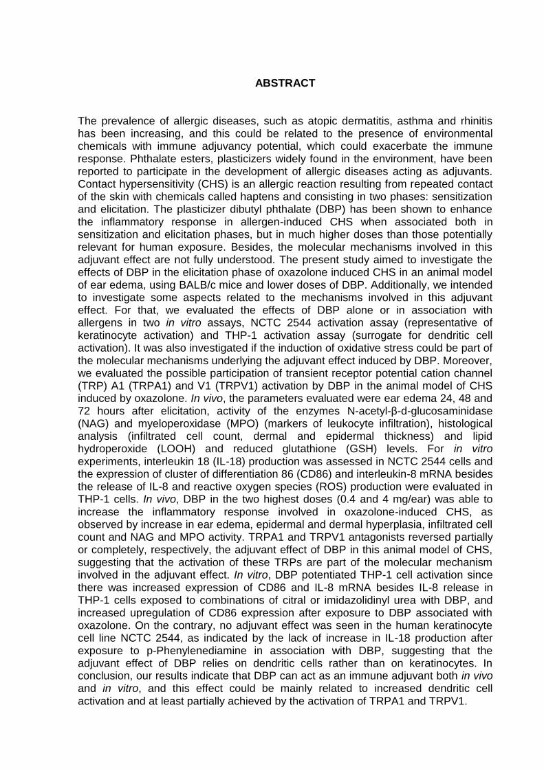
ABSTRACT
The prevalence of allergic diseases, such as atopic dermatitis, asthma and rhinitis has been increasing, and this could be related to the presence of environmental chemicals with immune adjuvancy potential, which could exacerbate the immune response. Phthalate esters, plasticizers widely found in the environment, have been reported to participate in the development of allergic diseases acting as adjuvants. Contact hypersensitivity (CHS) is an allergic reaction resulting from repeated contact of the skin with chemicals called haptens and consisting in two phases: sensitization and elicitation. The plasticizer dibutyl phthalate (DBP) has been shown to enhance the inflammatory response in allergen-induced CHS when associated both in sensitization and elicitation phases, but in much higher doses than those potentially relevant for human exposure. Besides, the molecular mechanisms involved in this adjuvant effect are not fully understood. The present study aimed to investigate the effects of DBP in the elicitation phase of oxazolone induced CHS in an animal model of ear edema, using BALB/c mice and lower doses of DBP. Additionally, we intended to investigate some aspects related to the mechanisms involved in this adjuvant effect. For that, we evaluated the effects of DBP alone or in association with allergens in two in vitro assays, NCTC 2544 activation assay (representative of keratinocyte activation) and THP-1 activation assay (surrogate for dendritic cell activation). It was also investigated if the induction of oxidative stress could be part of the molecular mechanisms underlying the adjuvant effect induced by DBP. Moreover, we evaluated the possible participation of transient receptor potential cation channel (TRP) A1 (TRPA1) and V1 (TRPV1) activation by DBP in the animal model of CHS induced by oxazolone. In vivo, the parameters evaluated were ear edema 24, 48 and 72 hours after elicitation, activity of the enzymes N-acetyl-β-d-glucosaminidase (NAG) and myeloperoxidase (MPO) (markers of leukocyte infiltration), histological analysis (infiltrated cell count, dermal and epidermal thickness) and lipid hydroperoxide (LOOH) and reduced glutathione (GSH) levels. For in vitro experiments, interleukin 18 (IL-18) production was assessed in NCTC 2544 cells and the expression of cluster of differentiation 86 (CD86) and interleukin-8 mRNA besides the release of IL-8 and reactive oxygen species (ROS) production were evaluated in THP-1 cells. In vivo, DBP in the two highest doses (0.4 and 4 mg/ear) was able to increase the inflammatory response involved in oxazolone-induced CHS, as observed by increase in ear edema, epidermal and dermal hyperplasia, infiltrated cell count and NAG and MPO activity. TRPA1 and TRPV1 antagonists reversed partially or completely, respectively, the adjuvant effect of DBP in this animal model of CHS, suggesting that the activation of these TRPs are part of the molecular mechanism involved in the adjuvant effect. In vitro, DBP potentiated THP-1 cell activation since there was increased expression of CD86 and IL-8 mRNA besides IL-8 release in THP-1 cells exposed to combinations of citral or imidazolidinyl urea with DBP, and increased upregulation of CD86 expression after exposure to DBP associated with oxazolone. On the contrary, no adjuvant effect was seen in the human keratinocyte cell line NCTC 2544, as indicated by the lack of increase in IL-18 production after exposure to p-Phenylenediamine in association with DBP, suggesting that the adjuvant effect of DBP relies on dendritic cells rather than on keratinocytes. In conclusion, our results indicate that DBP can act as an immune adjuvant both in vivo and in vitro, and this effect could be mainly related to increased dendritic cell activation and at least partially achieved by the activation of TRPA1 and TRPV1.

Key words: dibutyl phthalate, contact hypersensitivity,adjuvant effect, keratinocytes, dendritic cells, TRPA1, TRPV1, reactive oxygen species, oxidative stress.
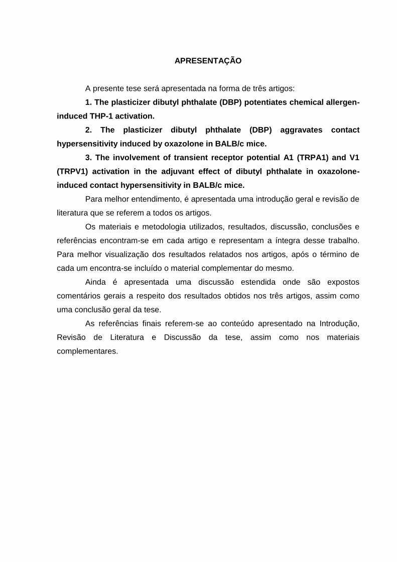
APRESENTAÇÃO
A presente tese será apresentada na forma de três artigos:
1. The plasticizer dibutyl phthalate (DBP) potentiates chemical allergen-
induced THP-1 activation.
2. The plasticizer dibutyl phthalate (DBP) aggravates contact
hypersensitivity induced by oxazolone in BALB/c mice.
3. The involvement of transient receptor potential A1 (TRPA1) and V1
(TRPV1) activation in the adjuvant effect of dibutyl phthalate in oxazolone-
induced contact hypersensitivity in BALB/c mice.
Para melhor entendimento, é apresentada uma introdução geral e revisão de
literatura que se referem a todos os artigos.
Os materiais e metodologia utilizados, resultados, discussão, conclusões e
referências encontram-se em cada artigo e representam a íntegra desse trabalho.
Para melhor visualização dos resultados relatados nos artigos, após o término de
cada um encontra-se incluído o material complementar do mesmo.
Ainda é apresentada uma discussão estendida onde são expostos
comentários gerais a respeito dos resultados obtidos nos três artigos, assim como
uma conclusão geral da tese.
As referências finais referem-se ao conteúdo apresentado na Introdução,
Revisão de Literatura e Discussão da tese, assim como nos materiais
complementares.
SUMÁRIO
1 INTRODUÇÃO ......................................................................... 14
2 REVISÃO DE LITERATURA ................................................... 20
2.1 FTALATOS ............................................................................... 20
2.1.1 Estrutura e propriedades físico-químicas ................................. 20
2.1.2 Tipos, fontes e usos ................................................................. 21
2.1.3 Efeitos dos ftalatos sobre o organismo humano ...................... 22
2.1.4 Dibutil Ftalato (DBP) ................................................................ 23
2.1.5 Farmacocinética do DBP ......................................................... 24
2.1.6 Aspectos Regulatórios dos Ftalatos ......................................... 26
2.2 PROCESSOS ALÉRGICOS CUTÂNEOS ............................... 27
2.2.1 Estrutura da pele ...................................................................... 27
2.2.2 Dermatite de contato alérgica (DCA) ou Hipersensibilidade de
Contato (HSC)...........................................................................
30
2.2.3 Papel do estresse oxidativo na HSC ........................................ 33
2.2.4 Influências do sistema nervoso periférico na HSC .................. 34
2.3 ABSORÇÃO E TOXICIDADE DÉRMICA DOS FTALATOS ..... 36
2.3.1 Ftalatos e processos alérgicos ................................................. 38
2.4 MODELOS DE HIPERSENSIBILIDADE DE CONTATO........... 39
3 HIPÓTESES E PREDIÇÕES ................................................... 41
4 OBJETIVOS ............................................................................. 42
4.1 OBJETIVO GERAL .................................................................. 42
4.2 OBJETIVOS ESPECÍFICOS .................................................... 42
5 MATERIAIS, MÉTODOS E RESULTADOS ............................ 44
5.1 CONSIDERAÇÕES GERAIS ................................................... 44
5.2 ARTIGO 1 ................................................................................. 45
5.2.1 Material complementar ............................................................. 70
5.2.1.1 Highlights ................................................................................. 70
5.2.1.2 Graphic abstract ....................................................................... 70
5.2.1.3 Figuras complementares .......................................................... 71
5.3 ARTIGO 2 …………………………………………………………. 72
5.3.1 Material complementar …………………………………………… 103
5.3.1.1 Highlights ………………………………………………………….. 103
5.3.1.2 Graphic abstract …………………………………………………... 103
5.3.1.2 Figuras complementares .......................................................... 104
5.4 ARTIGO 3 …………………………………………………………. 105
5.4.1 Material complementar …………………………………………… 129
5.4.1.1 Highlights ………………………………………………………….. 129
5.4.1.2 Graphic abstract …………………………………………………... 129
6 DISCUSSÃO GERAL .............................................................. 130
7 CONCLUSÕES ........................................................................ 140
REFERÊNCIAS ....................................................................... 141
14
1 INTRODUÇÃO
Ésteres de ftalatos são amplamente utilizados como plastificantes e aditivos
em uma variedade de produtos, como brinquedos, produtos de limpeza,
equipamentos médicos, produtos farmacêuticos, solventes, tintas, repelentes de
mosquito e cosméticos. Em razão de sua ubíqua distribuição no ambiente, são
considerados agentes com grande potencial para exposição humana (KOO; LEE,
2004; ANDERSON et al., 2001; SILVA et al., 2004; LYCHE et al., 2009; JEONG et
al., 2011).
Os ftalatos têm sido utilizados como plastificantes desde a década de 1920.
Em 1970, Jaeger e Rubin detectaram a presença de dietilexil ftalato (DEHP) em
órgãos e tecidos de dois pacientes hospitalizados que haviam recebido transfusão
de sangue. O sangue havia sido estocado em bolsas de policloreto de vinila (PVC)
que continham DEHP. Desde a publicação de Jaeger e Rubin (1970), existe grande
interesse sobre os possíveis efeitos tóxicos dos ésteres de ftalatos. Hoje se sabe
que os ftalatos não se ligam ao PVC e com o tempo e uso, são liberados da matriz
plástica, contaminando o ambiente (BAUER; HERRMANN, 1997; BRADBURY, 1996;
GIAM et al., 1978; GRIFFITHS; CAMARA; LERNER, 1985).
Alguns ésteres de ftalatos com cadeias curtas, como o dibutil ftalato (DBP),
são usados em cosméticos e repelentes de mosquito para uso tópico (INT. J.
TOXICOL., 2005; VARTAK; TUNGIKAR; SHARMA, 1994). Além disso, ftalatos com
menor peso molecular são mais facilmente absorvidos pela pele (ELSISI; CARTER;
SIPES, 1989). Em cosméticos os ftalatos são usados como umectantes e/ou
emolientes em cremes hidratantes, como potencializadores de penetração cutânea,
como promotores de brilho em esmaltes e como solventes em uma variedade de
outros produtos (KOO; LEE, 2004). Shen et al. (2007) detectaram a presença de
sete tipos de ftalatos em 14 produtos cosméticos incluindo sprays de cabelo,
perfumes, desodorantes e cremes hidratantes. Koo e Lee (2004) investigaram
diferentes níveis de ftalatos em um grande número de cosméticos, incluindo 42
perfumes disponíveis na Coréia do Sul. De acordo com seus resultados, 11
perfumes (26%) continham níveis detectáveis de DBP. O DBP pode ser encontrado
em diversos produtos cosméticos, como perfumes, cremes hidratantes, sprays de
cabelo e esmaltes (IPCS, 1997; SATHYANARAYANA et al., 2008).
15
O DBP, assim como outros ftalatos, tem atraído grande atenção da
comunidade cientifica por seus efeitos como desreguladores endócrinos (FISHER et
al., 2003; HEUDORF; MERSCH-SUNDERMANN; ANGERER, 2007; MARTINO-
ANDRADE et al., 2009). Um grande número de estudos experimentais avaliaram os
efeitos tóxicos do DBP. Este e outros ftalatos são reconhecidamente tóxicos para o
desenvolvimento do sistema reprodutor masculino. Ratos machos apresentam
alterações e malformações no sistema reprodutor após exposição in utero a certos
ftalatos (GRAY et al., 2006). Mesmo que muitos aspectos da toxicidade reprodutiva
dos ftalatos já tenham sido elucidados, os mecanismos celulares e moleculares
responsáveis pelas alterações reprodutivas continuam obscuros.
Além de efeitos reprodutivos e endócrinos, dados experimentais e
epidemiológicos indicam que os ftalatos poderiam desempenhar um papel no
desenvolvimento e/ou aumento de doenças alérgicas, contribuindo, então, para o
aumento na prevalência de doenças alérgicas mediadas por IgE em países
industrializados (IMAI et al., 2006; KOIKE et al., 2010; MATSUDA et al., 2010; LI et
al., 2014; BORNEHAG et al. 2004; JAAKKOLA; KNIGHT, 2004). Além disso, tem
sido sugerido que alguns tipos de ftalatos possam agir como adjuvantes, ou seja,
que eles possam acelerar, prolongar ou exacerbar uma resposta imune especifica
(KIMBER; DEARMAN, 2010). Em um estudo com 198 crianças foi reportada uma
associação entre sintomas alérgicos e a exposição a certos ftalatos presentes na
poeira doméstica. Neste estudo, o DEHP foi relacionado com o aparecimento de
asma, e o butil benzil ftalato (BBzP) foi associado a rinite e eczema (BORNEHAG et
al., 2004). Foi também demonstrado que alguns ésteres de ftalatos podem aumentar
a produção de anticorpos quando injetados juntamente a certos antígenos em
camundongos (LARSEN et al., 2001; 2002). Ainda, foi demonstrado que alguns
ésteres de ftalatos podem agir como adjuvantes na sensibilização por fenetil
isotiocianato (MATSUDA et al., 2010) ou isotiocianato de fluoresceína (FITC) (IMAI
et al., 2006) aumentando a intensidade da resposta inflamatória em modelos animais
de hipersensibilidade de contato. Yanagisawa et al. (2008) também demonstraram
que a exposição de camundongos ao DEHP durante a lactação foi capaz de
aumentar a intensidade da dermatite de contato na prole exposta, sugerindo que os
efeitos tóxicos resultantes da exposição perinatal aos ftalatos possam ir além das
alterações no desenvolvimento do sistema reprodutor masculino.
16
A dermatite de contato alérgica (DCA), também conhecida como
hipersensibilidade de contato (HSC), é uma reação inflamatória cutânea resultado de
contatos repetidos ou prolongados da pele com substâncias químicas de baixo peso
molecular denominadas haptenos (SAINT-MEZARD et al., 2004; LEPOITTEVIN;
LEBLOND, 1997) e é considerada uma das mais importantes manifestações de
imunotoxicidade em humanos (KIMBER, 2002). A dermatite de contato alérgica é
uma hipersensibilidade do tipo tardia, mediada por linfócitos T antígeno específicos
(KARLBERG et al., 2008). O início da hipersensibilidade de contato é dado pela
aplicação tópica de haptenos sensibilizantes à pele. Duas fases são necessárias
para atingir a reação máxima da hipersensibilidade de contato: as fases de
sensibilização e elicitação. A sensibilização ocorre após o primeiro contato da pele
com o hapteno e leva a ativação e expansão de células T específicas nos
linfonodos. A elicitação ocorre algumas horas após um contato subsequente da pele
com o mesmo hapteno, que resulta no recrutamento das células T específicas. A
elicitação ou indução leva 72 horas em humanos e 24 a 48 horas em camundongos.
A reação inflamatória persiste por dias e diminui progressivamente através de
mecanismos fisiológicos de regulação negativa.
Diversos tipos celulares estão envolvidos na hipersensibilidade de contato.
Queratinócitos tem papel importante em todas as fases da dermatite de contato
alérgica. Eles respondem a alérgenos produzindo citocinas pró-inflamatórias, que
são importantes para a maturação das células dendríticas e sua migração para os
linfonodos, onde há a ativação da resposta imune específica (VANDEBRIEL; VAN
OCH; VAN LOVEREN, 2005). Células dendríticas são reconhecidas como
apresentadoras de antígenos na resposta imune adaptativa por sua capacidade de
estimular linfócitos naïve (BANCHEREAU et al., 2000).
Evidências indicam um papel central do estresse oxidativo na dermatite de
contato induzida por alérgenos e doenças inflamatórias cutâneas (OKAYAMA, 2005;
BYAMBA et al., 2010; CORSINI et al., 2013b). Muitos estudos in vitro têm revelado
que a produção de espécies reativas de oxigênio (EROs) é induzida por alérgenos
de contato (CORSINI et al., 2013), e vários indícios apoiam a participação de EROs
na patogênese da dermatite de contato. O estresse oxidativo poderia ser o gatilho, já
que leva à ativação de fatores de transcrição e vias de sinalização, incluindo NF-κB
e p38 MAPK, que promovem a liberação de citocinas e quimiocinas (GLOIRE;
LEGRAND-POELS; PIETTE, 2006; KIM et al., 2012). A maioria dos alérgenos de
17
contato se ligam ao grupo tiol da cisteína (DIVKOVIC et al., 2005), o que pode levar
à depleção de glutationa, estresse oxidativo, dano tecidual e aumento de inflamação.
Além disso, já foi reportado que substâncias antioxidantes são capazes de prevenir
o processo de maturação de células dendríticas durante a apresentação de
antígenos e suprimir a reposta imune adaptativa em animais (MATSUE et al., 2007).
Também já foi proposto que um dos papéis biológicos da dermatite de contato
alérgica seja um mecanismo de defesa contra substâncias químicas capazes de
alterar o balanço redox celular (SASAKI; AIBA, 2007).
Além da participação de estresse oxidativo no desenvolvimento da
hipersensibilidade de contato, existem evidências de que o sistema imune está
conectado ao sistema nervoso periférico (BERESFORD et al., 2004; LIU et al.,
2006). Já foi sugerido que neurônios sensoriais que detectam estímulos nocivos
poderiam estar envolvidos com o início de algumas alergias (PALM et al., 2012). Foi
reportado que a HSC pode ser suprimida com a deleção de fibras sensíveis a
capsaicina (BERESFORD et al., 2004). Estas fibras expressam o receptor de
potencial transitório (TRP) do tipo vanilóide 1 (TRPV1), um canal de cálcio
permeável envolvido na nocicepção causada por estímulos químicos e térmicos,
também conhecido como receptor de capsaicina (CAP) (CATERINA et al., 1997;
CLAPHAM, 2003). Foi demonstrado que alguns neurônios sensoriais que expressam
TRPV1 também podem expressar TRPA1 (receptor de potencial transitório
relacionado à proteína anquirina 1), outro membro da família de canais permeáveis a
cálcio TRP (STORY et al., 2003), que pode ser ativado por substâncias químicas
como alil isotiocianato (AITC) e cinamaldeído (BANDELL et al., 2004; JORDT et al.,
2004). A ativação de TRPA1 ou TRPV1 pode resultar na liberação de
neuropeptídeos de terminações nervosas periféricas (BAUTISTA et al., 2005,
ZYGMUNT et al., 1999), como o peptídeo relacionado ao gene da calcitonina
(CGRP) e a substância P (SP), cuja participação na resposta inflamatória a HSC já
foi demonstrada (MARUYAMA et al., 2007; MIKAMI et al., 2011; LIU et al., 2013).
Ainda, tem sido reportado que a dessensibilização de TRPA1 e TRPV1 através de
tratamento local com AITC e CAP, respectivamente, suprimiu a sensibilização a
FITC em camundongos BALB/c (MARUYAMA et al., 2007).
Alguns modelos animais de inflamação cutânea auxiliam na identificação de
substâncias sensibilizantes, pois promovem condições que se assemelham com
alguns tipos de dermatites observadas em humanos (VANE; BOTTING, 1998;
18
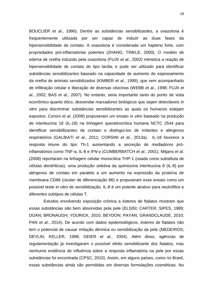
BOUCLIER et al., 1990). Dentre as substâncias sensibilizantes, a oxazolona é
frequentemente utilizada por ser capaz de induzir as duas fases da
hipersensibilidade de contato. A oxazolona é considerada um hapteno forte, com
propriedades pró-inflamatórias potentes (ZHANG; TINKLE, 2000). O modelo de
edema de orelha induzido pela oxazolona (FUJII et al., 2002) mimetiza a reação de
hipersensibilidade de contato do tipo tardia, e pode ser utilizado para identificar
substâncias sensibilizantes baseado na capacidade de aumento do espessamento
da orelha de animais sensibilizados (KIMBER et al., 1999), que vem acompanhado
de infiltração celular e liberação de diversas citocinas (WEBB et al., 1998; FUJII et
al., 2002; BAS et al., 2007). No entanto, seria importante tanto do ponto de vista
econômico quanto ético, desvendar marcadores biológicos que sejam detectáveis in
vitro para discriminar substâncias sensibilizantes as quais os humanos estejam
expostos. Corsini et al. (2009) propuseram um ensaio in vitro baseado na produção
de interleucina 18 (IL-18) na linhagem queratinocítica humana NCTC 2544 para
identificar sensibillizantes de contato e distingui-los de irritantes e alérgenos
respiratórios (GALBIATI et al., 2011; CORSINI et al., 2013a). IL-18 favorece a
resposta imune do tipo Th-1 aumentando a secreção de mediadores pró-
inflamatórios como TNF-α, IL-8 e IFN-γ (CUMBERBATCH et al., 2001). Mitjans et al.
(2008) reportaram na linhagem celular monocítica THP-1 (usada como substituta de
células dendríticas), uma produção seletiva da quimiocina interleucina 8 (IL-8) por
alérgenos de contato em paralelo a um aumento na expressão da proteína de
membrana CD86 (cluster de diferenciação 86) e propuseram esse ensaio como um
possível teste in vitro de sensibilização. IL-8 é um potente atrativo para neutrófilos e
diferentes subtipos de células T.
Estudos envolvendo exposição crônica a ésteres de ftalatos mostram que
essas substâncias são bem absorvidas pela pele (ELSISI; CARTER; SIPES, 1989;
DOAN; BRONAUGH; YOURICK, 2010; BEYDON; PAYAN; GRANDCLAUDE, 2010;
PAN et al., 2014). De acordo com dados epidemiológicos, ésteres de ftalatos não
tem o potencial de causar irritação dérmica ou sensibilização da pele (MEDEIROS;
DEVLIN; KELLER, 1999; GEIER et al., 2004). Além disso, agências de
regulamentação já investigaram o possível efeito sensibilizante dos ftalatos, mas
nenhuma evidência de influência sobre a resposta inflamatória na pele por essas
substâncias foi encontrada (CPSC, 2010). Assim, em alguns países, como no Brasil,
essas substâncias ainda são permitidas em diversas formulações cosméticas. No
19
entanto, esses estudos não incluíram o possível efeito adjuvante dos ftalatos em
modelos de hipersensibilidade de contato. Mesmo os ftalatos não sendo capazes de
induzir hipersensibilidade, o fato que essas substâncias possam ser capazes de
exacerbar a resposta inflamatória em modelos animais de hipersensibilidade de
contato (CHOWDHURY; STATHAM, 2002; IMAI et al., 2006; MATSUDA et al., 2010;
LI et al., 2014) sugere que a toxicidade dérmica dos ésteres de ftalatos necessite de
maior atenção e avaliação.
Com esse estudo, pretendemos investigar o possível efeito adjuvante dos
ésteres de ftalatos no desenvolvimento da hipersensibilidade de contato induzida por
oxazolona em modelo animal de edema de orelha, avaliando doses mais baixas de
DBP (0,4 mg/animal) (KOO; LEE, 2004; INT. J. TOXICOL., 2005). Além disso,
investigamos alguns aspectos relacionados ao mecanismo do efeito adjuvante do
DBP em processos alérgico-inflamatórios. Para isso, foram avaliados os efeitos do
DBP sozinho ou em associação com alérgenos em dois ensaios in vitro
representativos de respostas queratinocíticas e dendríticas, a linhagem
queratinocítica humana NCTC 2544 e a linhagem monocítica humana THP-1,
respectivamente. Foi também investigado se a indução de estresse oxidativo poderia
ser parte do mecanismo molecular do efeito adjuvante induzido por DBP. Isso
porque diversos autores já reportaram relação entre ftalatos e estresse oxidativo
demonstrando aumento da geração de espécies reativas de oxigênio (SEO et al.,
2004; TETZ et al., 2013). Outros têm associado a presença de diversos metabólitos
monoésteres de ftalatos detectados na urina com aumento de marcadores de
inflamação e estresse oxidativo (HAUSER et al., 2007; FERGUSON; LOCH-
CARUSO; MEEKER, 2011). Também avaliamos a possível participação da ativação
dos receptores TRPA1 e TRPV1 no efeito adjuvante do DBP no modelo de edema
de orelha induzida por oxazolona, já que já foi demonstrada a capacidade do DBP
em ativar tanto TRPA1 quanto TRPV1 in vitro (SHIBA et al., 2009).
Os resultados obtidos nesse estudo contribuem para uma maior
compreensão sobre o efeito adjuvante do dibutil ftalato em modelos de
hipersensibilidade de contato in vivo e in vitro.
20
2 REVISÃO DE LITERATURA
2.1 FTALATOS
2.1.1 Estrutura e propriedades físico-químicas
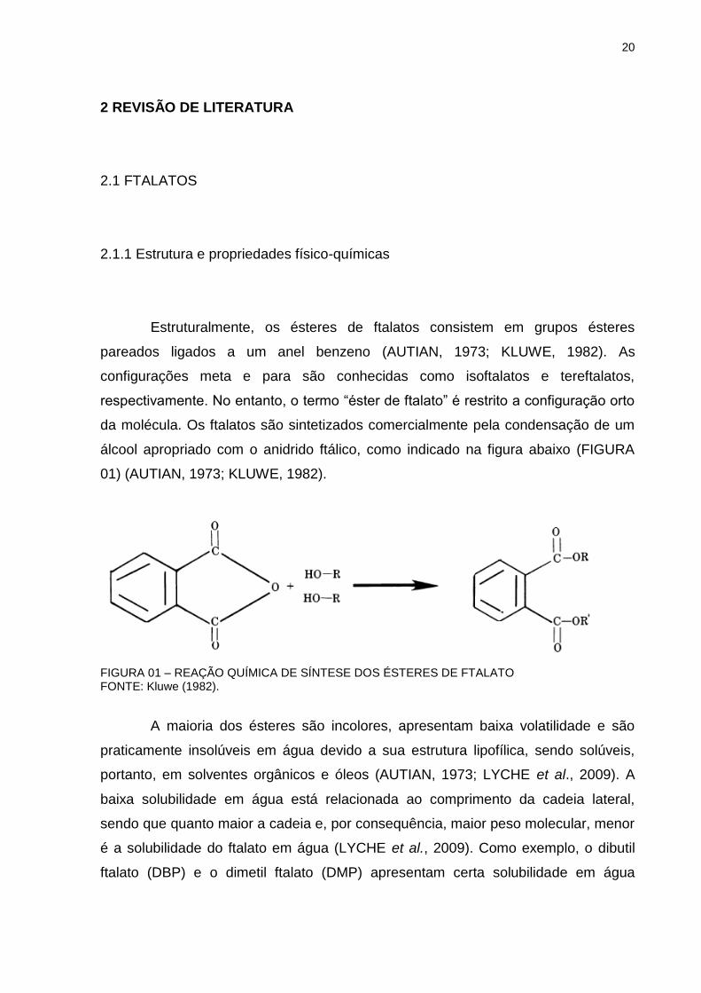
Estruturalmente, os ésteres de ftalatos consistem em grupos ésteres
pareados ligados a um anel benzeno (AUTIAN, 1973; KLUWE, 1982). As
configurações meta e para são conhecidas como isoftalatos e tereftalatos,
respectivamente. No entanto, o termo “éster de ftalato” é restrito a configuração orto
da molécula. Os ftalatos são sintetizados comercialmente pela condensação de um
álcool apropriado com o anidrido ftálico, como indicado na figura abaixo (FIGURA
01) (AUTIAN, 1973; KLUWE, 1982).
FIGURA 01 – REAÇÃO QUÍMICA DE SÍNTESE DOS ÉSTERES DE FTALATO FONTE: Kluwe (1982).
A maioria dos ésteres são incolores, apresentam baixa volatilidade e são
praticamente insolúveis em água devido a sua estrutura lipofílica, sendo solúveis,
portanto, em solventes orgânicos e óleos (AUTIAN, 1973; LYCHE et al., 2009). A
baixa solubilidade em água está relacionada ao comprimento da cadeia lateral,
sendo que quanto maior a cadeia e, por consequência, maior peso molecular, menor
é a solubilidade do ftalato em água (LYCHE et al., 2009). Como exemplo, o dibutil
ftalato (DBP) e o dimetil ftalato (DMP) apresentam certa solubilidade em água
21
enquanto o di-2-etilhexil ftalato (DEHP) e o di-isononil ftalato (DINP) são insolúveis
em água (AUTIAN, 1973; LYCHE et al., 2009).
2.1.2 Tipos, fontes e usos
Os ftalatos são amplamente utilizados como plastificantes, sendo
adicionados ao policloreto de vinila (PVC) para conferir maleabilidade e durabilidade
ao polímero (SILVA et al., 2004; LYCHE et al., 2009). Podem ser encontrados
também como solventes, óleos lubrificantes, estabilizante de cor e fragrância e como
detergentes em produtos de cuidados pessoais (como, por exemplo, cosméticos,
loções e perfumes) (KOO; LEE, 2004; DUTY et al., 2005). Em cosméticos, os
ftalatos atuam como umectantes e/ou emolientes em cremes hidratantes, como
potencializadores da penetração cutânea e para promoção de brilho e impedimento
de ressecamento e quebra de esmaltes (KOO; LEE, 2004).
Quando são incorporados ao PVC, os ftalatos não se ligam covalentemente
ao polímero e por isso são facilmente liberados no meio ambiente, principalmente
quando entram em contato com substâncias lipofílicas, resultando na exposição
humana e de animais (SHEA; COMMITTEE ON ENVIRONMENTAL HEALTH, 2003;
HEUDORF, MERSCH-SUNDERMANN; ANGERER, 2007; LYCHE et al., 2009;
JEONG et al., 2011). Além disso, eles são liberados diretamente no ambiente
durante a produção e utilização e após o descarte de PVC e outros produtos que os
contém (SHEA; COMMITTEE ON ENVIRONMENTAL HEALTH, 2003).
O uso dos vários tipos de ftalatos está relacionado ao seu peso molecular.
Os ftalatos de baixo peso molecular, tais como: DMP, DBP e o di-etil ftalato (DEP),
tendem a ser usados como solventes em cosméticos e em inseticidas, como
plastificantes em acetato de celulose, como promotores de liberação controlada em
produtos farmacêuticos, além de também serem encontrados no PVC (ATSDR,
1995; ATSDR, 2001; LYCHE et al., 2009). Já os ftalatos de alto peso molecular,
como DEHP, DiNP, butilbenzil ftalato (BBzP) e di-isodecil ftalato (DiDP), são mais
utilizados em materiais de construção (como, revestimentos de paredes e
pavimentos) e em uma grande gama de produtos a base de PVC, incluindo
vestuários (por exemplo: calçados e capas de chuva), embalagens de alimentos,
22
produtos infantis (como os brinquedos) e equipamentos médicos-hospitalares (como
bolsas de sangue) (ATSDR, 1995; ATSDR, 2001; PREUSS; KOCH; ANGERER ,
2005; LYCHE et al., 2009).
2.1.3 Efeitos dos ftalatos sobre o organismo humano
Os ésteres de ftalato são utilizados em vários produtos industrializados
(ANDERSON et al., 2001; SILVA et al., 2004), sendo a exposição humana a essas
substâncias inevitável, podendo ocorrer por ingestão, inalação ou absorção cutânea
(ANDERSON et al., 2001; SILVA et al., 2004; LYCHE et al., 2009; JEONG et al.,
2011).
Os ftalatos têm atraído grande atenção da comunidade científica não
somente devido à ampla possibilidade de exposição humana, bem como pelos
possíveis efeitos desreguladores endócrinos que podem ocasionar alterações no
desenvolvimento reprodutivo masculino (MYLCHREESTER et al., 1999; FISHER et
al., 2003; HEUDORF, MERSCH-SUNDERMANN; ANGERER, 2007; MARTINO-
ANDRADE et al., 2009). Desreguladores endócrinos são substâncias capazes de
mimetizar ou bloquear a ação de um hormônio endógeno, interferindo com o
funcionamento normal do sistema endócrino (ATSDR, 2001). Os ftalatos exibem
uma ação anti-androgênica, sendo que estudos em animais demonstraram que a
exposição gestacional e lactacional ao DBP, BBzP e DEHP provocam toxicidade
testicular, com sintomas característicos da Síndrome de Disgenesia Testicular (TDS)
- hipospádia, criptorquidia e função testicular comprometida (FOSTER et al., 2001;
FISHER et al., 2003; MARTINO-ANDRADE et al., 2009).
No entanto, estudos epidemiológicos e experimentais recentes também têm
demonstrado possíveis associações entre a exposição a alguns ftalatos e doenças
inflamatórias como asma e dermatites (BORNEHAG et al., 2004; TAKANO et al.,
2006; KOLARIK et al., 2008; YANAGISAWA et al., 2008). Além disso, existem dados
indicando um possível aumento na incidência de doenças alérgicas, principalmente
entre crianças e jovens, ao longo das últimas décadas (BEASLEY; ELLWOOD;
ASHER, 2003). Substâncias químicas como ftalatos poderiam aumentar a potência
23
de alérgenos e, assim, desempenhar um papel no desenvolvimento e/ou
agravamento de doenças alérgicas (CASILLAS et al., 1999).
2.1.4 Dibutil Ftalato (DBP)
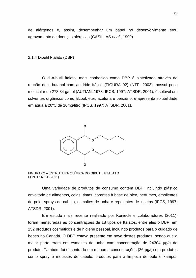
O di-n-butil ftalato, mais conhecido como DBP é sintetizado através da
reação do n-butanol com anidrido ftálico (FIGURA 02) (NTP, 2003), possui peso
molecular de 278,34 g/mol (AUTIAN, 1973; IPCS, 1997; ATSDR, 2001), é solúvel em
solventes orgânicos como álcool, éter, acetona e benzeno, e apresenta solubilidade
em água a 20ºC de 10mg/litro (IPCS, 1997; ATSDR, 2001).
FIGURA 02 – ESTRUTURA QUÍMICA DO DIBUTIL FTALATO FONTE: NIST (2011)
Uma variedade de produtos de consumo contém DBP, incluindo plástico
envoltório de alimentos, colas, tintas, corantes à base de óleo, perfumes, emolientes
de pele, sprays de cabelo, esmaltes de unha e repelentes de insetos (IPCS, 1997;
ATSDR, 2001).
Em estudo mais recente realizado por Koniecki e colaboradores (2011),
foram mensuradas as concentrações de 18 tipos de ftalatos, entre eles o DBP, em
252 produtos cosméticos e de higiene pessoal, incluindo produtos para o cuidado de
bebes no Canadá. O DBP estava presente em nove destes produtos, sendo que a
maior parte eram em esmaltes de unha com concentração de 24304 µg/g de
produto. Também foi encontrado em menores concentrações (36 µg/g) em produtos
como spray e mousses de cabelo, produtos para a limpeza de pele e xampus
24
infantis. Nesse mesmo estudo, foi calculada uma exposição diária de DBP em 0,36
µg/kg de peso corpóreo (KONIECKI et al., 2011).
Em um estudo realizado por Koo e Lee (2004), 26% dos perfumes (11 de 42
perfumes analisados) continham níveis detectáveis de DBP, sendo que a
concentração média de DBP encontrada foi de 0,44 mg/mL e a utilização mediana
de perfumes pela população, estimada através de questionário aplicado, foi de 1
mL/dia. Portanto, a quantidade de DBP aplicada em indivíduos que utilizam
determinados tipos de perfumes pode ser de aproximadamente 0,4 mg/dia.
Considerando que alguns indivíduos utilizam quantidades ainda maiores de
perfumes e outros cosméticos contendo DBP, esse nível de exposição tópica pode
ser ainda maior (KOO; LEE, 2004).
Além disso, em 2005, o International Journal of Toxicology publicou em sua
revisão anual sobre a segurança de ingredientes em produtos cosméticos uma
estimativa sobre a exposição humana aos ftalatos presentes em cosméticos. Os
dados foram fornecidos em quantidades de ftalatos presentes em diferentes tipos de
cosméticos (perfumes, desodorantes, spray de cabelo e esmalte), a quantidade
efetivamente aplicada sobre a pele e as taxas de absorção dessas substâncias (INT.
J. TOXICOL., 2005). De acordo com essas estimativas, a aplicação dérmica de
ftalatos para esses produtos varia entre 0,1 e 0,45 mg/dia, o que está de acordo com
as estimativas de Koo e Lee (2004).
2.1.5 Farmacocinética do DBP
Quando administrados pela via oral são hidrolisados por lipases e
absorvidos quase totalmente na forma de seus metabólitos correspondentes (os
monoésteres de ftalatos) (ROWLAND; COTTRELL; PHILLIPS, 1977), sendo que
para o DBP, o metabólito mais encontrado no plasma é o MBP (monobutil ftalato)
(FENNEL et al., 2004). Extensa absorção oral é observada, indicada pelo fato de
que, em ratos, 63-97% de uma dose oral foi quantificada na urina 24h após a
administração (FOSTER et al., 1983; TANAKA; MATSUMOTO; YAMAHA, 1978).
Exposições dérmicas e pela via respiratória também são importantes rotas
de exposição aos ftalatos, como DBP, que são usados em cosméticos (KOCH;
25
DREXLER; ANGERER, 2003; KOO; LEE, 2004). De acordo com Blount e
colaboradores (2000), os níveis urinários de metabólitos do DBP são
significativamente maiores em mulheres em idade reprodutiva (20 a 40 anos)
quando comparado com as concentrações em homens ou em outras faixas etárias.
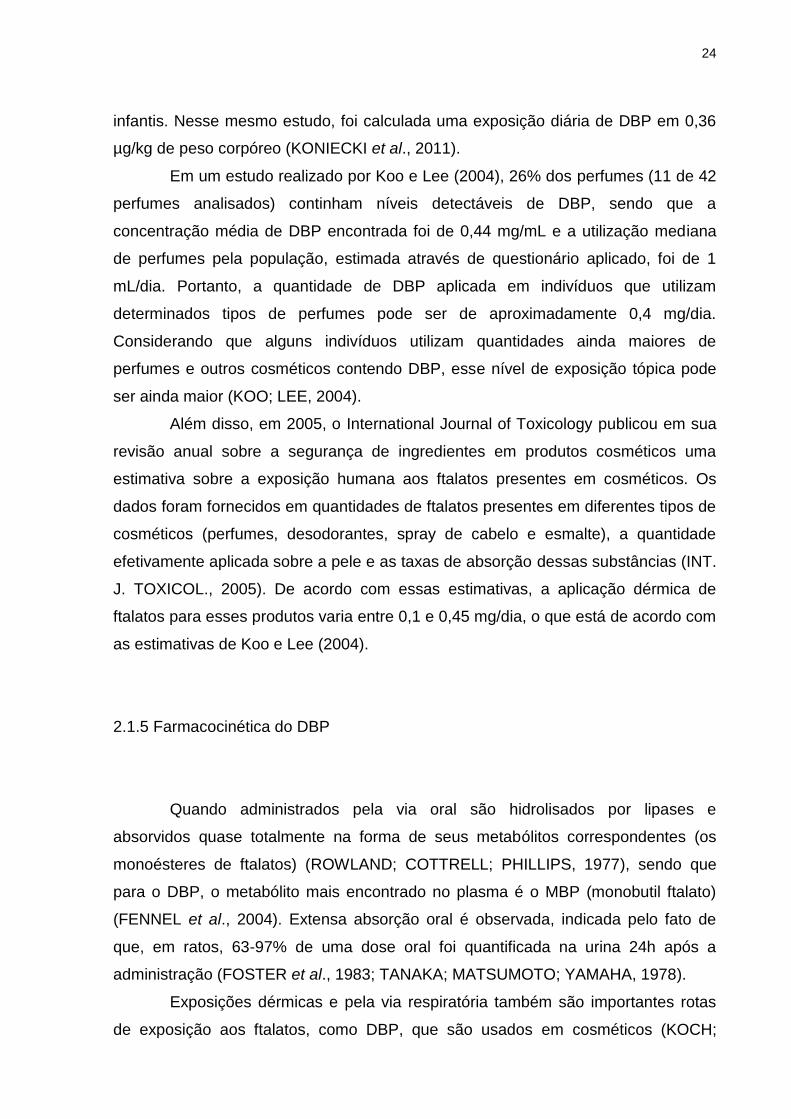
Em função da rápida metabolização, há pouca ou nenhuma bioacumulação.
Um estudo revelou que uma semana após a exposição a uma dose única de DBP,
nenhum tecido continha mais de 2% da dose administrada (ELSISI; CARTER;
SIPES, 1989). A principal rota de excreção dos metabólitos do DBP é a urina
(FOSTER et al., 1983; ELSISI; CARTER; SIPES, 1989), sendo que ele é excretado
após a conjugação com ácido glucorônico (LYCHE et al., 2009). O MBP é excretado
para a bile (cerca de 45%), mas apenas cerca de 5% é eliminado pelas fezes,
indicando uma eficiente circulação êntero-hepática (TANAKA; MATSUMOTO;
YAMAHA, 1978; ELSISI; CARTER; SIPES, 1989). A figura abaixo mostra o esquema
metabólico do DBP em animais (FIGURA 03).
FIGURA 03. ESQUEMA METABÓLICO DO DIBUTIL FTALATO FONTE: ATSDR (2001)
26
2.1.6 Aspectos Regulatórios dos Ftalatos
Regulamentações que restringem o uso de ftalatos vêm sendo
desenvolvidas na tentativa de reduzir ou até mesmo evitar exposição humana direta
a essas substâncias (KONIECKI et al., 2011).
Na legislação brasileira, a Resolução nº 105 de 19 de maio de 1999
(ANVISA/MS) aprova, através do seu art.1º, os Regulamentos Técnicos -
Disposições Gerais para Embalagens e Equipamentos Plásticos em contato com
Alimentos e seus Anexos (BRASIL, 1999). Apesar de estar em vigor, o Anexo III
desta lei, e seus Apêndices I e II, referente à Lista Positiva de Aditivos para Materiais
Plásticos destinados à elaboração de Embalagens e Equipamentos em contato com
Alimentos foram revogados pela RDC nº 17, de 17 de março de 2008 (BRASIL,
2008). Assim, os limites de ftalatos permitidos nas embalagens para alimentos estão
definidos por esta Resolução (BRASIL, 2008). No Apêndice I do Anexo desta RDC
consta que o DBP possui Limite de Migração Específica (LME) igual a 0,3 mg/kg,
podendo ser usado como plastificante somente em materiais reutilizáveis que
estejam em contato com alimentos não gordurosos; coadjuvante de tecnologia em
poliolefinas em concentrações de até 0,05% no produto final (BRASIL, 2008).
Já a Portaria nº 369 de 27 de setembro de 2007 (INMETRO/ MDIC)
estabelece, em seu art. 1º, os requisitos adicionais para ensaios toxicológicos em
brinquedos e, em seu art. 2º os requisitos para aceitação e ensaio de ftalatos em
brinquedos (BRASIL, 2007). No § 1º e 2º do art. 2º desta Portaria, contam que:
Art. 2º Estabelecer os requisitos para aceitação e ensaio de ftalatos em brinquedos. § 1º Os seguintes ftalatos: ftalato de di (2-etil-hexila) (DEHP), ftalato de dibutila (DBP), ftalato de benzilbutila (BBP) não devem ser utilizados, como substâncias ou componentes de preparações, em concentrações superiores a 0,1 % em massa de material plastificado, em todos os tipos de brinquedos de material vinílico. § 2º Os seguintes ftalatos: ftalato de di (2-etil-hexila) (DEHP), ftalato de dibutila (DBP), ftalato de benzilbutila (BBP), ftalato de di-isononila (DINP), ftalato de di-isodecila (DIDP) e ftalato de di-noctila (DNOP) não devem ser utilizados, como substâncias ou componentes de preparações, em concentrações superiores a 0,1 % em massa de material plastificado, em brinquedos de material vinílico destinados a crianças com idade inferior a 3 anos (BRASIL, 2007).
27
Além dessas regulamentações, está em análise pela Câmara dos
Deputados, o Projeto de Lei nº 3222/12, o qual proíbe o uso da substância ftalato na
composição de brinquedos e produtos destinados ao público infantil, tais como:
chupetas, mamadeiras, roupas, calçados e material escolar (CAMARA DOS
DEPUTADOS, 2012).
Já em relação ao uso de ftalatos em cosméticos, o Brasil ainda não possui
nenhuma regulamentação, diferentemente da União Europeia que proibiu o uso de
DBP e DEHP em cosméticos e produtos de higiene pessoal que participam do
mercado europeu (EUROPEAN COMISSION, 2007). No Brasil, a RDC nº 48 de 16
de março de 2006, a qual aprova o Regulamento Técnico sobre Lista de
Substâncias que não podem ser utilizadas em Produtos de Higiene Pessoal,
Cosméticos e Perfumes, não cita em sua listagem nenhum tipo de ftalato (BRASIL,
2006).
2.2 PROCESSOS ALÉRGICOS CUTÂNEOS
2.2.1 Estrutura da pele
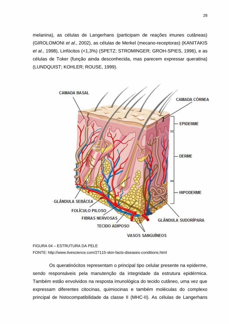
A pele recobre a superfície do corpo e apresenta-se constituída por uma
porção epitelial, a epiderme, e uma porção conjuntiva, a derme (FIGURA 04). A pele
exerce diversas funções, principalmente proteção contra fatores químicos, físicos e
biológicos, como a proteção contra raios ultravioleta através da melanina. Além
disso, impede perda de água para o meio, promove as sensações de dor, pressão,
tato e variações de temperatura, sintetiza hormônios (diidrotestosterona) e vitaminas
(vitamina D), promove a regulação térmica, metaboliza xenobióticos e excreta
substâncias através das glândulas sudoríparas (CHUONG et al., 2002; HAAKE;
SCOTT; HOLBROOK, 2000; SAMPAIO; CASTRO; RIVITTI, 2000; SHAEFER;
REDELMEIER, 1996; ROSS; REITH; ROMRELL, 1993).
A epiderme é um tecido não vascular constituído por epitélio estratificado
pavimentoso queratinizado, onde as células mais abundantes são os queratinócitos.
A pele ainda apresenta outros tipos celulares como os melanócitos (que sintetizam a
28
melanina), as células de Langerhans (participam de reações imunes cutâneas)
(GIROLOMONI et al., 2002), as células de Merkel (mecano-receptoras) (KANITAKIS
et al., 1998), Linfócitos (<1,3%) (SPETZ; STROMINGER; GROH-SPIES, 1996), e as
células de Toker (função ainda desconhecida, mas parecem expressar queratina)
(LUNDQUIST; KOHLER; ROUSE, 1999).
FIGURA 04 – ESTRUTURA DA PELE
FONTE: http://www.livescience.com/27115-skin-facts-diseases-conditions.html
Os queratinócitos representam o principal tipo celular presente na epiderme,
sendo responsáveis pela manutenção da integridade da estrutura epidérmica.
Também estão envolvidos na resposta imunológica do tecido cutâneo, uma vez que
expressam diferentes citocinas, quimiocinas e também moléculas do complexo
principal de histocompatibilidade da classe II (MHC-II). As células de Langerhans
29
estão localizadas nas camadas suprabasais da epiderme da pele e das mucosas,
onde desempenham um importante papel na reposta imune cutânea (CHAN, 2004).
As células de Langerhans são células migratórias que possuem vários receptores de
membrana envolvidos no processo imunológico (ex.: MHC-II, IgG - imunoglobulina
G, C3 - fator do complemento C3). Assim, essas células são responsáveis pelo
reconhecimento, captação, processamento e apresentação de antígenos solúveis e
haptenos aos linfócitos T. Em certas doenças inflamatórias cutâneas, como na
dermatite de contato e em processos alérgicos, verifica-se que as células de
Langerhans se tornam mais abundantes (NORRIS, 2004; HAAKE; SCOTT;
HOLBROOK, 2000).
Além das células residentes da epiderme (queratinócitos, melanócitos,
células de Langerhans e células de Merkel), outras células migram para a epiderme
em resposta aos mais variados estímulos: linfócitos, macrófagos, neutrófilos e
eosinófilos, sendo essas células elementos da resposta de defesa inata ou adquirida
(NORRIS, 2004).
A derme é o tecido conjuntivo compressivo e elástico onde se apóia a
epiderme e une a pele à hipoderme. A derme apresenta em sua constituição muitas
fibras, sendo a maioria delas, as fibras de colágeno, principalmente dos tipos I e III,
responsáveis pela resistência mecânica da pele. O tipo celular responsável pela
síntese de fibras, presente na derme, é o fibroblasto (EYDEN, 2001). Os fibroblastos
sintetizam diferentes macromoléculas que entram na constituição da matriz celular
como, por exemplo, o colágeno e a elastina (HAAKE; SCOTT; HOLBROOK, 2000;
SAMPAIO; CASTRO; RIVITTI, 2000; ROSS; REITH; ROMRELL, 1993). Durante um
processo inflamatório ocorre o aumento da proliferação e da atividade de
fibroblastos devido à ação de alguns mediadores pró-inflamatórios como a
interleucina-1α (IL-1α) e interleucina-1β (IL-1β) (FREINKEL; WOODLEY, 2001).
Como a epiderme é um tecido não vascular, na derme é onde se encontra
uma grande rede vascular responsável pela distribuição de nutrientes, cura de
lesões, reações imunes e controle da pressão arterial (BRAVERMAN, 2000). Os
vasos sangüíneos presentes na derme permitem que ocorra a infiltração de células
migratórias importantes no processo de resposta de defesa inata ou imune e de
cicatrização, como os macrófagos, linfócitos, eosinófilos, neutrófilos, entre outros
(RYAN, 2004). A interação coordenada entre os diferentes tipos celulares presentes
nas camadas da pele permite que este órgão responda, prontamente e
30
efetivamente, a uma variedade de estímulos nocivos que ocorrem na interface do
organismo com o meio externo, como a ação de toxinas, organismos patogênicos,
radiação ultravioleta, extremos de temperatura, garantindo assim a manutenção da
homeostasia cutânea (BURBACH; ANSEL; ARMSTRONG, 2000; HAAKE; SCOTT;
HOLBROOK, 2000; WILLIAMS; KUPPER, 1996). Nesse contexto, a pele é muito
mais do que simplesmente uma barreira física passiva entre o meio externo e
interno, mas também uma extensão do sistema imunológico (WILLIAMS; KUPPER,
1996).
2.2.2 Dermatite de contato alérgica (DCA) ou Hipersensibilidade de Contato (HSC)
Sabe–se que a prevalência de doenças alérgicas, como a dermatite atópica,
asma brônquica e rinite alérgica vem aumentando (SEGAWA; HIRASAWA, 2014). A
severidade e a indução de doenças alérgicas estão associadas a fatores genéticos e
ambientais. Entre os fatores ambientais, a poluição do ar e os contaminantes
químicos ambientais são considerados responsáveis pela exacerbação dessas
doenças (SEGAWA; HIRASAWA, 2014). Um exemplo pode ser os ftalatos, que são
considerados contaminantes onipresentes em alimentos, ar interno, solos e
sedimentos (SHEA; COMMITTEE ON ENVIRONMENTAL HEALTH, 2003).
A dermatite de contato é uma dermatose inflamatória frequente nos países
industrializados, sendo uma das doenças ocupacionais mais comuns. De acordo
com os mecanismos fisiopatológicos envolvidos, podem-se distinguir dois tipos de
dermatite de contato: a dermatite de contato irritativa, decorrente dos efeitos tóxicos
e pró-inflamatórios de xenobióticos capazes de ativar a imunidade inata da pele; e a
dermatite de contato alérgica, que requer a ativação da imunidade adquirida,
levando ao desenvolvimento de células T efetoras, que são mediadoras da
inflamação cutânea (HENNINO et al., 2005). É caracterizada por eritema, pápulas e
vesículas, seguidas de ressecamento e descamação.
A dermatite de contato alérgica (DCA), também conhecida como
hipersensibilidade de contato (HSC), é uma reação inflamatória cutânea mediada
por células T decorrente de contatos repetidos da pele com substâncias químicas
não protéicas, denominadas haptenos (SAINT-MEZARD et al., 2004; LEPOITTEVIN;
31
LEBLOND, 1997). A iniciação da HSC é gerada pela aplicação tópica de haptenos
sensibilizantes na epiderme. Duas fases são geralmente necessárias para atingir
uma reação de HSC máxima: as fases de sensibilização e de indução.
A fase de sensibilização ocorre ao primeiro contato da pele com o hapteno e
leva ao "priming" e a expansão de células T hapteno-específicas nos linfonodos. O
hapteno aplicado topicamente é captado pelas células dendríticas (CD) cutâneas,
especialmente as células de Langerhans (CL), que migram da epiderme para os
linfonodos de drenagem, onde apresentam complexos de moléculas de peptídeo
conjugado a hapteno aos precursores de células T hapteno-específicas. Células T
específicas emigram dos linfonodos e atingem o sangue e recirculam no sangue e
órgãos linfóides secundários. A fase de elicitação ou indução ocorre algumas horas
após um contato subsequente da pele com o mesmo hapteno, que induz a produção
de quimiocinas, a ativação de células endoteliais e mastócitos, e a infiltração de
neutrófilos, todos necessários para o recrutamento de células T específicas. As
células T interagem com células cutâneas apresentadoras de antígeno portadoras
de hapteno. As células T CD8+ citotóxicas ativadas produzem citocinas tipo 1 e
induzem a ativação de células cutâneas e a apoptose dos queratinócitos, levando a
amplificação da inflamação cutânea através da produção de todo um conjunto de
citocinas e quimiocinas. Esta fase de indução da HSC dura 72 horas em humanos e
24 a 48 horas em camundongos. A reação inflamatória persiste durante vários dias e
diminui progressivamente mediante mecanismos de regulação negativa fisiológicos
(FIGURA 05).
Além das células T, outras subpopulações de células linfóides contribuem
para a complexa via que finalmente leva a resposta de HSC (ASKENASE, 2001;
YOKOZEKI et al., 2001). As células B-1 são ativadas nos órgãos linfóides dentro de
horas após a sensibilização cutânea e produzem anticorpos IgM. Esses anticorpos
difundem-se na pele e ligam-se ao hapteno imediatamente após o desafio. A
presença de complexos imunes dá inicio a ativação do complemento, que parece
mandatória para o recrutamento de células T efetoras no local do desafio (CAMPOS
et al., 2003). Os neutrófilos desempenham um importante papel no desenvolvimento
da HSC. Em sua ausência a HSC é reduzida. Segundo a literatura, estão envolvidos
nas fases de sensibilização e elicitação da HSC. Os neutrófilos estão entre as
primeiras células a serem recrutadas após o desafio de camundongos sensibilizados
(DILULIO et al., 1999) e aparecem antes da infiltração de células T CD8 efetoras.
32
Uma vez que as células efetoras tenham sido ativadas, outro influxo de neutrófilos é
secundário a ativação de mastócitos que produzem TNF-α (BIEDERMANN et al.,
1999).
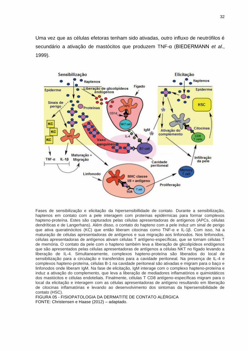
Fases de sensibilização e elicitação da hipersensibillidade de contato. Durante a sensibilização, haptenos em contato com a pele interagem com proteínas epidérmicas para formar complexos hapteno-proteína. Estes são capturados pelas células apresentadoras de antígenos (APCs, células dendríticas e de Langerhans). Além disso, o contato do hapteno com a pele induz um sinal de perigo que ativa queratinócitos (KC) que então liberam citocinas como TNF-α e IL-1β. Com isso, há a maturação de células apresentadoras de antígenos e sua migração aos linfonodos. Nos linfonodos, células apresentadoras de antígenos ativam células T antígeno-específicas, que se tornam células T de memória. O contato da pele com o hapteno também leva a liberação de glicolipídeos endógenos que são apresentados pelas células apresentadoras de antígenos a células NKT no fígado levando a liberação de IL-4. Simultaneamente, complexos hapteno-proteína são liberados do local de sensibilização para a circulação e transferidos para a cavidade peritoneal. Na presença de IL-4 e complexos hapteno-proteína, células B-1 na cavidade peritoneal são ativadas e migram para o baço e linfonodos onde liberam IgM. Na fase de elicitação, IgM interage com o complexo hapteno-proteína e induz a ativação do complemento, que leva a liberação de mediadores inflamatórios e quimiotáticos dos mastócitos e células endoteliais. Finalmente, células T CD8 antígeno-específicas migram para o local da elicitação e interagem com as células apresentadoras de antígeno resultando em liberação de citocinas inflamatórias e levando ao desenvolvimento dos sintomas da hipersensibilidade de contato (HSC). FIGURA 05 - FISIOPATOLOGIA DA DERMATITE DE CONTATO ALÉRGICA FONTE: Christensen e Haase (2012) – adaptado.
33
Dentre as citocinas, destacam-se as citocinas pró-inflamatorias IL-1β e TNF-
α (LI; CRUZ, 2004; ENK, 1997). A IL-1β liberada pelas células de Lngerhans faz com
que os queratinócitos produzam TNF-α que, juntamente com a IL-1β, determinam a
maturação e migração dessas CD para os linfonodos (ENK et al., 1993; STEINMAN;
HOFFMAN; POPE, 1995). IL-8, liberada por células dendríticas expostas a
alérgenos de contato, tem um papel importante na sensibilização e na elicitação de
reações alérgicas, por induzir o influxo de leucócitos para o local da inflamação. IL-8
é um potente atrativo de neutrófilos e diferentes subtipos de células T (BARKER et
al., 1991). Além de Il-8, IL-18, expressa por queratinócitos após exposição à
alérgenos de contato (NAIK et al., 1999; VAN OCH et al., 2005), é importante na
indução da hipersensibilidade de contato por aumentar a secreção de mediadores
pró-inflamatórios como TNFα, IL-8 and IFN-γ (OKAMURA et al., 1995;
CUMBERBATCH et al., 2001; ANTONOPOULOS et al., 2008).
2.2.3 Papel do estresse oxidativo na hipersensibilidade de contato
Evidências indicam que doenças cutâneas inflamatórias e alérgicas possam
ser mediadas por estresse oxidativo (OKAYAMA, 2005; BYAMBA et al., 2010).
Análises genômicas e proteômicas de queratinócitos humanos assim como de
células dendríticas revelaram um grande número de marcadores específicos de
sensibilizantes com diversas sobreposições interessantes.
No sistema imune, espécies reativas de oxigênio estimulam a resposta
imune através da ativação de vias de sinalização, aumento da expressão de
moléculas co-estimulatórioas, carbonilação de proteínas e secreção de citocinas
(LARBI et al., 2007; SAREILA et al., 2011; BERTOLOTTI; SITIA; RUBARTELLI,
2012). Recentemente, vários estudos apóiam a participacao de espécies reativas de
oxigênio como o gatilho da dermatite de contato alérgica (GLOIRE; LEGRAND-
POELS; PIETTE, 2006; KIM et al., 2012), outros propõe que um dos papéis
biológicos da dermatite de contato seja uma defesa contra substâncias químicas
com potencial de alterar o balanço redox das células (SASAKI; AIBA, 2007).
A habilidade de substâncias sensibilizantes de induzir estresse oxidativo em
queratinócitos (NATSCH; EMTER, 2007) foi recentemente confirmada por
34
Vandebriel, Van Och e Van Loveren (2010). Além disso, já foi demonstrado que a
produção de IL-18 induzida por alérgenos em queratinócitos requer a produçao de
espécies reativas de oxigênio e é prevenida pelo antioxidante pirrolidina
ditiocarbamato (PDTC) (GALBIATI et al., 2011; CORSINI et al., 2009).
Em células dendríticas, Mizuashi et al. (2005) mostraram que todas as
substâncias sensibilizantes testadas (níquel, formaldeído, DNCB, cloreto de
manganês e timerosal), mas nenhuma das substâncias não sensibilizantes reduziu a
razão entre as formas reduzida e oxidada da glutationa (GSH/GSSG), o que foi
acompanhado de ativação da proteína quinase ativada por mitógeno (MAPK) p38,
um marcador da maturação de células dendríticas induzida por sensibilizantes
químicos. O antioxidante N-acetil-L-cisteína foi capaz de suprimir a redução da razão
GSH/GSSG e revogar a ativação da p38 MAPK.
2.2.4 Influências do sistema nervoso periférico na HSC
O sistema nervoso cutâneo representa uma parte do sistema nervoso
periférico (BERESFORD et al., 2004; LIU et al., 2006). Anatomicamente, células de
Langerhans estão associadas a terminações nervosas (BERESFORD et al., 2004).
Neuropeptídios na pele são sintetizados e liberados predominantemente por
uma subpopulação de neurônios aferentes não-mielinizados chamados de
nociceptores polimodais C, que representam 70% de todas as fibras C cutâneas
(LAWSON, 1996). Essas fibras C expressam receptores sensíveis a capsaicina,
chamados TRPV1 (BÁNVÖLGYI et al., 2005; MURAI et al., 2008; RAZAAVI et al,
2006). Alguns dos neurônios sensoriais que expressam TRPV1 também expressam
TRPA1 (STORY et al., 2003), ativados por cinamaldeído, alil isotiocianato, dentre
outros estímulos (BAUTISTA et al., 2005). A ativação de TRPA1 ou TRPV1 em
terminações nervosas sensoriais resulta na liberação de neuropeptídios como o
peptídeo relacionado ao gene da calcitonina (CGRP) (BAUTISTA et al., 2005;
ZYGMUNT et al., 1999). Desde que o tratamento de humanos ou cobaias (Cavia
porcellus) com capsaicina gerou um aumento na intensidade da resposta de
hipersensibilidade de contato no local do tratamento, foi sugerido que neurônios

35
sensíveis a capsaicina poderiam modular a HSC através da liberação de
neuropeptídios (GIROLOMONI; TIGELAAR, 1990; EK; THEODORSSON, 1990).
Os receptores de potencial transitório (TRPs) constituem uma superfamília
de receptores que contribuem para mudanças nas concentrações intracelulares de
Ca2+, e que tem papel fundamental em diversos processos celulares como
contração muscular, liberação de transmissores e morte celular (BERRIDGE; LIPP;
BOOTMAN, 2000). Essa superfamília é subdividida em sete subfamílias: TRPC,
TRPM, TRPV, TRPA, TRPP, TRPML, TRPN (MONTELL; BIRNBAUMER;
FLOCKERZI, 2002; COREY, 2003; CLAPHAM, 2003; MORAN; XU; CLAPHAM,
2004).
A subfamília vanilóide, ou TRPV, consiste em quatro grupos nos mamíferos:
TRPV1/V2, TRPV3, TRPV4 e TRPV5/V6 (GUNTHORPE et al., 2002; BENHAM;
DAVIS; RANDALL, 2002). O TRPV1 foi o primeiro dessa família a ser identificado
em mamíferos e é um dos mais estudados. TRPV1 são altamente expressos em
terminações nervosas periféricas (PLANELLS-CASES et al., 2005) distribuídas na
derme, além de serem expressos em queratinócitos e células dendríticas
(GOPINATH et al., 2007; BASU; SRIVASTAVA, 2005) e são ativados por compostos
vanilóides, como a capsaicina, e por outros estímulos como calor moderado, pH
baixo e outros compostos (MONTELL; BIRNBAUMER; FLOCKERZI, 2002;
PLANELLS-CASES et al., 2005). A capsaicina não ativa nenhum outro receptor da
mesma família (PEDERSEN; OWSIANIK; NILIUS, 2005). Alguns estudos sugerem
que a ativação de TRPV1 em neurônios sensoriais possa ter influências sobre o
sistema imune (BASU; SRIVASTAVA, 2005; BÁNVÖLGYI et al., 2005; MURAI et al.,
2008; RAZAAVI et al., 2006).
Já a subfamília anquirina, ou TRPA, apresenta somente um membro
conhecido em mamíferos, o TRPA1. O TRPA1 é ativado por isotiocianatos e
cinamaldeído, entre outros compostos, mas é insensível a capsaicina (PEDERSEN;
OWSIANIK; NILIUS, 2005). Estudos já demonstraram que a ativação de receptores
TRPA1 na pele participa do desenvolvimento de respostas inflamatórias
desenvolvidas pela aplicação tópica de um agonista TRPA1 (SILVA et al., 2011) e
que estes receptores também são expressos em queratinócitos (ATOYAN;
SHANDER; BOTCHKAREVA, 2009).
Sabe-se também que, quando estimuladas, as fibras C podem liberar uma
série de neuropeptídeos que tem a capacidade de modular a resposta inflamatória
36
local. É conhecido que a ativação de receptores TRPA1 e TRPV1 em terminações
nervosas sensoriais resulta na liberação de neuropeptídeos como o CGRP (peptídeo
relacionado ao gene da calcitonina) e a substância P (SP) (BAUTISTA et al., 2005;
ZYGMUNT et al., 1999).
A contribuição dos neuropeptídeos para inflamação neurogênica vem de
estudos usando a capsaicina. A resposta aguda à aplicação tópica de capsaicina na
orelha de camundongos é característica de inflamação neurogênica, onde o
aumento do fluxo sanguíneo e formação de edema são dependentes da liberação e
ação do CGRP e SP (GRANT, 2002). A SP é o mediador clássico da resposta:
eritema, edema e prurido. SP e CGRP injetados em pele humana rapidamente
induzem infiltração de eosinófilos e neutrófilos (SMITH et al., 1993), sugerindo que
esses neuropeptídeos participam do recrutamento leucocitário. A SP também pode
modular o padrão de expressão de citocinas pelos queratinócitos e mastócitos,
aumentando a produção de citocinas pró-inflamatórias como IL-1 e IL-8 (BROWN et
al., 1990; VIAC et al., 1996) e induzindo a expressão do mRNA e proteína para TNF-
α (ANSEL et al., 1993) em cobaias. Sumarizando, a SP tem um papel direto ou
indireto aumentando a inflamação neurogênica cutânea. CGRP é um dos
neuropeptídeos mais abundantes na pele e é frequentemente colocalizado com a SP
(GIBBINS; WATTCHOW; COVENTRY, 1987). Um dos efeitos mais proeminentes do
CGRP in vivo são ações vasodilatadoras. Injeção intradérmica de CGRP causa
eritema de longa duração (JANSEN-OLESEN; MORTENSEN; EDVINSSON, 1996;
BRAIN et al., 1986). O CGRP potencializa a formação de edema causado por
neuropeptídeos como a SP, (SCHOLZEN et al., 1998). Além disso, o CGRP também
pode prejudicar a apresentação de antígeno necessária nas reações de HSC
através do aumento da produção de citocinas pró-inflamatórias como IL-10, que
pode então inibir ações das células T (TORII et al., 1997).
2.3 ABSORÇÃO E TOXICIDADE DÉRMICA DOS FTALATOS
Estudos recentes indicam que os ftalatos, em especial o DBP, são capazes
de serem absorvidos pela pele. Elsisi, Carter e Sipes (1989) identificaram que após
sete dias da aplicação tópica de DBP, cerca de 50 a 60% da dose foi excretada,
37
sendo a via urinária a principal via de excreção, ou seja, eles não são
bioacumulados. Além disso, identificaram que 33 ± 2 % da dose aplicada
permaneceu no local de aplicação. Segundo os autores, a extensão da absorção
tópica dos ftalatos pode estar associada a fatores que competem entre si, tais como
lipofilia, peso molecular e metabolismo.
Janjua e colaboradores (2008) identificaram e dosaram DBP e seus
metabólitos na urina após aplicação tópica em humanos e, Sathyanarayana et al.
(2008) demonstraram que mais de sete metabólitos de ftalatos foram detectados na
urina de 81% de crianças após o uso de produtos infantis. Em outro estudo, testes in
vivo (em cobaias) e testes in vitro mostram que o DBP inserido em uma formulação
(emulsão óleo/água) pode ser absorvido pela pele e ficar potencialmente disponível
para distribuição sistêmica (DOAN; BRONAUGH; YOURICK, 2010).
A absorção dérmica do DBP em ratos parece estar associada à atividade de
esterases, que o hidrolisam durante a absorção percutânea (BEYDON; PAYAN;
GRANDCLAUDE, 2009).
Em relação à toxicidade do DBP, uma irritação leve foi observada em
estudos em pele de coelhos usando uma dose de 520 mg/kg, uma dose
extremamente alta para exposição cutânea (NATIONAL TOXICOLOGY PROGRAM,
2003). Testes agudos e crônicos realizados pela Consumer Product Safety
Comission levaram a conclusão de que existe pouca evidência em humanos e
animais para designar o DBP como substância sensibilizante de acordo com os
critérios propostos pela Consumer Product Safety Comission (CPSC, 2010).
Em estudos crônicos realizados em cobaias e em estudos agudos em
coelhos não foi encontrada sensibilização causada pelo DBP (NICNAS, 2008). Em
humanos, os dados são inconclusivos. Alguns casos isolados foram reportados em
humanos. Duas mulheres desenvolveram dermatite após o uso de um desodorante
contendo DBP. As duas apresentaram resposta positiva para um teste de contato
contendo DBP, mas apresentaram respostas negativas para outros constituintes do
desodorante (NICNAS, 2008).
Chowdhury e Statham (2002) reportaram um caso de um homem de 65 anos
que desenvolveu hipersensibilidade de contato após usar uma pomada para prurido.
O indivíduo também apresentou resposta positiva para o teste de contato contendo
DBP. Mas, em um estudo que expôs 159 indivíduos a um teste de contato contendo
cosméticos que apresentavam uma concentração de DBP de 4,5 a 9%, nenhum
38
indivíduo apresentou resposta positiva. Mais recentemente, Pan et al. (2014)
observaram que após aplicação dérmica do DBP, houve apoptose de queratinócitos
e fibroblastos via ativação da via de capases -3.
Por fim, deve-se ressaltar a importância da exposição ocupacional aos
ftalatos. Em seu estudo, Kwapniewski et al. (2008) dosou MBP (monobutil ftalato),
principal metabólito do DBP, em urina de manicures e, observou um aumento de
17,4 ng/mL desse metabólito durante o turno de trabalho, sendo que o uso de luvas
reduziu esse valor para 15,1 ng/mL. Todos esses estudos em conjunto indicam que
a toxicidade dérmica dos ftalatos deve ser melhor avaliada.
2.3.1 Ftalatos e processos alérgicos
Há relatos de que alguns ésteres de ftalato estão associados a sintomas
alérgicos em crianças (BORNEHAG et al., 2004; KOLARIK et al., 2008), bem como
em adultos (CHOWDHURY; STATHAM, 2002). Latini e colaboradores (2003)
propuseram um papel dos ftalatos na indução de inflamação intrauterina devido a
uma semelhança estrutural entre os ésteres de ftalato e algumas prostaglandinas e
tromboxanas pró-inflamatórias. Scarano e colaboradores (2009) mostraram a
presença de focos de inflamação na próstata de ratos expostos in utero e durante a
lactação a 100 mg/kg/dia de DBP.
Em relação a processos alérgicos na pele, alguns estudos demonstram que
certos ésteres de ftalatos, como o DBP, não possuem um potencial significativo de
causarem sozinhos a sensibilização da pele e dermatites de contato alérgicas. Isto
porque essas moléculas não são capazes de se ligarem e serem reativas a
proteínas, pré-requisito para a estimulação alérgica por uma substância química
(KIMBER; DEARMAN, 2010). Porém, Chowdhury e Statham (2002) reportaram
casos de hipersensibilidade de contato induzidas pelo DBP. Além disso, Imai e
colaboradores (2006) observaram que durante o processo de sensibilização por
isotiociato de fluoresceina (FITC), o DBP exerceu forte efeito adjuvante, associado
ao aumento do tráfico de células dendríticas apresentadoras de antígenos a partir da
pele para os linfonodos de drenagem.
39
Matsuda et al. (2010) encontrou que o DBP pode aumentar a sensibilização
da pele quando associado a alguns haptenos como, o feniletil isocianato (PEITC),
alem de dois análogos do FITC (isotiocianato de eosina-5 e isotiocianato de
rodamina B). Porém, não é capaz de exacerbar a sensibilização quando associado à
oxazolona e 2,4-dinitrofluorobenzeno (que exibem uma resposta mediada por
linfócitos Th-1) ou ao anidrido trimelítico, metilenodifenilo 4,4-di-isocianato e 2,4-
diisocianato de tolueno (que exibem uma resposta mediada por linfócitos Th-2).
Em estudo mais recente, Li et al. (2014), utilizando o modelo de
hipersensibilidade por contato induzido por FITC, investigou a exposição prolongada
(40 dias) ao DBP em doses de 0,4, 4,0 e 40 mg/kg/dia. Os resultados mostraram
que essa exposição pode agravar a dermatite de contato alérgica em camundongos,
havendo uma reação linear entre dose-resposta, com aumento do edema, na
contagem de células infiltradas, entre outros parâmetros.
2.4 MODELOS DE HIPERSENSIBILIDADE DE CONTATO
Modelos de hipersensibilidade de contato em camundongo vêm sendo muito
utilizados, a fim de se obter informações sobre a apresentação de antígenos, bem
como a ativação de linfócitos T. Esses modelos são amplamente difundidos para
estudo da inflamação mediada pelo sistema imune, devido à facilidade de aplicação
da substância sensibilizante, medida da resposta, amostragem e acesso aos
linfonodos de drenagem (WEBB et al., 1998). Alguns modelos animais de
inflamação cutânea auxiliam a identificar substâncias sensibilizantes, pois promovem
condições que se assemelham com alguns tipos de dermatites observadas em
humanos (VANE; BOTTING, 1998; BOUCLIER et al., 1990).
Dentre as substâncias sensibilizantes, a oxazolona é frequentemente
utilizada por ser capaz de induzir as duas fases da hipersensibilidade de contato. A
oxazolona é considerada um hapteno forte, com propriedades pró-inflamatórias
potentes (ZHANG; TINKLE, 2000). A oxazolona é um potente agente sensibilizante
que produz pouca irritação. Por ser capaz de produzir uma inflamação crônica,
dependente de linfócitos T tipo 1 (Th1) na fase inicial, e tipo 2 (Th2) seguindo
exposição continuada do agente (WEBB et al., 1998), e de ser de fácil
40
reprodutibilidade, a hipersensibilidadade de contato induzida por oxazolona (FUJII et
al., 2002) tem se mostrado um modelo farmacologicamente útil nos estudos de
dermatite de contato alérgica, já que reproduz os aspectos dessa doença humana,
induzindo elevação nos níveis de IFN-γ e pronunciável hiperplasia epidérmica
(BONISH et al., 2000). O modelo de edema de orelha induzido pela oxazolona
(FUJII et al., 2002) pode ser utilizado para identificar substâncias sensibilizantes
baseado na capacidade de aumento do espessamento da orelha de animais
sensibilizados (KIMBER et al., 1999), que vem acompanhado de infiltração celular e
liberação de diversas citocinas (WEBB et al., 1998; FUJII et al., 2002; BAS et al.,
2007).
No entanto, seria importante tanto do ponto de vista econômico quanto ético,
desvendar marcadores biológicos que sejam detectáveis in vitro para discriminar
substâncias sensibilizantes as quais os humanos estejam expostos. Na última
década, grande progresso tem sido alcançado no desenvolvimento de testes in vitro
alternativos para substituir ou complementar os testes em animais.
Corsini et al. (2009) propuseram um ensaio in vitro baseado na produção de
interleucina 18 (IL-18) na linhagem queratinocítica humana NCTC 2544 para
identificar sensibillizantes de contato e distingui-los de irritantes e alérgenos
respiratórios (GALBIATI et al., 2011; CORSINI et al., 2013a). IL-18 favorece a
resposta imune do tipo Th-1 aumentando a secreção de mediadores pró-
inflamatórios como TNF-α, IL-8 e IFN-γ (CUMBERBATCH et al., 2001).
Mitjans et al. (2008) reportaram na linhagem celular monocítica THP-1 uma
produção seletiva da quimiocina interleucina 8 (IL-8) por alérgenos de contato em
paralelo a um aumento na expressão da proteína de membrana CD86 (cluster de
diferenciação 86) e propuseram esse ensaio como um possível teste in vitro de
sensibilização. Esse ensaio é utilizado como um substituto de ativação de células
dendríticas, já que a expressão de CD86 ocorre durante a maturação de céluas
dendríticas (QUAH; O’NEILL, 2005).
41
3 HIPÓTESES E PREDIÇÕES
Propomos que ao associarmos o dibutil ftalato a alérgenos em testes in vitro
e in vivo de hipersensibilidade de contato, possamos identificar um efeito adjuvante
do DBP. Se essa hipótese for verdadeira, esperamos observar uma exacerbação na
resposta inflamatória envolvida no modelo in vivo, levando a aumento de edema e
do recrutamento celular; ou no aumento da expressão e liberação de marcadores de
ativação celular nos ensaios in vitro. Além disso, acreditamos que o estresse
oxidativo e ativação de receptores TRPA1 e TRPV1 sejam parte do mecanismo
molecular envolvido no efeito adjuvante do DBP.
42
4 OBJETIVOS
4.1 OBJETIVO GERAL
Investigar o efeito adjuvante do plastificante DBP em modelos in vivo e in
vitro de HSC, caracterizando o perfil de células envolvidas nesses modelos, bem
como a determinação do possível papel do estresse oxidativo e dos receptores
TRPV1 e TRPA1 nesse efeito.
4.2 OBJETIVOS ESPECÍFICOS
- Investigar o potencial do DBP como sensibilizante de contato em dois
testes in vitro usando linhagens celulares humanas, o ensaio NCTC 2544 e o ensaio
de ativação de células THP-1.
- Investigar o possível efeito adjuvante do DBP pela avaliação da expressão
de CD86 e IL-8 mRNA e liberação de IL-8 em células THP-1 após exposição dos
alérgenos oxazolona, citral e imidazolidinil ureia associados ao DBP.
- Investigar o possível efeito adjuvante do DBP pela avaliação da liberação
de IL-18 em células NCTC 2544 após exposição ao alérgeno p-Fenilenodiamina
associado ao DBP.
-Avaliar o potencial adjuvante do DBP sobre a HSC induzida em
camundongos pela oxazolona.
- Avaliar o possível papel indutor do DBP (desafio) em camundongos
sensibilizados com oxazolona.
-Verificar a participação da indução de estresse oxidativo no efeito adjuvante
do DBP na HSC induzida por oxazolona.
-Avaliar a participação dos receptores TRPA1 e TRPV1 na ação adjuvante
do DBP na HSC.

43
-Analisar a infiltração de leucócitos, edema e espessura da epiderme por
meio de análise histológica do tecido auricular.
-Avaliar o perfil de células inflamatórias envolvidas no efeito adjuvante do
DBP no modelo de HSC induzida por oxazolona.
44
5 MATERIAIS, MÉTODOS E RESULTADOS
5.1 CONSIDERAÇÕES GERAIS
Os procedimentos experimentais in vivo do presente estudo foram realizados
de acordo com princípios éticos, e aprovados pela Comissão de Ética no Uso de
Animais do Setor de Ciências Biológicas da Universidade Federal do Paraná
(CEUA/BIO n° 633).
O material e metodologia empregados, bem como resultados e discussão
que compõe a presente tese estão descritos nos manuscritos apresentados a seguir
(artigos 1, 2 e 3). Ao final de cada artigo encontra-se incluído seu respectivo material
complementar para melhor visualização dos resultados.
45
5.2 ARTIGO 1
Artigo cientifico publicado na revista Toxicology In VItro
46
The plasticizer dibutyl phthalate (DBP) potentiates chemical allergen-induced
THP-1 activation
Lourenço, A.C.S.a, Galbiati, V.b, Corti, D.b, Papale, A.b, Martino-Andrade, A.J.a,
Corsini, E.b
aDepartment of Pharmacology, Universidade Federal do Paraná (UFPR), Curitiba, PR, Brazil
bDepartment of Pharmacological and Biomolecular Sciences, Università degli Studi di Milano, Milan,
Italy
Corresponding author:
Ana Carolina dos Santos Lourenço
Laboratório de Toxicologia, Departmento de Farmacologia, Universidade Federal do
Paraná
Rua Cel. Francisco H. dos Santos, 100 - Centro Politécnico, Caixa Postal 19031 -
CEP 81531-980 - Curitiba, PR, Brazil
e-mail: [email protected]
Tel.: +5515996848448
47
ABSTRACT
Recent studies suggest that phthalates may have a role in the development of allergic diseases, probably due to an adjuvant effect. The present study aimed to investigate the possible adjuvant effect of dibutyl phthalate (DBP) in two in vitro models of contact-allergen induced cell activation, namely the NCTC 2544 IL-18 assay and the THP-1 activation assay. Results show no adjuvant effect in the human keratinocyte cell line NCTC 2544, indicated by lack of increase in interleukin 18 (IL-18) production after exposure to p-Phenylenediamine (PPD) in association with DBP. On the contrary, increased upregulation of CD86 and interleukin 8 (IL-8) production were observed in THP-1 cells exposed to combinations of citral (Cit) or imidazolidinyl urea (IMZ) with DBP, indicative of an adjuvant effect. Additionally, higher production of reactive oxygen species (ROS) in THP-1 cells treated with DBP associated to Cit supports that oxidative stress could be part of the molecular mechanism of the observed adjuvant effect. In conclusion, we demonstrate that DBP presents in vitro an adjuvant effect for immune stimulation in dendritic cells but not in keratinocytes. Future studies are necessary to elucidate the precise mechanism underlying the adjuvant effect of DBP in vitro and in vivo. Key words: dibutyl phthalate, adjuvant effect, contact dermatitis, keratinocytes, dendritic cells, in vitro toxicology
48
1 INTRODUCTION
Phthalate esters are widely used as plasticizers in a variety of products,
including soft polyvinyl chloride toys, vinyl floorings, wall coverings, synthetic leather
and blood transfusion bags. Due to their wide distribution in the environment, they
are considered agents with high potential for human exposure (KOO; LEE, 2004;
ANDERSON et al., 2001; SILVA et al., 2004; LYCHE et al., 2009; JEONG et al.,
2011). Some phthalate esters with short alkyl chains, like dibutyl phthalate (DBP), are
used in cosmetics and mosquito repellents for topical use (INT. J. TOXICOL., 2005;
VARTAK; TUNGIKAR; SHARMA, 1994). Besides, phthalates with lower molecular
weights are more highly absorbed (ELSISI; CARTER; SIPES, 1989).
DBP, as well as certain other phthalates, has attracted great attention from
the scientific community because of their possible endocrine disruptive effects that
can alter the normal development of the male reproductive tract (FISHER et al.,
2003; HEUDORF; MERSCH-SUNDERMANN; ANGERER, 2007; MARTINO-
ANDRADE et al., 2009). Besides the reproductive and endocrine effects,
experimental and epidemiological data also suggest that phthalates could play a role
as a possible contributor in the development and/or enhancement of atopic allergic
diseases, thus, possibly contributing to the increased prevalence of atopic IgE-
mediated allergic diseases in industrialized countries (IMAI et al., 2006; KOIKE et al.,
2010; MATSUDA et al., 2010; LI et al., 2014; BORNEHAG et al. 2004; JAAKKOLA;
KNIGHT, 2004). It has been suggested that certain phthalates may act as adjuvants,
meaning that they could accelerate, prolong or enhance a specific immune response
(KIMBER; DEARMAN, 2010). However, no consistent pattern regarding to dose and
route of exposure has emerged. Additional research is needed to better understand
whether, to what extent, and under what conditions phthalates are able to effect a
meaningful change in immune function that may in turn impact human health
(KIMBER; DEARMAN, 2010).
Allergic contact dermatitis is a delayed type hypersensitivity reaction resulting
from repeated contacts of the skin with low molecular weight chemicals called
haptens (LEPOITTEVIN; LEBLOND, 1997; SAINT-MEZARD et al., 2004). Several
cell types are involved in the contact hypersensitivity response. Keratinocytes
respond to allergens by producing a range of pro-inflammatory cytokines, which are
49
important for dendritic cell maturation and migration to the lymph node, where the
activation of the specific immune response takes place (VANDEBRIEL; VAN OCH;
VAN LOVEREN, 2005). Dendritic cells are recognized as important antigen
presenting cells in adaptive immunity because of their capacity to stimulate naive
lymphocytes (BANCHEREAU et al., 2000).
Corsini et al. (2009) proposed an in vitro assay based on the interleukin 18
(IL-18) production in the human keratinocyte cell line NCTC 2544 to identify contact
sensitizers and discriminate them from irritants and respiratory allergens (GALBIATI
et al., 2011; CORSINI et al., 2013a). IL-18 has been demonstrated to favor Th-1 type
immune response by enhancing the secretion of pro-inflammatory mediators such as
TNF-α, IL-8 and IFN-γ, and to play a key proximal role in the induction of allergic
contact dermatitis (CUMBERBATCH et al., 2001). Mitjans et al. (2008) reported in the
human promyelocytic cell line THP-1 (used as a surrogate of dendritic cells), a
selective production of the chemokine interleukin 8 (IL-8) by contact allergens in
parallel to the surface marker cluster of differentiation 86 (CD86) upregulation, and,
proposed this as a possible in vitro sensitization test. IL-8 is a potent attractant for
neutrophils and different subtypes of T cells (BARKER et al., 1991).
Oxidative stress has been reported to be central to chemical allergen-
induced contact dermatitis and inflammatory skin diseases (OKAYAMA, 2005;
BYAMBA et al., 2010; CORSINI et al., 2013b). Several authors have reported a
relationship between phthalates and oxidative stress demonstrating increasing
generation of reactive oxygen species (ROS) (SEO et al., 2004; TETZ et al., 2013).
Others have associated the presence of several phthalate monoester metabolites
detected in urine, with increased serum markers of inflammation and oxidative stress
(HAUSER et al., 2007; FERGUSON; LOCH-CARUSO; MEEKER, 2011).
With this study, we aimed to investigate the mechanism underlying the
possible adjuvant effect of DBP. We evaluated the effects of DBP alone or in
association with selected chemical allergens in two in vitro models as representative
of keratinocytes and dendritic cells responses, the human keratinocyte cell line
NCTC 2544 and the human promyelocytic cell line THP-1, respectively. We also
investigated if the generation of ROS could be part of the molecular mechanism of
the adjuvant effect induced by DBP. Even though several studies have been
conducted in animals to evaluate the adjuvant effect of phthalates, little can be found
about this effect in vitro.
50
2 MATERIAL AND METHODS
2.1 CHEMICALS
p-Phenylenediamine (PPD), citral (Cit) and imidazolidinyl urea (IMZ) were
selected as skin sensitizers. Allergens and dibutyl phthalate (DBP) were purchased
from Sigma (St. Louis, MO, USA) at the highest purity available (PPD 99%, Cit 95%,
IMZ 95%, DBP 99%). PPD, Cit, IMZ and DBP were dissolved in DMSO (final
concentration of DMSO in culture medium < 0.2%). Lipopolysaccharide (LPS) from
Escherichia coli serotype 0127: B8 was obtained from Sigma and stock solution was
dissolved in PBS. LPS (10 ng/mL) was used as positive control in all the experiments
with THP-1 cells and DMSO was used as vehicle control for experiments with both
cell lines, THP-1 dendritic cells and NCTC 2544 keratinocytes. PPD was selected for
keratinocyte activation, while Cit and IMZ were used for activation of dendritic cells
based on previous experiments for the consistency and robustness of results. The
concentrations of DBP were first selected according to the literature (MARTINASSO
et al., 2006). The concentrations of the chemical sensitizers were chosen from
previous experiments in the laboratory with slight changes after cell viability tests.
2.2 CELLS
For experiments using NCTC 2544 cell line (Istituto Zooprofilattico di Brescia,
Italy), cells were cultured in 24 or 96-well plates (0.5 or 0.1 mL/well, respectively) at
density of 2.5 x 10⁵ cells/mL. Cells were treated in 0.5 or 0.1 mL of RPMI 1640
containing 2 mM L-glutamine, 0.1 mg/mL streptomycin, 100 IU/mL penicillin,
supplemented with 10% heated-inactivated fetal calf serum (media) and cultured at
37ºC in 5% CO2.
For experiments using THP-1 cells (Istituto Zooprofilattico di Brescia, Italy),
cells were used at a density of 106 cells/mL in RPMI 1640 containing 2mM L-
51
glutamine, 0.1 mg/mL streptomycin, 100 IU/mL penicillin, 50 uM 2-mercaptoethanol,
supplemented with 10% heated-inactivated fetal calf serum (media) and cultured at
37°C in 5% CO2. For cytotoxicity, IL-8 release, or CD86 expression, 0.5 x 10⁶ cells
were seeded in 24-well plates. For IL-8 mRNA expression 3.0 x 10⁶ cells were
seeded in 14 mL round bottom tubes.
2.3 CELL VIABILITY
NCTC cells (2.5 x 10⁵/mL) were seeded in a 96-well plate (0.1 mL/well) and
treated with PPD alone or in association with DBP, or DMSO as vehicle control. After
an incubation period of 24 hours, culture media was discarded and 100 µL/well of
MTT solution 7.5 mg/mL in culture media was added. Cells were incubated for 3
hours at 37ºC, media discarded and cells lysed in 100 µL/well of a mixture of HCl 1
N: isopropanol (1:24). The absorbance of the resulting solutions was read at a
wavelength of 595 nm in a microplate reader (Molecular Devices). Based on cell
viability, cells were treated using the concentration of chemicals resulting in a cell
viability > 80%.
For THP-1 cells, cytotoxicity was assessed by flow cytometric evaluation of
propidium iodide (PI)-stained cells. Cells were seeded in 24-well plates (0.5 mL/well)
and incubated for 24 hours with Cit or IMZ alone or in association with DBP, LPS as
positive control, or DMSO as vehicle control. After incubation, cells were centrifuged
at 1500 rpm for 5 minutes and suspended in 500 μL PBS containing 1 µg/mL PI. The
percentage of positive cells was analyzed using a FACSCalibur flow cytometer, and
data were quantified using CellQuest software (BD, Becton Dickinson). Based on cell
viability, cells were treated using the concentration of chemicals resulting in cell
viability > 75%.
52
2.4 CYTOKINE PRODUCTION
For IL-18 production, NCTC cells were incubated with PPD (30 µg/mL) alone
or in association with DBP, or with DMSO as vehicle control. After incubation, culture
media was discarded, monolayers gently washed once with 1 mL of PBS and cells
lysed in 0.25 mL of 0.5% Triton X-100 in PBS. Plates were stored at -80°C until
measurement. Intracellular IL-18 content was assessed by specific sandwich ELISA
commercially available (MBL, Nagoya, Japan). Results are expressed in pg/mg of
total intracellular protein content. Limit of detection is 15.6 pg/mL. The protein content
of the cell lysate was determined by the BCA method.
IL-8 release was measured in THP-1 cells after 24 hours of incubation in 24-
well plates (0.5 mL/well) with Cit or IMZ alone or in association with DBP, LPS as
positive control, or DMSO as vehicle control. Cell-free supernatants were obtained by
centrifugation at 1500 rpm for 5 minutes and stored at -20°C until measurement. IL-8
was assessed by a specific sandwich ELISA commercially available (BD, Becton
Dickinson). Results are expressed as pg/mL. Limit of detection is 15.6 pg/mL.
2.5 FLOW CYTOMETRIC ANALYSIS OF CD86 EXPRESSION
CD86 expression was evaluated by flow cytometric analysis. THP-1 cells
were treated with the selected chemicals and associations, LPS or DMSO as vehicle
control. After 24 hours, cells were centrifuged at 1500 rpm for 5 minutes.
Supernatants were collected for IL-8 assessment, and cells were stained for 30
minutes with a specific FITC-conjugated antibody against CD86 (BD, Becton
Dickinson) or with isotype control antibody at 4°C following supplier’s instructions.
The intensity of fluorescence and the percentage of positive cells were analyzed
using FACSCalibur flow cytometer, and data were quantified using CellQuest
software (BD, Becton Dickinson). 10,000 viable cells were analyzed for mean
fluorescence intensity (MFI) and percentage of positively labeled cells (PC). Changes
in CD86 expression are reported according to Azam et al. (2006) as stimulation index
(SI) calculated by the following equation:
53
PCc and MFIc stand for the percentage of positive cells and mean fluorescence
intensity in control cells, whereas PCt and MFIt stand for the percentage and mean
fluorescence intensity in chemical-treated cells.
2.6 REAL TIME RT-PCR
Total RNA was isolated from THP-1 cells after 3 hours of treatment with
selected chemicals and associations using commercial available kit (TriReagent from
Sigma) as described by the manufacturer. For the synthesis of cDNA, 2.0 µg of total
RNA was retro-transcribed using a high-capacity cDNA archive kit from Applied
Biosystems (Foster City, CA, USA) following the supplier’s instructions. IL-8 gene
expression was evaluated by Real Time reverse transcription polymerase chain
reaction (Real Time PCR). For PCR-analysis, Taq-ManTM-PCR technology was used.
For each PCR reaction, 10 ng of total RNA were used. The 18S ribosomal RNA
transcription was used as endogenous reference and the quantification of the
transcripts was performed by the ∆∆CT method.
2.7 DETERMINATION OF INTRACELLULAR REACTIVE OXYGEN SPECIES
For determination of intracellular reactive oxygen species (ROS), the
fluorescent intensity of dichlorofluorescein diacetate (DCFH-DA) (Molecular Probes,
Eugene, OR, USA) oxidized product, DCF, was measured. THP-1 cells (1 mL in
round bottom cytometer tubes) were loaded with DCFH-DA (2 µM) for 15 minutes
and kept away from light. After fixation, cells were analyzed with FACScan flow
cytometry at time 0, and then treated with DBP (500 µM/mL), Cit (15 µg/mL), Cit +
DBP, LPS (10 ng/mL) as positive control, or DMSO as vehicle control. Cells were
then acquired at different time points: 5, 15, 30, and 60 minutes. The fluorescence
intensity of fluorophore DCF, which was formed by peroxide oxidation of the non-
54
fluorescent precursor, was measured (in the FL1 channel set for log scale) using
FACSCalibur flow cytometer, and data were quantified using CellQuest software (BD,
Becton Dickinson).
2.8 DATA ANALYSIS
All experiments were performed at least two times, with representative
results shown. The data presented are expressed as mean ± SD. Statistical
significance was determined by ANOVA followed by Tukey multiple comparison test
using the GraphPad Prism software (GraphPad Software, La Jolla, CA). The
comparison of IMZ with negative control (DMSO) in the analysis of mRNA was
performed by unpaired t test with Welch’s correction. Differences were considered
significant for p ≤ 0.05.
55
3 RESULTS
3.1 EFFECT OF THE SELECTED CHEMICALS ON CELL VIABILITY
The chemical concentrations resulting in 80% of viability in relation to vehicle
treated NCTC cells 24 hours after treatment were calculated for PPD and the
association of this chemical with three concentrations of DBP. Based on these
results, a concentration of 30 µg/mL of PPD and 1000 µM of DBP were selected.
Two lower concentrations of DBP (500 and 250 µM) were selected to evaluate a
concentration response of the adjuvant effect of this substance.
THP-1 cells were treated with increasing concentrations of the selected
chemicals and associations for 24 hours. Cell viability was then assessed by flow
cytometry using PI staining. According to the results, based on cell viability, cells
were then treated with concentrations of chemicals resulting in more than 75% of
viability, which corresponded to 15 µg/mL of Cit and 30 µg/mL of IMZ. DBP was used
in the same concentrations as with NCTC.
3.2 EFFECTS OF THE SELECTED CHEMICALS ON THE RELEASE OF IL-18
NCTC cells were treated for 24 hours with PPD alone or in association with
one of the three concentrations of DBP. PPD induced an increase in intracellular IL-
18 content, but there was no higher increase in the groups exposed to the
association between PPD and any of the concentrations of DBP. DBP alone did not
induce the release of IL-18 (data not shown).
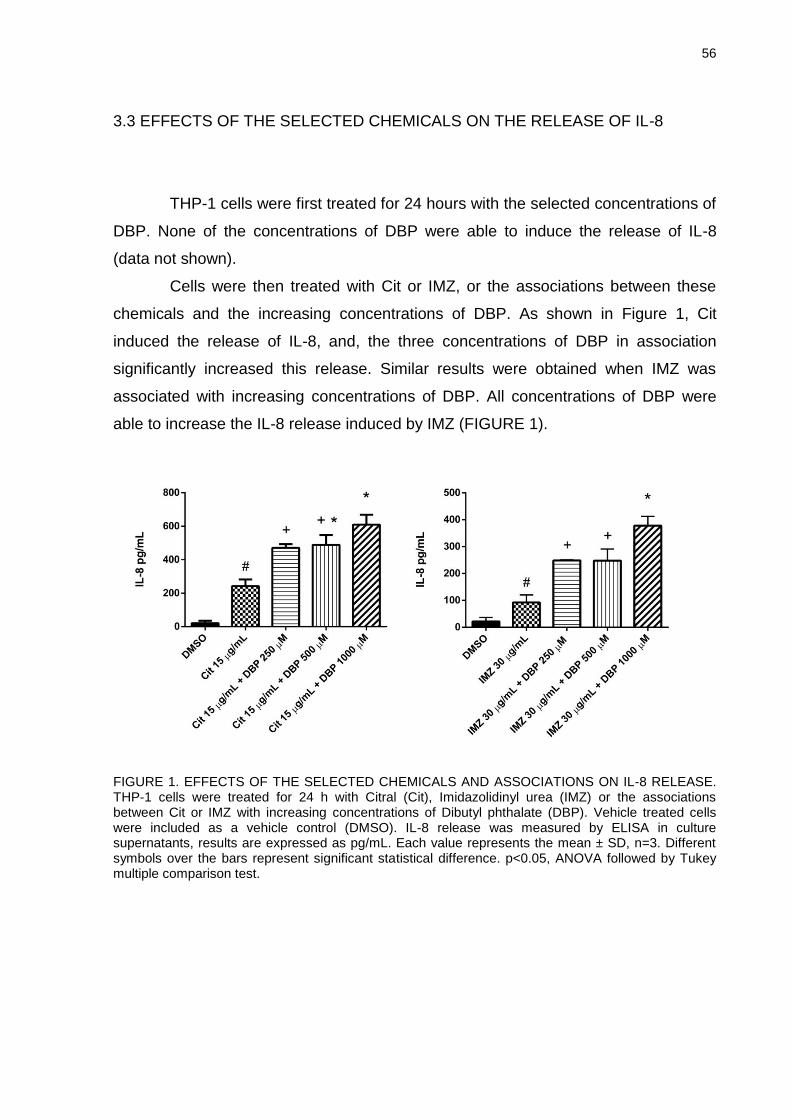
56
3.3 EFFECTS OF THE SELECTED CHEMICALS ON THE RELEASE OF IL-8
THP-1 cells were first treated for 24 hours with the selected concentrations of
DBP. None of the concentrations of DBP were able to induce the release of IL-8
(data not shown).
Cells were then treated with Cit or IMZ, or the associations between these
chemicals and the increasing concentrations of DBP. As shown in Figure 1, Cit
induced the release of IL-8, and, the three concentrations of DBP in association
significantly increased this release. Similar results were obtained when IMZ was
associated with increasing concentrations of DBP. All concentrations of DBP were
able to increase the IL-8 release induced by IMZ (FIGURE 1).
FIGURE 1. EFFECTS OF THE SELECTED CHEMICALS AND ASSOCIATIONS ON IL-8 RELEASE. THP-1 cells were treated for 24 h with Citral (Cit), Imidazolidinyl urea (IMZ) or the associations between Cit or IMZ with increasing concentrations of Dibutyl phthalate (DBP). Vehicle treated cells were included as a vehicle control (DMSO). IL-8 release was measured by ELISA in culture supernatants, results are expressed as pg/mL. Each value represents the mean ± SD, n=3. Different symbols over the bars represent significant statistical difference. p<0.05, ANOVA followed by Tukey multiple comparison test.
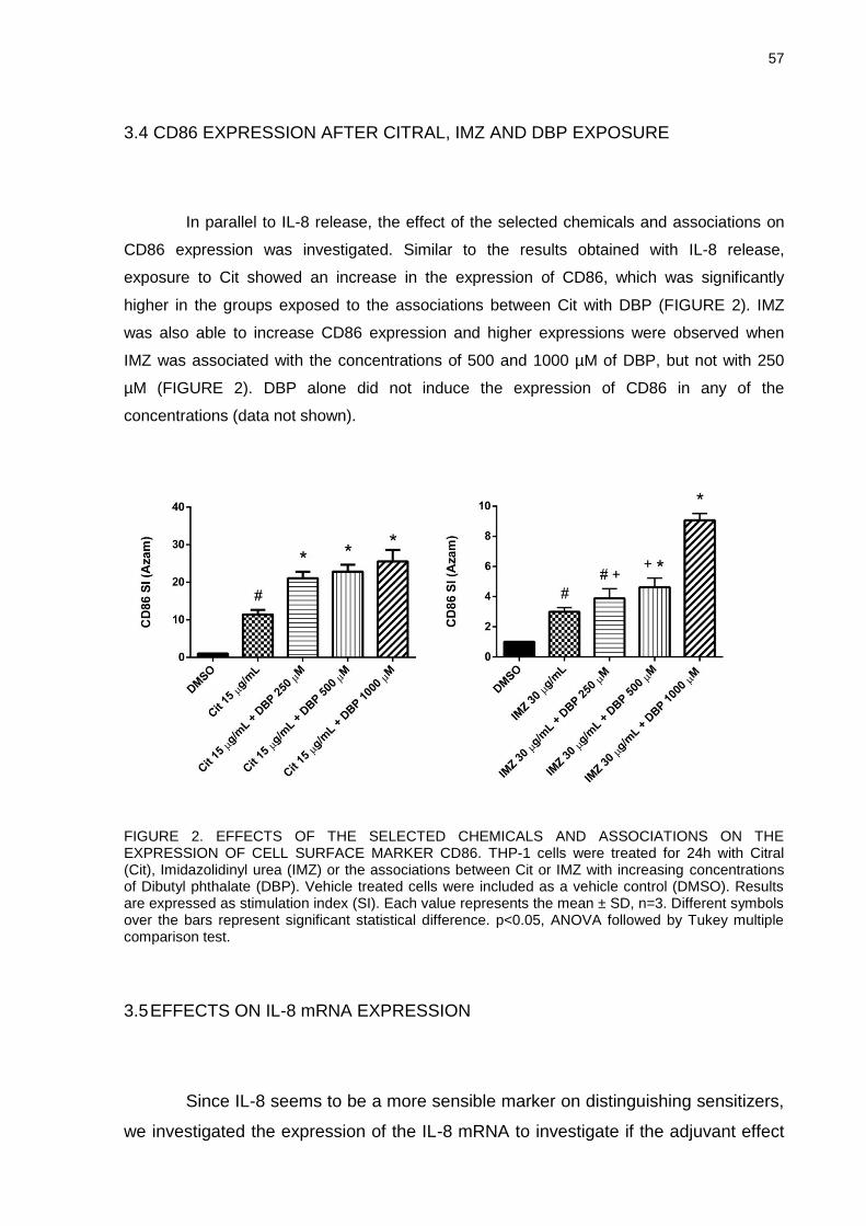
57
3.4 CD86 EXPRESSION AFTER CITRAL, IMZ AND DBP EXPOSURE
In parallel to IL-8 release, the effect of the selected chemicals and associations on
CD86 expression was investigated. Similar to the results obtained with IL-8 release,
exposure to Cit showed an increase in the expression of CD86, which was significantly
higher in the groups exposed to the associations between Cit with DBP (FIGURE 2). IMZ
was also able to increase CD86 expression and higher expressions were observed when
IMZ was associated with the concentrations of 500 and 1000 µM of DBP, but not with 250
µM (FIGURE 2). DBP alone did not induce the expression of CD86 in any of the
concentrations (data not shown).
FIGURE 2. EFFECTS OF THE SELECTED CHEMICALS AND ASSOCIATIONS ON THE EXPRESSION OF CELL SURFACE MARKER CD86. THP-1 cells were treated for 24h with Citral (Cit), Imidazolidinyl urea (IMZ) or the associations between Cit or IMZ with increasing concentrations of Dibutyl phthalate (DBP). Vehicle treated cells were included as a vehicle control (DMSO). Results are expressed as stimulation index (SI). Each value represents the mean ± SD, n=3. Different symbols over the bars represent significant statistical difference. p<0.05, ANOVA followed by Tukey multiple comparison test.
3.5 EFFECTS ON IL-8 mRNA EXPRESSION
Since IL-8 seems to be a more sensible marker on distinguishing sensitizers,
we investigated the expression of the IL-8 mRNA to investigate if the adjuvant effect
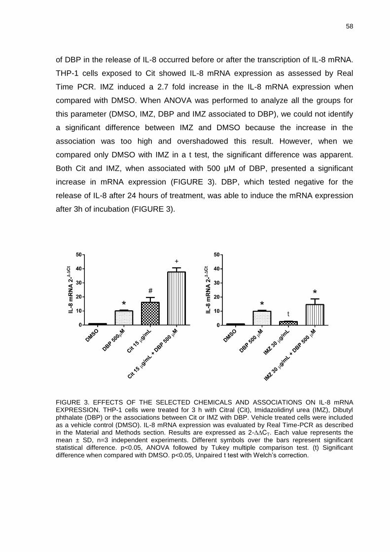
58
of DBP in the release of IL-8 occurred before or after the transcription of IL-8 mRNA.
THP-1 cells exposed to Cit showed IL-8 mRNA expression as assessed by Real
Time PCR. IMZ induced a 2.7 fold increase in the IL-8 mRNA expression when
compared with DMSO. When ANOVA was performed to analyze all the groups for
this parameter (DMSO, IMZ, DBP and IMZ associated to DBP), we could not identify
a significant difference between IMZ and DMSO because the increase in the
association was too high and overshadowed this result. However, when we
compared only DMSO with IMZ in a t test, the significant difference was apparent.
Both Cit and IMZ, when associated with 500 µM of DBP, presented a significant
increase in mRNA expression (FIGURE 3). DBP, which tested negative for the
release of IL-8 after 24 hours of treatment, was able to induce the mRNA expression
after 3h of incubation (FIGURE 3).
FIGURE 3. EFFECTS OF THE SELECTED CHEMICALS AND ASSOCIATIONS ON IL-8 mRNA EXPRESSION. THP-1 cells were treated for 3 h with Citral (Cit), Imidazolidinyl urea (IMZ), Dibutyl phthalate (DBP) or the associations between Cit or IMZ with DBP. Vehicle treated cells were included as a vehicle control (DMSO). IL-8 mRNA expression was evaluated by Real Time-PCR as described in the Material and Methods section. Results are expressed as 2-∆∆CT. Each value represents the mean ± SD, n=3 independent experiments. Different symbols over the bars represent significant statistical difference. p<0.05, ANOVA followed by Tukey multiple comparison test. (t) Significant difference when compared with DMSO. p<0.05, Unpaired t test with Welch’s correction.
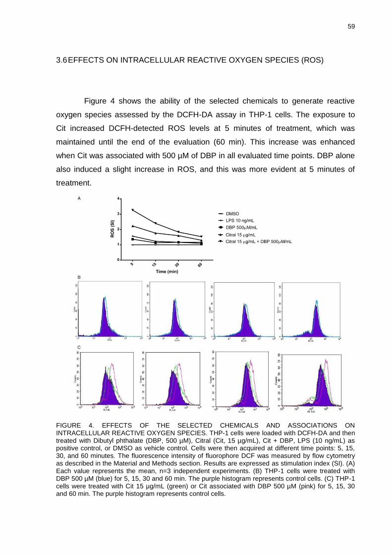
59
3.6 EFFECTS ON INTRACELLULAR REACTIVE OXYGEN SPECIES (ROS)
Figure 4 shows the ability of the selected chemicals to generate reactive
oxygen species assessed by the DCFH-DA assay in THP-1 cells. The exposure to
Cit increased DCFH-detected ROS levels at 5 minutes of treatment, which was
maintained until the end of the evaluation (60 min). This increase was enhanced
when Cit was associated with 500 µM of DBP in all evaluated time points. DBP alone
also induced a slight increase in ROS, and this was more evident at 5 minutes of
treatment.
FIGURE 4. EFFECTS OF THE SELECTED CHEMICALS AND ASSOCIATIONS ON INTRACELLULAR REACTIVE OXYGEN SPECIES. THP-1 cells were loaded with DCFH-DA and then treated with Dibutyl phthalate (DBP, 500 µM), Citral (Cit, 15 µg/mL), Cit + DBP, LPS (10 ng/mL) as positive control, or DMSO as vehicle control. Cells were then acquired at different time points: 5, 15, 30, and 60 minutes. The fluorescence intensity of fluorophore DCF was measured by flow cytometry as described in the Material and Methods section. Results are expressed as stimulation index (SI). (A) Each value represents the mean, n=3 independent experiments. (B) THP-1 cells were treated with DBP 500 µM (blue) for 5, 15, 30 and 60 min. The purple histogram represents control cells. (C) THP-1 cells were treated with Cit 15 µg/mL (green) or Cit associated with DBP 500 µM (pink) for 5, 15, 30 and 60 min. The purple histogram represents control cells.
60
4 DISCUSSION
It has been postulated that phthalate esters may be associated with
facilitation in the development of allergic diseases such as asthma and atopic
dermatitis (BORNEHAG et al., 2004; JAAKKOLA; KNIGHT, 2004). Additionally,
certain phthalates have been shown to enhance the response to allergens in animal
models of allergic contact dermatitis (IMAI et al., 2006; LI et al., 2014). The
underlying mechanisms, however, have not been fully elucidated. The present in vitro
study investigated the effects of the plasticizer DBP on chemical allergen-induced
cell activation. In particular, NCTC 2544 and IL-18 production were used as
surrogate of contact allergen-induced keratinocyte activation, while the upregulation
of CD86 and IL-8 production in THP-1 cells were used as markers of dendritic cell
(DC) activation. Results obtained suggest an adjuvant effect of DBP in DC but not in
epidermal keratinocytes, as demonstrated by the lack of co-stimulation in PPD-
induced IL-18 production.
Since humans are commonly exposed to phthalates, it is important to
elucidate potential immunotoxic effects of the same. With the widespread use of
DBP, its potential toxicity is getting increasing attention. It is known that phthalate
esters lack the ability to bind to proteins and form a hapten-protein complex, which is
required for the stimulation of allergic sensitization (KIMBER; DEARMAN, 2010).
According to epidemiological data, phthalate esters do not have the potential to
cause dermal irritation or skin sensitization (MEDEIROS; DEVLIN; KELLER, 1999;
GEIER et al., 2004). However, even though evidence supports a lack of sensitizing
potential, it has been suggested that certain phthalates may act as adjuvants by
enhancing immune responses.
The NCTC 2544 IL-18 assay is a useful in vitro tool that helps identify contact
sensitizers and discriminate them from irritants and respiratory allergens (CORSINI et
al., 2009; GALBIATI et al., 2011; CORSINI et al., 2013a). In this study, as expected,
the contact allergen PPD induced a statistically significant increase in the production
of IL-18, while DBP alone failed to induce it, supporting the evidence that phthalates
lack the potential to cause sensitization. The association with DBP failed, however, to
further increase IL-18 production, suggesting that DBP does not act as an adjuvant in
keratinocytes.
61
Mitjans et al. (2008) demonstrated that chemical allergens induce a
concentration-related release of interleukin-8 as well as the expression of the surface
marker CD86 in the human promyelocytic cell line THP-1, proposing this as an in
vitro assay for the screening of potential chemical allergens. In this work, the CD86
upregulation as well as IL-8 release were investigated after exposure of THP-1 cells
to two contact allergens, citral and imidazolidinyl urea, and their associations with
DBP. According to the local lymph node assay (LLNA), citral is classified as
moderate sensitizer with an EC3 value of 9.3%, while imidazolidinyl urea is classified
as a weak sensitizer with an EC3 value of 24%. Both allergens induced a statistically
significant upregulation in the expression of CD86. DBP alone did not induce this
expression, further supporting the lack of sensitizing potential. In accordance with our
data, Ito et al. (2012) showed that di (2-ethylhexyl) phthalate (DEHP) did not induce
the expression of dendritic cells differentiation and maturation markers, including
CD86, in mouse peripheral blood mononuclear‐derived dendritic cells. On the
contrary, Koike et al. (2009; 2010) using bone marrow‐derived dendritic cells
obtained from atopic prone NC/Nga mice, found that DEHP as well as diisononyl
phthalate (DINP) enhanced the expression of CD86 and other costimulatory
molecules. Different culture conditions, cells or experimental design could explain
these differences.
The exposure of THP-1 cells to Cit and IMZ revealed a statistically significant
release of IL-8 compared to vehicle treated cells. Again, DBP alone failed to induce
the release of IL-8. Data on phthalate-induced release of cytokines in vitro is
conflicting. Although it has been reported that some phthalate monoesters, including
the DBP metabolite monobutyl phthalate (MPB), do not induce the release of
cytokines after exposure in THP-1 cells (GLUE et al., 2002), others have shown that
phthalate monoesters lead to the release of IL-8 in the epithelial cell line A549
(JEPSEN; ABILDTRUP; LARSEN, 2004). In the study by Jepsen et al. (2004), high
concentrations of MBP induced only a slight increase in IL-8 release, and more
significant increases were obtained with the exposure to monoesters with more than
eight carbon atoms in their alkyl side chain. According to their results, these authors
suggest that phthalate monoesters with fewer than eight carbon atoms in their alkyl
side chain (like MBP) are weak cytokine inducers, whereas monoesters with eight or
more carbon atoms in the alkyl side chain are more potent cytokine inducers.
62
When THP-1 cells were exposed to either Cit or IMZ associated with DBP,
cells exhibited higher expression of CD86 and release of IL-8, when compared to the
exposure to the allergen alone. This indicates an adjuvant effect of DBP on THP-1
cells. Even though several studies have been performed to evaluate the effects of
phthalates in cell function, little can be found in the literature regarding the adjuvant
effect of these substances in vitro. Nevertheless, a great amount of studies have
been performed about the adjuvant effect of phthalates in vivo. Several authors have
showed that different phthalates can act as adjuvants in contact hypersensitivity and
airway inflammation, elevating the expression of surface markers and related
cytokines after exposure to associations between allergens and phthalates by
different protocols (IMAI et al., 2006; KOIKE et al., 2010; MATSUDA et al., 2010; LI
et al., 2014). Corroborating our results, Tanaka et al. (2012) demonstrated that
DEHP exacerbated ovalbumin-related peritoneal inflammation characterized by
enhanced infiltration of macrophages and neutrophils and amplified
production/release of cytokines such as keratinocyte-derived chemoattractant (KC) in
the peritoneal cavity. KC is a murine homolog of IL-8 (HOL; WILHELMSEN;
HARALDSEN, 2010). Furthermore, Takano et al. (2006) showed that exposure to
DEHP in the presence of allergen increased the expression of MIP-1α compared to
vehicle combined with allergen only. MIP-1α functions as chemotactic for neutrophils
similarly to IL-8 (HAMMOND et al., 1995; MENTEN; WUYTS; VAN DAMME, 2002).
According to Galbiati et al. (2012) the expression of IL-8 mRNA at 3 hours is
important since some chemical allergens fail to induce the release of IL-8 after 24
hours of exposure, but all chemical sensitizers tested induced IL-8 mRNA at 3 hours.
Both Cit and IMZ induced the expression of IL-8 mRNA, and this expression was
significantly higher when associated with 500 µM of DBP. As IMZ is a week
sensitizer, its induction of the expression of IL-8 mRNA was lower compared to the
induction following Cit exposure. IMZ induced a 2.7 fold increase in IL-8 mRNA, still
significantly different from the control group (Welch's t test). Interestingly, DBP, which
failed to induce the expression of the surface marker CD86 or the release of IL-8,
induced a significant expression of IL-8 mRNA. As mentioned before, data about the
release of pro-inflammatory cytokines, including IL-8, induced by phthalates is
conflicting. While some authors found that certain monophthalates can induce this
release at high concentrations, others report no release after monophthalates
exposure (GLUE et al., 2002; JEPSEN et al., 2004). Overall, little can be found in
63
literature relating DBP exposure and increase in pro-inflammatory cytokines mRNA
expression. The control of translation and mRNA degradation is an important part of
the regulation of gene expression. MicroRNAs (miRNAs) are a class of small RNA
molecules that modulate gene expression by interacting with the messenger RNA of
the respective target gene (SCHNEIDER, 2012). miRNAs can silence mRNAs either
by triggering endonuclease cleavage, by promoting translation repression, or by
accelerating mRNA decapping (VALENCIA-SANCHEZ et al., 2006). It’s been shown
that certain miRNAs are upregulated in allergic contact dermatitis (VENNEGAARD et
al., 2012) and other skin inflammatory diseases (SONKOLY et al., 2010; ZIBERT et
al., 2010), although still scarce information can be found about the function of
miRNAs in skin sensitization. Since the silencing of mRNAs by miRNAs occurs
through a very specific interaction between the miRNA and its targeted mRNA
(VALENCIA-SANCHEZ et al., 2006), this could be a possible explanation to why only
the IL-8 mRNA upregulated by DBP was not translated into protein. However, this is
something that needs further investigation.
Although previous in vivo studies have shown that certain phthalates
aggravate allergic response and/or diseases (even though in doses considerably
higher than those relevant for human exposure), the underlying cellular and
molecular mechanisms remain unclear. Evidence indicates that allergic and
inflammatory skin diseases could be mediated by oxidative stress (OKAYAMA, 2005;
BYAMBA et al., 2010). Mizuashi et al. (2005) hypothesized that sensitizers induce
oxidative stress in dendritic cells, which subsequently stimulates p38 MAPK. Their
results demonstrated that sensitizers reduced the oxidized (GSSG) versus reduced
(GSH) form of cellular glutathione (GSH/GSSG ratio) in THP-1 cells and
phosphorylated p38 MAPK. According to Mitjans et al. (2008), the activation of the
p38 MAPK by sensitizers plays a crucial role in the activation of IL-8 release. In this
study, together with increase in IL-8 release, we showed a slight increase of ROS
generation induced by DBP alone, but, more importantly, a significant increase in the
oxidative stress induced by DBP when in association with Cit. Others have
demonstrated that some phthalates can induce oxidative stress. Tetz et al. (2013)
reported an increase in ROS production induced by MEHP in human placental cells.
DEHP also increased the generation of ROS in testis inducing apoptosis of
spermatocytes (KASAHARA et al., 2002). Moreover, You et al. (2014) showed
significantly higher levels of ROS in ovalbumin sensitized mice that were co-treated
64
with DEHP when compared to the levels of ROS of the sensitized-only group. These
authors suggest that oxidative stress plays a critical role in the adjuvant effect of
DEHP.
In the present study we demonstrated a positive adjuvant effect of DBP when
associated with citral and imidazolidinyl urea as indicated by the increased
expression of CD86, IL-8 mRNA and IL-8 release. We also suggest that the
increased generation of ROS could be the trigger that leads to these effects. On the
contrary, we found no adjuvant effect on keratinocytes. Taken together, our results
support that the adjuvant effect of DBP relies on dendritic cells. Previously published
data corroborate our results. Koike et al. (2009) and Ito et al. (2012), based on their
in vivo and in vitro studies, suggest that DEHP accelerates murine dermatitis through
the inappropriate activation of dendritic cells. Koike et al. (2010) proposed that DINP
may activate dendritic cells in inflammatory sites through the enhancement of thymic
stromal lymphopoietin (TSLP) production. The production of TSLP has also been
shown to be induced by DBP (LARSON et al., 2010). Imai et al. (2006) suggested
that DBP and DPP facilitate the trafficking of Langerhans cells and/or myeloid
dendritic cells from skin sites in contact hypersensitivity induced by fluorescein
isothiocyanate.
In an immunological context an adjuvant is a material that is able to enhance
immune responsiveness without itself necessarily providing any specific antigenic
stimulus (KIMBER; DEARMAN, 2010). From the results on CD86 expression and IL-
8 release, where DBP alone had no effects, an enhanced upregulation of both
markers was observed when DBP was associated to Cit or IMZ, suggesting that DBP
acts as an adjuvant by immunological definition. Since evidence that phthalates can
act as sensitizers themselves is scarce and as other authors have suggested in
different animal models of dermatitis that phthalates act as adjuvants, we believe that
the effects found in our work are consistent with an adjuvant effect. Interestingly, this
effect seems to be specific for immune cells as no additive effects were observed in
chemical allergen-induced IL-18 production in keratinocytes in the presence of DBP.
In conclusion, we propose that DBP presents in vitro an adjuvant effect when
associated with sensitizers. However, the effects of DBP in vivo remain incompletely
understood, thus, future studies are necessary to elucidate the precise mechanism of
adjuvant effect of DBP not only in vitro but also in vivo.
65
REFERENCES
ANDERSON, W.A.C. et al. A biomarker approach to measuring human dietary exposure to certain phthalate diesters. Food Addit. Contam., v. 18, n.12, p. 1068-1074, 2001. AZAM, P. et al. The cytokine-dependent MUTZ-3 cell line as an in vitro model for the screening of contact sensitizers. Toxicol. Appl. Pharmacol., v. 212, p. 14-23, 2006. BANCHEREAU, J. et al. Immunobiology of dendritic cells. Annu. Rev. Immunol., v. 18, p. 767-811, 2000. BARKER, J.N. et al. Modulation of keratinocyte-derived interleukin-8 which is chemotactic for neutrophils and T lymphocytes. Am. J. Pathol., v. 139, p. 869-876, 1991. BORNEHAG, C.G. et al. The association between asthma and allergic symptoms in children and phthalates in house dust: a nested case-control study. Environ. Health Persp., v. 112, p. 1393-1397, 2004. BYAMBA, D. et al. The roles of reactive oxygen species produced by contact allergens and irritants in monocyte-derived dendritic cells. Ann. Dermatol., v. 22, p. 269-278, 2010. CORSINI, E. et al. NCTC 2544 and IL-18 production: a tool for the identification of contact allergens. Toxicol. In Vitro, v. 27, p. 1127-1134, 2013a. CORSINI, E. et al. Role of oxidative stress in chemical allergen induced skin cells activation. Food Chem. Toxicol., v. 61, p. 74-81, 2013b. CORSINI, E. et al. Use of IL-18 production in a human keratinocyte cell line to discriminate contact sensitizers from irritants and low molecular weight respiratory allergens. Toxicol. In Vitro, v. 23, p. 789-796, 2009. CUMBERBATCH, M. et al. Interleukin-18 induces Langerhans cell migration by a tumor necrosis factor-a and IL1b-dependent mechanism. Immunology, v. 102, n. 3, p. 323-330, 2001. ELSISI, A.; CARTER, D.E.; SIPES, I.G. Dermal absorption of phthalate diesters in rats. Fundam. Appl. Toxicol., v. 12, p. 70-77, 1989. FERGUSON, K.K.; LOCH-CARUSO, R.; MEEKER, J.D. Urinary phthalate metabolites in relation to biomarkers of inflammation and oxidative stress: NHANES 1999-2006. Environ. Res., v. 111, n. 5, p. 718-726, 2011. FISHER, J.S. et al. Human 'testicular dysgenesis syndrome': a possible model using in-utero exposure of the rat to dibutyl phthalate. Hum. Reprod., v. 18, p. 1383-1394, 2003.
66
GALBIATI, V. et al. Isoeugenol destabilizes IL-8 mRNA expression in THP-1 cells through induction of the negative regulator of mRNA stability tristetraprolin. Arch. Toxicol., v. 86, p. 239-248, 2012. GALBIATI, V. et al. Further development of the NCTC 2544 IL-18 assay to identify in vitro contact allergens. Toxicol. In Vitro, v. 25, p. 724-732, 2011. GEIER, J. et al. Patch test results with metalworking fluid series of the German Contact Dermatitis Research Group (DKG). Contact Dermatitis, v. 51, p. 118-130, 2004. GLUE, C. et al. In vitro effects of monophthalates on cytokine expression in themonocytic cell line THP-1 and in peripheral blood mononuclear cells from allergic and non-allergic donors. Toxicol. In Vitro, v. 16, p. 657-662, 2002. HAMMOND, M.E. et al. IL-8 induces neutrophil chemotaxis predominantly via type 1 IL-8 receptors. J. Immunol., v. 155, n. 3, p. 1428-1433, 1995. HAUSER, R. et al. DNA damage in human sperm is related to urinary levels of phthalate monoester and oxidative metabolites. Hum. Reprod., v. 22, n. 3, p. 688-695, 2007. HEUDORF, U.; MERSCH-SUNDERMANN, V.; ANGERER, J. Phthalates: Toxicology and exposure. Int. J. Hyg. Envir. Heal., v. 210, p. 623-634, 2007. HOL, J.; WILHELMSEN L.; HARALDSEN, G. The murine IL-8 homologues KC, MIP-2, and LIX are found in endothelial cytoplasmic granules but not in Weibel-Palade bodies. J. Leukocyte Biol., v. 87, n. 0741, p. 501-508, 2010. IMAI, Y. et al. Effects of phthalate esters on the sensitization phase of contact hypersensitivity induced by fluorescein isothiocyanate. Clin. Exp. Allergy, v. 36, p. 1462-1468, 2006. INTERNATIONAL JOURNAL OF TOXICOLOGY. Annual Review of Cosmetic Ingredient Safety Assessment- 2002/2003. Int. J. Toxicol., v. 24, n. 1, p. 1-102, 2005. ITO, T. et al. Phthalate esters modulate the differentiation and maturation of mouse peripheral blood mononuclear cell-derived dendritic cells. J. Appl. Toxicol., v. 32, n. 2, p. 142-148, 2012. JAAKKOLA, J.J.K.; KNIGHT, T.L. Asthma, wheezing, and allergies in Russian schoolchildren in relation to new surface materials in the home. Am. J. Public Health, v. 94, p. 560-562, 2004. JEONG, J.Y. et al. Determination of Phthalate Metabolites in Human Serum and Urine as Biomarkers for Phthalate Exposure Using Column-Switching LC-MS/MS. Safety and Health at Work, v. 2, n. 1, p. 57-64, 2011.
67
JEPSEN, K.F.; ABILDTRUP, A.; LARSEN, S.T. Monophthalates promote IL-6 and IL-8 production in the human epithelial cell line A549. Toxicol. In Vitro, v. 18, p. 265-269, 2004. KASAHARA, E. et al. Role of oxidative stress in germ cell apoptosis induced by di(2-ethylhexyl) phthalate. Biochem. J., v. 365, p. 849-856, 2002. KIMBER, I.; DEARMAN, R.J. An assessment of the ability of phthalates to influence immune and allergic responses. Toxicology, v. 271, p. 73-82, 2010. KOIKE, E. et al. Di-(2- ethylhexyl) phthalate affects immune cells from atopic prone mice in vitro. Toxicology, v. 259, p. 54-60, 2009. KOIKE, E. et al. Effects of diisononyl phthalate on atopic dermatitis in vivo and immunologic responses in vitro. Environ. Health Persp., v. 118, n. 4, p. 472-478, 2010. KOO, H.J.; LEE, B.M. Estimated exposure to phthalates in cosmetics and risk assessment. J. Toxicol. Env. Health A: Current Issues, v. 67, n. 23-24, p. 1901-1914, 2004. LARSON, R.P. et al. Dibutyl phthalate-induced thymic stromal lymphopoietin is required for Th2 contact hypersensitivity responses. J. Immunol., v. 184, p. 2974-2984, 2010. LEPOITTEVIN, J.P.; LEBLOND, I. Hapten-peptide T cell receptor interactions: molecular basis for the recognition of haptens by T lymphocytes. Eur. J. Dermatol., v. 7, p. 151-154, 1997. LI, J. et al. T-Helper Type-2 Contact Hypersensitivity of BALB/c Mice Aggravated by Dibutyl Phthalate via Long-Term Dermal Exposure. PLoS ONE, v. 9, n. 2, p. e87887, 2014. LYCHE, J.L. et al. Reproductive and Developmental Toxicity of Phthalates. J. Toxicol. Env. Health B: Critical Reviews, v. 12, n. 4, p. 225-249, 2009. MARTINASSO, G. et al. Effects of di(2-ethylhexyl) phthalate, a widely used peroxisome proliferator and plasticizer, on cell growth in the human keratinocyte cell line NCTC 2544. J. Toxicol. Env. Health A, v. 69, p. 353-365, 2006. MARTINO-ANDRADE, A.J. et al. Coadministration of active phthalates results in disruption of foetal testicular function in rats. Int. J. Androl., v. 32, n. 6, p. 704-712, 2009. MATSUDA, T. et al. Phthalate esters reveal skin-sensitizing activity of phenethyl isothiocyanate in mice. Food Chem. Toxicol., v. 48, p. 1704-1708, 2010. MEDEIROS, A.M.; DEVLIN, D.J; KELLER, L.H. Evaluation of skin sensitization response of dialkyl (C6–C13) phthalate esters. Contact Dermatitis, v. 41, n. 5, p. 287-289, 1999.
68
MENTEN, P.; WUYTS, A.; VAN DAMME, J. Macrophage inflammatory protein-1. Cytokine Growth F R, v. 13, p. 455-481, 2002. MITJANS, M. et al. Role of p38 MAPK in the selective release of IL-8 induced by chemical allergen in naive THP-1 cells. Toxicol. In Vitro, v. 22, p. 386-395, 2008. MIZUASHI, M. et al. Redox imbalance induced by contact sensitizers triggers the maturation of dendritic cells. J. Invest. Dermatol., v. 124, p. 579-586, 2005. OKAYAMA, Y. Oxidative stress in allergic and inflammatory skin diseases. Curr. Drug Targets Inflamm. Allergy, v. 4, n. 4, p. 517-519, 2005. SAINT-MEZARD, P. et al. Allergic contact dermatitis. Eur. J. Dermatol., v. 14, n. 5, p. 284-295, 2004. SCHNEIDER, M.R. MicroRNAs as novel players in skin development, homeostasis and disease. Brit. J. Dermatol., v. 166, n. 1, 22-28, 2012. SEO, K.W. et al. Comparison of oxidative stress and changes of xenobiotic metabolizing enzymes induced by phthalates in rats. Food Chem. Toxicol., v. 42, p. 107-114, 2004. SILVA, M.J. et al. Analysis of human urine for fifteen phthalate metabolites using automated solid-phase extraction. J. Chromatogr. B, v. 805, p. 161-167, 2004. SONKOLY, E. et al. MiR-155 is overexpressed in patients with atopic dermatitis and modulates T-cell proliferative responses by targeting cytotoxic T lymphocyte-associated antigen 4. J. Allergy Clin. Immunol., v. 126, n. 3, p. 581-589.e 1-20, 2010. TAKANO, H. et al. Di-(2-ethylhexyl) phthalate enhances atopic dermatitis-like skin lesions in mice. Environ. Health Persp., v. 114, n. 8, p. 1266-1269, 2006. TANAKA, M. et al. Immunoamplifying Effects of Di-(2-Ethylhexyl) Phthalate on Cytokine Response. Immunopharm. Immunot., v. 35, p. 1-4, 2012. TETZ, L.M. et al. Mono-2-ethylhexyl phthalate induces oxidative stress responses in human placental cells in vitro. Toxicol. Appl. Pharmacol., v. 268, n. 1, p. 47-54, 2013. VALENCIA-SANCHEZ, M.A. et al. Control of translation and mRNA degradation by miRNAs and siRNAs. Genes Dev., v. 20, n. 5, p. 515-524, 2006. VANDEBRIEL, R.J.; VAN OCH, F.M.; VAN LOVEREN, H.In vitro assessment of sensitizing activity of low molecular weight compounds. Toxicol. Appl. Pharmacol., v. 207, n. 2, p. 142-148, 2005. VARTAK, P.H.; TUNGIKAR, V.B.; SHARMA, R.N. Comparative repellent properties of certain chemicals against mosquitoes, house flies and cockroaches using modified techniques. J. Commun. Dis., v. 26, n. 3, p. 156-160, 1994.

69
VENNEGAARD, M.T. et al. Allergic contact dermatitis induces upregulation of identical microRNAs in humans and mice. Contact Dermatitis, v. 67, n. 5, p. 298-305, 2012. YOU, H. et al. The adjuvant effect induced by di-(2-ethylhexyl) phthalate (DEHP) is mediated through oxidative stress in a mouse model of asthma. Food Chem. Toxicol., v. 71, p. 272-281, 2014. ZIBERT, J.R. et al. MicroRNAs and potential target interactions in psoriasis. J. Dermatol. Sci., v. 58, p. 177–185, 2010.
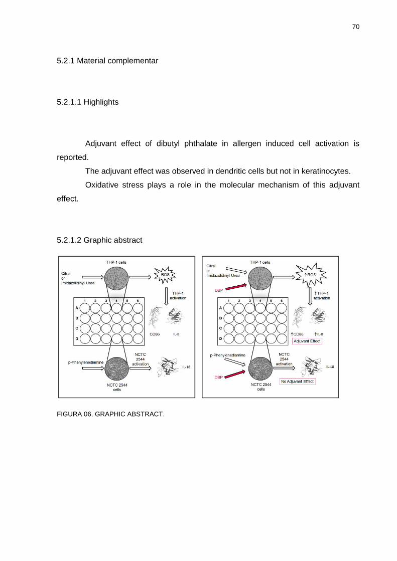
70
5.2.1 Material complementar
5.2.1.1 Highlights
Adjuvant effect of dibutyl phthalate in allergen induced cell activation is
reported.
The adjuvant effect was observed in dendritic cells but not in keratinocytes.
Oxidative stress plays a role in the molecular mechanism of this adjuvant
effect.
5.2.1.2 Graphic abstract
FIGURA 06. GRAPHIC ABSTRACT.
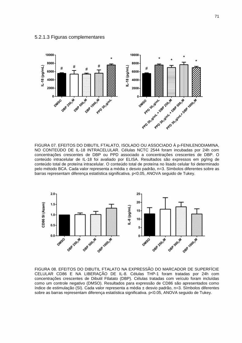
71
5.2.1.3 Figuras complementares
FIGURA 07. EFEITOS DO DIBUTIL FTALATO, ISOLADO OU ASSOCIADO À p-FENILENODIAMINA, NO CONTEÚDO DE IL-18 INTRACELULAR. Células NCTC 2544 foram incubadas por 24h com concentrações crescentes de DBP ou PPD associado a concentrações crescentes de DBP. O conteúdo intracelular de IL-18 foi avaliado por ELISA. Resultados são expressos em pg/mg de conteúdo total de proteína intracelular. O conteúdo total de proteína no lisado celular foi determinado pelo método BCA. Cada valor representa a média ± desvio padrão, n=3. Símbolos diferentes sobre as barras representam diferença estatística significativa. p<0.05, ANOVA seguido de Tukey.
FIGURA 08. EFEITOS DO DIBUTIL FTALATO NA EXPRESSÃO DO MARCADOR DE SUPERFÍCIE CELULAR CD86 E NA LIBERAÇÃO DE IL-8. Células THP-1 foram tratadas por 24h com concentrações crescentes de Dibutil Ftlatato (DBP). Células tratadas com veículo foram incluídas como um controle negativo (DMSO). Resultados para expressão de CD86 são apresentados como índice de estimulação (SI). Cada valor representa a média ± desvio padrão, n=3. Símbolos diferentes sobre as barras representam diferença estatística significativa. p<0.05, ANOVA seguido de Tukey.
72
5.3 ARTIGO 2
The plasticizer dibutyl phthalate aggravates contact hypersensitivity induced
by oxazolone in BALB/c mice.
Ana Carolina dos Santos Lourençoa, Renata Mercer Zaiaa, Arthur da Silveira
Prudenteb, Francislaine Aparecida dos Reis Líveroa, Valentina Galbiatic, Alexandra
Accoa, Emanuela Corsinic, Michel Fleith Otukia, Anderson Joel Martino Andraded
aDepartment of Pharmacology, Universidade Federal do Paraná (UFPR), Curitiba, PR, Brazil
bDepartment of Pharmacology, Universidade Federal de Santa Catarina (UFSC), Florianópolis, SC,
Brazil
cDepartment of Pharmacological and Biomolecular Sciences, Università degli Studi di Milano, Milan,
Italy
dDepartment of Physiology, Universidade Federal do Paraná (UFPR), Curitiba, PR, Brazil
Corresponding author:
Ana Carolina dos Santos Lourenço
Laboratório de Toxicologia, Departmento de Farmacologia, Universidade Federal do
Paraná
Rua Cel. Francisco H. dos Santos, 100 - Centro Politécnico, Caixa Postal 19031 -
CEP 81531-980 - Curitiba, PR, Brasil
e-mail: [email protected]
Tel.: +5515996848448
73
ABSTRACT
Phthalate esters have been reported to participate in the development of allergic diseases acting as adjuvants. Contact hypersensitivity (CHS) is an allergic reaction resulting from repeated contact with chemicals called haptens, consisted in two phases: sensitization and elicitation. The present study aimed to investigate the effects of dibutyl phthalate (DBP) in the elicitation phase of CHS induced by oxazolone in an animal model of ear edema. For the experiments, adult male BALB/c mice were sensitized (in shaved abdomen) and challenged (elicitation; in the right ear) with oxazolone or vehicle (acetone) topically, and subsequently treated 6 times (with 12 hour intervals) with DBP at three doses (0.04, 0.4 or 4 mg/ear) or vehicle (acetone) in the same ear (topically). Ear thickness was measured before elicitation (baseline) and 24, 48 and 72 hours after elicitation to calculate ear edema. After the last measure, animals were euthanized and 6 mm circles of ear tissue were collected to evaluate the activity of the enzymes N-acetyl-β-d-glucosaminidase (NAG) and myeloperoxidase (MPO) (markers of leukocyte infiltration), histological analysis (infiltrated cell count, dermal and epidermal thickness) and lipid hydroperoxide (LOOH) and reduced glutathione (GSH) levels. We also investigated the adjuvant effect of DBP in oxazolone-induced THP-1 activation (an in vitro assay used as surrogate for dendritic cell activation). For that, THP-1 cells were exposed to oxazolone alone or associated with DBP and the expression of cluster of differentiation 86 (CD86) and interleukin 8 (IL-8) mRNA were evaluated. As expected, animals sensitized and challenged with oxazolone presented increase in ear edema, dermal and epidermal thickness, number of infiltrated cells and MPO and NAG activity, when compared with the negative control group. DBP 0.4 and 4 mg/ear increased all the parameters induced by oxazolone, suggesting an enhancement of the inflammatory response. Oxazolone also increased the levels of LOOH and reduced GSH, and these effects were partially or completely reversed by DBP 0.4 and 0.04 mg/ear, respectively. These results indicate that DBP aggravates the contact hypersensitivity induced by oxazolone. DBP also enhanced the expression of CD86 induced by oxazolone in THP-1 cells, suggesting that the enhanced activation of dendritic cells could be part of the mechanism of DBP’s adjuvant effect. Our results indicate that DBP can act as adjuvant in the elicitation phase of oxazolone-induced CHS, and support data that suggest an adjuvant effect of phthalate esters in allergic diseases in certain doses. Key words: dibutyl phthalate, oxazolone, contact hypersensitivity, allergic contact dermatitis, adjuvant effect.
74
1 INTRODUCTION
Phthalate esters are plasticizers highly used in a variety of manufactured
products, and, due to their wide distribution in the environment, they are considered
agents with high potential for human exposure (KOO; LEE, 2004; ANDERSON et al.,
2001; SILVA et al., 2004; LYCHE et al., 2009). Dibutyl phthalate (DBP) can be found
in several products, such as plastics for food packing, glues, paints, solvents,
perfumes, moisturizers, hairsprays, nail polishes, insect repellents and children’s
products (IPCS, 1997; SATHYANARAYANA et al., 2008).
DBP, as well as other phthalates, have attracted great attention of the
scientific community due to the great possibility of human exposure, as well as their
possible endocrine disrupting effects that might lead to alterations in the development
of male reproductive tract in utero (FISHER et al., 2003; HEUDORF; MERSCH-
SUNDERMANN; ANGERER, 2007; MARTINO-ANDRADE et al., 2009). However,
besides the effects on endocrine and reproductive systems, epidemiological and
experimental data indicate other possible adverse effects due to the exposure to
these substances. It is believed that certain phthalates can exacerbate the potency of
allergens, and, so, play a role in the development and/or aggravation of allergic
diseases (CASILLAS et al., 1999). Experimental studies show that phthalate esters,
such as DBP, are well absorbed by the skin, but irritation and sensitization tests
indicate no evidence of skin inflammatory responses by these substances (ELSISI;
CARTER; SIPES, 1989; DOAN; BRONAUGH; YOURICK, 2010; BEYDON; PAYAN;
GRANDCLAUDE, 2010; CPSC, 2010; PAN et al., 2014). Therefore, in some
countries, like in Brazil, these substances are still allowed in cosmetic formulations.
However, most available dermal studies do not include the possible adjuvant effect of
phthalates in hypersensitivity models. Even though phthalate esters are not capable
of inducing hypersensitivity themselves, it has been reported that they are capable of
exacerbating the inflammatory response in contact hypersensitivity in animal models
(CHOWDHURY; STATHAM, 2002; IMAI et al., 2006; MATSUDA et al., 2010; LI et al.,
2014). These studies indicate that the dermal toxicity of phthalate esters needs better
evaluation and attention.
Allergic contact dermatitis (ACD) or contact hypersensitivity (CHS) is a
clinical manifestation in response to repeated or prolonged contact to a chemical
75
substance called hapten (HENNINO et al., 2005; KARLBERG et al., 2008), and it is
considered one of the most important manifestations of immunotoxicity in humans
(KIMBER, 2002). Allergic contact dermatitis is a delayed type hypersensitivity
reaction mediated by antigen specific T lymphocytes (KARLBERG et al., 2008), and
can be characterized in two phases: sensitization (first contact with the hapten,
antigen presenting to the lymph node and activation of specific T lymphocytes) and
elicitation (subsequent contacts with hapten, recognition by specific T lymphocytes
and development of inflammatory response) (GRABBE; SCHWARZ, 1998; KIMBER,
2002; HENNINO et al., 2005; KARLBERG et al., 2008).
Several cell types are involved in the contact hypersensitivity response.
Dendritic cells are recognized as important antigen presenting cells in adaptive
immunity because of their capacity to stimulate naive lymphocytes (BANCHEREAU
et al., 2000). Mitjans et al. (2008) reported in the human promyelocytic cell line THP-
1 (used as a surrogate of dendritic cells), a selective production of the chemokine
interleukin 8 (IL-8) by contact allergens in parallel to the surface marker cluster of
differentiation 86 (CD86) upregulation, and, proposed this as a possible in vitro
sensitization test. IL-8 is a potent attractant for neutrophils and different subtypes of T
cells (BARKER et al., 1991). We have previously demonstrated that DBP can
potentiate citral and imidazolidinyl urea-induced THP-1 activation, suggesting that
part of the mechanism of DBP’s adjuvant effect could be through enhanced activation
of dendritic cells (LOURENÇO et al., 2015).
It has been reported that oxidative stress can play a central role in chemical-
induced contact hypersensitivity and inflammatory diseases (OKAYAMA, 2005;
BYAMBA et al., 2010; CORSINI et al., 2013). Besides, evidence that phthalates are
associated to oxidative stress has been shown by several authors. Seo et al. (2004)
and Tetz et al. (2013) have demonstrated increased generation of reactive oxygen
species (ROS) induced by phthalates in rats and in a human placental cell line,
respectively, and others have shown associations between urinary concentrations of
phthalate monoester metabolites and increased markers of inflammation and
oxidative stress in humans (HAUSER et al., 2007; FERGUSON; LOCH-CARUSO;
MEEKER, 2011).
Prior animal studies on hypersensitivity contact have used high phthalate
doses and failed to comprehensively characterize the inflammatory process involved
in the adjuvant effects of phthalates (TAKANO et al., 2006; MATSUDA et al., 2010).

76
With this study, we aimed to investigate the possible adjuvant effects of lower DBP
doses (KOO; LEE, 2004; INTERNATIONAL JOURNAL OF TOXICOLOGY, 2005) in
the elicitation phase of contact hypersensitivity induced by oxazolone in an animal
model of ear edema. We also investigated the possible role of oxidative stress in this
adjuvant effect, besides evaluating the effects of DBP in oxazolone-induced THP-1
activation.
77
2 MATERIAL AND METHODS
2.1 ANIMALS
Adult male BALB/c mice obtained from Universidade de Campinas (Unicamp
– Campinas, SP, Brazil) were used for the experiments. The animals were kept in the
animal facilities of the Reproductive Toxicology Laboratory (Departamento de
Farmacologia, Universidade Federal do Paraná, Curitiba, PR, Brazil) under a 12 h
light/dark cycle and controlled temperature (22 ± 2°C). Standard pellet food and tap
water were provided ad libitum. The experimental protocol was approved by the
Committee on Animal Research and Ethics of the Universidade Federal do Paraná
(Curitiba, Brazil) under the number 633.
2.2 CHEMICALS AND DOSE SELECTION
Dibutyl phthalate (DBP, CAS number 84-74-2, purity 99%) and Oxazolone (4-
Ethoxymethylene-2-phenyl-2-oxazolin-5-one, CAS number 15646-46-5) were
obtained from Sigma-Aldrich (St. Louis, MO, USA). DBP and oxazolone were diluted
in acetone (vehicle) for in vivo experiments, and in DMSO for in vitro experiments
(final concentration of DMSO in culture medium < 0.2%).
DBP in vivo doses were based on DBP exposure estimations from use of
personal care products, in particular perfumes. According to Koo and Lee (2004), the
average concentration of DBP found in a variety of perfumes is 0.44 mg/mL, and the
average use of perfume by the general population, estimated through an applied
questionnaire, is 1mL/day. Therefore, the amount of DBP applied to the skin in
individuals that use determined kinds of perfumes can be approximately 0.4 mg/day,
which corresponds to 0.0067 mg/kg/day for a 60 kg individual or 0.25 mg/m2/day
using the conversion factor recommended by the FDA (FDA, 2005). These
estimations are in agreement with the data published by the International Journal of
Toxicology (2005) in an annual review on safety of ingredients from cosmetic
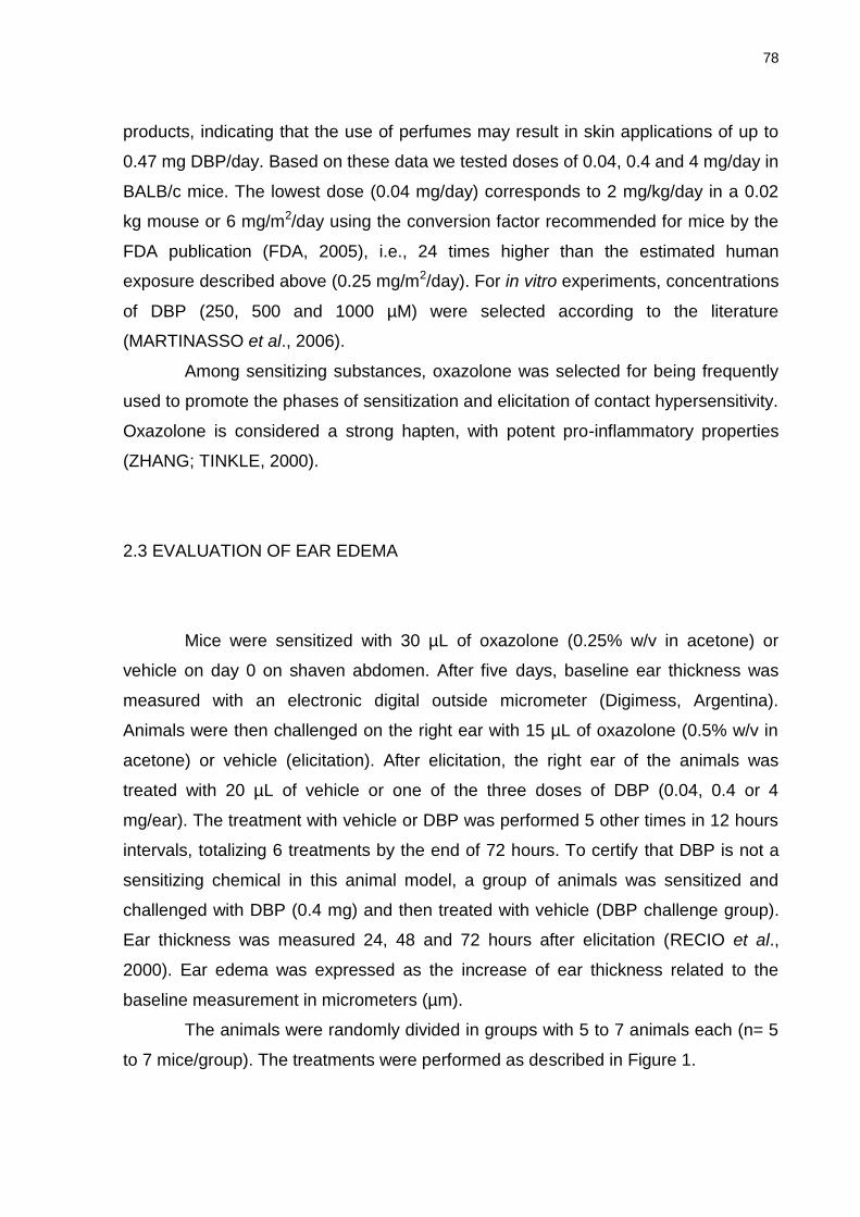
78
products, indicating that the use of perfumes may result in skin applications of up to
0.47 mg DBP/day. Based on these data we tested doses of 0.04, 0.4 and 4 mg/day in
BALB/c mice. The lowest dose (0.04 mg/day) corresponds to 2 mg/kg/day in a 0.02
kg mouse or 6 mg/m2/day using the conversion factor recommended for mice by the
FDA publication (FDA, 2005), i.e., 24 times higher than the estimated human
exposure described above (0.25 mg/m2/day). For in vitro experiments, concentrations
of DBP (250, 500 and 1000 µM) were selected according to the literature
(MARTINASSO et al., 2006).
Among sensitizing substances, oxazolone was selected for being frequently
used to promote the phases of sensitization and elicitation of contact hypersensitivity.
Oxazolone is considered a strong hapten, with potent pro-inflammatory properties
(ZHANG; TINKLE, 2000).
2.3 EVALUATION OF EAR EDEMA
Mice were sensitized with 30 µL of oxazolone (0.25% w/v in acetone) or
vehicle on day 0 on shaven abdomen. After five days, baseline ear thickness was
measured with an electronic digital outside micrometer (Digimess, Argentina).
Animals were then challenged on the right ear with 15 µL of oxazolone (0.5% w/v in
acetone) or vehicle (elicitation). After elicitation, the right ear of the animals was
treated with 20 µL of vehicle or one of the three doses of DBP (0.04, 0.4 or 4
mg/ear). The treatment with vehicle or DBP was performed 5 other times in 12 hours
intervals, totalizing 6 treatments by the end of 72 hours. To certify that DBP is not a
sensitizing chemical in this animal model, a group of animals was sensitized and
challenged with DBP (0.4 mg) and then treated with vehicle (DBP challenge group).
Ear thickness was measured 24, 48 and 72 hours after elicitation (RECIO et al.,
2000). Ear edema was expressed as the increase of ear thickness related to the
baseline measurement in micrometers (µm).
The animals were randomly divided in groups with 5 to 7 animals each (n= 5
to 7 mice/group). The treatments were performed as described in Figure 1.
79
FIGURE 1. EXPERIMENTAL DESIGN. Male BALB/c mice were treated with 30 μL of Oxazolone (OXA, 0.25% w/v) or Dibutyl phthalate (DBP, 0.4 mg) on their shaven abdomens on day 0 (Sensitization). On day 5, baseline ear thickness was measured followed by challenge with 15μL of vehicle (ACE, acetone), Oxazolone (0.5%) or DBP (0.4 mg) on the right ear. After challenge, the animals received 6 treatments on the right ear with DBP (0.04, 0.4 or 4 mg/ear) or vehicle. Treatments were performed with 12 hour intervals. Ear thickness was measured on days 6, 7 and 8 (24, 48 and 72 hours after challenge). On day 8, the animals were euthanized and 6 mm circles of ear tissue were collected for future analysis. n = 5 to 7 mice in each group. (1) Negative control group, (2) Positive control group for contact hypersensitivity, (3) Treated group with DBP 0.04 mg/ear, (4) Treated group with DBP 0.4 mg/ear, (5) Treated group with DBP 4 mg/ear, (6) DBP challenge group.
After ear edema evaluation the animals were euthanized by cervical
dislocation and 6 mm circles of ear tissue were collected for posterior histological
evaluation, enzyme activity assays (N-acetyl-β-D-glucosaminidase - NAG and
myeloperoxidase - MPO), and reduced glutathione (GSH) and hydroperoxide
(LOOH) content. The ears of naïve animals (that did not receive any treatment) were
collected for evaluation of MPO and NAG activity and GSH and LOOH content.
2.4 HISTOLOGICAL ANALYSIS
Ear tissue samples were fixed in ALFAC (10% formaldehyde, 85% ethanol
80%, 5% glacial acetic acid) solution. The ears were later embedded in paraffin, cut
at 5 μm sections and stained with hematoxylin–eosin.
The images for analysis were obtained through a motorized microscope Axio
Imager Z2 (Carl Zeiss, Jena, Germany), equipped with automated scanner VSlide
(Metasystems, Altlussheim, Germany). The scanned images were analyzed with
ImageJ software (NIH, USA). Epidermal thickness (considered as the distance from
80
the basal layer to the stratum granulosum) and dermal thickness (considered as the
distance from the basal layer of the epidermis to the auricular cartilage) were
measured, and the presence of inflammatory cells was quantified. Data are
expressed as the mean value of three representative areas of three histological
sections of each animal. Epidermal thickness measure and infiltrated cells count
were performed in 200x magnification sections, while dermal thickness was
measured in 100x magnification sections.
2.5 TISSUE MYELOPEROXIDASE (MPO) ASSAY
MPO is an enzyme used as a marker for the influx of polymorphonuclear
leukocytes into inflamed tissues. MPO activity was evaluated according to the
method proposed by Bradley et al. (1982) and modified by De Young et al. (1989).
Each ear sample (6 mm circles) was placed in 0.75 mL of 80 mM sodium phosphate
buffer (PBS, pH 5.4) containing 0.5% hexadecyltrimethylamonium bromide (HTAB).
Next, the sample was homogenized (45 s at 0 °C) in a motor-driven homogenizer.
The homogenate was decanted into a microfuge tube, and 0.75 mL of HTAB in PBS
was added to the tube. The 1.5 mL mixture was centrifuged at 11,200 × g at 4 °C for
20 min. The supernatant samples (triplicates of 30 μL) were added to 96-well plates.
For the assay, 200 μL of a mixture containing 100 μL of 80 mM PBS (pH 5.4), 85 μL
of 0.22 M PBS (pH 5.4) and 15 μL of 0.017% H2O2 were added to the wells. The
reaction was started by the addition of 20 μL of 18.4 mM tetramethylbenzidine HCl
(TMB) in dimethylformamide. The mixture was incubated for 3 min at 37 °C, and the
reaction was subsequently stopped by the addition of 30 μL of 1.46 M sodium
acetate (pH 3.0). Enzyme activity was determined colorimetrically using a plate
reader (Bio-Tek Multi-Mode Microplate reader Synergy HT) to measure absorbance
at 620 nm, and the results are expressed as mili optical density (mDO) per biopsy.
81
2.6 TISSUE N-ACETYL-β-D-GLUCOSAMINIDASE (NAG) ASSAY
The activity of NAG was used as a marker for the presence of mononuclear
leukocytes in inflamed tissue. According to the method used by Sanchez and Moreno
(1999), ear samples (6 mm circles) were treated using the same method described
for the MPO assay. The supernatant samples (triplicates of 25 μL) were added into
96-well plates. For the assay, 25 μL of p-nitrophenyl-acetamide-μ-d-glucopyranoside
(2.24 mM) and 100 μL of 50 mM buffer citrate (pH 4.5) per well were used. The
mixture was incubated for 60 min at 37 °C, and the reaction was stopped by the
addition of 100 μL of 200 nM glycine buffer (pH 10.4). The enzyme activity was
determined colorimetrically using a plate reader (Bio-Tek Multi-Mode Microplate
reader Synergy HT) to measure absorbance at 405 nm, and enzyme activity was
expressed as mDO per biopsy.
2.7 DETERMINATION OF REDUCED GLUTATHIONE (GSH) LEVELS AND LIPID
HYDROPEROXIDE (LOOH) CONTENT
Ear tissue samples were homogenized 1:10 (w/v) with potassium phosphate
buffer, pH 6.5. The homogenate was used to measure reduced glutathione (GSH)
and hydroperoxide (LOOH) levels.
Reduced glutathione levels were determined according to the method of
Sedlak and Lindsay (1968). Aliquots of tissue homogenate were mixed with 12.5%
trichloroacetic acid, vortexed, and centrifuged for 15 min at 6000 x g. The
supernatant was reserved, and TRIS buffer (0.4 M, pH 8.9) and 5,5’-dithiobis 2-
nitrobenzoic acid (DTNB; 0.01 M) were added. Absorbance was read at 415 nm
using a spectrophotometer. The procedures were performed at 4°C, and the
individual values were interpolated into a standard curve of GSH and are expressed
as µg·g of tissue- 1.
The levels of LOOH were determined using the ferrous oxidation-xylenol
orange (FOX2) method as described by Jiang, Hunt and Wolff (1992). Briefly, 100 μl
of methanol P.A. was added to 100 μl of supernatant, vortexed, and centrifuged at
82
9700 x g for 5 min at 4°C. The supernatant was mixed with FOX2 reagent (4 mM
butylated hydroxytoluene [BHT], 250 mM FeSO4, 25 mM H2SO4, and 100 mM
xylenol orange) and incubated in the dark for 30 min at room temperature.
Absorbance was read at 560 nm in a microplate reader, and the concentration of
LOOH is expressed as mmol hydroperoxide·mg of protein-1.
The protein content in ear tissue was measured to express the results of the
LOOH levels. Protein concentrations of the supernatants were determined by the
Bradford method (Bio-Rad, Hercules, CA, USA) using bovine serum albumin (125-
1000 μg) as the standard and performed according to the manufacturer’s
instructions.
2.8 IN VITRO EVALUATION OF THP-1 CELLS ACTIVATION
THP-1 cells (Istituto Zooprofilattico di Brescia, Italy), cells were used at a
density of 106 cells/mL in RPMI 1640 containing 2mM L-glutamine, 0.1 mg/mL
streptomycin, 100 IU/mL penicillin, 50 uM 2-mercaptoethanol, supplemented with
10% heated-inactivated fetal calf serum (media) and cultured at 37°C in 5% CO2. For
cytotoxicity or CD86 expression, 0.5 x 10⁶ cells were seeded in 24-well plates. For IL-
8 mRNA expression 3.0 x 10⁶ cells were seeded in 14 mL round bottom tubes.
2.8.1 CELL VIABILITY
Cytotoxicity was assessed by flow cytometric evaluation of propidium iodide
(PI)-stained cells. Cells were seeded in 24-well plates (0.5 mL/well) and incubated for
24 hours with oxazolone alone or in association with DBP, LPS (10 ng/mL) as
positive control, or DMSO as vehicle control. After incubation, cells were centrifuged
at 1500 rpm for 5 minutes and suspended in 500 μL PBS containing 1 µg/mL PI. The
percentage of positive cells was analyzed using a FACSCalibur flow cytometer, and
data were quantified using CellQuest software (BD, Becton Dickinson). Based on cell
83
viability, cells were treated using the concentration of chemicals resulting in cell
viability > 75%.
2.8.2 FLOW CYTOMETRIC ANALYSIS OF CD86 EXPRESSION
CD86 expression was evaluated by flow cytometric analysis. THP-1 cells
were treated with oxazolone, DBP, oxazolone associated with DBP, LPS or DMSO
as vehicle control. After 24 hours, cells were centrifuged at 1500 rpm for 5 minutes.
Cells were then stained for 30 minutes with a specific FITC-conjugated antibody
against CD86 (BD, Becton Dickinson) or with isotype control antibody at 4°C
following supplier’s instructions. The intensity of fluorescence and the percentage of
positive cells were analyzed using FACSCalibur flow cytometer, and data were
quantified using CellQuest software (BD, Becton Dickinson). 10,000 viable cells were
analyzed for mean fluorescence intensity (MFI) and percentage of positively labeled
cells (PC). Changes in CD86 expression are reported according to Azam et al.
(2006) as stimulation index (SI) calculated by the following equation:
PCc and MFIc stand for the percentage of positive cells and mean fluorescence
intensity in control cells, whereas PCt and MFIt stand for the percentage and mean
fluorescence intensity in chemical-treated cells.
2.8.3 REAL TIME RT-PCR
Total RNA was isolated from THP-1 cells after 3 hours of treatment with
selected chemicals and associations using commercial available kit (TriReagent from
Sigma) as described by the manufacturer. For the synthesis of cDNA, 2.0 µg of total
RNA was retro-transcribed using a high-capacity cDNA archive kit from Applied
Biosystems (Foster City, CA, USA) following the supplier’s instructions. IL-8 gene
expression was evaluated by Real Time reverse transcription polymerase chain
84
reaction (Real Time PCR). For PCR-analysis, Taq-ManTM-PCR technology was used.
For each PCR reaction, 10 ng of total RNA were used. The 18S ribosomal RNA
transcription was used as endogenous reference and the quantification of the
transcripts was performed by the ∆∆CT method.
2.9 DATA ANALYSIS
The data presented are expressed as mean ± SEM (in vivo experiments) or
as mean ± SD (in vitro experiments). Statistical significance was determined by
ANOVA followed by Tukey multiple comparison tests using the GraphPad Prism
software (GraphPad Software, La Jolla, CA). When necessary, Welch’s t test was
performed to confirm significant differences. Differences were considered significant
for p ≤ 0.05.
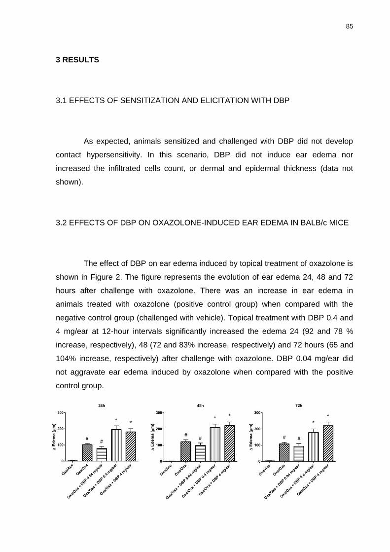
85
3 RESULTS
3.1 EFFECTS OF SENSITIZATION AND ELICITATION WITH DBP
As expected, animals sensitized and challenged with DBP did not develop
contact hypersensitivity. In this scenario, DBP did not induce ear edema nor
increased the infiltrated cells count, or dermal and epidermal thickness (data not
shown).
3.2 EFFECTS OF DBP ON OXAZOLONE-INDUCED EAR EDEMA IN BALB/c MICE
The effect of DBP on ear edema induced by topical treatment of oxazolone is
shown in Figure 2. The figure represents the evolution of ear edema 24, 48 and 72
hours after challenge with oxazolone. There was an increase in ear edema in
animals treated with oxazolone (positive control group) when compared with the
negative control group (challenged with vehicle). Topical treatment with DBP 0.4 and
4 mg/ear at 12-hour intervals significantly increased the edema 24 (92 and 78 %
increase, respectively), 48 (72 and 83% increase, respectively) and 72 hours (65 and
104% increase, respectively) after challenge with oxazolone. DBP 0.04 mg/ear did
not aggravate ear edema induced by oxazolone when compared with the positive
control group.
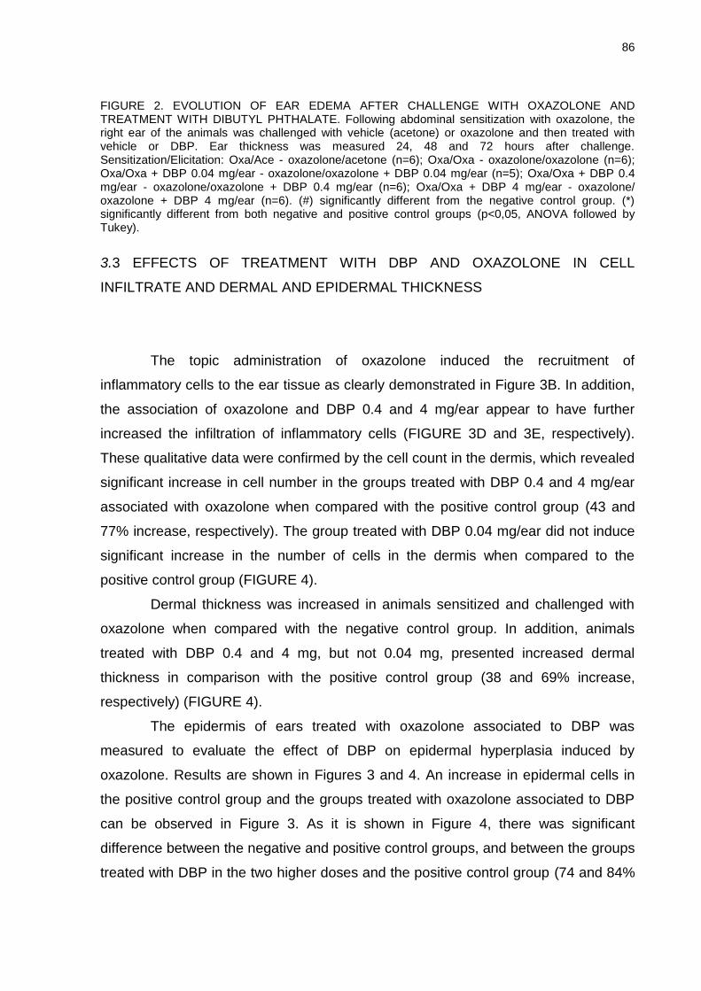
86
FIGURE 2. EVOLUTION OF EAR EDEMA AFTER CHALLENGE WITH OXAZOLONE AND TREATMENT WITH DIBUTYL PHTHALATE. Following abdominal sensitization with oxazolone, the right ear of the animals was challenged with vehicle (acetone) or oxazolone and then treated with vehicle or DBP. Ear thickness was measured 24, 48 and 72 hours after challenge. Sensitization/Elicitation: Oxa/Ace - oxazolone/acetone (n=6); Oxa/Oxa - oxazolone/oxazolone (n=6); Oxa/Oxa + DBP 0.04 mg/ear - oxazolone/oxazolone + DBP 0.04 mg/ear (n=5); Oxa/Oxa + DBP 0.4 mg/ear - oxazolone/oxazolone + DBP 0.4 mg/ear (n=6); Oxa/Oxa + DBP 4 mg/ear - oxazolone/ oxazolone + DBP 4 mg/ear (n=6). (#) significantly different from the negative control group. (*) significantly different from both negative and positive control groups (p<0,05, ANOVA followed by Tukey).
3.3 EFFECTS OF TREATMENT WITH DBP AND OXAZOLONE IN CELL
INFILTRATE AND DERMAL AND EPIDERMAL THICKNESS
The topic administration of oxazolone induced the recruitment of
inflammatory cells to the ear tissue as clearly demonstrated in Figure 3B. In addition,
the association of oxazolone and DBP 0.4 and 4 mg/ear appear to have further
increased the infiltration of inflammatory cells (FIGURE 3D and 3E, respectively).
These qualitative data were confirmed by the cell count in the dermis, which revealed
significant increase in cell number in the groups treated with DBP 0.4 and 4 mg/ear
associated with oxazolone when compared with the positive control group (43 and
77% increase, respectively). The group treated with DBP 0.04 mg/ear did not induce
significant increase in the number of cells in the dermis when compared to the
positive control group (FIGURE 4).
Dermal thickness was increased in animals sensitized and challenged with
oxazolone when compared with the negative control group. In addition, animals
treated with DBP 0.4 and 4 mg, but not 0.04 mg, presented increased dermal
thickness in comparison with the positive control group (38 and 69% increase,
respectively) (FIGURE 4).
The epidermis of ears treated with oxazolone associated to DBP was
measured to evaluate the effect of DBP on epidermal hyperplasia induced by
oxazolone. Results are shown in Figures 3 and 4. An increase in epidermal cells in
the positive control group and the groups treated with oxazolone associated to DBP
can be observed in Figure 3. As it is shown in Figure 4, there was significant
difference between the negative and positive control groups, and between the groups
treated with DBP in the two higher doses and the positive control group (74 and 84%
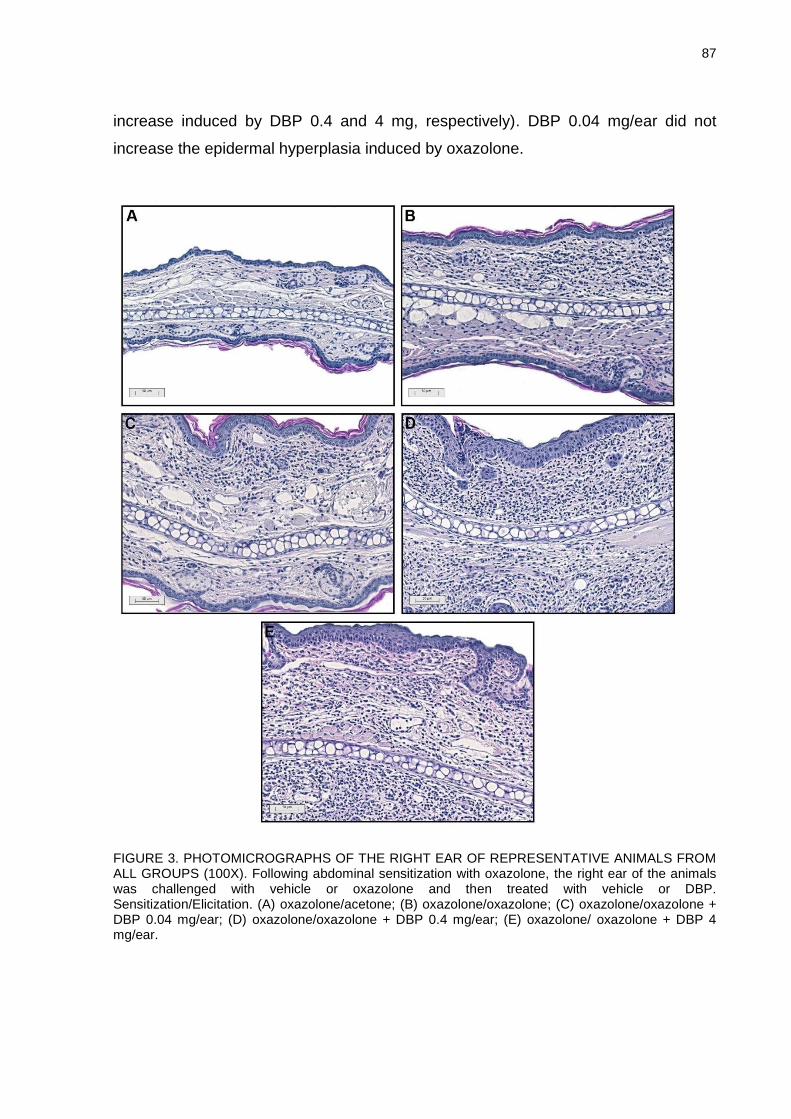
87
increase induced by DBP 0.4 and 4 mg, respectively). DBP 0.04 mg/ear did not
increase the epidermal hyperplasia induced by oxazolone.
FIGURE 3. PHOTOMICROGRAPHS OF THE RIGHT EAR OF REPRESENTATIVE ANIMALS FROM ALL GROUPS (100X). Following abdominal sensitization with oxazolone, the right ear of the animals was challenged with vehicle or oxazolone and then treated with vehicle or DBP. Sensitization/Elicitation. (A) oxazolone/acetone; (B) oxazolone/oxazolone; (C) oxazolone/oxazolone + DBP 0.04 mg/ear; (D) oxazolone/oxazolone + DBP 0.4 mg/ear; (E) oxazolone/ oxazolone + DBP 4 mg/ear.
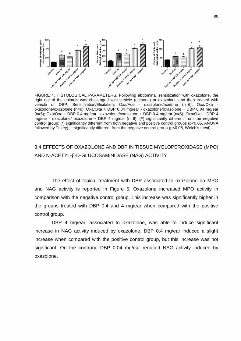
88
FIGURE 4. HISTOLOGICAL PARAMETERS. Following abdominal sensitization with oxazolone, the right ear of the animals was challenged with vehicle (acetone) or oxazolone and then treated with vehicle or DBP. Sensitization/Elicitation: Oxa/Ace - oxazolone/acetone (n=6); Oxa/Oxa - oxazolone/oxazolone (n=6); Oxa/Oxa + DBP 0.04 mg/ear - oxazolone/oxazolone + DBP 0.04 mg/ear (n=5); Oxa/Oxa + DBP 0.4 mg/ear - oxazolone/oxazolone + DBP 0.4 mg/ear (n=6); Oxa/Oxa + DBP 4 mg/ear - oxazolone/ oxazolone + DBP 4 mg/ear (n=6). (#) significantly different from the negative control group; (*) significantly different from both negative and positive control groups (p<0.05, ANOVA followed by Tukey); t: significantly different from the negative control group (p<0.05, Welch’s t test).
3.4 EFFECTS OF OXAZOLONE AND DBP IN TISSUE MYELOPEROXIDASE (MPO)
AND N-ACETYL-β-D-GLUCOSAMINIDASE (NAG) ACTIVITY
The effect of topical treatment with DBP associated to oxazolone on MPO
and NAG activity is reported in Figure 5. Oxazolone increased MPO activity in
comparison with the negative control group. This increase was significantly higher in
the groups treated with DBP 0.4 and 4 mg/ear when compared with the positive
control group.
DBP 4 mg/ear, associated to oxazolone, was able to induce significant
increase in NAG activity induced by oxazolone. DBP 0.4 mg/ear induced a slight
increase when compared with the positive control group, but this increase was not
significant. On the contrary, DBP 0.04 mg/ear reduced NAG activity induced by
oxazolone.
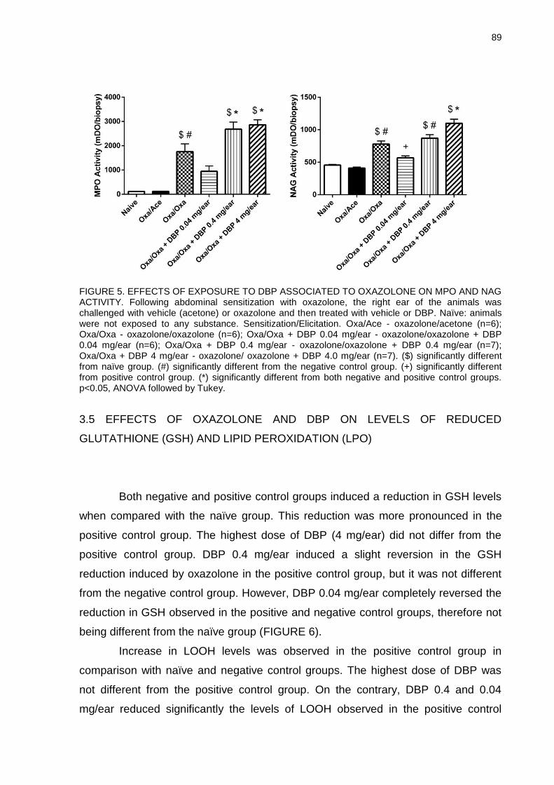
89
FIGURE 5. EFFECTS OF EXPOSURE TO DBP ASSOCIATED TO OXAZOLONE ON MPO AND NAG ACTIVITY. Following abdominal sensitization with oxazolone, the right ear of the animals was challenged with vehicle (acetone) or oxazolone and then treated with vehicle or DBP. Naïve: animals were not exposed to any substance. Sensitization/Elicitation. Oxa/Ace - oxazolone/acetone (n=6); Oxa/Oxa - oxazolone/oxazolone (n=6); Oxa/Oxa + DBP 0.04 mg/ear - oxazolone/oxazolone + DBP 0.04 mg/ear (n=6); Oxa/Oxa + DBP 0.4 mg/ear - oxazolone/oxazolone + DBP 0.4 mg/ear (n=7); Oxa/Oxa + DBP 4 mg/ear - oxazolone/ oxazolone + DBP 4.0 mg/ear (n=7). ($) significantly different from naïve group. (#) significantly different from the negative control group. (+) significantly different from positive control group. (*) significantly different from both negative and positive control groups. p<0.05, ANOVA followed by Tukey.
3.5 EFFECTS OF OXAZOLONE AND DBP ON LEVELS OF REDUCED
GLUTATHIONE (GSH) AND LIPID PEROXIDATION (LPO)
Both negative and positive control groups induced a reduction in GSH levels
when compared with the naïve group. This reduction was more pronounced in the
positive control group. The highest dose of DBP (4 mg/ear) did not differ from the
positive control group. DBP 0.4 mg/ear induced a slight reversion in the GSH
reduction induced by oxazolone in the positive control group, but it was not different
from the negative control group. However, DBP 0.04 mg/ear completely reversed the
reduction in GSH observed in the positive and negative control groups, therefore not
being different from the naïve group (FIGURE 6).
Increase in LOOH levels was observed in the positive control group in
comparison with naïve and negative control groups. The highest dose of DBP was
not different from the positive control group. On the contrary, DBP 0.4 and 0.04
mg/ear reduced significantly the levels of LOOH observed in the positive control
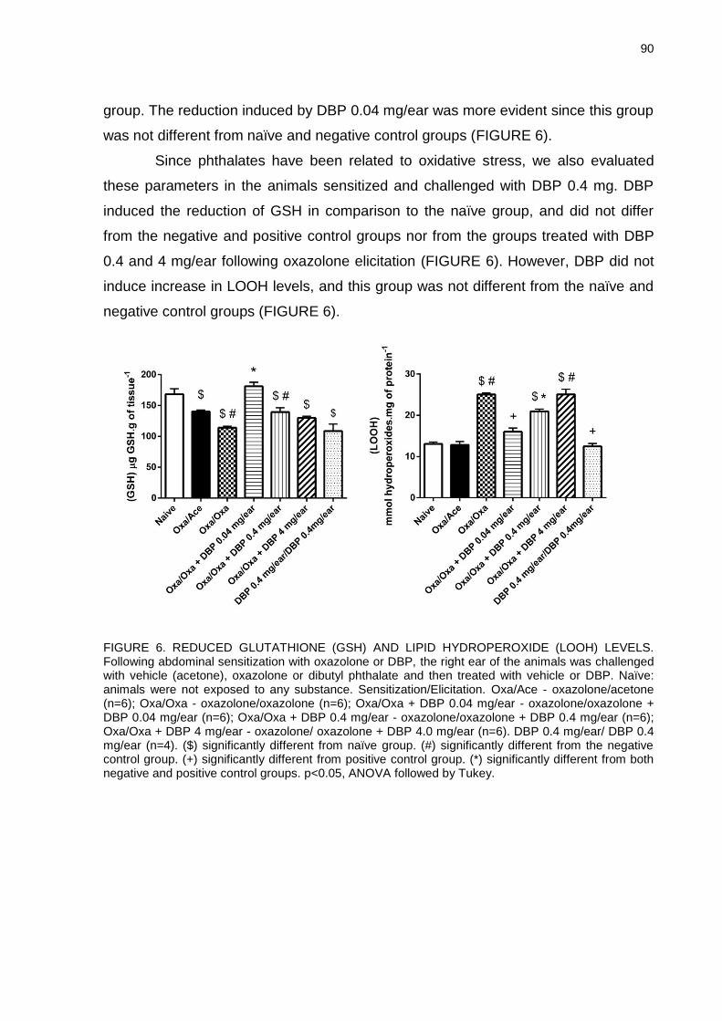
90
group. The reduction induced by DBP 0.04 mg/ear was more evident since this group
was not different from naïve and negative control groups (FIGURE 6).
Since phthalates have been related to oxidative stress, we also evaluated
these parameters in the animals sensitized and challenged with DBP 0.4 mg. DBP
induced the reduction of GSH in comparison to the naïve group, and did not differ
from the negative and positive control groups nor from the groups treated with DBP
0.4 and 4 mg/ear following oxazolone elicitation (FIGURE 6). However, DBP did not
induce increase in LOOH levels, and this group was not different from the naïve and
negative control groups (FIGURE 6).
FIGURE 6. REDUCED GLUTATHIONE (GSH) AND LIPID HYDROPEROXIDE (LOOH) LEVELS. Following abdominal sensitization with oxazolone or DBP, the right ear of the animals was challenged with vehicle (acetone), oxazolone or dibutyl phthalate and then treated with vehicle or DBP. Naïve: animals were not exposed to any substance. Sensitization/Elicitation. Oxa/Ace - oxazolone/acetone (n=6); Oxa/Oxa - oxazolone/oxazolone (n=6); Oxa/Oxa + DBP 0.04 mg/ear - oxazolone/oxazolone + DBP 0.04 mg/ear (n=6); Oxa/Oxa + DBP 0.4 mg/ear - oxazolone/oxazolone + DBP 0.4 mg/ear (n=6); Oxa/Oxa + DBP 4 mg/ear - oxazolone/ oxazolone + DBP 4.0 mg/ear (n=6). DBP 0.4 mg/ear/ DBP 0.4 mg/ear (n=4). ($) significantly different from naïve group. (#) significantly different from the negative control group. (+) significantly different from positive control group. (*) significantly different from both negative and positive control groups. p<0.05, ANOVA followed by Tukey.
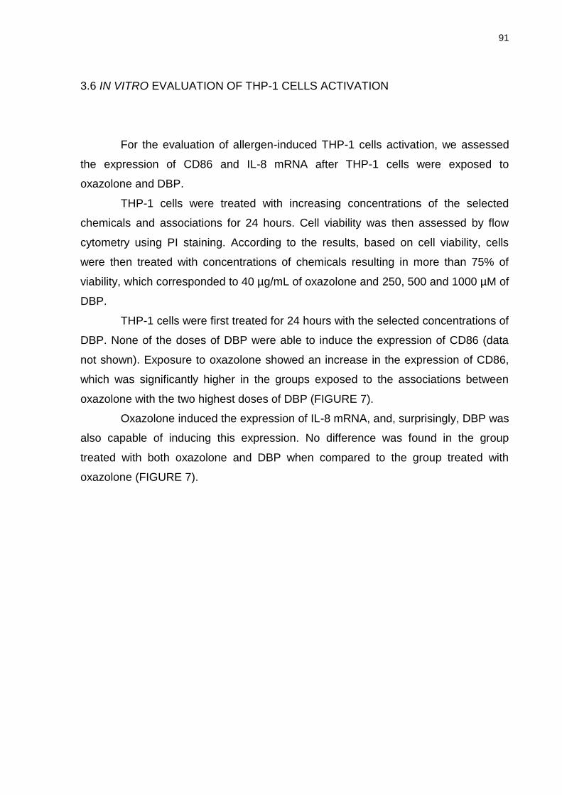
91
3.6 IN VITRO EVALUATION OF THP-1 CELLS ACTIVATION
For the evaluation of allergen-induced THP-1 cells activation, we assessed
the expression of CD86 and IL-8 mRNA after THP-1 cells were exposed to
oxazolone and DBP.
THP-1 cells were treated with increasing concentrations of the selected
chemicals and associations for 24 hours. Cell viability was then assessed by flow
cytometry using PI staining. According to the results, based on cell viability, cells
were then treated with concentrations of chemicals resulting in more than 75% of
viability, which corresponded to 40 µg/mL of oxazolone and 250, 500 and 1000 µM of
DBP.
THP-1 cells were first treated for 24 hours with the selected concentrations of
DBP. None of the doses of DBP were able to induce the expression of CD86 (data
not shown). Exposure to oxazolone showed an increase in the expression of CD86,
which was significantly higher in the groups exposed to the associations between
oxazolone with the two highest doses of DBP (FIGURE 7).
Oxazolone induced the expression of IL-8 mRNA, and, surprisingly, DBP was
also capable of inducing this expression. No difference was found in the group
treated with both oxazolone and DBP when compared to the group treated with
oxazolone (FIGURE 7).
92
FIGURE 7. EFFECTS OF OXAZOLONE AND ASSOCIATION WITH DBP ON THE EXPRESSION OF CELL SURFACE MARKER CD86 AND IL-8 mRNA. For CD86 expression, THP-1 cells were treated for 24h with oxazolone or its association with increasing doses of Dibutyl phthalate (DBP). Vehicle treated cells were included as a vehicle control (DMSO). Results are expressed as stimulation index (SI). Each value represents the mean ± SD, n=3. For IL-8 mRNA, THP-1 cells were treated for 3 h with oxazolone, DBP or the association between oxazolone and DBP. Vehicle treated cells were included as a vehicle control (DMSO). IL-8 mRNA expression was evaluated by Real Time-PCR as described in the Material and Methods section. Results are expressed as 2-∆∆CT. Each value represents the mean ± SD, n=3 independent experiments. (#) significantly different from the negative control group; (*) significantly different from both negative control group and oxazolone group. p<0.05, ANOVA followed by Tukey multiple comparison test.
93
4 DISCUSSION
Since humans are commonly exposed to phthalates, it is important to
elucidate their potential immunotoxic effects. Toxicological studies have
demonstrated that phthalate esters, like DBP, do not possess significant potential to
cause sensitization on skin. This is because these molecules are not capable of
bonding and reacting to proteins, which is mandatory for the allergic stimulation by a
substance (KIMBER; DEARMAN, 2010). However, phthalate esters have been
reported to facilitate the development of allergic diseases such as asthma and atopic
dermatitis, and to act as adjuvants in animal models of contact hypersensitivity (IMAI
et al., 2006; MATSUDA et al., 2010). The present in vivo study investigated the
effects of DBP on an animal model of contact hypersensitivity induced by oxazolone
in BALB/c mice. Results indicate that DBP aggravates the inflammatory response
involved in this model.
DBP, in the two highest doses (0.4 and 4 mg/ear) was able to increase the
ear edema and dermal thickness induced by oxazolone in mice, besides enhancing
the leukocyte recruitment to the dermis, as indicated by cell count and MPO and
NAG activity. Added to that, the thickness of the epidermis was significantly different
between the group treated with oxazolone and those associated with the two highest
doses of DBP (0.4 and 4 mg/ear). These results indicate that DBP can aggravate
several pathophysiological signs that characterize the oxazolone-induced model of
contact hypersensitivity in BALB/c mice. In addition to a persistent edema, the
infiltration of inflammatory cells and epidermal hyperplasia are typical features of this
animal model (FUJII et al., 2002).
The increase in ear edema (IMAI et al., 2006; SHIBA et al., 2009; MATSUDA
et al., 2010; LI et al., 2014) and cell infiltration (TAKANO et al., 2006; LI et al., 2014)
demonstrated in this study had already been reported by other authors. Imai et al.
(2006) demonstrated that, not only DBP, but also other phthalates, were capable of
exacerbating the ear edema induced by fluorescein isothiocyanate. Takano et al.
(2006) showed increase in ear edema and also cell infiltration to the inflammatory
site in an animal model of atopic dermatitis where animals were exposed to DEHP
(di-2-ethylhexyl phthalate). Moreover, Li et al. (2014) showed that prolonged dermal
exposure to DBP (4 and 40 mg/kg/day for forty days) was capable of increasing ear
94
edema and leukocyte infiltration to the dermis. The data presented here further
support the potential for adjuvancy of DBP, but we report an adjuvant effect after
short term dermal exposure to lower doses of DBP. Besides, our results indicate that
DBP can act as adjuvant even after the sensitization has already been established,
modulating the elicitation phase of oxazolone-induced CHS. Previous studies have
usually observed this effect in animal models of CHS associating phthalates in both
sensitization and elicitation phases, or with high doses of phthalates, long term
exposure to lower doses or in different routes of exposure, in some cases with
subcutaneous or even intraperitoneal injections, which are not routes by which
humans would normally encounter phthalates in domestic or occupational
enviroment.
Oxazolone is highly used in contact hypersensitivity models for promoting the
phases of sensitization and elicitation. However, the great majority of studies
evaluate the effects caused by the application of these substances in lymph nodes
and epidermis, and little is discussed about the inflammatory properties in the dermis
(ZHANG; TINKLE, 2000). The evaluation of the activity of NAG and MPO was
performed to indicate the presence of mononuclear and polymorphonuclear
leukocytes, respectively, in the inflamed tissue (SANCHEZ; MORENO, 1999;
BRADLEY et al., 1982), and characterize the probable profile of recruited cells to the
dermis. The results show that both polymorphonuclear (mainly neutrophils) and
mononuclear leukocytes (mainly macrophages) are present in the inflammation
induced by oxazolone. These results corroborate the findings of Wiedow et al.
(1992), who quantified macrophages and neutrophils’ proteases in the dermis, which
were increased in allergic contact dermatitis. DBP 0.4 increased the activity of MPO
and DBP 4 mg/ear increased the activity of both enzymes, indicating a greater
recruitment of cells to the inflammatory site. We can also suggest that as result of the
adjuvant effect of DBP a higher proportion of neutrophils are recruited to the
inflammatory site in relation to mononuclear leukocytes, since the treatment with DBP
led to a higher increase in the activity of MPO than NAG. Neutrophils are involved in
both sensitization and elicitation phases of CHS, and are one of the first cells to be
recruited after the elicitation in sensitized animals (DILULIO et al., 1999). Zhang and
Tinkle (2000) found that neutrophils are found in higher amount in the dermis of mice
treated with oxazolone and remain dominant until 72 hours after treatment. Besides,
Tanaka et al. (2012) showed a much higher increase in neutrophils rather than
95
macrophages induced by adjuvant effect of DEHP on allergen ovalbumin-induced
peritonitis in ICR mice. In this study DBP increased THP-1 oxazolone-induced
activation, as showed by increased CD86 expression. Even though we did not
identify the same adjuvant effect in IL-8 mRNA expression, we have previously
reported that DBP enhanced IL-8 mRNA expression as well as IL-8 release in THP-1
cells exposed to citral and imidazolidinyl urea (LOURENÇO et al., 2015) supporting
the hypothesis that DBP can potentiate THP-1 allergen-induced activation.
Therefore, DBP could be enhancing the activation of dendritic cells in vivo, which
could lead to higher production of IL-8 (a potent neutrophil chemoattractant)
(BARKER et al., 1991) and this could be reflected in higher neutrophil recruitment as
part of the mechanism of DBP’s adjuvant effect. Interestingly, DBP also induced a
significant expression of IL-8 mRNA by itself. In general, little can be found in
literature relating DBP exposure and increase in pro-inflammatory cytokines mRNA
expression, therefore, this induction needs further investigation.
Although many studies on adjuvant effect of phthalates have been
performed, little is known about the molecular mechanisms involved in such effect.
Evidence suggests that oxidative stress could play a role in allergic and inflammatory
skin diseases (OKAYAMA, 2005; BYAMBA et al., 2010; MIZUASHI et al., 2005).
Several authors have reported that some phthalates induce oxidative stress
demonstrating increased generation of reactive oxygen species (ROS) and serum
markers of inflammation and oxidative stress after exposure to phthalates (SEO et
al., 2004; HAUSER et al., 2007; FERGUSON; LOCH-CARUSO; MEEKER, 2011;
TETZ et al., 2013). We have previously observed a slight increase in ROS production
induced by 500 µM of DBP in the human promyelocytic cell line THP-1 (a surrogate
for dendritic cell activation model). Additionally, we reported that DBP significantly
increased the production of ROS induced by the allergen citral, suggesting that the
adjuvant effect of DBP could be through dendritic cell activation and that this could
be triggered by increase in oxidative stress (LOURENÇO et al., 2015). In this study,
we showed a decrease in GSH levels induced by oxazolone in the positive control
group and also in the negative control group, which was exposed to oxazolone in
only one phase of the sensitization model. This reduction was accompanied by
increase in LOOH levels in the positive control group. The oxidative stress induced
by oxazolone in contact hypersensitivity has already been reported (BRUNYÁNSZKI
et al., 2010). We also show that the sensitization and challenge with DBP 0.4 mg
96
reduced the levels of GSH, but no increase in LOOH was observed in this group.
What is interesting is that the same dose (0.4 mg) partially reversed the reduction in
GSH and increase in LOOH induced by oxazolone, while DBP 0.04 mg completely
reversed the same parameters, and the group treated with DBP 4 mg did not differ
from the positive control group. Overall, our results seem to indicate that although
DBP may alter some oxidative stress parameters, these mechanisms do not
participate in the adjuvant effect of DBP at the dose levels tested. Accordingly, Chen
et al. (2015) reported a weak adjuvant effect of DBP in a murine asthma model and
that this effect was not mediated through an oxidative stress mechanism. Although
we have reported that DBP could induce the production of ROS in vitro and increase
this production induced by an allergen, the time of evaluation of that parameter was
different from what was performed in the present study. In vitro we evaluated the
production of ROS immediately after the treatment of the cells with DBP associated
to allergen; on the other hand, here, GSH and LOOH were measured after three
days of exposure to DBP associated with oxazolone, so we should also consider the
role of the inflammatory response induced by both DBP and oxazolone in such
effects. In addition, phthalates tend to present opposite effects in studies about their
adjuvancy when using low or high doses, different times and routes of exposure
(KIMBER; DEARMAN, 2010); therefore different doses of phthalates could possibly
influence the cells’ redox balance in different ways.
Even though the doses 0.4 and 4 mg of DBP presented adjuvant effect in this
study, the lowest dose (0.04 mg) did not differ from the positive control group in most
of the parameters analyzed, besides reducing significantly NAG and MPO activity,
and reversing the oxidative stress parameters. Although the majority of data found in
literature reports an adjuvant effect induced by phthalates, some authors have
reported a negative adjuvant effect (DEARMAN et al., 2008; CHEN et al., 2015).
Furthermore, it has been reported that some phthalates in certain doses display what
were described as “immunosuppressive” effects (inhibition of IgG1 and/or IgE
antibody production) (LARSEN et al., 2001). It’s also been suggested that the
adjuvant activity of phthalates seems to be highly dependent of the length of the alkyl
side chain, giving rise to maximum adjuvant effect by phthalates with 8 or 9 carbon
atoms in their alkyl chains, like DEHP, but not DBP (LARSEN; NIELSEN, 2008).
In conclusion, we suggest that DBP presents an in vivo adjuvant effect when
associated with oxazolone in much lower doses and time of exposure than those

97
usually seen in animal models of contact hypersensitivity, supporting previous data of
this effect in allergic diseases. However, the mechanisms underlying these effects
and under what specific conditions (doses, time and route of exposure) phthalates
are able to effect a meaningful change in immune function have not been fully
understood, thus, future studies are necessary to elucidate the effects of phthalates
in vivo.
98
REFERENCES
ANDERSON, W.A.C. et al. A biomarker approach to measuring human dietary exposure to certain phthalate diesters. Food Addit. Contam., v. 18, n. 12, p. 1068-1074, 2001. AZAM, P. et al. The cytokine-dependent MUTZ-3 cell line as an in vitro model for the screening of contact sensitizers. Toxicol. Appl. Pharmacol., v. 212, p. 14-23, 2006. BANCHEREAU, J. et al. Immunobiology of dendritic cells. Annu. Rev. Immunol., v. 18, p. 767-811, 2000. BARKER, J.N. et al. Modulation of keratinocyte-derived interleukin-8 which is chemotactic for neutrophils and T lymphocytes. Am. J. Pathol., v. 139, p. 869-876, 1991. BEYDON, D.; PAYAN, J.P.; GRANDCLAUDE, M.C. Comparison of percutaneous absorption and metabolism of di-n-butylphthalate in various species. Toxicol. In Vitro, v. 24, p. 71–78, 2010. BRADFORD, M. A rapid and sensitive method for the quantitation of microgram quantities of protein utilizing the principle of protein dye binding. Anal Biochem., 1976, v. 72, p. 248-254, 1976. BRADLEY, P.P. et al. Measurement of cutaneous inflammation: estimation of neutrophil content with an enzyme marker. J. Invest. Dermatol., v. 78, n. 3, p. 206-209, 1982. BRUNYÁNSZKI, A. et al. Genetic Ablation of PARP-1 Protects Against Oxazolone-Induced Contact Hypersensitivity by Modulating Oxidative Stress. J. Invest. Dermatol., v. 130, p. 2629-2637, 2010. BYAMBA, D. et al. The roles of reactive oxygen species produced by contact allergens and irritants in monocyte-derived dendritic cells. Ann. Dermatol., v. 22, p. 269-278, 2010. CASILLAS, A.M. et al. Enhancement of allergic inflammation by diesel exhaust particles: permissive role of reactive oxygen species. Ann. Allerg. Asthma Im., v. 83, p. 624–629, 1999. CHEN, S. et al. Dibutyl phthalate induced oxidative stress does not lead to a significant adjuvant effect on a mouse asthma model. Toxicol. Res., v. 4, p. 260-269, 2015. CHOWDHURY, M.M.;, STATHAM, B.N. Allergic contact dermatitis from dibutyl phthalate and benzalkonium chloride in Timodine cream. Contact Dermatology, v. 46, p. 57, 2002.
99
CORSINI, E. et al. Role of oxidative stress in chemical allergen induced skin cells activation. Food Chem. Toxicol., v. 61, p. 74-81, 2013. CPSC (CONSUMER PRODUCT SAFETY COMMISSION). Overview of Phthalates Toxicity. Bestheda, United State of America, p. 1-31, 2010. DE YOUNG, L.M. et al. Edema and cell infiltration in the phorbol ester-treated mouse ear are temporally separate and can be differentially modulated by pharmacologic agents. Agents Actions, v. 26, n. 3-4, p. 335-341, 1989. DEARMAN, R. J. et al. Di-(2-ethylhexyl) phthalate is without adjuvant effect in mice on ovalbumin. Toxicology, v. 244, p. 231–241, 2008. DILULIO, N.A. et al. Groalpha-mediated recruitment of neutrophils is required for elicitation of contact hypersensitivity. Eur. J. Immunol., v. 29, p. 3485-3495, 1999. DOAN, K.; BRONAUGH, R.L.; YOURICK, J.J. In vivo and in vitro skin absorption of lipophilic compounds, dibutyl phthalate, farnesol and geraniol in the hairless guinea pig. Food Chem. Toxicol., v. 48, p. 18–23, 2010. ELSISI, A.; CARTER, D.E.; SIPES, I.G. Dermal absorption of phthalate diesters in rats. Fundam. Appl. Toxicol., v. 12, p. 70-77, 1989. FDA (FOOD AND DRUG ADMINISTRATION). Guidance for Industry. Estimating the Maximum Safe Starting Dose in Initial Clinical Trials for Therapeutics in Adult Healthy Volunteers, 30 pp., 2005. FERGUSON, K.K.; LOCH-CARUSO, R.; MEEKER, J.D. Urinary phthalate metabolites in relation to biomarkers of inflammation and oxidative stress: NHANES 1999-2006. Environ. Res., v. 111, n. 5, p. 718-726, 2011. FISHER, J. S. et al. Human 'testicular dysgenesis syndrome': a possible model using in-utero exposure of the rat to dibutyl phthalate. Hum. Reprod., v. 18, p. 1383-1394, 2003. FUJII, T. et al. Effects of FK506 (tacrolimus hydrate) on chronic oxazolone-induced dermatitis in rats. Eur. J. Pharmacol., v. 456, p. 115-121, 2002. GRABBE, S.; SCHWARZ, T. Immunoregulatory mechanisms involved in elicitation of allergic contact hypersensitivity. Immunol. Today, v. 19, n. 1, p. 37–44, 1998. HAUSER, R. et al. DNA damage in human sperm is related to urinary levels of phthalate monoester and oxidative metabolites. Hum. Reprod., v. 22, n. 3, p. 688-695, 2007. HENNINO, A. Fisiopatologia da dermatite de contato alérgica: papel das células T CD8 efetoras e das células T CD4 regulatórias. Anais Brasileiros de Dermatologia, v. 80, n. 4, p. 335-347, 2005.
100
HEUDORF, U.; MERSCH-SUNDERMANN, V.; ANGERER, J. Phthalates: Toxicology and exposure. Int. J. Hyg. Envir. Heal., v. 210, p. 623-634, 2007. IMAI, Y. et al. Effects of phthalate esters on the sensitization phase of contact hypersensitivity induced by fluorescein isothiocyanate. Clin. Exp. Allergy, v. 36, p. 1462–1468, 2006. INTERNATIONAL JOURNAL OF TOXICOLOGY. Annual Review of Cosmetic Ingredient Safety Assessment- 2002/2003. Int. J. Toxicol., v. 24, n. 1, p. 1-102, 2005. IPCS (INTERNATIONAL PROGRAMME ON CHEMICAL SAFETY). Environmental Health Criteria 189. Di-n-butyl phthalate. World Health Organization: Geneva, 1997. Disponível em: <http://www.inchem.org/documents/ehc/ehc/ehc189.htm>. Acesso em: 07/03/2015. JIANG, Z.Y.; HUNT, J.V.; WOLFF, S.P. Ferrous ion oxidation in the presence of xylenol orange for detection of lipid hydroperoxide in low density lipoprotein. Anal. Biochem., v. 202, p. 384–389, 1992. KARLBERG, A.T. et al. Allergic Contact Dermatitis–Formation, Structural Requirements, and Reactivity of Skin Sensitizers. Chem. Res. Toxicol., v. 21, n. 1, p. 53-69, 2008. KIMBER, I. et al. Allergic contact dermatitis. Int. Immunopharmacol., v. 2, p. 201–211, 2002. KIMBER, I.; DEARMAN, R.J. An assessment of the ability of phthalates to influence immune and allergic responses. Toxicology, v. 271, p. 73–82, 2010. KOO, H. J.; LEE, B. M. Estimated exposure to phthalates in cosmetics and risk assessment. J. Toxicol. Env. Health-Part A: Current Issues, v. 67, n. 23-24, p. 1901-1914, 2004. LARSEN, S.T. et al. Adjuvant and immunosuppressive effect of six monophthalates in a subcutaneous injection model with BALB/c strain mice. Toxicology, v. 169, p. 37–51, 2001. LARSEN, S.T.; NIELSEN, G.D. Structure–activity relationship of immunostimulatory effects of phthalates. BMC Immunol., v. 9, p. 61–69, 2008. LI, J. et al. T-Helper Type-2 Contact Hypersensitivity of BALB/c Mice Aggravated by Dibutyl Phthalate via Long-Term Dermal Exposure. PLoS ONE, v. 9, n. 2, 2014. LOURENÇO, A.C.S. et al. The plasticizer dibutyl phthalate (DBP) potentiates chemical allergen-induced THP-1 activation. Toxicol. In Vitro, v. 29, p. 2001–2008, 2015. LYCHE, J.L. et al. Reproductive and Developmental Toxicity of Phthalates. J. Toxicol. Env. Health B: Critical Reviews, v. 12, n. 4, p. 225-249, 2009.
101
MARTINASSO, G. et al. Effects of di(2-ethylhexyl) phthalate, a widely used peroxisome proliferator and plasticizer, on cell growth in the human keratinocyte cell line NCTC 2544. J. Toxicol. Env. Health A, v. 69, p. 353-365, 2006. MARTINO-ANDRADE, A.J. et al. Coadministration of active phthalates results in disruption of foetal testicular function in rats. Int. J. Androl., v. 32, n. 6, p. 704-712, 2009. MATSUDA, T. et al. Phthalate esters reveal skin-sensitizing activity of phenethyl isothiocyanate in mice. Food Chem. Toxicol., v. 48, p. 1704–1708, 2010. MITJANS, M. et al. Role of p38 MAPK in the selective release of IL-8 induced by chemical allergen in naive THP-1 cells. Toxicol. In Vitro, v. 22, p. 386-395, 2008. MIZUASHI, M. et al. Redox imbalance induced by contact sensitizers triggers the maturation of dendritic cells. J. Invest. Dermatol., v. 124, p. 579-586, 2005. OKAYAMA, Y. Oxidative stress in allergic and inflammatory skin diseases. Curr. Drug Targets Inflamm. Allergy, v. 4, n. 4, p. 517-519, 2005. PAN, T.L et al. Dermal toxicity elicited by phthalates: Evaluation of skin absorption, immunohistology, and functional proteomics. Food Chem. Toxicol., v. 65, p. 105–114, 2014. RECIO, M.C. et al. In vivo activity of pseudoguaianolide sesquiterpene lactones in acute and chronic inflammation. Life Science, v. 66, n. 26, p. 2509-2518, 2000. SANCHEZ, T.; MORENO, J.J. Role of leukocyte influx in tissue prostaglandin H synthase-2 overexpression induced by phorbol ester and arachidonic acid in skin. Biochem. Pharmacol., v. 58, p. 877-879, 1999. SATHYANARAYANA, S. et al. Baby care products: possible sources of infant phthalate exposure. Pediatrics, v. 121, p. e260–e268, 2008. SEDLAK, J.; LINDSAY, R.H. Estimation of total, protein-bound, and nonprotein sulfhydryl groups in tissue with Ellman's reagent. Anal. Biochem., v. 25, p. 192-205, 1968. SEO, K.W. et al. Comparison of oxidative stress and changes of xenobiotic metabolizing enzymes induced by phthalates in rats. Food Chem. Toxicol., v. 42, p. 107-114, 2004. SHIBA, T. et al. TRPA1 and TRPV1 activation is a novel adjuvant effect mechanism in contact hypersensitivity. J. Neuroimmunol., v. 207, p. 66–74, 2009. SILVA, M.J. et al. Analysis of human urine for fifteen phthalate metabolites using automated solid-phase extraction. J. Chromatogr. B., v. 805, p. 161–167, 2004. TAKANO, H. et al. Di-(2-ethylhexyl) phthalate enhances atopic dermatitis-like skin lesions in mice. Environ. Health Persp., v. 114, n. 8, p. 1266-1269, 2006.

102
TANAKA, M. et al. Immunoamplifying Effects of Di-(2-Ethylhexyl) Phthalate on Cytokine Response. Immunopharm. Immunot., v. 35, p. 1-4, 2012. TETZ, L.M. et al. Mono-2-ethylhexyl phthalate induces oxidative stress responses in human placental cells in vitro. Toxicol. Appl. Pharmacol., v. 268, n. 1, p. 47-54, 2013. WIEDOW, O. et al. Lesional Elastase Activity in Psoriasis, Contact Dermatitis, and Atopic Dermatitis. J. Invest. Dermatol., v. 99, n. 3, p. 306-309, 1992. ZHANG, L.; TINKLE, S.S. Chemical Activation of Innate and Specific Immunity in Contact Dermatitis. J. Invest. Dermatol., v. 115, n. 2, p. 168-172, 2000.
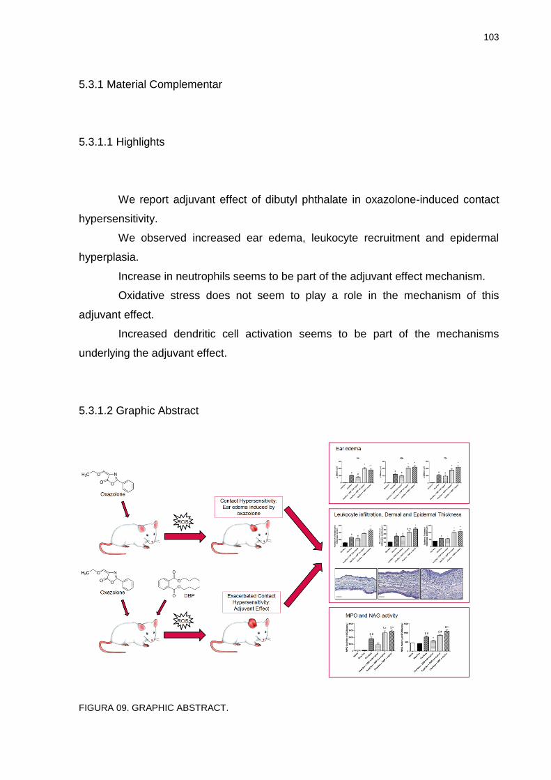
103
5.3.1 Material Complementar
5.3.1.1 Highlights
We report adjuvant effect of dibutyl phthalate in oxazolone-induced contact
hypersensitivity.
We observed increased ear edema, leukocyte recruitment and epidermal
hyperplasia.
Increase in neutrophils seems to be part of the adjuvant effect mechanism.
Oxidative stress does not seem to play a role in the mechanism of this
adjuvant effect.
Increased dendritic cell activation seems to be part of the mechanisms
underlying the adjuvant effect.
5.3.1.2 Graphic Abstract
FIGURA 09. GRAPHIC ABSTRACT.
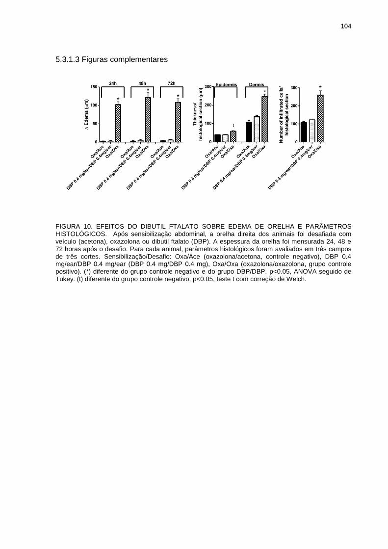
104
5.3.1.3 Figuras complementares
FIGURA 10. EFEITOS DO DIBUTIL FTALATO SOBRE EDEMA DE ORELHA E PARÂMETROS HISTOLÓGICOS. Após sensibilização abdominal, a orelha direita dos animais foi desafiada com veículo (acetona), oxazolona ou dibutil ftalato (DBP). A espessura da orelha foi mensurada 24, 48 e 72 horas após o desafio. Para cada animal, parâmetros histológicos foram avaliados em três campos de três cortes. Sensibilização/Desafio: Oxa/Ace (oxazolona/acetona, controle negativo), DBP 0.4 mg/ear/DBP 0.4 mg/ear (DBP 0.4 mg/DBP 0.4 mg), Oxa/Oxa (oxazolona/oxazolona, grupo controle positivo). (*) diferente do grupo controle negativo e do grupo DBP/DBP. p<0.05, ANOVA seguido de Tukey. (t) diferente do grupo controle negativo. p<0.05, teste t com correção de Welch.
105
5.4 ARTIGO 3
The involvement of transient receptor potential A1 (TRPA1) and V1 (TRPV1)
activation in the adjuvant effect of dibutyl phthalate in oxazolone-induced
contact hypersensitivity in BALB/c mice.
Ana Carolina dos Santos Lourençoa, Arthur da Silveira Prudenteb, Renata Mercer
Zaiaa, Michel Fleith Otukia, Anderson Joel Martino Andradea.
aDepartment of Pharmacology, Universidade Federal do Paraná (UFPR), Curitiba, PR, Brazil
bDepartment of Pharmacology, Universidade Federal de Santa Catarina (UFSC), Florianópolis, SC,
Brazil
Corresponding author:
Ana Carolina dos Santos Lourenço
Laboratório de Toxicologia, Departmento de Farmacologia, Universidade Federal do
Paraná
Rua Cel. Francisco H. dos Santos, 100 - Centro Politécnico, Caixa Postal 19031 -
CEP 81531-980 - Curitiba, PR, Brasil
e-mail: [email protected]
Tel.: +5515996848448
106
ABSTRACT
Contact hypersensitivity (CHS) is an allergic reaction resulting from repeated contact with chemicals called haptens, consisted in two phases: sensitization and elicitation. The plasticizer dibutyl phthalate (DBP) has been shown to enhance the inflammatory response in allergen-induced CHS when associated both in sensitization and elicitation phases, however the molecular mechanisms involved in this adjuvant effect are not fully understood. The present study aimed to investigate the possible participation of transient receptor potential cation channel (TRP) A1 (TRPA1) and V1 (TRPV1) activation by DBP in an animal model of CHS induced by oxazolone in BALB/c mice. For the experiments, adult male BALB/c mice were sensitized (in shaved abdomen) and challenged (elicitation; in the right ear) with oxazolone or vehicle (acetone) topically, and subsequently treated 6 times (with 12 hour intervals) with vehicle, TRPA1 or TRPV1 antagonists (HC030031 or SB366791, respectively) followed by treatment with DBP or vehicle (topically). Ear thickness was measured before elicitation (baseline) and 24, 48 and 72 hours after elicitation to calculate ear edema. After the last measure, animals were euthanized and 6 mm circles of ear tissue were collected to evaluate the activity of the enzymes N-acetyl-β-d-glucosaminidase (NAG) and Myeloperoxidase (MPO) (markers of leukocyte infiltration) and histological analysis (infiltrated cell count, dermal and epidermal thickness). Oxazolone induced CHS (increased ear edema, leukocyte infiltration and epidermal hyperplasia) and DBP increased all of these parameters confirming its adjuvant effect. Animals presenting CHS treated with TRPA1 antagonists and DBP showed reduction in the analyzed parameters when compared to those treated only with DBP, but this group was still different from its positive control group. Treatment with TRPV1 antagonist completely reversed the parameters to positive control group levels in animals with CHS treated with TRPV1 antagonist and DBP. In conclusion we suggest that the activation of TRPV1 and TRPA1 by DBP in the elicitation phase of oxazolone-induced CHS could be part of the mechanism of DBP’s adjuvant effect, and that TRPV1 activation seems to be more important in this adjuvant effect during the elicitation phase. Our results contribute to further understanding of the mechanisms involved in the adjuvant effect of phthalates in immune responses. Key words: dibutyl phthalate, contact hypersensitivity, oxazolone, elicitation, TRPA1, TRPV1.
107
1 INTRODUCTION
Allergic contact dermatitis (ACD) or contact hypersensitivity (CHS) is a
delayed type hypersensitivity reaction mediated by antigen specific T lymphocytes
(KARLBERG et al., 2008) in response to repeated or prolonged contact to chemical
substances called haptens (HENNINO et al., 2005; KARLBERG et al., 2008). There
are two phases involved in CHS: the sensitization phase (first contact with the
hapten, antigen presenting to the lymph node and activation of specific T
lymphocytes) and the elicitation phase (subsequent contacts with hapten, recognition
by specific T lymphocytes and development of inflammatory response) (GRABBE;
SCHWARZ, 1998; KIMBER, 2002; HENNINO et al., 2005; KARLBERG et al., 2008).
Several chemicals can induce CHS after repeated exposure to the skin. Although
many studies have focused on chemicals exhibiting sensitizing potential,
environmental chemicals with adjuvant effects may also play a role in the
development of hypersensitivity reactions.
Phthalate esters, industrial chemicals mainly used to impart flexibility to
polyvinyl chloride plastics, are candidate chemicals with adjuvant effect. These
substances are often found in a variety of manufactured products such as plastics for
food packing, glues, paints, solvents, children’s products, insect repellents, besides
different kinds of cosmetics, like perfumes, moisturizers, hairsprays and nail polishes
(IPCS, 1997; SATHYANARAYANA et al., 2008). It is been reported that, even though
phthalate esters are not capable of inducing hypersensitivity themselves (ELSISI;
CARTER; SIPES, 1989; DOAN; BRONAUGH; YOURICK, 2010; BEYDON; PAYAN;
GRANDCLAUDE, 2010; CPSC, 2010; PAN et al., 2014), certain compounds, such as
dibutyl phthalate (DBP), could be able to exacerbate the potency of allergens in vitro
and in vivo, and, so, play a role in the development and/or aggravation of allergic
diseases (CASILLAS et al., 1999, CHOWDHURY AND STATHAM, 2002; IMAI et al.,
2006; MATSUDA et al., 2010; LI et al., 2014, LOURENÇO et al., 2015). However, the
mechanisms underlying the adjuvant effects of phthalates are not yet fully
understood.
There is evidence that the immune system presents connection with the
peripheral nerve system (BERESFORD et al., 2004; LIU et al., 2006). It has been
suggested that sensory neurons that detect noxious stimuli could be involved in the
108
initiation of some allergies (PALM et al., 2012). It’s been reported that CHS can be
suppressed through systemic deletion of capsaicin (CAP)-sensitive fibers
(BERESFORD et al., 2004). These fibers express the transient receptor potential
(TRP) V1 (TRPV1), a calcium permeable cation channel involved in nociception
caused by chemical and thermal stimuli, also known as CAP receptor (CATERINA et
al., 1997; CLAPHAM, 2003). It’s been demonstrated that some TRPV1-expressing
sensory neurons could also express TRPA1, another member of the TRP family of
calcium permeable cation channels (STORY et al., 2003), which can be activated by
chemicals such as allyl isothiocyanate (AITC) and cinnamaldehyde (BANDELL et al.,
2004; JORDT et al., 2004). The activation of TRPA1 or TRPV1 may result in release
of neuropeptides from peripheral nerve endings (BAUTISTA et al., 2005; ZYGMUNT
et al., 1999), such as calcitonin gene-related peptide (CGRP) and substance P (SP),
that have been shown to participate in the inflammatory response of CHS
(MARUYAMA et al., 2007; MIKAMI et al., 2011; LIU et al., 2013). Moreover, it’s been
shown that desensitization of TRPA1 and TRPV1 after local pretreatment with AITC
and CAP, respectively, suppressed the sensitization to fluorescein isothiocyanate
(FITC) in BALB/c mice (MARUYAMA et al., 2007).
The ability of DBP to stimulate sensory neurons was demonstrated by the
activation of nerve cells isolated from mouse dorsal root ganglia (SHIBA et al., 2009).
In this study, DBP activated TRPA1 and TRPV1, which was shown by evocation of
calcium influx to TRPA1-expressing CHO cells and TRPV1-expressing CHO and
HEK 293 cells. In addition, Shiba et al. (2012) reported that that the TRPA1
antagonist HC030031 inhibited DBP-induced calcium influx into TRPA1-expressing
CHO cells.
Therefore, the present study aimed to investigate the possible participation of
TRPA1 and TRPV1 activation by DBP during the elicitation phase of an animal model
of contact hypersensitivity induced by oxazolone in BALB/c mice. This may contribute
to further understanding of the cellular mechanisms underlying the adjuvant effect of
phthalates in immune responses.
109
2 MATERIAL AND METHODS
2.1 ANIMALS
Adult male BALB/c mice obtained from Fundação Oswaldo Cruz (FIOCRUZ
– Rio de Janeiro, RJ, Brazil) were used for the experiments. The animals were kept
in the animal facilities of the Environmental Toxicology Laboratory (Escola Nacional
de Saúde Pública – ENSP, FIOCRUZ, Rio de Janeiro, RJ, Brazil) under a 12 h
light/dark cycle and controlled temperature (22 ± 2°C). Standard pellet food and tap
water were provided ad libitum. The experiments were conducted in accordance with
the orientations for laboratory animal care. The experimental protocol was approved
by the Committee on Animal Research and Ethics of the Universidade Federal do
Paraná (Curitiba, Brazil) under the number 633.
2.2 CHEMICALS AND DOSE SELECTION
Dibutyl phthalate (DBP, CAS number 84-74-2, purity 99%), Oxazolone (4-
Ethoxymethylene-2-phenyl-2-oxazolin-5-one, CAS number 15646-46-5), the vanilloid
receptor-1 (TRPV1) antagonist, SB366791, and the selective transient receptor
potential cation channel (TRPA1) blocker, HC030031, were purchased from Sigma-
Aldrich (St. Louis, MO, USA). Oxazolone was selected as skin sensitizer for being
frequently used to promote the phases of sensitization and elicitation of contact
hypersensitivity. Oxazolone is considered a strong hapten, with potent pro-
inflammatory properties (ZHANG; TINKLE, 2000). Antagonists, DBP and oxazolone
were diluted in acetone (vehicle).
DBP doses were based on available data on DBP concentrations on
personal care products, in particular perfumes. According to Koo and Lee (2004), the
average concentration of DBP found in a variety of perfumes is 0.44 mg/mL, and the
average use of perfume by the general population, estimated through an applied
questionnaire, is 1mL/day. Therefore, the amount of DBP applied to the skin in
110
individuals that use determined kinds of perfumes can be approximately 0.4 mg/day,
which corresponds to 0.0067 mg/kg/day for a 60 kg individual or 0.25 mg/m2/day
using the conversion factor recommended by the FDA (FDA, 2005). These
estimations are in agreement with the data published by the International Journal of
Toxicology (2005) in an annual review on safety of ingredients from cosmetic
products, indicating that the use of perfumes may result in skin applications of up to
0.47 mg DBP/day. Therefore, we selected the same nominal human dose of 0.4
mg/day of DBP, which corresponds to 20 mg/kg/day in a 0.02 kg mouse or 60
mg/m2/day using the conversion factor recommended for mice by the FDA
publication (FDA, 2005), i.e., 240 times higher than the estimated human exposure
described above (0.25 mg/m2/day).
The doses of the antagonists were selected according to the literature
(SILVA et al., 2011). The lowest dose with positive effect for antagonism (30
nmol/ear) was chosen for both antagonists.
2.3 EVALUATION OF EAR EDEMA
Mice were sensitized with 30 µL of oxazolone (0.25% w/v in acetone) on day
0 on shaven abdomen. After five days, baseline ear thickness was measured with a
digital micrometer (Great MT-04513). Animals were then challenged on the right ear
with 15 µL of oxazolone (0.5% w/v in acetone) or vehicle (elicitation). After elicitation,
the right ear of the animals was treated with 20 µL of vehicle or one of the TRP
antagonists (SB366791 – 30 nmol/ear; HC030031 – 30 nmol/ear). After 15 minutes,
the same ear was treated with vehicle or DBP (0.4 mg/ear). The treatment with
vehicle or antagonists followed by treatment with vehicle or DBP was performed 5
other times in 12 hour intervals, totalizing 6 treatments by the end of 72 hours. Ear
thickness was measured 24, 48 and 72 hours after elicitation (RECIO et al., 2000).
Ear edema was expressed as the increase of ear thickness related to the baseline
measurement in micrometers (µm). After ear edema evaluation the animals were
euthanized by cervical dislocation and 6 mm circles of ear tissue were collected for
posterior histological evaluation and enzyme activity assays (N-acetyl-beta-D-
glucosaminidase - NAG and myeloperoxidase - MPO).
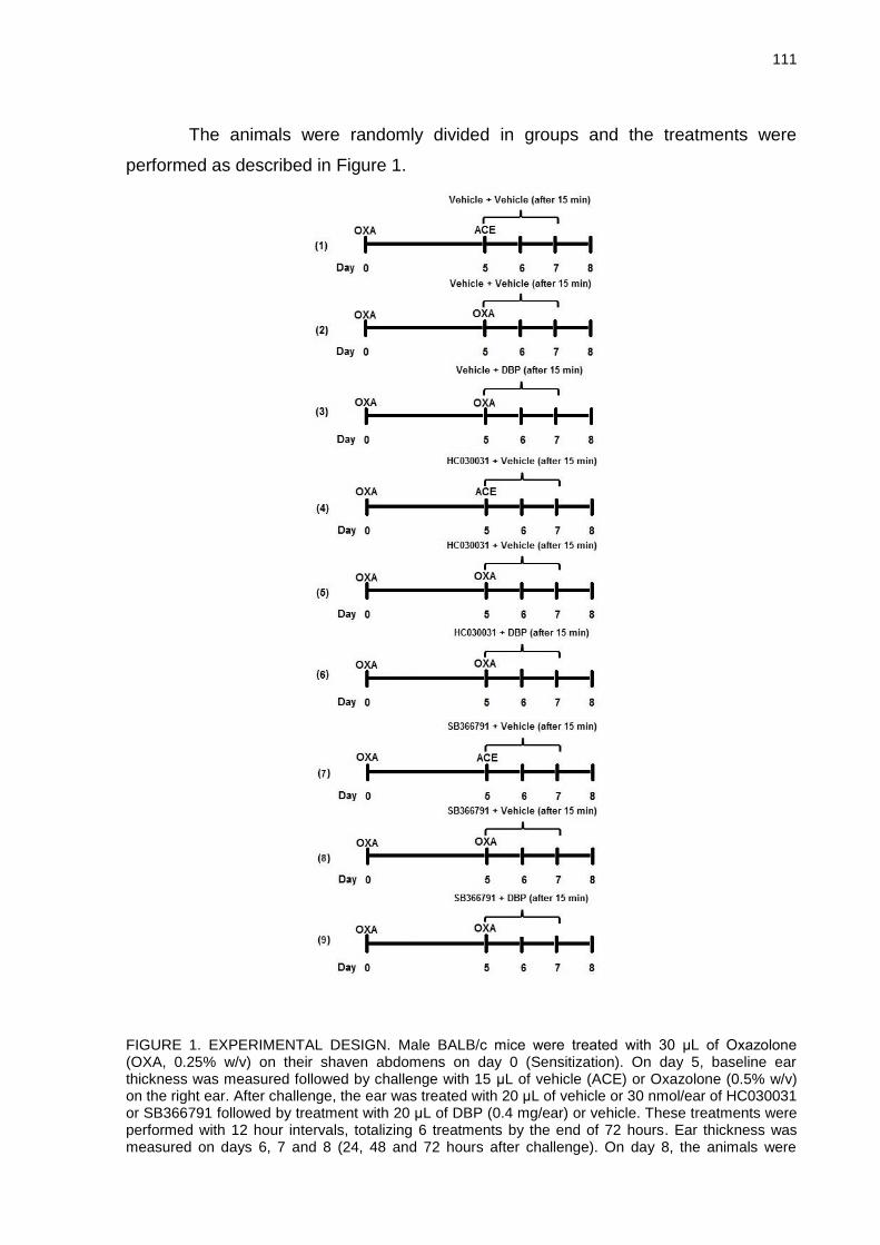
111
The animals were randomly divided in groups and the treatments were
performed as described in Figure 1.
FIGURE 1. EXPERIMENTAL DESIGN. Male BALB/c mice were treated with 30 μL of Oxazolone (OXA, 0.25% w/v) on their shaven abdomens on day 0 (Sensitization). On day 5, baseline ear thickness was measured followed by challenge with 15 μL of vehicle (ACE) or Oxazolone (0.5% w/v) on the right ear. After challenge, the ear was treated with 20 μL of vehicle or 30 nmol/ear of HC030031 or SB366791 followed by treatment with 20 μL of DBP (0.4 mg/ear) or vehicle. These treatments were performed with 12 hour intervals, totalizing 6 treatments by the end of 72 hours. Ear thickness was measured on days 6, 7 and 8 (24, 48 and 72 hours after challenge). On day 8, the animals were
112
euthanized and 6 mm circles of ear tissue were collected for future analysis. (1) Negative control group, (2) Positive control group for contact hypersensitivity, (3) DBP treated control group (treated with DBP 0.4 mg/ear), (4) TRPA1 negative control group, (5) TRPA1 positive control group, (6) TRPA1 treated group with DBP 0.4 mg/ear, (7) TRPV1 negative control group, (8) TRPV1 positive control group, (8) TRPV1 treated group with DBP 0.4 mg/ear.
2.4 HISTOLOGICAL ANALYSIS
Ear tissue samples were fixed in ALFAC (10% formaldehyde, 85% ethanol
80%, 5% glacial acetic acid) solution. The ears were later embedded in paraffin, cut
at 5 μm sections and stained with hematoxylin–eosin.
The images for analysis were obtained through a motorized microscope Axio
Imager Z2 (Carl Zeiss, Jena, Germany), equipped with automated scanner VSlide
(Metasystems, Altlussheim, Germany). The scanned images were analyzed with
ImageJ software (NIH, USA). Epidermal thickness (considered as the distance from
the basal layer to the stratum granulosum) and dermal thickness (considered as the
distance from the basal layer of the epidermis to the auricular cartilage) were
measured, and the presence of inflammatory cells was quantified. Data are
expressed as the mean value of three representative areas of three histological
sections of each animal. Epidermal thickness measure and infiltrated cells count
were performed in 200x magnification sections, while dermal thickness was
measured in 100x magnification sections.
2.5 TISSUE MYELOPEROXIDASE (MPO) ASSAY
MPO is an enzyme used as a marker for the influx of polymorphonuclear
leukocytes into inflamed tissues. MPO activity was evaluated according to the
method proposed by Bradley et al. (1982) and modified by De Young et al. (1989).
Each ear sample (6 mm circles) was placed in 0.75 mL of 80 mM sodium phosphate
buffer (PBS, pH 5.4) containing 0.5% hexadecyltrimethylamonium bromide (HTAB).
Next, the sample was homogenized (45 s at 0 °C) in a motor-driven homogenizer.
The homogenate was decanted into a microfuge tube, and 0.75 mL of HTAB in PBS
113
was added to the tube. The 1.5 mL mixture was centrifuged at 11,200 × g at 4 °C for
20 min. The supernatant samples (triplicates of 30 μL) were added to 96-well plates.
For the assay, 200 μL of a mixture containing 100 μL of 80 mM PBS (pH 5.4), 85 μL
of 0.22 M PBS (pH 5.4) and 15 μL of 0.017% H2O2 were added to the wells. The
reaction was started by the addition of 20 μL of 18.4 mM tetramethylbenzidine HCl
(TMB) in dimethylformamide. The mixture was incubated for 3 min at 37 °C, and the
reaction was subsequently stopped by the addition of 30 μL of 1.46 M sodium
acetate (pH 3.0). Enzyme activity was determined colorimetrically using a plate
reader (Bio-Tek Multi-Mode Microplate reader Synergy HT) to measure absorbance
at 620 nm, and the results were expressed as mili optical density (mDO) per biopsy.
2.6 TISSUE N-ACETYL-β-D-GLUCOSAMINIDASE (NAG) ASSAY
The activity of NAG was used as a marker for the presence of mononuclear
leukocytes in inflamed tissue. According to the method used by Sanchez and Moreno
(1999), ear samples (6 mm circles) were treated using the same method described
for the MPO assay. The supernatant samples (triplicates of 25 μL) were added into
96-well plates. For the assay, 25 μL of p-nitrophenyl-acetamide-μ-d-glucopyranoside
(2.24 mM) and 100 μL of 50 mM buffer citrate (pH 4.5) per well were used. The
mixture was incubated for 60 min at 37 °C, and the reaction was stopped by the
addition of 100 μL of 200 nM glycine buffer (pH 10.4). The enzyme activity was
determined colorimetrically using a plate reader (Bio-Tek Multi-Mode Microplate
reader Synergy HT) to measure absorbance at 405 nm, and enzyme activity was
expressed as mDO per biopsy.
2.7 DATA ANALYSIS
The data presented are expressed as mean ± SEM. Statistical significance
was determined by ANOVA followed by Tukey multiple comparison test or Kruskall
Wallis followed by Dunn, using the GraphPad Prism software (GraphPad Software,

114
La Jolla, CA). When necessary, Welch’s t test was performed to confirm significant
differences. Differences were considered significant for p ≤ 0.05.
115
3 RESULTS
3.1 EFFECTS OF DBP ON OXAZOLONE-INDUCED EAR EDEMA IN BALB/c MICE
AFTER TREATMENT WITH TRP ANTAGONISTS.
Figure 2 represents the evolution of ear edema 24, 48 and 72 hours after
challenge with vehicle or oxazolone followed by treatment with TRPA1 or TRPV1
antagonists and DBP. Oxazolone increased ear edema 24, 48 and 72 hours after
challenge when compared with the negative control group. This increase was
observed in the group exposed only to oxazolone (treated with vehicle after
challenge; positive control group) as well as in the groups exposed to oxazolone and
then treated with HC030031 (TRPA1 positive control group) or SB366791 (TRPV1
positive control group). Overall, these TRP antagonists did not change the oxazolone
response, except that SB366791 and HC030031 slightly reduced oxazolone-induced
edema 24h and 48h after challenge, respectively. No significant differences were
observed between these groups 72 hours after challenge.
In animals not treated with TPR antagonists (vehicle only), DBP treatment
significantly increased ear edema induced by oxazolone at all time points. However,
the adjuvant effect of DBP on ear edema was partially or totally blocked by TRPA1
and TRPV1 antagonists, respectively. Treatment with DBP and HC030031 increased
ear edema above the level seen for its respective positive control, but this was
significantly lower than the response observed in animals treated with DBP and
vehicle at all time points. Treatment with DBP and SB366791 did not increase ear
edema in relation to its concurrent positive control. The edema induced in this group
was also significantly lower than that seen in animals treated with DBP and vehicle at
all time points.
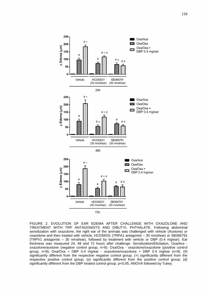
116
FIGURE 2. EVOLUTION OF EAR EDEMA AFTER CHALLENGE WITH OXAZOLONE AND TREATMENT WITH TRP ANTAGONISTS AND DIBUTYL PHTHALATE. Following abdominal sensitization with oxazolone, the right ear of the animals was challenged with vehicle (Acetone) or oxazolone and then treated with vehicle, HC030031 (TRPA1 antagonist – 30 nmol/ear) or SB366791 (TRPV1 antagonist – 30 nmol/ear), followed by treatment with vehicle or DBP (0.4 mg/ear). Ear thickness was measured 24, 48 and 72 hours after challenge. Sensitization/Elicitation. Oxa/Ace - oxazolone/acetone (negative control group, n=9); Oxa/Oxa - oxazolone/oxazolone (positive control group, n=9); Oxa/Oxa + DBP 0.4 mg/ear - oxazolone/oxazolone + DBP 0.4 mg/ear (n=9). (#) significantly different from the respective negative control group; (+) significantly different from the respective positive control group; (o) significantly different from the positive control group; (d) significantly different from the DBP treated control group. p<0,05, ANOVA followed by Tukey.
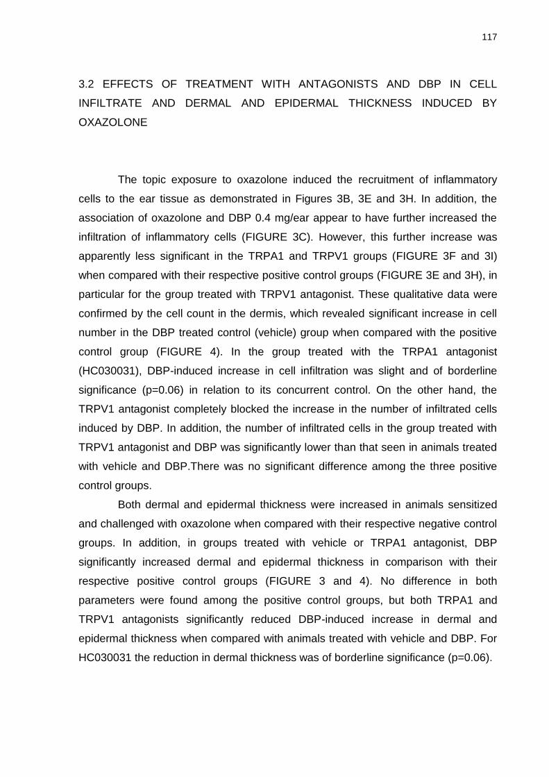
117
3.2 EFFECTS OF TREATMENT WITH ANTAGONISTS AND DBP IN CELL
INFILTRATE AND DERMAL AND EPIDERMAL THICKNESS INDUCED BY
OXAZOLONE
The topic exposure to oxazolone induced the recruitment of inflammatory
cells to the ear tissue as demonstrated in Figures 3B, 3E and 3H. In addition, the
association of oxazolone and DBP 0.4 mg/ear appear to have further increased the
infiltration of inflammatory cells (FIGURE 3C). However, this further increase was
apparently less significant in the TRPA1 and TRPV1 groups (FIGURE 3F and 3I)
when compared with their respective positive control groups (FIGURE 3E and 3H), in
particular for the group treated with TRPV1 antagonist. These qualitative data were
confirmed by the cell count in the dermis, which revealed significant increase in cell
number in the DBP treated control (vehicle) group when compared with the positive
control group (FIGURE 4). In the group treated with the TRPA1 antagonist
(HC030031), DBP-induced increase in cell infiltration was slight and of borderline
significance (p=0.06) in relation to its concurrent control. On the other hand, the
TRPV1 antagonist completely blocked the increase in the number of infiltrated cells
induced by DBP. In addition, the number of infiltrated cells in the group treated with
TRPV1 antagonist and DBP was significantly lower than that seen in animals treated
with vehicle and DBP.There was no significant difference among the three positive
control groups.
Both dermal and epidermal thickness were increased in animals sensitized
and challenged with oxazolone when compared with their respective negative control
groups. In addition, in groups treated with vehicle or TRPA1 antagonist, DBP
significantly increased dermal and epidermal thickness in comparison with their
respective positive control groups (FIGURE 3 and 4). No difference in both
parameters were found among the positive control groups, but both TRPA1 and
TRPV1 antagonists significantly reduced DBP-induced increase in dermal and
epidermal thickness when compared with animals treated with vehicle and DBP. For
HC030031 the reduction in dermal thickness was of borderline significance (p=0.06).
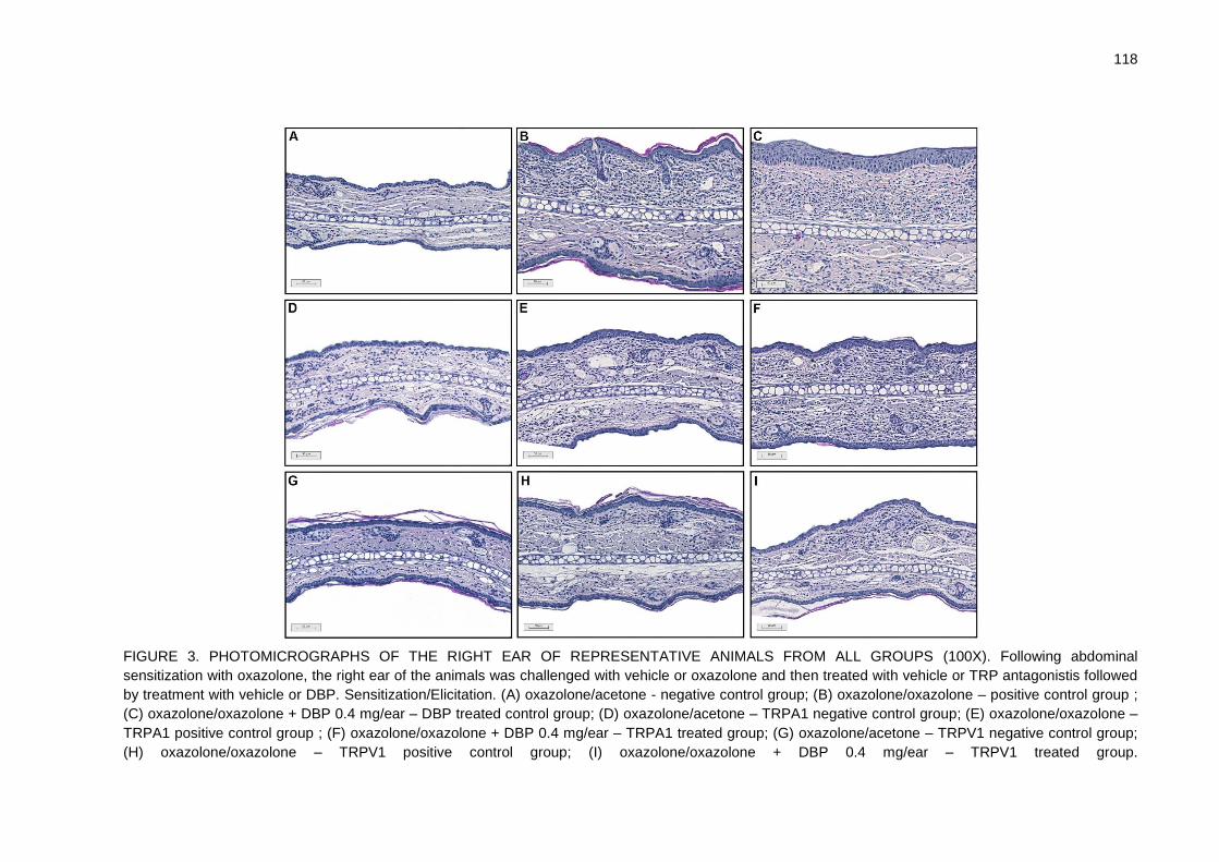
118
FIGURE 3. PHOTOMICROGRAPHS OF THE RIGHT EAR OF REPRESENTATIVE ANIMALS FROM ALL GROUPS (100X). Following abdominal
sensitization with oxazolone, the right ear of the animals was challenged with vehicle or oxazolone and then treated with vehicle or TRP antagonistis followed
by treatment with vehicle or DBP. Sensitization/Elicitation. (A) oxazolone/acetone - negative control group; (B) oxazolone/oxazolone – positive control group ;
(C) oxazolone/oxazolone + DBP 0.4 mg/ear – DBP treated control group; (D) oxazolone/acetone – TRPA1 negative control group; (E) oxazolone/oxazolone –
TRPA1 positive control group ; (F) oxazolone/oxazolone + DBP 0.4 mg/ear – TRPA1 treated group; (G) oxazolone/acetone – TRPV1 negative control group;
(H) oxazolone/oxazolone – TRPV1 positive control group; (I) oxazolone/oxazolone + DBP 0.4 mg/ear – TRPV1 treated group.

119
FIGURE 4. HISTOLOGICAL PARAMETERS. Following abdominal sensitization with oxazolone, the right ear of the animals was challenged with vehicle or oxazolone and then treated with vehicle or TRP antagonistis followed by treatment with vehicle or DBP. For each animal, histological parameters were evaluated in three representative areas of three histological sections. Sensitization/Elicitation. Oxa/Ace - oxazolone/acetone (n=5); Oxa/Oxa - oxazolone/oxazolone (n=5); Oxa/Oxa + DBP 0.4 mg/ear - oxazolone/oxazolone + DBP 0.4 mg/ear (n=5). (#) significantly different from the respective negative control group; (+) significantly different from the respective positive control group; (d) significantly different from the DBP treated control group; (* Number of infiltrated cells) p=0.06 when compared to the TRPA1 positive control group; (* Dermal thickness) p=0.06 when compared with the DBP treated control group. p<0,05, ANOVA followed by Tukey. (t) significantly different from the negative control group. p<0.05, Welch’s t test.
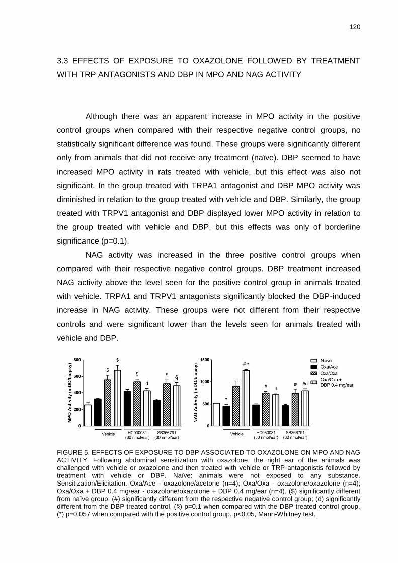
120
3.3 EFFECTS OF EXPOSURE TO OXAZOLONE FOLLOWED BY TREATMENT
WITH TRP ANTAGONISTS AND DBP IN MPO AND NAG ACTIVITY
Although there was an apparent increase in MPO activity in the positive
control groups when compared with their respective negative control groups, no
statistically significant difference was found. These groups were significantly different
only from animals that did not receive any treatment (naïve). DBP seemed to have
increased MPO activity in rats treated with vehicle, but this effect was also not
significant. In the group treated with TRPA1 antagonist and DBP MPO activity was
diminished in relation to the group treated with vehicle and DBP. Similarly, the group
treated with TRPV1 antagonist and DBP displayed lower MPO activity in relation to
the group treated with vehicle and DBP, but this effects was only of borderline
significance (p=0.1).
NAG activity was increased in the three positive control groups when
compared with their respective negative control groups. DBP treatment increased
NAG activity above the level seen for the positive control group in animals treated
with vehicle. TRPA1 and TRPV1 antagonists significantly blocked the DBP-induced
increase in NAG activity. These groups were not different from their respective
controls and were significant lower than the levels seen for animals treated with
vehicle and DBP.
FIGURE 5. EFFECTS OF EXPOSURE TO DBP ASSOCIATED TO OXAZOLONE ON MPO AND NAG ACTIVITY. Following abdominal sensitization with oxazolone, the right ear of the animals was challenged with vehicle or oxazolone and then treated with vehicle or TRP antagonistis followed by treatment with vehicle or DBP. Naïve: animals were not exposed to any substance. Sensitization/Elicitation. Oxa/Ace - oxazolone/acetone (n=4); Oxa/Oxa - oxazolone/oxazolone (n=4); Oxa/Oxa + DBP 0.4 mg/ear - oxazolone/oxazolone + DBP 0.4 mg/ear (n=4). ($) significantly different from naïve group; (#) significantly different from the respective negative control group; (d) significantly different from the DBP treated control, (§) p=0.1 when compared with the DBP treated control group, (*) p=0.057 when compared with the positive control group. p<0.05, Mann-Whitney test.
121
4 DISCUSSION
The prevalence of allergic diseases, as atopic dermatitis, asthma and rhinitis
has been increasing (SEGAWA; HIRASAWA, 2014), and this could be related to the
presence of chemicals with immune adjuvancy potential in the environment.
Phthalate esters, which are widely found in the environment, are candidate chemicals
with adjuvant effects (LARSEN et al., 2002). Epidemiological data indicate that
certain phthalate esters could participate in the development of allergic diseases
such as asthma and atopic dermatitis (BORNEHAG et al., 2004; JAAKKOLA;
KNIGHT, 2004). Besides, it’s been reported that phthalates could enhance the
immune response to allergens in animal models of CHS where phthalates are
present in the sensitization phase (IMAI et al., 2006; Li et al., 2014; MATSUDA et al.,
2010). We have previously demonstrated that DBP aggravates the immune response
in an animal model of CHS induced by oxazolone in BALB/c mice when DBP is
associated to oxazolone in the elicitation phase of the model. We observed increase
in ear edema, polymorphonuclear and mononuclear leukocyte infiltration and
epidermal hyperplasia. In addition, we have also investigated the adjuvant effect of
DBP in two in vitro models of contact hypersensitivity, the NCTC 2544 IL-18 assay
(used as surrogate of contact allergen-induced keratinocyte activation) and the THP-
1 activation assay (used as marker of dendritic cell activation). We found that DBP
potentiates chemical allergen-induced THP-1 activation but no adjuvant effect was
observed in the activation of NCTC 2544 cells (LOURENÇO et al., 2015).
It has been reported that phthalates could activate TRPA1 and TRPV1 in
vitro (SHIBA et al., 2009; JINNO; OHKAWARA; TANAKA-KAGAWA, 2012). In addition,
Maruyama et al. (2007) showed that desensitization of sensory neurons by local
treatment with AITC or CAP suppressed skin sensitization to FITC in the presence of
DBP in a mouse model. This was associated with reduced maturation and trafficking
of FITC-presenting dendritic cells. Besides, a CGRP antagonist also suppressed the
sensitization to FITC, suggesting a possible involvement of sensory neurons in
sensitization. Shiba et al. (2012) also reported that pretreatment with TRPA1
antagonist HC030031 upon skin sensitization to FITC suppressed the enhancing
effect of DBP on sensitization.
122
In the present study we demonstrated that local treatment with TRPA1
(HC030031) and TRPV1 antagonists (SB366791) partially or completely suppressed,
respectively, the adjuvant effect of DBP in a BALB/c model of CHS induced by the
allergen oxazolone. These TRP antagonists were able to reverse DBP’s adjuvant
effect on ear edema, leukocyte infiltration and epidermal hyperplasia in oxazolone-
challenged animals. Even though others have shown the reduction in ear swelling
enhanced by phthalates after desensitization of sensory neurons or using TRP or
neuropeptide antagonists (MARUYAMA et al., 2007; SHIBA et al., 2012), this was
showed when DBP was associated in both phases of the CHS model. Our results
show that even after the sensitization has already been established, DBP can still
exert adjuvant effect and enhance the inflammatory response, modulating the
elicitation phase of CHS induced by oxazolone. More importantly, treatment with
TRPA1 and TRPV1 antagonists after challenge with oxazolone was enough to
partially or completely prevent the adjuvant effect of DBP in oxazolone-induced CHS,
corroborating the hypothesis that TRPs are molecular targets that mediate
inflammatory responses induced by phthalates.
It is important to highlight that overall no difference was found among the
positive control groups, i.e., oxazolone response was not modified by treatment with
TRP antagonists. Banvolgyi et al. (2005) showed that TRPV1-deficient mice
presented enhanced response in CHS induced by oxazolone. On the other hand, Liu
et al. (2013) demonstrated that treatment with TRPA1 antagonist HC030031, prior
challenge, inhibited skin edema, keratinocyte hyperplasia, nerve growth, leukocyte
infiltration, and antihistamine-resistant scratching behavior in mice exposed to
oxazolone. In addition, it’s been shown that CGRP applied topically at the site of
contact with the allergen, during both sensitization and elicitation phase, enhanced
the sensitization in oxazolone-induced CHS (GUTWALD; GOEBELER; SORG, 1991).
Different from previous studies, that intended to investigate the effects of TRP in the
sensitization in oxazolone-induced CHS, in the present study we have chosen to
expose the animals to TRP antagonists only after challenge with oxazolone, aiming
to investigate the modulation of these antagonists in the adjuvant effect of DBP in
oxazolone-induced CHS; therefore no modulation on oxazolone effect was observed.
In this study, both TRPV1 and TRPA1 antagonists were able to reverse the
adjuvant effects of DBP. Animals treated with TRPA1 antagonist and DBP presented
reduced ear edema and dermal and epidermal thickness when compared with the
123
DBP control group, although these parameters were still higher in this group in
comparison with TRPA1 positive control group. On the contrary, TRPV1 antagonist
reversed all of the parameters analyzed in animals treated with TRPV1 antagonist
and DBP, in relation to TRPV1 positive control group. We have previously
demonstrated that DBP is capable of potentiating allergen-induced activation of THP-
1 cells in vitro, which are cells used as surrogates for dendritic cell-activation
(LOURENÇO et al., 2015). In addition, it has been reported that both TRPV1 and
TRPA1 are expressed on dendritic cells (DC) and that the activation of TRPV1 in DC
could induce their maturation and enhance their antigen presenting activity (BASU;
SRIVASTAVA, 2005). On the other hand, in our previous in vitro study, DBP did not
present any adjuvant effect in the allergen-induced activation of keratonocytes
(NCTC 2544 cells). However, others have shown that DBP increased the production
of thymic stromal lymphopoietin (TSLP) in FITC-induced contact hypersensitivity in
mice (SHIGENO et al., 2009; LARSON et al., 2010), but the mechanism of TSLP
production upon exposure to DBP was not evaluated in those studies. TSLP is an IL-
7–related cytokine, produced by epithelial cells, like keratinocytes, that has been
linked to atopic dermatitis and asthma (LEYVA-CASTILLO et al., 2013). It has been
also demonstrated that TRPA1 are expressed in human keratinocytes
(ATOYAN; SHANDER; BOTCHKAREVA, 2009), and Shiba et al. (2012) suggested
that it is possible that TRPA1 activation on keratinocytes could trigger the production
of TSLP, and that this could be part of the mechanism of enhanced skin-sensitization
to FITC induced by phthalates. DBP could activate TRPV1 and TRPA1 directly in
dendritic cells enhancing their maturation and antigen presenting activity, besides
activating these receptors in keratinocytes and also in sensory neurons triggering the
release of neuropeptides such as CGRP and SP, that have been shown to
participate in CHS response (MARUYAMA et al., 2007; MIKAMI et al., 2011; LIU et
al., 2013).
In conclusion, our data suggest that both TRPA1 and TRPV1 activation could
participate in the adjuvant effect of DBP in the elicitation phase of oxazolone-induced
CHS model. Our data contribute to further understanding of the molecular
mechanisms involved in the adjuvant effect of phthalates.
124
REFERENCES
ATOYAN, R.; SHANDER, D.; BOTCHKAREVA, N.V. Non-neuronal expression of transient receptor potential type A1 (TRPA1) in human skin. J. Invest. Dermatol., v. 129, p. 2312-2315, 2009. BANDELL, M. et al. Noxious cold ion channel TRPA1 is activated by pungent compounds and bradykinin. Neuron, v. 41, p. 849–57, 2004. BÁNVÖLGYI, A. et al. Evidence for a novel protective role of the vanilloid TRPV1 receptor in a cutaneous contact allergic dermatitis model. J. Neuroimmunol., v. 169, p. 86–96, 2005. BASU, S.; SRIVASTAVA, P. Immunological role of neuronal receptor vanilloid receptor 1 expressed on dendritic cells. Proc. Natl. Acad. Sci. USA, v. 102, p. 5120–5125, 2005. BAUTISTA, D.M. et al. Pungent products from garlic activate the sensory ion channel TRPA1. Proc. Natl. Acad. Sci. USA, v. 102, p. 12248–12252, 2005. BERESFORD, L. et al. Nerve fibres are required to evoke a contact sensitivity response in mice. Immunology, v. 111, p. 118–125, 2004. BEYDON, D.; PAYAN, J.P.; GRANDCLAUDE, M.C. Comparison of percutaneous absorption and metabolism of di-n-butylphthalate in various species. Toxicol. In Vitro, v. 24, p. 71–78, 2010. BORNEHAG, C.G. et al. The association between asthma and allergic symptoms in children and phthalates in house dust: a nested case-control study. Environ. Health Persp., v. 112, p. 1393-1397, 2004. BRADLEY, P.P. et al. Measurement of cutaneous inflammation: estimation of neutrophil content with an enzyme marker. J. Invest. Dermatol., v. 78, n. 3, p. 206-209, 1982. CASILLAS, A.M. et al. Enhancement of allergic inflammation by diesel exhaust particles: permissive role of reactive oxygen species. Ann. Allerg. Asthma Im., v. 83, p. 624–629, 1999. CATERINA, M.J. et al. The capsaicin receptor: a heat-activated ion channel in the pain pathway. Nature, v. 389, p. 816–824, 1997. CHOWDHURY, M.M.;, STATHAM, B.N. Allergic contact dermatitis from dibutyl phthalate and benzalkonium chloride in Timodine cream. Contact Dermatology, v. 46, p. 57, 2002.
125
CLAPHAM, D.E. TRP channels as cellular sensors. Nature, v. 426, p. 517–524, 2003. CPSC (CONSUMER PRODUCT SAFETY COMMISSION). Overview of Phthalates Toxicity. Bestheda, United State of America, p. 1-31, 2010. DE YOUNG, L.M. et al. Edema and cell infiltration in the phorbol ester-treated mouse ear are temporally separate and can be differentially modulated by pharmacologic agents. Agents Actions, v. 26, n. 3-4, p. 335-341, 1989. DOAN, K.; BRONAUGH, R.L; YOURICK, J.J. In vivo and in vitro skin absorption of lipophilic compounds, dibutyl phthalate, farnesol and geraniol in the hairless guinea pig. Food Chem. Toxicol., v. 48, p. 18–23, 2010. ELSISI, A.; CARTER, D.E.; SIPES, I.G. Dermal absorption of phthalate diesters in rats. Fundam. Appl. Toxicol., v. 12, p. 70-77, 1989. FDA (FOOD AND DRUG ADMINISTRATION). Guidance for Industry. Estimating the Maximum Safe Starting Dose in Initial Clinical Trials for Therapeutics in Adult Healthy Volunteers, 30 pp., 2005. GRABBE, S.; SCHWARZ, T. Immunoregulatory mechanisms involved in elicitation of allergic contact hypersensitivity. Immunol. Today, v. 19, n.1, p. 37–44, 1998. GUTWALD, J.; GOEBELER, M.; SORG, C. Neuropeptides enhance irritant and allergic contact dermatitis. J. Invest. Dermatol., v. 96, p. 695–698, 1991. HENNINO, A. Fisiopatologia da dermatite de contato alérgica: papel das células T CD8 efetoras e das células T CD4 regulatórias. Anais Brasileiros de Dermatologia, v. 80, n. 4, p. 335-347, 2005. IMAI, Y. et al. Effects of phthalate esters on the sensitization phase of contact hypersensitivity induced by fluorescein isothiocyanate. Clin. Exp. Allergy, v. 36, p. 1462–1468, 2006. INTERNATIONAL JOURNAL OF TOXICOLOGY. Annual Review of Cosmetic Ingredient Safety Assessment- 2002/2003. Int. J. Toxicol., v. 24, n. 1, p. 1-102, 2005. IPCS (INTERNATIONAL PROGRAMME ON CHEMICAL SAFETY). Environmental Health Criteria 189. Di-n-butyl phthalate. World Health Organization: Geneva, 1997. Disponível em: <http://www.inchem.org/documents/ehc/ehc/ehc189.htm>. Acesso em: 07/03/2015. JAAKKOLA, J.J.K.; KNIGHT, T.L. Asthma, wheezing, and allergies in Russian schoolchildren in relation to new surface materials in the home. Am. J. Public Health, v. 94, p. 560-562, 2004.
126
JINNO, H.; OHKAWARA, S.; TANAKA-KAGAWA, T. Activation of nociceptive transient receptor potential channels by phthalates. J. Toxicol. Sci., v. 37, n. 2, p. s428, 2012. JORDT, S.E. et al. Mustard oils and cannabinoids excite sensory nerve fibres through the TRP channel ANKTM1. Nature, v. 427, p. 260–5, 2004. KARLBERG, A.T. et al. Allergic Contact Dermatitis–Formation, Structural Requirements, and Reactivity of Skin Sensitizers. Chem. Res. Toxicol., v. 21, n. 1, p. 53-69, 2008. KIMBER, I. et al. Allergic contact dermatitis. Int. Immunopharmacol., v. 2, p. 201–211, 2002. KITAJIMA, M. et al. TSLP enhances the function of helper type 2 cells. Eur. J. Immunol., v. 41, p. 1862–1871, 2011. KOO, H.J.; LEE, B.M. Estimated exposure to phthalates in cosmetics and risk assessment. J. Toxicol. Env. Health A: Current Issues, v. 67, n. 23-24, p. 1901-1914, 2004. LARSEN, S. et al. Adjuvant effects of di-n-butyl-, di-n-octyl-, di-iso-nonyl- and di-iso-decyl phthalate in a subcutaneous injection model using BALB/c mice. Pharmacol. Toxicol., v. 91, p. 264–272, 2002. LARSON, R.P. et al. Dibutyl phthalate-induced thymic stromal lymphopoietin is required for Th2 contact hypersensitivity responses. J. Immunol., v. 184, p. 2974–2984, 2010. LEYVA-CASTILLO, J.M. et al. TSLP Produced by Keratinocytes Promotes Allergen Sensitization through Skin and Thereby Triggers Atopic March in Mice. J. Invest. Dermatol., v. 133, p. 154–163, 2013. LI, J. et al. T-Helper Type-2 Contact Hypersensitivity of BALB/c Mice Aggravated by Dibutyl Phthalate via Long-Term Dermal Exposure. PLoS ONE, v. 9, n. 2, p. e87887, 2014. LIU, B. et al. TRPA1 controls inflammation and pruritogen responses in allergic contact dermatitis. FASEB J., v. 27, p. 3549–3563, 2013. LIU, Y. et al. Neuron-mediated generation of regulatory T cells from encephalitogenic T cells suppresses EAE. Nat. Med., v. 12, p. 518–525, 2006. LOURENÇO, A.C.S. et al. The plasticizer dibutyl phthalate (DBP) potentiates chemical allergen-induced THP-1 activation. Toxicol. In Vitro, v. 29, p. 2001–2008, 2015. MARUYAMA, T. et al. Influence of local treatments with capsaicin or allyl isothiocyanate in the sensitization phase of a fluorescein-isothiocyanate-induced contact sensitivity model. Int. Arch. Allergy Immunol., v. 143, p. 144–154, 2007.
127
MATSUDA, T. et al. Phthalate esters reveal skin-sensitizing activity of phenethyl isothiocyanate in mice. Food Chem. Toxicol., v. 48, p. 1704–1708, 2010. MIKAMI, N. et al. Calcitonin gene-related peptide is an important regulator of cutaneous immunity: effect on dendritic cell and T cell functions. J. Immunol., v. 186, p. 6886–6893, 2011. PALM, N.W. et al. Allergic host defenses. Nature, v. 484, p. 465-472, 2012. PAN, T.L et al. Dermal toxicity elicited by phthalates: Evaluation of skin absorption, immunohistology, and functional proteomics. Food Chem. Toxicol., v. 65, p. 105–114, 2014. RECIO, M.C. et al. In vivo activity of pseudoguaianolide sesquiterpene lactones in acute and chronic inflammation. Life Science, v. 66, n. 26, p. 2509-2518, 2000. SANCHEZ, T.; MORENO, J.J. Role of leukocyte influx in tissue prostaglandin H synthase-2 overexpression induced by phorbol ester and arachidonic acid in skin. Biochem. Pharmacol., v. 58, p. 877-879, 1999. SATHYANARAYANA, S. et al. Baby care products: possible sources of infant phthalate exposure. Pediatrics, v. 121, p. e260–e268, 2008. SEGAWA, R.; HIRASAWA, N. Exacerbation of Allergic Diseases by Chemicals: Role of TSLP. J. Pharmacol. Sci., v. 124, p. 301-306, 2014. SHIBA, T. et al. Transient receptor potential ankyrin 1 activation enhances hapten sensitization in a T-helper type 2-driven fluorescein isothiocyanate-induced contact hypersensitivity mouse model. Toxicol. Appl. Pharmacol., v. 264, p. 370–376, 2012. SHIBA, T. et al. TRPA1 and TRPV1 activation is a novel adjuvant effect mechanism in contact hypersensitivity. J. Neuroimmunol., v. 207, p. 66–74, 2009. SHIGENO, T. et al. Phthalate ester-induced thymic stromal lymphopoietin mediates allergic dermatitis in mice. Immunology, v. 128, p. e849-e857, 2009. SILVA, C.R. et al. The involvement of TRPA1 channel activation in the inflammatory response evoked by topical application of cinnamaldehyde to mice Life Sciences, v. 88, p. 1077–1087, 2011. STORY, G.M. et al. ANKTM1, a TRP-like channel expressed in nociceptive neurons, is activated by cold temperatures. Cell, v. 112, p. 819–829, 2003. WATANABE, N. et al. Human TSLP promotes CD40 ligand–induced IL-12 production by myeloid dendritic cells but maintains their Th2 priming potential. Blood, v. 105, n. 12, p. 4749-4751, 2005.

128
WEBB, E.F. et al. Intralesional cytokines in chronic oxazolone-induced contact sensitivity suggests roles for Tumor Necrosis Factor α and Interleukin-4. The Society for Investigative Dermatology, v. 111, p. 86-92, 1998. ZHANG, L.; TINKLE, S.S. Chemical Activation of Innate and Specific Immunity in Contact Dermatitis. J. Invest. Dermatol., v. 115, n. 2, p. 168-172, 2000. ZYGMUNT, P.M. et al. Vanilloid receptors on sensory nerves mediate the vasodilator action of anandamide. Nature, v. 400, p. 452–457, 1999.
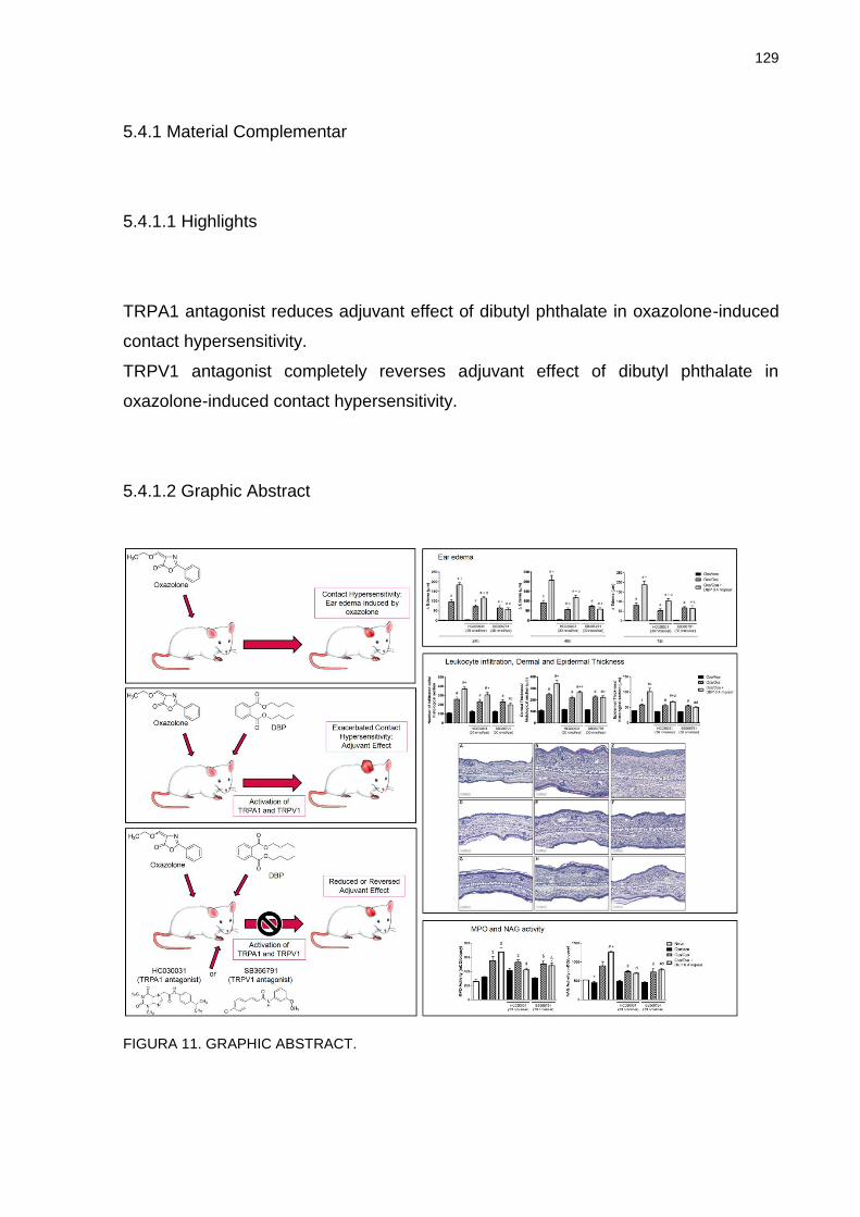
129
5.4.1 Material Complementar
5.4.1.1 Highlights
TRPA1 antagonist reduces adjuvant effect of dibutyl phthalate in oxazolone-induced
contact hypersensitivity.
TRPV1 antagonist completely reverses adjuvant effect of dibutyl phthalate in
oxazolone-induced contact hypersensitivity.
5.4.1.2 Graphic Abstract
FIGURA 11. GRAPHIC ABSTRACT.
130
6 DISCUSSÃO GERAL
Como os seres humanos são constantemente expostos a ftalatos, é
importante elucidar o potencial imunotóxico dos mesmos. Estudos toxicológicos têm
demonstrado que ésteres de ftalatos, como o DBP, não possuem um potencial
significativo de causarem sensibilização da pele, pois não são capazes de se
ligarem a proteínas e formar um complexo hapteno-proteína, pré-requisito para a
estimulação alérgica por uma substância química (KIMBER; DEARMAN, 2010). De
acordo com dados epidemiológicos, ésteres de ftalatos não tem o potencial de
causar irritação ou sensibilização da pele (GEIER et al., 2004; MEDEIROS; DEVLIN;
KELLER, 1999). No entanto, mesmo que evidências apoiem o potencial
sensibilizante negativo dos ftalatos, tem sido sugerido que certos ftalatos poderiam
atuar como adjuvantes, acelerando, prolongando ou exacerbando as respostas
imunes.
A prevalência de doenças alérgicas, como dermatites, asma e rinite, tem
aumentado nas últimas décadas (SEGAWA; HIRASAWA, 2014), e isso poderia estar
relacionado não somente com o aumento de substâncias sensibilizantes, como
também com a presença de substâncias químicas com potencial adjuvante no
ambiente. Os ésteres de ftalatos, amplamente encontrados no ambiente, são
possíveis candidatos com potencial adjuvante (LARSEN et al., 2002). Tem sido
proposto que os ftalatos possam estar associados com facilitação no
desenvolvimento de doenças alérgicas como asma e dermatite (BORNEHAG et al.,
2004; JAAKKOLA; KNIGHT, 2004), além de ter sido demonstrado que certos ftalatos
são capazes de aumentar a resposta a alérgenos em modelos animais de dermatite
de contato alérgica (IMAI et al., 2006; LI et al., 2014). Apesar de estudos anteriores
in vivo terem demonstrado que certos ftalatos são capazes de agravar a resposta
e/ou doenças alérgicas, os mecanismos celulares e moleculares envolvidos
permanecem obscuros.
A identificação de substâncias sensibilizantes é feita principalmente pelo uso
de modelos animais de inflamação cutânea, pois os mesmos mimetizam alguns
eventos observados em dermatites desenvolvidas por humanos (VANE; BOTTING,
1998; BOUCLIER et al., 1990). O modelo de edema de orelha induzido pela
131
oxazolona (FUJII et al., 2002) tem sido bastante utilizado com esse propósito, pois
se assemelha à hipersensibilidade de contato em humanos. No entanto, novos
ensaios in vitro têm sido desenvolvidos com o propósito de encontrar marcadores
biológicos capazes de discriminar substâncias sensibilizantes as quais os humanos
estejam expostos. Nesse contexto, o ensaio NCTC 2544 IL-18 e o teste de ativação
de células THP-1 (CORSINI et al., 2009; GALBIATI et al., 2011; CORSINI et al.,
2013a; MITJANS et al., 2008) têm sido bastante utilizados com esse propósito.
O presente estudo investigou os efeitos do DBP nos modelos in vitro e in
vivo de hipersensibilidade de contato acima citados. Os resultados obtidos indicam
que o DBP é capaz de agravar a resposta imune e inflamatória envolvida nestes
modelos. Neste estudo, observamos que o DBP foi capaz de potencializar a ativação
de células THP-1 induzida por alérgenos, aumentando assim a expressão do
marcador de superfície celular CD86 e a liberação de interleucina-8. Além disso,
demonstramos que o DBP foi capaz de agravar a resposta inflamatória no modelo
de edema de orelha induzido por oxazolona, quando associado a este hapteno
durante a fase de elicitação do modelo, o que foi demonstrado por aumento em
edema, recrutamento de leucócitos e hiperplasia epidérmica. Também investigamos
possíveis mecanismos do efeito adjuvante exercido pelo DBP. Identificamos in vitro
uma participação da produção de espécies reativas de oxigênio na ativação de
células THP-1. In vivo, observamos a participação da ativação dos receptores
TRPA1 e TRPV1 no efeito do DBP. Os dados obtidos neste estudo mostram que os
ftalatos podem estar agindo como adjuvantes não somente na fase de sensibilização
de processos alérgicos, como também na fase de elicitação, e que esse efeito pode
estar mais relacionado com o aumento da ativação de células dendríticas
apresentadoras de antígenos. Além disso, nossos resultados contribuem para um
maior entendimento dos mecanismos envolvidos no efeito adjuvante dos ftalatos em
modelos de hipersensibilidade de contato.
Os resultados encontrados neste estudo apoiam dados epidemiológicos e
experimentais que sugerem que o DBP não apresenta potencial significativo de
causar sensibilização ou irritação da pele (KIMBER; DEARMAN, 2010; GEIER et al.,
2004; MEDEIROS; DEVLIN; KELLER, 1999). Observamos que o DBP não foi capaz
de induzir edema, recrutamento celular ou hiperplasia epidérmica no modelo in vivo,
além de não induzir a ativação de céluas THP-1 ou NCTC nos ensaios in vitro
(FIGURAS 07, 08 e 10).
132
A partir da análise de edema e da contagem de células infiltradas na derme,
observou-se que o DBP nas doses mais altas (0,4 e 4 mg/orelha) foi capaz de
exacerbar o desenvolvimento do edema de orelha induzido por oxazolona em
camundongos, além de aumentar o recrutamento de leucócitos para a derme.
Somado a isso, a proliferação de células na epiderme é característica da dermatite
de contato causada pela oxazolona (FUJII et al., 2002). Novamente, DBP 0,4 e 4
mg/orelha induziram aumento na hiperplasia epidérmica induzida por oxazolona.
O aumento do edema de orelha (IMAI et al., 2006; SHIBA et al., 2009;
MATSUDA et al., 2010; LI et al., 2014) e da infiltração celular (TAKANO et al., 2006;
LI et al., 2014) demonstrados nesse experimento já foi relatado por outros autores,
embora utilizando doses maiores de ftalatos. Imai e colaboradores (2006)
demonstraram que, não só o DBP, como outros ftalatos foram capazes de exacerbar
o edema de orelha induzido pelo isotiocianato de fluoresceína. Takano e
colaboradores (2006) demonstraram o aumento do edema de orelha e também da
infiltração celular no sítio inflamatório em um modelo de dermatite atópica no qual os
animais foram expostos ao DEHP. Esses mesmos autores demonstraram ainda que
a exposição materna ao ftalato DEHP durante a lactação resultou em exacerbação
do edema de orelha e da migração celular na prole das fêmeas expostas, mostrando
que talvez os efeitos dos ftalatos após exposição durante períodos críticos para o
desenvolvimento, como a gestação e a lactação, não se resumam a alterações no
sistema reprodutivo masculino (YANAGISAWA et al., 2008). Já Li e colaboradores
(2014) mostraram que a exposição dérmica prolongada (40 dias) ao DBP em doses
de 4,0 e 40 mg/kg/dia foi capaz de aumentar o edema de orelha e a infiltração de
leucócitos na derme. Nossos dados apoiam o potencial adjuvante do DBP em
modelos animais de hipersensibilidade de contato, mas aqui reportamos um efeito
adjuvante após um curto período de exposição dérmica a baixas doses de DBP.
Adicionalmente, nossos resultados indicam que o DBP pode atuar como adjuvante
mesmo quando a sensibilização ao alérgeno já se encontra instalada, modulando a
fase de elicitação da HSC induzida por oxazolona. Estudos anteriores têm
observado este efeito em modelos animais de HSC associando ftalatos tanto na fase
de sensibilização quanto na fase de elicitação, ou com altas doses de ftalatos,
exposição por longos períodos de tempo ou em diferentes vias de exposição, em
alguns casos com injeções subcutâneas ou intraperitoneais, que não são vias pelas
quais os humanos encontrariam normalmente os ftalatos no ambiente.
133
O presente estudo se propôs a investigar alguns aspectos relacionados ao
mecanismo do efeito adjuvante do DBP em processos alérgico-inflamatórios. Para
isso foram utilizados dois ensaios in vitro baseados na ativação de dois tipos
celulares que desempenham papel importante no desenvolvimento da
hipersensibilidade de contato, os queratinócitos e as células dendríticas. Em
particular, a produção de IL-18 em células NCTC 2544 é representativa de ativação
de queratinócitos induzida por alérgenos, enquanto a expressão aumentada de
CD86 e produção de IL-8 em células THP-1 foram usadas como marcadores de
ativação de células dendríticas. Os resultados obtidos sugerem que o alvo do efeito
adjuvante do DBP são as células dendríticas, mas não os queratinócitos, já que o
DBP não foi capaz de aumentar a produção de IL-18 induzida por p-Fenilenodiamina
(PPD) em células NCTC.
Mitjans et al. (2008) demonstraram que alérgenos químicos induzem uma
liberação dose dependente de interleucina 8, assim como a expressão do marcador
de superfície CD86 na linhagem celular monocítica humana THP-1, propondo isto
como um ensaio in vitro para screening de potenciais alérgenos de contato. Neste
trabalho, o aumento na expressão de CD86 e na liberação de IL-8 foram
investigados após exposição de células THP-1 a dois alérgenos de contato, citral e
imidazolidinil ureia, e suas associações com DBP. De acordo com o teste do
linfonodo local (LLNA), citral é classificado como um sensibilizante moderado com
um valor EC3 de 9,3%, enquanto o imidazolidinil ureia é classificado como um
sensibilizante fraco com um valor EC3 de 24%. Ambos alérgenos induziram um
aumento significativo na expressão de CD86 e liberação de IL-8. Quando células
THP-1 foram expostas a Cit ou IMZ associados ao DBP, as células exibiram maior
expressão de CD86 e maior liberação de IL-8, quando comparadas as células
expostas a somente os alérgenos. Além disso, associação entre DBP e oxazolona
também revelou maior expressão de CD86 em relação ao grupo exposto a somente
oxazolona. Estes resultados indicam um efeito adjuvante do DBP sobre a ativação
de células THP-1. Apesar de vários estudos terem sido realizados para avaliar os
efeitos dos ftalatos no funcionamento celular, pouco se encontra na literatura sobre o
efeito adjuvante destas substâncias in vitro. No entanto, uma grande quantidade de
estudos sobre o efeito adjuvante de ftalatos in vivo tem sido realizada. Diversos
autores já demonstraram que diferentes ftalatos podem agir como adjuvantes em
hipersensibilidade de contato e inflamação das vias aéreas, elevando a expressão
134
de marcadores de superfície e citocinas relacionadas após exposição a associações
entre alérgenos e ftalatos em diferentes protocolos (IMAI et al., 2006; KOIKE et al.,
2010; MATSUDA et al., 2010; LI et al., 2014). Corroborando nossos dados, TANAKA
et al. (2012) demonstraram que o DEHP agravou a inflamação peritoneal induzida
por ovalbumina caracterizada por aumento na infiltração de macrófagos e neutrófilos
e por produção e liberação aumentada de citocinas como quimiocina derivada dos
queratinócitos (KC) na cavidade peritoneal. KC é um homólogo murino de IL-8 (HOL;
WILHELMSEN; HARALDSEN, 2010). Além disso, Takano et al. (2006) mostraram
que a exposição a DEHP na presença de alérgeno aumentou a expressão de MIP-
1α comparado a exposição a somente o alérgeno. MIP-1α funciona como
quimiotático de neutrófilos assim como IL-8 (HAMMOND et al., 1995; MENTEN;
WUYTS; VAN DAMME, 2002).
A oxazolona é muito utilizada como modelo de hipersensibilidade por
contato, por promover as fases de sensibilização e elicitação. Porém, a grande
maioria dos estudos avalia os efeitos causados pela aplicação dessas substâncias
nos linfonodos e na epiderme, sendo que pouco é discutido sobre as propriedades
inflamatórias na derme (ZHANG; THINKLE, 2000). A avaliação da atividade das
enzimas NAG e MPO foi realizada para indicar a presença de leucócitos
mononucleares e polimorfornucleares, respectivamente (SANCHEZ; MORENO,
1999; BRADLEY et al., 1982), e com isso, caracterizar o possível perfil das células
infiltradas na derme. A partir dessas dosagens, sugere-se que tanto leucócitos
polimorfonucleares (neutrófilos), quanto mononucleares (macrófagos) estão
presentes na resposta inflamatória causada pela oxazolona. Esse resultado está de
acordo com o encontrado por Wiedow e colaboradores (1992), que quantificaram
proteases de macrófagos e neutrófilos na derme, sendo que estas estavam
aumentadas na dermatite de contato alérgica. Sugere-se também que há uma
proporção maior de neutrófilos em relação aos leucócitos mononucleares, uma vez
que o efeito adjuvante ocasionado pelo DBP aumentou em maior proporção a
atividade da enzima MPO em relação à NAG. Os neutrófilos estão envolvidos nas
fases de sensibilização e elicitação da HSC, e são uma das primeiras células a
serem recrutadas após o desafio de camundongos sensibilizados (DILULIO et al.,
1999). Além disso, Zhang e Tinkle (2000) encontraram neutrófilos em maior
quantidade na derme em camundongos tratados com oxazolona e permanecem
dominantes até 72 horas após o tratamento. Assim, nossos resultados estão de
135
acordo com outros dados científicos, sendo que o DBP parece exacerbar a resposta
inflamatória da oxazolona através do aumento da migração dos neutrófilos,
principalmente. Nossos dados in vitro concordam com os dados in vivo sobre uma
maior participação de neutrófilos associados à resposta inflamatória. O aumento da
ativação de células dendríticas demonstrada in vitro leva a uma maior liberação de
IL-8, um potente atrativo de neutrófilos (BARKER et al., 1991). Como mencionado
anteriormente, já foi demonstrado em modelos animais um aumento de citocinas
como quimiocina derivada dos queratinócitos (KC) (TANAKA et al., 2012), um
homólogo murino de IL-8 (HOL; WILHELMSEN; HARALDSEN, 2010), e de MIP-1α
(TAKANO et al., 2006) que também atua como quimiotático de neutrófilos
(HAMMOND et al., 1995; MENTEN; WUYTS; VAN DAMME, 2002). Portanto, DBP
poderia estar potencializando a ativação de células dendríticas in vivo e isso poderia
refletir em maior recrutamento de neutrófilos como parte do mecanismo da ação
adjuvante do DBP.
Algumas evidências indicam que doenças inflamatórias e alérgicas de pele
poderiam ser mediadas por estresse oxidativo (OKAYAMA, 2005; BYAMBA et al.,
2010). Mizuashi et al. (2005) hipotetizaram que substâncias sensibilizantes induzem
estresse oxidativo em células dendríticas, o que subsequentemente estimula p38
MAPK. Seus resultados demonstraram que sensibilizantes reduziram a forma
oxidada (GSSG) versus a forma reduzida (GSH) de glutationa celular (razão
GSH/GSSG) em células THP-1 e levaram a fosforilação de p38 MAPK. De acordo
com Mitjans et al. (2008), a ativação de p38 MAPK por sensibilizantes desempenha
um papel crucial na ativação da liberação de IL-8. No presente estudo, juntamente
com o aumento na liberação de IL-8, foi demonstrado um pequeno aumento na
geração de espécies reativas de oxigênio induzido por DBP, e, mais importante, um
aumento significativo no estresse oxidativo induzido por DBP em associação ao
citral Outros já demonstraram que alguns ftalatos podem induzir estresse oxidativo.
Tetz et al. (2013) reportou um aumento da produção de ROS induzido por MEHP em
células placentárias humanas. DEHP também aumentou a geração de ROS em
testículos, levando a apoptose de espermatócitos (KASAHARA et al., 2002). Ainda,
You et al. (2014) mostraram níveis de ROS aumentados significativamente em
animais sensibilizados com ovalbumina e tratados com DEHP quando comparados
aos niveis de ROS dos animais apenas sensibilizados. Esses autores sugerem que
o estresse oxidativo desempenhe um papel crítico no efeito adjuvante do DEHP.
136
In vivo, demonstramos uma queda em níveis de GSH induzida por
oxazolona no grupo controle positivo e também no controle negativo, que foi exposto
a oxazolona somente na fase de sensibilização. Esta redução foi acompanhada de
aumento nos níveis de LOOH no grupo controle positivo. O estresse oxidativo
induzido pela oxazolona em modelo de hipersensibilidade de contato já foi reportado
(BRUNYÁNSZKI et al., 2010). Também demonstramos que a sensibilização e
desafio com DBP 0.4 mg reduziu os níveis de GSH, mas não foi observado aumento
em LOOH neste grupo. Já que muitas vias estão envolvidas na neutralização de
EROs, é difícil especular a razão pela qual DBP induziu uma diminuição em GSH
sem aumentar LOOH. O que é interessante, é que a mesma dose de DBP (0.4 mg)
reverteu parcialmente a redução em GSH e aumento em LOOH induzida por
oxazolona, enquanto DBP 0.04 mg reverteu completamente os mesmos parâmetros,
e o grupo tratado com a maior dose de DBP (4mg) não diferiu do grupo controle
positivo. Nossos resultados parecem indicar que o estresse oxidativo não participa
do efeito adjuvante do DBP no modelo animal avaliado com as doses escolhidas.
Chen et al. (2015) reportou um fraco efeito adjuvante do DBP em modelo murino de
asma e observou que esse efeito não foi mediado por um mecanismo de estresse
oxidativo. Apesar de termos reportado que DBP foi capaz de induzir produção de
EROs in vitro e aumentar essa produção quando induzida por um alérgeno, o tempo
no qual foi avaliado esse parâmetro foi diferente da análise in vivo. Enquanto in vitro
a produção de EROs foi avaliada imediatamente após o tratamento das células com
DBP associado ao alérgeno, in vivo a avaliação dos níveis de GSH e LOOH foi feita
três dias após a exposição ao ftalato e à oxazolona, assim, o papel da resposta
inflamatória induzida tanto pela oxazolona quanto pelo DBP na produção de EROs
deve ser considerado. Além disso, ftalatos parecem apresentar efeitos contrários em
estudos sobre seu potencial adjuvante quando são usadas doses altas ou baixas e
diferentes tempos e vias de exposição (KIMBER; DEARMAN, 2010), portanto,
diferentes doses de ftalatos poderiam influenciar o balanço redox celular de
maneiras diferentes.
Além do possível papel do estresse oxidativo no desenvolvimento da
hipersensibilidade de contato, avaliamos qual seria a participação da ativação dos
receptores TRPA1 e TRPV1 no efeito adjuvante do DBP no modelo de edema de
orelha induzido por oxazolona. Estudos já demonstraram que os ftalatos são
capazes de ativar tanto TRPA1 quanto TRPV1 in vitro (SHIBA et al., 2009; JINNO;
137
OHKAWARA; TANAKA-KAGAWA, 2012). Além disso, já foi reportada a participação
desses receptores em modelos animais de hipersensibilidade de contato. Maruyama
et al. (2007) mostraram que a dessensibilização de neurônios sensoriais através do
tratamento tópico com alil isotiocianato ou capsaicina suprimiram a sensibilização da
derme induzida por isotiocianato de fluoresceína (FITC) na presença de DBP em um
modelo animal, o que foi associado a redução da maturação e migração de células
dendríticas. Adicionalmente, um antagonista do neuropeptídeo CGRP também foi
capaz de suprimir a sensibilização ao FITC, apoiando o possível envolvimento dos
neurônios sensoriais na sensibilização. Shiba et al. (2012) também demonstraram
que o pré tratamento com o antagonista TRPA1 HC030031 durante a sensibilização
ao FITC revogou o efeito adjuvante do DBP na sensibilização.
Neste estudo demonstramos que o tratamento tópico com o antagonista
TRPA1 HC030031 e com o antagonista TRPV1 SB366791 após o tratamento com
oxazolona, mas antes da exposição ao DBP, suprimiram parcial ou completamente,
respectivamente, o efeito adjuvante do DBP, como pudemos observar através da
redução do edema de orelha, da infiltração de leucócitos e da hiperplasia epidérmica
nos grupos tratados com os antagonistas antes de serem expostos ao DBP. Apesar
de outros autores já terem reportado uma redução no edema de orelha aumentado
por ftalatos após a dessenbilização de neurônios sensoriais usando antagonistas de
TRPs ou de neuropeptídeos (MARUYAMA et al., 2007; SHIBA et al., 2012), tais
efeitos foram demonstrados após associação do DBP nas duas fases do modelo de
HSC, e do tratamento com os antagonistas ser realizado antes da fase de
sensibilização. Nossos resultados mostram que mesmo após a sensibilização ao
alérgeno já ter sido estabelicida, o DBP ainda pode exercer efeito adjuvante e
aumentar a resposta inflamatóriam através da modulação da elicitação da HSC
induzida por oxazolona, e, ainda, que o tratamento com os antagonistas dos
receptores TRPA1 ou TRPV1 após o desafio com oxazolona foi suficiente para
prevenir parcial ou completamente o efeito adjunte do DBP na HSC induzida por
oxazolona.
É importante ressaltar que não houve diferença significativa entre os grupos
controles positivos. Banvolgyi et al. (2005) mostrou que camundongos que não
expressavam TRPV1 apresentaram uma resposta aumentada durante a HSC
induzida por oxazolona. Por outro lado, Liu et al. (2013) demonstraram que o
tratamento com o antagonista TRPA1 HC030031, antes do desafio, inibiram o
138
edema, a hiperplasia epidérmica, a infiltração leucocitária e o comportamento de
coceira resiste a anti-histamínicos em camundongos expostos a oxazolona.
Diferentemente desses estudos, que pretendiam investigar os efeitos dos
antagonistas TRPs sobre a sensibilização induzida pela oxazolona, no presente
estudo optamos por expor os animais aos antagonistas TRPs apenas após o desafio
com oxazolona, procurando investigar somente a modulação desses antagonistas
sobre o efeito adjuvante do DBP na HSC induzida por oxazolona, portanto, nenhuma
modulação no efeito sensibilizante da oxazolona foi observada.
No presente estudo, ambos antagonistas TRPA1 e TRPV1 foram capazes
de reduzir os efeitos adjuvantes do DBP. Animais tratados com antagonista TRPA1
e DBP apresentaram redução no edema de orelha e na espessura da epiderme e da
derme, quando comparados aos animais do grupo controle DBP, apesar de estes
parâmetros ainda continuarem mais altos neste grupo quando comparado ao seu
respectivo controle positivo. Por outro lado, o antagonista TRPV1 foi capaz de
reverter completamente os parâmetros analisados, em animais tratados com o
antagonista TRPV1 e DBP, aos mesmos níveis do respectivo grupo controle
positivo. Demonstramos anteriormente que o DBP é capaz de potencializar a
ativação de células THP-1 induzida por alérgenos in vitro, modelo in vitro usado
como substituto de ativação de células dendríticas (LOURENÇO et al., 2015). Além
disso, já foi reportado que TRPV1 é expresso em células dendríticas e
queratinócitos, e que a ativação de TRPV1 em células dendríticas poderia induzir a
maturação e aumentar o potencial de apresentação de antígenos das mesmas
(BASU; SRIVASTAVA, 2005). Por outro lado, em nosso estudo in vitro, DBP não
apresentou efeito adjuvante sobre ativação de queratinócitos (NCTC 2544) induzida
por alérgeno. No entanto, outros estudos demonstraram que DBP aumentou a
produção da linfopoietina estromal tímica (TSLP) durante a hipersensibilidade de
contato induzida por FITC em camundongos (SHIGENO et al., 2009; LARSON et al.,
2010), mas o mecanismo de produção de TSLP após exposição ao DBP não foi
avaliado nesses estudos. TSLP é uma citotina relacionada a IL-7, produzida por
células epiteliais, como queratinócitos, que tem sido relacionada com dermatite
atopica e asma (LEYVA-CASTILLO et al., 2013). Também foi demonstrado que
TRPA1 é expresso em queratinócitos humanos (ATOYAN; SHANDER;
BOTCHKAREVA, 2009), e Shiba et al. (2012) sugeriram que é possível que a
ativação de TRPA1 em queratinócitos poderia induzir a produção de TSLP, e que

139
isso poderia ser parte do mecanismo de aumento da sensibilização ao FITC induzida
por ftalatos. DBP poderia ativar TRPV1 e TRPA1 diretamente em células dendríticas
aumentando sua maturação e atividade apresentadora de antígenos além de ativar
os receptores em queratinócitos e também em neurônios sensoriais levando a
liberação de neuropeptideos como CGRP e SP, cuja participação na resposta a HSC
já foi demonstrada (MARUYAMA et al., 2007; MIKAMI et al., 2011; LIU et al., 2013).
140
7 CONCLUSÕES
Os dados obtidos no presente estudo demonstram um efeito adjuvante
provocado pelo DBP não somente in vivo como também in vitro quando associado a
diferentes substâncias sensibilizantes. Koike et al. (2009) e Ito et al. (2012),
baseados em seus estudos in vivo e in vitro, sugeriram que o DEHP acelere a
dermatite em roedores através da ativação imprópria de células dendríticas. Koike et
al. (2010) propôs que DINP poderia ativar as células dendríticas em sítios
inflamatórios através do aumento da produção de linfopoietina estromal tímica
(TSLP). Imai et al. (2006) sugeriram que DBP e DPP facilitem o tráfego das células
de Langerhans na pele em hipersensibilidade de contato induzida por isotiocianato
de fluoresceína. Juntos, nossos resultados corroboram dados publicados
anteriormente, que apontam para um efeito adjuvante do DBP sobre as células
dendríticas. Demonstramos maior indução de CD in vitro, aumento do recrutamento
de neutrófilos in vivo, que poderia ser resultado de uma maior atividade
apresentadora de antígenos, além de reportarmos uma reversão do efeito adjuvante
do DBP após bloqueio dos receptores TRPA1 e TRPV1, expresso em células
dendríticas.
Nossos dados contribuem para maior esclarecimento sobre os mecanismos
moleculares envolvidos no efeito adjuvante do DBP.
141
REFERÊNCIAS
ANDERSON, W.A.C. et al. A biomarker approach to measuring human dietary exposure to certain phthalate diesters. Food Addit. Contam., v. 18, n.12, p. 1068-1074, 2001. ANSEL, J.C. et al. Substance P selectively activates TNF-alpha gene expression in murine mast cells. J. Immunol., v. 150, p. 4478–4485, 1993. ANTONOPOULOS, C. et al. IL-18 is a key proximal mediator of contact hypersensitivity and allergen-induced Langerhans cell migration in murine epidermis. J. Leukocyte Biol., v. 83, p. 361–367, 2008. ASKENASE, P.W. Yes T cells, but three different T cells (alphabeta, gammadelta and NK T cells), and also B-1 cells mediate contact sensitivity. Clin. Exp. Immunol., v. 125, p. 345-50, 2001. ATOYAN, R.; SHANDER, D.; BOTCHKAREVA, N.V. Non-neuronal expression of transient receptor potential type A1 (TRPA1) in human skin. J. Invest. Dermatol., v. 129, p. 2312-2315, 2009. ATSDR (AGENCY FOR TOXIC SUBSTANCES AND DISEASE REGISTRY). Toxicological Profile for Diethyl phthalate. Agency for Toxic Substances and Disease: Atlanta, p. 158, 1995. ATSDR (AGENCY FOR TOXIC SUBSTANCES AND DISEASE REGISTRY). Toxicological Profile for Di-n-Butyl Phthalate. Agency for Toxic Substances and Disease: Atlanta, p. 499, 2001. AUTIAN, J. Toxicity and health threats of phthalate esters: Review of the literature. Environ. Health Persp., v.4, p. 3-26, 1973. BANCHEREAU, J. et al. Immunobiology of dendritic cells. Annu. Rev. Immunol., v. 18, p. 767–811, 2000. BANDELL, M. et al. Noxious cold ion channel TRPA1 is activated by pungent compounds and bradykinin. Neuron, v. 41, p. 849–57, 2004. BÁNVÖLGYI, A. et al. Evidence for a novel protective role of the vanilloid TRPV1 receptor in a cutaneous contact allergic dermatitis model. J. Neuroimmunol., v. 169, p. 86–96, 2005. BARKER, J.N. et al. Modulation of keratinocyte-derived interleukin-8 which is chemotactic for neutrophils and T lymphocytes. Am. J. Pathol., v. 139, p. 869-876, 1991. BAS, E. et al. New insight into the inhibition of the inflammatory response to expeimentaldelayed-type hypersensitivity reactions in mice by scropolioside A. Eur. J. Pharmacol., v. 555, p. 199-210, 2007.
142
BASU, S.; SRIVASTAVA, P. Immunological role of neuronal receptor vanilloid receptor 1 expressed on dendritic cells. Proc. Natl. Acad. Sci. USA, v. 102, p. 5120–5125, 2005. BAUER, M.J.; HERRMANN, R. Estimation of the environmental contamination by phthalic acid esters leaching from household wastes. Sci. Total Environ., v. 8, p. 49–57, 1997. BAUTISTA, D.M. et al. Pungent products from garlic activate the sensory ion channel TRPA1. Proc. Natl. Acad. Sci. USA, v. 102, p.12248–12252, 2005. BEASLEY, R.; ELLWOOD, P.; ASHER I. International patterns of the prevalence of pediatric asthma. The ISAAC program. Pediatr. Clin. N. Am., v. 50, p. 539–553, 2003. BENHAM, C.D.; DAVIS, J.B.; RANDALL, A.D. Vanilloid and TRP channels: a family of lipid-gated cation channels. Neuropharmacology, v. 42, p. 873–888, 2002. BERESFORD, L. et al. Nerve fibres are required to evoke a contact sensitivity response in mice. Immunology, v. 111, p. 118–125, 2004. BERRIDGE, M.J.; LIPP, P.; BOOTMAN, M.D. The versatility and universality of calcium signalling, Nat. Rev. Mol. Cell Biol., v. 1, p. 11–21, 2000. BERTOLOTTI, M.; SITIA, R.; RUBARTELLI, A. On the redox control of B lymphocyte differentiation and function. Antioxid. Redox Signal., v. 16, p. 1139–1149, 2012. BEYDON, D.; PAYAN, J.P.; GRANDCLAUDE, M.C. Comparison of percutaneous absorption and metabolism of di-n-butylphthalate in various species. Toxicol. In Vitro, v. 24, p. 71–78, 2010. BIEDERMANN, T. et al. Mast cells control neutrophil recruitment during T cell-mediated delayed-type hypersensitivity reactions through tumor necrosis factor and macrophage inflammatory protein 2. J. Exp. Med., v.192, p.1441-1452, 1999. BLOUNT, B.C. et al. Levels of seven urinary phthalate metabolites in a human reference population. Environ. Health Persp., v. 108, p. 979–982, 2000. BONISH, B. et al. Overexpression of CD1d by keratinocytes in psoriasis and CD1d-dependent IFN-gamma production by NK-T cells. J. Immunol., v. 165, n. 7, p. 4076-85, 2000. BORNEHAG, C.G. et al. The association between asthma and allergic symptoms in children and phthalates in house dust: a nested case-control study. Environ. Health Persp., v. 112, p. 1393-1397, 2004. BOUCLIER, M. et al. Experimental models in skin pharmacology. Pharmacol. Rev., v. 42, p. 127-154, 1990.
143
BRADBURY, J. UK panics over phthalates in babymilk formulae. Lancet, v. 347, p. 1541, 1996. BRADLEY, P.P. et al. Measurement of cutaneous inflammation: estimation of neutrophil content with an enzyme marker. J. Invest. Dermatol., v. 78, n. 3, p. 206-209, 1982. BRAIN, S.D. et al. Potent vasodilator activity of calcitonin generelated peptide in human skin. J. Invest. Dermatol., v. 87, p. 533–536, 1986. BRASIL. Portaria nº 369 de 27 de setembro de 2007. Estabelece os requisitos adicionais para ensaios toxicológicos em brinquedos. INMETRO. Disponível em: <http://www.inmetro.gov.br/legislacao/detalhe.asp?seq_classe=1&seq_ato=1208>. Acesso em: 22/06/2015. BRASIL. RDC nº 17, de 17 de março de 2008. Dispõe sobre Regulamento Técnico sobre Lista Positiva de Aditivos para Materiais Plásticos destinados à Elaboração de Embalagens e Equipamentos em Contato com Alimentos. ANVISA. Disponível em: <http://portal.anvisa.gov.br/wps/wcm/connect/64805f004b0775bb92fcbfa337abae9d/Resolucao_RDC_n_17_de_17_de_marco_de_2008.pdf?MOD=AJPERES>. Acesso em: 22/06/2015. BRASIL. RDC nº 48 de 16 de março de 2006. Aprova o Regulamento Técnico sobre Lista de Substâncias que não podem ser utilizadas em Produtos de Higiene Pessoal, Cosméticos e Perfumes. ANVISA. Disponível em: <http://portal.anvisa.gov.br/wps/wcm/connect/de350b804aee4112b762bfa337abae9d/Resolu%C3%A7%C3%A3o+RDC+n%C2%BA+48,+de+16+de+mar%C3%A7o+de+2006.pdf?MOD=AJPERES>. Acesso em: 22/06/2015 BRASIL. Resolução nº 105 de 19 de maio de 1999. Aprova os Regulamentos Técnicos: Disposições gerais para embalagens e equipamentos plásticos em contato com alimentos. ANVISA. Disponível em: <http://portal.anvisa.gov.br/wps/wcm/connect/96d114004d8b6a7baa2debc116238c3b/ALIMENTOS+RESOLU%C3%87%C3%83O+N%C2%BA+105,+DE+19+DE+MAIO+DE+1999.pdf?MOD=AJPERES>. Acesso em: 22/06/2015. BRAVERMAN, I. The cutaneous microcirculation. J. Invest. Dermatol. Symposium Proceedings, v.5, p. 3-9, 2000. BROWN, J.R. et al. Neuropeptide modulation of keratinocyte cytokine production. In: Oppenheim, Powanda, Kluger, Dinarello, eds. Molecular and cellular biology of cytokines. Wiley- Liss, Inc., 1990. p. 451–456. BRUNYÁNSZKI, A. et al. Genetic Ablation of PARP-1 Protects Against Oxazolone-Induced Contact Hypersensitivity by Modulating Oxidative Stress. J. Invest. Dermatol., v. 130, p. 2629-2637, 2010. BURBACH, G.J.; ANSEL, J.C.; ARMSTRONG, C.A. Cytokines in the skin. In: The Biology of the Skin. 1 ed. The New York: Parthenon Publishing Group, p. 299-3319, 2000.
144
BYAMBA, D. et al. The roles of reactive oxygen species produced by contact allergens and irritants in monocyte-derived dendritic cells. Ann. Dermatol., v. 22, p. 269-278, 2010. CAMARA DOS DEPUTADOS. Uso do aditivo químico ftalato na fabricação de produtos infantis poderá ser proibido, 2012. Disponível em: <http://www2.camara.leg.br/camaranoticias/noticias/SAUDE/411589-USO-DO-ADITIVO-QUIMICO-FTALATO-NA-FABRICACAO-DE-PRODUTOS-INFANTIS-PODERA-SER-PROIBIDO.html>. Acesso em: 22/06/2015. CAMPOS, R.A. et al. Cutaneous immunization rapidly activates liver invariant Valpha14 NKT cells stimulating B-1 B cells to initiate T cell recruitment for elicitation of contact sensitivity. J. Exp. Med., v.198, p.1785-1796, 2003. CASILLAS, A.M. et al. Enhancement of allergic inflammation by diesel exhaust particles: permissive role of reactive oxygen species. Ann. Allerg. Asthma. Im., v.83, p.624–629, 1999. CATERINA, M.J. et al. The capsaicin receptor: a heat-activated ion channel in the pain pathway. Nature, v. 389, p. 816–824, 1997. CHAN, L.S. Animal model of human inflammatory skin diseases. Boca Raton: CRC Press, 2004. CHEN, S. et al. Dibutyl phthalate induced oxidative stress does not lead to a significant adjuvant effect on a mouse asthma model. Toxicol. Res., v. 4, p. 260-269, 2015. CHOWDHURY, M.M.; STATHAM, B.N. Allergic contact dermatitis from dibutyl phthalate and benzalkonium chloride in Timodine cream. Contact Dermatology, v. 46, p. 57, 2002. CHRISTENSEN, A.D.; HAASE, C. Immunological mechanisms of contact hypersensitivity in mice. Acta Path. Micro. Im., v. 120, n.1, p.1-27, 2012. CHUONG, C.M. et al. What is the “true” function of skin? Exp. Dermatol., v. 11, p. 159-187, 2002. CLAPHAM, D.E. TRP channels as cellular sensors. Nature, v. 426, p. 517–524, 2003. COREY, D.P. New TRP channels in hearing and mechanosensation. Neuron, v. 39, p. 585–588, 2003. CORSINI, E. et al. NCTC 2544 and IL-18 production: a tool for the identification of contact allergens. Toxicol. In Vitro, v. 27, p. 1127-1134, 2013a. CORSINI, E. et al. Role of oxidative stress in chemical allergen induced skin cells activation. Food Chem. Toxicol., v. 61, p. 74-81, 2013b.
145
CORSINI, E. et al. Use of IL-18 production in a human keratinocyte cell line to discriminate contact sensitizers from irritants and low molecular weight respiratory allergens. Toxicol. In Vitro, v. 23, p. 789-796, 2009. CPSC (CONSUMER PRODUCT SAFETY COMMISSION). Overview of Phthalates Toxicity. Bestheda, United State of America, p. 1-31, 2010. CUMBERBATCH, M. et al. Interleukin-18 induces Langerhans cell migration by a tumor necrosis factor-a and IL1b-dependent mechanism. Immunology, v. 102, n. 3, p. 323-330, 2001. DILULIO, N.A. et al. Groalpha-mediated recruitment of neutrophils is required for elicitation of contact hypersensitivity. Eur. J. Immunol., v. 29, p. 3485-3495, 1999. DIVKOVIC, M. et al. Hapten-protein binding: from theory to practical application in the in vitro prediction of skin sensitization. Contact Dermatitis, v.53, p.189–200, 2005. DOAN, K.; BRONAUGH, R.L; YOURICK, J.J. In vivo and in vitro skin absorption of lipophilic compounds, dibutyl phthalate, farnesol and geraniol in the hairless guinea pig. Food Chem. Toxicol., v. 48, p. 18–23, 2010. DUTY, S.M. et al. Personal Care Product Use Predicts Urinary Concentrations of Some Phthalate Monoesters. Environ. Health Persp., v. 113, n. 11, p.1530-1535, 2005. EK, L.; THEODORSSON, E. Tachykinins and calcitonin generelated peptide in oxazolone-induced allergic contact dermatitis in mice. J. Invest. Dermatol., v. 94, p. 761–763, 1990. ELSISI, A.; CARTER, D.E.; SIPES, I.G. Dermal absorption of phthalate diesters in rats. Fundam. Appl. Toxicol., v. 12, p. 70-77, 1989. ENK, A.H. Allergic contact dermatitis: understanding the immune response and potential for targeted therapy using cytokines. Mol. Med. Today, v. 3, p. 423-428, 1997. ENK, A.H. et al. An essential role for Langerhans cell-derived IL-1 beta in the initiation of primary immune responses in skin. J. Immunol., v. 150, p. 3698-3704, 1993. EUROPEAN COMISSION. Opinion on phthalates in cosmetic products: opinion adopted at the 11th plenary meeting of 21 March 2007, SCCP (Scientific Committee on Consumer Products): Brussels, p.19, 2007. EYDEN, B. The myofibroblast: an assessment of controversial issues and a definition useful in diagnosis and research. Ultrastruct. Pathol., v. 25, p. 39-50, 2001. FENNEL, T.R. et al. Pharmacokinetics of Dibutylphthalate in Pregnant Rats. Toxicol. Sci., v. 82, p. 407–418, 2004.
146
FERGUSON, K.K.; LOCH-CARUSO, R.; MEEKER, J.D. Urinary phthalate metabolites in relation to biomarkers of inflammation and oxidative stress: NHANES 1999-2006. Environ. Res., v. 111, n. 5, p. 718-726, 2011. FISHER, J.S. et al. Human 'testicular dysgenesis syndrome': a possible model using in-utero exposure of the rat to dibutyl phthalate. Hum. Reprod., v. 18, p. 1383-1394, 2003. FOSTER, P.M. et al. Differences in urinary metabolic profile from di-nbutyl phthalate-treated rats and hamsters: A possible explanation for species differences in susceptibility to testicular atrophy. Drug Metab. Dispos., v. 11, n.1, p. 59-61, 1983. FOSTER, P.M.D. et al. Effects of phthalate esters on the developing reproductive tract of male rats. Hum. Reprod. Update, v. 7, n. 3, p. 231-235, 2001. FREINKEL, R.K.; WOODLEY, D.T. Introduction. FREINKEIL, R.K.; WOODLEY, D.T. In: The Biology of the Skin. 1 ed. New York: The Parthenon Publishing Group, p.15-17, 2001. FUJII, T. et al. Effects of FK506 (tacrolimus hydrate) on chronic oxazolone-induced dermatitis in rats. Eur. J. Pharmacol., v. 456, p. 115-121, 2002. GALBIATI, V. et al. Further development of the NCTC 2544 IL-18 assay to identify in vitro contact allergens. Toxicol. In Vitro, v. 25, p. 724-732, 2011. GEIER, J. et al. Patch test results with metalworking fluid series of the German Contact Dermatitis Research Group (DKG). Contact Dermatitis, v. 51, p. 118-130, 2004. GIAM, C.S. et al. Phthalate ester plasticisers: a new class of marine pollutant. Science, v. 199, p. 419–421, 1978. GIBBINS, I.L.; WATTCHOW, D.; COVENTRY, B. Two immunohistochemically identified populations of calcitonin gene-related peptide (CGRP)- Immunoreactive axons in human skin. Brain Res., v. 414, p. 143–148, 1987. GIROLOMONI, G. et al. Langerhans cells: still a fundamental paradigm for studying the immunobiology of dendritec cells. Trends in Immunology, v. 23, p. 6-8, 2002. GIROLOMONI, G.; TIGELAAR, R.E. Capsaicin-sensitive primary sensory neurons are potent modulators of murine delayed type hypersensitivity reactions. J. Immunol., v. 145, p. 1105–1112, 1990. GLOIRE, G.; LEGRAND-POELS, S.; PIETTE, J. NF-kappaB activation by reactive oxygen species: fifteen years later. Biochem. Pharmacol., v. 72, p. 1493–1505, 2006. GOPINATH, P. et al. Increased capsaicin receptor TRPV1 in skin nerve fibres and related vanilloid receptors TRPV3 and TRPV4 in keratinocytes in human breast pain. BMC Women´s Health, v. 5, p. 2, 2005.
147
GRANT, A. Leukocytes and neurogenic inflammation. Inflammopharmacology, v. 9, p. 403– 420, 2002. GRAY J.R. et al. Adverse effects of environmental antiandrogens and androgens on reproductive development in mammals. Int. J. Androl., v. 29, p. 96–104, 2006. GRIFFITHS, W.C.; CAMARA, P.; LERNER, K.S. Bis-(2-ethylhexyl) phthalate, an ubiquitous environmental contaminant. Ann. Clin. Lab. Sci., v.15, p. 140–151, 1985. GUNTHORPE, M.J et al. The diversity in the vanilloid (TRPV) receptor family of ion channels. Trends Pharmacol. Sci., v. 23, p. 183–191, 2002. HAAKE, A.; SCOTT, G.A.; HOLBROOK, K.A. Structure and function of the skin: overview of the epidermis and dermis. FREINKEIL, R.K.; WOODLEY, D.T In: the Biology of the Skin. p. 15-17, 1 ed. New York: The Parthenon Publishing Group, 2000. HAMMOND, M.E. et al. IL-8 induces neutrophil chemotaxis predominantly via type 1 IL-8 receptors. J. Immunol., v. 155, n. 3, p. 1428-1433, 1995. HAUSER, R. et al. DNA damage in human sperm is related to urinary levels of phthalate monoester and oxidative metabolites. Hum. Reprod., v. 22, n. 3, p. 688-695, 2007. HENNINO, A. Fisiopatologia da dermatite de contato alérgica: papel das células T CD8 efetoras e das células T CD4 regulatórias. Anais Brasileiros de Dermatologia, v. 80, n. 4, p. 335-347, 2005. HEUDORF, U.; MERSCH-SUNDERMANN, V.; ANGERER, J. Phthalates: Toxicology and exposure. Int. J. Hyg. Envir. Heal., v. 210, p. 623-634, 2007. HOL, J.; WILHELMSEN L.; HARALDSEN, G. The murine IL-8 homologues KC, MIP-2, and LIX are found in endothelial cytoplasmic granules but not in Weibel-Palade bodies. J. Leukocyte Biol., v. 87, n. 0741, p. 501-508, 2010. IMAI, Y. et al. Effects of phthalate esters on the sensitization phase of contact hypersensitivity induced by fluorescein isothiocyanate. Clin. Exp. Allergy, v. 36, p. 1462–1468, 2006. INTERNATIONAL JOURNAL OF TOXICOLOGY. Annual Review of Cosmetic Ingredient Safety Assessment- 2002/2003. Int. J. Toxicol., v. 24, n. 1, p. 1-102, 2005. IPCS (INTERNATIONAL PROGRAMME ON CHEMICAL SAFETY). Environmental Health Criteria 189. Di-n-butyl phthalate. World Health Organization: Geneva, 1997. Disponível em: <http://www.inchem.org/documents/ehc/ehc/ehc189.htm>. Acesso em: 07/03/2015.
148
ITO, T. et al. Phthalate esters modulate the differentiation and maturation of mouse peripheral blood mononuclear cell-derived dendritic cells. J. Appl. Toxicol., v. 32, n. 2, p. 142-148, 2012. JAAKKOLA, J.J.K.; KNIGHT, T.L. Asthma, wheezing, and allergies in Russian schoolchildren in relation to new surface materials in the home. Am. J. Public Health, v. 94, p. 560-562, 2004. JAEGER, R.J.; RUBIN, R.J. Plasticizers from plastic devices: extraction, metabolism, and accumulation by biological systems. Science, v. 170, p. 460-462, 1970. JANJUA, N.R. et al. Urinary excretion of phthalates and paraben after repeated whole-body topical application in humans. Int. J. Androl., v. 31, p. 118–130, 2008. JANSEN-OLESEN, I.; MORTENSEN, A.; EDVINSSON, L. Calcitonin gene-related peptide is released from capsaicin-sensitive nerve fibres and induces vasodilatation of human cerebral arteries concomitant with activation of adenylyl cyclase. Cephalalgia, v. 16, p. 310–316, 1996. JEONG, J.Y. et al. Determination of Phthalate Metabolites in Human Serum and Urine as Biomarkers for Phthalate Exposure Using Column-Switching LC-MS/MS. Safety and Health at Work, v. 2, n. 1, p. 57-64, 2011. JINNO, H.; OHKAWARA, S.; TANAKA-KAGAWA, T. Activation of nociceptive transient receptor potential channels by phthalates. J. Toxicol. Sci., v. 37, n. 2, p. s428, 2012. JORDT, S.E. et al. Mustard oils and cannabinoids excite sensory nerve fibres through the TRP channel ANKTM1. Nature, v. 427, p. 260–265, 2004. KANITAKIS, J. Immunohistochemistry of normal human skin. In: Kanitakis, J.; Vassileva, S.; Woodley, D. eds. Diagnostic Immunohistochemistry of the Skin. An illustrated text. p.38-51. London (England): Chapman & Hall Med; 1998. KARLBERG, A.T et al. Allergic Contact Dermatitis–Formation, Structural Requirements, and Reactivity of Skin Sensitizers. Chem. Res. Toxicol., v. 21, n. 1, p. 53-69, 2008. KASAHARA, E. et al. Role of oxidative stress in germ cell apoptosis induced by di(2-ethylhexyl) phthalate. Biochem. J., v. 365, p. 849-856, 2002. KIM, D.H. et al. Different characteristic of reactive oxygen species production by human keratinocyte cell line cells in response to allergens and irritants. Exp. Dermatol., v. 21, p. 99–103, 2012. KIMBER, I. et al. Allergic contact dermatitis. Int. Immunopharmacol., v. 2, p. 201–211, 2002. KIMBER, I. et al. Immuneresponses to contact allergens: novel approaches to hazard evaluation. Toxicol. Lett., v. 106, p. 237-246, 1999.

149
KIMBER, I.; DEARMAN, R.J. An assessment of the ability of phthalates to influence immune and allergic responses. Toxicology, v. 271, p. 73-82, 2010. KITAJIMA, M. et al. TSLP enhances the function of helper type 2 cells. Eur. J. Immunol., v. 41, p. 1862–1871, 2011. KLUWE, M.W. Overview of Phthalate Ester Pharmacokinetics in Mammalian Species. Environ. Health Persp., v. 45, p. 3-10, 1982. KOCH, H. M.; DREXLER, H.; ANGERER, J. An estimation of the daily intake of di(2-ethylhexyl)phthalate (DEHP) and other phthalates in the general population. Int. J. Hyg. Envir. Heal., v. 206, p. 77–83, 2003. KOIKE, E. et al. Di-(2- ethylhexyl) phthalate affects immune cells from atopic prone mice in vitro. Toxicology, v. 259, p. 54-60, 2009. KOIKE, E. et al. Effects of diisononyl phthalate on atopic dermatitis in vivo and immunologic responses in vitro. Environ. Health Persp., v. 118, n. 4, p. 472-478, 2010. KOLARIK, B. et al. The association between phthalates in dust and allergic diseases among Bulgarian children. Environ. Health Persp., v. 116, p. 98–103, 2008. KONIECKI, D. Phthalates in cosmetic and personal care products: Concentrations and possible dermal exposure. Environ. Res., v. 111, p. 329–336, 2011. KOO, H.J.; LEE, B.M. Estimated exposure to phthalates in cosmetics and risk assessment. J. Toxicol. Env. Health A: Current Issues, v. 67, n. 23-24, p. 1901-1914, 2004. KWAPNIEWSKI, R. et al. Occupational Exposure to Dibutyl Phthalate Among Manicurists. J. Occup. Environ. Med., v. 50, n. 23-24, p. 1901-1914, 2008. LARBI, A.; KEMPF, J.; PAWELEC, G. Oxidative stress modulation and T cell activation. Exp. Gerontol., v. 42, p. 852–858, 2007. LARSEN, S. et al. Adjuvant effects of di-n-butyl-, di-n-octyl-, di-iso-nonyl- and di-iso-decyl phthalate in a subcutaneous injection model using BALB/c mice. Pharmacol. Toxicol., v. 91, p. 264–272, 2002. LARSEN, S.T. et al. Adjuvant and immunosuppressive effect of six monophthalates in a subcutaneous injection model with BALB/c strain mice. Toxicology, v. 169, p. 37–51, 2001. LARSON, R.P. et al. Dibutyl phthalate-induced thymic stromal lymphopoietin is required for Th2 contact hypersensitivity responses. J. Immunol., v. 184, p. 2974-2984, 2010. LATINI, G. et al. In Utero Exposure to Di-(2-ethylhexyl) phthalate and Duration of Human Pregnancy. Environ. Health Persp., v. 111, n. 14, p. 1783-1785, 2003.
150
LAWSON, S.N. Peptides and cutaneous polymodal nociceptor neurones. Prog. Brain Res., v. 113, p. 369–385, 1996. LEPOITTEVIN, J.P.; LEBLOND, I. Hapten-peptide T cell receptor interactions: molecular basis for the recognition of haptens by T lymphocytes. Eur. J. Dermatol., v. 7, p. 151-154, 1997. LEYVA-CASTILLO, J. M. et al. TSLP Produced by Keratinocytes Promotes Allergen Sensitization through Skin and Thereby Triggers Atopic March in Mice. J. Invest. Dermatol., v. 133, p. 154–163, 2013. LI, J. et al. T-Helper Type-2 Contact Hypersensitivity of BALB/c Mice Aggravated by Dibutyl Phthalate via Long-Term Dermal Exposure. PLoS ONE, v. 9, n. 2, 2014. LI, Y.L.; CRUZ, P.D. Allergic contact dermatitis: pathophysiology applied to future therapy. Dermatol. Therap., v. 17, p. 219-227, 2004. LIU, B. et al. TRPA1 controls inflammation and pruritogen responses in allergic contact dermatitis. FASEB J., v. 27, p. 3549–3563, 2013. LIU, Y. et al. Neuron-mediated generation of regulatory T cells from encephalitogenic T cells suppresses EAE. Nat. Med, v. 12, p. 518–525, 2006. LOURENÇO, A.C.S. et al. The plasticizer dibutyl phthalate (DBP) potentiates chemical allergen-induced THP-1 activation. Toxicol. In Vitro, v. 29, p. 2001–2008, 2015. LUNDQUIST, K.; KOHLER, S.; ROUSE, R.V. Intraepidermal cytokeratin 7 expression is not restricted to Paget cells but is also seen in Toker cells and Merkel cells. Am. J. Surg. Pathol., v. 23, p. 212-9, 1999. LYCHE, J.L. et al. Reproductive and Developmental Toxicity of Phthalates. J. Toxicol. Env. Health B: Critical Reviews, v. 12, n. 4, p. 225-249, 2009. MARTINO-ANDRADE, A.J. et al. Coadministration of active phthalates results in disruption of foetal testicular function in rats. Int. J. Androl., v. 32, n. 6, p. 704-712, 2009. MARUYAMA, T. et al. Influence of local treatments with capsaicin or allyl isothiocyanate in the sensitization phase of a fluorescein-isothiocyanate-induced contact sensitivity model. Int. Arch. Allergy Immunol., v. 143, p. 144–154, 2007. MATSUDA, T. et al. Phthalate esters reveal skin-sensitizing activity of phenethyl isothiocyanate in mice. Food Chem. Toxicol., v. 48, p. 1704-1708, 2010. MATSUE, H. et al. Generation and function of reactive oxygen species in dendritic cells during antigen presentation. J. Immunol., v. 171, p. 3010–3018, 2003.
151
MEDEIROS, A.M.; DEVLIN, D.J.; KELLER, L.H. Evaluation of skin sensitization response of dialkyl (C6–C13) phthalate esters. Contact Derm., v. 41, n. 5, p. 287-289, 1999. MENTEN, P.; WUYTS, A.; VAN DAMME, J. Macrophage inflammatory protein-1. Cytokine Growth F R, v. 13, p. 455-481, 2002. MIKAMI, N. et al. Calcitonin gene-related peptide is an important regulator of cutaneous immunity: effect on dendritic cell and T cell functions. J. Immunol., v. 186, p. 6886–6893, 2011. MITJANS, M. et al. Role of p38 MAPK in the selective release of IL-8 induced by chemical allergen in naive THP-1 cells. Toxicol. In Vitro, v. 22, p. 386-395, 2008. MIZUASHI, M. et al. Redox imbalance induced by contact sensitizers triggers the maturation of dendritic cells. J. Invest. Dermatol., v. 124, p. 579-586, 2005. MONTELL, C.; BIRNBAUMER, L.; FLOCKERZI, V. The TRP channels, a remarkable functional family. Cell, v. 108, p. 595–598, 2002. MORAN, M.M.; XU, H.; CLAPHAM, D.E. TRP ion channels in the nervous system. Curr. Opin. Neurobiol., v. 14, p. 362–369, 2004. MURAI, M. et al. SA13353 (1-[2-(1-Adamantyl)ethyl]-1-entyl-3-[3-(4-pyridyl)propyl]urea) inhibits TNF-α production through the activation of capsaicin-sensitive afferent neurons mediated via transient receptor potential vanilloid 1 in vivo. Eur. J. Pharmacol., v. 588, p. 309–315, 2008. MYLCHREEST, E. et al. Disruption of Androgen-Regulated male reproductive development by di(n-Butyl) phthalate during late gestation in rats is different from Flutamide. Toxicol. Appl. Pharm., v. 156, p. 81-95, 1999. NAIK, S.M. et al. Human keratinocytes constitutively express interleukin-18 and secrete biologically active interleukin-18 after treatment with pro-inflammatory mediators and dinitrochlorobenzene. J. Invest. Dermatol., v. 113, p. 766–772, 1999. NATSCH, A.; EMTER, R. Skin sensitizers induce antioxidant response elementdependent genes: application to the in vitro testing of the sensitization potential of chemicals. Toxicol. Sci., v. 102, p. 110–119, 2007. NICNAS (NATIONAL INDUSTRIAL CHEMICALS NOTIFICATION AND ASSESSMENT SCHEME). Existing Chemical Hazard Assessment Report; Diethylhexyl phthalate. Australian Government: Department of Health and Ageing. 68pp., 2008. NIST (NATIONAL INSTITUTE OF STANDARDS AND TECNOLOGY). Dibutyl phthalate, 2011. Disponível em: <http://webbook.nist.gov/cgi/cbook.cgi?ID=84-74-2>. Acesso em: 20/06/2015.
152
NORRIS, D.A. Structure and function of the skin. In: GOLDMAN, L. e ANSIELLO, D. Textbook of Medicine, 22 ed., p. 2444-2446, 2004. NTP (NATIONAL TOXICOLOGY PROGRAM). Monograph on the Potential Human Reproductive and Developmental Effects of DinButyl Phthalate (DBP). Center For The Evaluation Of Risks To Human Reproduction, 2003. OKAMURA, H. et al. Cloning of a new cytokine that induces IFN-c production by T cells. Nature, v. 378, p. 88–91, 1995. OKAYAMA, Y. Oxidative stress in allergic and inflammatory skin diseases. Curr. Drug Targets Inflamm. Allergy, v. 4, n. 4, p. 517-519, 2005. PALM, N. W. et al. Allergic host defenses. Nature, v. 484, p. 465-472, 2012. PAN, T.L et al. Dermal toxicity elicited by phthalates: Evaluation of skin absorption, immunohistology, and functional proteomics. Food Chem. Toxicol., v. 65, p. 105–114, 2014. PEDERSEN, S.F.; OWSIANIK, G.; NILIUS, B. TRP channels: An overview. Cell Calcium, v. 38, p. 233–252, 2005. PLANELLS-CASES, R. et al. Functional aspects and mechanisms of TRPV1 involvement in neurogenic inflammation that leads to thermal hyperalgesia. Pflugers Arch., v. 451, n. 1, p. 151-159, 2005. PREUSS, R.; KOCH, H.M.; ANGERER, J. Biological monitoring of the five major metabolites of di-(2-ethylhexyl)phthalate (DEHP) in human urine using column-switching liquid chromatography– tandem mass spectrometry. J. Chromatogr. B, v. 816, p. 269–280, 2005. QUAH, B.J.C.; O’NEILL, H.C. Maturation of function in dendritic cells for tolerance and immunity. J. Cell. Mol. Med., v. 9, p. 643–654, 2005. RAZAAVI, R. et al. TRPV1+ sensory neurons control β cell stress and islet inflammation in autoimmune diabetes. Cell, v. 127, p. 1123–1135, 2006. ROSS, M.H.; REITH, E.J.; ROMRELL, L.J. Histologia: Texto e Atlas. 2 ed. Rio de Janeiro: Ed. Médica Panamericana, p. 347, 1993. ROWLAND, I.R.; COTTRELL, R.C.; PHILLIPS, J.C. Hydrolysis of phthalate esters by the gastro-intestinal contents of the rat. Food. Cosmet. Toxicol., v. 15, n. 1, p. 17-21, 1997. RYAN, T. The ageing of the blood suplly and the lymphatic drainage of the skin. The International Research and Review Journal for Microscopy, v. 35, p. 161-171, 2004. SAINT-MEZARD, P. et al. Allergic contact dermatitis. Eur. J. Dermatol., v. 14, n. 5, p. 284-295, 2004.
153
SAMPAIO, S.A.P.; CASTRO, R.M.; RIVITTI, E.A. Dermatologia Básica, 2 ed. São Paulo: Artes médicas, p. 1-5, 2000. SANCHEZ, T.; MORENO, J.J. Role of leukocyte influx in tissue prostaglandin H synthase-2 overexpression induced by phorbol ester and arachidonic acid in skin. Biochem. Pharmacol., v. 58, p. 877-879, 1999. SAREILA, O. et al. NOX2 complexderived ROS as immune regulators. Antioxid. Redox Signal., v. 15, p. 2197–2208, 2011. SASAKI, Y.; AIBA, S. Dendritic cells and contact dermatitis. Clin. Rev. Allergy Immunol., v. 33, p. 27–34, 2007. SATHYANARAYANA, S. et al. Baby care products: possible sources of infant phthalate exposure. Pediatrics, v. 121, p. e260–e268, 2008. SCARANO, W.R. et al. Long-term effects of developmental exposure to di-n-butyl-phthalate (DBP) on rat prostate: proliferative and inflammatory disorders and a possible role of androgens. Toxicology, v. 262, p. 215-223, 2009. SCHOLZEN, T. et al. Neuropeptides in the skin: interactions between the neuroendocrine and the skin immune systems. Exp. Dermatol., v. 7, p. 81–96, 1998. SEGAWA, R.; HIRASAWA, N. Exacerbation of Allergic Diseases by Chemicals: Role of TSLP. J. Pharmacol. Sci., v. 124, p. 301-306, 2014. SEO, K.W. et al. Comparison of oxidative stress and changes of xenobiotic metabolizing enzymes induced by phthalates in rats. Food Chem. Toxicol., v. 42, p. 107-114, 2004. SHAEFER, H.; REDELMEIER, T. E. Skin Barrier: Principles of Percutaneous Absorption. Switzerland : Karger, p. 1-3, 1996. SHEA, K.M.; COMMITTEE ON ENVIRONMENTAL HEALTH. Pediatric Exposure and Potential Toxicity of Phthalate Plasticizers. Pediatrics, v. 111, n. 6, 2003. SHEN, H.Y. et al. Simultaneous determination of seven phthalates and four parabens in cosmetic products using HPLC-DAD and GC-MS methods. J. Sep. Sci., v. 30, p. 48-54, 2007. SHIBA, T. et al. Transient receptor potential ankyrin 1 activation enhances hapten sensitization in a T-helper type 2-driven fluorescein isothiocyanate-induced contact hypersensitivity mouse model. Toxicol. Appl. Pharmacol., v. 264, p. 370–376, 2012. SHIBA, T. et al. TRPA1 and TRPV1 activation is a novel adjuvant effect mechanism in contact hypersensitivity. J. Neuroimmunol., v. 207, p. 66–74, 2009. SHIGENO, T. et al. Phthalate ester-induced thymic stromal lymphopoietin mediates allergic dermatitis in mice. Immunology, v. 128, p. e849-e857, 2009.
154
SILVA, C.R. et al. The involvement of TRPA1 channel activation in the inflammatory response evoked by topical application of cinnamaldehyde to mice. Life Sciences, v. 88, p. 1077–1087, 2011. SILVA, M.J. et al. Analysis of human urine for fifteen phthalate metabolites using automated solid-phase extraction. J. Chromatogr. B, v. 805, p. 161-167, 2004. SMITH, C.H. et al. Neuropeptides induce rapid expression of endothelial cell adhesion molecules and elicit granulocytic infiltration in human skin. J. Immunol., v. 151, p. 3274– 3282, 1993. SPETZ, A.L.; STROMINGER, J.; GROH-SPIES V. T cell subsets in normal human epidermis. Am. J. Pathol., v. 149, p. 665-74, 1996. STEINMAN, R.; HOFFMAN, L.; POPE, M. Maturation and migration of cutaneous dendritic cells. J. Invest. Dermatol., v. 105, n. 1, p. 2S-7S, 1995. STORY, G. M. et al. ANKTM1, a TRP-like channel expressed in nociceptive neurons, is activated by cold temperatures. Cell, v. 112, p. 819–829, 2003. TAKANO, H. et al. Di-(2-ethylhexyl) phthalate enhances atopic dermatitis-like skin lesions in mice. Environ. Health Persp., v. 114, n. 8, p. 1266-1269, 2006. TANAKA A, MATSUMOTO A, YAMAHA T. Biochemical studies on phthalic esters. III. Metabolism of dibutyl phthalate (DBP) in animals. Toxicology, v. 9, p.109-123, 1978. TANAKA, M. et al. Immunoamplifying Effects of Di-(2-Ethylhexyl) Phthalate on Cytokine Response. Immunopharm. Immunot., v. 35, p. 1-4, 2012. TETZ, L.M. et al. Mono-2-ethylhexyl phthalate induces oxidative stress responses in human placental cells in vitro. Toxicol. Appl. Pharmacol., v. 268, n. 1, p. 47-54, 2013. TORII, H. et al. Calcitonin gene-related peptide and Langerhans cell function. J. Invest. Dermatol. Symp. Proc., v. 2, p. 82-86, 1997. VAN OCH, F.M. et al. Assessment of potency of allergenic activity of low molecular weight compounds based on IL-1alpha and IL-18 production by a murine and human keratinocyte cell line. Toxicology, v. 210, p. 95–109, 2005. VANDEBRIEL, R.J.; VAN OCH, F.M.; VAN LOVEREN, H. In vitro assessment of sensitizing activity of low molecular weight compounds. Toxicol. Appl. Pharmacol., v. 207, n. 2, p. 142-148, 2005. VANE, J.R.; BOTTING, R.M. Mechanisms of action of non-steroidal anti-inflammatory drugs. Am. J. Med., v. 104, n. 3A, p. 2S-8S, 1998. VARTAK, P.H.; TUNGIKAR, V.B.; SHARMA, R.N. Comparative repellent properties of certain chemicals against mosquitoes, house flies and cockroaches using modified techniques. J. Commun. Dis., v. 26, n. 3, p. 156-160, 1994.
155
VIAC, J. et al. Substance P and keratinocyte activation markers: an in vitro approach. Arch. Dermatol. Res., v. 288, p. 85–90, 1996. WATANABE, N. et al. Human TSLP promotes CD40 ligand–induced IL-12 production by myeloid dendritic cells but maintains their Th2 priming potential. Blood, v. 105, n. 12, p. 4749-4751, 2005. WEBB, E.F. et al. Intralesional cytokines in chronic oxazolone-induced contact sensitivity suggests roles for Tumor Necrosis Factor α and Interleukin-4. The Society for Investigative Dermatology, v. 111, p. 86-92, 1998. WIEDOW, O. et al. Lesional Elastase Activity in Psoriasis, Contact Dermatitis, and Atopic Dermatitis. J. Invest. Dermatol., v. 99, n. 3, p. 306-309, 1992. WILLIAMS, I.R., KUPPER, T.S. Immunity at the surface: homeostatic mechanisms of the skin immune system. Life Science, v. 58, n.18, p. 1485-507, 1996. YANAGISAWA, R. et al. Effects of Maternal Exposure to Di-(2-ethylhexyl) Phthalate during Fetal and/or Neonatal Periods on Atopic Dermatitis in Male Offspring. Environ. Health Persp., v. 116, n. 9, p. 1136-1141, 2008. YOKOZEKI, H. et al. Gammadelta T cells assist alphabeta T cells in the adoptive transfer of contact hypersensitivity to para-phenylenediamine. Clin. Exp. Immunol., v. 125, p. 351-359, 2001. YOU, H. et al. The adjuvant effect induced by di-(2-ethylhexyl) phthalate (DEHP) is mediated through oxidative stress in a mouse model of asthma. Food Chem. Toxicol., v. 71, p. 272-281, 2014. ZHANG, L.; TINKLE, S.S. Chemical Activation of Innate and Specific Immunity in Contact Dermatitis. J. Invest. Dermatol., v. 115, n. 2, p. 168-172, 2000. ZYGMUNT, P.M. et al. Vanilloid receptors on sensory nerves mediate the vasodilator action of anandamide. Nature, v. 400, p. 452–457, 1999.
Top Related