


Línguas
Páginas
Legal


ADANS AGUSTIN COLMAN
ADIÇÕES À MICOBIOTA ASSOCIADA À PLANTA INVASORA
Dolichandra unguis-cati NO BRASIL E NO PARAGUAI COM PARTICULAR
REFERÊNCIA AOS FUNGOS FITOPATOGÊNICOS PARA O CONTROLE
BIOLÓGICO
Dissertação apresentada à Universidade Federal de Viçosa, como parte das exigências do Programa de Pós-Graduação em Fitopatologia, para obtenção do título Magister Scientiae.
VIÇOSA
MINAS GERAIS - BRASIL
2014

ADANS AGUSTIN COLMAN
ADIÇOES A MICOBIOTA ASSOCIADA À PLANTA INVASORA
Dolichandra unguis-cati NO BRASIL E PARAGUAI COM PARTICULAR
REFERÊNCIA AOS FUNGOS FITOPATOGÊNICOS PARA O CONTROLE
BIOLÓGICO
Dissertação apresentada à Universidade Federal de Viçosa, como parte das exigências do Programa de Pós-Graduação em Fitopatologia, para obtenção do título Magister Scientiae.
APROVADA: 25 de fevereiro de 2014
__________________________ ______________________________ Davi Mesquita de Macedo Olinto Liparini Pereira
_____________________________ Robert Weingart Barreto
(Orientador)
i
A Deus;
A minha mãe e avó, Elida e Vicenta;
As meus irmãos, Fabiola, Alba, Alexis, Dante e Juan;
A minha namorada Olga McLeod;
Por serem o meu apoio.
Dedico!

ii
AGRADECIMENTOS
A Deus, por ser o meu refúgio, minha fortaleza e estar presente em todos os
momentos da minha vida.
A minha familia, pelo apoio incondicional, amor e confiança. Por acreditarem
e incentivarem para os logros de minhas metas.
Ao Departamento de Fitopatologia da Universidade Federal de Viçosa, pela
oportunidade da realização do Curso de Mestrado.
A Organização dos Estados Americanos (OEA) e a CAPES pela concessão da
bolsa de estudo.
Aos Professores Aida Orrego, Cristhian Grabowski, Laura Soilan, Alicia
Aquino e Maria Ramirez do Depatamento de Protecão Vegetal da Universidad
Nacional de Asunción (UNA), pela confiança, apoio e incentivo.
Ao Prof. Robert Weingart Barreto, pela orientação, ensinamentos, paciência e
por ter acreditado que eu era capaz.
Ao Dr. Davi Macedo, pelo apoio no desenvolvimento dos trabalhos.
Aos professores do Departamento de Fitopatologia, pelos valiosos
conhecimentos transmitidos durante o curso.
Aos amigos e colegas da Clínica de Doenças de Plantas, Henrique, Célio,
José Orlando, Fabiano, Davi, Janaina, Bruno, Alessandra, Lidiane, Meiriele,
Eduardo, Bruno (Bruninho), Carol, Naira e Wylliane pelo divertido convívio e
amizade.
Aos funcionários do Departamento de Fitopatologia e da Clínica de Doenças
de Plantas, pelos serviços prestados e pela gentileza e educação com que sempre me
trataram.
Aos amigos do mestrado, Diego, Inorbert, Nilmara, Rosemeire, Sara, Renata
e Cecília pelos bons momentos
A todas as pessoas que direta ou indiretamente contribuíram para a realização
deste trabalho.
Muito obrigado!

iii
BIOGRAFIA
ADANS AGUSTIN COLMAN, filho de Elida Rosa Colmán Medina,
nasceu na cidade de Assunção, no dia 20 de janeiro de 1988, onde cursou o ensino
fundamental e médio, concluindo-os em Dezembro de 2005.
Em 2006, iniciou o curso de Engenharia Agronômica da Universidad
Nacional de Asunción (UNA) em San Lorenzo- Central, graduando-se em marzo de
2011.
Em março de 2012, iniciou o Programa de Pós- Graduação em
Fitopatologia, em nível de Mestrado, na Universidade Federal de Viçosa,
concentrando seus estudos na área de Micologia (Taxonomia de fungos
fitopatogênicos) e controle biológico de plantas daninhas.

iv
SUMÁRIO
RESUMO ..................................................................................................................... v
ABSTRACT ............................................................................................................... vii
Introdução geral ........................................................................................................... 1
REFERENCES ............................................................................................................. 6
Adition to mycobiota associated with the invasive plant Dolichandra unguis-cati in Brazil and Paraguay with particular reference to fungal pathogens for biological control ........................................................................................................................ 10
Materials and methods ............................................................................................... 12
Results ........................................................................................................................ 15
Discussion .................................................................................................................. 32
Acknowledgements .................................................................................................... 34
References .................................................................................................................. 35
v
RESUMO
COLMAN, Adans Agustín, M.Sc., Universidade Federal de Viçosa, Fevereiro de 2014. Adições à micobiota associada à planta invasora Dolichandra unguis-cati no Brasil e no Paraguai com particular referência aos fungos fitopatogênicos para o controle biológico. Orientador: Robert Weingart Barreto.
Dolichandra unguis-cati (Bignoniaceae), comumente conhecida no Brasil e Paraguai
como unha de gato, é uma planta trepadeira lenhosa perene, nativa da América, do
México até a Argentina que tem uma ampla distribuição pelo Brasil e Paraguai.
Depois de introduzida como uma planta ornamental em vários países naturalizou-se e
tornou-se uma importante invasora causando sérios problemas em jardins, pomares,
florestas cultivadas e nativas na Austrália, África do Sul, China, USA (Sul da
Flórida) entre outros. A unha de gato representa uma ameaça significativa para a
biodiversidade, em áreas densamente afetadas, pois cobre totalmente a vegetação,
dominando-a e levando ao declínio de morte das plantas subjacentes. Os métodos
convencionais de controle, como o químico e mecânico não podem ser aplicados no
seu controle pois são inviáveis economicamente e danosos ao meio ambiente. Com
isso, o controle biológico com fungos fitopatogénicos é considerado uma das
medidas para o manejo desta invasora. Com o objetivo de complementar o trabalho
feito na busca de agentes adicionais para o biocontrole de D. unguis-cati foi
realizado um levantamento ampliado dos fungos associados a esta planta invasora no
Brasil e Paraguai visando produzir uma descrição mais completa da micobiota
fitopatogênica e orientar a seleção de potenciais agentes de controle biológico.
Durante o levantamento foram obtidos 45 amostras, provenientes das regiões sul,
sudeste do Brasil e da região oriental do Paraguai. Foram encontrados, descritos e
fotografados quatorze fungos, dentre os quais três são aqui reconhecidos como novos
taxa, dez são novas adições a micobiota do Brasil e doze são novos relatos para o
Paraguai. Compreendem esta micobiota: sete hifomicetos- Alternaria alternata,
Cercospora rodmanii, Cercospora appii, Passalora sp. nov. Passalora macfadyenae,
Pseudocercospora unguis-cati, Ramularia sp. nov., e Myrothecium roridum; três
celomicetos- Colletotrichum dematium, Colletotrichum karssi e Phoma sp. nov. e
duas ferrugens- Uropyxis rickiana e Prospodium macfadyenae. Dentre as espécies
encontradas, Uropyxis rickiana, Prospodium macfadyenae e Passalora macfadyenae

vi
são considerados como tendo o maior potencial para uso em programas de controle
biológico clássico.
vii
ABSTRACT
COLMAN, Adans Agustín, M.Sc., Universidade Federal de Viçosa, February 2014. Addition to mycobiota associated with the invasive plant Dolichandra unguis-cati in Brazil and Paraguai with particular reference to fungal pathogens for biological control. Adviser: Robert Weingart Barreto Dolichandra unguis-cati (Bignoniaceae), popularly known in Brazil and Paraguay as
cat's claws, is a woody perennial climbing plant, native to America, being found from
Mexico to Argentina and having a wide distribution in Brazil and Paraguay.
Introduced as an ornamental plant in several countries it has became naturalized and
it is causing serious problems as invasive in gardens, orchards, cultivated and natural
forests of Australia, South Africa, China and USA (south Florida), among others. The
cat's claws represent a significant threat to the biodiversity once its covers the
vegetation, dominating it and leading to the decline and death to the adjacent plants.
The conventional methods of control, as the chemical and mechanical controls, can
not be used since they are uneconomical and damaging to the environment. Thereby
the biological control is considered as the only viable measure to the management of
this weed. In order to complement the search for biocontrol agents for cat's claws it
was conducted a another survey of the fungi associated with this weed in Brazil and
Paraguay aimed at the description of the plant pathogenic mycobiota associated with
it and select potentials agents of biocontrol. This survey resulted in 45 samples, from
the south, southeast of Brazil and oriental region of Paraguay. It was found,
described and photographed fourteen fungi, among witch three are recognized as
new taxa, ten as new reports to Brazil and twelve to Paraguay. Comprise this
mycobiota: seven hyphomycetes - Alternaria alternata, Cercospora rodmanii,
Cercospora appii, Passalora sp. nov., Passalora macfadyenae, Pseudocercospora
unguis-cati, Ramularia sp. nov., Pseudocercosporella sp. nov. and Myrothecium
roridum; three coelomycetes- Colletotrichum dematium, Colletotrichum karssi and
Phoma sp. nov. and two rust- Prospodium macfadyenaeand Uropyxis rickiana.
Among the species that were collected in the course of this work, Uropyxis rickiana,
Prospodium macfadyenae and Passalora macfadyenae are consideredhere
preliminarly to have the greatest potential for use in classical biological control
programs.
1
INTRODUÇAO GERAL
As invasões biológicas são hoje a segunda maior ameaça a biodiversidade,
perdendo apenas em importância para a destruição direta de habitats pela atividade
humana. As primeiras transferências de espécies vegetais de uma região a outra do
planeta foram intencionais e visavam, basicamente, suprir necessidades agrícolas,
florestais e outras de uso direto. A introdução de plantas agrícolas, florestais ou
ornamentais vindas de outras regiões pode produzir invasões de ecossistemas agrícolas
e naturais provocando impactos desastrosos sobre o ambiente e afetando atividades
econômicas ali realizadas (Ziller, 2001; Alves, 2008)
Trepadeiras e lianas estão entre as plantas invasoras mais destrutivas,
impactando de forma significativa os ecossistemas que invadem (Harris & Gallager
2010). Como elas não são auto-sustentáveis, as trepadeiras podem destinar uma maior
proporção de seus recursos no alongamento e produção de folhas em comparação com
espécies arbóreas e arbustivas, permitindo-lhes, quando em situação de desequilíbrio,
sufocar rapidamente a vegetação existente (Putz & Mooney, 1991). Devido a seu rápido
crescimento, as trepadeiras são capazes de monopolizar a luz disponível no dossel,
reduzindo a quantidade de radiação fotossinteticamente ativa que atinge tanto a planta
que lhe serve de suporte quanto as que crescem no sub-bosque, reduzindo ainda mais o
crescimento das árvores hospedeiras e suprimindo a regeneração das espécies nativas
(Putz & Mooney, 1991; Harris et al. 2007)
Dolichandra unguis-cati (L.) Gentry, (syn. Macfadyena unguis-cati)
Bignoniaceae, também conhecida como unha de gato ou unha de morcego, é uma
trepadeira lenhosa perene que tem uma ampla distribuição na sua área nativa que vai
desde o México e ao longo da América Central (incluindo Trinidad e Tobago) até o sul
do Brasil e a Argentina (Everett, 1980; Rafter et al., 2008).
Escolhida como planta ornamental em função de sua bela floração amarela
passou a ser cultivada em jardins e introduzida em muitas regiões tropicais e
subtropicais escapando do cultivo, naturalizando-se e passando a invasora de
ecossistemas naturais. Atualmente a sua erradicação de bosques, florestas, pomares e
2
plantações florestais tornou-se impossível (Henderson, 2001). Esta trepadeira se
naturalizou em vários países da Ásia (China, Índia, Malasia, Nepal, Sri Lanka e
Tailândia), Austrália e Oceania eilhas do Pacifico (Nova Zelândia, Indonésia,
Micronésia, Nova Caledônia e Havaí), Europa, (Suíça, Servia e Montenegro, França e
Grécia), África (Quênia, Ilhas Maurícias, África do Sul, Uganda e Zimbawe) e sul
dosEUA (King & Dhileepan, 2009; Williams et al. 2008; Osunkoya et al. 2009;
Dhileepan et al. 2010).
A unha de gato é uma trepadeira lenhosa alta, com hastes de até 6 cm de
diâmetro e raízes que com a idade vão se intumescendo eformando tubérculos. Os
ramos são verticais e horizontais e com o tempo podem desenvolver raízes adventícias.
As folhas são opostas, compostas, com dois folíolos e três garras terminais bifurcadas
que permitem que a planta se adira aos troncos de árvores, outros tipos de vegetação e
estruturas artificiais, como cercas. As flores são amarelas e em forma de trombeta
solitárias ou em pequenos grupos nas axilas das folhas. O fruto é uma cápsula linear
plana, com 50 -100 cm de comprimento, oblongo com sementes aladas. Na Austrália, a
planta geralmente tem um único pulso anual de floração no final da primavera ou no
início do verão. A planta pode ser propagada a partir da semente, e vegetativamente a
partir de tubérculos abaixo do solo. Hastes rasteiras ao longo do solo são capazes de
produzir raízes nos nós. As sementes são dispersas pelo vento e pela água, as mesmas
não permanecem viáveis por mais de um ano, o que sugere que, embora o mecanismo
de propagação seja por meio de sementes, o mecanismo de persistência seja através do
banco de tubérculos (Osunkoya et al. 2009; Vivian-Smith & Panetta 2004).
Na Austrália, a unha de gato é uma das principais plantas daninhas no ambiente
de Queensland e Nova Gales do Sule tem o potencial de se espalhar por todo o leste da
Austrália. Em Queensland e nordeste de Nova Gales do Sul , a trepadeira foi declarada
oficialmente “erva daninha nociva”. A unha de gato representa uma ameaça
significativa para a biodiversidade em áreas ribeirinhas, as comunidades da floresta,
áreas não agrícolas e remanescentes de vegetação natural (Vivian-Smith & Panetta
2004; Downey & Turnbull 2007).
Em áreas densamente infestadas, esta trepadeira cobre totalmente a vegetação,
incluindo arbustos e árvores de grande porte até 30 m de altura, causando finalmente
colapso do dossel (Sparks, 1999). Em áreas sem vegetação em pé nem estruturas feitas

3
pelo homem, as trepadeiras crescem na superfície do solo da floresta formando tapetes
densos o que suprime o desenvolvimento de plantas menores e a germinação das
sementes de plantas nativas (Neser 1996, King & Dhileepan 2009).
O manejo desta trepadeira está focado na redução da taxa de crescimento da
parte aérea para limitar a capacidade desta para subir e sufocar a vegetação nativa, bem
como a redução da biomassa do tubérculo. A inacessibilidade dos tubérculos das raízes
e sua capacidade para regenerar são um grande problema para o controle desta planta
daninha. Opções de controle químico para o manejo desta trepadeira estão disponíveis,
mas são de discutível viabilidade econômica e tem pouco sucesso por terem como alvo
a parte aérea, tendo pouco impacto direto sobre o banco de tubérculos. A utilização e
aplicação de produtos químicos são ainda mais complicadas pelo risco de efeitos sobre
organismos não-alvos. Herbicidas de folhas largas só podem ser utilizados
pontualmente pelo fato de que os ecossistemas ecologicamente mais sensíveis à invasão
por D. unguis-cati e de maior importância econômica serem a vegetação ribeirinha e as
florestas (Sparks, 1999; Dhileepan et al. 2005).
O controle mecânico de trepadeiras não é uma prática economicamente viável
pois, os cortes de partes da planta localizadas acima do solo resultam em alivio apenas
temporário para o problema (Pérez-Salicrup et al. 2001). A regeneração de plantas a
partir dos tubérculos subterrâneos continua ao longo de muitos anos. Para alcançar o
controle existe uma necessidade de tratar áreas infestadas com controle mecânico ou
químico repetidamente, o que leva a efeitos não desejados. Portanto, o controle
biológico é considerado como a única possibilidade para o manejo a longo prazo desta
invasora em locais onde ela é generalizada (Raghu et al. 2007).
Os fungos representam o principal grupo de fitopatógenos utilizados para o
controle biológico de plantas daninhas. Isso se deve à diversidade destes organismos
associados a estas plantas, seu elevado potencial em produzir propágulos em grande
quantidade, à capacidade de produzirem estruturas de resistência e ao fato de se
dipersarem com facilidade (Barreto, 2009).
Há duas abordagens para o uso de fitopatógenos como agentes de controle
biológico de plantas invasoras: o método clássico e o método bioherbicida. O método
clássico ou inoculativo envolve a introdução de um patógeno inimigo natural de uma

4
planta-alvo desde seu centro de origem até a nova área de distribuição da planta onde
ela, estando livre de seus inimigos naturais, tornou-se agressiva, visando restabelecer o
equilíbrio. O método de bioherbicida ou inundativo, que tipicamente envolve o uso de
fitopatógenos endêmicos, predominantemente fungos (denominados de mico-
herbicidas) associados à planta alvo, que são produzidos em massa, formulados e
aplicados de modo semelhante a um herbicida químico onde a população da invasora
está estabelecida (Barreto, 2009; Pereira et al. 2003)
Em 1996 o controle biológico de D. unguis-cati foi iniciado na África do Sul
pelo Plant Protection Research Institute (ARC-PPRI). Desde então, cinco insetos foram
investigados e três foram aprovados para liberação no campo (King et al. 2011, Sparks,
1999).
Dolichandra unguis-cati pode exibir uma ampla diversidade genética ao longo
de sua área nativa. Em contraste, a sua diversidade genética em áreas exóticas onde foi
introduzida é muito baixa. Estudos genéticos utilizando microssatélites de cloroplastos
para estimar a diversidade haplotípica demonstraram que mais de 90% das amostras das
populações da unha de gato na maioria das áreas introduzidas, incluindo Austrália e
África do Sul, pertenciam a um único haplotipo que tem afinidade com amostras
provenientes do Paraguai e estão intimamente relacionadas geneticamente as
populações da região da Bolívia e Argentina, representando a porção sul da faixa de
distribuição em áreas nativas (Prentis et al. 2009).
Na Austrália, duas populações morfologicamente e geneticamente distintas
ocorrem. (Sigg et al. 2006; Prentis et al. 2009). A variedade „short-pod‟ é a mais
invasiva e difundida em Queensland e Nova Gales do Sul, com uma segunda variedade
„long-pod‟ restrita a alguns locais no sudeste de Queensland. Ambas variedades tem
uma flor amarela em forma de trombeta, mas a flor da variedade „long-pod‟ tem um tom
de amarelo mais profundo do que as flores da „short-pod‟. As vagens da variedade
„short-pod‟ amadurecem no final do verão ou início do outono e as vagens da variedade
„long-pod‟‟ amadurecem no final do inverno e início da primavera. A variedade „long
pod‟ tem folhas e vagens significativamente maiores, e mais sementes por vagens do
que a variedade „short-pod‟ (Shortus & Dhileepan 2011).
5
Rafter et al. (2008) realizaram estudos sobre a similaridade climática (utilizando
o software CLIMEX) para priorizar áreas para a exploração de agentes de controle
biológico onde M. ungüis-cati é nativa adequados para a liberação na Austrália e África
do Sul e verificou-se que as áreas do centro e leste da Argentina, Sul do Brasil, Uruguai
e partes de Bolívia e do Paraguai devem ser priorizadas para a exploração de novos
agentes de controle biológico para esta planta.
O estudo de fungos associados a D. unguis-cati é recente. Levantamentos
preliminares feitos no Brasil resultaram na descrição de cinco fungos associados a esta
planta-alvo: Guignardia mangiferae, Meliola heteri, Passalora macfadyenae,
Pseudocercospora ungüis-cati e Prospodium macfadyenae. Duas espécies descritas
Passalora macfadyenae e Prospodium macfadyenae foramconsideradas pelos autores
como tendo maior potencial para o uso no controle biológico clássico (Silva et al.
2012). No entanto, os autores reconheceram que o levantamento efetuado era apenas
parciais e que o trabalho deveria ser continuado em busca de agentes adicionais para uso
no controle biológico clássico desta importante invasora.
6
REFERENCES
Alves JL. Fungos para o controle biológico de Miconia calvescens com especial referência a Coccodiella miconiae. Dissertação de Mestrado. Viçosa-MG. Universidade Federal de Viçosa. 2008
Barreto RW. Controle biológico de plantas daninhas com fitopatógenos. 2009. In: Biocontrole de doenças de plantas: uso e perspectivas. Jaguariúna: Embrapa Meio Ambiente. p. 101-128.
Dhileepan K. 2012. Macfadyena unguis-cati (L.) AH Gentry-cat‟s claw creeper. Biological Control of Weeds in Australia, 351
Dhileepan K, Treviño M, Donnelly GP, Raghu S. 2005. Risk to non-target plants from Charidotis auroguttata (Chrysomelidae: Coleoptera), a potential biocontrol agent for cat‟s claw creeper Macfadyena unguis-cati (Bignoniaceae) in Australia. Biological Control 32, 450–460.
Dhileepan K, Bayliss D, Treviño M. 2010. Thermal tolerance and potential distribution of Carvalhotingis visenda (Hemiptera: Tingidae), a biological control agent for cat‟s claw creeper Macfadyena unguis-cati (Bignoniaceae). Bulletin of Entomological Research 100, 159–166.
Downey PO, Turnbull I. 2007. The biology of Australia weeds 48. Macfadyena unguis-cati (L.) A.H.Gentry. Plant Protection Quarterly 23:82–91.
Everett TH. 1980. The New York Botanical Garden illustrated encyclopedia of horticulture. Garland Publishing, Inc., New York.
Harris CJ, Gallager, R. 2010. Vines and lianas. In „Encyclopedia of biological invasions‟. (Eds D Simberloff, M Rejmánek) pp. 627–631. (University of California Press: Berkeley, CA) Harris CJ, Murray BR, Hose GC.
Harris CJ, Murray BR, Hose GC, Hamilton MA. 2007. Introduction history and invasion success in exotic vines introduced to Australia. Diversity & Distributions 13, 467–475.
Henderson L. 2001. Alien weeds and invasive plants. Plant Protection Research Institute Handbook. No 12. Agricultural Research Council, Pretoria, RSA
King A, Dhileepan K. 2009. Clinging on: a review on the biological control of cat‟s claw creeper. Biocontrol News and Information 30, 53N–56N.
King AM, Williams HE, Madire LG. 2011. Biological control of cat‟s claw creeper, Macfadyena unguis-cati (L.) AH.Gentry (Bignoniaceae), in South Africa. African Entomology 19, 366–377.

7
Osunkoya OO, Pyle K, Scharaschkin T, Dhileepan K. 2009. What lies beneath? The pattern and abundance of the subterranean tuber bank of the invasive liana cat‟s claw creeper, Macfadyena unguis-cati (Bignoniaceae). Australian Journal of Botany 57, 132–138.
Pereira JM, Barreto RW, Ellison CA, Maffia LA. 2003. Corynespora cassiicola f. sp. lantanae: a potential biocontrol agent for Lantana camara from Brazil. Biological Control 26:21-31.
Perez-Salicrup DR, Claros A, Guzmán R, Licona JC, Ledezma F, Pinard MA, Putz FE, 2001. Cost and efficiency of cutting lianas in a lowland forest of Bolivia. Biotropica 33, 324–329.
Prentis PJ, Sigg D, Raghu S, Dhileepan K, Pavasovic A, Lowe A. 2009. Understanding invasion history: genetic structure and diversity of two globally invasive plants and implications for their management. Diversity and Distributions, 15: 822-830.
Putz FE, Mooney HA. 1991. The biology of vines. Cambridge University Press: Cambridge. 526 p
Rafter MA, Wilson AJ, Wilmot Senaratne KAD, Dhileepan K. 2008. Modelling climatic similarities in native and introduced ranges of cat‟s claw creeper Macfadyena unguis-cati (Bignoniaceae) to prioritise areas for exploration and release of biological control agents. Biological Control 44, 169–179.
Raghu S, Dhileepan K, Scanlan JC. 2007. Predicting risk and benefit a priori in biological control of invasive plant species: A systems modeling approach. Ecological Modelling 208, 247-262.
Silva M, Barreto RW, Pereira OL. 2012. Fungal pathogens of „cat‟s claws‟ from Brazil for biocontrol of Macfadyena unguis-cati. Mycotaxom. 119, 181-195
Shortus M, Dhileepan K. 2011. Two varieties of the invasive cat‟s claw creeper, Macfadyena unguis-cati (Bignoniaceae) in Queensland, Australia. Proceedings of the Royal Society of Queensland 116, 13.
Sigg DP, Lowe A, Raghu S, Dhileepan K. 2006. Using genetic tools to assess the provenance of an invasive liana Macfadyena unguis-cati, in Australia (Internal Report), Queensland Department of Natural Resources and Water, pp. 25
Sparks HE. 1999. The initiation of a biological control programme against Macfadyena unguis-cati (L.) Gentry (Bignoniaceae) in South Africa. African Entomology Memoir 1, 153–157.

8
Vivian-Smith G, Panetta FD. 2004. Seedbank ecology of the invasive vine, cat‟s claw creeper (Macfadyena unguis-cati (L.) Gentry). In: Johnson, S.B. (Ed.), Proceedings of the 14th Australian Weeds Conference. Wagga Wagga, New South Wales: Weed Society of New South Wales. pp. 531–537.
Williams HE, Neser S, Madire S. 2008. Candidates for biocontrol of Macfadyena unguis-cati in South Africa: biology, host ranges and potential impact of Carvalhotingis visenda and Carvalhotingis hollandi under quarantine. Biocontrol 53, 945–956.
Ziller, SR. 2001. A Estepe Gramíneo-Lenhosa no segundo planalto do Paraná: diagnóstico ambiental com enfoque à contaminação biológica. Tese de doutorado. Curitiba: Universidade Federal do Paraná.
Adition to mycobiota associated with the invasive plant Dolichandra unguis-cati in
Brazil and Paraguay with particular reference to fungal pathogens for biological
control
(Preparado de acordo com as normas da revista Mycologia)

9

10
Adition to mycobiota associated with the invasive plant Dolichandra unguis-cati in
Brazil anda Paraguay with particular reference to fungal pathogens for biological
control
Departamento de Fitopatologia, Universidade Federal de Viçosa, Minas Gerais 36570-
900, Brazil

11
Abstract: A survey of fungi associated with the invasive plant Dolichandra unguis-cati
was conducted in Brazil and Paraguay aiming at finding potential biological control
agents for use in classical introductions in areas of the world where it has become a
noxious invader. Fourteen fungal species were collected, identified, described and
illustrated, including: two rust fungi (Uropyxis rickiana and Prospodium macfadyenae),
three coelomycetes (Colletotrichum dematium, Colletotrichum karsii and Phoma
sp.nov.), seven hyphomycetes (Alternaria alternata, Cercospora rodmanii, Cercospora
appi, Pseudocercospora unguis-cati., Passalora sp. nov (Mycovelliosella-like).,
Passalora unguis-cati., Ramulariopis sp. nov. and Myrothecium roridum). Four among
these fungi represented new taxa which are described herein whereas ten are new
adition to the mycobiota of D. unguis-cati in Brazil, and eleven are new reports for
Paraguay. Observations of damage in the field and preliminary inoculation studies
indicated that Prospodium macfadyenae, Uropyxis rickiana and Passalora sp.nov. have
the greatestpotencial for use in classical biological control. Koch‟s postulates were
performed with most culturable fungal species but typical disease symtoms were
obtained only for inoculations involving Alternaria tenuissima, Colletotrichum
dematium, Myrothecium roridum and Passalora sp. nov. Uropyxis rickiana was seen
causing large gall symptoms on stems and, under controlled conditions caused rust
symptoms on the two forms of cat‟s claws found in Australia.
Key words: Bignoniaceae, Macfadyena, fungal survey, taxonomy
General introduction
Biotic invaders are species that establish a new range in which they proliferate,
spread, and persist to the detriment of the environment. They are the most important
ecological outcomes from the unprecedented alterations in the distribution of the earth‟s
biota brought about largely through human transport and commerce (Mack et al. 2000).
Cat‟s claw creeper, Dolichandraunguis-cati (L.) A.H.Gentry (Bignoniaceae), is
a perennial woody climbing vine that is native from Mexico through Central America to
tropical South America, including Trinidad e Tobago (Rafter et al. 2008; Dhileepan,
2013).Introduced as an ornamental vine, the exotic vine cat's claw creeper, has become
a significant threat to the biodiversity of a variety of sensitive ecosystems in coastal and
12
subcoastal areas of subtropical eastern Australia and South Africa. (King and
Dhileepan, 2012).
Two morphologically distinct cat‟s claw varieties have been identified ocurring
in Australia, one of wich (short pod) is found throughout south eastern Australia, while
the other (long pod) appears to be restricted to several sites in south-eastern Queensland
(Shortus and Dhileepan 2010).Plant genotypic studies suggest that the invasive
haplotypes in Australia and South Africa are similar to those in the southern parts of the
native range of cat‟s claw creeper, Peru, Bolivia, Paraguay and Argentina (Sigg et al.,
2006).
The seed of cat‟s claw creeper are capable of hydrochory and the plant can
regenerate from under-ground tubers and broken stems. Costs, logistical constraints and
the need reapplication of treatments associated with mechanical or chemical controls for
this weed are such that biological control is regarded as the most practical and
sustainable means of successfully managing the weed for long-term (Dhileepan, 2005;
King et al. 2012)
The study of fungi associated with D. unguis- cati is recent. Preliminary studies
in Brazil resulted in the description of five fungi associated with this plant: Guignardia
mangiferae, Meliola heteri, Passalora macfadyenae, Pseudocercospora ungüis-cati and
Prospodium macfadyenae. Two species described Passalora macfadyenae and
Prospodium macfadyenaewere considered as having the greatest potential for use in
classical biological control (da Silva et al. 2012). However, the authors acknowledged
that their survey was only partial and that the work should be continued in search of
additional agents to use for this purpose. During 2012 and 2013, the search was
expanded to encompass novel areas in native situations in Brazil and Paraguay. This
publication presents the results of this work including information on fungal taxonomy
and the potential to use components of the mycobiota of D. unguis-cati in classical
biological control.
Materials and methods
Survey and sampling
The samples included in this study were obtained during surveys conducted
between the 2012 and 2013, covering the southern and southeastern Brazilian states of
13
Minas Gerais, Rio de Janeiro, São Paulo, Paraná, Santa Catarina and Rio Grande do Sul
and the Central, Cordillera, Caazapa, Guaira, Paraguari and Caaguazudepartments of
Paraguay. Sampling locations were selected arbitrarily during the trips and whenever
target-plant populations were found representative parts bearing disease symptoms were
taken, photographs were made and notes were taken. The collected samples were dried
in a botanical press subsequently transferred to envelopes and deposited at the
herbarium of the Universidade Federal de Viçosa (VIC). All samples were carefully
examined under a dissecting microscope and whenever fungal structures seemed to be
associated with the symptoms of the disease (and fungi were recognized as cultivable)
attempts to obtain monosporic pure cultures were made by direct transfer of fungal
structures onto plates containing vegetable broth-agar (VBA) as described by Pereira et
al. (2003) with the help of sterile fine pointed needle. Pure cultures were preserved in
tubes containing potato-carrot-agar (PCA), in silica-gel, as described in Dhingra and
Sinclair (1996), and in cryotubes containing glycerol 10% and maintained at-80 ºC in a
deep-freezer. Cultures were deposited in the culture collection Oswaldo Almeida
Drummond of the Universidade Federal de Viçosa (COAD).
Culture descriptions
Culture description were based on observations of the colonies formed in plates
containing potato dextrose-agar (PDA), PCA and oatmeal-agar (OA).These were
incubated at 25oC undera 12 h daily light regime (light provided by two fluorescent
daylight lamps and oneNUV lamp placed 35 cm above the plates) for 10 days. An
isolate ofAlternariawas grown inV8-agar medium and plates were kept under the same
conditions. Color terminology followed Rayner (1970).
Morphology studies
Observations of the fungus morphology were made in slides containing
representative structures of the fungus mounted in lactophenol or lactofuchsin. Slides
were performed by scraping externally produced structures with a scalpel or by freehand
sectioning the plant tissue bearing the fungal structures or with afreezing microtome
(Leitz, Kriomat) - sections having a 14–20 µm thickness. Observations of the
morphology and illustrations were made under a Olympus BX 51 light microscope
equipped with DIC, camera tube and Olympus E 330 camera.

14
Patogenicity and host range test
Demonstration of the pathogenicity of the fungus (Koch's Postulates) and
preliminary evaluation of the possible impact of each fungus as a biocontrol agent was
performed as follows: five D. ungüis-cati seeds (short pod and long pod varieties)
from (Australia, South Africa, Brazil and Paraguay) were sown in plastic trays
containing a commercial substrate (Bioplant) Thirty days after emergence the plants
were transferred to plastic vases containing 1: 1 sand-cow manure and when they were
well established Concentration of the inoculum used in each test was calibrated with a
Neubauer chamber.
The fungus was seeded onto plates (60 mm diam) containing PDA and placed
in a controlled temperature room adjusted to 25 ± 2º C under a light regime of 12 hours.
After plates were fully colonized by the fungus (ten days for Colletotrichum spp., and
thirty five days for cercosporoid fungi), 10 mL of sterile water were poured on each
plate and the surface of the sporulating cultures was scraped with a rubber spatula. Two
droplets of Tween 20 were added to the conidial suspension, and the concentration was
adjusted to 1×10⁶ conidia/mL (for Colletotrichum isolates). For cercosporoid fungi
which did not sporulate in the culture inoculations were performed by the deposition of
three 5mm diam culture discs (obtained from the margin of actively growing colonies)
onto healthy young leaves.
Conidial suspensions were sprayed on testplants until runoff. Plants sprayed
with a diluted Tween 20 solution served as controls. The plants were left in a moist
chamber for 48 hours and taken to a greenhouse. Plants were examined at5-days
intervals for the emergence of disease symptoms for45days after the inoculation.
A selected species – the rust fungus Uropyxis rickianaMagnus was
preliminarly evaluated for its host-specificity to D. unguis-cati. A test plant list
comprising 21 plant species (Table 1), similar to the one used for previously tested
biological control agents for cat‟s claw creeper in Australia (Dhileeepan et al. 2005;
2007 a; b; 2013), was assembled. The test plant species were selected using the
centrifugal phylogenetic method (Wapshere 1974; Briese 2003). The test, starting with
the two forms of cat‟s claws and including the nearest relatives of the target within the
family Bignoniaceae available and also including plants in other families in the order
Lamiales (Dhillepan et al. 2013). For this test, plants were inoculated with a suspension

15
of 1×10⁶ teliospores–uredinospores/mL, teliospores germinated on AA were also used
in inoculations.
Results
Numerous samples of diseased D. unguis-cati were collected during the survey
in Brazil and Paraguay. Fourteen different fungal pathogens were collected in
association with cat‟s claws causing following disease: leaf spot, anthracnose and rust.
Ten fungal species were isolate in culture pure. The mycobiota obtained in the present
study is listed below and fungi that were newly collected on this work are described.
Alternaria alternata(Fr.) Keissler, Beih. Bot. Zentralbl. 29:434. 1912. Fig. 1
Lesions on living leaves starting as small necrotic spots becomingcircular to
irregular dark brown, 5–10 mm diam and eventually coalescingand leading to necrosis
of large portions of leaves. External mycelium absent. Internal mycelium indistinct.
Stromata absent. Conidiophores amphigenous, single or in small groups of up to six,
cylindrical, straight or slightly sinuous, geniculate, 25–102 × 2.5–4 m, 2–11 septate,
pale brown to brown, smooth. Conidiogenous cells terminal, integrated, proliferating
simpodially, cylindrical 4–12 × 3.0–4.5 m. Conidiogenous loci conspicuous 1–4 per
cell, 2–3m diam, darkened, thickened. Conidia single or catenulate pyriform to ovoid-
obclavate, 22–64 × 6–10 m, golden brown to brown, 3–7 tranversal septa, 1–5
longitudinal septa, smooth or slightly verruculose.
In culture: On PDA and V8, slow-growing (5.5 – 6 cm diam after 10 days), flat
to slightly convex, entire edged, aerial mycelium cottony centrally to felty periphery,
strong diurnal zonation alternatingly smoke grey and mouse grey, humid centrally, dark
mouse grey reverse; abundant sporulation. In PCA, slow-growing (5–5.5 cm diam after
10 days), slightly convex, entire edged, of cottony to felty mycelium centrally becoming
low at peryphery, pale olivaceous grey, with subtle diurnal zonation and humid
centrally; smoke grey reverse, sporulation scarse.
Material examined:PARAGUAY, Central, San Lorenzo, on living leaves of D. unguis-cati „long
pod‟, 22 June 2013, A. A. Colman (VIC 39830).
Notes: No species of Alternariahave ever been reported in association with D.
unguis – cati. Nevertheless three species of Alternaria have been found in asssociation

16
with other members of the Bignoniaceae: A. alternata , A. tenuissima ( Nees ) Wiltshire
and A. catalpa ( Ellis & Mont.) P.Joly (Farr & Rossman, 2014). Alternaria tenuissima is
a species that is morphologically similar to A. alternata, but differs from the latter
because of its longer conidial chains and darker conidia (Simmons 2007). The
morphology of the fungus found on D. unguis-cati is equivalent to that describedby
Simmons for A. alternata.
The genus Alternariais a ubiquitous fungal genus that includes saprophytic,
endophytic and pathogenic species associated with a wide variety of substrates
(Woudenberg et al. 2013). Alternaria alternata is a polyphagous and cosmopolitan
species which has been recorded either as a saprophyte or as a pathogen on hundreds of
hosts (Farr & Rossman, 2014). The fungus was observed only once in a single location
causing necrotic lesions in D. unguis – catiplants. In the pathogenicity test, necrotic
symptoms were observed 8 days after inoculation. Under controlled conditions, the
disease caused by the fungus was less severe.This is the first report of A. alternata
causing leaf spots on D. unguis – cati, in Paraguay and worldwide. ( Farr & Rossman,
2014). The occurrence of A. alternata was observed only once in the field and the
fungus was causing only a minor disease. Additionally it belongs to a
polyphagouspathogenic species. Therefore it is regarded here as having no potential for
classical biological of D. unguis-cati.
Cercospora apii Fresen. sensu lato, emend. Crous and Braun. Mycosphaerella and its
anamorphs: 1. Names published in Cercospora and Passalora CBA Biodiversity Ser. 1:
33-36 (2003). Fig. 2
Lesions on living leaves, necrotic, initially circular to ellipsoid, later coalescing
to form large spots, with indistinct margins, brow, 5–30 mm. Internal mycelium
indistinct. External mycelium absent. Stromata reduced to few cells on substomatal
cavity. Conidiophores hypophyllous arising through stomata, fasciculate, erect, straight,
subcylindrical, 137– 475 × 3–4.5 m, 4–14 septate, unbranched, brown paler at apices,
thin-walled, smooth. Conidiogenous cells terminal, integrated, proliferating
simpodially, 25 – 40 × 2.5– 4m, light brown. Conidiogenous loci conspicuous, 1–3 per
cell, 2.5–3.5 m diam, thickened, darkened. Conidia dry, solitary, ranging from
oblcavate-cylindrical to (mostly) acicular-filiform, straight to curved, 87.5–300 × 3–4
17
m, apex subacute, base truncate, 7–22 septate, guttulate, hyaline, thin walled, smooth,
hila thickened and darkened.
In culture: on PDA, slow-growing (3.6–4.5 cm diam after 20 days), flat to
slightly convex, lobate, aerial mycelium scarce, cottony centrally,pale olivaceous grey,
peripheral region of immersed mycelium, subtle diurnal zonation, greenish black in the
reverse; no sporulation. On PCA, slow-growing (2–2.5 cm diam after 20 days), colony
edges entire, flat to slightly convex white to pale olivaceous grey, centrally, periphery
immersed, diurnal zonation either pronounced or subtle, reverse olivaceous black; no
sporulation.
Material examined: BRAZIL, Minas Gerais, Brumadinho, on living leaves of D. unguis-cati
„short pod variety‟, 13 June 2013, A. A. Colmán (VIC 39819).BRAZIL, Minas Gerais, Viçosa, on living
leaves of D. unguis-cati „short pod variety‟, 13 June 2013, A. A. Colmán (VIC 39821). BRAZIL, Minas
Gerais, Paraopeba, on living leaves of D. unguis-cati „short pod variety‟, 23 July 2013, A. A. Colmán
(VIC 39826).
Notes: The genus Cercospora (Fresen) was first described by Fresenius in 1863
(in Fuckel) and currently is one of the largest and most heterogeneous genera of
hyphomycetes (Crous & Braun, 2003). Johnson & Valleau (1949) stated that most of
the morphologically uniform Cercospora belong to a single species of Cercospora
which occur in a wide host range and is morphologically indistinguishable from C. apii.
Cercospora apii is the oldest available name for this large complex of morphologically
indistinguishable species (Groenewald et al. 2006). Eleven species of Cercospora have
been reported occurring on members of the Bignoniaceae, and C. apii was one such
species. It was reported in association with Tabebuia serrattifolia (Farr & Rossman,
2014). The fungus found on D. unguis-cati in our surveys fits well within the
morphological delimitation of Cercospora apii sensu lato as described in Crous and
Braun (2003).This fungus was found in samples collected in Brazil and Paraguay
causing necrotic spots the plant D.unguis-cati.
Pathogenicity tests were performed and typical symptoms were observed 9 days
after inoculation. Recent studies have indicated that Cercospora apii is a species
complex including forms having a broad host range and others that are phylogenetically
distinct and are host-specific, deserving recognition as separate species (Groenewald et
al., 2006). This may be the case of the Cercospora on cat‟s claws but this remains to be

18
clarified. Inoculation studies indicated a weak impact of this fungus on the host and
other fungi in the mycobiota of D. unguis-cati deserve more attention for seemingly
having better potential for use in biocontrol of this weed.
Cercospora rodmanii Conway,Can. J. Bot.54(10): 1082 (1976).Fig. 3
Lesions on living leaves, similar to those caused by C. apii(see above), necrotic,
initially circular to ellipsoid, later coalescing to form large spots, with indistinct
margins, dark brow 20 – 50 mm.External mycelia and stromata lacking. Internal
mycelium indistinct. Conidiophores arising through the stomata, predominantly
hypophyllous, solitary, or forming fascicles of up to 5, sub-cylindrical, straight or
slightly curved or sinuose and geniculate, 44–107.5 × 3.5–4.5 m, unbranched, 3–4
septate, pale brown smooth. Conidiogenous cells integrated, holoblastic, sub-
cylindrical, terminal, sympodial, 15–30 × 3–4.5 m, pale brown. Conidiogenous loci 1–
4 per cell, 2.5–3 m, thickened, darkened. Conidia obclavate, straight to somewhat
curved or slightly sinuous, 31–175 × 2.5– 3.5 m, apex rounded, base subtruncate, 2–8
septate, hyaline, smooth.
In culture:on PDA, slow-growing (1.5–2.5 cm diam after 20 days), edge entire,
flat to slightly convex, aerial mycelium scarce, cottony, dark olivaceous greycentrally,
periphery of immersed mycelium, pigmenting the medium in pink,humid centrally,
diurnal zonation subtle, greenish black reverse; no sporulation. On PCA, slow-growing
(3.5–4.5 cm diam after 20 days), entire edge, flat to slightly convex, aerial mycelia
organza-like to powdery, olivaceous grey, peripheral region of immersed mycelium,
diurnal zonation absent; olivaceous black to dark olivaceous grey reverse; no
sporulation.
Material examined: PARAGUAY, Central, Capiatá, on living leaves of D. unguis-cati „short and
long pod variety‟, 22 May 2013, A. A. Colmán (VIC 39806). PARAGUAY, Guaira, Tebicuary, on living
leaves of D. unguis-cati „short pod variety‟, 27 May 2013, A. A. Colmán (VIC 39814). BRAZIL, Rio
Grande do Sul, Itaimbezinho, on living leaves of D. unguis-cati „short pod variety‟, 13 June 2013, A. A.
Colmán (VIC 39820). BRAZIL, Bom Retiro, on living leaves of D. unguis-cati ‘short pot variety‟, 15
May 2013, A. A. Colmán (VIC 39797). PARAGUAY, Central, Capiatá, on living leaves of D. unguis-
cati „long pod variety‟, 23 December 2013, A. A. Colmán (VIC 39833).
Notes: The genus Cercospora includes numerous important pathogenic fungi,
which affect a wide range of hosts; information on most species is restricted to in vivo
19
morphological characters (Groenewald et al. 2012).Morphological features of
conidiophores, conidiogenous cells and conidia have been traditionally used to divide
morphologically similar genera within cercosporoid fungi (Crous & Braun 2003).
Within the family Bignoniaceae only one species of Cercospora has been
reported to occur in D. unguis-cati namely Cercospora duplicata Ellis & Everh (1889).
This was described on basis of a specimen collected in Barbados (Farr & Rossman
2014). Our sample was compared with this species and also with Cercospora species
occurring in the Bignoniaceae and it was found that its morphology fits well within that
described for Cercospora rodmanii.
After 20 days, plants inoculated with C. rodmanii showed characteristic foliage
necrosis as seen in the field. This fungus has been originally described in association
with with Eichhornia crassipes (Mart.), Pontederiaceae (Farr & Rosmann, 2014). To
our knowledge this is the first report of Cercospora rodmanii causing leaf spotting on
D. unguis-cati found in Brazil and Paraguay. Unlike C. apii, C. rodmanii was observed
in the field causing a more severe disease. Atlthough, at this stage, this species doesn‟t
deserve priority as a candidate for use in cat‟s claws biocontrol, because of the higher
potential of other species it would deserve further evaluation at later stages if other
biocontrol agents fail to deliver an adequate level of control.
Psedocercospora unguis-cati (Speg.) U. Braun, Mycotaxon 51:49. 1994. Fig. 4
Description see in: Da Silva M, Barreto RW, Pereira OL. 2012. Fungal pathogens of „cat‟s claws‟
from Brazil for biocontrol of Macfadyena unguis-cati. Mycotaxom. 119, 181-195
In culture:On PDA, slow-growing (1.5–2 cm diam after 38 days), colonies
umbonate, undulate, aerial mycelium scarce, slightly raised centrally, cottony to
lavender grey, periphery immersed, no diurnal zonation; greenish grey reverse; no
sporulation. On PCA, slow- growing (2–3 cm diam after 38 days), colonies with slightly
lobate edges, aerial mycelium cottony to pale olivaceous grey centrally, periphery
composed of immersed mycelium,slightly humid centrally, diurnal zonation absent ;
dark honey to olivaceous black reverse; no sporulation.
Material examined:BRAZIL, São Paulo, Bragança Paulista, on living leaves of M. unguis-cati
‘long pot variety‟, 2 May 2013, A. A. Colmán. (VIC 39792). BRAZIL, São Paulo, Barra do Turvo, on
20
living leaves and stems of M. unguis-cati ‘short pot variety‟, 13 May 2013, A. A. Colmán (VIC 39793).
BRAZIL, São Paulo, Iporanga, on living leaves of M. unguis-cati ‘long pot variety‟, 13 May 2013, A. A.
Colmán (VIC 39795). BRAZIL, Santa Catarina, Alfredo Wagner, on living leaves of M. unguis-cati ‘long
pot variety‟, 15 May 2013, A. A. Colmán, (VIC 39796). BRAZIL, Santa Catarina, Alfredo Wagner, Bom
Retiro, on living leaves of M. unguis-cati ‘short pot variety‟, 15 May 2013, A. A. Colmán (VIC 39797).
BRAZIL, Santa Catarina, Urubici, on living leaves of M. unguis-cati ‘long pot variety‟, 15 May 2013, A.
A. Colmán (VIC 39798). BRAZIL, Paraná, Jaguanaúva, Itararé, on living leaves of M. unguis-cati „short
and long pod variety‟, 19 April 2013, A. A. Colmán (VIC 39799). BRAZIL, Paraná, Sengés, 19 May
2013, A. A. Colmán, (VIC 39800). BRAZIL, Paraná, Jaguanaúva, Itararé, on living leaves of M. unguis-
cati „short pod variety‟, 19 April 2013, A. A. Colmán (VIC 39799). BRAZIL, Paranáá, Itararé, on living
leaves of M. unguis-cati „long pot variety‟, 19, April 2013, A. A. Colmán (VIC 39801). BRAZIL, São
Paulo, Cerquilho, on living leaves of M. unguis-cati „long pot variety‟,19 April 2013, A. A. Colmán (VIC
39803). BRAZIL, Minas Gerais, Viçosa, on living leaves of M. unguis-cati „long pot variety‟, 8 May
2013, A. A. Colmán (VIC 39804). BRAZIL, Rio de Janeiro, Nova Petrópolis, on living leaves of M.
unguis-cati „short pot variety‟, 13 January 2013, R. W. Barreto (VIC39836). BRAZIL, Minas Gerais,
Paraopeba, on living leaves of M. unguis-cati „long pot variety‟, 31 March 2013, M. Silva (VIC 39838).
PARAGUAY, Central, San Lorenzo, on living leaves of M. unguis-cati „short pod variety‟, 20 May 2013,
A. A. Colmán (VIC 39805). PARAGUAY, Cordillera, Piribebuy, on living leaves of M. unguis-cati „long
pod variety‟, 23 May 2013, A. A. Colmán (VIC 39808). PARAGUAY, Cordillera, Caraguatay, on living
leaves of M. unguis-cati „short pod variety‟, 25 May 2013, A. A. Colmán (VIC 39809). PARAGUAY,
Caazapa, General Morinigo, on living leaves of M. unguis-cati „long pod variety‟, 23 May 2013, A. A.
Colmán (VIC 39810). PARAGUAY, Caazapa, General Morinigo, on living leaves of M. unguis-cati
„short pod variety‟, 27 May 2013, A. A. Colmán (VIC 39811). PARAGUAY, Guaira, Ñumi, on living
leaves of M. unguis-cati „short pod variety‟, 27 May 2013, A. A. Colmán (VIC 39812). PARAGUAY,
Guaira, Coronel Martinez, on living leaves of M. unguis-cati „short pod variety‟, 27 May 2013, A. A.
Colmán (VIC 39813). PARAGUAY, Guaira, Tebicuary, on living leaves of M. unguis-cati „long pod
variety‟, 27 May 2013, A. A. Colmán (VIC 39814). PARAGUAY, Paraguari, on living leaves of M.
unguis-cati „long pod variety‟, 27, May 2013, A. A. Colmán (VIC 39815). BRAZIL, Minas Gerais,
Juatuba, on living leaves of M. unguis-cati „short pod variety‟, 13 June 2013, A. A. Colmán (VIC 39817).
BRAZIL, Minas Gerais, Florestal, on living leaves of M. unguis-cati „long pod variety‟, 13 June 2013, A.
A. Colmán (VIC 39818). BRAZIL, Minas Gerais, Brumadinho, on living leaves of M. unguis-cati „long
pod variety‟, 13 June 2013, A. A. Colmán (VIC 39819). BRAZIL, Minas Gerais, Viçosa, on living leaves
of M. unguis-cati „short pod variety‟, 13 June 2013, A. A. Colmán (VIC 39821). BRAZIL, Minas Gerais,
Bocaiuva, on living leaves of M. unguis-cati „short pod variety‟, 22 July 2013, A. A. Colmán (VIC
39823). BRAZIL, Minas Gerais, Mirabela, on living leaves of M. unguis-cati „short pod variety‟, 22 July
2013, A. A. Colmán (VIC 39824). BRAZIL, Minas Gerais, Paraopeba, on living leaves of M. unguis-cati
„short pod variety‟, 23 July 2013, A. A. Colmán (VIC 39825) BRAZIL, Minas Gerais, Paraopeba, on
living leaves of M. unguis-cati „long pod variety‟, 23 July 2013, A. A. Colmán (VIC 39826). BRAZIL,
São Paulo, on living leaves of M. unguis-cati „long pod variety‟, 22 July 2013, A. A. Colmán (VIC
39828)

21
Notes:Pseudocercospora is a large genus of hyphomycete fungi, including
more than 1200 species (Kirk et al. 2008). A fungus originally collected in Argentina
on D.unguis-cati was described by Spegazzini as - Cercosporella unguis- cati Speg.
Later Braun (1994) recombined that species to Pseudocercospora unguis-cati by Braun
(1994). The latter author observed inconspicuous, unthickened conidial scars and faintly
coloured stromata and conidiophores. Several species of Pseudocercospora have been
reported on members of the Bignoniaceae (Farr & Rossmam, 2014). Specimen collected
in Brazil and Paraguay, showed morphological similarities to P. unguis-cati. This is the
first report of P. unguis-cati, originally described from Argentina and Brazil, on
D.unguis-cati in Paraguay. ( Farr & Rossman, 2014). Disease caused by P. unguis-cati
is rather severe. Inoculations with our isolates reproduced the disease forty days after
inoculation, plants inoculated showed severe defoliation. It appears to have good
potential for use in classical biocontrol of D. unguis-cati.
Passalora sp. nov. Fig. 6
Lesions on living leaves, amphigenous, subcircular, well delimited, infected
tissue initially dark brown surrounded by a pale brown halo, becoming grayish centrally
with a narrow dark brown outer rim at periphery, 3–3.5 mm diam, coalescending and
leading to blight of extensive areas on leavesand leaf drop. Internal mycelium
intercellular 1–2 m. External mycelium present 2.5–3 m, septate, branched, pale
brown. Stromata absent or small and composed of only a few subtomatal swollen cells,
pale brown. Conidiophores hypophyllous, either primary or secondary - primary
emerging through stomata, solitary or forming loose fascicles of few conidiophores,
straight to slightly curved, cylindrical, 22–175 × 3–4 m, 2–24 septate, branched –
secondary light brown 20–45 × 2.5–3.0 m, pale brown, smooth, with inconspicuous
scars. Conidiogenous cells terminal, integrated, proliferating simpodially, holoblastic,
cylindrical, 12.5–22.5 × 2.5–3.5, brown. Conidiogenous loci inconspicuous. Conidia
dry, solitary, acicular-obclavate, straight to slightly curved, 25–150 × 2.5–3.5m, base
slightly subtruncate to obconic, apex rounded, 3-16 septata, hilum unthickened, not
darkened, pale brown, smooth.
In culture:on PDA, slow-growing (3–3.5 cm diam after 20 days) slightly
convex, lobate margins, aerial mycelium abundant velvety, pale mouse grey to mouse

22
grey, periphery composed of superficial mycelium; diurnal zonation absent; slightly
humid centrally; reverse olivaceous black; no sporulation. On PCA, slow-growing (1–2
cm diam after days), slightly convex, lobate margins, cottony centrally, pale olivaceous
grey, periphery of immersed mycelium. Reverse greenish-black, diurnal zonation
absent; no sporulation.
Material examined: BRAZIL, Minas Gerais, Viçosa, on living leaves of D. unguis-cati ‘long pot
variety‟, 26 August 2013, A. A. Colmán. (VIC 39829)
Notes: Only one species of Passalora has been described in association with D.
unguis-cati, namely Passalora macfadyenae ( Silva et al. 2012). Passalora sp. nov.
was found to be significantly different from the other species in this genus described on
the Bignoniaceae (Table 2). Various morphological features, such as conidial size, are
used for differentiating Passalora species. Passalora sp.nov. is clearly different from P.
macfadyenae. Differences are in conidial size (31.5–114 × 3–4.5 µm in P.
macfadyenae) the lack of stromata and presence of external mycelium in the new
species, among others . P. markhamiae (X.J Y.L. Liu & Guo), P. tabebuiae (JJ &
Muchovej F.AFerreira), P. tabebuiae-ochraceae (Ignácio & Dianese) and P. tecomariae
(Crous & Sutton, 1997) are all different from the the new species, because because of
having shorter conidia. Additionally P. catalapae (Chupp) Braun & Crous, P.
pyrostegiae (Viégas) U. Braun & Crous, P. leprous (Speg) U. Braunhave well
developed stromata. Passalora adenocalymmatis (Chupp) Crous & Braun, differs from
the new species on having stromata and having conidiophores of different length. P.
arrabidaeae (Chupp & Viégas) has shorter conidiophores and produces no superficial
mycelium. Finally P. catalparum (Chupp) Crous & Braun, has conidia and
conidiophores of size differing from that of the new species.
Inoculations with our isolates did not reproduce the disease. Passalora sp. nov.
causes a more severe disease than P. macfadyeane and appears to have more potential
for the biologically control of D. unguis-cati.
Passalora macfadyenae Meir. Silva, O.L. Pereira & R.W. Barreto .Mycotaxon. 119:15
(2012). Fig. 7
Description: See in: Silva M, Barreto RW, Pereira OL. 2012. Fungal pathogens of
„cat‟s claws‟ from Brazil for biocontrol of Macfadyena unguis-cati. Mycotaxom. 119, 181-195

23
In culture: on PDA, slow- growing (2.5–3.5 cm diam after 20 days),
pronouncedly lobateedges, flat convex, aerial mycelium velvety, dark olivaceous grey
cerebriform centrally, periphery of immersed mycelium, diurnal zonation absent,
humid centrally; reverse olivaceous black; no sporulation. On PCA, slow-growing (2–
2.5 cm diam after 20 days), silghtly convex, lobate margins, irregular cottony centrally,
dark olivaceous grey, diurnal zonation absent; reverse greenish black; no sporulation.
Material examined: PARAGUAY, Caazapa, General Morinigo, on living leaves of D. unguis-
cati „short pod variety‟, 23 May 2013, A. A. Colmán (VIC 39810). BRAZIL, Minas Gerais, Florestal, on
living leaves of D. unguis-cati „long pod variety‟, 13 June 2013, A. A. Colmán (VIC 39818). BRAZIL,
Santa Catarina, Urubici, on living leaves of D. unguis-cati „long pod variety‟, 22 July 2013, A. A.
Colmán (VIC 39822). BRAZIL, Minas Gerais, Bocaiuva, on living leaves of D. unguis-cati „short pod
variety‟, 22 July 2013, A. A. Colmán (VIC 39823). BRAZIL, Minas Gerais, Mirabela, on living leaves of
D. unguis-cati „long pod variety‟, 22 July 2013, A. A. Colmán (VIC 39824). BRAZIL, Minas Gerais,
Juatuba, on living leaves of D. unguis-cati „short pod variety‟, 23 July 2013, A. A. Colmán (VIC 39831).
Notes:Passalora macfadyenaewas widely collected in Brazil and Paraguayan
causing necrotic spots on D. unguis-cati. This fungus was first reported in Brazil, but
this is the first report of P. macfadyenae in Paraguay. Pathogenicity tests were done but
typical disease symptoms did not result. Again, here this failure on reproducing the
disease may have resulted from the use of culture disks instead of spore as inoculums.
This fungus was already considered as a potential candidate for further evaluation as a
biocontrol agent of cat‟s claw by Silva et al. (2012) and our observations here
confirmed those views.Inoculations with our isolates did not reproduce the disease.
Colletotrichum dematium (Pers.) Grove, J. Bot., Lond. 56: 341 (1918). Fig. 8
Lesion on living leaves, amphigenous, starting as small necrotic spots which
later coalesced resulting in irregularly shaped necrotic areas 5–50 mm. Internal
mycelium indistinct. External mycelium absent. Conidiomata acervular, amphigenous,
subcuticular, 30–80 µm diam. Setae abundant, mostly uniformly medium to dark brown
but lighter apically when larger, cylindrical, slightly swollen at base, tapering apically
to subacute apex, straight to slightly curved, (1–3 septate, 55–150 × 4–6 µm).
Conidiophores mostly reduced to the conidiogenous cells, cylindrical, phialidic, 6–19 ×
3 – 4.5, 1–2 septate, unbranched, hyaline, smooth. Conidia falcate with acute ends, 15–
24
21 × 2.5–3.5 µm, aseptate, gutulate, hyaline to sub-hyaline, smooth. Apressoria
(observed in slide cultures) borne on hyaline thin-walled supporting hyphae, globose to
ellipsoid, sometimes irregular to lobate, solitary or in groups (6–28 × 5–10), medium
brown to dark brown, smooth.
In culture: On PDA and PCA, fast-growing (5.5–7.5 mm diam after 7 days),
edge entire, slightly convex, aerial mycelium cottony centrally, followed by an area of
sparse mycelia, centrally pale greenish grey alternate with dark mouse grey, periphery
composed of immersed mycelium, diurnal zonation present; slightly humid centrally,
olivaceous black reverse; no sporulation.
Material examined: BRAZIL, Minas Gerais, Viçosa, on living leaves of D. unguis-cati „short
pod variety‟, 13 June 2013, A. A. Colmán (VIC 39821). BRAZIL, Minas Gerais, Brumadinho, on living
leaves of D. unguis-cati „long pod variety‟, 13 June 2013, A. A. Colmán (VIC 39816). BRAZIL, Minas
Gerais, Juatuba, on living leaves of D. unguis-cati „short pod variety‟, 13 June 2013, A. A. Colmán (VIC
39817). BRAZIL, Minas Gerais, Florestal, on living leaves of D. unguis-cati „long pod variety‟, 13 June
2013, A. A. Colmán (VIC 39818). BRAZIL, Rio Grande do Sul, Itaimbezinho, on living leaves of D.
unguis-cati „long pod variety‟, 13 June 2013, A. A. Colmán (VIC 39820). BRAZIL, Minas Gerais,
Viçosa, on living leaves of D. unguis-cati „short pod variety‟, 13 June 2013, A. A. Colmán (VIC 39821).
BRAZIL, Minas Gerais, Paraopeba, on living leaves of D. unguis-cati „short pod variety‟, 23 July 2013,
A. A. Colmán (VIC 39826). BRAZIL, Minas Gerais, Paraopeba, on living leaves of D. unguis-cati„long
pod variety‟, 23 July 2013, A. A. Colmán (VIC 39827). BRAZIL, São Paulo, on living leaves of D.
unguis-cati „short pod variety‟, 22 July 2013, A. A. Colmán (VIC 39828)
Notes: Colletotrichum is a causal agent of anthracnose and other diseases on
leaves, stems and fruits of many plants species, including many crops of importance
(Cai et al. 2009). Five species of Colletotrichumare know to occur on members ofthe
Bignoniaceae. Colletotrichum dematium has already been reported causin leaf spots in
D. unguis-cati in India (Farr & Rossman, 2014). Discriminating members of
Colletotrichum on morphology alone has always been problematic. These have few
reliable morphological features and many of them are influenced by environmental and
cultural conditions. The samples collected in Brazil and Paraguay had a morphology
similar to that described for C. dematium . Molecular information gathered for isolates
obtained in this survey confirmed its placement in C. dematium (data not presented).
This is the first report of C. demaitum causing anthracnose in plants of D. unguis-cati in
Brazil and Paraguay. ( Farr & Rossman, 2014).

25
After 8 days, plants inoculated with C. dematium showed characteristic foliage
necrosis as seen in the field. A significant contrast between disease severity in the field
and under controlled conditions was observed. In the greenhouse damage was weaker
than in the field. We suspect that this may be because of interaction of the fungus with
insects in the field but this requires further investigation.
Colletotrichum species have been used in several instances in weed biocontrol
(Meyer et al 2008; Killgore et al. 1999; Peng et al 2005; Trujillo et al 1986). The fungus
isolated from D. unguis-catimay have potentialfor use in classical biological control of
D. unguis-cati but additional studies are needed in order to confirme that.
Colletotrichum karstti Y.LYang, Zuo Y. Liu, K.D. Hyde & L. Cai, Cryptogamie
Mycologie 32: 241. 2011. Fig. 9
Lesions on leaves, initially as small circular spots, brown, surrounded by a
chlorotic halo, later becoming large circular, elliptic to irregular spots, leading to
necroses 10–60 mm, sometimes with dark brown margin. Internal mycelium
intercellular 1–2 µm, septate, branched, pale brown. External mycelium absent.
Conidiomata acervular, subepidermal, 25–100 µm wide, olivaceous brown, disrupting
outer epidermal cell wall of host. Setae scarce or absent, pale brown, cylindrical,
slightly swollen at base,tapering apically to subacute apex (40–80 × 2.5–5,.5), 1–3
septate. Conidiogenous cells terminal, cylindrical to. rarely ampuliform, with acute
apices, 9.5–26 × 2–5 µm, hyaline. Conidia in yellow to orange mucilaginous masses,
cylindrical, straight to slightly allantoid, apices rounded, 10–15 × 4.5–5 µm, aseptate,
guttulate hyaline, smooth. Appressoria globose to ellipsoid, sometimes irregular, 7–10 ×
5–8 µm, dark brown to olivaceous brown, smooth.
In culture: On PDA, fast-growing ( 6.4–7 mm diam after 7 days), flat or effuse,
edges entire, aerial mycelium cottony centrally followed by periphery of powdery
mycelium, white centrally to rosy at periphery, periphery of immersed mycelium,
diurnal zonation present, humid centrally; apricot to ochraceous reverse. Small
stromatic aggregates bearing abundant sporulation produced on surface. On PCA, fast-
growing (5.5 – 6 mm diam after 7 days), flat or effuse, edge entire, mycelium cottony

26
centrally to scarce in the periphery, white to light rosy buff; diurnal zonation present;
light apricot centrally reverse; sporulation abundant.
Material examined: BRAZIL, São Paulo, Bragança Paulista, on living leaves of D. unguis-cati
‘long pot variety‟, 2 May 2013, A. A. Colmán. (VIC 39792). BRAZIL, Santa Catarina, Urubici, on living
leaves of D. unguis-cati ‘long pot variety‟, 15 May 2013, A. A. Colmán (VIC 39798). BRAZIL, Sao
Paulo, Itapevá, on living leaves and stems of D. unguis-cati ‘long pot variety‟, 19 April 2013, A. A.
Colmán (VIC 39802). BRAZIL, Sentido Cerquillo, on living leaves of D. unguis-cati „long pot
variety‟,19 April 2013, A. A. Colmán (VIC 39803). BRAZIL, Minas Gerais, Viçosa, on living leaves of
D. unguis-cati „long pot variety‟, 8 May 2013, A. A. Colmán (VIC 39804).
Notes:Only one species of Colletotrichum was previously known to occur on D.
unguis-cati-C. dematium ( Farr & Rossman, 2014). Colletotrichum karstii was recently
described from Vanda sp. (Orchidaceae) in China, and was also reported on several
other orchidsas a pathogen causing dark brown to black, ellipsoid lesions on leaves and
was also isolated as an endophyte from roots (Yang et al. 2011).
The fungus on D. unguis-cati clearly fits the morphological delimitation of C.
karstii.Thispathogen occurs on many host plants and is the most common and
geographically diverse species in the C. boninense complex (Damm et al. 2012). In
addition, C. karstii was isolated from grape (Vitis vinifera), chili (Capsicum spp.) and
tomato (Lycopersicon esculentum) associated with anthracnose in China (unpublished
data), and this suggested this taxon has a wide range of hosts (Yang et al. 2011).
This species was frequently found in all Brazil and Paraguay during the survey.
This is the first report of Colletotrichum karstii causing leaf spots on M. ungui-cati
worldwide. ( Farr & Rossman, 2014).
Eight days after inoculation, the first symptoms observed were small spots that
quickly led to the formation of necrotic area. In the field conditions, it caused large
necrotic lesions on leaves of D. unguis-cati and was particularly damaging to the host.
The broad host range (including species of agricultural importance) would prevent
further considerations about the use of this fungus for biological control. Nevertheless,
the possibility of host-specificity within C. karstii at the forma specialis level must be
evaluated before a final decision.
Myrothecium roridum Tode, Fung. macklenb. sel. (Lunerburg) 1: 25 (1790). Fig. 10

27
Lesions on living leaves and other parts of the plant, pale brown necrotic spots
which may eventually lead to infected tissue being shed causing shot-hole symptoms.
External mycelium absent. Internal mycelium indistinct. Stromata absent.
Conidiophores, cylindrical, 1–2 m diam. Sporodochia sessile, up to 1,5 mm diamenter,
at first green, later black with a white margin. Setae present unbranched (80 –105 × 1–
1.5 m). Conidiogenous cells monophialidic, discrete, cylindrical, 14–34 × 1.5–2 m.
Conidia aggregated in slimy masses, cylindrical with rounded ends, hyaline topale
olive, green to black in mass, mostly 5.5–7.5 × 1 –2 m.
In culture: on PDA, slow- growing ( 6 – 6.5 cm diam after 10 days), slightly
convex, edge entire, aerial mycelium cottony to felty, white to light rosy vinaceous,
centrally with olivaceous blackdroplets, corresponding to sporulation, periphery of
immersedmycelium; humidity centrally, pronounced diurnal zonation; reverse saffron;
sporulation abundant. On PCA, (5.5 – 6 cm diam after 10 days), slightly convex, edge
entire, aerial mycelium cottony to felty, white to rosy buff, centrally with small
olivaceous blackdropscorresponding to sporulation, periphery immersed, humid
centrally, with subtle diurnal zonation; reverse light saffron; sporulation abundant.
Material examined:BRAZIL, Minas Gerais, Viçosa, on living leaves of D. unguis-cati „short pod
variety‟, 23 February 2013, A. A. Colmán (VIC 39801). BRAZIL, Minas Gerais, Paraopeba, on living
leaves of D. unguis-cati „short pod variety‟, 23 July 2013, A. A. Colmán (VIC 39800)
Notes: The genus Myrothecium Tode (Sordariomycetes) was originally proposed
by Tode in 1790 having as type species Myrothecium inundatum. Sixteen species are
currently accepted for the genus Myrothecium (Kirk et al. 2008). Several of the most
common species are polyphagous causing spots on leaves or other parts of different
living plants or grow on decaying plant tissue (Ellis, 1971; Sutton 1985).
Biometric data showed that our isolated belong to the species M. roridum, which
has been reported causing leaf spots in Campsis radicans(L) Seem (Bignoniaceae) in
Texas (Anonymus, 1960). This is the first report of M. roridum causing leaf spots on D.
unguis – cati, in Brazil and Paraguay ( Farr & Rossman, 2014). Although other species
of Myrothecium (as M. verrucaria) have been investigated as potential mycoherbicide
against several weed species with very satisfactory results (Anderson & Hallett, 2004),

28
this is not the case for M. roridum because it is a highly polyphagous fungus which
attacks crops of agricultural importance.
Phoma sp. nov. Fig. 10
Lesions on living leaves, amphigenous, subcircular, well delimited, infected
tissue initially dark brown surrounded by a pale brown halo, centrally with dots that
correspond to areas where picnidia accumulates, 10 – 40 mm coalescing and leading to
blight of extensive areas on leaves. External mycelium absent. Internal mycelium
indistinct. Conidiomata pycnidial, amphigenous, subcuticular, group scattered in
necrotic tissues, subglobose, , 60–170 × 65.5–92 µm, walls of brown textura angularis;
dehiscence ostiolate. Conidiophores reduced to the conidiogenous cell, enteroblastic,
lageniform or ampulliform, 5 – 10 × 1.5 – 3 µm, hyaline, smooth. Conidia ellipsoid to
sub cylindrical, straight to slightly curved, 5–7 × 2–3µm, aseptate, ends rounded, with 2
small polar guttules, subhyaline, smooth.
Material examined:PARAGUAY, Cordillera, Caacupe, on living leaves of M. unguis-cati „short
pod variety‟, 23, May 2013, A. A. Colmán (VIC 39807). PARAGUAY, Cordillera, Piribebuy, on living
leaves of M. unguis-cati „long pod variety‟, 23, May 2013, A. A. Colmán (VIC 39808). BRAZIL, Minas
Gerais, Paraopeba, on living leaves of M. unguis-cati „long pod variety‟, 23, July 2013, A. A. Colmán
(VIC 39825).
Notes:Phoma is a coelomycete genus characterized by having hyaline,
unicellular conidia that may become septate due to secondary septation, phialidic,
ampulliform to dolliform conidiogenous cells and (sub) globose, glabrous to pilose or
setose, pseudoparenchymatous or scleroplectenchymatous pycnidia. (Gruyter, 2010).
The generic name Phoma was originally reserved for plant pathogenson stem,
but nowadays the genus comprises pathogens, opportunistic as well as saprophytic
species on a much wider range of substrates (Aveskamp, 2008). The nearly 220 Phoma
species that are currently recognized were classified in nine sections according to
pycnidial, conidial and cultural characters (Boerema et al. 2004).
Only one specie of Phoma has been recorded on D. unguis-cati. This was
described based on specimens collected on D. unguis-cati in Cuba and West Indies
(Farr & Rossman, 2014). Five Phoma species are known to attack Bignoniaceae
representatives, but all are dissimilar to the fungus collected on D. unguis-cati in Brazil
29
and Paraguay. Conidial sizes was used to distinguish this species from other, Phoma sp.
nov. have larger conidia,while Phoma botryoidea Gz. Frag. (3 –6 × 3 – 2 µm) and
Phoma anemopaegmae Gz. Frag (3 –4 × 1 µm) have lower conidia. This is recognized
here as a taxonomic novelty for the genus whichwill be published as new in the future.
This fungus only found at three occasions on cat‟s claws during this survey (in
Brazil and Paraguay). Inoculation performed under controlled conditions did not result
in any symptom development. Further studies are necessary to clarify the potential of
this fungus as a biocontrol agent.
Ramulariopsis sp. nov. Fig. 11
Leaf spots amphigenous, subcircular to irregular, 1-10 mm wide, medium to
dark brown, ocacionally somewhat zonate, margin indefinite or darker. Internal
mycellium indistinc. External mycelium absent. Stromata absent or small and composed
of only a few substomatal sub-hyaline swollen cells. Conidiophores hyphophyllous
emerging through stomata, solitary or forming loose fascicles of few conidiophores,
usually dense, subcylindrical, straight to slightly curved or sinuose, sometimes restricted
to the conidiogenous cell, 10–60 × 2.5–4 m, 0–5 septate, branched, sub-hyaline,
smooth. Conidiogenous cell integrated terminal, 13–16 × 2.5–3,0 m, sub-hyaline.
Conidiogenous loci conspicuous, 1-3 per conidiogenous cell, 1–1.5 m wide, slightly
thickened. Conidia dry, in simple chains, narrowly ellipsoid-ovoid, fusiform to
cylindrical, 8–40 × 2.5–4 m, 1–6 septate, smooth, ends obtuse to subacute.
In culture: on PDA, slow-growing (1–1.5 cm diam after38 days), edge entire,
slightly convex to flat, aerial mycelium scarce felty, centrally white to vinaceous buff,
periphery of superficial mycelium; slightly humid centrally; hazel to honeyreverse; no
sporulation. On PCA, slow growing (1.5 cm diam after 38 days), edge entire to lobate,
white to lavender grey centrally, felty, periphery composed of superficial mycelium.
Honey reverse; no sporulation.
Material examined: PARAGUAY, Central, San Lorenzo, on living leaves of D. unguis-cati „long
pod variety‟, 20, May 2013, A. A. Colmán (VIC 39805). PARAGUAY, Central, Capiatá, on living leaves
of D. unguis-cati „long pod variety‟, 23, December 2013, A. A. Colmán (VIC 39833).

30
Notes:The genus Ramulariopsis Speg.(Mycosphaerellaceae, Ascomycota)
comprises moniliaceous hyphomycetes, most of which are plant pathogens causing leaf
spots, but occasionally also saprobic. The conidiophores of Ramulariopsisare mostly
fasciculate, arising from internal hyphae or stromata, through stomata or erumpent,
hyaline, septate smooth, simple or branched and the conidiogenous cells are integrated,
terminal, intercalary, subcylindric, poliblastic, sympodial, cicatrized. The conidia
catenate, formed singly or in chains, ellipsoid-ovoid, 0–1, to pluriseptate. (Braun, 1998).
No Ramulariopsis species are known in association with D. unguis-cati. This species is
dissimilar to the fungus collected on D. unguis-cati
Ramulariopsis sp. was found only twice during this survey and only in
Paraguay. Symptom observed on leaves were not very conspicuous and damage caused
by the fungus do not appear significant. Inoculations performed under controlled
conditions did not result in any symptom development. Such a failure may have been
caused by the use of mycelium as inoculum (which might be not infective), the fungus
having lost pathogenicity in culture, incompatible host biotype/fungus strain association
among others. At this stage, Ramulariopsissp. appears inadequate for use in biological
control of D. unguis-cati. This is a new species of Ramulariopsis sp., which will be
published later.
Prospodium macfadyenae Meir. Silva, O.L. Pereira & R.W. Barreto. Mycotaxon.
119:15 (2012). Fig. 13
Description: See in: Silva M, Barreto RW, Pereira OL. 2012. Fungal pathogens of „cat‟s claws‟ from
Brazil for biocontrol of Macfadyena unguis-cati. Mycotaxom. 119, 181-195
Material examined:BRAZIL, Paraná, Jaguanaúva, Itarare, on living leaves of M. unguis-cati „short pod variety‟, 19 April 2013, A. A. Colmán (VIC 39799)
This rust species was only collected once during this survey and was not found
to be abundant in the field. Its presence was only noticed during the screening of
samples collected at the locality because of attack by other fungi. Although Silva et al.
(2012) considered the potential of this species for biocontrol as high in the context of
the present paper this do not appear to be the case.

31
Uropyxis rickianaMagnus, Hedwigia 45: 176. 1906(= Uropyxis reticulata Cummins,
Mycologia 31.171. 1939). Fig. 14
Galls up to 10 cm diam 25 – 30 cm long formed on stems, smaller on pods and
leaves, interfering with seed formation; on leaves producing small amphigenous galls
associated to the formation of slight depression on the reverse.Spermogonia
amphigenous, type 7, subepidermal, erumpent, in groups, forming on small distorted
convex portion of leaves. Aecia developing around spermogonia, dark brown, without
paraphyses; aeciospores borne singly on pedicels, variable in size and shape, mostly
obovoid, 25 –30 × 20 –28 m , wall 1.5 –3 m thick, cinnamon to chestnut brown,
reticulate with meshes 2–3 µm diam, germ pores,two, equatorial on slightly flattened
sides. Uredinia hypophyllous, small, scattered, not associated to hypertrophic tissues,
dark brown, without paraphyses; urediniospores mostly obovoid, 26 –37.5 × 20 – 34m,
wall 2–2.5m thick, cinnamonbrown, reticulate as for aeciospores, germ pores,two,
equatorial.Telia associated with spermogonia and aecia, brown, formed in fissures of the
galls and on leaves with uredinia; teliospores mostly broadly oblong-ellipsoid, 81– 175×
25-37.5m, wall 2.5– 4m thick, chestnut brown, inconspicuously bilaminate in
lactophenol mounts, verrucose,germ pores two on each probasidial cell, equatorial;
pedicel subcylindrical thick-walled, rugose totowards the base with spiral ornament, 50
– 112. 5 – 5-7 m.
Material examined: BRAZIL, Rio Grande do Sul, Nova Petropolis, on living leaves and stems of
D. unguis-cati ‘long pot variety‟, 11 November 2012, R. W. Barreto (VIC 39837).BRAZIL, Paraná,
Itararé, on living leaves, pod and stems of D. unguis-cati „long pot variety‟, 19 April 2013, A. A. Colmán
(VIC 39801).BRAZIL, Paraná, Itapevá, on living leaves and stems of D. unguis-cati ‘long pod variety‟,
19 April 2013, A. A. Colmán (VIC 39802). PARAGUAY, Caazapa, General Morinigo, on living leaves
and stems of D. unguis-cati„long and short pot variety‟, 27 June 2013, A. A. Colmán (VIC 39811).
BRAZIL, Rio Gande do Sul, Nova Petrópolis, on living leaves and stems of D. unguis-cati „long pot
variety‟, 13 January 2013, R. W. Barreto (VIC 39837)
Notes: Two species of Uropyxis have been described on D. unguis-cati: U.
rickiana Magnus and U. reticulata Cummins but these are now considered to be
synonyms. Uropyxis rickiana is an autoecious macrocyclic rust which causes infections
through basidiospores formed on metabasidia originating from teliospores, but also
through uredinospores and aeciospores. Such infections lead to the formation of galls on
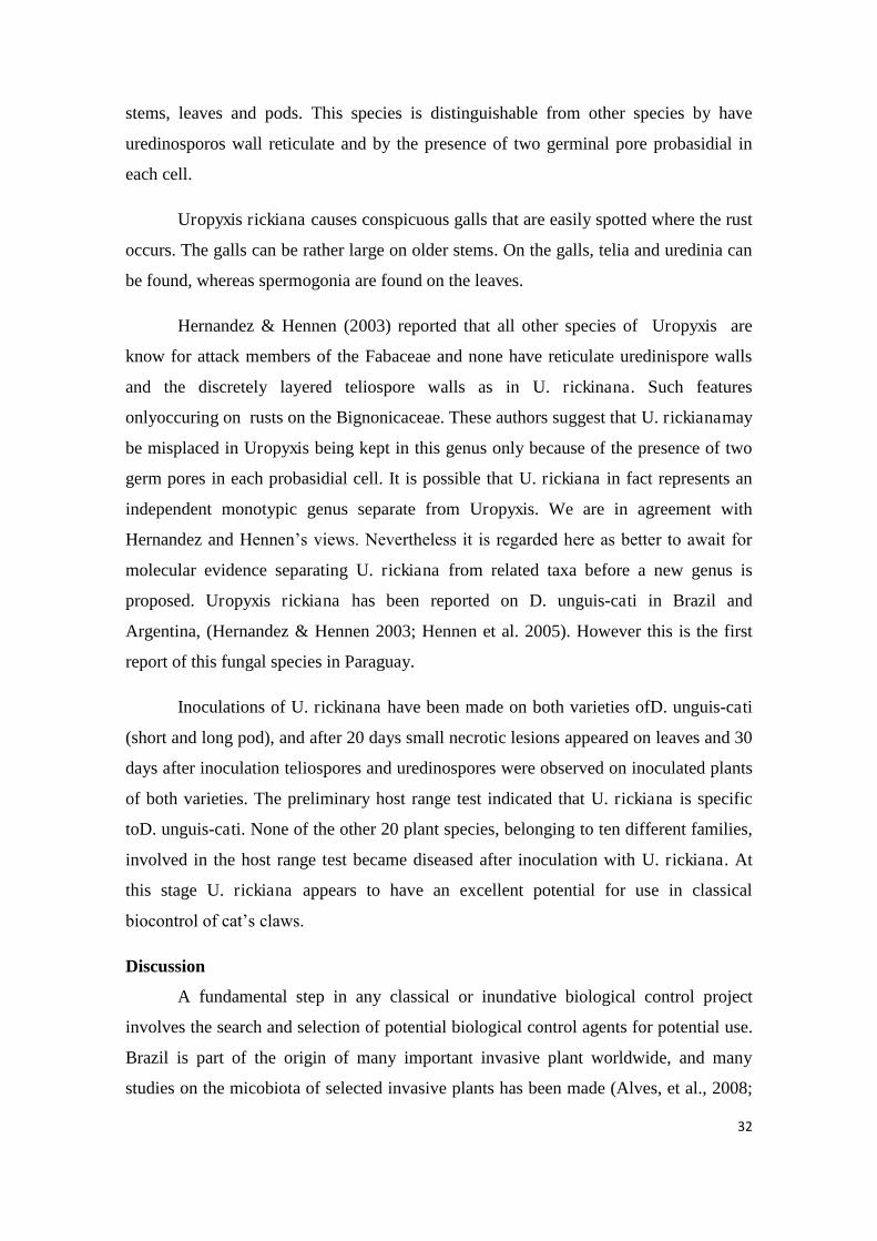
32
stems, leaves and pods. This species is distinguishable from other species by have
uredinosporos wall reticulate and by the presence of two germinal pore probasidial in
each cell.
Uropyxis rickiana causes conspicuous galls that are easily spotted where the rust
occurs. The galls can be rather large on older stems. On the galls, telia and uredinia can
be found, whereas spermogonia are found on the leaves.
Hernandez & Hennen (2003) reported that all other species of Uropyxis are
know for attack members of the Fabaceae and none have reticulate uredinispore walls
and the discretely layered teliospore walls as in U. rickinana. Such features
onlyoccuring on rusts on the Bignonicaceae. These authors suggest that U. rickianamay
be misplaced in Uropyxis being kept in this genus only because of the presence of two
germ pores in each probasidial cell. It is possible that U. rickiana in fact represents an
independent monotypic genus separate from Uropyxis. We are in agreement with
Hernandez and Hennen‟s views. Nevertheless it is regarded here as better to await for
molecular evidence separating U. rickiana from related taxa before a new genus is
proposed. Uropyxis rickiana has been reported on D. unguis-cati in Brazil and
Argentina, (Hernandez & Hennen 2003; Hennen et al. 2005). However this is the first
report of this fungal species in Paraguay.
Inoculations of U. rickinana have been made on both varieties ofD. unguis-cati
(short and long pod), and after 20 days small necrotic lesions appeared on leaves and 30
days after inoculation teliospores and uredinospores were observed on inoculated plants
of both varieties. The preliminary host range test indicated that U. rickiana is specific
toD. unguis-cati. None of the other 20 plant species, belonging to ten different families,
involved in the host range test became diseased after inoculation with U. rickiana. At
this stage U. rickiana appears to have an excellent potential for use in classical
biocontrol of cat‟s claws.
Discussion
A fundamental step in any classical or inundative biological control project
involves the search and selection of potential biological control agents for potential use.
Brazil is part of the origin of many important invasive plant worldwide, and many
studies on the micobiota of selected invasive plants has been made (Alves, et al., 2008;
33
Barreto & Evans 1994, 1995; Silva et al. 2014, Pereira & Barreto 2000; Pereira et al.,
2007; da Silva et al., 2012). Such searches normally start in literature reviews. A list of
fifteen fungi likely to be pathogenic to D. unguis-cati have been reported in the the
literature (Table 3).Here this list is expanded to twenty-two species. It is worthy of
notice that many of the fungi included in Table 3 are of no interest for classical
biocontrol either because of being weak pathogens (such as the three species of Meliola
which cause black mildew – usually a minor/benign disease) or polyphagous pathogens
that attack important crops such as Phymatotrichum omnivorum.Interestingly,
promising agents found among the existing list and the novel taxa found on D. unguis-
cati during our surveys are either cercosporoid fungi, anthracnose-causing fungi or rust
fungi.
The cercosporoids Passalora macfadyenae and Passalorasp.nov. appear
promising biological control agent although initially P. unguis-cati not having been
considered useful (Silva et al., 2012). During this new round of survey it was observed
that this pathogen can cause severe lesions on young plants, unlike P. macfadyenae and
Passalora sp. nov. which were observed only attacking adult plants. Preventing the
establishment of young plants is particularly useful in weed biocontrol. Complementary
studies are being conducted to evaluate the potential of these fungi.
Rust fungi have a long and favorable history in weed biocontrol with some
spectacular results obtained from their introduction against major invasive weeds
including the first examples of the use of plant pathogens in classical biocontrol of
weeds Puccinia chondrillina Bubak & Sidenham against Chondrilla juncea L. (Cullen
et al. 1973) and Phragmidium violaceum against Rubus constrictus (Oehrens, 1977) and
also Maravalia cryptostegiae (Cummins) Ono used in Australia to control rubbervine
Cryptostegia grandiflora Roxb. Ex R. Br (Evans et al. 2001) and Puccinia myrsiphylli
G.Winterwhich was introduced in Australia with great success for the control of
Asparagus asparagoides (L.) W. Wight (Morin & Edwards, 2006). These fungi are
often considered to be the first choice fungal pathogens to be deployed against invasive
weeds. Gall causing-rusts have also demonstrated their value in the spectatular example
of the classical introduction of Uromycladium tepperianum (Sacc.) McAlpine that led to
the successful control of a “difficult” target Acacia saligna (Labill.) Wendl. an invasive
alien shrub from Australia that was widely established in South Africa causing great

34
environmental damage (Morris, 1987, Wood & Morris, 2007; Wood 2012). Uropyxis
rickiana is therefore the natural choice as a priority agent to concentrate studies upon
for possible later introductions into Australia, South Africa and other regions where
cat‟s claw became a noxious invader.
Little is known about the biology this fungus but circumstantial and
experimental evidence indicate that this rust species is both autoecious and host-
specific, which are critical requirements for a rust to be used in classical biocontrol. The
mycobiota described herein may represent only a fraction of the existing diversity of
fungal pathogens on this host as only part of the distribution range of this plant was
covered during the two survey rounds (da Silva et al. 2012 and this work). It is likely
that an expansion of the survey, in Brazil, Southern Paraguay and Argentina will
significantly increase the list of fungi associated to D. unguis-cati and maybe uncover
additional fungi with biocontrol potential.
Acknowledgements
This work forms part of a research project submitted as a M.Sc. thesis to the
Departamento de Fitopatologia/Universidade Federal de Viçosa by A.A. Colmán. The
authors thanks Dr. K. Dhileepan and M. King for providing seeds biotypes from
Australia and South Africa used in this work. Dr. K. Dhileepan is also thanked for his
suggestions and general collaboration during the course of this work. The authors
thanks CNPq, CAPES, Department of Agriculture, Fisheries and Forestry of
Queensland and FAPEMIG for financial support.

35
References
Alves JL. Fungos para o controle biológico de Miconia calvescens com especial referência a Coccodiella miconiae. Dissertação de Mestrado. Viçosa-MG. Universidade Federal de Viçosa. 2008Anderson KI, Hallett SG. 2004.Herbicidal spectrum and activity of Myrothecium verrucaria. Weed Science 52 (4): 623-627.
Anonymous. 1960. Index of Plant Diseases in the United States. U.S.D.A. Agric. Handb. 165: 1-531. (94).
Aveskamp MM, De Gruyter J, Crous PW. 2008. Biology and recent developments in the systematics of Phoma, a complex genus of major quarantine significance. Fungal Diversity 31: 1-18
Barreto RW, Evans HC. 1994. The mycobiota of the weed Chromolaena odorata in southern Brazil with particular reference to fungal pathogens for biological control. Mycological Research 98: 1107-1116.
Barreto RW, Evans HC. 1995. The mycobiota of the weed Mikania micrantha in southern Brazil with particular reference to fungal pathogens for biological control. Mycological Research 99: 343-352.
Boerema GH, De Gruyter J, Noordelos ME, Hamers MEC. 2004. Phoma identification manual. Wallingford: CABI. 448p
Braun U. 1994. Miscellaneous notes on phytophatogenic hyphomycetes. Mycotaxon 51:49
Braun U. 1998. A monograh of Cercosporella, Ramularia and allied genera (Phytopathogenic Hyphomycetes). Vol.2. IHW-Verlag, Eching. 493p.
Briese DT. 2003. The centrifugal phylogenetic method used to select plants for hos-specificity testing of weed biological control agents: can and should it be modernized? In: Improving the Selection, Testing and Evaluation of Weed Biological Control Agents (eds H Spafford Jacob, DT Briese), pp. 23-33. Technical Series No.7. CRC for Australian Weed Management, University of Adelaide, Adelaide, Australia.
CaiL, Hyde KD, Taylor PWJ, Weir BS, Waller J, Abang MM, Zhang JZ, Yang YL, Phoulivong S, Liu ZY, Prihastuti H, Shivas RG, McKenzie EHC, Johnston PR. (2009). A polyphasic approach for studying Colletotrichum. Fungal Diversity 39: 183-204
Chupp C. 1954. A monograph of the fungus genus Cercospora. Published by author, Ithaca.

36
Crous PW, Sutton BC. 1997. New cercosporoid fungi from southern Africa. South Africa Journal of Botany 63: 280-285.
Crous PW, Braun U. 2003. Mycosphaerella and its anamorphs: 1. Names published in Cercosporaand Passalora. Utrecht: Centraalbureau voor Schimmelcultures. 571p.
Cullen, J.M., Kable, P.F. & Catt, M. 1973. Epidemic spread of a rust imported for biological control. Nature 244: 462–464
Silva M, Barreto RW, Pereira OL. 2012. Fungal pathogens of „cat‟s claws‟ from Brazil for biocontrol of Macfadyena unguis-cati. Mycotaxon 119: 181-195.
Damm U, Cannon PF, Woudenberg JHC, Joshnston PR, Weir BS, Tan YP, Shivas RG, Crous PW. 2012. The Colletotrichum boniniense species complex. Studies in Mycology 73: 1-36.
Dhileepan K, Taylor DBJ, Lockett C, Treviño M. 2013. Cat‟s claw creeper leaf-mining jewel beetle Hylaeogena jureceki Obenberger (Coleoptera: Buprestidae), a host-specific biological control agent for Dolichandra unguis-cati (Bignoniaceae) in Australia. Australian Journal of Entomology 52: 175-181
Dhileepan K, Treviño M, Donnelly GP, Raghu S. 2005. Risk to no target plants from Charidotis auroguttata (Chrysomelidae: Coleoptera), a potential biocontrol agent for cat‟s claw creeper Macfadyena unguis-cati (Bignoniaceae) in Australia. Biological Control 32: 450-460.
Dhileepan K, Snow EL, Rafter MA, McCarthy J, Treviño M, Wilmot Senaratne KAD. 2007a. Leaf-tying moth Hypocosmia pyrochroma (Lepidoptera: Pyralidae), a host specific biological control agent for Macfadyena unguis-cati (Bignoniaceae) in Australia. Australian Journal of Entomology 131: 564-568.
Dhileepan K, Treviño M, Baylis D et al. 2007b. Specificity of Carvalhotingis visenda (Hemiptera: Tingidae) as a biological control agent- for Macfadyena unguis-cati (Bignoniaceae) in Australia. Biological Control 55: 58-62.
Dhingra OD, & Sinclair JB.1995.Basic plant pathology methods. Boca Raton: CRC Press, 1995. 434p
Ellis MB. 1971. Dematiaceous Hyphomycetes. Commonwealth Mycological Institute, Kew, Surrey. 608p.
Evans HC, Greaves MP, Watson AK. 2001. Fungal Biocontrol Agents of Weeds, In. Butt TM, Jackson CW, Magan N. Fungi as Biocontrol Agents: progress problems and potential. Wallingford: CAB International; pp. 169-172.

37
Farr DF & Rossman AY. Fungal Databases, Systematic Mycology and Microbiology Laboratory, ARS, USDA. Retrieved February 10, 2014, from http://nt.ars-grin.gov/fungaldatabases/
Groenewald M, Groenewald JZ, Braun U, Crous PW. 2006. Host range of Cercospora apii and C. beticola and description of C. apiicola, a novel species from celery. Myccologia 98 (2): 275-285.
Groenewald JZ, Nakashima C, Nishikawa J, Shin HD, Park AN, Jama M, Groenewald M, Braun U, Crous PW. 2012. Species concepts in Cercospora: spotting the weeds among roses. Studies in Mycology 75: 115-170.
Gruyter J de, Woudenberg JHC, Aveskamp MM, Verkley GJM, Groenewald JZ, Crous PW. 2010. Systematic reappraisal of species in Phoma section Paraphoma, Pyrenochaeta and Pleurophoma. Mycologia 102: 1066–1081
Hennen JF, Figueredo MB, Carvalho Jr AA, Hennen PG. 2005. Catalogue of the speciesof plant rust fungi (Uredinales) of Brazil.http://www.jbrj.gov.br/publica/uredinales/Brazil_Catalogue1drevisado.pdf(Accessed 15 January 2014).
Hernández JR, Hennen JF. 2003. Rust fungi causing galls, witches‟ brooms, and otherabnormal plant growths in northwestern Argentina. Mycologia 95: 728-755.
Inácio CA, Dianese JC. 2006. Foliicolous fungi on Tabebuia species from the cerrado. Mycological Progress 5:120-127.
Johnston EM,Valleau, WD. 1949. Synonymy in some commonspecies of Cercospora. Phytopathology 39:763-770.
Killgore EM, Sugiyama LS, Barreto RW, Gardner DE. 1999. Evaluation of Colletotrichum gloesoporioides for biological control of Miconia calvescens in Hawaii. Plant Disease 83, 964.
Kirk PM, Cannon, PF, Minter, DW, Stalpers, JA. 2008. Dictionary of the fungi. 10th ed. CAB International. 771 p.
Kirschner R. 2009. Cercosporella and Ramularia. Mycologia 101(1): 110-119
Liu XJ, Guo YL. 1982. Studies on some species of the genus phaeoramularia in China. Acta Phytopathologica Sinica 12: 1-15.
Meyer JY, Killgore EM. 2000. First and successful release of a bio-control pathogen agent to combat the invasive alien tree Miconia calvescens (Melastomataceae)

38
in Thaiti. Aliens. Newsletter of the invasive Species Specialist Group of the IUCN Species Survival Commission 12:8.
Meyer JY, Taputuarai R, Killgore E. 2008. Dissemination and impacts of the fungal pathogen Colletotrichum gloeosporioidesf. sp.miconiae (Deuteromycetinae) on the invasive alien tree Miconia calvescens (Melastomataceae) in rainforests of Thaiti (French Polynesia, South Pacific). In: Julien MH, Sforza, RS, Evans HC, Hatcher PE, Hinz HL, Rector BG. (Eds.). Procedings of the XII. International Symposium on Biological Control of Weeds. Wallingford. CABI. pp. 594-600.
Morin L, Edwards PB. 2006. Selection of biological control agents for bridal creeper: a retrospective review. Australian Journal of Entomology45: 287-291
Morris MJ. 1987. Biology of the acacia galls rust, Uromycladium tepperianum. Plant Patholohy 36: 100-106
Muchovej JJ, Ferreira FA. 1981. A new species of Phaeoramularia from Brazil. Mycologia 73: 345-347.
Oehrens E. Biological control of blackberry through the introduction of the rust, Pragmidium violaceum, in Chile. FAO Plant Proteccion Bulletin 25:26-28. 1977.
Parreira DF. Micobiota fitopatogênica associada a planta invasora Tibuchina herbaceae. Dissertação de Mestrado. Viçosa-MG. Universidade Federal de Viçosa. 2008
Peng G, Bailey KL, Hinz HL, Byer KN. (2005). Colletotrichum sp.: a potential candidate for biocontrol of scentless chamomile (Matricaria perforata) in western Canada. Biocontr. Sci.Technol. 15: 497–511.
Pereira JM, Barreto RW. 2000. Additions to the mycobiota of the weed Lantana camara (Verbenaceae) in Southeastern Brazil. Mycopathologia 151: 71-80.
Pereira OL, Barreto RW, Cavalazzi JRP, Braun U. 2007. The mycobiota of cactos weed Pereskia aculeata in Brazil, with comments on the lifw-cycle of Uromyces pereskiae. Fungal Diversity 25: 167-180
Rayner RW. 1970. A mycological color chart. Commonwealth Mycological Institute, Kew, Surrey and British Mycological Society. 34p.
Simmons, EG. 2007. Alternaria: an identification manual. USA: APS press. 775p.
Sutton BC. 1985. The coelomycetes. Fungi Imperfecti with pycnidia, acervuli and stromata. New York: CABI Publishing. 696 p

39
Trujillo EE, Latterell FM, Rossi AE. 1986. Colletotrichum gloeosporioides, a possible biological control agent for Clidemia hirta in Hawaiian forests. Plant Disease 70: 974-976
Tulloch M. 1972. The genus Myrothecium TODE ex FR. Mycological Papers 130
Wapshere AJ. 1974. A strategy for evaluating the safety of organisms for biological control. The Annals of Applied Biology 77: 201-211.
Wood AR. Morris MJ. 2007. Impact of the gall-forming rust fungus Uromycladium tepperianum on the invasive tree Acacia saligna in South Africa: 15 years of monitoring. Biological Control41: 68-77
Wood AR. 2012. Uromycladium tepperianum (a gall-forming rust fungus causes a sustained epidemic on the weed Acacia saligna in South Africa. Australasian Plant Pathology Society 41: 255-261.
Woundenberg JHC, Groenewald JZ, Binder M, Crous PW (2013). Alternaria redefined. Studies in Mycology 75: 175-212
Yang YL, Cai L, Yu ZN, Liu ZY, Hyde KD (2011). Colletotrichum species onOrchidaceae in southwest China. Cryptogamie Mycologie32: 229–25

40
Tables
Table 1. List of plants used in host specificity tests
Order Family Genus Results
Lamiales
Achantaceae Pachystachys sp. -
Thumbergia sp. -
Bignoniaceae
Adenocalymmba sp. -
Cuspidaria sp -
Friedericia sp. -
Jacaranda sp. -
Dolichandra unguis-cati ''shor pod'' +
Dolichandra unguis-cati ''long pod'' +
Pandorea sp. -
Spathodea campanulata -
Pyrostegia sp. -
Tabebuia sp. -
Tecoma sp. -
Episcia sp. -
Lamiaceae Origanum sp. -
Oleaceae Jasminum sp. -
Pedaliaceae Sesamum sp. -
Plantaginaceae Plantago sp. -
Scrophulariaceae Nemesia sp. -
Solanales Solanaceae Solanum sp. -
Ericales Ericaceae Azalea sp. -
+ (Positive) − (Negative)

41
Table 2. Biometric data for Passalora species recorded on members of the Bignoniaceae.
Species Conidia (µm) Stromata(µm) Conidiophores (µm)
P. adenocalymmatis a,b
351–50 × 03–4,5 40–60 4–5.5 × 20–65
P. arrabidaeae a,b 35–150 × 3.5 –5 Absent 4–6 × 10–75
P. catalpae a,b 40–120 × 2.5 –4,5 0–50 3–5.5 × 10–125
P. catalparum a,b 35 –125 × 3.5– 6 absent 4–7.5 × 15–70
P. leprosa a,b 30–80 × 3.5 –6 300–500 5–8 × 40–150
P. macfadyenae c 31.5–114 × 3– 4.5 63–142 ×35–95 13–6 × 16–114
P. markhamiae d 16–92.5 × 2.5–55.5 15–60 16–92.5 × 2.5–55.6
P. pyrostegiae a,b 25–170 × 3 – 4.5 40–80 4–6 × 10–45
P. tabebuiae e (6–) 8–30 (34) × 2.5– 3 absent 2.5-3.2 × 0–50
P.tabebuiae-
ochraceae f 31–75 × 5–8 24–130 5–8 × 31–75
Passalora sp. nov. g 25–150 × 2.5–35 absent 3–4 × 22–175
P. tecomariae b 35–90 × 5–6 absent 5–6 × 35–90
a. Chupp (1954) b. Crous and Braun (2003) c. Da Silva et al. (2012) d. Liu & Guo (1982) e. Muchovej & Ferreira (1981) f. Inacio & Dianese (2006) g. This paper

42
Table 3. Fungi recorded on Dolichandra unguis-cati worldwide*
Fungi Distribution
Cercospora duplicata Ellis & Everh (1989) Barbados Cercosporella unguis-cati Speg. (1911) Argentina Colletotrichum dematium Pers. Grove. (1918) India Diplodia catalpae Speg.. (1879) Portugal Glomerella cingulata (Stoneman) Spauld. & H. Scherenk. (1903)
Barbados
Meliola bidentata Cooke. (1882) Puerto Rico, Virgin Islands, West Indies
Meliola stevensiana Cif. (1954) Trinidad & Tobago
Meliola thaxteri Hansf. (1961) Ecuador, Trinidad & Tobago, West Indies
Phoma sp. Cuba, West Indies Phymatotrichum omnivorum Duggar. (1916) Texas Pseudocercospora unguis-cati (Speg.) Braun (1994)
Argentina, Brazil
Mycena citricolor (Berk.& M.A. Curtis) Sacc. (1887)
Venezuela
Passalora macfadyenae Meir. Silva, O. L. Pereira & R.W. Barreto (2012)
Brazil
Prospodium macfadyenae Meir. Silva, O. L. Pereira & R.W. Barreto (2012)
Brazil
Uropyxis rickiana Magnus (1906) Argentina, Brazil *Adapted from Farr & Rossman, 2014

43
Figures
Fig.1: Alternaria alternata on Dolichandra unguis-cati.A. Leaf spot symptons. B. Conidiphores dark C. Germinating conidium - D. Obclavate muriform conidium. E. Chain of conidia. F. Conidium still attached to the conidiophore. Bars= 20 µm.
44
Fig. 2: Cercospora apii on Dolichandra unguis-cati.A. Symptons of leaf spots B. Conidiophores fasciculate. C. Conidiophore and conidia. D-E. Filiform conidia. Bars= 20 µm

45
Fig.3: Cercospora rodmanii on Dolichandra unguis-cati. A.Leaf spot symptons B.
Conidiophores fascicle C. Filiform conidia. F. Conidiogenous cell and loci. Barrs= 20
µm

46
Fig. 4: Pseudocercospora unguis-cati on Dolichandra unguis-cati.A. Symptons of leaf
spots on young plant. B. White tufts of conidia on foliar surface. C. Conidiophores
arising through stoma. D. Filiform conidia. Bars= 20 µm
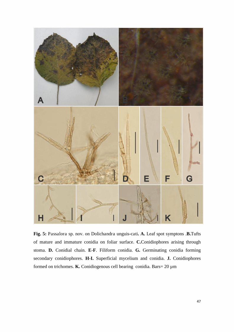
47
Fig. 5: Passalora sp. nov. on Dolichandra unguis-cati. A. Leaf spot symptons .B.Tufts
of mature and immature conidia on foliar surface. C.Conidiophores arising through
stoma. D. Conidial chain. E-F. Filiform conidia. G. Germinating conidia forming
secondary conidiophores. H-I. Superficial mycelium and conidia. J. Conidiophores
formed on trichomes. K. Conidiogenous cell bearing conidia. Bars= 20 µm
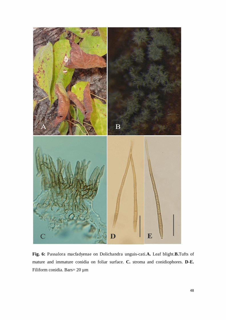
48
Fig. 6: Passalora macfadyenae on Dolichandra unguis-cati.A. Leaf blight.B.Tufts of
mature and immature conidia on foliar surface. C. stroma and conidiophores. D-E.
Filiform conidia. Bars= 20 µm

49
Fig. 7: Colletotrichum dematium on Dolichandra unguis-cati.A. Antracnosesymptons.
B. Acervuli on foliar surface. C. Section through acervulus. D. Conidiogenous cell and
conidia. E. Falcate conidia. E. Apressoria. Bars= 20 µm
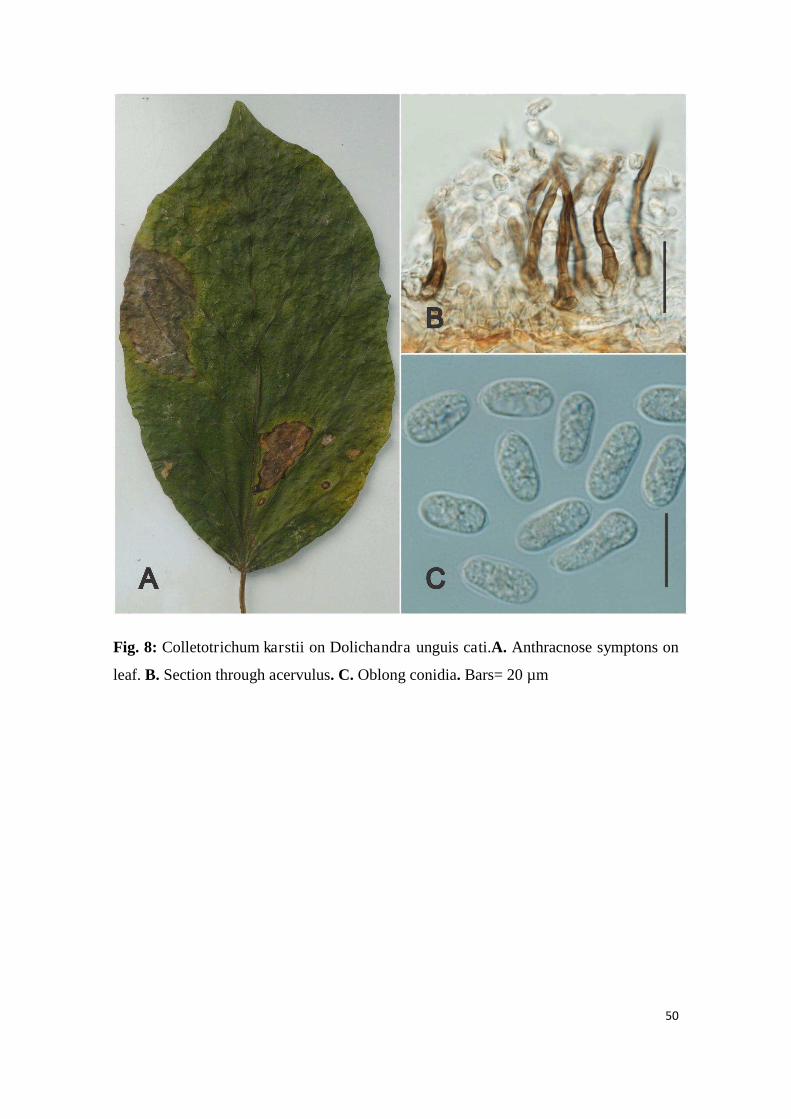
50
Fig. 8: Colletotrichum karstii on Dolichandra unguis cati.A. Anthracnose symptons on
leaf. B. Section through acervulus. C. Oblong conidia. Bars= 20 µm

51
Fig. 9: Myrothecium roridum on Dolichandra unguis-cati.A-B. Leaf spot symptons. C.
Conidiophores and setae. D. Sporodochium on leaf surface. E. Conidia . Bars= 20 µm

52
Fig. 10: Phoma sp. nov. on Dolichandra unguis-cati. A. Blight symptons of leaves. B.
Leaf spot bearing several pycnidia. C. Section through pycnidium. D. Conidiogenous
cells and conidia. Bars= 20 µm
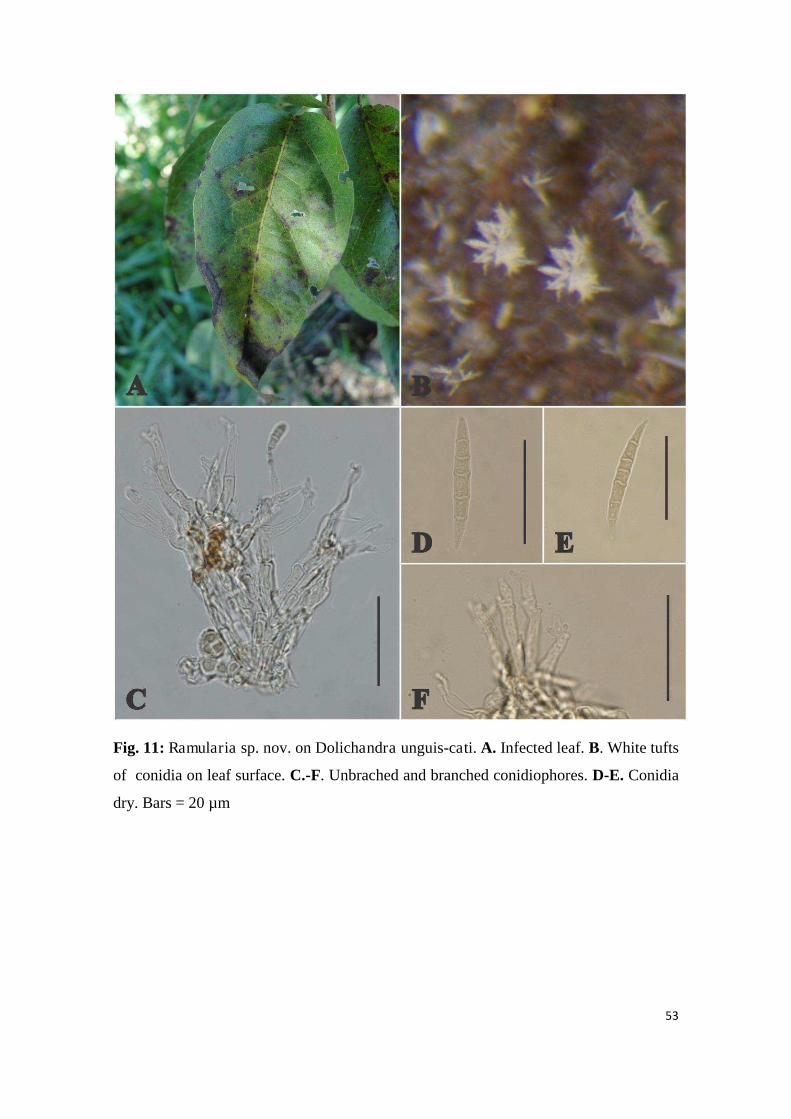
53
Fig. 11: Ramularia sp. nov. on Dolichandra unguis-cati. A. Infected leaf. B. White tufts
of conidia on leaf surface. C.-F. Unbrached and branched conidiophores. D-E. Conidia
dry. Bars = 20 µm

54
Fig. 13: Prospodium macfadyenae on Dolichandra unguis-cati.A-B. Telia and uredinia
on infected plant. C. Basket-like sori (squash-mount). D. Echinulate uredinospore and
pedicellate 2-celled teliospore. Bars = 20 µm.

55
Fig. 12: Uropyxis rickiana on Dolichandra unguis cati. A-E. Galls on stem –
progressive stages of development. F-G. Distortions on leaves to which aecia and
spermogonia are associated. H Gall on pod. I. Symptons observed in greenhouse after

56
30 days of inoculation. J. Type 7 spermogonium. K. Aecia and aeciospores. L .
Uredinospore and teliospore with spirall in the base. M. Germinated with metabasidia
and basidiospores. Bars = 20µm
Top Related