UNIVERSIDADE FEDERAL DO PARÁ PROGRAMA DE PÓS … · universidade federal do parÁ instituto de...
39
UNIVERSIDADE FEDERAL DO PARÁ INSTITUTO DE CIÊNCIAS DA SAÚDE FACULDADE DE ODONTOLOGIA PROGRAMA DE PÓS-GRADUAÇÃO EM ODONTOLOGIA JOYCE FIGUEIRA DE ARAÚJO 01 ANO DE AVALIAÇÃO DE UMA TÉCNICA SIMPLIFICADA DE ADESÃO ÚMIDA POR ETANOL: UM ENSAIO CLÍNICO RANDOMIZADO BELÉM 2012
Transcript of UNIVERSIDADE FEDERAL DO PARÁ PROGRAMA DE PÓS … · universidade federal do parÁ instituto de...

UNIVERSIDADE FEDERAL DO PARÁ
INSTITUTO DE CIÊNCIAS DA SAÚDE
FACULDADE DE ODONTOLOGIA
PROGRAMA DE PÓS-GRADUAÇÃO EM ODONTOLOGIA
JOYCE FIGUEIRA DE ARAÚJO
01 ANO DE AVALIAÇÃO DE UMA TÉCNICA SIMPLIFICADA DE
ADESÃO ÚMIDA POR ETANOL: UM ENSAIO CLÍNICO
RANDOMIZADO
BELÉM
2012

ii
UNIVERSIDADE FEDERAL DO PARÁ
INSTITUTO DE CIÊNCIAS DA SAÚDE
FACULDADE DE ODONTOLOGIA
PROGRAMA DE PÓS-GRADUAÇÃO EM ODONTOLOGIA
JOYCE FIGUEIRA DE ARAÚJO
01 ANO DE AVALIAÇÃO DE UMA TÉCNICA SIMPLIFICADA DE
ADESÃO ÚMIDA POR ETANOL: UM ENSAIO CLÍNICO
RANDOMIZADO
Dissertação apresentada à Faculdade de
Odontologia da Universidade Federal do
Pará, como pré-requisito para obtenção do
título de Mestre em Odontologia pelo
Programa de Pós-Graduação.
Área de concentração: Dentística.
Orientador: Prof. Dr. Mário Honorato
da Silva e Souza Júnior
Belém – Pará
2012
iii
JOYCE FIGUEIRA DE ARAÚJO
01 ANO DE AVALIAÇÃO DE UMA TÉCNICA SIMPLIFICADA DE
ADESÃO ÚMIDA POR ETANOL: UM ENSAIO CLÍNICO
RANDOMIZADO
Dissertação apresentada à Faculdade de
Odontologia da Universidade Federal do Pará,
como pré-requisito para obtenção do título de
Mestre em Odontologia pelo Programa de Pós-
Graduação em Odontologia.
Orientador: Prof. Dr. Mário Honorato da Silva
e Souza Jr.
Belém, ___/___/___.
Banca Examinadora
Prof. Dr. Mário Honorato da Silva e Souza Júnior
Prof. Dr. Sandro Cordeiro Loretto
Prof. Dr. Igor Studart Medeiros

iv
AGRADECIMENTOS
A DEUS, que sempre foi a minha fortaleza e guia em todos os momentos,
e me permitiu alcançar mais uma meta em minha vida.
Aos meus pais JOAQUIM FILHO e VIRGINIA ARAÚJO, pela presença
e apoio incondicional em todas as horas.
A meu querido orientador e mestre Prof. Dr. MÁRIO HONORATO DA
SILVA E SOUZA JÚNIOR, a quem devo o meu muito obrigado pela ajuda na
elaboração deste trabalho, bem como pela amizade conquistada, pela
colaboração ao meu crescimento profissional e pela confiança a mim depositada.
Aos meus amigos que me apoiaram e ajudaram no decorrer deste trabalho:
THAÍS FIGUEIREDO, ESTHER MARINA e RAILSON OLIVEIRA.
Aos amigos farmacêuticos e bioquímicos DANIELLA PATERNOSTRO
e ALAN GRISOLIA que foram fundamentais no desenvolvimento da pesquisa,
sempre disponíveis e atenciosos.
Ao Prof. Dr. SANDRO CORDEIRO LORETTO pela colaboração na
busca das referências e revisão do trabalho.
Aos colegas de turma do mestrado que juntamente comigo superaram
mais uma batalha na vida profissional.
Aos professores do curso de Odontologia integrados ao programa de pós-
graduação, em especial àqueles que diretamente colaboraram no decorrer do
desenvolvimento deste trabalho.
A todos os pacientes que participaram da pesquisa e compareceram
fielmente, permitindo que obtivéssemos os resultados em sua totalidade.
Ao meu amor ANDRÉ GATTI, por estar sempre ao meu lado dando força
e entendendo os momentos finais de reclusão para a conclusão deste trabalho.

v
“Tudo o que um sonho precisa para ser realizado é alguém
que acredite que ele possa ser realizado”.
Roberto Shinyashiki

vi
RESUMO
O objetivo deste estudo clínico randomizado foi avaliar o comportamento
clínico de restaurações adesivas utilizando uma técnica simplificada de adesão
úmida por etanol (G3) em lesões cervicais não cariosas e compará-las com
outras realizadas pela técnica convencional de três passos (G1) e pela técnica
autocondicionante de um passo (G2). Noventa e três restaurações (31 para cada
grupo) foram realizadas em 17 pacientes por um único operador. Nenhum
preparo cavitário foi realizado. Após 6 e 12 meses, as restaurações foram
avaliadas por 2 examinadores previamente treinados usando um critério de Ryge
modificado para adaptação/manchamento marginal (kappa=0,81) e retenção
(kappa=1,00) e os dados analisados inter-grupo pelos testes de Kruskal-Wallis e
Exato de Fisher, respectivamente. Não foram observadas diferenças
significantes entre os grupos aos 6 e 12 meses para qualquer um dos critérios
avaliados (p≥0.05). A análise intra-grupo feita pelos testes Q de Cochran (para
retenção) e Wilcoxon (para adaptação/manchamento marginal) revelou
diferenças significantes entre os intervalos de tempo baseline/12 meses na
adaptação marginal do G2 (p=0,0180) e no manchamento marginal do G1
(p=0,0117). A análise de sobrevivência para o critério retenção realizada
utilizando o teste de log-rank não apresentou diferenças significativas (p≥0,05).
As restaurações feitas utilizando a técnica simplificada da adesão úmida por
etanol comportaram-se igualmente às outras estratégias adesivas empregadas.
Palavras-chave: ensaio clínico randomizado, adesivos dentais, dentina.

vii
LISTA DE FIGURAS
FIGURE 1- Survival curves for each adhesive system for up to 12 months…..16
FIGURA 1- Adesivo convencional AdperTM
ScotchbondTM
Multi-Purpose......31
FIGURA 2- Adesivo autocondicionante AdperTM
Easy One.............................32
FIGURA 3- “Primer hidrofóbico”- substâncias e mistura................................32
FIGURA 4- Balança eletrônica (Bel Mark).......................................................33
FIGURA 5- Agitador de tubos (Phoenix, Modelo AP 56).................................34
FIGURA 6- Soluções de etanol 50% e 100% e “primer hidrofóbico”..............34

viii
LISTA DE TABELAS
TABLE 1 – Adhesive compositions and instructions for use……………...…..14
TABLE 2 – Restoration distributions……………………………………….…15
TABLE 3 – Modified USPHS criteria….…………………………………...…15
TABLE 4 – Clinical evaluation for each criterion at 6 and 12 months….….....16
TABLE 5 – Intra-group analysis for each adhesive systems over 12 month.....16
TABELA 1- Passo-a-passo da aplicação do sistema adesivo SBMP.................31
TABELA 2- Passo-a-passo da aplicação do sistema adesivo AdperTM
Easy
One......................................................................................................................32
TABELA 3- Passo-a-passo da técnica de adesão à dentina saturada com
etanol...................................................................................................................33

ix
SUMÁRIO
1 ARTIGO EM INGLÊS..................................................................................10
Abstract.........................................................................................................10
Introduction...................................................................................................11
Material and Methods…………………………………………..…….……12
Results………………………………………………………………..….....15
Discussion……………………………………………………………..…...17
Acknowleggments………………………………………….…………..…..21
References.....................................................................................................21
2 COMPLEMENTO.........................................................................................25
2.1 REVISÃO DE LITERATURA...................................................................25
2.2 OBJETIVO..................................................................................................28
2.3 MATERIAL E MÉTODOS........................................................................29
2.3.1 Aspectos éticos...........................................................................................29
2.3.2 Seleção dos pacientes.................................................................................29
2.3.3 Randomização da amostra..........................................................................30
2.3.4 Procedimentos restauradores......................................................................30
2.3.5 Grupos experimentais.................................................................................31
G1- Grupo SBMP (G1).............................................................................31
G2- Grupo EO (G2)...................................................................................32
G3- Grupo EWBT (G3).............................................................................32
2.3.6 Análise estatística.......................................................................................34
2.3.7 Padronização do estudo clínico randomizado............................................35
REFERÊNCIAS BIBLIOGRÁFICAS
APÊNDICE A
APÊNDICE B

10
1 ARTIGO EM INGLÊS (Enviado para a Brazilian Dental Journal)
Title: One-Year Evaluation of a Simplified Ethanol-Wet Bonding Technique: a
Randomized Clinical Trial
Short Title: Simplified Ethanol-Wet Bonding Technique: Clinical Trial
Authors:
Joyce Figueira de Araújo1, Mário Honorato da Silva e Souza Júnior
2
1DDS, Graduate student, Federal University of Pará, Belém, PA, Brazil
2DDS, MSc, PhD, Associate Professor, Department of Restorative Dentistry,
Dental School, Federal University of Pará, Belém, PA, Brazil
_________________________________________________________________________
Abstract
The objective of this randomized clinical trial was to evaluate the clinical
performance of adhesive restorations using a three-step etch-and-rinse adhesive
(G1), a one-step self-etching adhesive (G2), and a simplified ethanol-wet
bonding technique (G3) prior to the application of a composite resin in non-
carious cervical lesions. Ninety-three restorations (31 for each group) were
performed in 17 patients by a single operator. No cavity preparation was
executed. After 6 and 12 months, the restorations were assessed by two
previously trained examiners using modified Ryge criteria for retention
(kappa=1.00); marginal adaptation/staining (kappa=0.81) and analyzed by
Fisher’s exact and Kruskal-Wallis tests, respectively. Significant differences
were not observed among groups at the 6- and 12-month time points for any of
the assessed criteria (p≥0.05). The intra-group analysis performed by Cochran’s
test (for retention) and Wilcoxon test (for marginal adaptation/staining)
revealed significant differences between the time intervals baseline/12-months
in marginal adaptation in G2 (p=0.0180) and in marginal staining in G1
(p=0.0117). The survival analysis for retention criteria performed using a log-
rank test did not show significant differences (p≥0.05). The restorations placed
using the simplified ethanol-wet bonding technique performed equally well as
the other adhesive strategies employed.
Key words: randomized controlled trial, dentin-bonding agents, dentin

11
INTRODUCTION
Despite the immediate in vitro efficacy of the adhesive systems, interface
failures can be observed after a short period of time (1,2), and the high
hydrophilicity of these systems may partially explain this outcome (3-5). The
presence of hydrophilic monomers and the incomplete resin infiltration in
demineralized dentin by etch-and-rinse technique may increase water sorption
and the risk of hydrolytic degradation, which may reduce the adhesive
mechanical properties over time (1,6-9).
The self-etching (SE) systems eliminate the need for previous acid
etching, critical step for adhesion (10-13). Theoretically, there is no or small
difference between the depths of demineralization and resin infiltration induced
by this system because both processes occur simultaneously (14-16). However,
this adhesive strategy also has high hydrophilicity, which attracts water and
compromises adhesive interface integrity (17,18).
The ethanol-wet bonding technique (EWBT) was introduced in an attempt
to overcome the problems caused by incomplete penetration of most adhesive
systems and the high hydrophilicity observed for commercially available
adhesive systems. In EWBT the dentin is saturated by increasing concentration
of ethanol solutions prior to the application of hydrophobic monomers to
produce a more stable and hydrolysis-resistant hybrid layer (3,5,19).
As mentioned, EWBT may reduce the hydrophilic characteristics of the
interface. Dentin dehydration occurs with application of increasing
concentrations of ethanol solutions, allowing the subsequent infiltration of a
hydrophobic primer followed by a hydrophobic adhesive resin (3,6). This hybrid
layer is less hydrophilic and more resistant overtime to hydrolytic degradation
caused by the endogenous enzymes, such as metalloproteinases (MMPs), due to
better collagen protection (19,20), and phase separation prevention (8,18,21,22).
Some laboratory studies have demonstrated higher adhesive interface
bond strengths when ethanol solutions were used to dehydrate dentin prior to
12
primer and adhesive application compared with the strengths achieved using
conventional adhesive techniques (4,7,18,21). However, there is a lack of
clinical data for this strategy.
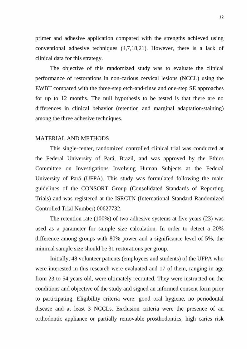
The objective of this randomized study was to evaluate the clinical
performance of restorations in non-carious cervical lesions (NCCL) using the
EWBT compared with the three-step etch-and-rinse and one-step SE approaches
for up to 12 months. The null hypothesis to be tested is that there are no
differences in clinical behavior (retention and marginal adaptation/staining)
among the three adhesive techniques.
MATERIAL AND METHODS
This single-center, randomized controlled clinical trial was conducted at
the Federal University of Pará, Brazil, and was approved by the Ethics
Committee on Investigations Involving Human Subjects at the Federal
University of Pará (UFPA). This study was formulated following the main
guidelines of the CONSORT Group (Consolidated Standards of Reporting
Trials) and was registered at the ISRCTN (International Standard Randomized
Controlled Trial Number) 00627732.
The retention rate (100%) of two adhesive systems at five years (23) was
used as a parameter for sample size calculation. In order to detect a 20%
difference among groups with 80% power and a significance level of 5%, the
minimal sample size should be 31 restorations per group.
Initially, 48 volunteer patients (employees and students) of the UFPA who
were interested in this research were evaluated and 17 of them, ranging in age
from 23 to 54 years old, were ultimately recruited. They were instructed on the
conditions and objective of the study and signed an informed consent form prior
to participating. Eligibility criteria were: good oral hygiene, no periodontal
disease and at least 3 NCCLs. Exclusion criteria were the presence of an
orthodontic appliance or partially removable prosthodontics, high caries risk
13
(presence of three or more active caries lesions), heavy bruxism and patients
who did not accept the conditions of the project.
Each patient received at least 3 or multiple of 3 restorations. Each
adhesive system was randomly allocated to one of randomized cervical lesions
until that the three groups were present in the same subject and in equal
amounts.
A total of 93 NCCLs (31 for each group) were restored by one
experienced operator using three types of adhesive strategies: Group one (G1),
Adper Scotchbond Multi-Purpose/SBMP (3M ESPE, St Paul, MN, USA); Group
two (G2), Adper Easy One/EO (3M ESPE, Seefeld, Germany); and Group three
(G3), EWBT (dentin saturation with ethanol solutions + hydrophobic primer
experimental + step 3 of SBMP – 3M ESPE). The hydrophobic primer used in
the G3 was prepared by diluting 2ml step 3 of SBMP (3M ESPE) with
corresponding to 10mass% absolute ethanol.
All restorations were placed under relatively dry conditions using gingival
retraction cord, cotton rolls and saliva ejector. Enamel margins were not
beveled, and no mechanical retention was performed. The surfaces were slightly
roughened with a #3118 diamond bur (K.G. Sorensen, Alphaville, São Paulo,
Brazil).
After adhesive systems application, the cavities were restored by applying
at least two layers of Filtek Z350 XT (3M ESPE), which were light-cured for 40
s using a LED source (D-2000; DMC, Joinvile, SC, Brazil) at 1.100 mW/cm2.
Restorations were polished with #3195 fine diamond burs (K.G. Sorensen), and
candle flame rubber points (Dentsply/Caulk, Milford, DE, USA). Table 1 lists
the materials and techniques used for each group and the distributions of the
restorations with respect to tooth type, adhesive, quadrant and arch are shown in
Table 2.
Two calibrated examiners evaluated all restorations using the modified
US Public Health Service guidelines (USPHS) shown in Table 3. In cases where

14
the two examiners disagreed on a rating, both re-examined the restoration and
arrived at a joint final decision. The examiners were blinded regarding the type
of adhesive used in each restoration. The primary outcome was retention
(kappa=1.00), with marginal adaptation/staining (kappa=0.81) as secondary
outcomes, both analyzed at 6 and 12 months.
The obtained data were recorded and the differences among adhesive
systems at each evaluation period were analyzed using Fisher’s exact test for
retention and the Kruskal-Wallis test for marginal adaptation/staining at the 5%
significance level. An intra-group comparison among baseline, 6- and 12-month
evaluation for each adhesive system was performed using Cochran’s test (for
retention) and the Wilcoxon test (for marginal adaptation/staining) at the 5%
significance level. All restorations received A score for all evaluation criteria at
baseline. The survival analysis for retention criteria was performed using a log-
rank test at the 5% significance level.
Table 1. Adhesive compositions and instructions for use. Material Adhesive technique
SBMP (G1):
Adper Scotchbond
Multi-Purpose +
FiltekTM
Z350 XT
(3M ESPE)
1- etching of enamel and dentin for 15 s, with 35% phosphoric acid gel and
rinsing by air-water spraying for 30 s
2- dentin blotting with absorbent paper to keep the surface visibly moist
3- application of primer to enamel and dentin. Dry gently for 5 s
4- application of adhesive and light curing for 10 s
5- restoration with ≅1-mm layers of FiltekTM
Z350XT
EO (G2):
Adper Easy One +
FiltekTM
Z350 XT
(3M ESPE)
1- dentin washing and blotting with absorbent paper to keep the surface
visibly moist
2- application of Easy One adhesive for 20 s, air-drying for 5 s and light
curing for 10 s
3- restoration with ≅1-mm layers of FiltekTM
Z350XT
EWBT (G3):
“hydrophobic
primer” experimental
+ step 3 of Adper
Scotchbond Multi-
Purpose + FiltekTM
Z350 XT (3M ESPE)
1- etching of enamel and dentin for 15 s, with 35% phosphoric acid gel and
rinsing by air-water spraying for 30 s
2- dentin blotting with absorbent paper to keep the surface visibly moist
3- application of 50% ethanol for 10 s and allowed to sit for 10 s
4- application of 100% ethanol for 10 s and allowed to sit for 10 s
5- application of a “hydrophobic primer” to enamel and dentin. Dry gently
for 5 s
6- application of the SBMP adhesive (step 3) and light curing for 10 s
7- restoration with ≅1-mm layers of FiltekTM
Z350XT

15
Table 2. Restorations distribution. Arch Adhesive
System
Group
Right quadrant (45)
___________________________
Molar Premolar Canine Incisor
(7) (27) (3) (8)
Left quadrant (48)
____________________________
Molar Premolar Canino Incisor
(10) (29) (5) (4)
Maxilla
(52)
G1 (18)
G2 (17)
G3 (17)
0
3
1
6
1
7
1
0
2
3
3
0
2
1
3
4
8
3
1
1
1
1
0
0
Mandible
(41)
G1 (13)
G2 (14)
G3 (14)
1
0
2
1
6
6
0
0
0
1
0
1
1
1
2
8
4
2
1
1
0
0
2
1
( ) total number of restorations
Table 3. Modified USPHS criteria.
Criteria Score Description
Retention A
C
Restoration is present
Restoration is partially or completely lost
Marginal Adaptation A
B
C
Undetectable margin or slight detectable step
Detectable crevice
Obvious crevice or fracture
Marginal Staining A
B
C
No marginal discoloration
Superficial staining (removable, usually localized)
Deep staining (no-removable, generalized)
RESULTS
The overall analysis revealed no significant differences (p≥0.05) among
groups for up to 12 months in terms of marginal adaptation/staining and
retention (Table 4). The survival analysis did not detect significant differences
among the groups regarding retention (p≥0.05). Figure 1 shows the survival
curves for adhesive systems and p values.
From the intra-group analysis that assessed the behavior of each adhesive
system over time, significant differences were detected in marginal adaptation in
G2 and marginal staining in G1 among baseline and 12 months (Table 5).

16
Table 4. Clinical evaluation for each criterion at 6 and 12 months.
Criteria Adhesive
System
Group
6 months
______________________
A B C
12 months
_____________________
A B C
Retention G1
G2
G3
31/31 - 0
30/31 - 1/31
29/31 - 2/31
p=1.000 (G1xG2)a
p=1.000 (G2xG3)a
p=0.4918 (G1xG3)a
31/31 - 0
27/30 - 3/30
29/29 - 0
p=0.1128 (G1xG2)a
p=0.2373 (G2xG3)a
p=1.000 (G1xG3)a
Marginal
Adaptation
G1
G2
G3
30/31 1/31 0
26/30 4/30 0
28/29 1/29 0
p= 0.8763b
29/31 2/31 0
20/27 7/27 0
27/29 2/29 0
p= 0.3582b
Marginal
Staining
G1
G2
G3
27/31 4/31 0
28/30 2/30 0
28/29 1/29 0
p= 0.8130b
23/31 8/31 0
26/27 1/27 0
25/29 4/29 0
p= 0.3473b
Number of restorations rated / total number of restorations. a : Fisher’s exact test
b : Kruskal-Wallis test
p≤0.05
Table 5. Intra-group analysis for each adhesive systems over 12 months.
Adhesive
System
Group
Retention
_________________
B x 6 M x 12 M
Marginal Adaptation
___________________
B x 6 M B x 12 M
Marginal Staining
___________________
B x 6 M B x 12 M
G1 p = 1.000a
p = 0.3173b
p = 0.1797b
p = 0.0679b *
p = 0.0117b
G2 p = 1.000a
p = 0.1088b *
p = 0.0180b
p = 0.1797b
p = 0.3173b
G3 p = 1.000a
p = 0.3173b
p = 0.1797b
p = 0.3173b
p = 0.0679b
a : Cochran’s test p≤0.05
b : Wilcoxon’s test
* significant results
Figure 1. Survival curves for each adhesive system for up to 12 months.
0
10
20
30
40
50
60
70
80
90
100
Baseline 6 months 12 months
% EWBT
% SBMP
% EO
p=0.97 (G3xG1) p=0.92 (G3xG2) p=0.79 (G2xG1)
%
A
L
P
H
A
V
A
L
U
E

17
DISCUSSION
One of the most important criteria for evaluating the longevity of
restorations is the retention. Considering this fact, and according to Kubo et al.
(23) and Santiago et al. (24), NCCL restorations are appropriate for evaluating
the clinical performance of direct adhesive restorations.
Hydrophilicity of commercially available adhesive systems is one
necessary characteristic, especially during clinical application, however, brings
along some unfavorable scenario, afterwards. This characteristic allows water
sorption and adhesive interface hydrolysis, which causes a significant decrease
in adhesion over time (1,3,18). From the laboratory point of view, the EWBT
seems to overcome part of this problem (6,8,20).
Several laboratory studies (4,5,7,21,25) have demonstrated increased
adhesion when dentin is saturated with progressive concentrations of ethanol
solutions before the application of bonding agents. There is an increase in the
spaces among the collagen fibrils, which makes hydrophobic monomer
infiltration easier, reducing the hydrophilicity of the interface (6,18,20).
EWBT requires the application of increasing concentrations of ethanol
solutions on enamel and dentin in laboratory. In the most commonly used
technique, the dentin is first saturated with 50% ethanol, followed by 70, 80, 95
and 100% solutions (4,8,9). Gradual ethanol saturation seems to be necessary to
prevent collagen from collapsing and to replace the total amount of water within
the demineralized dentin (3,6,8). However, this laboratory procedure takes a
long time, which makes the technique clinically unsuitable (8). Therefore, it was
necessary to adapt this technique to clinical applications.
Different dentin saturation times are described in the literature, varying
from 20 seconds to 3 minutes and a half, as well as the concentration of the
ethanol solutions that can be gradually increased or unique. (5-7,18). Tay et al.
(5) did not observe differences in bond strength between EWBT, applied using
one application of 100% ethanol solution, and conventional etch-and-rinse

18
strategy. On the other hand, Sadek et al. (9) showed poor results when one
application of 100% ethanol was used. Due to the lack of findings of clinical
studies in the literature using EWBT, and considering that the main goal of this
strategy is to replace the water within the demineralized dentin, the present
study adjusted this step using two concentrations (50 and 100%) of ethanol
solutions, which were applied for 20 seconds, as used by Nishitani et al. (18)
whose laboratory study results were satisfactory. Therefore, from now on, the
strategy used in this study will be named simplified EWBT, which takes
approximately 2 minutes, compared with approximately 1 minute for the etch-
and-rinse technique and 35 seconds for the SE protocol.
Another feature that contributes to increase the in vitro interface
durability is the reduction of hybrid layer permeability and the passage of water
through it, achieved by EWBT. This phenomenon reduces the possibility of
hydrolytic degradation and promotes long-lasting adhesion (3,21). So far, based
on the clinical results of this research, it is not possible to confirm improvement
in durability of restorations by simplified EWBT.
Regarding retention criterion, although a decreasing retention rate
(87,10%) was observed up to 12 months to restorations performed with the one-
step SE system (G2), compared to 100% retention to the three-step etch-and-
rinse system (G1) and 93,55% retention to the simplified EWBT (G3), no
significant differences were detected among them (Figure 1) (Table 4).
One alleged advantage of simplified SE adhesive systems is the associated
reduction in the number of clinical steps and low technique sensitivity. In recent
years, there has been a trend toward developing SE adhesives that require fewer
steps and have decreased pH, taking to increased aggressiveness (11,14,16).
However, systems with such combination of features had high hydrophilicity
and short shelf lives (12). Hence, reduced aggressiveness has once again become
preferable. The adhesive Adper Easy One (3M ESPE) used in this study has
pH=2.3 and is classified according to acidity as a ‘mild’ one-step SE system

19
(14). Less acidic interfaces are less hydrophilic; however, ‘mild’ SE systems
exhibit poor demineralization ability, which can be considered a disadvantage,
especially when enamel and sclerotic dentin conditioning are considered
(2,12,14). Peumans et al. (2) and Dalkilic and Omurlu (13) observed poor
clinical performance for the tested one-step SE adhesive systems.
Enamel acid conditioning prior to the application of mild SE adhesives is
mentioned as a method for overcoming these unfavorable characteristics in some
studies (12,13). However, this procedure would require an additional step and
the modification of the SE approach. In the present investigation, this extra step
was not applied, i.e., the adhesion was completely SE-dependent.
The intra-group analysis (Table 5) revealed that the adhesive technique of
group 3 yielded satisfactory results for each evaluated criterion up to one year
compared with baseline. The EWBT facilitates adequate hydrophobic monomer
infiltration without phase separation (3,6,22). This process takes place once the
demineralized dentin is saturated by ethanol, which is a compatible solvent for
the monomers. Once a fully interpenetrated hydrophobic layer is formed, the
demineralized collagen matrix is better protected against the deleterious actions
of water and MMPs (3,6,18,20,21). Despite the short evaluation period, these
results encourage the testing of this new adhesive technique.
On the other hand, the SE system (G2) exhibited the worst marginal
adaptation when baseline is compared to 12- month period. Significant marginal
degradation was observed in a short period of time (one year). Although ‘mild’
SE adhesives are less hydrophilic compared with the ‘strong’ SE adhesives (16),
the absence of a hydrophobic layer, which is present in the other systems, could
be partially responsible for the observed results. The use of a hydrophobic
coating in association with SE adhesives has been suggested by some
investigators (3,10,25) to reduce hybrid layer permeability. Another point to be
stressed is the low conditioning capacity of the ‘mild’ SE adhesives when
enamel and sclerotic dentin are the substrates. As mentioned before, the

20
approach chosen for this study was completely SE-dependent, and separate acid
etching was not performed, which may have influenced the marginal adaptation
results.
Another evaluated criterion was marginal staining, which can result either
from superficial imperfections (score B), that can be removed by finishing and
polishing instruments, or from a deeper flaw, that requires partial or total
replacement of the restoration (10). After 1 year, the three-step etch-and-rinse
system (G1) resulted in marginal staining (score B) in 25,81% of the restorations
(8 out of 31), which was statistically significant compared to baseline. However,
clinical inspection revealed that this staining represented only superficial
imperfections.
Some of the features that can be related to marginal staining are
overhangs, poor marginal adaptation and caries (1). In this study, however, none
of these features could be strictly related to the observed discoloration. As can
be seen in Table 4, only 2 out of 31 restorations in Group 1 were scored B for
marginal adaptation. It is likely that the thicker interface could be one reason for
the high incidence of marginal staining, once the air thinning-step was not
applied on the adhesive coat.
As mentioned before, a highly hydrophilic hybrid layer can facilitate
interface degradation and subsequent loss of restoration. The results of this study
showed that the simplified EWBT behaved similarly to the one-step SE and
three-step etch-and-rinse strategies, the latter of which is considered as the gold
standard and a reference for clinical studies (3,4). Besides, the simplified EWBT
showed satisfactory individual performance for all evaluated criteria along time
(p≥0.05), which was not verified when the other strategies were analyzed
separated for each criteria at the evaluation periods. Therefore, subsequent
evaluations will be performed to assess its potential in clinical use.

21
ACKNOWLEDGEMENTS
This study was supported by CNPq (Ministry of Science and Technology /
Brazil).
The authors would like to thanks the volunteer’s participation.
REFERENCES
1. Breschi L, Mazzoni A, Ruggeri A, Cadenaro M, Di Lenarda R, Dorigo
ES. Dental adhesion review: aging and stability of the bonded interface.
Dent Mater 2008;24:90-101.
2. Peumans M, Kanumilli P, De Munck J, Van Landuyt K, Lambrechts P,
Van Meerbeek B. Clinical effectiveness of contemporary adhesives: a
systematic review of current clinical trials. Dent Mater 2005;21:864-881.
3. Liu Y, Tjäderhane L, Breschi L, Mazzoni A, Li N; Mao J, Pashley DH,
Tay FR. Limitations in bonding to dentin and experimental strategies to
prevent bond degradation. J Dent Res 2011;90:953-968.
4. Sadek FT, Castellan CS, Braga RR, Mai S, Tjäderhane L, Pashley DH,
Tay FR. One-year stability of resin-dentin bonds created with a
hydrophobic ethanol-wet bonding technique. Dent Mater 2010;26:380-
386.
5. Tay FR, Pashley DH, Kapur RR, Carrilho MRO, Hur YB, Garrett LV,
Tay KCY. Bonding BisGMA to dentin – a proof of concept for
hydrophobic dentin bonding. J Dent Res 2007;86:1034-1039.
6. Guimaraes LA, Almeida JCF, Wang L, D'alpino PHP, Garcia FCP.
Effectiveness of immediate bonding of etch-and-rinse adhesives to
simplified ethanol-saturated dentin. Braz Oral Res 2012;26:177-182.
7. Hosaka K, Nishitani Y, Tagami J, Yoshiyama M, Brackett WW, Agee
KA, Tay FR, Pashley DH. Durability of resin-dentin bonds to water- vs.
ethanol-saturated dentin. J Dent Res 2009;88:146-151.
22
8. Osorio E, Toledano M, Aguilera FS, Tay FR, Osorio R. Ethanol wet-
bonding technique sensitivity assessed by AFM. J Dent Res
2010;89:1264-1269.
9. Sadek FT, Mazzoni A, Breschi L, Tay FR, Braga RR. Six-month
evaluation of adhesives interface created by a hydrophobic adhesive to
acid-etched ethanol-wet bonded dentine with simplified dehydration
protocols. J Dent 2010;38:276-283.
10. Loguercio AD, Manica D, Fernanda F, Grande CZ, Amaral R,
Stanislawczuk R, Carvalho RM, Manso A, Reis A. A randomized clinical
evaluation of a one- and two-step self-etch adhesive over 24 months. Oper
Dent 2010; 35:265-272.
11. Mozsner N, Salz U, Zimmermann J. Chemical aspects of self-etching
enamel-dentin adhesives: a systematic review. Dent Mater 2005;21:895-
910.
12. Van Meerbeek B, Yoshihara K, Yoshida Y, Mine A, De Munck J, Van
Landuyt KL. State of the art of self-etch adhesives. Dent Mater
2011;27:17-28.
13. Dalkilic EE, Omurlu H. Two-year clinical evaluation of three adhesive
systems in non-carious cervical lesions. J Appl Oral Sci 2012;20:192-199.
14. Silva e Souza Jr MH, Carneiro KGK, Lobato MF, Silva e Souza PAR,
Góes MF. Adhesive systems: important aspects related to their
composition and clinical use. J Appl Oral Sci 2010;18-3:207-214.
15. Susin AH, Vasconcelos WA, Saad JRC, Oliveira Jr. OB. Tensile bond
strength of self-etching versus total-etching adhesive systems under
different dentinal substrate conditions. Braz Oral Res 2007;21:81-86.
16. Van Meerbeek B, De Munck J, Yoshida Y, Inoue S, Vargas M, Vijay P et
al. Adhesion to enamel and dentin: current status and future challenges.
Oper Dent 2003;28:215-235.

23
17. Chermont AB, Carneiro KK, Lobato MF, Machado SMM, Silva e Souza
Jr MH. Clinical evaluation of postoperative sensitivity using self-etching
adhesives containing glutaraldehyde. Braz Oral Res 2010;24:349-354.
18. Nishitani Y, Yoshiyama M, Donnelly AM, Agee KA, Sword J, Tay FR,
Pashley DH. Effects of resin hydrophilicity on dentin bond strength. J
Dent Res 2006;85:1016-1021.
19. Pashley DH, Tay FR, Carvalho RM, Rueggeberg FA, Agee KA, Carrilho
MRO, Donnelly A, Garcia-Godoy F. From dry bonding to water-wet
bonding to ethanol-wet bonding. A review of the interactions between
dentin matrix and solvated resins using a macromodel of the hybrid layer.
Am J Dent 2007;20:7-21.
20. Pashley DH, Tay FR, Breschi L, Tjäderhane L, Carvalho RM, Carrilho
MRO, Tezvergil-Mutluay A. State of the art etch-and-rinse adhesives.
Dent Mater 2011;27:1-16.
21. Cadenaro M, Breschi L, Rueggeberg FA, Agee K, Di Lenarda R, Carrilho
MRO, Tay FR; Pashley DH. Effect of adhesive hydrophilicity and curing-
time on the permeability of resins bonded to water vs. ethanol-satured
acid-etched dentin. Dent Mater 2009;25-1:39-47.
22. Shin TP, Yao X, Huenergardt R, Walker MP, Wang Y. Morphological
and chemical characterization of bonding hydrophobic adhesive to dentin
using ethanol wet bonding technique. Dent Mater 2009;25:1050-1057.
23. Kubo S, Kawasaki K, Yokota H, Hayashi Y. Five-year clinical evaluation
of two adhesive systems in non-carious cervical lesions. J Dent
2006;34:97-105.
24. Wilder Jr AD, Swift Jr EJ, Heymann HO, Ritter AV, Sturdevant JR,
Bayne SC. A 12-year clinical evaluation of a three-step dentin adhesive in
non-carious cervical lesions. J Am Dent Assoc 2009;140:526-535.

24
25. Santiago SL; Passos, VF; Vieira AHM; Navarro MFL; Lauris JRP;
Franco EB. Two-year clinical evaluation of resinous restorative systems
in non-carious cervical lesions. Braz Dent J 2010;21-3:229-234.
26. Sadek FT, Pashley DH, Ferrari M, Tay FR. Tubular occlusion optimizes
bonding of hydrophobic resins to dentin. J Dent Res 2007;86:524-528.
25
2. COMPLEMENTO
2.1 REVISÃO DA LITERATURA
Desde o início da era adesiva, através da introdução do condicionamento
ácido por Buonocore (1965) e o estabelecimento da camada híbrida, é que os
materiais estéticos vêm sendo largamente utilizados.
As restaurações diretas têm fornecido um tratamento confiável para a
substituição da estrutura dental perdida, tendo um baixo custo, pouca remoção
de estrutura dental sadia e bom desempenho clínico inicial1.
Um dos passos mais importantes para alcançar uma restauração adesiva
eficiente e durável é a aplicação dos sistemas adesivos, pois estes são
responsáveis pela união do material restaurador às estruturas dentárias2.
O grande desafio envolvendo as técnicas adesivas está na união dentina-
resina, uma vez que a dentina apresenta múltiplos túbulos dentinários,
preenchidos com fluido dentinário, e que se estendem desde a junção amelo-
dentinária até a polpa, o que a torna um substrato intrinsicamente úmido que
requer um procedimento adesivo altamente sensível3.
Atualmente, a união dos sistemas adesivos com os substratos dentários é
feita de dois modos. Nos casos dos adesivos convencionais, ocorre aplicação de
monômeros hidrofílicos sobre a dentina visivelmente úmida previamente
desmineralizada com ácido fosfórico, enquanto que nos adesivos
autocondicionantes esses monômeros hidrofílicos são mais ácidos e aplicados
diretamente sobre a dentina úmida e parcialmente coberta pela smear layer4.
Nos sistemas convencionais, a umidade na dentina aumenta ainda mais
após o condicionamento ácido, pois a água substitui o volume de mineral
removido da matriz de colágeno, evitando a sua contração e redução dos espaços
interfibrilares através do rompimento das pontes de hidrogênio formadas entre
as cadeias polipeptídicas das fibrilas colágenas, mantendo-as afastadas5,6
.

26
Na maioria das vezes, não é possível remover toda a água dos espaços
interfibrilares para posterior penetração dos monômeros hidrofílicos. Isso
acarreta em penetração incompleta dos adesivos em toda extensão da dentina
desmineralizada. Como consequência, ao longo do tempo ocorre degradação das
fibras colágenas expostas e desprotegidas, além de maior sorção de água,
diminuindo a durabilidade das restaurações3,7
.
Já nos sistemas autocondicionantes o condicionamento ácido da dentina
ocorre por meio da ação de um primer ácido forte, que ao mesmo tempo
desmineraliza e leva monômeros hidrofílicos para o interior da rede colágena.
Teoricamente a camada híbrida formada tem a mesma profundidade da
desmineralização, não tendo fibras colágenas expostas desprotegidas8.
De acordo com Perdigão et al. (2009)4 e Silva e Souza Jr. et al. (2010)
7, a
dificuldade maior nessa técnica seria controlar a degradação hidrolítica, já que
esses sistemas adesivos são mais hidrofílicos que os convencionais e
comportam-se como membranas semi-permeáveis. Além disso, formam uma
camada híbrida menor, obtendo resultados não satisfatórios ao longo do tempo.
A água desempenha papel fundamental na obtenção da adesão na dentina,
já que impede o colapso das fibras colágenas, deixando entre elas, espaços para
a infiltração dos monômeros. No entanto, como já exposto, a presença da mesma
favorece também mecanismos de degradação da interface adesiva, pois seus
resíduos podem permanecer no interior na dentina desmineralizada,
comprometendo a conversão dos monômeros e contribuindo com a formação de
polímeros frágeis e permeáveis2.
Os monômeros hidrofílicos - utilizados nos sistemas adesivos para
facilitar a penetração na dentina úmida - são suscetíveis à sorção de água e
rápida deterioração de suas propriedades mecânicas9. Podem ainda não
conseguir se infiltrar completamente na matriz dentinária desmineralizada,
deixando fibras colágenas condicionadas desprotegidas, sujeitas à degradação

27
hidrolítica e à ação de metaloproteinases, o que compromete a longevidade da
união resina-dentina6.
Com a comprovação de que o componente resinoso hidrofílico da camada
híbrida é o mais susceptível à degradação hidrolítica, diminuindo a durabilidade
e efetividade das restaurações adesivas, buscou-se estabilidade da camada
híbrida através da aplicação de monômeros mais hidrófobos. No entanto, a
infiltração de monômeros mais hidrofóbicos na dentina desmineralizada e úmida
é inviável, pois os mesmos são insolúveis em água, não conseguindo, dessa
forma, penetrar nos espaços fibrilares2,10
.
Para se alcançar a penetração de monômeros hidrofóbicos, como o Bis-
GMA, é necessário realizar, com etanol absoluto, a saturação da dentina
desmineralizada. Isto manterá a dentina expandida, evitando o colapso das fibras
colágenas e permitindo a posterior infiltração do monômero hidrofóbico, já que
esse componente é solúvel em etanol2,6,11
.
O etanol é, portanto, o solvente mais indicado para a técnica de adesão
com monômeros hidrofóbicos. Para uma melhor condução desses monômeros ao
interior da dentina desmineralizada saturada com etanol, deve-se diluí-los em
soluções de etanol absoluto, formando a substância conhecida como “primer
hidrofóbico”. Essa mistura é mais permeável e compatível à nova condição da
dentina e cria uma camada híbrida mais resistente e durável12
.
Vários estudos laboratoriais já vêm sendo desenvolvidos para testar essa
nova técnica, dentre eles Shin et al. (2009)13
, que obtiveram em seu estudo uma
adesão mais duradoura através da técnica de saturação com etanol. Resultado
semelhante foi obtido por Sadek et al. (2010)14
, onde a resistência de união
adesiva e a camada híbrida formada pela técnica da saturação com etanol foi
preservada ao longo do tempo.
No entanto, embora conceitualmente promissora, a técnica de adesão à
dentina saturada com etanol necessita ser ainda extensamente avaliada em
estudos laboratoriais e clínicos antes que seja incluída na rotina odontológica6.
28
2.2 OBJETIVO
OBJETIVO GERAL:
O objetivo da presente pesquisa foi avaliar após o período de 01 ano o
comportamento clínico de restaurações feitas por uma técnica simplificada de
adesão úmida por etanol por meio da análise dos critérios de Ryge para
manchamento/adaptação marginal e retenção. A hipótese nula é de que a técnica
da adesão úmida por etanol não difere das técnicas convencional de 3 passos e
autocondicionante de 1 passo.
OBJETIVOS ESPECÍFICOS:
Comparar inter-grupo o comportamento clínico (manchamento/adaptação
marginal e retenção) de restaurações feitas pelas técnicas simplificada de adesão
úmida por etanol, convencional e autocondicionante nos períodos de 6 meses e
01 ano.
Comparar intra-grupo o comportamento clínico (manchamento/adaptação
marginal e retenção) de cada técnica adesiva testada ao longo de 01 ano.
29
2.3. MATERIAL E MÉTODOS
2.3.1 Aspectos Éticos
Esta pesquisa foi submetida e aprovada pelo Comitê de Ética em Pesquisa
em Seres Humanos da Universidade Federal do Pará (UFPA), através do parecer
n.148/10, de 10 de novembro de 2011.
2.3.2 Seleção dos Pacientes
A convocação de pacientes para participar da pesquisa foi feita através de
divulgação no site próprio da UFPA para funcionários e estudantes locais. Os
sujeitos que se interessaram pela pesquisa foram avaliados na clínica
odontológica da UFPA, considerando os seguintes critérios:
Critérios de Inclusão: para a presente pesquisa, os critérios de inclusão
foram boa higiene oral – determinada pela ausência de tártaro em mais de 1/3 da
face dos dentes e ausência de inflamação gengival generalizada – e presença de
ao menos 3 ou múltiplos de 3 lesões cervicais não-cariosas.
Critérios de Exclusão: presença de aparelho ortodôntico, próteses parciais
removíveis ou totais em uma das arcadas dentárias, alto risco de cárie – em
pacientes com elevada quantidade de dentes cariados, restaurados ou perdidos
por motivos de cárie dental – e bruxismo excessivo, que foi considerado nos
pacientes que apresentavam várias facetas de desgaste nas faces oclusais dos
dentes e restaurações presentes. Pacientes que, após esclarecidos dos objetivos
da pesquisa, não aceitaram as condições do projeto também foram excluídos.
Dos 48 pacientes avaliados, apenas 17 encaixavam-se nas necessidades da
pesquisa. Após serem instruídos sobre os objetivos da pesquisa, todos assinaram
o Termo de consentimento livre e esclarecido (APÊNDICE A) e receberam
orientações sobre hábitos alimentares, técnicas de escovação e higiene dental, e
tipos de escovas dentais adequadas para o uso diário.
30
2.3.3 Randomização da Amostra
A amostra foi randomizada aleatoriamente, através de sorteio de ambos os
sujeitos envolvidos, dente e sistema adesivo. Por se tratar de um trabalho dito
boca completa, onde todos os grupos do estudo estão presentes no mesmo
indivíduo em quantidades iguais, o sorteio foi feito de forma pareada, até que a
mesma quantidade de grupos estivesse presente na cavidade bucal do
participante da pesquisa. O sorteio era sempre feito somente no momento da
execução dos procedimentos.
2.3.4 Procedimentos Restauradores
Foram realizadas 93 restaurações em lesões cervicais não-cariosas
supragengivais, com três tipos de técnicas adesivas e um único material resinoso
restaurador. Antes dos procedimentos restauradores, foi realizada a profilaxia
com pedra pomes e água.
A confecção das restaurações se deu por um único operador especialista
em Dentística, sob isolamento relativo, com afastador labial, roletes de algodão,
fio retrator e sugador de saliva. Nenhuma retenção adicional foi realizada na
cavidade, apenas uma asperização com ponta diamantada em forma de chama
#3118 em baixa rotação. A hibridização da cavidade seguiu-se de acordo com o
grupo experimental. Em seguida, foram colocados incrementos oblíquos de
aproximadamente 1mm de espessura da resina Filtek Z350XT (3M ESPE) e
fotopolimerização de cada incremento por 40 segundos por meio de aparelho de
LED (D-2000; DMC, Joinvile, SC, Brasil) com potência de 1.100 mW/cm2.
O acabamento das restaurações foi feito com pontas diamantadas ultra-
finas #3195F, seguido de polimento com pontas de borracha Enhance
(Dentsply).

31
2.3.5 Grupos experimentais
Os grupos experimentais foram classificados, de acordo com os sistemas
adesivos empregados, em: Grupo 1 (G1) – ScotchbondTM
Multi-Purpose
(SBMP), da 3M ESPE, Grupo 2 (G2) – Adper Easy One (EO), da 3M ESPE e
Grupo 3 (G3) – técnica da adesão úmida por etanol.
Os grupos SBMP e EO foram utilizados como controles, representando as
técnicas adesivas utilizadas atualmente.
A composição química dos sistemas adesivos utilizados está descrita no
APÊNDICE B.
G1- Grupo SBMP
No grupo SBMP foram realizadas 31 restaurações através da técnica de
adesão à dentina saturada com água e aplicação do sistema adesivo AdperTM
(FIGURA 1), de acordo com os passos descritos na tabela 1.
FIGURA 1- Adesivo convencional Adper
TM Scotchbond
TM Multi-Purpose
Tabela 1- Passo-a-passo da aplicação do sistema adesivo SBMP.
Passos Procedimentos
Passo 1 Condicionamento com gel de ácido fosfórico a 37% (Condac 37 –
FGM) por 30s e lavagem por mais 30s.
Passo 2 Remoção do excesso de água da cavidade com papel absorvente,
deixando-a levemente úmida.
Passo 3 Aplicação do primer no esmalte e dentina agitando por 20s e secagem
com jato de ar suave por 30s.
Passo 4 Aplicação do adesivo no esmalte e dentina e fotopolimerização por 20s.

32
G2- Grupo EO
No grupo EasyOne foram realizadas 31 restaurações através da técnica de
adesão à dentina úmida e aplicação do sistema adesivo AdperTM
Easy One
(FIGURA 2), de acordo com os passos descritos na tabela 2.
FIGURA 2- Adesivo autocondicionante Adper
TM Easy One
Tabela 2- Passo-a-passo da aplicação do sistema adesivo AdperTM
Easy One
Passos Procedimentos
Passo 1 Secagem da dentina com papel absorvente.
Passo 2 Aplicação de forma ativa do adesivo por 20s, secagem com jato de
ar suave por 5s e fotopolimerização por 20s.
G3- Grupo EWBT
No grupo Etanol foram realizadas 31 restaurações através da técnica de
adesão à dentina saturada com etanol, aplicação do “primer hidrófobo”
(FIGURA 3) seguida de uma camada do adesivo hidrófobo (frasco 3 do sistema
SBMP), de acordo com os passos descritos na tabela 3.
FIGURA 3- “Primer hidrofóbico”- substâncias e mistura

33
Tabela 3- Passo-a-passo da técnica de adesão à dentina saturada com etanol
Passos Procedimentos
Passo 1 Condicionamento com gel de ácido fosfórico a 37% (Condac 37 – FGM) por
30s e lavagem por mais 30s.
Passo 2 Remoção do excesso de água da cavidade com papel absorvente, deixando-a
levemente úmida.
Passo 3 Aplicação com microbrush de etanol 50% na dentina agitando por 10s e
deixando em repouso por mais 10s
Passo 4 Aplicação com microbrush de etanol 100% na dentina agitando por 10 s e
deixando em repouso por mais 10 s
Passo 5 Aplicação no esmalte e dentina do “primer hidrófobo” agitando por 20s e
secagem com jato de ar suave por 30s
Passo 6 Aplicação do adesivo hidrófobo (frasco 3 do SBMP) no esmalte e dentina e
fotopolimerização por 20 s.
O “primer hidrófobo” experimental foi confeccionado no bloco de
Ciências Biológicas – Laboratório Neuroendocrino – UFPA – Campus Guamá,
por dois farmacêuticos bioquímicos, da seguinte maneira: através de uma
balança eletrônica - Bel Mark (FIGURA 4), foi determinado o peso em massa
(gramas) de 2 ml de adesivo puro (frasco 3 do SBMP), excluindo-se o peso do
ependorf – recipiente onde a mistura foi armazenada – e acrescentou-se um
volume em peso de etanol absoluto correspondente a 10% do peso em gramas
do adesivo. A homogeneização das substâncias foi feita através de um agitador
de tubos – Phoenix, Modelo AP 56 (FIGURA 5) por 60 s. A mistura foi
armazenada sob refrigeração e protegida do contato direto com a luz até o
momento de sua utilização, juntamente com as soluções de etanol 50% e 100%
(FIGURA 6). A mistura foi utilizada dentro de um período de 30 dias.
FIGURA 4- Balança eletrônica (Bel Mark)

34
FIGURA 5- Agitador de tubos (Phoenix, Modelo AP 56)
FIGURA 6- Soluções de etanol 50% e 100% e “primer hidrofóbico”
2.3.6 Análise Estatística
Para a análise inter-grupo foram realizados os testes de Kruskal-Wallis e
Exato de Fisher para adaptação/manchamento marginal e retenção,
respectivamente.
Para a análise intra-grupo foram realizados os testes de Wilcoxon e Q de
Cochran para adaptação/manchamento marginal e retenção, respectivamente.
Para análise de sobrevivência no intuito de determinar qual grupo
apresentou desempenho melhor ao longo do tempo para o critério de retenção,
foi realizado o teste estatístico Loghank de Cox-Mantel, onde se obtém o p
valor, e as curvas de sobrevivência das restaurações ao longo do tempo foram
feitas pelo método de Kaplan-Meier.

35
2.3.7 Padronização do estudo clínico randomizado
O presente trabalho foi formulado seguindo as diretrizes mais relevantes
do grupo CONSORT (Consolidated Standards of Reporting Trials). Essas
diretrizes são um conjunto de recomendações para o desenvolvimento do
formato específico do estudo clínico randomizado e abrangem uma lista de itens
a relatar e um fluxograma para o paciente. Essas diretrizes foram adotadas para
padronizar o estudo e pelo fato de diversos editoriais exigirem o cadastro da
pesquisa no CONSORT como condição para aceite de publicação.
A pesquisa clínica foi registrada no ISRCTN trial register, como requisito
para publicações em revistas científicas, uma vez que este registro permite um
controle maior dos ensaios clínicos randomizados. O trabalho pode ser
localizado pelo número 00627732.
REFERÊNCIAS
1. MOURA FRR; ROMANO AR; LUND RG; PIVA E; RODRIGUES JR SA;
DEMARCO FF. Three-year clinical performance of composite restorations
placed by undergraduate dental students. Braz Dent J (2011); 22(2): 111-116.
2. OLIVEIRA NA; DINIZ LSM; SVIZERO NR; D’ALPINO PHP;
PEGORARO CACC. Sistemas adesivos: conceitos atuais e aplicações clínicas.
Rev Dent Online (2010); 9(19): 6-14.
3. SUSIN AH; VASCONCELLOS WA; SAAD JRC; OLIVEIRA JR OB.
Tensile bond strength of self-etching versus total-etching adhesive systems
under diferente dentinal substrate conditions. Braz Oral Res (2007); 21: 81-86.
4. PERDIGÃO J; DUTRA-CORRÊA M; ANAUATE-NETTO C; CASTILHOS
N; CARMO ARP; LEWGOY HR; AMORE R; CORDEIRO HJD. Two-year
clinical evaluation of self-etching adhesives in posterior restorations. J Adhes
Dent (2009); 11: 149-159.
5. NISHITANI Y; YOSHIYAMA M; DONNELLY AM; AGEE KA; SWORD
J; TAY FR; PASHLEY DH. Effects of resin hydrophilicity on dentin bond
strength. J Dent Res (2006); 85(11): 1016-1021.
6. VITO-MORAES AG. Avaliação da resistência de união e caracterização da
camada híbrida formada por adesivos experimentais a base de metacrilato
hidrófobo ou solvatado em 10% e 30% de etanol, aplicados sobre dentina
saturada com etanol em um estudo de longevidade. Dissertação apresentada à
Faculdade de Odontologia da Universidade de São Paulo – Mestrado: São
Paulo, 2009.
7. SILVA E SOUZA JR MH, CARNEIRO KGK, LOBATO MF, SILVA E
SOUZA PAR, GÓES MF. Adhesive systems: important aspects related to their
composition and clinical use. J Appl Oral Sci (2010); 18-3: 207-214.

8. MOZSNER N, SALZ U, ZIMMERMANN J. Chemical aspects of self-
etching enamel-dentin adhesives: a systematic review. Dent Mater (2005); 21:
895-910.
9. SADEK FT; MAZZONI A; BRESCHI L; TAY FR; BRAGA RR. Six-month
evaluation of adhesives interface created by a hydrophobic adhesive to acid-
etched ethanol-wet bonded dentine with simplified dehydration protocols.
Journal of Dentistry (2010); 38: 276-283.
10.TAY FR; PASHLEY DH; KAPUR RR; CARRILHO MRO; HUR YB;
GARRETT LV; TAY KCY. Bonding BisGMA to dentin – a proof of concept
for hydrophobic dentin bonding. J Dent Res (2007); 86(11): 1034-1039.
11. LIU Y; TJÄDERHANE L; BRESCHI L; MAZZONI A; LI N; MAO J;
PASHLEY DH; TAY FR. Limitations in bonding to dentin and experimental
strategies to prevent bond degradation. J Dent Res (2011); 90(8): 953-968.
12. PASHLEY DH; TAY FR; CARVALHO RM; RUEGGEBERG FA; AGEE
KA; CARRILHO M; DONNELLY A; GARCIA-GODOY F. From dry bonding
to water-wet bonding to ethanol-wet bonding. A review of the interactions
between dentin matrix and solvated resins using a macromodel of the hybrid
layer.
13. SHIN TP; YAO X; HUENERGARDT R; WALKER MP; WANG Y.
Morphological and chemical characterization of bonding hydrophobic adhesive
to dentin using ethanol wet bonding technique. Dent Mater (2009); 25: 1050-
1057.
14. SADEK FT; CASTELLAN CS; BRAGA RR; MAI S; TJÄDERHANE L;
PASHLEY DH; TAY FR. One-year stability of resin-dentin bonds created with
a hydrophobic ethanol-wet bonding technique. Dent Mater (2010); 26: 380-386.

APÊNDICE A
UNIVERSIDADE FEDERAL DO PARÁ
INSTITUTO DE CIÊNCIAS DA SAÚDE – ICS
PROGRAMA DE PÓS-GRADUAÇÃO EM ODONTOLOGIA – NÍVEL MESTRADO
TERMO DE CONSENTIMENTO LIVRE E ESCLARECIDO DO PARTICIPANTE (TCLE)
O Sr(a).__________________________________________________________Idade________ Sexo _____
natural de _______________________domiciliado em ___________________________________________ de
profissão___________________________ e RG ________________________, tel:__________________
Foi plenamente informado e esclarecido que ao assinar este termo estará participando de um estudo de
cunho científico que tem como objetivo avaliar adaptação marginal e retenção de restaurações de resina
composta em cavidades cervicais não cariosas após receberem tratamento especial na dentina.
Os dados serão coletados durante o período de tratamento e fase de proservação do paciente e mantido
em seu prontuário. Foi esclarecido ainda que, por ser uma participação voluntária e sem interesse financeiro, o(a)
sr(a) não terá direito a nenhuma remuneração. A participação na pesquisa não incorrerá em riscos ou prejuízos
de qualquer natureza, e ainda trará benefícios, uma vez que o(a) participante receberá tratamento odontológico
restaurador feito dentro de técnica altamente padronizada e posteriormente avaliações odontológicas periódicas.
Uma cópia do TCLE ficará com o paciente e uma cópia com o pesquisador.
Estou ciente e autorizo a realização dos procedimentos acima citados e a utilização dos dados
originados destes procedimentos para fins didáticos e de divulgação em revistas científicas brasileiras ou
estrangeiras contanto que seja mantido em sigilo informações relacionadas à privacidade, bem como garantido o
direito de receber resposta a qualquer pergunta ou esclarecimento de dúvidas acerca dos procedimentos, riscos e
benefícios relacionados à pesquisa, além de que se cumpra a legislação em caso de dano. Caso haja algum efeito
inesperado que possa prejudicar o estado de saúde físico e/ou mental, poderei entrar em contato com o
pesquisador responsável e/ou com demais pesquisadores. É possível retirar o meu consentimento a qualquer hora
e deixar de participar do estudo sem que isso traga qualquer prejuízo à minha pessoa. Desta forma, concordo
voluntariamente e dou meu consentimento, sem ter sido submetido a qualquer tipo de pressão ou coação.
Belém, ____/____/______.
_____________________________________________________________
Assinatura (do pesquisado ou responsável) ou impressão datiloscópica
Eu, ____________________________________________________________, declaro que forneci todas as
informações referentes ao estudo ao paciente.
Para maiores esclarecimentos, entrar em contato com os pesquisadores nos endereços abaixo relacionados:
Nome: ________________________________________________________________
Endereço: ____________________________________________________
Cidade: _____________________________ UF: _____________________
Telefone: _________________________________ e-mail: ____________________________________

APÊNDICE B
- COMPOSIÇÃO QUÍMICA DOS SISTEMAS ADESIVOS:
Adper Scothbond Multi-Uso
Primer: HEMA, copolímero do ácido polialcenóico, água.
Adesivo: Bis-GMA, HEMA, copolímero do ácido polialcenóico,
canforoquinona.
Adper Easy One
Ésteres fosfatados de metacrilato, copolímeros do Viterbond TM
, nanopartículas
de carga, etanol, dimetacrilatos, HEMA, água e iniciadores
Adesivo Etanol
1. Composição Etanol 50%:
50% em volume de água destilada + 50% em volume de etanol absoluto.
2. Composição Etanol Absoluto 100%:
99,8% de álcool etílico; 0,20% água.
3. Composição do “primer hidrófobo”:
Bis-GMA, HEMA, copolímero do ácido polialcenóico, canforoquinona,
acrescido de etanol 100% correspondente a 10% de seu volume.
4. Composição do adesivo hidrófobo (frasco 3 do SBMP):
Bis-GMA, HEMA, copolímero do ácido polialcenóico, canforoquinona.
Abreviações: Bis-GMA: Bisfenol A – di-glicidil éter di-metacrilato; HEMA: 2-
Hidroxi-etil metacrilato.