THE ROLE OF ADIPOKINES IN MONOCLONAL … · acumulada que suporta uma ligação entre obesidade e o...
46
FACULDADE DE MEDICINA DA UNIVERSIDADE DE COIMBRA TRABALHO FINAL DO 6º ANO MÉDICO COM VISTA À ATRIBUIÇÃO DO GRAU DE MESTRE NO ÂMBITO DO CICLO DE ESTUDOS DE MESTRADO INTEGRADO EM MEDICINA LUIS ALBERTO RESENDES DE OLIVEIRA THE ROLE OF ADIPOKINES IN MONOCLONAL GAMMOPATHIES ARTIGO CIENTÍFICO ÁREA CIENTÍFICA DE HEMATOLOGIA TRABALHO REALIZADO SOB A ORIENTAÇÃO DE: PROFESSORA DOUTORA ANA BELA SARMENTO RIBEIRO PROFESSORA DOUTORA RAQUEL SEIÇA JANEIRO 2015
Transcript of THE ROLE OF ADIPOKINES IN MONOCLONAL … · acumulada que suporta uma ligação entre obesidade e o...
FACULDADE DE MEDICINA DA UNIVERSIDADE DE COIMBRA
TRABALHO FINAL DO 6º ANO MÉDICO COM VISTA À ATRIBUIÇÃO DO GRAU DE MESTRE
NO ÂMBITO DO CICLO DE ESTUDOS DE MESTRADO INTEGRADO EM MEDICINA
LUIS ALBERTO RESENDES DE OLIVEIRA
THE ROLE OF ADIPOKINES IN MONOCLONAL
GAMMOPATHIES
ARTIGO CIENTÍFICO
ÁREA CIENTÍFICA DE HEMATOLOGIA
TRABALHO REALIZADO SOB A ORIENTAÇÃO DE:
PROFESSORA DOUTORA ANA BELA SARMENTO RIBEIRO
PROFESSORA DOUTORA RAQUEL SEIÇA
JANEIRO 2015
The Role Of Adipokines In Monoclonal Gammopathies
1
Table of contents
Abstract ............................................................................................................................... 3
Resumo ................................................................................................................................ 5
Abbreviations ...................................................................................................................... 7
List of tables and figures ..................................................................................................... 9
1. Introduction ..................................................................................................................... 10
1.1 Aims ..................................................................................................................... 14
2. Materials and methods .................................................................................................... 15
2.1 Ethical statement .................................................................................................. 15
2.2 Study population .................................................................................................. 15
2.3 Evaluation of adipokines levels in MG patients and controls ............................. 15
2.4 Statistical analysis ................................................................................................ 16
3. Results ............................................................................................................................. 17
3.1 Subjects characteristics ........................................................................................ 17
3.2 Evaluation of adipokines levels in patients and controls ..................................... 18
3.3 Correlation of adipokines levels with patient's subtype of paraprotein ............... 22
3.4 Analysis of adipokines levels according to CRAB symptoms ............................ 27
3.5 Analysis of adipokines according to the ISS stage .............................................. 31
3.6 Survival curves .................................................................................................... 32
4. Discussion and Conclusion ............................................................................................. 35
5. Acknowledgments ........................................................................................................... 42
6. Conflicts of interest ......................................................................................................... 42
7. References ....................................................................................................................... 42
The Role Of Adipokines In Monoclonal Gammopathies
2
THE ROLE OF ADIPOKINES IN MONOCLONAL
GAMMOPATHIES
L Oliveira1, C Geraldes
2,3,4, AC Gonçalves
2,4,5, AS Pais
2,4, E Cortesão
2,3,4, P Matafome
7,8,
R Seiça7,8
, L Ribeiro3, J Nascimento-Costa
4,9,10, AB Sarmento
2,3,4,5
1Medicine Student/Faculty of Medicine, University of Coimbra, Portugal
2Applied Molecular Biology
and University Clinic of Hematology, Faculty of Medicine, University of Coimbra; 3Clinical
Hematology Service, Centro Hospitalar e Universitário de Coimbra (CHUC), Portugal; 4Center of
Investigation on Environment Genetics and Oncobiology (CIMAGO), Faculty of Medicine, University
of Coimbra; 5Center for Neuroscience and Cell Biology (CNC), University of Coimbra;
6Medicine
Service, Hospital Distrital da Figueira da Foz, Portugal; 7Fisiology, Faculty of Medicine, University of
Coimbra; 8IBILI, Faculty of Medicine, University of Coimbra;
9University Clinic of Oncology,
Faculty of Medicine, University of Coimbra; 10
Oncology Service, Centro Hospitalar e Universitário de
Coimbra (CHUC), Portugal;
The Role Of Adipokines In Monoclonal Gammopathies
3
Abstract
In the pathogenesis of monoclonal gammopathies (MG), namely in multiple myeloma (MM),
the bone marrow microenvironment displays a critical role. As there is accumulating evidence
supporting a link between obesity and MM, it is possible that this association is made through
altered adipokines secretion levels. Whereas these hormones are important in several
physiologic functions, there are studies showing that they also participate in the
carcinogenesis process of some solid tumors. However, adipokine's role in hematological
malignancies is poorly understood.
We studied the role of adiponectin, leptin, resistin, MCP-1, and TNF-α in patients with MG,
by correlating the peripheral blood (PB) and bone marrow (BM) levels of these adipokines
with patient's clinical and laboratorial characteristics, as well with prognostic factors.
A total of 111 MG patients - 52 with monoclonal gammopathy of undetermined significance
(MGUS), 20 with smoldering multiple myeloma (sMM) and 39 with symptomatic MM - and
69 non-neoplastic controls were included in this study. PB and BM adipokines levels were
accessed using ELISA commercial kits. Comparison between groups of patients and controls
was performed using nonparametric Mann-Whitney test. Survival analysis was made by the
Kaplan-Meier method.
Our results show an increase in leptin and resistin PB levels in MG patients compared with
controls. In MGUS patients we observe the highest adiponectin PB and BM levels, and in
symptomatic MM the lowest, having sMM patients an intermediate value. On the other hand,
leptin levels were higher in the PB and BM of sMM individuals when comparing with MGUS
and symptomatic MM. MCP-1 tended for higher levels in patients in the later stages of the
disease. sMM was also associated with higher PB TNF-α levels. Results from BM of patients
were somewhat overlapping with those from the PB.
The Role Of Adipokines In Monoclonal Gammopathies
4
When considering laboratorial characteristics, adipokine secretion seems to be dependent of
the type of heavy chain produced. Light chain symptomatic MM patients were associated with
the higher levels of adiponectin and TNF-α, as well with the lower levels of leptin and
resistin, when compared with IgG and IgA symptomatic MM patients. In what concerns the
type of light chain that is produced, MGUS individuals producing κ light chains, presented
higher BM levels of MCP-1.
Additionally, the presence of hypercalcemia and bone lesions in symptomatic MM patients, is
associated with higher PB and BM leptin levels. Also MCP-1 levels in the BM of patients
who had anemia were higher than those who did not have. According to the ISS, stage III
patients, seem to have higher PB levels of resistin than those in stage I. However they show
the inverse tendency in the BM. Finally, higher levels of PB adiponectin and BM leptin were
associated with decreased overall survival.
These results suggest that, adipokines may be involved in MG, namely in MM, by creating a
pro-inflammatory BM microenvironment, that could contribute to the carcinogenesis process,
and by participating in the immunological changes underlying the pathogenesis of MM. As
MM remains an incurable disease, better understanding of the MM pathogenesis, is of critical
importance to create new therapeutic approaches and improve the patient's prognosis.
Keywords
Monoclonal Gammopathy; Multiple Myeloma; Adipokines; Microenvironment; Prognosis
The Role Of Adipokines In Monoclonal Gammopathies
5
Resumo
Na patogénese das gamapatias monoclonais (GM), nomeadamente no mieloma múltiplo
(MM), o microambiente da medula óssea exibe um papel importante. Com a evidência
acumulada que suporta uma ligação entre obesidade e o MM, é possível que esta associação
seja feita através de uma alteração dos níveis de secreção das adipocitocinas. Enquanto estas
hormonas são importantes em diversas funções fisiológicas, existem estudos que mostram que
estas também participam no processo de carcinogénese de alguns tumores sólidos. No
entanto, o papel das adipocitocinas em neoplasias hematológicas é pouco compreendido.
Nós estudámos o papel da adiponectina, leptina, resistina, MCP-1 e TNF-α em doentes com
GM, correlacionando os níveis de adipocitocinas no sangue periférico (SP) e medula óssea
(MO), com as características clínicas e laboratoriais dos doentes, bem como com factores
prognósticos.
Um total de 111 doentes com GM - 52 com gamapatia monoclonal de significado
indeterminado (MGUS), 20 com mieloma múltiplo indolente (sMM) e 39 com MM
sintomático - e 69 controlos não neoplásicos foram incluídos neste estudo. Os níveis de
adipocitocinas no SP e MO foram quantificados através de kits comerciais de ELISA. A
comparação entre grupos de doentes e controlos foi realizada através do teste não paramétrico
de Mann-Whitney. A análise de sobrevivência foi conseguida pelo método de Kaplan-Meier.
Os nossos resultados mostram um aumento dos níveis de leptina e resistina no SP dos doentes
com GM, comparado com os controlos. Observámos nos doentes com MGUS níveis maiores
de adiponectina no SP e na MO, e menores nos doentes com MM sintomáticos, tendo os
doentes com sMM um valor intermédio. Por outro lado, os níveis de leptina no SP e MO de
doentes com sMM estavam elevados, quando comparados com MGUS e MM sintomático.
MCP-1 tendeu para níveis superiores em doentes nas fases mais tardias da doença. sMM
The Role Of Adipokines In Monoclonal Gammopathies
6
também estava associado a níveis superiores de TNF-α no SP. Os resultados da MO dos
doentes eram, de algum modo, sobreponíveis aos encontrados no SP.
Quando consideradas características laboratoriais, a secreção de adipocitocinas parece ser
dependente do tipo de cadeia pesada produzida. Doentes com MM sintomático de cadeias
leves, estavam associados a maiores níveis de adiponectina e TNF-α, bem como a menores
níveis de leptina e resistina, comparativamente ao doentes com MM sintomático IgG e IgA.
No que diz respeito ao tipo de cadeia leve produzida, os indivíduos com MGUS produtores de
cadeias leves κ, apresentaram níveis superiores de MCP-1 na MO.
Adicionalmente, a presença de hipercalcémia e lesões ósseas em doentes com MM
sintomático, está associada a níveis superiores de leptina no SP e MO. Também, os níveis de
MCP-1 na MO dos doentes com anemia, estavam mais altos dos que os que não tinham. De
acordo com a ISS, doentes em estádio III, parecem ter níveis superiores de resistina no SP em
relação aos do estádio I. No entanto, eles mostram a tendência inversa na MO. Finalmente,
níveis elevados de adiponectina no SP e de leptina na MO, foram associados com diminuição
da sobrevivência geral.
Estes resultados sugerem que, as adipocitocinas podem estar envolvidas nas GM,
nomeadamente no MM, ao criarem um estado pró-inflamatório no microambiente da MO, que
poderá contribuir para o processo de carcinogénese, e ao participarem nas alterações
imunológicas subjacentes à patogénese do MM. Como o MM permanece uma doença
incurável, uma melhor compreensão da patogénese do MM, é de grande importância para
criar novas abordagens terapêuticas e melhorar o prognóstico dos doentes.
Palavras-chave
Gamapatia monoclonal; Mieloma Múltiplo; Adipocitocinas; Microambiente; Prognóstico
The Role Of Adipokines In Monoclonal Gammopathies
7
Abbreviations
AKT/PKB - Protein kinase B
BM - Bone marrow
BMI - Body mass index
BMM - Bone marrow microenvironment
BMSC - Bone marrow stem cells
CCR2 - C-C chemokine receptor type 2
CHUC - Centro Hospitalar e Universitário de Coimbra
c-MYC - c-MYC protein
CRAB - Hypercalcemia, Renal lesion, Anemia and Bone lesion
CT - Computed tomography
DNA - Deoxyribonucleic acid
ECM - Extracellular matrix
EDTA - Ethylenediamine tetraacetic acid
ELISA - Enzyme-Linked Immunosorbent Assay
Ig - Immunoglobulin
IGF-1 - Insulin-like growth factor 1
IgH - Immunoglobulin heavy chain
IL-6 - Interleukin 6
IQR - Interquartile range
ISS - International Staging System
JAK - Janus Kinase
K-RAS - Kirsten rat sarcoma viral oncogene homolog
LC - Light chain
MAPK - Mitogen activated protein kinases
The Role Of Adipokines In Monoclonal Gammopathies
8
MCP-1 - Monocyte chemoattractant protein 1
MCP-2 - Monocyte chemoattractant protein 2
MCP-3 - Monocyte chemoattractant protein 3
MG - Monoclonal gammopathy
MGUS - Monoclonal gammopathy of undetermined significance
MM - Multiple myeloma
mTOR - Mammalian target of rapamycin
n - Number of cases
NF-κB - Nuclear factor κB
N-RAS - Neuroblastoma rat sarcoma viral oncogene homolog
PB - Peripheral blood
PET-CT - Positron emission tomography - computed tomography
PI3K - Phosphatidylinositol-3 kinases
RAF - Rapidly accelerated fibrosarcoma related proteins
RANKL - Receptor activator of nuclear factor κB ligand
RAS - Rat sarcoma related proteins
SD - Standard deviation
sMM - Smoldering multiple myeloma
STAT-3 - Signal transducer and activator of transcription 3
TNF-α - Tumor necrosis factor α
VEGF - Vascular endothelial growth factor
VLA-4 - Very late antigen 4
The Role Of Adipokines In Monoclonal Gammopathies
9
List of Tables and Figures
Tables
1. Descriptive characteristics of cases and controls ................................................................. 18
2. Levels of adiponectin, leptin and resistin in the PB of controls and MG subjects ............... 19
3. Levels of adipokines in the PB and BM of controls, MGUS, sMM and symptomatic MM
subjects ..................................................................................................................................... 20
4. Levels of adipokines in the PB and BM of MGUS, sMM and MM subjects according to the
type of heavy chain secreted .................................................................................................... 23
5. Levels of adipokines in the PB and BM of MGUS, sMM and MM subjects according to the
type of light chain secreted ....................................................................................................... 26
6. Adipokines levels according to absence or presence of hypercalcemia ............................... 27
7. Adipokines levels according to absence or presence of renal lesion ................................... 28
8. Adipokines levels according to absence or presence of anemia .......................................... 29
9. Adipokines levels according to absence or presence of bone lesion .................................... 30
10. Adipokines levels according to the ISS .............................................................................. 31
11. Cut-off values used for each adipokine in PB and BM of symptomatic MM patients ...... 34
Figures
1. Adipokines levels in the PB and BM of controls, MGUS, sMM and symptomatic MM
individuals ................................................................................................................................ 21
2. Adipokines levels in the PB and BM of symptomatic MM individuals according to the type
of heavy chain secreted ............................................................................................................ 24
3. Resistin levels in the PB and BM of MM individuals according to the ISS stage ............... 32
4. Overall survival of symptomatic MM individuals according to adiponectin and leptin levels
in the PB and BM ..................................................................................................................... 33
The Role Of Adipokines In Monoclonal Gammopathies
10
1. Introduction
Monoclonal gammopathies (MG) are a condition that arises from an overproduction of one
plasma or lymphoplasmacytic cell clone, which is recognized through serum or urine
electrophoresis that detects the abnormal amounts of immunoglobulins produced by those
cells (M-protein). MG that derive from plasma cells include pre-malignant and malignant
conditions such as monoclonal gammopathy of undetermined significance (MGUS) and
multiple myeloma (MM), respectively (1)
.
MGUS is a plasma cell derived pre-malignant disorder, affecting at least 3% of the population
above the age of 50, that is clinically defined by the presence of a serum M-protein inferior to
3g/dL, bone marrow plasma cells lower than 10% and absence of end-organ damage related
symptoms (2–4)
. MM cases are usually preceded by MGUS, which is considered the first
pathogenic step into this plasma cell malignancy(5,6)
. MGUS has a 1% annual risk of
progression to MM or related disorder (7)
.
MM is a malignant plasma cell disorder that represents approximately 10% of all
hematological cancers and 1% of neoplastic diseases, with an annual age-adjusted incidence
of 5.6 cases per 100,000 persons. The median age of diagnosis is 70 years, and it's two times
more frequent in African Americans compared with Caucasians, and slightly more common
in males than females (5,8,9)
. The disease is defined by the following three criteria: presence of
a serum M-protein superior to 3g/dL; bone marrow plasma cells higher than 10% and
presence of end-organ damage related symptoms, defined as CRAB symptoms
(hypercalcemia, renal insufficiency, anemia and lytic bone lesions). When the patient has no
end-organ damage related symptoms, the disorder is classified as smoldering or asymptomatic
multiple myeloma (sMM) (7,10)
. sMM has an annual risk of progression to MM of 10% in the
first five years, 3% in the following five years, and then 1% (4)
.
The Role Of Adipokines In Monoclonal Gammopathies
11
Relatively to the pathogenesis of MM, it has been proposed a model that involves two
different, but perhaps overlapping, pathways. One, with a high incidence of gene
translocations of the immunoglobulin heavy chain (IgH) gene, leading to nonhyperdiploid
tumors, and another with multiple trisomies, leading to hyperdiploid tumors (11)
. Primary
translocations occurs in the germinal center B cells, and they lead to a deregulation of a
variety of oncogenes such as cyclin D1 or D3, which leaves the cells more susceptible to
proliferative stimuli, resulting in the expansion of a certain clone. Secondary translocations
are responsible for the progression to MM, and occur later in the disease. They usually
involve activating mutations of c-MYC, N- and K-RAS oncogenes (6,11)
.
Besides the genetic and epigenetic changes, also the bone marrow microenvironment (BMM)
displays a major role in the pathogenesis of MM. The BMM includes extracellular matrix
(ECM) proteins and various cell types such as bone marrow stromal cells (BMSCs),
endothelial cells, osteoclasts and osteoblasts (12)
. Homing of MM cells to the bone marrow
mediated through the release of specific chemokines, such as monocyte chemotactic proteins
1, 2 and 3 (MCP-1, -2 and -3), and adhesion molecules, such as syndecan-1 and very late
antigen 4 (VLA-4), leads to an interaction of the tumor cells with the BMSCs and ECM
proteins (11,13)
. This interaction promotes activation of proliferative/anti-apoptotic signaling
pathways (e.g JAK/STAT3, RAS/RAF/MAPK, and PI3K/AKT/mTOR) and secretion of
various types of cytokines such as interleukin 6 (IL-6), insulin-like growth factor 1 (IGF-1),
vascular endothelial growth factor (VEGF), tumor necrosis factor α (TNF-α) and receptor
activator of NF-κB ligand (RANKL). Such interactions are crucial for tumor cell growth and
proliferation, angiogenesis, bone destruction, resistance to pro-apoptotic stimuli (including
conventional therapies) and metastasis (11,12,14)
. Another cell type that also belongs to the bone
marrow, and it is often forgotten, is the adipocyte.
The Role Of Adipokines In Monoclonal Gammopathies
12
The adipose tissue, not only serves as an energy storage depot and mechanical barrier, but it
also acts as an active endocrine organ through the secretion of adipokines (15)
, which play an
important role in energy balance, insulin-sensitivity, lipid metabolism, blood pressure,
angiogenesis, hemostasis, immunity, inflammation and acute-phase response (16)
. These
hormones are produced predominantly by adipocytes of white adipose tissue (e.g. leptin and
adiponectin) in adipocytes and other tissues (e.g TNF-α, and IL-6) and in adipocytes and
others adipose tissue cells (e.g resistin) (17)
.
The role of adipokines in inflammation has been extensively studied due to its implications in
obesity-related pathologies. They have pro-inflammatory (resistin, TNF-α, MCP-1) and anti-
inflammatory properties (adiponectin), or play a role in inflammation in accordance to its
levels and tissue's resistance (leptin) (18)
. Furthermore, there is strong evidence that adipose
tissue expansion is on the basis of several obesity related disorders such as metabolic
syndrome, hypertension, atherosclerosis, chronic kidney disease diabetes, respiratory
disorders and, more recently, cancer (19)
. One mechanism behind this relationship is the
crosstalk between adipocytes and the immune system through deregulation of the adipokines
secretion (20,21)
. In fact, obesity is characterized by a state of low-grade inflammation (16)
, with
secretion of high levels of pro-inflammatory adipokines and cytokines, at least in part due to
an increased number of resident macrophages in the adipose tissue (22)
. Whereas pro-
inflammatory adipokines are overproduced in obesity, adiponectin production is reduced (15)
.
The role of adipokines in cancer is being studied to clarify their participation in the
carcinogenesis process, given the relationship between obesity and certain types of cancer
such as breast, endometrial and colon cancer (17)
. A recent review also linked obesity with the
risk for hematological malignancies such as leukemia, lymphoma and multiple myeloma (23)
.
In vitro studies on different cancer cell lines using leptin showed that this adipokine had
The Role Of Adipokines In Monoclonal Gammopathies
13
various effects such as increased cell proliferation, growth, transformation, invasion, and
apoptosis suppression (17)
, suggesting a role of this hormone in the carcinogenesis process.
Moreover, accumulating evidence suggests that adiponectin can have anti-neoplastic effects
by acting directly on cancer cells, and perhaps indirectly by modulating insulin-sensitivity at
the target tissue site regulating inflammatory responses and influencing tumor angiogenesis
(24). The influence of other adipokines is not so widely studied.
In MM, following the compelling evidence that obesity is positively related with incidence
and mortality of this malignancy (9,25,26)
, some studies aiming to discover the role of
adipokines in the pathogenesis of MM have been undertaken. As the mechanisms behind the
observed association between obesity and MM are not very clear, only now the focus has
started to change to the adipocyte. As stated above, the adipocyte is a bone marrow cell that is
often forgotten. The number of adipocytes increases with age, resulting in adipocyte deposits
occupying up to 70% of the bone marrow cavity in elderly persons (27)
. It has been shown that
increased visceral adiposity is associated with more bone marrow fat, despite no correlation
between bone marrow fat and body mass index (BMI), subcutaneous or total abdominal
adipose tissue (28)
. Knowing that MM median age of diagnosis is 70 years and giving the
positive association with obesity, it is plausible to think that bone marrow adipocytes may
interact with MM cells, namely through deregulation of adipokines production.
However the role of the adipokines in MM, and in all obesity-related cancers is poorly
understood. A better understanding of its role in the pathogenesis of MM may provide a new
approach to this malignancy, prevent clinical MM, design new strategies of treatment and
improve prognosis classification.

The Role Of Adipokines In Monoclonal Gammopathies
14
1.1 Aims
In the present study, we aim to explore the role of pro- and anti-inflammatory adipokines,
namely adiponectin, leptin, resistin, MCP-1 and TNF-α in the pathogenesis of MG, namely in
MGUS, sMM and symptomatic MM, and correlate its levels with patient's clinical and
laboratorial characteristics, as well with prognostic factors.

The Role Of Adipokines In Monoclonal Gammopathies
15
2. Materials and Methods
2.1. Ethical statement
The present study was conducted in accordance with the Helsinki declaration. The Ethics
Committee of Faculty of Medicine of University of Coimbra (Coimbra, Portugal) approved
all research procedures. All participants provided their informed consent for participation in
this study prior to enrollment.
2.2. Study population
To fulfill our objectives, we studied a total of 111 MG patients followed in the Hematology
Service of Centro Hospitalar e Universitário de Coimbra (CHUC): 52 were patients with
MGUS, 20 with sMM and 39 with symptomatic MM. We also studied 69 non-neoplastic
controls with mild cognitive impairment followed in the Neurology Service of CHUC for
Alzheimer's disease screening (they did not had any kind of dementia). We collected
demographic characteristics for patients and controls, and recorded patient's clinical
characteristics, namely the type of paraprotein, ISS stage and CRAB symptoms. We also
maintained patient's follow-up in order to collect survival data and correlate it with adipokines
levels.
2.3 Evaluation of adipokines levels in MG patients and controls
We analyzed samples obtained from MG patients and control's peripheral blood (PB), and
from bone marrow (BM) of MG patients, that were collected before any therapeutic approach.
PB and BM samples were collected into EDTA tubes, and were centrifuged for 10 minutes at
1000 g. After this, samples were immediately aliquoted and stored at -80ºC.
PB and BM adiponectin, leptin and resistin levels were measured using ELISA sets from
R&D Systems® (human Adiponectin Duoset
® ELISA; human Leptin Duoset
® ELISA; human
The Role Of Adipokines In Monoclonal Gammopathies
16
Resistin Duoset® ELISA). The levels of MCP-1 and TNF-α were also measured using ELISA
sets from BD Biosciences (BD OptEIA™
Human MCP-1 ELISA Set; BD OptEIA™
Human
TNF-α ELISA Set). We did not analyze MCP-1 and TNF-α levels in the control's PB
samples.
2.4 Statistical analysis
Statistical analysis of the data was performed with IBM® SPSS
® Statistics Version 22. We
performed descriptive analysis of the characteristics of patients and controls. PB and BM
adipokines levels are presented in median and interquartile range (IQR), and presented as
whisker and box plots. Comparison between cases and controls, and between the subgroups of
cases were conducted using nonparametric Mann-Whitney test, after testing for the normality
of our parameters. The same test was performed to compare the levels of adipokines
according to the case's clinical characteristics. Survival analysis was performed using the
Kaplan-Meier method. To indicate statistical significance we used p value < 0.05.
The Role Of Adipokines In Monoclonal Gammopathies
17
3. Results
3.1 Subjects characteristics
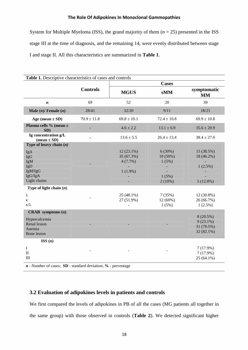
As we can see in Table 1, we studied 69 controls and 111 Monoclonal Gammopathies (MG)
patients, being 52 with MGUS, 20 with sMM and 30 with symptomatic MM. The mean age
was similar in all four groups of subjects, being approximately 70 years. Controls were
mainly females, and MG patients were mainly males. BM plasmocytosis and serum Ig
concentration increased with disease progression, as they were lower in MGUS subjects (4.6
± 2.2 % and 13.6 ± 5.5 g/L, respectively) and higher in the symptomatic MM patients (35.6 ±
20.9% and 38.4 ± 27.0 g/L, respectively).
In MGUS patients the monoclonal immunoglobulin was mainly of the IgG subtype (n = 35),
being the rest IgA, IgM and biclonal IgM/IgG. sMM were mainly of the IgG and IgA subtype
(n = 10 and n = 6, respectively) with only one individual with the IgM subtype, one with a
biclonal component of IgA/IgG and two individuals with the light chain subtype. In what
concerns the symptomatic MM subjects, 18 presented with IgG, 15 with IgA, five with light
chain and one with IgD subtype. In what concerns the type of light chain, MGUS subjects had
similar distribution between λ and κ light chains, as sMM and symptomatic MM patients
produced mainly κ light chains.
CRAB symptoms were defined as: serum calcium value higher than 11 mg/dL or more than 1
mg/dL above the normal value (hypercalcemia), serum creatinine value higher than 2 mg/dL
(renal lesion), hemoglobin value lower than 10 g/dL or more than 2g/dL bellow the normal
value (anemia) and one or more osteolytic lesions in skeletal radiography, CT or PET-CT
(bone lesion). Of the 39 MM patients, the main presenting symptoms were bone lesion and
anemia (32 and 31 patients, respectively). Renal lesion and hypercalcemia only accounted for
nine and eight patients, respectively. Finally, in what concerns the International Staging
The Role Of Adipokines In Monoclonal Gammopathies
18
System for Multiple Myeloma (ISS), the grand majority of them (n = 25) presented in the ISS
stage III at the time of diagnosis, and the remaining 14, were evenly distributed between stage
I and stage II. All this characteristics are summarized in Table 1.
Table 1. Descriptive characteristics of cases and controls
Controls
Cases
MGUS sMM symptomatic
MM
n 69 52 20 39
Male (n)/ Female (n) 28/41 32/20 9/11 18/21
Age (mean ± SD) 70.9 ± 11.8 69.8 ± 10.1 72.4 ± 10.8 69.9 ± 10.8
Plasma cells % (mean ±
SD) - 4.6 ± 2.2 13.1 ± 6.9 35.6 ± 20.9
Ig concentration g/L
(mean ± SD) - 13.6 ± 5.5 26.4 ± 13.4 38.4 ± 27.0
Type of heavy chain (n)
IgA
IgG
IgM
IgD
IgM/IgG
IgG/IgA
Light chains
-
12 (23.1%)
35 (67.3%)
4 (7.7%)
-
1 (1.9%)
-
-
6 (30%)
10 (50%)
1 (5%)
-
-
1 (5%)
2 (10%)
15 (38.5%)
18 (46.2%)
-
1 (2.5%)
-
-
5 (12.8%)
Type of light chain (n)
λ
κ
κ/λ
-
25 (48.1%)
27 (51.9%)
-
7 (35%)
12 (60%)
1 (5%)
12 (30.8%)
26 (66.7%)
1 (2.5%)
CRAB symptoms (n)
Hypercalcemia
Renal lesion
Anemia
Bone lesion
- - -
8 (20.5%)
9 (23.1%)
31 (79.5%)
32 (82.1%)
ISS (n)
I
II
III
- - -
7 (17.9%)
7 (17.9%)
25 (64.1%)
n - Number of cases; SD - standard deviation; % - percentage
3.2 Evaluation of adipokines levels in patients and controls
We first compared the levels of adipokines in PB of all the cases (MG patients all together in
the same group) with those observed in controls (Table 2). We detected significant higher

The Role Of Adipokines In Monoclonal Gammopathies
19
levels of leptin and resistin in PB of MG patients (13.2 ng/mL and 9.49 ng/mL, respectively)
compared with controls (7.95 ng/mL and 7,42 ng/mL, respectively). No significant
differences were found in what regards to adiponectin in this analysis.
In order to explore if there were any differences if we considered each subtype of MG
separately, we compared control individuals with each MG subtype and between the different
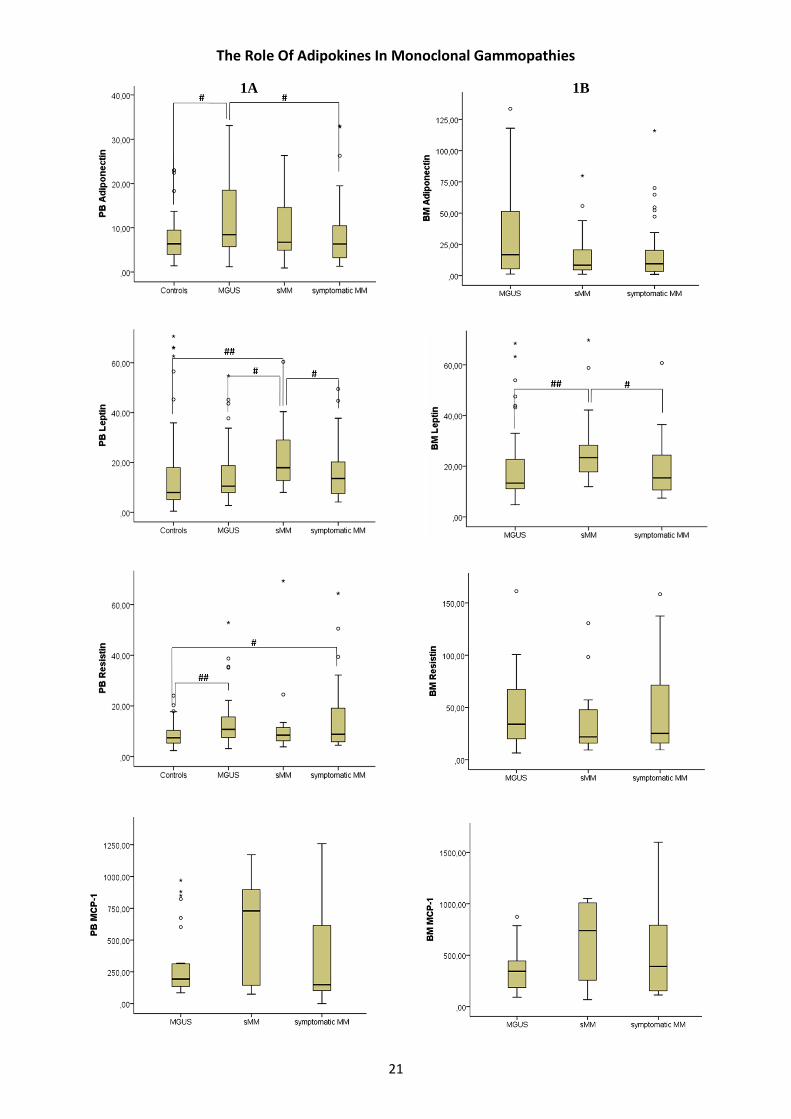
subtypes of MG patients (Table 3 and Figure 1A, B).
In the PB samples, adiponectin was significantly higher in the MGUS patients when
compared with the controls (p = 0.012) and with symptomatic MM subjects (p = 0.04).
MGUS patients also showed higher levels of resistin than the controls (p = 0.001). On the
other hand, we found that sMM was associated with increased PB leptin when compared with
controls (p = 0.001), MGUS (p = 0.012) and symptomatic MM (p = 0.043). TNF-α levels
were also increased in sMM compared with MGUS individuals (p = 0.034). Symptomatic
MM patients had higher levels of resistin than controls (p = 0.014). Although not statistically
significant, symptomatic MM individuals tended to have higher levels of MCP-1 than MGUS
judging by the IQR (Table 3).
In the BM samples, the results were somewhat overlapping with those from the PB samples.
We found significant increase in leptin levels of sMM subjects compared with MGUS (p =
Table 2. Levels of adiponectin, leptin and resistin in the PB of controls and MG subjects
Controls
n = 69 MG
Adiponectin μg/mL 6.36 (5.65) 6.92 (11.85)
n = 110
Leptin ng/mL 7.95 (13.64) 13.2 (13.52)
a
n = 111
Resistin ng/mL 7.42 (5.13) 9.49 (9.73)
a
n = 111
Results in median (IQR); n - Number of cases; a - p < 0.01
The Role Of Adipokines In Monoclonal Gammopathies
20
0.003) and symptomatic MM patients (p = 0.021). The BM levels of resistin and MCP-1 in
symptomatic MM patients tended for higher levels comparing with MGUS judging by their
respective IQR, even though it was not statistically significant. PB and BM results are
illustrated in Figure 1A and B, respectively.
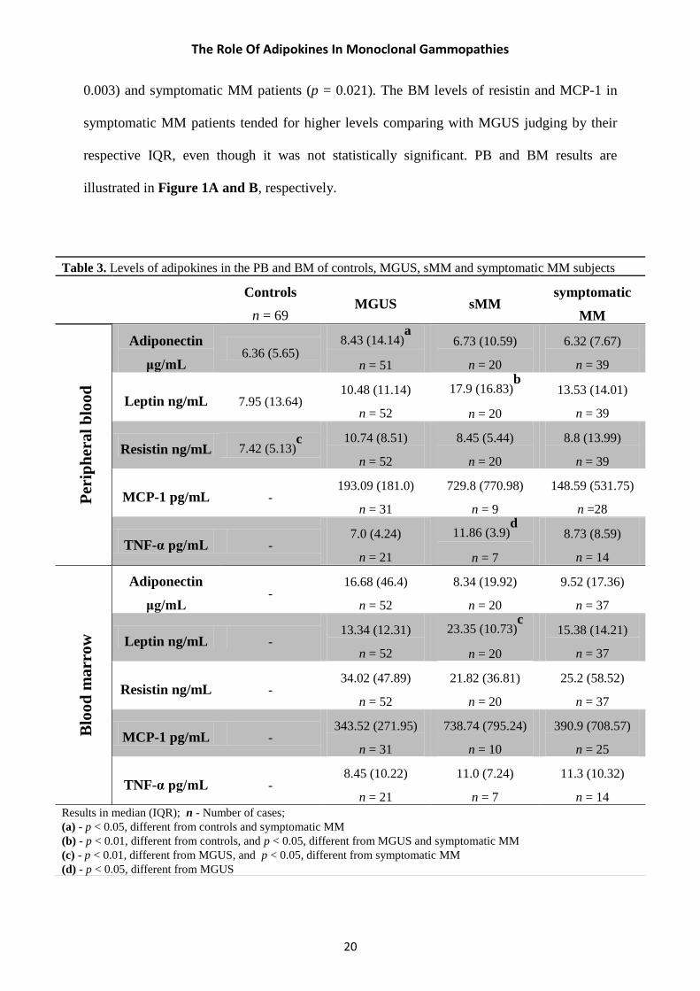
Table 3. Levels of adipokines in the PB and BM of controls, MGUS, sMM and symptomatic MM subjects
Controls
n = 69 MGUS sMM
symptomatic
MM
Per
iph
eral
blo
od
Adiponectin
μg/mL 6.36 (5.65)
8.43 (14.14)a
n = 51
6.73 (10.59)
n = 20
6.32 (7.67)
n = 39
Leptin ng/mL 7.95 (13.64) 10.48 (11.14)
n = 52
17.9 (16.83)b
n = 20
13.53 (14.01)
n = 39
Resistin ng/mL 7.42 (5.13)c
10.74 (8.51)
n = 52
8.45 (5.44)
n = 20
8.8 (13.99)
n = 39
MCP-1 pg/mL - 193.09 (181.0)
n = 31
729.8 (770.98)
n = 9
148.59 (531.75)
n =28
TNF-α pg/mL - 7.0 (4.24)
n = 21
11.86 (3.9)d
n = 7
8.73 (8.59)
n = 14
Blo
od
marr
ow
Adiponectin
μg/mL -
16.68 (46.4)
n = 52
8.34 (19.92)
n = 20
9.52 (17.36)
n = 37
Leptin ng/mL - 13.34 (12.31)
n = 52
23.35 (10.73)c
n = 20
15.38 (14.21)
n = 37
Resistin ng/mL - 34.02 (47.89)
n = 52
21.82 (36.81)
n = 20
25.2 (58.52)
n = 37
MCP-1 pg/mL - 343.52 (271.95)
n = 31
738.74 (795.24)
n = 10
390.9 (708.57)
n = 25
TNF-α pg/mL - 8.45 (10.22)
n = 21
11.0 (7.24)
n = 7
11.3 (10.32)
n = 14
Results in median (IQR); n - Number of cases;
(a) - p < 0.05, different from controls and symptomatic MM
(b) - p < 0.01, different from controls, and p < 0.05, different from MGUS and symptomatic MM
(c) - p < 0.01, different from MGUS, and p < 0.05, different from symptomatic MM
(d) - p < 0.05, different from MGUS
The Role Of Adipokines In Monoclonal Gammopathies
21
1A 1B
The Role Of Adipokines In Monoclonal Gammopathies
22
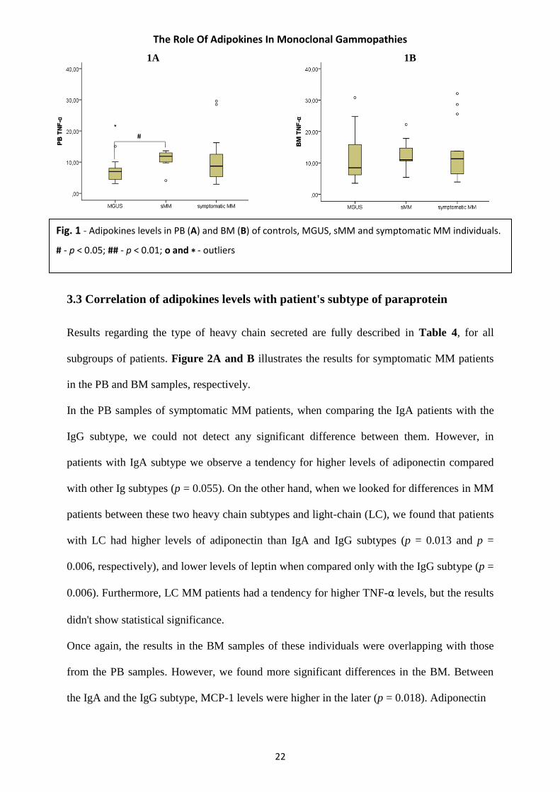
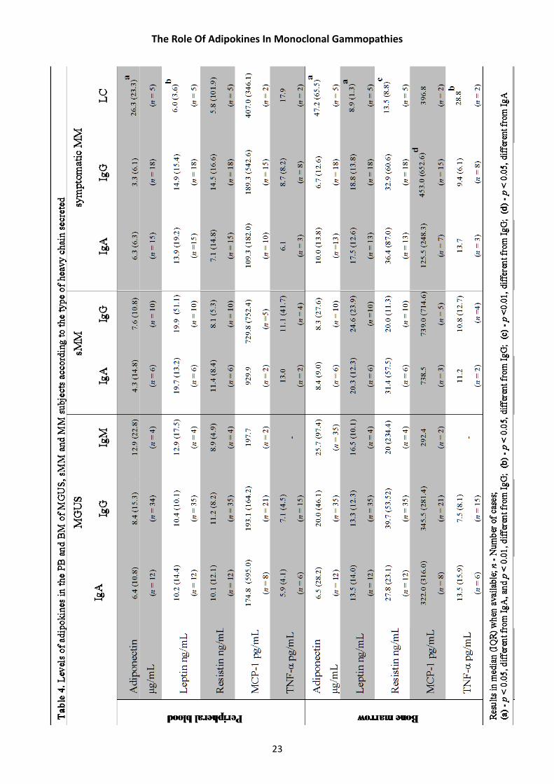
3.3 Correlation of adipokines levels with patient's subtype of paraprotein
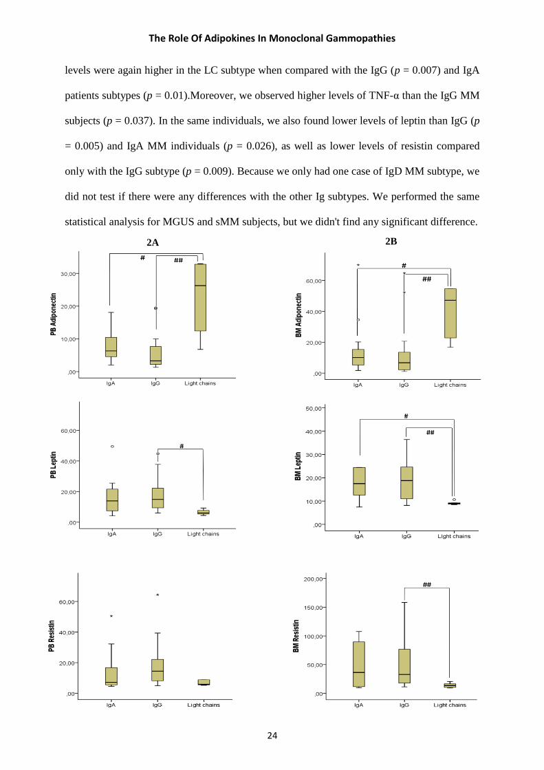
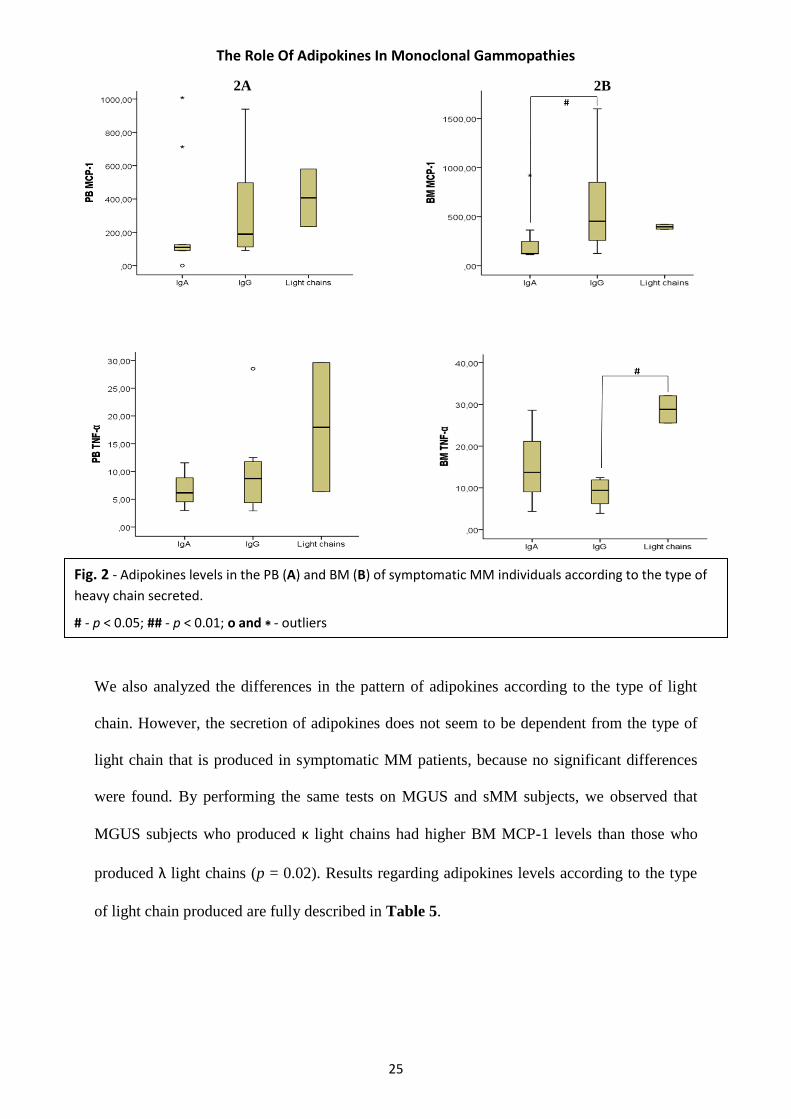
Results regarding the type of heavy chain secreted are fully described in Table 4, for all
subgroups of patients. Figure 2A and B illustrates the results for symptomatic MM patients
in the PB and BM samples, respectively.
In the PB samples of symptomatic MM patients, when comparing the IgA patients with the
IgG subtype, we could not detect any significant difference between them. However, in
patients with IgA subtype we observe a tendency for higher levels of adiponectin compared
with other Ig subtypes (p = 0.055). On the other hand, when we looked for differences in MM
patients between these two heavy chain subtypes and light-chain (LC), we found that patients
with LC had higher levels of adiponectin than IgA and IgG subtypes (p = 0.013 and p =
0.006, respectively), and lower levels of leptin when compared only with the IgG subtype (p =
0.006). Furthermore, LC MM patients had a tendency for higher TNF-α levels, but the results
didn't show statistical significance.
Once again, the results in the BM samples of these individuals were overlapping with those
from the PB samples. However, we found more significant differences in the BM. Between
the IgA and the IgG subtype, MCP-1 levels were higher in the later (p = 0.018). Adiponectin
1A 1B
Fig. 1 - Adipokines levels in PB (A) and BM (B) of controls, MGUS, sMM and symptomatic MM individuals.
# - p < 0.05; ## - p < 0.01; o and * - outliers
The Role Of Adipokines In Monoclonal Gammopathies
23
The Role Of Adipokines In Monoclonal Gammopathies
24
levels were again higher in the LC subtype when compared with the IgG (p = 0.007) and IgA
patients subtypes (p = 0.01).Moreover, we observed higher levels of TNF-α than the IgG MM
subjects (p = 0.037). In the same individuals, we also found lower levels of leptin than IgG (p
= 0.005) and IgA MM individuals (p = 0.026), as well as lower levels of resistin compared
only with the IgG subtype (p = 0.009). Because we only had one case of IgD MM subtype, we
did not test if there were any differences with the other Ig subtypes. We performed the same
statistical analysis for MGUS and sMM subjects, but we didn't find any significant difference.
2A 2B
The Role Of Adipokines In Monoclonal Gammopathies
25
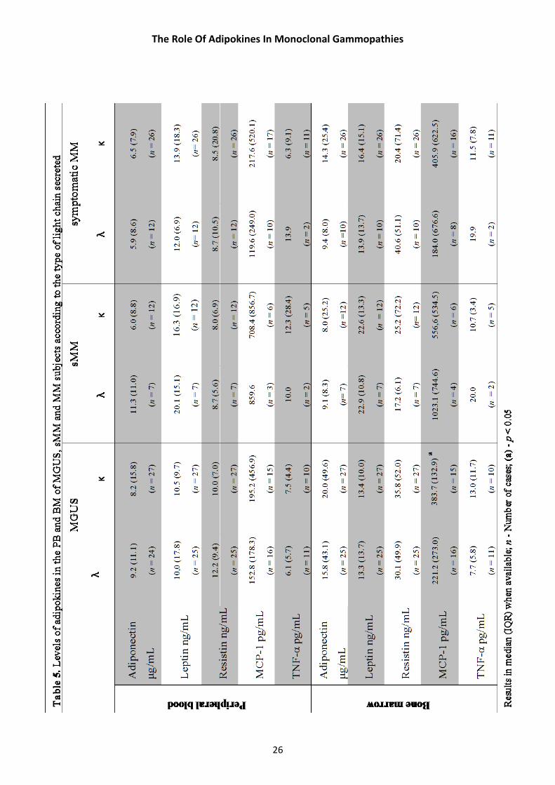
We also analyzed the differences in the pattern of adipokines according to the type of light
chain. However, the secretion of adipokines does not seem to be dependent from the type of
light chain that is produced in symptomatic MM patients, because no significant differences
were found. By performing the same tests on MGUS and sMM subjects, we observed that
MGUS subjects who produced κ light chains had higher BM MCP-1 levels than those who
produced λ light chains (p = 0.02). Results regarding adipokines levels according to the type
of light chain produced are fully described in Table 5.
2A 2B
Fig. 2 - Adipokines levels in the PB (A) and BM (B) of symptomatic MM individuals according to the type of
heavy chain secreted.
# - p < 0.05; ## - p < 0.01; o and * - outliers
The Role Of Adipokines In Monoclonal Gammopathies
26
The Role Of Adipokines In Monoclonal Gammopathies
27
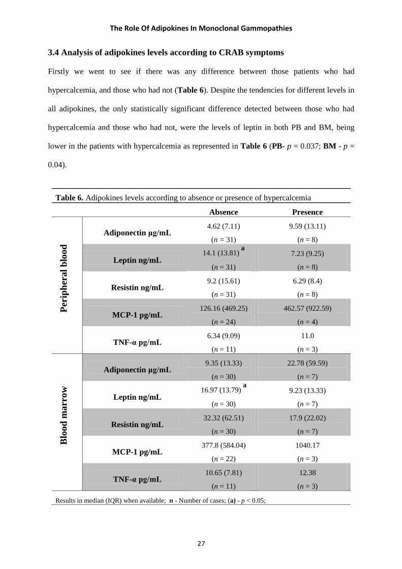
3.4 Analysis of adipokines levels according to CRAB symptoms
Firstly we went to see if there was any difference between those patients who had
hypercalcemia, and those who had not (Table 6). Despite the tendencies for different levels in
all adipokines, the only statistically significant difference detected between those who had
hypercalcemia and those who had not, were the levels of leptin in both PB and BM, being
lower in the patients with hypercalcemia as represented in Table 6 (PB- p = 0.037; BM - p =
0.04).
Table 6. Adipokines levels according to absence or presence of hypercalcemia
Absence Presence
Per
iph
eral
blo
od
Adiponectin μg/mL 4.62 (7.11)
(n = 31)
9.59 (13.11)
(n = 8)
Leptin ng/mL 14.1 (13.81)
a
(n = 31)
7.23 (9.25)
(n = 8)
Resistin ng/mL 9.2 (15.61)
(n = 31)
6.29 (8.4)
(n = 8)
MCP-1 pg/mL 126.16 (469.25)
(n = 24)
462.57 (922.59)
(n = 4)
TNF-α pg/mL 6.34 (9.09)
(n = 11)
11.0
(n = 3)
Blo
od
marr
ow
Adiponectin μg/mL 9.35 (13.33)
(n = 30)
22.78 (59.59)
(n = 7)
Leptin ng/mL 16.97 (13.79)
a
(n = 30)
9.23 (13.33)
(n = 7)
Resistin ng/mL 32.32 (62.51)
(n = 30)
17.9 (22.02)
(n = 7)
MCP-1 pg/mL 377.8 (584.04)
(n = 22)
1040.17
(n = 3)
TNF-α pg/mL 10.65 (7.81)
(n = 11)
12.38
(n = 3)
Results in median (IQR) when available; n - Number of cases; (a) - p < 0.05;
The Role Of Adipokines In Monoclonal Gammopathies
28
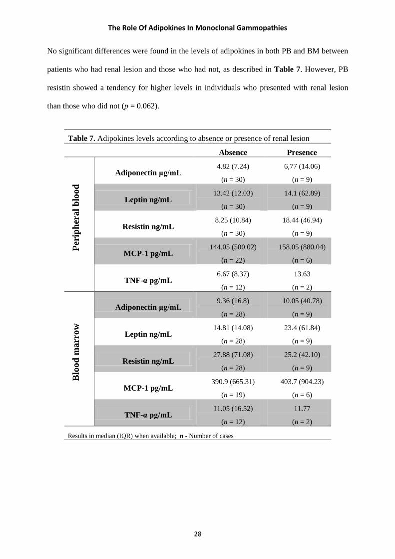
No significant differences were found in the levels of adipokines in both PB and BM between
patients who had renal lesion and those who had not, as described in Table 7. However, PB
resistin showed a tendency for higher levels in individuals who presented with renal lesion
than those who did not (p = 0.062).
Table 7. Adipokines levels according to absence or presence of renal lesion
Absence Presence
Per
iph
eral
blo
od
Adiponectin μg/mL 4.82 (7.24)
(n = 30)
6,77 (14.06)
(n = 9)
Leptin ng/mL 13.42 (12.03)
(n = 30)
14.1 (62.89)
(n = 9)
Resistin ng/mL 8.25 (10.84)
(n = 30)
18.44 (46.94)
(n = 9)
MCP-1 pg/mL 144.05 (500.02)
(n = 22)
158.05 (880.04)
(n = 6)
TNF-α pg/mL 6.67 (8.37)
(n = 12)
13.63
(n = 2)
Blo
od
marr
ow
Adiponectin μg/mL 9.36 (16.8)
(n = 28)
10.05 (40.78)
(n = 9)
Leptin ng/mL 14.81 (14.08)
(n = 28)
23.4 (61.84)
(n = 9)
Resistin ng/mL 27.88 (71.08)
(n = 28)
25.2 (42.10)
(n = 9)
MCP-1 pg/mL 390.9 (665.31)
(n = 19)
403.7 (904.23)
(n = 6)
TNF-α pg/mL 11.05 (16.52)
(n = 12)
11.77
(n = 2)
Results in median (IQR) when available; n - Number of cases
The Role Of Adipokines In Monoclonal Gammopathies
29
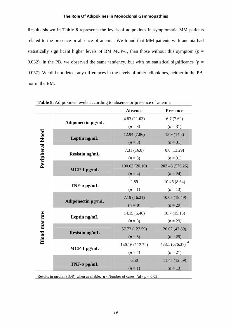
Results shown in Table 8 represents the levels of adipokines in symptomatic MM patients
related to the presence or absence of anemia. We found that MM patients with anemia had
statistically significant higher levels of BM MCP-1, than those without this symptom (p =
0.032). In the PB, we observed the same tendency, but with no statistical significance (p =
0.057). We did not detect any differences in the levels of other adipokines, neither in the PB,
nor in the BM.
Table 8. Adipokines levels according to absence or presence of anemia
Absence Presence
Per
iph
eral
blo
od
Adiponectin μg/mL 4.83 (11.03)
(n = 8)
6.7 (7.69)
(n = 31)
Leptin ng/mL 12.94 (7.86)
(n = 8)
13.9 (14.8)
(n = 31)
Resistin ng/mL 7.31 (16.8)
(n = 8)
8.8 (13.29)
(n = 31)
MCP-1 pg/mL 100.62 (20.10)
(n = 4)
203.46 (576.26)
(n = 24)
TNF-α pg/mL 2.89
(n = 1)
10.46 (8.64)
(n = 13)
Blo
od
marr
ow
Adiponectin μg/mL 7.19 (16.21)
(n = 8)
10.05 (18.49)
(n = 29)
Leptin ng/mL 14.15 (5.46)
(n = 8)
18.7 (15.15)
(n = 29)
Resistin ng/mL 57.73 (127.59)
(n = 8)
20.02 (47.89)
(n = 29)
MCP-1 pg/mL 140.16 (112.72)
(n = 4)
438.1 (676.37) a
(n = 21)
TNF-α pg/mL 6.50
(n = 1)
11.45 (12.59)
(n = 13)
Results in median (IQR) when available; n - Number of cases; (a) - p < 0.05
The Role Of Adipokines In Monoclonal Gammopathies
30
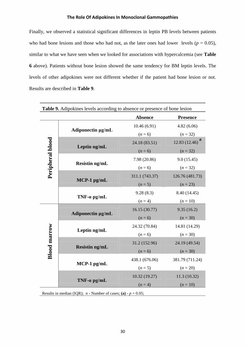
Finally, we observed a statistical significant differences in leptin PB levels between patients
who had bone lesions and those who had not, as the later ones had lower levels (p = 0.05),
similar to what we have seen when we looked for associations with hypercalcemia (see Table
6 above). Patients without bone lesion showed the same tendency for BM leptin levels. The
levels of other adipokines were not different whether if the patient had bone lesion or not.
Results are described in Table 9.
Table 9. Adipokines levels according to absence or presence of bone lesion
Absence Presence
Per
iph
eral
blo
od
Adiponectin μg/mL 10.46 (6.91)
(n = 6)
4.82 (6.06)
(n = 32)
Leptin ng/mL 24.18 (83.51)
(n = 6)
12.83 (12.46) a
(n = 32)
Resistin ng/mL 7.98 (20.86)
(n = 6)
9.0 (15.45)
(n = 32)
MCP-1 pg/mL 311.1 (743.37)
(n = 5)
126.76 (481.73)
(n = 23)
TNF-α pg/mL 9.28 (8.3)
(n = 4)
8.40 (14.45)
(n = 10)
Blo
od
marr
ow
Adiponectin μg/mL 16.15 (30.77)
(n = 6)
9.35 (16.2)
(n = 30)
Leptin ng/mL 24.32 (70.84)
(n = 6)
14.81 (14.29)
(n = 30)
Resistin ng/mL 31.2 (152.96)
(n = 6)
24.19 (49.54)
(n = 30)
MCP-1 pg/mL 438.1 (676.06)
(n = 5)
381.79 (711.24)
(n = 20)
TNF-α pg/mL 10.32 (19.27)
(n = 4)
11.3 (10.32)
(n = 10)
Results in median (IQR); n - Number of cases; (a) - p = 0.05;
The Role Of Adipokines In Monoclonal Gammopathies
31
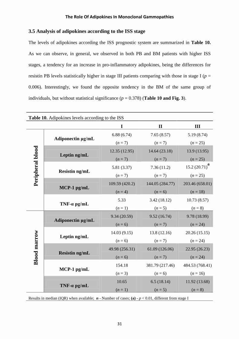
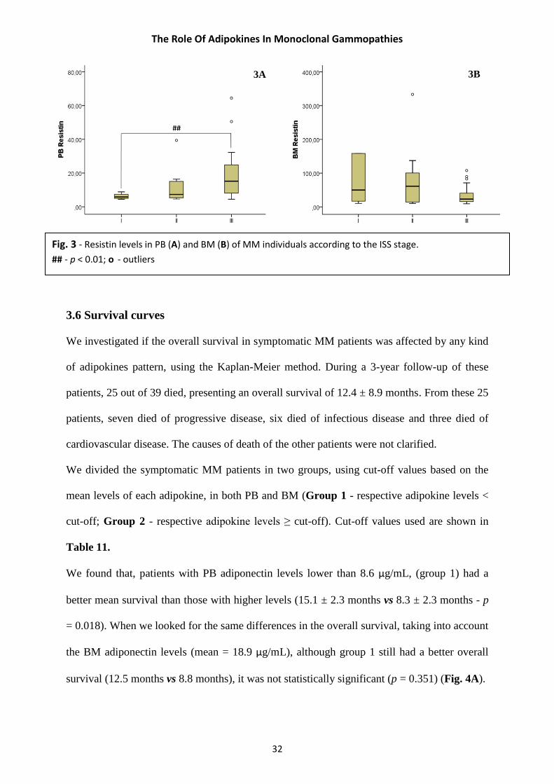
3.5 Analysis of adipokines according to the ISS stage
The levels of adipokines according the ISS prognostic system are summarized in Table 10.
As we can observe, in general, we observed in both PB and BM patients with higher ISS
stages, a tendency for an increase in pro-inflammatory adipokines, being the differences for
resistin PB levels statistically higher in stage III patients comparing with those in stage I (p =
0.006). Interestingly, we found the opposite tendency in the BM of the same group of
individuals, but without statistical significance (p = 0.378) (Table 10 and Fig. 3).
Table 10. Adipokines levels according to the ISS
I II III
Per
iph
eral
blo
od
Adiponectin μg/mL 6.88 (6.74)
(n = 7)
7.65 (8.57)
(n = 7)
5.19 (8.74)
(n = 25)
Leptin ng/mL 12.35 (12.95)
(n = 7)
14.64 (23.18)
(n = 7)
13.9 (13.95)
(n = 25)
Resistin ng/mL 5.81 (3.37)
(n = 7)
7.36 (11.2)
(n = 7)
15.2 (20.71)a
(n = 25)
MCP-1 pg/mL 109.59 (420.2)
(n = 4)
144.05 (284.77)
(n = 6)
203.46 (658.01)
(n = 18)
TNF-α pg/mL 5.33
(n = 1)
3.42 (18.12)
(n = 5)
10.73 (8.57)
(n = 8)
Blo
od
marr
ow
Adiponectin μg/mL 9.34 (20.59)
(n = 6)
9.52 (16.74)
(n = 7)
9.78 (18.99)
(n = 24)
Leptin ng/mL 14.03 (9.15)
(n = 6)
13.8 (12.16)
(n = 7)
20.26 (15.15)
(n = 24)
Resistin ng/mL 49.98 (256.31)
(n = 6)
61.09 (126.06)
(n = 7)
22.95 (26.23)
(n = 24)
MCP-1 pg/mL 154.18
(n = 3)
381.79 (217.46)
(n = 6)
484.53 (768.41)
(n = 16)
TNF-α pg/mL 10.65
(n = 1)
6.5 (18.14)
(n = 5)
11.92 (13.68)
(n = 8)
Results in median (IQR) when available; n - Number of cases; (a) - p < 0.01, different from stage I
The Role Of Adipokines In Monoclonal Gammopathies
32
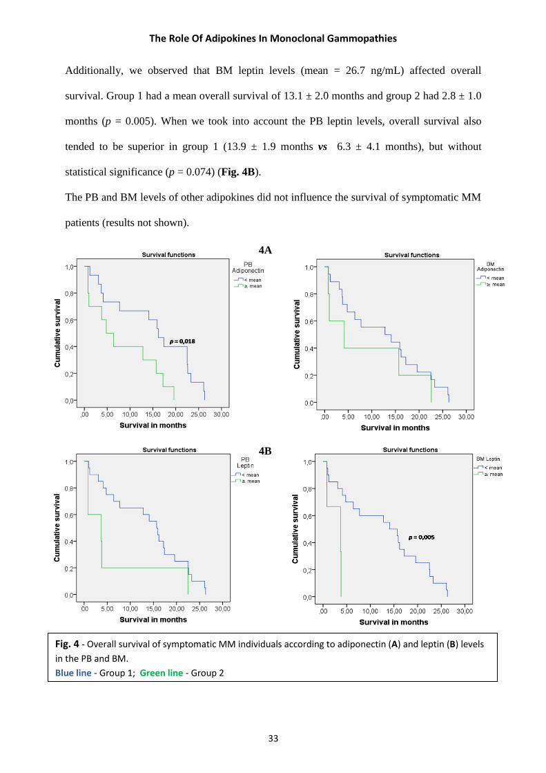
3.6 Survival curves
We investigated if the overall survival in symptomatic MM patients was affected by any kind
of adipokines pattern, using the Kaplan-Meier method. During a 3-year follow-up of these
patients, 25 out of 39 died, presenting an overall survival of 12.4 ± 8.9 months. From these 25
patients, seven died of progressive disease, six died of infectious disease and three died of
cardiovascular disease. The causes of death of the other patients were not clarified.
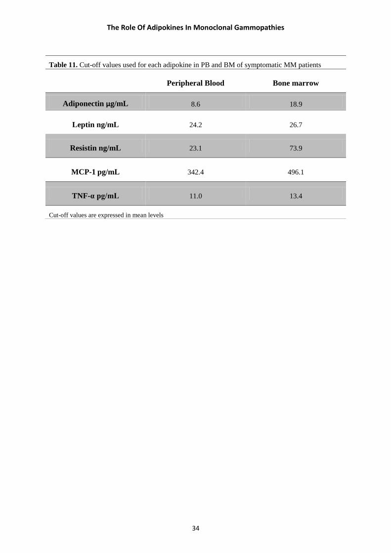
We divided the symptomatic MM patients in two groups, using cut-off values based on the
mean levels of each adipokine, in both PB and BM (Group 1 - respective adipokine levels <
cut-off; Group 2 - respective adipokine levels ≥ cut-off). Cut-off values used are shown in
Table 11.
We found that, patients with PB adiponectin levels lower than 8.6 μg/mL, (group 1) had a
better mean survival than those with higher levels (15.1 ± 2.3 months vs 8.3 ± 2.3 months - p
= 0.018). When we looked for the same differences in the overall survival, taking into account
the BM adiponectin levels (mean = 18.9 μg/mL), although group 1 still had a better overall
survival (12.5 months vs 8.8 months), it was not statistically significant (p = 0.351) (Fig. 4A).
3A 3B
Fig. 3 - Resistin levels in PB (A) and BM (B) of MM individuals according to the ISS stage.
## - p < 0.01; o - outliers
The Role Of Adipokines In Monoclonal Gammopathies
33
Additionally, we observed that BM leptin levels (mean = 26.7 ng/mL) affected overall
survival. Group 1 had a mean overall survival of 13.1 ± 2.0 months and group 2 had 2.8 ± 1.0
months (p = 0.005). When we took into account the PB leptin levels, overall survival also
tended to be superior in group 1 (13.9 ± 1.9 months vs 6.3 ± 4.1 months), but without
statistical significance (p = 0.074) (Fig. 4B).
The PB and BM levels of other adipokines did not influence the survival of symptomatic MM
patients (results not shown).
4A
4B
Fig. 4 - Overall survival of symptomatic MM individuals according to adiponectin (A) and leptin (B) levels
in the PB and BM.
Blue line - Group 1; Green line - Group 2
The Role Of Adipokines In Monoclonal Gammopathies
34
Table 11. Cut-off values used for each adipokine in PB and BM of symptomatic MM patients
Peripheral Blood Bone marrow
Adiponectin μg/mL 8.6 18.9
Leptin ng/mL 24.2 26.7
Resistin ng/mL 23.1 73.9
MCP-1 pg/mL 342.4 496.1
TNF-α pg/mL 11.0 13.4
Cut-off values are expressed in mean levels
The Role Of Adipokines In Monoclonal Gammopathies
35
4. Discussion and Conclusion
Adipokines display an important role in several physiological functions in the organism.
Obesity, is related with a low-grade inflammation, since there is a decrease in adiponectin and
an increase in pro-inflammatory adipokines with an oncogenic potential (18,29)
. Although it is
well documented that, obesity increase the risk for some types of solid tumors, most of the
studies, showing that adipokines participate in this relationship, are in vitro studies (17)
.
However, the role of adipokines in hematological malignancies is poorly understood, and in
what regards MM, only a few studies were undertaken.
Our results show that in PB MG patients have higher levels of leptin and resistin than control
individuals. The analysis of the adipokines PB levels in each different MG subgroups, showed
that MGUS patients had higher levels of adiponectin than symptomatic MM individuals, as
well as lower levels of TNF-α. Also, sMM patients had higher levels of leptin compared with
controls, MGUS and symptomatic MM patients. PB resistin levels were higher in both MGUS
and symptomatic MM patients than control individuals. MCP-1 tended for higher levels in the
symptomatic MM group comparing with MGUS. In the BM, the sMM also presented with
higher levels of leptin than MGUS and symptomatic MM patients. Adiponectin levels tended
as well to be lower in the later stages of the disease. Resistin and MCP-1 were higher in the
symptomatic MM subgroup of individuals. As the BM levels appears to have the same pattern
of the PB, this indicates that the PB levels reflects the levels in the BM microenvironment.
As adipokines can exhibit anti- and pro-inflammatory properties, these results suggest that in
the progression from from MGUS to MM, deregulation of the production of adipokines
creates a pro-inflammatory state in the BM microenvironment, which can contribute to the
carcinogenic process.
The Role Of Adipokines In Monoclonal Gammopathies
36
In what concerns adiponectin, Hoffman et al. (30)
observed an inverse association between
MM risk and adiponectin plasma levels. Moreover, Dalamaga et al. (31)
, also reported that
MM individuals had lower serum levels of adiponectin. Our results are in agreement with
both of these studies. Adiponectin is an anti-inflammatory adipokine that may exert its effects
through inhibition of the NF-κB pathway. This in turn, suppresses production of various
cytokines such as IL-6 and TNF-α, which are important growth factors for MM cells (12–
14,32,33). Adiponectin may as well increase the production of anti-inflammatory cytokine IL-10
(24). It is possible that adiponectin may act as a protective factor in MGUS individuals and as a
marker of progression from MGUS to MM. A study conducted by Fowler et al. (34)
discovered
that, host-derived adiponectin reduced tumor burden, increased survival of myeloma-bearing
mice and prevented bone disease. This gives adiponectin some potential as a novel biomarker
and therapeutic target.
Leptin is an adipokine that regulates caloric intake and energy homeostasis. Other studies
found that MM individuals presented with higher plasma levels of leptin than controls (31,35–
38). Recently, it was shown that MM cells express leptin receptors, and that blockage of those
receptors leads to a decrease in the DNA synthesis (27)
. Reseland et al., in 2009, reported that
leptin induced the expression of several genes involved in cell growth, survival and signaling
in two myeloma cell lines (35)
. As stated before, the adipocytes are often forgotten in the
studies of MM. Reseland's and our results suggest that adipocytes may have a role in
pathogenesis of MM, since they are the major producers of leptin. Interestingly, in our study,
leptin levels were higher in the sMM subgroup, rather than in the symptomatic MM subgroup.
It could be due to the fact that, during the disease development, BM adipocytes tend to
disappear, suggesting that the role of BM adipocytes may be restricted to the initial stages of
the disease, before a remodeling of the BMM has occurred (27)
. This can mean that the
The Role Of Adipokines In Monoclonal Gammopathies
37
maximum levels of leptin are produced during sMM phase, before the progression to MM,
when symptoms, like bone lesions, change the BMM.
Our results suggest that resistin contributes to the creation of a pro-inflammatory state in the
BM. Two other studies (36,37)
focusing on leptin and resistin serum levels didn't found any
significant differences between MM patients and controls. Interestingly, another study (31)
observed significant lower serum levels of resistin in MM patients compared with controls.
The authors reported that this finding could be possibly due to a compensatory response of the
resistin pathway to the upregulation of other inflammatory and proliferative factors. PB
resistin levels in our study, where higher in MGUS and symptomatic MM patients than in
controls.
Resistin is an adipokine related with insulin-resistance and activation of inflammatory
processes. Whereas in mice, resistin is produced mainly in the adipocytes, in humans it is
produced mainly by macrophages and monocytes (18)
. This promotes the production of IL-6
and TNF-α, which are important growth factors for MM cells (12–14,18,33)
. To our knowledge,
there are no studies showing that in MM resistin prodution, is mainly due to BM
macrophages.
Additionally, we observed that MCP-1 tended to be higher in PB and BM in patients in the
later stages of the disease, when compared with MGUS patients. In what regards to TNF-α,
we only found higher levels in the PB of sMM patients compared with MGUS subgroup.
MCP-1 is a potent chemoattractant that acts through CCR2 receptor, which is also expressed
in MM cells (39)
, resulting in enhanced migration of the malignant clone. This makes MCP-1
an attractive therapeutic target for MM (39)
. In fact, blockage of CCR2 receptor and
neutralization of MCP-1, -2 and -3 significantly reduced migration of human MM cells to
marrow stromal cell-conditioned medium (39)
. TNF-α acts not only as a growth factor, but also
The Role Of Adipokines In Monoclonal Gammopathies
38
upregulates the production of MCP-1 by the BM stromal cells. We did not find any other
studies measuring MCP-1 and TNF-α levels in MM patients.
According with Ig subtype, namely between the IgA and IgG symptomatic MM patients, we
only found higher levels of BM MCP-1 in those who produced IgG heavy chains. However,
we observed higher PB and BM levels of adiponectin and lower levels of BM leptin of those
who only produced light chains relatively to those who produced IgA or IgG heavy chains.
Additionally, MM patients with light chains were associated with lower leptin PB levels and
lower resistin and TNF-α BM levels than patients with IgG subtype. In what regards the type
of light chain secreted, we only observed higher BM MCP-1 levels in κ light chain MGUS
patients.
The type of immunoglobulin that is secreted can affect the outcome of patients. For example,
the IgA subtype was associated with more severe anemia, and with lower overall survival (40).
Also the LC subtype is associated with higher incidence of renal lesion. However, in one
study, overall survival was comparable to the IgG MM patients (40)
. Furthermore, one study
showed a positive correlation between serum levels of leptin and IgG levels in MM patients
(36). Our results suggest that, given the differences especially between the light chain and
IgG/IgA subtype, the adipokines may have different roles in the different MM subtypes, as
they could participate in the immunological changes underlying the pathogenesis of MM.
Some features of MGUS are referred as risk factors for progression to MM. For MGUS, non-
IgG subtype is associated with higher progression rates to MM, especially when associated
with an M-protein concentration higher than 1,5 g/dL and an abnormal free-light chain
ratio(7)
. However, we did not find any differences in what concerns the type of heavy chain
secreted by MGUS patients.
The Role Of Adipokines In Monoclonal Gammopathies
39
The presence of CRAB symptoms is required to define the diagnosis of symptomatic MM. In
our study we observed higher levels of leptin in the PB and BM of patients without
hypercalcemia and without bone lesion. Leptin stimulates bone formation through promotion
of osteoblast differentiation, bone mineralization and modification of osteoclast
differentiation (41)
. It is possible that the high levels of leptin in MM act as protective factor
for bone disease, and therefore, prevent the occurrence of these symptoms.
We also found higher levels of BM MCP-1 in patients who developed anemia. Anemia in
MM is present in about 73% of the patients at diagnosis (5). The origin of the anemia is
multifactorial and complex, but it is mainly due to the expansion of malignant cells in the
BM. As MCP-1 acts as a chemoattractant to MM cells to the BM, our results suggest that
MCP-1 may contribute to anemia related with BM infiltration.
The International Staging System (ISS) for MM is currently used for disease staging. It takes
into account albumin and β2-microglobulin serum levels. Our results show that there is a
significant increase in PB resistin levels from patients in stage I to patients in stage III.
Interestingly, we found the opposite tendency for BM resistin. We could not find any
explanation for this finding. As resistin, in humans, is mainly produced by macrophages, it
could be interesting to see if there are any differences in the BM macrophages of MM patients
in the different stages of the ISS. However, our results suggest that higher ISS stages tend to
be associated with higher levels of pro-inflammatory adipokines. Comparing patients
stratified according with the Durie-Salmon staging system, Dalamaga et al. (31)
found that
higher stages tended to present higher levels of leptin, and Esheba et al. (36)
found that stage II
patients had higher leptin levels than those in stage I.
There is some controversy in what regards the ISS, since it was established using data from
patients who did not had access to novel therapeutics agents (42)
, meaning that this staging
The Role Of Adipokines In Monoclonal Gammopathies
40
system could fail to predict correctly the prognosis of some patients. One study suggested the
creation of a new staging system consisting in the presence of anemia and plasmocytoma (42)
.
With the introduction of novel therapeutics, MM overall 5-year relative survival increased
from 28,8% to 34,7%, and 10-year relative survival from 11,1% to 17,4% (43)
. By analyzing
the impact of the levels of adipokines in overall survival of symptomatic MM patients, we
observed that high PB adiponectin and BM leptin levels were associated with significantly
reduced overall survival. As sMM and symptomatic MM patients had lower levels of
adiponectin compared with MGUS, it is possible that the decrease in the levels of adiponectin
act as marker of progression to MM, and, during the disease, as a protective factor.
In summary, as disease progresses from MGUS to symptomatic MM, there is a decrease in
the levels of anti-inflammatory adipocytokine adiponectin, and an increase in pro-
inflammatory adipokines, which could contribute to the mielomagenesis process through the
generation of a pro-inflammatory microenvironment. There seems to exist different roles for
adipokines levels according to the type of paraprotein produced, as MM patients producing
different subtypes of Igs, have different patterns of adipokines production. Patients with
symptomatic MM without bone lesion and hypercalcemia have higher levels of leptin, which
could reflect the ability of promoting osteoblastic differentiation and bone mineralization.
Moreover, MCP-1 was higher in patients presenting with anemia, which could mean that
MCP-1 plays a role in BM infiltration by malignant cells and development of anemia.
Resistin levels were higher in the PB of MM patients in stage III, and lower in the BM of the
same individuals. Additionally, high levels of adiponectin and leptin affected patient's overall
survival, which could translated that these adipokines could have a potential prognostic value.
As MM remains an incurable disease and with poor prognosis, all efforts to better understand
the pathogenesis of MM are of major importance. Not only it will allow us to clarify the

The Role Of Adipokines In Monoclonal Gammopathies
41
mechanisms of the disease, but it will also allow us to discover new therapeutic targets and
create groups of risk for the disease, with the objective of improving the prognosis of the
patients. However, further studies are needed to investigate the importance of the adipokines
in MM and to clarify its therapeutic and prognostic value in the clinical practice.
The Role Of Adipokines In Monoclonal Gammopathies
42
5. Acknowledgments
I want to express my gratitude to my supervisor, Professora Doutora Ana Bela Sarmento
Ribeiro, and my co-supervisor, Professora Doutora Raquel Seiça, for all the advices and
support during the development of this project.
Also, I want to thank Dra. Ana Cristina Gonçalves for all the availability and knowledge
transmission that were of unquestionable value, and to Doutor Paulo Matafome, for all the
support in the laboratory work, the availability and the friendship.
Finally, I want to thank the Faculty of Medicine of University of Coimbra, Centro Hospitalar
e Universitário de Coimbra and CIMAGO - Center of Investigation on Environment Genetics
and Oncobiology for supporting our research.
6. Conflicts of interest
All authors have no conflicts of interest to declare.
7. References
1. Attaelmannan M, Levinson SS. Understanding and identifying monoclonal
gammopathies. Clinical Chemistry. 2000 Aug; 46(8 Pt 2):1230–8.
2. Palladino C, Bruno B, Boccadoro M. Discovering the meaning of monoclonal
gammopathy of undetermined significance: current knowledge, future challenges.
Translational Medicine @ UniSa. 2014 Jan; 8:12–8.
3. Rajkumar SV. MGUS and smoldering multiple myeloma: update on pathogenesis,
natural history, and management. American Society Hematology Educational Program.
2005 Jan; 340–5.
4. Landgren O. Monoclonal gammopathy of undetermined significance and smoldering
myeloma: new insights into pathophysiology and epidemiology. American Society
Hematology Educational Program. 2010 Jan; 2010:295–302.
5. Palumbo A, Anderson K. Multiple Myeloma. New England Journal of Medicine. 2011;
364:1046–60.
6. Kuehl WM, Bergsagel PL. Multiple myeloma: evolving genetic events and host
interactions. Nature Reviews: Cancer. 2002 Mar; 2(3):175–87.
The Role Of Adipokines In Monoclonal Gammopathies
43
7. Landgren O, Kyle RA, Rajkumar SV. From myeloma precursor disease to multiple
myeloma: new diagnostic concepts and opportunities for early intervention. Clinical
Cancer Research. 2011 Mar 15; 17(6):1243–52.
8. Kyle RA, Rajkumar SV. Multiple myeloma. Blood. 2008 Mar 15; 111(6):2962–72.
9. Alexander DD, Mink PJ, Adami H-O, Cole P, Mandel JS, Oken MM, et al. Multiple
myeloma: a review of the epidemiologic literature. International Journal of Cancer.
2007 Jan; 120 Suppl :40–61.
10. Dimopoulos M, Terpos E. Multiple myeloma. Annals of Oncology. 2010 Oct; 21 Suppl
7(Supplement 7):143–50.
11. Hideshima T, Bergsagel PL, Kuehl WM, Anderson KC. Advances in biology of
multiple myeloma: clinical applications. Blood. 2004 Aug 1; 104(3):607–18.
12. Manier S, Sacco A, Leleu X, Ghobrial IM, Roccaro AM. Bone marrow
microenvironment in multiple myeloma progression. Journal of Biomedicine and
Biotechnology. 2012 Jan; 2012:157496.
13. Roodman GD. Role of the bone marrow microenvironment in multiple myeloma.
Journal of Bone and Mineral Research. 2002 Nov; 17(11):1921–5.
14. Mitsiades CS, Mitsiades NS, Munshi NC, Richardson PG, Anderson KC. The role of
the bone microenvironment in the pathophysiology and therapeutic management of
multiple myeloma: interplay of growth factors, their receptors and stromal interactions.
European Journal of Cancer. 2006 Jul; 42(11):1564–73.
15. Haluzík M, Parízková J, Haluzík MM. Adiponectin and its role in the obesity-induced
insulin resistance and related complications. Physiological Research. 2004 Jan;
53(2):123–9.
16. Trayhurn P, Wood IS. Adipokines: inflammation and the pleiotropic role of white
adipose tissue. British Journal of Nutrition. 2007 Mar 9; 92(03):347.
17. Housa D, Housová J, Vernerová Z, Haluzík M. Adipocytokines and cancer.
Physiological Research. 2006 Jan; 55(3):233–44.
18. Ouchi N, Parker JL, Lugus JJ, Walsh K. Adipokines in inflammation and metabolic
disease. Nature Reviews: Immunology. 2011 Feb; 11(2):85–97.
19. Vucenik I, Stains JP. Obesity and cancer risk: evidence, mechanisms, and
recommendations. Annals of the New York Academy of Sciences. 2012 Oct; 1271:37–
43.
20. Cao H. Adipocytokines in obesity and metabolic disease. Journal of Endocrinology.
2014 Feb; 220(2):T47–59.
21. Nigro E, Scudiero O, Monaco ML, Palmieri A, Mazzarella G, Costagliola C, et al. New
Insight into Adiponectin Role in Obesity and Obesity-Related Diseases. Biomedical
Research International. 2014 Jan; 2014:658913.
The Role Of Adipokines In Monoclonal Gammopathies
44
22. Rasouli N, Kern PA. Adipocytokines and the metabolic complications of obesity.
Journal of Clinical Endocrinology and Metabolism. 2008 Nov; 93(11 Suppl 1):S64–73.
23. Lichtman MA. Obesity and the risk for a hematological malignancy: leukemia,
lymphoma, or myeloma. The Oncologist. 2010 Jan; 15(10):1083–101.
24. Dalamaga M, Diakopoulos KN, Mantzoros CS. The role of adiponectin in cancer: a
review of current evidence. Endocrine Reviews. 2012 Aug; 33(4):547–94.
25. Wallin A, Larsson SC. Body mass index and risk of multiple myeloma: a meta-analysis
of prospective studies. European Journal of Cancer. 2011 Jul; 47(11):1606–15.
26. Birmann BM, Giovannucci E, Rosner B, Anderson KC, Colditz GA. Body mass index,
physical activity, and risk of multiple myeloma. Cancer Epidemiology, biomarkers &
Prevention. 2007 Jul; 16(7):1474–8.
27. Caers J, Deleu S, Belaid Z, De Raeve H, Van Valckenborgh E, De Bruyne E, et al.
Neighboring adipocytes participate in the bone marrow microenvironment of multiple
myeloma cells. Leukemia. 2007 Jul; 21(7):1580–4.
28. Bredella MA, Torriani M, Ghomi RH, Thomas BJ, Brick DJ, Gerweck A V, et al.
Vertebral bone marrow fat is positively associated with visceral fat and inversely
associated with IGF-1 in obese women. Obesity (Silver Spring). 2011 Jan; 19(1):49–53.
29. Dalamaga M. Interplay of adipokines and myokines in cancer pathophysiology:
Emerging therapeutic implications. World Journal of Experimental Medicine. 2013
Aug 20; 3(3):26–33.
30. Hofmann JN, Liao LM, Pollak MN, Wang Y, Pfeiffer RM, Baris D, et al. A
prospective study of circulating adipokine levels and risk of multiple myeloma. Blood.
2012 Nov 22; 120(22):4418–20.
31. Dalamaga M, Karmaniolas K, Panagiotou A, Hsi A, Chamberland J, Dimas C, et al.
Low circulating adiponectin and resistin, but not leptin, levels are associated with
multiple myeloma risk: a case-control study. Cancer Causes & Control. 2009 Mar ;
20(2):193–9.
32. Ajuwon KM, Spurlock ME. Adiponectin inhibits LPS-induced NF-kappaB activation
and IL-6 production and increases PPARgamma2 expression in adipocytes. American
Journal of Physiology: Regulatory, integrative and comparative physiology. 2005
May; 288(5):R1220–5.
33. Zhou J, Mauerer K, Farina L, Gribben JG. The role of the tumor microenvironment in
hematological malignancies and implication for therapy. Frontiers in Bioscience. 2005
Jan; 10:1581–96.
34. Fowler JA, Lwin ST, Drake MT, Edwards JR, Kyle RA, Mundy GR, et al. Host-
derived adiponectin is tumor-suppressive and a novel therapeutic target for multiple
myeloma and the associated bone disease. Blood. 2011 Nov 24; 118(22):5872–82.
The Role Of Adipokines In Monoclonal Gammopathies
45
35. Reseland JE, Reppe S, Olstad OK, Hjorth-Hansen H, Brenne AT, Syversen U, et al.
Abnormal adipokine levels and leptin-induced changes in gene expression profiles in
multiple myeloma. European Journal of Haematology. 2009 Nov; 83(5):460–70.
36. Esheba NE, Shahba A, El Shora O. Assessment of leptin and resistin levels in non-
obese multiple myeloma patients and their relation with Ig level and disease stage.
Journal of the Egyptian National Cancer Institute. 2014 Jun; 26(2):61–6.
37. Pamuk GE, Demir M, Harmandar F, Yesil Y, Turgut B, Vural O. Leptin and resistin
levels in serum of patients with hematologic malignancies: correlation with clinical
characteristics. Experimental Oncology. 2006 Sep; 28(3):241–4.
38. Alexandrakis MG, Passam FH, Sfiridaki A, Pappa CA, Moschandrea JA, Kandidakis
E, et al. Serum levels of leptin in multiple myeloma patients and its relation to
angiogenic and inflammatory cytokines. The International Journal of Biological
Markers. 2004 Jan-Mar; 19(1):52–7.
39. Aggarwal R, Ghobrial IM, Roodman GD. Chemokines in multiple myeloma.
Experimental Hematology. 2006 Oct; 34(10):1289–95.
40. Zhang L, Qi J-Y, Qi P-J, Wang Y-F, Zou D-H, Yao H-J, et al. Comparison among
immunologically different subtypes of 595 untreated multiple myeloma patients in
northern China. Clinical lymphoma, myeloma & leukemia. 2010 Jun; 10(3):197–204.
41. Gordeladze JO, Drevon C a, Syversen U, Reseland JE. Leptin stimulates human
osteoblastic cell proliferation, de novo collagen synthesis, and mineralization: Impact
on differentiation markers, apoptosis, and osteoclastic signaling. Journal of Cellular
Biochemistry. 2002 Jan; 85(4):825–36.
42. Iriuchishima H, Saitoh T, Handa H, Isoda A, Matsumoto M, Sawamura M, et al. A new
staging system to predict prognosis of patients with multiple myeloma in an era of
novel therapeutic agents. European Journal of Haematology. 2014 Jun 30; 1–7.
43. Palumbo A, Cerrato C. Diagnosis and therapy of multiple myeloma. The Korean
Journal of Internal Medicine. 2013 May; 28(3):263–73.