SÁVIO MORATO DE LACERDA GONTIJO
34
SÁVIO MORATO DE LACERDA GONTIJO TRATAMENTO ORTODÔNTICO PRÉ-PROTÉTICO Faculdade de Odontologia Universidade Federal de Minas Gerais Belo Horizonte 2018
Transcript of SÁVIO MORATO DE LACERDA GONTIJO
SÁVIO MORATO DE LACERDA GONTIJO
TRATAMENTO ORTODÔNTICO PRÉ-PROTÉTICO
Faculdade de Odontologia Universidade Federal de Minas Gerais
Belo Horizonte 2018
Sávio Morato de Lacerda Gontijo
TRATAMENTO ORTODÔNTICO PRÉ-PROTÉTICO
Monografia apresentada ao Curso de Especialização em Ortodontia da Faculdade de Odontologia da Universidade Federal de Minas Gerais, como requisito parcial para obtenção do título de Especialista em Ortodontia. Orientadora: Prof. (ª) Elizabeth Maria Bastos Lages
Belo Horizonte 2018
Ficha Catalográfica
G641t2018ME
Gontijo, Sávio Morato de Lacerda.Tratamento ortodôntico pré-protético / Sávio Morato de
Lacerda Gontijo. -- 2018.
32 f. : il.
Orientadora: Elizabeth Maria Bastos Lages.
Monografia (Especialização) -- Universidade Federal deMinas Gerais, Faculdade de Odontologia.
1. Interrelação ortodontia-dentística. 2. Tratamentomultidisciplinar. 3. Distribuição de espaços interdentais.I. Lages, Elizabeth Maria Bastos. II. Universidade Federalde Minas Gerais. Faculdade de Odontologia. III. Título.
BLACK - D4
Elaborada pela Biblioteca da Faculdade de Odontologia - UFMG
AGRADECIMENTO
Agradeço a Deus, acima de tudo, por permitir a conclusão desse trabalho.
À minha família pelo apoio e por estarem sempre presentes nessa
caminhada de minha viva.
À minha orientadora, Profª. Drª. Elizabeth Maria Bastos Lages, pelos
ensinamentos, incentivos na pesquisa e por ter me acolhido como um filho, me
ajudando e apoiando nos momentos de maiores dificuldades e anseios.
Ao Prof. Dr. Hugo Henriques Alvim, pelos ensinamentos, pela
oportunidade e confiança no meu trabalho.
Ao Prof. Dr. Esdras Campos Franças, pela grande amizade,
disponibilidade, ajuda, confiança e incentivo para o meu crescimento profissional.
Ao Cirurgião dentista Phelipe Morgado pelas contribuições na execução
do presente caso clínico.
À Profª. Drª. Soraia Macari pela imensa ajuda na leitura e no
desenvolvimento da revisão sistemática, que ainda se encontra em construção.
Ao Prof. Dr. Lucas Guimarães Abreu, pelos ensinamentos e ajuda na
condução da revisão sistemática.
Aos Profs. Eliézer Andrade e Marcelo Lombardi pelo exemplo de
profissionalismo e por todos os ensinamentos e ajudas intra e extraclasse.
Aos professores da Ortodontia (Prof. Dr. Alexandre Drummond, Profa.
Flávia Bartolomeo, Prof. Dr. Henrique Pretti, Prof. Profa. Dra. Giselle Cabral, Prof. Dr.
Leonardo Foresti, Prof. Marcelo Souki, Profa. Dra. Leniana Neves) por todos os
ensinamentos adquiridos. Sou muito grato a todos vocês.
Aos colegas de especialização por toda amizade e carinho, em especial
ao Henrique Eto, Ênio Barreto e Ana Luiza por todos os momentos de
companheirismo.
Aos funcionários da Ortodontia, em especial à Eloiza, pela amizade,
confiança e dedicação para o bom funcionamento desse curso.
Aos pacientes que permitiram que os conhecimentos adquiridos
pudessem ser praticados e aprimorados.
Aos ex-alunos pelos ensinamentos e convívio agradável.
Aos funcionários do Núcleo Ortodôntico por toda disponibilidade e ajuda.

A todos, que de alguma maneira contribuíram para a realização deste
trabalho.
RESUMO
Muitas vezes para atender as necessidades dos pacientes é necessária uma abordagem multidisciplinar. Assim sendo, o presente trabalho descreve um caso clínico em que foi realizado o tratamento ortodôntico previamente ao tratamento protético. A ortodontia foi realizada para melhorar os espaços interdentais dos dentes antêro-superiores e inferiores para a confecção das facetas de porcelana. Para melhor distribuição dos espaços interdentais foi realizado previamente o alinhamento e nivelamento. Posteriormente, foi utilizado molas de NiTi abertas para melhorar a distribuição dos espaços. O presente caso clínico mostra a importância do tratamento multidisciplinar de forma a atender as necessidades e anseios da paciente.
Palavras-chave: Interrelação ortodontia-dentística. Tratamento multidisciplinar. Distribuição de espaços interdentais.
ABSTRACT
Pre-prosthetic orthodontic treatment
A multidisciplinary approach is often needed to meet patients' needs. Thus, the present study describes a clinical case in which orthodontic treatment was performed prior to prosthetic treatment. Orthodontics was performed to improve the interdental spaces of the upper and lower teeth for the porcelain veneers. For better distribution of interdental spaces, alignment and leveling were performed previously. Subsequently, open NiTi springs were used to improve the distribution of the spaces. The present clinical case shows the importance of the multidisciplinary treatment in order to attend to the needs and desires of the patient.
Keywords: Orthodontic-dental interrelations. Multidisciplinary treatment. Distribution
of interdental spaces.
LISTA DE FIGURAS
Figura 1 - Análise facial. A) vista facial frontal, B) vista frontal do sorriso, C)
vista facial de perfil.......................................................................... 12
Figura 2 - Análise dentária inicial. A) vista lateral direita, B) vista frontal, C)
vista lateral esquerda, D) vista oclusal superior, E) vista oclusal
inferior..............................................................................................13
Figura 3 - Radiografia panorâmica inicial........................................................14
Figura 4 - Telerradiografia em norma lateral...................................................14
Figura 5 - Vista frontal do sorriso durante o alinhamento e nivelamento.........15
Figura 6 - Arco de aço 0,020” com molas de NiTi aberta para melhor
distribuição dos espaços interdentais..............................................15
Figura 7 - Conteção móvel superior do tipo wraparound e inferior do tipo
spring............................................................................................... 16
Figura 8 - Análise dentária pós-ortodontia. A) vista lateral direita, B) vista
frontal, C) vista lateral esquerda, D) vista frontal superior.............. 16
LISTA DE ABREVIATURAS E SIGLAS
DSD Digital Smile Design
NiTi Níquel-Titânio
PDS Planejamento Digital do Sorriso
UFMG Universidade Federal de Minas Gerais
SUMÁRIO
1 INTRODUÇÃO.................................................................................................... 10
1.1. Objetivos da pesquisa..................................................................................... 10
2 METODOLOGIA................................................................................................. 11
3 RESULTADOS E DISCUSSÃO.......................................................................... 12
3.1 Caso clínico...................................................................................................... 12
4 CONCLUSÃO.................................................................................................... 18
REFERÊNCIAS.................................................................................................. 19
ANEXO – ARTIGO CIENTÍFICO....................................................................... 20
10
1 INTRODUÇÃO
Para atender as necessidades dos pacientes cada vez mais é necessária
uma abordagem multidisciplinar. Assim sendo, as especialidades odontológicas
trabalham juntas de forma a atender as necessidades estéticas, funcionais e os
anseios dos pacientes (SHROFF et al., 1996).
Dentre as abordagens multiciplinares pode-se destacar a interação entre
a dentista/prótese e a ortodontia. Essa interrelação entre essas especialidades
acontece principalmente nos casos em que ocorre uma discrepância no número ou
tamanho dos elementos dentários, em casos de lesões cariosas ou traumas com
invasão do espaço biológico, na necessidade de desgastes dos elementos dentários
e nos casos em que precisa de uma melhor distribuição dos espaços interdentários
para a reabilitação protética (ARSLAN et al., 2006; DAHL e KROGSTAD, 1985;
FREITAS-JÚNIOR e SILVA, 2012; SABATOSKI et al., 2015; SCHMITZ et al., 2001).
1.1 Objetivos da pesquisa
Este trabalho tem como objetivo descrever um caso clínico em que foi
realizado o tratamento ortodôntico pré-protético. A ortodontia foi realizada para
melhor distribuir os espaços interdentários para permitir a confecção das facetas de
porcelanas. As facetas foram planejadas através do Planejamento Digital do Sorriso
(PDS), em que as decisões reabilitadoras foram tomadas conjuntamente com a
paciente, antes de qualquer procedimento invasivo, de forma a atender suas
expectativas.

11
2 METODOLOGIA
O presente trabalho descreverá a parte ortodôntica para melhor
distribuição dos espaços interdentários para a confecção das facetas de porcelana.
O artigo científico (em anexo) descreve a reabilitação que foi planejado através da
técnica PDS.
12
3 RESULTADOS E DISCUSSÃO
Os resultados e discussão serão apresentados na forma de descrição da
parte ortodôntica do presente caso clínico. O artigo científico com a parte protética
está descrito nos anexos de acordo com as normas da revista científica “The
International Journal of Periodontics & Restorative Dentistry”.
3.1 Caso clínico
Paciente do sexo feminino de 23 anos compareceu a Faculdade de
Odontologia da Universidade Federal de Minas Gerais insatisfeita com a estética de
seus dentes devido à presença de diastemas anteriores. Após o exame clínico, foi
concluído que a melhor maneira de abordar essa questão seria o tratamento
ortodôntico, seguido da colocação dos laminados de porcelana.
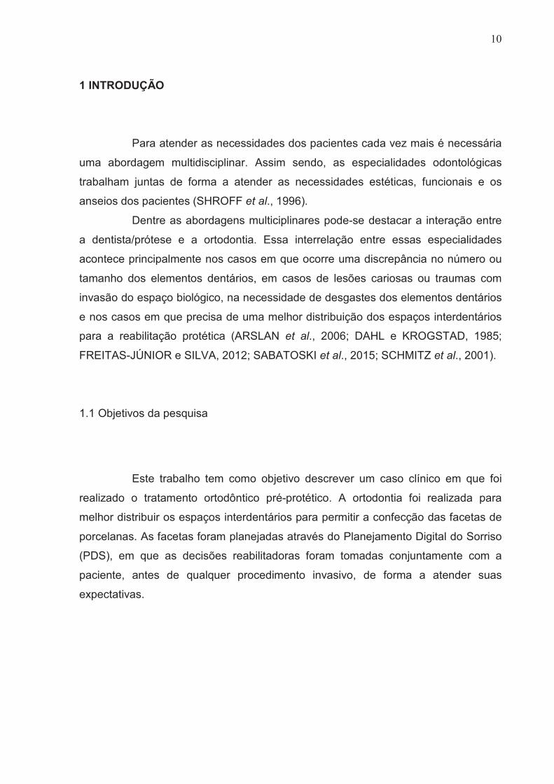
A análise facial mostra uma paciente assimétrica, mesofacial, selamento
labial presente, linha do sorriso baixa, linha média dentária inferior desviada para a
direita, padrão facial convexo, ângulo nasolabial obtuso e linha queixo pescoço
normal (FIGURA 1).
Figura 1 - Análise facial.
Legenda: a) vista facial frontal
b) vista frontal do sorriso
c) vista facial de perfil
Fonte: Elaborada pelo autor, 2018.
13
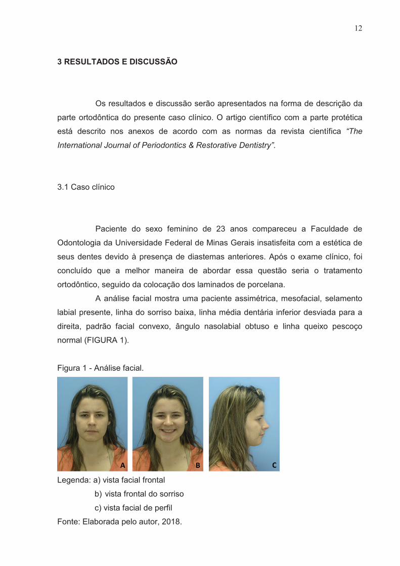
A análise dentária mostra a presença de diastemas anteriores na arcada
superior e inferior, canino em Classe II do lado direito e em Classe I do lado
esquerdo, overjet e overbite ligeiramente aumentados, presença de alguns dentes
girovertidos (dentes 15, 14, 23, 24, 25, 34, 33 e 43) (FIGURA 2).
Figura 2 - Análise dentária inicial.
Legenda: a) vista lateral direita
b) vista frontal
c) vista lateral esquerda
d) vista oclusal superior
e) vista oclusal inferior.
Fonte: Elaborada pelo autor, 2018.
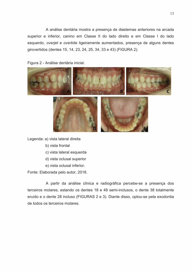
A partir da análise clínica e radiográfica percebe-se a presença dos
terceiros molares, estando os dentes 18 e 48 semi-inclusos, o dente 38 totalmente
eruído e o dente 28 incluso (FIGURAS 2 e 3). Diante disso, optou-se pela exodontia
de todos os terceiros molares.
14
Figura 3 - Radiografia panorâmica inicial.
Fonte: Elaborada pelo autor, 2018.
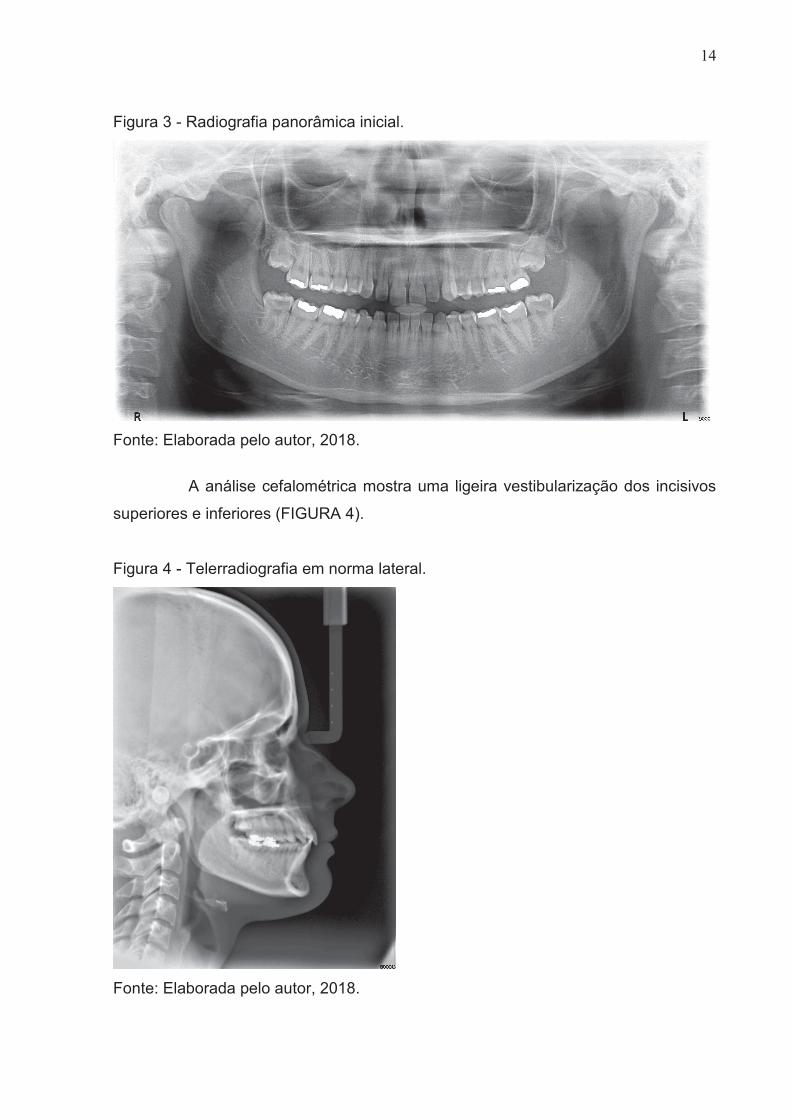
A análise cefalométrica mostra uma ligeira vestibularização dos incisivos
superiores e inferiores (FIGURA 4).
Figura 4 - Telerradiografia em norma lateral.
Fonte: Elaborada pelo autor, 2018.
15
Diante disso, foi proposto um tratamento ortodôntico pré-protético. O
objetivo principal do tratamento ortodôntico foi melhor distribuir os espaços
interdentários para facilitar na confecção das facetas de porcelana e colocar os
caninos em chave.
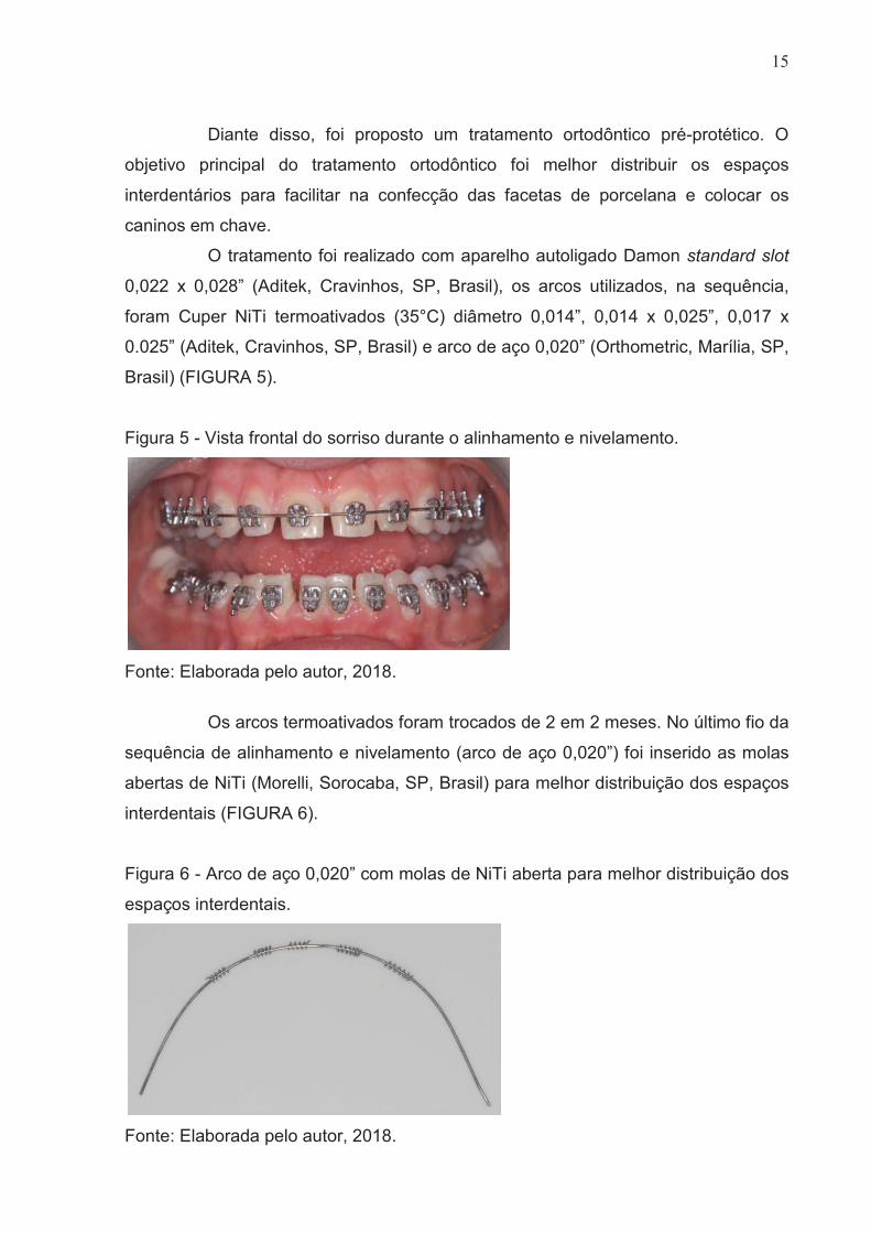
O tratamento foi realizado com aparelho autoligado Damon standard slot
0,022 x 0,028” (Aditek, Cravinhos, SP, Brasil), os arcos utilizados, na sequência,
foram Cuper NiTi termoativados (35°C) diâmetro 0,014”, 0,014 x 0,025”, 0,017 x
0.025” (Aditek, Cravinhos, SP, Brasil) e arco de aço 0,020” (Orthometric, Marília, SP,
Brasil) (FIGURA 5).
Figura 5 - Vista frontal do sorriso durante o alinhamento e nivelamento.
Fonte: Elaborada pelo autor, 2018.
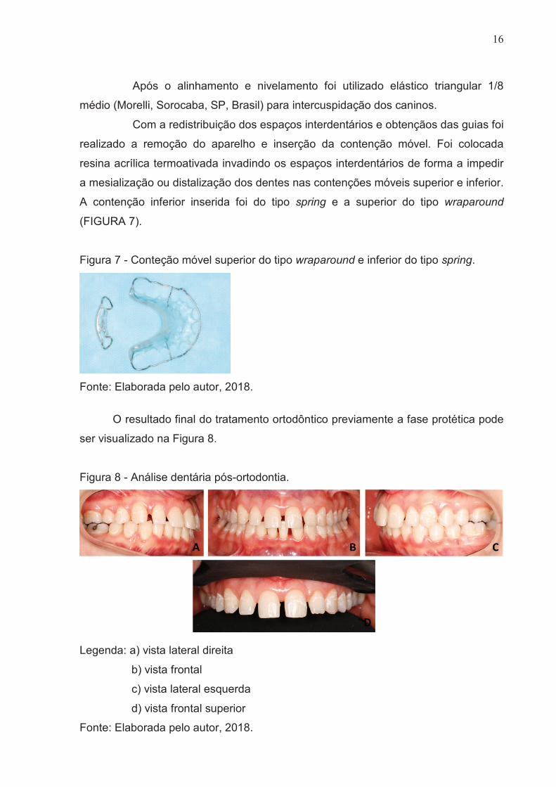
Os arcos termoativados foram trocados de 2 em 2 meses. No último fio da
sequência de alinhamento e nivelamento (arco de aço 0,020”) foi inserido as molas
abertas de NiTi (Morelli, Sorocaba, SP, Brasil) para melhor distribuição dos espaços
interdentais (FIGURA 6).
Figura 6 - Arco de aço 0,020” com molas de NiTi aberta para melhor distribuição dos
espaços interdentais.
Fonte: Elaborada pelo autor, 2018.
16
Após o alinhamento e nivelamento foi utilizado elástico triangular 1/8
médio (Morelli, Sorocaba, SP, Brasil) para intercuspidação dos caninos.
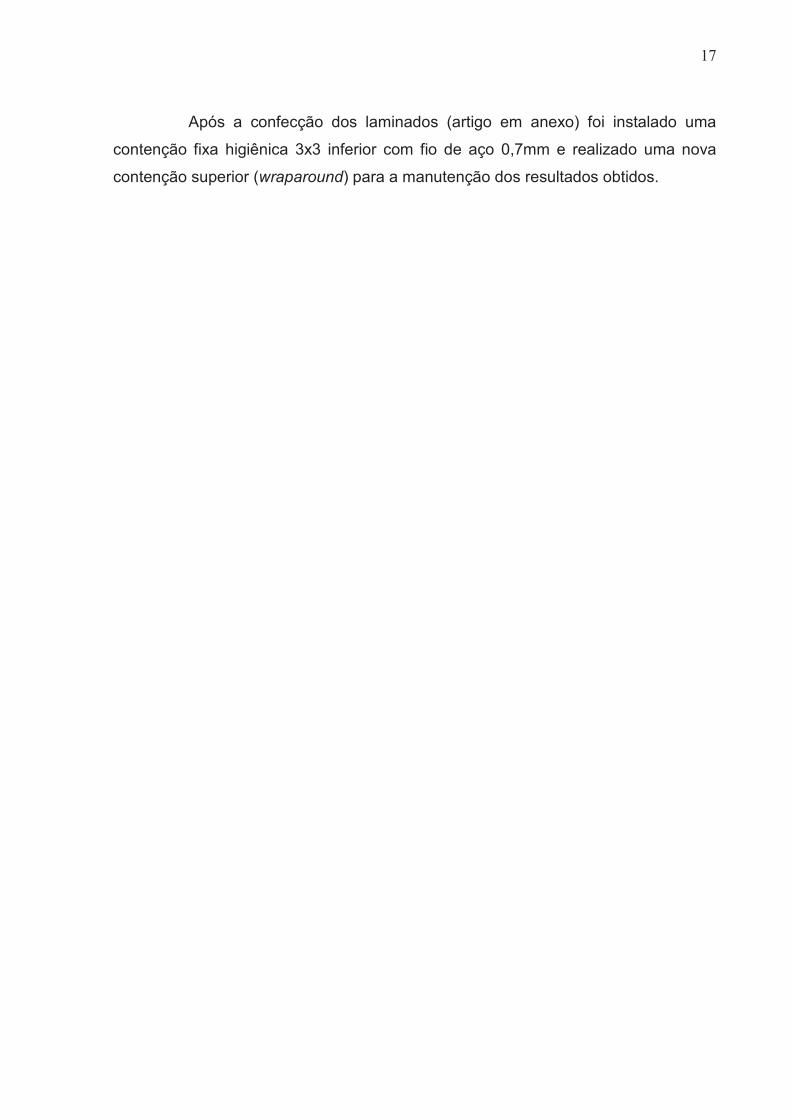
Com a redistribuição dos espaços interdentários e obtençãos das guias foi
realizado a remoção do aparelho e inserção da contenção móvel. Foi colocada
resina acrílica termoativada invadindo os espaços interdentários de forma a impedir
a mesialização ou distalização dos dentes nas contenções móveis superior e inferior.
A contenção inferior inserida foi do tipo spring e a superior do tipo wraparound
(FIGURA 7).
Figura 7 - Conteção móvel superior do tipo wraparound e inferior do tipo spring.
Fonte: Elaborada pelo autor, 2018.
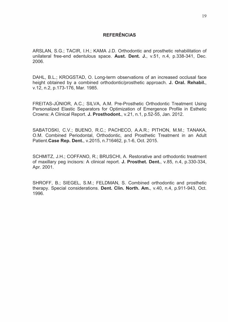
O resultado final do tratamento ortodôntico previamente a fase protética pode
ser visualizado na Figura 8.
Figura 8 - Análise dentária pós-ortodontia.
Legenda: a) vista lateral direita
b) vista frontal
c) vista lateral esquerda
d) vista frontal superior
Fonte: Elaborada pelo autor, 2018.
17
Após a confecção dos laminados (artigo em anexo) foi instalado uma
contenção fixa higiênica 3x3 inferior com fio de aço 0,7mm e realizado uma nova
contenção superior (wraparound) para a manutenção dos resultados obtidos.

18
4 CONCLUSÃO
O presente trabalho mostrou a importância da interdisciplinaridade na
condução de casos clínicos. O tratamento ortodôntico foi essencial para melhor
distribuição dos espaços interdentários para a confecção das facetas de porcelana.
19
REFERÊNCIAS
ARSLAN, S.G.; TACIR, I.H.; KAMA J.D. Orthodontic and prosthetic rehabilitation of unilateral free-end edentulous space. Aust. Dent. J., v.51, n.4, p.338-341, Dec. 2006.
DAHL, B.L.; KROGSTAD, O. Long-term observations of an increased occlusal face height obtained by a combined orthodontic/prosthetic approach. J. Oral. Rehabil., v.12, n.2, p.173-176, Mar. 1985.
FREITAS-JÚNIOR, A.C.; SILVA, A.M. Pre-Prosthetic Orthodontic Treatment Using Personalized Elastic Separators for Optimization of Emergence Profile in Esthetic Crowns: A Clinical Report. J. Prosthodont., v.21, n.1, p.52-55, Jan. 2012.
SABATOSKI, C.V.; BUENO, R.C.; PACHECO, A.A.R.; PITHON, M.M.; TANAKA, O.M. Combined Periodontal, Orthodontic, and Prosthetic Treatment in an Adult Patient.Case Rep. Dent., v.2015, n.716462, p.1-6, Oct. 2015.
SCHMITZ, J.H.; COFFANO, R.; BRUSCHI, A. Restorative and orthodontic treatment of maxillary peg incisors: A clinical report. J. Prosthet. Dent., v.85, n.4, p.330-334, Apr. 2001.
SHROFF, B.; SIEGEL, S.M.; FELDMAN, S. Combined orthodontic and prosthetic therapy. Special considerations. Dent. Clin. North. Am., v.40, n.4, p.911-943, Oct. 1996.
20
ANEXO - ARTIGO CIENTÍFICO
Digital smile design as a tool in the planning of porcelain
laminate veneers restoration: a clinical report
Sávio Morato de Lacerda Gontijo, DDS, MS, PhD,a,1 Phelipe Marke Morgado,
DDS,b,1 Leniana Santos Neves, DDS, MS, PhD,c Esdras Campos França,
DDS, MS, PhD,c Elisabeth Maria Bastos Lages, DDS, MS, PhD,c Hugo
Henriques Alvim, DDS, MS, PhDd*
a Resident, Department of Pediatric Dentistry and Orthodontics; Faculty of
Dentistry, Federal University of Minas Gerais (UFMG), Belo Horizonte, MG,
Brazil. b Undergraduate Student, Department of Restorative Dentistry; Faculty of
Dentistry, Federal University of Minas Gerais (UFMG), Belo Horizonte, MG,
Brazil. c Professor, Department of Pediatric Dentistry and Orthodontics; Faculty of
Dentistry, Federal University of Minas Gerais (UFMG), Belo Horizonte, MG,
Brazil. d Professor, Department of Restorative Dentistry; Faculty of Dentistry, Federal
University of Minas Gerais (UFMG), Belo Horizonte, MG, Brazil.
This research did not receive any specific grant from funding agencies in the
public, commercial, or not-for-profit sectors.
* Corresponding author: Professor Hugo H. Alvim, Department of Restorative
Dentistry, Faculty of Dentistry, Federal University of Minas Gerais (UFMG), Av.
Presidente Antônio Carlos 6627, CEP 31270-901, Belo Horizonte, MG, Brazil. E-
mail: [email protected]; Tel.: +55-31-3409-2440; Fax: +55-31-3409-2440.
1 Authors who made equal and major contributions to this research.
21
Digital smile design as a tool in the planning of porcelain
laminate veneers restoration: a clinical report
This research did not receive any specific grant from funding agencies in the
public, commercial, or not-for-profit sectors.
ABSTRACT
Digital smile design (DSD) is an important tool for esthetic planning in
dentistry as it facilitates ease of communication between professionals and
patients. This clinical report describes a procedure involving DSD for the
placement of porcelain laminate veneer restorations. The DSD included
dental and facial analyzes. The smile curve was drawn and a dental ruler
was used to delimitate the spaces between each tooth. An approximate
simulation of the smile was generated using Adobe Photoshop software. For
esthetic and functional evaluation of the digital plan was created a diagnostic
waxing and mock-up. DSD was essential for successful planning because it
enabled better patient contact with the end result. This case demonstrates
the importance of this tool in esthetic dentistry.
Keywords: Digital smile design; Porcelain laminate veneer; Dental esthetics.
22
Dental treatment addresses the esthetic, emotional and functional requirements of
patients.1-7 Therefore, dental surgeons and laboratory technicians must use all
available resources in order to ensure predictability in treatment and to meet
patients’ expectations. Through the use of images, digital smile design (DSD) allows
dental professionals to show the patient what will be achieved with the proposed
treatment.2-5,7-15
Digital smile design allows rehabilitation planning through digital images and
imaging software based on the principles of cosmetic dentistry, such as symmetry,
golden proportion and width-to-length ratio, in order to achieve a harmonious
smile.2,4,5,8-10,12,14-16 In addition, DSD is a technique that does not damage the dental
structure and does not use temporary materials.4
Prior to the introduction of DSD, the major problem faced by dentists in
relation to oral rehabilitation was showing the patient in an invasive way what was
being proposed.4,5,8,13-15 The present study presents a clinical report of treatment
planned with DSD, in which the rehabilitation decisions were made together with the
patient prior to any invasive procedures in order to meet the patient’s expectations.
CLINICAL REPORT
A 23 year-old Caucasian woman dissatisfied with the appearance of her teeth
visited the Restorative Dentistry Clinic of the Dental School of the Universidade
Federal de Minas Gerais (UFMG). After clinical examination, it was concluded that
the best way to address this issue would be orthodontic treatment followed by the
placement of porcelain laminate veneer.
23
The orthodontic treatment (Supplementary Material) was performed before
the restorative dentistry, with the aim to better distribute the spacing of teeth for the
manufacture of laminate veneer.
After orthodontic treatment, photographs were taken, and these were used for
digital planning and to produce a preview of the final treatment result. Photographs
were taken in forced smile, with retractors and at 12 o’clock position to evaluate the
distance between the teeth and the vermilion of the lip.2,4
The DSD started with three-line tracing including facial midline and bipupilar
lines to verify correct angulation of the photographs and the intercommissural line,
which showed slight lip asymmetry (Fig. 1A). The smile curve was then drawn taking
into account the shape of the lower lip, and a dental ruler was used to delimitate the
spaces between each tooth. The drawn lines were transferred to the photograph
with retractors (Fig. 1B).
From analysis of photographs, it was decided that the ideal width-to-height
ratio of the maxillary central incisors was 80%. Afterwards, the limits of each tooth
were drawn to ensure that the esthetics of the smile were harmonic (Fig. 1C).
Finally, an approximate simulation of the smile after treatment was generated
using Adobe Photoshop software (Adobe Systems, San Jose, CA, USA), as shown
in Fig. 1D. It was proposed that eight laminate veneers be placement along the
upper arch from the first right premolar to the first left premolar, and six laminate
veneers along the lower arch from right canine to left canine.
For esthetic and functional evaluation of the digital plan, we created a mock-
up. For this, diagnostic waxing was carried out, followed by molding with addition
silicone (Express™XT; 3M ESPE, Saint Paul, MN, USA), cutting of the final
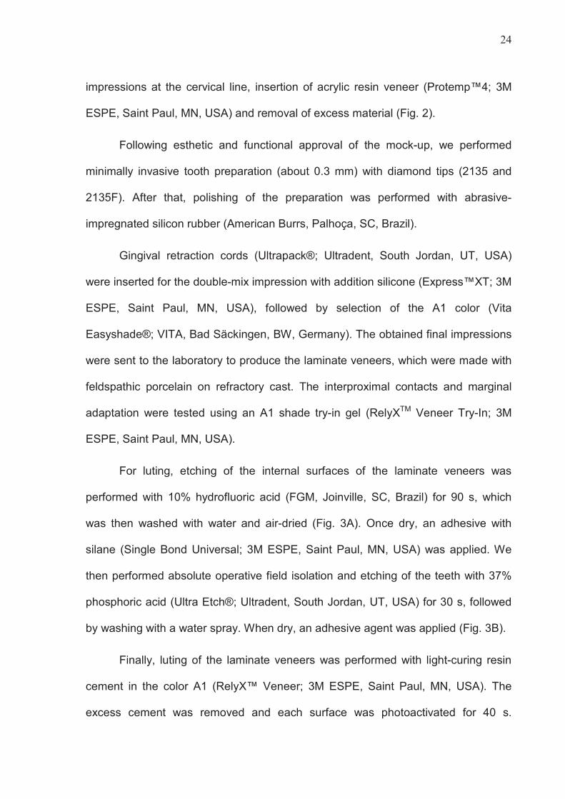
24
impressions at the cervical line, insertion of acrylic resin veneer (Protemp™4; 3M
ESPE, Saint Paul, MN, USA) and removal of excess material (Fig. 2).
Following esthetic and functional approval of the mock-up, we performed
minimally invasive tooth preparation (about 0.3 mm) with diamond tips (2135 and
2135F). After that, polishing of the preparation was performed with abrasive-
impregnated silicon rubber (American Burrs, Palhoça, SC, Brazil).
Gingival retraction cords (Ultrapack®; Ultradent, South Jordan, UT, USA)
were inserted for the double-mix impression with addition silicone (Express™XT; 3M
ESPE, Saint Paul, MN, USA), followed by selection of the A1 color (Vita
Easyshade®; VITA, Bad Säckingen, BW, Germany). The obtained final impressions
were sent to the laboratory to produce the laminate veneers, which were made with
feldspathic porcelain on refractory cast. The interproximal contacts and marginal
adaptation were tested using an A1 shade try-in gel (RelyXTM Veneer Try-In; 3M
ESPE, Saint Paul, MN, USA).
For luting, etching of the internal surfaces of the laminate veneers was
performed with 10% hydrofluoric acid (FGM, Joinville, SC, Brazil) for 90 s, which
was then washed with water and air-dried (Fig. 3A). Once dry, an adhesive with
silane (Single Bond Universal; 3M ESPE, Saint Paul, MN, USA) was applied. We
then performed absolute operative field isolation and etching of the teeth with 37%
phosphoric acid (Ultra Etch®; Ultradent, South Jordan, UT, USA) for 30 s, followed
by washing with a water spray. When dry, an adhesive agent was applied (Fig. 3B).
Finally, luting of the laminate veneers was performed with light-curing resin
cement in the color A1 (RelyX™ Veneer; 3M ESPE, Saint Paul, MN, USA). The
excess cement was removed and each surface was photoactivated for 40 s.
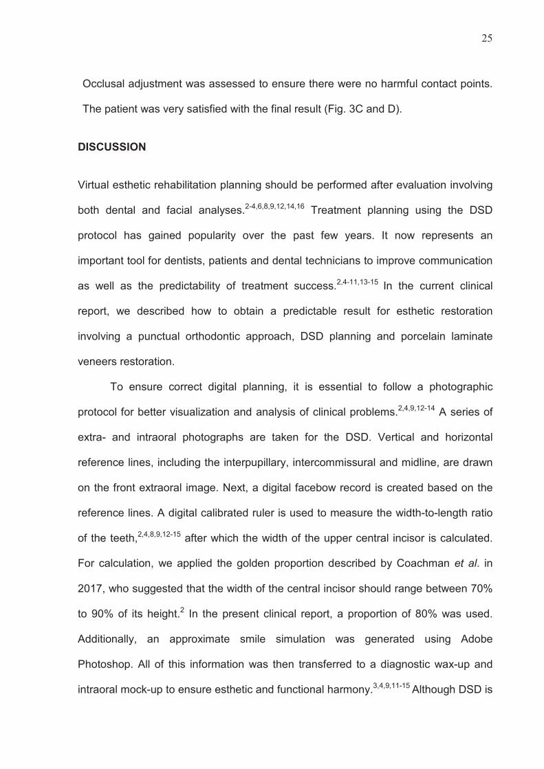
25
Occlusal adjustment was assessed to ensure there were no harmful contact points.
The patient was very satisfied with the final result (Fig. 3C and D).
DISCUSSION
Virtual esthetic rehabilitation planning should be performed after evaluation involving
both dental and facial analyses.2-4,6,8,9,12,14,16 Treatment planning using the DSD
protocol has gained popularity over the past few years. It now represents an
important tool for dentists, patients and dental technicians to improve communication
as well as the predictability of treatment success.2,4-11,13-15 In the current clinical
report, we described how to obtain a predictable result for esthetic restoration
involving a punctual orthodontic approach, DSD planning and porcelain laminate
veneers restoration.
To ensure correct digital planning, it is essential to follow a photographic
protocol for better visualization and analysis of clinical problems.2,4,9,12-14 A series of
extra- and intraoral photographs are taken for the DSD. Vertical and horizontal
reference lines, including the interpupillary, intercommissural and midline, are drawn
on the front extraoral image. Next, a digital facebow record is created based on the
reference lines. A digital calibrated ruler is used to measure the width-to-length ratio
of the teeth,2,4,8,9,12-15 after which the width of the upper central incisor is calculated.
For calculation, we applied the golden proportion described by Coachman et al. in
2017, who suggested that the width of the central incisor should range between 70%
to 90% of its height.2 In the present clinical report, a proportion of 80% was used.
Additionally, an approximate smile simulation was generated using Adobe
Photoshop. All of this information was then transferred to a diagnostic wax-up and
intraoral mock-up to ensure esthetic and functional harmony.3,4,9,11-15 Although DSD is
26
considered a simple technique which does not involve any sophisticated equipment,
operator training is always desirable as DSD is only a tool.6-8,13-15 After planning, a
mock-up test was realized.3,4,9,11-15
Following esthetic and functional approval of the mock-up, the porcelain
laminate veneers restorations were performed. Enamel preservation is an essential
clinical parameter for ensuring the success of laminate veneers, as dentin exposure
may reduce the longevity of the restoration.9,11,13,14 Thus, the present case describes
laminates minimally invasive preparations (about 0.3 mm) involving only the dental
enamel. Moreover, we chose to use feldspathic porcelain as these provide excellent
esthetic value. However, the final esthetics of the ceramic depends on the luting
composite, the color of which can be assessed with the try-in gel, which simulates the
shade of the ceramic restorations without compromising the final esthetic
result.3,9,14,15
CONCLUSION
In this case, prediction of the approximate final result was important in
communication with the patient, which generated more security and motivation.
Communication with the dental laboratory technicians was also considerably more
effective as it was possible to clearly express the desired outcome.
Diagnostic waxing, along with a mock-up, was essential for successful
planning because it enabled better patient contact with the end result. In addition, the
mock-up allowed us to confirm the functionality of the planned treatment.
Finally, the present clinical report demonstrates the potential of this technique
and the need for advancements in this area.
27
REFERENCES
1. Coachman C, Paravina RD. Digitally Enhanced Esthetic Dentistry - From
Treatment Planning to Quality Control. J Esthet Restor Dent 2016;28 Suppl 1:S3-4.
2. Coachman C, Calamita MA, Sesma N. Dynamic Documentation of the Smile
and the 2D/3D Digital Smile Design Process. Int J Periodontics Restorative Dent
2017;37:183-93.
3. Trushkowsky R, Arias DM, David S. Digital Smile Design concept delineates
the final potential result of crown lengthening and porcelain veneers to correct a
gummy smile. Int J Esthet Dent 2016;11:338-54.
4. Tak On T, Kois JC. Digital Smile Design Meets the Dento-Facial Analyzer:
Optimizing Esthetics While Preserving Tooth Structure. Compend Contin Educ Dent
2016;37:46-50.
5. Miranda ME, Olivieri KA, Rigolin FJ, de Vasconcellos AA. Esthetic Challenges
in Rehabilitating the Anterior Maxilla: A Case Report. Oper Dent 2016;41:2-7.
6. Lin WS, Harris BT, Phasuk K, Llop DR, Morton D. Integrating a facial scan,
virtual smile design, and 3D virtual patient for treatment with CAD-CAM ceramic
veneers: A clinical report. J Prosthet Dent 2018;119:200-5.
7. Rojas-Vizcaya F. Prosthetically guided bone sculpturing for a maxillary
complete-arch implant-supported monolithic zirconia fixed prosthesis based on a
digital smile design: A clinical report. J Prosthet Dent 2017;118:575-80.
8. Omar D, Duarte C. The application of parameters for comprehensive smile
esthetics by digital smile design programs: A review of literature. Saudi Dent J
2018;30:7-12.
9. Veneziani M. Ceramic laminate veneers: clinical procedures with a
multidisciplinary approach. Int J Esthet Dent 2017;12:426-48.
28
10. Santos FR, Kamarowski SF, Lopez CAV, Storrer CLM, Neto AT, Deliberador
TM. The use of the digital smile design concept as an auxiliary tool in periodontal
plastic surgery. Dent Res J (Isfahan) 2017;14:158-61.
11. Cattoni F, Mastrangelo F, Gherlone EF, Gastaldi G. A New Total Digital Smile
Planning Technique (3D-DSP) to Fabricate CAD-CAM Mockups for Esthetic Crowns
and Veneers. Int J Dent 2016;2016:6282587.
12. Zanardi PR, Laia Rocha Zanardi R, Chaib Stegun R, Sesma N, Costa BN,
Cruz Lagana D. The Use of the Digital Smile Design Concept as an Auxiliary Tool in
Aesthetic Rehabilitation: A Case Report. Open Dent J 2016;10:28-34.
13. Pimentel W, Teixeira ML, Costa PP, Jorge MZ, Tiossi R. Predictable
Outcomes with Porcelain Laminate Veneers: A Clinical Report. J Prosthodont
2016;25:335-40.
14. Meereis CT, de Souza GB, Albino LG, Ogliari FA, Piva E, Lima GS. Digital
Smile Design for Computer-assisted Esthetic Rehabilitation: Two-year Follow-up.
Oper Dent 2016;41:E13-22.
15. Arias DM, Trushkowsky RD, Brea LM, David SB. Treatment of the Patient with
Gummy Smile in Conjunction with Digital Smile Approach. Dent Clin North Am
2015;59:703-16.
16. Pozzi A, Arcuri L, Moy PK. The smiling scan technique: Facially driven guided
surgery and prosthetics. J Prosthodont Res 2018.
29
Figures
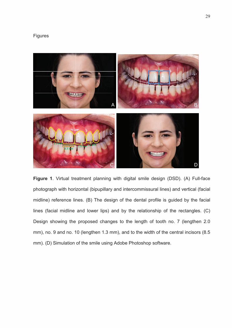
Figure 1. Virtual treatment planning with digital smile design (DSD). (A) Full-face
photograph with horizontal (bipupillary and intercommissural lines) and vertical (facial
midline) reference lines. (B) The design of the dental profile is guided by the facial
lines (facial midline and lower lips) and by the relationship of the rectangles. (C)
Design showing the proposed changes to the length of tooth no. 7 (lengthen 2.0
mm), no. 9 and no. 10 (lengthen 1.3 mm), and to the width of the central incisors (8.5
mm). (D) Simulation of the smile using Adobe Photoshop software.
30
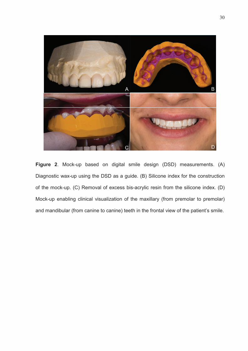
Figure 2. Mock-up based on digital smile design (DSD) measurements. (A)
Diagnostic wax-up using the DSD as a guide. (B) Silicone index for the construction
of the mock-up. (C) Removal of excess bis-acrylic resin from the silicone index. (D)
Mock-up enabling clinical visualization of the maxillary (from premolar to premolar)
and mandibular (from canine to canine) teeth in the frontal view of the patient’s smile.
31
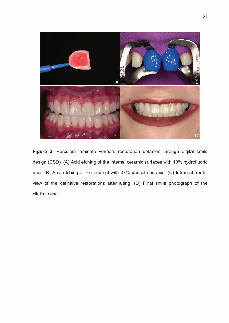
Figure 3. Porcelain laminate veneers restoration obtained through digital smile
design (DSD). (A) Acid etching of the internal ceramic surfaces with 10% hydrofluoric
acid. (B) Acid etching of the enamel with 37% phosphoric acid. (C) Intraoral frontal
view of the definitive restorations after luting. (D) Final smile photograph of the
clinical case.
32
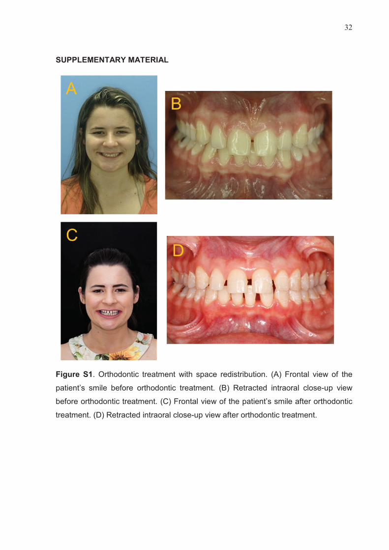
SUPPLEMENTARY MATERIAL
Figure S1. Orthodontic treatment with space redistribution. (A) Frontal view of the
patient’s smile before orthodontic treatment. (B) Retracted intraoral close-up view
before orthodontic treatment. (C) Frontal view of the patient’s smile after orthodontic
treatment. (D) Retracted intraoral close-up view after orthodontic treatment.