Somatic pairing, endomitosis and chromosome aberrations in ... · ring. The antiparallel aspect...
16
Anais da Academia Brasileira de Ciências (2003) 75(3): 285-300 (Annals of the Brazilian Academy of Sciences) ISSN 0001-3765 www.scielo.br/aabc Somatic pairing, endomitosis and chromosome aberrations in snakes (Viperidae and Colubridae) MARIA LUIZA BEÇAK, WILLY BEÇAK and ALEXANDRE PEREIRA Laboratório de Genética, Instituto Butantan, 05503-900 São Paulo, SP, Brasil Manuscript received on August 26, 2002; accepted for publication on May 7, 2003; contributed by Willy Beçak* ABSTRACT The positioning of macrochromosomes of Bothrops jararaca and Bothrops insularis (Viperidae) was studied in undistorted radial metaphases of uncultured cells (spermatogonia and oogonia) not subjected to spindle inhibitors. Colchicinized metaphases from uncultured (spleen and intestine) and cultured tissues (blood) were also analyzed. We report two antagonic non-random chromosome arrangements in untreated premeiotic cells: the parallel configuration with homologue chromosomes associated side by side in the metaphase plate and the antiparallel configuration having homologue chromosomes with antipolar distribution in the metaphase ring. The antiparallel aspect also appeared in colchicinized cells. The spatial chromosome arrangement in both configurations is groupal size-dependent and maintained through meiosis. We also describe, in untreated gonia cells, endomitosis followed by reductional mitosis which restores the diploid number. In B. jararaca males we observed that some gonad regions present changes in the meiotic mechanism. In this case, endoreduplicated cells segregate the diplochromosomes to opposite poles forming directly endoreduplicated second metaphases of meiosis with the suppression of first meiosis. By a successive division, these cells form nuclei with one set of chromosomes. Chromosome doubling in oogonia is known in hybrid species and in parthenogenetic salamanders and lizards. This species also presented chromosome rearrangements leading to aneuploidies in mitosis and meiosis. It is suggested that somatic pairing, endomitosis, meiotic alterations, and chromosomal aberrations can be correlated processes. Similar aspects of nuclei configurations, endomitosis and reductional mitosis were found in other Viperidae and Colubridae species. Key words: snakes, somatic pairing, chromosome positioning, endomitosis, diplochromosomes. INTRODUCTION The polemic question whether chromosomes have a non-random positioning or a casual distribution in the nucleus is a matter of importance regarding some epigenetic events as non-random chromosome rear- rangements, non-disjunctions, and genetic imprint- ing. The idea of a fixed chromosome location arose from early cytogenetic evidences as in Drosophila *Member of Academia Brasileira de Ciências Correspondence to: Dra. Maria Luiza Beçak E-mail: [email protected] (Dobzhansky 1936) indicating that the relative posi- tions of the chromosomes remain the same through mitosis. In this species of Diptera homologue chro- mosomes present the peculiar somatic pairing phe- nomenon. Another interesting evidence showing that chromosomes have fixed location was the vi- sualization of homologue chromosomes positioned in opposite halves of the metaphase plate. This as- pect observed in early embryogenesis of a turbellar- ian (Costello 1970) indicated the occurrence of an ordered chromosome transmission from gametes to An Acad Bras Cienc (2003) 75 (3)
Transcript of Somatic pairing, endomitosis and chromosome aberrations in ... · ring. The antiparallel aspect...
Anais da Academia Brasileira de Ciências (2003) 75(3): 285-300(Annals of the Brazilian Academy of Sciences)ISSN 0001-3765www.scielo.br/aabc
Somatic pairing, endomitosis and chromosome aberrations in snakes(Viperidae and Colubridae)
MARIA LUIZA BEÇAK, WILLY BEÇAK and ALEXANDRE PEREIRA
Laboratório de Genética, Instituto Butantan, 05503-900 São Paulo, SP, Brasil
Manuscript received on August 26, 2002; accepted for publication on May 7, 2003;
contributed by Willy Beçak*
ABSTRACT
The positioning of macrochromosomes ofBothrops jararaca andBothrops insularis (Viperidae) was studied
in undistorted radial metaphases of uncultured cells (spermatogonia and oogonia) not subjected to spindle
inhibitors. Colchicinized metaphases from uncultured (spleen and intestine) and cultured tissues (blood) were
also analyzed. We report two antagonic non-random chromosome arrangements in untreated premeiotic cells:
the parallel configuration with homologue chromosomes associated side by side in the metaphase plate and
the antiparallel configuration having homologue chromosomes with antipolar distribution in the metaphase
ring. The antiparallel aspect also appeared in colchicinized cells. The spatial chromosome arrangement
in both configurations is groupal size-dependent and maintained through meiosis. We also describe, in
untreated gonia cells, endomitosis followed by reductional mitosis which restores the diploid number. InB.
jararaca males we observed that some gonad regions present changes in the meiotic mechanism. In this case,
endoreduplicated cells segregate the diplochromosomes to opposite poles forming directly endoreduplicated
second metaphases of meiosis with the suppression of first meiosis. By a successive division, these cells form
nuclei with one set of chromosomes. Chromosome doubling in oogonia is known in hybrid species and in
parthenogenetic salamanders and lizards. This species also presented chromosome rearrangements leading to
aneuploidies in mitosis and meiosis. It is suggested that somatic pairing, endomitosis, meiotic alterations, and
chromosomal aberrations can be correlated processes. Similar aspects of nuclei configurations, endomitosis
and reductional mitosis were found in other Viperidae and Colubridae species.
Key words: snakes, somatic pairing, chromosome positioning, endomitosis, diplochromosomes.
INTRODUCTION
The polemic question whether chromosomes have a
non-random positioning or a casual distribution in
the nucleus is a matter of importance regarding some
epigenetic events as non-random chromosome rear-
rangements, non-disjunctions, and genetic imprint-
ing. The idea of a fixed chromosome location arose
from early cytogenetic evidences as inDrosophila
*Member of Academia Brasileira de CiênciasCorrespondence to: Dra. Maria Luiza BeçakE-mail: [email protected]
(Dobzhansky 1936) indicating that the relative posi-
tions of the chromosomes remain the same through
mitosis. In this species of Diptera homologue chro-
mosomes present the peculiar somatic pairing phe-
nomenon. Another interesting evidence showing
that chromosomes have fixed location was the vi-
sualization of homologue chromosomes positioned
in opposite halves of the metaphase plate. This as-
pect observed in early embryogenesis of a turbellar-
ian (Costello 1970) indicated the occurrence of an
ordered chromosome transmission from gametes to
An Acad Bras Cienc (2003)75 (3)
286 MARIA LUIZA BEÇAK, WILLY BEÇAK and ALEXANDRE PEREIRA
following generations. Non-random chromosome
distribution and somatic pairing were also evidenced
in uncultured cells from Chinese hamster (Juricek
1975). Electronmicroscopy data from human fi-
broblast cells also indicated the occurrence of a non-
random spatial positioning of the chromosomes
(Mosgöller et al. 1991). Fluorescencein situ hy-
bridization (FISH) studies in prometaphase rosettes
of human cells also claimed that chromosomes are
fixed located with the homologues separated from
each other by at least 90o (Nagele et al. 1995).
Conflicting with this view is the evidence that chro-
mosomes do not have a fixed position in the nuclei,
as reported in experiments ofin situ hybridization
and three-dimensional reconstruction (Manuelidis
and Borden 1988). More recently, fluorescencein
situ hybridization (FISH) studies also showed that
in human cells there is a relatively random array
of chromosomes on the mitotic ring (Allison and
Nestor 1999). According to these authors, a fixed
location of the chromosomes on the mitotic ring
is not required for mitotic distribution. Further-
more, the relative positions of the chromosomes on
each metaphase ring are most likely carried through
anaphase into telophase. According to Rabl’s sug-
gestion (1885), the relative positions of the chro-
mosomes in somatic metaphase rings would be a
reflection of their locations in interphase. More-
over, later investigations showed that the relative
positions of the chromosomes are comparable in
prophase and metaphase nuclei (Bajer and Molè-
Bajer 1981, Hiraoka et al. 1990). Alternatively,
it has been shown that in interphase the relative
positions of the chromosomes may be affected by
gene activity (Manuelidis and Borden 1988). In
Drosophila melanogaster, bromodeoxyuridine in-
corporation and DNA quantification in combina-
tion with fluorescencein situ hybridization (FISH)
were used to analyze aspects of heterochromatin, the
Rabl configuration and pairing of homologue chro-
mosomes. The chromosomes showed a dynamic
behavior in interphase, the Rabl configuration ap-
pearing only about 2h after mitosis, the somatic
pairing being distorted during S-phase more rapidly
for a euchromatic than for a heterochromatic region
(Csink and Henikoff 1998). According to Sun et
al. (2000), two biophysical models could explain
chromosome positioning. In the volume exclusion
model, a non-random size dependent spatial posi-
tioning is explained by steric hindrance among chro-
mosomes. In the mitotic preset model confirmed
by their findings, the size-dependent positioning is
established and maintained in mitosis and G1 inter-
phase.
Data on the spatial placement of chromosomes
in mitosis and meiosis of snakes are lacking in the lit-
erature. This fact can be attributed to the difficulty to
identify the microchromosomes by standard meth-
ods and to obtain G-bands. Most cytogenetic anal-
ysis in snakes are based in C and NOR bands stud-
ies using colchicinized nuclei. A complete study of
premeiosis and meiosis is laborious because cell cy-
cle is seasonal and female meiotic cells are scarce.
Several results in other organisms addressed to the
question whether premeiosis would influence chro-
mosome pairing in meiosis (Bennett 1984). In this
paper we study chromosome spatial arrangement in
mitotic, premeiotic, and meiotic cells ofBothrops
jararaca andB. insularis. Premeiosis is here con-
sidered in a wide sense as a period previous to first
meiosis division appearance. Considering that spin-
dle inhibitors as colchicine and Colcemid may dis-
tort chromosome order in nuclei (Juricek 1975), we
studied chromosome positioning in uncultured mi-
totic and meiotic cells not subjected to these agents.
The aspects obtained in both sexes were compared
with those obtained in colchicinized nuclei. The
analysis included only the macrochromosomes. We
describe the occurrence of two non-random and an-
tagonic types of chromosome distribution in somatic
metaphases of these species. The first type, found
only in colchicine untreated gonia cells, is similar
to the somatic pairing known in Diptera. The sec-
ond type, that is characterized by the bipolarity of
the homologues in the metaphase plate, appeared in
colchicine treated cells from spleen, intestine and
blood, and in untreated nuclei from gonia cells. The
two types of configurations present a groupal size-
An Acad Bras Cienc (2003)75 (3)
CHROMOSOME POSITIONING IN SNAKES 287
dependent chromosome distribution that is main-
tained in meiotic cells. We also describe the pres-
ence of premeiotic endomitosis followed by reduc-
tional mitosis in both species. Moreover we report
alterations in the meiotic mechanisms and the oc-
currence of chromosome aberrations inB. jararaca.
Previous studies in Viperidae species us-
ing colchicinized cells showed that the karyotypes
of Bothrops species have conserved morphology and
chromosome number including eight pairs of macro-
chromosomes (M) and ten pairs of microchromo-
somes (m), 2n = 16 M + 20 m. The eight pairs
of M are recognized by size and ordered in kary-
otypes according to two groups of larger and smaller
macrochromosomes each with four pairs. The sex-
pair is the fourth of the karyotype (ZZ, male and
ZW, female) (Beçak W et al. 1962, Beçak W 1968).
Chromosome C banding inB. insularis is telom-
eric and centromeric in all macrochromosomes and
in the Z, being the microchromosomes totally het-
erochromatic. The W chromosome is polymorphic
presenting different size and C band. The smaller
W1 type found in females is entirely heterochro-
matic. The larger W2 type found in intersexes is
variegated with positive and negative C bands. This
W2 chromosome appeared dicentric with variation
of C band pattern. Polymorphism of the W chro-
mosome also occurs inB. jararacussu, a related
species from the continent. The abnormal sex phe-
notypes and the altered fertility inB. insularis fe-
males were attributed to chromosomal rearrange-
ments probably created by insertion sequences in
the W (Beçak ML et al. 1990). This possibility was
previously indicated to explain the occurrence of
multiple sex-chromosomes ofBungarus caeruleus
(Elapidae) (Singh et al. 1979). ZW sex bivalent of
B. jararaca females segregates reductionally in the
first meiosis division and equationally in the second
division (Beçak ML and Beçak W 1981). Meiotic
cells ofB. jararaca show 4 or 5 microchromosomes
associated with the nucleolus (Beçak W et al. 1966).
Bothrops insularis NOR-bands were identified on
the short arm of four microchromosomes (Beçak
ML et al. 1990). Differential activity of ribosomal
cistrons during development was shown in another
species of the same genus,B. alternatus. Although
the karyotypes of adult animals do not exhibit satel-
lites, chromosomes 5 and 6 revealed satellites dur-
ing embryo development (Beçak ML and Beçak W
1973). Presence of NOR bands in two microchro-
mosomes, in macrochromosome number 6, or just
in one homologue of chromosome 6 and one mi-
crochromosome was also described inB. neuwiedi
subspecies. Moreover, molecular analysis of DNA
repeats of these species demonstrated a highly con-
served organization of the ribosomal repeats in the
total genomic DNA (Trajtengertz et al. 1995).
Our observations on nuclei configuration, en-
domitosis and reductional mitosis were extended to
other Bothrops species whose karyotypes are
known: B. jararacussu, B. moojeni (2n = 36) (Beçak
W 1968), and to the colubrid speciesThamnody-
nastes strigatus (2n = 32),Spilotes pullatus (2n = 36)
(BeçakW and Beçak ML 1969), andPhilodryas nat-
tereri (2n = 36). The karyotype of this later species
will be described elsewhere. Among Colubridae
species there are a wide variation of chromosome
number and different levels of sex-chromosome dif-
ferentiation. Morphologically differentiated sex-
chromosomes appear in the 4th pair (BeçakW 1968).
SomePhilodryas species show secondary constric-
tion in pair five (Beçak ML et al. 1971). AsP.
chamissonis exhibits NOR in the long arm of pair 2,
it was proposed that translocation of insertion type
may be accounted for polymorphisms in this genus
(Moreno et al. 1987).
MATERIALS AND METHODS
Chromosome positioning inB. jararaca was stud-
ied in colchicine untreated metaphases (spermato-
gonia and oogonia) from 11 specimens being 1�from Santa Catarina State (Canoinhas), 4� from
São Paulo State (Ibiuna, Jundiaí, Juquitiba and Co-
tia), 2� from Paraná State, and 2� and 2� from
unidentified origin. The chromosomes were ob-
tained by the standard squash technique and stained
with Giemsa. Chromosome location was also an-
An Acad Bras Cienc (2003)75 (3)
288 MARIA LUIZA BEÇAK, WILLY BEÇAK and ALEXANDRE PEREIRA
alyzed in colchicinized lymphocytes obtained from
short-term blood cultures (Beçak W et al. 1962)
from 1� and 4�. The male and one female of this
sample came from Canoinhas, being the other an-
imals of unidentified origin. In the case ofB. in-
sularis, chromosome positioning was studied in
colchicinized nuclei of cultured lymphocytes and of
uncultured cells (spleen and intestine) from 4�, 3�and 6�� and in untreated spermatogonia nuclei from
1�. The specimens ofB. insularis came from
Queimada Grande Island (São Paulo State). Chro-
mosome location was studied in karyotyped nuclei
whose centromeres were plotted in diagrams of
metaphase plates. Only macrochromosomes loca-
tion was analyzed.
Endomitosis was studied inB. jararaca andB.
insularis with the same untreated animals used for
chromosome positioning. The analysis of meiosis
in B. jararaca was carried out with untreated nuclei
from the same animals used for chromosome po-
sitioning plus 1� from Juquitiba. Meiotic cells of
this species were AgAs stained according to Vidal
and Mello method (1995), including treatment with
Triton X-100 prior to staining. This pre-treatment
did not remove nuclear proteins potentially reactive
to the AgNOR method and improved the positive
images.
Chromosome positioning and endomitosis
were also studied in untreated spermatogonia from
1� of Thamnodynastes strigatus from Piedade (São
Paulo State), 1� of Spilotes pullatus from São Ma-
noel (São Paulo State) and 1� of Philodryas nat-
tereri from Tocantins State, all belonging to the Col-
ubridae family. All the animals were anesthetized
before death.
RESULTS
Parallel and Antiparallel Chromosome
Location in Bothrops
Chromosome location inBothrops jararaca was
studied in 75 metaphases obtained from 24 colchi-
cinized lymphocyte nuclei (from 1� and 4�) and
from 51 untreated spermatogonia and oogonia nu-
clei (from 9� and 2�). In B. insularis this study
included 88 metaphases obtained from 76 colchi-
cinized lymphocytes, spleen, and intestine nuclei
(from 4�, 3� and 6�� ) and from 12 untreated sper-
matogonia nuclei (from 1�).
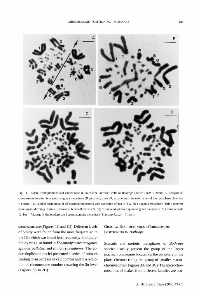
Two antagonic types of chromosome position-
ing were found in both species. The antiparallel
type showed homologue chromosomes located in
opposite halves of the metaphase plate (Figure 1A).
This bipolarity can be total including the eight pairs
of macrochromosomes, or incomplete. The paral-
lel type exhibited homologue chromosomes associ-
ated side by side in a closely parallel location in the
metaphase plate (Figure 1B). The number of asso-
ciated homologues varied in nuclei. The antiparal-
lel aspect appeared in both colchicine treated nuclei
(from lymphocytes, spleen, and intestine) and in un-
treated nuclei (from gonia cells). The parallel con-
figuration was detected only in colchicine untreated
nuclei (from gonia cells).
The number of each type of nuclei configura-
tion was estimated in both species. InB. jararaca,
among 51 untreated metaphases, 8 presented total
bipolarity and 5 showed total parallelism. Treated
metaphases of this species showed only antiparal-
lelism. Among 24 treated nuclei, 11 presented total
bipolarity. In B. insularis, colchicinized nuclei ex-
hibited only the antiparallel configuration, total or
incomplete. Among 76 of these nuclei, 23 showed
total bipolarity. Both parallel and antiparallel con-
figurations appeared in untreated spermatogonia nu-
clei of this species. Among 12 untreated nuclei of
the male studied, 1 metaphase showed total bipolar-
ity and 2 revealed total parallelism.
The peculiar parallel positioning found in un-
treated gonia cells ofB. jararaca andB. insularis,
was also found in untreated spermatogonia ofB.
jararacussu andB. moojeni (Figure 3A).
Endomitosis
Endomitosis was found in untreated oogonia and
spermatogonia ofB. jararaca, B. insularis, B. jara-
racussu, andB. moojeni. Endoreduplicated chromo-
somes appeared thick or exhibited diplochromo-
An Acad Bras Cienc (2003)75 (3)
CHROMOSOME POSITIONING IN SNAKES 289
Fig. 1 – Nuclei configurations and endomitosis in colchicine untreated cells ofBothrops species (16M + 20m): A. Antiparallel
chromosome location in a spermatogonia metaphase (B. jararaca; male 28; axis delimits the two halves of the metaphase plate; bar
= 8.9µm). B. Parallel positioning of all macrochromosomes with exception of pair 4 (ZW) in a oogonia metaphase. Pair 3 presents
homologues differing in size (B. jararaca; female 8; bar = 7.9µm); C. Endoreduplicated spermatogonia metaphase (B. jararaca; male
12; bar = 7.8µm); D. Endoreduplicated spermatogonia metaphase (B. insularis; bar = 7.1µm).
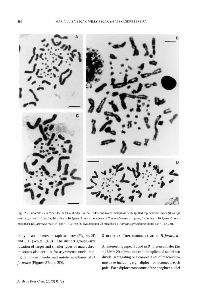
some structure (Figures 1C and 1D). Different levels
of ploidy were found from the most frequent 4n to
the 16n which was found less frequently. Endopoly-
ploidy was also found inThamnodynastes strigatus,
Spilotes pullatus, andPhilodryas nattereri.The en-
doreduplicated nuclei presented a series of mitoses
leading to an increase of cell number and to a reduc-
tion of chromosome number restoring the 2n level
(Figures 2A to 2D).
Groupal Size-dependent Chromosome
Positioning in Bothrops
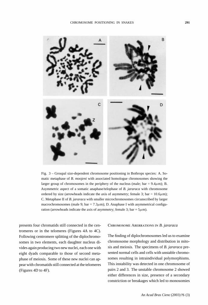
Somatic and meiotic metaphases ofBothrops
species usually present the group of the larger
macrochromosomes located on the periphery of the
plate, circumscribing the group of smaller macro-
chromosomes (Figures 3A and 3C). The microchro-
mosomes of snakes from different families are cen-
An Acad Bras Cienc (2003)75 (3)
290 MARIA LUIZA BEÇAK, WILLY BEÇAK and ALEXANDRE PEREIRA
Fig. 2 – Endomitosis in Viperidae and Colubridae: A. An endoreduplicated metaphase with splitted diplochromosomes (Bothrops
jararaca; male 41 from Juquitiba; bar = 10.2µm); B. A 4n metaphase ofThamnodynastes strigatus, (male; bar = 10.2µm); C. A 4n
metaphase (B. jararaca; male 15; bar = 14.3µm); D. Two daughter 2n metaphases (Bothrops jararacussu; male; bar = 11.4µm).
trally located in most metaphase plates (Figures 2D
and 3D) (White 1973). The distinct groupal-size
location of larger and smaller types of macrochro-
mosomes also account for asymmetric nuclei con-
figurations in meiotic and mitotic anaphases ofB.
jararaca (Figures 3B and 3D).
Structural Diplochromosomes in B. jararaca
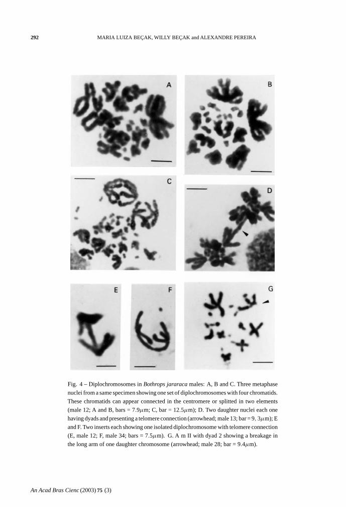
An interesting aspect found inB. jararaca males (2n
= 16 M + 20 m) was that endoreduplicated nuclei can
divide, segregating one complete set of macrochro-
mosomes including eight diplochromosomes to each
pole. Each diplochromosome of the daughter nuclei
An Acad Bras Cienc (2003)75 (3)
CHROMOSOME POSITIONING IN SNAKES 291
Fig. 3 – Groupal size-dependent chromosome positioning in Bothrops species: A. So-
matic metaphase ofB. moojeni with associated homologue chromosomes showing the
larger group of chromosomes in the periphery of the nucleus (male; bar = 9.4µm); B.
Asymmetric aspect of a somatic anaphase/telophase ofB. jararaca with chromosome
ordered by size (arrowheads indicate the axis of asymmetry; female 3; bar = 10.6µm);
C. Metaphase II ofB. jararaca with smaller microchromosomes circunscribed by larger
macrochromosomes (male 9; bar = 7.3µm); D. Anaphase I with asymmetrical configu-
ration (arrowheads indicate the axis of asymmetry; female 3; bar = 5µm).
presents four chromatids still connected in the cen-
tromeres or in the telomeres (Figures 4A to 4C).
Following centromere splitting of the diplochromo-
somes in two elements, each daughter nucleus di-
vides again producing two new nuclei, each one with
eight dyads comparable to those of second meta-
phase of meiosis. Some of these new nuclei can ap-
pear with chromatids still connected at the telomeres
(Figures 4D to 4F).
Chromosome Aberrations in B. jararaca
The finding of diplochromosomes led us to examine
chromosome morphology and distribution in mito-
sis and meiosis. The specimens ofB. jararaca pre-
sented normal cells and cells with unstable chromo-
somes resulting in intraindividual polymorphisms.
This instability was detected in one chromosome of
pairs 2 and 3. The unstable chromosome 2 showed
either differences in size, presence of a secondary
constriction or breakages which led to monosomies
An Acad Bras Cienc (2003)75 (3)
292 MARIA LUIZA BEÇAK, WILLY BEÇAK and ALEXANDRE PEREIRA
Fig. 4 – Diplochromosomes inBothrops jararaca males: A, B and C. Three metaphase
nuclei from a same specimen showing one set of diplochromosomes with four chromatids.
These chromatids can appear connected in the centromere or splitted in two elements
(male 12; A and B, bars = 7.9µm; C, bar = 12.5µm); D. Two daughter nuclei each one
having dyads and presenting a telomere connection (arrowhead; male 13; bar = 9, 3µm); E
and F. Two inserts each showing one isolated diplochromosome with telomere connection
(E, male 12; F, male 34; bars = 7.5µm). G. A m II with dyad 2 showing a breakage in
the long arm of one daughter chromosome (arrowhead; male 28; bar = 9.4µm).
An Acad Bras Cienc (2003)75 (3)
CHROMOSOME POSITIONING IN SNAKES 293
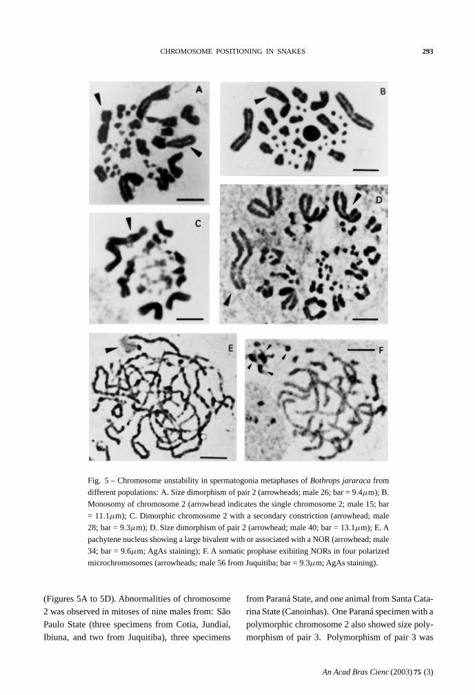
Fig. 5 – Chromosome unstability in spermatogonia metaphases ofBothrops jararaca from
different populations: A. Size dimorphism of pair 2 (arrowheads; male 26; bar = 9.4µm); B.
Monosomy of chromosome 2 (arrowhead indicates the single chromosome 2; male 15; bar
= 11.1µm); C. Dimorphic chromosome 2 with a secondary constriction (arrowhead; male
28; bar = 9.3µm); D. Size dimorphism of pair 2 (arrowhead; male 40; bar = 13.1µm); E. A
pachytene nucleus showing a large bivalent with or associated with a NOR (arrowhead; male
34; bar = 9.6µm; AgAs staining); F. A somatic prophase exibiting NORs in four polarized
microchromosomes (arrowheads; male 56 from Juquitiba; bar = 9.3µm; AgAs staining).
(Figures 5A to 5D). Abnormalities of chromosome
2 was observed in mitoses of nine males from: São
Paulo State (three specimens from Cotia, Jundiaí,
Ibiuna, and two from Juquitiba), three specimens
from Paraná State, and one animal from Santa Cata-
rina State (Canoinhas). One Paraná specimen with a
polymorphic chromosome 2 also showed size poly-
morphism of pair 3. Polymorphism of pair 3 was
An Acad Bras Cienc (2003)75 (3)
294 MARIA LUIZA BEÇAK, WILLY BEÇAK and ALEXANDRE PEREIRA
also found in one female of unknown origin (Fig-
ure 1B).
The behavior of polymorphic pairs was ana-
lyzed in meiosis. In males besides apparently nor-
mal diplotenes with eight bivalents (8 II) M, we
found nuclei with bivalent 2 showing breakages, as-
sociations or exhibiting one arm completely open or
only with telomere connection (Figures 6A to 6E).
In females, we observed one or two chromosome
bridges in first anaphase (Figures 6F and 6G). The
female with a polymorphic pair 3 in mitosis (Figure
1B) presented one chromosome bridge in first meio-
sis (Figure 6G). Second metaphases studied in males
appear either with 8 apparently normal dyads (M)
(Figure 3C) or with a breakage in one arm of dyad
2 or lacking this dyad (Figure 4G). AgAs studies
in males somatic cells showed NORs in one to two
pairs of microchromosomes (Figure 5F). We also
observed that a large macrochromosome can appear
associated with a NOR in male pachytene nuclei.
This macrochromosome seems to correspond to the
unstable chromosome 2 (Figure 5E).
A complete quantitative analysis of both pre-
meiotic events and meiosis in a same specimen is
difficult because cell cycle is seasonal. In general the
specimens exhibit only mitosis or meiosis. Yet, both
cell types were observed in nine colchicine untreated
males. The frequencies of mitotic and meiotic nuclei
with endoreduplication and unstable chromosome 2
were estimated in this sample. The analysis of 592
nuclei included 173 mitoses, 297 diplotenes, and
122 m II (Table I). In mitosis, regarding the macro-
chomosomes three classes of nuclei were consid-
ered. Class A includes apparently normal diploid
nuclei; class B has diploid nuclei with one unstable
chromosome 2, and class C has endoreduplicated
nuclei (≥ 4n and with aneuploidy). Among class
B nuclei it was included those with dimorphism or
monosomy of chromosome 2. Classes A, B, and
C appeared with the frequencies of 57.2%, 14.4%,
and 28.3% respectively. In first meiosis, class A in-
cludes 80.8% of apparently normal nuclei with eight
II; class B has 15.8% of abnormal nuclei with bi-
valent 2 unequal, broken or associated, and class C
presents 3.4% of endoreduplicated nuclei with≥ 16
II (up to 64 II) and 15 II. An intriguing fact noted
is that m I = 7 II + 1 I were not observed though
the occurrence of monosomic cells with 2n – 1 =
15. In second meiosis, class A includes 69.7% of
nuclei with eight apparently normal dyads; class B
contains 17.2% of nuclei with eight dyads and pre-
senting a breakage in one arm of dyad 2 or lacking
this dyad, and class C has 13.1% of endoreduplicated
nuclei. Among these endoreduplicated nuclei, 8.2%
presents 16 and 15 dyads lacking dyad 2. The re-
maining 4.9% of endoreduplicated m II includes nu-
clei with eight four-chromatids diplochromosomes.
DISCUSSION
We report here for the first time some peculiar cy-
tological aspects of chromosome positioning and
distribution in mitosis and meiosis of snakes with
premeiotic endomitosis, reductional mitosis, altered
meiosis, and aneuploidies. Regarding nuclei con-
figurations, we describe that homologue chromo-
somes of premeiotic cells display two types of spa-
tial order in the metaphase plate, positioned on op-
posite sides of the ring or closely apposed. This
latter type is similar to the somatic pairing known
in Diptera. Antiparallel and parallel aspects were
found in eight and five nuclei respectively, in a total
of 51 untreated nuclei (gonia cells) ofB. jararaca.
Among 24 colchicine treated cells, 11 nuclei showed
bipolar configuration. InB. insularis, 23 nuclei
presented complete bipolar aspect in a total of 76
colchicine treated cells. Among 12 untreated nuclei
(spermatogonia cells) of this species, total antipar-
allelism and total parallelism appeared in one and
two nuclei respectively. Considering that the ran-
dom probability of complete parallel and complete
bipolar distribution of the eight pairs of macrochro-
mosomes is one in 28 (1/256) in each case, the as-
pects here described cannot be explained as ran-
dom events. In both configuration types the chro-
mosomes have a constant groupal size-dependent
distribution in mitosis and meiosis. The groupal
size-dependent chromosome location observed in
An Acad Bras Cienc (2003)75 (3)
CHROMOSOME POSITIONING IN SNAKES 295
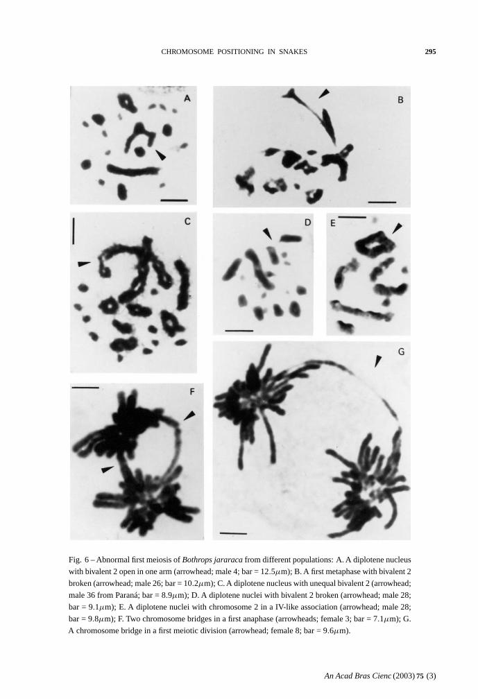
Fig. 6 – Abnormal first meiosis ofBothrops jararaca from different populations: A. A diplotene nucleus
with bivalent 2 open in one arm (arrowhead; male 4; bar = 12.5µm); B. A first metaphase with bivalent 2
broken (arrowhead; male 26; bar = 10.2µm); C. A diplotene nucleus with unequal bivalent 2 (arrowhead;
male 36 from Paraná; bar = 8.9µm); D. A diplotene nuclei with bivalent 2 broken (arrowhead; male 28;
bar = 9.1µm); E. A diplotene nuclei with chromosome 2 in a IV-like association (arrowhead; male 28;
bar = 9.8µm); F. Two chromosome bridges in a first anaphase (arrowheads; female 3; bar = 7.1µm); G.
A chromosome bridge in a first meiotic division (arrowhead; female 8; bar = 9.6µm).
An Acad Bras Cienc (2003)75 (3)
296 MARIA LUIZA BEÇAK, WILLY BEÇAK and ALEXANDRE PEREIRA
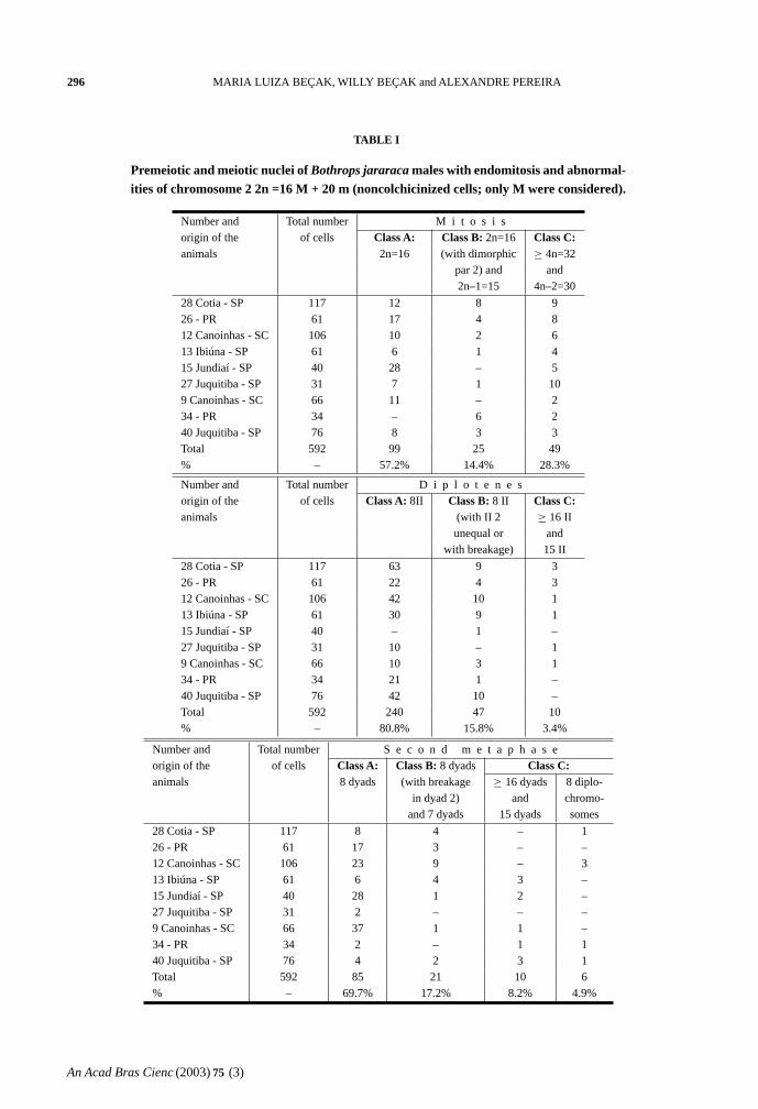
TABLE I
Premeiotic and meiotic nuclei of Bothrops jararaca males with endomitosis and abnormal-ities of chromosome 2 2n =16 M + 20 m (noncolchicinized cells; only M were considered).
Number and Total number M i t o s i sorigin of the of cells Class A: Class B: 2n=16 Class C:animals 2n=16 (with dimorphic ≥ 4n=32
par 2) and and2n–1=15 4n–2=30
28 Cotia - SP 117 12 8 926 - PR 61 17 4 812 Canoinhas - SC 106 10 2 613 Ibiúna - SP 61 6 1 415 Jundiaí - SP 40 28 – 527 Juquitiba - SP 31 7 1 109 Canoinhas - SC 66 11 – 234 - PR 34 – 6 240 Juquitiba - SP 76 8 3 3Total 592 99 25 49% – 57.2% 14.4% 28.3%
Number and Total number D i p l o t e n e sorigin of the of cells Class A: 8II Class B: 8 II Class C:animals (with II 2 ≥ 16 II
unequal or andwith breakage) 15 II
28 Cotia - SP 117 63 9 326 - PR 61 22 4 312 Canoinhas - SC 106 42 10 113 Ibiúna - SP 61 30 9 115 Jundiaí - SP 40 – 1 –27 Juquitiba - SP 31 10 – 19 Canoinhas - SC 66 10 3 134 - PR 34 21 1 –40 Juquitiba - SP 76 42 10 –Total 592 240 47 10% – 80.8% 15.8% 3.4%
Number and Total number S e c o n d m e t a p h a s eorigin of the of cells Class A: Class B: 8 dyads Class C:animals 8 dyads (with breakage ≥ 16 dyads 8 diplo-
in dyad 2) and chromo-and 7 dyads 15 dyads somes
28 Cotia - SP 117 8 4 – 126 - PR 61 17 3 – –12 Canoinhas - SC 106 23 9 – 313 Ibiúna - SP 61 6 4 3 –15 Jundiaí - SP 40 28 1 2 –27 Juquitiba - SP 31 2 – – –9 Canoinhas - SC 66 37 1 1 –34 - PR 34 2 – 1 140 Juquitiba - SP 76 4 2 3 1Total 592 85 21 10 6% – 69.7% 17.2% 8.2% 4.9%
An Acad Bras Cienc (2003)75 (3)
CHROMOSOME POSITIONING IN SNAKES 297
meiosis and mitosis ofB. jararaca seems to indi-
cate that chromosome order is maintained through
cell divisions. This conclusion gives support to data
showing that the relative chromosomal position on
each individual metaphase ring seems to be car-
ried through anaphase into telophase (Allison and
Nestor 1999). Our interpretation also support the
mitotic preset biophysical model which assumes that
chromosome-size-dependent positioning is estab-
lished and maintained in mitosis and G1 interphase
(Sun et al. 2000). As to the presence of two antago-
nic aspects of nuclei configuration in a same tissue
(gonia cells) we suppose it may reflect the occur-
rence of two cells types. Nagele et al. (1999) sug-
gest the coexistence of two cellular subclones to ex-
plain preparations exhibiting simultaneously human
cells with chromosomes 8 and 11 apposed and cells
with these chromosomes widely separated. High
frequencies of translocations among certain chro-
mosomes may be related to the fact they are adjacent
on the chromosome rosette (Nagele et al. 1995). As
we used squash technique, the different positions of
the metaphase plates may result in alterations of the
final figures. In some cases parallel configurations
may appear as antiparallel or vice versa. Although
our conclusions are mainly based on morphologi-
cal evidences, the use of more precise techniques as
painting and 3-D analysis could be helpful to con-
firm our observations. Until now these assays were
not used for snakes because in this group of ani-
mals molecular markers are still not available as for
chromosomes of human and other mammals.
Our findings that premeiotic cells ofB. jara-
raca with somatic pairing and endomitosis also
present altered meiosis and chromosome aberrations
led us to consider whether these events could be cor-
related. It is known that diplochromosomes were
found in spermatogonia mitosis ofLocusta migra-
toria in experiments using radiation or other agents
(White 1935, 1973). The homologue diplochromo-
somes observed in premeiotic cells ofB. jararaca
showed peculiar behavior, segregating to opposite
poles in cell division. This fact observed in 4.9% of
mII may indicate that first meiotic division would be
suppressed. Suppression of first meiosis division is
known inRana esculenta, a hybrid species between
R. ridibunda andR. lessonae. In this case, meiosis
is preceded in males and females by elimination of
theR. lessonae genome, followed by doubling ofR.
ridibunda genome. By this way onlyR. ridibunda
gametes are formed. Meiotic pairing in the hybrid
occurs between sister chromosomes without link-
age groups recombination (Heppich et al. 1982).
Yet, in non-hybrid species with few exceptions, at
first meiosis the two homologues of a pair move
to opposite poles and non-homologous pairs seg-
regate independently of each other. InDrosophila
melanogaster, female non-random segregation of
nonhomologues might occur when chiasma forma-
tion has failed to occur (Grell 1969). Further, it
was demonstrated that the Nod kinesin-like protein
is specifically required for achiasmate chromosome
segregation inDrosophila melanogaster (Zhang et
al. 1990). Also it was shown thatα-tubulin 67C
gene impair achiasmate chromosome segregation in
this species (Matthies et al. 1999). According to our
assumption, the cells with somatic pairing and pre-
meiotic endoreduplication would enter directly to
second meiosis division (m II). In this case, the m II
= 7 dyads (M) would be explained by the elimination
of the unstable parental four-chromatids diplochro-
mosome. Although 2 n – 1 = 15cells were found,
the absence of diplotene and of first metaphases with
univalents (m I = 7 II + 1 I) could be explained by
this assumption. Accordingly, in false first meio-
sis the endoreduplicated homologues remain con-
nected as dense cylindral structures. These com-
plex structures do not reveal clear splitting of the
parental homologues as well as do not show clear
chiasmata aspects. Yet, diplotene nuclei with visi-
ble chiasmata were also observed. In this case it re-
mains to be explained whether these chiasmata are
true exchanges between homologue chromosomes
or produced by sister chromatid recombinations in
diplochromosomes.
The dimorphism of chromosomes, probably
the 2nd or 3rd pairs, may explain the presence of un-
equal bivalents and of chromosome bridges in first
An Acad Bras Cienc (2003)75 (3)
298 MARIA LUIZA BEÇAK, WILLY BEÇAK and ALEXANDRE PEREIRA
meiosis. The observation that a large macrochro-
mosome bivalent is associated with a NOR in meio-
sis of animals with a dimorphic chromosome 2 in
mitosis may indicate that the abnormalities could
be caused by translocations involving a NOR. Non-
homologous association of nucleolar chromosomes
in human may be accounted for certain non-dis-
junctions and translocations causing diseases, as in
myeloid leukemia (Ohno et al. 1961). That model
was supported by further studies using the electron
microscope to study human meiotic cells (Stahl et
al. 1983). Moreover, the analysis of Robertsonian
translocations in mice indicated that fusion of nu-
cleoli is not the key factor predisposing to centric
fusions (Miller et al. 1978). According to these au-
thors, Robertsonian translocations may result from
close association of heterochromatin and the nucle-
oli. The chromosome bridges observed in anaphase
I of snakes could result from breakage-fusion events
in the nucleolar organizing region, as previously
suggested (Newman 1966). These translocations
can be of insertion type. Unequal crossing-over
would result in duplications and deletions produc-
ing morphological and genetical alterations in the
chromosomes involved. The eventual presence of a
hotspot could enhance this phenomenon. Reiter et
al. (1996) found such a hotspot inside the region
involved in unequal crossing-over in human chro-
mosome 17 and sequence analysis revealed a insect
mariner transposon-like element (MITE) near the
hotspot which could mediate the strands exchange.
Alternatively, we may suppose that in endomitotic
nuclei a more prolonged S-phase due to succes-
sive rounds of DNA replication could increase hete-
rochromatic associations, predisposing to transloca-
tions. According to Csink and Henikoff (1998), in
Drosophila the heterochromatin can influence the
relative position of a chromosomal region during
interphase and length increase of G I phase allows
heterochromatic association.
Though the exact mechanism that operates in
B. jararaca chromosome abnormalities is not under-
stood, we postulate that the parallel configuration
and endomitosis could be correlated to the chro-
mosomal aberrations. These aberrations could re-
sult from translocations involving the NOR or the
heterochromatin in telomeric associations between
nonhomologue chromosomes. Too, aberrations and
mutations induced by deleterious ambiental agents
cannot be excluded. We assume the occurrence of a
"mixed" gonad with heterozygous cells and unequal
transmission of a member of a pair of homologe
chromosomes. Nevertheless, we cannot dismiss the
possibility that the chromosome aberrations could
result from complex translocations which heterozy-
gosis would be maintained in natural populations by
suppression of first meiosis. Again changes in the
meiotic mechanism in a sexually reproducing or-
ganism are not frequent. There are some instances
of two types of meiosis occurring in the same go-
nad. According toWhite (1973), some of these alter-
ations probably experienced special gonad regions
rather than the whole gonad. In this case, the pre-
meiotic doubling would be a prerequisite to orig-
inate parthenogenetic species such as the triploid
salamanders (Sessions 1982) and lizards (Cuellar
1978).
ACKNOWLEDGMENTS
We thank Laerte Paula Santos, Nair Aparecida Silva
Pereira, Heitor Costa, and Luiz Antonio Tadeu Dias
for technical help; Helir Serralvo for photomicro-
graphy and editorial help. This work was supported
by CNPq, FAPESP and Fundação Butantan.
RESUMO
O posicionamento dos macrocromossomos deBothrops
jararaca eBothrops insularis (Viperidae) foi estudado em
metáfases radiais não alteradas de células não cultivadas
(espermatogônias e oogônias) não submetidas a inibidores
do fuso. Metáfases colchicinizadas de tecidos não culti-
vados (baço e intestino) e cultivados (sangue) foram tam-
bém analisadas. Relatamos dois arranjos cromossômi-
cos antagônicos não casuais em células premeióticas não
tratadas: a configuração paralela, com cromossomos ho-
mólogos associados lado a lado na placa metafásica, e a
configuração antiparalela, tendo os cromossomos homó-
An Acad Bras Cienc (2003)75 (3)
CHROMOSOME POSITIONING IN SNAKES 299
logos com distribuição antipolar no anel metafásico. O
aspecto antiparalelo apareceu também em células colchi-
cinizadas. O arranjo espacial dos cromossomos em ambas
configurações é grupal, dependente do tamanho e mantido
na meiose. Descrevemos também endomitoses seguidas
de mitoses reducionais que restabelecem o número diplói-
de em gônias não tratadas. Em machos deB. jararaca ob-
servamos alterações no mecanismo meiótico em algumas
regiões da gônada. Neste caso, as células endoredupli-
cadas segregam os diplocromossomos para polos opos-
tos, formando diretamente metáfases da segunda divisão
da meiose com supressão da primeira divisão. Numa di-
visão sucessiva, estas metáfases formam núcleos com um
conjunto de cromossomos. Duplicação cromossômica em
oogônias é conhecida em espécies híbridas e em salaman-
dras e lagartos partenogenéticos.Bothrops jararaca tam-
bém apresentou rearranjos cromossômicos, ocasionando
aneuploidias na mitose e meiose. Sugerimos que o parea-
mento somático, a endomitose, as alterações da meiose
e as aberrações cromossômicas podem ser processos cor-
relacionados. Aspectos similares de configurações nucle-
ares, endomitose e mitose reducional foram encontrados
em outras espécies de Viperidae e Colubridae.
Palavras-chave: serpentes, pareamento somático, posi-
cionamento cromossômico, endomitose, diplocromosso-
mos.
REFERENCES
Allison DC and Nestor AL. 1999. Evidence for a
relatively random array of human chromosomes on
the mitotic ring. J Cell Biol 145: 1-14.
Bajer AS and Molè-Bajer J. 1981. Mitoses: studies
of living cells: a revision of basic concepts. In Mi-
toses/Cytokineses. AM Zimmerman and A. Forer,
editors, Academic Press, New York. 227-299.
Beçak ML and Beçak W. 1973. Chromosome sec-
ondary constriction in different stages of develop-
ment. Experientia (Basel) 29: 359-361.
Beçak ML and Beçak W. 1981. Behavior of the ZW
sex bivalent in the snakeBothrops jararaca. Chro-
mosoma (Berl.) 83: 289-293.
Beçak ML, Beçak W, Roberts FL, Shoffner RN and
Volpe EP. 1971. Chromosome Atlas: Fish, Amphib-
ians, Reptiles and Birds. vol. 1 Ed. K. Benirschke
and TC. Hsu. Springer-Verlag. Berl.
Beçak ML, Rabello-Gay MN, Beçak W, Soma M,
Batistic RF and Trajtengertz I. 1990. The W
chromosome during the evolution and in sex abnor-
malities of snakes. DNA content, C banding. Cy-
togenetics of Amphibians and Reptiles. Birkhauser
Verlag. Basel.
Beçak W. 1968. Karyotypes, sex chromosomes and chro-
mosomal evolution in snakes. In Venomous Animals
and their Venoms. W. Bucherl, E. Buckley and V.
Deulofeu, editors. Vol. I, p. 53-95. Academic Press,
New York.
Beçak W and Beçak ML. 1969. Cytotaxonomy and
chromosomal evolution in Serpentes. Cytogenetics
8: 247.
Beçak W, Beçak ML and Nazareth HRS. 1962.
Karyotypic studies of two species of South Ameri-
can snakes (Boa constrictor amarali and Bothrops
jararaca). Cytogenetics 1: 305-313.
Beçak W, Beçak ML and Nazareth HRS. 1966. Evo-
lution and sex chromosomes in Serpentes. Mem Inst
Butantan 33: 151-152.
Bennett MD. 1984. Premeiotic events and meiotic chro-
mosome pairing. Society for Experimental Biology
87-121.
Costello DP. 1970. Identical linear order of chromo-
somes in both gametes of acoel turbellarianPoly-
choerus carmelensis: a preliminary note. Proc Nat
Acad Sci USA 67: 195-1958.
Csink AK and Henikoff S. 1998. Large-scale chromo-
somal movements during interphase progression in
Drosophila. J Cell Biol 143: 13-22.
Cuellar O. 1978. Parthenogenetic lizards. Science 201:
1115.
Dobzhansky TH. 1936. The persistence of the chromo-
some pattern in successive cell divisions inDroso-
phila pseudoobscura. J Exp Zool 74: 119-135.
Grell RF. 1969. Meiotic and somatic pairing. Chapter 6,
pp. 361-492. In Genetic Organization, I. EW Gaspari
and AW Ravin editors. New York: Academic Press.
Heppich S, Tunner HG and Greilhuber J. 1982. Pre-
meiotic chromosome doubling after elimination dur-
ing spermatogenesis of the species hybridRana escu-
lenta. Theoretical and Applied Genetic 61: 101-104.
Hiraoka Y, Agard DA and Sedat JW. 1990. Temporal
and spatial coordination of chromosomal movement,
spindle formation, and nuclear envelope breakdown
An Acad Bras Cienc (2003)75 (3)
300 MARIA LUIZA BEÇAK, WILLY BEÇAK and ALEXANDRE PEREIRA
during prometaphase inDrosophila melanogaster
embryos. J Cell Biol 111: 2815-2828.
Juricek DK. 1975. Non-random chromosome distribu-
tion in radial metaphases from the Chinese Hamster.
Chromosoma (Berl.) 50: 313-326.
Manuelidis L and Borden J. 1988. Reproducible
compartmentalization of individual chromosome do-
mains in human CNS cell revealed by in situ hy-
bridization and three dimensional reconstruction.
Chromosoma (Berl.) 96: 397-410.
Matthies HJG, Messina LG, Namba R, Greer KJ,
Walker MY and Hawley RS. 1999. Mutations
in theα-tubulin 67C gene specifically impair achias-
mate segregation inDrosophila melanogaster. J Cell
Biol 147: 1137-1144.
Miller OJ, Miller DA, Tantravahi R and Dev VG.
1978. Nucleolus organizer activity and the origin
of Robertsonian translocations. Cytogenetics. Cell
Genet 20: 40-50.
Moreno R, Navarro J, Iturra P and Veloso AM.
1987. The karyotype ofPhilodryas chamisso-
nis (Colubridae). Identification of nucleolar orga-
nizer region (NOR) and sex chromosomes by band-
ing methods. Braz. J Genetics 3: 497-506.
Mosgöller W, Leitch AR, Brown JKM and Heslop-
Harrison JS. 1991. Chromosome arrangements in
human fibroblasts at mitosis. Human Genetics 88:
27-33.
Nagele R, Freeman T, McMorrow L and Lee H. 1995.
Precise spatial positioning of chromosomes during
prometaphase: evidence for chromosome order. Sci-
ence 270: 1831-1835.
Nagele RG, Freeman T, McMorrow L, Thomson Z,
Kitson-Wind K and Lee H. 1999. Chromosome
exhibit preferential positioning in nuclei of quiescent
human cells. J Cell Sci 112: 525-535.
Newman LJ. 1966. Bridge and fragment aberrations in
Podophillum peltatum. Genetics 53: 55-63.
Ohno S, Trujillo JM, Kaplan WD and Kinosita P.
1961. Nucleolus-organizers in the causation of chro-
mosomal anomalies in man. Lancet II: 123-125.
Rabl C. 1885. Uber Zelltheilung. Morphol Jahrbot 10:
214-330.
Reiter LT, Murakami T, Koeuth T, Pentao L, Muzny
DM, Gibbs RA and Lupski JR. 1996. A recombina-
tion hotspot responsible for two inherited peripheral
neuropathies is located near amariner transposon-
like element. Nature Genetics 12: 288-297.
Sessions SK. 1982. Cytogenetics of diploid and triploid
salamanders of theAmbystoma jeffersonianum com-
plex. Chromosoma (Berl.) 84: 599-621.
Singh L, Ray-Chaudhuri SP, Majumdar K, Purdom
IF and Jones KW. 1979. Sex specific chromosome
polymorphisms in the common indian krait,Bun-
garus caeruleus Schneider (Ophidia, Elapidae).
Chromosoma (Berl.) 73: 93-108.
Stahl A, Luciani JM, Hartung M, Devictor
M, Bergé-Lefranc JL and Guichaoua M. 1983.
Structural basis for Robertsonian translocation in
man: association of ribosomal genes in the nucleolar
fibrillar center in meiotic spermatocytes and oocytes.
Proc Nat Acad Sci USA 80: 5946-5960.
Sun HB, Shen J and Yokota H. 2000. Size-dependent
positioning of human chromosomes in interphase nu-
clei. Biophis J 79: 184-190.
Trajtengertz I, Beçak ML and Ruiz RG. 1995. Ri-
bosomal cistrons inBothrops neuwiedi (Serpentes)
subspecies from Brasil. Genome 38: 601-606.
Vidal BC and Mello MLS. 1995. Re-evaluating the
AgNOR staining response in Triton X-100- treated
liver cells by image analysis. Anat Cell Pathol 9:
39-43.
White MJD. 1935. The effects of X-rays on mitosis in
the spermatogonial divisions ofLocusta migratoria
L. Proc Roy Soc Lond B 119: 61-84.
White MJD. 1973. Animal Cytology and Evolution (3
ed.). Cambridge University Press, 961pp.
Zhang P, Knowles BA and Goldstein LS. 1990. A
kinesin-like protein required from distributive chro-
mosome segregation inDrosophila. Cell 62: 1053-
1062.
An Acad Bras Cienc (2003)75 (3)