REDE NORDESTE DE BIOTECNOLOGIA UNIVERSIDADE FEDERAL …
122
REDE NORDESTE DE BIOTECNOLOGIA UNIVERSIDADE FEDERAL DO ESPÍRITO SANTO CENTRO DE CIÊNCIAS DA SAÚDE PROGRAMA DE PÓS-GRADUAÇÃO EM BIOTECNOLOGIA LIDIANE PIGNATON AGOSTINI ALTERAÇÃO DE EXPRESSÃO GÊNICA EM CÉLULAS MONONUCLEARES DO SANGUE PERIFÉRICO HUMANO SUBMETIDAS À EXPOSIÇÃO COM HERBICIDA À BASE DE GLIFOSATO VITÓRIA 2018
Transcript of REDE NORDESTE DE BIOTECNOLOGIA UNIVERSIDADE FEDERAL …
REDE NORDESTE DE BIOTECNOLOGIA
UNIVERSIDADE FEDERAL DO ESPÍRITO SANTO
CENTRO DE CIÊNCIAS DA SAÚDE
PROGRAMA DE PÓS-GRADUAÇÃO EM BIOTECNOLOGIA
LIDIANE PIGNATON AGOSTINI
ALTERAÇÃO DE EXPRESSÃO GÊNICA EM CÉLULAS MONONUCLEARES DO
SANGUE PERIFÉRICO HUMANO SUBMETIDAS À EXPOSIÇÃO COM
HERBICIDA À BASE DE GLIFOSATO
VITÓRIA
2018

LIDIANE PIGNATON AGOSTINI
ALTERAÇÃO DE EXPRESSÃO GÊNICA EM CÉLULAS MONONUCLEARES DO
SANGUE PERIFÉRICO HUMANO SUBMETIDAS À EXPOSIÇÃO COM
HERBICIDA À BASE DE GLIFOSATO
Tese apresentada ao Programa de Pós-
Graduação em Biotecnologia do Centro de
Ciências da Saúde da Universidade Federal
do Espírito Santo e à Rede Nordeste de
Biotecnologia, como requisito parcial para a
obtenção do título de Doutora em
Biotecnologia.
Orientador: Prof. Dr. Iúri Drumond Louro
VITÓRIA
2018


AGRADECIMENTOS
À Ufes e ao Programa de Pós-Graduação em Biotecnologia, vinculado à Rede
Nordeste de Biotecnologia, pela possibilidade de desenvolver esse trabalho e me
formar Doutora.
À Fapes, pelo apoio financeiro e bolsa de estudo.
Ao Iúri, pela orientação, ensinamentos, conversas e compreensão ao longo desses 9
anos de NGHM. Obrigada por tudo!
Às professora Flávia de Paula e Flávia Errera e aos professores Dalton Vassalo e
Marco Cunegundes, por aceitarem compor a banca examinadora.
Ao Professor Wilson Araújo, por ter permitido a realização dos experimentos no
Laboratório de Genética Molecular e Bioinformática (USP/Ribeirão Preto). E à todas
as pessoas do laboratório, especialmente Andrés e Kamila, que me auxiliaram na
realização dos experimentos e me receberam como um membro do laboratório.
À Elaine, minha amiga e companheira de trabalho há muitos anos. Obrigada pela
imensa ajuda no desenvolvimento dessa tese, especialmente na realização das
análises estatísticas.
À Raquel Spinassé, Raquel Reis, Fernanda, Elda e Diego pelo auxílio durante a
realização deste trabalho e pela amizade, dentro e fora do NGHM, nesses anos todos.
À todos os amigos do NGHM e da Biotecnologia que, direta ou indiretamente,
contribuíram para o desenvolvimento deste trabalho, pela companhia sempre
agradável e pela amizade.
Aos meus pais, por sempre colocarem a minha educação como prioridade e me
apoiarem.
Ao Cris, meu esposo, por sempre me apoiar e me incentivar a ser verdadeiramente
Doutora.
À todos que, direta ou indiretamente, contribuíram com esse trabalho. Muito obrigada!
ESTRUTURA DA TESE
Esta Tese está organizada em formato de artigo científico e de acordo com o exigido
pelo Regimento desse Programa - com a comprovação de submissão de dois artigos
em periódicos Qualis B1 ou superior.
As Listas de Figuras, Siglas, Abreviaturas e as Referências Bibliográficas contemplam
as informações descritas na Introdução e Revisão de Literatura.

RESUMO
AGOSTINI, L. P. Alteração de expressão gênica em células mononucleares do
sangue periférico humano submetidas à exposição com herbicida à base de
glifosato. 2018. 121f. Tese (Doutorado em Biotecnologia) - Programa de Pós-
Graduação em Biotecnologia, vinculado à Rede Nordeste de Biotecnologia
(Renorbio), UFES, Espírito Santo. Brasil.
O Glifosato [N-(fosfonometil)glicina] é um herbicida pós-emergente, não seletivo e
sistêmico. No processo de criação das formulações comerciais de herbicidas a base
de glifosato (GBHs, do inglês glyphosate-based herbicides), como o Roundup®, são
adicionados surfactantes com o intuito de aumentar a eficiência do composto base. A
rota prioritária de degradação do glifosato por micro-organismos no solo resulta na
formação do ácido aminometilfosfônico (AMPA). As respostas moleculares ao
glifosato têm sido extensivamente estudadas em espécies de plantas e em alguns
vertebrados. Em humanos, apesar dos estudos até agora realizados, não se conhece
exatamente quais os riscos e mecanismos de atuação que explicariam a toxicidade
ao glifosato relatada em alguns experimentos. Sendo assim, a hipótese dessa tese é
de que a exposição rápida ao Roundup® e ao AMPA leva à alterações de expressão
gênica em importantes processos celulares. Dessa forma, o objetivo desse trabalho é
identificar genes diferencialmente expressos (DEGs, do inglês differentially expressed
genes) em células mononucleares do sangue periférico (PBMCs, do inglês, peripheral
blood mononuclear cells) humano submetidas à exposição rápida com herbicida à
base de glifosato (Roundup®) e AMPA. O teste de MTT [3(4,5-dimetiltiazol-2-il)-2,5-
difeniltetrazólio brometo], realizado em triplicatas, foi utilizado para avaliar a
viabilidade celular e para a escolha das condições de tratamento utilizadas na técnica
de microarray (GeneChip® Human Transcriptome Array 2.0, Affymetrix). As condições
analisadas foram controle (3 chips), AMPA (10 mM; 3 chips) e Roundup® (0,05%; 2
chips), expostos durante 3 horas. Utilizando um valor de p<0,05 e fold-change de 1,5
foram identificados 5 DEGs no tratamento com o AMPA e 26 no tratamento com
Roundup®. As análises de enriquecimento mostraram que os genes com expressão

alterada após exposição ao Roundup® estavam associados a 33 processos celulares,
principalmente relacionados à regulação destes processos. A plataforma digital
Pathview foi utilizada para identificar a atuação dos DEGs após exposição ao
Roundup® em diferentes vias. Os genes TNF, LTA, TAB2 e ATM foram relacionados
à via de sinalização NF-kappa β; BCL2L11 e ATM à via de sinalização FoxO; SESN3
e ATM à via de sinalização p53; e TNF, BCL2L11 e ATM à apoptose. Dessa forma, os
resultados sugerem que o Roundup® altera o padrão de expressão gênica de diversos
genes associados com o controle do ciclo celular, regulação de processos celulares e
apoptose.
Palavras-chave: Expressão gênica, microarray, MTT, Roundup®, PBMC,
humanos.

ABSTRACT
AGOSTINI, L.P. Gene expression alteration in human peripheral blood
mononuclear cells submitted to glyphosate-based herbicide exposure. 2018.
121f. Thesis (Doctoral in Biotechnology) - Postgraduation Biotechnological
Programme, UFES, Espírito Santo. Brazil.
Glyphosate is a post-emergent, non-selective and systemic herbicide. In the creating
process of glyphosate-based herbicides (GBHs) such as Roundup®, surfactants are
added to improve efficiency. The priority route of glyphosate’s degradation in soil
results in aminomethylphosphonic acid (AMPA). Molecular responses to glyphosate
were analyzed in some species of plants and in some vertebrates. In humans, it is not
known exactly what risks and mechanisms of action would explain glyphosate’s toxicity
reported in some experiments. The hypothesis is that fast exposure to Roundup® and
AMPA leads to the differentiated expression of genes related to important cellular
processes. Thus, the aim was identified these genes in human peripheral blood
mononuclear cells when exposed to Roundup® and AMPA. The MTT [3-(4,5-
Dimethylthiazol-2-yl)-2,5-diphenyltetrazolium bromide] test was performed in
triplicates to evaluate cell viability and the choice of treatment conditions used in the
microarray technique (GeneChip® Human Transcriptome Array 2.0, Affymetrix). Eight
chips were used: 3 for controls, 3 for AMPA (10 mM) and 2 for Roundup® (0.05%).
The exposure time was 3 hours. Using a p <0.05 and a fold-change of ≥1.5 and ≤ −1.5,
there were 26 differentially expressed genes (DEGs) identified after Roundup®
exposure (3h; 0.05%) and 5 DEGs after AMPA treatment (3h; 10 mM). DEGs after
Roundup® treatment showed association with 33 Gene Ontology (GO) cellular
processes (enrichment analysis), mainly related to regulation. Pathview web was used
to identify the effect off DEGs in different pathways. Only genes differentially expressed
in Roundup® treatment were included in the pathways. TNF, LTA, TAB2 and ATM
genes are related to NF-kappa B signaling pathway; BCL2L11 and ATM genes to FoxO
signaling pathway; SESN3 and ATM genes to p53 signaling pathway; and TNF,
BCL2L11 and ATM genes to apoptosis. Our results suggest that Roundup® change

expression pattern of a several genes associated with cell cycle control, cellular
processes regulation and apoptosis.
Keywords: Gene expression, microarray, MTT, Roundup®, PBMC, humans.

LISTA DE FIGURAS
Figura 1. Consumo de agrotóxicos e afins (2000 – 2014). Fonte: Boletins anuais de
produção, importação, exportação e vendas de agrotóxicos no Brasil (IBAMA, 2016).
.................................................................................................................................. 19
Figura 2. Comercialização de agrotóxicos, por área plantada (2012). Fonte:
Indicadores de Desenvolvimento Sustentável – Brasil 2015 (IBGE, 2015). .............. 19
Figura 3. Estrutura molecular do Glifosato. Fonte: IARC, 2015. ................................ 21
Figura 4. Inibição da enzima 5-enolpiruvilchiquimato-3-fosfato sintase (EPSPS) pelo
glifosato em uma das etapas da via do chiquimato. Fonte: CZELUSNIAK et al., 2012
(Modificada). .............................................................................................................. 23
Figura 5. Mecanismos de degradação do glifosato. Acima: Via da C-P liase. Abaixo:
Via do AMPA. Fonte: POLLEGIONI; SCHONBRUNN; SIEHL, 2011 (Modificada). ... 25
Figura 6. Comprovação de submissão do manuscrito 1 para a revista Regulatory
Toxicology and Pharmacology. ................................................................................. 44
Figura 7. Comprovação de submissão do manuscrito 2 para a revista International
Journal of Hygiene and Environmental Health. ......................................................... 78

LISTA DE SIGLAS E ABREVIATURAS
AChE Acetilcolinesterase eritrocitária
AHS Agricultural Health Study
AMPA Ácido aminometilfosfônico
BChE Butirilcolinesterase plasmática
CARC Comitê de Revisão das Avaliações de Câncer
CAS# Chemical Abstracts Service (número de registro de produtos químicos)
DNA Ácido Desoxirribonucleico
EFSA Autoridade Europeia para a Segurança dos Alimentos
ELISA Ensaio de imunoabsorção enzimática
EPA Agência de Proteção Ambiental
EPSP 5-enolpiruvilchiquimato-3-fosfato
EPSPS Enzima 5-enolpiruvilchiquimato-3-fosfato sintase
et al. E outros
FAO Organização das Nações Unidas para Agricultura e Alimentação
FCMIA Imunoensaio de microbola (microbead) covalente por fluorescência
GBHs Herbicidas à base de glifosato
GC-MS Cromatografia gasosa com espectrômetro de massa
GOX Enzima glifosato oxidoredutase
h Hora(s)
HaCaT Linhagem de queratinócitos
HepG2 Linhagem de câncer de fígado
HILIC Cromatografia líquida com interação hidrofílica

HPLC Cromatografia líquida de alta performance
IARC Agência Internacional de Pesquisas sobre o Câncer
IBAMA Instituto Brasileiro do Meio Ambiente e dos Recursos Naturais
Renováveis
IC Intervalo de confiança
IUPAC União Internacional de Química Pura e Aplicada
LC-MS/MS Cromatografia líquida acoplada à espectrometria de massas sequencial
LD Limite de detecção
LPS Lipopolisacarídeo
M Molar
mg/kg Miligrama/kilo
mg/ml Miligrama/mililitro
min Minutos
mM Milimolar
NNG N-Nitrosoglifosato
OMS Organização Mundial da Saúde
OR Odds ratio, razão de chances ou de possibilidades
p Probabilidade de significância
PBMCs Células mononucleares do sangue periférico.
PEP Fosfoenolpiruvato
Pi Fósforo inorgânico
PMIDA Ácido N-(fosfonometil) iminodiacético
POEA Polietoxilenoamina
ppm Partes por milhão
RNAm RNA mensageiro

S3P Chiquimato-3-fosfato
SCE Troca de cromátides irmãs
SINAN Sistema de Informação de Agravos de Notificação
SINITOX Sistema Nacional de Informações Tóxico-Farmacológicas
TOXCEN Centro de Atendimento Toxicológico do Espírito Santo
μg/L Micrograma/litro
μg/ml Micrograma/microlitro

SUMÁRIO
1 INTRODUÇÃO ....................................................................................................... 15
2 REVISÃO BIBLIOGRÁFICA ................................................................................... 18
2.1 Agrotóxicos ...................................................................................................... 18
2.2 Glifosato ........................................................................................................... 20
2.2.1 Histórico, características e comercialização .............................................. 20
2.2.2 Mecanismo de ação .................................................................................. 22
2.2.3 Degradação ............................................................................................... 24
2.2.4 Indicações de uso ..................................................................................... 25
2.2.5 Avaliações de risco ................................................................................... 26
2.2.6 Legislação brasileira sobre uso de agrotóxicos ......................................... 27
2.3 Efeitos biológicos do glifosato .......................................................................... 28
2.4 Efeitos do glifosato em humanos ..................................................................... 31
2.4.1 Experimentos in vitro ................................................................................. 31
2.4.2 Exposição in vivo: estudos epidemiológicos ............................................. 33
2.4.3 Intoxicação aguda ..................................................................................... 36
2.4.4 Exposição e intoxicação por glifosato no Brasil ........................................ 37
2.4.5 Detecção de contaminação por glifosato .................................................. 39
2.5 Expressão gênica alterada por glifosato .......................................................... 40
2.6 Hipótese ........................................................................................................... 42
3 OBJETIVOS ........................................................................................................ 43
3.1 Objetivo geral ................................................................................................... 43
3.2 Objetivos específicos ....................................................................................... 43
4 ARTIGOS CIENTÍFICOS DERIVADOS DA TESE ................................................. 44
4.1 Manuscrito 1 .................................................................................................... 44
4.2 Manuscrito 2 .................................................................................................... 78
5 CONSIDERAÇÕES FINAIS ................................................................................. 106
6 REFERÊNCIAS BIBLIOGRÁFICAS ..................................................................... 107

15
1 INTRODUÇÃO
A partir da década de 1940 começaram a operar as primeiras unidades produtivas de
agrotóxicos no Brasil com a finalidade de aumentar a produtividade agrícola. Em
meados da década de 1970, o parque industrial de agrotóxicos passou a operar,
aumentando a comercialização de agrotóxicos de forma contínua desde então
(IBAMA, 2010). No ano de 2014, os herbicidas responderam por 58% de todos os
tipos de agrotóxicos comercializados (IBAMA, 2016). O principal ingrediente ativo
comercializado foi o glifosato, com 194 mil toneladas vendidas (IBAMA, 2016).
O Glifosato (N-(fosfonometil)glicina) é um herbicida pós-emergente, não seletivo e
sistêmico, que mata ou suprime plantas (IARC, 2015; MYERS et al., 2016). No
processo de criação das formulações comerciais, são adicionados surfactantes para
melhorar a eficiência (VANDEMBERG et al., 2017). Os surfactantes mais utilizados
são a polietoxilenoamina (POEA) e os ácidos sulfúrico e fosfórico (SZÉKÁCS;
DARVAS, 2012). Os produtos compostos pelo glifosato e pelo surfactante podem ser
chamados de herbicidas à base de glifosato (GBHs, do inglês glyphosate-based
herbicides). O Roundup® é o GBH mais comum (IARC, 2015).
O mecanismo de ação do glifosato baseia-se na inibição da enzima 5-
enolpiruvilchiquimato-3-fosfato sintase (EPSPS), que atua na via do chiquimato,
catalisando a conversão do chiquimato-3-fosfato (S3P) em 5-enolpiruvilchiquimato-3-
fosfato (EPSP) (BOOCOCK; COGGINS,1983).
A rota prioritária de degradação do glifosato por micro-organismos no solo resulta na
formação do ácido aminometilfosfônico (AMPA) (RIBEIRO et al., 2015). A meia-vida
do glifosato em solo varia de 2 a 197 dias, e do AMPA de 76 a 240 dias (GIESY;
DOBSON; SOLOMON, 2000). Nos mamíferos, o glifosato não é metabolizado
eficientemente e é, principalmente, excretado inalterado na urina (MYERS et al.,
2016).
A Agência Internacional de Pesquisas sobre o Câncer (IARC, do inglês International

16
Agency for Research on Cancer) classificou o glifosato como "provavelmente
cancerígeno para os seres humanos (Grupo 2A)" em 2015. Entretanto, outras
agências sugerem outras classificações. A Organização Mundial da Saúde (OMS) e a
Organização das Nações Unidas para Agricultura e Alimentação (FAO, do inglês Food
and Agriculture Organization) classificam o glifosato como "improvável que apresente
risco carcinogênico para os seres humanos devido à exposição através da dieta"
(FAO/ OMS, 2016). O Comitê de Revisão das Avaliações de Câncer (CARC, do inglês
Cancer Assessment Review Committee) classificou o glifosato como "provavelmente
não cancerígeno para humanos" (EPA, 2016).
Em 1974, quando a Monsanto iniciou a comercialização do Roundup®, defendia-se
que o princípio ativo glifosato era seguro para o meio ambiente e para a saúde humana
e animal (WILLIAMS et al., 2000). A partir da década de 1980, diversos estudos foram
realizados buscando a confirmação das informações divulgadas através de
experimentos de avaliação de toxicidade, genotoxicidade e carcinogenicidade
(VIGFUSSOM; VYSE, 1980; BOLOGNESI et al., 1997; WILLIAMS et al., 2000;
KOLLER et al., 2012; KWIATKOWSKA et al., 2017). A exposição in vivo ao glifosato
também foi avaliada em diferentes organismos e através de estudos epidemiológicos
em humanos (TARAZONA et al., 2017).
A toxicidade aguda do glifosato é classificada como baixa em ratos, sendo que o LD50
oral do glifosato puro é de 5.600 mg/kg (FAO/OMS, 2016). As intoxicações agudas
após ingestão de GBHs são observadas em casos de suicídios ou acidentes. Os
sintomas relacionados à intoxicação são diversos, variando de leves à mais graves, e
algumas vezes levam o indivíduo à morte (POTREBIĆ et al., 2009; ZOUAOUI et al.,
2013; KAMIJO; TAKAI; SAKAMOTO, 2015).
Os registros sobre os casos de intoxicação por agrotóxicos no Brasil são obtidos
através de dois sistemas: o Sistema Nacional de Informações Tóxico-Farmacológicas
(SINITOX) e o Sistema de Informação de Agravos de Notificação (SINAN). Segundo
os dados disponíveis no SINITOX (2017), em 2014 foram registrados 5116 casos de
intoxicação por agrotóxico. Infelizmente, esses dados não representam a realidade do
país pois nem todos os estados repassam os dados para o sistema nacional

17
(SINITOX, 2017).
Em humanos, o diagnóstico de contaminação por glifosato é confirmado por testes
como a cromatografia líquida acoplada à espectrometria de massas sequencial (LC-
MS/MS), cromatografia gasosa com espectrômetro de massa (GC-MS), ELISA e nível
da enzima colinesterase (YOSHIOKA et al., 2011; KRUGER et al., 2014; PEARSON;
PATEL, 2016).
As respostas moleculares ao glifosato já foram analisadas para algumas espécies de
plantas e para alguns vertebrados. Em humanos, são poucos os estudos realizados.
Pesquisas que identifiquem as alterações a nível molecular são importantes para
determinar em quais processos biológicos o glifosato atua alterando a expressão
gênica.
A hipótese dessa tese é de que a exposição rápida ao Roundup® e ao AMPA leva à
expressão diferenciada de genes relacionados a importantes processos celulares, o
que explicaria a toxicidade ao glifosato relatada na literatura disponível.
Dessa forma, o objetivo desse trabalho é identificar genes diferencialmente expressos
em células mononucleares do sangue periférico humano submetidas à exposição
rápida com herbicida à base de glifosato (Roundup®) e AMPA.
18
2 REVISÃO BIBLIOGRÁFICA
2.1 Agrotóxicos
O modelo agrícola contemporâneo, que apresenta elevados índices de produtividade,
tem como parte fundamental o uso de agrotóxicos (IBAMA, 2010). A Lei nº 7.802, de
11 de Julho de 1989, define agrotóxicos como:
Os produtos e os agentes de processos físicos, químicos ou biológicos,
destinados ao uso nos setores de produção, no armazenamento e
beneficiamento de produtos agrícolas, nas pastagens, na proteção de
florestas, nativas ou implantadas, e de outros ecossistemas, e também de
ambientes urbanos, hídricos e industriais, cuja finalidade seja alterar a
composição da flora ou da fauna, a fim de preservá-las da ação danosa de
seres vivos considerados nocivos (BRASIL, 1989).
Os agrotóxicos (defensivos agrícolas, pesticidas ou produtos fitossanitários)
apresentam em sua composição ingredientes ativos, que são substâncias químicas
tóxicas que interferem na atividade biológica normal dos seres vivos alvos de controle.
São os ingredientes ativos que conferem eficácia aos agrotóxicos e afins, por ação
química, física ou biológica (BRASIL, 1989).
A partir da década de 1940 começaram a operar as primeiras unidades produtivas de
agrotóxicos no Brasil. Em meados da década de 1970 o parque industrial de
agrotóxicos foi instituído, aumentando a comercialização de agrotóxicos de forma
contínua desde então (IBAMA, 2010).
A figura 1 apresenta o consumo crescente de agrotóxicos e afins entre os anos de
2000 a 2014.

19
Figura 1. Consumo de agrotóxicos e afins (2000 – 2014). Fonte: Boletins anuais de produção, importação, exportação e vendas de agrotóxicos no Brasil (IBAMA, 2016).
A comercialização de agrotóxicos (Figura 2) ocorre de forma mais acentuada nos
Estados de São Paulo e Goiás, seguidos por Espírito Santo, Minas Gerais, Mato
Grosso e Santa Catarina (IBGE, 2015).
Figura 2. Comercialização de agrotóxicos, por área plantada (2012). Fonte: Indicadores de Desenvolvimento Sustentável – Brasil 2015 (IBGE, 2015).
20
Os agrotóxicos podem ser classificados de acordo com os mecanismos de ação
contra os diferentes tipos de organismos: herbicidas (plantas), inseticidas (insetos),
fungicidas (fungos), nematicidas (microorganismos de solo), moluscicidas (moluscos),
dentre outros (IBAMA, 2010).
Os herbicidas são agentes químicos que evitam, reduzem ou eliminam plantas
infestantes, chamadas comumente de ervas daninhas, que competem por água e
nutrientes com a planta cultivada (IBAMA, 2010).
No ano de 2014, segundo o Boletim anual de produção, importação, exportação e
vendas de agrotóxicos no Brasil (IBAMA, 2016), os herbicidas responderam por 58%
de todos os tipos de agrotóxicos comercializados (294 mil toneladas).
O principal ingrediente ativo comercializado foi o glifosato, com 194 mil toneladas
vendidas em 2014 (IBAMA, 2016).
No Espírito Santo, foram comercializadas 2,5 mil toneladas de herbicidas em 2014,
sendo que o glifosato corresponde a 72% dessas vendas (IBAMA, 2016).
2.2 Glifosato
2.2.1 Histórico, características e comercialização
Os herbicidas à base de glifosato foram sintetizados pela primeira vez em 1950 como
um potencial composto farmacêutico, mas suas atividades herbicidas não foram
descobertas até serem ressintetizados e testados em 1970, sendo utilizados desde
1974 (SZÉKÁCS; DARVAS, 2012; WILLIANS et al., 2016). A patente original expirou
em 2000 nos EUA, e em 1991 no restante do mundo (IARC, 2015).
21
O Glifosato, cujo nome IUPAC é N-(fosfonometil)glicina (CAS# 1071-83-6), é um
herbicida pós-emergente, não seletivo e sistêmico, que mata ou suprime plantas,
incluindo gramíneas, plantas perenes, videiras, arbustos e árvores. Quando aplicado
a baixas doses, atua como regulador de crescimento e dessecante (IARC, 2015;
FAO/WHO, 2016; MYERS et al., 2016).
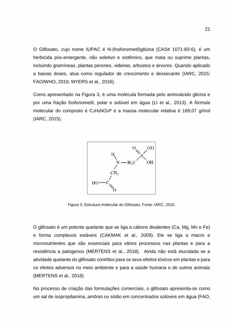
Como apresentado na Figura 3, é uma molécula formada pelo aminoácido glicina e
por uma fração fosfonometil, polar e solúvel em água (LI et al., 2013). A fórmula
molecular do composto é C3H8NO5P e a massa molecular relativa é 169,07 g/mol
(IARC, 2015).
Figura 3. Estrutura molecular do Glifosato. Fonte: IARC, 2015.
O glifosato é um potente quelante que se liga a cátions divalentes (Ca, Mg, Mn e Fe)
e forma complexos estáveis (CAKMAK et al., 2009). Ele se liga a macro e
micronutrientes que são essenciais para vários processos nas plantas e para a
resistência a patógenos (MERTENS et al., 2018). Ainda não está elucidada se a
atividade quelante do glifosato contribui para os seus efeitos tóxicos em plantas e para
os efeitos adversos no meio ambiente e para a saúde humana e de outros animais
(MERTENS et al., 2018).
No processo de criação das formulações comerciais, o glifosato apresenta-se como
um sal de isopropilamina, amônio ou sódio em concentrados solúveis em água (FAO,

22
2000). Nesse processo, são adicionados surfactantes (também chamados de
adjuvantes) para melhorar a eficiência do agrotóxico. Essa melhora ocorre pois esses
compostos permitem um transporte mais rápido do agrotóxico para a planta (através
das folhas e meristemas), garantindo uma absorção mais eficaz do ingrediente ativo,
protegendo-o da evaporação e retardando o processo de lixiviação (IPCS, 1996;
SZÉKÁCS; DARVAS, 2012; VANDEMBERG et al., 2017).
Os surfactantes mais utilizados são a polietoxilenoamina (POEA) e os ácidos sulfúrico
e fosfórico. Além disso, os GBHs podem conter outros ingredientes ativos, como ácido
simasino, ácido 2,4-diclorofenoxiacético (2,4-D) ou ácido 4-cloro-2-metilfenoxiacético
(IPCS, 1996). Geralmente, os GBHs causam mudanças mais fortes do que o próprio
glifosato (MARTINEZ; REYES; REYES, 2007; MESNAGE; BERNAY; SERALINI,
2013; FOLMAR; SANDERS; JULIN, 1979).
O Roundup® é o GBH mais comum e é apresentado em muitas formulações (IARC,
2015). Outros nomes comerciais dos GBHs são: Abundit Extra, Credit, Xtreme,
Glifonox, Glyphogan, Ground-Up, Rodeo, Touchdown, Tragli, Wipe Out e Yerbimat
(FCI, 2015).
2.2.2 Mecanismo de ação
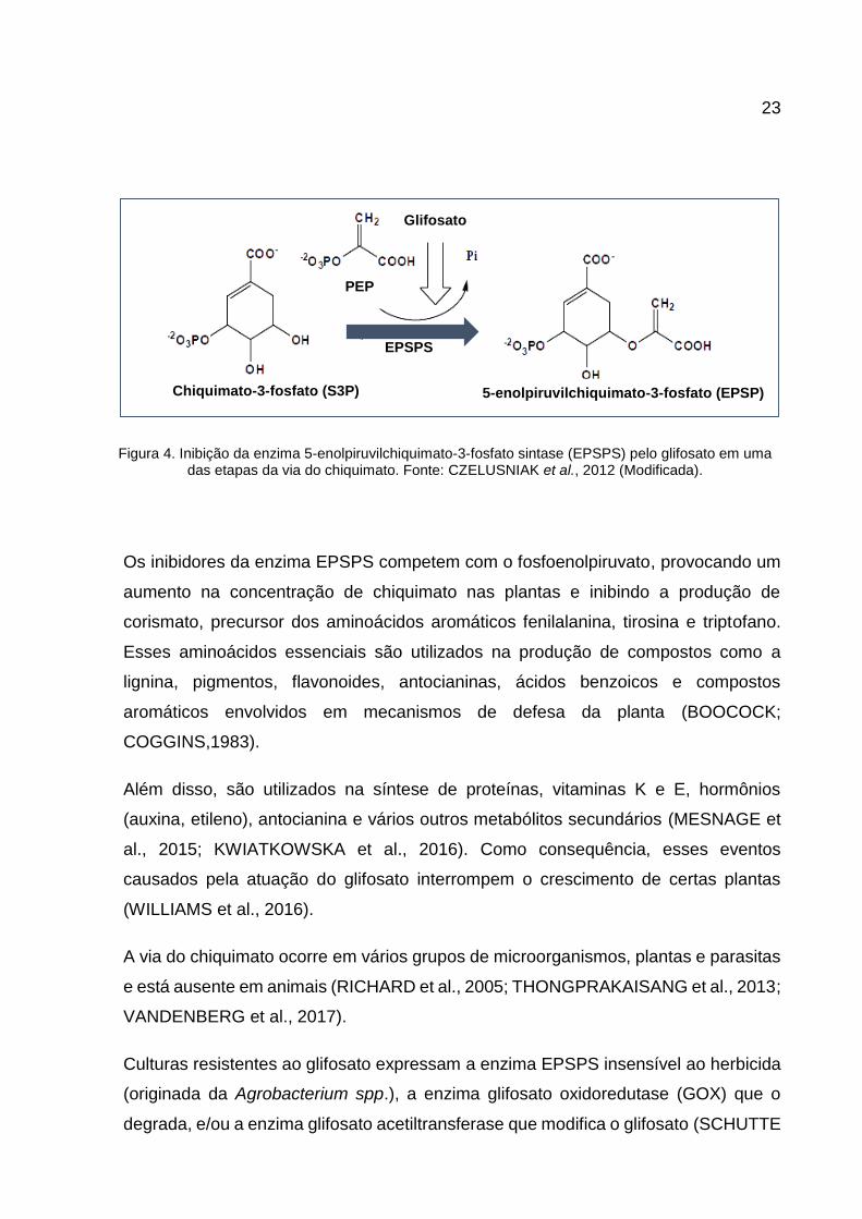
O mecanismo de ação do glifosato em plantas e algumas bactérias, baseia-se na
inibição da enzima 5-enolpiruvilchiquimato-3-fosfato sintase (EPSPS), que atua na via
do chiquimato (Figura 4). A enzima EPSPS, sintetizada no citoplasma, mas atuante
no cloroplasto, catalisa a conversão do chiquimato-3-fosfato (S3P) em 5-
enolpiruvilchiquimato-3-fosfato (EPSP), com liberação de fósforo inorgânico (Pi),
utilizando fosfoenolpiruvato (PEP) como substrato (BOOCOCK; COGGINS,1983;
CZELUSNIAK et al., 2012; VANDENBERG et al., 2017).
23
Os inibidores da enzima EPSPS competem com o fosfoenolpiruvato, provocando um
aumento na concentração de chiquimato nas plantas e inibindo a produção de
corismato, precursor dos aminoácidos aromáticos fenilalanina, tirosina e triptofano.
Esses aminoácidos essenciais são utilizados na produção de compostos como a
lignina, pigmentos, flavonoides, antocianinas, ácidos benzoicos e compostos
aromáticos envolvidos em mecanismos de defesa da planta (BOOCOCK;
COGGINS,1983).
Além disso, são utilizados na síntese de proteínas, vitaminas K e E, hormônios
(auxina, etileno), antocianina e vários outros metabólitos secundários (MESNAGE et
al., 2015; KWIATKOWSKA et al., 2016). Como consequência, esses eventos
causados pela atuação do glifosato interrompem o crescimento de certas plantas
(WILLIAMS et al., 2016).
A via do chiquimato ocorre em vários grupos de microorganismos, plantas e parasitas
e está ausente em animais (RICHARD et al., 2005; THONGPRAKAISANG et al., 2013;
VANDENBERG et al., 2017).
Culturas resistentes ao glifosato expressam a enzima EPSPS insensível ao herbicida
(originada da Agrobacterium spp.), a enzima glifosato oxidoredutase (GOX) que o
degrada, e/ou a enzima glifosato acetiltransferase que modifica o glifosato (SCHUTTE
EPSPS
Chiquimato-3-fosfato (S3P) 5-enolpiruvilchiquimato-3-fosfato (EPSP)
PEP
Glifosato
Figura 4. Inibição da enzima 5-enolpiruvilchiquimato-3-fosfato sintase (EPSPS) pelo glifosato em uma das etapas da via do chiquimato. Fonte: CZELUSNIAK et al., 2012 (Modificada).

24
et al., 2017).
2.2.3 Degradação
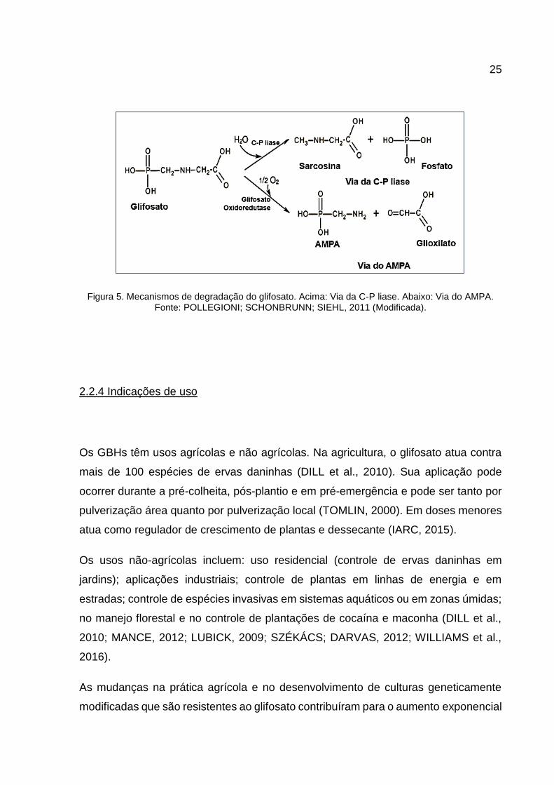
A degradação do glifosato por micro-organismos no solo pode seguir duas rotas
metabólicas (Figura 5). A primeira consiste na transformação do glifosato em
sarcosina por ação da bactéria Agrobacterium radiobacter ou da Enterobacter
aeroneges (enzima C-P liase) (JUNIOR; SANTOS, 2002; POLLEGIONI;
SCHONBRUNN; SIEHL, 2011). A segunda rota resulta na formação do ácido
aminometilfosfônico (AMPA) como resultado da clivagem oxidativa do glifosato pela
enzima GOX, sob a ação da bactéria Anthrobacter atrocyaneus e Flavobacterium sp.
(RIBEIRO et al., 2015, POLLEGIONI; SCHONBRUNN; SIEHL, 2011).
A sarcosina é um metabólito de difícil detecção, já que em solo é rapidamente
degradada pelos micro-organismos. O AMPA é considerado o metabólito principal por
ser mais persistente, podendo se acumular no meio ambiente, e por ser a rota
predominante nas bactérias presentes no solo (JACOB et al., 1988). A meia-vida do
glifosato em solo varia de 2 a 197 dias, e do AMPA de 76 a 240 dias (GIESY;
DOBSON; SOLOMON, 2000).
Nos mamíferos, o glifosato não é metabolizado eficientemente e é, principalmente,
excretado inalterado na urina (MYERS et al., 2016). Em seres humanos, pequenas
quantidades de AMPA foram encontradas no sangue após intoxicação por glifosato,
degradado pela microbiota intestinal (MOTOJYUKU et al., 2008).
25
Figura 5. Mecanismos de degradação do glifosato. Acima: Via da C-P liase. Abaixo: Via do AMPA. Fonte: POLLEGIONI; SCHONBRUNN; SIEHL, 2011 (Modificada).
2.2.4 Indicações de uso
Os GBHs têm usos agrícolas e não agrícolas. Na agricultura, o glifosato atua contra
mais de 100 espécies de ervas daninhas (DILL et al., 2010). Sua aplicação pode
ocorrer durante a pré-colheita, pós-plantio e em pré-emergência e pode ser tanto por
pulverização área quanto por pulverização local (TOMLIN, 2000). Em doses menores
atua como regulador de crescimento de plantas e dessecante (IARC, 2015).
Os usos não-agrícolas incluem: uso residencial (controle de ervas daninhas em
jardins); aplicações industriais; controle de plantas em linhas de energia e em
estradas; controle de espécies invasivas em sistemas aquáticos ou em zonas úmidas;
no manejo florestal e no controle de plantações de cocaína e maconha (DILL et al.,
2010; MANCE, 2012; LUBICK, 2009; SZÉKÁCS; DARVAS, 2012; WILLIAMS et al.,
2016).
As mudanças na prática agrícola e no desenvolvimento de culturas geneticamente
modificadas que são resistentes ao glifosato contribuíram para o aumento exponencial
26
do uso desse herbicida (MYERS et al., 2016).
2.2.5 Avaliações de risco
A IARC classificou o glifosato como "provavelmente cancerígeno para os seres
humanos (Grupo 2A)" em 2015. Esta categoria é usada quando há evidências
limitadas de carcinogenicidade em seres humanos e dados fortes sobre como o
agente causa câncer em animais experimentais (IARC, 2015). De acordo com quatro
painéis de especialistas que realizaram uma crítica detalhada da avaliação do IARC,
as evidências não suportam esta conclusão, sendo consistente que “é improvável que
o glifosato represente um risco carcinogênico para os seres humanos" (WILLIAMS et
al., 2016).
A OMS e a FAO classificam o glifosato como "improvável que represente um risco
carcinogênico para os seres humanos devido à exposição através da dieta (exposição
a alimentos e água)" (FAO/OMS, 2016). De acordo com os estudos avaliados por
essas organizações, a evidência indica que a administração de GBHs em doses tão
altas quanto 2000 mg/kg, a via mais relevante para a exposição dietética humana, não
foi associada a efeitos genotóxicos em uma esmagadora maioria de estudos em
mamíferos (FAO/OMS, 2016).
O CARC classificou o glifosato como "provavelmente não cancerígeno para humanos"
(EPA, 2016). Em novembro de 2015, a Autoridade Europeia para a Segurança dos
Alimentos (EFSA, do inglês European Food Safety Authority) determinou que “era
improvável que o glifosato representasse um risco carcinogênico para os seres
humanos" (EFSA, 2015). A Agência de Proteção Ambiental (EPA, do inglês
Environmental Protection Agency) apoia que o glifosato "provavelmente não é
cancerígeno para os seres humanos" em doses relevantes (EPA, 2016).

27
2.2.6 Legislação brasileira sobre uso de agrotóxicos
A principal lei que regulamenta a produção, comercialização e uso de agrotóxicos é a
“Lei dos Agrotóxicos e Afins” (Lei nº 7.802, de 11 de Julho de 1989) (BRASIL, 1989).
São regidos por essa Lei:
A pesquisa, a experimentação, a produção, a embalagem e rotulagem, o transporte,
o armazenamento, a comercialização, a propaganda comercial, a utilização, a
importação, a exportação, o destino final dos resíduos e embalagens, o registro, a
classificação, o controle, a inspeção e a fiscalização de agrotóxicos, e seus
componentes (BRASIL, 1989).
Essa lei é regulamentada pelo Decreto nº 4.074, de 04 de Janeiro de 2002 (BRASIL,
2002). Segundo esse decreto, as empresas são obrigadas a apresentar ao poder
público relatórios de comercialização dos produtos agrotóxicos, com periodicidade
semestral. O Instituto Brasileiro do Meio Ambiente e dos Recursos Naturais
Renováveis (IBAMA) é o órgão responsável pela divulgação desses relatórios (IBGE,
2015).
É responsabilidade do IBAMA realizar a Avaliação do Potencial de Periculosidade
Ambiental de diversas substâncias químicas, incluindo os agrotóxicos. São avaliadas
as propriedades físico-químicas e a toxicidade para os diversos organismos; o quanto
o produto se acumula em tecidos vivos; se persiste por muito tempo no ambiente; e
se consegue se deslocar (solo, ar ou água). São considerados ainda o risco de causar
mutações, câncer, más-formações em fetos ou embriões, e se podem colocar em risco
a reprodução de aves e de mamíferos (IBAMA, 2010).
Existem 4 classes de periculosidade: Classe I – produto altamente perigoso; Classe II
– produto muito perigoso; Classe III – produto perigoso; e Classe IV – produto pouco
perigoso (IBGE, 2015).
O Roundup Original® é classificado no grupo III, enquanto outros produtos da mesma

28
empresa (Roundup Ready®, Roundup Transorb®, Roundup Ultra®) se enquadram na
classe II (IBAMA, 2010).
Atualmente, existe uma comissão especial da Câmara dos Deputados que analisa 18
projetos que alteram a Lei de Agrotóxicos. Os objetivos desses projetos são simplificar
o processo para registro de pesticidas novos, facilitar o uso de genéricos, alterar o
nome “agrotóxico” para “defensivo fitossanitário”, criar novo órgão federal para
administrar esse tema e diminuir o poder dos estados na fiscalização (BRASIL, 2017).
2.3 Efeitos biológicos do glifosato
Quando iniciou a comercialização do Roundup®, a Monsanto anunciou que o princípio
ativo glifosato era seguro para o meio ambiente e para a saúde humana (incluindo o
agricultor em contato direto) e animal, desde que utilizado de acordo com as
recomendações técnicas e as indicações das bulas e rótulos dos produtos (WILLIAMS
et al., 2000).
A partir da década de 1980, diversos estudos foram realizados buscando a
confirmação das informações divulgadas pela Monsanto através de experimentos de
avaliação de toxicidade, genotoxicidade e carcinogenicidade (VIGFUSSOM; VYSE,
1980; BOLOGNESI et al., 1997; WILLIAMS et al., 2000; KOLLER et al., 2012;
KWIATKOWSKA et al., 2017).
Esses testes foram realizados com diversos tipos de vertebrados e avaliando as
respostas in vitro ao glifosato e Roundup®, e também a exposição in vivo a esses
compostos (TARAZONA et al., 2017).
No estudo de Romano et al. (2012), fêmeas de ratos foram tratadas com água
contendo glifosato (50 mg/kg) entre o dia gestacional 18 e o quinto dia pós-natal, e os
machos foram avaliados 60 dias após o nascimento. Os autores relataram que a

29
exposição materna ao glifosato afetou o desenvolvimento comportamental e
reprodutivo em ratos machos, sendo este resultado relacionado à hipersecreção de
andrógenos. Os resultados incluíram aumentos nas concentrações séricas de
testosterona, estradiol e hormônio luteinizante; na produção de RNAm e proteína na
hipófise; e na produção de esperma, além de puberdade precoce. Esses resultados
foram contestados por DeSesso e Williams (2012) em relação à formulação do GBH,
ausência de grupos de controle adicionais e uso de protocolo não padronizado para
teste de preferência de parceiro sexual.
Resultados opostos aos de Romano et al. foram obtidos por Dallegrave et al. (2007).
Neste estudo, os ratos Wistar foram expostos, via oral, a 50, 150 ou 450 mg/kg de
glifosato durante a gravidez (21-23 dias) e lactação (21 dias). Os principais efeitos
adversos foram observados na prole masculina: redução da contagem de
espermatozoides e sua produção diária em adultos; elevação de espermatozoides
anormais; e diminuição no nível sérico de testosterona na puberdade. Outro estudo
do mesmo grupo avaliou os efeitos teratogênicos de Roundup® na prole de ratos
Winstar. Os animais receberam por via oral 500, 750 ou 1000 mg/kg de glifosato do
6º ao 15º dia gestacional e a cesariana foi feita no 21º dia. A porcentagem de
anormalidades esqueléticas (ausência de ossos ou partes de ossos, ossos dobrados,
assimetria, fusões e fissuras) aumentou com o aumento da dose. Observou-se uma
taxa de mortalidade de 50% em fêmeas gestantes tratadas com 1000 mg/kg de
glifosato (DALLEGRAVE et al., 2003).
No estudo de Winnick e Dzialowski (2013), embriões de frango foram tratados com
glifosato e Roundup® entre os dias 6 a 18. Os embriões expostos a 2% de glifosato e
1% de Roundup® apresentaram taxas aumentadas de mortalidade; diminuição do
embrião, da massa cardíaca e hepática; e redução do tibiotarso e do comprimento do
bico.
Lajmanovich, Sandoval e Peltzer (2003) observaram deformidades craniofaciais e
bucais, anormalidades oculares e caudas curvadas em sapos expostos a glifosato e
GBH. Tanto a mortalidade quanto os defeitos aumentaram com o tempo e a
concentração de herbicidas. Jayawardena et al. (2010) relataram menor sobrevida,
30
metamorfose retardada e altas frequências (69%) de malformações como cifose,
escoliose, úlceras da pele e edema também em sapos.
Em ostras, Akcha, Spagnol e Rouxel (2012) não encontraram efeitos sobre o
desenvolvimento de embriões larvais ou espermatozoides dos animais mantidos em
água do mar contendo glifosato e Roundup® durante 24 h em todas as concentrações
testadas (0,5, 1,0, 1,5, 2,5, 5,0 μg/L). Também não foram detectados efeitos no
desenvolvimento da larva embrionária de ostra em concentrações de glifosato, AMPA
e Roundup® variando de 0,1 a 1000 μg/L, após exposição de 48 h (Mottier et al. 2013).
Em relação à sobrevida, nenhuma larva foi observada acima da concentração de
10.000 μg/L de Roundup®. Apesar disso, não foram relatadas alterações na
mortalidade independentemente das concentrações de glifosato e AMPA.
Peixes-zebra também foram utilizados para estudar os efeitos iniciais da exposição
ao glifosato e Roundup Classic®, diluídos até a concentração de 50 µg/ml de glifosato
(ROY; CARNEIRO; OCHS, 2016). Após 24h, foram encontradas anormalidades
morfológicas (reduções encefálicas e oculares) e perda de ventrículos cerebrais
delineados. Além de diminuições nos genes expressos nas regiões do olho,
prosencéfalo e mesencéfalo.
O efeito de GBH sobre o sistema imunológico de jacarés-de-papo-amarelo foi avaliado
por Siroski et al. (2016). Os répteis foram expostos durante dois meses a diferentes
concentrações do herbicida que foram diminuindo progressivamente para simular a
degradação do glifosato na água. No grupo 1, de 11 mg/L para 2,5 mg/L e no 2, de 21
mg/L para 5 mg/L. Além disso, foi injetada nos animais uma solução de
lipopolisacarídeo (LPS) de Escherichia coli para desencadear uma resposta imune e
avaliar os parâmetros associados a ela. Os resultados mostraram que os animais
expostos ao GBH apresentaram menor atividade do sistema complemento do que os
animais controle, mas não foram observadas diferenças nas células brancas do
sangue.

31
2.4 Efeitos do glifosato em humanos
Como descrito anteriormente, muitas agências de avaliação de risco para a saúde
humana classificam o glifosato como seguro para humanos. Essas avaliações são
baseadas em, principalmente, estudos carcinogênicos com outros animais (MINK et
al., 2012). Entretanto, nas últimas décadas, vários estudos foram publicados utilizando
células humanas ou dados obtidos in vivo, que lançam dúvidas sobre a segurança
dessas substâncias.
2.4.1 Experimentos in vitro
O teste in vivo dos efeitos do herbicida sobre o organismo humano é complexo. Não
é possível realizar experimentos controlados com humanos utilizando agentes
químicos que são tóxicos à saúde. Os modelos animais são utilizados, mas a resposta
pode ser bastante diferente. Portanto, estudos in vitro com células ou linhagens
celulares são essenciais para a compreensão dos efeitos dos herbicidas na saúde
humana.
Não há consenso sobre a exposição de células humanas ao glifosato. As respostas
variam de acordo com o tipo de célula, concentração, fórmula química, tempo de
exposição e metodologia.
O primeiro estudo que avaliou os efeitos sobre o DNA causado por Roundup® em
linfócitos humanos foi realizado por Vigfusson e Vyse, em 1980. Esses autores
sugeriram que este produto químico é, no máximo, deficientemente mutagênico.
Alvarez-Moya et al. (2014) e Mladinic et al. (2009) também realizaram experimentos
com linfócitos humanos. O primeiro estudo indica que o glifosato é genotóxico,
dependendo do tempo e da concentração de uso. Os autores do segundo sugeriram

32
que menores concentrações de glifosato não apresentavam efeitos perigosos sobre o
DNA.
Martinez et al. (2007) compararam a toxicidade do glifosato e Roundup® nas células
mononucleares do sangue periférico humano (PBMCs) e identificaram que as
formulações eram mais citotóxicas do que o componente ativo.
Em 2017, Kwiatkowska et al. observaram uma diminuição no nível global de metilação
e aumento de danos ao DNA em PBMCs quando expostas ao glifosato.
A viabilidade celular das linhagens celulares prostáticas epiteliais normais
imortalizadas utilizadas por Li et al. (2013) não diminuiu quando expostas ao glifosato.
Benachour e Seralini (2009) usaram linhagens de rim embrionário humano, além das
células endoteliais da veia umbilical humana, para avaliar a toxicidade de quatro
GBHs, com concentrações abaixo das recomendações agrícolas e correspondentes
a baixos níveis de resíduos nos alimentos. Todas as formulações causaram morte
celular total dentro de 24 h de incubação devido à inibição da atividade da succinato
desidrogenase mitocondrial. Para os autores, os efeitos deletérios são proporcionais
às concentrações de glifosato e dependem da natureza dos adjuvantes, o que mostra
que os adjuvantes não são inertes.
Devido à facilidade de obtenção de linhagens celulares tumorais e à fácil manipulação
dessas células, muitos estudos avaliam os efeitos do GBH nessas linhagens,
conforme descrito abaixo. Além disso, fornecem informações sobre os possíveis
efeitos do glifosato na progressão da doença.
Li et al. (2013) observaram que o glifosato e AMPA inibiram o crescimento celular em
oito linhagens celulares: quatro de câncer de próstata, duas de câncer de ovário, uma
de câncer cervical e uma de câncer de pulmão.
Efeito oposto foi observado por Kasuba et al. (2017), que observaram proliferação de
células HepG2 quando expostas ao glifosato.
Richard et al. (2005) realizaram um estudo com Roundup® em uma linhagem celular

33
placentária humana derivada de coriocarcinoma e observaram que o GBH reduziu a
viabilidade celular pelo menos duas vezes mais do que o glifosato.
Chaufan, Coalova e Molina (2014) examinaram os efeitos do glifosato, AMPA e GBH
sobre o equilíbrio oxidativo e os endpoints celulares na linhagem HepG2. Observou-
se que a exposição ao glifosato e ao AMPA não afetou a viabilidade celular, enquanto
o GBH induziu uma rápida diminuição.
Desta forma, os dados atualmente disponíveis mostraram que os GBHs são mais
tóxicos do que o próprio componente ativo, apoiando a ideia de que os aditivos nas
formulações comerciais não são inertes e desempenham um papel na toxicidade do
herbicida (CHAUFAN; COALOVA; MOLINA, 2014; COALOVA; MOLINA; CHAUFAN,
2014).
2.4.2 Exposição in vivo: estudos epidemiológicos
O aumento da exposição humana ao GBH é quase certo, considerando o aumento
exponencial do uso desses produtos nas últimas décadas. O impacto do glifosato em
doenças humanas foi avaliado por poucos estudos epidemiológicos. Esses trabalhos
examinaram a associação entre exposições a GBHs e efeitos na saúde em humanos,
incluindo câncer (VANDEMBERG et al., 2017).
O primeiro grande projeto que analisou a associação da exposição a pesticidas e o
risco de câncer em cerca de 26.000 indivíduos (incluindo os trabalhadores diretamente
expostos e suas famílias) foi o The Agricultural Health Study (AHS) (ALAVANJA et al.,
1996).
Em 2003, em um dos estudos desse projeto, não foi encontrada nenhuma associação
entre o uso de glifosato e o risco de câncer de próstata (ALAVANJA et al., 2003). De
Ross et al. (2005) e Sorahan et al. (2015), utilizando os dados do AHS, também não
34
encontraram nenhuma associação entre ao uso de glifosato e o risco de diversos tipos
de câncer.
Da mesma forma, outros estudos realizados com o AHS não encontraram associação
entre o uso de glifosato e câncer colorretal, cólon, câncer retal, pâncreas e melanoma
(LEE et al., 2007; ANDREOTTI et al., 2009; DENNIS et al., 2010).
A associação mais provável entre o uso de glifosato e o risco de câncer foi com
Linfoma não-Hodgkin. Em diferentes populações, diversos estudos, como o de Cantor
et al (1992), De Roos et al. (2003), McDuffie et al. (2008), Eriksson et al. (2008),
sugeriram uma possível associação. Entretanto, nenhum deles conseguiu
efetivamente provar.
Fatores que podem explicar essa ausência de associação entre câncer e glifosato é o
baixo número de indivíduos utilizados nos estudos, bem como o pequeno número de
indivíduos que utilizaram especificamente o glifosato. Muitos estudos são realizados
através de questionários, e dessa forma, a quantificação e a identificação dos tipos
específicos de herbicidas utilizados ficam comprometidas.
O glifosato, além da ingestão através do alimento e da exposição ocupacional, pode
ser inalado do ar. Alguns estudos relacionaram o glifosato como fator de risco para o
desenvolvimento de doenças respiratórias (CHANG; SIMCIK; CAPEL, 2011;
WILLIAMS et al., 2016).
O glifosato foi avaliado como fator de risco para asma em mulheres com asma atópica
(HOPPIN et al., 2008). Em agricultores de Iowa e Carolina do Norte, Slager et al.
(2010) verificaram uma associação entre glifosato e rinite e episódios aumentados de
rinite. Hoppin et al., em 2017, avaliou a associação do glifosato com sibilância alérgica
e não alérgica em agricultores. Os resultados mostraram que o herbicida aumenta o
risco para os dois grupos.
Já para Henneberger et al. (2014), existe uma associação inversa entre a exposição
ao glifosato e o agravamento dos sintomas da asma, pois é possível que os pacientes
com asma possam ser menos sensíveis ao herbicida ou que os indivíduos suscetíveis
35
evitem a exposição enquanto os outros permanecem expostos sem sintomas
adicionais. Por outro lado, Faria et al. (2005) não detectou qualquer relação entre o
glifosato e os sintomas de asma ou doença respiratória crônica em agricultores do
Brasil.
Dentre os estudos epidemiológicos realizados, muitos avaliaram o risco de
desenvolvimento de doenças em trabalhadores diretamente expostos ao glifosato. O
objetivo do estudo de Jauhiainen et al. (1991) foi medir a exposição dos trabalhadores
florestais ao herbicida glifosato durante o trabalho de limpeza feito com serras
equipadas com pulverizadores de herbicidas. Nesse estudo, não foram identificados
problemas de saúde relacionados ao glifosato.
Resultados opostos foram obtidos por Jayasumana et al. (2015), que observaram
aumentos na frequência de doença renal crônica entre agricultores no Sri Lanka.
Diversos problemas de reprodução humana foram associados à utilização de
glifosato. Curtis et al. (1999) identificaram uma diminuição da fertilidade (no mínimo
20%) de mulheres expostas à GBH e Sanin et al. (2009) observaram que mulheres
expostas ao glifosato levaram mais tempo para conceber. Arbuckle, Lin e Mery (2001)
relataram associação entre abortos espontâneos tardios (12-19 semanas) e exposição
ao glifosato preconcepção.
Garry et al. (2002) examinaram a ocorrência de transtornos de nascimento e
desenvolvimento em filhos de agricultores de Minnesota que aplicavam GBH. Os
autores descobriram que a exposição ao glifosato/Roundup® aumenta o risco de
déficit de atenção e transtornos de hiperatividade com déficit de atenção em crianças
de 6 a 14 anos de idade. Curiosamente, ao investigar os níveis de exposição materna
e fetal, Aris e Leblanc (2011) não detectaram o glifosato no sangue total materno nem
no cordão umbilical.
Normalmente, o contato rápido da pele humana com GBH não causa grandes danos.
No entanto, podem ocorrer lesões quando o contato é prolongado. Mariager et al.
(2013) relataram queimaduras graves em uma mulher após prolongada exposição
dérmica acidental (24 h) a GBH. A paciente desenvolveu inchaço local, bolhas e

36
feridas. Também apresentou deficiência neurológica na mão devido a uma condução
nervosa reduzida. Para esses autores, o tempo de exposição prolongado poderia
explicar o dano mais profundo do que o relatado anteriormente aos nervos e músculos.
2.4.3 Intoxicação aguda
A toxicidade aguda do glifosato é classificada como baixa em ratos, sendo que o LD50
oral do glifosato puro é de 5.600 mg/kg (FAO/OMS, 2016). Não existem valores de
LD50 em humanos.
As intoxicações agudas após ingestão de GHBS são observadas em casos de
suicídios ou de acidentes. Os sintomas relacionados à intoxicação são diversos,
variando de leves à mais graves, e muitas vezes levam o indivíduo à morte. Muitos
casos de intoxicação aguda são relatados na literatura.
Potrebić et al. (2009) descreveram um caso de envenenamento de uma mulher de 56
anos que ingeriu cerca de 500 mL de GBH. Os sintomas foram hipotensão,
hipercalemia, insuficiência respiratória e renal. A paciente sobreviveu à fase aguda de
envenenamento, mas desenvolveu um dano maciço no cérebro que levou ao coma e
posteriormente à morte.
Ptok (2009) relatou um caso de uma professora de 26 anos que usou glifosato
corretamente, mas sofreu de disfonia grave após algumas horas. Este problema foi
causado por uma menor mobilidade da dobra vocal, sugerindo comprometimento de
inervação. Os sintomas foram resolvidos espontaneamente 6 semanas depois e a
mobilidade da dobra vocal retornou ao normal.
Malhotra et al. (2010) relataram o caso de um homem de 71 anos que tentou suicídio
com GBH. Ao atendimento, o paciente estava em choque cardiogênico com acidose
metabólica grave e apresentou falta de resposta clínica (> 7 dias). A tomografia

37
computadorizada cerebral foi normal. A leitura de eletroencefalograma no dia 8 pós-
ingestão demonstrou dados consistentes com uma encefalopatia. Ele foi transferido
da unidade de terapia intensiva no dia 10 e recebeu alta no dia 16, com recuperação
clínica completa.
No estudo de Zouaoui et al, (2013) a finalidade foi associar as características clínicas
de pacientes com intoxicação aguda com a concentração de glifosato no sangue e na
urina para prever resultados clínicos. Os autores classificaram 13 casos de
intoxicação por glifosato de acordo com a gravidade dos sintomas clínicos (sendo 3
laudos forenses). Entre os 10 pacientes, 5 tiveram intoxicação leve a moderada, 2
tiveram envenenamento grave e 3 morreram. Os sintomas mais comuns foram a
ulceração orofaríngea (5/10), náuseas e vômitos (3/10), dificuldade respiratória (3/10),
arritmia cardíaca (4/10), insuficiência renal (2/10) e consciência alterada (3/10). Outros
sintomas foram choque cardiovascular, parada cardiorrespiratória, distúrbios
hemodinâmicos, coagulação disseminada intravascular e, em casos fatais, falência
múltipla de órgãos. Em casos de intoxicação leve a moderada e de casos fatais, as
concentrações de glifosato no sangue apresentaram um valor médio de 61 mg/L
(intervalo de 0,6-150 mg/L) e 4146 mg/L (intervalo 690-7480 mg/L), respectivamente.
As estratégias de tratamento para intoxicação por glifosato são principalmente de
suporte, pois não há antídoto. A técnica de irrigação por sonda gástrica oral e a
administração de carvão ativado são métodos que podem ser utilizados para
descontaminação (LEE et al., 2008). Suporte ventilatório e inotrópico e
hemodiafiltração venovenosa contínua também podem ser necessários de acordo o
estado do paciente (MALHOTRA et al., 2010; ZOUAOUI et al., 2013).
2.4.4 Exposição e intoxicação por glifosato no Brasil
Os registros sobre os casos de intoxicação por agrotóxicos no Brasil são obtidos

38
através de dois sistemas: o Sistema Nacional de Informações Tóxico-Farmacológicas
(SINITOX) e o Sistema de Informação de Agravos de Notificação (SINAN). O SINITOX
tem como principal atribuição coordenar a coleta, a compilação, a análise e a
divulgação dos casos de intoxicação e envenenamento notificados no país, e está
vinculado à Fundação Oswaldo Cruz (SINITOX, 2017). O SINAN é alimentado,
principalmente, pela notificação e investigação de casos de doenças e agravos que
constam da lista nacional de doenças de notificação compulsória, sendo facultado aos
estados e municípios incluírem nesse sistema outros problemas de saúde importantes
em sua região (SINAN, 2017).
Segundo os dados disponíveis no SINITOX (2017) mais atualizados, em 2014 foram
registrados no Brasil 2854 casos de intoxicação por uso agrícola de agrotóxico e 2262
por uso doméstico.
Infelizmente, esses dados não representam a realidade do país pois nem todos os
centros estaduais de registro de intoxicação repassam os dados para o sistema
nacional (SINITOX, 2017). Uma outra dificuldade é a falta de diagnóstico e a
subnotificação, pois muitas pessoas não comparecem aos hospitais ou não
identificam a causa da intoxicação (RIGOTTO; VASCONCELOS; ROCHA, 2014).
No Espírito Santo, o Centro de Atendimento Toxicológico do Espírito Santo (TOXCEN)
é o responsável pelo registro dos casos de intoxicação por agrotóxicos. Em 2015,
foram registrados 771 casos de exposição/intoxicação por uso agrícola de agrotóxico
e 35 por uso residencial (TOXCEN, 2015).
Os dados apresentados pelo SINITOX e TOXCEN não especificam o tipo de
agrotóxico ingerido. Dessa forma, não há um registro de intoxicação específico por
GBH. Porém, considerando que o glifosato corresponde a uma grande porcentagem
de todo o agrotóxico vendido, é provável que uma parcela considerável desses casos
sejam por intoxicação por glifosato.
Em relação à intoxicação crônica, não existe um sistema de registro desses casos e
toda a informação disponível sobre isso ocorre através de estudos epidemiológicos
isolados.
39
2.4.5 Detecção de contaminação por glifosato
Existem vários métodos de detecção do glifosato e do AMPA no ar, água, solo, urina
e soro sanguíneo (IARC, 2015). Em humanos, o diagnóstico de contaminação por
glifosato é estabelecido pela ocorrência de quadro clínico compatível com a exposição
e, nos casos de ingestão, confirmado por testes que detectam a presença do herbicida
no corpo.
Yoshioka et al. (2011) desenvolveram um método para detectar glifosato no soro
humano por cromatografia líquida acoplada à espectrometria de massas sequencial
(LC-MS/MS) usando cromatografia líquida com interação hidrofílica (HILIC). O LD
para o glifosato foi de 0,03 μg/mL. Mariager et al. (2013) também utilizaram essa
técnica.
Acquavella et al. (2004) quantificaram o glifosato na urina usando cromatografia
líquida de alta performance (HPLC) com reação pós-coluna e detecção de
fluorescência. Antes do HPLC, o glifosato foi isolado e concentrado através de troca
iônica. Por essa técnica, o LD foi de 1 μg/L. Curvin et al. (2007) também quantificaram
o glifosato na urina, porém utilizaram um imunoensaio de microbola (microbead)
covalente por fluorescência (FCMIA) e o LD foi 0,9 μg/L.
A cromatografia gasosa com espectrômetro de massa (GC-MS) e o ensaio de
imunoabsorção enzimática (ELISA) para detecção de glifosato foram utilizados para
medir a quantidade do herbicida em urina (KRUGER et al., 2014). A detecção por
ELISA foi mais eficiente (mínimo de 0,1 μg/mL e máximo de 71,3 μg/mL) do que a
quantificação por GC-MS (mínimo de 1,0 μg/mL e máximo de 40 μg/mL).
Um outro teste utilizado é a medida do nível da enzima colinesterase. Essa enzima
atua na regulação dos impulsos nervosos por meio da degradação do
neurotransmissor acetilcolina nas sinapses e nas junções neuromusculares
(PEARSON; PATEL, 2016). Existem dois tipos dessa enzima: a acetilcolinesterase
eritrocitária (AChe), que é encontrada nos eritrócitos, no pulmão e no tecido nervoso;

40
e a butirilcolinesterase plasmática (BChE), sintetizada no fígado e que circula pelo
plasma sanguíneo (LOCKRIDGE et al., 2016).
Em casos de intoxicação por organofosforados, como o glifosato, a colinesterase é
inibida e consequentemente ocorre um acúmulo de acetilcolina, resultando em
depressão dos centros respiratórios e circulatórios na medula, fraqueza dos músculos
respiratórios e edema pulmonar (LOCKRIDGE et al., 2016). Entretanto, níveis baixos
dessa enzima também estão presentes em outras situações, como por exemplo,
hepatites, estados de desnutrição, infecções agudas, anemias, infarto do miocárdio e
dermatomiosite (FARIA; FASSA; FACCHINI, 2007).
Utilizando esse método, Oliveira-Silva et al. (2011) conseguiu detectar o glifosato.
Outros estudos não foram bem sucedidos nessa detecção (ETGES et al., 2002; SALVI
et al., 2003).
Além dessas variações na efetividade do teste, essa medida só é indicada para
contatos recentes (até duas semanas). Um outro problema é determinar os limites
aceitáveis da exposição ocupacional para populações expostas continuamente aos
agrotóxicos (FARIA; FASSA; FACCHINI, 2007). Todos esses métodos de avaliação
só detectam exposições mais imediatas ao glifosato e não permitem uma avaliação
de exposição contínua e/ou crônica a esse herbicida.
Até o momento, não existem testes moleculares para detecção da contaminação por
glifosato.
2.5 Expressão gênica alterada por glifosato
As respostas moleculares ao glifosato já foram analisadas para alguns organismos,
como ostra-do-pacífico, peixe-chato europeu, mosquito Aedes e pulga-da-água
(TANGUY et al., 2005; MARCHAND et al., 2006; RIAZ et al., 2009; LE et al., 2010).

41
Em humanos, são poucos os estudos realizados.
Hokanson et al. (2007) avaliaram a expressão gênica em linhagens MCF-7
(adenocarcinoma de mama) expostas ao GBH e identificaram 680 genes
desregulados. Desses, os três principais foram EGR1, HIF1 e CXCL12, que parecem
impactar potencialmente as taxas de início de apoptose e alterar os níveis de
vascularização associados à formação de tumor.
Thongprakaisang et al. (2013) identificou que o glifosato altera a expressão dos genes
dos receptores de estrógeno em linhagens de câncer de mama. Esse resultado
também foi obtido por Mesnage et al. (2017).
Alterações na expressão de proteínas atuantes na fase G1/S do ciclo celular e também
de S100A6/S100A9 (proteínas reguladoras de Ca2+), IP3R1 (receptor responsável por
liberar Ca2+ do retículo endoplasmático) e SOD1 (superóxido dismutase) foram
identificadas por George e Shukla (2013) em células HaCaT (queratinócitos). Essas
alterações em conjunto induziram a proliferação das células e inibição da apoptose.
Estudos que utilizam técnicas que analisam a expressão gênica global das células
expostas ao glifosato são raros. Também não foram identificados os padrões de
expressão gênica em células humanas saudáveis quando expostas ao glifosato.
Dessa forma, pesquisas que identifiquem as alterações a nível molecular são
importantes para determinar em quais processos biológicos o glifosato atuaria através
de modificações no padrão de expressão gênica.

42
2.6 Hipótese
A hipótese testada neste estudo é de que a exposição rápida ao Roundup® e ao
AMPA leva à expressão diferenciada de genes que governam processos celulares,
podendo explicar a toxicidade relatada na literatura.

43
3 OBJETIVOS
3.1 Objetivo geral
Identificar genes diferencialmente expressos em células mononucleares do sangue
periférico (PBMCs) humano submetidas à exposição rápida com herbicida à base de
glifosato (Roundup®) e AMPA.
3.2 Objetivos específicos
Avaliar viabilidade e morte das células submetidas aos tratamentos;
Identificar alterações de expressão gênica nas células expostas ao Roundup®
e AMPA;
Identificar em quais processos biológicos os genes diferencialmente expressos
estão atuando.

44
4 ARTIGOS CIENTÍFICOS DERIVADOS DA TESE
4.1 Manuscrito 1
O manuscrito intitulado “Glyphosate's effects in humans: an evaluation of in vitro and
epidemiological studies” foi submetido para avaliação ao periódico Regulatory
Toxicology and Pharmacology (Qualis Capes B1; Fator de Impacto: 2,221), de acordo
com os critérios estipulados pelo Regimento do Programa de Pós-Graduação em
Biotecnologia (Figura 6).
Figura 6. Comprovação de submissão do manuscrito 1 para a revista Regulatory Toxicology and Pharmacology.
45
Glyphosate's effects in humans: an evaluation of in vitro and epidemiological studies
Authors
Lidiane Pignaton Agostinia,b*, Raquel Spinassé Dettognia,b, Raquel Silva dos Reisa,b, Elaine Stura,b,
Eldamária de Vargas Wolfgramm dos Santosa,b, Diego do Prado Ventorima,b, Fernanda Mariano
Garciaa,b and Iúri Drumond Louroa,b.
Affiliations
a Programa de Pós-graduação em Biotecnologia. Universidade Federal do Espírito Santo, Vitória,
Espírito Santo, Brasil.
b Departamento de Ciências Biológicas-Núcleo de Genética Humana e Molecular. Universidade Federal
do Espírito Santo, Vitória, Espírito Santo, Brasil.
*Corresponding author: Lidiane Pignaton Agostini
Address: avenue Fernando Ferrari, nº 514, Vitória, ES, Brazil, 29075-910.
Phone: 55 27 4009 2324, ramal *5324. FAX: 55 27 4009 2324.
Email: [email protected]

46
Abstract
Glyphosate (N-(phosphonomethyl)glycine) is a post-emergent, non-selective and systemic herbicide,
whose commercial formulations are called glyphosate-based herbicides (GBHs). These herbicides can
be found in soil, air, water and groundwater, as well as in food. Glyphosate is degraded by soil microbes
to carbon dioxide and aminomethylphosphonic acid (AMPA). The aim of this review is describe in vitro
and in vivo studies about human exposure to GBHs. In vitro studies with human cells are essential for
understanding the effects of a chemical on human health, but there is no consensus. The responses
change according to the cell type, chemical's concentration and formula, exposure time and
methodology. Human exposure to GBHs can occur through food, soil, water and air by directly or
indirectly contact at occupational exposure or even at household. The association between glyphosate
and human diseases was evaluated by few epidemiological studies.
Key words: glyphosate-based herbicides, toxicity, risk evaluations, food.
Background
Glyphosate (N-(phosphonomethyl)glycine) [CAS# 1071-83-6] is a post-emergent, non-selective and
systemic herbicide (IARC, 2015; FAO/WHO, 2016). This molecule consists of the amino acid glycine
and a phosphonomethyl moiety (Li et al., 2013). Glyphosate has as trade names: Abundit Extra; Credit;
Xtreme; Glifonox; Glyphogan; Ground-Up; Rodeo; Roundup®; Touchdown; Tragli; Wipe Out;
Yerbimat and others (FCI, 2015). These products can be called GBHs. Roundup® is the most common
GBH and is presented in many formulations. These pesticides can be found in soil, air, surface water
and groundwater, as well as in food (IARC, 2015).
In the GBHs production, glyphosate is as an isopropylamine, ammonium or sodium salt in water soluble
concentrates and water-soluble granules (FAO, 2000). The relevant impurities are formaldehyde
(maximum, 1.3 g/kg), N-nitrosoglyphosate (maximum, 1 mg/kg), Nnitroso-N-phosphonomethylglycine

47
and N-(phosphonomethyl)iminodiacetic acid (PMIDA) (FAO, 2000; Kwiatkowska et al., 2016).
Surfactants (also called adjuvants), most notably polyethyloxylated tallowamine (POEA) and sulfuric
and phosphoric acids, may be added to the product to facilitate uptake by plants, being the type and
concentration characteristic of each formulation (IPCS, 1996; Székács and Darvas, 2012).
Generally, GBHs cause stronger effects than glyphosate itself (Martinez et al., 2007; Mesnage et al.,
2013; Folmar et al,, 1979). This may be due to significant toxicity of adjuvants present in herbicide
preparations (Song et al., 2012). Martinez et al. (2007) showed that cytotoxicity caused by Roundup®
were stronger than induced by glyphosate.
Glyphosate-based herbicides were first synthesized in 1950 as a potential pharmaceutical compound,
but its herbicidal activity was not discovered until it was re-synthesized and tested in 1970, being in use
since 1974 (Székács and Darvas, 2012; Willians et al., 2016).
Use of glyphosate-based herbicides
The GBHs have agricultural and non-agricultural uses. In agriculture, the glyphosate acts against more
than 100 species of weeds and more than 60 of perennial weed plants (Dill et al., 2010). Its application
can occur in pre-harvest, post-planting and in pre-emergence and can be both by spray and aquatic
herbicide (Tomlin, 2000). In smaller doses, it acts as plant-growth regulator and desiccant (IARC, 2015;
FAO/WHO, 2016). Non-agricultural applications include residential and industrial use; control of plants
in power lines and on roads; in forest management and in the control of cocaine and marijuana
plantations (Dill et al., 2010; Mance, 2012; Lubick, 2009; Székács and Darvas, 2012; Williams et al.,
2016). Changes in farming practice and the development of genetically modified crops that are resistant
to glyphosate have contributed to the increase in use (Myers et al., 2016).

48
Glyphosate’s metabolization
Glyphosate competitively inhibits the activity of a key plant enzyme called synthase
5-enolpyruvylshikimate-3-phosphate synthase (EPSPS), present in shikamate pathway and which
participates of chorismate’s production, a precursor for aromatic amino acids using in the synthesis of
a number of pigments as flavonoids and anthocyanins (Mesnage et al., 2015; Kwiatkowska et al., 2016).
This pathway is present in various groups of microorganisms, plants and parasites and is absent in
animals (Richard et al., 2005; Thongprakaisang et al., 2013). The inhibition of the synthesis of aromatic
amino acids stops growth of certain plants (Williams et al., 2016).
Glyphosate is degraded by soil microbes to carbon dioxide and AMPA [CAS# 1066-51-9; Molecular
Formula: CH6NO3P; Molecular Weight: 111.04 g/mol] as a result of oxidative cleavage by glyphosate
oxidoreductase (GOX) (Ribeiro et al., 2015). AMPA can accumulate in the environment (Jacob et al.,
1988). A minor pathway for the degradation of glyphosate in bacteria (Pseudomonas sp. Strain LBr) is
via conversion to glycine (Jacob et al., 1988). In mammals, glyphosate is not metabolized efficiently
and is mainly excreted unchanged into the urine (Myers et al., 2016). Is has suggest that metabolism in
AMPA also occurs in humans because small amounts of this component have been found in the blood
following glyphosate poisoning; however, it may be that this metabolization is carried out by gut
microbial metabolism (Motojyuku et al., 2008). In agreement with this latter suggestion, a study in rats
showed that after oral administration of glyphosate, a small amount of AMPA was detected in the colon
2 hours later, which was attributed to intestinal microbial metabolism (Brewster et al., 1991).
In 2015, the IARC has classified the glyphosate as “probably carcinogenic to humans (Group 2A)”.
According four Expert Panels witch conducted a detailed critique of the IARC evaluation, the evidences
does not support this conclusion of the IARC, being consistent that glyphosate is “unlikely to pose a
carcinogenic risk to humans” (Williams et al., 2016). The WHO and the Food FAO classifies the
glyphosate as “unlikely to pose a carcinogenic risk to humans from exposure through the diet (food and

49
water exposures)” (FAO/WHO, 2016). In January 2018, IARC responded to criticisms of the
Monographs and the glyphosate evaluation, and confirmed risk assessment (IARC, 2018).
Methodology
Articles were selected using PubMed database. There was no selection of publication time. Inclusion
criteria were: utilization of human cells or epidemiological studies; exclusive use of glyphosate/GBH
for in vitro studies or specific association with glyphosate’s effects for epidemiological studies; exposure
to glyphosate alone or GBH.
Effects of glyphosate and Roundup® in vitro
In vivo testing of the herbicide’s effects on human organism is complicated. Animal models can be used,
but the answer may be quite different. Therefore, in vitro studies with cells lines are essential for
understanding the effects of these chemicals on human health.
Healthy human cells
There is no consensus on the exposure of human cells to glyphosate. The responses change according
to the cell type, concentration, GBH’s chemical formulation, exposure time and methodology.
The first study to evaluate the effects on DNA caused by Roundup® in human lymphocytes was
conducted by Vigfusson and Vyse in 1980. They showed significant increases in the Sister-Chromatid
Exchanges (SCE) index upon exposure to Roundup®, but only in high concentrations (65X 10-5 M and
65X 10-4 M). These authors suggested that this chemical is at most weakly mutagenic based on the SCE
test.

50
Alvarez-Moya et al. (2014) and Mladinic et al. (2009a and 2009b) also performed experiments with
human lymphocytes. In the first study, the comet assay was used to examine the genotoxicity of
isopropylamine glyphosate. The DNA damage increased with the glyphosate’s concentration (0.0007,
0.007, 0.07 and 0.7 mM), usually proportionally, with an exposure time of 20h. For authors, this result
indicates that glyphosate is genotoxic, depending on the time and concentration of use.
Mladinic et al. (2009a) evaluated the genotoxic and oxidative potential of glyphosate at concentrations
likely to be encountered in residential (2.91 µg/ml) and occupational (3.50 µg/ml) exposure. The tests
were done with and without metabolic activation. Ferric-reducing ability of plasma (FRAP),
thiobarbituric acid reactive substances (TBARS) and the hOGG1 modified comet assay were used to
measure glyphosate’s oxidative potential and its impact on DNA. Genotoxicity was evaluated by
alkaline comet and analysis of micronuclei and other nuclear instabilities applying centromere probes.
Because of inefficient or incorrect DNA repair, chromosomal damage is expressed during the cell
division and represents an index of accumulated genotoxic effects (Bolognesi et al., 2009). The alkaline
comet assay showed significantly increased tail length (20.39 µm) and intensity (2.19%) for 580 µg/ml,
and increased tail intensity (1.88%) at 92.8 µg/ml. With metabolic activation (S9), tail length was
significantly increased for all concentrations tested: 3.5, 92.8 and 580 µg/ml. Using the hOGG1 comet
assay, a significant increase in tail intensity was observed at 2.91 µg/ml with S9 and 580 µg/ml without
S9. Without S9, the frequency of micronuclei, nuclear buds and nucleoplasmic bridges slightly increased
at concentrations 3.5 µg/ml and higher. The presence of S9 significantly elevated the frequency of
nuclear instabilities only for 580 µg/ml. FRAP values slightly increased only at 580 µg/ml regardless of
metabolic activation, while TBARS values increased significantly. Since for any of the assays applied,
no clear dose-dependent effect was observed, Mladinic et al. indicated that glyphosate in concentrations
relevant to human exposure do not pose significant health risk.
In the third one (Mladinic et al., 2009b), glyphosate’s concentrations and the use or not of metabolic
activation were similar to the previous study and an evaluation of the possible clastogenic and aneugenic
effects was performed. Frequency of the micronuclei, nuclear buds and nucleoplasmic bridges in

51
cultures treated with glyphosate slightly increased from 3.5 µg/ml onward. The presence of metabolic
activation significantly elevated cytome assay parameters only at 580 µg/ml. No concentration-related
increase of centromere (C+) and DAPI (4’,6-diamidino-2-phenylindole) signals (DAPI+) was observed
for glyphosate treatment. As the previous study, Mladinic et al. suggested that lower concentrations of
glyphosate have no hazardous effects on DNA.
Martinez et al. (2007) compared the toxicity of glyphosate and Roundup® on peripheral blood
mononuclear cells (PBMCs). Cells were exposed to different glyphosate’s (1, 50, 100, 200, 300, 400,
500, 1000, 1500 and 2000 μg/ml) or Roundup®’s concentrations (1, 10, 20, 40, 60, 80 and 100 μg/ml)
for 24, 48, 72 and 96 h. Both herbicides were toxic to PBMCs. The Roundup® cytotoxicity was greater
than glyphosate, since the LC50 determined by the trypan blue exclusion method, at 24 h was the
equivalent of 56.4 μg/ml for Roundup® and 1640 μg/ml (1.64 mg/ml) for glyphosate. According
toauthors, this in vitro study confirmed the toxic effects on human cells by glyphosate and its
commercial preparations. Furthermore, they reported that commercial formulations were more cytotoxic
than the active component alone, supporting the concept that additives in commercial formulations play
a role in the toxicity attributed to GBHs.
Kwiatkowska et al., in 2016, performed experiments with PBMCs. Was observed a decrease in PBMCs
viability (2.7%) after 24 h of incubation with glyphosate at 10 mM. A decrease in adenosine-5'-
triphosphate (ATP) level also was observed in cells treated with glyphosate from 5 mM after 24h of
incubation. Measurement of cell morphology revealed changes in PBMCs granularity that occurred after
24 h of incubation with 0.5 mM of glyphosate. The conclusion was that glyphosate caused toxic effects
on PBMCs only at very high concentrations. These concentrations correspond to those resulting from
acute or subacute intoxication with GBH.
In 2017, Kwiatkowska et al. observed a decrease in global DNA methylation level in PBMCs at 0.25
mM of glyphosate for 24 h. Concentrations at 0.25 mM and 0.5 mM increased p53 promoter
methylation, while it did not induce statistically significant changes in methylation of p16 promoter.

52
The study also observed that glyphosate induced DNA damage in PBMCs in the concentrations range
from 0.5 to 10 mM. Moreover, we noticed that PBMCs significantly repaired glyphosate-induced DNA
damage (mainly after 0 min post incubation), but they were unable to repair completely DNA strand-
breaks after 120 min post incubation.
Kwiatkowska et al. (2014a) evaluated the effect of glyphosate on human erythrocytes. Authors noticed
that even with long incubation time up to 24 h did not affect the level of hemolysis and were not observed
morphological changes induced by glyphosate in these cells (0.01–5 mM). Therefore, they concluded
that glyphosate do not have hemolytic properties.
Kwiatkowska et al. (2014b) also performed experiments with erythrocytes to evaluate
acetylcholinesterase (AChE) activity. The results showed that glyphosate (0.25–5 mM),)caused
statistically significant inhibition of AChE activity after 1 h and 4 h of incubation. Glyphosate, in the
concentrations ranging from 0.5 mM to 5 mM for 1h treatment and from 0.25 mM to 5 mM after 4h
incubation, respectively, caused decrease (about 20%) in this parameter. This change in AChE activity
was observed only at high glyphosate’s concentrations (0.25–5 mM), which is resulting only of an acute
poisoning, according to Kwiatkowska et al.
Another study that analyzed erythrocytes (Pieniazek et al., 2004), showed that after 1 h of incubation
the Roundup Ultra 360 SL® increased the level of methemoglobin and the products of lipid peroxidation
at 500 ppm and hemolysis at 1500 ppm. Already, the glyphosate increased the level of methemoglobin
at 1000 ppm and caused hemolysis only after a long time of exposure (24 h) to high doses (1500 ppm).
Both Roundup Ultra 360 SL® and glyphosate did not cause statistically significant changes in the level
of glutathione (GSH), but increased the activity of catalase.
Gehin et al. (2006), Heu et al. (2012) and George and Shukla (2013) investigated the effects of
glyphosate in Human Skin Keratinocytes (HaCaT). Gehin et al. (2006) showed that glyphosate alone
or included in Roundup 3 plus® (0-25 mM), induced significant changes in cellular antioxidant status as
a glutathione depletion (dose dependent depletion, which was more sensitive with Roundup® than with

53
glyphosate), enzymatic (catalase, glutathione-peroxidase and superoxide dismutase) disorders, and
increased lipid peroxidation.
The aim of Heu et al. (2012) was examine glyphosate's cytotoxic effects in intracellular mechanisms of
apoptosis. They conducted different incubation periods. From 6 to 18 h of incubation periods, cytotoxic
profiles were superimposed and presented an LC50 of approximately 30 mM. For shorter incubation
periods (0.5 and 1 h), the LC50 increased to 53 mM. They observed an increase in the number of early
apoptotic cells at a low cytotoxicity level (15%) and then, at 45% cytotoxicity, lot of cells reached not
only early but also late apoptotic state and even a necrotic state. They also showed that the glyphosate-
induced mitochondrial membrane potential disruption could be a cause of apoptosis in keratinocyte
cultures.
George and Shukla (2013) aimed to clarify if the imbalance between [Ca2+] levels and oxidative stress
is associated with glyphosate-induced proliferation in HaCaT cells. The [Ca2+] levels, reactive oxygen
species (ROS) generation and expressions of G1/S cyclins, IP3R1, S100A6, S100A9 and SOD 1 and
apoptosis-related proteins were investigated upon glyphosate exposure in HaCaT cells. Glyphosate (0.1
mM for 72h) significantly induced proliferation, decreases [Ca2+], and increased ROS generation. This
pesticide also enhanced the expression of G1/S phase’s proteins and the cell proliferation. Additionally,
glyphosate also triggers S100A6/S100A9 and decreases IP3R1 and SOD 1 expressions. Notably, Ca2+
suppression also prevented apoptotic related events including Bax/Bcl-2 ratio and caspases activation.
According to authors, this study highlights that glyphosate promotes proliferation in HaCaT cells
probably by disrupting the balance between [Ca2+]𝑖 levels and oxidative stress which in turn facilitated
the down regulation of mitochondrial apoptotic signaling pathways.
The immortalized human normal prostatic epithelial cell lines RWPE-1 and pRNS-1-1 were used by Li
et al. (2013) to evaluate cell viability when exposed to glyphosate. Glyphosate’s concentrations of 15,
25, and 50 mM did not significantly decrease the cell viability, when compared with the untreated
control group.

54
To evaluated human cell toxicity for GBHs and glyphosate alone (concentration 0.1 to 10000 ppm),
Mesnage et al. (2013) measured mitochondrial activities, membrane degradation and caspases 3/7
activities. The chemicals were cytotoxic, inducing similar dose-dependent patterns on the human
embryonic kidney 293 cell line (HEK 293) after 24 h of incubation.
Benachour and Seralini (2009) also used HEK 293, besides Human Umbilical Vein Endothelial Cells
(HUVEC), to evaluate the toxicity of four GBHs, with concentrations below agricultural
recommendations (1-2%) and correspondents to low levels of residues in food. All formulations caused
total cell death within 24 h of incubation, because of the inhibition of the mitochondrial succinate
dehydrogenase activity and also caused necrosis, by release of cytosolic adenylate kinase measuring
membrane damage. The GBHs also induced apoptosis via activation of caspases 3/7 enzymatic activity.
This is confirmed by characteristic DNA fragmentation, nuclear shrinkage (pyknosis) and nuclear
fragmentation (karyorrhexis), which is demonstrated by DAPI in apoptotic round cells. Glyphosate
caused only apoptosis. For the authors, the deleterious effects are proportional to glyphosate’s
concentrations and dependent on the nature of the adjuvants, which confirms that the adjuvants are not
inert.
Tumor cells
Studies with healthy cells are important for assessing the glyphosate’s effects on human health, as
previously discussed. However, due to the facility of obtaining tumor cell lines and the ease of working
with these cells, many studies evaluate GBHs’s effects on tumor lines, as described below. These studies
provide insights into the possible effects of glyphosate on disease progression.
Li et al. (2013) observed that glyphosate and AMPA inhibited cell growth in eight cancer cell lines,
including four prostate cancer cell (C4-2B, LNCaP, DU-145, and PC-3), two ovarian cancer cell lines
(SKOV-3 and OVCAR-3), one cervical cancer HeLa cell line and one lung cancer cell line (A549) at

55
concentrations up to 50 mM. Glyphosate at a concentration of 50 mM decreased 27%, 73.4%, 39.3%,
36.9%, 28%, 58.8%, 25% and 17% the cell viability in the LNCaP, CA-2B, DU-145, PC-3, SKOV-3,
OVCAR-3, HeLa and A549 cell lines, respectively.
Some studies have demonstrated that glyphosate have a disrupting effect on estrogen receptor alpha
(ERα) and beta (ERβ) transcriptional activities. In the study of Thongprakaisang et al. (2013), it was
evaluated estrogenic and/or antiestrogenic effects of glyphosate compared with endogeneous estrogen
(E2) in the estrogen dependent human breast cancer cells T47D, being observed that the herbicide caused
the proliferative effects in T47D of approximately 15–30% in the absence of E2 condition. This study
also demonstrated that glyphosate altered the levels of ERα and ERβ proteins: at 6 h of exposure, it
increased the levels of both ERα and ERβ while at 24 h of exposure, only ERα showed a significant
induction at 10-7M glyphosate concentration.
Richard et al. (2005) realized a study with Roundup® in human placental cell line derived from
choriocarcinoma (JEG3 cells) and observed that GBHs reduced JEG3 cell viability at least twice more
efficiently than glyphosate. This effect increased with time and was obtained with Roundup®’s
concentrations lower than that of the agricultural use. The toxicity increased with time (8-fold at 0.8%
between 24 and 48 h), and the LD50 was approximately 1.8 times lower for Roundup® (0.7%) than for
glyphosate.
In 2012, Koller et al. analysed the cytotoxic and genotoxic properties of glyphosate and Roundup® in
the TR146 cell line, which is derived from a neck metastasis of buccal epithelial origin. In this study
was observed that both products (20 min; 10–20 mg/L), induced strand breaks that led to formation of
comets as well as nuclear anomalies that reflected DNA instability including chromosomal damage;
presence of micronuclei markers; binucleated cells with micronuclei and increase of nuclear buds. The
most sensitive endpoint was micronuclei induction. The treatment of the cells with highest dose of
Roundup® (20 mg/L) caused a threefold increase over the background, and the corresponding
concentration of glyphosate, a weaker effect was seen.

56
Besides these studies, several assays evaluated the impact of glyphosate in the HepG2 human hepatoma
cell line, a pertinent model for xenobiotic actions. Gasnier et al. (2009), studying four different
formulations of Roundup and pure glyphosate, verified that all glyphosate-based formulations, by
contrast to glyphosate alone, induced a rapid decrease in cell viability according to the formulation and
the test, within 24 h only. The R400 formulation caused around 50% of DNA strand breaks at 5ppm, a
sub agricultural dilution. The caspases 3/7 were significantly activated with nontoxic doses of R450 (60
ppm) up to 156% in 24 h. Their levels were considerably enhanced to 765% within 48 h.
In a second study carried out by Gasnier et al. (2010) was observed that in the cell lines, mortality
increased with time and glyphosate concentrations in GBHs (Express® 7.2 g/L; Bioforce® 360 g/L;
GT® 400 g/L; GT+® 450 g/L), however the increase was not proportional to alone glyphosate
concentration. In this way, glyphosate had no toxic action alone under the conditions used in studies.
The direct glyphosate action was most probably amplified by vesicles formed by adjuvants or detergent-
like substances that allowed cell penetration, stability, and probably changed its bioavailability and thus
metabolism (Gasnier et al., 2010).
Kasuba et al. (2017) studied the toxic effects of glyphosate on HepG2 cells exposed for 4 and 24 h in
low concentrations (0.5 μg/mL - acceptable daily intake; 2.91 μg/mL - residential exposure level and
3.5 μg/mL - occupational exposure level). The results obtained indicate that, at the tested concentrations,
the herbicide stimulated cell proliferation at both exposure times. However, the effect was more
pronounced after 4 h of exposure (8-9%). It seems that low concentrations effectively stimulated cell
proliferation, especially in inherently unstable and proliferative tumor cells. In the comet assay analysis,
it was consistently observed that HepG2 cells differed in the DNA contained in their nuclei, which also
indicated an impact on cell proliferation. However, this study did not confirm DNA-damaging effects
of glyphosate when tested as a pure active compound. The level of ROS did not change significantly
after 4 and 24h treatments. Apoptosis and necrosis effect were not seen.

57
Chaufan et al. (2014) examined the effects on oxidative balance and cellular endpoints of glyphosate,
AMPA and a GBH in HepG2 cell line. It was observed that glyphosate and AMPA exposure did not
affect cell viability until 1000 mg/L, while GBH treatment induced a rapid decrease in cell viability
depending on concentration and on duration of exposure. The ROS production was investigated and
neither glyphosate nor AMPA treatment caused differences in ROS formation. In relation to cell death,
it was observed that when cells were exposed to GBH, 23.5% of the nuclei showed a condensed and
fragmented chromatin, while it was not modified by glyphosate alone or AMPA. In this way, this was
verified that GBH induced dose-dependent cytotoxicity, while they were not observed toxic effects with
glyphosate and AMPA at assayed concentrations. Therefore, this indicated that GBHs have adjuvants
that, together with the active ingredient, caused toxic effects not observed with glyphosate itself.
In a study realized by Coalova et al. (2014), analyzing the influence of the spray adjuvant on the toxicity
effects of a glyphosate formulation in HEp-2 cell line, was observed that all the agrochemicals assayed
(glyphosate formulation, spray adjuvant and mixture) induced a decrease in cell viability depending on
concentration and time exposure. Mixture (spray adjuvant with glyphosate formulation) resulted in
significantly higher toxicity to cell cultures compared to each agrochemical alone suggesting additive
effect. ROS production was investigated and was verified that glyphosate formulation and mixture
treatment showed significantly higher levels of ROS (139% and 116% respectively). In relation to
apoptotic cell death involving caspase 3/7 activation, was observed that these enzymes were
significantly activated with LC20 concentration of all agrochemicals, what could be indicating that
oxidative stress triggers caspase activation. Morphological changes occurred only with glyphosate
formulation treatment. In this way, the currently available data showed that GBHs are more toxic than
the active component itself, supporting the idea that additives in commercial formulations play a role in
herbicide’s toxicity (Chaufan et al., 2014; Coalova et al., 2014).
Effects of glyphosate and Roundup® in vivo

58
The human exposure to GHBs is increasing following the exponential rise of use of these products in
the last decades. Both glyphosate and AMPA can be relatively persistent in the environment, which may
result in a wide range of ecological risks (Bai and Ogbourne, 2016). Humans exposure to these chemicals
can occur through food, soil, water, and air by directly or indirectly contact at occupational exposure or
even at household (IARC, 2015).Glyphosate’s impact on human diseases was evaluated by few human
epidemiology studies. These works had examinated the association between exposures to GBHs and
effects in human health, including cancer (Vandenberg et al., 2017).
Cancer
Alavanja et al. (1996) described the first big project, the Agricultural Health Study (AHS), to analyze
the association of exposure to pesticides and the cancer risk. About 26.000 individuals, including the
pesticides applicators and their families, composed the cohort. With this group, many studies were
published.
In 2003, Alavanja et al. examined the association between pesticide use and risk of prostate cancer and
found a correlation with the joint use of all pesticide analyzed (OR= 1.14, 95% IC: 1.05, 1.24). However,
when analyzed only the use of glyphosate, was not found any association.
De Ross et al. (2005), using the AHS database, had studied all cancers described in that population and
found no risk associated with glyphosate use, but the authors suggested an association (without
significance) with multiple myeloma (RR=1.0; 95% IC: 0.9–1.2). Due this result, Sorahan et al. (2015)
reanalyzed the same data with more complete information to prove the association. Again, was not found
any significance (RR= 1.12; 95% IC: 0.50- 2.49).
Lee et al. (2007) studied the association of glyphosate’s exposure with risk of colorectal cancer (OR=
1.2, 95% IC: 0.9-1.6.), colon cancer (OR= 1.0, 95% IC: 0.7-1.5) and rectal cancer (OR= 1.6, 95% IC=
0.9-2.9). Andreotti et al. (2009) studied cases of pancreas cancer (OR= 1.1; 95% IC: 0.6-1.7). Dennis et

59
al. (2010) studied melanoma (data not showed), but as well as all others studies done with AHS, did not
found any association between the use of glyphosate and cancer. The groups suggested a possible
correlation, but the fact was never proved.
Currently, the most plausible association of glyphosate and cancer was with Non Hodgkin Lymphoma.
Many studies as Cantor et al. (1992) (OR= 1.1; 95% IC: 0.7-1.9), De Roos et al. (2003) (OR= 1.6; 95%
IC: 0.9- 2.8), McDuffie et al. (2001) (OR= 1.20; 95% IC: 0.83–1.74), Eriksson et al. (2008) (OR= 1.51;
95% IC: 0.77–2.94) studied this cancer. All these groups evaluated different populations and in all cases,
suggested a possible association, but none found a statistically significant association.
In 1998, Nordstrom et al. published a study realized at Sweden, where were analyzed 121 cases of hairy
cell leukemia (HCL). An association was found between the exposure to any herbicides and the
increased risk of HCL, but when the model was adjusted, the effect disappear (OR=1.8; 95% IC: 0.7-
4.6). For glyphosate, was not found association (OR= 3.1; 95%IC: 0.8-12).
Lee et al. (2004) analyzed the effect of glyphosate on stomach and esophagus cancer. For both cases,
was not found association (OR= 0.8.; 95% IC: 0.4-1.5 and OR= 0.7; 95% IC: 0.3-1.4, respectively).
It is very important to highlight that the number of subjects is usually very low when the count includes
solely those who are exclusively exposed to glyphosate. Even with large sampling, it is complicate to
estimate the exactly number of individuals who have used glyphosate alone to allow a perfect correlation
between exposure and cancer development.
Respiratory diseases
Beyond the food and occupational exposure, glyphosate can be inhaled from the air. Even considering
100% absorption at maximum concentration, it’s important to note that the exposure is about five times
60
smaller than the systemic acceptable daily intake proposed by European Food Safety Authority (EFSA)
(Chang et al., 2011; Williams et al., 2016).
Hoppin et al. (2008) assessed glyphosate as risk factor for asthma in two groups: women with atopic
asthma and those with nonatopic asthma. Glyphosate were significantly associated with atopic asthma
(OR= 1.31; 95% CI= 1.02–1.67). Henneberger et al. (2014) found an inverse association between
glyphosate exposition and asthma symptoms exacerbation. For authors it is possible that patients with
asthma may be less sensitive to herbicide or that susceptible individuals to exacerbation avoid exposure
while the others remain exposed without additional symptoms. Faria et al. (2005) didn’t detect any
relationship between glyphosate and asthma symptoms or chronic respiratory disease in farmers from
Brazil.
In a posterior work, Hoppin et al. (2017) examinated the association of glyphosate with allergic and
non-allergic wheeze in male farmers. The results evidenced an exposure–response relationship in which
the herbicide increases the risk to both allergic status (OR = 1.56; 95% CI = 1.19–2.03 for allergic and
OR = 1.24; CI = 1.07–1.44 for non-allergic).
While studying the pesticide exposition as predictor of rhinitis in farmers from Iowa and North Carolina,
Slager et al. (2010) verified an association between glyphosate and current rhinitis and increased rhinitis
episodes (OR = 1.09; 95% CI = 1.05–1.13). Except Faria et al. (2005) all cited studies used data from
AHS cohort study.
Others diseases or health problems
Jayasumana et al. (2015) observed increases in the frequency of chronic kidney disease among farmers
in Sri Lanka. In the multivariable analysis the highest risk for this disease was observed among
participants who used glyphosate (OR 5.12, 95% CI 2.33-11.26) as a pesticide.
61
A decrease of fertility (at minimum by 20%) was associated with women exposure to GBHs (Curtis et
al., 1999). Differences in the time to get pregnant in populations exposed to GBHs from aerial spraying
for the control of coca plants were noticed between Colombia's different regions. Reduced fecundability
in some regions was not associated with glyphosate's spraying. However, women in Valle del Cauca, a
sugar-cane region with a history of use of glyphosate and others chemicals for more than 30 years, took
longer to conceive (OR =0.15; 95% CI: 0.12-0.18), (Sanin et al., 2009).
Bolognesi et al. (2009) also develop a study in these Colombia regions. A cytogenetic biomonitoring
study was carried out in subjects from five regions, characterized by different exposure to glyphosate
and other pesticides. Compared with Santa Marta (where organic coffee is grown without pesticides),
the baseline frequency of binucleated cells with micronuclei (BNMN) was significantly greater in
subjects from the other four regions, which means that these cells presented bigger defects in
cytokinesis. The highest frequency of BNMN was in Boyacá (no aerial spraying of glyphosate) and in
Valle del Cauca. There was no association between self-reported direct contact with sprays and
frequency of BNMN. Four months after spraying, a statistically significant decrease in the mean
frequency of BNMN, compared with the sampling of 5 days after spraying, was observed in Nariño, but
not in Putumayo and Valle del Cauca. For these authors, these data suggest that genotoxic damage
associated with glyphosate spraying as evidenced by micronuclei test is small and appears to be
transient.
Arbuckle et al. (2001) reported an association between late spontaneous abortions (12–19 weeks) and
preconception exposure to glyphosate (OR = 1.7; 95% CI 1.0–2.9). Interestingly, when investigating
maternal and fetus exposure levels, Aris and Leblanc (2011) reported no detectable serum glyphosate in
maternal whole blood nor umbilical cord.
There are rare studies that evaluated the association between neurological diseases and GBHs’s use.
Garry et al. (2002) examinated the birth and developmental disorders occurrence in farm families. The

62
authors found that glyphosate/Roundup® exposition increases the risk of attention deficit and attention-
deficit hyperactivity disorders in 6-14 years old children (OR = 3.6; 95% CI 1.35–9.65).
Usually, rapid human skin contact with GBHs does not cause large lesions. Nevertheless, lesions can
occur when the contact is prolonged. Mariager et al. (2013) reported severe burns in a woman after
prolonged accidental dermal exposure (24 h) to a GBH. The patient developed local swelling, bullae
and exuding wounds. Neurological impairment followed affecting finger flexion and sensation with
reduced nerve conduction. For these authors, long exposure time could explain the deeper symptoms
than previously reported damage to nerves and muscles.
Acute Intoxication
Acute intoxications are observed in suicidal or accidental cases after ingesting GBHs. Symptoms, severe
outcomes and fatalities related to intoxication are descript below.
Potrebić et al. (2009) described a case report poisoning of a 56-year old woman ingested about 500 mL
of herbicide containing glyphosate isopropylamine salt. The most prominent manifestations of
poisoning included hypotension, hyperkaliemia, respiratory and renal failure. The patient survived the
acute phase of poisoning, but she developed massive white matter damage that led to vigil coma and
lethal outcome. A similar outcome was presented by Chang et al. (2009), which reported the case of a
57-year-old woman who was admitted unconscious to the hospital after ingestion of GBHs (suicide
attempt).
Ptok (2009) related a case of a 26-year-old teacher who used glyphosate correctly but suffered from
severe dysphonia after some hours. This problem was caused by decreased vocal fold mobility,
suggesting innervation impairment. Symptoms resolved spontaneously 6 weeks later and vocal fold
mobility returned to normal.
63
Malhotra et al. (2010) reported a 71-year-old male who attempted suicide with a GBH. The patient
suffered cardiogenic shock with severe metabolic acidosis and demonstrated prolonged clinical
unresponsiveness (>7 days). Computed Tomography brain was normal. Electroencephalogram reading
on day 8 demonstrated generalized slow wave activity and slow wave complexes consistent with an
encephalopathy. He was transferred from the intensive care unit on day 10 and discharged home on day
16 with full clinical recovery.
The purpose of Zouaoui et al. (2013) was to describe the clinical feature and determinate the utility of
the glyphosate’s concentrations in blood and urine and the dose taken for predicting clinical outcomes.
They classified 13 glyphosate poisoning cases by intoxication severity using clinical criteria (10 was
clinical observations and 3 are forensic cases). Among the 10 patients, 5 had mild to moderate poisoning,
2 had severe poisoning and 3 died. Most common symptoms were oropharyngeal ulceration (5/10),
nausea and vomiting (3/10), respiratory distress (3/10), cardiac arrhythmia (4/10), impaired renal
function (2/10) and altered consciousness (3/10). Other symptoms were cardiovascular shock,
cardiorespiratory arrest, hemodynamic disturbance, intravascular disseminated coagulation and multiple
organ failure in fatalities. In mild–moderate intoxication and fatal cases, blood glyphosate
concentrations had a mean value of 61 mg/L (range 0.6–150 mg/L) and 4146 mg/L (range 690–7480
mg/L), respectively.
The aim of Lee et al. (2008) study was to establish an early prognostic model of patients with GBH
intoxication. Variables as age, sex, estimated amount of ingestion, symptoms/signs including first vital
signs, chest x-ray, and biochemical studies (assays for serum urea nitrogen, creatinine, alanine
aminotransferase, sodium, potassium) from 58 patients (19 men and 39 women) were analyzed.
Seventeen patients died. Five variables (respiratory distress needing intubation, metabolic acidosis,
tachycardia, elevated creatinine level and hyperkalemia) were found to be highly associated with poor
outcome and mortality. The conclusion of the authors was that GBHs poisoning induced multiorgan
toxicity and that pulmonary and renal toxicity seems to be responsible for its mortality. Useful
64
prognostic factors for predicting GBH’s mortality was metabolic acidosis, abnormal chest x-ray,
tachycardia and elevated creatinine level.
Many associations between glyphosate’s concentrations found in human health remain indeterminate,
without robust epidemiological and biomonitoring studies. These studies are essential to improve
conclusions about the GBHs’s safety.
Risk of glyphosate’s presence in food
Humans are indirectly exposed to glyphosate through food, making worry the amount of glyphosate
residues present in meat (and/or animals derived products), fruits and vegetables. There is also
concern about the risks from this type of glyphosate exposure (Van Eenennaam and Young, 2017).
According to the Food and Agriculture Organization of the United Nations (FAO), glyphosate’s residues
amount in food must obey the dietary daily intake and the maximum residue limits (MRL), which range
between 0.025 – 2 mg/kg among food types. Meat, bean and milk present a MRL of 0.05, 2 and 0.05
mg/kg, respectively. MRL is above 30 mg/kg for some cereals (including rice, wheat and oat) (FAO,
2016). Furthermore, Williams et al. (2000) suggested in their study a theoretical maximum value
intake for glyphosate of 0.238 mg/kg body weight/day (adults) through the food .
The EFSA showed data glyphosate’s residues concentration in different body parts of cattle and pig.
No samples presented values above acceptable MRL (0.05 mg/kg) (EFSA, 2015). Granby et al. (2003)
analyzed glyphosate’s concentration in different cereals among 1998-2001. All samples (50 per year)
showed glyphosate residual values lower than acceptable limits.
Reddy et al. (2008) analyzed glyphosate’s concentrations in glyphosate resistant (GR) and non-GR
corns. Results showed that non-GR corn presents a higher glyphosate concentration than GR-corn (0.871
and 0.308 mg/kg respectively), however, both values are under MRL (5 mg/kg). Arregui et al. (2003)

65
evaluated the presence of glyphosate residues in leaves, stems and grains of GR-soybean. Values ranged
0.1 – 4.4 mg/kg, bellow the MRL (5 mg/kg). However, some studies found that foods produced from
genetically modified glyphosate resistant crops showed a higher residue concentrations compared with
non-genetically modified crops (Bohn et al., 2014).
Chhapekar et al. (2014) showed that a transgenic variety of rice confers tolerance to glyphosate
applications, and this kind of rice could tolerate 1% of Roundup® commercial, which is 5-fold more
than that used to kill weeds under normal conditions. This data intensifies the concern with the
indiscriminate use of glyphosate without the due concern of the residues accumulation in foods.
USDA GYPSA laboratory analyzed 300 soybeans samples and was detected 13 kinds of pesticides
including glyphosate. Among these samples, 271 (90.3%) was positive for glyphosate’s residues with
levels ranging from 0.26 mg/kg – 18.5 mg/kg (USDA, 2013).
Kruger et al. (2014) tested urine samples of humans, dairy cows, hares and rabbits. Cows kept in
genetically modified free area had significantly lower glyphosate concentrations in urine than
conventional husbandry cows (p<0.001). Moreover, glyphosate concentration was significantly higher
(p<0.0002) in urine of people consuming conventional food than in urine of people consuming
predominantly organic food. In addition, chronically ill humans showed significantly higher glyphosate
residues in urine than in the healthy population (p=0.03). Niemann et al. (2015) also evaluated
glyphosate residues in human urine. Glyphosate was regularly found in urine at levels corresponding to
a dietary daily intake of around 0.1-3.3 µg/kg body weight/day.
McGuire et al. (2016) evaluated the glyphosate concentration in maternal milk. None of samples was
positive for glyphosate presence in lactating women. Thus, these authors suggest that dietary glyphosate
exposure is not a health concern for breastfed infants.
On the other hand, McQueen et al. (2012) evaluated the presence of glyphosate’s residues in foods
consumed by 40 mothers. Although the presence of glyphosate in 75% of foods was confirmed, the total

66
concentration was 0.4% of the acceptable daily intake. MRL value is measure to analytical methods
rather than on toxicology methods, thus, available MRLs may not necessarily suggest a safe level of a
pesticide residue in food (Bai and Ogbourne, 2016).
Although international scientists and regulatory agencies still maintain the glyphosate as unlikely to be
carcinogenic through food intake (JMPR, 2016), there are few studies about a low chronic exposure to
glyphosate through food consumption. Thus cannot be discarded a potential risk to human health. The
role of glyphosate exposition via food remains poorly explained, being necessary more studies that seek
to achieve the potential of chronic glyphosate toxicity.
Conclusion
This review shows that many in vitro studies associate glyphosate with genotoxic and cytotoxic effects.
On the other hand, many studies have not found such relationships, especially the epidemiological
studies. Most agencies that assess the risk of exposure to glyphosate do not consider it to be carcinogen
to humans. Despite this, there are no studies evaluating the chronic effects after decades of exposure.
Thus, it is essential that robust epidemiological studies be conducted to confirm the assumed safety of
glyphosate in relation to human health.
Acknowledgements
First author was supported by Fundação de Amparo à Pesquisa do Estado do Espírito Santo (FAPES)
with a study grant.
References

67
ALAVANJA, M. C. et al. The Agricultural Health Study. Environ Health Perspect, v. 104, n. 4, p.
362-369, 1996.
ALAVANJA, M. C. et al. Use of agricultural pesticides and prostate cancer risk in the Agricultural
Health Study cohort. Am J Epidemiol, v. 157, n. 9, p. 800-814, 2003.
ALVAREZ-MOYA, C. et al. Comparison of the in vivo and in vitro genotoxicity of glyphosate
isopropylamine salt in three different organisms. Genetics and Molecular Biology, v. 37, n. 1, p. 105-
110, 2014.
ANDREOTTI, G. et al. Agricultural pesticide use and pancreatic cancer risk in the Agricultural Health
Study Cohort. Int J Cancer, v. 124, n. 10, p. 2495-500, 2009.
ARBUCKLE, T. E. et al. An exploratory analysis of the effect of pesticide exposure on the risk of
spontaneous abortion in an ontario farm population. Environ Health Perspect, v. 109, n. 8, p. 851–857,
2001.
ARREGUI, M. C. et al. Monitoring glyphosate residues in transgenic glyphosate-resistant soybean. Pest
Manag Sci, v. 60, p. 163–166, doi: 10.1002/ps.775, 2003.
BAI, S. H.; OGBOURNE, S. M. Glyphosate: environmental contamination, toxicity and potential risks
to human health via food contamination. Environ Sci Pollut Res, doi:10.1007/s11356-016-7425-3,
2016.
BENACHOUR, N.; SERALINI, G-E. Glyphosate Formulations Induce Apoptosis and Necrosis in
Human Umbilical, Embryonic, and Placental Cells. Chem. Res. Toxicol, v. 22, p. 97–105, 2009.
BOHN, T. et al. Primicerio. Compositional differences in soybeans on the market: Glyphosate
accumulates in Roundup® Ready GM soybeans. Food Chemistry, v. 153, p. 207–215, 2014.

68
BOLOGNESI, C. et al. Biomonitoring of Genotoxic Risk in Agricultural Workers from Five Colombian
Regions: Association to Occupational Exposure to Glyphosate. Journal of Toxicology and
Environmental Health, Part A, v. 72, n. 15-16, p. 986-997, doi: 10.1080/15287390902929741, 2009.
BREWSTER, D. W. et al. Metabolism of glyphosate in Sprague-Dawley rats: tissue distribution,
identifcation, and quantitation of glyphosate-derived materials following a single oral dose. Fundam
Appl Toxicol, v. 17, n. 1, p. 43–51, doi:10.1016/0272-0590(91)90237-X PMID:1916078, 1991.
CANTOR, K. P. et al. Pesticides and other agricultural risk factors for non-Hodgkin's lymphoma among
men in Iowa and Minnesota. Cancer Res, v. 52, n. 9, p. 2447-55, 1992.
CHANG, C-B.; CHANG, C-C. Refractory cardiopulmonary failure after glyphosate surfactant
intoxication: a case report. Journal of Occupational Medicine and Toxicology, v. 4, n. 2,
doi:10.1186/1745-6673-4-2, 2009.
CHANG, F. C.; SIMCIK, M. F.; CAPEL, P. D. 2011. Occurrence and fate of the herbicide glyphosate
and its degradate aminomethylphosphonic acid in the atmosphere. Environ Toxicol Chem, v. 30, p.
548–555, 2011.
CHAUFAN, G. et al. Glyphosate commercial formulation causes cytotoxicity, oxidative effects, and
apoptosis on human cells: differences with its active ingredient. Int J Toxicol, v. 33, n. 1, p. 29-38. doi:
10.1177/1091581813517906, 2014.
CHHAPEKAR, S. et al. Transgenic rice expressing a codon-modified synthetic CP4-EPSPS confers
tolerance to broad-spectrum herbicide, glyphosate. Plant Cell Rep, doi: 10.1007/s00299-014-1732-2,
2014.
COALOVA, I. et al. Influence of the spray adjuvant on the toxicity effects of a glyphosate formulation.
Toxicol In Vitro, v. 28, n. 7, p. 1306-1311, doi: 10.1016/j.tiv.2014.06.014, 2014.

69
CURTIS, K. M. et al. The Effect of Pesticide Exposure on Time to Pregnancy. Epidemology, v. 10, n.
2, 1999.
DE ROOS, A. J. et al. Cancer incidence among glyphosate-exposed pesticide applicators in the
Agricultural Health Study. Environ Health Perspect, v. 113, n. 1, p. 49-54, 2005.
DE ROOS, A. J. et al. Integrative assessment of multiple pesticides as risk factors for non-Hodgkin's
lymphoma among men. Occup Environ Med, v. 60, n. 9, p. E11, 2003.
DENNIS, L. K. et al. Pesticide use and cutaneous melanoma in pesticide applicators in the agricultural
heath study. Environ Health Perspect, v. 118, n. 6, p. 812-7, 2010.
DILL, G. M. et al. Chapter 1: Glyphosate: discovery, development, applications, and properties. In:
NANDULA, V. K. editor. Glyphosate resistance in crops and weeds: history, development, and
management. Wiley, Hoboken (NJ), p. 1–33, 2010.
EFSA (European Food Safety Authority). Conclusion on pesticide peer review of the pesticide risk
assessment of the active substance glyphosate EFSA Journal, v. 13, n. 11, p.4302, 2015.
ERIKSSON, M. et al. Pesticide exposure as risk factor for non-Hodgkin lymphoma including
histopathological subgroup analysis. Int J Cancer, v. 123, n. 7, p. 1657-63, 2008.
FAO (Food And Agriculture Organization of the United Nations). Glyphosate, N-
(phosphonomethyl)glycine. Specifcations and evaluations for plant protection products. Rome: Food
and Agriculture Organizationof the United Nations. 2000. http://www.fao. IARC MonogRAphs – 112
84 org/fleadmin/templates/agphome/documents/Pests_ Pesticides/Specs/glypho01.pdf (accessed 28
July 2015).
FAO/WHO (Food And Agriculture Organization of the United Nations / World Health Organization).
Pesticide Residues in Food 2016. Special Session of The Joint FAO/WHO Meeting on Pesticide
Residues. Report 2016. FAO Plant Production and Protection Paper 227. ISSN 2070-2515. Rome.

70
https://www.google.com.br/url?sa=t&rct=j&q=&esrc=s&source=web&cd=1&ved=0ahUKEwiYspybn
TWAhWEEZAKHTP0AdgQFggqMAA&url=http%3A%2F%2Fwww.fao.org%2F3%2Fa-
i5693e.pdf&usg=AOvVaw1f2DsK27Dz_BVvGhedj9v6 (accessed 2 October 2017).
FARIA, N. M. et al. Pesticides and respiratory symptoms among farmers. Rev Saude Publica, v. 39, n.
6, p. 973-981, 2005.
FCI (Farm Chemicals International) Glyphosate. In: Crop Protection Database. Willoughby (OH):
Meister Media Worldwide. 2015.
http://www.farmchemicalsinternational.com/cropprotectiondatabase/#/product/detail/203900/
(accessed 2 October 2017).
FOLMAR, L. C. et al. Toxicity of the herbicide glyphosphate and several of its formulations to fish and
aquatic invertebrates. Arch Environ Contam Toxicol, v. 8, n. 3, p. 269–278, 1979.
GARRY, V. F.et al. Birth defects, season of conception, and sex of children born to pesticide applicators
living in the Red River Valley of Minnesota, USA. Environ Health Perspect, v. 110, n. 3, p. 441–449,
2002.
GASNIER, C. et al. Dig1 protects against cell death provoked by glyphosate-based herbicides in human
liver cell lines. J Occup Med Toxicol, v. 5, n. 29, doi: 10.1186/1745-6673-5-29, 2010.
GASNIER, C. et al. Glyphosate-based herbicides are toxic and endocrine disruptors in human cell lines.
Toxicology, v. 262, n. 3, p. 184-191, doi: 10.1016/j.tox.2009.06.006, 2009.
GEHIN, A. et al. Glyphosate-induced antioxidant imbalance in HaCaT: The protective effect of
Vitamins C and E. Environmental Toxicology and Pharmacology, v. 22, p. 27–34,
doi:10.1016/j.etap.2005.11.003, 2006.

71
GEORGE, J.; SHUKLA, Y. Emptying of Intracellular Calcium Pool and Oxidative Stress Imbalance
Are Associated with the Glyphosate-Induced Proliferation in Human Skin Keratinocytes HaCaT Cells.
ISRN Dermatology, v. 2013, p. 1-12, 2013.
GRANBY, K. et al. Analysis of glyphosate residues in cereals using liquid chromatography-mass
spectrometry (LC-MS/MS). Food Additives and Contaminants, v. 20, n. 8, p. 692-698, 2003.
HENNEBERGER, P. K. et al. Exacerbation of symptoms in agricultural pesticide applicators with
asthma. Int Arch Occup Environ Health, v. 87, n. 4, p. 423-32, doi: 10.1007/s00420-013-0881-x,
2014.
HEU, C. et al. Glyphosate-induced stiffening of HaCaT keratinocytes, a Peak Force Tapping study on
living cells. Journal of Structural Biology, v. 178, p. 1–7, doi:10.1016/j.jsb.2012.02.007, 2012.
HOPPIN, J. A. el al. Pesticides and atopic and nonatopic asthma among farm women in the agricultural
health study. Am J Respir Crit Care Med, v. 177, n. 1, p. 11-18, 2008.
HOPPIN, J. A. el al. Pesticides are associated with allergic and non-allergic wheeze among male
farmers. Environ Health Perspect, v. 125, n. 4, p. 535-543, doi: 10.1289/EHP315, 2017.
IARC (International Agency for Research on Cancer). Glyphosate. In: Some organophosphate
insecticides and herbicides: diazinon, glyphosate, malathion, parathion, tetrachlorvinphos. IARC
Monographs on the Evaluation of Carcinogen Risks to Humans. (March 2015). Lyon (France). v. 112,
p. 1–92. http://mono graphs.iarc.fr/ENG/Monographs/vol112/index.php (accessed 9 October 2017).
IARC (International Agency for Research on Cancer). IARC response to criticisms of the Monographs
and the glyphosate evaluation. Prepared by the IARC Director. (January 2018). p. 1-10.
http://www.iarc.fr/en/media-
centre/iarcnews/pdf/IARC_response_to_criticisms_of_the_Monographs_and_the_glyphosate_evaluati
on.pdf. (accessed 10 April 2018).

72
IPCS (International Programme on Chemical Safety). Glyphosate. WHO/FAO Data Sheets on
Pesticides, No. 91 (WHO/PCS/DS/96.91). 1996. World Health Organization. http://apps.who.
int/iris/handle/10665/63290 (accessed 4 October 2017).
JACOB, G. S. et al. Metabolism of glyphosate in Pseudomonas sp. strain LBr. Appl Environ Microbiol,
v. 54, n. 12, p. 2953–2958, PMID:3223761, 1988.
JAYASUMANA, C. et al. Drinking well water and occupational exposure to Herbicides is associated
with chronic kidney disease, in Padavi-Sripura, Sri Lanka. Environmental Health, v. 14, n. 6,
doi:10.1186/1476-069X-14-6, 2015.
JMPR (Joint FAO/WHO Meeting on Pesticide Residues). 2016. Summary Report, Geneva, Switzerland,
2016. http://www.who.int/foodsafety/jmprsummary2016.pdf?ua=1 (accessed 5 September 2017).
KASUBA, V. et al. Effects of low doses of glyphosate on DNA damage, cell proliferation and oxidative
stress in the HepG2 cell line. Environ Sci Pollut Res Int,, doi: 10.1007/s11356-017-9438-y, 2017.
KOLLER, V. J. et al. Cytotoxic and DNA-damaging properties of glyphosate and Roundup® in human-
derived buccal epithelial cells. Arch Toxicol, v. 86, n. 5, p 805-813, doi: 10.1007/s00204-012-0804-8,
2012.
KRUGER, M. et al. Detection of Glyphosate Residues in Animals and Humans. J Environ Anal
Toxicol, v. 4, n. 210, doi: 10.4172/2161-0525.1000210, 2014.
KWIATKOWSKA, M. et al. DNA damage and methylation induced by glyphosate in human peripheral
blood mononuclear cells (in vitro study). Food and Chemical Toxicology, v. 105, p 93-98, 2017.
KWIATKOWSKA, M. et al. The effect of glyphosate, its metabolites and impurities on erythrocyte
acetylcholinesterase activity. Environmental Toxicology and Pharmacology, v. 37, p. 1101–1108,
2014b.

73
KWIATKOWSKA, M. et al. The effect of metabolites and impurities of glyphosate on human
erythrocytes (in vitro). Pesticide Biochemistry and Physiology, v. 109, p. 34–43, 2014a.
KWIATKOWSKA, M. et al. The Impact of Glyphosate, Its Metabolites and Impurities on Viability,
ATP Level and Morphological changes in Human Peripheral Blood Mononuclear Cells. PLoS ONE, v.
11, n. 6, e0156946, doi:10.1371/ journal.pone.0156946, 2016.
LEE, C-H. et al. The early prognostic factors of glyphosate-surfactant intoxication. American Journal
of Emergency Medicine, v. 26, p. 275–281. doi:10.1016/j.ajem.2007.05.011, 2008.
LEE, W. J. et al. Pesticide use and colorectal cancer risk in the Agricultural Health Study. Int J Cancer,
v. 121, n. 2, p. 339-346, 2007.
LEE, W. J. et al. Agricultural pesticide use and adenocarcinomas of the stomach and oesophagus. Occup
Environ Med, v. 61, p. 743–749, doi: 10.1136/oem.2003.011858, 2004.
LI, Q. et al. Glyphosate and AMPA inhibit cancer cell growth through inhibiting intracellular glycine
synthesis. Drug Des Devel Ther, v. 24, n. 7, p. 635-643, doi: 10.2147/DDDT.S49197, 2013.
LUBICK, N. Environmental impact of cocaine strategy assessed [News. Nature, Published online 12
November, doi:10.1038/news.2009.1080, 2009.
MALHOTRA, R. C. et al. Glyphosate–surfactant herbicide-induced reversible encephalopathy. Case
Reports. Journal of Clinical Neuroscience, v. 17, p. 1472–1473, doi:10.1016/j.jocn.2010.02.026,
2010.
MANCE, D. The great glyphosate debate. Northern Woodlands [online magazine]. 2012.
http://northernwoodlands.org/articles/article/ the-great-glyphosate-debate (accessed 28 July 2017).
MARIAGER, T. P. et al. Severe adverse effects related to dermal exposure to a glyphosate-surfactant
herbicide. Clinical Toxicology, v. 51, p. 111–113, doi: 10.3109/15563650.2013.763951, 2013.

74
MARTINEZ, A. et al. Citotoxicidad del glifosato em células mononucleares de sangre periférica
humana. Biomedica, v. 27, p. 594–604, PMID: 18320126, 2007.
MCDUFFIE, H. H. et al. Non-Hodgkin's lymphoma and specific pesticide exposures in men: cross-
Canada study of pesticides and health. Cancer Epidemiol Biomarkers Prev, v. 10, n. 11, p. 1155-1163,
2001.
MCGUIRE, M. K. et al. Glyphosate and aminomethylphosphonic acid are not detectable in human milk.
Am J Clin Nutr, doi:10.3945/ajcn.115.126854, 2016.
MCQUEEN, H. et al. Estimating maternal and prenatal exposure to glyphosate in the community setting.
International Journal of Hygiene and Environmental Health, v. 215, p. 570– 576,
doi:10.1016/j.ijheh.2011.12.002, 2012.
MESNAGE, R. et al. Ethoxylated adjuvants of glyphosate-based herbicides are active principles of
human cell toxicity. Toxicology, v. 313, p. 122–128., doi: 10.1016/j.tox.2012.09.006 PMID: 23000283,
2013.
MESNAGE, R. et al. Review Potential toxic effects of glyphosate and its commercial formulations
below regulatory limits. Food and Chemical Toxicology, v. 84, p. 133–153, doi:
10.1016/j.fct.2015.08.012 PMID: 26282372, 2015.
MLADINIC, M. et al. Evaluation of Genome Damage and Its Relation to Oxidative Stress Induced by
Glyphosate in Human Lymphocytes in Vitro. Environmental and MolecularMutagenesis, v. 50, p.
800-807, doi: 10.1002/em.20495, 2009a.
MLADINIC, M.; PERKOVIC, P.; ZELJEZIC, D. Characterization of chromatin instabilities induced
by glyphosate, terbuthylazine and carbofuran using cytome FISH assay. Toxicology Letters, v. 189, p.
130–137, doi:10.1016/j.toxlet.2009.05.012, 2009b.

75
MOTOJYUKU, M. et al. Determination of glyphosate, glyphosate metabolites, and glufosinate in
human serum by gas chromatography-mass spectrometry. J Chromatogr B Analyt Technol Biomed
Life Sci, v. 875, n. 2, p.509–514, doi:10.1016/j.jchromb.2008.10.003 PMID:18945648, 2008.
MYERS, J. A. et al. Concerns over use of glyphosate-based herbicides and risks associated with
exposures: a consensus statement. Environmental Health, v. 15, n. 19, doi:10.1186/s12940-016-0117-
0, 2016.
NIEMANN, L. et al. A critical review of glyphosate findings in human urine samples and comparison
with the exposure of operators and consumers. J. Verbr. Lebensm, v. 10, p. 3–12, doi:10.1007/s00003-
014-0927-3, 2015.
NORDSTROM, M. et al. Occupational exposures, animal exposure and smoking as risk factors for
hairy cell leukaemia evaluated in a case-control study. Br J Cancer, v. 77, n. 11, p. 2048-2052, 1998.
PIENIAZEK, D. et al. Comparison of the effect of Roundup® Ultra 360 SL pesticide and its active
compound glyphosate on human erythrocytes. Pesticide Biochemistry and Physiology, v. 79, p. 58–
63, doi:10.1016/j.pestbp.2004.03.003, 2004.
PTOK, M. Dysphonia following glyphosate exposition. HNO, v. 57, n. 11, p.1197-1202, doi:
10.1007/s00106-009-1962-8, 2009.
REDDY, K. N. et al. Aminomethylphosphonic Acid Accumulation in Plant Species Treated with
Glyphosate. J. Agric. Food Chem, v. 56, p. 2125–2130, 2008.
RIBEIRO, D. N. et al. Possible glyphosate tolerance mechanism in pitted morningglory (Ipomoea
lacunosa L.). Journal of agricultural and food chemistry, v. 63, n. 6, p. 1689–1697, doi:
10.1021/jf5055722 PMID: 25625294, 2015.
RICHARD, S. et al. Differential effects of glyphosate and Roundup®® on human placental cells and
aromatase. Environmental Health Perspectives, v. 113, n. 6, p. 716–720, 2005.
76
SANIN, L-H. et al. Regional Differences in Time to Pregnancy Among Fertile Women from Five
Colombian Regions with Different use of Glyphosate. Journal of Toxicology and Environmental
Health. Part A, v. 72, p. 949–960, doi: 10.1080/15287390902929691, 2009.
SLAGER, R. E. et al. Rhinitis associated with pesticide use among private pesticide applicators in the
agricultural health study. J Toxicol Environ Health A, v. 73, n. 20, p. 1382-1393, doi: 10.1080, 2010.
SONG, H. et al. In vitro cytotoxic effect of glyphosate mixture containing surfactants. Journal of
Korean Medical Science, v. 27, p. 711–71, doi: 10.3346/jkms.2012.27.7.711 PMID: 22787363, 2012.
SORAHAN, T. Multiple myeloma and glyphosate use: a re-analysis of US Agricultural Health Study
(AHS) data. Int J Environ Res Public Health, v. 12, n. 2, p. 1548-1559, 2015.
SZÉKÁCS, A.; DARVAS, B. Forty years with glyphosate. In: MNAE-G, H. editor. Herbicides –
properties, synthesis and control of weeds. InTech, Croatia,. p. 247–284, 2012.
(http://cdn.intechweb.org/ pdfs/25624.pdf (accessed 28 July 2017).
THONGPRAKAISANG, S. et al. Glyphosate induces human breast cancer cells growth via estrogen
receptors. Food and Chemical Toxicology, v. 59, p. 129–136, doi: 10.1016/j.fct.2013.05.057, 2013.
TOMLIN, C. D. S. The pesticide manual: a world compendium. Croydon: British Crop Protection
Council, 12th ed, 2000. http://trove.nla.gov.au/work/6273016. (accessed 28 July 2017).
VAN EENENNAAM, L.; YOUNG, A. E. Detection of dietary DNA, protein, and glyphosate in meat,
milk, and eggs. J. Anim. Sci, v. 95, p. 3247–3269, doi:10.2527/jas2016.1346, 2017.
VANDENBERG, L. N. et al. Is it time to reassess current safety standards for glyphosate-based
herbicides? J Epidemiol Community Health, v. 71, p. 613–618, doi:10.1136/jech-2016-208463, 2017.
VIGFUSSON, N. V.; VYSE, E. R. The effect of the pesticides, dexon, captan and roundup, on sister-
chromatid exchanges in human lymphocytes in vitro. Mutation Research, v. 79, p. 53—57, 1980.
77
WILLIAMS, G. M. et al. A review of the carcinogenic potential of glyphosate by four independent
expert panels and comparison to the IARC assessment. Critical Reviews in Toxicology, v. 46, n. 1, p.
3-20, doi: 10.1080/10408444.2016.1214677, 2016.
WILLIAMS, G. M. et al. Safety Evaluation and Risk Assessment of the Herbicide Roundup® and Its
Active Ingredient, Glyphosate, for Humans. Regulatory Toxicology and Pharmacology, v. 31, p. 117–
165, doi:10.1006/rtph.1999.1371, 2000.
ZOUAOUI, K. et al. Determination of glyphosate and AMPA in blood and urine from humans: About
13 cases of acute intoxication. Forensic Science International, v. 226, p. 20–25. 2013.

78
4.2 Manuscrito 2
O manuscrito intitulado “Gene expression profiling in PBMCs treated at low doses and
short-time exposure to glyphosate-based herbicides” foi submetido para avaliação ao
periódico International Journal of Hygiene and Environmental Health (Qualis Capes
A2; Fator de Impacto: 4,643), de acordo com os critérios estipulados pelo Regimento
do Programa de Pós-Graduação em Biotecnologia (Figura 7).
Figura 7. Comprovação de submissão do manuscrito 2 para a revista International Journal of Hygiene and Environmental Health.

79
GENE EXPRESSION PROFILING IN PBMCS TREATED AT LOW DOSES AND
SHORT-TIME EXPOSURE TO GLYPHOSATE-BASED HERBICIDES
Lidiane Pignaton Agostini1,2, Elaine Stur1,2, Andrés Felipe Aristizabal-Pachon3,4, Kamila
Chagas Peronni3,4, Sabine J. Waigel5, Wilson Araujo Silva Junior3,4, Iúri Drumond Louro1,2
(1) Programa de Pós-graduação em Biotecnologia. Universidade Federal do Espírito Santo,
Vitória, Espírito Santo, Brasil.
(2) Departamento de Ciências Biológicas-Núcleo de Genética Humana e Molecular.
Universidade Federal do Espírito Santo, Vitória, Espírito Santo, Brasil.
(3) Department of Genetics at Ribeirão Preto Medical School, and Center for Medical
Genomics - HCRP, University of São Paulo, Ribeirão Preto, São Paulo, Brazil.
(4) National Institute of Science and Technology in Stem Cell and Cell Therapy and Center for
Cell-Based Therapy, Ribeirão Preto, São Paulo, Brazil.
(5) Molecular Targets Program, JG Brown Cancer Center, University of Louisville, Louisville,
Kentucky.
Corresponding author: Lidiane Pignaton Agostini
Address: avenue Fernando Ferrari, nº 514, Vitória, ES, Brazil, 29075-910.
Phone: 55 27 4009 2324, ramal *5324. FAX: 55 27 4009 2324.

80
Email: [email protected]
ACKNOWLEDGMENTS
Agostini, L.P. was supported by FAPES (Fundação de Amparo a Pesquisa do Espírito Santo).
Part of this work was performed with assistance of the UofL Genomics Facility, which is
supported by NIH P20GM103436 (KY IDeA Networks of Biomedical Research Excellence),
NIH P30GM106396 (UofL J. G. Brown Cancer Center Phase III CoBRE), the J. G. Brown
Foundation, and user fees.
ABSTRACT
Glyphosate is a post-emergent, non-selective and systemic herbicide. Roundup® is the most
common glyphosate-based herbicides (GBHs). The priority route of glyphosate’s degradation
in soil results in aminomethylphosphonic acid (AMPA). However, the effect of glyphosate on
gene expression pattern in healthy cells remains unknown. Therefore, we aimed identify
differentially expressed genes (DEGs) in human peripheral blood mononuclear cells (PBMCs)
submitted to a short-time exposure with glyphosate-based herbicide (Roundup®) and AMPA,
using microarray technique for gene expression profiling. There were identified 26 DEGs after
Roundup® exposure (3h; 0.05%) and 5 DEGs after AMPA treatment (3h; 10 mM). The
enrichment analysis of DEGS after Roundup® treatment showed association with 33 cellular
processes. Pathview web was used to identify the effect of DEGs in different pathways. Only
genes differentially expressed in Roundup® treatment were included in the pathways. TNF,
LTA, TAB2 and ATM genes are related to NF-kappa B signaling pathway; SESN3 and ATM
genes to p53 signaling pathway; and TNF, BCL2L11 and ATM genes to apoptosis. Our results

81
suggest that Roundup® change the expression of genes associated with cell cycle control,
regulation of cellular process and apoptosis.
Key words: GeneChip Human Transcriptome Arrays 2.0; cell cycle; herbicide; regulation of
cell processes; peripheral blood mononuclear cells; GBH; AMPA.
INTRODUCTION
Glyphosate (N- (phosphonomethyl) glycine) is a broad spectrum, post-emergent, non-selective
and systemic herbicide (IARC, 2015; MYERS et al., 2016). Commercial formulations add
surfactants to improve efficiency (VANDENBERG et al., 2017). These products are known as
glyphosate-based herbicides (GBHs), being the Roundup® the most common (IARC, 2015).
The priority route of degradation of glyphosate by microorganisms in the soil results in the
formation of aminomethylphosphonic acid (AMPA) (RIBEIRO et al., 2015). In mammals,
glyphosate is not metabolized efficiently and is mainly excreted unchanged in the urine
(MYERS et al., 2016).
Molecular responses to glyphosate have already been analyzed for some organisms, like pacific
oyster, european flounder, mosquito, Daphnia magna (TANGUY et al., 2005; MARCHAND et
al., 2006; RIAZ et al., 2009; LE et al., 2010). In humans, there are few studies.
Hokanson et al. (2007) evaluated gene expression in MCF-7 cell lines exposed to GBH and
identified 680 deregulated genes. HIF1, CXCL12 and EGR1 were significantly dysregulated by
glyphosate. According authors, altered EGR1 levels in response to glyphosate salts are less
clear than for HIF1 and CXCL12, but appear to potentially impact rates of apoptosis initiation
and alter the levels of vascularization associated with tumor formation.

82
Thongprakaisang et al. (2013) identified that glyphosate alters the expression of estrogen
receptor genes in breast cancer cell lines. Futhermore, for the authors the additive estrogenic
effects of glyphosate may pose a risk of breast cancer because of their potential additive
estrogenicity.
Mesnage et al. (2017) also evaluated the estrogenic potential of glyphosate in in breast cancer
cell lines. Their results reveals that glyphosate activates ERalpha in breast cancer cells but only
at relatively high concentrations, and that this activation is through a ligand-independent
pathway.
George and Shukla (2013) identified that glyphosate changed expression pattern of proteins
acting on the G1/S phase of cell cycle, also triggers S100A6/S100A9 expression and decreases
IP3R1 and SOD 1 expressions in HaCaT cells. For authors, their study highlights that
glyphosate promotes proliferation in HaCaT cells probably by disrupting the balance in between
[Ca2+]𝑖 levels and oxidative stress which in turn facilitated the downregulation of
mitochondrial apoptotic signaling pathways.
Glyphosate’ effects on gene expression pattern in healthy cells are poorly understood.
Therefore, we propose identify differentially expressed genes in human peripheral blood
mononuclear cells submitted to short exposure with glyphosate-based herbicide (Roundup®)
and AMPA.
MATERIAL AND METHODS
Chemicals reagents
83
The commercial formulation of the herbicide glyphosate (N-phosphonomethyl-glycine)
Roundup Original® [glyphosate 41% (360 g/L), POEA ≅ 15%, Monsanto] were considered in
this study as 100% and, for the accomplishment of the experiments, it was diluted with water.
Aminomethylphosphonic acid (AMPA) [99% (CAS # 1066-51-9, Sigma-Aldrich] was also
diluted in water for the experiments. 3-(4,5-dimethylthiazol-2-yl)-2,5-diphenyl tetrazolium
bromide (MTT) [Sigma Aldrich] and dimethyl sulfoxide (DMSO) [Sigma Aldrich] were used
in cell viability experiments.
Human peripheral blood mononuclear cells isolation and cell culture conditions
PBMCs were isolated from peripheral blood obtained from 3 healthy and non-smoking
volunteers (2 female and 1 male; 28-33 years old), who do not have direct contact with
herbicides. Blood were diluted with PBS (1:1). Cells were isolated by density gradient using
Ficoll® Paque Plus (GE Healthcare). After centrifugation at 800G for 20 min, PBMCs were
collected and PBS was added. The tube was centrifuged at 600G for 10 min and then the
supernatant was discarded. This step was repeated. Pellet was resuspended and PBMCs were
cultured in RPMI 1640 (Gibco-Invitrogen Life Technology) and supplemented with 10% Fetal
Bovine Serum (FBS), 1% penicillin-streptomycin, phytohemagglutinin (1ug/ml) and
interleukin 2 (300U/mL). Cells were maintained at 37 °C with 95% air and 5% CO2. PBMCs
were cultured for 60 h before being used in the experiments.
Cell viability MTT Assay
84
MTT reagent assay were used to evaluated cell growth and viability. 3.5 x 10⁴ cells were added
in each well (96-well microplates), in triplicates. PBMCs were treated with different
concentrations of Roundup® (0.01, 0.05, 0.1, 0.15; 0.2; e 0.3%) and AMPA (0.01, 0.05, 0.5,
1.0, 5.0 e 10 mM). After 3h, 15h, 24 and 48h of incubation, 0.5mg/mL of MTT was added into
each well. DMSO was added after 3h to dissolve the precipitated dye and 48h later, color
alterations were measured using FLUOstar Omega® (BGM LabTech) at 570 nm. Statistical
analysis was performed using one - way variance analysis (ANOVA), followed by Bonferroni
(post hoc) test to evaluate the differences between groups. Significance level considered was p
<0.05. Treatments were compared in relation to the control (CNT).
RNA Extraction
After treatment with Roundup® at 0.05% (1.1mM of glyphosate) and AMPA at 10mM for 3
hours, total RNA was extracted from PBMCs using RNeasy Mini Kit (Qiagen) according to
manufacturer’s protocols. RNA purity and quality was evaluated using a NanoDrop 2000
(ThermoFisher Scientific) and Agilent Bioanalyzer 2100 (Agilent Technologies), respectively.
RNA integrity number (RIN) > 7 were considered for microarray analysis.
Microarray and data analysis
GeneChip Human Transcriptome Arrays 2.0 (HTA 2.0 - Affymetrix) is a highest resolution
microarray for gene expression profiling of all transcript isoforms. Covered more than 245,000
coding transcripts, 40,000 non-coding transcripts and 339,000 probe sets covering exon-exon
junctions (Friess et al. 2017). RNA was analyzed using HTA 2.0 according to the

85
manufacturer’s instructions. Eight chips were used: 3 for controls, 3 for AMPA (10 mM) and 2
for Roundup® (0.05%).
The data were evaluated using the Partek Genomics Suite v 6.6 (Partek Inc., Louis, MO) and
to assess the samples’s distribution, Principal Component Analysis (PCA) was applied. Pre-
processing of Affymetrix CEL-files was performed using the robust multi-chip analysis (RMA)
algorithm, which performs background adjustment, quartile normalization and probe
summarization. Differential expression analysis was realized using a two-way ANOVA. p-
value <0.05 was considered as significant for the biological and molecular function analyses.
Up and down-regulated genes were identified using a fold-change of 1.5.
An enrichment analysis (EA) was performed using MetaCore (GeneGo™, Thomson Reuters,
NY), where genes with altered expression were mapped to Gene Ontology (GO). GO
annotations were used as indicators of biological functions. Pathview (LUO et al. 2017) was
used to construct the maps of different cellular pathways.
Validation of critical differential expressed gene
The quantitative real-time PCR (qPCR) was used to confirm the Microarray data. RNA from
the samples was used for the reverse-transcription reaction according to SuperScript™ IV
VILO™ Master Mix with ezDNase enzyme (Invitrogen). PCR reactions were carried out using
PowerUp™ SYBR™ Green Master Mix (Applied Biosystems®) on 7500 Fast Real-time PCR
System (Applied Biosystems®). Primers of the selected genes are listed in Table 1. The relative
quantification was performed using the 2–ΔΔCt method (LIVAK; SCHMITTGEN, 2001).
GAPDH was used as a reference gene.

86
RESULTS
Cell viability
MTT analysis is shown in Figure 1. Survival rate was dose-dependent for both treatments
(Figure 1). Cell death was less than 30% for all conditions. Roundup® at 0.01 and 0.05%
showed similar viability after 15, 24 and 48 h. In general, cell viability reduced more with a
bigger exposure times to Roundup®. Roundup® at 0.30% after 48 hours caused more cell death
(~30%) than others concentrations and times. The small level of cell death were with 24h
exposure to AMPA. AMPA at 10mM after 3 hours caused highest cell death.
Differential gene expression profile
There were 26 differentially expressed genes (DEGs) identified after Roundup® exposure. 21
genes were up regulated (13 protein coding, 5 RNA genes and 3 pseudogenes; Table 2). Top
10 up regulated DEGs were NFE2L3, TXK, MIR548L, RNU6-82P, MIR4439, OR2J2, TAB2,
LRRC37A4P, LOC727896 and SESN3. Five genes were down regulated (Table 3): 4 protein
coding (RHOU, TNF, LTA and FOSB) and 1 RNA gene (HIF1A-AS2).
After AMPA treatment, there were 5 DEGs (2 protein coding, 2 RNA genes and 1 pseudogene;
Table 4). Combining both treatments, 2 DEGs were common (HIF1A-AS2 and LRRC37A4P).
Roundup® exposure lead to a different gene expression patterns when compared to AMPA
exposure and control (hierarchical distribution of the top 500 DEGs; Figure 2).
Enrichment and pathway analysis

87
DEGs after Roundup® treatment showed association with 33 Gene Ontology (GO) cellular
processes. Table 5 present the top 10 GO cellular processes. DEGs are mainly related to the
regulation of several processes: cell communication, signaling and signal transduction. Also,
were related to positive regulation of cellular metabolic process, macromolecule metabolic
process, cellular process, signal transduction and metabolic process. No significant results were
obtained for AMPA treatment.
Pathview web (https://pathview.uncc.edu/analysis) was used to identify the effect of DEGs in
different pathways (LUO et al., 2017). Only genes differentially expressed in Roundup®
treatment were included in the pathways. TNF, LTA, TAB2 and ATM genes are related to NF-
kappa B signaling pathway (Figure 3). Nuclear factor-kappa B (NF-kappa B) is the generic
name of a family of transcription factors that function as dimers and regulate genes involved in
immunity, inflammation and cell survival (LIU et al, 2017; KEGG, 2018). This pathway is
activated by various intra- and extra-cellular stimuli such as cytokines, oxidant-free radicals,
ultraviolet irradiation, and bacterial or viral products. Canonical pathway is induced by Tumor
Necrosis Factor-alpha (TNF-alpha) and the non-canonical pathway is triggered by particular
members of the TNFR superfamily, such as lymphotoxin-beta (; LIU et al, 2017; ZHANG et
al., 2017).
BCL2L11 and ATM genes are related to FoxO signaling pathway (Figure 4). The forkhead box
O (FOXO) family of transcription factors regulates the expression of genes in cellular
physiological events including apoptosis, cell-cycle control, cellular differentiation, cell
proliferation, glucose metabolism, DNA damage and repair oxidative stress resistance and
longevity (FARHAN et al., 2017; KEGG, 2018).
SESN3 and ATM genes are related to p53 signaling pathway (Figures 5). The p53 signaling
pathway is an important via that control cell cycle. p53 activation is induced by DNA damage,
88
oxidative stress and activated oncogenes. This protein is a transcriptional activator of p53-
regulated genes related to cell cycle arrest, cellular senescence or apoptosis (MIRZAYANS et
al., 2017; SLATTERY et al., 2018).
TNF, BCL2L11 and ATM genes are related to apoptosis (Figure 6). Apoptosis is a genetically
programmed process for the elimination of damaged or redundant cells by activation of
caspases. The 'extrinsic' pathway involves stimulation of members of the tumor necrosis factor
(TNF) receptor subfamily by their specific ligands, such as TNF-alpha (ICHIM; TAIT, 2016;
CAMPBELL; TAIT, 2018). TNF family of ligands activates anti-apoptotic or cell-survival
signals as well as apoptotic signals (CORREIA et al., 2015; KEGG, 2018). The 'intrinsic'
pathway is engaged by a wide array of stimuli that are sensed intracellularly, including cytokine
deprivation, DNA damage, endoplasmic reticulum stress, metabolic stress, UV radiation and
growth-factor deprivation (ICHIM; TAIT, 2016).
Quantitative real time PCR
Five DEGs were selected to be validated. ATM, SESN3 and BCL2L11 were chosen because
were related to Pathview pathways. NFE2L3 and TXK because they presented highest fold-
change. All of the genes found to be similarly up- or down-regulated by both methods. Three
genes (ATM, BCL2L11 and TXK) confirmed microarray results (fold-change > 1.5) (Table 6).
DISCUSSION
89
Several studies showed that glyphosate is toxic to PBMCs, depending of the exposure time and
concentration (MARTINEZ et al., 2007; ALVAREZ-MOYA et al., 2014; KWIATKOWSKA
et al., 2016).
Martinez et al. (2007) compared the toxicity of glyphosate and Roundup® on peripheral blood
mononuclear cells (PBMCs). Cells were exposed to different glyphosate’s (1-2000 μg/ml) or
Roundup®’s concentrations (1-100 μg/ml) for 24, 48, 72 and 96 h. Both herbicides were toxic
to PBMCs. Alvarez-Moya et al. (2014) performed experiments with human lymphocytes. The
comet assay was used to examine the genotoxicity of glyphosate. The DNA damage increased
with the glyphosate’s concentration (0.0007-0.7 mM), usually proportionally, with an exposure
time of 20h. Ours MTT results showed similar results. Survival rate was dose-dependent for
AMPA and Roundup® treatments; however, cell death was less than 30% for all conditions.
Kwiatkowska et al. (2016) showed a less decrease of cell viability. Authors observed a 2.7%
decrease in PBMCs viability after 24h of incubation with glyphosate at 10 mM. The conclusion
was that glyphosate caused toxic effects on PBMCs only at very high concentrations.
This is the first work that evaluated the effects of Roundup® and AMPA on global gene
expression in PBMCs. Our results suggest that Roundup® change expression pattern of a several
genes associated with cell cycle control, regulation of several cellular processes and apoptosis,
which can explain glyphosate effects describes in literature.
ATM (ATM Serine/Threonine Kinase) belongs to the PI3/PI4-kinase family and is an important
cell cycle checkpoint kinase. ATM plays a critical role in the response to DNA double strand
breaks by phosphorylating a large number of downstream substrates that are involved in DNA
repair, cell arrest, chromatin remodeling and apoptosis (DING et al., 2017). ATM were up

90
regulated after Roundup® exposure, which could indicate that these herbicide induced cellular
stress.
BCL2L11 encodes a BH3-only protein named BIM. This protein, a key regulator of pro-
apoptosis, is released from the cytoskeleton after activation by cytotoxic signaling, and is then
translocated to the mitochondria, where it results in apoptosis (LEI; DAVIS, 2003). BIM has
been shown to be critical for apoptosis in B and T lymphocytes, macrophages and granulocytes
(STRASSER, 2005). Bim also leads to uncoupling of mitochondrial respiration and the
subsequent increase in the cellular levels of reactive oxygen species (ROS) (SIONOV et al.,
2015). Upregulation of BH3-only proteins can occur at transcriptional/post-translational levels
in response to stress to trigger cell death (CAMPBELL; TAIT, 2018). Our results showed that
BCL2L11 is up regulated after Roundup® treatment, which could leadto apoptosis as showed
in MTT results.
Micro RNAs (miRNAs) are small non-coding RNAs (~ 20–24 nucleotides in length) that are
involved in the post transcriptional control of gene expression (ABDI et al., 2017).
Approximately, 60% or more of human protein coding genes may be subject to regulation by
miRNAs (FRIEDMAN et al., 2009). Ji et al. (2018) showed that glyphosate (50 mg of
glyphosate/kg/day) exposure during pregnancy and lactation altered microRNA expression in
brain of mouse offspring, leading to neurological disorders.
The MIR548L, MIR4439 and MIR581 genes, that codifying miRNAs, were up regulated after
exposure to Roundup®. Besides that, 9 of top 10 GO cellular processes altered after treatment
with Roundup® are related to regulation of several cellular processes. Therefore, our results
suggest that Roundup® affect strongly the regulation of several cellular process through up
regulated miRNAs.

91
Long noncoding RNAs (lncRNAs) have regulatory roles in important biological processes, and
many of them are deregulated in several human cancers. LncRNA hypoxia-inducible factor 1
alpha antisense RNA-2 (HIF1A-AS2) is a natural antisense transcript of hypoxia-inducible
factor 1alpha (HIF-1α) (CHEN et al. 2015). HIF1A-AS2, via regulating the cancer-relevant
HIF-1α pathway, plays a crucial role in cancer development (BERTOZZI et al., 2011).
Chen et al. (2016) suggested that silencing HIF1A-AS2 could lead to cell proliferation
inhibition, cell migration suppression, and apoptosis induction in bladder cancer cells. Our
results showed that, after Roundup® and AMPA treatment, the HIF1A-AS2 gene was down
regulated, which could be a sign for apoptosis.
CONCLUSION
Short exposure to Roundup® at low doses affect regulation of important cellular pathways
(apoptosis, cell cycle control), leading cell to DNA repair or apoptosis. These alterations altered
cellular functioning and could leading to development of health problems. Rapid exposure to
AMPA does not significantly affect the pattern of gene expression of PBMCs.
CONFLICT OF INTEREST
The authors declare that they have no conflict of interest.

92
REFERENCES
ABDI, J. et al. Role of tumor suppressor p53 and micro-RNA interplay in multiple myeloma
pathogenesis. J Hematol & Oncol. v. 10:169. DOI 10.1186/s13045-017-0538-4. 2017.
ALVAREZ-MOYA, C. et al. Comparison of the in vivo and in vitro genotoxicity of glyphosate
isopropylamine salt in three different organisms. Genetics and Molecular Biology, v. 37, n. 1,
p. 105-110, 2014.
BERTOZZI, D. et al. Characterization of novel antisense HIF-1alpha transcripts in human
cancers. Cell Cycle.v. 10, p. 3189–3197, 2011.
CAMPBELL, K.J.; TAIT, S.W.G. Targeting BCL-2 regulated apoptosis in cancer. Open Biol.
v. 8: 180002. http://dx.doi.org/10.1098/rsob.180002. 2018.
CHEN, M. et al. Tetracycline-inducible shRNA targeting antisense long non-coding RNA
HIF1A-AS2 represses the malignant phenotypes of bladder cancer. Cancer Lett. V. 376, p.
155–64, 2016.
CHEN, W.M. et al. Antisense Long Noncoding RNA HIF1A-AS2 Is Upregulated in Gastric
Cancer and Associated with Poor Prognosis. Dig Dis Sci. v. 60(6), p. 1655-62. doi
10.1007/s10620-015-3524-0, 2015.
CORREIA, C. et al. Emerging Understanding of Bcl-2 Biology: Implications for Neoplastic
Progression and Treatment. Biochim Biophys Acta. v. 1853(7), p.1658–1671.
doi:10.1016/j.bbamcr. 2015.

93
DING, X. et al. Polymorphism rs189037C > T in the promoter region of the ATM gene may
associate with reduced risk of T2DM in older adults in China: a case control study. BMC
Medical Genetics. v. 97(4), p. 9747, doi 10.1186/s12881-017-0446-z, 2017.
FARHAN, M. et al. FOXO Signaling Pathways as Therapeutic Targets in Cancer. Int. J. Biol.
Sci. v. 13, 2017.
FRIEDMAN, R.C. et al. Most mammalian mRNAs are conserved targets of microRNAs.
Genome Res. v. 19(1), p. 92–105, 2009.
FRIESS, J. et al. Fingolimod alters the transcriptome profile of circulating CD4+ cells in
multiple sclerosis. Sci Rep, v. 7:42087. https://doi.org/10.1038/srep42087. 2017.
GEORGE, J.; SHUKLA, Y. Emptying of Intracellular Calcium Pool and Oxidative Stress
Imbalance Are Associated with the Glyphosate-Induced Proliferation in Human Skin
Keratinocytes HaCaT Cells. ISRN Dermatology, v. 2013, p. 1-12, 2013.
HOKANSON, R. et al. Alteration of estrogen-regulated gene expression in human cells induced
by the agricultural and horticultural herbicide glyphosate. Hum Exp Toxicol, v. 26(9), p. 747-
52, 2007.
IARC (Agência Internacional de Pesquisas sobre o Câncer). Glyphosate. In: Some
organophosphate insecticides and herbicides: diazinon, glyphosate, malathion, parathion,
tetrachlorvinphos. IARC Monographs on the Evaluation of Carcinogen Risks to Humans.
Lyon (França). v. 112, p. 1–92, 2015.
ICHIM, G.; TAIT, S.W.G. A fate worse than death: apoptosis as an oncogenic process. Nature
Reviews: Cancer. v. 16, p. 539-548, 2016.

94
JI, H. et al. Differential microRNA expression in the prefrontal cortex of mouse offspring
induced by glyphosate exposure during pregnancy and lactation. Exp Ther Med. v. 15, p. 2457-
2467, 2018.
KEGG (Kyoto Encyclopedia of Genes and Genomes). KEGG Pathway Database. 2018.
KWIATKOWSKA, M. et al. The Impact of Glyphosate, Its Metabolites and Impurities on
Viability, ATP Level and Morphological changes in Human Peripheral Blood Mononuclear
Cells. PLoS ONE, v. 11, n. 6, e0156946, doi:10.1371/ journal.pone.0156946. 2016.
LE, T. H. et al. Effects of glyphosate and methidathion on the expression of the Dhb, Vtg, Arnt,
CYP4 and CYP314 in Daphnia magna. Chemosphere, v. 79(1), p. 67-71. doi:
10.1016/j.chemosphere.2009.12.067. 2010.
LEI, K.; DAVIS, R.J. Jnk phosphorylation of bim-related members of the bcl2 family induces
bax-dependent apoptosis. Proc Natl Acad Sci USA v. 100(5), p. 2432-2437, 2003.
LIVAK, KJ.; SCHMITTGEN, TD. Analysis of relative gene expression data using real-time
quantitative PCR and the 2(-Delta Delta C(T)) Method. Methods. v. 25(4), p. 402-8, 2001.
LIU, T. et al. NF-κB signaling in inflammation. Signal Transduction and Targeted Therapy.
V. 2, e17023. doi:10.1038/sigtrans.2017.23. 2017.
LUO, W. et al. Pathview Web: user friendly pathway visualization and data integration. Nucleic
Acids Res, doi: 10.1093/ nar/gkx372, 2017.
MARCHAND, J. et al. Molecular identification and expression of differentially regulated genes
of the European flounder, Platichthys flesus, submitted to pesticide exposure. Mar Biotechnol
(NY), v. 8(3), p. 275-94, 2006.

95
MARTINEZ, A. et al. Citotoxicidad del glifosato em células mononucleares de sangre
periférica humana. Biomedica, v. 27, p. 594–604, PMID: 18320126, 2007.
MESNAGE, R. et al. Evaluation of estrogen receptor alpha activation by glyphosate-based
herbicide constituents. Food and Chemical Toxicology, v. 108, p. 30-42, 2017.
MIRZAYANS, R. et al. Significance of Wild-Type p53 Signaling in Suppressing Apoptosis in
Response to Chemical Genotoxic Agents: Impact on Chemotherapy Outcome. Int. J. Mol. Sci.
v. 18, p. 928, doi:10.3390/ijms18050928, 2017.
MYERS, J. A. et al. Concerns over use of glyphosate-based herbicides and risks associated with
exposures: a consensus statement. Environmental Health, v. 15, n. 19, doi:10.1186/s12940-
016-0117-0, 2016.
RIAZ, M. A. et al. Impact of glyphosate and benzo[a]pyrene on the tolerance of mosquito larvae
to chemical insecticides. Role of detoxification genes in response to xenobiotics. Aquat
Toxicol, v. 93(1), p. 61-9. doi: 10.1016/j.aquatox.2009.03.005, 2009.
RIBEIRO, D. N. et al. Possible glyphosate tolerance mechanism in pitted morningglory
(Ipomoea lacunosa L.). Journal of agricultural and food chemistry, v. 63, n. 6, p. 1689–1697,
doi: 10.1021/jf5055722, 2015.
SIONOV, R.V. et al. Regulation of Bim in Health and Disease. Oncotarget, v. 6, n. 27, 2015.
SLATTERY, M.L. et al. The p53-signaling pathway and colorectal cancer: Interactions
between downstream p53 target genes and miRNAs. Genomics.
https://doi.org/10.1016/j.ygeno. 2018.

96
STRASSER, A. The role of BH3-only proteins in the immune system. Nat Rev Immunol. v.
5(3), p. 189-200, 2005.
TANGUY, A. et al. Molecular identification and expression study of differentially regulated
genes in the Pacific oyster Crassostrea gigas in response to pesticide exposure. FEBS J, v.
272(2), p. 390-403, 2005.
THONGPRAKAISANG, S. et al. Glyphosate induces human breast cancer cells growth via
estrogen receptors. Food and Chemical Toxicology, v. 59, p. 129–136, doi:
10.1016/j.fct.2013.05.057, 2013.
VANDENBERG, L. N. et al. Is it time to reassess current safety standards for glyphosate-based
herbicides? J Epidemiol Community Health, v. 71, p. 613–618, doi:10.1136/jech-2016-
208463, 2017.
ZHANG, Y. et al. Transcriptomics, NF-κB Pathway, and Their Potential Spaceflight-Related
Health Consequences. Int. J. Mol. Sci. v. 18.doi:10.3390/ijms18061166. 2017

97
FIGURES AND TABLES
Fig 1. Results of cell viability (MTT Assay) after treatments with Roundup® and AMPA.
Fig 2. The hierarchical distribution of the Top 500 DEGs.
0 .0 1 0 .0 5 0 .1 0 0 .1 5 0 .2 0 0 .3 06 0
7 0
8 0
9 0
1 0 0
1 1 0
R o u n d U P (% v /v )
Su
rv
iva
l R
ate
(%
)
3 h
1 5 h
2 4 h
4 8 h

98
Fig 3. NF-kappa β signaling pathway. Associated genes (protein and fold-change): TNF (TNF-alfa, -1.54228);
TAB2 (TaB, 1.68298); LTA (LTA, -1.60701) and ATM (ATM, 1.57376).

99
Fig 6. FoxO signaling pathway. Associated genes (protein and fold-change): BCL2L11 (Bim, 1.57768) and ATM
(ATM, 1.57376).

100
Fig 7. p53 signaling pathway. Associated genes (protein and fold-change): SESN3 (Sestrin, 1.60232) and ATM
(ATM, 1.57376).

101
Fig 8. Apoptosis. Associated genes (protein and fold-change): TNF (TNF-alfa, -1.54228), BCL2L11 (Bim,
1.57768) and ATM (ATM, 1.57376).

102
Table 1. Primers used in qPCR experiments.
Primer Sequence (5’ – 3’) Reference
gapdh-F ACAACTTTGGTATCGTGGAAGG Primer Bank
gapdh-R GCCATCACGCCACAGTTTC
atm-F TTGATCTTGTGCCTTGGCTAC Primer Bank
atm-R TATGGTGTACGTTCCCCATGT
sesn3-F CTGGGAAAATCATGGGTTCTCC Primer Bank
sesn3-R GCATGGTTGTGTCAACATCCT
nfe2l3-F TTCAGCCAGGCTATAAGTCAGG Primer Bank
nfe2l3-R GCTCAGGATTGGTGGTATGAGA
txk-F ACGGAGGCTGCCATAAAACAT Primer Bank
txk-R GGATTGATTGAAAGGCGTGTCT
bcl2l11-F TGGATATTGTCAGGCCACTT Primer Bank
bcl2l11-R TTTCACTGCTCCCTAACCTG

103
Table 2. Differentially expressed genes (p<0.05), up regulated after Roundup® treatment.
Gene Symbol Name Fold-change
NFE2L3 Nuclear Factor, Erythroid 2 Like 3 (Protein Coding) 1,91527
TXK Tyrosine Kinase (Protein Coding) 1,83919
MIR548L MicroRNA 548L (RNA Gene) 1,82434
RNU6-82P RNA, U6 Small Nuclear 82 (Pseudogene) 1,81198
MIR4439 MicroRNA 4439 (RNA Gene) 1,79652
OR2J2 Olfactory Receptor Family 2 Subfamily J Member 2 (Protein Coding) 1,73639
TAB2
TGF-Beta Activated Kinase 1/MAP3K7 Binding Protein 2 (Protein
Coding) 1,68298
LRRC37A4P Leucine Rich Repeat Containing 37 Member A4 (Pseudogene) 1,63838
LOC727896 Cysteine And Histidine Rich Domain Containing 1 (Pseudogene) 1,61015
SESN3 Sestrin 3 (Protein Coding) 1,60232
LOC100132686 Uncharacterized LOC100132686 (RNA Gene) 1,59648
BCL2L11 BCL2 Like 11 (Protein Coding) 1,57768
MIR581 MicroRNA 581 (RNA Gene) 1,57706
ATM ATM Serine/Threonine Kinase (Protein Coding) 1,57376
OR5V1
Olfactory Receptor Family 5 Subfamily V Member 1 (Protein
Coding) 1,5707
GPRIN3
GPRIN Family Member 3/ G protein-regulated inducer of neurite
outgrowth 3 (Protein Coding) 1,561
LINC01004 Long Intergenic Non-Protein Coding RNA 1004 (RNA Gene) 1,55941
SMAD9 SMAD Family Member 9 (Protein Coding) 1,5569
ANKRD36C Ankyrin Repeat Domain 36C (Protein Coding) 1,54102
YLPM1 YLP Motif Containing 1 (Protein Coding) 1,53064
MSMO1 Methylsterol Monooxygenase 1 (Protein Coding) 1,52546
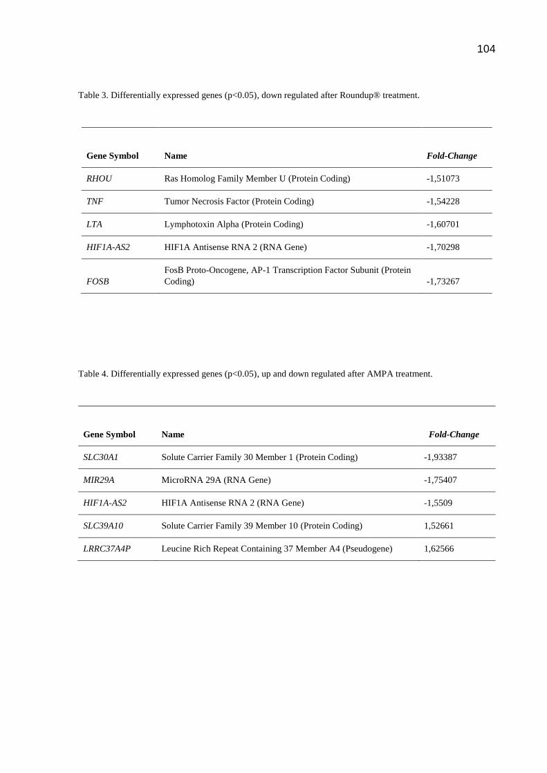
104
Table 3. Differentially expressed genes (p<0.05), down regulated after Roundup® treatment.
Gene Symbol
Name Fold-Change
RHOU Ras Homolog Family Member U (Protein Coding) -1,51073
TNF Tumor Necrosis Factor (Protein Coding) -1,54228
LTA Lymphotoxin Alpha (Protein Coding) -1,60701
HIF1A-AS2 HIF1A Antisense RNA 2 (RNA Gene) -1,70298
FOSB
FosB Proto-Oncogene, AP-1 Transcription Factor Subunit (Protein
Coding) -1,73267
Table 4. Differentially expressed genes (p<0.05), up and down regulated after AMPA treatment.
Gene Symbol
Name Fold-Change
SLC30A1 Solute Carrier Family 30 Member 1 (Protein Coding) -1,93387
MIR29A MicroRNA 29A (RNA Gene) -1,75407
HIF1A-AS2 HIF1A Antisense RNA 2 (RNA Gene) -1,5509
SLC39A10 Solute Carrier Family 39 Member 10 (Protein Coding) 1,52661
LRRC37A4P Leucine Rich Repeat Containing 37 Member A4 (Pseudogene) 1,62566

105
Table 5: Top 10 GO cellular processes after treatment with Roundup®.
GO process FDR
Regulation of cell communication 9.285E-03
Regulation of signaling 9.653E-03
Cellular response to stimulus 1.197E-02
Regulation of signal transduction 1.815E-02
Regulation of biological quality 1.828E-02
Positive regulation of cellular metabolic process 1.828E-02
Positive regulation of macromolecule metabolic process 1.828E-02
Positive regulation of cellular process 1.871E-02
Positive regulation of signal transduction 2.182E-02
Positive regulation of metabolic process 2.292E-02
Table 6. Comparison between fold-change results in Microarray and qPCR analysis.
Gene
Fold-change
Microarray qPCR
TXK 1.83919 3.31214
ATM 1.57376 2.43361
BCL2L11 1.57768 2.01408
NFE2L3 1.91527 1.18129
SESN3 1.60232 1.07630

106
5 CONSIDERAÇÕES FINAIS
A literatura disponível sobre o glifosato é ampla, porém existem muitos pontos de
divergência ou aspectos não abordados. Como os experimentos são realizados de
forma independente, vários fatores como desenho experimental, qualidade dos
reagentes, materiais disponíveis, modelo experimental e qualificação dos
pesquisadores podem influenciar os resultados. Por outro lado, os efeitos da
exposição crônica ao glifosato, principalmente em humanos, ainda não foram
elucidados de forma satisfatória, o que pode ser explicado pela dificuldade na
realização de estudos epidemiológicos e pela impossibilidade de expor pessoas
saudáveis ao herbicida.
Baseada nas evidências atuais, muitas agências de avaliação de risco consideram o
glifosato como provavelmente não carcinogênico, e não tóxico aos humanos, desde
que utilizado de acordo com as instruções do fabricante. Entretanto, ainda não existe
um consenso global sobre a classificação de risco.
Quanto às análises moleculares, foi observado que nas condições escolhidas, o
AMPA alterou a expressão de 5 genes.
Em relação ao tratamento com Roundup®, foi possível perceber que uma curta
exposição a baixas concentrações alterou o padrão de expressão gênica de 26 genes
associados com controle do ciclo celular, regulação de processos celulares e
apoptose, o que poderia explicar os efeitos biológicos do glifosato descritos na
literatura.
Devido à escassez de estudos avaliando as alterações da expressão gênica em
células humanas, esse trabalho foi pioneiro na tentativa de identificar os genes
diferencialmente expressos quando PBMCs são submetidas ao Roundup®. Assim, as
informações contidas nessa tese serão importantes para a realização de estudos
adicionais que busquem elucidar o efeito do glifosato sobre a saúde humana.
107
6 REFERÊNCIAS BIBLIOGRÁFICAS
ACQUAVELLA, J. F. et al. Glyphosate Biomonitoring for Farmers and Their Families:
Results from the Farm Family Exposure Study. Environmental Health
Perspectives, v. 112, n. 3, p. 321-326, 2004.
AKCHA, F; SPAGNOL, C; ROUXEL, J. Genotoxicity of diuron and glyphosate in
oyster spermatozoa and embryos. Aquatic Toxicology, v. 106–107(15), p. 104-113,
2012.
ALAVANJA, M. C. et al. The Agricultural Health Study. Environ Health Perspect, v.
104, n. 4, p. 362-369, 1996.
ALAVANJA, M. C. et al. Use of agricultural pesticides and prostate cancer risk in the
Agricultural Health Study cohort. Am J Epidemiol, v. 157, n. 9, p. 800-814, 2003.
ALVAREZ-MOYA, C. et al. Comparison of the in vivo and in vitro genotoxicity of
glyphosate isopropylamine salt in three different organisms. Genetics and
Molecular Biology, v. 37, n. 1, p. 105-110, 2014.
ANDREOTTI, G. et al. Agricultural pesticide use and pancreatic cancer risk in the
Agricultural Health Study Cohort. Int J Cancer, v. 124, n. 10, p. 2495-500, 2009.
ARBUCKLE, T. E.; LIN, Z.; MERY, L. S. An exploratory analysis of the effect of
pesticide exposure on the risk of spontaneous abortion in an ontario farm population.
Environ Health Perspect, v. 109, n. 8, p. 851–857, 2001.
ARIS, A.; LEBLANC, S. Maternal and fetal exposure to pesticides associated to
genetically modified foods in Eastern Townships of Quebec, Canada. Reprod
Toxicol, v. 31, n. 4, p. 528-533, doi: 10.1016/j.reprotox.2011.02.004, 2011.
BENACHOUR, N.; SERALINI, G-E. Glyphosate Formulations Induce Apoptosis and
Necrosis in Human Umbilical, Embryonic, and Placental Cells. Chem. Res. Toxicol,
v. 22, p. 97–105, 2009.

108
BOLOGNESI, C. et al. Genotoxic Activity of Glyphosate and Its Technical
Formulation Roundup. J. Agric. Food Chem, v. 45, p. 1957-1962, 1997.
BOOCOCK, M. R.; COGGINS, J. R. Kinetics of Senolpyruvylshikimate-3-phosphate
synthase inhibition by glyphosate. Febs Letters. v. 154, n. 1, p. 127-133, 1983.
BRASIL, 2017. Câmara dos Deputados. Reportagem sobre nos projetos sobre
mudanças na Lei dos Agrotóxicos. Disponível em:
<http://www2.camara.leg.br/camaranoticias/radio/materias/REPORTAGEM-
ESPECIAL/534968-AGROTOXICOS-MUDANCAS-NA-LEI,-RESTRICOES-ATUAIS,-
CRITICAS-E-SUGESTOES-BLOCO-1.html>. Acesso em: 31 de outubro de 2017.
BRASIL. Lei nº 7802, de 11 de julho de 1989. Dispõe sobre a pesquisa, a
experimentação, a produção, a embalagem e rotulagem, o transporte, o
armazenamento, a comercialização, a propaganda comercial, a utilização, a
importação, a exportação, o destino final dos resíduos e embalagens, o registro, a
classificação, o controle, a inspeção e a fiscalização de agrotóxicos, seus
componentes e afins, e dá outras providências. Disponível em: <
http://www.planalto.gov.br/ccivil_03/Leis/L7802.htm >. Acesso em: 31 de outubro de
2017.
BRASIL. Ministério da Agricultura, Pecuária e Abastecimento. Decreto nº 4074, de
04 de janeiro de 2002. Regulamenta a Lei no 7.802, de 11 de julho de 1989, que
dispõe sobre a pesquisa, a experimentação, a produção, a embalagem e rotulagem,
o transporte, o armazenamento, a comercialização, a propaganda comercial, a
utilização, a importação, a exportação, o destino final dos resíduos e embalagens, o
registro, a classificação, o controle, a inspeção e a fiscalização de agrotóxicos, seus
componentes e afins, e dá outras providências. Disponível em:
<http://www.planalto.gov.br/ccivil_03/decreto/2002/d4074.htm>. Acesso em: 31 de
outubro de 2017.
CAKMAK, I. et al. Glyphosate reduced seed and leaf concentrations of calcium,
manganese, magnesium, and iron in non-glyphosate resistant soybean. Eur J
Agron, v. 31(3), p. 114–119, 2009.
109
CANTOR, K. P. et al. Pesticides and other agricultural risk factors for non-Hodgkin's
lymphoma among men in Iowa and Minnesota. Cancer Res, v. 52, n. 9, p. 2447-55,
1992.
CHANG, F. C.; SIMCIK, M. F.; CAPEL, P. D. 2011. Occurrence and fate of the
herbicide glyphosate and its degradate aminomethylphosphonic acid in the
atmosphere. Environ Toxicol Chem, v. 30, p. 548–555, 2011.
CHAUFAN, G.; COALOVA, I.; MOLINA, M. C. R. Glyphosate commercial formulation
causes cytotoxicity, oxidative effects, and apoptosis on human cells: differences with
its active ingredient. Int J Toxicol, v. 33, n. 1, p. 29-38. doi:
10.1177/1091581813517906, 2014.
COALOVA, I.; MOLINA, M. C. R.; CHAUFAN, G. Influence of the spray adjuvant on
the toxicity effects of a glyphosate formulation. Toxicol In Vitro, v. 28, n. 7, p. 1306-
1311, doi: 10.1016/j.tiv.2014.06.014, 2014.
CURTIS, K. M. et al. The effect of pesticide exposure on time to pregnancy.
Epidemiology, v. 10, n. 2, p. 112-117, 1999.
CZELUSNIAK, K. E. et al. Farmacobotânica, fitoquímica e farmacologia do Guaco:
revisão considerando Mikania glomerata Sprengel e Mikania laevigata Schulyz Bip.
ex Baker. Rev. Bras. Pl. Med., Botucatu, v. 14, n. 2, p. 400-409, 2012.
DALLEGRAVE, E. et al. Pre- and postnatal toxicity of the commercial glyphosate
formulation in Wistar rats. Arch Toxicol, v. 81, p. 665-673, 2007.
DALLEGRAVE, E. et al. The teratogenic potential of the herbicide glyphosate-
Roundup in Wistar rats. Toxicol Lett, v. 142, p. 45-52, 2003.
DE ROOS, A. J. et al. Integrative assessment of multiple pesticides as risk factors
for non-Hodgkin's lymphoma among men. Occup Environ Med, v. 60, n. 9, p. E11,
2003.
DENNIS, L. K. et al. Pesticide use and cutaneous melanoma in pesticide applicators
in the agricultural heath study. Environ Health Perspect, v. 118, n. 6, p. 812-7,

110
2010.
DESESSO, J. M.; WILLIAMS, A. L. Comment on ‘‘Glyphosate impairs male offspring
reproductive development by disrupting gonadotropin expression’’ by Romano et al.
2012. Arch Toxicol, v. 86(11), p. 1791-3, doi: 10.1007/s00204-012-0894-3, 2012.
DILL, G. M. et al. Chapter 1: Glyphosate: discovery, development, applications, and
properties. In: NANDULA, V. K. editor. Glyphosate resistance in crops and weeds:
history, development, and management. Wiley, Hoboken (NJ), p. 1–33, 2010.
EFSA (European Food Safety Authority). Conclusion on pesticide peer review of
the pesticide risk assessment of the active substance glyphosate EFSA
Journal, v. 13, n. 11, p.4302, 2015.
EPA (Environmental Protection Agency). Glyphosate Issue Paper: Evaluation of
Carcinogenic Potetion. EPA’S Office of Pesticide Programs, 2016. Disponível em:
<https://www.regulations.gov/document?D=EPA-HQ-OPP-2016-0385-0094>. Acesso
em: 29 de outubro de 2017.
ERIKSSON, M. et al. Pesticide exposure as risk factor for non-Hodgkin lymphoma
including histopathological subgroup analysis. Int J Cancer, v. 123, n. 7, p. 1657-63,
2008.
ETGES, V. E. et al. O impacto da cultura do tabaco no ecossistema e na saúde
humana. Textual, v. 1, n. 1, p. 14-21, 2002.
FAO (Organização das Nações Unidas para Agricultura e Alimentação).
Glyphosate, N-(phosphonomethyl)glycine. Specifcations and evaluations for
plant protection products. Rome: Food and Agriculture Organizationof the United
Nations, 2000. Disponível em: <http://www.fao. IARC MonogRAphs – 112 84
org/fleadmin/templates/agphome/documents/Pests_
Pesticides/Specs/glypho01.pdf>. Acesso em: 28 de Julho de 2015.
FAO/OMS (Organização das Nações Unidas para Agricultura e Alimentação/
Organização Mundial da Saúde). Pesticide Residues in Food 2016. Special
Session of The Joint FAO/WHO Meeting on Pesticide Residues. Report 2016. FAO

111
Plant Production and Protection Paper 227. ISSN 2070-2515. Roma, 2016.
Disponível em:
<https://www.google.com.br/url?sa=t&rct=j&q=&esrc=s&source=web&cd=1&ved=0ah
UKEwiYspybnTWAhWEEZAKHTP0AdgQFggqMAA&url=http%3A%2F%2Fwww.fao.
org%2F3%2Fa-i5693e.pdf&usg=AOvVaw1f2DsK27Dz_BVvGhedj9v6>. Acesso em:
27 de outubro de 2017.
FARIA, N. M. et al. Pesticides and respiratory symptoms among farmers. Rev Saude
Publica, v. 39, n. 6, p. 973-981, 2005.
FARIA, N. M. X.; FASSA, A. G.; FACCHINI, L. A. Intoxicação por agrotóxicos no
Brasil: os sistemas oficiais de informação e desafios para realização de estudos
epidemiológicos. Ciência & Saúde Coletiva, v. 12, n. 1, p. 25-38, 2007.
FCI (Farm Chemicals International) Glyphosate. In: Crop Protection Database.
Willoughby (OH): Meister Media Worldwide. 2015. Disponível em:
http://www.farmchemicalsinternational.com/cropprotectiondatabase/#/product/detail/2
03900/. Acesso em: 2 de outubro de 2017.
FOLMAR, L. C.; SANDERS, H. O.; JULIN, A. M. Toxicity of the herbicide
glyphosphate and several of its formulations to fish and aquatic invertebrates. Arch
Environ Contam Toxicol, v. 8, n. 3, p. 269–78, 1979.
GARRY, V. F.et al. Birth defects, season of conception, and sex of children born to
pesticide applicators living in the Red River Valley of Minnesota, USA. Environ
Health Perspect, v. 110, n. 3, p. 441–449, 2002.
GIESY, J. P.; DOBSON, S.; SOLOMON, K. R. Ecotoxicological Risk Assessment for
Roundap Herbicide. Rev Environ Contam Toxicol. v. 167, p. 35-120, 2000.
HENNEBERGER, P. K. et al. Exacerbation of symptoms in agricultural pesticide
applicators with asthma. Int Arch Occup Environ Health, v. 87, n. 4, p. 423-32, doi:
10.1007/s00420-013-0881-x, 2014.
HOKANSON, R. et al. Alteration of estrogen-regulated gene expression in human
cells induced by the agricultural and horticultural herbicide glyphosate. Hum Exp

112
Toxicol, v. 26(9), p. 747-52, 2007.
HOPPIN, J. A. el al. Pesticides and atopic and nonatopic asthma among farm women
in the agricultural health study. Am J Respir Crit Care Med, v. 177, n. 1, p. 11-18,
2008.
HOPPIN, J. A. el al. Pesticides are associated with allergic and non-allergic wheeze
among male farmers. Environ Health Perspect, v. 125, n. 4, p. 535-543, doi:
10.1289/EHP315, 2017.
IARC (Agência Internacional de Pesquisas sobre o Câncer). Glyphosate. In: Some
organophosphate insecticides and herbicides: diazinon, glyphosate,
malathion, parathion, tetrachlorvinphos. IARC Monographs on the Evaluation of
Carcinogen Risks to Humans. Lyon (França). v. 112, p. 1–92, 2015. Disponível em:
<http://mono graphs.iarc.fr/ENG/Monographs/vol112/index.php>. Acesso em: 9 de
outubro de 2017.
IBAMA (Instituto Brasileiro do Meio Ambiente e dos Recursos Naturais Renováveis).
Produtos Agrotóxicos e afins comercializados em 2009 no Brasil: Uma
abordagem ambiental. ISBN 978-85-7300-6. Brasília, 2010.
IBAMA (Instituto Brasileiro do Meio Ambiente e dos Recursos Naturais Renováveis).
Boletins anuais de produção, importação, exportação e vendas de agrotóxicos
no Brasil - 2014. Dados atualizados em 2016. Disponível em:
<http://ibama.gov.br/agrotoxicos/relatorios-de-comercializacao-de-agrotoxicos>.
Acesso em: 12 de setembro de 2017.
IBGE (Instituto Brasileiro de Geografia e Estatística). Indicadores de
Desenvolvimento Sustentável: Brasil 2015. Ministério do Planejamento,
Orçamento e Gestão. ISBN 978-85-240-4347-5. Rio de Janeiro, 2015.
IPCS (International Programme on Chemical Safety). Glyphosate. OMS/FAO Data
Sheets on Pesticides, No. 91 (WHO/PCS/DS/96.91). Organização Mundial da
Saúde, 1996. Disponível em: <http://apps.who. int/iris/handle/10665/63290>. Acesso
em: 4 de outubro de 2017.

113
JACOB, G. S. et al. Metabolism of glyphosate in Pseudomonas sp. strain LBr. Appl
Environ Microbiol, v. 54, n. 12, p. 2953–2958, PMID:3223761, 1988.
JAUHIAINEN, A. et al. Occupational exposure of forest workers to glyphosate during
brush saw spraying work. Am Ind Hyg Assoc J, v. 52(2), p. 61-64,1991.
JAYASUMANA, C. et al. Drinking well water and occupational exposure to Herbicides
is associated with chronic kidney disease, in Padavi-Sripura, Sri Lanka.
Environmental Health, v. 14, n. 6, doi:10.1186/1476-069X-14-6, 2015.
JAYAWARDENA, et al. Toxicity of agrochemicals to common hourglass tree frog
(Polypedates cruciger) in acute and chronic exposure. Int J Agric Biol, v. 12, p.
641–648, 2010.
JUNIOR, O. P. A.; SANTOS, T. C. R. Glifosato: propriedades, toxicidade, usos e
legislação. Quim. Nova, v. 25, n. 4, p. 589-593, 2002.
KAMIJO, Y.; TAKAI, M. SAKAMOTO, T. A multicenter retrospective survey of
poisoning after ingestion of herbicides containing glyphosate potassium salt or other
glyphosate salts in Japan. Clinical Toxicology,
doi:10.3109/15563650.2015.1121271, 2015.
KASUBA, V. et al. Effects of low doses of glyphosate on DNA damage, cell
proliferation and oxidative stress in the HepG2 cell line. Environ Sci Pollut Res Int,
doi: 10.1007/s11356-017-9438-y, 2017.
KOLLER, V. J. et al. Cytotoxic and DNA-damaging properties of glyphosate and
Roundup® in human-derived buccal epithelial cells. Arch Toxicol, v. 86, n. 5, p. 805-
813, doi: 10.1007/s00204-012-0804-8, 2012.
KRUGER, M. et al. Detection of Glyphosate Residues in Animals and Humans. J
Environ Anal Toxicol, v. 4, n. 210, doi: 10.4172/2161-0525.1000210, 2014.
KWIATKOWSKA, M. et al. DNA damage and methylation induced by glyphosate in
human peripheral blood mononuclear cells (in vitro study). Food and Chemical
Toxicology, v. 105, p 93-98, 2017.

114
KWIATKOWSKA, M. et al. The Impact of Glyphosate, Its Metabolites and Impurities
on Viability, ATP Level and Morphological changes in Human Peripheral Blood
Mononuclear Cells. PLoS ONE, v. 11, n. 6, e0156946, doi:10.1371/
journal.pone.0156946. 2016.
KWIATKOWSKA, M.; NOWACKA-KRUKOWSKAB, H.; BUKOWSKA, B. The effect of
glyphosate, its metabolites and impurities on erythrocyte acetylcholinesterase
activity. Environmental Toxicology and Pharmacology, v. 37, p. 1101–1108, 2014.
LAJMANOVICH, R. C; SANDOVAL, M. T; PELTZER, P. M. Induction of mortality and
malformation in Scinax nasicus tadpoles exposed to glyphosate formulations. Bull
Environ Contam Toxicol, v. 70, p. 612-618, 2003.
LE, T. H. et al. Effects of glyphosate and methidathion on the expression of the Dhb,
Vtg, Arnt, CYP4 and CYP314 in Daphnia magna. Chemosphere, v. 79(1), p. 67-71.
doi: 10.1016/j.chemosphere.2009.12.067. 2010.
LEE, C. H. et al. The early prognostic factors of glyphosate-surfactant intoxication.
American Journal of Emergency Medicine, v. 26, p. 275–281.
doi:10.1016/j.ajem.2007.05.011, 2008.
LEE, W. J. et al. Pesticide use and colorectal cancer risk in the Agricultural Health
Study. Int J Cancer, v. 121, n. 2, p. 339-346, 2007.
LI, Q. et al. Glyphosate and AMPA inhibit cancer cell growth through inhibiting
intracellular glycine synthesis. Drug Des Devel Ther, v. 24, n. 7, p. 635-643, doi:
10.2147/DDDT.S49197, 2013.
LOCKRIDGE, O. et al. Naturally Occurring Genetic Variants of Human
Acetylcholinesterase and Butyrylcholinesterase and Their Potential Impact on the
Risk of Toxicity from Cholinesterase Inhibitors. Chem. Res. Toxicol, v. 29, p.
1381−1392, doi: 10.1021/acs.chemrestox.6b00228, 2016.
LUBICK, N. Environmental impact of cocaine strategy assessed [News. Nature,
Publicado online 12 November, doi:10.1038/news.2009.1080, 2009.

115
MALHOTRA, R. C. et al. Glyphosate–surfactant herbicide-induced reversible
encephalopathy. Case Reports. Journal of Clinical Neuroscience, v. 17, p. 1472–
1473, doi:10.1016/j.jocn.2010.02.026, 2010.
MANCE, D. The great glyphosate debate. Northern Woodlands [online magazine].
2012. Disponível em: <http://northernwoodlands.org/articles/article/ the-great-
glyphosate-debate>. Acesso em 28 de julho de 2017.
MARCHAND, J. et al. Molecular identification and expression of differentially
regulated genes of the European flounder, Platichthys flesus, submitted to pesticide
exposure. Mar Biotechnol (NY), v. 8(3), p. 275-94, 2006.
MARIAGER, T. P. et al. Severe adverse effects related to dermal exposure to a
glyphosate-surfactant herbicide. Clinical Toxicology, v. 51, p. 111–113, doi:
10.3109/15563650.2013.763951, 2013.
MARTINEZ, A. REYES, I.; REYES, N. Citotoxicidad del glifosato em células
mononucleares de sangre periférica humana. Biomedica, v. 27, p. 594–604, PMID:
18320126, 2007.
MCDUFFIE, H. H. et al. Non-Hodgkin's lymphoma and specific pesticide exposures
in men: cross-Canada study of pesticides and health. Cancer Epidemiol
Biomarkers Prev, v. 10, n. 11, p. 1155-1163, 2001.
MERTENS, M. et al. Glyphosate, a chelating agent—relevant for ecological risk
assessment? Environ Sci Pollut Res. v. 25, p. 5298–5317, 2018.
MESNAGE, R. et al. Evaluation of estrogen receptor alpha activation by glyphosate-
based herbicide constituents. Food and Chemical Toxicology, v. 108, p. 30-42,
2017.
MESNAGE, R. et al. Review Potential toxic effects of glyphosate and its commercial
formulations below regulatory limits. Food and Chemical Toxicology, v. 84, p. 133–
153, doi: 10.1016/j.fct.2015.08.012 PMID: 26282372, 2015.
MESNAGE, R.; BERNAY, B.; SERALINI, G. E. Ethoxylated adjuvants of glyphosate-
116
based herbicides are active principles of human cell toxicity. Toxicology, v. 313, p.
122–128, doi: 10.1016/j.tox.2012.09.006 PMID: 23000283, 2013.
MINK, P. J. et al. Epidemiologic studies of glyphosate and cancer: A review.
Regulatory. Toxicology and Pharmacology, v. 63, p. 440–452, 2012.
MLADINIC, M.; PERKOVIC, P.; ZELJEZIC, D. Characterization of chromatin
instabilities induced by glyphosate, terbuthylazine and carbofuran using cytome FISH
assay. Toxicology Letters, v. 189, p. 130–137, doi:10.1016/j.toxlet.2009.05.012,
2009.
MOTOJYUKU, M. et al. Determination of glyphosate, glyphosate metabolites, and
glufosinate in human serum by gas chromatography-mass spectrometry. J
Chromatogr B Analyt Technol Biomed Life Sci, v. 875, n. 2, p. 509–514,
doi:10.1016/j.jchromb.2008.10.003, 2008.
MOTTIER, A. et al. Effects of glyphosate-based herbicides on embryo-larval
development and metamorphosis in the Pacific oyster, Crassostrea gigas. Aquat
Toxicol, v. 15(128-129), p. 67-78, doi: 10.1016/j.aquatox.2012.12.002, 2013.
MYERS, J. A. et al. Concerns over use of glyphosate-based herbicides and risks
associated with exposures: a consensus statement. Environmental Health, v. 15, n.
19, doi:10.1186/s12940-016-0117-0, 2016.
OLIVEIRA-SILVA, J. J. et al. Influência de fatores socioeconômicos na contaminação
por agrotóxicos. Brasil. Rev Saúde Pública, v. 35, n. 2, p. 130-135, 2001.
PEARSON, J. N.; PATEL, M. The role of oxidative stress in organophosphate and
nerve agent toxicity. Ann N Y Acad Sci, v. 1378(1), p. 17–24.
doi:10.1111/nyas.13115, 2016.
POLLEGIONI, L.; SCHONBRUNN, E.; SIEHL, D. Molecular basis of glyphosate
resistance – different approaches through protein engineering. FEBS Journal, v.
278, p. 2753–2766, 2011.
PTOK, M. Dysphonia following glyphosate exposition. HNO, v. 57, n. 11, p.1197-
117
1202, doi: 10.1007/s00106-009-1962-8, 2009.
RIAZ, M. A. et al. Impact of glyphosate and benzo[a]pyrene on the tolerance of
mosquito larvae to chemical insecticides. Role of detoxification genes in response to
xenobiotics. Aquat Toxicol, v. 93(1), p. 61-9. doi: 10.1016/j.aquatox.2009.03.005,
2009.
RIBEIRO, D. N. et al. Possible glyphosate tolerance mechanism in pitted
morningglory (Ipomoea lacunosa L.). Journal of agricultural and food chemistry,
v. 63, n. 6, p. 1689–1697, doi: 10.1021/jf5055722, 2015.
RICHARD, S. et al. Differential effects of glyphosate and Roundup®® on human
placental cells and aromatase. Environmental Health Perspectives, v. 113, n. 6, p.
716–720, 2005.
RIGOTTO, R. M.; VASCONCELOS, D. P.; ROCHA, M. M. Uso de agrotóxicos no
Brasil e problemas para a saúde pública. Cad. Saúde Pública, v. 30, n. 7, p.1-3,
2014.
ROMANO, M. A. et al. Glyphosate impairs male offspring reproductive development
by disrupting gonadotropin expression. Arch Toxicol, v. 86(4), p. 663–73, 2012.
ROY, N. M.; CARNEIRO, B.; OCHS, J. Glyphosate induces neurotoxicity in
zebrafish. Environmental Toxicology and Pharmacology, v. 42, p. 45–54, 2016.
SALVI, R. M. et al. Neuropsychiatric evaluation in subjects chronically exposed to
organophosphate pesticides. Toxicol Sci, v. 72, n. 2, p. 267-271, 2003.
SANIN, L-H. et al. Regional Differences in Time to Pregnancy Among Fertile Women
from Five Colombian Regions with Different use of Glyphosate. Journal of
Toxicology and Environmental Health. Part A, v. 72, p. 949–960, doi:
10.1080/15287390902929691, 2009.
SCHUTTE, G. et al. Herbicide resistance and biodiversity: agronomic and
environmental aspects of genetically modified herbicide-resistant plants. Environ Sci
Eur, v. 29, n. 5, doi:10.1186/s12302-016-0100-y, 2017.

118
SINAN (Sistema Nacional de Agravos de Notificação). Ministério da Saúde/Brasil.
Brasília, 2017. Disponível em: <http://portalsinan.saude.gov.br/o-sinan>. Acesso em:
15 de Setembro de 2017.
SINITOX (Sistema Nacional de Informações Tóxico-Farmacológicas). Fundação
Oswaldo Cruz - Ministério da Saúde/Brasil. Rio de Janeiro, 2017. Disponível em:
<https://sinitox.icict.fiocruz.br/>. Acesso em: 20 de Setembro de 2017.
SIROSKI, P. A. et al. Immunotoxicity of commercial-mixed glyphosate in broad
snouted caiman (Caiman latirostris). Chemico-Biological Interactions, v. 244, p.
64-70, 2016.
SLAGER, R. E. et al. Rhinitis associated with pesticide use among private pesticide
applicators in the agricultural health study. J Toxicol Environ Health A, v. 73, n. 20,
p. 1382-1393, doi: 10.1080, 2010.
SORAHAN, T. et al. Multiple myeloma and glyphosate use: a re-analysis of US
Agricultural Health Study (AHS) data. Int J Environ Res Public Health, v. 12, n. 2,
p. 1548-1559, 2015.
SZÉKÁCS, A.; DARVAS, B. Forty years with glyphosate. In: MNAE-G, H. editor.
Herbicides – properties, synthesis and control of weeds. InTech, Croácia. p. 247–
284, 2012. Disponível em: <http://cdn.intechweb.org/ pdfs/25624.pdf>. Acesso em:
28 de Julho de 2017.
TANGUY, A. et al. Molecular identification and expression study of differentially
regulated genes in the Pacific oyster Crassostrea gigas in response to pesticide
exposure. FEBS J, v. 272(2), p. 390-403, 2005.
TARAZONA, J. V. et al. Glyphosate toxicity and carcinogenicity: a review of the
scientific basis of the European Union assessment and its differences with IARC.
Arch Toxicol, v. 91, p. 2723–2743, doi: 10.1007/s00204-017-1962-5, 2017.
THONGPRAKAISANG, S. et al. Glyphosate induces human breast cancer cells
growth via estrogen receptors. Food and Chemical Toxicology, v. 59, p. 129–136,
doi: 10.1016/j.fct.2013.05.057, 2013.

119
TMR (Transparency Market Research). Global glyphosate market expected to
reach US$8.79 billion in 2019. New York: Transparency Market Research, 2014.
Disponível em: <http://www.transparencymarketresearch.com/pressrelease/
glyphosate-market.htm>. Acesso em: 21 de setembro de 2017.
TOMLIN, C. D. S. The pesticide manual: a world compendium. Croydon: British
Crop Protection Council, 12ª Ed., 2000. Disponível em:
<http://trove.nla.gov.au/work/6273016>. Acesso em: 28 de julho de 2017.
TOXCEN (Centro de Atendimento Toxicológico do Espírito Santo. Governo do
Espírito Santo). Dados Estatísticos - Intoxicações - Toxcen ES, 2015. Disponível em:
<https://toxcen.es.gov.br/Media/toxcen/Arquivos/Dados%20Estat%C3%ADsticos%20
-
%20Intoxica%C3%A7%C3%B5es%20%20Toxcen%20ES%20%20ano%20de%2020
15.pdf>. Acesso em: 25 de outubro de 2017.
VANDENBERG, L. N. et al. Is it time to reassess current safety standards for
glyphosate-based herbicides? J Epidemiol Community Health, v. 71, p. 613–618,
doi:10.1136/jech-2016-208463, 2017.
VIGFUSSON, N. V.; VYSE, E. R. The effect of the pesticides, dexon, captan and
roundup, on sister-chromatid exchanges in human lymphocytes in vitro. Mutation
Research, v. 79, p. 53—57, 1980.
WILLIAMS, G. M. et al. A review of the carcinogenic potential of glyphosate by four
independent expert panels and comparison to the IARC assessment. Critical
Reviews in Toxicology, v. 46, n. 1, p. 3-20, doi: 10.1080/10408444.2016.1214677,
2016.
WILLIAMS, G. M. et al. Safety Evaluation and Risk Assessment of the Herbicide
Roundup® and Its Active Ingredient, Glyphosate, for Humans. Regulatory
Toxicology and Pharmacology, v. 31, p. 117–165, doi:10.1006/rtph.1999.1371,
2000.
WINNICK, B; DZIALOWSKI, E. M. The Effects of glyphosate based herbicides on

120
chick embryo morphology during development. The FASEB Journal, v. 27, n. 1,
supplement 874.12, 2013.
YOSHIOKA, N. et al. Rapid determination of glyphosate, glufosinate, bialaphos, and
their major metabolites in serum by liquid chromatography–tandem mass
spectrometry using hydrophilic interaction chromatography. Journal of
Chromatography A, v. 1218, p. 3675–3680, 2011.
ZOUAOUI, K. et al. Determination of glyphosate and AMPA in blood and urine from
humans: About 13 cases of acute intoxication. Forensic Science International, v.
226, p. 20–25. 2013.