PERTURBAÇÕES INTUITIVAS DE 1901 CONFISCO (Baseados em conclusões do - b b - do Brasil )
68
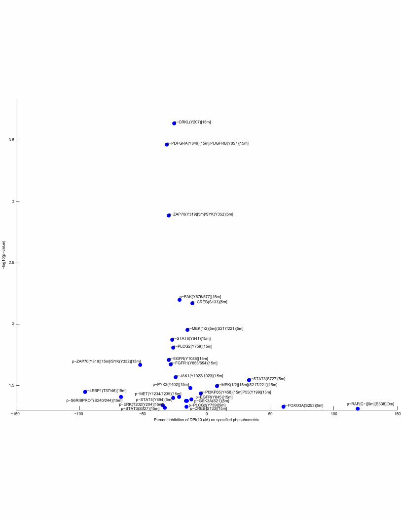
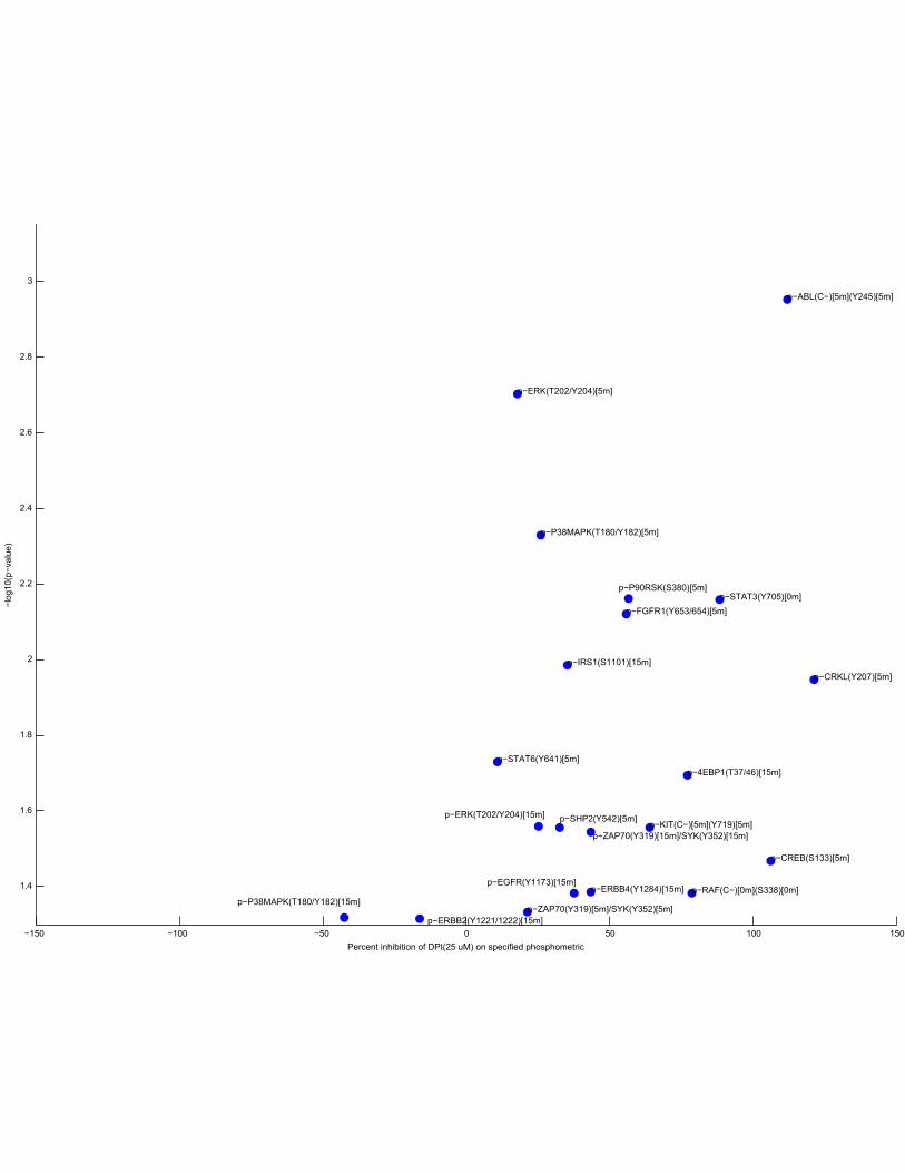
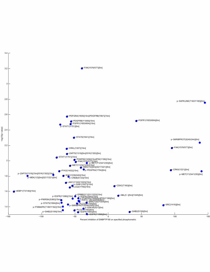
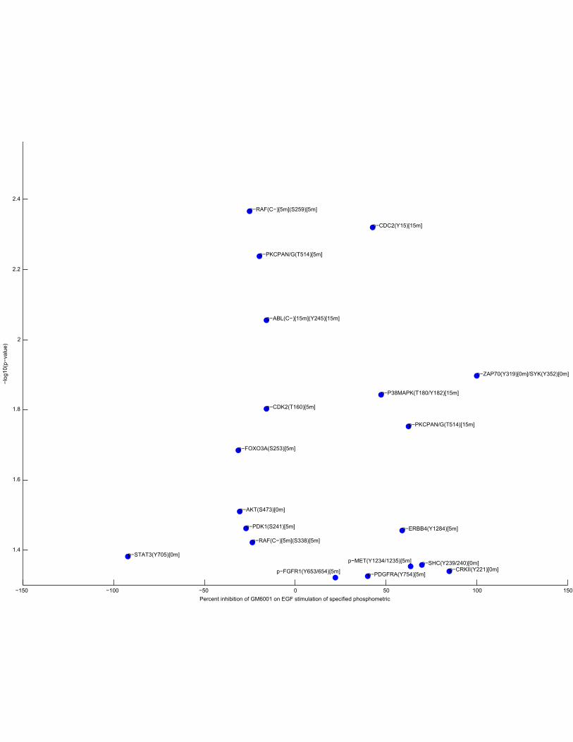
The DIONESUS algorithm provides scalable and accurate reconstruction of dynamic phosphoproteomic networks to reveal new drug targets: Supplement Mark F. Ciaccio, Vincent C. Chen, Richard B. Jones, and Neda Bagheri May 5, 2015 Extended Notes 1 Supplementary Notes 2 1.1 Phosphorylation kinetics, together with cell viability, inform modeling of informa- tion flow downstream of EGFR................................ 2 1.1.1 Autocrine signaling alters carcinogenic pathways in A431 cells......... 3 1.1.2 Distinct antioxidants demonstrate positive and negative influences on A431 cell viability ....................................... 3 1.1.3 Reactive oxygen species affects cell receptor cross-phosphorylation. ..... 4 List of Figures 1 Viability as a function of EGF concentration in A431 cells ................ 6 2 Serial graphs of phosphorylation kinetics for Panel 1 of growth factors ........ 7 3 Serial graphs of phosphorylation kinetics for Panel 2 of small molecules ....... 13 4 Serial graphs of phosphorylation kinetics for Panel 3 of small molecules ....... 19 5 Serial graphs of phosphorylation kinetics for Panel 4 of small molecules ....... 25 6 Serial graphs of phosphorylation kinetics for Panel 5 of small molecules ....... 31 7 Serial graphs of phosphorylation kinetics for Panel 6 of small molecules ....... 37 8 Clustergram of phosphorylation kinetics for Panel 1 of growth factors ........ 43 9 Clustergram of phosphorylation kinetics for Panel 2 of small molecules ....... 44 10 Clustergram of phosphorylation kinetics for Panel 3 of small molecules ....... 45 11 Clustergram of phosphorylation kinetics for Panel 4 of small molecules ....... 46 12 Clustergram of phosphorylation kinetics for Panel 5 of small molecules ....... 47 13 Clustergram of phosphorylation kinetics for Panel 6 of small molecules ....... 48 14 Volcano plots for all inhibitors ............................... 49 15 Comparison of simple linear regression models ..................... 64 16 Receiver operating characteristic (ROC) curves and precision-recall curves for two in silico networks........................................ 65 17 Inference of three node in silico networks using DIONESUS .............. 66 18 Comparison of DIONESUS algorithm to GENIE3, Inferelator, and TIGRESS ..... 67 19 Quantification of microwestern bands............................ 68 1 Electronic Supplementary Material (ESI) for Integrative Biology. This journal is © The Royal Society of Chemistry 2015
-
Upload
1901-atos-das-policias-internacionais -
Category
Internet
-
view
105 -
download
0
Transcript of PERTURBAÇÕES INTUITIVAS DE 1901 CONFISCO (Baseados em conclusões do - b b - do Brasil )
The DIONESUS algorithm provides scalable andaccurate reconstruction of dynamic phosphoproteomic
networks to reveal new drug targets: Supplement
Mark F. Ciaccio, Vincent C. Chen, Richard B. Jones, and Neda Bagheri
May 5, 2015
Extended Notes
1 Supplementary Notes 21.1 Phosphorylation kinetics, together with cell viability, inform modeling of informa-
tion flow downstream of EGFR. . . . . . . . . . . . . . . . . . . . . . . . . . . . . . . . 21.1.1 Autocrine signaling alters carcinogenic pathways in A431 cells. . . . . . . . . 31.1.2 Distinct antioxidants demonstrate positive and negative influences on A431
cell viability. . . . . . . . . . . . . . . . . . . . . . . . . . . . . . . . . . . . . . . 31.1.3 Reactive oxygen species affects cell receptor cross-phosphorylation. . . . . . 4
List of Figures
1 Viability as a function of EGF concentration in A431 cells . . . . . . . . . . . . . . . . 62 Serial graphs of phosphorylation kinetics for Panel 1 of growth factors . . . . . . . . 73 Serial graphs of phosphorylation kinetics for Panel 2 of small molecules . . . . . . . 134 Serial graphs of phosphorylation kinetics for Panel 3 of small molecules . . . . . . . 195 Serial graphs of phosphorylation kinetics for Panel 4 of small molecules . . . . . . . 256 Serial graphs of phosphorylation kinetics for Panel 5 of small molecules . . . . . . . 317 Serial graphs of phosphorylation kinetics for Panel 6 of small molecules . . . . . . . 378 Clustergram of phosphorylation kinetics for Panel 1 of growth factors . . . . . . . . 439 Clustergram of phosphorylation kinetics for Panel 2 of small molecules . . . . . . . 4410 Clustergram of phosphorylation kinetics for Panel 3 of small molecules . . . . . . . 4511 Clustergram of phosphorylation kinetics for Panel 4 of small molecules . . . . . . . 4612 Clustergram of phosphorylation kinetics for Panel 5 of small molecules . . . . . . . 4713 Clustergram of phosphorylation kinetics for Panel 6 of small molecules . . . . . . . 4814 Volcano plots for all inhibitors . . . . . . . . . . . . . . . . . . . . . . . . . . . . . . . 4915 Comparison of simple linear regression models . . . . . . . . . . . . . . . . . . . . . 6416 Receiver operating characteristic (ROC) curves and precision-recall curves for two
in silico networks. . . . . . . . . . . . . . . . . . . . . . . . . . . . . . . . . . . . . . . . 6517 Inference of three node in silico networks using DIONESUS . . . . . . . . . . . . . . 6618 Comparison of DIONESUS algorithm to GENIE3, Inferelator, and TIGRESS . . . . . 6719 Quantification of microwestern bands. . . . . . . . . . . . . . . . . . . . . . . . . . . . 68
1
Electronic Supplementary Material (ESI) for Integrative Biology.This journal is © The Royal Society of Chemistry 2015
1 Supplementary Notes
1.1 Phosphorylation kinetics, together with cell viability, inform modeling ofinformation flow downstream of EGFR.
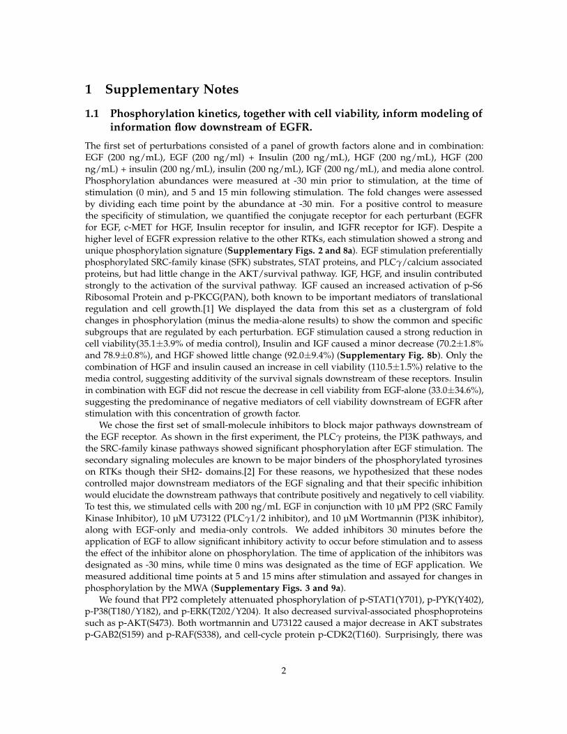
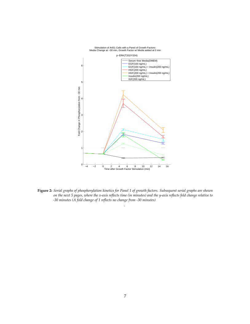
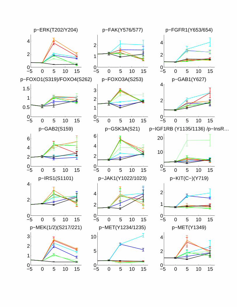
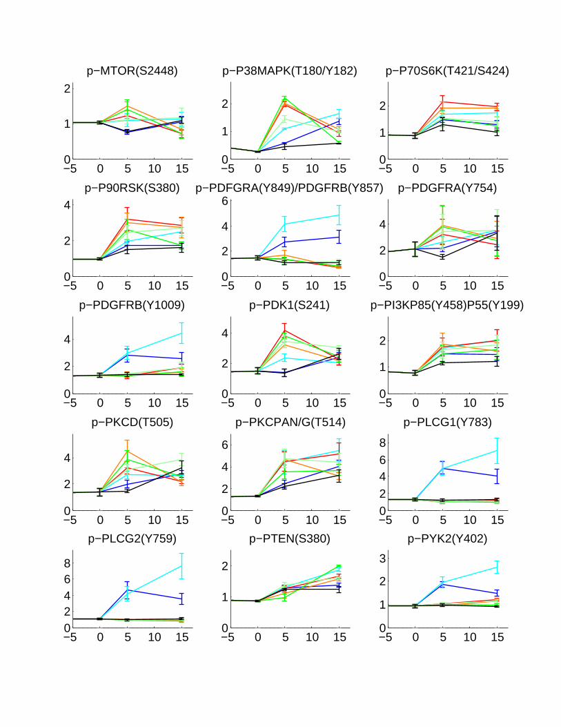
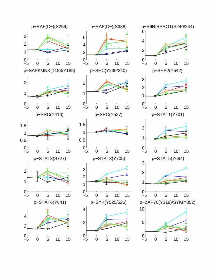
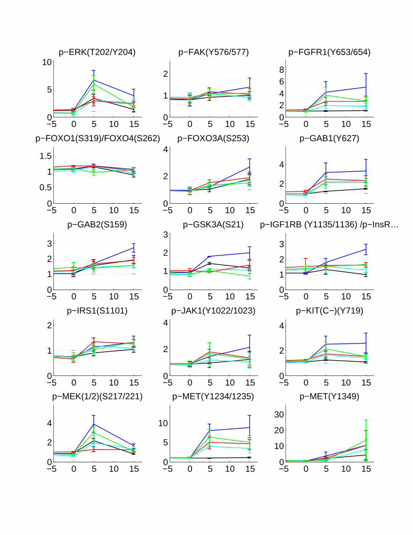
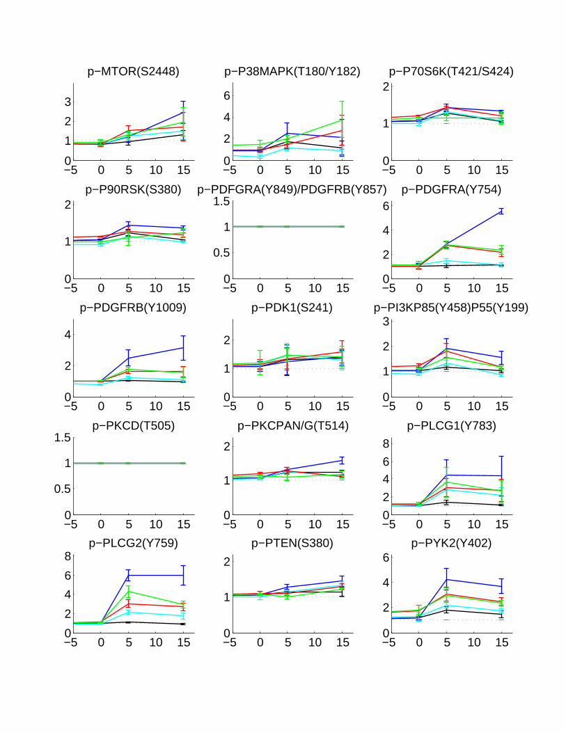
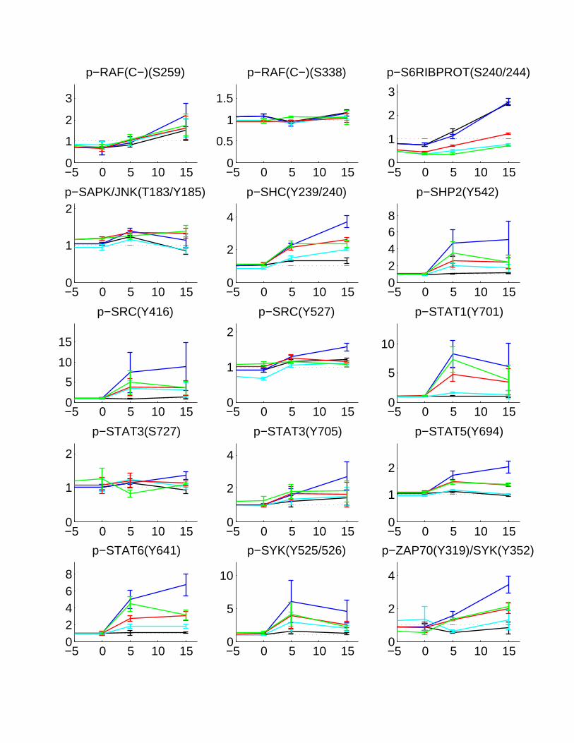
The first set of perturbations consisted of a panel of growth factors alone and in combination:EGF (200 ng/mL), EGF (200 ng/ml) + Insulin (200 ng/mL), HGF (200 ng/mL), HGF (200ng/mL) + insulin (200 ng/mL), insulin (200 ng/mL), IGF (200 ng/mL), and media alone control.Phosphorylation abundances were measured at -30 min prior to stimulation, at the time ofstimulation (0 min), and 5 and 15 min following stimulation. The fold changes were assessedby dividing each time point by the abundance at -30 min. For a positive control to measurethe specificity of stimulation, we quantified the conjugate receptor for each perturbant (EGFRfor EGF, c-MET for HGF, Insulin receptor for insulin, and IGFR receptor for IGF). Despite ahigher level of EGFR expression relative to the other RTKs, each stimulation showed a strong andunique phosphorylation signature (Supplementary Figs. 2 and 8a). EGF stimulation preferentiallyphosphorylated SRC-family kinase (SFK) substrates, STAT proteins, and PLCγ/calcium associatedproteins, but had little change in the AKT/survival pathway. IGF, HGF, and insulin contributedstrongly to the activation of the survival pathway. IGF caused an increased activation of p-S6Ribosomal Protein and p-PKCG(PAN), both known to be important mediators of translationalregulation and cell growth.[1] We displayed the data from this set as a clustergram of foldchanges in phosphorylation (minus the media-alone results) to show the common and specificsubgroups that are regulated by each perturbation. EGF stimulation caused a strong reduction incell viability(35.1±3.9% of media control), Insulin and IGF caused a minor decrease (70.2±1.8%and 78.9±0.8%), and HGF showed little change (92.0±9.4%) (Supplementary Fig. 8b). Only thecombination of HGF and insulin caused an increase in cell viability (110.5±1.5%) relative to themedia control, suggesting additivity of the survival signals downstream of these receptors. Insulinin combination with EGF did not rescue the decrease in cell viability from EGF-alone (33.0±34.6%),suggesting the predominance of negative mediators of cell viability downstream of EGFR afterstimulation with this concentration of growth factor.
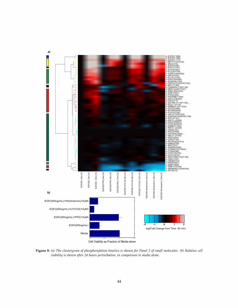
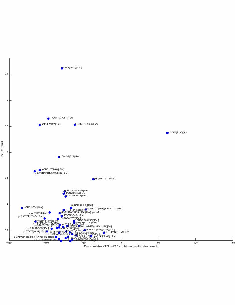
We chose the first set of small-molecule inhibitors to block major pathways downstream ofthe EGF receptor. As shown in the first experiment, the PLCγ proteins, the PI3K pathways, andthe SRC-family kinase pathways showed significant phosphorylation after EGF stimulation. Thesecondary signaling molecules are known to be major binders of the phosphorylated tyrosineson RTKs though their SH2- domains.[2] For these reasons, we hypothesized that these nodescontrolled major downstream mediators of the EGF signaling and that their specific inhibitionwould elucidate the downstream pathways that contribute positively and negatively to cell viability.To test this, we stimulated cells with 200 ng/mL EGF in conjunction with 10 µM PP2 (SRC FamilyKinase Inhibitor), 10 µM U73122 (PLCγ1/2 inhibitor), and 10 µM Wortmannin (PI3K inhibitor),along with EGF-only and media-only controls. We added inhibitors 30 minutes before theapplication of EGF to allow significant inhibitory activity to occur before stimulation and to assessthe effect of the inhibitor alone on phosphorylation. The time of application of the inhibitors wasdesignated as -30 mins, while time 0 mins was designated as the time of EGF application. Wemeasured additional time points at 5 and 15 mins after stimulation and assayed for changes inphosphorylation by the MWA (Supplementary Figs. 3 and 9a).
We found that PP2 completely attenuated phosphorylation of p-STAT1(Y701), p-PYK(Y402),p-P38(T180/Y182), and p-ERK(T202/Y204). It also decreased survival-associated phosphoproteinssuch as p-AKT(S473). Both wortmannin and U73122 caused a major decrease in AKT substratesp-GAB2(S159) and p-RAF(S338), and cell-cycle protein p-CDK2(T160). Surprisingly, there was
2
an increase in the amplitude and duration of p-P38(T180/Y182) and p-JNK(T183/Y182). While200 ng/mL EGF with wortmannin, and EGF with U73122, caused a decrease in cell viability(28.1±1.3% and 15.1±0.4%, respectively) in comparison to EGF-only (32.9±8.2%), application ofPP2 caused a complete attenuation of EGF-stimulated cell death (97.3±10.5%) (SupplementaryFig. 9b).
1.1.1 Autocrine signaling alters carcinogenic pathways in A431 cells.
Autocrine signaling has been reported to cause cross-phosphorylation of receptor tyrosine kinasesin A431 cells.[3] A panel of sheddase inhibitors (aprotinin, GM6001, and TAPI1) also reportedto be effective in surveying autocrine signaling in A431 cells[4] was used in conjunction withEGF stimulation. We hypothesized that sheddases could mediate the EGFR-induced RTK cross-phosphorylation. 1 ug/mL aprotinin (a plasmin and general serine protease inhibitor), 25 µMGM6001 (a broad-range metalloprotease inhibitor), and 100 µM TAPI1 (a TACE inhibitor) wereapplied before 200 ng/mL EGF stimulation and the phosphorylation kinetics were assayed byMWA (Supplementary Figs. 4 and 10a). Aprotinin showed a major increase in phosphorylationof p-INS/IGF1R(Y1134/1135), p-PKCG(PAN)(T514), p-P70S6 Ribosomal Kinase(T421/S422), andp-S6 Ribosomal Protein(S240/244) at 0 min. These proteins match the profile of those stimulatedby IGF, suggesting that aprotinin inhibits the cleavage of an IGF binding protein. GM6001and TAPI1 showed a significant delay in p-INS/IGF1R(Y1134/1135) phosphorylation as well asthe reduction in phosphorylation of many known downstream mediators in the IGF-pathwayincluding p-AKT(S473), p-GAB2(S159), and p-RAF(S259). Aprotinin treatment did not cause achange in cell viability (30.24±9.88%) in comparison to 200 ng/mL EGF alone (32.88±8.18%), whileGM6001 and TAPI1 showed almost complete loss of cell viability (12.25±8.25% and 2.48±2.48%)(Supplementary Fig. 10b). Together, these results suggest that autocrine signaling is a keymediator of cell viability in these cancer cell lines and that phosphorylation profiles of RTK anddownstream targets are modified by inhibition of these pathways. Autocrine signaling may beone means by which a cancer cell overexpressing EGFR can activate multiple pathways leading tocarcinogenic behavior.
1.1.2 Distinct antioxidants demonstrate positive and negative influences on A431 cell viabil-ity.
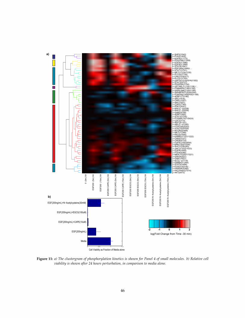
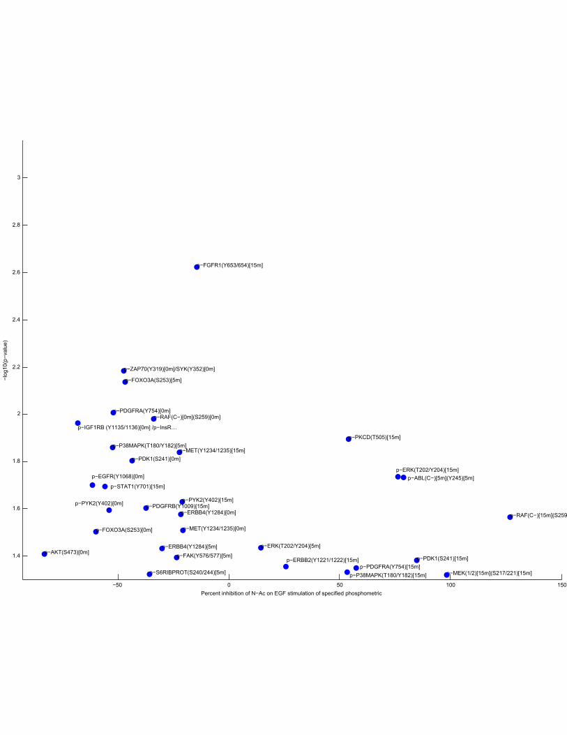
Reactive Oxygen Species (ROS) and its inhibition of phosphatase activity has been shown to beimportant in EGF-induced RTK cross-phosphorylation in A431 cells.[5] To survey the contributionof ROS, A431 cells were treated with the antioxidant panel, 100 µM Epigallactocatechin Gallate(EGCG),10 µM Caffeic Acid Phenylethyl Ester (CAPE), and 30 mM N-acetylcysteine (N-ac)30 minutes prior to EGF stimulation (Supplementary Figs. 5 and 11a). All antioxidants thatcaused a common down-regulation of the PI3K/AKT pathway included p-INS/IGFR(Y1134/1135),suggesting that much of the impact of ROS is mediated through this receptor. EGCG specificallycaused A431 cells to become insensitive to EGF stimulation. EGCG also caused significant andspecific increase in the phosphorylation of P38, JNK, and IGFR, with a total loss of cell viability(Supplementary Fig. 11b). Since EGCG had zero viability and a phosphorylation profile divorcedfrom other perturbations, it was excluded from the final PLSR analysis. In addition to suppressingthe AKT-pathway, CAPE caused a specific decrease in the phosphorylation of p-CDK2(T160) witha strong decrease in cell viability (3.60±3.20%). N-ac caused a decrease in the survival pathway butan increase in the MEK/ERK pathway, resulting in a net increase in cell viability (58.21±15.15%) incomparison to EGF alone (37.52±20.72%). In summary, antioxidants decreased the AKT pathway,but they had variable effects on other cell pathways and increased or decreased viability of the
3
cancer cells, suggesting that antioxidant effect alone is probably insufficient as a general treatmentin this cancer model.
1.1.3 Reactive oxygen species affects cell receptor cross-phosphorylation.
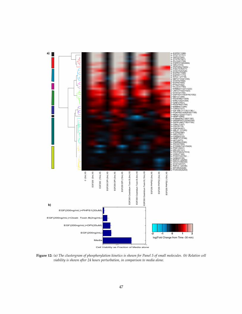
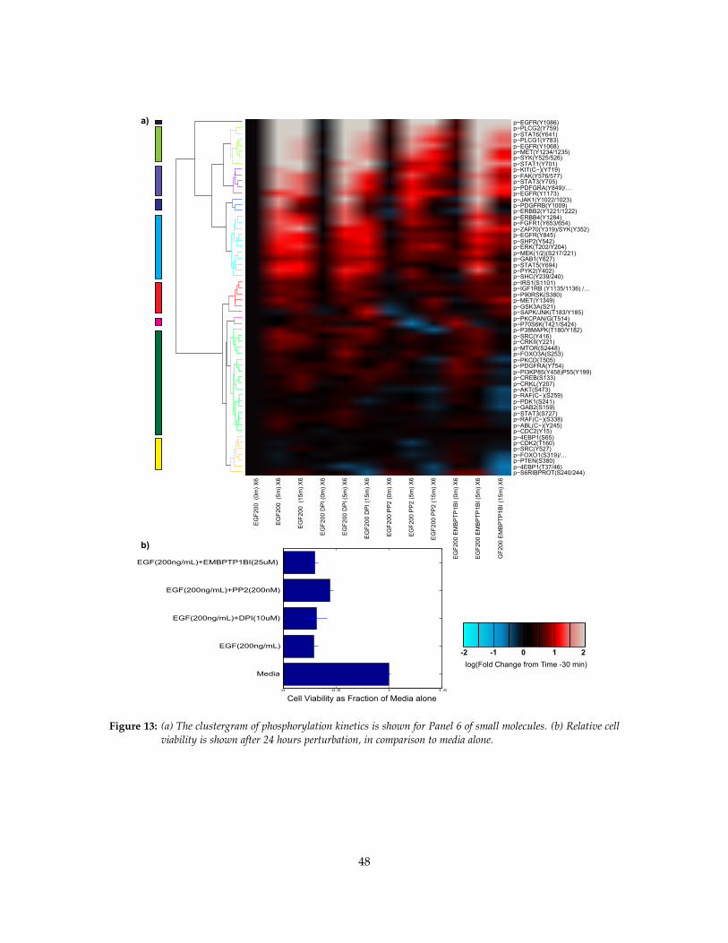
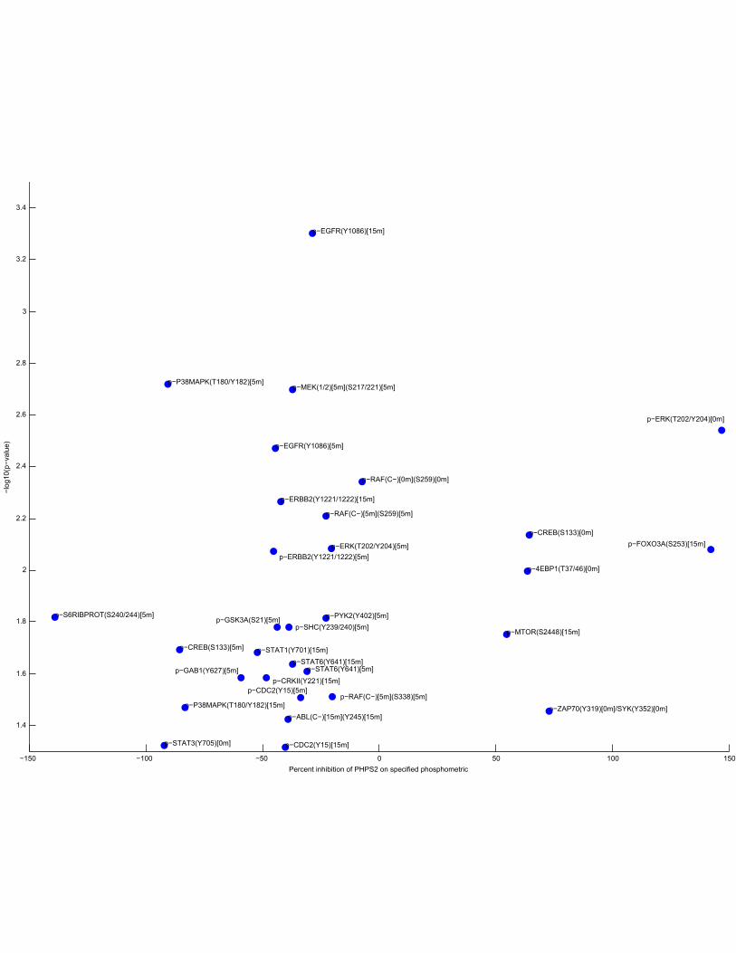
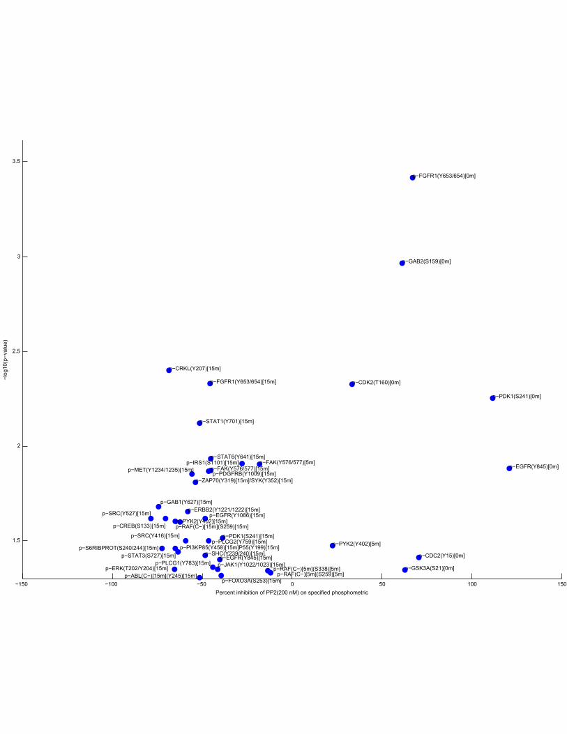
Stimulation of A431 cells was reported to cause inactivation of protein-tyrosine phosphatase1B through the release of reactive oxygen species.[6] We therefore chose the fourth and fifthpanels of inhibitors to study how ROS release affects cross-phosphorylation (Supplementary Figs.6,7,12,13). Studies have shown expression of NADPH Oxidase in A431 cells and its stimulationby EGF to produce ROS[5]. Therefore, two concentrations (10 µM and 25 µM) of the NADPHoxidase inhibitor, Diphenyleneiodonium chloride (DPI), were used, along with an inhibitor of theregulatory subunit of the NADPH oxidase complex, inhibitor of RAC1 (2 ng/mL ClostridiumToxin B), and a PTP1B inhibitor, 25 µM EMBPTP1B1. As SHP2 is an SH2-domain containingphosphatase relating to the highest fold change after EGF stimulation [7], the SHP2 inhibitor, at20 µM PHPS1, was used after EGF stimulation to observe its contribution to phosphokinetics andcell viability. Both concentrations of DPI showed no change in cell viability (10 µM = 31.69±9.74%;25 µM = 26.35±3.85%) from EGF alone (29.36±3.77%). However, Clostridium Toxin demonstrateda decrease in viability (15.29±2.98%). The PTP1B inhibitor resulted in a slight decrease in cellviability (30.12±3.16%), whereas the SHP2 inhibitor showed a major decrease in cell viability(4.50±4.17%) with a very large increase in the fold change of STAT1 phosphorylation, suggestingthat this phosphatase is a major mediator of the pro-apoptotic STAT1 phosphorylation and apossible target in a subset of cancers. Finally, the cells were stimulated with the SRC-inhibitor, PP2,at a lower concentration (200 nM) than in Inhibitor Set 1 to show greater specificity to the SRCFamily Kinases. Again, we confirmed that this condition increased cell viability (44.29±3.60%),over EGF-stimulation alone, demonstrating that a subset of phosphosites downstream of Src-familykinases negatively contribute to cell viability in this cancer model.
References
[1] Dufner, A. & Thomas, G. Ribosomal s6 kinase signaling and the control of transla-tion. Experimental Cell Research 253, 100–109 (1999). URL http://www.sciencedirect.com/science/article/pii/S0014482799946839.
[2] Jones, R. B., Gordus, A., Krall, J. A. & MacBeath, G. A quantitative protein interactionnetwork for the ErbB receptors using protein microarrays. Nature 439, 168174 (2006). URLhttp://www.ncbi.nlm.nih.gov/pubmed/16273093. PMID: 16273093.
[3] Shvartsman, S. Y. et al. Autocrine loops with positive feedback enable context-dependent cellsignaling. American Journal of Physiology - Cell Physiology 282, C545 –C559 (2002). URLhttp://ajpcell.physiology.org/content/282/3/C545.abstract.
[4] Roztocil, E., Nicholl, S. M. & Davies, M. G. Insulin-induced epidermal growth factor activationin vascular smooth muscle cells is ADAM-dependent. Surgery 144, 245–251 (2008). URLhttp://www.sciencedirect.com/science/article/pii/S0039606008001864.
[5] Lou, Y. et al. Redox regulation of the protein tyrosine phosphatase PTP1B in cancercells. The FEBS Journal 275, 69âAS88 (2008). URL http://www.ncbi.nlm.nih.gov/pubmed/18067579. PMID: 18067579.
4

[6] Lee, S., Kwon, K., Kim, S. & Rhee, S. G. Reversible inactivation of Protein-Tyrosine phosphatase1B in a431 cells stimulated with epidermal growth factor. Journal of Biological Chemistry 273,15366–15372 (1998). URL http://www.jbc.org/content/273/25/15366.
[7] Ciaccio, M. F., Wagner, J. P., Chuu, C., Lauffenburger, D. A. & Jones, R. B. Systems analysisof EGF receptor signaling dynamics with microwestern arrays. Nature Methods 7, 148–155(2010). URL http://www.ncbi.nlm.nih.gov/pubmed/20101245. PMID: 20101245.
5
0
0.2
0.4
0.6
0.8
1
1.2
1.4Re
lativ
e ce
ll vi
abili
ty (M
edia
alo
ne =
1.0
)Cell viability as a function of EGF concentration
Media with
8% Serum
Media alone
0.2 ng/mL EGF
2 ng/mL EGF
50 ng/mL EGF
100 ng/mL EGF
200 ng/mL EGF
Figure 1: Viability as a function of EGF concentration in A431 cells. Low concentrations increase cell viability inA431 cells after 24 hrs relative to media-alone stimulation while high concentrations of EGF induce a loss ofcell viability.
.
6
Stimulation of A431 Cells with a Panel of Growth Factors:Media Change at −30 min, Growth Factor w/ Media added at 0 min
−4 −2 0 2 4 6 8 10 12 14 160
1
2
3
4
5
6
p−ERK(T202/Y204)
Time after Growth Factor Stimulation (min)
Fol
d C
hang
e in
Pho
spho
ryla
tion
from
−30
min
Serum−free Media(DMEM)EGF(100 ng/mL)EGF(100 ng/mL) + Insulin(200 ng/mL)HGF(200 ng/mL)HGF(200 ng/mL) + Insulin(200 ng/mL)Insulin(200 ng/mL)IGF(200 ng/mL)
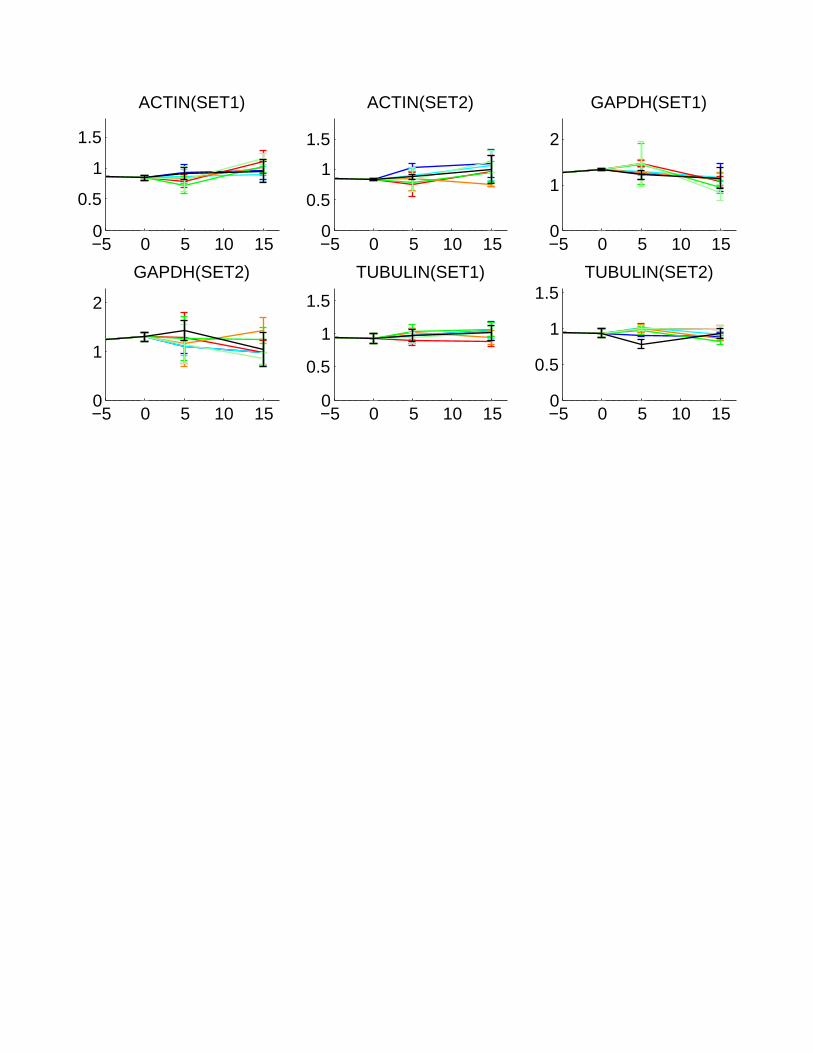
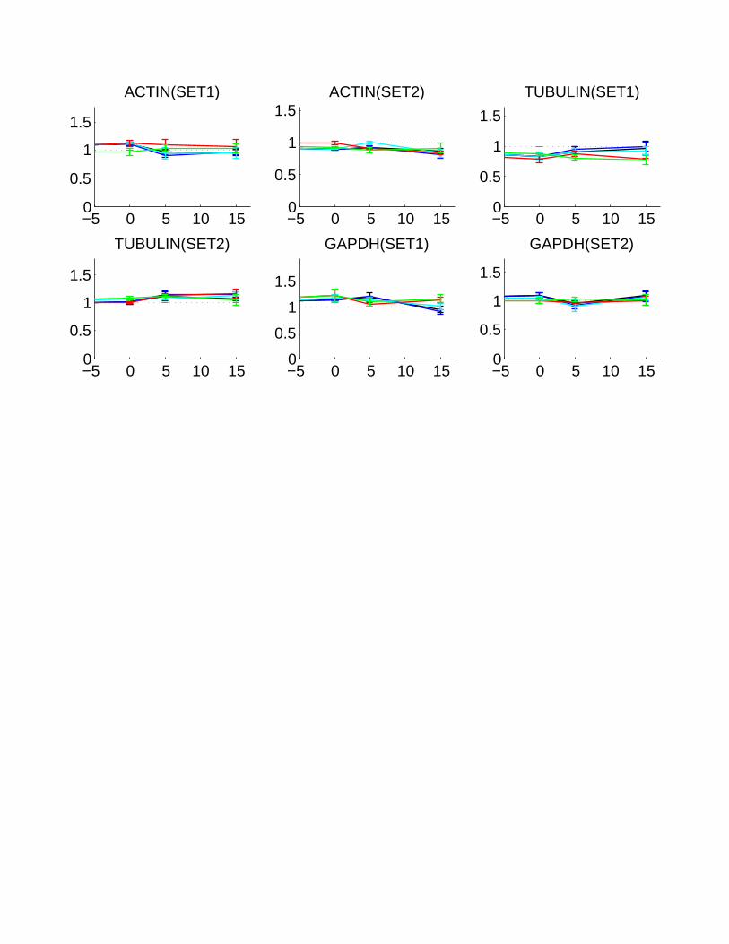
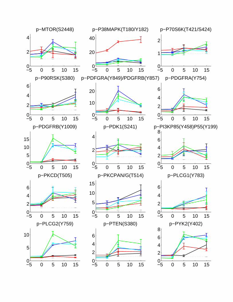
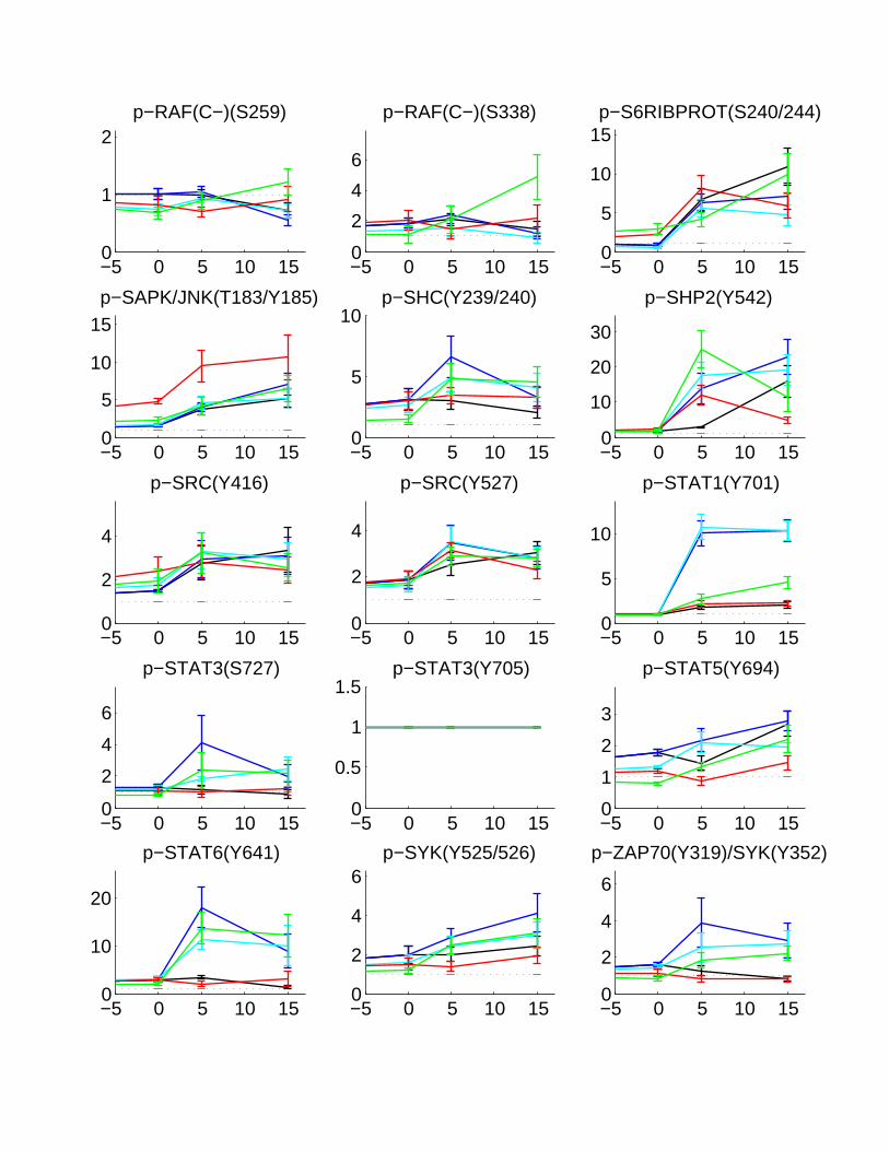
Figure 2: Serial graphs of phosphorylation kinetics for Panel 1 of growth factors. Subsequent serial graphs are shownon the next 5 pages, where the x-axis reflects time (in minutes) and the y-axis reflects fold change relative to-30 minutes (A fold change of 1 reflects no change from -30 minutes)
.
7

−5 0 5 10 150
1
2
3p−4EBP1(S65)
−5 0 5 10 150
0.5
1
1.5
p−4EBP1(T37/46)
−5 0 5 10 150
0.5
1
1.5p−ABL(C−)(Y245)
−5 0 5 10 1502468
p−AKT(S473)
−5 0 5 10 150
1
2
p−CDC2(Y15)
−5 0 5 10 150
2
4
6
p−CDK2(T160)
−5 0 5 10 150
1
2
3
p−CREB(S133)
−5 0 5 10 150
2
4
6p−CRKII(Y221)
−5 0 5 10 150
1
2
p−CRKL(Y207)
−5 0 5 10 1502468
p−EGFR(Y1068)
−5 0 5 10 150
10
20
p−EGFR(Y1086)
−5 0 5 10 150
5
10p−EGFR(Y1173)
−5 0 5 10 150
5
10p−EGFR(Y845)
−5 0 5 10 150
1
2
3p−ERBB2(Y1221/1222)
−5 0 5 10 150
1
2p−ERBB4(Y1284)
−5 0 5 10 150
2
4
p−ERK(T202/Y204)
−5 0 5 10 150
1
2
p−FAK(Y576/577)
−5 0 5 10 150
2
4
p−FGFR1(Y653/654)
−5 0 5 10 150
0.5
1
1.5
p−FOXO1(S319)/FOXO4(S262)
−5 0 5 10 150
1
2
3
p−FOXO3A(S253)
−5 0 5 10 150
2
4p−GAB1(Y627)
−5 0 5 10 150
2
4
6
p−GAB2(S159)
−5 0 5 10 150
2
4
6p−GSK3A(S21)
−5 0 5 10 150
10
20
p−IGF1RB (Y1135/1136) /p−InsR…
−5 0 5 10 150
2
4p−IRS1(S1101)
−5 0 5 10 150
2
4
p−JAK1(Y1022/1023)
−5 0 5 10 150
1
2
p−KIT(C−)(Y719)
−5 0 5 10 150
1
2
3
p−MEK(1/2)(S217/221)
−5 0 5 10 150
5
10
p−MET(Y1234/1235)
−5 0 5 10 150
2
4
p−MET(Y1349)
−5 0 5 10 150
1
2
p−MTOR(S2448)
−5 0 5 10 150
1
2
p−P38MAPK(T180/Y182)
−5 0 5 10 150
1
2
p−P70S6K(T421/S424)
−5 0 5 10 150
2
4p−P90RSK(S380)
−5 0 5 10 150
2
4
6p−PDFGRA(Y849)/PDGFRB(Y857)
−5 0 5 10 150
2
4
p−PDGFRA(Y754)
−5 0 5 10 150
2
4
p−PDGFRB(Y1009)
−5 0 5 10 150
2
4
p−PDK1(S241)
−5 0 5 10 150
1
2
p−PI3KP85(Y458)P55(Y199)
−5 0 5 10 150
2
4
p−PKCD(T505)
−5 0 5 10 150
2
4
6
p−PKCPAN/G(T514)
−5 0 5 10 1502468
p−PLCG1(Y783)
−5 0 5 10 1502468
p−PLCG2(Y759)
−5 0 5 10 150
1
2
p−PTEN(S380)
−5 0 5 10 150
1
2
3
p−PYK2(Y402)
−5 0 5 10 150
1
2
3
p−RAF(C−)(S259)
−5 0 5 10 150
2
4
6
p−RAF(C−)(S338)
−5 0 5 10 150
2
4
6p−S6RIBPROT(S240/244)
−5 0 5 10 150
1
2
p−SAPK/JNK(T183/Y185)
−5 0 5 10 150
1
2
p−SHC(Y239/240)
−5 0 5 10 150
1
2
3
p−SHP2(Y542)
−5 0 5 10 150
0.5
1
1.5
p−SRC(Y416)
−5 0 5 10 150
0.5
1
1.5
p−SRC(Y527)
−5 0 5 10 150
1
2
p−STAT1(Y701)
−5 0 5 10 150
1
2
p−STAT3(S727)
−5 0 5 10 150
1
2
3
p−STAT3(Y705)
−5 0 5 10 150
1
2
3p−STAT5(Y694)
−5 0 5 10 150
2
4
p−STAT6(Y641)
−5 0 5 10 150
2
4
p−SYK(Y525/526)
−5 0 5 10 150
5
10
p−ZAP70(Y319)/SYK(Y352)
−5 0 5 10 150
0.5
1
1.5
ACTIN(SET1)
−5 0 5 10 150
0.5
1
1.5
ACTIN(SET2)
−5 0 5 10 150
1
2
GAPDH(SET1)
−5 0 5 10 150
1
2
GAPDH(SET2)
−5 0 5 10 150
0.5
1
1.5
TUBULIN(SET1)
−5 0 5 10 150
0.5
1
1.5TUBULIN(SET2)

Stimulation of A431 Cells with a Panel of Small Molecules (X2):Media Change at −30 min, Growth Factor w/ Media added at 0 min
−4 −2 0 2 4 6 8 10 12 14 16
0
0.5
1
1.5
2
2.5
3
3.5
p−S6RIBPROT(S240/244)
Time after Growth Factor Stimulation (min)
Fold
Cha
nge
in P
hosp
hory
latio
n fro
m −
30 m
in
Serum−free Media(DMEM)EGF(200 ng/mL)EGF(200 ng/mL)+PP2 (10 uM)EGF(200 ng/mL)+U73122 (10 uM)EGF(200 ng/mL)+Wortmannin (10 uM)
Figure 3: Serial graphs of phosphorylation kinetics for Panel 2 of small molecules. Subsequent serial graphs are shownon the next 5 pages, where the x-axis reflects time (in minutes) and the y-axis reflects fold change relative to-30 minutes (A fold change of 1 reflects no change from -30 minutes).
13

−5 0 5 10 150
1
2
p−4EBP1(S65)
−5 0 5 10 150
1
2
3
p−4EBP1(T37/46)
−5 0 5 10 150
0.5
1
1.5p−ABL(C−)(Y245)
−5 0 5 10 150
2
4
p−AKT(S473)
−5 0 5 10 150
2
4p−CDC2(Y15)
−5 0 5 10 150
1
2p−CDK2(T160)
−5 0 5 10 150
1
2
p−CREB(S133)
−5 0 5 10 150
0.5
1
1.5p−CRKII(Y221)
−5 0 5 10 150
1
2
p−CRKL(Y207)
−5 0 5 10 150
20
40
p−EGFR(Y1068)
−5 0 5 10 150
20
40
60
80p−EGFR(Y1086)
−5 0 5 10 150
5
10
p−EGFR(Y1173)
−5 0 5 10 1502468
p−EGFR(Y845)
−5 0 5 10 150
1
2
p−ERBB2(Y1221/1222)
−5 0 5 10 150
1
2
p−ERBB4(Y1284)
−5 0 5 10 150
5
10p−ERK(T202/Y204)
−5 0 5 10 150
1
2
p−FAK(Y576/577)
−5 0 5 10 1502468
p−FGFR1(Y653/654)
−5 0 5 10 150
0.5
1
1.5
p−FOXO1(S319)/FOXO4(S262)
−5 0 5 10 150
2
4p−FOXO3A(S253)
−5 0 5 10 150
2
4
p−GAB1(Y627)
−5 0 5 10 150
1
2
3
p−GAB2(S159)
−5 0 5 10 150
1
2
3p−GSK3A(S21)
−5 0 5 10 150
1
2
3
p−IGF1RB (Y1135/1136) /p−InsR…
−5 0 5 10 150
1
2
p−IRS1(S1101)
−5 0 5 10 150
2
4p−JAK1(Y1022/1023)
−5 0 5 10 150
2
4
p−KIT(C−)(Y719)
−5 0 5 10 150
2
4
p−MEK(1/2)(S217/221)
−5 0 5 10 150
5
10
p−MET(Y1234/1235)
−5 0 5 10 150
10
20
30
p−MET(Y1349)
−5 0 5 10 150
1
2
3
p−MTOR(S2448)
−5 0 5 10 150
2
4
6
p−P38MAPK(T180/Y182)
−5 0 5 10 150
1
2p−P70S6K(T421/S424)
−5 0 5 10 150
1
2p−P90RSK(S380)
−5 0 5 10 150
0.5
1
1.5p−PDFGRA(Y849)/PDGFRB(Y857)
−5 0 5 10 150
2
4
6p−PDGFRA(Y754)
−5 0 5 10 150
2
4
p−PDGFRB(Y1009)
−5 0 5 10 150
1
2
p−PDK1(S241)
−5 0 5 10 150
1
2
3p−PI3KP85(Y458)P55(Y199)
−5 0 5 10 150
0.5
1
1.5p−PKCD(T505)
−5 0 5 10 150
1
2
p−PKCPAN/G(T514)
−5 0 5 10 150
2
46
8
p−PLCG1(Y783)
−5 0 5 10 150
2
4
6
8p−PLCG2(Y759)
−5 0 5 10 150
1
2
p−PTEN(S380)
−5 0 5 10 150
2
4
6p−PYK2(Y402)
−5 0 5 10 150
1
2
3
p−RAF(C−)(S259)
−5 0 5 10 150
0.5
1
1.5
p−RAF(C−)(S338)
−5 0 5 10 150
1
2
3
p−S6RIBPROT(S240/244)
−5 0 5 10 150
1
2p−SAPK/JNK(T183/Y185)
−5 0 5 10 150
2
4
p−SHC(Y239/240)
−5 0 5 10 1502468
p−SHP2(Y542)
−5 0 5 10 150
5
10
15
p−SRC(Y416)
−5 0 5 10 150
1
2
p−SRC(Y527)
−5 0 5 10 150
5
10
p−STAT1(Y701)
−5 0 5 10 150
1
2
p−STAT3(S727)
−5 0 5 10 150
2
4
p−STAT3(Y705)
−5 0 5 10 150
1
2
p−STAT5(Y694)
−5 0 5 10 1502468
p−STAT6(Y641)
−5 0 5 10 150
5
10
p−SYK(Y525/526)
−5 0 5 10 150
2
4
p−ZAP70(Y319)/SYK(Y352)
−5 0 5 10 150
0.5
1
1.5
ACTIN(SET1)
−5 0 5 10 150
0.5
1
1.5ACTIN(SET2)
−5 0 5 10 150
0.5
1
1.5
TUBULIN(SET1)
−5 0 5 10 150
0.5
1
1.5
TUBULIN(SET2)
−5 0 5 10 150
0.5
1
1.5
GAPDH(SET1)
−5 0 5 10 150
0.5
1
1.5
GAPDH(SET2)
Stimulation of A431 Cells with a Panel of Protease Inhibitors (X3):Media Change at −30 min, Growth Factor w/ Media added at 0 min
−4 −2 0 2 4 6 8 10 12 14 16
5
10
15
20
25
p−S6RIBPROT(S240/244)
Time after Growth Factor Stimulation (min)
Fol
d C
hang
e in
Pho
spho
ryla
tion
from
−30
min
Serum−free Media(DMEM)EGF(200 ng/mL)EGF(200 ng/mL)+Aprotinin(1 ug/mL)EGF(200 ng/mL)+GM6001(25 uM)EGF(200 ng/mL)+TAPI1(100 uM)
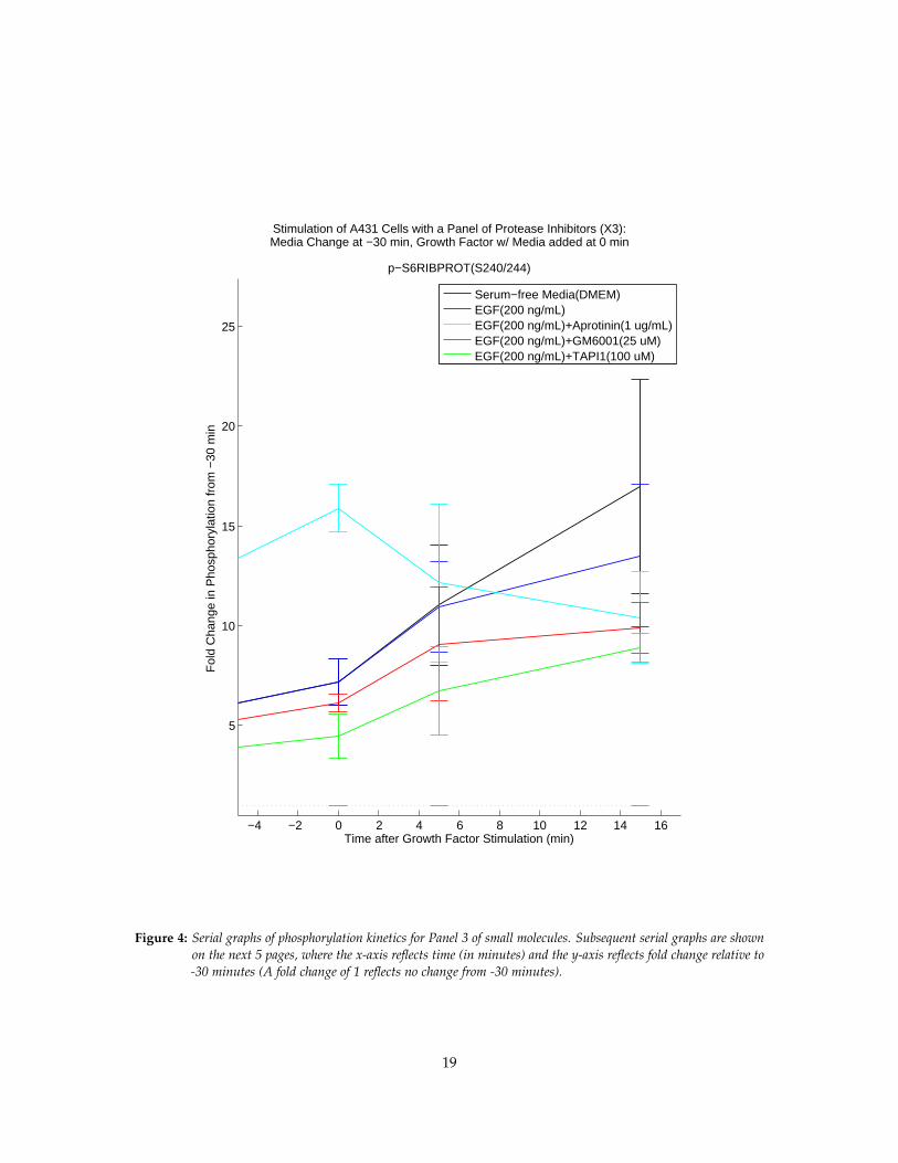
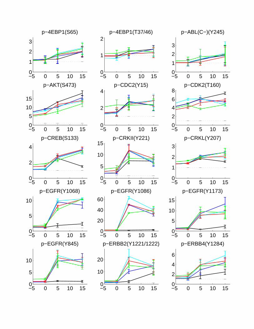
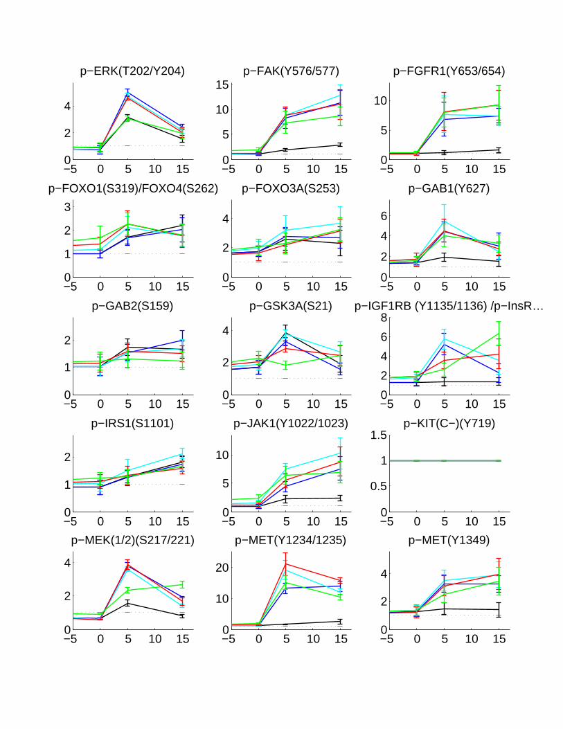
Figure 4: Serial graphs of phosphorylation kinetics for Panel 3 of small molecules. Subsequent serial graphs are shownon the next 5 pages, where the x-axis reflects time (in minutes) and the y-axis reflects fold change relative to-30 minutes (A fold change of 1 reflects no change from -30 minutes).
19
−5 0 5 10 150
1
2
3
p−4EBP1(S65)
−5 0 5 10 150
1
2p−4EBP1(T37/46)
−5 0 5 10 150
1
2
3
p−ABL(C−)(Y245)
−5 0 5 10 150
5
10
15
p−AKT(S473)
−5 0 5 10 150
2
4p−CDC2(Y15)
−5 0 5 10 150
2
4
6
8p−CDK2(T160)
−5 0 5 10 150
2
4
p−CREB(S133)
−5 0 5 10 150
5
10
15p−CRKII(Y221)
−5 0 5 10 150
1
2
3
p−CRKL(Y207)
−5 0 5 10 150
5
10
p−EGFR(Y1068)
−5 0 5 10 150
20
40
60
p−EGFR(Y1086)
−5 0 5 10 150
5
10
15
p−EGFR(Y1173)
−5 0 5 10 150
5
10
p−EGFR(Y845)
−5 0 5 10 150
10
20
p−ERBB2(Y1221/1222)
−5 0 5 10 150
2
4
6
p−ERBB4(Y1284)
−5 0 5 10 150
2
4
p−ERK(T202/Y204)
−5 0 5 10 150
5
10
15p−FAK(Y576/577)
−5 0 5 10 150
5
10
p−FGFR1(Y653/654)
−5 0 5 10 150
1
2
3
p−FOXO1(S319)/FOXO4(S262)
−5 0 5 10 150
2
4
p−FOXO3A(S253)
−5 0 5 10 150
2
4
6
p−GAB1(Y627)
−5 0 5 10 150
1
2
p−GAB2(S159)
−5 0 5 10 150
2
4
p−GSK3A(S21)
−5 0 5 10 150
2
4
6
8p−IGF1RB (Y1135/1136) /p−InsR…
−5 0 5 10 150
1
2
p−IRS1(S1101)
−5 0 5 10 150
5
10
p−JAK1(Y1022/1023)
−5 0 5 10 150
0.5
1
1.5p−KIT(C−)(Y719)
−5 0 5 10 150
2
4
p−MEK(1/2)(S217/221)
−5 0 5 10 150
10
20
p−MET(Y1234/1235)
−5 0 5 10 150
2
4
p−MET(Y1349)
−5 0 5 10 150
1
2
3
p−MTOR(S2448)
−5 0 5 10 150
2
4
6
p−P38MAPK(T180/Y182)
−5 0 5 10 150
2
4
p−P70S6K(T421/S424)
−5 0 5 10 150
2
4p−P90RSK(S380)
−5 0 5 10 150
10
20
30
p−PDFGRA(Y849)/PDGFRB(Y857)
−5 0 5 10 150
2
4
p−PDGFRA(Y754)
−5 0 5 10 150
5
10
p−PDGFRB(Y1009)
−5 0 5 10 150
2
4
p−PDK1(S241)
−5 0 5 10 150
2
4
6p−PI3KP85(Y458)P55(Y199)
−5 0 5 10 150
5
10p−PKCD(T505)
−5 0 5 10 150
10
20p−PKCPAN/G(T514)
−5 0 5 10 150
5
10
15p−PLCG1(Y783)
−5 0 5 10 150
5
10
15
p−PLCG2(Y759)
−5 0 5 10 150
1
2p−PTEN(S380)
−5 0 5 10 150
5
10
p−PYK2(Y402)
−5 0 5 10 150
2
4p−RAF(C−)(S259)
−5 0 5 10 150
5
10p−RAF(C−)(S338)
−5 0 5 10 150
10
20
p−S6RIBPROT(S240/244)
−5 0 5 10 150
1
2
p−SAPK/JNK(T183/Y185)
−5 0 5 10 150
1
2
3
p−SHC(Y239/240)
−5 0 5 10 150
5
10
15
p−SHP2(Y542)
−5 0 5 10 150
5
10
15p−SRC(Y416)
−5 0 5 10 150
1
2
p−SRC(Y527)
−5 0 5 10 150
10
20p−STAT1(Y701)
−5 0 5 10 150
1
2
3p−STAT3(S727)
−5 0 5 10 150
2
4
6
8p−STAT3(Y705)
−5 0 5 10 150
0.5
1
1.5p−STAT5(Y694)
−5 0 5 10 150
20
40p−STAT6(Y641)
−5 0 5 10 150
10
20
30p−SYK(Y525/526)
−5 0 5 10 150
5
10
15
p−ZAP70(Y319)/SYK(Y352)

−5 0 5 10 150
0.5
1
1.5
ACTIN(SET1)
−5 0 5 10 150
1
2
ACTIN(SET2)
−5 0 5 10 150
0.5
1
1.5
GAPDH(SET1)
−5 0 5 10 150
0.5
1
1.5
GAPDH(SET2)
−5 0 5 10 150
0.5
1
1.5
TUBULIN(SET1)
−5 0 5 10 150
1
2
TUBULIN(SET2)
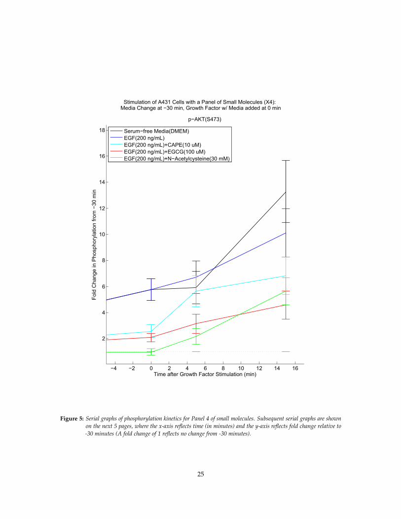
Stimulation of A431 Cells with a Panel of Small Molecules (X4):Media Change at −30 min, Growth Factor w/ Media added at 0 min
−4 −2 0 2 4 6 8 10 12 14 16
2
4
6
8
10
12
14
16
18
p−AKT(S473)
Time after Growth Factor Stimulation (min)
Fold
Cha
nge
in P
hosp
hory
latio
n fro
m −
30 m
in
Serum−free Media(DMEM)EGF(200 ng/mL)EGF(200 ng/mL)+CAPE(10 uM)EGF(200 ng/mL)+EGCG(100 uM)EGF(200 ng/mL)+N−Acetylcysteine(30 mM)
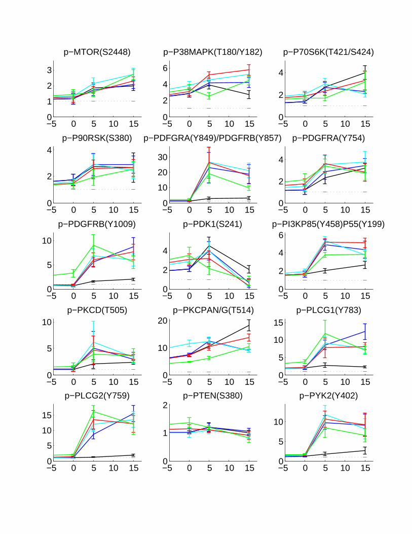
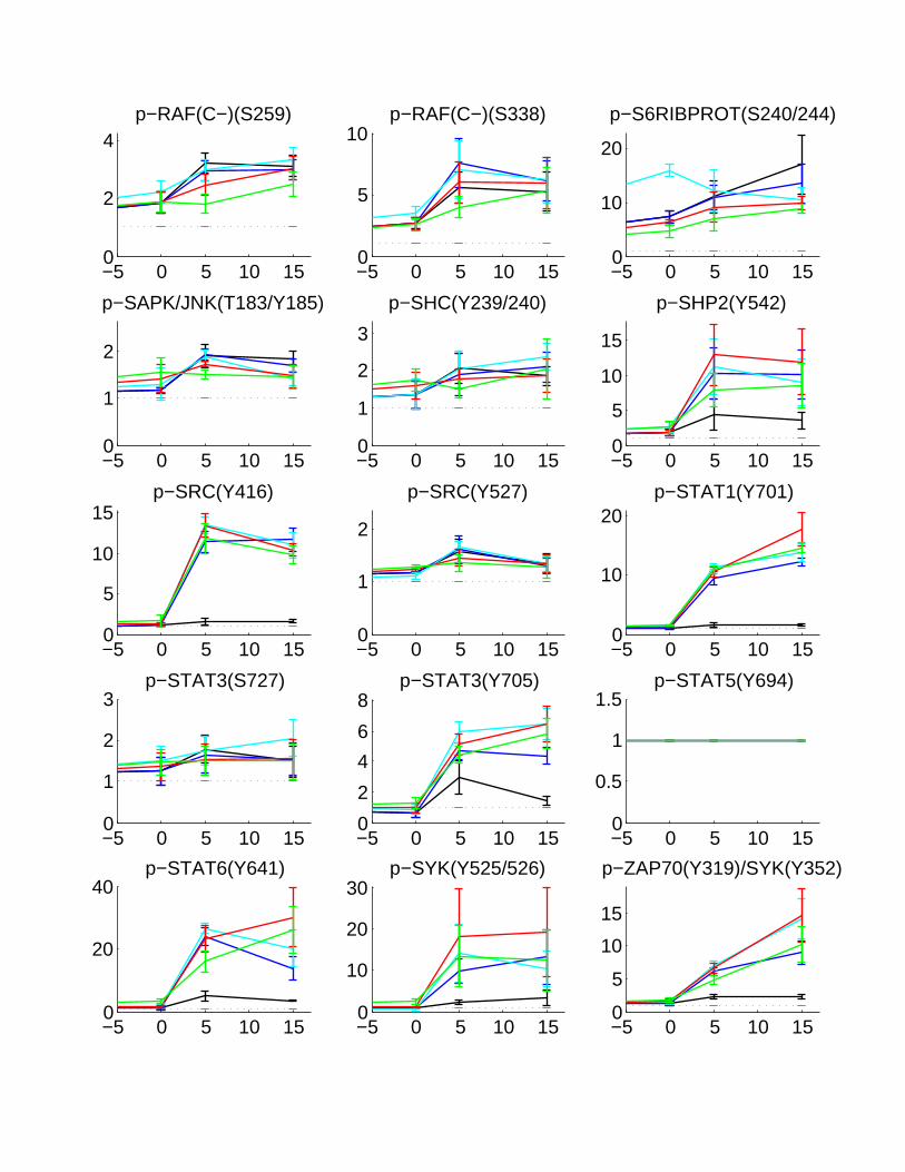
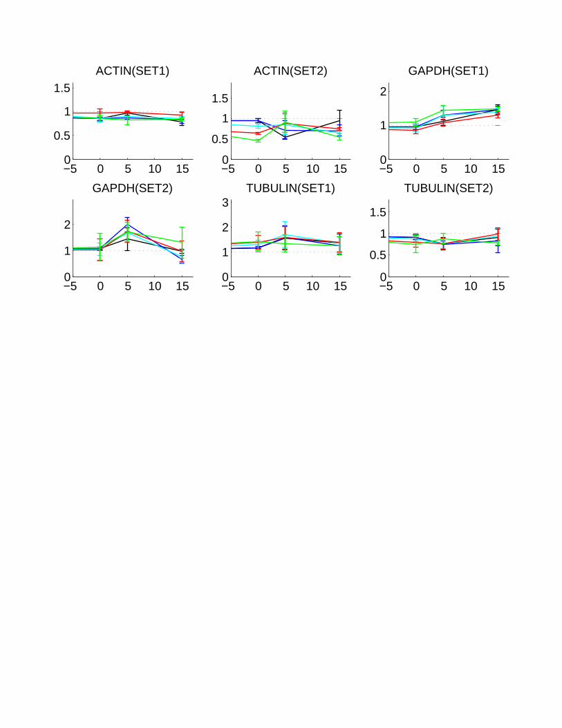
Figure 5: Serial graphs of phosphorylation kinetics for Panel 4 of small molecules. Subsequent serial graphs are shownon the next 5 pages, where the x-axis reflects time (in minutes) and the y-axis reflects fold change relative to-30 minutes (A fold change of 1 reflects no change from -30 minutes).
25

−5 0 5 10 150
0.5
1
1.5p−4EBP1(S65)
−5 0 5 10 150
1
2
3
p−4EBP1(T37/46)
−5 0 5 10 150
0.5
1
1.5
p−ABL(C−)(Y245)
−5 0 5 10 150
5
10
15
p−AKT(S473)
−5 0 5 10 150
2
4
6
p−CDC2(Y15)
−5 0 5 10 150
2
4
6
8p−CDK2(T160)
−5 0 5 10 150
2
4
p−CREB(S133)
−5 0 5 10 150
2
4
p−CRKII(Y221)
−5 0 5 10 150
1
2
3
p−CRKL(Y207)
−5 0 5 10 150
2
4
6
p−EGFR(Y1068)
−5 0 5 10 150
10
20
30
p−EGFR(Y1086)
−5 0 5 10 150
2
4
6
p−EGFR(Y1173)
−5 0 5 10 150
2
4
p−EGFR(Y845)
−5 0 5 10 150
2
4p−ERBB2(Y1221/1222)
−5 0 5 10 150
1
2
3
p−ERBB4(Y1284)

−5 0 5 10 150
5
10p−ERK(T202/Y204)
−5 0 5 10 150
2
4
6
p−FAK(Y576/577)
−5 0 5 10 150
2
4
p−FGFR1(Y653/654)
−5 0 5 10 150
1
2
3
p−FOXO1(S319)/FOXO4(S262)
−5 0 5 10 150
2
4
p−FOXO3A(S253)
−5 0 5 10 150
2
4
6
p−GAB1(Y627)
−5 0 5 10 150
1
2p−GAB2(S159)
−5 0 5 10 150
1
2
p−GSK3A(S21)
−5 0 5 10 150
5
10
p−IGF1RB (Y1135/1136) /p−InsR…
−5 0 5 10 150
2
4p−IRS1(S1101)
−5 0 5 10 150
2
4p−JAK1(Y1022/1023)
−5 0 5 10 150
2
4
p−KIT(C−)(Y719)
−5 0 5 10 150
5
10p−MEK(1/2)(S217/221)
−5 0 5 10 150
2
4
6
p−MET(Y1234/1235)
−5 0 5 10 150
1
2
3p−MET(Y1349)

−5 0 5 10 150
2
4
p−MTOR(S2448)
−5 0 5 10 150
20
40
p−P38MAPK(T180/Y182)
−5 0 5 10 150
1
2
p−P70S6K(T421/S424)
−5 0 5 10 150
2
4
6
p−P90RSK(S380)
−5 0 5 10 150
10
20
p−PDFGRA(Y849)/PDGFRB(Y857)
−5 0 5 10 150
2
4
6
p−PDGFRA(Y754)
−5 0 5 10 150
5
10
15
p−PDGFRB(Y1009)
−5 0 5 10 150
2
4
p−PDK1(S241)
−5 0 5 10 150
2
4
6
8p−PI3KP85(Y458)P55(Y199)
−5 0 5 10 150
2
4
6
p−PKCD(T505)
−5 0 5 10 150
5
10
15
p−PKCPAN/G(T514)
−5 0 5 10 150
2
4
6
p−PLCG1(Y783)
−5 0 5 10 150
5
10
p−PLCG2(Y759)
−5 0 5 10 150
2
4
6
p−PTEN(S380)
−5 0 5 10 150
2
4
6
8p−PYK2(Y402)

−5 0 5 10 150
1
2p−RAF(C−)(S259)
−5 0 5 10 150
2
4
6
p−RAF(C−)(S338)
−5 0 5 10 150
5
10
15p−S6RIBPROT(S240/244)
−5 0 5 10 150
5
10
15p−SAPK/JNK(T183/Y185)
−5 0 5 10 150
5
10p−SHC(Y239/240)
−5 0 5 10 150
10
20
30
p−SHP2(Y542)
−5 0 5 10 150
2
4
p−SRC(Y416)
−5 0 5 10 150
2
4
p−SRC(Y527)
−5 0 5 10 150
5
10
p−STAT1(Y701)
−5 0 5 10 150
2
4
6
p−STAT3(S727)
−5 0 5 10 150
0.5
1
1.5p−STAT3(Y705)
−5 0 5 10 150
1
2
3
p−STAT5(Y694)
−5 0 5 10 150
10
20
p−STAT6(Y641)
−5 0 5 10 150
2
4
6p−SYK(Y525/526)
−5 0 5 10 150
2
4
6
p−ZAP70(Y319)/SYK(Y352)
−5 0 5 10 150
0.5
1
1.5ACTIN(SET1)
−5 0 5 10 150
0.5
1
1.5
ACTIN(SET2)
−5 0 5 10 150
1
2
GAPDH(SET1)
−5 0 5 10 150
1
2
GAPDH(SET2)
−5 0 5 10 150
1
2
3TUBULIN(SET1)
−5 0 5 10 150
0.5
1
1.5
TUBULIN(SET2)
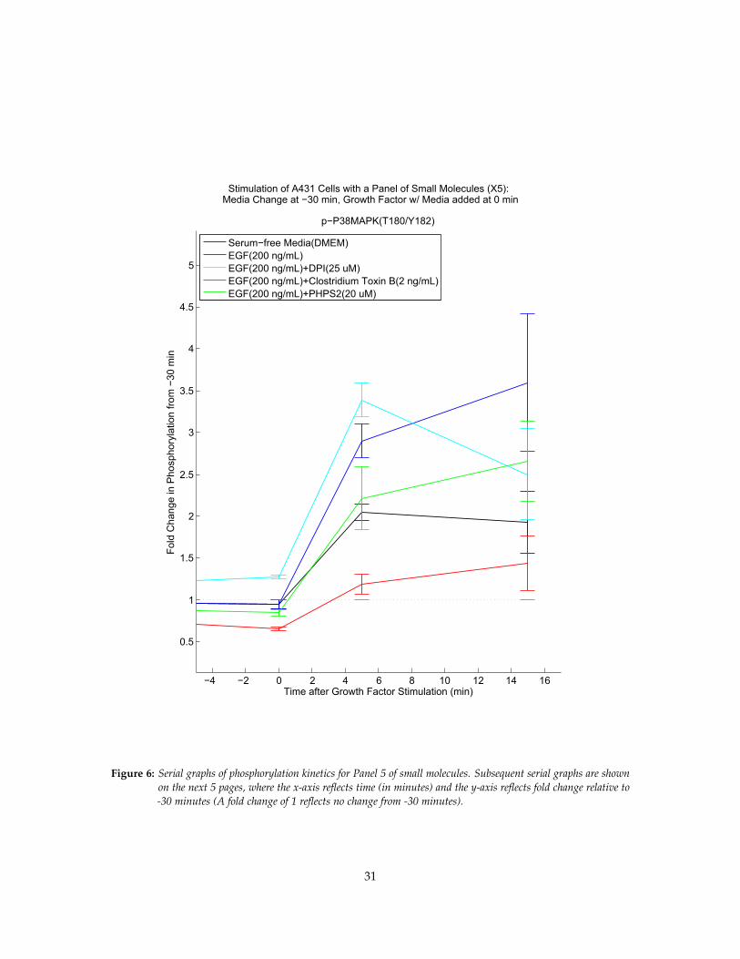
Stimulation of A431 Cells with a Panel of Small Molecules (X5):Media Change at −30 min, Growth Factor w/ Media added at 0 min
−4 −2 0 2 4 6 8 10 12 14 16
0.5
1
1.5
2
2.5
3
3.5
4
4.5
5
p−P38MAPK(T180/Y182)
Time after Growth Factor Stimulation (min)
Fold
Cha
nge
in P
hosp
hory
latio
n fro
m −
30 m
in
Serum−free Media(DMEM)EGF(200 ng/mL)EGF(200 ng/mL)+DPI(25 uM)EGF(200 ng/mL)+Clostridium Toxin B(2 ng/mL)EGF(200 ng/mL)+PHPS2(20 uM)
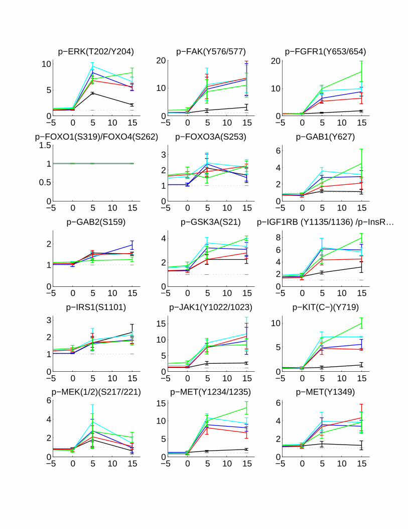
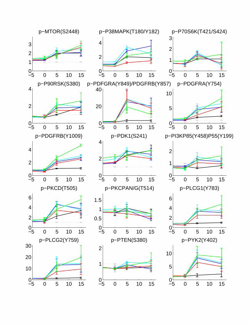
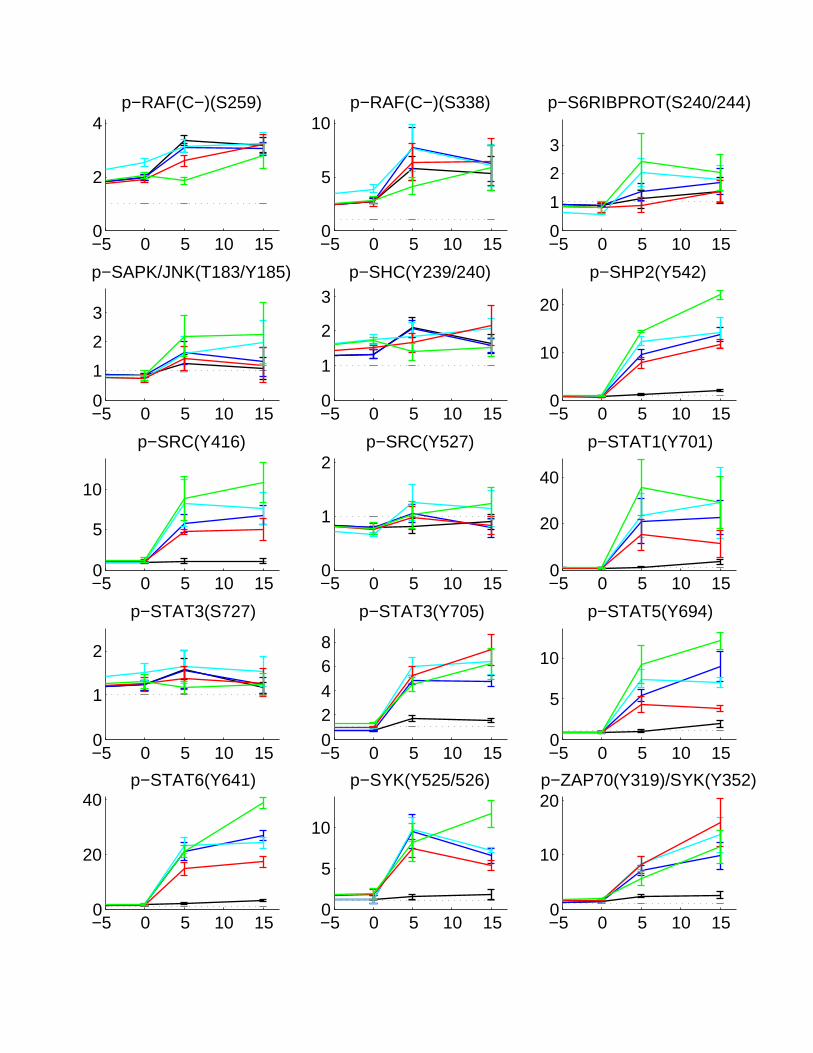
Figure 6: Serial graphs of phosphorylation kinetics for Panel 5 of small molecules. Subsequent serial graphs are shownon the next 5 pages, where the x-axis reflects time (in minutes) and the y-axis reflects fold change relative to-30 minutes (A fold change of 1 reflects no change from -30 minutes).
31

−5 0 5 10 150
2
4
68
p−4EBP1(S65)
−5 0 5 10 150
2
4
p−4EBP1(T37/46)
−5 0 5 10 150
2
4
p−ABL(C−)(Y245)
−5 0 5 10 150
1
2
3
p−AKT(S473)
−5 0 5 10 150
2
4
6p−CDC2(Y15)
−5 0 5 10 150
0.5
1
1.5p−CDK2(T160)
−5 0 5 10 150
1
2
p−CREB(S133)
−5 0 5 10 150
2
4
6
8p−CRKII(Y221)
−5 0 5 10 150
1
2
p−CRKL(Y207)
−5 0 5 10 150
10
20
p−EGFR(Y1068)
−5 0 5 10 150
50
100
p−EGFR(Y1086)
−5 0 5 10 150
5
10
p−EGFR(Y1173)
−5 0 5 10 150
2
4
6
p−EGFR(Y845)
−5 0 5 10 150
5
10
15
p−ERBB2(Y1221/1222)
−5 0 5 10 150
2
4
6
p−ERBB4(Y1284)
−5 0 5 10 150
5
10p−ERK(T202/Y204)
−5 0 5 10 150
10
20p−FAK(Y576/577)
−5 0 5 10 150
10
20p−FGFR1(Y653/654)
−5 0 5 10 150
0.5
1
1.5p−FOXO1(S319)/FOXO4(S262)
−5 0 5 10 150
1
2
3
p−FOXO3A(S253)
−5 0 5 10 150
2
4
6
p−GAB1(Y627)
−5 0 5 10 150
1
2
p−GAB2(S159)
−5 0 5 10 150
2
4
p−GSK3A(S21)
−5 0 5 10 1502468
p−IGF1RB (Y1135/1136) /p−InsR…
−5 0 5 10 150
1
2
3
p−IRS1(S1101)
−5 0 5 10 150
5
10
15
p−JAK1(Y1022/1023)
−5 0 5 10 150
5
10
p−KIT(C−)(Y719)
−5 0 5 10 150
2
4
6p−MEK(1/2)(S217/221)
−5 0 5 10 150
5
10
15p−MET(Y1234/1235)
−5 0 5 10 150
2
4
6p−MET(Y1349)
−5 0 5 10 150
1
2
3
p−MTOR(S2448)
−5 0 5 10 150
2
4
p−P38MAPK(T180/Y182)
−5 0 5 10 150
1
2
3p−P70S6K(T421/S424)
−5 0 5 10 150
2
4p−P90RSK(S380)
−5 0 5 10 150
20
40p−PDFGRA(Y849)/PDGFRB(Y857)
−5 0 5 10 150
5
10
p−PDGFRA(Y754)
−5 0 5 10 150
2
4
p−PDGFRB(Y1009)
−5 0 5 10 150
2
4p−PDK1(S241)
−5 0 5 10 150
1
2
p−PI3KP85(Y458)P55(Y199)
−5 0 5 10 150
2
4
6
p−PKCD(T505)
−5 0 5 10 150
0.5
1
1.5
p−PKCPAN/G(T514)
−5 0 5 10 150
2
4
6
p−PLCG1(Y783)
−5 0 5 10 150
10
20
30p−PLCG2(Y759)
−5 0 5 10 150
1
2
p−PTEN(S380)
−5 0 5 10 150
5
10
p−PYK2(Y402)
−5 0 5 10 150
2
4p−RAF(C−)(S259)
−5 0 5 10 150
5
10p−RAF(C−)(S338)
−5 0 5 10 150
1
2
3
p−S6RIBPROT(S240/244)
−5 0 5 10 150
1
2
3
p−SAPK/JNK(T183/Y185)
−5 0 5 10 150
1
2
3p−SHC(Y239/240)
−5 0 5 10 150
10
20
p−SHP2(Y542)
−5 0 5 10 150
5
10
p−SRC(Y416)
−5 0 5 10 150
1
2p−SRC(Y527)
−5 0 5 10 150
20
40
p−STAT1(Y701)
−5 0 5 10 150
1
2
p−STAT3(S727)
−5 0 5 10 1502468
p−STAT3(Y705)
−5 0 5 10 150
5
10
p−STAT5(Y694)
−5 0 5 10 150
20
40p−STAT6(Y641)
−5 0 5 10 150
5
10
p−SYK(Y525/526)
−5 0 5 10 150
10
20p−ZAP70(Y319)/SYK(Y352)
−5 0 5 10 150
2
4
ACTIN(SET1)
−5 0 5 10 150
1
2ACTIN(SET2)
−5 0 5 10 150
1
2
3
GAPDH(SET1)
−5 0 5 10 150
0.5
1
1.5
GAPDH(SET2)
−5 0 5 10 150
0.5
1
1.5
TUBULIN(SET1)
−5 0 5 10 150
1
2
TUBULIN(SET2)
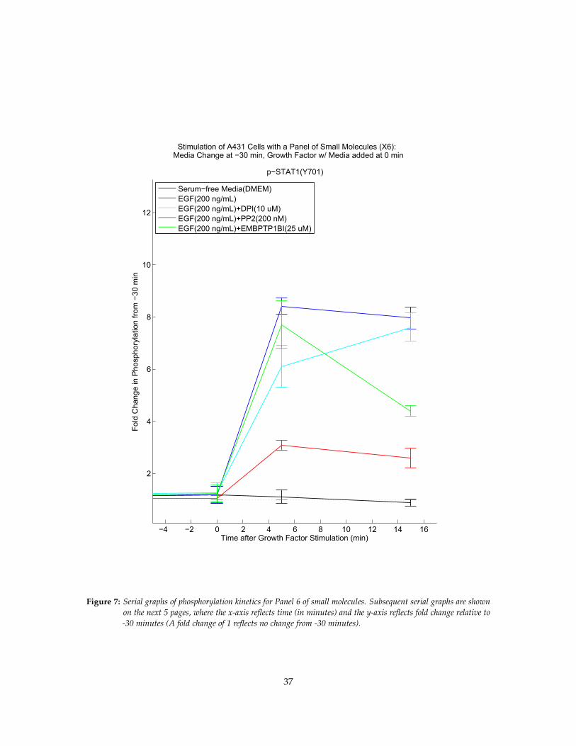
Stimulation of A431 Cells with a Panel of Small Molecules (X6):Media Change at −30 min, Growth Factor w/ Media added at 0 min
−4 −2 0 2 4 6 8 10 12 14 16
2
4
6
8
10
12
p−STAT1(Y701)
Time after Growth Factor Stimulation (min)
Fold
Cha
nge
in P
hosp
hory
latio
n fro
m −
30 m
in
Serum−free Media(DMEM)EGF(200 ng/mL)EGF(200 ng/mL)+DPI(10 uM)EGF(200 ng/mL)+PP2(200 nM)EGF(200 ng/mL)+EMBPTP1BI(25 uM)
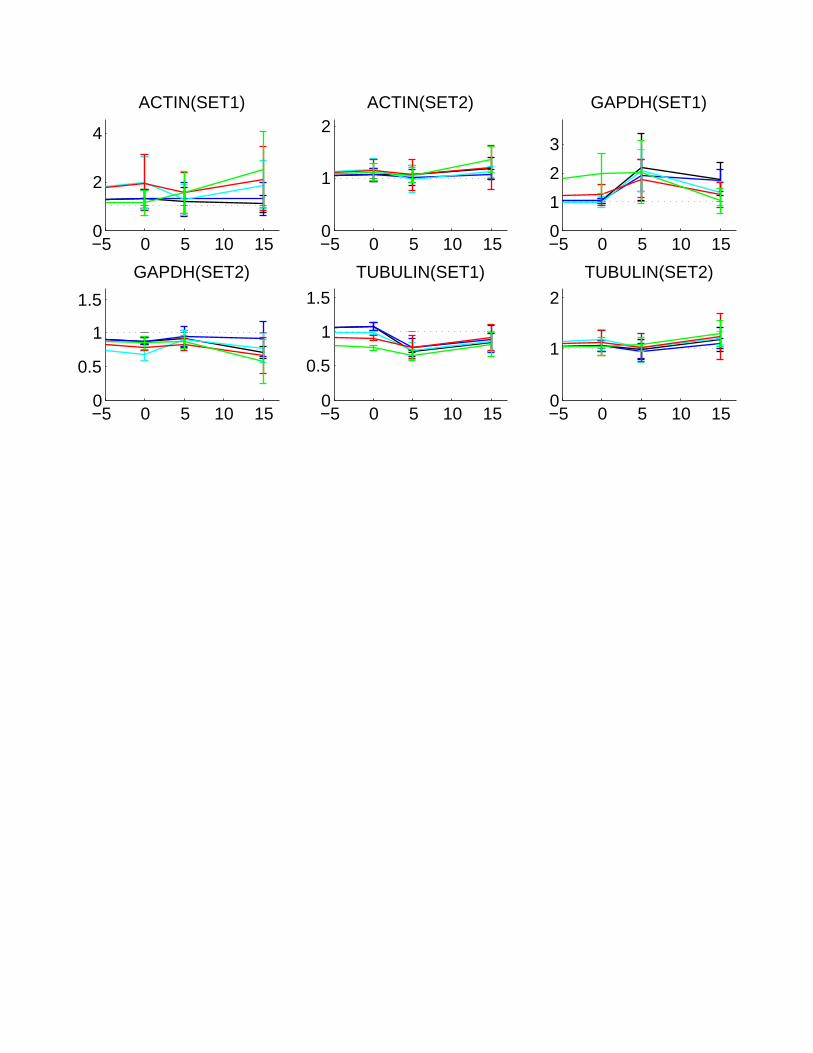
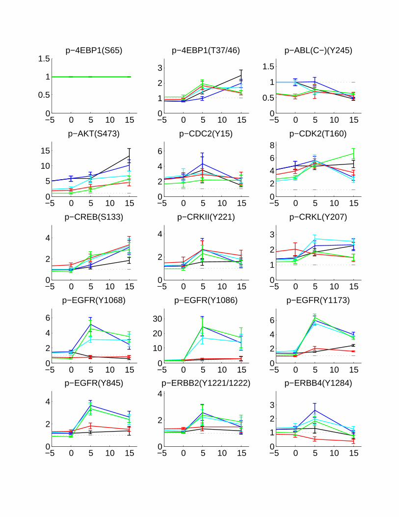
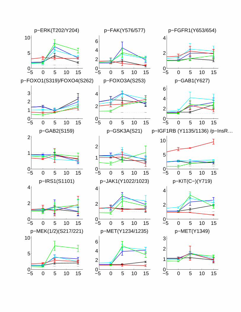
Figure 7: Serial graphs of phosphorylation kinetics for Panel 6 of small molecules. Subsequent serial graphs are shownon the next 5 pages, where the x-axis reflects time (in minutes) and the y-axis reflects fold change relative to-30 minutes (A fold change of 1 reflects no change from -30 minutes).
37
−5 0 5 10 150
0.5
1
1.5p−4EBP1(S65)
−5 0 5 10 150
1
2
3
p−4EBP1(T37/46)
−5 0 5 10 150
0.5
1
1.5
p−ABL(C−)(Y245)
−5 0 5 10 150
5
10
15
p−AKT(S473)
−5 0 5 10 150
2
4
6
p−CDC2(Y15)
−5 0 5 10 150
2
4
6
8p−CDK2(T160)
−5 0 5 10 150
2
4
p−CREB(S133)
−5 0 5 10 150
2
4
p−CRKII(Y221)
−5 0 5 10 150
1
2
3
p−CRKL(Y207)
−5 0 5 10 150
2
4
6
p−EGFR(Y1068)
−5 0 5 10 150
10
20
30
p−EGFR(Y1086)
−5 0 5 10 150
2
4
6
p−EGFR(Y1173)
−5 0 5 10 150
2
4
p−EGFR(Y845)
−5 0 5 10 150
2
4p−ERBB2(Y1221/1222)
−5 0 5 10 150
1
2
3
p−ERBB4(Y1284)
−5 0 5 10 150
5
10p−ERK(T202/Y204)
−5 0 5 10 150
2
4
6
p−FAK(Y576/577)
−5 0 5 10 150
2
4
p−FGFR1(Y653/654)
−5 0 5 10 150
1
2
3
p−FOXO1(S319)/FOXO4(S262)
−5 0 5 10 150
2
4
p−FOXO3A(S253)
−5 0 5 10 150
2
4
6
p−GAB1(Y627)
−5 0 5 10 150
1
2p−GAB2(S159)
−5 0 5 10 150
1
2
p−GSK3A(S21)
−5 0 5 10 150
5
10
p−IGF1RB (Y1135/1136) /p−InsR…
−5 0 5 10 150
2
4p−IRS1(S1101)
−5 0 5 10 150
2
4p−JAK1(Y1022/1023)
−5 0 5 10 150
2
4
p−KIT(C−)(Y719)
−5 0 5 10 150
5
10p−MEK(1/2)(S217/221)
−5 0 5 10 150
2
4
6
p−MET(Y1234/1235)
−5 0 5 10 150
1
2
3p−MET(Y1349)
−5 0 5 10 150
2
4
p−MTOR(S2448)
−5 0 5 10 150
20
40
p−P38MAPK(T180/Y182)
−5 0 5 10 150
1
2
p−P70S6K(T421/S424)
−5 0 5 10 150
2
4
6
p−P90RSK(S380)
−5 0 5 10 150
10
20
p−PDFGRA(Y849)/PDGFRB(Y857)
−5 0 5 10 150
2
4
6
p−PDGFRA(Y754)
−5 0 5 10 150
5
10
15
p−PDGFRB(Y1009)
−5 0 5 10 150
2
4
p−PDK1(S241)
−5 0 5 10 150
2
4
6
8p−PI3KP85(Y458)P55(Y199)
−5 0 5 10 150
2
4
6
p−PKCD(T505)
−5 0 5 10 150
5
10
15
p−PKCPAN/G(T514)
−5 0 5 10 150
2
4
6
p−PLCG1(Y783)
−5 0 5 10 150
5
10
p−PLCG2(Y759)
−5 0 5 10 150
2
4
6
p−PTEN(S380)
−5 0 5 10 150
2
4
6
8p−PYK2(Y402)
−5 0 5 10 150
1
2p−RAF(C−)(S259)
−5 0 5 10 150
2
4
6
p−RAF(C−)(S338)
−5 0 5 10 150
5
10
15p−S6RIBPROT(S240/244)
−5 0 5 10 150
5
10
15p−SAPK/JNK(T183/Y185)
−5 0 5 10 150
5
10p−SHC(Y239/240)
−5 0 5 10 150
10
20
30
p−SHP2(Y542)
−5 0 5 10 150
2
4
p−SRC(Y416)
−5 0 5 10 150
2
4
p−SRC(Y527)
−5 0 5 10 150
5
10
p−STAT1(Y701)
−5 0 5 10 150
2
4
6
p−STAT3(S727)
−5 0 5 10 150
0.5
1
1.5p−STAT3(Y705)
−5 0 5 10 150
1
2
3
p−STAT5(Y694)
−5 0 5 10 150
10
20
p−STAT6(Y641)
−5 0 5 10 150
2
4
6p−SYK(Y525/526)
−5 0 5 10 150
2
4
6
p−ZAP70(Y319)/SYK(Y352)

−5 0 5 10 150
0.5
1
1.5ACTIN(SET1)
−5 0 5 10 150
0.5
1
1.5
ACTIN(SET2)
−5 0 5 10 150
1
2
GAPDH(SET1)
−5 0 5 10 150
1
2
GAPDH(SET2)
−5 0 5 10 150
1
2
3TUBULIN(SET1)
−5 0 5 10 150
0.5
1
1.5
TUBULIN(SET2)

EG
F200
(0m
) X1
EG
F200
(5m
) X1
EG
F200
(15
m) X
1
EG
F200
+Ins
ulin
200
(0m
) X1
EG
F200
+Ins
ulin
200
(5m
) X1
EGF2
00+I
nsul
in20
0 (1
5m) X
1
HG
F200
(0m
) X1
HG
F200
(5m
) X1
HG
F200
(15
m) X
1
HG
F200
+Ins
ulin
200
(0m
) X1
HG
F200
+Ins
ulin
200
(5m
) X1
HG
F200
+Ins
ulin
200
(15m
) X1
Insu
lin20
0 (0
m) X
1
Insu
lin20
0 (5
m) X
1
Insu
lin20
0 (1
5m) X
1
IGF2
00 (
0m) X
1
IGF2
00 (
5m) X
1
IGF2
00 (
15m
) X1
p−MEK(1/2)(S217/221)p−ERK(T202/Y204)p−P38MAPK(T180/Y182)p−AKT(S473)p−RAF(C−)(S338)p−GSK3A(S21)p−FOXO3A(S253)p−PDK1(S241)p−RAF(C−)(S259)p−GAB2(S159)p−IGF1RB (Y1135/1136) /…p−ZAP70(Y319)/SYK(Y352)p−CRKII(Y221)p−STAT6(Y641)p−MET(Y1349)p−JAK1(Y1022/1023)p−PDGFRA(Y754)p−PKCD(T505)p−STAT3(S727)p−IRS1(S1101)p−SHC(Y239/240)p−ERBB2(Y1221/1222)p−MTOR(S2448)p−CDC2(Y15)p−FOXO1(S319)/…p−SAPK/JNK(T183/Y185)p−SYK(Y525/526)p−FAK(Y576/577)p−STAT3(Y705)p−STAT1(Y701)p−ERBB4(Y1284)p−SRC(Y416)p−ABL(C−)(Y245)p−4EBP1(S65)p−4EBP1(T37/46)p−PTEN(S380)p−SRC(Y527)p−CRKL(Y207)p−CREB(S133)p−P70S6K(T421/S424)p−PI3KP85(Y458)P55(Y199)p−CDK2(T160)p−P90RSK(S380)p−PKCPAN/G(T514)p−S6RIBPROT(S240/244)p−GAB1(Y627)p−SHP2(Y542)p−STAT5(Y694)p−FGFR1(Y653/654)p−PDGFRB(Y1009)p−PYK2(Y402)p−KIT(C−)(Y719)p−PDFGRA(Y849)/…p−EGFR(Y1068)p−EGFR(Y1086)p−PLCG2(Y759)p−PLCG1(Y783)p−MET(Y1234/1235)p−EGFR(Y845)p−EGFR(Y1173)
a)
b)
log(Fold Change from Time -30 min)
0 1 2-1-2
0 0.2 0.4 0.6 0.8 1 1.2 1.4
Media
EGF(200ng/mL)
EGF(200ng/mL)+Ins(200ng/mL)
HGF(200ng/mL)
HGF(200ng/mL)+Ins(200ng/mL)
Ins(200ng/mL)
IGF(200ng/mL)
Cell Viability as Fraction of Media alone
Figure 8: (a) The clustergram of phosphorylation kinetics is shown for Panel 1 of growth factors. (b) Relative cellviability is shown after 24 hours perturbation, in comparison to media alone.
43
EG
F200
(0m
) X2
EG
F200
(5m
) X2
EG
F200
(15
m) X
2
EG
F200
PP
2 (0
m) X
2
EG
F200
PP
2 (5
m) X
2
EG
F200
PP
2 (1
5m) X
2
EG
F200
U73
122
(0m
) X2
EG
F200
U73
122
(5m
) X2
EG
F200
U73
122
(15m
) X2
EG
F200
Wor
tman
nin
(0m
) X2
EG
F200
Wor
tman
nin
(5m
) X2
EG
F200
Wor
tman
nin
(15m
) X2
p−AKT(S473)p−S6RIBPROT(S240/244)p−4EBP1(T37/46)p−4EBP1(S65)p−GSK3A(S21)p−CREB(S133)p−CRKL(Y207)p−SAPK/JNK(T183/Y185)p−STAT3(S727)p−FOXO1(S319)/…p−PTEN(S380)p−P70S6K(T421/S424)p−P90RSK(S380)p−SRC(Y527)p−GAB2(S159)p−PKCPAN/G(T514)p−CDK2(T160)p−PDK1(S241)p−ABL(C−)(Y245)p−PDFGRA(Y849)/…p−PKCD(T505)p−CRKII(Y221)p−RAF(C−)(S338)p−ERBB4(Y1284)p−FOXO3A(S253)p−FAK(Y576/577)p−RAF(C−)(S259)p−IRS1(S1101)p−PI3KP85(Y458)P55(Y199)p−JAK1(Y1022/1023)p−STAT3(Y705)p−MTOR(S2448)p−STAT5(Y694)p−ERBB2(Y1221/1222)p−KIT(C−)(Y719)p−IGF1RB (Y1135/1136) /…p−CDC2(Y15)p−SHC(Y239/240)p−PDGFRB(Y1009)p−PYK2(Y402)p−GAB1(Y627)p−ERK(T202/Y204)p−MEK(1/2)(S217/221)p−P38MAPK(T180/Y182)p−MET(Y1349)p−ZAP70(Y319)/SYK(Y352)p−PDGFRA(Y754)p−SYK(Y525/526)p−PLCG1(Y783)p−SHP2(Y542)p−FGFR1(Y653/654)p−PLCG2(Y759)p−STAT6(Y641)p−STAT1(Y701)p−EGFR(Y845)p−SRC(Y416)p−MET(Y1234/1235)p−EGFR(Y1173)p−EGFR(Y1068)p−EGFR(Y1086)
log(Fold Change from Time -30 min)
0 1 2-1-2
0 0.5 1 1.5
Media
EGF(200ng/mL)
EGF(200ng/mL)+PP2(10uM)
EGF(200ng/mL)+U73122(10uM)
EGF(200ng/mL)+Wortmannin(10uM)
Cell Viability as Fraction of Media alone
a)
b)
Figure 9: (a) The clustergram of phosphorylation kinetics is shown for Panel 2 of small molecules. (b) Relative cellviability is shown after 24 hours perturbation, in comparison to media alone.
44

E
GF2
00 (0
m) X
3
EG
F200
(5m
) X3
EG
F200
(15
m) X
3
EG
F200
Apr
otin
in (0
m) X
3
EG
F200
Apr
otin
in (5
m) X
3
EG
F200
Apr
otin
in (1
5m) X
3
EG
F200
GM
6001
(0m
) X3
EG
F200
GM
6001
(5m
) X3
EG
F200
GM
6001
(15m
) X3
EG
F200
TA
PI−
1 (0
m) X
3
EG
F200
TA
PI−
1 (5
m) X
3
EG
F200
TA
PI−
1 (1
5m) X
3
p−PDK1(S241)p−PKCPAN/G(T514)p−S6RIBPROT(S240/244)p−AKT(S473)p−CDK2(T160)p−P70S6K(T421/S424)p−FOXO1(S319)/…p−PDGFRA(Y754)p−CRKII(Y221)p−4EBP1(S65)p−ERK(T202/Y204)p−CRKL(Y207)p−FOXO3A(S253)p−MTOR(S2448)p−GSK3A(S21)p−CDC2(Y15)p−SHC(Y239/240)p−STAT3(S727)p−IRS1(S1101)p−ABL(C−)(Y245)p−KIT(C−)(Y719)p−STAT5(Y694)p−SRC(Y527)p−4EBP1(T37/46)p−PTEN(S380)p−SAPK/JNK(T183/Y185)p−GAB2(S159)p−RAF(C−)(S259)p−P90RSK(S380)p−RAF(C−)(S338)p−P38MAPK(T180/Y182)p−CREB(S133)p−ERBB4(Y1284)p−PKCD(T505)p−PI3KP85(Y458)P55(Y199)p−GAB1(Y627)p−MEK(1/2)(S217/221)p−MET(Y1349)p−SHP2(Y542)p−IGF1RB (Y1135/1136) /…p−ZAP70(Y319)/SYK(Y352)p−STAT3(Y705)p−JAK1(Y1022/1023)p−PLCG1(Y783)p−PDGFRB(Y1009)p−STAT6(Y641)p−SYK(Y525/526)p−EGFR(Y1068)p−FAK(Y576/577)p−PYK2(Y402)p−ERBB2(Y1221/1222)p−FGFR1(Y653/654)p−STAT1(Y701)p−EGFR(Y845)p−PLCG2(Y759)p−SRC(Y416)p−PDFGRA(Y849)/…p−MET(Y1234/1235)p−EGFR(Y1173)p−EGFR(Y1086)
log(Fold Change from Time -30 min)
0 1 2-1-2
b)
a)
0 0.2 0.4 0.6 0.8 1 1.2 1.4
Media
EGF(200ng/mL)
EGF(200ng/mL)+Aprotinin(1ug/mL)
EGF(200ng/mL)+GM6001(25uM)
EGF(200ng/mL)+TAPI−1(100uM)
Cell Viability as Fraction of Media alone
Figure 10: (a) The clustergram of phosphorylation kinetics is shown for Panel 3 of small molecules. (b) Relative cellviability is shown after 24 hours perturbation, in comparison to media alone.
45
0 (0
m) X
4
EG
F200
(5m
) X4
EG
F200
(15
m) X
4
EG
F200
CA
PE
(0m
) X4
EG
F200
CA
PE
(5m
) X4
EG
F200
CA
PE
(15m
) X4
EG
F200
EG
CG
(0m
) X4
EG
F200
EG
CG
(5m
) X4
EG
F200
EG
CG
(15m
) X4
EG
F200
N−A
cety
lcys
tein
e (0
m) X
4
EG
F200
N−A
cety
lcys
tein
e (5
m) X
4
EG
F200
N−A
cety
lcys
tein
e (1
5m) X
4
p−AKT(S473)p−PKCPAN/G(T514)p−P90RSK(S380)p−FOXO1(S319)/…p−STAT5(Y694)p−ERBB4(Y1284)p−KIT(C−)(Y719)p−GAB1(Y627)p−GSK3A(S21)p−MEK(1/2)(S217/221)p−PTEN(S380)p−EGFR(Y845)p−JAK1(Y1022/1023)p−SHC(Y239/240)p−ERK(T202/Y204)p−FGFR1(Y653/654)p−CREB(S133)p−CRKII(Y221)p−ERBB2(Y1221/1222)p−PKCD(T505)p−MET(Y1349)p−MTOR(S2448)p−SYK(Y525/526)p−FOXO3A(S253)p−ABL(C−)(Y245)p−IRS1(S1101)p−CDC2(Y15)p−P70S6K(T421/S424)p−STAT3(Y705)p−4EBP1(S65)p−GAB2(S159)p−RAF(C−)(S259)p−RAF(C−)(S338)p−PDK1(S241)p−CDK2(T160)p−SRC(Y527)p−CRKL(Y207)p−SRC(Y416)p−4EBP1(T37/46)p−PI3KP85(Y458)P55(Y199)p−S6RIBPROT(S240/244)p−SAPK/JNK(T183/Y185)p−P38MAPK(T180/Y182)p−IGF1RB (Y1135/1136) /…p−PDGFRA(Y754)p−STAT3(S727)p−ZAP70(Y319)/SYK(Y352)p−PLCG1(Y783)p−FAK(Y576/577)p−PLCG2(Y759)p−MET(Y1234/1235)p−STAT1(Y701)p−PDFGRA(Y849)/…p−STAT6(Y641)p−EGFR(Y1068)p−EGFR(Y1086)p−PDGFRB(Y1009)p−EGFR(Y1173)p−PYK2(Y402)p−SHP2(Y542)
log(Fold Change from Time -30 min)
0 1 2-1-2
0 0.5 1 1.5
Media
EGF(200ng/mL)
EGF(200ng/mL)+CAPE(10uM)
EGF(200ng/mL)+EGCG(100uM)
EGF(200ng/mL)+N−Acetylcysteine(30mM)
Cell Viability as Fraction of Media alone
a)
b)
Figure 11: a) The clustergram of phosphorylation kinetics is shown for Panel 4 of small molecules. b) Relative cellviability is shown after 24 hours perturbation, in comparison to media alone.
46
0 (0
m) X
5
EG
F200
(5m
) X5
EG
F200
(15
m) X
5
EG
F200
DP
I (0m
) X5
EG
F200
DP
I (5m
) X5
EG
F200
DP
I (15
m) X
5
EG
F200
Clo
strid
ium
Tox
in B
(0m
) X5
EG
F200
Clo
strid
ium
Tox
in B
(5m
) X5
EG
F200
Clo
strid
ium
Tox
in B
(15m
) X5
EG
F200
PH
PS
2 (0
m) X
5
EG
F200
PH
PS
2 (5
m) X
5
EG
F200
PH
PS
2 (1
5m) X
5p−FOXO3A(S253)p−MTOR(S2448)p−RAF(C−)(S338)p−STAT3(S727)p−SHC(Y239/240)p−RAF(C−)(S259)p−GAB2(S159)p−FOXO1(S319)/…p−CDK2(T160)p−PKCPAN/G(T514)p−IRS1(S1101)p−PDK1(S241)p−P70S6K(T421/S424)p−PKCD(T505)p−P90RSK(S380)p−AKT(S473)p−4EBP1(T37/46)p−CREB(S133)p−SRC(Y527)p−PTEN(S380)p−ABL(C−)(Y245)p−GSK3A(S21)p−CDC2(Y15)p−CRKL(Y207)p−SAPK/JNK(T183/Y185)p−S6RIBPROT(S240/244)p−P38MAPK(T180/Y182)p−4EBP1(S65)p−MEK(1/2)(S217/221)p−PI3KP85(Y458)P55(Y199)p−IGF1RB (Y1135/1136) /…p−CRKII(Y221)p−ERBB4(Y1284)p−PDGFRA(Y754)p−GAB1(Y627)p−ERK(T202/Y204)p−PDGFRB(Y1009)p−MET(Y1349)p−ZAP70(Y319)/SYK(Y352)p−STAT3(Y705)p−JAK1(Y1022/1023)p−ERBB2(Y1221/1222)p−PLCG1(Y783)p−PYK2(Y402)p−EGFR(Y845)p−STAT5(Y694)p−MET(Y1234/1235)p−KIT(C−)(Y719)p−EGFR(Y1173)p−EGFR(Y1068)p−SYK(Y525/526)p−FAK(Y576/577)p−PDFGRA(Y849)/…p−SRC(Y416)p−FGFR1(Y653/654)p−PLCG2(Y759)p−STAT6(Y641)p−SHP2(Y542)p−STAT1(Y701)p−EGFR(Y1086)
log(Fold Change from Time -30 min)0 1 2-1-2
0 0.5 1 1.5
Media
EGF(200ng/mL)
EGF(200ng/mL)+DPI(25uM)
EGF(200ng/mL)+Clostr. Toxin B(2ng/mL)
EGF(200ng/mL)+PHPS1(20uM)
Cell Viability as Fraction of Media alone
a)
b)
Figure 12: (a) The clustergram of phosphorylation kinetics is shown for Panel 5 of small molecules. (b) Relative cellviability is shown after 24 hours perturbation, in comparison to media alone.
47
EG
F200
(0m
) X6
EG
F200
(5m
) X6
EG
F200
(15
m) X
6
EG
F200
DP
I (0m
) X6
EG
F200
DP
I (5m
) X6
EG
F200
DP
I (15
m) X
6
EG
F200
PP
2 (0
m) X
6
EG
F200
PP
2 (5
m) X
6
EG
F200
PP
2 (1
5m) X
6
EG
F200
EM
BP
TP1B
I (0m
) X6
EG
F200
EM
BP
TP1B
I (5m
) X6
EG
F200
EM
BP
TP1B
I (15
m) X
6
p−S6RIBPROT(S240/244)p−4EBP1(T37/46)p−PTEN(S380)p−FOXO1(S319)/…p−SRC(Y527)p−CDK2(T160)p−4EBP1(S65)p−CDC2(Y15)p−ABL(C−)(Y245)p−RAF(C−)(S338)p−STAT3(S727)p−GAB2(S159)p−PDK1(S241)p−RAF(C−)(S259)p−AKT(S473)p−CRKL(Y207)p−CREB(S133)p−PI3KP85(Y458)P55(Y199)p−PDGFRA(Y754)p−PKCD(T505)p−FOXO3A(S253)p−MTOR(S2448)p−CRKII(Y221)p−SRC(Y416)p−P38MAPK(T180/Y182)p−P70S6K(T421/S424)p−PKCPAN/G(T514)p−SAPK/JNK(T183/Y185)p−GSK3A(S21)p−MET(Y1349)p−P90RSK(S380)p−IGF1RB (Y1135/1136) /…p−IRS1(S1101)p−SHC(Y239/240)p−PYK2(Y402)p−STAT5(Y694)p−GAB1(Y627)p−MEK(1/2)(S217/221)p−ERK(T202/Y204)p−SHP2(Y542)p−EGFR(Y845)p−ZAP70(Y319)/SYK(Y352)p−FGFR1(Y653/654)p−ERBB4(Y1284)p−ERBB2(Y1221/1222)p−PDGFRB(Y1009)p−JAK1(Y1022/1023)p−EGFR(Y1173)p−PDFGRA(Y849)/…p−STAT3(Y705)p−FAK(Y576/577)p−KIT(C−)(Y719)p−STAT1(Y701)p−SYK(Y525/526)p−MET(Y1234/1235)p−EGFR(Y1068)p−PLCG1(Y783)p−STAT6(Y641)p−PLCG2(Y759)p−EGFR(Y1086)
log(Fold Change from Time -30 min)0 1 2-1-2
a)
b)
0 0.5 1 1.5
Media
EGF(200ng/mL)
EGF(200ng/mL)+DPI(10uM)
EGF(200ng/mL)+PP2(200nM)
EGF(200ng/mL)+EMBPTP1BI(25uM)
Cell Viability as Fraction of Media alone
Figure 13: (a) The clustergram of phosphorylation kinetics is shown for Panel 6 of small molecules. (b) Relative cellviability is shown after 24 hours perturbation, in comparison to media alone.
48
−150 −100 −50 0 50 100 150
1.4
1.6
1.8
2
2.2
2.4
2.6
2.8
3
p−CDK2(T160)[0m]
p−PI3KP85(Y458)[0m]P55(Y199)[0m]
p−PKCPAN/G(T514)[0m]
p−CDC2(Y15)[5m]
p−EGFR(Y1086)[5m]
p−EGFR(Y845)[5m]
p−ERBB4(Y1284)[5m]p−ERK(T202/Y204)[5m]
p−FGFR1(Y653/654)[5m]
p−GSK3A(S21)[5m]
p−JAK1(Y1022/1023)[5m]
p−MET(Y1234/1235)[5m]
p−MTOR(S2448)[5m]
p−PLCG2(Y759)[5m]
p−PTEN(S380)[5m]
p−STAT3(Y705)[5m]
p−4EBP1(T37/46)[15m]
p−CDK2(T160)[15m]p−CRKL(Y207)[15m]
p−EGFR(Y1086)[15m]
p−EGFR(Y1173)[15m]
p−ERBB4(Y1284)[15m]
p−MEK(1/2)[15m](S217/221)[15m]
p−P38MAPK(T180/Y182)[15m]
p−P90RSK(S380)[15m]
p−PYK2(Y402)[15m]
p−SAPK/JNK(T183/Y185)[15m]
Percent inhibition of Aprotinin on EGF stimulation of specified phosphometric
−log
10(p
−val
ue)
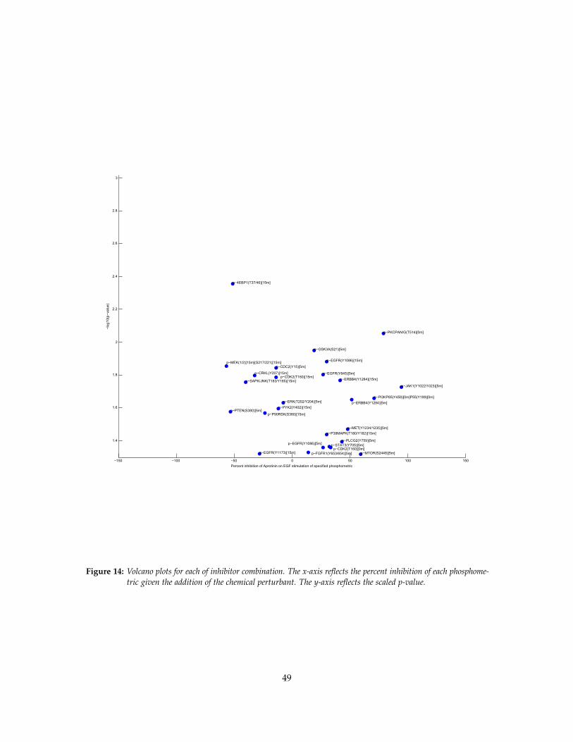
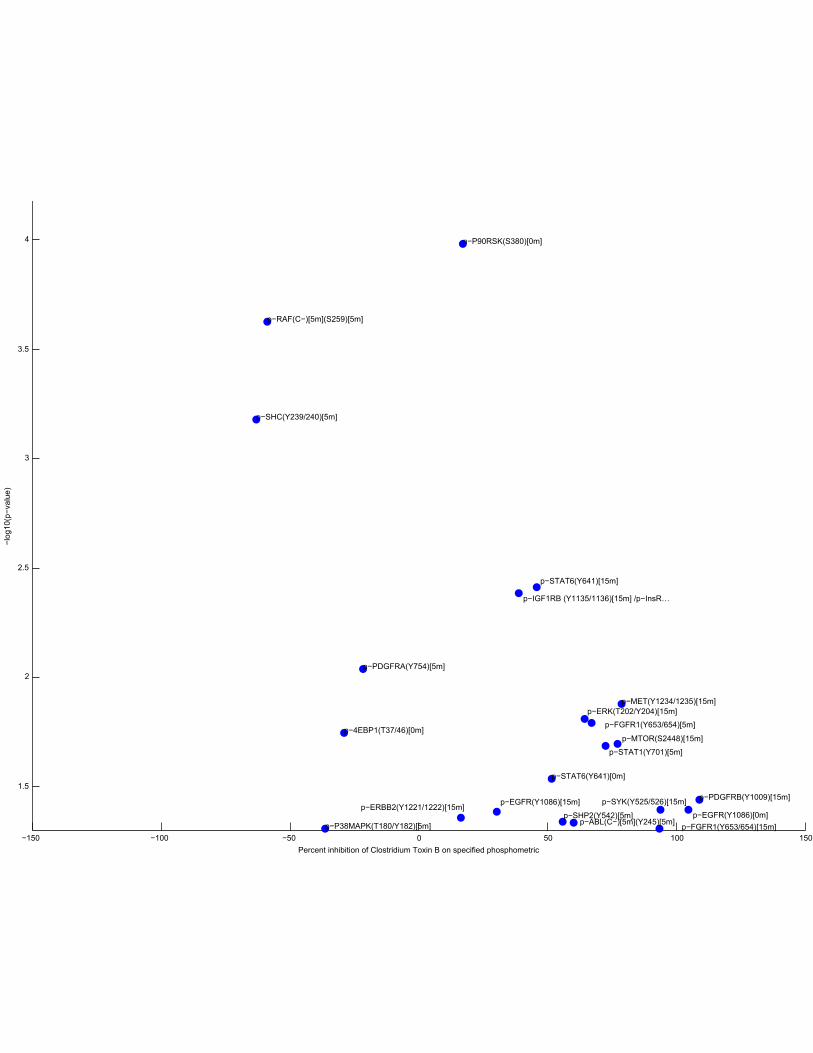
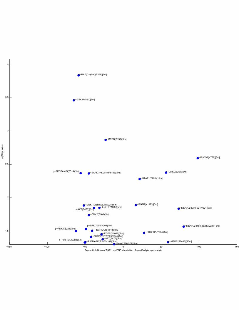
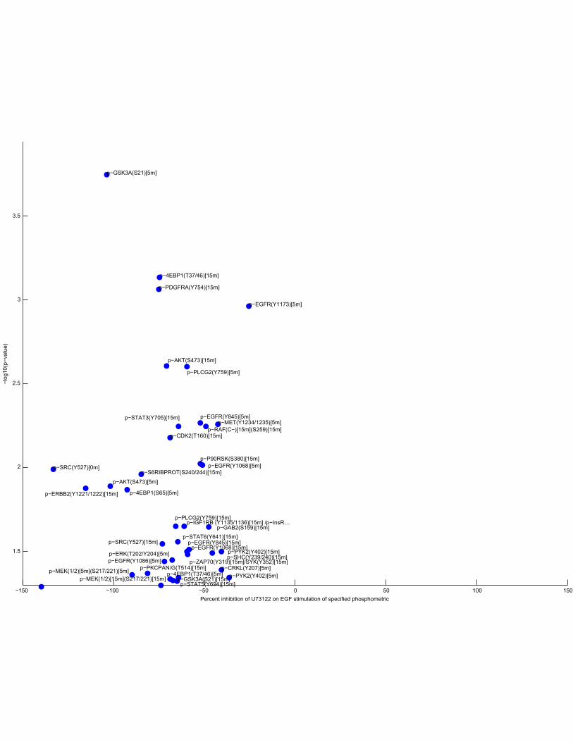
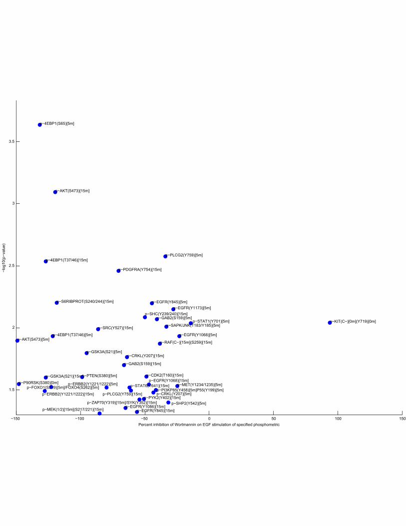
Figure 14: Volcano plots for each of inhibitor combination. The x-axis reflects the percent inhibition of each phosphome-tric given the addition of the chemical perturbant. The y-axis reflects the scaled p-value.
49

−60 −40 −20 0 20 40 60 80
1.4
1.6
1.8
2
2.2
2.4
2.6
2.8
3
3.2
3.4
p−EGFR(Y1086)[0m]
p−ERK(T202/Y204)[0m]
p−MET(Y1234/1235)[0m]p−P38MAPK(T180/Y182)[0m]
p−PLCG1(Y783)[0m]
p−PLCG2(Y759)[0m]
p−PTEN(S380)[0m]
p−STAT1(Y701)[0m]
p−ZAP70(Y319)[0m]/SYK(Y352)[0m]
p−ABL(C−)[5m](Y245)[5m]p−CREB(S133)[5m]
p−ERBB4(Y1284)[5m]
p−ERK(T202/Y204)[5m]
p−FAK(Y576/577)[5m]p−MTOR(S2448)[5m]
p−P38MAPK(T180/Y182)[5m]
p−P90RSK(S380)[5m]p−PLCG2(Y759)[5m]
p−RAF(C−)[5m](S259)[5m]
p−RAF(C−)[5m](S338)[5m]
p−AKT(S473)[15m]
p−CDK2(T160)[15m]
p−CRKII(Y221)[15m]
p−ERK(T202/Y204)[15m]
p−FAK(Y576/577)[15m]
p−GAB2(S159)[15m]
p−IGF1RB (Y1135/1136)[15m] /p−InsR…
p−MTOR(S2448)[15m]
p−P38MAPK(T180/Y182)[15m]
p−PDGFRB(Y1009)[15m]
p−PLCG2(Y759)[15m]
p−RAF(C−)[15m](S259)[15m]
p−RAF(C−)[15m](S338)[15m]
Percent inhibition of CAPE on EGF stimulation of specified phosphometric
−log
10(p
−val
ue)
−150 −100 −50 0 50 100 150
1.5
2
2.5
3
3.5
4
p−4EBP1(T37/46)[0m]
p−EGFR(Y1086)[0m]
p−P90RSK(S380)[0m]
p−STAT6(Y641)[0m]
p−ABL(C−)[5m](Y245)[5m]
p−FGFR1(Y653/654)[5m]
p−P38MAPK(T180/Y182)[5m]
p−PDGFRA(Y754)[5m]
p−RAF(C−)[5m](S259)[5m]
p−SHC(Y239/240)[5m]
p−SHP2(Y542)[5m]
p−STAT1(Y701)[5m]
p−EGFR(Y1086)[15m]p−ERBB2(Y1221/1222)[15m]
p−ERK(T202/Y204)[15m]
p−FGFR1(Y653/654)[15m]
p−IGF1RB (Y1135/1136)[15m] /p−InsR…
p−MET(Y1234/1235)[15m]
p−MTOR(S2448)[15m]
p−PDGFRB(Y1009)[15m]
p−STAT6(Y641)[15m]
p−SYK(Y525/526)[15m]
Percent inhibition of Clostridium Toxin B on specified phosphometric
−log
10(p
−val
ue)
−150 −100 −50 0 50 100 150
1.5
2
2.5
3
3.5
p−RAF(C−)[0m](S338)[0m]
p−CREB(S133)[5m]
p−FOXO3A(S253)[5m]p−GSK3A(S21)[5m]
p−MEK(1/2)[5m](S217/221)[5m]
p−PLCG2(Y759)[5m]
p−STAT3(S727)[5m]
p−STAT5(Y694)[5m]
p−ZAP70(Y319)[5m]/SYK(Y352)[5m]
p−4EBP1(T37/46)[15m]
p−CREB(S133)[15m]
p−CRKL(Y207)[15m]
p−EGFR(Y1086)[15m]
p−EGFR(Y845)[15m]
p−ERK(T202/Y204)[15m]
p−FAK(Y576/577)[15m]
p−FGFR1(Y653/654)[15m]
p−JAK1(Y1022/1023)[15m]
p−MEK(1/2)[15m](S217/221)[15m]
p−MET(Y1234/1235)[15m]
p−PDFGRA(Y849)[15m]/PDGFRB(Y857)[15m]
p−PI3KP85(Y458)[15m]P55(Y199)[15m]
p−PLCG2(Y759)[15m]
p−PYK2(Y402)[15m]
p−S6RIBPROT(S240/244)[15m]
p−STAT3(S727)[15m]
p−STAT6(Y641)[15m]
p−ZAP70(Y319)[15m]/SYK(Y352)[15m]
Percent inhibition of DPI(10 uM) on specified phosphometric
−log
10(p
−val
ue)
−150 −100 −50 0 50 100 150
1.4
1.6
1.8
2
2.2
2.4
2.6
2.8
3
p−RAF(C−)[0m](S338)[0m]
p−STAT3(Y705)[0m]
p−ABL(C−)[5m](Y245)[5m]
p−CREB(S133)[5m]
p−CRKL(Y207)[5m]
p−ERK(T202/Y204)[5m]
p−FGFR1(Y653/654)[5m]
p−KIT(C−)[5m](Y719)[5m]
p−P38MAPK(T180/Y182)[5m]
p−P90RSK(S380)[5m]
p−SHP2(Y542)[5m]
p−STAT6(Y641)[5m]
p−ZAP70(Y319)[5m]/SYK(Y352)[5m]
p−4EBP1(T37/46)[15m]
p−EGFR(Y1173)[15m]
p−ERBB2(Y1221/1222)[15m]
p−ERBB4(Y1284)[15m]
p−ERK(T202/Y204)[15m]
p−IRS1(S1101)[15m]
p−P38MAPK(T180/Y182)[15m]
p−ZAP70(Y319)[15m]/SYK(Y352)[15m]
Percent inhibition of DPI(25 uM) on specified phosphometric
−log
10(p
−val
ue)

−300 −200 −100 0 100 200 300 400 500 600 700
1.5
2
2.5
3
3.5
p−AKT(S473)[0m]
p−EGFR(Y1173)[0m]
p−ERBB4(Y1284)[0m]
p−ERK(T202/Y204)[0m]
p−FOXO3A(S253)[0m]
p−GAB2(S159)[0m]
p−IGF1RB (Y1135/1136)[0m] /p−InsR…
p−P38MAPK(T180/Y182)[0m]
p−PDK1(S241)[0m]p−SAPK/JNK(T183/Y185)[0m]p−EGFR(Y1068)[5m]
p−EGFR(Y1173)[5m]
p−ERBB4(Y1284)[5m]
p−ERK(T202/Y204)[5m]
p−FAK(Y576/577)[5m]
p−FOXO3A(S253)[5m]
p−IGF1RB (Y1135/1136)[5m] /p−InsR…
p−JAK1(Y1022/1023)[5m]
p−MTOR(S2448)[5m]
p−P38MAPK(T180/Y182)[5m]
p−PDFGRA(Y849)[5m]/PDGFRB(Y857)[5m]
p−PDGFRB(Y1009)[5m]
p−PDK1(S241)[5m]
p−PLCG2(Y759)[5m]
p−RAF(C−)[5m](S259)[5m]
p−RAF(C−)[5m](S338)[5m]
p−STAT1(Y701)[5m]
p−AKT(S473)[15m]p−CRKII(Y221)[15m]
p−EGFR(Y1173)[15m]
p−ERBB4(Y1284)[15m]
p−GAB2(S159)[15m]
p−GSK3A(S21)[15m]
p−IGF1RB (Y1135/1136)[15m] /p−InsR…
p−MET(Y1234/1235)[15m]
p−P38MAPK(T180/Y182)[15m]
p−PDFGRA(Y849)[15m]/PDGFRB(Y857)[15m]
p−PDGFRB(Y1009)[15m]
p−PYK2(Y402)[15m]
p−STAT1(Y701)[15m]
Percent inhibition of EGCG on EGF stimulation of specified phosphometric
−log
10(p
−val
ue)
−150 −100 −50 0 50 100 150
1.4
1.6
1.8
2
2.2
2.4
2.6
2.8
3
3.2
3.4
p−CDK2(T160)[0m]
p−CRKII(Y221)[0m]
p−FAK(Y576/577)[0m]
p−FGFR1(Y653/654)[0m]
p−GAB2(S159)[0m]
p−MEK(1/2)[0m](S217/221)[0m]p−MET(Y1234/1235)[0m]
p−S6RIBPROT(S240/244)[0m]
p−SAPK/JNK(T183/Y185)[0m]
p−SRC(Y416)[0m]
p−ABL(C−)[5m](Y245)[5m]
p−EGFR(Y1068)[5m]p−EGFR(Y845)[5m]
p−ERK(T202/Y204)[5m]
p−FAK(Y576/577)[5m]
p−GSK3A(S21)[5m]
p−MEK(1/2)[5m](S217/221)[5m]
p−MET(Y1234/1235)[5m]
p−PDGFRA(Y754)[5m]
p−PI3KP85(Y458)[5m]P55(Y199)[5m]
p−PLCG1(Y783)[5m]
p−PLCG2(Y759)[5m]
p−RAF(C−)[5m](S338)[5m]
p−STAT1(Y701)[5m]
p−STAT5(Y694)[5m]
p−ZAP70(Y319)[5m]/SYK(Y352)[5m]
p−4EBP1(T37/46)[15m]
p−CREB(S133)[15m]
p−CRKL(Y207)[15m]
p−EGFR(Y1086)[15m] p−EGFR(Y845)[15m]p−ERBB2(Y1221/1222)[15m]
p−FAK(Y576/577)[15m]
p−FGFR1(Y653/654)[15m]
p−GAB1(Y627)[15m]
p−GAB2(S159)[15m]
p−JAK1(Y1022/1023)[15m]
p−MET(Y1234/1235)[15m]
p−P38MAPK(T180/Y182)[15m]
p−P90RSK(S380)[15m]
p−PDFGRA(Y849)[15m]/PDGFRB(Y857)[15m]
p−PDGFRB(Y1009)[15m]
p−PI3KP85(Y458)[15m]P55(Y199)[15m]
p−PLCG2(Y759)[15m]
p−PYK2(Y402)[15m]
p−S6RIBPROT(S240/244)[15m]
p−SHC(Y239/240)[15m]
p−STAT1(Y701)[15m]
p−STAT6(Y641)[15m]
p−ZAP70(Y319)[15m]/SYK(Y352)[15m]
Percent inhibition of EMBPTP1BI on specified phosphometric
−log
10(p
−val
ue)
−150 −100 −50 0 50 100 150
1.4
1.6
1.8
2
2.2
2.4
p−AKT(S473)[0m]
p−CRKII(Y221)[0m]p−SHC(Y239/240)[0m]
p−STAT3(Y705)[0m]
p−ZAP70(Y319)[0m]/SYK(Y352)[0m]
p−CDK2(T160)[5m]
p−ERBB4(Y1284)[5m]
p−FGFR1(Y653/654)[5m]
p−FOXO3A(S253)[5m]
p−MET(Y1234/1235)[5m]
p−PDGFRA(Y754)[5m]
p−PDK1(S241)[5m]
p−PKCPAN/G(T514)[5m]
p−RAF(C−)[5m](S259)[5m]
p−RAF(C−)[5m](S338)[5m]
p−ABL(C−)[15m](Y245)[15m]
p−CDC2(Y15)[15m]
p−P38MAPK(T180/Y182)[15m]
p−PKCPAN/G(T514)[15m]
Percent inhibition of GM6001 on EGF stimulation of specified phosphometric
−log
10(p
−val
ue)
−50 0 50 100 150
1.4
1.6
1.8
2
2.2
2.4
2.6
2.8
3
p−AKT(S473)[0m]
p−EGFR(Y1068)[0m]
p−ERBB4(Y1284)[0m]
p−FOXO3A(S253)[0m]
p−IGF1RB (Y1135/1136)[0m] /p−InsR…
p−MET(Y1234/1235)[0m]
p−PDGFRA(Y754)[0m]
p−PDK1(S241)[0m]
p−PYK2(Y402)[0m]
p−RAF(C−)[0m](S259)[0m]
p−ZAP70(Y319)[0m]/SYK(Y352)[0m]
p−ABL(C−)[5m](Y245)[5m]
p−ERBB4(Y1284)[5m] p−ERK(T202/Y204)[5m]
p−FAK(Y576/577)[5m]
p−FOXO3A(S253)[5m]
p−P38MAPK(T180/Y182)[5m]
p−S6RIBPROT(S240/244)[5m]
p−ERBB2(Y1221/1222)[15m]
p−ERK(T202/Y204)[15m]
p−FGFR1(Y653/654)[15m]
p−MEK(1/2)[15m](S217/221)[15m]
p−MET(Y1234/1235)[15m]
p−P38MAPK(T180/Y182)[15m]p−PDGFRA(Y754)[15m]
p−PDGFRB(Y1009)[15m]
p−PDK1(S241)[15m]
p−PKCD(T505)[15m]
p−PYK2(Y402)[15m]
p−RAF(C−)[15m](S259)[15m]
p−STAT1(Y701)[15m]
Percent inhibition of N−Ac on EGF stimulation of specified phosphometric
−log
10(p
−val
ue)
−150 −100 −50 0 50 100 150
1.4
1.6
1.8
2
2.2
2.4
2.6
2.8
3
3.2
3.4
p−4EBP1(T37/46)[0m]
p−CREB(S133)[0m]
p−ERK(T202/Y204)[0m]
p−RAF(C−)[0m](S259)[0m]
p−STAT3(Y705)[0m]
p−ZAP70(Y319)[0m]/SYK(Y352)[0m]
p−CDC2(Y15)[5m]
p−CREB(S133)[5m]
p−EGFR(Y1086)[5m]
p−ERBB2(Y1221/1222)[5m]p−ERK(T202/Y204)[5m]
p−GAB1(Y627)[5m]
p−GSK3A(S21)[5m]
p−MEK(1/2)[5m](S217/221)[5m]p−P38MAPK(T180/Y182)[5m]
p−PYK2(Y402)[5m]
p−RAF(C−)[5m](S259)[5m]
p−RAF(C−)[5m](S338)[5m]
p−S6RIBPROT(S240/244)[5m]
p−SHC(Y239/240)[5m]
p−STAT6(Y641)[5m]
p−ABL(C−)[15m](Y245)[15m]
p−CDC2(Y15)[15m]
p−CRKII(Y221)[15m]
p−EGFR(Y1086)[15m]
p−ERBB2(Y1221/1222)[15m]
p−FOXO3A(S253)[15m]
p−MTOR(S2448)[15m]
p−P38MAPK(T180/Y182)[15m]
p−STAT1(Y701)[15m]p−STAT6(Y641)[15m]
Percent inhibition of PHPS2 on specified phosphometric
−log
10(p
−val
ue)
−150 −100 −50 0 50 100 150
1.5
2
2.5
3
3.5
4
4.5
p−4EBP1(S65)[5m]
p−4EBP1(T37/46)[5m]
p−AKT(S473)[5m]
p−CDK2(T160)[5m]
p−CRKL(Y207)[5m]
p−EGFR(Y1068)[5m]
p−EGFR(Y1086)[5m]
p−EGFR(Y1173)[5m]
p−EGFR(Y845)[5m]
p−ERK(T202/Y204)[5m]
p−GSK3A(S21)[5m]
p−JAK1(Y1022/1023)[5m]
p−MET(Y1234/1235)[5m]
p−P90RSK(S380)[5m]
p−PDGFRA(Y754)[5m]
p−PKCPAN/G(T514)[5m]
p−PLCG2(Y759)[5m]
p−SHC(Y239/240)[5m]
p−SRC(Y527)[5m]
p−4EBP1(S65)[15m]
p−4EBP1(T37/46)[15m]
p−AKT(S473)[15m]
p−CDK2(T160)[15m]
p−CRKL(Y207)[15m]
p−EGFR(Y1068)[15m]
p−EGFR(Y1086)[15m]
p−EGFR(Y845)[15m]
p−FOXO3A(S253)[15m]
p−GAB2(S159)[15m]
p−GSK3A(S21)[15m]
p−IGF1RB (Y1135/1136)[15m] /p−InsR…p−MEK(1/2)[15m](S217/221)[15m]
p−P70S6K(T421/S424)[15m]
p−P90RSK(S380)[15m]
p−PDGFRA(Y754)[15m]
p−PKCPAN/G(T514)[15m]
p−PLCG2(Y759)[15m]
p−PYK2(Y402)[15m]
p−RAF(C−)[15m](S259)[15m]
p−S6RIBPROT(S240/244)[15m]
p−SHC(Y239/240)[15m]
p−STAT3(Y705)[15m]
p−STAT5(Y694)[15m]
p−STAT6(Y641)[15m]
p−ZAP70(Y319)[15m]/SYK(Y352)[15m]
Percent inhibition of PP2 on EGF stimulation of specified phosphometric
−log
10(p
−val
ue)
−150 −100 −50 0 50 100 150
1.5
2
2.5
3
3.5
p−CDC2(Y15)[0m]
p−CDK2(T160)[0m]
p−EGFR(Y845)[0m]
p−FGFR1(Y653/654)[0m]
p−GAB2(S159)[0m]
p−GSK3A(S21)[0m]
p−PDK1(S241)[0m]
p−FAK(Y576/577)[5m]
p−PYK2(Y402)[5m]
p−RAF(C−)[5m](S259)[5m]p−RAF(C−)[5m](S338)[5m]
p−ABL(C−)[15m](Y245)[15m]
p−CREB(S133)[15m]
p−CRKL(Y207)[15m]
p−EGFR(Y1086)[15m]
p−EGFR(Y845)[15m]
p−ERBB2(Y1221/1222)[15m]
p−ERK(T202/Y204)[15m]
p−FAK(Y576/577)[15m]
p−FGFR1(Y653/654)[15m]
p−FOXO3A(S253)[15m]
p−GAB1(Y627)[15m]
p−IRS1(S1101)[15m]
p−JAK1(Y1022/1023)[15m]
p−MET(Y1234/1235)[15m]p−PDGFRB(Y1009)[15m]
p−PDK1(S241)[15m]
p−PI3KP85(Y458)[15m]P55(Y199)[15m]
p−PLCG1(Y783)[15m]
p−PLCG2(Y759)[15m]
p−PYK2(Y402)[15m]p−RAF(C−)[15m](S259)[15m]
p−S6RIBPROT(S240/244)[15m]p−SHC(Y239/240)[15m]
p−SRC(Y416)[15m]
p−SRC(Y527)[15m]
p−STAT1(Y701)[15m]
p−STAT3(S727)[15m]
p−STAT6(Y641)[15m]
p−ZAP70(Y319)[15m]/SYK(Y352)[15m]
Percent inhibition of PP2(200 nM) on specified phosphometric
−log
10(p
−val
ue)
−150 −100 −50 0 50 100 150
1.5
2
2.5
3
3.5
4
p−AKT(S473)[0m]
p−CRKL(Y207)[0m]
p−MEK(1/2)[0m](S217/221)[0m]
p−PKCPAN/G(T514)[0m]
p−AKT(S473)[5m]
p−CDK2(T160)[5m]
p−CREB(S133)[5m]
p−EGFR(Y1068)[5m]
p−EGFR(Y1086)[5m]p−EGFR(Y1173)[5m]
p−ERK(T202/Y204)[5m]
p−FAK(Y576/577)[5m]
p−GSK3A(S21)[5m]
p−MEK(1/2)[5m](S217/221)[5m]
p−P38MAPK(T180/Y182)[5m]p−P90RSK(S380)[5m]
p−PDGFRA(Y754)[5m]p−PDK1(S241)[5m]
p−PKCPAN/G(T514)[5m]
p−PLCG2(Y759)[5m]
p−RAF(C−)[5m](S259)[5m]
p−S6RIBPROT(S240/244)[5m]
p−SAPK/JNK(T183/Y185)[5m]
p−MEK(1/2)[15m](S217/221)[15m]
p−MTOR(S2448)[15m]
p−STAT1(Y701)[15m]
Percent inhibition of TAPI1 on EGF stimulation of specified phosphometric
−log
10(p
−val
ue)
−150 −100 −50 0 50 100 150
1.5
2
2.5
3
3.5
p−SRC(Y527)[0m]
p−4EBP1(S65)[5m]
p−4EBP1(T37/46)[5m]
p−AKT(S473)[5m]
p−CRKL(Y207)[5m]
p−EGFR(Y1068)[5m]
p−EGFR(Y1086)[5m]
p−EGFR(Y1173)[5m]
p−EGFR(Y845)[5m]
p−ERK(T202/Y204)[5m]
p−GSK3A(S21)[5m]
p−MEK(1/2)[5m](S217/221)[5m]
p−MET(Y1234/1235)[5m]
p−PLCG2(Y759)[5m]
p−PYK2(Y402)[5m]
p−4EBP1(T37/46)[15m]
p−AKT(S473)[15m]
p−CDK2(T160)[15m]
p−EGFR(Y1068)[15m]p−EGFR(Y845)[15m]
p−ERBB2(Y1221/1222)[15m]
p−GAB2(S159)[15m]
p−GSK3A(S21)[15m]
p−IGF1RB (Y1135/1136)[15m] /p−InsR…
p−MEK(1/2)[15m](S217/221)[15m]
p−P90RSK(S380)[15m]
p−PDGFRA(Y754)[15m]
p−PKCPAN/G(T514)[15m]
p−PLCG2(Y759)[15m]
p−PYK2(Y402)[15m]
p−RAF(C−)[15m](S259)[15m]
p−S6RIBPROT(S240/244)[15m]
p−SHC(Y239/240)[15m]
p−SRC(Y527)[15m]
p−STAT3(Y705)[15m]
p−STAT5(Y694)[15m]
p−STAT6(Y641)[15m]
p−ZAP70(Y319)[15m]/SYK(Y352)[15m]
Percent inhibition of U73122 on EGF stimulation of specified phosphometric
−log
10(p
−val
ue)
−150 −100 −50 0 50 100 150
1.5
2
2.5
3
3.5
p−KIT(C−)[0m](Y719)[0m]
p−P90RSK(S380)[0m]
p−4EBP1(S65)[5m]
p−4EBP1(T37/46)[5m]p−AKT(S473)[5m]
p−CRKL(Y207)[5m]
p−EGFR(Y1068)[5m]
p−EGFR(Y1173)[5m]p−EGFR(Y845)[5m]
p−ERBB2(Y1221/1222)[5m]p−FOXO1(S319)[5m]/FOXO4(S262)[5m]
p−GAB2(S159)[5m]
p−GSK3A(S21)[5m]
p−MET(Y1234/1235)[5m]p−PI3KP85(Y458)[5m]P55(Y199)[5m]
p−PLCG2(Y759)[5m]
p−PTEN(S380)[5m]
p−SAPK/JNK(T183/Y185)[5m]
p−SHP2(Y542)[5m]
p−STAT1(Y701)[5m]
p−4EBP1(T37/46)[15m]
p−AKT(S473)[15m]
p−CDK2(T160)[15m]
p−CRKL(Y207)[15m]
p−EGFR(Y1068)[15m]
p−EGFR(Y1086)[15m]p−EGFR(Y845)[15m]
p−ERBB2(Y1221/1222)[15m]
p−GAB2(S159)[15m]
p−GSK3A(S21)[15m]
p−MEK(1/2)[15m](S217/221)[15m]
p−PDGFRA(Y754)[15m]
p−PLCG2(Y759)[15m]p−PYK2(Y402)[15m]
p−RAF(C−)[15m](S259)[15m]
p−S6RIBPROT(S240/244)[15m]
p−SHC(Y239/240)[15m]
p−SRC(Y527)[15m]
p−STAT6(Y641)[15m]
p−ZAP70(Y319)[15m]/SYK(Y352)[15m]
Percent inhibition of Wortmannin on EGF stimulation of specified phosphometric
−log
10(p
−val
ue)
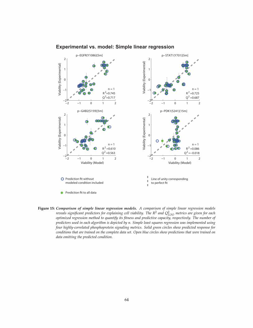
Experimental vs. model: Simple linear regression
Viab
ility
(Exp
erim
enta
l)Vi
abili
ty (E
xper
imen
tal)
Viability (Model) Viability (Model)
−2 −1 0 1 2−2
−1
0
1
2p−EGFR(Y1086)[5m]
n = 1
Q2=0.717R 2=0.745
−2 −1 0 1 2−2
−1
0
1
2p−STAT1(Y701)[5m]
n = 1
Q2 =0.687R 2 =0.725
−2 −1 0 1 2−2
−1
0
1
2p−GAB2(S159)[5m]
n = 1
Q2 =0.563R 2 =0.610
−2 −1 0 1 2−2
−1
0
1
2p−PDK1(S241)[15m]
n = 1
Q2 =−0.018R 2 =0.086Vi
abili
ty (E
xper
imen
tal)
Viab
ility
(Exp
erim
enta
l)
Prediction �t to all data
Prediction �t withoutmodeled condition included
Line of unity correspondingto perfect �t
Figure 15: Comparison of simple linear regression models. A comparison of simple linear regression modelsreveals significant predictors for explaining cell viability. The R2 and Q2
LOO metrics are given for eachoptimized regression method to quantify its fitness and predictive capacity, respectively. The number ofpredictors used in each algorithm is depicted by n. Simple least squares regression was implemented usingfour highly-correlated phosphoprotein signaling metrics. Solid green circles show predicted response forconditions that are trained on the complete data set. Open blue circles show predictions that were trained ondata omitting the predicted condition.
64
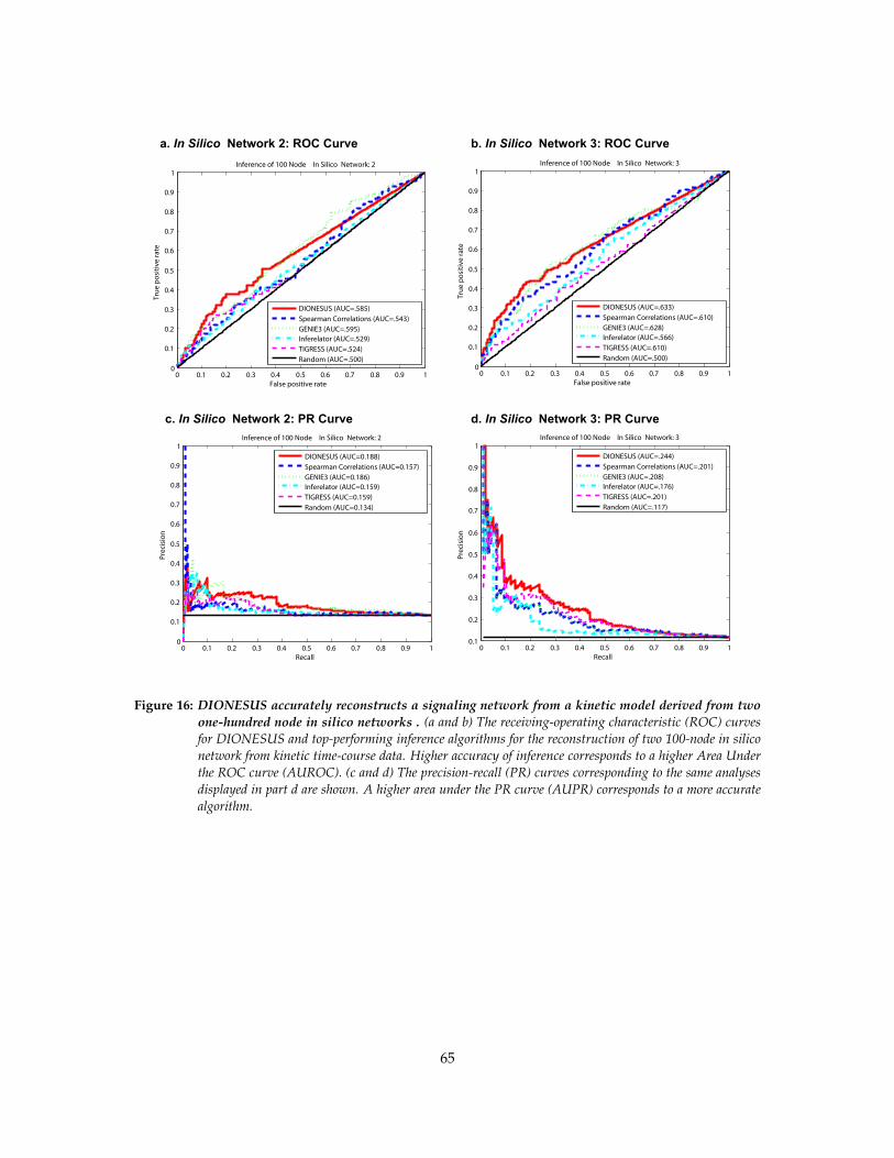
0 0.1 0.2 0.3 0.4 0.5 0.6 0.7 0.8 0.9 10
0.1
0.2
0.3
0.4
0.5
0.6
0.7
0.8
0.9
1
False positive rate
True
pos
itive
rate
Inference of 100 Node In Silico Network: 3
DIONESUS (AUC=.633)Spearman Correlations (AUC=.610)GENIE3 (AUC=.628)Inferelator (AUC=.566)TIGRESS (AUC=.610)Random (AUC=.500)
0 0.1 0.2 0.3 0.4 0.5 0.6 0.7 0.8 0.9 10
0.1
0.2
0.3
0.4
0.5
0.6
0.7
0.8
0.9
1
False positive rate
True
pos
itive
rate
Inference of 100 Node In Silico Network: 2
DIONESUS (AUC=.585)Spearman Correlations (AUC=.543)GENIE3 (AUC=.595)Inferelator (AUC=.529)TIGRESS (AUC=.524)Random (AUC=.500)
0 0.1 0.2 0.3 0.4 0.5 0.6 0.7 0.8 0.9 10.1
0.2
0.3
0.4
0.5
0.6
0.7
0.8
0.9
1
Recall
Prec
isio
n
Inference of 100 Node In Silico Network: 3
DIONESUS (AUC=.244)Spearman Correlations (AUC=.201)GENIE3 (AUC=.208)Inferelator (AUC=.176)TIGRESS (AUC=.201)Random (AUC=.117)
0 0.1 0.2 0.3 0.4 0.5 0.6 0.7 0.8 0.9 10
0.1
0.2
0.3
0.4
0.5
0.6
0.7
0.8
0.9
1
Recall
Prec
isio
n
Inference of 100 Node In Silico Network: 2
DIONESUS (AUC=0.188)Spearman Correlations (AUC=0.157)GENIE3 (AUC=0.186)Inferelator (AUC=0.159)TIGRESS (AUC=0.159)Random (AUC=0.134)
a. In Silico Network 2: ROC Curve b. In Silico Network 3: ROC Curve
c. In Silico Network 2: PR Curve d. In Silico Network 3: PR Curve
Figure 16: DIONESUS accurately reconstructs a signaling network from a kinetic model derived from twoone-hundred node in silico networks . (a and b) The receiving-operating characteristic (ROC) curvesfor DIONESUS and top-performing inference algorithms for the reconstruction of two 100-node in siliconetwork from kinetic time-course data. Higher accuracy of inference corresponds to a higher Area Underthe ROC curve (AUROC). (c and d) The precision-recall (PR) curves corresponding to the same analysesdisplayed in part d are shown. A higher area under the PR curve (AUPR) corresponds to a more accuratealgorithm.
65
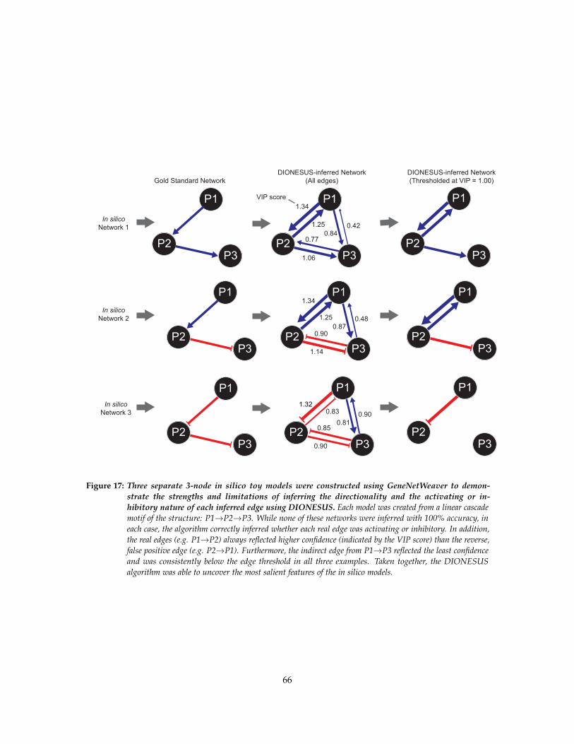
P1
P2P3
P1
P2P3
P1
P2P31.06
1.34
1.25
0.770.84
0.42
Gold Standard NetworkDIONESUS-inferred Network
(All edges)DIONESUS-inferred Network(Thresholded at VIP = 1.00)
P1
P3P2
P31.14
1.34
1.25
0.900.87
0.48
P3P2
P30.90
0.831.32
0.850.81
0.90
In silicoNetwork 1
In silicoNetwork 2
In silicoNetwork 3
P3
P3
P1
P2
P1
P2
P1
P2P2
VIP score
P1P1
Figure 17: Three separate 3-node in silico toy models were constructed using GeneNetWeaver to demon-strate the strengths and limitations of inferring the directionality and the activating or in-hibitory nature of each inferred edge using DIONESUS. Each model was created from a linear cascademotif of the structure: P1→P2→P3. While none of these networks were inferred with 100% accuracy, ineach case, the algorithm correctly inferred whether each real edge was activating or inhibitory. In addition,the real edges (e.g. P1→P2) always reflected higher confidence (indicated by the VIP score) than the reverse,false positive edge (e.g. P2→P1). Furthermore, the indirect edge from P1→P3 reflected the least confidenceand was consistently below the edge threshold in all three examples. Taken together, the DIONESUSalgorithm was able to uncover the most salient features of the in silico models.
66
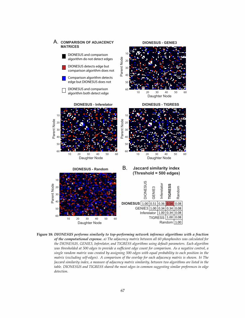
Daughter Node
Pare
nt N
ode
DIONESUS - GENIE3
10 20 30 40 50 60
10
20
30
40
50
60
Pare
nt N
ode
Daughter Node
DIONESUS - Inferelator
10 20 30 40 50 60
10
20
30
40
50
60
Pare
nt N
ode
Daughter Node
DIONESUS - TIGRESS
10 20 30 40 50 60
10
20
30
40
50
60
Pare
nt N
ode
Daughter Node
DIONESUS - Random
10 20 30 40 50 60
10
20
30
40
50
60
DIONESUS
DIO
NES
US
GEN
IE3
TIG
RES
S
Infe
rela
tor
Ran
dom
1.001.00
1.001.00
1.00
Jaccard similarity index(Threshold = 500 edges)
DIONESUS and comparison algorithm do not detect edges
DIONESUS and comparison algorithm both detect edge
DIONESUS detects edge but comparison algorithm does not
Comparison algorithm detects edge but DIONESUS does not
A.
B.
COMPARISON OF ADJACENCYMATRICES
GENIE3
TIGRESS Inferelator
Random
0.51 0.36 0.540.34 0.34
0.34
0.080.080.080.08
Figure 18: DIONESUS performs similarly to top-performing network inference algorithms with a fractionof the computational expense. a) The adjacency matrix between all 60 phosphosites was calculated forthe DIONESUS, GENIE3, Inferelator, and TIGRESS algorithms using default parameters. Each algorithmwas thresholded at 500 edges to provide a sufficient edge count for comparison. As a negative control, asingle random matrix was created by assigning 500 edges with equal probability to each position in thematrix (excluding self-edges). A comparison of the overlap for each adjacency matrix is shown. b) TheJaccard similarity index, a measure of adjacency matrix similarity, between two algorithms are listed in thetable. DIONESUS and TIGRESS shared the most edges in common suggesting similar preferences in edgedetection.
67

Figure 19: ImageJ software was used to quantify microwestern arrays where bands were difficult to differ-entiate by visual examination. In cases where there were multiple bands in close proximity in a laneof the microwestern array, the intensity of pixels from the top to the bottom of the microwestern lane wasassessed using the ImageJ software. A line was manually drawn from the top to the bottom of the lane andthe ’Plot Intensity’ tool was used to create a plot of the pixel intensity across the length of the line. Thisfigure reflects the microwestern array lane of A431 cell treated with EGF+INS stimulation and blottedwith the p-c-MET(Y1349) antibody. The value for each band is the integral of the pixel intensities betweeninflection points on each side of a peak. This method removes human variability in the quantification ofband intensities.
68