Identificação e expressão de genes relacionados à via ... · A todos os professores do...
79
1 Universidade Federal do Rio Grande – FURG Instituto de Ciências Biológicas – ICB Programa de Pós - graduação em Ciências Fisiológicas – Fisiologia Animal Comparada Identificação e expressão de genes relacionados à via esteroidogênica e resposta antioxidante após a exposição à Atrazina em Poecilia vivipara (Poeciliidae, Cyprinodontiformes) Biól. Cássia Rodrigues da Silveira Orientador: Luis Fernando Marins Co-orientador: Juliano Zanette Março, 2012 Dissertação defendida no âmbito do Programa de Pós-graduação em Ciências Fisiológicas – Fisiologia Animal Comparada, como parte dos requisitos para a obtenção do título de MESTRE em Fisiologia Animal Comparada.
Transcript of Identificação e expressão de genes relacionados à via ... · A todos os professores do...

1
Universidade Federal do Rio Grande – FURG
Instituto de Ciências Biológicas – ICB
Programa de Pós - graduação em Ciências Fisiológicas – Fisiologia Animal Comparada
Identificação e expressão de genes relacionados à via
esteroidogênica e resposta antioxidante após a
exposição à Atrazina em Poecilia vivipara (Poeciliidae,
Cyprinodontiformes)
Biól. Cássia Rodrigues da Silveira
Orientador: Luis Fernando Marins
Co-orientador: Juliano Zanette
Março, 2012
Dissertação defendida no âmbito do
Programa de Pós-graduação em Ciências
Fisiológicas – Fisiologia Animal
Comparada, como parte dos requisitos
para a obtenção do título de MESTRE em
Fisiologia Animal Comparada.
2
AGRADECIMENTOS
Eu gostaria de agradecer a todos aqueles que, de alguma forma, contribuíram
para a elaboração dessa dissertação.
Primeiramente, eu gostaria de agradecer ao meu orientador, Luis Fernando
Marins, o Luf. Na verdade, acho que nunca vou conseguir agradecer o suficiente.
Obrigada, mais uma vez, por me aceitar a seis anos atrás, e por ter acreditado em mim.
Eu com certeza eu devo a ti e ao laboratório todo o amadurecimento profissional (e
grande parte do amadurecimento pessoal) que eu tive até o presente momento. Obrigada
por toda a contribuição, pelos conselhos, pelos ensinamentos, e pelos puxões de orelha
também. Aqui, se fecha um ciclo que foi muito proveitoso pra mim e que deu muito
certo. E eu sei que ele não vai se fechar completamente, porque vou continuar
convivendo e aprendendo muito contigo.
Gostaria de agradecer também ao meu co-orientador, Juliano Zanette, por todo o
conhecimento repassado, pelas análises das sequências, pela ajuda que foi essencial para
o desenvolvimento do trabalho, e pela contribuição para o meu desenvolvimento
profissional. Mas também pela amizade, pela parceria, pela paciência. Por estar sempre
disponível para nos orientar nos momentos de dúvidas, e porque não dizer também nos
momentos de desespero. Obrigada Juca!
Ao professor Pablo, por ter aceitado participar da banca, pela amizade, e pela
contribuição no meu interesse pela Toxicologia. Nos últimos momentos do meu
mestrado, a dúvida em relação ao futuro como era de se esperar apareceu, e
aproveitando a oportunidade, gostaria de agradecer também ao professor Pablo pela

3
ajuda, pelas conversas, e principalmente por me aceitar em seu grupo de pesquisa.
Tenho certeza de que, mais uma vez, estou iniciando uma ótima parceria.
A todos os professores do programa, pelos ensinamentos e também pela
amizade, pela atenção, pelos conselhos. A convivência com cada um de vocês seja em
sala de aula, nos corredores, ou tomando um café na cozinha, sempre traz muitas coisas
boas, e principalmente, muito aprendizado.
Aos meus colegas da Fisiologia, por todos os momentos agradáveis que
proporcionaram nesses últimos dois anos, pela amizade, troca de experiências,
conversas no corredor, lanches no CC, congressos, etc. Obrigada por estarem comigo
sempre, nas horas boas e ruins!
Ao pessoal do Laboratório de Biologia Molecular, pelo apoio e aprendizado não
só no período do mestrado, mas nos últimos seis anos. Obrigada em especial ao Márcio,
Dani, Lupe, Ju, Nino e Bruna, por, junto com o Luf, me acolherem no laboratório e me
ensinarem cada passo lá dentro, sendo os principais responsáveis pelo o que hoje eu sei
e o que me trouxe até aqui. Obrigada também a Rê, por ter estado do meu lado todo esse
tempo, aprendendo junto, acertando, errando, compartilhando os ensinamentos, e
principalmente construindo uma sólida amizade. Gostaria de agradecer também a Isabel,
por cada dia de experimento que passamos juntas, pela ajuda que foi fundamental, pelas
palavras animadoras quando ficávamos bem cansadas, enfim, por todo o apoio!
Obrigada minha grande parceira de experimentos do mestrado!
À salinha de permanência quatro, simplesmente pelo dia-a-dia. Pelo convívio
fácil, pelas risadas, pelos momentos bons, pelos lanches divididos, pelas alegrias
divididas, por tudo. Nos últimos dois anos vocês foram de extrema importância em
4
minha vida, ouvindo meus lamentos, comemorando minhas vitórias, presenciando meus
cansaços e respeitando meus silêncios. Acho que somos muito mais que alunos de pós
graduação em uma sala em comum, somos um grupo forte de amigos, e isso faz toda a
diferença.
Também não posso esquecer os colegas que começaram comigo toda essa
jornada na pesquisa, mesmo que em áreas diferentes. Gostaria de agradecer então, aos
meus colegas de graduação, muitos que por agora estão também se tornando mestres.
Em especial, à Gabi, Fabi, Tamy, Lu e Rê, pela parceria e amizade que já dura seis anos
e que tem um força enorme, obrigada gurias, por tudo o que representam pra mim.
Aos meus amigos, que são muitos, então não vou citar nomes pra não esquecer
ninguém. Obrigada por tudo, principalmente por entenderem a minha ausência em
alguns momentos, por ouvirem minhas reclamações, por me dar colo pra chorar quando
eu precisei. Não só no período do mestrado, mas alguns deles durante a minha vida
toda.
Gostaria de agradecer também a toda a minha família, simplesmente pela base e
pelo exemplo que sempre vão ser pra mim. Obrigada pelo apoio, pelos colos, por
acreditarem e se orgulharem de mim a cada vitória!
E por fim, gostaria de agradecer às pessoas que realmente fizeram com que tudo
isso pudesse acontecer, com que eu pudesse chegar até aqui: Mãe e pai. OBRIGADA
POR TUDO! Amo vocês.
5
Sumário
RESUMO ........................................................................................................................ 7
ABSTRACT.....................................................................................................................9
INTRODUÇÃO GERAL ............................................................................................... 11
1. Desreguladores endócrinos..................................................................................13
1.1. Atrazina ............................................................................................................ 14
2. Via esteroidogênica ............................................................................................. 17
3. Sistema de defesa antioxidante ............................................................................ 21
4. Biomarcadores .............................................................................................................. 23
5. Modelo experimental ................................................................................................... 25
OBJETIVO GERAL ....................................................................................................... 28
Objetivos específicos ................................................................................................ 28
ARTIGO - Atrazine exposure modified the expression of steroidogenesis and
oxidative stress-related genes in the fish Poecilia vivipara
Abstract ....................................................................................................................... 29
1.Introduction ............................................................................................................. 30
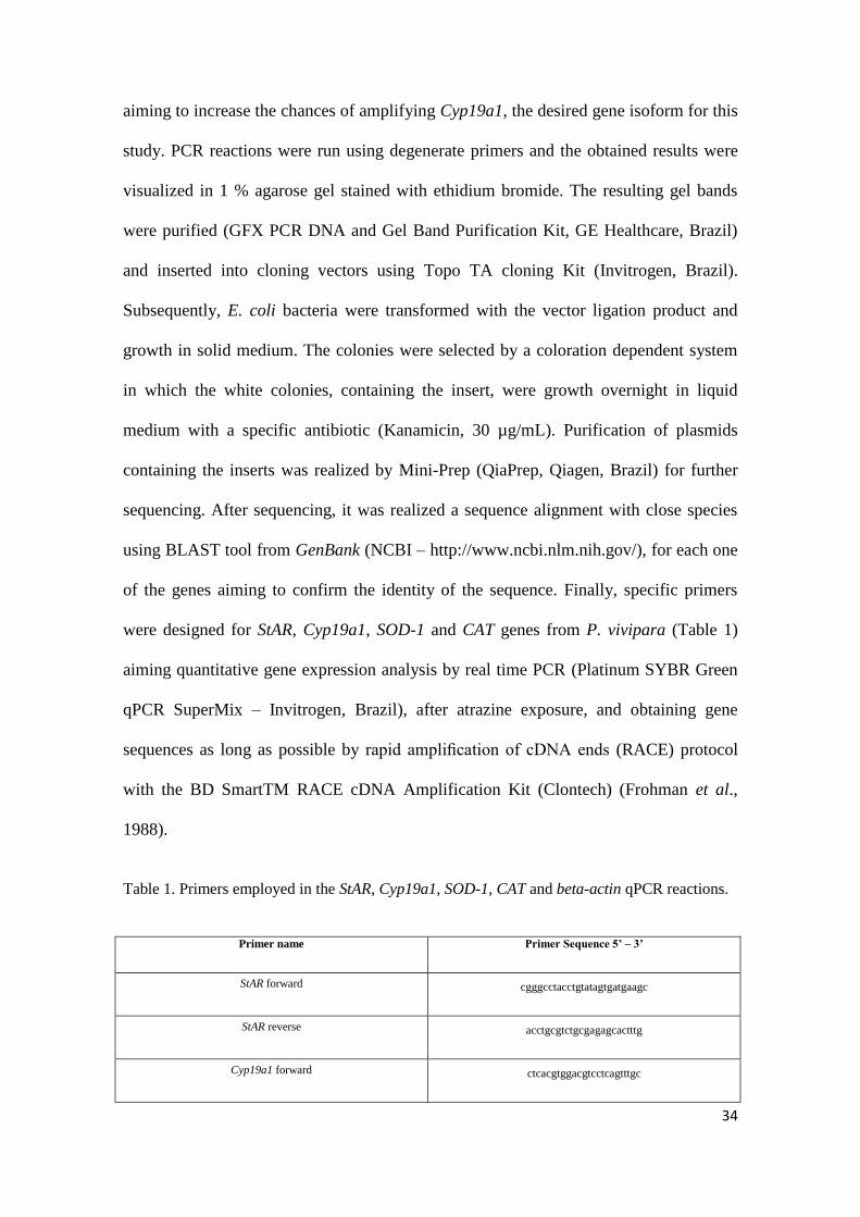
2.Methodology ............................................................................................................ 33
6
3.Results ..................................................................................................................... 36
4.Discussion ................................................................................................................ 43
5.References ............................................................................................................... 50
CONCLUSÕES GERAIS .......................................................................................... 60
PERSPECTIVAS ....................................................................................................... 60
REFERÊNCIAS ......................................................................................................... 62
7
RESUMO
O herbicida Atrazina (ATR) é um agrotóxico utilizado há cerca de 50 anos, responsável
pelo controle seletivo de plantas daninhas em cultivo de arroz, milho e cana-de-açúcar,
principalmente. Estudos recentes apontam diversos efeitos desse herbicida em
invertebrados e vertebrados, através da contaminação do solo, bem como da lixiviação
para os ecossistemas aquáticos. Foi demonstrado que a ATR é um desregulador
endócrino, além de causar efeitos como estresse oxidativo, imunotoxicidade e distúrbios
no metabolismo energético. No presente estudo, a espécie nativa Poecilia vivipara foi
utilizada como modelo experimental para identificar e analisar a expressão de genes
atuantes na via esteroidogênica (StAR e Cyp19a1) e genes atuantes no sistema de defesa
antioxidante enzimático (SOD-1 e CAT), frente a exposição à diferentes concentrações
de ATR. Sequências parciais dos genes-alvo foram obtidas e comparadas com
sequências disponíveis de espécies próximas. Foram analisadas a expressão órgão-
específica para cada um dos genes isolados, bem como a expressão dos genes frente à
exposição ao herbicida atrazina. Os animais foram expostos a ATR em concentrações
de 2, 10 e 100 µg/L e a expressão dos genes em gônadas e fígado desses animais foram
analisadas em 24 e 96 horas de exposição. As sequências obtidas dos genes StAR,
Cyp19a1, SOD-1 e CAT apresentaram 821, 80, 954, 350 pares de bases
respectivamente, com identidades que variam de 86 a 100% com espécies
filogeneticamente próximas a P. vivipara. Os animais apresentaram uma maior
expressão dos genes StAR e Cyp19a1 nas gônadas e no fígado, enquanto a menor
expressão se mostrou em órgãos como intestino e baço. Já os genes SOD e CAT
apresentaram uma maior expressão no fígado, e menor expressão no intestino. Em
relação à expressão gênica frente à exposição à ATR, os resultados apontaram para uma
indução dos genes StAR, SOD e CAT em 24 horas, nas gônadas e no fígado, enquanto
8
que a expressão do gene Cyp19a1 foi aumentada apenas após 96 horas de exposição.
Foi demonstrado que o herbicida ATR, mesmo em baixas concentrações, é capaz de
desregular a expressão de genes que codificam tanto para proteínas componentes da via
de síntese de hormônios esteróides, quanto para enzimas atuantes na resposta
antioxidante celular de P. vivipara.
Palavras-chave: Atrazina, Aromatase, Defesa antioxidante, Poecilia vivipara, Via
esteroidogênica
9
ABSTRACT
The herbicide Atrazine (ATR) is a pesticide used in the last 50 years and responsible for
the selective control of weeds mainly in rice, corn and sugarcane fields. Recent studies
have documented the effects of this herbicide in invertebrates and vertebrates by soil
and lixiviation of aquatic environments contamination. ATR has been shown as
endocrine disruptor and caused effects, such as oxidative stress, immunotoxicity and
energetic metabolism disturbances. The native species Poecilia vivipara was used in the
present study, as an experimental animal model, to identify and analyze the expression
of genes involved in the steroidogenic pathway (StAR and Cyp19a1) and enzymatic
antioxidant defense system (SOD-1 and CAT) by challenging the animal with different
ATR concentrations. Partial sequences from the target genes were obtained and
compared with available sequences from close species. We analyzed the specific organ
expression for each of the isolated genes and the expression of genes before exposure to
atrazine. The animals were exposed to ATR concentrations of 2, 10 and 100 µg/L and
genes expression, in gonads and liver from these animals, was analyzed in 24 and 96
hours of exposition. The obtained sequences of StAR, Cyp19a1, SOD-1 and CAT genes
presented 821, 80, 954 and 350 base pairs, respectively, with identities of
phylogenetically close species to P. vivipara varying from 86 to 100%. The animals
exhibited a higher expression of StAR and Cyp19a1 genes in gonads and liver and lower
in tissues such as intestine and spleen. SOD and CAT genes have presented higher
expression in liver and lower in intestine. Regarding gene expression challenged by
ATR exposure, the results have evidenced an induction of StAR, SOD and CAT genes in
24 hours, in gonads and liver, while Cyp19a1 gene expression was increased only after
96 hours of exposure. Even in low concentrations, it was demonstrated that ATR
herbicide is able to interfere over the expression of genes coding for proteins from the

10
steroid hormones synthesis pathway and for enzymes involved in cellular antioxidant
response in P. vivipara.
Keywords: Atrazine, Aromatase, Antioxidant Defense, Poecilia vivipara, Steroidogenic
Pathway

11
INTRODUÇÃO GERAL
Estima-se que cerca de 1,4 milhões de pessoas no mundo vivem em áreas de bacias
hidrográficas, onde a utilização destes recursos excede os níveis de reposição,
conduzindo assim a dessecação, poluição e ao esgotamento de águas subterrâneas. Esta
má utilização dos recursos hídricos vai de encontro ao paradoxo de vivermos em um
planeta com 70,8% de sua superfície coberta de água, sendo que somente cerca de 2%
deste montante é de água doce e que destes somente 0,3% encontram-se disponíveis
para o consumo humano (GEO - Recursos Hídricos, 2007). Dentro do atual
desenvolvimento tecnológico, a utilização da água tem dois componentes em conflito:
por um lado representa um item indispensável para a existência humana, sendo seu
consumo cada vez mais aumentado, e por outro lado a água serve como veículo de
transporte e diluição de diferentes compostos tóxicos que são direta ou indiretamente
gerados como consequência da atividade humana (Schnurstein e Braunbeck, 2001).
Mais de um terço da água doce renovável do planeta está sendo utilizada na
agricultura, indústria e no âmbito doméstico (Schwarzenbach et al., 2006). No século
XX, estima-se que milhares de poluentes orgânicos, como os bifenilos policlorados
(PCB), pesticidas organoclorados (POPs), hidrocarbonetos policíclicos aromáticos
(HPAs), dibenzofuranos policlorados (PCDF) e dioxinas tenham sido produzidos e, em
parte, liberados no meio ambiente (Oost et al., 2003). Entre essas substâncias liberadas,
calcula-se que apenas 6.000 possuem avaliação considerada satisfatória sobre os riscos à
saúde do homem e do meio ambiente, levando-se em consideração que, a cada ano,
entre 1.000 e 2.000 novas substâncias são liberadas para o mercado (Freitas e Sá, 2003).
Desde o início dos anos 60, a humanidade tornou-se consciente dos potenciais
efeitos em longo prazo destes produtos químicos e seus riscos para os ecossistemas

12
aquáticos e terrestres. O destino final da maioria destes contaminantes é o ambiente
aquático, devido a descargas diretas ou a processos hidrológicos e atmosféricos (Hahn e
Stegeman, 1994). Entre os setores que contribuem para a liberação de compostos
tóxicos no ambiente, um dos mais importantes é a agricultura, através da utilização de
pesticidas em geral, e os conflitos entre a produção agrícola e qualidade ambiental têm
crescido, nas últimas décadas, bem como as preocupações relativas à utilização de
pesticidas formam um foco nos debates sobre sustentabilidade (Freemark e Bontin,
1995; Mineau e McLaughlin, 1996).
Os pesticidas são definidos pela Agência de proteção Ambiental dos EUA dentro da
Ação federal de inseticidas, fungicidas, e rodenticidas (USEPA, 2003) como uma
substância ou mistura destinada para prevenir, destruir, repelir ou mitigar qualquer
praga incluindo insetos, roedores e ervas daninhas. Os pesticidas utilizados
exclusivamente na agricultura são mais comumente chamados de agrotóxicos (Laws e
Hayes, 1991). Esses compostos e seus resíduos estão entre os agentes mais devastadores
atualmente tanto para os ecossistemas aquáticos, quanto para os organismos, afetando a
cadeia alimentar desde o menor nível até níveis superiores (Duursma e Marchand,
1974). A grande maioria dos pesticidas não são rapidamente degradáveis por razões
técnicas, já que a degradação rápida pode reduzir sua aplicabilidade. Portanto, é
provável que um grande volume de resíduos de pesticidas se acumule no ambiente em
um processo contínuo (Islam e Tanaka, 2004).
No Brasil, o uso de herbicidas, incluindo a ATR, dobrou nos últimos 10 anos
(Sindag, 2003), o que está ligado à classificação do Brasil como um líder global em
exportações agrícolas. Dado que os agrotóxicos estão sendo cada vez mais utilizados e
que eles não são facilmente substituídos em larga escala, então o conhecimento do
13
destino desses produtos químicos em ambientes tropicais é necessário para minimizar a
bioacumulação desses poluentes nas reservas de água doce (Correia et al., 2007).
1.Desreguladores Endócrinos
Alguns desses compostos liberados no ambiente por atividades antrópicas, como
os agrotóxicos, têm se mostrado desreguladores da produção e da atividade hormonal
dos animais, os quais são importantes para diversos processos biológicos, como o
desenvolvimento (Phillips e Harrison, 1999). Esses compostos podem alterar os
mecanismos de regulação hormonal atuando de forma direta ou indireta, ligando-se a
receptores hormonais ou interferindo na produção, liberação, transporte, metabolização
ou eliminação dos hormônios endógenos. Essa interferência na regulação endócrina por
poluentes ambientais em geral e alguns químicos naturais é chamada de desregulação
endócrina química (Norris, 2007). A interferência dos poluentes na regulação endócrina,
relacionada principalmente às funções reprodutivas, tem gerado crescente interesse
público uma vez que os efeitos têm sido observados tanto em humanos quanto em
outros animais. Esses efeitos incluem queda de fertilidade (Petersen et al., 1998), câncer
de mama (Wolff et al.,1993; 2000) puberdade precoce (Honma et al., 2001) ou
puberdade tardia (Faqi et al., 1998), e anormalidades gonadais (Roos et al., 2001; Cook
et al., 2003). Em relação aos efeitos reprodutivos, a maioria dos desreguladores
endócrinos possui um efeito estrogênico ao organismo, sendo que alguns destes
compostos, como o bisfenol A, apresentam esses efeitos ligando-se diretamente aos
receptores de estrógeno (Kuiper et al., 1998; Safe et al., 2001; Scippo et al., 2004). No
entanto, outros desreguladores endócrinos não são capazes de competir com estrógenos
naturais, ou seja, não têm capacidade de se ligar diretamente a um receptor específico,
mas ainda assim causam efeitos estrogênicos nos organismos, provavelmente através de
14
outros mecanismos. Nesse último grupo está incluído um dos herbicidas mais
conhecidos e utilizados mundialmente, a Atrazina (ATR) (Roberge et al., 2004).
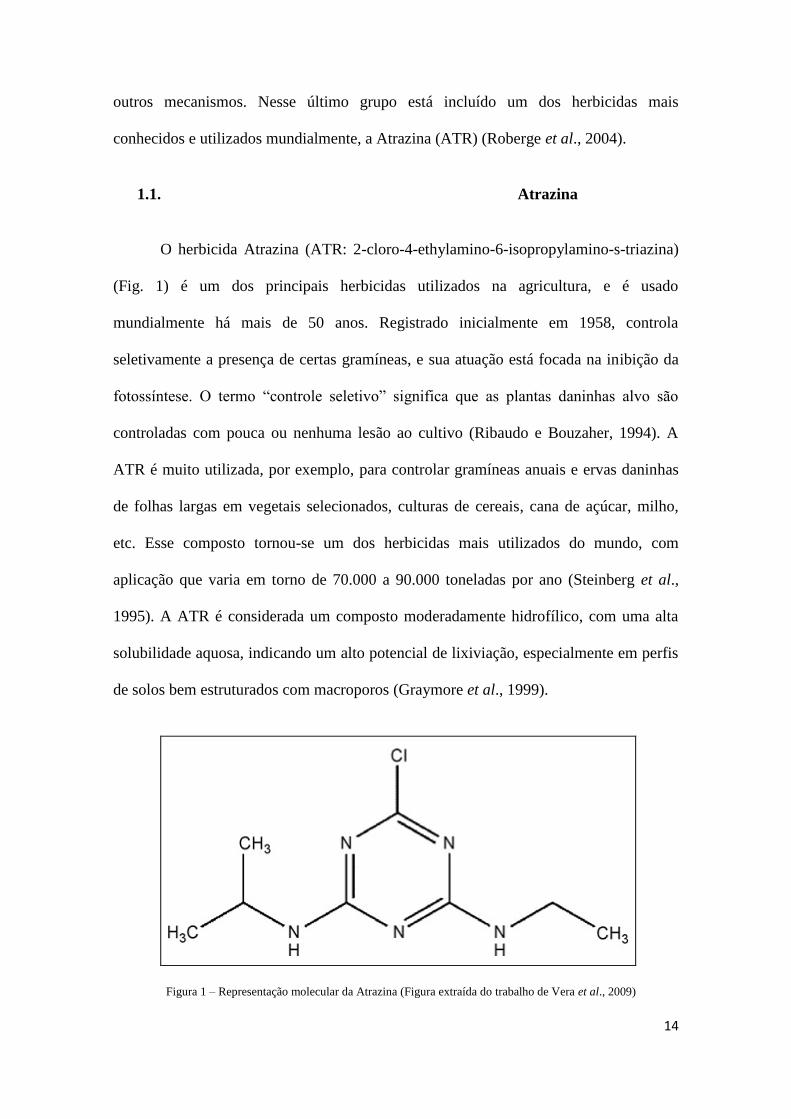
1.1. Atrazina
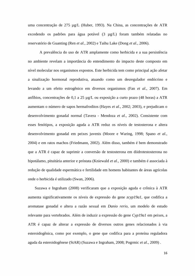
O herbicida Atrazina (ATR: 2-cloro-4-ethylamino-6-isopropylamino-s-triazina)
(Fig. 1) é um dos principais herbicidas utilizados na agricultura, e é usado
mundialmente há mais de 50 anos. Registrado inicialmente em 1958, controla
seletivamente a presença de certas gramíneas, e sua atuação está focada na inibição da
fotossíntese. O termo “controle seletivo” significa que as plantas daninhas alvo são
controladas com pouca ou nenhuma lesão ao cultivo (Ribaudo e Bouzaher, 1994). A
ATR é muito utilizada, por exemplo, para controlar gramíneas anuais e ervas daninhas
de folhas largas em vegetais selecionados, culturas de cereais, cana de açúcar, milho,
etc. Esse composto tornou-se um dos herbicidas mais utilizados do mundo, com
aplicação que varia em torno de 70.000 a 90.000 toneladas por ano (Steinberg et al.,
1995). A ATR é considerada um composto moderadamente hidrofílico, com uma alta
solubilidade aquosa, indicando um alto potencial de lixiviação, especialmente em perfis
de solos bem estruturados com macroporos (Graymore et al., 1999).
Figura 1 – Representação molecular da Atrazina (Figura extraída do trabalho de Vera et al., 2009)
15
Apesar do uso difundido mundialmente desse herbicida, nas últimas décadas
foram realizados estudos apontando diversos danos aos organismos frente à presença
desse poluente principalmente em ecossistemas aquáticos, decorrente da lixiviação do
solo (Graymore et al., 2001). Na União Européia a utilização desse herbicida foi
recentemente proibida (2004/248/CE), mas passou a ser utilizada uma fórmula quase
idêntica, a Terbutilazina. No entanto, no resto do mundo a ATR continua sendo
amplamente utilizada, sendo que em algumas regiões o seu uso é restrito. Nos Estados
Unidos, a quantidade máxima de ATR permitida pela Agência de Proteção Ambiental
(USEPA) é de 3 µg/L em água potável (definida em 1991), entretanto, nos últimos anos
o país aprovou um recadastramento do composto, com atualização de rótulo e de riscos.
Já no Brasil, segundo a resolução 357/2005 do Conselho Nacional do Meio Ambiente
(CONAMA), a quantidade máxima permitida de ATR em água doce é 2 µg/L.
A ATR pode alcançar o ambiente de diversas maneiras, e é frequentemente
introduzida em águas superficiais e subterrâneas por escoamento e de filtração do solo,
ou então pode permanecer adsorvida às partículas do solo após a aplicação. O transporte
e a deposição de ATR em ambientes aquáticos adjacentes a áreas de agricultura
intensiva já estão bem documentados (Thurman e Cromwell, 2000). Após a entrada no
ambiente aquático, esse composto, em água doce, por exemplo, têm uma meia-vida
entre 8 e 350 dias, dependendo do ambiente e suas características físico-químicas
(Diana et al., 2000; Tavera-Mendoza et al., 2002). Em relação à água doce e estuários a
taxa de ATR pode variar em média de 1.000 µg/L em águas próximas às regiões de
aplicação do herbicida, a 0,2 µg/L (Pereira e Hostettler, 1993; Shottler et al., 1994) . Na
América do Norte, esse composto foi detectado nos rios, em concentrações de até 108
µg/L (USEPA, 2002), e em água de enxurrada na mesma região os níveis chegaram a
16
uma concentração de 275 µg/L (Huber, 1993). Na China, as concentrações de ATR
excedendo os padrões para água potável (3 µg/L) foram também relatadas no
reservatório de Guanting (Ren et al., 2002) e Taihu Lake (Dong et al., 2006).
A prevalência do uso de ATR amplamente como herbicida e a sua persistência
no ambiente revelam a importância do entendimento do impacto deste composto em
nível molecular nos organismos expostos. Este herbicida tem como principal ação afetar
a sinalização hormonal reprodutiva, atuando como um desregulador endócrino e
levando a um efeito estrogênico em diversos organismos (Fan et al., 2007). Em
anfíbios, concentrações de 0,1 a 25 µg/L ou exposição a curto prazo (48 horas) a ATR
aumentam o número de sapos hermafroditos (Hayes et al., 2002; 2003), e prejudicam o
desenvolvimento gonadal normal (Tavera - Mendoza et al., 2002). Consistente com
esses fenótipos, a exposição aguda a ATR reduz os níveis de testosterona e altera
desenvolvimento gonadal em peixes juvenis (Moore e Waring, 1998; Spano et al.,
2004) e em ratos machos (Friedmann, 2002). Além disso, também é bem demonstrado
que a ATR é capaz de suprimir a conversão de testosterona em diidrotestosterona no
hipotálamo, pituitária anterior e próstata (Kniewald et al., 2000) e também é associada à
redução de qualidade espermática e fertilidade em homens habitantes de áreas agrícolas
onde o herbicida é utilizado (Swan, 2006).
Suzawa e Ingraham (2008) verificaram que a exposição aguda e crônica à ATR
aumenta significativamente os níveis de expressão do gene zcyp19a1, que codifica a
aromatase gonadal e altera a razão sexual em Danio rerio, um modelo de estudo
relevante para vertebrados. Além de induzir a expressão do gene Cyp19a1 em peixes, a
ATR é capaz de alterar a expressão de diversos outros genes relacionados à via
esteroidogênica, como por exemplo, o gene que codifica para a proteína reguladora
aguda da esteroidogênese (StAR) (Suzawa e Ingraham, 2008; Pogrmic et al., 2009) .

17
Além dos efeitos reprodutivos e comportamentais, a exposição à ATR pode causar
também, segundo estudos recentes, alterações na expressão de enzimas relacionadas ao
metabolismo energético, detoxificação celular e sistema de defesa antioxidante
(Londono et al., 2007; Anderson et al., 2008; Jin et al., 2010). Entre esses estudos, Jin
et al. (2010), por exemplo, verificaram uma indução na expressão gênica e na atividade
proteica das enzimas superóxido dismutase 1 (SOD-1) e catalase (CAT), atuantes no
sistema de defesa antioxidante enzimático, indicando que a presença desse composto
pode estar causando uma situação de estresse oxidativo no animal.
2. Via Esteroidogênica
Os hormônios esteróides desempenham papéis críticos no desenvolvimento
sexual, homeostase, respostas ao estresse, metabolismo de carboidratos e reprodução, e
são produzidos principalmente em órgãos esteroidogênicos especializados (Bentley,
1998; Norris, 2007). Diversos estudos comparativos sugerem que as enzimas chave da
produção de esteróides, bem como a via em si, são extremamente conservadas entre
membros de diferentes classes de vertebrados (Bourne, 1991; Selcer e Leavitt, 1991).
Os esteróides são sintetizados a partir do colesterol, através de uma série de
reações enzimáticas, onde participam principalmente enzimas da família citocromo
p450 (Norris, 2007). As enzimas responsáveis pela síntese dos hormônios esteróides
sexuais estão representadas na figura 2. A produção de esteroides ocorre em órgãos
esteroidogênicos clássicos, tais como o córtex adrenal, testículo, ovário e placenta, no
caso de mamíferos. Estes órgãos são capazes, portanto, de converter colesterol em
pregnenolona pela enzima P450 a partir da clivagem da cadeia lateral do colesterol
(P450scc), no interior da mitocôndria, e a partir da pregnenolona, produzir os outros

18
esteróides ativos, utilizando a maquinaria enzimática específica da via esteroidogênica
(Ueyama et al., 2002).
Figura 2 - Representação da via de produção de hormônios esteróides sexuais (Kronenberg et al., 2011)
Além da síntese de esteróides pelas células requerer a atividade de diversas
enzimas Citocromo P450 e desidrogenases, existe também um fator importante na
regulação do tempo e da taxa de esteroidogênese, descoberto recentemente, que é a
proteína de regulação aguda da esteroidogênese (StAR) (Stocco e Clark, 1996). Essa
proteína tem como função promover a transferência do colesterol através do espaço
aquoso que separa a membrana mitocondrial externa da membrana mitocondrial interna
(Christenson e Strauss, 2001). Os dados mais relevantes implicando a função da StAR
na regulação aguda da esteroidogênese vieram de estudos com humanos, de pacientes
com hiperplasia lipoide adrenal congênita (lipóide CAH), onde esta doença tem sido
associada a mutações no gene StAR (Lin et al., 1995). A entrada do colesterol com o
auxílio da proteína StAR acontece para que, por sua vez, a enzima de clivagem de
cadeia lateral do colesterol, P450scc, dentro da mitocôndria, converta o colesterol em
19
pregnenolona, e a partir de então a diversas enzimas envolvidas na via esteroidogênica
sintetizem os hormônios esteróides (Stocco, 2001).
Em contraste com a regulação crônica da esteroidogênese, que é em grande parte
mediada pelo aumento da transcrição de genes que codificam para as enzimas da via, a
proteína reguladora aguda da esteroidogênese atua, portanto, regulando a entrada de
colesterol na mitocôndria, que é uma etapa crucial para o processo de síntese dos
hormônios esteróides (Stocco e Clark, 2005). A proteína StAR é localizada
principalmente nas gônadas e nas glândulas supra-renais e é rapidamente sintetizada em
resposta à estimulação hormonal e aumento do AMPc intracelular (Clark et al., 1994;
Stocco e Clark, 1996). Além disso, estudos sugerem que elementos de resposta do gene
StAR são alvos de receptores nucleares da família NR5A, e portanto esses receptores
também atuam regulando a sua expressão (Suzawa e Ingraham, 2008). A maioria dos
estudos de estrutura e função da proteína StAR são provenientes de mamíferos.
Entretanto, o gene StAR tem sido isolado e identificado em peixes, anfíbios e aves
(Bauer et al., 2000; Kusakabe et al., 2002; Li et al., 2003). A partir desses estudos,
parece que há uma alta conservação da estrutura desta proteína dentro do grupo dos
vertebrados (Bauer et al., 2000; Kusakabe et al., 2002).
Outra componente chave da via esteroidogênica é a enzima Citocromo P450
Aromatase (CYP19) que catalisa uma etapa final e limitante da via, atuando na
conversão de andrógenos em estrógenos. Os estrógenos e andrógenos são hormônios
esteróides que atuam no desenvolvimento sexual dos organismos e são encontrados em
representantes de todas as classes de vertebrados terrestres e marinhos, incluindo peixes,
anfíbios, répteis, aves e mamíferos (Lange et al., 2003), bem como em alguns
invertebrados (Zhu et al., 2003; Osada et al., 2004). A presença de atividade da

20
aromatase em uma célula ou tecido é um indicador da capacidade de transformar
andrógenos em estrógenos (Norris, 2007). Em mamíferos, com exceção dos suínos
(Corbin et al., 1999), existe um único gene cyp19 que se expressa em uma variedade de
órgãos (Simpson, 1994). Teleósteos, em contraste, adquiriram ao longo da evolução
uma duplicação gênica do Cyp19, contendo, portanto dois genes estruturalmente
distintos que compartilham apenas a identidade de 60%: cyp19a1 e cyp19a2,
preferencialmente expressos no ovário e no cérebro, respectivamente. Em zebrafish, o
gene zcyp19a1, que codifica para a aromatase gonadal, se expressa em maior
intensidade nas gônadas e contém em sua região promotora, assim como o gene StAR,
um sítio de ligação de receptores nucleares da família 5A (NR5A), o qual é
provavelmente reconhecido também por ortólogos do fator esteroidogênico 1 (SF-1) /
receptor do hormônio luteinizante 1 (LRH-1) (vonhofsten e Olsson, 2005). Já o gene
zcyp19a2 contém um elemento de resposta a estrogénos (ERE) e é sensível aos
estrogénos ou xenoestrógenos (Kazeto et al., 2004). Ambos promotores de aromatase
em zebrafish contêm sítios de elementos de ligação ao AMPc (CREB) em seu promotor
e que, portanto, são responsivos à sinalização por AMPc.
Bauer et al. (2000) demonstraram que os genes StAR e Cyp19a1, em peixes, são
concomitantemente expressos e parecem apresentar uma maior expressão nos órgãos
com alta atividade esteroidogênica, como gônadas, supra-renais e fígado. Além disso,
sua regulação parece ter alguns pontos em comum como os elementos de resposta nos
promotores alvos de receptores NR5A e a resposta ao aumento de AMPc celular
(Suzawa e Ingraham, 2008; Abarikwu et al., 2011).
Diversos estudos têm demonstrado um efeito do herbicida ATR na expressão de
genes atuantes na via esteroidogênica. Tais estudos focam principalmente sobre a
21
expressão do gene Cyp19a1, que codifica para a aromatase gonadal em peixes, enquanto
a quantidade de estudos com o efeito do composto na expressão do gene StAR, que
codifica para proteína reguladora aguda a esteroidogênese, é bem menor. Sabe-se que
doses de ATR 20 µg/L aumentam a expressão e atividade da aromatase em linhagens
celulares seletivas de mamíferos (Sanderson et al., 2002) e doses maiores são capazes
de provocar reversão sexual em peixes (Suzawa e Ingraham., 2008). Recentemente, foi
proposto que a ATR pode se ligar e ativar o receptor nuclear SF-1 (família NR5A) (Fan
et al., 2007). Esta hipótese é interessante, uma vez que ortólogos de SF-1 são
encontrados em todos os vertebrados, incluindo teleósteos, principalmente tendo em
conta o papel crítico do SF-1 no desenvolvimento sexual dos mamíferos e
esteroidogênese, tendo como alvo a região promotora de genes-chave dessa via, entre
eles, StAR e Cyp19a1 (Shen et al., 1994). Neste mesmo sentido, em análises de
microarranjo, também foi observado um aumento da expressão de diversos genes
envolvidos na via esteroidogênica, como StAR, CYP11A1,Cyp17a1, LHR e 3b-HSD
frente a baixas doses de ATR, em linhagens celulares de mamíferos (Abarikwu et al.,
2011; Suzawa e Ingraham, 2008)
3. Sistema de Defesa Antioxidante
As espécies reativas de oxigênio (EROS) abrangem o ânion superóxido (O2
-),
peróxido de hidrogênio (H2O2), e radicais hidroxilas (HO-), que estão envolvidas em
diferentes vias de sinalização celular, incluindo apoptose, mas também estão implicados
no desenvolvimento de patologias, produzindo dano em vários componentes celulares
como lipídios não saturados, proteínas e ácidos nucléicos. O estresse oxidativo ocorre
quando a geração de EROS excede a capacidade de remoção e os efeitos deletérios
incluem oxidação de proteínas, DNA e componentes esteróides, bem como peroxidação
22
dos lipídios insaturados das membranas celulares (Sies, 1993). Estima-se que cerca de
0,1% de todo o oxigênio consumido por um organismo seja parcialmente reduzido na
mitocôndria formando EROS (Fridovich, 2004). A situação de dano oxidativo, pode se
dar, entre outros fatores, pela presença de xenobióticos nos organismos. Águas
residuais, por exemplo, contém uma grande variedade de poluentes orgânicos e
metálicos, incluindo pesticidas, hidrocarbonetos aromáticos, dibenzofuranos, compostos
estrogênicos e muitos metais, sendo a maioria dessas substâncias, agentes oxidantes
(Avci et al., 2005).
A defesa dos organismos contra danos oxidativos pode ser não enzimática,
através de substâncias antioxidantes, como algumas vitaminas, ácido úrico, glutationa e
carotenóides. Além disso, existe em todas as classes de vertebrados e em invertebrados
um sistema de defesa antioxidante enzimático, onde diversas enzimas antioxidantes,
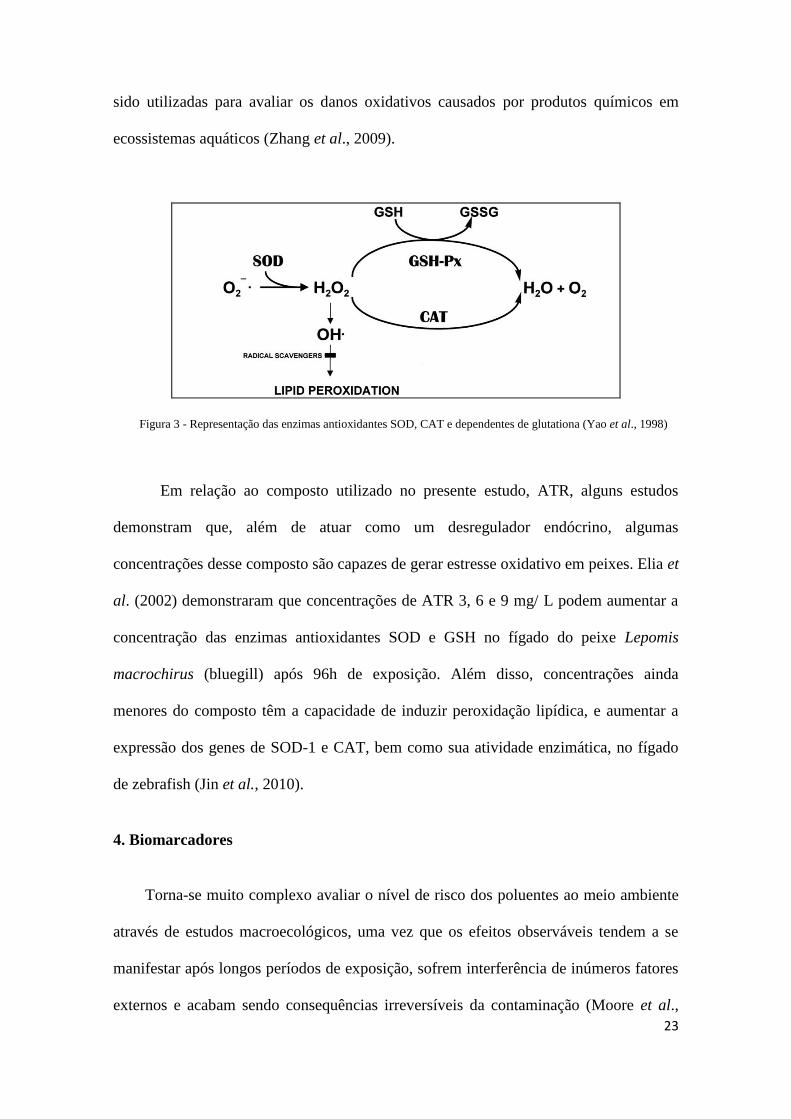
entre elas superóxido dismutase (SOD) e catalase (CAT),atuam impedindo a cascata de
reações oxidantes, interceptando e inativando os compostos reativos de oxigênio
intermediários (Figura 3). As enzimas antioxidantes são, portanto, cruciais no esforço
de neutralizar a toxicidade de oxigênio quando o fornecimento de outros compostos
antioxidantes são escassos ou esgotados (Ahmad, 1995).
A enzima SOD, por exemplo, parece ser a primeira linha de defesa enzimática
contra ROS, e atua catalisando a dismutação de O2
- para O
2 e H2O2 (Ken et al., 2003).
Sendo o O2
- um precursor para várias outras espécies altamente reativas, o controle da
concentração de radicais livres por SOD constitui um importante mecanismo de
proteção celular contra danos oxidativos em macromoléculas (Fridovich, 1997).
Subsequentemente, a enzima CAT catalisa a produção de H2O a partir de H2O2 (Tripathi
et al., 2006). Recentemente, medidas da atividade dessas enzimas antioxidantes têm
23
sido utilizadas para avaliar os danos oxidativos causados por produtos químicos em
ecossistemas aquáticos (Zhang et al., 2009).
Figura 3 - Representação das enzimas antioxidantes SOD, CAT e dependentes de glutationa (Yao et al., 1998)
Em relação ao composto utilizado no presente estudo, ATR, alguns estudos
demonstram que, além de atuar como um desregulador endócrino, algumas
concentrações desse composto são capazes de gerar estresse oxidativo em peixes. Elia et
al. (2002) demonstraram que concentrações de ATR 3, 6 e 9 mg/ L podem aumentar a
concentração das enzimas antioxidantes SOD e GSH no fígado do peixe Lepomis
macrochirus (bluegill) após 96h de exposição. Além disso, concentrações ainda
menores do composto têm a capacidade de induzir peroxidação lipídica, e aumentar a
expressão dos genes de SOD-1 e CAT, bem como sua atividade enzimática, no fígado
de zebrafish (Jin et al., 2010).
4. Biomarcadores
Torna-se muito complexo avaliar o nível de risco dos poluentes ao meio ambiente
através de estudos macroecológicos, uma vez que os efeitos observáveis tendem a se
manifestar após longos períodos de exposição, sofrem interferência de inúmeros fatores
externos e acabam sendo consequências irreversíveis da contaminação (Moore et al.,

24
2004). Logo, elucidar os mecanismos iniciais (a níveis moleculares e/ou celulares) de
resposta rápida na presença de contaminantes pode garantir uma compreensão mais
rápida sobre a situação do meio ambiente, a tempo de evitar danos maiores a níveis
ecológicos. Assim, um biomarcador pode ser definido como medidas de fluidos
corporais, células ou tecidos que indicam em termos bioquímicos ou celulares a
presença de contaminantes ou a magnitude da resposta do hospedeiro (Livingstone et
al., 2000). Usualmente, os biomarcadores são classificados como específicos ou não
específicos. Alguns biomarcadores podem ser considerados específicos para
determinado grupo de poluente, desde que condições fisiológicas e naturais, sejam
levadas em conta. O uso de biomarcadores específicos como metalotioneínas, por
exemplo, tem sido amplamente empregado para indicar a presença de metais pesados no
ambiente (Giguère et al., 2003), apesar de alguns estudos mais recentes apontarem
efeitos abióticos como salinidade influenciando nesse parâmetro. Outro exemplo de
biomarcador específico é a medição atividade da enzima acetilcolinesterase, por sua vez
é considerada um biomarcador específico de organofosforados, pesticidas carbamatos e
neurotoxinas como a anatoxina-a (Monserrat e Bianchini, 2000; Monserrat et al., 2002;
Hyne e Maher, 2003)
Por outro lado, uma vez que diversos poluentes podem modificar o equilíbrio
entre a concentração de pró-oxidantes e antioxidantes nos organismos, a determinação
de estresse oxidativo, bem como de respostas antioxidantes, como expressão gênica e
atividade enzimática de enzimas como SOD-1 CAT e GPx, são comumente empregadas
como biomarcadores não-específicos (Bainy et al., 1996; Geracitano et al., 2004).
Os genes relacionados à via esteroidogênica, StAR e Cyp19a1, parecem ser
biomarcadores para a presença de desreguladores endócrinos no ambiente, com
especificidade intermediária (Kortner e Arukwe, 2007; Storvik et al., 2011). Já os
25
genes que codificam para as enzimas antioxidantes SOD-1 e CAT são considerados por
muitos autores como biomarcadores inespecíficos, utilizados para inferir poluição
ambiental, em geral (Olsvik et al., 2005; Torres et al., 2008).
A identificação dos genes que codificam para Aromatase em peixes (Cyp19a1 e
Cyp19a2) já foi realizada para diversas espécies, incluindo Ictalurus punctatus (Trant,
1994), medaka Oryzias latipes (Fukada et al., 1996), tilápia do Nilo Oreochromis
niloticus (Kwon et al., 2001; Chang et al., 2005), Carassius auratus (Tchoudakova e
Callard, 1998), Danio rerio (Kishida e Callard, 2001), truta arco-íris Oncorhynchus
mykiss (Dalla Valle et al., 2002), Fundulus heteroclitus (Greytak et al., 2005), peixe-rei
Odontesthes bonariensis (Strobl -Mazzulla et al., 2005), entre outros. Já o gene que
codifica para a proteína StAR foi identificado para um número reduzido de espécies de
teleósteos como o bacalhau Gadus morhua (Kortner e Arukwe, 2007) e o bagre africano
Clarias gariepinus (Sreenivasulu et al., 2009).
Os genes relacionados ao sistema de defesa antioxidante, como os utilizados no
presente trabalho, também têm sido identificados em espécies de teleósteos. O gene
completo SOD-1 já foi caracterizado em Danio rerio (Ken et al., 2003) e para o peixe
antártico Trematomus bernacchii (Santovito et al., 2006), além de diversas outros
grupos taxonômicos, incluindo alguns invertebrados (Park et al., 2009; Kim et al.,
2011). O gene CAT, por sua vez, também já foi identificado para Danio rerio (Ken et
al., 1998), bem como para Salmo salar (Olsvik et al., 2005).
26
5. Modelo experimental
Muitos estudos têm sido dedicados ao entendimento das causas da desregulação
endócrina em peixes, não só porque estes animais aquáticos são frequentemente
expostos a múltiplas fontes de desreguladores endócrinos, e diversos casos de
desenvolvimento anormal possivelmente causada pela exposição a esses compostos
foram relatados, mas também por causa da possibilidade de extrapolação e previsão de
risco para vertebrados superiores, incluindo seres humanos (Guillette et al., 1995;
Eggen et al., 2003; Goksoyr, 2006)
Os peixes do gênero Poecilia, conhecidos popularmente como barrigudinhos,
são abundantes e habitam regiões dulciaquícolas e estuarinas desde os Estados Unidos
até a Argentina (Neves e Monteiro, 2003). Sua alta tolerância a condições ambientais
extremas, especialmente de salinidade e temperatura, faz com que seja uma das poucas
espécies presente em todos os ambientes lênticos (Bizerril e Primo, 2001). Dessa forma,
muitas espécies desta família são comumente encontradas em regiões limpas e em
córregos contendo resíduos de esgotos domésticos e pluviais (Araújo et al., 2009).
Existem casos de reversão sexual em algumas espécies de Poeciliidae, natural
(Howell et al., 1980; Yan, 1986) ou induzida por certos agentes químicos (Denton et al.,
1985; Howell e Denton, 1989). A técnica de reversão sexual induzida quimicamente,
via esteróides, é frequente dentre os aquariófilos, que a usam para a seleção artificial de
características genéticas na formação de novas variedades da espécie, com interesse
comercial (Fernando e Phang, 1985). A determinação genotípica do sexo em
barrigudinhos é mediada pelo sistema endócrino, com possibilidades de geração de
hermafroditas, onde a partir de uma gônada juvenil primária indiferenciada pode-se
desenvolver a gônada masculina e feminina. Entretanto, para a espécie utilizada no

27
presente estudo, Poecilia vivipara, a questão da reversão sexual ainda não é bem
esclarecida, devido à pequena quantidade de estudos (Betito, 2006).
Entre os poecilídeos, Poecilia vivipara (Bloch e Schneider, 1801) é uma das
espécies de peixes mais comuns em pequenas lagoas, rios e regiões costeiras
ecossistemas do Brasil (Santos et al., 2011). A espécie é amplamente distribuída, ao
longo da costa da América do Sul, habitando cursos de grandes rios (Parenti e
Rauchemberger, 1989).
No presente trabalho identificamos sequências parciais dos genes StAR,
Cyp19a1, SOD-1 (citosólica) e CAT em P. vivipara, e verificamos a expressão desses
genes em vários órgãos do animal (gônadas, fígado, olho, cérebro, intestino, brânquias e
baço), bem como frente à exposição a diferentes concentrações do herbicida Atrazina.
28
OBJETIVO GERAL
Identificar e caracterizar fragmentos dos genes StAR, cyp19a1, SOD-1 e CAT de
Poecilia vivipara e analisar o perfil de expressão desses genes em diferentes órgãos
expostos a concentrações do herbicida Atrazina.
Objetivos específicos
- Identificar, em Poecilia vivipara, fragmentos dos genes StAR e Cyp19a1, que
codificam para proteínas componentes da via esteroidogênica, e SOD-1 e CAT, que
codificam para proteínas componentes do sistema de defesa antioxidante enzimático.
- Realizar análises das sequências em comparação com genes já descritos de espécies
filogeneticamente próximas a espécie do estudo.
- Desenhar iniciadores específicos a partir das sequências obtidas, para a realização dos
experimentos de expressão gênica em Tempo Real (qPCR).
- Analisar o perfil de expressão dos genes StAR, Cyp19a1, SOD-1 e CAT, nos diferentes
órgãos do animal.
- Verificar a expressão do gene StAR, Cyp19a1, SOD-1 e CAT em gônadas e fígado de
P. vivipara, frente às diferentes concentrações e tempos de exposição do herbicida
Atrazina.
29
Atrazine exposure modified the expression of steroidogenesis and oxidative stress-
related genes in the fish Poecilia vivipara
Silveira, C.R., Abril, S.I., Zanette, J., Marins, L.F.
Abstract
Atrazine (ATR) herbicide is a vertebrate controversial endocrine disruptor and also
causes effects adverse such as oxidative stress, immunotoxicity and energetic problems.
We identified and verified gene expression of steroidogenenic pathway: steroidogenic
acute regulatory protein (StAR) and aromatase (Cyp19a1) and enzymatic antioxidant
defense system: Cu/Zn Superoxide dismutase (SOD-1) and catalase (CAT) in Poecilia
vivipara fish exposed to ecological relevant ATR concentrations. The genes were
isolated by PCR using degenerate primers. After sequencing, specific primers were
designed for use in quantitative PCR. StAR, Cyp19a1, SOD-1 and CAT partial gene
sequences isolated present 821, 80, 954 and 358 base pairs, respectively, and nucleotide
identity range from 86 to 100% when compared to other close species. The animals
have presented a higher expression of Cyp19a1 and StAR genes in gonads and liver and
a lower expression was observed in intestine and spleen. On the other hand, SOD and
CAT gene expression was higher in liver and lower in the intestine. ATR exposure has
increased gene expression in gonads and liver for StAR, SOD-1 and CAT in 24 hours,
and in 96 hours for Cyp19a1. These findings are of environmental interest considering
that exposure to low concentrations of ATR caused effect on expression of genes coding
for proteins related to steroid hormones synthesis pathway and for enzymes involved in
cellular antioxidant response in P. vivipara.
30
Keywords: Atrazine, Aromatase, Antioxidant Defense, Poecilia vivipara, Steroidogenic
Pathway
1. INTRODUCTION
Atrazine (2-chloro-4-ethylamino-6-isopropylamino-s-triazine) is a selective
herbicide from triazine family that acts over weed plants by interfering in
photosynthesis electron transport mechanisms (Graziano et al., 2006). Initially
registered in 1958, this compound has been an important herbicide in agriculture
worldwide used over more than 50 years (Brodeur et al., 2009). With regard to
physicochemical properties, atrazine (ATR) has a molecular weight of 215.7 g/mol,
water solubility of 33 mg / L (at 25° C) and half-life in soil approximately 60 days
(Wauchope et al., 1991; Tomlin, 2000).
Although the reports about ATR as endocrine disruptor (ED) on aquatic
vertebrates are inconsistent according Solomon et al. (2008), its wide usage and
environmental persistence showed that affects organisms (Graymore et al., 2002).
Concerning amphibian, ATR levels of 0.1 to 25 mg/L have raised the number of
hermaphrodite frogs (Hayes et al., 2002; 2003), and disturbs normal gonad development
(Tavera-Mendoza et al., 2002). Thus, ATR acute exposure, in the developing alligator
(Crain et al., 1997), and in young peripubertal male rats (Friedman, 2002). In fish, ATR
is able to alter the ionic balance (Paulino et al., 2012), reduces testosterone levels and
hinders juvenile fish gonad development (Moore and Waring, 1998; Spano et al., 2004),
and cause feminization in chronic exposure (Suzawa and Ingraham, 2008).
ATR molecular mechanism of action has been studied and, in a study realized by
Suzawa and Ingraham (2008), it was verified that acute and chronic herbicide exposure

31
significantly raises the expression of gonad aromatase coding gene, zcyp19a1, and alters
the sex ratio in zebrafish Danio rerio. Aromatase is an enzyme present in all vertebrates
that acts by converting androgens to estrogens during a process named aromatization
(Thompson and Siiter, 1974; Simpson, 1994). Therefore, those compounds acting by
enhancing the expression of Cyp19a1 gene, which encodes for a gonadal aromatase in
fishes, may cause sexual reversion in population (Cheshenko et al., 2008).
Moreover, other effects caused by this compound have been already documented
in vertebrates, such as immunotoxicity, oxygen consumption rise and oxidative stress
induction (Elia et al., 2002; Jelaso et al., 2008; Anderson et al., 2008). Like all aerobic
organisms, fish are susceptible to the attack of reactive oxygen species and have
developed antioxidant defenses demonstrated by research primarily dating to the 1980s.
Specially adapted enzymes, such as superoxide dismutase (SOD) and catalase (CAT)
have been detected in most fish species investigated to date (Rudneva, 1997).
Superoxide dismutase (SOD) is a representative antioxidant enzyme which
catalyzes dismutation of superoxide to oxygen and hydrogen peroxide. SODs are
ubiquitous and known as three forms, based on the metal cofactor in active sites:
copper/zinc (Cu/Zn-SOD), iron SOD (Fe-SOD) and manganese SOD (Mn-SOD) in
eukaryotes. In animals, two kinds of SODs have been commonly well-studied: cytosolic
Cu/Zn-SOD (SOD-1) and mitochondrial Mn-SOD (SOD-2) (Gómez-Anduro et al.,
2006). Catalase is other important antioxidant enzymes that catalyze the decomposition
of hydrogen peroxide to oxygen and water (Ken et al., 1998). Recently, antioxidant
enzymes were demonstrated to be useful biomarkers of environmental pollutants which
cause oxidative stress in fish (Roche and Boge, 1996).
32
Regarding oxidative stress responses, it was observed a significant increase in
gene expression and activity of the enzymes superoxide dismutase – 1 (Cu/Zn-SOD)
and catalase (CAT) from livers of Danio rerio exposed to low ATR doses (10µg/L) (Jin
et al., 2010). Elia et al. (2011) also observed increased antioxidant responses before
exposure to ATR in Bluegill Sunfish Lepomis macrochirus. Additionally, Qian et al.
(2008) have reported that ATR exposure caused a significant dose-dependent induction
of SOD and CAT activity in the microalgae Chlorella sp.
Estuarine areas act as final receptors of organic matter and pollutants that are
usually derived from anthropogenic activity by fluvial and atmospheric lixiviation
(Gagosian and Peltzer, 1986; Bouloubassi and Saliot, 1993). Fish species inhabiting
those areas have been proposed as sentinels for pollution monitoring by sensible
biomarkers evaluation. Biomarkers can be defined as changes in biological responses
ranging from molecular to behavioral changes, which may be related to exposition or
environment contamination effects (Depledge et al., 1995).
The present study used Poecilia vivipara (Poeciliidae) as experimental animal
model, a species widely distributed along South America coast, inhabiting courses of
great rivers and estuarine regions of Patos Lagoon, RS, Brazil (Parenti and
Rauchemberger, 1989). The afore mentioned species is omnivore, viviparous and
tolerant to extreme environmental conditions such as salinity and temperature (Bizerril
and Primo, 2001). Therefore, the present work aimed to isolate and characterize the
genes acting in steroidogenic pathway, StAR and Cyp19a1, and composing the
antioxidant defense system, SOD-1 and CAT, for further gene expression analysis after
P. vivipara acute exposure to ecological relevant atrazine herbicide concentrations.

33
2. METHODOLOGY
2.1. Animals
The animals used in the experiments of exposure to ATR herbicide were
collected in Cassino Beach (Rio Grande, RS – Brazil) during the period of
autumn/winter. Only males were used in the experiment, which were kept in laboratory
during 15 days under a temperature of 28ºC, salinity 15 ppt, 12L: 12D photoperiod and
daily fed with commercial ration (Tetracolor). Animals from this group were taken for
RNA extraction and further PCRs reactions using degenerate primers. Concerning the
ATR exposure experiment, the animals were acclimated at salinity 24 ppt for more 7
days.
2.2. StAR, Cyp19a1, SOD-1
and CAT genes cloning
Gonad excision and total RNA extraction was realized, using Trizol reagent
(Invitrogen, Brazil), from a randomly selected non-treated fish acclimated during 15
days after capture. The RNA was treated with DNAse (DNASE I AMP GRADE,
Invitrogen, Brazil), intending to remove any genomic DNA traces in the sample,
quantified (QUANT-IT SSDNA ASSAY KIT, Invitrogen, Brazil) and after used for
cDNA synthesis by reverse transcription (High Capacity, Applied Biosystems, Brazil).
Degenerate primers were designed for the analyzed genes in the present study (StAR,
Cyp19a1, SOD and CAT), using conserved sequences from genes of fish species of
Cyprinodontiformes order, which are phylogenetically close to P. vivipara (ex.:
Poecilia reticulata, Fundulus heteroclitus and Jenynsia multidentata). Concerning
CYP19 family, conserved region sequences among different isoforms were avoided
34
aiming to increase the chances of amplifying Cyp19a1, the desired gene isoform for this
study. PCR reactions were run using degenerate primers and the obtained results were
visualized in 1 % agarose gel stained with ethidium bromide. The resulting gel bands
were purified (GFX PCR DNA and Gel Band Purification Kit, GE Healthcare, Brazil)
and inserted into cloning vectors using Topo TA cloning Kit (Invitrogen, Brazil).
Subsequently, E. coli bacteria were transformed with the vector ligation product and
growth in solid medium. The colonies were selected by a coloration dependent system
in which the white colonies, containing the insert, were growth overnight in liquid
medium with a specific antibiotic (Kanamicin, 30 µg/mL). Purification of plasmids
containing the inserts was realized by Mini-Prep (QiaPrep, Qiagen, Brazil) for further
sequencing. After sequencing, it was realized a sequence alignment with close species
using BLAST tool from GenBank (NCBI – http://www.ncbi.nlm.nih.gov/), for each one
of the genes aiming to confirm the identity of the sequence. Finally, specific primers
were designed for StAR, Cyp19a1, SOD-1 and CAT genes from P. vivipara (Table 1)
aiming quantitative gene expression analysis by real time PCR (Platinum SYBR Green
qPCR SuperMix – Invitrogen, Brazil), after atrazine exposure, and obtaining gene
sequences as long as possible by rapid amplification of cDNA ends (RACE) protocol
with the BD SmartTM RACE cDNA Amplification Kit (Clontech) (Frohman et al.,
1988).
Table 1. Primers employed in the StAR, Cyp19a1, SOD-1, CAT and beta-actin qPCR reactions.
Primer name Primer Sequence 5’ – 3’
StAR forward cgggcctacctgtatagtgatgaagc
StAR reverse acctgcgtctgcgagagcactttg
Cyp19a1 forward ctcacgtggacgtcctcagtttgc
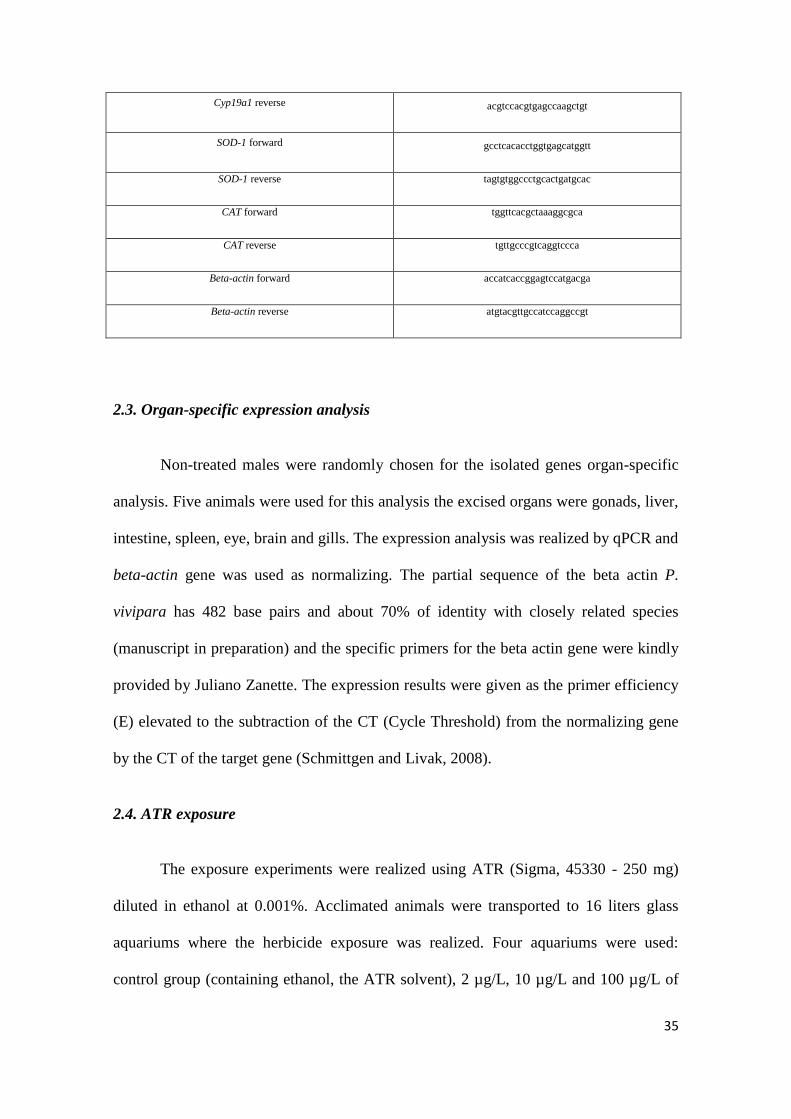
35
Cyp19a1 reverse acgtccacgtgagccaagctgt
SOD-1 forward gcctcacacctggtgagcatggtt
SOD-1 reverse tagtgtggccctgcactgatgcac
CAT forward tggttcacgctaaaggcgca
CAT reverse tgttgcccgtcaggtccca
Beta-actin forward accatcaccggagtccatgacga
Beta-actin reverse atgtacgttgccatccaggccgt
2.3. Organ-specific expression analysis
Non-treated males were randomly chosen for the isolated genes organ-specific
analysis. Five animals were used for this analysis the excised organs were gonads, liver,
intestine, spleen, eye, brain and gills. The expression analysis was realized by qPCR and
beta-actin gene was used as normalizing. The partial sequence of the beta actin P.
vivipara has 482 base pairs and about 70% of identity with closely related species
(manuscript in preparation) and the specific primers for the beta actin gene were kindly
provided by Juliano Zanette. The expression results were given as the primer efficiency
(E) elevated to the subtraction of the CT (Cycle Threshold) from the normalizing gene
by the CT of the target gene (Schmittgen and Livak, 2008).
2.4. ATR exposure
The exposure experiments were realized using ATR (Sigma, 45330 - 250 mg)
diluted in ethanol at 0.001%. Acclimated animals were transported to 16 liters glass
aquariums where the herbicide exposure was realized. Four aquariums were used:
control group (containing ethanol, the ATR solvent), 2 µg/L, 10 µg/L and 100 µg/L of
36
ATR. Total number of animals in each aquarium was 16 and exposure time was 24 and
96 hours. Thus, after 24 hours exposure, 8 animals from each aquarium were taken to
tissues RNA extraction and further cDNA synthesis used for 24 hours exposure gene
expression analysis. From then, the aquarium’s water was renovate (about 80%) every
24 hours until 96 hours were accomplished, aiming to conserve the compound under
experimental concentrations. After 96 hours of exposure, 8 animals from each group
were taken for gene expression analysis. Gene expression was performed by qPCR and
beta-actin gene was used as normalizing gene.
2.5. Statistical Analysis
Analysis of variance (ANOVA – p < 0,05) followed by a posteriori Tukey’s test
was realized for organ-specific gene expression analysis. The statistical analysis of
qPCR gene expression experiments was realized by the statistical software REST 2009
(Pfaffl, 2002).
3. RESULTS
3.1. Gene Isolation
After running a PCR with degenerate primers, the initial fragments of the genes
studied in the present work were obtained. The initial sequences obtained for StAR,
Cyp19a1, SOD-1 and CAT presented 156, 80, 355 and 350 base pairs respectively.
These fragments were aligned with fragments/genes already sequenced of
phylogenetically close species and reached identities varying from 83 to 100%. After
partial sequences were obtained, specific primers were designed and used for gene
expression experiments and performing PCR, using RACE kit, intending to obtain the
3’ e 5’ extremities sequences and cover most part of gene sequences. Based on the
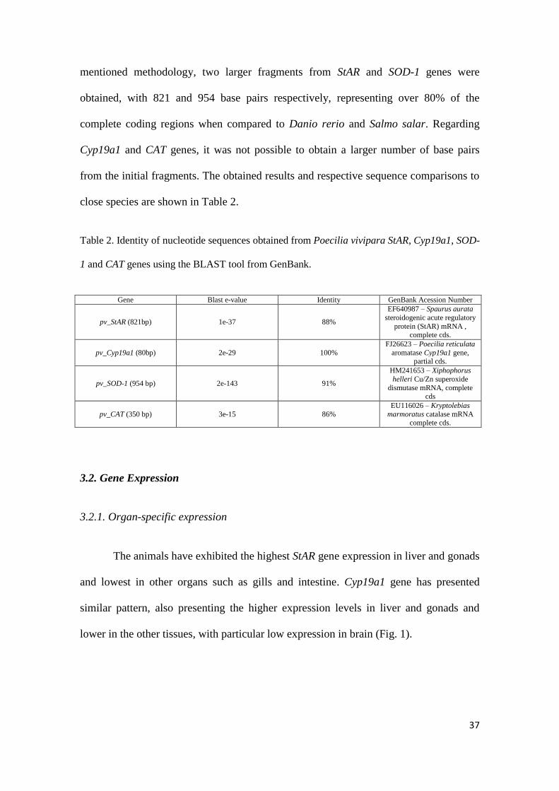
37
mentioned methodology, two larger fragments from StAR and SOD-1 genes were
obtained, with 821 and 954 base pairs respectively, representing over 80% of the
complete coding regions when compared to Danio rerio and Salmo salar. Regarding
Cyp19a1 and CAT genes, it was not possible to obtain a larger number of base pairs
from the initial fragments. The obtained results and respective sequence comparisons to
close species are shown in Table 2.
Table 2. Identity of nucleotide sequences obtained from Poecilia vivipara StAR, Cyp19a1, SOD-
1 and CAT genes using the BLAST tool from GenBank.
Gene Blast e-value Identity GenBank Acession Number
pv_StAR (821bp) 1e-37 88%
EF640987 – Spaurus aurata
steroidogenic acute regulatory
protein (StAR) mRNA , complete cds.
pv_Cyp19a1 (80bp) 2e-29 100%
FJ26623 – Poecilia reticulata
aromatase Cyp19a1 gene, partial cds.
pv_SOD-1 (954 bp) 2e-143 91%
HM241653 – Xiphophorus
helleri Cu/Zn superoxide
dismutase mRNA, complete cds
pv_CAT (350 bp) 3e-15 86%
EU116026 – Kryptolebias
marmoratus catalase mRNA complete cds.
3.2. Gene Expression
3.2.1. Organ-specific expression
The animals have exhibited the highest StAR gene expression in liver and gonads
and lowest in other organs such as gills and intestine. Cyp19a1 gene has presented
similar pattern, also presenting the higher expression levels in liver and gonads and
lower in the other tissues, with particular low expression in brain (Fig. 1).

38
Figure 1. Expression of genes related to steroidogenic pathway, StAR and Cyp19a1, in different organs (n = 5). The results are given
by subtracting the CT from the normalizing gene by the CT of the target gene. Different letters represent significant differences
among groups (ANOVA – p< 0.05 – Tukey).
Concerning the genes involved in the antioxidant pathway, SOD-1 presented a
considerably higher expression in liver and lower in other organs, such as gills and
spleen, while CAT demonstrated its highest expression in brain and liver and lowest in
intestine and eye (Fig. 2).
39
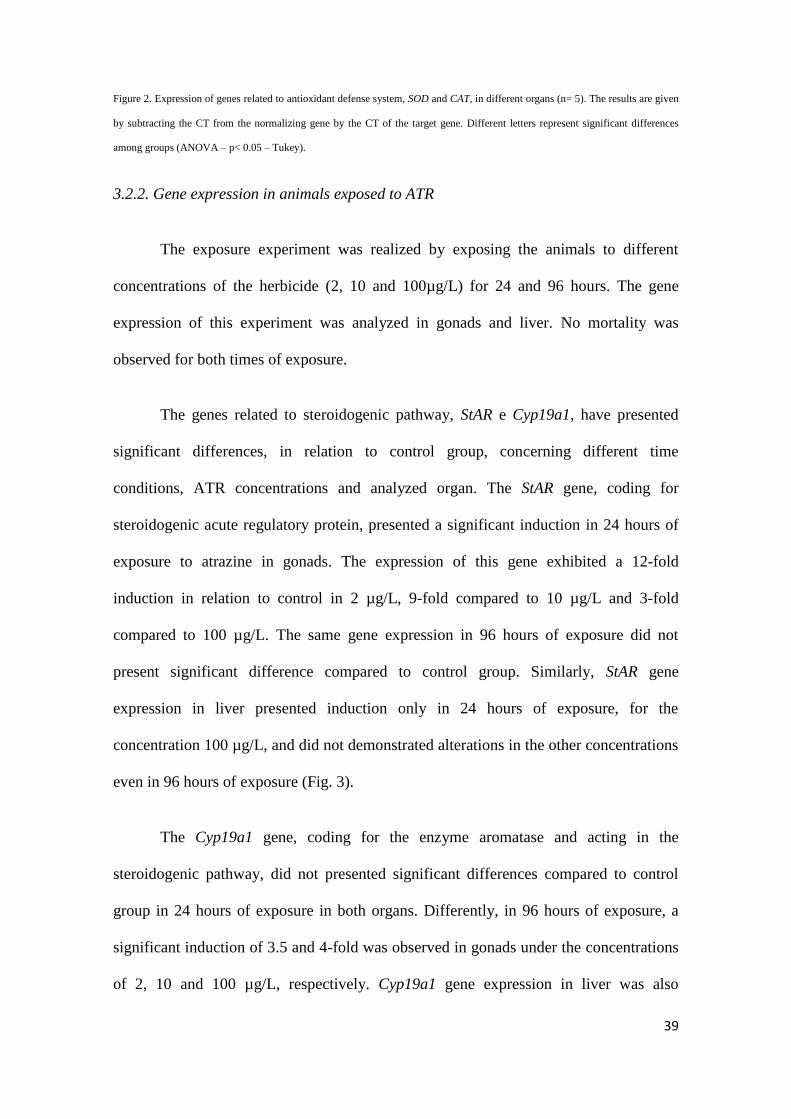
Figure 2. Expression of genes related to antioxidant defense system, SOD and CAT, in different organs (n= 5). The results are given
by subtracting the CT from the normalizing gene by the CT of the target gene. Different letters represent significant differences
among groups (ANOVA – p< 0.05 – Tukey).
3.2.2. Gene expression in animals exposed to ATR
The exposure experiment was realized by exposing the animals to different
concentrations of the herbicide (2, 10 and 100µg/L) for 24 and 96 hours. The gene
expression of this experiment was analyzed in gonads and liver. No mortality was
observed for both times of exposure.
The genes related to steroidogenic pathway, StAR e Cyp19a1, have presented
significant differences, in relation to control group, concerning different time
conditions, ATR concentrations and analyzed organ. The StAR gene, coding for
steroidogenic acute regulatory protein, presented a significant induction in 24 hours of
exposure to atrazine in gonads. The expression of this gene exhibited a 12-fold
induction in relation to control in 2 µg/L, 9-fold compared to 10 µg/L and 3-fold
compared to 100 µg/L. The same gene expression in 96 hours of exposure did not
present significant difference compared to control group. Similarly, StAR gene
expression in liver presented induction only in 24 hours of exposure, for the
concentration 100 µg/L, and did not demonstrated alterations in the other concentrations
even in 96 hours of exposure (Fig. 3).
The Cyp19a1 gene, coding for the enzyme aromatase and acting in the
steroidogenic pathway, did not presented significant differences compared to control
group in 24 hours of exposure in both organs. Differently, in 96 hours of exposure, a
significant induction of 3.5 and 4-fold was observed in gonads under the concentrations
of 2, 10 and 100 µg/L, respectively. Cyp19a1 gene expression in liver was also

40
significantly increased after 96 hours of ATR exposure in 10 µg/L (10-fold induction)
and 100 µg/L (9-fold induction) (Fig.4).
Figure 3. StAR gene expression in gonads and liver of P. vivipara exposed to ATR concentrations of 2, 10 and 100 µg/L for 24 and
96 hours (n = 8). The relative expression results are normalized by the values of beta-actin gene expression and the control group is
represented by the line of the x axis (1). Asterisks represent significant differences in relation to control group.

41
Figure 4. Cyp19a1 gene expression in gonads and liver of P. vivipara exposed to ATR concentrations of 2, 10 and 100 µg/L for 24
and 96 hours (n = 8). The relative expression results are normalized by the values of beta-actin gene expression and the control
group is represented by the line of the x axis (1). Asterisks represent significant differences in relation to control group.
Concerning the genes coding for enzymes related to the antioxidant defense
system, there were observed expression alterations in gonads and liver only by 24 hours
of exposure time. SOD-1 gene was induced in gonads only in the lowest ATR used
concentration (2 µg/L), by the time of 24 hours, presenting a 7-fold induction in relation
to control group, and then returned to basal levels in higher concentrations. Concerning
the liver, SOD-1 exhibited a significant induction, compared to control group, at the
concentrations of 2 and 10 µg/L, also by the time of 24 hours, and returned to control
similar values at the concentration of 100 µg/L. This gene did not present significant
differences, in relation to control group, by the time of 96 hours of exposure in none of
the analyzed organs neither in the used concentrations (Fig. 5).
Finally, the gene coding for catalase enzyme exhibited a similar pattern to SOD-
1 expression, presenting a 2-fold induction in gonads, by the time of 24 hours, only at
the minor concentration. CAT was significantly induced in liver, at the three tested
concentrations, presenting a 6, 3 and 4-fold induction at the concentrations of de 2, 10
and 100 µg/L, respectively. Comparatively to SOD-1, this gene did not present
significant alteration compared to control group by the time of 96 hours (Fig. 6).

42
Figure 5. SOD-1 gene expression in gonads and liver of P. vivipara exposed to ATR concentrations of 2, 10 and 100 µg/L for 24
and 96 hours (n = 8). The relative expression results are normalized by the values of beta-actin gene expression and the control
group is represented by the line of the x axis (1). Asterisks represent significant differences in relation to control group.
Figure 6. CAT gene expression in gonads and liver of P. vivipara exposed to ATR concentrations of 2, 10 and 100 µg/L for 24 and
96 hours (n = 8). The relative expression results are normalized by the values of beta-actin gene expression and the control group is
represented by the line of the x axis (1). Asterisks represent significant differences in relation to control group.

43
4. DISCUSSION
4.1. Genes isolation
Several studies have been realized taking Poecillidae as model animal for
toxicological researches due to, mainly, its high tolerance to extreme environmental
conditions, including extremely polluted environments (Larsson et al., 2002; Betito,
2006; Mattos et al., 2009). Molecular tools, such as pollution biomarkers coding genes,
are used in fish and aquatic invertebrates for biomonitoring studies of aquatic
environments (Monserrat et al., 2007). However, these tools are still poorly applied
concerning South American guppy species because little information on these animals is
available, except for studies focused on ecological population (Betito, 2006).
Intending to investigate the molecular responses of these animals, against the
ATR herbicide, we have carried out the sequencing of fragments of target genes. Thus,
the cloning of Cyp19a1, StAR, SOD-1 and CAT gene fragments, from Poecilia vivipara,
was realized using degenerate primers designed based on phylogenetically close species
such as Poecilia reticulata, Jenynsia multidentata and Oryzias latipes.
StAR gene was already identified in teleost species such as the codfish Gadus
morhua (Kortner and Arukwe, 2007), the African catfish Clarias gariepinus
(Sreenivasulu et al., 2009) and the Atlantic salmon Salmo salar (Arukwe, 2005). The
genes Cyp19a1 (gonadal aromatase) and Cyp19a2 (brain aromatase) have been already
identified in teleost fish such as Ictalurus punctatus (Trant, 1994), medaka Oryzias
latipes (Fukada et al., 1996), Fundulus heteroclitus (Greytak et al., 2005) and others.
The StAR fragment obtained contained 821 base pairs and presented identity of 88%
with sequence fragments from the species Spaurus aurata (Fig. 1). Besides, it also
44
presented relatively high identity compared to Salmo salar and Xiphophorus helleri, up
to 86 and 88 % respectively.
The antioxidant enzymes coding genes, SOD-1 and CAT, have been already
sequenced in teleost species, such as Salmo salar and Danio rerio, and in some aquatic
invertebrates (Ken et al., 1998; 2003; Park et al., 2010; Kim et al., 2011). The obtained
sequence for SOD-1 from P. vivipara consists of 954 pair bases and, according to
GenBank alignments, presents 91% of identity with SOD-1 from Xiphophorus helleri,
which is also phylogenetically close to P. vivipara. The fragment obtained for CAT is
composed by 358 base pairs and presents higher identity (86%) with Kryptolebias
marmoratus (Fig. 1).
4.2. Gene expression
4.2.1 Organ-specific expression
Aiming to realize a relative quantitative distribution analysis of the target genes
from P. vivipara, different organs (brain, eye, spleen, intestine, gonads and liver) were
used for qPCR tests and for comparison of gene expression in non-treated animals
The results of StAR gene expression have shown that its higher expression
occurs in liver and testis, although its expression was also observed in non-
steroidogenic organs (ex. spleen and intestine). Studies realized in mammals have been
demonstrated that a predominant site for StAR gene transcripts expression is present in
steroidogenic organs, including testis, ovaries, brain and kidneys, and the expression of
the mentioned gene is, therefore, strictly related to steroidogenesis (Bauer et al., 2000;
45
Kusakabe et al., 2002). However, in the present study we have found StAR gene
expression in steroidogenic and non-steroidogenic organs, such as intestine and spleen,
reinforcing the results of Kusabake et al. (2002) which, in a similar study realized with
the rainbow trout, have also reported a relatively elevated StAR gene expression in non-
steroidogenic organs. The difference of the StAR gene expression pattern, observed in
fishes and mammals, may be indicating a different biological function for the protein,
principally in non-steroidogenic organs.
Cyp19a1 gene presented higher expression in gonads and its minor expression
levels were detected in brain and non-steroidogenic organs, such as intestine and spleen.
Tang et al. (2010) have performed a differential expression study of Cyp19a1 and
Cyp19a2 in organs from Cyprinus carpio, and reported low basal expression of
Cyp19a1 in brain regions while Cyp19a2 presented high expression levels in these same
regions. Additionally, it was reported by the same authors a relatively high expression
of Cyp19a1 in males’ testis as well as in females’ ovaries, with a 3-fold induction
observed in females. Our work has only concerned about males Cyp19a1 gene
expression, in which gonadal gene expression was approximately 5-fold higher than in
brain, evidencing that, in P. vivipara, this gene codes for a kind of aromatase more
intensively expressed in gonads (gonadal aromatase) following data found in other fish
species (Cheshenko et al., 2008; Suzawa and Ingraham, 2008; Tang et al., 2010).
Concerning SOD-1 e CAT genes, both exhibited similar expression patterns,
with higher liver expression and less variable expression levels among organs when
compared to steroidogenesis-related genes StAR e Cyp19a1. Although these genes are
expressed in almost all organs (Alvarez et al., 2005), its basal expression and induction
by xenobiotics may occur as organ-specific, presenting higher levels in detoxification

46
organs, such as liver and hepatopancreas, besides muscular and brain organs (Ozcan et
al., 2004).
4.2.2 Gene expression in ATR
exposed animals
ATR herbicide is widely used in Brazil, principally regarding rice fields regions,
and its maximum load allowed in fresh water is 2 µg/L following resolution 357–
(CONAMA, 2005). The interaction of this compound with biological systems and
reproductive problems in males exposed to ATR has been documented in juvenile fishes
(Moore and Waring, 1998; Spano et al., 2004), crocodilian reptiles (Crain et al., 1997)
and peripubertal rats (Friedmann, 2002). The findings of the present work reinforced
these studies and have demonstrated the capacity of the herbicide of altering genes from
the steroidogenic pathway as well as from the antioxidant response system in P.
vivipara.
The StAR protein is present in mitochondrial membrane of steroidogenic cells
and is essential for initial and acute regulation of the steroid hormone production
process, in general, and also a velocity limiting factor of this pathway (Norris, 2007).
Although there are vertebrates studies evidencing alterations in the expression of this
gene caused by endocrine disruptors and natural estrogens, the mechanisms relying
under these disturbances still remains not well elucidated (Wang et al., 2006). Studies
focused on endocrine disruptors with steroidogenic action have demonstrated that these
compounds are capable of inducing the StAR gene expression in fish species, such as
Kryptolebias marmoratus and Salmo salar, evidencing that this gene, jointly with the
other genes coding for proteins from the steroidogenic pathway, appears to be a main
target of endocrine disruptors (Arukwe, 2008; Rhee et al., 2011). Concerning the
47
compound used in the present work, earlier studies have been already demonstrated
alterations in the expression of this gene caused by relatively low ATR doses (Suzawa e
Ingraham, 2008; Abariwku et al., 2011).
StAR gene was induced after 24 hours of acute exposure, and only at lower
concentrations in gonads, suggesting that a compensatory response against the herbicide
could be occurring over the hormonal synthesis in the highest and subchronic (96 h)
exposure, in which no induction of this gene was observed. Our findings supports the
studies that demonstrates the induction of StAR gene in mammals cells, as an efficient
biomarker to acute ATR exposure and also highlights that this gene becomes less
responsive at high ATR doses, presenting its higher response at considered average
dosages (Abarikwu et al., 2011). Based on the expression of StAR gene, against ATR
exposition, it can be suggested that this gene might be an interesting model concerning
acute ATR contamination studies, at moderate/low doses, including the maximum load
of 2 µg/L allowed by Brazilian legislation (CONAMA, 2005).
Cyp19a1 gene did not shown significant differences, compared to control group,
in 24 hours for any of the analyzed organs. However, a significant induction was
observed in 96 hours at all concentrations in gonads and at 10 and 100 µg/L in liver.
The induction response of this gene against ATR exposure in 96 hours, presented in our
study, supports the findings of Suzawa and Ingraham (2008) in zebrafish exposed to
similar ATR concentrations. Cyp19a1 gene has been suggested as one of the main
affected genes by ATR (Fan et al., 2007; Anderson et al., 2008; Suzawa and Ingraham,
2008; Tinfo et al., 2011). Nevertheless, several studies demonstrate that the ATR-
dependent induction of Cyp19a1 occurs in a late matter, between 48 and 96 hours of
48
exposure, as it was observed in the present study (Anderson et al., 2008; Suzawa and
Ingraham, 2008).
ATR molecular action over the induction of genes from steroidogenic pathway is
still poorly understood. However, it is known that, in the promoter region from StAR
and Cyp19a1 genes there are target response elements from nuclear receptors of NR5A
family. Thus, some studies have been demonstrated that ATR is able to activate these
nuclear receptors enabling their ligation to their target genes response elements and
increasing the transcription of these. Therefore, the mechanism of induction of these
genes by ATR apparently occurs via NR5A nuclear receptors (Suzawa and Ingraham,
2008; Fan et al., 2007).
The genes coding to key enzymes from the enzymatic antioxidant defense
system, SOD-1 and CAT, were induced in liver and gonads from ATR-treated animals.
Likewise StAR, these genes have exhibited a similar induction pattern only in the lower
concentrations, except for CAT gene in the liver, which was induced in all tested
concentrations. These results corroborate to Jin et al. (2010) study that demonstrated an
expression increase of this gene caused by low ATR concentrations in zebrafish and
then returning to normality in the highest concentration applied in the study.
Pesticides and its metabolites may be responsible for the oxidative stress
induction in fishes once that, normally, it presents a considerably high oxidative
potential (Alvarez et al., 2005). Similarly to the present work, exposition to xenobiotics
in fishes has shown that the response pattern for SOD-1, as well as for CAT, appears to
be organ-specific (Ahmad et al., 2004; Ozcan et al., 2004).

49
Concerning the present study, except for Cyp19a1 gene, the observed response
pattern presented genes induction at the lower ATR concentrations applied while in
higher doses the expression returned to levels near to the control group. The absence of
SOD-1, CAT and StAR genes induction at higher doses may be related to accessory
mechanisms impeding the compound accumulation in cells at a certain concentration. In
this sense, Islam et al. (2002) have demonstrated that this compound is able to induce
genes from multidrug resistance family (MDR), such as P-glycoprotein. Besides,
detoxification components such as GST and CYPs can be induced as well, which seems
to contribute for a resistance phenotype. Therefore, a mechanism associated to cell
exportation pumps may be acting when the organism is challenged with relatively high
concentrations of the herbicide.
The activity of exportations pumps, such as P-glycoprotein, is directly related to
ATP expense (Hennessy and Spiers, 2007) and, therefore, is a mechanism of elevated
energetic costs to animals’ cells. Studies involving insects and amphibian species have
shown that higher doses and subchronic times of ATR exposure affects the energetic
metabolism of these animals and rises the oxygen consumption and key genes
expression from the main cell’s energy production pathways (Anderson et al., 2008;
Zaya et al., 2011). The high energetic expense of the exposed animals becomes
worrying once it affects the efficiency of essential physiological mechanism such as
reproduction and growth.
Our results have demonstrated that the herbicide ATR, even at relatively low
concentrations, is capable of altering the expression of genes from the steroidogenic
pathway, such as StAR and Cyp19a1, as well as genes related to cellular antioxidant
response, such as SOD-1 and CAT evidencing, therefore, that the used model is sensible
50
to the herbicide presence in the water. Besides, although Cyp19a1 was also induced by
ATR, the StAR gene expression appears to serve as a potential contamination biomarker
for this endocrine disruptor, once it presents induction at the lower concentrations, and
in acute exposures, representing, therefore, an animal’s early response to the pollutant in
the aquatic environment.
5. REFERENCES
Abarikwu S.O., Farombi, E.O., Kashyap, M.P., Pant, A.B., 2011. Atrazine induces
transcriptional changes in marker genes associated with steroidogenesis in primary
cultures of rat Leydig cells. Toxicol. in Vitro. doi:10.1016/j.tiv.2011.06.002
Ahmad, I., Pacheco, M., Santos, M.A., 2004. Enzymatic and non enzymatic
antioxidants as an adaptation to phagocyte-induced damage in Anguilla anguilla L.
following in situ harbor water exposure. Ecotoxicol. Environ. Saf. 57, 290 – 302.
Alvarez, R.M., Morales, A.E., Sanz, A., 2005. Antioxidant defenses in fish: Biotic and
abiotic factors. Rev. Fish Biol. Fish. 15, 75 – 88.
Anderson T.D., Jin-Clark, Y., Begum, K., et al., 2008. Gene expression profiling
reveals decreased expression of two hemoglobin genes associated with increased
consumption of oxygen in Chironomusn tentans exposed to atrazine: A possible
mechanism for adapting to oxygen deficiency. Aq. Toxicol. 86, 148 – 156.
Arukwe, A., 2005. Modulation of brain steroidogenesis by affecting transcriptional
changes of steroidogenic acute regulatory (StAR) protein and cholesterol side chain

51
cleavage (P450scc) in juvenile atlantic salmon (Salmo salar) is a novel aspect of
nonylphenol toxicity. Environ. Sci. Technol. 39, 9791 – 9798.
Arukwe, A., 2008. Steroidogenic acute regulatory (StAR) protein and cholesterol side-
chain cleavage (P450scc)- regulated steroidogenesis as an organ-specific molecular and
cellular target for endocrine disrupting chemicals in fish. Cell. Biol. Toxicol. 24,
527 – 540.
Bauer, M.P., Bridgham, J.T., Langenau, D.M., et al., 2000. Conservation of
steroidogenic acute regulatory (StAR) protein structure and expression in vertebrates.
Mol. Cell. Endocrinol. 168, 119 – 125.
Betito, R., 2006. Comparação da complexidade das adaptações bioecológicas de dois
Peixes (Jenynsia multidentata e Poecilia vivipara) (Cyprinodontiformes) no estuáro da
Lagoa dos Patos (RS - Brasil). Ver. Didat. Sist. 3, 71 – 100.
Bizerril, C. R. S., Primo, P. B. S., 2001. Peixes de Aguas Interiores do Estado do Rio de
Janeiro. Rio de Janeiro: FEMAR-SEMADS.
Bouloubassi I., Saliot A., 1993. Investigation of anthropogenic and natural organic
inputs in estuarine sediments using hydrocarbon markers (NAH, LAB, PAH). Oceanol.
Acta. 16, 145 – 61.
Brodeur., J.C., Svartz, G., Perez-coll, C.S., et al., 2009. Comparative susceptibility to
atrazine of three developmental stages of Rhinella arenarum and influence on
metamorphosis: Non-monotonous acceleration of the time to climax and delayed tail
resorption. Aq. Toxicol. 91, 161 – 170.
52
Cheshenko, K., Pakdel, F., Segner, H., et al., 2008. Interference of endocrine disrupting
chemicals with aromatase CYP19 expression or activity, and consequences for
reproduction of teleost fish. Gen. Comp. Endocrinol. 155, 31 – 62.
CONAMA, 2005. Conselho Nacional do Meio Ambiente/ Ministério do Meio
Ambiente. Resolução No. 357 de 17 de março de 2005. Disponível em <http://
www.mma.gov.br/port/conama/legiano1.cfm?codlegitipo=3&ano=2005>.
Crain, D.A., Guillette, L.J., Rooney, A.A., Pickford, D.B., 1997. Alterations in
steroidogenesis in alligators (Alligator mississippiensis) exposed naturally and
experimentally to environmental contaminants. Environ. Health Perspect. 105, 528 –
533.
Depledge, M.H., Aagaard, A., Gyorko, R., 1995. Assessment of trace metal toxicity
using molecular, physiological and behavioural biomarkers. Mar. Pollut. Bull. 31, 19 –
27.
Elia, A.C., Waller, W.T., Norton S.J., 2002. Biochemical responses of Bluegill Sunfish
(Lepomis macrochirus, Rafinesque) to atrazine induced oxidative stress. Bull. Environ.
Contam. Toxicol. 68, 809 – 816.
Fan, W., Yanase, T., Morinaga, H., et al., 2007. Atrazine - induced aromatase
expression is SF-1 dependent: Implications for endocrine disruption in wildlife and
reproductive cancers in humans. Environ. Health Perspect. 115, 720 - 727.
Friedmann, A.S., 2002. Atrazine inhibition of testosterone production in rat males
following peripubertal exposure. Reprod. Toxicol. 16, 275 – 279.
53
Frohman, M. A., Dush, M. K., Martin, G. R., 1988. Rapid production of full-length
cDNA from rare transcripts: Amplification using a single gene-specific oligonucleotide
primer. Proc. Natl. Acad. Sci. USA 85, 8998 – 9002.
Fukada, S., Tanaka, M., Matsuyama, K., et al., 1996. Isolation, characterization, and
expression of cDNAs encoding the medaka (Oryzias latipes) ovarian follicle
cytochrome P-450 aromatase. Mol. Reprod. Dev. 45, 285 – 290.
Gagosian R.B., Peltzer E.T., 1986. The importance of atmospheric input of terrestrial
organic material to deep sea sediments. Org. Geochem. 10, 661 – 9.
Gómez-Anduro, G.A., Barillas-Mury, C.V., Peregrino-Uriarte, A.B., et al., 2006. The
cytosolic manganese superoxide dismutase from the shrimp Litopenaeus vannamei:
molecular cloning and expression. Dev. Comp. Immunol. 30, 893 – 900.
Graymore, M., Allinson, G., Allinson, M., et al., 1999. Environmental fate of pesticides
used in Australian viticulture: V. behavior of atrazine in the soils of the South
Australian Riverland. Toxicol. Environ. Chem. 70, 427 – 39.
Graymore, M., Stagnitti, F., Allinson, G., 2001. Impacts of atrazine in aquatic
ecosystems. Environ. Internat. 26, 483 – 495.
Graziano N., Mcguire, M.J., Roberson A., et al., 2006. 2004 national atrazine
occurrence monitoring program using the abraxis ELISA method. Environ. Sci.
Technol. 40, 1163 - 1171.
Greytak, S.R., Champlin, D., Callard, G.V., 2005. Isolation and characterization of two
cytochrome P450 aromatase forms in killifish (Fundulus heteroclitus): differential
54
expression in fish from polluted and unpolluted environments. Aquat.Toxicol. 71, 371 –
389.
Hayes, T.B., Haston, K., Tsui B., et al., 2002. Feminization of male frogs in the wild.
Nature. 419, 895 – 896.
Hayes, T.B., Haston, K., Tsui B., et al., 2003. Atrazine-induced hermaphroditism at 0.1
ppb in American leopard frogs (Rana pipiens): laboratory and field evidence. Environ.
Health Perspect. 111, 568 – 575.
Hayes, T.B., Beasley, V.R., Solla, S., et al., 2011. Demasculinization and feminization
of male gonads by atrazine: Consistent effects across vertebrate classes. J. Steroid
Biochem. Molec. Biol. 127, 64 – 73.
Henessy, M., Spiers, J.P., 2007. A primer on the mechanics of P-glycoprotein the
multidrug transporter. Pharmacol. Res. 55, 1 – 15.
Islam, M.O., Hara, M., Myiake, J., 2002. Induction of P-glycoprotein, glutathione-S-
transferase and cytochrome P450 in rat liver by atrazine. Environ. Toxicol. Pharmacol.
12, 1 – 6.
Jelaso, A., Celestine, R., Zaya, R., et al., 2008. Chronic exposure to high levels of
atrazine alters expression of genes that regulate immune and growth-related functions in
developing Xenopus laevis tadpoles. Environ. Res. 109, 379 – 389.
Jin, Y., Zhang, X., Shu, L., et al., 2010. Oxidative stress response and gene expression
with atrazine exposure in adult female zebrafish (Danio rerio). Chemosphere. 78, 846 –
852.
55
Ken, C., Lin, C., Wu, J., Shaw, J., 1998. Cloning and expression of a cDNA coding for
catalase from zebrafish (Danio rerio). J.Agric.Food Chem. 48, 2092 − 2096.
Kim, B., Rhee, J., Park, G.S., et al., 2011. Cu/Zn- and Mn-superoxide dismutase (SOD)
from the copepod Tigriopus japonicus: Molecular cloning and expression in response
to environmental pollutants. Chemosphere. 84, 1467 – 147
Kortner, T.M., Arukwe, A., 2007. The xenoestrogen, 4-nonylphenol, impaired
steroidogenesis in previtellogenic oocyte culture of Atlantic cod (Gadus morhua) by
targeting the StAR protein and P450scc expressions. Gen. Comp. Endocrinol. 150, 419
– 429.
Kusakabe, M., Todo, T., McQuillan, H.J., et al., 2002. Characterization and expression
of steroidogenic acute regulatory protein and MLN64 cDNAs in trout. Endocrinology.
143, 2062 – 2070.
Larsson., D.J.K., Kinnberg, K., Sturve, J., et al., 2002. Studies of masculinization,
detoxification, and oxidative stress responses in Guppies (Poecilia reticulata) exposed
to efluent from a Pulp Mill. Ecotoxicol. Environ. Saf. 52, 13 – 20.
Mattos, J.J., Siebert, M.N., Luchmann, K.H., et al., 2009. Differential gene
expression in Poecilia vivipara exposed to diesel oil water accommodated fraction.
Mar. Environ. Res. 69, 31 – 33.
Monserrat., J.M., Martínez, P.E., Geracitano, L.A., et al., 2007. Pollution biomarkers
in estuarine animals: Critical review and new perspectives. Comp. Biochem. Physiol.
146, 221 – 234.
56
Moore, A., Waring, C., 1998. Mechanistic effects of a triazine pesticide on reproductive
endocrine function in mature male Atlantic salmon (Salmo salar L.) parr. Pest.
Biochem. Physiol. 62, 41 – 50.
Norris, D. 2007. Vertebrate Endocrinology. 4ª ed. Elsevier Academic Press.
Ozcan, E., Sevgiler, Y., Uner, N., 2004. Tissue - specific oxidative stress responses in
fish exposed to 2,4-D and azin-phosmethyl. Comp. Biochem. Physiol. 137, 43 – 51.
Parenti, L. R., Rauchemberger, M., 1989. Systematic overview of the Poeciliines. In
The Ecology and Evolution of Poeciliid Fishes (Poeciliidae)
(Meffe,A.&Snelson,F.F.,eds), pp. 3–12. New Jersey: Prentice Hall.
Park, H., Ahn, I., Lee, J.K., et al., 2009. Molecular cloning, characterization, and the
response of manganese superoxide dismutase from the Antarctic bivalve Laternula
elliptica to PCB exposure. Fish Shellfish Immunol. 27, 522 – 528.
Paulino, M.G., Sakuragui, M.M., Fernandes M.N., 2012. Effects of atrazine on the gill
cells and ionic balance in a neotropical fish, Prochilodus lineatus. Chemosphere. 86, 1-
7.
Pfaffl, M. W., Horger, G. W., Dempfle, L., 2002. Relative expression software tool
(REST) for group-wise comparison an statistical analysis of relative expression results
in Real time PCR. Nucl. Ac. Res. 9, 30 - 36.
Qian, H.F., Sheng, G.D., Liu, W.P., et al., 2008. Inhibitory effects of atrazine on
Chlorella vulgaris as assessed by real-time polymerase chain reaction. Environ.
Toxicol. Chem. 27, 182 – 187.
57
Rhee, J., Kim, B., Li, C.J., et al., 2011. Bisphenol A modulates expression of sex
differentiation genes in the self-fertilizing fish, Kryptolebias marmoratus. Aq.
Toxicol. 104, 218 – 229.
Roche, H., Boge, G., 1996. Fish blood parameters as a potential tool for identification of
stress caused by environmental factors and chemical intoxication.Mar. Environ. Res. 41,
27 - 43.
Rudneva, I.I., 1997. Blood antioxidant system of Black Sea elasmobranch and teleost.
Comp. Biochem. Physiol. 118, 255–260.
Schmittgen, T.D., Livak, K.J., 2008. Analyzing real-time PCR data by the comparative
CT method. Nat. Protoc. 3, 1101–1108.
Simpson, E.R., 1994. Aromatase cytochrome - P450: structure, function and regulation.
Faseb J. 8, 12 - 41.
Spano, L., Tyler, C.R., Van Aerle, R., et al., 2004. Effects of atrazine on sex steroid
dynamics, plasma vitellogenin concentration and gonad development in adult goldfish
(Carassius auratus). Aq. Toxicol. 66, 369 – 379.
Sreenivasulu, G., Sidrevi, P., Sahoo, P.K., et al., 2009. Cloning and expression of StAR
during gonadal cycle and hCG-induced oocyte maturation of air-breathing catfish,
Clarias gariepinus. Comp. Biochem. Physiol. 154, 6 – 11.
Suzawa, M., Ingraham, H.A., 2008. The herbicide atrazine activates endocrine gene
networks via non-steroidal NR5A nuclear receptors in fish and mammalian cells. Plos
one 3, 1-9.

58
Tang, B., Hu,W., Hao, J., Zhu, Z. , 2010. Developmental expression of steroidogenic
factor-1, cyp19a1a and cyp19a1b from common carp (Cyprinus carpio). Gen. Comp.
Endocrinol. 167, 408 – 416.
Tavera-Mendoza, L., Ruby, S., Brousseau, P., et al., 2002. Reponse of the amphibian
tadpole (Xenopus laevis) to atrazine during sexual differentiation of the testis. Environ.
Toxicol. Chem. 21, 527 – 531.
Thompson, E.A., Siiter, J., 1974. Utilization of oxygen and reduced nicotinamide
adenine dinucleotide phosphate by human placental microsomes during aromatization
of androstenedione. J. Biol. Chem. 249, 5364 – 5372.
Tinfo, N.S., Hotchkiss, M.G., Buckalew, A.R., et al., 2011. Understanding the effects
of atrazine on steroidogenesis in rat granulose and H295R adrenal cortical carcinoma
cells. Reprod. Toxicol. 31, 184 – 193.
Tomlin C. 2000. The pesticide Manual: a world compendium. Twelfthed. Farnham
(UK) 7 The British Crop Protection Council.
Trant, J., 1994. Isolation and characterization of the cDNA encoding the channel catfish
(Ictalurus punctatus) form of cytochrome P450 aromatase. Gen. Comp. Endocrinol. 95,
155 –168.
Wang L., Song, L., Hong, X., et al., 2006. Low concentrations mono-butyl phthalate
stimulates steroidogenesis by facilitating steroidogenic acute regulatory protein
expression in mouse Leydig tumor cells (MLTC-1). Chem. Biol. Interact. 164, 15 – 24.
59
Wauchope, R.D., Butler, T.M., Hornsby, A.G., et al., 1991. The SCS/ARS/CES
pesticide properties database for environmental decision-making. Rev. Environ.
Contam. Toxicol. 123, 1–36.
Zaya, R.M., Amini, Z., Whitaker, A.S., Ide, C.F., 2011. Exposure to atrazine affects
the expression of key genes in metabolic pathways integral to energy homeostasis
in Xenopus laevis tadpoles. Aq. Toxicol. 104, 254 – 262.
60
CONCLUSÕES GERAIS
- As sequências parciais dos genes StAR, Cyp19a1, SOD-1 e CAT foram identificadas e
apresentaram um alta identidade com espécies filogeneticamente próximas a P.
vivipara, possibilitando uma anotação preliminar das mesmas.
- O herbicida Atrazina foi capaz de alterar a expressão tanto dos genes relacionados à
via esteroidogênica, StAR e Cyp19a1, afirmando o seu papel como desregulador
endócrino, quanto os genes que codificam para enzimas antioxidantes, SOD-1 e CAT.
- O modelo experimental utilizado no presente trabalho se mostrou, portanto, sensível à
presença do poluente na água, inclusive na concentração máxima permitida pelo
CONAMA (2 µg/L).
- O gene StAR mostrou ser um biomarcador potencial de contaminação por ATR em
baixas concentrações e exposições agudas.
- Nas maiores concentrações de ATR não houve indução de genes como StAR, SOD-1 e
CAT, o que indica que outro sistema pode estar atuando frente a concentrações maiores
do herbicida, como por exemplo o aumento da expressão/atividade de bombas de
exportação como as glicoproteínas - P, o que implicaria em um alto custo energético ao
animal.
61
PERSPECTIVAS
Como perspectivas do presente trabalho, temos a identificação do gene que
codifica para a glicoproteína-P, bem como a expressão desse gene frente à
concentrações de ATR semelhantes a do presente estudo. Ainda como perspectivas
estão a realização de exposições subcrônicas e crônicas e análise histopatológica das
gônadas dos animais, já que o gene que codifica para aromatase (Cyp19a1) demonstrou
uma indução nos animais expostos por 96 horas a ATR.
62
REFERÊNCIAS BIBLIOGRÁFICAS
Abarikwu, S.O., Farombi, E.O., Kashyap, M.P., Pant, A.B., 2011. Atrazine induces
transcriptional changes in marker genes associated with steroidogenesis in primary
cultures of rat Leydig cells. Toxicol. in Vitro. 25, 1588 – 1595.
Ahmad, S., 1995. Oxidative stress and antioxidant defenses in biology. Chapman e Hall,
New York.
Anderson, T.D., Jin-Clark, Y., Begum, K., et al., 2008. Gene expression profiling
reveals decreased expression of two hemoglobin genes associated with increased
consumption of oxygen in Chironomus tentans exposed to atrazine: A possible
mechanism for adapting to oxygen deficiency. Aq. Toxicol. 86, 148 – 156.
Araújo F.G., 2009. Distribution of Guppies Poecilia reticulata (Peters, 1860) and
Phalloceros caudimaculatus (Hensel, 1868) Along a polluted stretch of the Paraíba do
Sul River, Brazil. Brazilian J. Biol. 69, 41 - 48.
Avci, A., Kacmaz, M., Durak, I., 2005. Peroxidation in muscle and liver tissues from
fish in a contaminated river due to a petroleum refinety industry. Ecotoxicol. Environ.
Saf. 60, 101 – 105.
63
Bainy, A.C.D., Saito, E., Carvalho, P.S.M., Junqueira, V.B.C., 1996. Oxidative stress
in gill, erythrocytes, liver and kidney of Nile tilapia (Oreochromis niloticus) from a
polluted site. Aq. Toxicol. 34, 151 - 162.
Bauer, M.P., Bridgham, J.T., Langenau, D.M., Johnson, A.L., et al., 2000. Conservation
of steroidogenic acute regulatory (StAR) protein structure and expression in vertebrates.
Mol. Cell. Endocrinol. 168, 119 – 125.
Bentley, P.J., 1998. Comparative Vertebrate Endocrinology, Cambridge University
Press, Cambridge, UK.
Betito, R., 2006. Comparação da complexidade das adaptações bioecológicas de dois
Peixes (Jenynsia multidentata e Poecilia vivipara) (Cyprinodontiformes) no estuáro da
Lagoa dos Patos (RS - Brasil). Ver. Didat. Sist. 3, 71 – 100.
Bizerril, C. R. S., Primo, P. B. S., 2001. Peixes de Aguas Interiores do Estado do Rio de
Janeiro. Rio de Janeiro: FEMAR-SEMADS.
Bourne, A., 1991. Androgens. In: Pang, P.K.T., Schreibman, M.P.(Eds.), Vertebrate
endocrinology: fundamental sand biomedical implications. Academic Press, NewYork,
115–147.
Chang, X., Kobayashi, T., Senthilkumaran, B., et al., 2005. Two types of Aromatase
with different encoding genes, tissue distribution and developmental expression in Nile
tilapia (Oreochromis niloticus). Gen. Comp. Endocrinol. 141, 101 – 115.
Christenson, L.K., Strauss, J.F., 2001. Steroidogenic acute regulatory protein: an update
on its regulation and mechanism of action. Arch. Med. Res. 32, 576 – 586.
64
Clark, B.J., Wells, J., King, S.R., Stocco, D.M., 1994. Purification, cloning and
expression of a novel luteinizing hormone – induced mitochondrial protein in mouse
Leydig tumor cells. Characterization of the steroidogenic acute regulatory protein
(StAR). J. Biol. Chem. 269, 28314 – 28322.
CONAMA, 2005. Conselho Nacional do Meio Ambiente/Ministério do Meio Ambiente.
Resolução No. 357 de 17 de março de 2005. Disponível em <http://
www.mma.gov.br/port/conama/legiano1.cfm?codlegitipo=3&ano=2005>
Cook, P.M., Robbins J.A., Endicott, D.D., et al., 2003. Effects of aryl hydrocarbon
receptor mediated early life stage toxicity on lake trout populations in lake Ontario
during the 20th
century. Environ. Sci. Technol. 37, 3864 - 3877.
Corbin, C.J., Trant, J.M., Walters, K.W., Conley, A.J., 1999. Changes in testosterone
metabolism associated with the evolution of placental and gonadal isozymes of porcine
Aromatase cytochrome P450. Endocrinology. 140, 5202 - 5210.
Correia, F.V., Macrae, A., Guilherme, L.R.G., Langenbach, T., 2007. Atrazine sorption
and fate in a Ultisol from humid tropical Brazil. Chemosphere. 67, 847 – 854.
Decisão da comissão, 10 de Março de 2004. Relativa à não inclusão da substância ativa
atrazina no anexo I da Directiva 91/414/CEE do Conselho e à retirada das autorizações
dos produtos fitofarmacêuticos que a contenham. J. União Européia. 2004/248/CE.
Denton, T.E., Howell, W.M., Allison, J.J., et al., 1985. Masculinization of female
mosquitofish by exposure to plant sterols and Mycobacterium smegmatis. Bull
Environ. Contam. Toxicol. 35, 627- 32.

65
Dalla Valle, L., Ramina, A., Vianello, S., et al., 2002. Cloning of two mRNA variants
of brain Aromatase cytochrome P450 in rainbow trout (Oncorhynchus mykissalbaum).
J.Steroid Biochem. Mol. Biol. 82, 19 – 32.
Diana, L., Sthephen,G., Resetarits, W., et al., 2000. Effects of atrazine on amphibian
growth and survival in artificial aquatic communities. Environ. Toxicol. Chem. 19,
2961 – 2967.
Dong, L., Chen, L., Li, Z., et al., 2006. Quality assurance/quality control for monitoring
and analysis of trace triazines in water. J. Saf. Environ. 6, 35 – 38.
Duursma, E.K., Marchand, M., 1974. Aspects of organic marine pollution.
Oceanography Mar. Biol. Annu. Rev. 12, 315 - 431.
Eggen, R.I.L., Bengtsson, B.E., Bowmer, C.T., et al., 2003. Search for the evidence of
endocrine disruption in the aquatic environment: lessons to be learned from joint
biological and chemical monitoring in the European Project COMPREHEND. Pure
Appl. Chem. 75, 2445 – 2450.
Elia, A.C., Waller, W.T., Norton, S.J., 2002. Biochemical responses of Bluegill Sunfish
(Lepomis macrochirus, Rafinesque) to atrazine induced oxidative stress. Bull. Environ.
Contam. Toxicol. 68, 809 – 816.
Fan, W., Yanase, T., Morinaga, H., et al., 2007. Atrazine-induced aromatase expression
is SF-1 dependent: Implications for endocrine disruption in wildlife and reproductive
cancers in humans. Environ. Health Perspect. 115, 720 - 727.
Faqi A.S., Dalsenter P.R., Merker, H.J., Chahoud, I, 1998. Reproductive toxicity and
tissue concentrations of low doses of 2,3,7,8-tetrachlorodibenzo-p-dioxin in male
66
offspring rats exposed throughout pregnancy and lactation. Toxicol. Appl. Pharmacol.
150, 383 – 392.
Fernando, A.A., Phang, V.P.E., 1985. Culture of the guppy, Poecilia reticulata, in
Singapore. Aquaculture. 51, 49 - 63.
Freitas, C.M., Sá, I.M.B., 2003 Por um gerenciamento de riscos integrado e
participativo na questão dos agrotóxicos. In: PERES, F.; MOREIRA, J. C. (orgs). É
veneno ou é remédio? Rio de Janeiro: Fiocruz 211-50.
Freemark, K., Boutin, C., 1995. Impacts of agricultural herbicide use on terrestrial
wildlife in temperate landscapes: A review with special reference to North America.
Agricult. Ecosyst. Environ. 52, 67 – 91.
Fridovich, I., 1997. Superoxide anion radical, superoxide dismutases, and related
matters. J. Biol. Chem. 250, 18515 – 18517.
Fridovich, I., 2004. Mitochondria: are they these at of senescence? Aging Cell. 3 , 13–
16.
Friedmann, A.S., 2002. Atrazine inhibition of testosterone production in rat males
following peripubertal exposure. Reprod. Toxicol. 16, 275 - 9.
Fukada, S., Tanaka, M., Matsuyama, et al., 1996. Isolation, characterization, and
expression of cDNAs encoding the medaka (Oryzias latipes) ovarian follicle
cytochrome P-450 aromatase. Mol. Reprod. Dev. 45, 285 – 290.
GEO Brasil, 2007. Recursos hídricos : componente da série de relatórios sobre o estado
e perspectivas do meio ambiente no Brasil. / Ministério do Meio Ambiente ; Agência

67
Nacional de Águas ; Programa das Nações Unidas para o Meio Ambiente. Brasília :
MMA;. 264 p. : il. (GEO Brasil Série Temática : GEO Brasil Recursos Hídricos).
Geracitano, L.A., Monserrat, J.M., Bianchini, A., 2004. Oxidative stress in Laeonereis
acuta (Polychaeta: Nereididae): environmental and seasonal effects. Mar. Environ. Res.
58, 625 – 630.
Giguère, A., Couillard, Y., Campbell, P.G.C., et al., 2003. Steady-state distribution of
metals among metallothionein and other cytosolic ligands and links to cytotoxicity in
bivalves living along a polymetallic gradient. Aq. Toxicol. 64, 185 - 200.
Goksoyr, A., 2006. Endocrine disruptors in the marine environment: mechanisms of
toxicity and their influence on reproductive processes in fish. J. Toxicol. Environ.
Health. 69, 175–184.
Graymore M., Allinson, G., Allinson, M., et al., 1999. Environmental fate of pesticides
used in Autralian viticulture: V. Behavior of atrazine in the soils of the South
Australian Riverland. Toxicol. Environ. Chem. 70, 427 – 39.
Graymore, M., Stagnitti, F., Allinson. G., 2001. Impact of atrazine in aquatic
ecosystems. Environ. Internat. 26, 483 - 495.
Greytak, S.R., Champlin, D., Callard, G.V., 2005. Isolation and characterization of two
cytochrome P450 aromatase forms in killifish (Fundulus heteroclitus): differential
expression in fish from polluted and unpolluted environments. Aq.Toxicol. 71, 371 –
389.

68
Guillette, L.J.J., Crain, D.A., Rooney, A.A., Pickford, D.B., 1995. Organization versus
activation: the role of endocrine-disrupting contaminants (EDCs) during embryonic
development in wildlife. Environ. Health Perspect. 103, 157–164.
Hahn, M.E., Stegeman, J.J., 1994. Regulation of cytochrome p4501A1 in teleosts:
sustained induction of CYP1A1 mRNA, protein, and catalytic activity by 2,3,7,8-
Tetrachlorodibenzofuran in the marine fish Stenotomus crhysops. Toxicol. Appl.
Pharmacol. 127, 187 - 198.
Hayes, T., Haston, K., Tsui, M., et al., 2002. Feminization of male frogs in the wild:
Water-borne herbicide threatens amphibian populations in parts of the United States.
Nature. 419, 895.
Hayes, T., Haston, K., Tsui, M., et al., 2003. Atrazine induced hermaphroditism at 0.1
ppb in American leopard frogs (Rana pipiens): laboratory and field evidence. Environ.
Health Perspect. 111, 568 – 575.
Honma, S., Suzuki, A., Buchanan, D.L., et al., 2001. Low dose effect of in utero
exposure to bisphenol A and diethylstilbestrol on female mouse reproduction. Reprod.
Toxicol. 16, 117 – 122.
Howell, W.M., Black, D.A., Bortone, S.A., 1980. Abnormal expression of secondary
sex characters in a population of mosquitofish, Gambusia affinis holbrooki: evidence
for environmentally-induced masculinization. Copeia. 4, 676 - 681.
Howell, W.M., Denton, T.E., 1989. Gonopodial morphogenesis in female mosquitofish,
Gambusia affinis affinis, masculinized by exposure to degradation products from plant
sterols. Environ. Biol. Fish. 24, 43 - 51.
69
Huber, W., 1993. Ecotoxicological relevance of atrazine in aquatic systems. Environ.
Toxicol. Chem. 12, 1865 – 1881.
Hyne R.V., Maher, W.A., 2003. Invertebrate biomarkers: links to toxicos is that predict
population decline. Ecotoxicol. Environ. Saf. 54, 366 – 374.
Islam, M.S., Tanaka, M., 2004. Impacts of pollution on coastal and marine ecosystems
including coastal and marine fisheries and approach for management: a review and
synthesis. Mar. Pollut. Bull. 48, 624 – 649.
Jin, Y., Zhang, X., Shu, L., et al., 2010. Oxidative stress response and gene expression
with atrazine exposure in adult female zebrafish (Danio rerio). Chemosphere. 78, 846 –
852.
Kazeto, R.G., Kight, K.E., Zohar, Y., et al., 2004. Localization and expression of
aromatase mRNA in adult zebrafish. Gen. Comp. Endocrinol. 139, 72 – 84.
Ken, C., Lin, C., Wu, J., Shaw, J., 1998. Cloning and expression of a cDNA coding for
catalase from zebrafish (Danio rerio). J. Agric. Food Chem. 48, 2092 − 2096.
Ken, C., Lin, C., Shaw, J.F., Wu, J., 2003. Characterization of fish Cu/Zn–superoxide
dismutase and its protection from oxidative stress. Mar. Biotechnol. 5, 167 – 173.
Kim, B., Rhee, J., Park, G.S., et al., 2011. Cu/Zn- and Mn-superoxide dismutase (SOD)
from the copepod Tigriopus japonicus: Molecular cloning and expression in response
to environmental pollutants. Chemosphere. 84, 1467 – 1475.

70
Kishida, M., Callard, G.V., 2001. Distinct cytochrome P450 aromatase isoforms in
zebrafish (Danio rerio) brain and ovary are differentially programmed and estrogen
regulated during early development. Endocrinology. 142, 740 - 750.
Kniewald, J., Jakominic, M., Tomljenovic, A., et al., 2000. Disorders of male rat
reproductive tract under the influence of atrazine. J. Appl. Toxicol. 20, 61 – 68.
Kortner T.M., Arukwe, A., 2007. The xenoestrogen, 4-nonylphenol, impaired
steroidogenesis in previtellogenic oocyte culture of Atlantic cod (Gadus morhua) by
targeting the StAR protein and P450scc expressions. Gen. Comp. Endocrinol. 150, 419
– 429.
Kronenberg, H.M., Memed, S., Polonsky, S., Larsen, P.R., 2011. Willians Textbook of
Endocrinology. 11 ed.
Kuiper, G.G., Lemmen, J.G., Carlsson, B., et al., 1998. Interaction of estrogenic
chemicals and phytoestrogens with estrogen receptor beta. Endocrinology. 139, 4252 –
4263.
Kusakabe, M., Todo, T., McQuillan, H.J., et al., 2002. Characterization and expression
of steroidogenic acute regulatory protein and MLN64 cDNAs in trout. Endocrinology.
143, 2062 – 2070.
Kwon, J.Y., Mcandrew, B.J., Penman, D.J., 2001. Cloning of brain aromatase gene and
expression of brain and ovarian aromatase genes during sexual differentiation in genetic
male and female Nile tilapia Oreochromis niloticus. Mol. Reprod. Dev. 59, 359 - 370.
Lange, I.G., Hartel, A., Meyer, H.H.D., 2003. Evolution of estrogen functions
invertebrates. J. Steroid Biochem. Mol. Biol. 83, 219 – 226.

71
Laws, E.R., Hayes, W.J., 1991. Handbook of Pesticide Toxicology. Agric. Food Chem.
Academic Press, SanDiego, CA.
Li, Y.Y., Inoue, K., Takei, Y., 2003. Steroidogenic acute regulatory protein in eels:
cDNA cloning and effects of ACTH and sea water transfer on its mRNA expression.
Zool. Sci. 20, 211 – 219.
Lin, D., Sugawara, T., Strauss, J.F., et al., 1995. Role of steroidogenic acute regulatory
protein in adrenal and gonadal steroidogenesis. Science. 267, 1828 – 1831.
Livingstone, D.R., Chipman, J.K., Lowe, D.M., et al., 2000. Development of
biomarkers to detect the effects of organic pollution on aquatic invertebrates: recent
molecular, genotoxic, cellular and immunological studies on the common mussel
(Mytilus edulis L.) and other mytilids. Int. J. Environ. Pollut.13, 56 – 91.
Londono, D.K., Siqueira, H.A.A., Wang, H., et al., 2007. Cloning and expression of an
atrazine inducible cytochrome P450, CYP4G33, from Chironomustentans (Diptera:
Chironomidae). Pestic. Biochem. Physiol. 89, 104 – 110.
Mineau, P., McLaughlin, A. 1996. Conservation of biodiversity with in canadian
agricultural landscapes: Integrating habitat for wildlife. J. Agric. Environ. Ethics. 9, 93
– 113.
Monserrat, J.M., Bianchini, A., 2000. Methodological and biological aspects to be
considered in acetylcholinesterase reactivation assays using 2-PAM. Environ. Toxicol.
Pharmacol. 9, 39 – 47.

72
Monserrat, J.M., Bianchini, A., Bainy, A.C.D., 2002. Kinetic and toxicological
characteristics of acetylcholinesterase from the gill so foysters (Crassostrea
rhizophorae) and other aquatic species. Mar. Environ. Res. 54, 781 – 785.
Moore, A., Waring, C.P., 1998. Mechanistic effects of a triazine pesticide on
reproductive endocrine function in mature male Atlantic Salmon (Salmo salar L.) Parr.
Pestic. Biochem. Physiol. 62, 41 – 50.
Moore, M.N., Depledge, M.H., Readman, J.W., Leonard, P.D.R., 2004. An integrated
biomarker-based strategy for ecotoxicological evaluation of risk in environmental
management. Mut. Res. 552, 247 – 268.
Neves, F.M., Monteiro, L.R., 2003. Body Shape and size divergence among
populations of Poecilia vivipara in coastal lagoons of south eastern Brazil. J. Fish Biol.
63, 928 - 941.
Norris, D. 2007. Vertebrate Endocrinology. 4ª ed. Elsevier Academic Press.
Olsvik, P.A., Krinstensen, A., Wagboo, R., et al., 2005. mRNA expression of
antioxidant enzymes (SOD, CAT and GSH-Px) and lipid peroxidative stress in liver of
Atlantic salmon (Salmo salar) exposed to hyperoxic water during smoltification. Comp.
Biochem. Physiol. 141, 314 – 323.
Oost, R,V., Beyer, J., Vermeulen, N.P.E., 2003. Fish bioaccumulation and biomarkers
in environmental risk assessment: a review. Environ. Toxicol. Pharmacol. 13, 57 - 149.
Osada, M., Tawarayama, H., Mori, K., 2004. Estrogen synthesis in relation to gonadal
development of Japanese scallop, Patinopecten yessoensis: gonadal profile and
73
immunolocalization of P450 aromatase and estrogen. Comp. Biochem. Physiol. 139,
123 – 128.
Parenti, L. R., Rauchemberger, M., 1989. Systematic overview of the Poeciliines. In
The Ecology and Evolution of Poeciliid Fishes (Poeciliidae) (Meffe,A.&Snelson,F.F.,
eds), pp. 3–12. New Jersey: Prentice Hall.
Park, H., Ahn, I., Lee, J.K., et al., 2009. Molecular cloning, characterization, and the
response of manganese superoxide dismutase from the Antarctic bivalve Laternula
elliptica to PCB exposure. Fish Shellfish Immunol. 27, 522 – 528.
Pereira, W.E., Hostettler, F.D., 1993. Nonpoint source contamination of the Mississipi
river and its tributaries by herbicides. Environ. Sci. Technol. 27, 1542 - 1552.
Petersen, P.M., Giwercman, A., Skakkebaek, N.E., Rorth, M., 1998. Gonadal function
in men with testicular cancer. Semin. Oncol. 25, 224 – 233.
Phillips, B., Harrison, P., 1999. Overview of the endocrine disruptor issue. Royal Soc.
of Chem., Cambridge, UK.
Pogrmic, K., Fa, S., Dakic, V., et al., 2009. Atrazine oral exposure of peripubertal male
rats downregulates steroidogenesis gene expression in Leydig cells. Toxicol. Sci. 111,
189 – 197.
Ren, J., Jiang, K., Zhou, H., 2002. The concentration and source of atrazine residue in
water of guanting reservoir. Environ. Sci. 23, 126 – 128.

74
Ribaudo, M.O., Bouzaher, A., 1994. Atrazine: environmental characteristics and
economics of management. Agricultural Economic Report. Resources and Technology
Division, Economic Research Service, U.S. Department of Agriculture.
Roberge, M., Hakk, H., Larsen, G., 2004. Atrazine is a competitive inhibitor of
phosphodiesterase but does not affect the estrogen receptor. Toxicol. Lett. 154, 61 -68.
Roos, A., Greyerz, E., Olsson, M., Sandegren, F., 2001. The otter (Lutra lutra) in
Sweden population trends in relation to sigma DDT and total PCB concentrations
during 1968–99. Environ. Pollut. 111, 457 – 469.
Rudneva, I.I., 1997. Blood antioxidant system of Black Sea elasmobranch and
teleosts. Comp. Biochem. Physiol. 118, 255 - 260.
Safe, S.H., Pallaroni, L., Yoon, K., et al., 2001. Toxicology of environmental estrogens.
Reprod. Fertil. Dev.13, 307 – 315.
Santos, E.G.N., Cunha, R.A., Santos, C.P., 2011. Behavioral responses of Poecilia
vivipara (Osteichthyies: Cyprinodontiformes) to experimental infections of
Acanthocollaritrema umbilicatum (Digenea:Cryptogonimidae). Exper. Parasit. 127, 522
– 526.
Sanderson, J.T., Boerma, J., Lansbergen, G.W., Vandenberg, M., 2002. Induction and
Inhibition of Aromatase (CYP19) activity by various classes of pesticides I n H295R
human adrenocortical carcinoma cells. Toxicol. Appl. Pharmacol. 182, 44 – 54.
75
Santovito, G., Cassini, A., Piccini E., 2006. Cu, Zn superoxide dismutase from
Trematomus bernacchii: Functional conservation and erratic molecular evolution in
Antarctic teleosts. Comp. Biochem. Physiol. 143, 444 – 454.
Schnurstein, A., Braunbeck, T., 2001. Tail moment versus tail length — Application of
an in vitro version of the comet assay in biomonitoring for genotoxicity in native
surface waters using primary hepatocytes and gill cells from zebrafish (Danio rerio).
Ecotoxicol. Environ. Saf. 49, 187 – 196.
Schottler, S.P., Eisenreich, S.J., Capel, P.D., 1994. Atrazine, alachlor and cyanazine in a
large agricultural river system. Environ. Sci. Technol. 28, 1079 – 1089.
Schwarzenbach, R.P., Escher, B.I., Fenner, K., et al., 2006. The challenge of
micropollutants in aquatic systems. Science. 313, 1072 - 1077.
Scippo, M.L., Argiris, C., Weerdt, C.V., et al., 2004. Recombinant human estrogen,
androgen and progesterone receptors for detection of potential endocrine disruptors.
Anal Bioanal Chem. 378, 664 – 669.
Selcer, K.W., Leavitt, W.W., 1991. Progesterone downregulates progesterone receptor,
but not estrogen receptor, in the estrogen-primed oviduct of a turtle (Trachemys
scripta). Gen. Comp. Endocrinol. 83, 316 – 323.
Shen, W.H., Moore, C.C., Ikeda, Y., et al., 1994. Nuclear receptor steroidogenic factor
1 regulates the mullerian inhibiting substance gene: a link to the sex determination
cascade. Cell. 77, 651 – 661.
Sies, H., 1993. Strategies of antioxidant defense. Eur. J. Biochem. 215, 213 - 219.
76
SINDAG, 2003. Estatísticas de consumo de defensivos agrícolas no Brasil.
Simpson, E.R., 1994. Aromatase cytochrome-P450— structure, function and regulation.
Faseb J. 8, 12 - 41.
Spano, L., Tyler, C.R., Aerle, R.V., et al., 2004. Effects of atrazine on sex steroid
dynamics, plasma vitellogenin concentration and gonad development in adult goldfish
(Carassius auratus). Aq. Toxicol. 66, 369 – 379.
Sreenivasalu, G., Sidrevi, P., Sahoo, P.K. et al., 2009. Cloning and expression of StAR
during gonadal cycle and hCG-induced oocyte maturation of air-breathing catfish,
Clariasgariepinus. Comp. Biochem. Physiol. 154, 6 – 11.
Steinberg,C.E.W., Lorenz, R., Spienser, O.H., 1995. Effects of atrazine on swimming
behavior of zebrafish, Brachydanio rerio. Water Res. 29, 981 - 985.
Stocco, D.M., 2001. StAR protein and the regulation of steroid hormone biosynthesis.
Annu. Rev. Physiol. 63, 193 - 213.
Stocco, D.M., Clark, B.J., 1996. Role of the steroidogenic acute regulatory protein
(StAR) in steroidogenesis. Biochem. Pharmacol. 51, 197 – 205.
Stocco, D.M., Wang, X.J., Jo, Y., Manna, P.R, 2005. Multiple signaling pathways
regulating steroidogenesis and steroidogenic acute regulatory protein expression: more
complicated than we thought. Mol. Endocrinol. 19, 2647 – 2659.
Storvik, M., Huuskonen, P., Kyllonen, T. et al., 2011. Aflatoxin B1 – a potential
endocrine disruptor – up-regulates CYP19A1 in JEG-3 cells. Toxicol. Lett. 202, 161 –
167.
77
Strobl-Mazzulla, P.H., Moncaut, N.P., Lopez, G.C., et al., 2005. Brain Aromatase from
pejerrey fish (Odontesthes bonariensis): cDNA cloning, tissue expression, and
immunohistochemical localization. Gen. Comp. Endocrinol. 143, 21 – 32.
Suzawa, M., Ingraham, H.A., 2008. The herbicide atrazine activates endocrine gene
networks via non-steroidal NR5A nuclear receptors in fish and mammalian cells. Plos
one 3, 1 - 9.
Swan, S.H., 2006. Semen quality infertile US men in relation to geographical area and
pesticide exposure. Int. J. Androl. 29, 62 – 68.
Tavera- Mendoza, L., Ruby, S., et al., 2002. Reponse of the amphibian tadpole
(Xenopus laevis) to atrazine during sexual differentiation of the testis. Environ. Toxicol.
Chem. 21, 527 – 531.
Tchoudakova, A., Callard, G.V., 1998. Identification of multiple cyp19 genes encoding
different cytochrome P450 aromatase isozymes in brain and ovary. Endocrinol. 139,
2179 – 2189.
Thurman, E.M., Cromwell, A.E., 2000. Atmospheric transport, deposition, and fate of
triazine herbicides and their metabolites in pristine areas at Isle Royale National Park.
Environ. Sci. Technol. 34, 3079 – 3085.
Torres, M.A., Barros, M.P., Campos, S.C.G., et al., 2008. Biochemical biomarkers in
algae and marine pollution: A review. Ecotoxicol. Environ. Saf. 71, 1– 15.
Trant, J., 1994. Isolation and characterization of the cDNA encoding the channel catfish
(Ictalurus punctatus) form of cytochrome P450 aromatase. Gen. Comp. Endocrinol. 95,
155 – 168.
78
Tripathi, B.N., Metha, S.K., Amar, A., Gaur, J.P., 2006. Oxidative stress I
(Scenedesmus sp.) during short and long-term exposure to Cu2+ and Zn2.
Chemosphere. 62, 538 – 544.
Ueyama, T., Shirasawa, N., Numazawa, M., 2002. Gastric parietal cells: potent
endocrine role in secreting estrogen as a possible regulator of gastro-hepatic axis.
Endocrinology.143, 3162 – 3170.
USEPA, 2002. Summary of Atrazine Risk Assessment; UnitedStates Environmental
Protection Agency: Washington, DC, 2002. http://www.epa.gov/oppsrrd1/registration/
atrazine/srrd_summary_may02.pdf.
USEPA, 2003. Transmittal of Meeting Minutes of the FIFRA Scientific Advisory Panel
meeting held June 17-20, 2003. August 4, 2003.
Vera, Y.M., Carvalho, R.J., Torem, M.L. Calfa, B.A., 2009. Atrazine degradation by in
situ electrochemically generated ozone. Chem. Eng. J. 155, 691 – 697.
Vonhofsten, J., Olsson, P.E., 2005. Zebrafish sex determination and differentiation:
Involvement of FTZ-F1 genes. Reprod. Biol. Endocrinol. 3, 63.
Wolff, M.S., Toniolo, P.G., Leel, E.W., et al., 1993. Blood levels of organochlorine
residues and risk of breast cancer. J. Natl .Cancer Inst. 85, 648 – 652.
Wolff, M.S., Berkowitz, G.S., Brower, S., et al., 2000. Organochlorine exposures and
breast cancer risk in New York City women. Environ. Res. 84, 151 – 161.
Yan, H.Y., 1986. Report of a naturally masculinized female of the Clear Creek
gambusia, Gambusia heterochir. Hubbs. J. Fish Biol . 28, 55 - 60.

79
Yao, J.K., Reddy, R., McElhinny, L.G., vam Kammen, D.P., 1998. E}ects of
haloperidol on antioxidant defense system enzymes in schizophrenia. J. Psych. Res. 21,
274 – 280.
Zhang, X., Xie, P., Li, D., et al., 2009. Time-dependent oxidative stress responses of
Crucian carp (Carassius auratus) to intraperitoneal injection of extracted microcystins.
Bull. Environ. Contam. Toxicol. 82, 574 – 578.
Zhu, W., Mantione, K., Jones, D., et al., 2003. The presence of 17-beta estradiol in
Mytilus edulis gonadal tissues: evidence for estradiol isoforms. Neuro. Endocrinol. Lett.
24, 137 – 140.