ESCOLA DE CIÊNCIAS DA SAÚDE PROGRAMA DE PÓS …
87
ESCOLA DE CIÊNCIAS DA SAÚDE PROGRAMA DE PÓS-GRADUAÇÃO EM ODONTOLOGIA MESTRADO EM ESTOMATOLOGIA JOÃO MATHEUS SCHERBAUM EIDT RELAÇÃO DE FATORES CLÍNICOS, Candida spp., E-CADERINA E VIMENTINA COM ALTERAÇÕES DISPLÁSICAS NA LEUCOPLASIA ORAL Porto Alegre 2018
Transcript of ESCOLA DE CIÊNCIAS DA SAÚDE PROGRAMA DE PÓS …
ESCOLA DE CIÊNCIAS DA SAÚDE PROGRAMA DE PÓS-GRADUAÇÃO EM ODONTOLOGIA
MESTRADO EM ESTOMATOLOGIA
JOÃO MATHEUS SCHERBAUM EIDT
RELAÇÃO DE FATORES CLÍNICOS, Candida spp., E-CADERINA E VIMENTINA COM ALTERAÇÕES DISPLÁSICAS NA LEUCOPLASIA ORAL
Porto Alegre
2018

ESCOLA DE CIÊNCIAS DA SAÚDE
PROGRAMA DE PÓS-GRADUAÇÃO EM ODONTOLOGIA
RELAÇÃO DE FATORES CLÍNICOS, Candida spp., E-CADERINA
E VIMENTINA COM ALTERAÇÕES DISPLÁSICAS NA
LEUCOPLASIA ORAL
JOÃO MATHEUS SCHERBAUM EIDT
2018

PONTIFÍCIA UNIVERSIDADE CATÓLICA DO RIO GRANDE DO SUL
ESCOLA DE CIÊNCIAS DA SAÚDE
PROGRAMA DE PÓS-GRADUAÇÃO EM ODONTOLOGIA
JOÃO MATHEUS SCHERBAUM EIDT
RELAÇÃO DE FATORES CLÍNICOS, Candida spp., E-CADERINA E
VIMENTINA COM ALTERAÇÕES DISPLÁSICAS NA LEUCOPLASIA ORAL
RELATIONSHIP OF CLINICAL FEATURES, Candida spp. AND E-CADHERIN
AND VIMENTIN EXPRESSION WITH DYSPLASTIC ALTERATIONS IN
ORAL LEUKOPLAKIA
Porto Alegre
2018
DADOS INTERNACIONAIS DE CATALOGAÇÃO NA PUBLICAÇÃO (CIP)
Eidt, João Matheus Scherbaum
I Relação de fatores clínicos, Candida spp., E-caderina e vimentina com alterações displásicas na leucoplasia oral – Porto Alegre, 2018. 84 f. : il. Diss. (Mestrado) – Escola de Ciências da Saúde. Programa de Pós-Graduação em Odontologia. Área de concentração: Estomatologia Clínica, PUCRS, 2018.
Orientador: Profª. Drª. Karen Cherubini.
1. Odontologia. 2. Estomatologia Clínica. 3. Diagnóstico 4. Transição Epitélio-Mesenquimal. 5. E-Caderina. 6. Vimentina 7. Câncer oral.. I. Cherubini, Karen. Título.
JOÃO MATHEUS SCHERBAUM EIDT
RELAÇÃO DE FATORES CLÍNICOS, Candida spp., E-CADERINA E
VIMENTINA COM ALTERAÇÕES DISPLÁSICAS NA LEUCOPLASIA ORAL
Dissertação apresentada como requisito para
obtenção do título de Mestre pelo Programa
de Pós-Graduação em Odontologia, Área de
Concentração: Estomatologia Clínica
Orientadora: Profª. Drª. Karen Cherubini
Porto Alegre
2018

Epígrafe

Determinação, coragem e autoconfiança são fatores decisivos para o sucesso. Não
importa quais sejam os obstáculos e as dificuldades, se estamos possuídos por uma
inabalável determinação, conseguiremos superá-los. Independentemente das
circunstâncias, devemos ser sempre humildes, recatados e despidos de orgulho.
Dalai Lama (1935 - )

Dedicatória

Dedico este trabalho, especialmente, aos meus pais,
João Antônio e Silvana, ao meu irmão, Gabriel, e à minha
noiva, Mayara. Obrigado pelo apoio incondicional durante
esses dois anos. Amo muito vocês.

Agradecimentos
Agradeço ao Programa de Pós-Graduação em Odontologia da Pontifícia Universidade
Católica do Rio Grande do Sul e ao Serviço de Estomatologia do Hospital São Lucas da
PUCRS, pela estrutura, organização e todo suporte necessário para o desenvolvimento
desta pesquisa.
Ao laboratório Patologistas Reunidos e, em especial à Dra. Ana Maria Franco Gaiger, pela
atenção que recebi na etapa mais delicada e importante deste trabalho.
Às professoras Maria Antonia Zancanaro de Figueiredo e Fernanda Salum, o meu sincero
obrigado pela hospitalidade, amizade e por partilharem seus conhecimentos, os quais
considero de extrema importância na minha evolução profissional e de vida.
Gostaria de agradecer, em especial, à minha orientadora e amiga, Profa. Dra. Karen
Cherubini, a qual considero sinônimo de exemplo tanto na área de Estomatologia quanto
na dedicação em tudo a que se propõe. Foi um privilégio ser seu orientado. Obrigado pela
paciência, compreensão e ótimos conselhos durante a construção desta conquista.
Aos amigos de Mestrado, em especial, Bruna, Dieni, Gabriel, Juliane, Letícia, Marcelo,
Mariana, Rafael, Ruchielli, Valesca e demais colegas, obrigado pela parceria e por
contribuírem, e muito, com a minha formação.
Deixo registrado aqui um agradecimento especial... Ao meu colega de Mestrado, colega de
formação em Cirurgia e Traumatologia Buco-Maxilo-Facial, amigo e parceiro, Marcello
Piacentini. Obrigado por esses cinco anos. Sucesso sempre e tudo de melhor na tua vida.
À minha família, João Antônio, Silvana e Gabriel, minha eterna gratidão por sempre
estarem ao meu lado e me apoiarem, sem medir esforços, nas minhas decisões. Vocês
foram fundamentais na concretização deste ideal. Que eu consiga retribuir e dar o meu
melhor por vocês sempre. Amo muito vocês.
Mayara, minha parceira, minha amiga, meu amor. Muito obrigado por confiar em mim e
no meu potencial sempre. Obrigado por estar ao meu lado durante esta caminhada e
sempre estar na retaguarda resolvendo, agilizando e fazendo acontecer por nós. Meu amor
por ti é imenso, e a gratidão por ter você em minha vida é infinita.

Finalmente, acredito que nada é por acaso. Tudo que acontece em minha vida está na mais
pura ordem divina. Agradeço à consciência maior que permeia o Universo por mais esta
oportunidade de evolução.

Resumo
RESUMO
A leucoplasia é a lesão potencialmente maligna mais prevalente na cavidade oral. Embora
alterações displásicas do epitélio sejam um indicador do potencial maligno da leucoplasia,
a determinação exata do grau da displasia é uma tarefa difícil, o que compromete esse fator
preditivo. Dessa forma, a predição da transformação maligna da leucoplasia oral é um
desafio, e biomarcadores específicos são necessários para esse fim. O objetivo do presente
estudo foi investigar a relação entre alterações displásicas da leucoplasia oral e fatores
clínicos, Candida spp., e expressão de E-caderina e vimentina. Prontuários médicos e
espécimes de biópsia emblocados em parafina pertencentes a 60 pacientes foram alocados
em quatro grupos de acordo com as características histológicas da lesão: (1) sem-displasia:
15 casos de leucoplasia sem displasia epitelial; (2) displasia epitelial: 15 casos de
leucoplasia com displasia epitelial (moderada ou severa); (3) carcinoma de células
escamosas oral (OSCC): 15 casos de leucoplasia com diagnóstico histopatológico de
OSCC; (4) grupo-controle: 15 casos de hiperplasia fibroepitelial da mucosa oral. Os
prontuários foram revisados considerando-se os fatores idade e sexo dos pacientes, uso de
álcool e/ou tabaco, sítio anatômico da lesão. Foi realizada análise imunoistoquímica para
avaliar a expressão de E-caderina e vimentina, e a coloração de ácido periódico de Schiff
(PAS) para detecção de Candida spp.. Sítios de alto risco exibiram associação com
displasia epitelial e OSCC. Não houve diferença significativa entre os grupos para os
demais fatores clínicos avaliados e para detecção de Candida spp. na coloração PAS. A
avaliação quantitativa de expressão de E-caderina não diferiu significativamente entre os
grupos avaliados, enquanto a expressão de vimentina foi significativamente maior na
displasia epitelial e no OSCC do que nos demais grupos.
Conclusão: De acordo com os resultados do presente estudo, sítios de alto-risco (borda e
ventre de língua e assoalho de boca) estão associados com o fenótipo de displasia epitelial
da leucoplasia oral, enquanto idade, sexo, álcool, tabaco e Candida spp. não exibem essa
associação. A expressão de vimentina está associada com o fenótipo de displasia epitelial e
parece ser mais específica que a E-caderina para uso como marcador imunoistoquímico de
detecção dessas alterações.
Palavras-chave: Câncer oral; transição epitélio-mesenquimal; leucoplasia oral; E-
caderina; vimentina; Candida spp.

Summary

SUMMARY
Leukoplakia is the most prevalent potentially malignant lesion in the oral cavity, and
histopathological examination is the gold standard for its diagnosis. Even though epithelial
dysplastic features can be an indicator of malignant potential in oral leukoplakia, the exact
determination of the grade of dysplasia is a hard task, which compromises this predictive
factor. Therefore, predicting malignant transformation of oral leukoplakia is a challenge,
and specific biomarkers are necessary for this purpose. The aim of the present study was to
investigate the relationship of dysplastic changes in oral leukoplakia and clinical factors,
Candida spp., and E-cadherin and vimentin expression. Medical records and paraffin
blocks of biopsied specimens of 60 patients were distributed into 4 groups: (1) no-
dysplasia: 15 cases of leukoplakia without epithelial dysplasia; (2) epithelial dysplasia: 15
cases of leukoplakia with epithelial dysplasia (moderate or severe); (3) oral squamous cell
carcinoma (OSCC): 15 cases of leukoplakia with histopathological diagnosis of OSCC;
and (4) control group: 15 cases of fibroepithelial hyperplasia. Medical records were
reviewed regarding age, sex, alcohol and tobacco use, and anatomical site of the lesion.
Immunohistochemical analysis was carried out for determination of E-cadherin and
vimentin expression, and periodic acid of Schiff (PAS) staining for Candida spp. detection.
High-risk sites showed association with the epithelial dysplasia and OSCC groups. There
was no significant difference between the groups for the other clinical features analyzed
and for Candida spp. positivity with PAS. Quantitative E-cadherin expression did not
significantly differ between the groups analyzed. Vimentin expression was significantly
greater in the epithelial dysplasia and OSCC groups than the others.
Conclusion: According to our results, high-risk sites (border/ventral surface of the tongue
and floor of the mouth) are associated with the dysplastic phenotype of leukoplakia,
whereas age, sex, alcohol, tobacco and Candida spp. do not show such association.
Vimentin expression is associated with the oral dysplastic epithelial phenotype and it
seems to be more specific than E-cadherin for use as an immunohistochemical marker to
detect such alterations.
Key words: Oral cancer; epithelial-mesenchymal transition; oral leukoplakia; E-cadherin,
vimentin; Candida spp.

Sumário

SUMÁRIO
1 INTRODUÇÃO……………………………………………………………....….. 17
2 ARTIGO 1………………………………………………………………….....….. 20
2.1 Introduction……...………………………………………………………………. 22
2.2 EMT……...…………………………………………..…………………………… 23
2.3 E-cadherin………………………………………………………………………... 26
2.4 Vimentin………………………………….............................................................. 28
2.5 EMT and oral cancer………………………………………………………......... 29
2.6 Signaling pathways and transcription factors in EMT……………………...... 31
2.7 Final Considerations…………..………………………………………………… 32
2.8 Acknowledgments…………………..................................................................... 34
2.9 References…………………………..………………………..………………….. 37
3 ARTIGO 2………………………………….………………….……………..….. 44
3.1 Introduction……………………………………………………………………… 46
3.2 Material and methods…………………………………………………………… 48
3.3 Results...................................................................................................................... 51
3.4 Discussion................................................................................................................ 57
3.5 Acknowledgments…………………….................................................................. 60
3.6 References............................................................................................................... 60
4 DISCUSSÃO GERAL............................................................................................ 64
5 REFERÊNCIAS..................................................................................................... 69
6 ANEXOS ................................................................................................................ 78

Introdução
17
1 INTRODUÇÃO
O câncer de boca é o sexto tipo de câncer mais prevalente no mundo, tem elevado
índice de mortalidade e é representado, em 90% dos casos, pelo carcinoma de células
escamosas (Habiba et al., 2017; Warnakulasuriya, 2010). A maioria dos carcinomas orais
é precedida por lesões potencialmente malignas, que sinalizam o risco de transformação
carcinomatosa (Radhika et al., 2016).
A leucoplasia é a lesão potencialmente maligna mais frequente na cavidade oral
(Cheng et al., 2016), e merece destaque em função de sua alta prevalência e elevado
potencial de transformação maligna (von Zeidler et al., 2014). Tabagismo e etilismo,
localização e duração das lesões, idade avançada, ocorrência no sexo feminino e infecção
por Candida spp., são considerados fatores de risco para a transformação maligna dessa
lesão (Cheng et al., 2016; Yardimci et al., 2014). Entretanto, avaliar e estimar esse risco
ainda é um desafio (Habiba et al., 2017).
A infecção por Candida spp. em lesões leucoplásicas ocorre, principalmente, em
adultos que fazem uso de tabaco e álcool (Dilhari et al., 2016). A Candida albicans tem
sido associada à progressão de leucoplasias orais desde 1960 (Bakri et al., 2014). Em
1966, foi relatada na literatura, pela primeira vez, a possível influência da Candida spp.
na progressão de lesões ceratóticas da mucosa oral para carcinoma (Cawson, 1966).
Entretanto, ainda não está claro de que forma a infecção influenciaria o desenvolvimento
ou a progressão da displasia epitelial (Hebbar et al., 2013).
A alteração da expressão de biomarcadores celulares durante a progressão do
câncer oral tem sido estudada no intuito de identificar-se a gravidade e o potencial de
transformação maligna dessas lesões (Dmello et al., 2017; Lee et al., 2015; Park et al.,
18
2016; Xu et al., 2017). Durante a transformação carcinomatosa, as células epiteliais
reorganizam seu citoesqueleto adquirindo um fenótipo mesenquimal, por meio do
processo denominado transição epitélio-mesenquimal (EMT) (von Zeidler et al., 2014).
Esse processo está presente em displasias epiteliais orais e em sua progressão para o
câncer (Theveneau; Mayor, 2012).
A E-caderina, considerada a principal caderina das células epiteliais, tem
importante função nas junções de aderência epitelial, que estabelecem os contatos célula-
célula (Rosado et al., 2013). A redução de sua expressão está fortemente ligada à perda
da diferenciação celular e acentuada invasividade (von Zeidler et al., 2014). Outra
proteína relacionada ao aumento da invasividade e capacidade migratória de células
epiteliais é a vimentina. Essa proteína está, normalmente, presente em células
mesenquimais. Entretanto, sua expressão pode ocorrer, fisiologicamente, em células
epiteliais migratórias, como acontece na embriogênese e na cicatrização de feridas, o que
confere a tais células maior mobilidade. Nas células epiteliais orais, a expressão de
vimentina também está associada a tumores, favorecendo a invasão e a formação de
metástases (Chaw et al., 2012). A diminuição de E-caderina combinada ao aumento da
expressão de vimentina torna essas proteínas importantes marcadores das alterações da
EMT em células epiteliais (Chaw et al., 2012).
A presente dissertação teve por objetivo investigar a relação de fatores clínicos,
infecção por Candida spp., e expressão imunoistoquímica de E-caderina e vimentina com
alterações displásicas em leucoplasias orais. O trabalho está estruturado sob a forma de
dois artigos científicos. O primeiro consiste em uma revisão da literatura enfocando o
papel da EMT no câncer oral, e o segundo apresenta o experimento desenvolvido.

Artigo 1

20
2 ARTIGO 1
O artigo a seguir intitula-se Epithelial mesenchymal transition: an overview focusing on
oral squamous cell carcinoma e foi formatado de acordo com as normas do periódico
Archives of Oral Biology (Anexo A).

21
Epithelial mesenchymal transition: an overview focusing on oral squamous cell
carcinoma
João Matheus Scherbaum Eidta
Fernanda Gonçalves Salumb
Maria Antonia Figueiredob
Karen Cherubinib
a M.Sc. Student, Post-Graduate Program, Dental College, Pontifical Catholic University of
Rio Grande do Sul b Ph.D., Post-Graduate Program, Dental College, Pontifical Catholic University of Rio
Grande do Sul
Post-Graduate Program, Dental College, Pontifical Catholic University of Rio Grande do
Sul, Porto Alegre, Brazil
Corresponding author
Karen Cherubini
Serviço de Estomatologia – Hospital São Lucas, PUCRS
Av. Ipiranga, 6690 Sala 231
Porto Alegre, RS, Brazil CEP: 90610-000
Key words: oral cancer; epithelial mesenchymal transition; E-cadherin; vimentin
Running title: EMT and oral cancer
Review Article
Highlights
-EMT is the best known process of malignant cell transformation.
-EMT is the first step in the metastatic invasion cascade.
-Changes in E-cadherin and vimentin mark EMT induction in oral cancer.
22
ABSTRACT
Background: Alterations in signaling pathways, transcription factors and cell biomarkers
can trigger epithelial-mesenchymal transition (EMT), which represents the change in the
phenotype of normal epithelial tissue.
Objective: We present here a literature review of EMT, with special focus on its role and
specificities involved in oral cancer.
Method: The key words epithelial-mesenchymal transition, oral cancer, E-cadherin,
vimentin, transcription factor, signal pathway, metastasis and their combinations were
searched in MeSH in the PubMed database.
Results: EMT is a key mechanism of cancer cell invasion and an early event in the
multistep process of invasion and metastasis. EMT markers are expressed at different
patterns in normal oral tissue and oral cancer. Despite numerous studies in this field, there
is still no ideal biomarker for identifying the initiation and progression of oral squamous
cell carcinoma (OSCC).
Keywords: epithelial-mesenchymal transition, oral cancer, malignant transformation,
metastasis, E-cadherin, vimentin, signaling pathways, transcription factors
INTRODUCTION
The conversion of epithelial cells into mesenchymal cells is essential for embryonic
development and involves profound phenotypic alterations such as loss of cell adhesion
and acquisition of migratory properties (Thiery, Acloque, Huang & Nieto, 2009). This
process is called epithelial-mesenchymal transition (EMT), which participates in normal
development allowing embryonic epithelial cells to become motile and capable of
colonizing specific areas of the embryo (Mohd-Sarip et al., 2017). Besides taking part in
embryonic development (Nieto, Huang, Jackson & Thiery, 2016), EMT is associated with
23
tissue healing and regeneration processes (Kalluri & Weinberg, 2009) and, paradoxically,
has an important role in carcinomatous transformation (de Freitas Silva, Yamamoto-Silva,
Pontes & Pinto Júnior Ddos, 2014; Huang & Zong, 2017).
Oral squamous cell carcinoma (OSCC), the most prevalent tumor in the oral
cavity, has high rates of local invasiveness and regional lymph node metastases (Cheng &
Schmidt, 2008). Cancer cell metastasis has a substantial impact on mortality (Kita et al.,
2017), accounting for about 90% of cancer death causes (Dutton, Graham & Hoffman,
2002). Even though the oral cavity is easily accessed for clinical examination, most
tumors are not diagnosed until they have grown extensively or have metastasized. This
compromises the efficacy of treatment, either surgery, radiotherapy, or brachytherapy
(Manikandan et al., 2016), and their combinations, with or without chemotherapy and/or
targeted therapy (Arunkumar et al., 2018; Huang & O'Sullivan, 2013). The development
of secondary tumors hinders treatment success, leading to poor prognosis with low rates
of patient survival (Manikandan et al., 2016). Therefore, understanding biological
processes involved in the genesis of oral cancer and the identification of biomarkers
capable of enhancing early diagnosis are critical factors for improving the clinical
management of the disease. The present study reviewed, in the scientific literature,
important aspects of EMT’s role in the genesis of OSCC.
EMT
Normal oral mucosa consists of stratified squamous epithelium, whose primary cell type
is the keratinocyte. Melanocytes, Langerhans cells, Merkel cells and transitory
inflammatory cells also make part of this tissue. Structurally, these cells are organized in
layers known as basal layer, spinous layer, granular layer and cornified layer in
keratinized sites; and basal layer, intermediate layer and superficial layer in non-
keratinized sites. Cell proliferation occurs in the basal layer, and cells undergo
24
differentiation as they move upwards through the strata (Rodini, Lopes, Lara &
Mackenzie, 2017). Epithelial cells contact to each other very closely forming a structured
barrier. These contacts are called intercellular junctions and work in the maintenance of
epithelial tissue integrity. Epithelial cell layers are separated from the subjacent
connective tissue by the basal lamina, and mesenchymal cells that form this connective
tissue, in turn, are loosely arranged (Thiery et al., 2009). An organized and balanced cell
renewal is typical of normal oral mucosa but is progressively lost in cancer development
(Rodini et al., 2017).
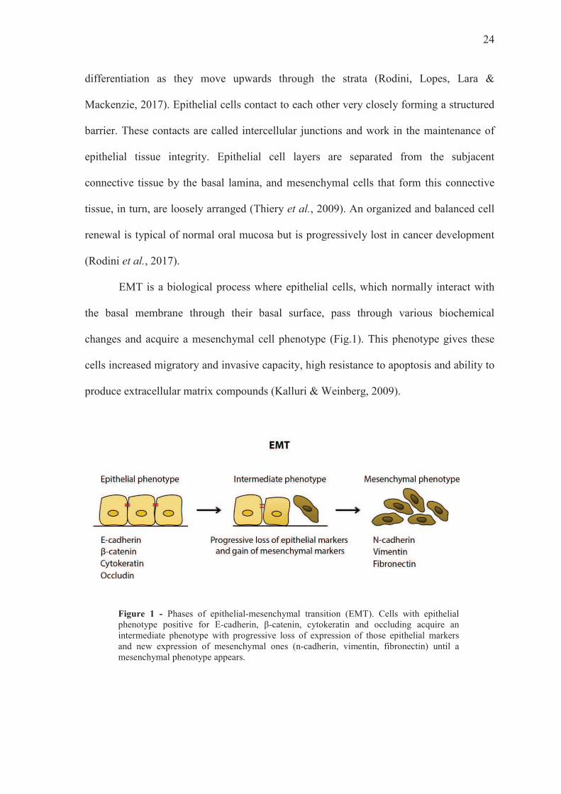
EMT is a biological process where epithelial cells, which normally interact with
the basal membrane through their basal surface, pass through various biochemical
changes and acquire a mesenchymal cell phenotype (Fig.1). This phenotype gives these
cells increased migratory and invasive capacity, high resistance to apoptosis and ability to
produce extracellular matrix compounds (Kalluri & Weinberg, 2009).
Figure 1 - Phases of epithelial-mesenchymal transition (EMT). Cells with epithelial phenotype positive for E-cadherin, β-catenin, cytokeratin and occluding acquire an intermediate phenotype with progressive loss of expression of those epithelial markers and new expression of mesenchymal ones (n-cadherin, vimentin, fibronectin) until a mesenchymal phenotype appears.

25
Many adult tissues and organs develop from a series of conversions of epithelial
cells and mesenchymal cells, through EMT and its reverse process called mesenchymal-
epithelial transition (MET) (Thiery et al., 2009). Along specific phases of embryogenesis
and organ development, cells from some tissues show plasticity, which gives them the
ability to display sometimes an epithelial phenotype and sometimes mesenchymal
(Kalluri & Weinberg 2009; Lee, Dedhar, Kalluri & Thompson, 2006). Thus, many rounds
of EMT and MET are needed for the final differentiation of specialized types of cells and
acquisition of the complex tridimensional structure of internal organs (Thiery et al.,
2009).
EMT occurs under three different biological forms (Fig.2), with their respective
functions (Kalluri & Weinberg, 2009). EMT type I is related to embryonic development
and implantation, and organ development as well, generating many types of cells that
have a mesenchymal phenotype in common. This type of EMT can generate primary
mesenchymal cells that have the potential to induce the reverse process (MET), to
generate secondary epithelium (Kalluri & Weinberg, 2009). EMT is silenced in the adult
body, but can be reactivated under pathological conditions such as wound healing,
fibrosis and carcinomatous progression (Thiery et al., 2009; Zidar et al., 2018).
EMT type II is associated with healing, tissue regeneration and fibrosis. This
process starts in situations of tissue trauma, where there is production of fibroblasts and
inflammatory cells involved in tissue repair. As the inflammatory response diminishes,
EMT type II ceases. But in organ fibrosis, EMT type II can persist in response to the
ongoing inflammatory process, eventually leading to organ destruction (Kalluri &
Weinberg, 2009).
EMT type III occurs in neoplastic cells that have previously undergone genetic
and epigenetic alterations. Malignant cells undergoing this type of EMT can invade and
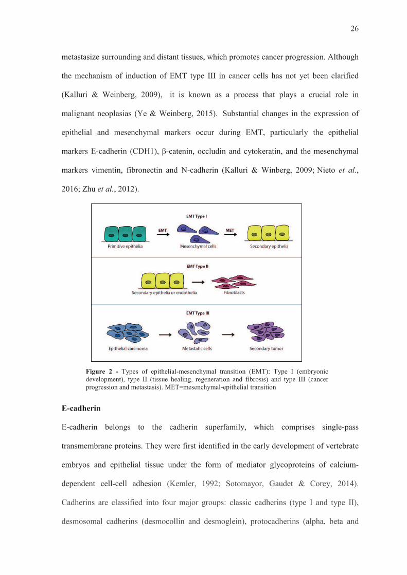
26
metastasize surrounding and distant tissues, which promotes cancer progression. Although
the mechanism of induction of EMT type III in cancer cells has not yet been clarified
(Kalluri & Weinberg, 2009), it is known as a process that plays a crucial role in
malignant neoplasias (Ye & Weinberg, 2015). Substantial changes in the expression of
epithelial and mesenchymal markers occur during EMT, particularly the epithelial
markers E-cadherin (CDH1), β-catenin, occludin and cytokeratin, and the mesenchymal
markers vimentin, fibronectin and N-cadherin (Kalluri & Winberg, 2009; Nieto et al.,
2016; Zhu et al., 2012).
Figure 2 - Types of epithelial-mesenchymal transition (EMT): Type I (embryonic development), type II (tissue healing, regeneration and fibrosis) and type III (cancer progression and metastasis). MET=mesenchymal-epithelial transition
E-cadherin
E-cadherin belongs to the cadherin superfamily, which comprises single-pass
transmembrane proteins. They were first identified in the early development of vertebrate
embryos and epithelial tissue under the form of mediator glycoproteins of calcium-
dependent cell-cell adhesion (Kemler, 1992; Sotomayor, Gaudet & Corey, 2014).
Cadherins are classified into four major groups: classic cadherins (type I and type II),
desmosomal cadherins (desmocollin and desmoglein), protocadherins (alpha, beta and
27
gamma) and atypical cadherins (Priest, Shafraz & Sivasankar, 2017; Sotomayor et al.,
2014). Because of its early identification and complete characterization, classic E-
cadherin type I (CDH1) is considered the prototype of cadherins, either in normal or
pathological conditions (van Roy & Berx, 2008).
The detection, transmission and response to mechanical forces promoted by
classic cadherins are responsible for tissue integrity (Priest et al., 2017) and have a key
role in epithelial homeostasis (Kourtidis, Lu, Pence & Anastasiadis, 2017). Cadherins
form adhesion complexes of mechanical support associated with the actin cytoskeleton
and coupled to neighboring cells, transmitting mechanical forces from the extracellular
environment to cytosol and triggering intracellular signaling events (LeckBand & Rooij,
2014; Priest et al., 2017).
There are reports of an association of reduced expression of E-cadherin with
higher severity of epithelial dysplasia and phenotypic alterations of initial stages of oral
cancer (von Zeidler, de Souza Botelho, Mendonça & Batista, 2014). The suppression of
cell adhesion consequent to E-cadherin loss of function is believed to favor the onset of
metastasis in various types of cancer (Priest et al., 2017; van Roy & Berx, 2008). E-
cadherin is considered a key molecule in cell adhesion, which binds to β-catenin, a
cytoplasmic adapter protein. Besides participating in cell-cell adhesion, this association
works in the transduction of signaling pathways that involve functions such as cell
growth, differentiation and polarity (Angadi et al., 2016). β-Catenin works as a
transcriptional co-factor in the Wnt signaling pathway. As with E-cadherin, β-catenin can
affect cell adhesion and contribute to tumorigenesis via EMT (González-Moles, Ruiz-
Ávila, Gil-Montoya, Plaza-Campillo & Scully, 2014).
28
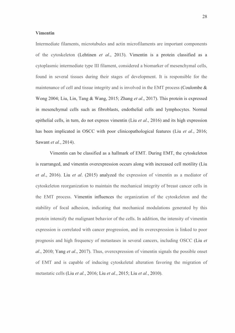
Vimentin
Intermediate filaments, microtubules and actin microfilaments are important components
of the cytoskeleton (Lehtinen et al., 2013). Vimentin is a protein classified as a
cytoplasmic intermediate type III filament, considered a biomarker of mesenchymal cells,
found in several tissues during their stages of development. It is responsible for the
maintenance of cell and tissue integrity and is involved in the EMT process (Coulombe &
Wong 2004; Liu, Lin, Tang & Wang, 2015; Zhang et al., 2017). This protein is expressed
in mesenchymal cells such as fibroblasts, endothelial cells and lymphocytes. Normal
epithelial cells, in turn, do not express vimentin (Liu et al., 2016) and its high expression
has been implicated in OSCC with poor clinicopathological features (Liu et al., 2016;
Sawant et al., 2014).
Vimentin can be classified as a hallmark of EMT. During EMT, the cytoskeleton
is rearranged, and vimentin overexpression occurs along with increased cell motility (Liu
et al., 2016). Liu et al. (2015) analyzed the expression of vimentin as a mediator of
cytoskeleton reorganization to maintain the mechanical integrity of breast cancer cells in
the EMT process. Vimentin influences the organization of the cytoskeleton and the
stability of focal adhesion, indicating that mechanical modulations generated by this
protein intensify the malignant behavior of the cells. In addition, the intensity of vimentin
expression is correlated with cancer progression, and its overexpression is linked to poor
prognosis and high frequency of metastases in several cancers, including OSCC (Liu et
al., 2010; Yang et al., 2017). Thus, overexpression of vimentin signals the possible onset
of EMT and is capable of inducing cytoskeletal alteration favoring the migration of
metastatic cells (Liu et al., 2016; Liu et al., 2015; Liu et al., 2010).
29
EMT and oral cancer
Oral carcinogenesis occurs due to an imbalance and accumulation of genetic and
epigenetic alterations (Eljabo et al., 2018), as well as changes in the expression of coding
and non-coding RNAs (Arunkumar et al., 2018). miRNAs are a family of small non-
coding RNAs, whose major function is to control gene expression (Hema, Smitha,
Sheethal & Mirnalini, 2017). In general, they are located in genomic regions that are often
prone to alterations in many types of cancer, including oral cancer (Manikandan et al.,
2016). The miR-200 family comprises miR-200a, miR-200b, miR-200c, miR-141 and
miR-429, which share the same genetic sequence, and modulate EMT through the
regulation of epithelial expression of E-cadherin (Park, Gaur, Lengyel & Peter, 2008).
EMT is the best known process in cell motility acquisition during malignant cell
transformation (Kalluri & Weinberg, 2009).
EMT and MET are not binary processes, and cancer cells can go through the
transition at different levels. Some cells can acquire an epithelial/mesenchymal phenotype
(E/M) (Kalluri & Weinberg, 2009), also called partial EMT phenotype (Nieto, 2013).
Many carcinomatous cells can metastasize without losing their epithelial morphology at
all or acquiring a complete mesenchymal phenotype (Jolly et al., 2015; Klymkowsky &
Savagner, 2009). These hybrid cells (E/M) show epithelial characteristics, such as cell
adhesion, and mesenchymal ones, such as migratory capacity, allowing collective cell
migration (Kalluri & Weinberg, 2009).
Metastasis consists of a sequence of events of neoplastic cell proliferation,
neoangiogenesis, detachment of cancer cells from the primary site and their invasion into
the bloodstream. This creates a new microenvironment, which comprises inflammatory
cells and stroma with restricted oxygen and nutrients, and including attacks from the
immune system (Pavithra et al., 2017). These tissue microenvironment factors exert a
30
critical role, especially in the signaling pathways that regulate cell-cell and cell-matrix
interactions (da Silva et al., 2014; Howell & Grandis, 2005).
EMT marks the first step in the metastatic invasion cascade, since progression of
OSCC from early to invasive stages is associated with morphological alterations of cancer
cells that promote cell dissemination to distant organs (da Silva et al., 2014). Epithelial
cells of primary tumor lose cell adhesion and apical-basal polarity and, with mesenchymal
phenotype acquisition, they gain the capacity of individual migration, penetrating the
basal membrane and blood vessels (Fig.3). These cells stay in the bloodstream as
circulating tumor cells, until they migrate to distant organs forming micrometastases.
During this process, MET (reverse) also occurs, which is associated with cancer cell
colonization at the metastatic site, and as mesenchymal cells achieve their destiny,
epithelial characteristics are recovered and secondary tumors or macrometastases can be
formed, completing the metastatic invasion cascade (Jolly et al., 2015). EMT triggers the
dissociation of cells from a primary carcinoma, which subsequently migrate and
disseminate to distant sites. MET, in turn, ceases the migration of these cells inducing
them to colonize and proliferate in the new tumor (Nieto et al., 2016). EMT and MET
then allow solid tumors, where 90% are carcinomas (Christiansen & Rajasekaran, 2006),
to disseminate and colonize distant organs.
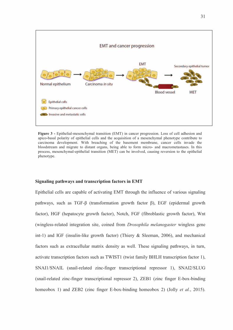
31
Figure 3 - Epithelial-mesenchymal transition (EMT) in cancer progression. Loss of cell adhesion and apico-basal polarity of epithelial cells and the acquisition of a mesenchymal phenotype contribute to carcinoma development. With breaching of the basement membrane, cancer cells invade the bloodstream and migrate to distant organs, being able to form micro- and macrometastases. In this process, mesenchymal-epithelial transition (MET) can be involved, causing reversion to the epithelial phenotype.
Signaling pathways and transcription factors in EMT
Epithelial cells are capable of activating EMT through the influence of various signaling
pathways, such as TGF-β (transformation growth factor β), EGF (epidermal growth
factor), HGF (hepatocyte growth factor), Notch, FGF (fibroblastic growth factor), Wnt
(wingless-related integration site, coined from Drosophila melanogaster wingless gene
int-1) and IGF (insulin-like growth factor) (Thiery & Sleeman, 2006), and mechanical
factors such as extracellular matrix density as well. These signaling pathways, in turn,
activate transcription factors such as TWIST1 (twist family BHLH transcription factor 1),
SNAI1/SNAIL (snail-related zinc-finger transcriptional repressor 1), SNAI2/SLUG
(snail-related zinc-finger transcriptional repressor 2), ZEB1 (zinc finger E-box-binding
homeobox 1) and ZEB2 (zinc finger E-box-binding homeobox 2) (Jolly et al., 2015).
32
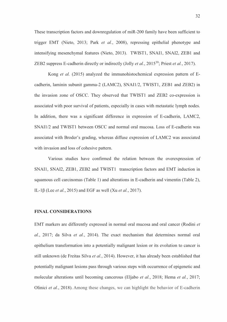
These transcription factors and downregulation of miR-200 family have been sufficient to
trigger EMT (Nieto, 2013; Park et al., 2008), repressing epithelial phenotype and
intensifying mesenchymal features (Nieto, 2013). TWIST1, SNAI1, SNAI2, ZEB1 and
ZEB2 suppress E-cadherin directly or indirectly (Jolly et al., 201539; Priest et al., 2017).
Kong et al. (2015) analyzed the immunohistochemical expression pattern of E-
cadherin, laminin subunit gamma-2 (LAMC2), SNAI1/2, TWIST1, ZEB1 and ZEB2) in
the invasion zone of OSCC. They observed that TWIST1 and ZEB2 co-expression is
associated with poor survival of patients, especially in cases with metastatic lymph nodes.
In addition, there was a significant difference in expression of E-cadherin, LAMC2,
SNAI1/2 and TWIST1 between OSCC and normal oral mucosa. Loss of E-cadherin was
associated with Broder’s grading, whereas diffuse expression of LAMC2 was associated
with invasion and loss of cohesive pattern.
Various studies have confirmed the relation between the overexpression of
SNAI1, SNAI2, ZEB1, ZEB2 and TWIST1 transcription factors and EMT induction in
squamous cell carcinomas (Table 1) and alterations in E-cadherin and vimentin (Table 2),
IL-1β (Lee et al., 2015) and EGF as well (Xu et al., 2017).
FINAL CONSIDERATIONS
EMT markers are differently expressed in normal oral mucosa and oral cancer (Rodini et
al., 2017; da Silva et al., 2014). The exact mechanism that determines normal oral
epithelium transformation into a potentially malignant lesion or its evolution to cancer is
still unknown (de Freitas Silva et al., 2014). However, it has already been established that
potentially malignant lesions pass through various steps with occurrence of epigenetic and
molecular alterations until becoming cancerous (Eljabo et al., 2018; Hema et al., 2017;
Olinici et al., 2018). Among these changes, we can highlight the behavior of E-cadherin
33
and vimentin, signaling pathways TGF-β, EGF, HGF, Notch, FGF, Wnt and IGF and
transcription factors SNAI1, SNAI2, ZEB1, ZEB2 and TWIST1, which indicate the start
of EMT.
Some investigations on oral cancer preventive therapies have focused on these
targets. The anti-metastatic effect of black tea polyphenol extracts (BTE) was tested in
oral squamous cell culture system (SCC-4). BTE repressed vimentin expression and
increased E-cadherin expression in SCC-4 cells, suggesting that BTE may be useful as an
effector for prevention of cancer metastasis, in addition to supporting the role of black tea
as an oral cancer chemopreventive agent (Chang et al., 2012). Tang et al. (2009) reported
that S-allylcysteine (SAC) can modulate in vitro the expression of E-cadherin and inhibit
malignant progression by suppression of the signal transduction pathways MAPK/ERK
(mitogen-activated protein kinase/extracellular-signal-regulated kinase) and SLUG
repressor protein. Kita et al. (2017) investigated the role of activin B in OSCC. Activin B
knockdown cells showed higher expression of E-cadherin and Zo-1, where activin B is
highly expressed in OSCC. The study provides new insight into a highly metastatic
phenotype by controlling the expression of EMT-related genes and suggest that this
multifunctional cytokine might be a potential therapeutic target for OSCC.
It seems that blocking EMT would be an interesting approach to improve OSCC
management. Nonetheless, considering the great number of signaling pathways involved
in this process, there is no consensus on which would be the best target (da Silva et al.,
2014; Wang et al., 2017; Zhang et al., 2017). Therefore, a deeper understanding of EMT
and its biomolecular processes is crucial for the investigation of chemopreventive
therapies and alternative strategies in oral cancer.
34
ACKNOWLEDGMENTS
We thank Dr. A. Leyva (U.S.A.) for English editing of the manuscript. This study was
funded in part by the Coordenação de Aperfeiçoamento de Pessoal de Nível Superior
(CAPES) – Finance Code 001.
CONFLICT OF INTEREST
The authors declare that there are no conflicts of interest related to this work.
35
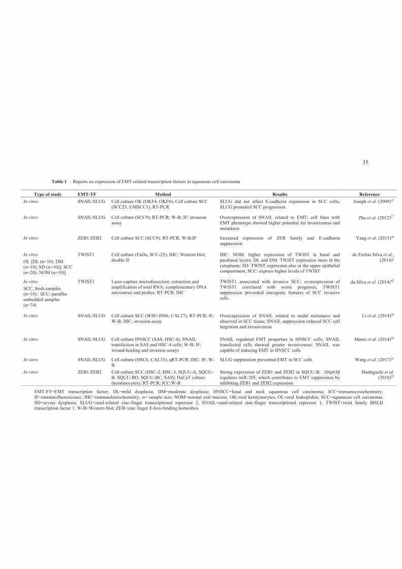
Table 1 – Reports on expression of EMT-related transcription factors in squamous cell carcinoma
Type of study EMT-TF Method Results Reference
In vitro SNAIL/SLUG Cell culture OK (OKF4, OKF6); Cell culture SCC (SCC25, UMSCC1); RT-PCR
SLUG did not affect E-cadherin expression in SCC cells; SLUG promoted SCC progression
Joseph et al. (2009)47
In vitro SNAIL/SLUG Cell culture (SCC9); RT-PCR; W-B; IF; invasion assay
Overexpression of SNAIL related to EMT; cell lines with EMT phenotype showed higher potential for invasiveness and metastasis
Zhu et al. (2012)17
In vitro ZEB1/ZEB2 Cell culture SCC (SCC9); RT-PCR; W-B;IF Increased expression of ZEB family and E-cadherin suppression
Yang et al. (2013)48
In vitro
OL [DL (n=10), DM (n=10), SD (n=10)]; SCC (n=20); NOM (n=10)]
TWIST1 Cell culture (FaDu, SCC-25); IHC; Western blot; double IF
IHC: NOM: higher expression of TWIST in basal and parabasal layers; DL and DM: TWIST expression more in the cytoplasm; SD: TWIST expression also in the upper epithelial compartment; SCC: express higher levels of TWIST
de Freitas Silva et al., (2014)5
In vitro
SCC, fresh samples (n=10); SCC; paraffin embedded samples (n=74)
TWIST1 Laser-capture microdissection; extraction and amplification of total RNA; complementary DNA microarray and probes; RT-PCR; IHC
TWIST1 associated with invasive SCC; overexpression of TWIST1 correlated with worse prognosis; TWIST1 suppression prevented oncogenic features of SCC invasive cells
da Silva et al. (2014)42
In vitro SNAIL/SLUG Cell culture SCC (WSU-HN6; CAL27); RT-PCR; IF; W-B; IHC; invasion assay
Overexpression of SNAIL related to nodal metastasis and observed in SCC tissue; SNAIL suppression reduced SCC cell migration and invasiveness
Li et al. (2014)49
In vitro SNAIL/SLUG Cell culture HNSCC (SAS, HSC-4); SNAIL transfection in SAS and HSC-4 cells; W-B; IF; wound-healing and invasion assays
SNAIL regulated EMT properties in HNSCC cells; SNAIL transfected cells showed greater invasiveness; SNAIL was capable of inducing EMT in HNSCC cells
Masui et al. (2014)50
In vitro SNAIL/SLUG Cell culture (HSC6, CAL33); qRT-PCR; IHC; IF; W-B
SLUG suppression prevented EMT in SCC cells Wang et al. (2017)51
In vitro ZEB1/ZEB2 Cell culture SCC (HSC-2, HSC-3, SQUU-A, SQUU-B, SQUU-BO, SQUU-BC, SAS); HaCaT culture (keratinocytes); RT-PCR; ICC;W-B
Strong expression of ZEB1 and ZEB2 in SQUU-B; ΔNp63β
regulates miR-205, which contributes to EMT suppression by inhibiting ZEB1 and ZEB2 expression
Hashiguchi et al. (2018)52
EMT-FT=EMT transcription factor; DL=mild dysplasia; DM=moderate dysplasia; HNSCC=head and neck squamous cell carcinoma; ICC=immunocytochemistry; IF=immunofluorescence; IHC=immunohistochemistry; n= sample size; NOM=normal oral mucosa; OK=oral keratynocytes; OL=oral leukoplakia; SCC=squamous cell carcinoma; SD=severe dysplasia; SLUG=snail-related zinc-finger transcriptional repressor 2; SNAIL=snail-related zinc-finger transcriptional repressor 1; TWIST=twist family BHLH transcription factor 1; W-B=Western blot; ZEB=zinc finger E-box-binding homeobox

36
Table 2 – E-cadherin and vimentin profile in squamous cell carcinoma
Type of study/(n) Marker Method Results Reference
In vitro
(n=28)
E-cadherin Vimentin
IHC of biopsies of SCC of the oral cavity, oropharynx, hypopharynx or larynx
E-cadherin expressed in cell membrane; cytoplasmic vimentin expression Low E-cadherin and high vimentin could identify tumors in which EMT has occurred
Nijkamp et al. (2011)53
In vitro
OL (n=31); OCSCC N+ (n=12); control (n=9)
E-cadherin IHC Moderate-severe dysplasia: reduced E-cadherin expression
Epithelial dysplastic changes plus risk of malignant transformation increased: reduction in or loss of E-cadherin expression by keratinocytes
von Zeidler et al. (2014)24
In vitro
NE (n=25); OL (n=25);
OSCC (n=25)
E-cadherin Cell culture (HSC-3), RNA extraction, cell lysates, W-B, IF, IHC
SD: reduced E-cadherin expression in epithelial cell membrane
OSCC grade III: extreme loss of membrane expression and switch to weak cytoplasmic expression
Kyrodimou et al. (2014)54
In vitro
OSCC( n=85)
Vimentin Cell culture of OSCC cell lines (HN4. HN12), immunostaining and immunoblotting, RT-PCR, IHC
Vimentin expression is essential for the increased migration activity of OSCC cells
Vimentin expression via IHC staining predicts poor survival rate of OSCC patients
Liu et al. (2016)31
In vitro E-cadherin Vimentin
Cell culture of OSCC cell lines (SCC-4, SCC-9, SCC-15), W-B, RT-PCR, IF
Upregulation of vimentin; downregulation of E-cadherin
OSCC cell lines exhibiting EMT signatures showed a decrease in mechanical stiffness compared with those without EMT signatures
Park et al. (2016)55
In vitro Vimentin Cell culture (AW13516, AW8507, DOK, HaCat and A431)
qRT-PCR, RT-PCR, W-B, IF, IHC
Vimentin downregulation causes keratin profile alteration in OSCC cells; vimentin modulates the differentiation status and tumorigenic potential of epithelial cells
Dmello et al. (2017)56
n=sample size; IF=immunofluorescence; IHC=immunohistochemistry; W-B=Western blot; NE=normal epithelium; OSCC=oral squamous cell carcinoma; SD=severe
dysplasia; SCC=squamous cell carcinoma; OCSCCN+= oral cavity squamous cell carcinoma with cervical lymph node metastasis
37
REFERENCES
Angadi, P.V., Patil, P.V., Angadi, V., Mane, D., Shekar, S., Hallikerimath, S., Kale, A.D., & Kardesai, S.G. (2016). Immunoexpression of epithelial mesenchymal transition proteins E-cadherin, β-catenin, and N-cadherin in oral squamous cell carcinoma. Int J Surg Pathol, 24(8), 696-703. https://doi.org/10.1177/1066896916654763.
Arunkumar, G., Deva Magendhra Rao, A.K., Manikandan, M., Prasanna Srinivasa Rao, H., Subbiah, S., Ilangovan, R., Murugan, A.K., & Munirajan, A.K. (2018). Dysregulation of miR-200 family microRNAs and epithelial-mesenchymal transition markers in oral squamous cell carcinoma. Oncol Lett, 15(1), 649-657. https://doi.org/10.3892/ol.2017.7296.
Chang, Y.C., Chen, P.N., Chu, S.C., Lin, C.Y., Kuo, W.H., & Hsieh, Y.S. (2012). Black tea polyphenols reverse epithelial-to-mesenchymal transition and suppress cancer invasion and proteases in human oral cancer cells. J Agric Food Chem, 60(34), 8395-403. https://doi.org/10.1021/jf302223g.
Cheng, A., & Schmidt, B.L. (2008). Management of the N0 neck in oral squamous cell carcinoma. Oral and maxillofacial surgery clinics of North America, 20, 477-497. https://doi.org/10.1016/j.coms.2008.02.002.
Christiansen, J.J., & Rajasekaran, A.K. (2006). Reassessing epithelial to mesenchymal transition as a prerequisite for carcinoma invasion and metastasis. Cancer Res, 66(17), 8319-8326. https://doi.org/10.1158/0008-5472.CAN-06-0410.
Coulombe, P.A., & Wong, P. (2004). Cytoplasmic intermediate filaments revealed as dynamic and multipurpose scaffolds. Nat Cell Biol, 6(8), 699-706. https://doi.org/10.1038/ncb0804-699.
da Silva, S.D., Alaoui-Jamali, M.A., Soares, F.A., Carraro, D.M., Brentani, H.P., Hier, M., Rogatto, S.R., & Kowalski, L.P. (2014). TWIST1 is a molecular marker for a poor prognosis in oral cancer and represents a potential therapeutic target. Cancer, 120(3), 352-362. https://doi.org/10.1002/cncr.28404.
de Freitas Silva, B.S., Yamamoto-Silva, F.P., Pontes, H.A., & Pinto Júnior Ddos, S. (2014). E-cadherin downregulation and Twist overexpression since early stages of oral carcinogenesis. J Oral Pathol Med, 43(2), 125-131. https://doi.org/10.1111/jop.12096.
Dmello, C., Sawant, S., Alam, H., Gangadaran, P., Mogre, S., Tiwari, R., D'Souza, Z., Narkar, M., Thorat, R., Patil, K., Chaukar, D., Kane, S., & Vaidya, M. (2017). Vimentin regulates differentiation switch via modulation of keratin 14 levels and their expression together correlates with poor prognosis in oral cancer patients. PLoS One, 12(2), e0172559. https://doi.org/10.1371/journal.pone.0172559.
Dutton, J.M., Graham, S.M., & Hoffman, H.T. (2002). Metastatic cancer to the floor of mouth: the lingual lymph nodes. Head Neck, 24, 401-405. https://doi.org/10.1002/hed.10026.
Eljabo, N., Nikolic, N., Carkic, J., Jelovac, D., Lazarevic, M., Tanic, N., & Milasin, J. (2018). Genetic and epigenetic alterations in the tumour, tumour margins, and normal
38
buccal mucosa of patients with oral cancer. Int J Oral Maxillofac Surg, 47(8), 976-982. https://doi.org/10.1016/j.ijom.2018.01.020.
González-Moles, M.A., Ruiz-Ávila, I., Gil-Montoya, J.A., Plaza-Campillo, J., & Scully, C. (2014). β-catenin in oral cancer: an update on current knowledge. Oral Oncol, 50, 818-824. https://doi.org/10.1016/j.oraloncology.2014.06.005.
Hashiguchi, Y., Kawano, S., Goto, Y., Yasuda, K., Kaneko, N., Sakamoto, T., Matsubara, R., Jinno, T., Maruse, Y., Tanaka, H., Morioka, M., Hattori, T., Tanaka, S., Kiyoshima, T., & Nakamura, S. (2018). Tumor-suppressive roles of ΔNp63β-miR-205 axis in epithelial-mesenchymal transition of oral squamous cell carcinoma via targeting ZEB1 and ZEB2. J Cell Physiol, 233(10), 6565-6577. https://doi.org/10.1002/jcp.26267.
Hema, K.N., Smitha, T., Sheethal, H.S., & Mirnalini, S.A. (2017). Epigenetics in oral squamous cell carcinoma. J Oral Maxillofac Pathol, 21(2), 252-259. https://doi.org/10.4103/jomfp.JOMFP_150_17.
Howell, G.M., & Grandis, J.R. (2005). Molecular mediators of metastasis in head and neck squamous cell carcinoma. Head Neck, 27, 710-717. https://doi.org/10.1002/hed.20222.
Huang, R., & Zong, X. (2017). Aberrant cancer metabolism in epithelial-mesenchymal transition and cancer metastasis: Mechanisms in cancer progression. Crit Rev Oncol Hematol, 115, 13-22. https://doi.org/10.1016/j.critrevonc.2017.04.005.
Huang, S.H., & O'Sullivan, B. (2013). Oral cancer: Current role of radiotherapy and chemotherapy. Med Oral Patol Oral Cir Bucal, 18(2), e233-240. http://doi.org/10.4317/medoral.18772.
Jolly, M.K., Boareto, M., Huang, B., Jia, D., Lu, M., Ben-Jacob, E., Onuchic, J.N., & Levine, H. (2015). Implications of the hybrid epithelial/mesenchymal phenotype in metastasis. Front Oncol, 5, 155. https://doi.org/10.3389/fonc.2015.00155.
Joseph, M.J., Dangi-Garimella, S., Shields, M.A., Diamond, M.E., Sun, L., Koblinski, J.E., & Munshi, H.G. (2009). Slug is a downstream mediator of transforming growth factor-beta1-induced matrix metalloproteinase-9 expression and invasion of oral cancer cells. J Cell Biochem, 108(3), 726-736. https://doi.org/10.1002/jcb.22309.
Kalluri, R., & Weinberg, R.A. (2009). The basics of epithelial-mesenchymal transition. J Clin Invest, 119(6), 1420-1428. https://doi.org/10.1172/JCI39104.
Kemler, R. (1992). Classical cadherins. Semin Cell Biol, 3, 149–155. https://doi.org/10.1016/S1043-4682(10)80011-X.
Kita, A., Kasamatsu, A., Nakashima, D., Endo-Sakamoto, Y., Ishida, S., Shimizu, T., Kimura, Y., Miyamoto, I., Yoshimura, S., Shiiba, M., Tanzawa, H., & Uzawa, K. (2017). Activin B regulates adhesion, invasiveness, and migratory activities in oral cancer: a potential biomarker for metastasis. J Cancer, 8(11), 2033-2041. https://doi.org/10.7150/jca.18714.
Klymkowsky, M.W., & Savagner, P. (2009). Epithelial-mesenchymal transition: a cancer researcher's conceptual friend and foe. Am J Pathol, 174(5), 1588-1593.

39
https://doi.org/10.2353/ajpath.2009.080545.
Kong, Y.H., Syed Zanaruddin, S.N., Lau, S.H., Ramanathan, A., Kallarakkal, T.G., Vincent-Chong, V.K., Wan Mustafa, W.M., Abraham, M.T., Abdul Rahman, Z.A., Zain, R.B., & Cheong, S.C. (2015). Co-Expression of TWIST1 and ZEB2 in oral squamous cell carcinoma is associated with poor survival. PLoS One, 10(7), e0134045. https://doi.org/10.1371/journal.pone.0134045.
Kourtidis, A., Lu, R., Pence, L.J., & Anastasiadis, P.Z. (2017). A central role for cadherin signaling in cancer. Exp Cell Res., 358(1), 78-85. https://doi.org/10.1016/j.yexcr.2017.04.006.
Kyrodimou, M., Andreadis, D., Drougou, A., Amanatiadou, E.P., Angelis, L., Barbatis, C., Epivatianos, A., & Vizirianakis, I.S. (2014). Desmoglein-3/γ-catenin and E-cadherin/ß-catenin differential expression in oral leukoplakia and squamous cell carcinoma. Clin Oral Investig, 18(1), 199-210. https://doi.org/10.1007/s00784-013-0937-z.
Leckband, D.E., & de Rooij, J. (2014). Cadherin adhesion and mechanotransduction. Annu Rev Cell Dev Biol, 30, 291-315. https://doi.org/10.1146/annurev-cellbio-100913-013212.
Lee, C.H., Chang, J.S., Syu, S.H., Wong, T.S., Chan, J.Y., Tang, Y.C., Yang, Z.P., Yang, W.C., Chen, C.T., Lu, S.C., Tang, P.H., Yang, T.C., Chu, P.Y., Hsiao, J.R., & Liu, K.J. (2015). IL-1β promotes malignant transformation and tumor aggressiveness in oral cancer. J Cell Physiol, 230(4), 875-884. https://doi.org/10.1002/jcp.24816.
Lee, J.M., Dedhar, S., Kalluri, R., & Thompson, E.W. (2006). The epithelial-mesenchymal transition: new insights in signaling, development, and disease. J Cell Biol, 172(7), 973-981. https://doi.org/10.1083/jcb.200601018.
Lehtinen, L., Ketola, K., Mäkelä, R., Mpindi, J.P., Viitala, M., Kallioniemi, O., & Iljin, K. (2013). High-throughput RNAi screening for novel modulators of vimentin expression identifies MTHFD2 as a regulator of breast cancer cell migration and invasion. Oncotarget, 4(1), 48-63. https://doi.org/10.18632/oncotarget.756.
Li, Y.Y., Zhou, C.X., & Gao, Y. (2014). Snail regulates the motility of oral cancer cells via RhoA/Cdc42/p-ERM pathway. Biochem Biophys Res Commun, 452(3), 490-496. https://doi.org/10.1016/j.bbrc.2014.08.110.
Liu, C.Y., Lin, H.H., Tang, M.J., & Wang, Y.K. (2015). Vimentin contributes to epithelial-mesenchymal transition cancer cell mechanics by mediating cytoskeletal organization and focal adhesion maturation. Oncotarget, 6(18), 15966-15983. https://doi.org/10.18632/oncotarget.3862.
Liu, L.K., Jiang, X.Y., Zhou, X.X., Wang, D.M., Song, X.L., & Jiang, H.B. (2010). Upregulation of vimentin and aberrant expression of E-cadherin/beta-catenin complex in oral squamous cell carcinomas: correlation with the clinicopathological features and patient outcome. Mod Pathol, 23(2), 213-224. https://doi.org/10.1038/modpathol.2009.160.
Liu, S., Liu, L., Ye, W., Ye, D., Wang, T., Guo, W., Liao, Y., Xu, D., Song, H., Zhang, L., Zhu, H., Deng, J., & Zhang, Z. (2016). High vimentin expression associated with lymph node metastasis and predicated a poor prognosis in oral squamous cell carcinoma. Sci Rep,

40
6, 38834. https://doi.org/10.1038/srep38834.
Manikandan, M., Deva Magendhra Rao, A.K., Arunkumar, G., Manickavasagam, M., Rajkumar, K.S., Rajaraman, R., & Munirajan, A.K. (2016). Oral squamous cell carcinoma: microRNA expression profiling and integrative analyses for elucidation of tumourigenesis mechanism. Mol Cancer, 15, 28. https://doi.org/10.1186/s12943-016-0512-8.
Masui, T., Ota, I., Yook, J.I., Mikami, S., Yane, K., Yamanaka, T., & Hosoi, H. (2014). Snail-induced epithelial-mesenchymal transition promotes cancer stem cell-like phenotype in head and neck cancer cells. Int J Oncol, 44(3), 693-699. https://doi.org/10.3892/ijo.2013.2225.
Mohd-Sarip, A., Teeuwssen, M., Bot, A.G., De Herdt, M.J., Willems, S.M., Baatenburg de Jong, R.J., Looijenga, L.H.J., Zatreanu, D., Bezstarosti, K., van Riet, J., Oole, E., van Ijcken, W.F.J., van de Werken, H.J.G., Demmers, J.A., Fodde, R., & Verrijzer, C.P. (2017). DOC1-Dependent Recruitment of NURD Reveals Antagonism with SWI/SNF during Epithelial-Mesenchymal Transition in Oral Cancer Cells. Cell Rep, 20(1), 61-75. https://doi.org/10.1016/j.celrep.2017.06.020.
Nieto, M.A., Huang, R.Y., Jackson, R.A., & Thiery J.P. (2016). EMT: 2016. Cell, 166(1), 21-45. https://doi.org/10.1016/j.cell.2016.06.028.
Nieto, M.A. (2013). Epithelial plasticity: a common theme in embryonic and cancer cells. Science, 342(6159), 1234850. https://doi.org/10.1126/science.1234850.
Nijkamp, M.M., Span, P.N., Hoogsteen, I.J., van der Kogel, A.J., Kaanders, J.H., & Bussink, J. (2011). Expression of E-cadherin and vimentin correlates with metastasis formation in head and neck squamous cell carcinoma patients. Radiother Oncol, 99(3), 344-348. https://doi.org/10.1016/j.radonc.2011.05.066.
Olinici, D., Cotrutz, C.E., Mihali, C.V., Grecu, V.B., Botez, E.A., Stoica, L., Onofrei, P., Condurache, O., & Dimitriu, D.C. (2018). The ultrastructural features of the premalignant oral lesions. Rom J Morphol Embryol, 59(1), 243-248.
Park, S., Jang, W.J., & Jeong, C.H. (2016). Nano-biomechanical validation of epithelial-mesenchymal transition in oral squamous cell carcinomas. Biol Pharm Bull, 39(9), 1488-1495. https://doi.org/10.1248/bpb.b16-00266.
Park, S.M., Gaur, A.B., Lengyel, E., & Peter, M.E. (2008). The miR-200 family determines the epithelial phenotype of cancer cells by targeting the E-cadherin repressors ZEB1 and ZEB2. Genes and Dev, 22(7), 894-907. https://doi.org/10.1101/gad.1640608.
Pavithra, V., Kumari, K., Haragannavar, V.C., Rao, R.S., Nambiar, S., Augustine, D., & Sowmya, S.V. (2017). Possible Role of Bcl-2 Expression in metastatic and non metastatic oral squamous cell carcinoma. J Clin Diagn Res, 11(9), ZC51-ZC54. https://doi.org/10.7860/JCDR/2017/29363.10601.
Priest, A.V., Shafraz, O., & Sivasankar, S. (2017). Biophysical basis of cadherin mediated cell-cell adhesion. Exp Cell Res, 358(1), 10-13. https://doi.org/10.1016/j.yexcr.2017.03.015.

41
Rodini, C.O., Lopes, N.M., Lara, V.S., & Mackenzie, I.C. (2017). Oral cancer stem cells - properties and consequences. J Appl Oral Sci., 25(6), 708-715. https://doi.org/10.1590/1678-7757-2016-0665.
Sawant, S.S., Vaidya, M.M., Chaukar, D.A., Alam, H., Dmello, C., Gangadaran, P., Kannan, S., Kane, S., Dange, P.P., Dey, N., Ranganathan, K., & D'Cruz, A.K. (2014). Clinical significance of aberrant vimentin expression in oral premalignant lesions and carcinomas. Oral Dis, 20(5), 453-465. https://doi.org/10.1111/odi.12151.
Sotomayor, M., Gaudet, R., & Corey, D.P. (2014). Sorting out a promiscuous superfamily: towards cadherin connectomics. Trends Cell Biol, 24(9), 524-536. https://doi.org/10.1016/j.tcb.2014.03.007.
Tang, F.Y., Chiang, E.P., Chung, J.G., Lee, H.Z., & Hsu, C.Y. (2009). S-allylcysteine modulates the expression of E-cadherin and inhibits the malignant progression of human oral cancer. J Nutr Biochem, 20(12), 1013-1020. https://doi.org/10.1016/j.jnutbio.2008.09.007.
Thiery, J.P., Acloque, H., Huang, R.Y., & Nieto, M.A. (2009). Epithelial-mesenchymal transitions in development and disease. Cell, 139(5), 871-890. https://doi.org/10.1016/j.cell.2009.11.007.
Thiery, J.P., & Sleeman, J.P. (2006). Complex networks orchestrate epithelial-mesenchymal transitions. Nat Rev Mol Cell Biol, 7(2), 131-142. https://doi.org/10.1038/nrm1835.
van Roy, F., & Berx, G. (2008). The cell-cell adhesion molecule E-cadherin. Cell Mol Life Sci., 65(23), 3756-3788. https://doi.org/10.1007/s00018-008-8281-1.
von Zeidler, S.V., de Souza Botelho, T., Mendonça, E.F., & Batista, A.C. (2014). E-Cadherin as a potential biomarker of malignant transformation in oral leukoplakia: a retrospective cohort study. BMC Cancer, 14, 972. https://doi.org/10.1186/1471-2407-14-972.
Wang, H., Liang, X., Li, M., Tao, X., Tai, S., Fan, Z., Wang, Z., Cheng, B., & Xia, J. (2017). Chemokine (CC motif) ligand 18 upregulates Slug expression to promote stem-cell like features by activating the mammalian target of rapamycin pathway in oral squamous cell carcinoma. Cancer Sci, 108(8), 1584-1593. https://doi.org/10.1111/cas.13289.
Xu, Q., Zhang, Q., Ishida, Y., Hajjar, S., Tang, X., Shi, H., Dang, C.V., & Le, A.D. (2017). EGF induces epithelial-mesenchymal transition and cancer stem-like cell properties in human oral cancer cells via promoting Warburg effect. Oncotarget, 8(6), 9557-9571. https://doi.org/10.18632/oncotarget.13771.
Yang, C.C., Zhu, L.F., Xu, X.H., Ning, T.Y., Ye, J.H., & Liu, L.K. (2013). Membrane type 1 matrix metalloproteinase induces an epithelial to mesenchymal transition and cancer stem cell-like properties in SCC9 cells. BMC Cancer, 13, 171. https://doi.org/10.1186/1471-2407-13-171.
Yang, Y., Ye, C., Wang, L., An, G., Tian, Z., Meng, L., Qu, L., Lian, S., & Shou, C. (2017). Repressor activator protein 1-promoted colorectal cell migration is associated with
42
the regulation of vimentin. Tumour Biol, 39(4), 1010428317695034. https://doi.org/10.1177/1010428317695034.
Ye, X., & Weinberg, R.A. (2015). Epithelial-mesenchymal plasticity: a central regulator of cancer progression. Trends Cell Biol, 25(11), 675-686. https://doi.org/10.1016/j.tcb.2015.07.012.
Zhang, L., Niyazi, H.E., Zhao, H.R., Cao, X.P., Abudula, M.N., Ye, W.J., Zhang, S.A., Yiming, R.H., Zhang, Y., Su, W.P., Chen, R., Ouyang, Y., Miao, N., & Bao, Y.X. (2017). Effects of miRNA-143 and the non-coding RNA MALAT1 on the pathogenesis and metastasis of HeLa cells. Genet Mol Res, 16(1). https://doi.org/10.4238/gmr16019269.
Zhu, L.F., Hu, Y., Yang, C.C., Xu, X.H., Ning, T.Y., Wang, Z.L., Ye, J.H., & Liu, L.K. (2012). Snail overexpression induces an epithelial to mesenchymal transition and cancer stem cell-like properties in SCC9 cells. Lab Invest, 92(5), 744-752. https://doi.org/10.1038/labinvest.2012.8.
Zidar, N., Boštjančič, E., Malgaj, M., Gale, N., Dovšak, T., & Didanovič, V. (2018). The role of epithelial-mesenchymal transition in squamous cell carcinoma of the oral cavity. Virchows Arch, 472(2), 237-245. https://doi.org/10.1007/s00428-017-2192-1.

Artigo 2
44
3 ARTIGO 2
O artigo a seguir intitula-se Relationship of clinical features, Candida spp. and
expression of E-cadherin and vimentin with dysplastic alterations in oral leukoplakia e
foi formatado de acordo com as normas do periódico Oral Oncology (Anexo B).
45
Relationship of clinical features, Candida spp. and expression of E-cadherin and
vimentin with dysplastic alterations in oral leukoplakia
João Matheus Scherbaum Eidt1
Fernanda Gonçalves Salum2
Maria Antonia Figueiredo2
Karen Cherubini2
1 DDS, Postgraduate Program, Dental College, Pontifical Catholic University of Rio Grande do Sul – PUCRS, Porto Alegre, Brazil
2 Ph.D., Postgraduate Program, Dental College, Pontifical Catholic University of Rio Grande do Sul – PUCRS, Porto Alegre, Brazil
Key words: epithelial-mesenchymal transition; oral cancer; E-cadherin, protein, human; vimentin
Running title: Candida spp., E-cadherin and vimentin in oral leukoplakia
Word count: 2,417
Corresponding author
Karen Cherubini
Serviço de Estomatologia, Hospital São Lucas, PUCRS
Av Ipiranga, 6690, sala 231
Porto Alegre RS Brazil
CEP 90610-000
Telephone/fax: 55(51)33203254
E-mail: [email protected]
46
ABSTRACT
Objective: To investigate the relationship of dysplastic changes in oral leukoplakia and
clinical factors, Candida spp., and E-cadherin and vimentin expression.
Methods: Medical records and paraffin blocks of biopsied specimens of 60 patients were
distributed into 4 groups: (1) no-dysplasia: 15 cases of leukoplakia without epithelial
dysplasia; (2) epithelial dysplasia: 15 cases of leukoplakia with epithelial dysplasia
(moderate or severe); (3) oral squamous cell carcinoma (OSCC): 15 cases of leukoplakia
with histopathological diagnosis of OSCC; and (4) control group: 15 cases of
fibroepithelial hyperplasia. Records were reviewed regarding age, sex, alcohol and tobacco
use, and anatomical site of the lesion. Immunohistochemical analysis was carried out for
determination of E-cadherin and vimentin expression, and periodic acid of Schiff (PAS)
staining for Candida spp. detection.
Results: High-risk sites showed association with the epithelial dysplasia and OSCC
groups. There was no significant difference between the groups for the other clinical
features analyzed and for Candida spp. positivity with PAS. Quantitative E-cadherin
expression did not significantly differ between the groups analyzed. Vimentin expression
was significantly greater in the epithelial dysplasia and OSCC groups than the others.
Conclusion: According to our results, high-risk sites (border/ventral surface of the tongue
and floor of the mouth) are associated with the dysplastic phenotype of leukoplakia,
whereas age, sex, alcohol, tobacco and Candida spp. do not show such association.
Vimentin expression is associated with the oral dysplastic epithelial phenotype and it
seems to be more specific than E-cadherin for use as an immunohistochemical marker to
detect such alterations.
INTRODUCTION
Oral cancer is the sixth most prevalent cancer in humans and one of the major causes of
death worldwide [1,2]. Squamous cell carcinoma is the prototype of oral cancer,
corresponding to about 90% of malignancies in the mouth. Frequently, it is preceded by
easily identifiable oral lesions, which are called potentially malignant lesions [3]. Early
diagnosis and clinical management of these lesions are crucial to reduce the morbidity and
47
mortality of oral cancer [1]. Leukoplakia is the most prevalent potentially malignant lesion
in the oral cavity, with annual rates of malignant transformation between 2 and 3% [4].
Histopathological examination is the gold standard for its diagnosis [5], and even though
dysplastic features can be an indicator of malignant potential in oral leukoplakia, the exact
determination of the grade of dysplasia is a hard task, which compromises this predictive
factor [1,6,7]. Therefore, predicting malignant transformation of oral leukoplakia is a
challenge, and specific biomarkers are necessary for this purpose.
C. albicans is the most common species of Candida in the oral cavity of healthy
individuals [8]. Alterations in the oral mucosa associated with trauma, atrophy, hyperplasia
and dysplasia can compromise the mucosal barrier and predispose to this fungal infection
[8,9]. Candida spp. have been implicated in malignant transformation of candidal
leukoplakia, a type of oral leukoplakia associated with chronic infection with this fungus
[10].
Carcinogenesis is related to epithelial-mesenchymal transition (EMT) [11].
During this process, epithelial differentiation is lost and a mesenchymal phenotype
acquired. Embryogenesis, tissue repair and cancer cell metastasis are known as events that
also develop through this process [12]. E-cadherin and vimentin are proteins associated
with EMT, whose up-regulation or down-regulation can signal events of invasion and
migration [11,12].
Considering that (1) some clinical features and Candida spp. infection can favor
epithelial dysplasia in oral leukoplakia and that (2) changes in E-cadherin and vimentin
expression are related to carcinogenesis, this study aimed to evaluate the relationship of
clinical features, Candida spp. infection and immunohistochemical expression of E-
cadherin and vimentin with dysplastic alterations in oral leukoplakia.

48
MATERIAL AND METHODS
We conducted a retrospective study, which was first approved by the Research Ethics
Committee of Pontifical Catholic University of Rio Grande do Sul, protocol #
78767317.0.0000.5336. The sample was composed of medical records and paraffin blocks
of specimens previously biopsied from patients with clinical diagnosis of oral leukoplakia
and oral mucosa fibroepithelial hyperplasia. The sample was allocated into 4 groups
according to histopathological diagnosis: (1) no-dysplasia group: 15 cases of leukoplakia
without epithelial dysplasia; (2) epithelial dysplasia group: 15 cases of leukoplakia with
epithelial dysplasia (only moderate or severe grades); (3) OSCC group: 15 cases of
leukoplakia with histopathological diagnosis of oral squamous cell carcinoma; (4) control
group: 15 cases of oral mucosa fibroepithelial hyperplasia. The sample comprised only the
records with complete data and paraffin blocks with adequate specimens for histological
analysis. Cases of lesions located in the vermillion border of the lips, as well as patients
who had used antifungal agents within 14 days period prior to biopsy were excluded from
the sample. Leukoplakias histopathologically diagnosed as mild epithelial dysplasia were
also excluded; the group of epithelial dysplasia only comprised moderate and severe
grades.
Data concerning age and sex of the patients, alcohol and tobacco use, and
anatomical site of the lesions were collected from the records. Border and ventral surface
of the tongue and floor of the mouth were considered high-risk sites; the other sites of oral
mucosa were classified as low-risk sites [13].
Histological processing
Hematoxylin and eosin (H&E) slides were reviewed to confirm the histopathological
diagnosis, according to World Health Organization (WHO) criteria [14]. Next, specimens
49
embedded in paraffin were subjected to periodic acid Schiff (PAS) staining and
immunohistochemistry as follows.
PAS
Four-micrometer-thick histological sections were deparaffinized, re-hydrated in deionized
water and immersed in 0.5% periodic acid solution for 20 min at room temperature (18 to
26ºC). The sections were washed and immersed in Schiff reagent for 20 min at room
temperature; they were then washed again for 5 min and counterstained with Harris
hematoxylin. The slides were mounted with xylene-based mounting media.
Immunohistochemistry processing
Three-micrometer-thick histological sections on silanized slides were deparaffinized with
xylene at 59ºC and rehydrated in decreasing grades of ethanol. Processing was automated
in Dako Autostainer Link 48 (Dako, Carpinteria, CA, USA). Antigens were retrieved using
PT-Link (Dako) and EnVision Flex target retrieval solution (high pH). The sections were
incubated with the antibodies FLEX monoclonal mouse anti-human E-cadherin (Clone
NCH-38, Dako) and FLEX monoclonal mouse anti-vimentin (Clone V9, Dako). Sections
were then counterstained with hematoxylin and coverslipped. The negative control
comprised samples processed without the primary antibodies, and samples of breast and
cecal appendix were used as positive controls for E-cadherin and vimentin, respectively.
Histological analysis
Images were captured by using a digital system with an Olympus BX-43 light microscope
(Olympus, Tokyo, Japan), connected to a computer with an Olympus DP-73 digital camera
(Olympus). Images were captured with a 20x objective and stored as uncompressed TIFF
(true image format file). Three fields were captured in each slide (left side, middle and
right side fields in the sections).

50
Images were analyzed in Image Pro Plus 4.5.0 (Media Cybernetics, Silver Spring, USA).
E-cadherin and vimentin expression in the epithelial tissue was quantified by using the
semiautomated segmentation technique [15] in the three fields captured for each slide (Fig.
1). PAS staining was classified as positive for Candida spp. considering stained structures
morphologically compatible with the fungus [16]. Analysis was performed by a blinded
and calibrated examiner. Calibration consisted in the evaluation of a series of 30 images
for each marker (E-cadherin and vimentin) at two different moments. The results were
subjected to the intraclass correlation test resulting in r=0.946 for E-cadherin and r=0.988
for vimentin.
Figure 1 - Quantification of E-cadherin immunostaining by means of semiautomated segmentation technique in Image ProPlus software (Media Cybernetics, Bethesda, MD, USA)
Statistical analysis
Data were analyzed with descriptive and inferential statistics. Qualitative variables were
expressed through absolute and relative frequency, whereas quantitative variables were
analyzed with mean, standard deviation and median. Kolmogorov-Smirnov was used to
test the normality of the data. Dichotomous variables were compared between the groups
51
with the chi-square test; ANOVA was used for age of the patients and immunostaining for
E-cadherin and vimentin. Correlation of the variables was analyzed with Spearman
correlation coefficient. Analysis was run in SPSS 17.0, at a significance level of 5%.
RESULTS
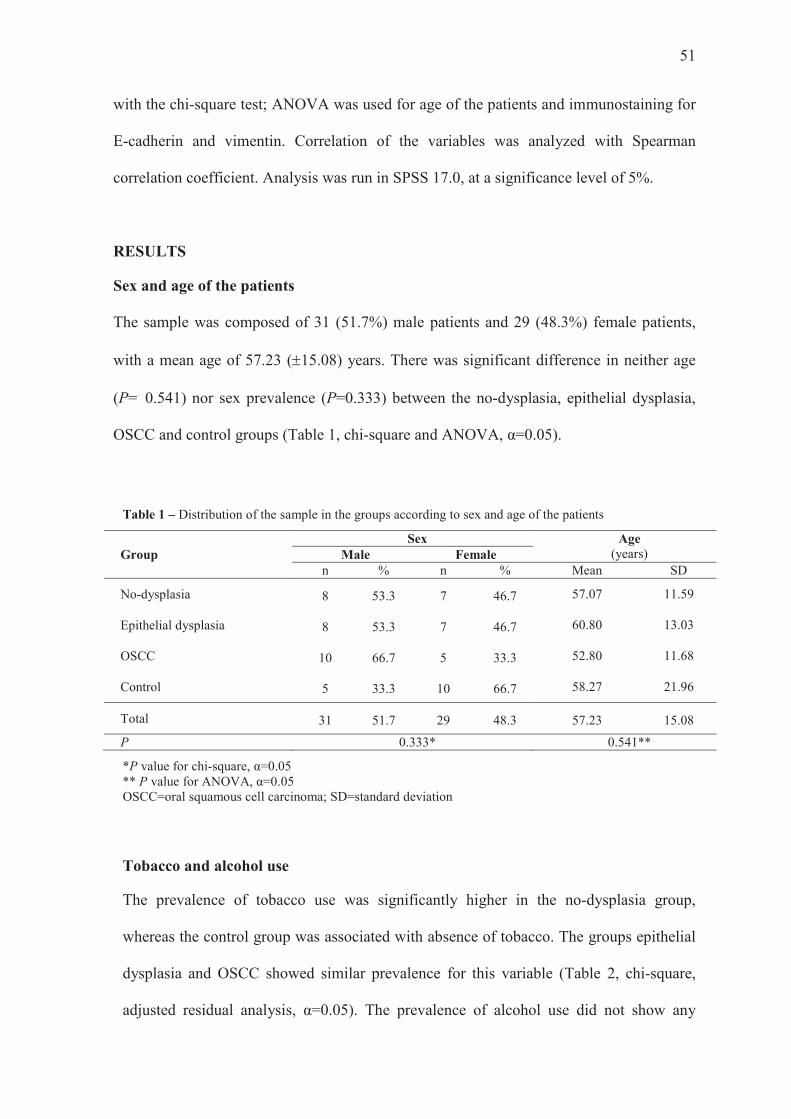
Sex and age of the patients
The sample was composed of 31 (51.7%) male patients and 29 (48.3%) female patients,
with a mean age of 57.23 (±15.08) years. There was significant difference in neither age
(P= 0.541) nor sex prevalence (P=0.333) between the no-dysplasia, epithelial dysplasia,
OSCC and control groups (Table 1, chi-square and ANOVA, α=0.05).
Table 1 – Distribution of the sample in the groups according to sex and age of the patients
Group Sex Age
(years) Male Female n % n % Mean SD
No-dysplasia 8 53.3 7 46.7 57.07 11.59
Epithelial dysplasia 8 53.3 7 46.7 60.80 13.03
OSCC 10 66.7 5 33.3 52.80 11.68
Control 5 33.3 10 66.7 58.27 21.96
Total 31 51.7 29 48.3 57.23 15.08
P 0.333* 0.541**
*P value for chi-square, α=0.05 ** P value for ANOVA, α=0.05 OSCC=oral squamous cell carcinoma; SD=standard deviation
Tobacco and alcohol use
The prevalence of tobacco use was significantly higher in the no-dysplasia group,
whereas the control group was associated with absence of tobacco. The groups epithelial
dysplasia and OSCC showed similar prevalence for this variable (Table 2, chi-square,
adjusted residual analysis, α=0.05). The prevalence of alcohol use did not show any
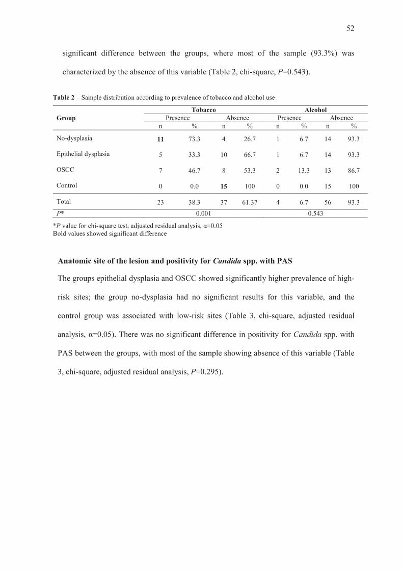
52
significant difference between the groups, where most of the sample (93.3%) was
characterized by the absence of this variable (Table 2, chi-square, P=0.543).
Table 2 – Sample distribution according to prevalence of tobacco and alcohol use
Group Tobacco Alcohol
Presence Absence Presence Absence n % n % n % n %
No-dysplasia 11 73.3 4 26.7 1 6.7 14 93.3
Epithelial dysplasia 5 33.3 10 66.7 1 6.7 14 93.3
OSCC 7 46.7 8 53.3 2 13.3 13 86.7
Control 0 0.0 15 100 0 0.0 15 100
Total 23 38.3 37 61.37 4 6.7 56 93.3
P* 0.001 0.543
*P value for chi-square test, adjusted residual analysis, α=0.05 Bold values showed significant difference
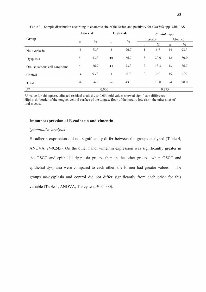
Anatomic site of the lesion and positivity for Candida spp. with PAS
The groups epithelial dysplasia and OSCC showed significantly higher prevalence of high-
risk sites; the group no-dysplasia had no significant results for this variable, and the
control group was associated with low-risk sites (Table 3, chi-square, adjusted residual
analysis, α=0.05). There was no significant difference in positivity for Candida spp. with
PAS between the groups, with most of the sample showing absence of this variable (Table
3, chi-square, adjusted residual analysis, P=0.295).
53
Table 3 – Sample distribution according to anatomic site of the lesion and positivity for Candida spp. with PAS
Group
Low risk High risk Candida spp.
n % n % Presence Absence
n % n %
No-dysplasia 11 73.3 4 26.7 1 6.7 14 93.3
Dysplasia 5 33.3 10 66.7 3 20.0 12 80.0
Oral squamous cell carcinoma 4 26.7 11 73.3 2 13.3 13 86.7
Control 14 93.3 1 6.7 0 0.0 15 100
Total 34 56.7 26 43.3 6 10.0 54 90.0
P* 0.000 0.295
*P value for chi-square, adjusted residual analysis, α=0.05; bold values showed significant difference High risk=border of the tongue; ventral surface of the tongue; floor of the mouth; low risk= the other sites of oral mucosa
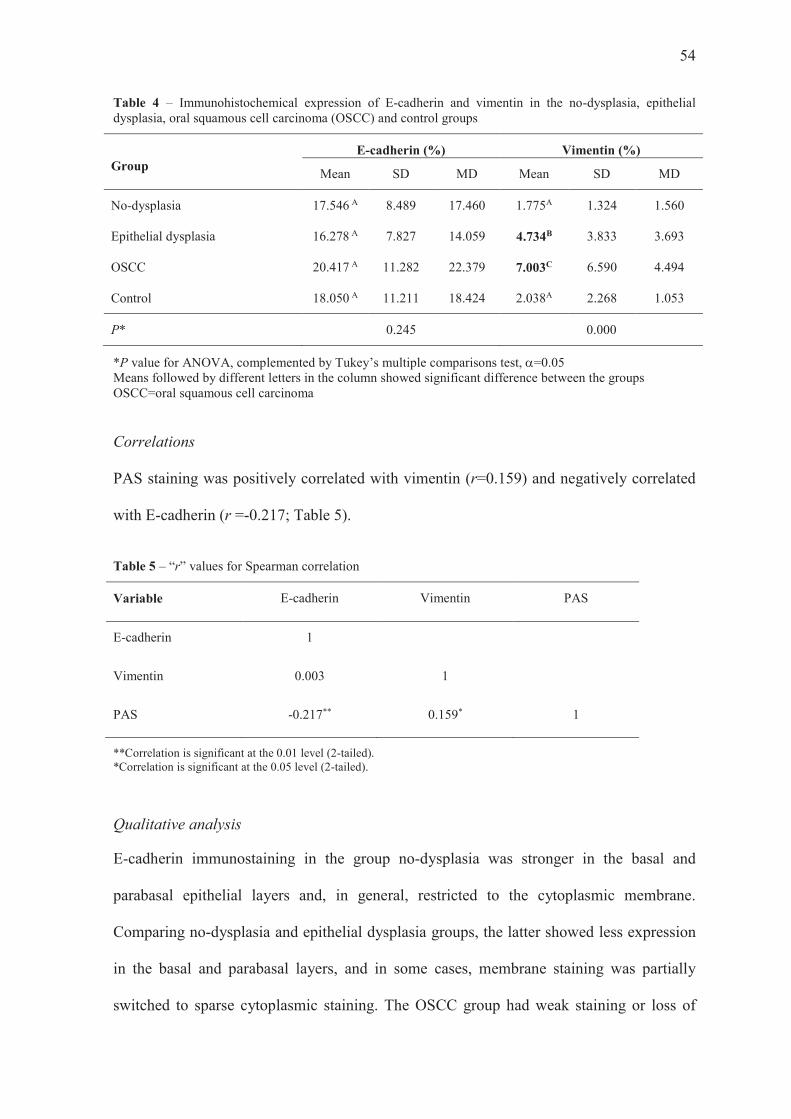
Immunoexpression of E-cadherin and vimentin
Quantitative analysis
E-cadherin expression did not significantly differ between the groups analyzed (Table 4,
ANOVA, P=0.245). On the other hand, vimentin expression was significantly greater in
the OSCC and epithelial dysplasia groups than in the other groups; when OSCC and
epithelial dysplasia were compared to each other, the former had greater values. The
groups no-dysplasia and control did not differ significantly from each other for this
variable (Table 4, ANOVA, Tukey test, P=0.000).
54
Table 4 – Immunohistochemical expression of E-cadherin and vimentin in the no-dysplasia, epithelial dysplasia, oral squamous cell carcinoma (OSCC) and control groups
Group E-cadherin (%) Vimentin (%)
Mean SD MD Mean SD MD
No-dysplasia 17.546 A 8.489 17.460 1.775A 1.324 1.560
Epithelial dysplasia 16.278 A 7.827 14.059 4.734B 3.833 3.693
OSCC 20.417 A 11.282 22.379 7.003C 6.590 4.494
Control 18.050 A 11.211 18.424 2.038A 2.268 1.053
P* 0.245 0.000
*P value for ANOVA, complemented by Tukey’s multiple comparisons test, a=0.05 Means followed by different letters in the column showed significant difference between the groups OSCC=oral squamous cell carcinoma
Correlations
PAS staining was positively correlated with vimentin (r=0.159) and negatively correlated
with E-cadherin (r =-0.217; Table 5).
Table 5 – “r” values for Spearman correlation
Variable E-cadherin Vimentin PAS
E-cadherin 1
Vimentin 0.003 1
PAS -0.217** 0.159* 1
**Correlation is significant at the 0.01 level (2-tailed). *Correlation is significant at the 0.05 level (2-tailed).
Qualitative analysis
E-cadherin immunostaining in the group no-dysplasia was stronger in the basal and
parabasal epithelial layers and, in general, restricted to the cytoplasmic membrane.
Comparing no-dysplasia and epithelial dysplasia groups, the latter showed less expression
in the basal and parabasal layers, and in some cases, membrane staining was partially
switched to sparse cytoplasmic staining. The OSCC group had weak staining or loss of
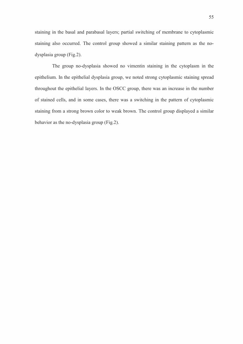
55
staining in the basal and parabasal layers; partial switching of membrane to cytoplasmic
staining also occurred. The control group showed a similar staining pattern as the no-
dysplasia group (Fig.2).
The group no-dysplasia showed no vimentin staining in the cytoplasm in the
epithelium. In the epithelial dysplasia group, we noted strong cytoplasmic staining spread
throughout the epithelial layers. In the OSCC group, there was an increase in the number
of stained cells, and in some cases, there was a switching in the pattern of cytoplasmic
staining from a strong brown color to weak brown. The control group displayed a similar
behavior as the no-dysplasia group (Fig.2).
56
Figure 2 - Immunohistochemical staining for E-cadherin and vimentin in the control, no-dysplasia, epithelial dysplasia and oral squamous cell carcinoma (OSCC) groups.
57
DISCUSSION
It is believed that female sex, advanced age, tobacco and alcohol use, anatomical site and
duration of the lesion, and Candida spp. infection as well, are risk factors for
carcinomatous transformation of leukoplakia [10,13]. In the present study, only lesion site
showed a significant difference between the groups. There were no significances for sex,
age, alcohol and tobacco use and Candida spp. infection, which meant that there was no
association of these factors with leukoplakia showing dysplasia and/or malignant
transformation. Accordingly, it is important to emphasize that the criteria for the selection
and allocation of leukoplakias into the study groups were not the clinical features, but the
histopathological ones. Considering the presence or absence of epithelial dysplasia or
carcinoma in the histopathological examination, the groups were determined, and
afterwards, clinical features of the patients (sex, age, alcohol and tobacco use, site of the
lesions) were evaluated. Therefore, our findings suggest that, regardless of age and sex of
the patients and alcohol or tobacco habits, leukoplakia has per se the potential for
malignant transformation [13,17]. This is in regard especially to the idiopathic cases,
which in fact show reportedly higher malignant transformation rates [13]. Likewise, even
though Candida has been reported as a risk factor for carcinomatous transformation of
leukoplakia [10,18,19], its detection with PAS showed no significant difference between
the groups. At first, as happened with the clinical factors previously discussed, this
suggested that leukoplakia did not depend on Candida infection to undergo dysplastic
alterations. However, this finding does not rule out the possibility of Candida contributing
to such alterations. In fact, we found only a few cases with positive PAS staining for
Candida. Although we tried to control biases, including in our sample only patients
without history of use of antifungal agents within 14 days prior to biopsy, this was a
retrospective study and it is possible that patients had previously used antifungal agents at
58
some time. Further prospective studies comprising a larger standardized sample and more
specific techniques such as culture and PCR could better determine Candida at dysplastic
alterations of the oral epithelium. Interestingly, both the epithelial dysplasia and OSCC
groups were associated with high-risk anatomical sites, whereas the no-dysplasia group did
not show such association. Accordingly, some authors report that, regardless of the clinical
feature of being homogeneous, leukoplakias on the ventral surface and border of the
tongue and floor of the mouth do show high risk for malignant transformation [13].
In the quantitative analysis, we found no significant difference in E-cadherin
expression between the groups. This was not expected, since the literature reports E-
cadherin down-regulation in EMT [7,20,21]. Anyway, there are reports of similar results
as ours, indicating that E-cadherin use as a prognostic indicator needs further evaluation,
where E-cadherin had a heterogeneous expression with 54% of OSCC specimens showing
strong expression [22]. Qualitative analysis, on the other hand, showed some variation in
the expression pattern of this marker in the groups, characterized by some tendency for
switching to weaker expression in cytoplasmic membrane with some sparse expression in
the cytoplasm in the epithelial dysplasia and OSCC groups. Such profile was already
reported by Kyrodimou et al. [21]. At first, we could point out that our sample was small
and, for this reason, significant differences in the E-cadherin quantitative analysis could
not be determined. Nevertheless, for vimentin, this did not happen, as vimentin did show
significant differences between the groups, with higher expression in the groups with
dysplastic changes (epithelial dysplasia and OSCC). Thus, some considerations regarding
specificities of these markers and their relation with the tissue analyzed should be raised.
E-cadherin normally occurs at high levels with uniform distribution in the normal
epithelium [21-23], and therefore progressive alterations in its expression during initial
phases of cancer may not be so evident, especially in a quantitative approach. Our groups

59
corresponded to lesions clinically diagnosed as leukoplakias and, when carcinomatous
transformation occurred in these lesions, it really represented early stages of OSCC. This
fact could explain our quantitative results for E-cadherin, suggesting that, if used as a
marker for EMT, it should be analyzed in a qualitative manner considering switching
expression between membrane and cytoplasm, especially in the lower layers of the
epithelium, instead of quantitative one.
The vimentin profile was different from that of E-cadherin, showing important
differences in the quantitative analysis. The epithelial dysplasia and OSCC groups showed
greater vimentin expression compared to the other groups and also differed from each
other for this variable, with greater values in OSCC. Unlike E-cadherin, vimentin is not
normally expressed in epithelial cells [24], which seems to make it a better marker for
dysplastic alterations since it would be easier to identify its abnormal behavior either
quantitative- or qualitatively. Accordingly, the literature has reported increased expression
of this protein in advanced dysplastic oral lesions and OSCC [25].
CONCLUSION
According to our results, high-risk sites (border and ventral surface of the tongue and floor
of the mouth) were associated with the dysplastic phenotype of leukoplakia, whereas age,
sex, alcohol, tobacco and Candida spp. did not show such association. Vimentin
expression was indeed involved in the oral epithelial dysplastic phenotype of leukoplakia,
and it seemed to be more specific than E-cadherin for use as an immunohistochemical
marker to detect such alterations. Further prospective and controlled studies investigating
these biomarkers and genetic/epigenetic alterations are needed to establish new strategies
for determining risk of carcinomatous transformation in leukoplakia.
60
ACKNOWLEDGMENTS
We thank Dr. A. Leyva (U.S.A.) for English editing of the manuscript. This study was
funded in part by the Coordenação de Aperfeiçoamento de Pessoal de Nível Superior
(CAPES) – Finance Code 001.
ETHICS STATEMENT
This study was approved by the Research Ethics Committee of Pontifical Catholic
University of Rio Grande do Sul, protocol # 78767317.0.0000.5336.
CONFLICT OF INTEREST
The authors declare that there are no conflicts of interest related to this work.
HIGHLIGHTS
-In some leukoplakia cases, there is paradoxical upregulation of E-cadherin.
-Vimentin is shown to be relevant when leukoplakia undergoes dysplastic changes.
-Candida may help induce epithelial dysplastic alterations.
REFERENCES
[1] Habiba U, Hida K, Kitamura T, Matsuda AY, Higashino F, Ito YM, Ohiro Y, Totsuka Y, Shindoh M. ALDH1 and podoplanin expression patterns predict the risk of malignant transformation in oral leukoplakia. Oncol Lett 2017;13(1):321-328.
[2] Warnakulasuriya S. Living with oral cancer: Epidemiology with particular reference to prevalence and life-style changes that influence survival. Oral Oncol 2010;46(6):407-410.
[3] Radhika T, Jeddy N, Nithya S, Muthumeenakshi RM. Salivary biomarkers in oral squamous cell carcinoma - An insight. J Oral Biol Craniofac Res 2016. doi:10.1016/j.jobcr.2016.07.003
[4] van der Waal I. Oral potentially malignant disorders: is malignant transformation predictable and preventable? Med Oral Patol Oral Cir Bucal 2014;19(4):e386-390.
61
[5] van der Waal I. Potentially malignant disorders of the oral and oropharyngeal mucosa; terminology, classification and present concepts of management. Oral Oncol 2009;45(4-5):317-323.
[6] Kujan O, Khattab A, Oliver RJ, Roberts SA, Thakker N, Sloan P. Why oral histopathology suffers inter-observer variability on grading oral epithelial dysplasia: An attempt to understand the sources of variation. Oral Oncol. 2007;43(3):224-231.
[7] von Zeidler SV, de Souza Botelho T, Mendonça EF, Batista AC. E-cadherin as a potential biomarker of malignant transformation in oral leukoplakia: a retrospective cohort study. BMC Cancer 2014;14:972.
[8] Williams D, Lewis M. Pathogenesis and treatment of oral candidosis. J Oral Microbiol 2011;3. doi:10.3402/jom.v3i0.5771
[9] Wu L, Feng J, Shi L, Shen X, Liu W, Zhou Z. Candidal infection in oral leukoplakia: a clinicopathologic study of 396 patients from eastern China. Ann Diagn Pathol 2013;17(1):37-40.
[10] Cheng R, Li D, Shi X, Gao Q, Wei C, Li X, Li Y, Zhou H. Reduced CX3CL1 Secretion Contributes to the Susceptibility of oral leukoplakia-associated fibroblasts to Candida albicans. Front Cell Infect Microbiol 2016;6:150.
[11] Webber LP, Wagner VP, Curra M, Vargas PA, Meurer L, Carrard VC, Squarize CH, Castilho RM, Martins MD. Hypoacetylation of acetyl-histone H3 (H3K9ac) as marker of poor prognosis in oral cancer. Histopathology 2017; Mar 22. doi:10.1111/his.13218
[12] Chaw SY, Majeed AA, Dalley AJ, Chan A, Stein S, Farah CS. Epithelial to mesenchymal transition (EMT) biomarkers--E-cadherin, beta-catenin, APC and vimentin--in oral squamous cell carcinogenesis and transformation. Oral Oncol 2012; 48(10):997-1006.
[13] Warnakulasuriya S, Ariyawardana A. Malignant transformation of oral leukoplakia: a systematic review of observational studies. J Oral Pathol Med 2016;45(3):155-166. doi:10.1111/jop.12339
[14] Slootweg PJ, Eveson JW. Tumours of the oral cavity and oropharynx. In: Barnes L, Eveson JW, Reichart P, Sidransky L (eds). World Health Organization, Classification of tumours, pathology and genetics of head and neck tumours. IARC Press 2005:163-208.
[15] Amenábar JM, Martins GB, Cherubini K, Figueiredo MA. Comparison between semi-automated segmentation and manual point-counting methods for quantitative analysis of histological sections. J Oral Sci 2006;48:139-143.
[16] Luna M. Candidiasis. In: Pathology of infectious diseases. Connor DH, Chandler FW, Schwartz DA, Manz HJ, Lack EE (eds.), v.2. Hong Kong: Stamford, Appleton & Lange Co., 1997; 953–964.
[17] Masthan KM, Babu NA, Sankari SL, Priyadharsini C. Leukoplakia: A short review on malignant potential. J Pharm Bioallied Sci 2015;7(Suppl 1):S165-166.
62
[18] Field EA, Field JK, Martin MV. Does Candida have a role in oral epithelial neoplasia? J Med Vet Mycol 1989;27(5):277-294.
[19] Lehner T. Chronic candidiasis. Trans St Johns Hosp Dermatol Soc 1964;50:8-21.
[20] Park S, Jang WJ, Jeong CH. Nano-biomechanical validation of epithelial-mesenchymal transition in oral squamous cell carcinomas. Biol Pharm Bull 2016;39(9):1488-1495.
[21] Kyrodimou M, Andreadis D, Drougou A, Amanatiadou EP, Angelis L, Barbatis C, Epivatianos A, Vizirianakis IS. Desmoglein-3/γ-catenin and E-cadherin/ß-catenin differential expression in oral leukoplakia and squamous cell carcinoma. Clin Oral Investig 2014;18(1):199-210.
[22] Rosado P, Lequerica-Fernández P, Fernández S, Allonca E, Villallaín L, de Vicente JC. E-cadherin and β-catenin expression in well-differentiated and moderately-differentiated oral squamous cell carcinoma: relations with clinical variables. Br J Oral Maxillofac Surg 2013;51(2):149-156.
[23] Kourtidis A, Lu R, Pence LJ, Anastasiadis PZ. A central role for cadherin signaling in cancer. Exp Cell Res 2017;358(1):78-85.
[24] Liu S, Liu L, Ye W, Ye D, Wang T, Guo W, Liao Y, Xu D, Song H, Zhang L, Zhu H, Deng J, Zhang Z. High Vimentin expression associated with lymph node metastasis and predicated a poor prognosis in oral squamous cell carcinoma. Sci Rep 2016;6:38834. doi:10.1038/srep38834
[25] Sawant SS, Vaidya Mm, Chaukar DA, Alam H, Dmello C, Gangadaran P, Kannan S, Kane S, Dange PP, Dey N, Ranganathan K, D'Cruz AK. Clinical significance of aberrant vimentin expression in oral premalignant lesions and carcinomas. Oral Dis 2014;20(5):453-465.

Discussão Geral
64
4 DISCUSSÃO GERAL
A leucoplasia é a lesão potencialmente maligna mais prevalente na cavidade oral,
e seu diagnóstico e manejo clínico constituem um processo complexo e desafiador,
principalmente em função do amplo espectro de alterações histopatológicas que essa
lesão contempla. Leucoplasia é um termo clínico, que se refere a uma placa branca que,
ao exame histopatológico, pode variar de hiperceratose, acantose e hiperplasia epitelial
até displasia epitelial, carcinoma in situ ou mesmo carcinoma invasivo (van der Waal,
2018; Villa; Woo, 2017).
O principal fator indicador de risco de transformação maligna da leucoplasia oral
é a presença de displasia epitelial (Warnakulasuriya et al., 2007). Lesões que se
apresentem, ao exame clínico, espessas e não-homogêneas têm maior probabilidade de
ocorrência de displasia, entretanto, uma lesão leucoplásica homogênea pode exibir
atipias epiteliais sem que seu aspecto clínico seja alterado (Lee et al., 2006). Também ao
exame histopatológico, é difícil precisar o grau das alterações displásicas. Tendo em
vista a subjetividade em questão, diversos biomarcadores têm sido investigados com o
objetivo de se otimizarem os processos de diagnóstico e avaliação de risco de
transformação carcinomatosa da leucoplasia oral e, assim, favorecer o desfecho dos
casos e aumentar as taxas de cura e sobrevida dos pacientes. Embora os marcadores p53
e Ki67 sejam apontados como ferramentas úteis (Gissi et al., 2015), até o momento, não
existe um marcador específico e preciso para cumprir essa função.
A transição epitélio-mesenquimal (EMT), considerada o processo biológico mais
conhecido durante a transformação carcinomatosa (Kalluri; Weinberg, 2009), tem sido
amplamente estudada nesse sentido, evidenciando diferentes comportamentos de
diversos biomarcadores. Via de regra, a supressão de E-caderina e o aumento da
65
expressão de vimentina, sinalizam o início do processo de EMT (Chaw et al., 2012;
Webber et al., 2017).
O presente estudo investigou a relação entre displasia epitelial em leucoplasias
orais e fatores clínicos, Candida spp. e expressão de E-caderina e vimentina. Entre os
fatores clínicos investigados (sexo, idade, álcool, tabaco e sítio anatômico das lesões),
somente o sítio anatômico das lesões exibiu associação com displasia epitelial. Tal
achado está de acordo com os relatos da literatura (Warnakulasuriya; Ariyawardana,
2016), segundo os quais borda/ventre de língua e assoalho de boca são considerados
sítios de alto risco para transformação carcinomatosa da leucoplasia.
Os resultados obtidos no presente estudo para expressão imunoistoquímica de E-
caderina foram um pouco paradoxais, se comparados aos relatos da literatura. Na
avaliação quantitativa, a expressão de E-caderina não diferiu significativamente entre os
grupos sem displasia, com displasia, OSCC e controle. Essa proteína é considerada
proteína-chave de adesão das células epiteliais (Adams; Nelson, 1998), o que pode
prevenir a mobilidade celular e disseminação metastática. Entretanto, segundo Maeda et
al. (2005), a alteração da expressão das caderinas, apesar de essencial, não é
indispensável para ocorrerem alterações celulares morfológicas que acompanham a
EMT. Além disso, alguns autores consideram a expressão de N-caderina mais
importante do que a E-caderina, quando se trata de metástase (Nakajima et al., 2004).
A expressão de vimentina, por outro lado, exibiu resultados que corroboram os
relatos da literatura. O aumento de expressão dessa proteína tem sido associado a um
pior prognóstico em vários tipos de carcinoma, incluindo o OSCC (Liu et al., 2010).
Também tem sido proposto que a localização de marcadores moleculares, especialmente
na zona de invasão do tumor, tem importante valor prognóstico (Schliephake, 2003). É
66
possível que, pelo fato de a vimentina não ser normalmente expressa no epitélio, a
alteração de seu padrão de comportamento e sua identificação neste tecido sejam mais
evidentes por ocasião da avaliação.
Ainda não está claro de que forma a infecção oral por Candida spp. influenciaria
a progressão da displasia epitelial (Cheng et al., 2016). A ideia do presente estudo foi de
relacionar a presença do fungo com as alterações de EMT, mais especificamente, com a
diminuição de expressão da E-caderina e expressão aberrante de vimentina. Mesmo
tendo identificado hifas de Candida spp. em espécimes de leucoplasia oral com displasia
epitelial e, até mesmo, em espécimes com diagnóstico de OSCC, não foi possível
estabelecer a associação desse fungo com o desenvolvimento de displasia, já que sua
ocorrência não exibiu diferenças significativas entre os grupos avaliados. Aspectos
metodológicos como técnica de identificação, que empregou espécimes em parafina, e o
caráter de estudo retrospectivo, em que não é possível ter a certeza de que os pacientes
não usaram algum antimicrobiano, entre outros, podem ter operado como vieses nessa
avaliação. Portanto, novos estudos prospectivos e controlados seriam de valia para
definir o real papel da Candida spp. no desenvolvimento e na progressão da displasia
epitelial em leucoplasias orais.
De acordo com os resultados do presente estudo, os sítios anatômicos de alto
risco, borda/ventre de língua e assoalho de boca, bem como a expressão de vimentina,
são fatores associados à ocorrência de displasia epitelial na leucoplasia oral. Dessa
forma, é importante que se atente à localização anatômica das lesões e, por ora, a
expressão imunoistoquímica de vimentina pode ser uma alternativa para complementar a
avaliação histológica de rotina (H&E) como fator preditivo de transformação
carcinomatosa. Com relação ao comportamento da E-caderina, a avaliação quantitativa,
67
embora diminua significativamente a subjetividade do observador, não se mostrou uma
ferramenta eficaz, estando mais indicada, no caso de seu emprego, a interpretação
qualitativa.
A trajetória na busca de novos biomarcadores que auxiliem na predição da
transformação maligna de lesões epiteliais, bem como o desenvolvimento de novos
métodos de diagnóstico precoce e predição prognóstica confiável, deve ser permanente.
Até o momento, o manejo das lesões leucoplásicas baseia-se em controle clínico e
excisão cirúrgica, esta com elevado risco de recidiva ou mesmo agravamento das lesões
(Starzyńska et al., 2015). A identificação de mediadores do processo de EMT que
possam ser alvo de imunoterapia pode ser uma perspectiva de otimização do manejo
desses pacientes.

Referências
69
5 REFERÊNCIAS
Adams CL, Nelson WJ. Cytomechanics of cadherin-mediated cell-cell adhesion. Curr Opin Cell Biol 1998;10(5):572-577.
Amenábar JM, Martins GB, Cherubini K, Figueiredo MA. Comparison between semi-automated segmentation and manual point-counting methods for quantitative analysis of histological sections. J Oral Sci 2006;48:139-143.
Angadi PV, Patil PV, Angadi V, Mane D, Shekar S, Hallikerimath S, Kale AD, Kardesai SG. Immunoexpression of epithelial mesenchymal transition proteins E-cadherin, β-catenin, and N-cadherin in oral squamous cell carcinoma. Int J Surg Pathol 2016;24(8):696-703.
Arunkumar G, Deva Magendhra Rao AK, Manikandan M, Prasanna Srinivasa Rao H, Subbiah S, Ilangovan R, Murugan AK, Munirajan AK. Dysregulation of miR-200 family microRNAs and epithelial-mesenchymal transition markers in oral squamous cell carcinoma. Oncol Lett 2018;15(1):649-657.
Bakri MM, Cannon RD, Holmes AR, Rich AM. Detection of Candida albicans ADH1 and ADH2 mRNAs in human archival oral biopsy samples. J Oral Pathol Med 2014;43(9):704-710.
Cawson RA. Chronic oral candidiasis and leukoplakia. Oral Surg Oral Med Oral Pathol 1966;22(5):582-591.
Chang YC, Chen PN, Chu SC, Lin CY, Kuo WH, Hsieh YS. Black tea polyphenols reverse epithelial-to-mesenchymal transition and suppress cancer invasion and proteases in human oral cancer cells. J Agric Food Chem 2012;60(34):8395-403.
Chaw SY, Majeed AA, Dalley AJ, Chan A, Stein S, Farah CS. Epithelial to mesenchymal transition (EMT) biomarkers--E-cadherin, beta-catenin, APC and vimentin--in oral squamous cell carcinogenesis and transformation. Oral Oncol 2012; 48(10):997-1006.
Cheng A, Schmidt BL. Management of the N0 neck in oral squamous cell carcinoma. Oral and maxillofacial surgery clinics of North America 2008;20:477-497.
Cheng R, Li D, Shi X, Gao Q, Wei C, Li X, Li Y, Zhou H. Reduced CX3CL1 secretion contributes to the susceptibility of oral leukoplakia-associated fibroblasts to Candida
albicans. Front Cell Infect Microbiol 2016;6:150.
Christiansen JJ, Rajasekaran AK. Reassessing epithelial to mesenchymal transition as a prerequisite for carcinoma invasion and metastasis. Cancer Res 2006;1;66(17):8319-8326.
Coulombe PA, Wong P. Cytoplasmic intermediate filaments revealed as dynamic and multipurpose scaffolds. Nat Cell Biol 2004;6(8):699-706.

70
da Silva SD, Alaoui-Jamali MA, Soares FA, Carraro DM, Brentani HP, Hier M, Rogatto SR, Kowalski LP. TWIST1 is a molecular marker for a poor prognosis in oral cancer and represents a potential therapeutic target. Cancer 2014;120(3):352-362.
de Freitas Silva BS, Yamamoto-Silva FP, Pontes HA, Pinto Júnior D dos S. E-cadherin downregulation and Twist overexpression since early stages of oral carcinogenesis. J Oral Pathol Med 2014;43(2):125-131.
Dilhari A, Weerasekera MM, Siriwardhana A, Maheshika O, Gunasekara C, Karunathilaka S, Nagahawatte A, Fernando N. Candida infection in oral leukoplakia: an unperceived public health problem. Acta Odontol Scand 2016;74(7):565-569.
Dmello C, Sawant S, Alam H, Gangadaran P, Mogre S, Tiwari R, D'Souza Z, Narkar M, Thorat R, Patil K, Chaukar D, Kane S, Vaidya M. Vimentin regulates differentiation switch via modulation of keratin 14 levels and their expression together correlates with poor prognosis in oral cancer patients. PLoS One 2017;12(2):e0172559. doi:10.1371/journal.pone.0172559
Dutton JM, Graham SM, Hoffman HT. Metastatic cancer to the floor of mouth: the lingual lymph nodes. Head Neck 2002;24:401-405.
Eljabo N, Nikolic N, Carkic J, Jelovac D, Lazarevic M, Tanic N, Milasin J. Genetic and epigenetic alterations in the tumour, tumour margins, and normal buccal mucosa of patients with oral cancer. Int J Oral Maxillofac Surg 2018. pii: S0901-5027(18)30038-9. doi:10.1016/j.ijom.2018.01.020
Field EA, Field JK, Martin MV. Does Candida have a role in oral epithelial neoplasia? J Med Vet Mycol 1989;27(5):277-294.
Gissi DB, Gabusi A, Servidio D, Cervellati F, Montebugnoli L. redictive Role of p53 Protein as a Single Marker or Associated with ki67 Antigen in Oral Leukoplakia: A retrospective longitudinal study. Open Dent J 2015;9:41-45.
González-Moles MA, Ruiz-Ávila I, Gil-Montoya JA, Plaza-Campillo J, Scully C. β-catenin in oral cancer: an update on current knowledge. Oral Oncol. 2014;50:818-824.
Habiba U, Hida K, Kitamura T, Matsuda AY, Higashino F, Ito YM, Ohiro Y, Totsuka Y, Shindoh M. ALDH1 and podoplanin expression patterns predict the risk of malignant transformation in oral leukoplakia. Oncol Lett 2017;13(1):321-328.
Hashiguchi Y, Kawano S, Goto Y, Yasuda K, Kaneko N, Sakamoto T, Matsubara R, Jinno T, Maruse Y, Tanaka H, Morioka M, Hattori T, Tanaka S, Kiyoshima T, Nakamura S. Tumor-suppressive roles of ΔNp63β-miR-205 axis in epithelial-mesenchymal transition of oral squamous cell carcinoma via targeting ZEB1 and ZEB2. J Cell Physiol 2018;233(10):6565-6577. doi: 10.1002/jcp.26267
Hebbar PB, Pai A, D S. Mycological and histological associations of Candida in oral mucosal lesions. J Oral Sci 2013;55(2):157-160.
Hema KN, Smitha T, Sheethal HS, Mirnalini SA. Epigenetics in oral squamous cell
71
carcinoma. J Oral Maxillofac Pathol 2017;21(2):252-259.
Howell GM, Grandis JR. Molecular mediators of metastasis in head and neck squamous cell carcinoma. Head Neck 2005;27:710-717.
Huang R, Zong X. Aberrant cancer metabolism in epithelial-mesenchymal transition and cancer metastasis: Mechanisms in cancer progression. Crit Rev Oncol Hematol 2017;115:13-22.
Huang SH, O'Sullivan B. Oral cancer: Current role of radiotherapy and chemotherapy. Med Oral Patol Oral Cir Bucal 2013;18(2):e233-240.
Jolly MK, Boareto M, Huang B, Jia D, Lu M, Ben-Jacob E, Onuchic JN, Levine H. Implications of the hybrid epithelial/mesenchymal phenotype in metastasis. Front Oncol 2015;5:155.
Joseph MJ, Dangi-Garimella S, Shields MA, Diamond ME, Sun L, Koblinski JE, Munshi HG. Slug is a downstream mediator of transforming growth factor-beta1-induced matrix metalloproteinase-9 expression and invasion of oral cancer cells. J Cell Biochem 2009;108(3):726-736.
Kalluri R, Weinberg RA. The basics of epithelial-mesenchymal transition. J Clin Invest 2009;119(6):1420-1428.
Kemler R. Classical cadherins. Semin Cell Biol 1992; 3:149–155.
Kita A, Kasamatsu A, Nakashima D, Endo-Sakamoto Y, Ishida S, Shimizu T, Kimura Y, Miyamoto I, Yoshimura S, Shiiba M, Tanzawa H, Uzawa K. Activin B regulates adhesion, invasiveness, and migratory activities in oral cancer: a potential biomarker for metastasis. J Cancer 2017;5;8(11):2033-2041.
Klymkowsky MW, Savagner P. Epithelial-mesenchymal transition: a cancer researcher's conceptual friend and foe. Am J Pathol 2009;174(5):1588-1593.
Kong YH, Syed Zanaruddin SN, Lau SH, Ramanathan A, Kallarakkal TG, Vincent-Chong VK, Wan Mustafa WM, Abraham MT, Abdul Rahman ZA, Zain RB, Cheong SC. Co-Expression of TWIST1 and ZEB2 in oral squamous cell carcinoma is associated with poor survival. PLoS One 2015;10(7):e0134045.
Kourtidis A, Lu R, Pence LJ, Anastasiadis PZ. A central role for cadherin signaling in cancer. Exp Cell Res 2017;358(1):78-85.
Kujan O, Khattab A, Oliver RJ, Roberts SA, Thakker N, Sloan P. Why oral histopathology suffers inter-observer variability on grading oral epithelial dysplasia: An attempt to understand the sources of variation. Oral Oncol. 2007;43(3):224-231.
Kyrodimou M, Andreadis D, Drougou A, Amanatiadou EP, Angelis L, Barbatis C, Epivatianos A, Vizirianakis IS. Desmoglein-3/γ-catenin and E-cadherin/ß-catenin differential expression in oral leukoplakia and squamous cell carcinoma. Clin Oral Investig 2014;18(1):199-210.
72
Leckband DE, de Rooij J. Cadherin adhesion and mechanotransduction. Annu Rev Cell Dev Biol 2014;30:291-315.
Lee CH, Chang JS, Syu SH, Wong TS, Chan JY, Tang YC, Yang ZP, Yang WC, Chen CT, Lu SC, Tang PH, Yang TC, Chu PY, Hsiao JR, Liu KJ. IL-1β promotes malignant transformation and tumor aggressiveness in oral cancer. J Cell Physiol 2015;230(4):875-884.
Lee JJ, Hung HC, Cheng SJ, Chen YJ, Chiang CP, Liu BY, Jeng JH, Chang HH, Kuo YS, Lan WH, Kok SH. Carcinoma and dysplasia in oral leukoplakias in Taiwan: prevalence and risk factors. Oral Surg Oral Med Oral Pathol Oral Radiol Endod. 2006;101(4):472-480.
Lee JM, Dedhar S, Kalluri R, Thompson EW. The epithelial-mesenchymal transition: new insights in signaling, development, and disease. J Cell Biol 2006;27;172(7):973-981.
Lehner T. Chronic candidiasis. Trans St Johns Hosp Dermatol Soc 1964;50:8-21.
Lehtinen L, Ketola K, Mäkelä R, Mpindi JP, Viitala M, Kallioniemi O, Iljin K. High-throughput RNAi screening for novel modulators of vimentin expression identifies MTHFD2 as a regulator of breast cancer cell migration and invasion. Oncotarget 2013; 4(1):48-63.
Li YY, Zhou CX, Gao Y. Snail regulates the motility of oral cancer cells via RhoA/Cdc42/p-ERM pathway. Biochem Biophys Res Commun 2014;452(3):490-496.
Liu CY, Lin HH, Tang MJ, Wang YK. Vimentin contributes to epithelial-mesenchymal transition cancer cell mechanics by mediating cytoskeletal organization and focal adhesion maturation. Oncotarget 2015;6(18):15966-15983.
Liu LK, Jiang XY, Zhou XX, Wang DM, Song XL, Jiang HB. Upregulation of vimentin and aberrant expression of E-cadherin/beta-catenin complex in oral squamous cell carcinomas: correlation with the clinicopathological features and patient outcome. Mod Pathol 2010;23(2):213-224.
Liu S, Liu L, Ye W, Ye D, Wang T, Guo W, Liao Y, Xu D, Song H, Zhang L, Zhu H, Deng J, Zhang Z. High Vimentin expression associated with lymph node metastasis and predicated a poor prognosis in oral squamous cell carcinoma. Sci Rep 2016;6:38834. doi:10.1038/srep38834
Luna M. Candidiasis. In: Pathology of infectious diseases. Connor DH, Chandler FW, Schwartz DA, Manz HJ, Lack EE (eds.), v.2. Hong Kong: Stamford, Appleton & Lange Co., 1997; 953–964.
Maeda M, Johnson KR, Wheelock MJ. Cadherin switching: essential for behavioral but not morphological changes during an epithelium to mesenchyme transition. J Cell Sci 2005;118:873–887.
Manikandan M, Deva Magendhra Rao AK, Arunkumar G, Manickavasagam M,

73
Rajkumar KS, Rajaraman R, Munirajan AK. Oral squamous cell carcinoma: microRNA expression profiling and integrative analyses for elucidation of tumourigenesis mechanism. Mol Cancer. 2016;15:28.
Masthan KM, Babu NA, Sankari SL, Priyadharsini C. Leukoplakia: A short review on malignant potential. J Pharm Bioallied Sci 2015;7(Suppl 1):S165-166.
Masui T, Ota I, Yook JI, Mikami S, Yane K, Yamanaka T, Hosoi H. Snail-induced epithelial-mesenchymal transition promotes cancer stem cell-like phenotype in head and neck cancer cells. Int J Oncol 2014;44(3):693-699.
Mohd-Sarip A, Teeuwssen M, Bot AG, De Herdt MJ, Willems SM, Baatenburg de Jong RJ, Looijenga LHJ, Zatreanu D, Bezstarosti K, van Riet J, Oole E, van Ijcken WFJ, van de Werken HJG, Demmers JA, Fodde R, Verrijzer CP. DOC1-Dependent Recruitment of NURD Reveals Antagonism with SWI/SNF during Epithelial-Mesenchymal Transition in Oral Cancer Cells. Cell Rep 2017;20(1):61-75.
Nakajima S, Doi R, Toyoda E, Tsuji S, Wada M, Koizumi M, Tulachan SS, Ito D, Kami K, Mori T, Kawaguchi Y, Fujimoto K, Hosotani R, Imamura M. N-cadherin expression and epithelial-mesenchymal transition in pancreatic carcinoma. Clin Cancer Res 2004;10(12 Pt 1):4125-4133.
Nieto MA, Huang RY, Jackson RA, Thiery JP. EMT: 2016. Cell 2016;166(1):21-45.
Nieto MA. Epithelial plasticity: a common theme in embryonic and cancer cells. Science 2013;342(6159):1234850.
Nijkamp MM, Span PN, Hoogsteen IJ, van der Kogel AJ, Kaanders JH, Bussink J. Expression of E-cadherin and vimentin correlates with metastasis formation in head and neck squamous cell carcinoma patients. Radiother Oncol 2011;99(3):344-348.
Olinici D, Cotrutz CE, Mihali CV, Grecu VB, Botez EA, Stoica L, Onofrei P, Condurache O, Dimitriu DC. The ultrastructural features of the premalignant oral lesions. Rom J Morphol Embryol 2018;59(1):243-248.
Park S, Jang WJ, Jeong CH. Nano-biomechanical validation of epithelial-mesenchymal transition in oral squamous cell carcinomas. Biol Pharm Bull 2016;39(9):1488-1495.
Park SM, Gaur AB, Lengyel E and Peter ME: The miR-200 family determines the epithelial phenotype of cancer cells by targeting the E-cadherin repressors ZEB1 and ZEB2. Genes and Dev 2008;22(7):894-907.
Pavithra V, Kumari K, Haragannavar VC, Rao RS, Nambiar S, Augustine D, Sowmya SV. Possible Role of Bcl-2 Expression in metastatic and non metastatic oral squamous cell carcinoma. J Clin Diagn Res 2017;11(9):ZC51-ZC54.
Priest AV, Shafraz O, Sivasankar S. Biophysical basis of cadherin mediated cell-cell adhesion. Exp Cell Res 2017;pii: S0014-4827(17)30119-30122.
Radhika T, Jeddy N, Nithya S, Muthumeenakshi RM. Salivary biomarkers in oral
74
squamous cell carcinoma - An insight. J Oral Biol Craniofac Res 2016. doi:10.1016/j.jobcr.2016.07.003
Rodini CO, Lopes NM, Lara VS, Mackenzie IC. Oral cancer stem cells - properties and consequences. J Appl Oral Sci. 2017;25(6):708-715.
Rosado P, Lequerica-Fernández P, Fernández S, Allonca E, Villallaín L, de Vicente JC. E-cadherin and β-catenin expression in well-differentiated and moderately-differentiated oral squamous cell carcinoma: relations with clinical variables. Br J Oral Maxillofac Surg 2013;51(2):149-156.
Sawant SS, Vaidya Mm, Chaukar DA, Alam H, Dmello C, Gangadaran P, Kannan S, Kane S, Dange PP, Dey N, Ranganathan K, D'Cruz AK. Clinical significance of aberrant vimentin expression in oral premalignant lesions and carcinomas. Oral Dis 2014;20(5):453-465.
Schliephake H. Prognostic relevance of molecular markers of oral cancer--a review. Int J Oral Maxillofac Surg 2003;32(3):233-245.
Slootweg PJ, Eveson JW. Tumours of the oral cavity and oropharynx. In: Barnes L, Eveson JW, Reichart P, Sidransky L (eds). World Health Organization, Classification of tumours, pathology and genetics of head and neck tumours. IARC Press 2005:163-208.
Sotomayor M, Gaudet R, Corey DP. Sorting out a promiscuous superfamily: towards cadherin connectomics. Trends Cell Biol 2014;24(9):524-536.
Starzyńska A, Pawłowska A, Renkielska D, Michajłowski I, Sobjanek M, Błażewicz I, Włodarkiewicz A. Estimation of oral leukoplakia treatment records in the research of the Department of Maxillofacial and Oral Surgery, Medical University of Gdansk. Postepy Dermatol Alergol. 2015;32(2):114-122.
Tang FY, Chiang EP, Chung JG, Lee HZ, Hsu CY. S-allylcysteine modulates the expression of E-cadherin and inhibits the malignant progression of human oral cancer. J Nutr Biochem 2009;20(12):1013-1020.
Theveneau E, Mayor R. Cadherins in collective cell migration of mesenchymal cells. Curr Opin Cell Biol 2012;24(5):677-684.
Thiery JP, Acloque H, Huang RY, Nieto MA. Epithelial-mesenchymal transitions in development and disease. Cell 2009;139(5):871-890.
Thiery JP, Sleeman JP. Complex networks orchestrate epithelial-mesenchymal transitions. Nat Rev Mol Cell Biol 2006;7(2):131-142.
van der Waal I. Oral leukoplakia: A diagnostic challenge for clinicians and pathologists. Oral Dis 2018. doi:10.1111/odi.12976
van der Waal I. Oral potentially malignant disorders: is malignant transformation predictable and preventable? Med Oral Patol Oral Cir Bucal 2014;19(4):e386-390.
75
van der Waal I. Potentially malignant disorders of the oral and oropharyngeal mucosa; terminology, classification and present concepts of management. Oral Oncol 2009;45(4-5):317-323.
van Roy F, Berx G. The cell-cell adhesion molecule E-cadherin. Cell Mol Life Sci. 2008;65(23):3756-3788.
Villa A, Woo SB. Leukoplakia-A Diagnostic and Management Algorithm. J Oral Maxillofac Surg 2017;75(4):723-734.
von Zeidler SV, de Souza Botelho T, Mendonça EF, Batista AC. E-cadherin as a potential biomarker of malignant transformation in oral leukoplakia: a retrospective cohort study. BMC Cancer 2014;14:972
Wang H, Liang X, Li M, Tao X, Tai S, Fan Z, Wang Z, Cheng B, Xia J. Chemokine (CC motif) ligand 18 upregulates Slug expression to promote stem-cell like features by activating the mammalian target of rapamycin pathway in oral squamous cell carcinoma. Cancer Sci 2017;108(8):1584-1593.
Warnakulasuriya S, Ariyawardana A. Malignant transformation of oral leukoplakia: a systematic review of observational studies. J Oral Pathol Med 2016;45(3):155-166. doi:10.1111/jop.12339
Warnakulasuriya S, Johnson NW, van der Waal I. Nomenclature and classification of potentially malignant disorders of the oral mucosa. J Oral Pathol Med 2007;36(10):575-580.
Warnakulasuriya S. Living with oral cancer: Epidemiology with particular reference to prevalence and life-style changes that influence survival. Oral Oncol 2010;46(6):407-410.
Webber LP, Wagner VP, Curra M, Vargas PA, Meurer L, Carrard VC, Squarize CH, Castilho RM, Martins MD. Hypoacetylation of acetyl-histone H3 (H3K9ac) as marker of poor prognosis in oral cancer. Histopathology 2017 Mar 22. doi:10.1111/his.13218
Williams D, Lewis M. Pathogenesis and treatment of oral candidosis. J Oral Microbiol 2011;3. doi:10.3402/jom.v3i0.5771
Wu L, Feng J, Shi L, Shen X, Liu W, Zhou Z. Candidal infection in oral leukoplakia: a clinicopathologic study of 396 patients from eastern China. Ann Diagn Pathol 2013;17(1):37-40.
Xu Q, Zhang Q, Ishida Y, Hajjar S, Tang X, Shi H, Dang CV, Le AD. EGF induces epithelial-mesenchymal transition and cancer stem-like cell properties in human oral cancer cells via promoting Warburg effect. Oncotarget 2017;8(6):9557-9571.
Yang CC, Zhu LF, Xu XH, Ning TY, Ye JH, Liu LK. Membrane type 1 matrix metalloproteinase induces an epithelial to mesenchymal transition and cancer stem cell-like properties in SCC9 cells. BMC Cancer 2013;13:171.
76
Yang Y, Ye C, Wang L, An G, Tian Z, Meng L, Qu L, Lian S, Shou C. Repressor activator protein 1-promoted colorectal cell migration is associated with the regulation of Vimentin. Tumour Biol 2017;39(4):1010428317695034. doi:10.1177/1010428317695034
Yardimci G, Kutlubay Z, Engin B, Tuzun Y. Precancerous lesions of oral mucosa. World J Clin Cases 2014;2(12):866-872.
Ye X, Weinberg RA. Epithelial-mesenchymal plasticity: a central regulator of cancer progression. Trends Cell Biol 2015; 25(11):675-686.
Zhang L, Niyazi HE, Zhao HR, Cao XP, Abudula MN, Ye WJ, Zhang SA, Yiming RH, Zhang Y, Su WP, Chen R, Ouyang Y, Miao N, Bao YX. Effects of miRNA-143 and the non-coding RNA MALAT1 on the pathogenesis and metastasis of HeLa cells. Genet Mol Res 2017;16(1). doi:10.4238/gmr16019269
Zhu LF, Hu Y, Yang CC, Xu XH, Ning TY, Wang ZL, Ye JH, Liu LK. Snail overexpression induces an epithelial to mesenchymal transition and cancer stem cell-like properties in SCC9 cells. Lab Invest 2012;92(5):744-752.
Zidar N, Boštjančič E, Malgaj M, Gale N, Dovšak T, Didanovič V. The role of epithelial-mesenchymal transition in squamous cell carcinoma of the oral cavity. Virchows Arch 2017. doi:10.1007/s00428-017-2192-1

Anexos

78
ANEXO A
Normas para submissão de artigos ao periódico Archives of Oral Biology
https://www.elsevier.com/wps/find/journaldescription.cws_home/203?generatepdf=true

79
ANEXO B
Normas para submissão de artigos ao periódico Oral Oncology
https://www.elsevier.com/journals/oral-oncology/1368-8375?generatepdf=true
80
ANEXO C
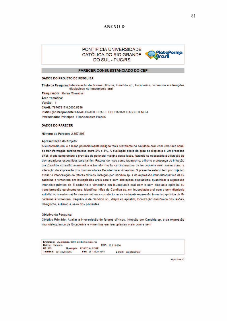
81
ANEXO D
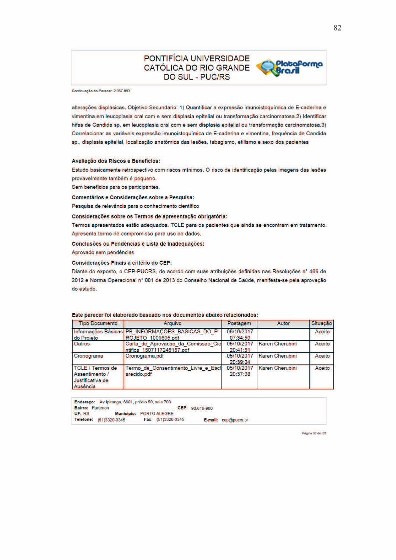
82
83
84
ANEXO E