EFFECTS OF Cowpea mild mottle virus ON Bemisia tabaci€¦ · experiment was carried out letting B....
37
FELIPE VIGATO PRADO EFFECTS OF Cowpea mild mottle virus ON Bemisia tabaci Dissertação apresentada à Universidade Federal de Viçosa, como parte das exigências do Programa de Pós-Graduação em Ecologia, para obtenção do título de Magister Scientiae. VIÇOSA MINAS GERAIS – BRASIL 2014
Transcript of EFFECTS OF Cowpea mild mottle virus ON Bemisia tabaci€¦ · experiment was carried out letting B....
FELIPE VIGATO PRADO
EFFECTS OF Cowpea mild mottle virus ON Bemisia tabaci
Dissertação apresentada à Universidade Federal de Viçosa, como parte das exigências do Programa de Pós-Graduação em Ecologia, para obtenção do título de Magister Scientiae.
VIÇOSA
MINAS GERAIS – BRASIL
2014
Ficha catalográfica preparada pela Biblioteca Central da
Universidade Federal de Viçosa - Campus Viçosa
T
Prado, Felipe Vigato, 1988-
P896e
2014
Effects of Cowpea mild mottle virus on Bemisia tabaci /
Felipe Vigato Prado. - Viçosa, MG, 2014.
vii, 28f. : il. (algumas color.) ; 29 cm.
Orientador: Simon Luke Elliot.
Dissertação (mestrado) - Universidade Federal de Viçosa.
Referências bibliográficas: f.25-28.
1. Bemisia tabaci. 2. Mosca-branca. 3. Soja - Doenças e
pragas. 4. Vetor-planta-patógeno. I. Universidade Federal de
Viçosa. Departamento de Biologia Animal. Programa de
Pós-graduação em Ecologia. II. Título.
CDD 22. ed. 595.754
FELIPE VIGATO PRADO
EFFECTS OF Cowpea mild mottle virus ON Bemisia tabaci
Dissertação apresentada à Universidade Federal de Viçosa, como parte das exigências do Programa de Pós-Graduação em Ecologia, para obtenção do título de Magister Scientiae.
APROVADA: 29 de abril de 2014.
Angelo Pallini Filho Paulo Fellipe Cristaldo
Simon Luke Elliot (Orientador)
ii
AGRADECIMENTOS
À Universidade Federal de Viçosa/Programa de Pós-Graduação
em Ecologia pelas oportunidades oferecidas.
À Fundação de Amparo à Pesquisa do estado de Minas Gerais
(FAPEMIG) pela concessão da bolsa de Mestrado.
Ao Professor Simon Luke Elliot pela orientação e ensinamentos,
desde 2009.
À Professora Claudine Márcia Carvalho, Fábio Nascimento Silva e
Larissa Goulart Zanardo pela imprescindível colaboração.
Aos colegas do Laboratório de Interação Inseto-Microrganismo por
toda a ajuda teórica e prática nesses vários anos.
Aos colegas do PET (Programa de Educação Tutorial) por tudo que
aprendemos juntos.
Aos amigos de Viçosa e de Paraguaçu por todo o apoio nos
momentos em que mais precisei.
À Lívia Ferreira Diniz pelo amor e amizade.
Ao meu irmão e demais familiares por serem minha inspiração.
Aos meus pais por serem meus exemplos de ser humano e a base
que me sustenta.
iii
SUMÁRIO
RESUMO................................................................................................... iv
ABSTRACT ............................................................................................... vi
1 INTRODUCTION .................................................................................1
2 MATERIAL AND METHODS................................................................6
Insects and plants ...................................................................................6
Identification of whiteflies ........................................................................7
Oviposition experiment............................................................................9
Transmission experiment ......................................................................13
3 RESULTS ..........................................................................................15
Identification of whiteflies ......................................................................15
Oviposition experiment..........................................................................17
Transmission experiment ......................................................................20
4 DISCUSSION.....................................................................................22
5 REFERENCES ..................................................................................25
iv
RESUMO
PRADO, Felipe Vigato, M.Sc., Universidade Federal de Viçosa, abril, 2014. EFEITO DO Cowpea mild mottle virus EM Bemisia tabaci. Orientador: Simon Luke Elliot.
As relações ecológicas entre plantas, patógenos e vetores são complexas
e são muitos os possíveis efeitos diretos e indiretos sobre os
participantes. Bemisia tabaci (Hemiptera: Aleyrodidae) são os insetos que
causam as maiores perdas econômicas em plantações. Moscas-brancas,
devido a sua polifagia, transmitem vírus para uma ampla gama de
hospedeiros. O vírus CPMMV (Cowpea mild mottle virus) é o agente
etiológico da Necrose da haste da soja e é transmitido de maneira não
persistente pela mosca-branca MEAM1 (Middle East-Asia Minor 1). O
objetivo desse estudo é testar: (i) se a mosca-branca MEAM1 transmite o
isolado CPMMV:BR:GO:01:1 para a soja cultivar (cv.) CD206 e (ii) se a
soja infectada com o CPMMV diminui o fitness da mosca-branca MEAM1.
O experimento de oviposição foi realizado com ninfas de B. tabaci
dispostas em folíolos de soja sadios ou infectados com CPMMV no
interior de placas de Petri previamente preparadas. O experimento de
transmissão foi realizado deixando B. tabaci se alimentarem de soja
infectada com CPMMV e posteriormente transferindo esses insetos para
plantas de soja saudáveis. Foi verificado que é possível a transmissão de
CPMMV:BR:GO:01:1 para soja cv. CD206 através de B. tabaci MEAM1.
Isso é fundamental para entender os parâmetros evolutivos que moldam
as relações entre esses organismos. A respeito do experimento de
oviposição, ambos os parâmetros usados para estimar o fitness dos
insetos (eclosão de ninfas e oviposição) aumentaram com o aumento da
área da folha e, além disso, as folhas das plantas infectadas
apresentaram tamanhos menores do que as folhas de plantas saudáveis.
v
Dessa maneira, o vírus teve um efeito negativo direito sobre sobre as
plantas e um efeito negativo indireto sobre as moscas-brancas. Visto que
vírus transmitidos de maneira não persistente tem efeitos negativos sobre
seus vetores, a não permanência dos insetos nas plantas após picadas
de prova pode favorecer a transmissão do vírus e o fitness desses
insetos. Assim, esse estudo demonstrou uma pequena parte das relações
entre esses organismos, mas baseado nele é possível que sejam feitas
predições que vão contribuir para a delimitação mais precisa de
experimentos futuros que irão contribuir para a compreensão dos
aspectos ecológicos de sistemas inseto-planta-vírus.
vi
ABSTRACT
PRADO, Felipe Vigato. M.Sc., Universidade Federal de Viçosa, April, 2014. EFFECT OF Cowpea mild mottle virus ON Bemisia tabaci. Adviser: Simon Luke Elliot.
The ecological relationships between plants, pathogens and vectors are
complex and there are many possible direct and indirect effects on the
participants. Bemisia tabaci (Hemiptera: Aleyrodidae) are the most
damaging insects in relation to economic losses. Whiteflies, due to their
polyphagy, transmit viruses to a wide range of plant hosts. The Cowpea
mild mottle virus (CPMMV) is the etiologic agent of the Soybean Stem
Necrosis Disease and is nonpersistently transmitted by the whitefly
MEAM1 (Middle East-Asia Minor 1). The objective of this study is to test:
(i) if the whitefly MEAM1 transmit the isolate CPMMV:BR:GO:01:1 to
soybean cultivar (cv.) CD206 and (ii) if CPMMV-infected soybean
decreases the fitness of the whitefly MEAM1. Oviposition experiment was
carried out with B. tabaci nymphs placed on CPMMV-infected and healthy
soybean leaflets, inside previous prepared Petri dishes. Transmission
experiment was carried out letting B. tabaci feed on CPMMV-infected
soybean and further transferring them to healthy soybean plants. It was
verified that is possible to transmit CPMMV:BR:GO:01:1 to soybean cv.
CD206 by the whitefly MEAM1. This is fundamental to understand the
evolution parameters that shape the relationship between these
organisms. Regarding the oviposition experiment, both fitness parameters
(nymphs hatching and oviposition) did increase with leaf area and the
leaves of infected plants were smaller than those of uninfected plants.
Thus, the virus has a direct negative effect on the plants and also an
indirect negative effect on the whiteflies. Since nonpersistently transmitted
viruses have negative effects on vectors, post-probing repellence could
favor the transmission of the virus and the fitness of the whiteflies. This

vii
study demonstrated a small piece of these relationships, but based on it
was possible to make predictions that will help in more precise
delimitations of future experiments that will contribute to improve the
comprehension of the ecological aspects of insect-plant-virus systems.
1
1 INTRODUCTION
The ecological relationships between plants, pathogens and vectors
are complex and there are many possible direct and indirect effects on the
organisms involved. Direct effects involve the vectorborne transmission of
the pathogen; the presence and replication of the pathogen in the insect
and in the plant; and the sharing of the plant as a resource by the insect
and the pathogen. In contrast, indirect effects come from the changes that
infection could cause to the plant. All these possible effects could be
positive or negative to each of the parties, depending upon the species
involved and the evolutionary relationship between them (Belliure et al.
2005; Jiu et al. 2007).
Plant diseases caused by viruses are of great economic importance
and the spread of most of these diseases depends upon vectors (Gray &
Banerjee 1999). Transmission of a virus by a vector is a very complex
event and, due to this, many plant viruses are transmitted by only one
vector species. Arthropods are the most common vectors of plant viruses;
of these, Hemiptera are most important. These vectors are very common
on many crops planted in large monocultures, thus facilitating the spread
of diseases (Ng & Falk 2006).
Furthermore, Hemiptera are the order of insect vectors with
greatest economic importance. The families that cause most damage
transmitting viruses to crops are Aphididae, Aleyrodidae, Cicadellidae and
Delphacidae. Aphids (Aphididae) and whiteflies (Aleyrodidae) stand out
among these families, as they transmit 325 plant viruses (Hogenhout et al.
2008). Virus transmission by these insects is possible due to their
specialized piercing mouthparts. As stated by Cranston & Gullan (2003),
these insects have mouthparts with mandibles and jaws modified in stylets
similarly to needles, surrounded by a grooved lip in a beak shape,
collectively forming a rostrum or proboscis. On the inside, the bundle of
stylets contains two ducts, one by which saliva is released and another
through which the food flows.
2
As reviewed by Hogenhout et al. (2008), there are four described
mechanisms of insect transmission of plant viruses: nonpersistent,
semipersistent, persistent circulative and persistent propagative. In
nonpersistent transmission the vector is capable to start to inoculate the
virus almost immediately after acquisition and retains this ability just for a
few minutes after feeding. In semipersistent transmission the insect is
capable of inoculation also in a short time, but this ability last a few more
hours or even days. The other two types of transmission are persistent,
one being circulative and the other propagative. In persistent circulative
transmission the virus does not replicate inside the insect and cannot be
transmitted to the insect´s offspring. Meanwhile, in persistent propagative
transmission, the virus replicates inside the insect and can be transmitted
to the embryos and germ cells of females.
Mauck et al. (2012), in their review, proposed and tested some
predictions about the relationship between transmission mechanisms of
plant viruses and host-vector interactions. In this review, viruses with
persistent circulative and persistent propagative transmission were
merged just as persistent transmitted viruses. After literature analysis, they
found that the most common patterns found was consistent with their
predictions, that is: (i) vectors are attracted to virus infected plants
independently of the mechanism of transmission; (ii) semipersistent and
persistent transmitted viruses prefer to settle and feed on infected plants,
whereas nonpersistent have no preference or prefer healthy plants and (iii)
vectors perform better on plants infected with semipersistent and
persistent transmitted viruses and worse on plants infected with
nonpersistent viruses.
Navas-Castillo et al. (2011) reviewed the emergent diseases
transmitted by whiteflies (Aleyrodidae) and concluded that these are the
most damaging insects in relation to economic losses to many agricultural
sectors. The insects of this family are usually very small (1-3 mm) and are
called whiteflies due to the powder that covers their wings. Oviposition
behavior involves selecting young plants and laying eggs on the abaxial
3
surface of the leaves. Whitefly abundance is higher in the tropics, but their
distribution is cosmopolitan and they are pests also in temperate climates.
These insects cause direct damage to plants through feeding (sucking sap
and reducing plant vigor) and indirectly through excretion of honeydew,
which leads to reducing the photosynthetic capacity of the plant (De Barro
et al. 2011).
The main concern with these insects is their capacity to transmit
viruses and among them B. tabaci is the most important, which may
transmit Begomovirus, Carlavirus, Crinivirus, Ipomovirus and Torradovirus
(Navas-Castillo et al. 2011). The current consensus is that B. tabaci is a
species complex and that there is no morphological character usable to
correctly classify them and the best approach for this classification is the
sequencing of the mitochondrial cytochrome oxidase 1 (mtCOI) gene
(Brown 2000; De Barro et al. 2005).
Dinsdale et al. (2010) proposed a threshold divergence of 3.5% on
the sequences of the mtCOI gene. From that, B. tabaci was separated in
24 morphologically indistinguishable species (low level groups). These 24
species could be grouped, by the threshold of 11% divergence in 11 high
level groups. Based on this report, B. tabaci biotype B is now part of the
species/group Middle East-Asia Minor 1 species (MEAM1) and so will be
called from now on.
The first report of B. tabaci in Brazil occurred in Bahia in 1928
(Bondar 1928). After this, Lourenção & Nagai (1994) reported that the
numbers of the insects in Brazil remained low, until the beginning of the
1990’s, when the former “biotype B” (now MEAM1 species) was
introduced, probably through the international trade of ornamental plants.
Most of the data on the biology of B. tabaci in Brazil are from
studies before the renaming of biotype B to MEAM1. Despite this, as
MEAM1 nomenclature encompasses biotype B, they are still quite useful.
According to Eichelkraut & Cardona (1990) these insects start feeding
immediately after emergence and after a few hours they already are
4
capable of mating, which can be repeated several times during their lives.
After mating, the time necessary to begin oviposition varies from eight
hours to five days depending on the season. A female lays 100 to 300
eggs during her entire life, according to the temperature, host plant and
food availability. As reported by Dittrich et al. (1990), when these insects
are exposed to insecticides more eggs are produced and in a more
female-biased sex ratio than when not exposed. Overall, males live less
than females, ranging between 9 to 17 days for males and 38 to 74 days
for females, according to host plant and temperature.
As stated by Gill (1990), whiteflies have incomplete metamorphosis
and have the stages of egg, nymph (four instars) and adult. The last
nymphal stage could also be called pseudo-pupa, due to the interruption
of alimentary activity. Adults are the only individuals that can migrate to
new plants and the young stages are immobile with exception of the first
nymphal stage, also called crawler, which could move short distances on
the leaf. Reproduction can be either sexual (production of males and
females) or parthenogenetic (arrhenotoky) producing only males (Salguero
et al. 1992). According to Villas Bôas et al. (2002) the eggs of the
whiteflies are yellowish and pear-shaped, measuring 0.2 to 0.3 cm and are
connected to the leaf by a small rod. Nymphs are also yellowish but
translucent and the fourth nymphal stage (pseudo-pupa) have red eyes
when the adult is about to hatch. Egg to adult time varies between 20 to
27 days, depending on temperature and host plant.
Whiteflies, due to their polyphagy, transmit viruses to a wide range
of plant hosts. Begomovirus are the only DNA viral genus that has been
detected in whiteflies. On the other hand, there are four genera of RNA
viruses transmitted by whiteflies. The number of DNA species vectored by
whiteflies is much greater than RNA, because whiteflies transmit 280
species of Begomovirus and less than 60 species of the four RNA virus
genera (Rosario et al. 2014).
The Cowpea mild mottle virus (CPMMV, family Betaflexiviridae,
genus Carlavirus) is the etiologic agent of the Soybean Stem Necrosis
5
Disease and the symptomatology is: chlorosis, mosaic, vein clearing,
dwarfing and leaf/stem necrosis of Glycine max (Almeida et al. 2003,
2005; Iwaki et al. 1982; Tavasoli et al. 2009). The first report of the virus
was infecting cowpea (Vigna unguiculata) in Ghana in 1973 (Brunt &
Kenten 1973) and, the second report, was almost ten years later, in
soybean in Thailand (Iwaki et al. 1982) and the Ivory Coast (Thouvenel et
al. 1983). In the same period, the virus was also found in Brazil in common
bean (Phaseolus vulgaris) cv. Jalo (Costa et al. 1983). The next reports in
Brazil were in the beginning of the 21st century in soybean at Goiás and
after two years at Mato Grosso, Bahia, Maranhão and Pará (Almeida et al.
2003).This disease is of great importance in Brazil, due to its severity
(Inácio et al. 2003) and also due to the fact that this country is the second
largest producer of soybean in the world and in 2013 produced 82 million
tons in 27 million hectares (IBGE 2013).
Based on all this, the objective of this study is to test the following
hypotheses about the whitefly-soybean-CPMMV system: (i) the whitefly
MEAM1 transmit the isolate CPMMV:BR:GO:01:1 to soybean cv. CD206
and (ii) CPMMV-infected soybean decreases the fitness of the whitefly
MEAM1.
6
2 MATERIAL AND METHODS
Insects and plants
The rearing of B. tabaci was established in 2008 from field
infestations of several different crops. The insects were maintained on
cabbage plants Brassica oleracea var. capitata (IslaTM) inside cages made
of wood and gauze (60cm x 60cm x 60cm).
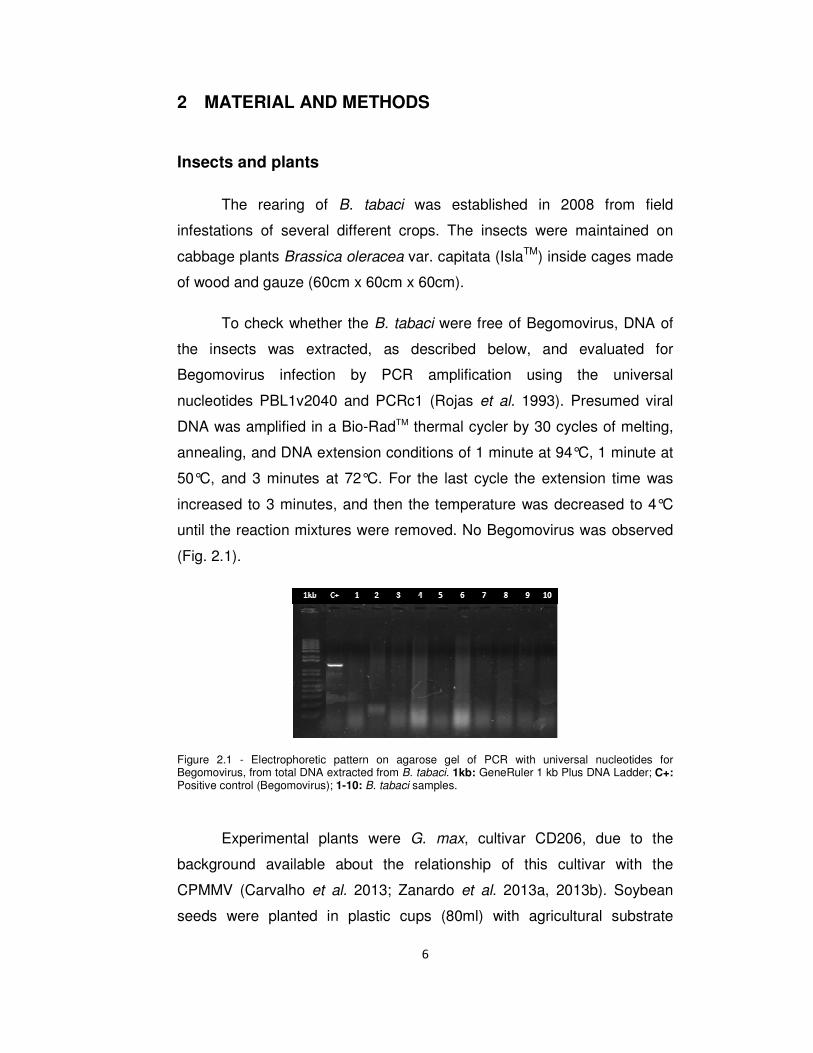
To check whether the B. tabaci were free of Begomovirus, DNA of
the insects was extracted, as described below, and evaluated for
Begomovirus infection by PCR amplification using the universal
nucleotides PBL1v2040 and PCRc1 (Rojas et al. 1993). Presumed viral
DNA was amplified in a Bio-RadTM thermal cycler by 30 cycles of melting,
annealing, and DNA extension conditions of 1 minute at 94°C, 1 minute at
50°C, and 3 minutes at 72°C. For the last cycle the extension time was
increased to 3 minutes, and then the temperature was decreased to 4°C
until the reaction mixtures were removed. No Begomovirus was observed
(Fig. 2.1).
Figure 2.1 - Electrophoretic pattern on agarose gel of PCR with universal nucleotides for Begomovirus, from total DNA extracted from B. tabaci. 1kb: GeneRuler 1 kb Plus DNA Ladder; C+: Positive control (Begomovirus); 1-10: B. tabaci samples.
Experimental plants were G. max, cultivar CD206, due to the
background available about the relationship of this cultivar with the
CPMMV (Carvalho et al. 2013; Zanardo et al. 2013a, 2013b). Soybean
seeds were planted in plastic cups (80ml) with agricultural substrate

7
(Tropstrato HT, Vida VerdeTM) and, after 15 days, the seedlings were
transplanted to pots (1l) full of soil. These were maintained in greenhouses
with average daily temperatures of 26 ± 2 °C and irrigated daily.
Identification of whiteflies
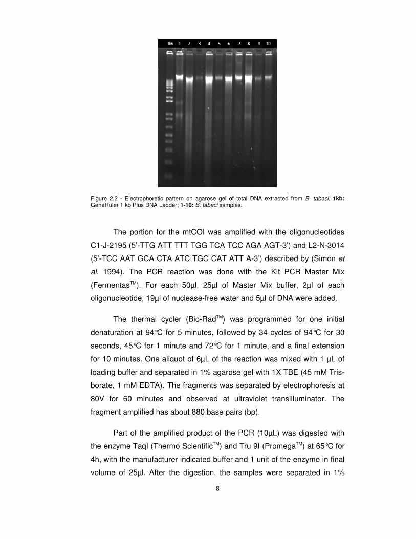
To extract the DNA of B. tabaci, one hundred whiteflies from the
rearing were collected and divided into ten samples. Each sample was
allocated inside a microtube. In each microtube, 30µl of extraction buffer
(50 mM Tris-HCl, pH=7, 100 mM NaCl, 10 mM EDTA and 1% SDS) was
added and crushed with a pistil. A further 170µl of extraction buffer was
added, with 100µl of saturated phenol and chloroform/isoamyl alcohol
(24:1 vol/vol), vortexed for 3 minutes and centrifuged for 3 minutes at
12,000 rpm. The supernatant was removed, transferred to a new tube with
18µl of sodium acetate 3M (0.1 volume; pH=5.2) and 540µL (3 volumes) of
absolute ethanol and incubated at -80°C for 20 minutes. After this, the
samples were centrifuged for 10 minutes at 14,000 rpm and 4°C. The
supernatant was discarded. To the pellet were added 300µl of 80%
ethanol and this was centrifuged for 5 minutes. The ethanol was removed
with a micropipette and the pellet was dried and resuspended with 20µl of
distilled water. The presence of DNA in the samples was checked by
loading 1µl of each sample in a well of a 1% agarose gel and proceeding
to electrophoresis. In all samples, the DNA extraction was successful (Fig.
2.2).
To identify the species/group of B. tabaci three analyses were
performed: (i) comparison of patterns of the mitochondrial cytochrome
oxidase 1 gene (mtCOI) digested by the enzymes enzyme TaqI (Thermo
ScientificTM) and Tru 9I (PromegaTM), (ii) a phylogenetic analyses of the
mtCOI and (iii) a pair-to-pair identity matrix of the nucleotides of the same
gene.
8
Figure 2.2 - Electrophoretic pattern on agarose gel of total DNA extracted from B. tabaci. 1kb: GeneRuler 1 kb Plus DNA Ladder; 1-10: B. tabaci samples.
The portion for the mtCOI was amplified with the oligonucleotides
C1-J-2195 (5’-TTG ATT TTT TGG TCA TCC AGA AGT-3’) and L2-N-3014
(5’-TCC AAT GCA CTA ATC TGC CAT ATT A-3’) described by (Simon et
al. 1994). The PCR reaction was done with the Kit PCR Master Mix
(FermentasTM). For each 50µl, 25µl of Master Mix buffer, 2µl of each
oligonucleotide, 19µl of nuclease-free water and 5µl of DNA were added.
The thermal cycler (Bio-RadTM) was programmed for one initial
denaturation at 94°C for 5 minutes, followed by 34 cycles of 94°C for 30
seconds, 45°C for 1 minute and 72°C for 1 minute, and a final extension
for 10 minutes. One aliquot of 6µL of the reaction was mixed with 1 µL of
loading buffer and separated in 1% agarose gel with 1X TBE (45 mM Tris-
borate, 1 mM EDTA). The fragments was separated by electrophoresis at
80V for 60 minutes and observed at ultraviolet transilluminator. The
fragment amplified has about 880 base pairs (bp).
Part of the amplified product of the PCR (10µL) was digested with
the enzyme TaqI (Thermo ScientificTM) and Tru 9I (PromegaTM) at 65°C for
4h, with the manufacturer indicated buffer and 1 unit of the enzyme in final
volume of 25µl. After the digestion, the samples were separated in 1%
9
agarose gel at 80V for 60 minutes and the patterns were compared with
the patterns described by Bosco et al. (2006).
The phylogenetic analysis of the mtCOI was based on the partial
sequences obtained from 5 isolates of B. tabaci from our rearing aligned
with different species of whiteflies with the software Muscle (Edgar 2004)
and implemented at the software Mega 5.5 (Tamura et al. 2011). The
phylogenetic analyses were generated with the maximum likelihood
method and the model Tamura Nei, using transition–transversion ratio (R)
with Gamma distribution (Tamura Nei + G), with 2000 repetitions to obtain
the bootstrap values.
The identity matrix was done with the same isolates of B. tabaci that
were used for the phylogenetic analyses, the comparison between the
mtCOI nucleotide sequences was done by pair-to-pair p-distance
(proportion of nucleotide sites at which two sequences being compared
are different) with the software Mega 5.5 (Tamura et al. 2011).
Oviposition experiment
Soybean plants were mechanically inoculated using a CPMMV-
positive samples of the isolate CPMMV:BR:GO:01:1 from Laboratório de
Virologia (Zanardo et al. 2013a). Inoculum was obtained by grinding this
material in 0.1M phosphate buffer with 0.1% sodium sulphite (pH 7.2). The
suspension obtained was used to perform the mechanical inoculation,
which consists of producing microlesions on the leaves with gauze soaked
in the solution mixed with carborundum. These lesions are then the route
of entry of the virus into plants. The leaves inoculated were the first
trifoliate of the plants, which were 15 days old, with the second trifoliate
still in early development. Control plants were subjected to the same
procedures, but without the virus. Each treatment (control and inoculated)
had ten repetitions, namely one leaflet of one different plant.
Past 11 days of the inoculation, i.e. when the plants were 26 days
old, ten plants with characteristic symptoms of CPPMV infection (Fig. 2.3a)
10
and ten that were characteristically healthy (Fig. 2.3b) were selected. At
this age the plants were with the third trifoliate incompletely developed, so
in order to standardize the material collected, we worked only with the
second trifoliate of the plants. In each trifoliate the central leaflet was
collected to be used in the trial and the side leaflets were collected for
further confirmation of the presence or absence of the CPMMV by
Reverse Transcription Polymerase Chain Reaction (RT-PCR), described
below.
After collection, the leaves were held in aluminum foil envelopes
and stored in polystyrene boxes until they were either stored in a -80°C
freezer (leaves for confirmation) or taken to the laboratory to be washed,
on both sides, with sterile water before being placed in the Petri dishes
(leaves for the experiment).
Figure 2.3 – Characteristic plants. A: CPMMV-infected; B: Healthy.
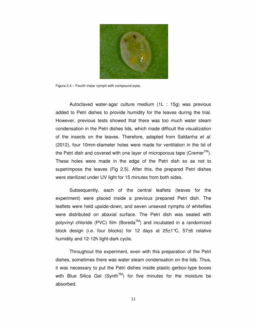
In this same day, whiteflies were collected at the fourth instar, using
the visualization of compound eyes to indicate that the insects have
reached this stage (Fig. 2.4). Insect collection was done with a scalpel,
carefully removing the nymphs from the rearing plant and transferring
them to the leaf used in the trial. The placement of insects on the leaves
was done alternately between treatment and control. Between each leaf
the scalpel was cleaned with 70% alcohol to prevent accidental
contamination with the virus.
A B
11
Figure 2.4 – Fourth instar nymph with compound eyes.
Autoclaved water-agar culture medium (1L : 15g) was previous
added to Petri dishes to provide humidity for the leaves during the trial.
However, previous tests showed that there was too much water steam
condensation in the Petri dishes lids, which made difficult the visualization
of the insects on the leaves. Therefore, adapted from Saldanha et al.
(2012), four 10mm-diameter holes were made for ventilation in the lid of
the Petri dish and covered with one layer of microporous tape (CremerTM).
These holes were made in the edge of the Petri dish so as not to
superimpose the leaves (Fig 2.5). After this, the prepared Petri dishes
were sterilized under UV light for 15 minutes from both sides.
Subsequently, each of the central leaflets (leaves for the
experiment) were placed inside a previous prepared Petri dish. The
leaflets were held upside-down, and seven unsexed nymphs of whiteflies
were distributed on abaxial surface. The Petri dish was sealed with
polyvinyl chloride (PVC) film (BoredaTM) and incubated in a randomized
block design (i.e. four blocks) for 12 days at 25±1°C, 57±6 relative
humidity and 12-12h light-dark cycle.
Throughout the experiment, even with this preparation of the Petri
dishes, sometimes there was water steam condensation on the lids. Thus,
it was necessary to put the Petri dishes inside plastic gerbox-type boxes
with Blue Silica Gel (SynthTM) for five minutes for the moisture be
absorbed.
12
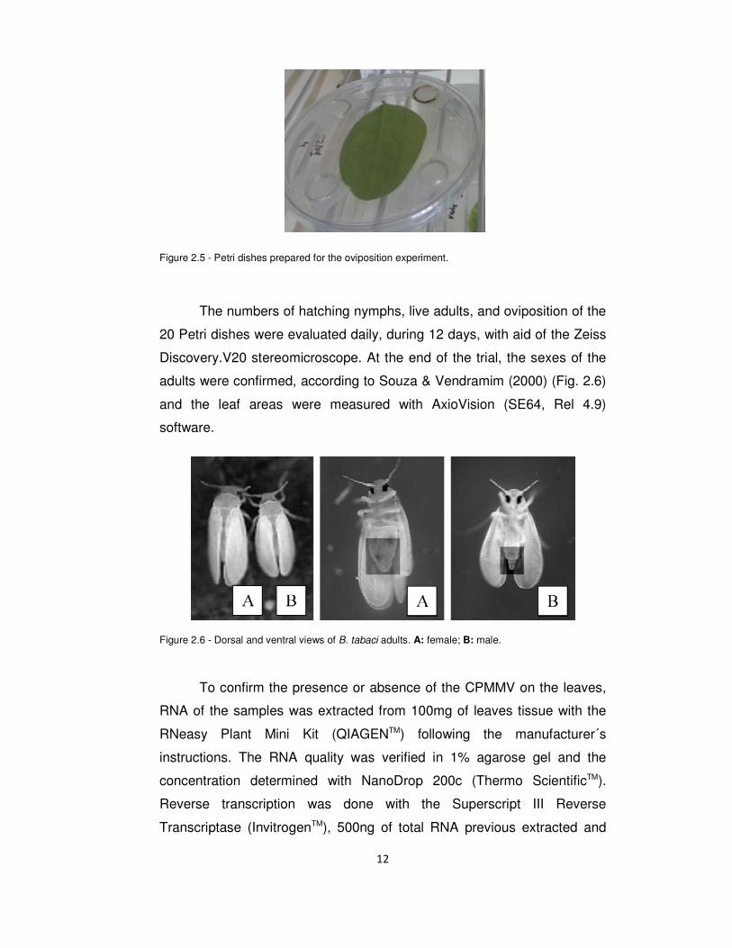
Figure 2.5 - Petri dishes prepared for the oviposition experiment.
The numbers of hatching nymphs, live adults, and oviposition of the
20 Petri dishes were evaluated daily, during 12 days, with aid of the Zeiss
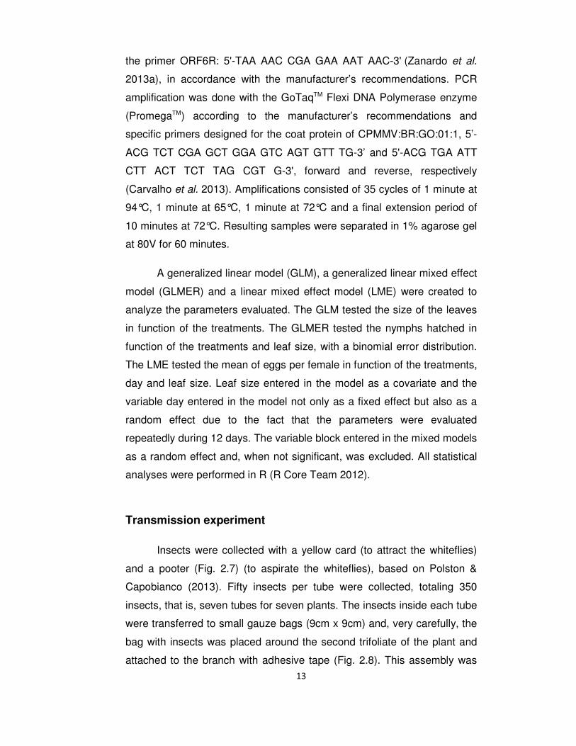
Discovery.V20 stereomicroscope. At the end of the trial, the sexes of the
adults were confirmed, according to Souza & Vendramim (2000) (Fig. 2.6)
and the leaf areas were measured with AxioVision (SE64, Rel 4.9)
software.
Figure 2.6 - Dorsal and ventral views of B. tabaci adults. A: female; B: male.
To confirm the presence or absence of the CPMMV on the leaves,
RNA of the samples was extracted from 100mg of leaves tissue with the
RNeasy Plant Mini Kit (QIAGENTM) following the manufacturer´s
instructions. The RNA quality was verified in 1% agarose gel and the
concentration determined with NanoDrop 200c (Thermo ScientificTM).
Reverse transcription was done with the Superscript III Reverse
Transcriptase (InvitrogenTM), 500ng of total RNA previous extracted and
13
the primer ORF6R: 5'-TAA AAC CGA GAA AAT AAC-3' (Zanardo et al.
2013a), in accordance with the manufacturer’s recommendations. PCR
amplification was done with the GoTaqTM Flexi DNA Polymerase enzyme
(PromegaTM) according to the manufacturer’s recommendations and
specific primers designed for the coat protein of CPMMV:BR:GO:01:1, 5’-
ACG TCT CGA GCT GGA GTC AGT GTT TG-3’ and 5'-ACG TGA ATT
CTT ACT TCT TAG CGT G-3', forward and reverse, respectively
(Carvalho et al. 2013). Amplifications consisted of 35 cycles of 1 minute at
94°C, 1 minute at 65°C, 1 minute at 72°C and a final extension period of
10 minutes at 72°C. Resulting samples were separated in 1% agarose gel
at 80V for 60 minutes.
A generalized linear model (GLM), a generalized linear mixed effect
model (GLMER) and a linear mixed effect model (LME) were created to
analyze the parameters evaluated. The GLM tested the size of the leaves
in function of the treatments. The GLMER tested the nymphs hatched in
function of the treatments and leaf size, with a binomial error distribution.
The LME tested the mean of eggs per female in function of the treatments,
day and leaf size. Leaf size entered in the model as a covariate and the
variable day entered in the model not only as a fixed effect but also as a
random effect due to the fact that the parameters were evaluated
repeatedly during 12 days. The variable block entered in the mixed models
as a random effect and, when not significant, was excluded. All statistical
analyses were performed in R (R Core Team 2012).
Transmission experiment
Insects were collected with a yellow card (to attract the whiteflies)
and a pooter (Fig. 2.7) (to aspirate the whiteflies), based on Polston &
Capobianco (2013). Fifty insects per tube were collected, totaling 350
insects, that is, seven tubes for seven plants. The insects inside each tube
were transferred to small gauze bags (9cm x 9cm) and, very carefully, the
bag with insects was placed around the second trifoliate of the plant and
attached to the branch with adhesive tape (Fig. 2.8). This assembly was
14
attached to a bamboo skewer to avoid injury to the plant. In order to
maximize the chances of acquisition the insects were left for an acquisition
access aeriod (AAP) of 48h.
Figure 2.7 - Pooter to aspirate the whiteflies.
Figure 2.8 - Bag with insects placed around the second trifoliate of the plant and attached to the branch with adhesive tape. This photo was taken 18 days after the beginning of the experiment.
After this time, the trifoliate was cut, removed from the gauze bag
and stored for further confirmation of the infectivity status with indirect RT-
PCRs in the same way as for the oviposition trial. Then, the bag with the
insects was placed around the healthy plant (second trifoliate), attached
as described previously and left there for an inoculation access period
(IAP) of 48h. The insects were not removed from the branch until the
emergence of the symptons to maximize the chances of inoculation. New
symptomatic leaves were collected for confirmation of the infectivity status
with RT-PCRs.
15
3 RESULTS
Identification of whiteflies
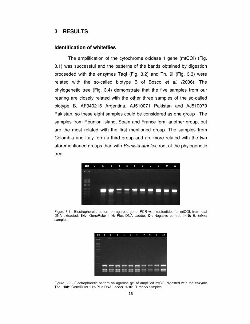
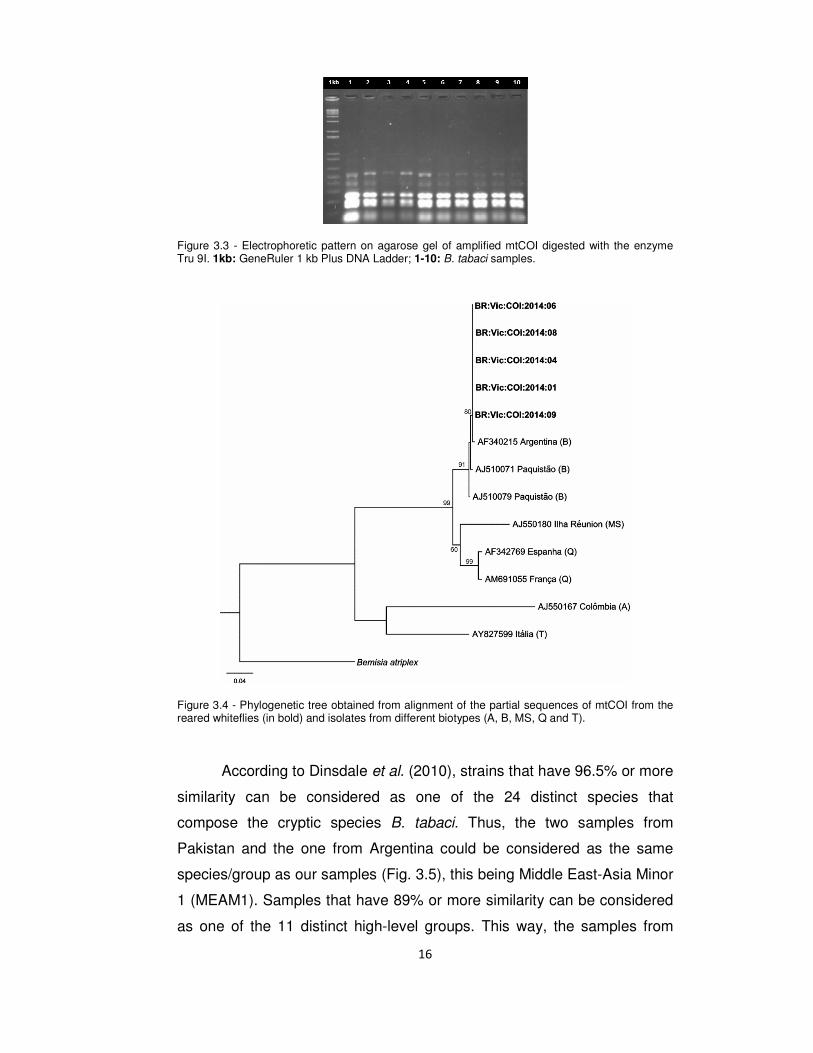
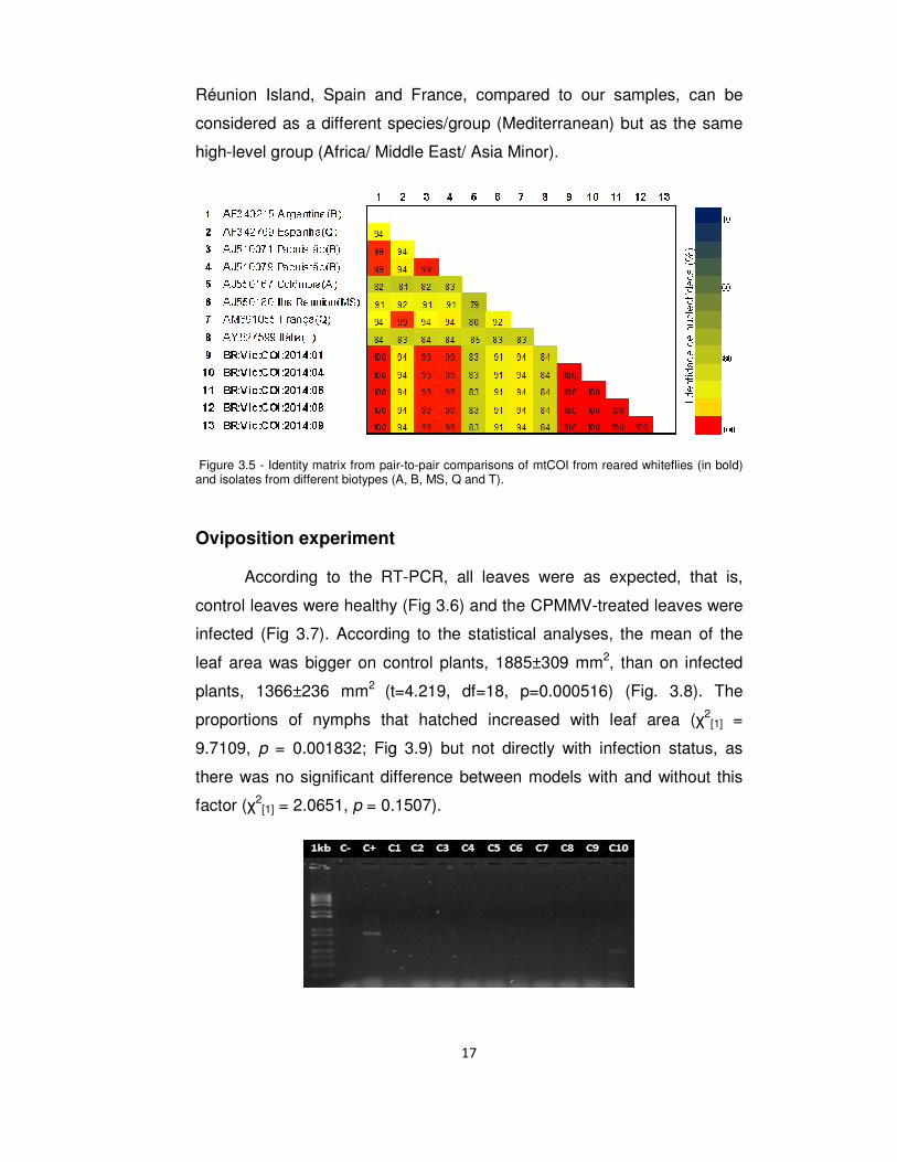
The amplification of the cytochrome oxidase 1 gene (mtCOI) (Fig.
3.1) was successful and the patterns of the bands obtained by digestion
proceeded with the enzymes TaqI (Fig. 3.2) and Tru 9I (Fig. 3.3) were
related with the so-called biotype B of Bosco et al. (2006). The
phylogenetic tree (Fig. 3.4) demonstrate that the five samples from our
rearing are closely related with the other three samples of the so-called
biotype B, AF340215 Argentina, AJ510071 Pakistan and AJ510079
Pakistan, so these eight samples could be considered as one group . The
samples from Réunion Island, Spain and France form another group, but
are the most related with the first mentioned group. The samples from
Colombia and Italy form a third group and are more related with the two
aforementioned groups than with Bemisia atriplex, root of the phylogenetic
tree.
Figure 3.1 - Electrophoretic pattern on agarose gel of PCR with nucleotides for mtCOI, from total DNA extracted. 1kb: GeneRuler 1 kb Plus DNA Ladder; C-: Negative control; 1-10: B. tabaci samples.
Figure 3.2 - Electrophoretic pattern on agarose gel of amplified mtCOI digested with the enzyme TaqI. 1kb: GeneRuler 1 kb Plus DNA Ladder; 1-10: B. tabaci samples.
16
Figure 3.3 - Electrophoretic pattern on agarose gel of amplified mtCOI digested with the enzyme Tru 9I. 1kb: GeneRuler 1 kb Plus DNA Ladder; 1-10: B. tabaci samples.
Figure 3.4 - Phylogenetic tree obtained from alignment of the partial sequences of mtCOI from the reared whiteflies (in bold) and isolates from different biotypes (A, B, MS, Q and T).
According to Dinsdale et al. (2010), strains that have 96.5% or more
similarity can be considered as one of the 24 distinct species that
compose the cryptic species B. tabaci. Thus, the two samples from
Pakistan and the one from Argentina could be considered as the same
species/group as our samples (Fig. 3.5), this being Middle East-Asia Minor
1 (MEAM1). Samples that have 89% or more similarity can be considered
as one of the 11 distinct high-level groups. This way, the samples from
17
Réunion Island, Spain and France, compared to our samples, can be
considered as a different species/group (Mediterranean) but as the same
high-level group (Africa/ Middle East/ Asia Minor).
Figure 3.5 - Identity matrix from pair-to-pair comparisons of mtCOI from reared whiteflies (in bold) and isolates from different biotypes (A, B, MS, Q and T).
Oviposition experiment
According to the RT-PCR, all leaves were as expected, that is,
control leaves were healthy (Fig 3.6) and the CPMMV-treated leaves were
infected (Fig 3.7). According to the statistical analyses, the mean of the
leaf area was bigger on control plants, 1885±309 mm2, than on infected
plants, 1366±236 mm2 (t=4.219, df=18, p=0.000516) (Fig. 3.8). The
proportions of nymphs that hatched increased with leaf area (χ2[1] =
9.7109, p = 0.001832; Fig 3.9) but not directly with infection status, as
there was no significant difference between models with and without this
factor (χ2[1] = 2.0651, p = 0.1507).
18
Figure 3.6 – Electrophoretic pattern on agarose gel of RT-PCR from RNA extracted of control leaves of oviposition experiment. 1kb: GeneRuler 1 kb Plus DNA Ladder; C-: Negative control; C+: Positive control (CPMMV); C1-C10: Control leaves samples.
Figure 3.7 - Electrophoretic pattern on agarose gel of RT-PCR from RNA extracted of CPMMV-treated leaves of oviposition experiment. 1kb: GeneRuler 1 kb Plus DNA Ladder; C-: Negative control; C+: Positive control (CPMMV); V1-V10: CPMMV-infected leaves samples.
Treatments
Leaf
siz
e (m
m2)
0
500
1000
1500
2000
2500
ControlVirus
Figure 3.8 – Mean leaf sizes of control plants (1885±309 mm2) and infected plants (1366±236 mm2) (t=4.219, df=18, p=0.000516).
19
Leaf size (mm2)
800 1000 1200 1400 1600 1800 2000 2200 2400P
ropo
rtion
of n
ymph
s ha
tche
d
0,0
0,2
0,4
0,6
0,8
1,0
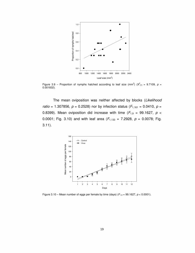
Figure 3.9 – Proportion of nymphs hatched according to leaf size (mm2) (X2[1] = 9.7109, p =
0.001832).
The mean oviposition was neither affected by blocks (Likelihood
ratio = 1.307856, p = 0.2528) nor by infection status (F1,161 = 0.0410, p =
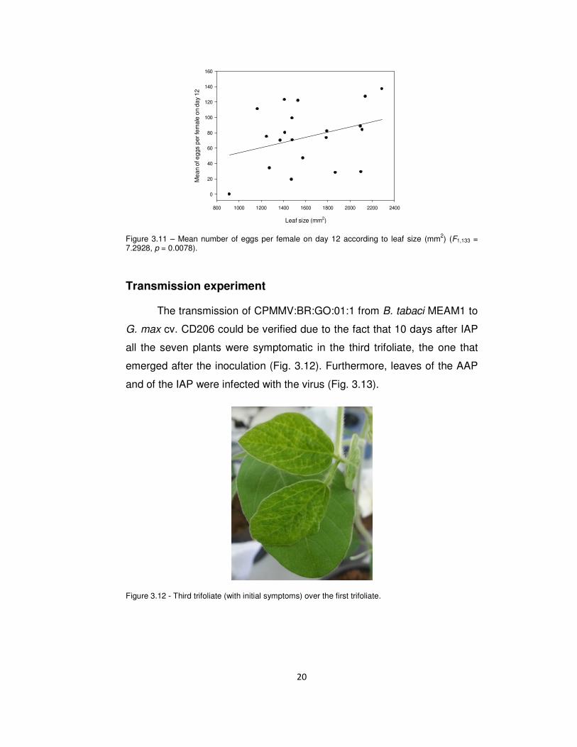
0.8399). Mean oviposition did increase with time (F1,9 = 99.1627, p <
0.0001; Fig. 3.10) and with leaf area (F1,133 = 7.2928, p = 0.0078; Fig.
3.11).
Days
1 2 3 4 5 6 7 8 9 10 11 12
Mea
n nu
mbe
r of e
ggs
per f
emal
e
0
20
40
60
80
100
120
140
160
Control
Virus
Figure 3.10 – Mean number of eggs per female by time (days) (F1,9 = 99.1627, p < 0.0001).
20
Leaf size (mm2)
800 1000 1200 1400 1600 1800 2000 2200 2400
Mea
n of
egg
s pe
r fem
ale
on d
ay 1
20
20
40
60
80
100
120
140
160
Figure 3.11 – Mean number of eggs per female on day 12 according to leaf size (mm2) (F1,133 = 7.2928, p = 0.0078).
Transmission experiment
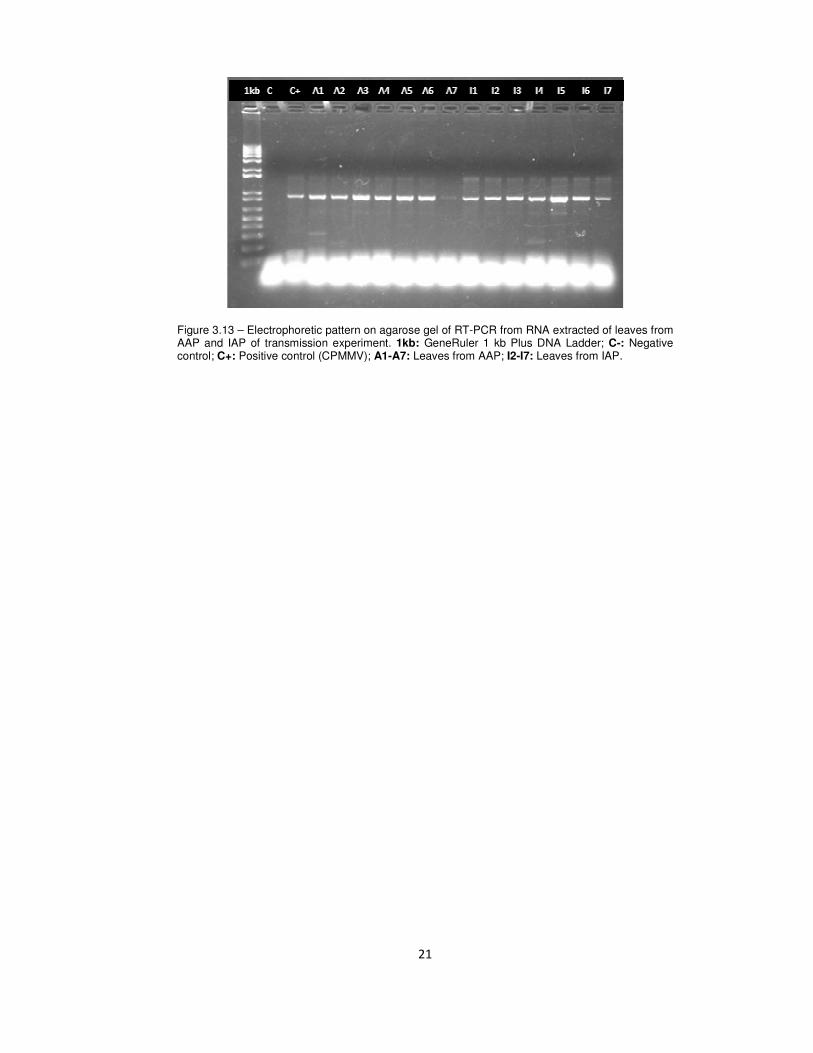
The transmission of CPMMV:BR:GO:01:1 from B. tabaci MEAM1 to
G. max cv. CD206 could be verified due to the fact that 10 days after IAP
all the seven plants were symptomatic in the third trifoliate, the one that
emerged after the inoculation (Fig. 3.12). Furthermore, leaves of the AAP
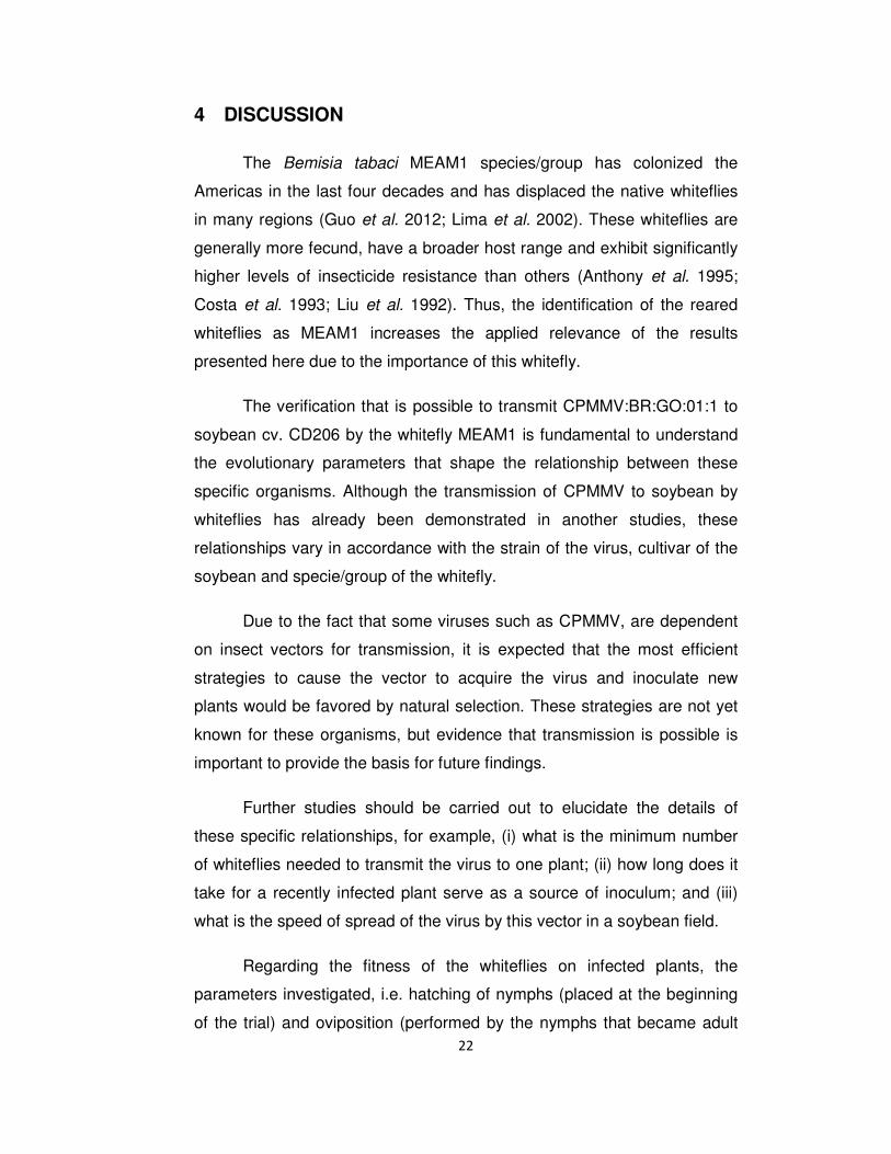
and of the IAP were infected with the virus (Fig. 3.13).
Figure 3.12 - Third trifoliate (with initial symptoms) over the first trifoliate.
21
Figure 3.13 – Electrophoretic pattern on agarose gel of RT-PCR from RNA extracted of leaves from AAP and IAP of transmission experiment. 1kb: GeneRuler 1 kb Plus DNA Ladder; C-: Negative control; C+: Positive control (CPMMV); A1-A7: Leaves from AAP; I2-I7: Leaves from IAP.
22
4 DISCUSSION
The Bemisia tabaci MEAM1 species/group has colonized the
Americas in the last four decades and has displaced the native whiteflies
in many regions (Guo et al. 2012; Lima et al. 2002). These whiteflies are
generally more fecund, have a broader host range and exhibit significantly
higher levels of insecticide resistance than others (Anthony et al. 1995;
Costa et al. 1993; Liu et al. 1992). Thus, the identification of the reared
whiteflies as MEAM1 increases the applied relevance of the results
presented here due to the importance of this whitefly.
The verification that is possible to transmit CPMMV:BR:GO:01:1 to
soybean cv. CD206 by the whitefly MEAM1 is fundamental to understand
the evolutionary parameters that shape the relationship between these
specific organisms. Although the transmission of CPMMV to soybean by
whiteflies has already been demonstrated in another studies, these
relationships vary in accordance with the strain of the virus, cultivar of the
soybean and specie/group of the whitefly.
Due to the fact that some viruses such as CPMMV, are dependent
on insect vectors for transmission, it is expected that the most efficient
strategies to cause the vector to acquire the virus and inoculate new
plants would be favored by natural selection. These strategies are not yet
known for these organisms, but evidence that transmission is possible is
important to provide the basis for future findings.
Further studies should be carried out to elucidate the details of
these specific relationships, for example, (i) what is the minimum number
of whiteflies needed to transmit the virus to one plant; (ii) how long does it
take for a recently infected plant serve as a source of inoculum; and (iii)
what is the speed of spread of the virus by this vector in a soybean field.
Regarding the fitness of the whiteflies on infected plants, the
parameters investigated, i.e. hatching of nymphs (placed at the beginning
of the trial) and oviposition (performed by the nymphs that became adult
23
and reproduced), were not influenced directly by infection with CPMMV.
However, both fitness parameters did increase with leaf area and the
leaves of infected plants were smaller than those of uninfected plants.
Thus, the virus has a direct negative effect on the plants and also an
indirect negative effect on the whiteflies. As dwarfism is one of the typical
symptoms of Soybean Stem Necrosis caused by this virus (Almeida et al.
2003), it is expected that the CPMMV-infected leaves be smaller than
healthy plants, although this has not been specifically described in the
literature.
Mauck et al. (2012) reviewed the relationship between transmission
mechanisms of plant viruses and host-vector interactions. One of the
patterns found is that vectors perform worse on plants infected with
nonpersistent viruses than on healthy plants. For example, Donaldson &
Gratton (2007) found that the soybean aphid (Aphis glycines) density on
healthy soybean plants is nearly double than on soybean mosaic virus
infected plants. Hodge & Powell (2008) reported that bean yellow mosaic
virus causes a reduction in pea aphid (Acyrthosiphon pisum) survival on
fava beans (Vicia faba).
Beyond oviposition, vector survival or longevity are parameters
commonly studied to test the effects of viruses on vectors. These
parameters have not yet been tested in whitefly-soybean-CPMMV system,
thus approach this parameters is an important extension of the studies
already carried on. Construction of life tables is also recommended, due to
the fact that this approach allows a more holistic comprehension of the
effects on vector.
Mauck et al. (2012) also found a pattern that vectors are attracted
to virus infected plants independently of the mechanism of transmission.
For nonpersistently transmitted viruses only four studies were found and,
of these, three reported insects attracted to infected plants (Eckel &
Lampert 1996; Eigenbrode et al. 2002; Mauck et al. 2010) and one
reported no preference (Fereres & Kampmeier 1999).
24
A third pattern found by Mauck et al. (2012) is that vectors of
nonpersistent viruses prefer to settle and feed on healthy plants or have
no preference. Trials to test if the same patterns occur with the CPMMV-
whitefly-soybean system have not yet been done, but are already in
planning (T. Amaral, personal communication).
The negative effects of CPMMV on the MEAM1 whiteflies at
soybean, although indirect, is still consistent with the first aforementioned
pattern found by Mauck et al. (2012). Moreover, considering that the other
two patterns found are also true for this system, one possible evolutionary
explanation for this pattern could be proposed. This one is that the
chlorosis induced on the soybean by the CPMMV attract the whiteflies, as
the attraction to yellow is a well know trait of this insect (Berlinger 1986;
Zanardo et al. 2013a).
Futhermore, the transmission of a nonpersistent transmitted virus
will be increased if the vector rapidly disperse to a new (healthy) plant
after acquire the virus, because this type of virus is possible to be
transmitted by his vector almost immediately after been acquired and this
lasts just for a short time. Thus, if the whiteflies probe the infected plant
and rapidly migrate to a healthy one the virus dispersal and also the
whiteflies fitness will be greater than if the whiteflies remains on the
infected plant.
This study demonstrated a small piece of these relationships, but
based on it was possible to make predictions that will help in more precise
delimitations of future experiments. In addition to this, understanding the
roles that each organism plays on the whitefly-soybean-CPMMV system is
important to improve the comprehension of the ecological aspects of
insect-plant-virus systems and could lead to possible ways to control plant
diseases related with these organisms.
25
5 REFERENCES
Almeida, A.M.R., Piuga, F.F., Kitajima, E.W., Gaspar, J.O., Valentin, N., Benato, L.C., et al. (2003). Necrose da haste da soja. Embrapa Soja. Doc.
Almeida, Á.M.R., Piuga, F.F., Marin, S.R.R., Kitajima, E.W., Gaspar, J.O., Oliveira, T.G. De, et al. (2005). Detection and Partial Characterization of a Carlavirus Causing Stem Necrosis of Soybean in Brazil, 191–194.
Anthony, N.M., Brown, J.K., Markham, P.G. & Ffrenchconstant, R.H. (1995). Molecular analysis of cyclodiene resistance-associated mutations among populations of the sweetpotato whitefly Bemisia tabaci. Pestic. Biochem. Physiol., 51, 220–228.
De Barro, P.J., Liu, S.-S., Boykin, L.M. & Dinsdale, A. (2011). Bemisia tabaci: a statement of species status. Annu. Rev. Entomol., 56, 1–19.
De Barro, P.J., Trueman, J.W.H. & Frohlich, D.R. (2005). Bemisia argentifolii is a race of B. tabaci (Hemiptera: Aleyrodidae): the molecular genetic differentiation of B. tabaci populations around the world. Bull. Entomol. Res., 95, 193–203.
Belliure, B., Janssen, A., Maris, P.C., Peters, D. & Sabelis, M.W. (2005). Herbivore arthropods benefit from vectoring plant viruses. Ecol. Lett., 8, 70–79.
Bondar, G. (1928). Relatório. Bol. do Laboratório Patol. Veg., 4, 39–46.
Bosco, D., Loria, A., Sartor, C. & Cenis, J.L. (2006). PCR-RFLP Identification of Bemisia tabaci Biotypes in the Mediterranean Basin, 34, 243–251.
Brown, J.K. (2000). Molecular markers for the identification and global tracking of whitefly vector–< i> Begomovirus</i> complexes. Virus Res., 71, 233–260.
Brunt, A.A. & Kenten, R.H. (1973). Cowpea mild mottle, a newly recognized virus infecting cowpeas (Vigna unguiculata) in Ghana. Ann. Appl. Biol., 74, 67–74.
Carvalho, S.L. De, Silva, F.N., Zanardo, L.G., Almeida, Á.M.R., Murilo, F. & Carvalho, C.M. (2013). Production of polyclonal antiserum against Cowpea mild mottle virus coat protein and its application in virus detection. Tropical, 38, 49–54.
Costa, A.S., Gaspar, J.O. & Vega, J. (1983). Angular mosaic of the Phaseolus vulgaris variety Jalo, caused by a carlavirus transmitted by the white-fly Bemisia tabaci. Fitopatol. Bras., 8, 325–337.
Costa, H.S., Johnson, M.W., Ullman, D.E., Omer, A.D. & Tabashnik, B.E. (1993). Sweetpotato whitefly (Homoptera: Aleyrodidae): Analysis of biotypes and distribution in Hawaii. Environ. Entomol., 22, 16–20.
Cranston, P.S. & Gullan, P.J. (2003). Encyclopedia of Insects. In: Encycl. Insects. Academic Press. Elsevier Science.
26
Dinsdale, A., Cook, L., Riginos, C., Buckley, Y.M. & De Barro, P. (2010). Refined Global Analysis of Bemisia tabaci (Hemiptera: Sternorrhyncha: Aleyrodoidea: Aleyrodidae) Mitochondrial Cytochrome Oxidase 1 to Identify Species Level Genetic Boundaries. Ann. Entomol. Soc. Am., 103, 196–208.
Dittrich, V., Ernst, G.H., Ruesch, O. & Uk, S. (1990). Resistance mechanisms in sweetpotato whitefly (Homoptera: Aleyrodidae) populations from Sudan, Turkey, Guatemala, and Nicaragua. J. Econ. Entomol., 83.
Donaldson, J.R. & Gratton, C. (2007). Antagonistic effects of soybean viruses on soybean aphid performance. Environ. Entomol., 36, 918–25.
Eckel, R.V.W. & Lampert, E.P. (1996). Relative attractiveness of tobacco etch virus-infected and healthy flue-cured tobacco plants to aphids (Homoptera: Aphididae). J. Econ. Entomol., 89, 1017–1027.
Edgar, R.C. (2004). MUSCLE: multiple sequence alignment with high accuracy and high throughput. Nucleic Acids Res., 32, 1792–7.
Eichelkraut, K. & Cardona, C. (1990). Biologia, cria masal y aspectos ecologicos de la mosca blanca Bemisia tabaci (Gennadius)(Homoptera: Aleyrodidae), como plaga del frijol comun. Turrialba, 40.
Eigenbrode, S.D., Ding, H., Shiel, P. & Berger, P.H. (2002). Volatiles from potato plants infected with potato leafroll virus attract and arrest the virus vector, Myzus persicae (Homoptera: Aphididae). Proc. Biol. Sci., 269, 455–60.
Fereres, A. & Kampmeier, G.E. (1999). Aphid Attraction and Preference for Soybean and Pepper Plants Infected with Potyviridae. Entomol. Soc. Am., July, 542–548.
Gill, R.J. (1990). The morphology of whiteflies. Whiteflies their bionomics, pest status Manag., 13–46.
Gray, S.M. & Banerjee, N. (1999). Mechanisms of Arthropod Transmission of Plant and Animal Viruses Mechanisms of Arthropod Transmission of Plant and Animal Viruses, 63.
Guo, J.-Y., Dong, S.-Z., Yang, X., Cheng, L., Wan, F.-H., Liu, S.-S., et al. (2012). Enhanced vitellogenesis in a whitefly via feeding on a begomovirus-infected plant. PLoS One, 7, e43567.
Hodge, S. & Powell, G. (2008). Do Plant Viruses Facilitate Their Aphid Vectors by Inducing Symptoms that Alter Behavior and Performance ? Do Plant Viruses Facilitate Their Aphid Vectors by Inducing Symptoms that Alter Behavior and Performance ?, 37, 1573–1581.
Hogenhout, S. a, Ammar, E.-D., Whitfield, A.E. & Redinbaugh, M.G. (2008). Insect vector interactions with persistently transmitted viruses. Annu. Rev. Phytopathol., 46, 327–59.
IBGE. (2013). Levantamento sistemático da produção agrícola [WWW Document]. Fundação Inst. Bras. Geogr. e Estatística.
27
Inácio, L., Rodrigues, R., Campanhola, C., Pires, A.K. & Quast, D.G. (2003). Necrose da Haste da Soja.
Iwaki, M., Thongmeearkom, P., Prommin, M., Honda, Y. & Hibi, T. (1982). Whitefly transmission and some properties of cowpea mild mottle virus on soybean in Thailand. Plant Dis., 66, 365–368.
Jiu, M., Zhou, X.-P., Tong, L., Xu, J., Yang, X., Wan, F.-H., et al. (2007). Vector-virus mutualism accelerates population increase of an invasive whitefly. PLoS One, 2, e182.
Lima, L.H.C., Campos, L., Moretzsohn, M.C., Návia, D. & De Oliveira, M.R. V. (2002). Genetic diversity of Bemisia tabaci (Genn.) populations in Brazil revealed by RAPD markers. Genet. Mol. Biol., 25, 217–223.
Liu, H.Y., Cohen, S. & Duffus, J.E. (1992). The use of isozyme patterns to distinguish sweetpotato whitefly (Bemisia tabaci) biotypes. Phytoparasitica, 20, 187–194.
Lourenção, A.L. & Nagai, H. (1994). Surtos populacionais de Bemisia tabaci no Estado de São Paulo. Bragantia, 53, 53–59.
Mauck, K., Bosque-Pérez, N. a., Eigenbrode, S.D., De Moraes, C.M. & Mescher, M.C. (2012). Transmission mechanisms shape pathogen effects on host-vector interactions: evidence from plant viruses. Funct. Ecol., 26, 1162–1175.
Mauck, K.E., De Moraes, C.M. & Mescher, M.C. (2010). Deceptive chemical signals induced by a plant virus attract insect vectors to inferior hosts. Proc. Natl. Acad. Sci. U. S. A., 107, 3600–5.
Navas-Castillo, J., Fiallo-Olivé, E. & Sánchez-Campos, S. (2011). Emerging virus diseases transmitted by whiteflies. Annu. Rev. Phytopathol., 49, 219–48.
Ng, J. & Falk, B. (2006). Virus-vector interactions mediating nonpersistent and semipersistent transmission of plant viruses. Annu. Rev. Phytopathol.
Polston, J.E. & Capobianco, H. (2013). Transmitting plant viruses using whiteflies. J. Vis. Exp., 1–10.
R Core Team, D. (2012). R: A Language and Environment for Statistical Computing. R Foundation for Statistical Computing, Vienna, Austria, 2012. ISBN 3-900051-07-0.
Rojas, M.R., Gilbertson, R.L., Russell, D.R. & Maxwell, D.P. (1993). Use of Degenerate Primers in the Polymerase Chain Reaction to Detect Whitefly-Transmitted Geminiviruses. Am. Phytopathol. Soc., 77, 340–347.
Rosario, K., Capobianco, H., Ng, T.F.F., Breitbart, M. & Polston, J.E. (2014). RNA viral metagenome of whiteflies leads to the discovery and characterization of a whitefly-transmitted carlavirus in north america. PLoS One, 9, e86748.
Saldanha, C.W., Otoni, C.G., Azevedo, J.L.F., Dias, L.L.C., do Rêgo, M.M. & Otoni, W.C. (2012). A low-cost alternative membrane system that promotes
28
growth in nodal cultures of Brazilian ginseng [Pfaffia glomerata (Spreng.) Pedersen]. Plant Cell, Tissue Organ Cult., 110, 413–422.
Salguero, V., Hilje, L. & Arboleda, O. (1992). Perspectivas para el manejo del complejo mosca blanca-virosis. Las moscas blancas (Homoptera Aleyrodidae) en Am. Cent. e El Caribe.
Simon, C., Fratj, I.F., Beckenbach, I.A., Crespi, B., Liu, H. & Flooks, P. (1994). Evolution, Weighting, and Phylogenetic Utility of Mitochondrial Gene Sequences and a Compilation of Conserved Polymerase Chain Reaction Primers. Ann. Entomol. Soc. Am.
Souza, A. de & Vendramim, J. (2000). Efeito de axtratos aquosos de Meliaceas sobre Bemisia tabaci biotipo B em tomateiro. Bragantia, 59, 173–179.
Tamura, K., Peterson, D., Peterson, N., Stecher, G., Nei, M. & Kumar, S. (2011). MEGA5: molecular evolutionary genetics analysis using maximum likelihood, evolutionary distance, and maximum parsimony methods. Mol. Biol. Evol., 28, 2731–2739.
Tavasoli, M., Shahraeen, N. & Ghorbani, S.H. (2009). Serological and RT-PCR detection of Cowpea mild mottle carlavirus infecting soybean. J. Gen. Mol. Virol., 1, 7–11.
Thouvenel, J.-C., Monsarrat, A. & Fauquet, C. (1983). Isolation of cowpea mild mottle virus from diseased soybeans in the Ivory Coast. Plant Dis., 66, 336–337.
Villas Bôas, G.L., França, F.H. & Macedo, N. (2002). Biotic potential of Bemisia argentifolii to different host plants. Hortic. Bras., 20, 71–79.
Zanardo, L.G., Silva, F.N., Bicalho, a. a. C., Urquiza, G.P.C., Lima, a. T.M., Almeida, a. M.R., et al. (2013a). Molecular and biological characterization of Cowpea mild mottle virus isolates infecting soybean in Brazil and evidence of recombination. Plant Pathol., n/a–n/a.
Zanardo, L.G., Silva, F.N., Lima, a T.M., Milanesi, D.F., Castilho-Urquiza, G.P., Almeida, a M.R., et al. (2013b). Molecular variability of cowpea mild mottle virus infecting soybean in Brazil. Arch. Virol.