
EFEITO DE QUATRO DIFERENTES MEIOS DE CULTURA NA...
59
i RAQUEL DI FALCO COSSIELLO EFEITO DE QUATRO DIFERENTES MEIOS DE CULTURA NA QUALIDADE MORFOLÓGICA DE ZIGOTOS E EMBRIÕES Dissertação de Mestrado ORIENTADOR: Prof. Dr. CARLOS ALBERTO PETTA Unicamp 2009
-
Upload
hoangduong -
Category
Documents
-
view
214 -
download
0
Transcript of EFEITO DE QUATRO DIFERENTES MEIOS DE CULTURA NA...
i
RAQUEL DI FALCO COSSIELLO
EFEITO DE QUATRO DIFERENTES MEIOS DE CULTURA NA QUALIDADE MORFOLÓGICA DE ZIGOTOS E EMBRIÕES
Dissertação de Mestrado
ORIENTADOR: Prof. Dr. CARLOS ALBERTO PETTA
Unicamp 2009

ii
RAQUEL DI FALCO COSSIELLO
EFEITO DE QUATRO DIFERENTES MEIOS DE CULTURA NA QUALIDADE MORFOLÓGICA DE ZIGOTOS E EMBRIÕES
Dissertação de Mestrado apresentada à Pós-Graduação da Faculdade de Ciências Médicas da Universidade Estadual de Campinas para obtenção do Título de Mestre em Tocoginecologia, área de Ciências Biomédicas
ORIENTADOR: Prof. Dr. CARLOS ALBERTO PETTA
Unicamp 2009
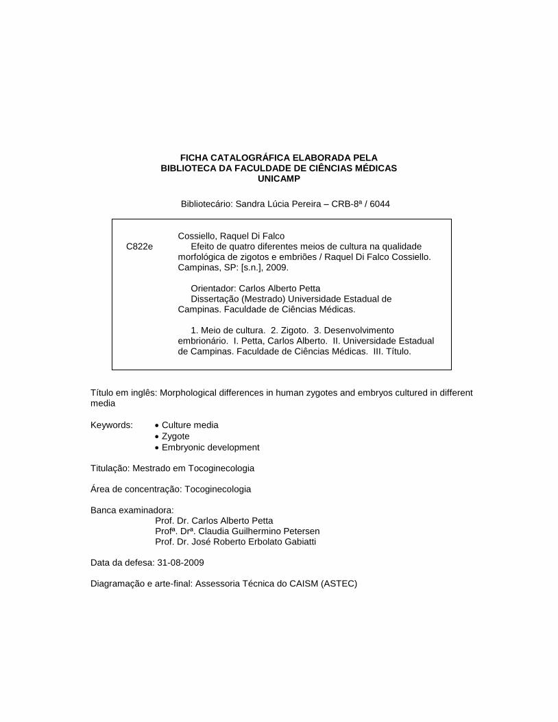
FICHA CATALOGRÁFICA ELABORADA PELA
BIBLIOTECA DA FACULDADE DE CIÊNCIAS MÉDICAS UNICAMP
Bibliotecário: Sandra Lúcia Pereira – CRB-8ª / 6044
Título em inglês: Morphological differences in human zygotes and embryos cultured in different media
Keywords: Culture media
Zygote
Embryonic development Titulação: Mestrado em Tocoginecologia Área de concentração: Tocoginecologia Banca examinadora:
Prof. Dr. Carlos Alberto Petta Profª. Drª. Claudia Guilhermino Petersen Prof. Dr. José Roberto Erbolato Gabiatti
Data da defesa: 31-08-2009
Diagramação e arte-final: Assessoria Técnica do CAISM (ASTEC)
Cossiello, Raquel Di Falco C822e Efeito de quatro diferentes meios de cultura na qualidade
morfológica de zigotos e embriões / Raquel Di Falco Cossiello. Campinas, SP: [s.n.], 2009.
Orientador: Carlos Alberto Petta Dissertação (Mestrado) Universidade Estadual de
Campinas. Faculdade de Ciências Médicas. 1. Meio de cultura. 2. Zigoto. 3. Desenvolvimento
embrionário. I. Petta, Carlos Alberto. II. Universidade Estadual de Campinas. Faculdade de Ciências Médicas. III. Título.

iv
Dedico este trabalho...
Aos meus pais,
por acreditarem sempre em todo meu trabalho...
À minha linda avó,
pela presença e infinito apoio durante todos os anos de convivência...
Ao meu avô,
pelo exemplo de vida...
Ao meu irmão,
por toda experiência compartilhada.

v
Agradecimentos
Ao Prof. Dr. Carlos Alberto Petta pela ideia inicial, paciência, dedicação e essenciais
ensinamentos ao longo desta jornada.
Ao Dr. Alexandros Aggelis pela paciência, carinho e dedicação indispensável.
Ao Dr. Daniel Faúndes por permitir o desenvolvimento do estudo.
À equipe do Centro de Reprodução Humana de Campinas pelo carinho e disponibilidade na
realização deste projeto.
À equipe da Huntington – Medicina Reprodutiva, pelo apoio e compreensão na fase
final do estudo, em especial ao José Roberto Alegretti, e ao André Rocha pela
disponibilidade em estar presente na correção do artigo.
À Sirlei, pela amizade e infinita paciência na avaliação estatística.
À Adriana, CEMICAMP, pela dedicação e auxílio na busca pelos artigos científicos.
A Deus, por tornar todos os ensinamentos e aprendizados da vida em fases superáveis e
executáveis.

vi
“Cada um de nós compõe a sua história.
Cada ser carrega em si o dom de ser capaz e ser feliz...”
Almir Sater

vii
Sumário
Resumo ........................................................................................................................................ viii
Summary ........................................................................................................................................ x
1. Introdução ............................................................................................................................... 12
1.1. Meio de cultura ................................................................................................................ 12
1.2. Classificação dos pronúcleos .......................................................................................... 18
1.3. Qualidade embrionária .................................................................................................... 20
2. Objetivos ................................................................................................................................. 23
2.1. Objetivo geral .................................................................................................................. 23
2.2. Objetivos específicos....................................................................................................... 23
3. Publicação ............................................................................................................................... 24
4. Conclusões.............................................................................................................................. 51
5. Referências Bibliográficas ....................................................................................................... 52
6. Anexos .................................................................................................................................... 57
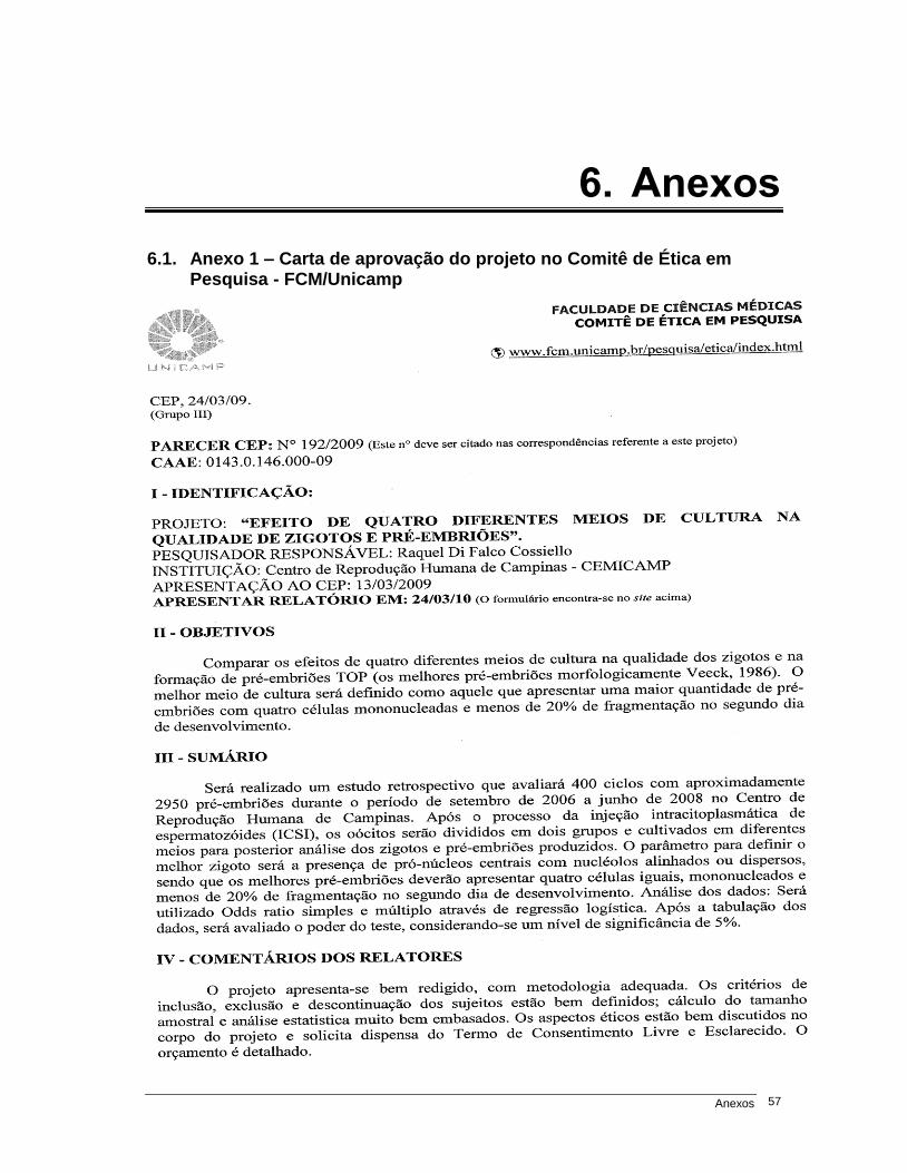
6.1. Anexo 1 – Carta de aprovação do projeto no Comitê de Ética em Pesquisa - FCM/Unicamp .................................................................................................................. 57
Resumo viii
Resumo
Objetivo: comparar os efeitos de quatro diferentes meios de cultura na morfologia
dos zigotos e embriões. Materiais e métodos: estudo retrospectivo conduzido
no Centro de Reprodução Humana de Campinas, em que 2.289 embriões de 319
ciclos de ICSI foram avaliados de setembro de 2006 a setembro de 2008. O
protocolo longo foi usado para estimulação ovariana em todos os casos. Todos
os oócitos foram cultivados em dois meios diferentes. O meio HTF (Irvine Scientific)
foi usado como meio-padrão, enquanto que os meios Universal IVF Medium
(Medicult), Global (LifeGlobal) e IVF-30 (Vitrolife) foram usados como secundários. A
separação dos oócitos em meios diferentes foi realizada alternadamente após
ICSI. A presença e a posição de pronúcleos e Nuclear Precusror Bodies (NPBs)
foram checadas 18 a 20 horas após ICSI. Baseado na classificação descrita por
Gianaroli et al., os zigotos foram identificados como: (A1) pronúcleos justapostos
e centralizados com NPBs grandes e alinhados; (A2) pronúcleos justapostos e
centralizados com NPBs grandes e dispersos. Os embriões foram avaliados 44 a 46
horas após ICSI, de acordo com o número de blastômeros, porcentagem de
fragmentação e multinucleação. Os embriões considerados top apresentaram
quatro blastômeros regulares, fragmentação menor que 20% do volume embrionário
e blastômeros não multinucleados. Para a análise dos dados foram utilizados
Resumo ix
Z-test, odds ratio simples e múltiplo através de regressão logística com seu
respectivo intervalo de confiança a 95%. Resultados: quando a classificação
dos zigotos foi analisada, o meio IVF-30 mostrou maior porcentagem (55,2%)
de zigotos A1+A2, em relação ao HTF, Global e Universal IVF Medium (49,1%,
44,7% e 44,2%, respectivamente). A porcentagem de embriões top foi
significativamente maior no meio Global (40,4%) comparado com HTF (21,1%),
IVF-30 (25,0%) e Universal IVF Medium (11,1%). No segundo dia de
desenvolvimento, Medicult produziu mais embriões com três células em relação
aos outros meios que produziram mais embriões com quatro células.
Conclusão: Houve diferenças significativas entre os quatro meios de cultura
sobre a morfologia dos zigotos e a morfologia embrionária. IVF-30 (Vitrolife) resultou
em maior número de zigotos com pronúcleos centralizados e nucléolos justapostos
e dispersos. Global (LifeGlobal) sustentou maior formação de embriões top no
dia 2 e maiores taxas de clivagem em relação aos demais meios.
Palavras-chave: meio de cultura, zigoto, desenvolvimento embrionário.

Summary x
Summary
Objective: compare the effects of four different culture media on the quality of
zygotes and embryos. Methods: This retrospective study, performed at the Center
for Human Reproduction of Campinas-Brazil analyzed 2289 embryos were
assessed from September 2006 to September 2008. Long protocol was used for
ovarian stimulation in all cases. The oocytes of each patient were cultivated in
two different culture media. The medium HTF - Irvine was set as the default for
all cycles and IVF Medium - Medicult, GGG 20 - Global and IVF 30 - Vitrolife
defined as secondary media. The sibling oocytes were divided in the two culture
media after ICSI. The confirmation of fertilization and classification as described
by Gianaroli were evaluated 18-20 hours after ICSI. On the second day (day 2)
of development, the embryos were evaluated according the number of cells,
percentage of fragmentation and number of nuclei. On day 2, the embryos that
had four cells with less than 20% of fragmentation and were mononucleated
embryos were classified as Top. Z-test and Odds ratios were used for statistical
analysis. Results: IVF-30 showed a higher percentage (55.2) of zygotes A1 +
A2 when compared to HTF, Global and Universal IVF Medium media (49.1%;
44.7%; 44.2% respectively) The percentage of Top embryos was significantly
higher in Global medium (40.4%) compared to HTF (21.1%), IVF-30 (25.0%)
and Universal IVF medium (11.1%). On day 2 Universal IVF Medium produced

Summary xi
more embryos with three blastomeres when compared to other media that
produced more embryos with four blastomeres. Conclusions: The use of IVF-
30 medium resulted in a higher number of zygotes with centralized pronuclei
with juxtaposed or scattered nucleoli. Meanwhile, Global medium produced a
greater number of morphologically good embryos (TOP) and higher cleavage
rate on the second day of development.
Keywords: culture media, zygote, embryonic development.
Introdução 12
1. Introdução
As técnicas de reprodução assistida utilizadas no tratamento da infertilidade
conjugal vêm apresentando grande desenvolvimento tecnológico nos últimos
anos. O sucesso durante a realização dos procedimentos de fertilização in vitro
(FIV) depende do aprimoramento das técnicas de alta complexidade e das
condições de cultivo oferecidas ao embrião em desenvolvimento (1).
Um dos pilares que possibilitou a evolução da área da reprodução assistida
se deve ao conhecimento das necessidades fisiológicas do embrião humano,
desde o estágio de zigoto até o estágio mais avançado de desenvolvimento pré-
implantacional, o blastocisto. Aliada à essa informação, a busca pela composição
das secreções tubárias e uterinas tornou-se importante para a criação de meios de
cultura existentes atualmente (2).
1.1. Meio de cultura
O processo de fertilização in vivo ocorre na região da ampola do oviduto,
onde a partir da união do oócito com espermatozoide, a origem do zigoto torna-
se possível. Após essa fusão, o zigoto, em seu primeiro dia de desenvolvimento,
Introdução 13
“caminha” pela tuba, a fim de encontrar o útero como ambiente final para o
processo de implantação. Durante esse processo, que possui aproximadamente
cinco a seis dias de duração, o zigoto passa a ser chamado de embrião, por
apresentar características morfológicas e necessidades fisiológicas distintas em
relação aos estágios iniciais de desenvolvimento.
Inicialmente, os meios para cultura embrionária tinham a glicose como
substrato de energia. Somente após a observação do meio in natura foi possível
a fabricação comercial de meios de cultura com diferentes fontes energéticas
para os diferentes estágios de desenvolvimento.
Uma das primeiras descrições da composição da tuba uterina foi feita em
um estudo em que foram coletados fluidos da ampola do oviduto e do útero de
mulheres em diferentes estágios do ciclo menstrual. Os resultados mostraram a
presença de lactato, glicose e piruvato como fontes de energia, além de
diferenças na concentração desses três componentes na tuba e no ambiente
uterino (3). Enquanto a concentração de lactato e piruvato é maior na região da
ampola comparada à região do útero durante a fase lútea, a concentração de
glicose aumenta durante a fase lútea no ambiente uterino (4;5;6).
Da mesma maneira, foi possível observar que durante o desenvolvimento
embrionário ocorrem mudanças dinâmicas em seu metabolismo e, que, além de
alterações morfológicas, as necessidades fisiológicas dos embriões são diferentes
em cada estágio de desenvolvimento. No estágio de zigoto, o embrião está
inicialmente quiescente, caracterizado pelo baixo metabolismo e atividade
Introdução 14
biossintética (7). Durante essa fase, é predominante a utilização de ácido
carboxílico, piruvato e lactato como substratos energéticos, aliados a aminoácidos
específicos, como o aspartato (8).
Durante as fases iniciais de desenvolvimento, o metabolismo embrionário é
completamente dependente do genoma materno para a geração de ATP e,
portanto, o consumo de glicose é restrito a baixos níveis nessa fase (3).
Posteriormente, Leese et al (9) mostraram que a necessidade de glicose
aumenta nos estágios a partir de oito células em relação ao consumo de lactato
e piruvato, assim como a atividade metabólica e o consumo de oxigênio.
Os diferentes componentes dos meios de cultura são essenciais para o
desenvolvimento do embrião in vitro, oferecendo cada um deles o suporte para
a cultura embrionária, sendo os aminoácidos e macromoléculas considerados
mais importantes para estágios mais avançados (10).
Apesar da diversidade dos meios disponíveis para o cultivo embrionário, os
carboidratos, sais minerais, proteínas, vitaminas, antibióticos, EDTA, aminoácidos e
água são componentes para o desenvolvimento in vitro, exercendo, cada um
deles, sua função na manutenção da cultura do embrião no laboratório.
Os carboidratos, como o piruvato de sódio e lactato de cálcio, são fontes
de energia essenciais no início do desenvolvimento embrionário, e a glicose
como fonte energética indispensável para os estágios mais avançados de
desenvolvimento embrionário. Os sais minerais, como o cálcio, são importantes
para a compactação celular, enquanto o sódio atua no controle da osmolaridade.
Introdução 15
A albumina é a proteína mais comumente utilizada para absorção de toxinas e
regulação da osmolaridade. As vitaminas são importantes por atuar como
antioxidantes. Os antibióticos, como gentamicina, penicilina ou estreptomicina
fornecem proteção ao embrião durante o período de cultivo. Além desses
componetes, o EDTA, caracterizado por ser um agente quelante de metais
como cálcio e ferro, atua na diminuição do estresse oxidativo, sendo importante
nos estágios iniciais de desenvolvimento (11). Além disso, os aminoácidos
presentes no meio de cultura atuam na proliferação e diferenciação celular,
principalmente nos estágios mais avançados de desenvolvimento dos embriões.
A utilização de diferentes cultivos embrionários difere nos laboratórios,
podendo variar desde cultivos curtos até o segundo dia, a cultivos prolongados até o
quinto dia de desenvolvimento (12). Dependendo do sistema de cultivo empregado,
um meio de cultura diferente pode ser utilizado. Atualmente, os meios para o cultivo
embrionário são classificados como simples, complexos ou sequenciais.
Consideram-se meios simples aqueles que suportam o desenvolvimento
embrionário até o terceiro dia, caracterizado principalmente pela ausência de
aminoácidos e presença ou ausência de EDTA em sua composição. Por outro
lado, meios complexos, que apresentam aminoácidos e ausência de EDTA,
permitem o desenvolvimento in vitro até o quinto dia (3). Ambos são considerados
monofásicos, por não necessitarem de outros meios durante todos os dias de
cultivo. Além desses meios, existem os sequenciais, que são destinados a cultivos
prolongados. Na realidade, um meio sequencial é composto por dois meios de

Introdução 16
cultura com diferentes composições, um para cada fase do desenvolvimento
embrionário (13).
Contudo, para cada tipo de cultivo pode ser empregado um meio de cultura
diferente; em cultivos curtos, a utilização de meios simples é geralmente empregada,
enquanto que para culturas prolongadas usualmente são empregados meios
complexos ou sequenciais. Sendo assim, os meios de cultura disponíveis no
mercado variam sua composição de acordo com a finalidade de cultivo.
Independentemente do sistema de cultivo ou meio de cultura utilizados,
estudos recentes demonstraram que as taxas de gestação não apresentam
aumento com a transferência de blastocistos no dia 5, principalmente porque muitos
ciclos acabam sendo cancelados por não apresentarem embrião viável (14).
As variações nas composições dos meios parecem não diferir quanto à
habilidade de promover o suporte para o desenvolvimento dos embriões. Porém,
ainda existe a preocupação em tornar os sistemas de cultivos existentes em
cultivos mais complexos, com a finalidade de oferecer o devido suporte ao
embrião nas diferentes etapas do desenvolvimento (15). Isso tem levantado
alguns questionamentos relacionados à formulação e composição dos meios,
bem como a dificuldade de definir a função e importância exata de cada
componente para o desenvolvimento embrionário (12;16).
Diante da variedade de meios, a escolha do protocolo de cultivo deve
considerar que o sistema empregado nessa etapa necessita proporcionar condições
adequadas para as primeiras clivagens, ativação do genoma embrionário e
Introdução 17
consequente desenvolvimento embrionário. Além disso, esses sistemas de
cultivo permitem a manutenção da fisiologia celular, a regulação da expressão
gênica e o controle do desenvolvimento embrionário (17).
Atualmente, os meios de cultura podem ser adquiridos comercialmente
ou produzidos em laboratórios de pesquisa. A maioria dos meios adquiridos já
se encontra pronto para utilização e tem a vantagem de possuir o controle de
qualidade e a segurança necessária para a aplicação das culturas em embriões
humanos (18).
Os meios comerciais largamente utilizados no cultivo embrionário são
representados no Brasil principalmente pelos laboratórios Irvine, Medicult,
LifeGlobal e Vitrolife. Cada laboratório apresenta diferentes meios de cultura
que são empregados em sistemas de cultivos distintos, desde cultivos simples a
cultivos com meios sequenciais. O principal meio representado pela empresa
LifeGlobal, chamado Global, se diferencia por ser um meio complexo, que permite o
desenvolvimento embrionário desde o estágio de zigoto até blastocisto. A
utilização dos meios da Irvine, Medicult e Vitrolife está baseada no cultivo
simples até o terceiro dia de desenvolvimento ou no cultivo com meios
sequenciais, caracterizado pela existência de meios com diferentes componentes
para cada estágio de desenvolvimento embrionário.
Diante da grande diversidade de meios para o cultivo embrionário
disponíveis no mercado, a divergência quanto ao meio de cultura a ser empregado
Introdução 18
durante o cultivo in vitro, constitui um dos grandes desafios práticos dos laboratórios
de reprodução assistida.
Paralelamente ao protocolo escolhido para a cultura do embrião, a
estimulação ovariana e os demais procedimentos que ocorrem no laboratório da
embriologia são elementos de grande importância que influenciam na eficácia
do método de fertilização in vitro.
Atualmente, os parâmetros empregados para avaliar a qualidade embrionária
são embasados na análise morfológica dos embriões desde o estágio de zigoto até
estágios mais avançados de desenvolvimento (19). A produção de embriões com
boa qualidade morfológica pode ser, em parte, atribuída à eficácia do meio de
cultura, podendo ser um excelente critério para avaliação dos meios utilizados.
1.2. Classificação dos pronúcleos
Após a fusão do espermatozoide com o oócito, ocorre a ativação oocitária,
evienciada pela retomada da meiose e extrusão do segundo corpúsculo polar.
A partir dessa fase, forma-se então o pronúcleo masculino, oriundo da
descondensação do DNA espermático, seguido pela formação do pronúcleo
feminino localizado próximo ao corpúsculo polar. Os dois pronúcleos, inicialmente
com tamanhos distintos e compostos por nuclear precursor bodies (NPBs)
movem-se no interior do oócito durante o processo de fertilização. Sendo assim, a
aparência e a localização dos pronúcleos no interior do oócito e a migração dos
NPBs que compõem os pronúcleos (PN) podem ser observadas e avaliadas em

Introdução 19
diferentes momentos até aproximadamente 25 horas após a ICSI (20). Estudos
prévios mostraram a importância da utilização de escores embasados na morfologia
dos PN e NPBs diante do desenvolvimento embrionário e potencial de implantação
(21;22;23). Esses estudos sugeriram que a disposição dos pronúcleos pode variar
dependendo da constituição cromossômica dos zigotos (24).
Através de um estudo baseado nesses princípios, Gianaroli et al., 2003
propuseram uma classificação de acordo com as configurações das morfologias
pronuclear e nucleolar (20). Cinco padrões de pronúcleos foram estabelecidos:
(A) justapostos e centralizados, (B) justapostos e periféricos, (C) centralizados e
separados, (D) diferentes tamanhos, (E) fragmentados. A morfologia nucleolar
foi avaliada de acordo com a posição e o número dos NPBs: (1) grandes e
alinhados, (2) grandes e dispersos, (3) grandes, alinhados em somente um dos
pronúcleos, (4) pequenos e espalhados (Figura 1).
Figura 1. Diagrama mostrando as configurações pronuclear e nucleolar de Gianaroli et al., 2003 (20).
Introdução 20
Devido à baixa frequência dos formatos B, C,‟ D e E foram avaliados
somente os embriões derivados de zigotos com pronúcleo A, observando que o
padrões A1 e A2 apresentaram maior probabilidade de resultar em embriões
euploides (42% e 40% respectivamente) (20).
1.3. Qualidade embrionária
Uma das etapas mais intrigantes dos laboratórios de fertilização in vitro é
a escolha dos embriões com maior potencial de implantação, com a finalidade
de reduzir o número de embriões a serem transferidos e mesmo assim manter
taxas adequadas de gravidez, mas com menor risco de gestação múltipla.
Historicamente, a avaliação morfológica foi o método preliminar usado
para a avaliação do embrião (25;26) e, apesar de suas limitações reconhecidas
(27), o método ainda permanece como a abordagem mais usual para seleção.
Sendo assim, diversas características embrionárias, além da avaliação dos
pronúcleos nos zigotos, têm sido consideradas para decidir a escolha do embrião
para transferência.
A primeira delas está baseada na velocidade de clivagem dos embriões,
que, quando muito rápida ou muito lenta, representa um impacto negativo na
taxa de implantação (28;29). Outra avaliação é a divisão celular, que quando
inicialmente apresenta-se irregular, produz embrião com dois blastômeros de
tamanhos desiguais ou fragmentados, o que acaba por apresentar um efeito
negativo na capacidade de desenvolvimento embrionário (30;31). A presença
Introdução 21
de blastômeros multinucleados, que ocorre entre 11,9% a 33,6%, geralmente
está associada à formação de embriões com alterações cromossômicas e à
diminuição na capacidade de formação de blastocistos (32;33), embora existam
relatos de bebês saudáveis nascidos a partir desses embriões (34). Além disso,
a porcentagem de fragmentação é relatada como tendo um valor importante na
previsão do potencial de implantação do embrião (35).
Observa-se uma grande proporção de embriões humanos pré-implantacionais
que sofrem desvios de desenvolvimento in vitro, deixando de seguir a rotina de
desenvolvimento normal e frequentemente exibindo altos níveis de fragmentação
e assimetria dos blastômeros.
A fragmentação é um achado comum que limita o desenvolvimento in
vitro do embrião. Uma das influências negativas da fragmentação na qualidade
do embrião é que esses fragmentos impediriam o perfeito contato espacial entre
os blastômeros, levando à compactação, cavitação e formação de blastocistos
anormais. Esses fragmentos poderiam provocar uma degeneração secundária
devido à presença de detritos celulares próximos aos blastômeros sadios (36).
Os fragmentos celulares contêm, além do citoplasma, organelas citoplasmáticas
intactas, e ocasionalmente, parte de cromatina condensada. Esses fragmentos
podem ser uma consequência de morte celular programada em estádios precoces
de desenvolvimento embrionário, com efeitos prejudiciais que podem levar à
morte celular em alguns blastômeros, ou, em alguns casos, à morte ou parada
do desenvolvimento embrionário (37).
Introdução 22
A fragmentação - eliminação de parte dos blastômeros pelo embrião -
parece ser um esforço deste em restaurar ou manter a viabilidade quando certas
anomalias afetam algum blastômero em particular. Essa eliminação precoce de
células afetadas previne uma contribuição alterada dessas células para a massa
celular interna ou o trofoectoderma do blastocisto (38).
Diante dos dados conflitantes relacionados ao dia ideal para a transferência
embrionária, o cultivo pode variar do segundo ao quinto dia de desenvolvimento
(39). Sendo assim, o embrião será avaliado quanto à sua morfologia nos diferentes
dias de desenvolvimento.
Rotineiramente, após a realização das avaliações morfológicas em diferentes
estágios do desenvolvimento, os embriões que apresentarem pronúcleos centrais
com NPB alinhados ou dispersos, tempo de clivagem compatível ao dia de
desenvolvimento, blastômeros regulares e menos de 20% de fragmentação,
serão eleitos para transferência uterina (19).
Quando a morfologia embrionária é avaliada no segundo dia de
desenvolvimento, os embriões de melhor qualidade morfológica, classificados
como top, devem apresentar quatro blastômeros regulares, fragmentação menor
que 20% do volume embrionário e blastômeros não multinucleados (19).
Sendo a morfologia um dos critérios usualmente estabelecidos para a
classificação dos zigotos e embriões, a preocupação em oferecer condições
ideais de cultivo embrionário com a finalidade de obter embriões top no dia da
transferência, instiga a embriologia a buscar melhores protocolos de cultivo.

Objetivos 23
2. Objetivos
2.1. Objetivo geral
Comparar os efeitos de quatro meios de cultura diferentes na qualidade dos
zigotos e na formação de embriões de boa qualidade morfológica (embriões top).
2.2. Objetivos específicos
Comparar a qualidade dos zigotos através da classificação dos pronúcleos
estabelecida por Gianaroli et al., 2003 (20) nos quatro meios de
cultura diferentes.
Comparar o número de embriões de boa qualidade morfológica
(embriões top) obtidos nos quatro meios de cultura diferentes.
Definir qual meio de cultura produz melhores embriões de acordo com
a classificação morfológica.

Publicação 24
3. Publicação
Thank you for submitting your article to Reproductive BioMedicine Online entitled:
Morphological differences in human zygotes and embryos cultured in different media.
Abstract:
Objective: To compare the effects of four culture media on the quality of zygotes and
embryos. Methods: Retrospective study analyzing 2,289 embryos cultivated in two
different culture media: HTF, the default medium, with Universal IVF, LGGG-20 and IVF-
30 as the secondary media. The sibling oocytes were divided between the two culture
media following ICSI. Fertilization and classification were confirmed 18-20 hours after
ICSI. On day 2, embryos were evaluated according to the number of cells, percentage of
fragmentation and number of nuclei. Z-test and odds ratios were used in the statistical
analysis. Results: There was a higher percentage (55.2%) of class A1+A2 zygotes with
IVF-30 compared to HTF, Global or Universal IVF media (49.1%, 44.7% and 44.2%,
respectively). The percentage of Top embryos was significantly higher with Global
(40.4%) compared to HTF (21.1%), IVF-30 (25.0%) or Universal IVF media (11.1%). On
day 2, Universal IVF produced more embryos with three blastomeres compared to the
other media, which produced more embryos with four blastomeres. Conclusions: Use of
IVF-30 resulted in more zygotes with centralized pronuclei and juxtaposed or
scattered nucleoli. Nonetheless, Global medium produced more Top embryos and
higher cleavage rate on the second day of development.
Keywords:
culture media, pronuclei, embryo quality
Authors:
MSc Raquel Cossiello (Universidade Estadual de Campinas)
embriologist, develope protocol, data collection. interpretation of results,
preparation of manuscript
PhD Alexandros Aggelis (Centro de Reprodução Humana de Campinas)
embriologist, interpretation of results, preparation of manuscript
MD, PhD Daniel Faúndes (Centro de Reprodução Humana de Campinas)
Reproductive endocrinologist, interpretation of results, preparation of manuscript
MD, PhD Carlos Alberto Petta (Universidade Estadual de Campinas, Centro de
Reprodução Humana de Campinas)

Publicação 25
Reproductive endocrinologist, developed protocol, data collection, interpretation of
results, preparation of manuscript
Submitted by: [email protected]
Your submission will now be processed and you will be contacted by RBMOnline in
due course.
Yours sincerely,
RBMOnline
--
Duck End Farm, Dry Drayton, Cambridge CB23 8DB, UK
http://www.rbmonline.com
Tel. +44 (0) 1954 781812; Fax. +44 (0) 1954 781816
Publicação 26
Morphological differences in human zygotes and embryos cultured in different media
Raquel Di Falco Cossiello1, Alexandros Aggelis
1, Daniel Faúndes
1, Carlos A. Petta
1,2
1 Campinas Center of Human Reproduction, Campinas, São Paulo, Brazil.
2 Department of Obstetrics and Gynecology, University of Campinas, Campinas, São
Paulo, Brazil.
Correspondence to:
Carlos A. Petta MD
Rua Eduardo Lane 380
13073-002 Campinas SP, Brazil.
E-mail: [email protected]

Publicação 27
Abstract
Objective: To compare the effects of four culture media on the quality of zygotes and
embryos. Methods: Retrospective study analyzing 2,289 embryos cultivated in two
different culture media: HTF, the default medium, with Universal IVF, LGGG-20 and
IVF-30 as the secondary media. The sibling oocytes were divided between the two
culture media following ICSI. Fertilization and classification were confirmed 18-20
hours after ICSI. On day 2, embryos were evaluated according to the number of cells,
percentage of fragmentation and number of nuclei. Z-test and odds ratios were used in
the statistical analysis. Results: There was a higher percentage (55.2%) of class A1+A2
zygotes with IVF-30 compared to HTF, Global or Universal IVF media (49.1%, 44.7%
and 44.2%, respectively). The percentage of Top embryos was significantly higher with
Global (40.4%) compared to HTF (21.1%), IVF-30 (25.0%) or Universal IVF media
(11.1%). On day 2, Universal IVF produced more embryos with three blastomeres
compared to the other media, which produced more embryos with four blastomeres.
Conclusions: Use of IVF-30 resulted in more zygotes with centralized pronuclei and
juxtaposed or scattered nucleoli. Nonetheless, Global medium produced more Top
embryos and higher cleavage rate on the second day of development.
Keywords: culture media; pronuclear stage; embryonic development.
Publicação 28
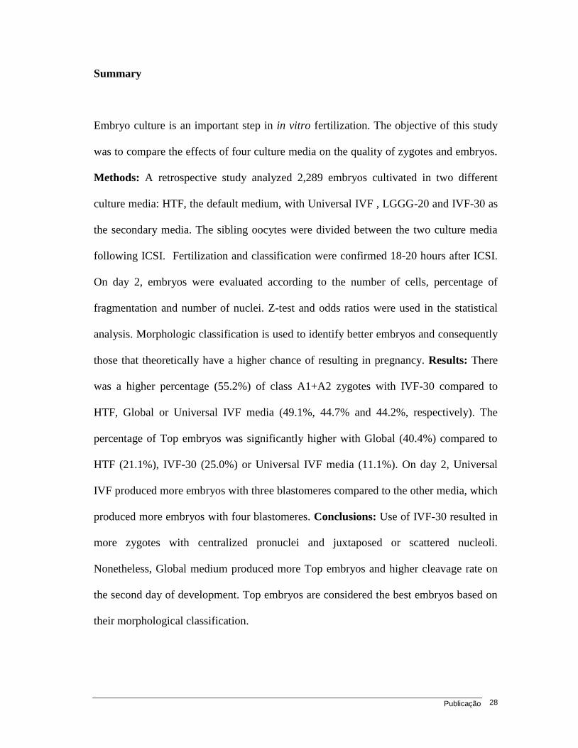
Summary
Embryo culture is an important step in in vitro fertilization. The objective of this study
was to compare the effects of four culture media on the quality of zygotes and embryos.
Methods: A retrospective study analyzed 2,289 embryos cultivated in two different
culture media: HTF, the default medium, with Universal IVF , LGGG-20 and IVF-30 as
the secondary media. The sibling oocytes were divided between the two culture media
following ICSI. Fertilization and classification were confirmed 18-20 hours after ICSI.
On day 2, embryos were evaluated according to the number of cells, percentage of
fragmentation and number of nuclei. Z-test and odds ratios were used in the statistical
analysis. Morphologic classification is used to identify better embryos and consequently
those that theoretically have a higher chance of resulting in pregnancy. Results: There
was a higher percentage (55.2%) of class A1+A2 zygotes with IVF-30 compared to
HTF, Global or Universal IVF media (49.1%, 44.7% and 44.2%, respectively). The
percentage of Top embryos was significantly higher with Global (40.4%) compared to
HTF (21.1%), IVF-30 (25.0%) or Universal IVF media (11.1%). On day 2, Universal
IVF produced more embryos with three blastomeres compared to the other media, which
produced more embryos with four blastomeres. Conclusions: Use of IVF-30 resulted in
more zygotes with centralized pronuclei and juxtaposed or scattered nucleoli.
Nonetheless, Global medium produced more Top embryos and higher cleavage rate on
the second day of development. Top embryos are considered the best embryos based on
their morphological classification.
Publicação 29
Introduction
Since the advent of in vitro fertilization (IVF), a large number of different media
formulations and culture systems have been proposed for the development of human
zygotes and embryos. Culture media range from simple balanced saline solutions to
more complex media. Simple culture media were used in assisted reproductive
technology (ART) until the mid-1990s. These media, consisting basically of saline
solutions, glucose, pyruvate, lactate and carbohydrates supplemented with protein, were
effective for culture up to the third day of development. However, the introduction of
more complex media (Fissore et al., 1989) containing amino acids presented new
alternatives for in vitro culture.
The capacity to simulate natural conditions in the laboratory became fundamental,
since the culture of embryos in suboptimal or distressing conditions compels the embryo
to undergo physiological adaptations. The features presented by embryos thus affected
include delayed cell division (Bowman, 1970), poorer incorporation of amino acids
(Jung et al., 1987), a reduction in oxidative metabolism and an increase in lactate
production (Gardner, 1990; Gott et al., 1990; Leese et al, 1993). Such factors may lead
to consequences that include low pregnancy rates, significantly greater fetal loss (Lane,
2007) and low birth weight (Pool, 2005).
Several mechanisms are involved in the development process from oocyte
retrieval until embryo transfer to the uterus, with modifications occurring at each stage;
however, the embryologist is able to observe and analyze only a few of these steps, one
of these being embryo development.

Publicação 30
In IVF laboratories, the parameters generally used to evaluate embryo quality
and consequently the effectiveness of the culture media are based on analysis of the
morphologic criteria between days 1 and 5 following fertilization.
For analyses on day 1, embryo pronuclear (PN) classification and morphology
represent important variables for assessing different culture media. Evaluation of the
pronucleated zygote is based on analysis of the morphology and position of the
pronuclei. Alterations in the appearance or numbers of nuclear precursor bodies (NPBs)
(Tesarik, 1989) may cause abnormalities in embryo division and lead to uneven cleavage
or fragmentation. Based on these considerations, the morphology of the pronuclei and
NPBs has been proposed as a scoring system and a correlation has been suggested
between the different patterns observed, embryo development and implantation potential
(Montag, 2001).
On day 2 and 3 of development, embryos are graded according to blastomere
morphology, cleavage stage and fragmentation, and there appears to be a clear
relationship between these parameters and implantation and pregnancy rates after IVF.
This procedure can also be used to select the best embryos and test the effectiveness of
culture media (Ziebe et al., 1997).
The aim of this study was to assess and compare the effects of four different
culture media on human zygote and embryo morphology on day 2 of culture.

Publicação 31
Material and methods
This retrospective study was conducted at the Human Reproduction Center,
Campinas, Brazil, where 2,289 embryos were assessed between September 2006 and
September 2008 in 319 ICSI cycles. The exclusion criteria consisted of embryos
obtained from spermatozoa that were recovered using testicular or epididymal
aspiration. The study was approved by the Institutional Review Board of the University
of Campinas (UNICAMP). All patients were informed with respect to the laboratory
procedures to which they would be submitted and signed an informed consent form prior
to the initiation of treatment.
Ovulation induction and oocyte recovery
Ovarian stimulation was initiated following pituitary blockade by leuprolide
acetate (Lupron, TAP Pharmaceutical, Abbott Laboratories, Chicago, IL, USA), 0.15
mL/day, beginning on the 21st or 23
rd day of the menstrual cycle and maintained until
the beginning of the following cycle. On the 3rd
day of bleeding, leuprolide was reduced
to 0.05 mL/day and ovarian stimulation was initiated with recombinant follicle-
stimulating hormone (rFSH) (Gonal F, Serono, São Paulo, SP, Brazil) at a dose of 150-
225 IU/day, or highly purified human menopausal gonadotropin (hphMG) (Menopur,
Ferring, São Paulo, SP, Brazil) at a dose of 75-225 IU/day. Follicular development was
monitored by transvaginal ultrasonography and the dose of medication was adjusted
according to ovarian response.
Publicação 32
When at least 2 follicles 18 mm in diameter were observed, a single dose of
recombinant human chorionic gonadotropin (r-hCG, 250 g, Ovidrel, Serono, São
Paulo, SP, Brazil) was given. Oocytes were aspirated 34-36 hours after injection of r-
hCG. The oocytes were separated from the follicular fluid under a stereoscope and
washed in a modified HTF medium with Hepes (Irvine Scientific, Santa Ana, CA,
USA), supplemented with 10% synthetic serum substitute (SSS - Irvine Scientific). All
the oocytes were placed in HTF medium (Irvine Scientific) supplemented with 10% SSS
and incubated at 37oC in 5% CO2.
The cumulus oophorus cells were removed by exposing them to hyaluronidase
(H-4272, Sigma, Saint Louis, Missouri) at a concentration of 80 IU/mL in modified HTF
medium with Hepes, supplemented with 10% SSS, for 30 seconds. The cells were
removed with the use of fine hand-drawn glass pipettes. After denudation, the oocytes
were washed with HTF culture medium supplemented with 10% SSS and incubated
until ICSI was performed (Palermo et al., 1992).
Sperm collection
Semen samples were collected by masturbation immediately after oocyte pickup,
evaluated in a Makler counting chamber (Sefi-Medical Instruments, Haifa, Israel) and
then processed through a discontinuous density gradient (Isolate Lower / Isolate Upper,
Irvine Scientific) in accordance with the manufacturer's protocol.

Publicação 33
In vitro fertilization and embryo culture
All sibling oocytes were cultured in two different media: Human Tubal Fluid
(HTF [Irvine Scientific, Santa Ana, CA, USA]), which was used as the default medium
for all the patients, and one of the secondary media (Universal IVF Medium [Medicult,
Denmark]; LGGG Global [LifeGlobal, Guelph, ON, Canada]; or IVF-30 [Vitrolife, CO,
USA]). The secondary media used in each case were determined by local availability.
Following ICSI, the oocytes were separated into the different media. Injected
oocytes were transferred alternately to HTF medium or to one of the secondary media
until all presumed zygotes were divided between two types of media. Embryo culture
conditions were 37oC with 5% CO2 to maintain pH within an optimal range (7.20-7.40)
according to the manufacturers‟ instructions.
Assessment of zygotes
An inverted microscope equipped with Hoffman modulation optics was used to
check for the presence of pronuclei and the position of NPBs 18-20 hours after ICSI.
Based on the classification established by Gianaroli et al. (2003), the zygotes were found
to be juxtaposed with large centralized pronuclei and aligned NPBs, classified as “A1”,
or juxtaposed with centralized pronuclei and large, scattered NPBs, classified as “A2”
(Figure 1). After fertilization was verified and pronuclei classified, the zygotes were
transferred to fresh drops of culture media.
Publicação 34
Embryo Quality
The individually cultured embryos were evaluated 44–46 hours after ICSI on the
basis of the number of blastomeres, percentage of fragmentation and presence of
multinucleated blastomeres. Embryos were considered to be of top quality (Top) when
they had four regular blastomeres, fragmentation in less than 20% of the volume of the
embryo and no multinucleated blastomeres (Guerif et al., 2007).
Statistical analysis
Quantitative data were assessed for normality and homoscedasticity using the
Kolmogorov-Smirnov and F-tests, respectively. The number of mature oocytes (MII)
was compared using analysis of variance (ANOVA) followed by Tukey‟s test for means.
The number of blastomeres of the embryos on day 2 was evaluated using the Kruskal-
Wallis test followed by a group by group comparison using the Mann-Whitney test.
Frequencies of zygotes with A1 and A2 pronuclear morphology and embryos with top
quality morphology in each group were compared using the Z-test for two proportions.
Parametric data were presented as means ± standard deviations (SD) and non-parametric
data were presented as medians and ranges. The Bonferroni correction was used to
determine statistically significant differences and significance was defined at p<0.008.
Publicação 35
Results
There was no difference in the age of the woman between the cycles in which the
embryos were cultured in different media nor was there any difference in the number of
mature oocytes (MII) between cycles (Table I).
A total of 2,289 embryos were included in the analysis: 1,170 were cultivated in
HTF medium; 199 in Global medium; 260 in Universal IVF medium; and 660 in IVF-30
medium. When both the pronuclei and the position of the NPBs were taken into
consideration, the percentage of A1 + A2 zygotes was found to be higher with the IVF-
30 medium. The frequency of the A1 + A2 pattern was 55.2% in zygotes cultured in
IVF-30 medium compared to 49.1%, 44.7% and 44.2% in HTF, Global and Universal
IVF media, respectively. The results found with IVF-30 were significantly higher when
compared with those of the other media; however, the differences between the Global,
HTF and Universal IVF media were not statistically significant (Table II).
When the number of blastomeres was compared after two days of culture, the
HTF, Global and IVF-30 media produced more 4-cell embryos, while Universal IVF
medium produced more 3-cell embryos. In embryos cultivated in Global medium, the
variation in blastomere numbers on day 2 was less than that found with embryos
cultivated in HTF or IVF-30 media, in which there were a significant number of
embryos with fewer than four cells (Figure 2).
The presence of Top embryos was evaluated in all the different media between
44 and 46 hours after ICSI. The percentage of embryos with four regular blastomeres,
fragmentation in less than 20% of the volume of the embryo and no multinucleated

Publicação 36
blastomeres was significantly higher in Global medium (40.4%) compared with HTF
(21.1%), IVF-30 (25.0%) or Universal IVF media (11.1%) (Table III).
Discussion
In this study, use of the IVF-30 medium resulted in good morphology in a
substantial number of zygotes on day one; however, embryo morphology was found to
be better when the embryos were cultivated in Global medium. When the pronuclear and
nucleolar parameters were analyzed, Universal IVF medium was found to be associated
with fewer zygotes with good (A1+A2) PN morphology (44.2%) compared to the other
culture media, and with fewer top quality day-2 embryos. This finding confirms that
these parameters should be analyzed together and not individually.
Our results are in agreement with previous studies showing that pronuclear
morphology was not directly associated with embryo quality on day 2 (James et al.,
2006). Although the IVF-30 culture medium produced a high number of zygotes with
good (A1+A2) PN morphology (55.2%), the number of good quality embryos produced
with this medium was low. On the other hand, Global medium produced the highest
percentage (40.4%) of good quality embryos on day 2; however, this medium produced
fewer good PN on day one. Universal IVF medium produced the smallest percentage of
good quality embryos compared to the other culture media used in this study;
nevertheless, there were no differences in the percentage of good PN obtained with this
medium compared to the HTF and Global media. With respect to the formation of
zygotes with A1+A2 pronuclear patterns, no difference was found between the HTF
medium and the Global and Universal IVF media, and when the production of good

Publicação 37
quality embryos was compared, results achieved with the HTF medium were found to be
similar to those obtained with the IVF-30 medium.
The correlation between zygote and embryo morphology has already been
demonstrated (Scott et al., 1998; Tesarik et al., 1999; Ludwig et al., 2000; Scott et al.,
2000); however, this correlation was not linear, suggesting that some degree of
predictability is lost when considering either zygote or embryo morphology alone
(Rijnders et al., 1998). One study compared the predictive value of different
morphological parameters using a score system for human zygotes and preimplantation
embryos and showed that zygote and embryo morphology were independent variables
predictive of IVF outcome, and that both may be used as criteria to select the best
embryos for transfer (De Placido et al., 2002).
Nowadays, most IVF laboratories select the best embryo according to embryo
morphology parameters on the day of transfer. However, there are additional criteria that may
be used to select the best embryo such as evaluation of the pronuclear morphology, NPB
size and distribution, cytoplasmic halo and early cleavage (Rienzi et al., 2005; Scott et
al., 2007). It is important to determine the pronuclear and NPB morphology of zygotes
on day one and embryo quality on day two of development, since this represents a non-
invasive instrument for performing a quick, precise and reproducible analysis that does not
interfere with the daily clinical work of the IVF laboratory (Gianaroli et al., 2003).
The development of good-quality embryos was shown to be related to the
pronuclear pattern and was better in the case of zygotes with centralized, juxtaposed
pronuclei (pattern A, 96%) compared with the other patterns. Moreover, embryo
development was similar in the four patterns of zygotes, whereas chromosomal
condition varied depending on nuclear morphology (Gianaroli et al., 2003).

Publicação 38
Differences found in the composition of the culture media may explain the
variation in Top embryo formation. Global medium was the only monoculture medium
used in this study. The formulation used in Global medium is capable of supporting
embryo development from pronuclear formation to blastocyst stage. The addition of
amino acids is the principal difference between Global medium and the other simple
media used in the present study. This feature may explain the higher formation of top
quality embryos found with this medium. Previous studies have confirmed that the
addition of amino acids to culture media enhances embryo development, while when
mouse zygotes were exposed to medium without amino acids their subsequent potential
for development was impaired (Gardner et al., 1996; Lane et al., 2007).
The addition of EDTA is another characteristic that differentiates some media from
others used in the present study. The beneficial effects of EDTA are confined to the
early cleavage stage embryo (Gardner et al., 1996). Of all the media used in this study,
only IVF-30 contained EDTA in its composition and this may explain the difference in
the location and distribution of the pronuclei and NPBs. It has already been shown that
human zygote pronuclei are formed in different locations in the oocyte, both migrating
towards the center through time. During this movement, cytoplasmic rotation and
cytoskeleton structures are probably involved, possibly coordinated by calcium levels.
The presence of EDTA in the culture medium may affect the efficiency of the migration
of the pronuclei and result in different pronuclear patterns in the zygotes.
In the present study, Global medium produced more top quality embryos (40.4%)
with four regular blastomeres, fragmentation of less than 20% of the embryo and no
multinucleated blastomeres. This was followed by IVF-30 (25.0%), HTF (21.2%) and

Publicação 39
Universal IVF (11.1%) media according to the morphological analysis of the embryo on
the second day of development.
Several studies comparing different commercial culture media for IVF have been
published in the literature. Xella et al. (2009) showed that IMS1 (Medicult) culture
medium appears to improve the performance of embryonic growth and development and
increase pregnancy rates when compared to Universal IVF medium (Medicult). One
study compared four media and found better embryo quality with Global medium rather
than with Cleavage medium, HTF or G1.2 (Aoki et al., 2005). However, no differences
were found in day-2 embryo score or pregnancy, implantation or abortion rates between
the P-1 and IVF-50 media (Mauri et al., 2001) or in the implantation rates achieved with
the different media (Zollner et al., 2004).
This comparison between different media for the culture of sibling oocytes has
proven to constitute a valuable tool for the assessment of embryo quality. Traditional
studies comparing culture media have evaluated different patients; however, in the present
study the same patient received transferred embryos that had been cultivated in different
media. Nevertheless, the assessment of implantation and pregnancy rates is problematic
due to a high frequency of “mixed” transfers, resulting in a reduced number of cases in
which the embryos transferred were grown exclusively in one type of medium.
Zygote score is a good predictor of embryo quality, but should be used in
combination with the evaluation of embryo morphology on the day of transfer. A1 + A2
zygote morphology is associated with chromosomally normal embryos; however, not all
chromosomally normal embryos are morphologically normal as well. Top embryos
cannot have blastomeres with more than one nucleus or a high percentage of
Publicação 40
fragmentation, and these characteristics are not directly dependent on pronuclear
morphology (Gianaroli et al., 2007).
One of the principal strengths of the present study is that it is entirely
independent, having received no financial support from any of the manufacturers of the
different types of media. The experience described here of a single clinic in which two
types of culture medium were used concurrently in each cycle may help other services
decide which media should be used and also stimulate them to implement this strategy to
prevent the unlikely but feasible occurrence that an alteration in one of the media could
compromise the whole treatment. Nevertheless, not being able to establish a correlation
between the different culture media and pregnancy rate constitutes a limitation to this
study. This limitation, however, was unavoidable, since each patient received embryos
from different culture media.
In conclusion, the present data show significant differences between the four
culture media with respect to pronuclear, NPB and embryo morphology. The use of IVF-
30 medium resulted in a higher number of zygotes with centralized pronuclei and
juxtaposed or scattered nucleoli, while Global culture medium resulted in the formation
of more top quality embryos on day 2 of development. In addition, cleavage rates were
lower with the HTF, Universal IVF and IVF-30 media compared to Global medium.
More studies with larger sample sizes are needed to permit evaluation of implantation
and pregnancy rates following the culture of sibling embryos in different media.

Publicação 41
References
Aoki VW, Wilcox AL, Peterson CM et al. 2005 Comparison of four media types during
3-day human IVF embryo culture. Reprod Biomed Online 10, 600-606.
Bowman P, McLaren A 1970 Cleavage rate of mouse embryos in vivo and in vitro. J
Embryol Exp Morphol 24, 203-207.
De Placido G, Wilding M, Strina I et al. 2002 High outcome predictability after IVF
using a combined score for zygote and embryo morphology and growth rate. Hum
Reprod 17, 2402-2409.
Fissore RA, Jackson KV, Kiessling AA 1989 Mouse zygote development in culture
medium without protein in the presence ethylenediaminetetraacetic acid. Biol Reprod
41, 835-841.
Gardner DK, Lane M 1996 Alleviation of the „2-cell-block‟ and development to the
blastocyst of CF1 mouse embryos: role of amino acids, EDTA and physical parameters.
Hum Reprod 11, 2703-2712.
Gardner DK, Leese HJ 1990 Concentrations of nutrients in mouse oviduct fluid and their
effects on embryo development and metabolism in vitro. J Reprod Fertil 88, 361-368.
Gianaroli L, Magli MC, Ferraretti AP et al. 2003 Pronuclear morphology and chromosomal
abnormalities as scoring criteria for embryo selection. Fertil Steril 80, 341-349.
Gianaroli L, Magli MC, Ferraretti AP et al. 2007 Oocyte euploidy, pronuclear zygote
morphology and chromosomal complement. Hum Reprod 22, 241-249.

Publicação 42
Gott AL, Hardy K, Winston RM et al. 1990 The nutrition and environment of early
human embryos. Proc Nutr Sci 49, Abstract 2A.
Guerif F, Le Gouge A, Giraudeau B et al. 2007 Limited value of morphological
assessment at days 1 and 2 to predict blastocyst development potential: A prospective
study based on 4042 embryos. Hum Reprod 22, 1973–1981.
James AN, Hennessy S, Reggio B et al. 2006 The limited importance of pronuclear
scoring of human zygotes. Hum Reprod 21, 1599-1604.
Jung T, Fischer B, Beier HM 1987 Quantitative aspects of protein synthesis in non-
cultured and cultured rabbit blastocysts. Hum Reprod 2, 23-27.
Lane M, Gardner DK 2007 Embryo culture medium: which is the best? Best Pract Res
Clin Obstet Gynaecol 21, 83-100.
Leese HJ, Conaghan J, Martin KL et al. 1993 Early human embryo metabolism.
Bioessays 15, 259-264.
Ludwig M, Schöpper B, Al-Hasani S et al. 2000 Clinical use of a pronuclear stage score
following intracytoplasmic sperm injection: impact on pregnancy rates under the
conditions of the German embryo protection law. Hum Reprod 15, 325–329.
Mauri AL, Petersen CG, Baruffi RL et al. A prospective, randomized comparison of
two commercial media for ICSI and embryo culture. J Assist Reprod Genet 18, 378-381.
Montag M, van der Ven H; German Pronuclear Morphology Study Group 2001 Evaluation
of pronuclear morphology as the only selection criterion for further embryo culture and
transfer: results of a prospective multicenter study. Hum Reprod 16, 2384–2389.

Publicação 43
Palermo G, Joris H, Devroey P et al. 1992 Pregnancies after intracytoplasmic injection of
single spermatozoon into an oocyte. Lancet 340, 17-18.
Pool TB 2005 An update on embryo culture for human assisted reproductive
technology: media, performance and safety. Semin Reprod Med 23, 309-318.
Rienzi L, Ubaldi F, Iacobelli M et al. 2005 Significance of morphological attributes of
the early embryo. Reprod Biomed Online 10, 669-681.
Rijnders PM, Jansen CA 1998 The predictive value of day 3 embryo morphology regarding
blastocyst formation, pregnancy, and implantation rate after day 5 transfer following in-
vitro fertilization or intracytoplasmic sperm injection. Hum Reprod 13, 2869-2873.
Scott L, Alvero R, Leondires M et al. 2000 The morphology of human pronuclear embryos is
positively related to blastocyst development and implantation. Hum Reprod 15, 2394-2403.
Scott L, Finn A, O'Leary T et al. 2007 Morphologic parameters of early cleavage-stage
embryos that correlate with fetal development and delivery: prospective and applied data
for increased pregnancy rates. Hum Reprod 22, 230-240.
Scott LA, Smith S 1998 The successful use of pronuclear embryo transfers the day
following oocyte retrieval. Hum Reprod 13, 1003-1013.
Tesarik J, Greco E 1999 The probability of abnormal preimplantation development can
be predicted by a single static observation on pronuclear stage morphology. Hum Reprod
14, 1318-1323.
Publicação 44
Tesarik J, Kopecny V 1989 Development of human male pronucleus: ultrastructure and
timing. Gamete Res 24, 135–149.
Xella S, Marsella T, Tagliasacchi D et al. 2009 Embryo quality and implantation rate in
two different culture media: ISM1 versus Universal IVF Medium. Fertil Steril Correct proof.
Ziebe S, Petersen K, Lindenberg S et al. 1997 Embryo morphology or cleavage stage:
how to select the best embryos for transfer after in-vitro fertilization. Hum Reprod 12,
1545-1549.
Zollner KP, Zollner U, Schneider M et al. 2004 Comparison of two media for sequential
culture after IVF and ICSI shows no differences in pregnancy rates: a randomized trial.
Med Sci Monit 10, CR1-7.

Publicação 45
Figure Legends:
Figure 1. Diagram showing the configurations that identify pronuclear morphology and
NPB morphology. Adapted from Gianaroli et al, 2003 (with permission).
Figure 2. Boxplot showing the variation in the number of blastomeres between the HTF,
Global, IVF-30 and Universal IVF media.

Publicação 46
Figure 1
Pronuclear morphology
Nucleolar morphology
Centralized/ Juxtaposed
Large-size scattered
Large-size aligned
A
A1 A2

Publicação 47
Global Irvine Medicult Vitrol ife
2
4
6
8
bla
sto
mere
s o
n d
ay 2
*Global vs HTF, IVF-30 and Universal IVF = p <0.008
HTF vs IV• FV-30 = p NS
Figure 2

Publicação 48
Table I. General characteristics of ICSI cycles
HTF Global IVF-30 Universal IVF
Age (mean±SD) 33.6±4.6 32.7±4.4 33.3±4.6 34.6±4.6
Number of MII median (range) 12(1-32) 11(2-17) 12(2-32) 16(3-27)
Number of blastomeres on Day 2 4(1-8A) 4(1-8
B) 4(1-8
A) 3(1-8
C)
Different letters indicate p<0.008

Publicação 49
Table II. Comparison of frequencies of pronúcleolar and NPBs among HTF, Global, IVF-30 and Universal IVF media
Total HTF Global Universal IVF IVF-30
n % n % OR(CI 95%) n % OR(CI 95%) n % OR(CI 95%)
1170 199 260 660
Control vs HTF p<0.2486 p<0.1514 p<0.0007
A1+A2 575 49.1 89 44.7 0.84(0.62–1.13) 115 44.2 0.82(0.63-1.08) 364 55.2 1.27(1.05-1.54)
Control vs Global p<0.9161 p<0.0098
A1+A2 115 44.2 0.98(0.68-1.42) 364 55.2 1.52(1.11-2.09)
Control vs Universal IVF p<0.0028
A1+A2 364 55.2 1.55(1.16-2.07)
OR= Odds Ratio;
CI= Confidence interval
p value<0.008

Publicação 50
Table III. Comparison of frequencies of good quality (TOP) embryos among HTF, Global, IVF-30 and Universal IVF media
Total HTF Global Universal IVF IVF-30
n % n % OR(CI 95%) n % OR(CI 95%) n % OR(CI 95%)
1170 199 260 660
Control vs HTF p<0.00001 p<0.00001 p<0.071
Top embryos 249 21.3 80 40.2 2.49(1.81–3.41) 29 11.2 0.46(0.31-0.70) 165 25.0 1.23(0.98-1.54)
Control vs Global p<0.00001 p<0.00001
Top embryos 29 11.2 0.19(0.12-0.30) 165 25.0 0.50(0.36-0.69)
Control vs Universal IVF p<0.00001
Top embryos 165 25.0 2.66(1.74-4.06)

Conclusões 51
4. Conclusões
– A formação de zigotos com pronúcleos centrais e nucléolos alinhados
(A1+A2) foi maior no meio IVF-30 quando comparado aos meios HTF, Global
e Universal IVF.
– Quanto à qualidade embrionária no segundo dia de desenvolvimento, o meio
Global obteve a maior porcentagem de embriões de boa qualidade (top).
– O uso do meio Global esteve relacionado à maior formação de embriões de
boa qualidade no dia 2 e a maiores taxas de clivagem em relação aos
demais meios.
Referências Bibliográficas 52
5. Referências Bibliográficas
1. Balaban B, Yakin K, Urman B, Isiklar A, Tesarik J. Pronuclear morphology
predicts embryo development and chromosome constitution Reproductive
BioMedicine Online. 2004;8(6):695-700.
2. Gardner DK, Lane M. Culture and selection of viable blastocysts: a feasible
proposition for human IVF? Hum Reprod Update. 1997;3(4):367-82.
3. Gardner DK, Lane M, Calderon I, Leeton J. Environment of the preimplantation
human embryo in vivo: metabolite analysis of oviduct and uterine fluids and
metabolism of cumulus cells. Fertil Steril. 1996; 65(2):349-53.
4. Casslen B, Nilsson B. Human uterine fluid, examined in undiluted samples
for osmolarity and the concentrations of inorganic ions, albumin, glucose
and urea. Am J Obstet Gynecol.1984;150(7):877-81.
5. Bavister BD. Glucose and culture of human embryos. Fertil Steril. 1999;
72(2):223-4.
6. Martin KL. Nutritional and metabolic requirements of early cleavage stage
embryos and blastocysts. Hum Fertil. 2000;3(4):247-54.
Referências Bibliográficas 53
7. Gardner DK, Lane M, Batt P. Uptake and metabolism of pyruvate and
glucose by individual sheep preattachment embryos developed in vivo. Mol
Reprod Dev. 1993;36(3):313-9.
8. Lane M, Gardner DK. Lactate regulates pyruvate uptake and metabolism in
the preimplantation mouse embryo. Biol Reprod. 2000;62(1):16-22.
9. Leese HJ, Conaghan J, Martin KL, Hardy K. Early human embryo
metabolism. BioEssays. 1993;15(4):259-64.Review.
10. Lane M, Gardner DK. Embryo culture medium: which is the best? Best Pract
Res Clin Obstet Gynaecol. 2007;21(1):83-100.
11. Gardner DK, Lane M. Alleviation of the „2-cell-block‟ and development to the
blastocyst of CFI mouse embryos: role of amino acids, EDTA and physical
parameters. Hum Reprod. 1996;11 (12):2703-12.
12. Aoki VW. Comparison of four media types during 3-day human IVF embryo
culture. Reproductive BioMedicine Online. 2005,10(5):600-6.
13. Gardner DK, Lane M. Towards a single embryo tranfer. Reprod BioMed
Online. 2003;6(4):470-81.
14. Blake DA, Farquhar CM, Johnson N, Proctor M. Cleavage stage versus
blastocyst stage embryo transfer in assisted conception. Cochrane
Database Syst Rev. 2007;19(4):CD00218.Review.
15. Balaban B, Urman B. Embryo culture as a diagnostic tool. Reproductive
BioMedicine Online. 2003;7(6):671-82.Review.
16. Xella S, Marsella T, Tagliasacchi D, Giulini S, Marca A, Tirelli A et al.Embryo
quality and implantation rate in two different culture media: ISM1 versus
Universal IVF Medium. Fertil Steril. 2009; [Epub ahead of print].

Referências Bibliográficas 54
17. Van Langendonckt A, Demylle D, Wyns C, Nisolle M, Donnez J.
Comparasion of G1.2/G2.2 and Sidney IVF cleavage/blastocyst sequential
media for the culture of human embryos: a prospective, randomized,
comparative study. Fertil Steril. 2001;76(5):1023-31.
18. Mauri AL, Petersen CG, Baruffi RL, Franco JG Jr. A prospective,
randomized comparison of two commercial media for ICSI and embryo
culture. J Assist Reprod Genet. 2001; 18(7):378-81.
19. Rienzi L, Ubaldi F, Iacobelli M, Romano S, Minasi MG, Ferrero S et al.
Significance of morphological attributes of the early embryo. Reprod Biomed
Online. 2005;10(5):669-81.Review.
20. Gianaroli L, Magli MC, Ferraretti AP, Fortini D, Grieco N. Pronuclear
morphology and chromosomal abnormalities as scoring criteria for embryo
selection. Fertil Steril. 2003;80(2):341-9.
21. Tesarik J, Junca AM, Hazout A, Abriot FX, Nathan C, Cohen-Bacrie P et al.
Embryos with high implantation potencial after intracytoplasmic sperm
injection can be recognized by a simple, noninvasive examination of PN
morphology. Hum Reprod. 2000;15(6):1396-9.
22. Scott L, Alvero R, Leondires M, Miller B. The morphology of human
pronuclear embryos is positively related to blastocyst development and
implantation. Hum Reprod. 2000;15(11):2394-403.
23. Montag M, van der Ven H. Evaluation of pronuclear morphology as the only
selection criterion for further embryo culture and transfer: results of a
prospective multicenter study. Hum Reprod. 2001;16(11): 2384–9.
24. Gianaroli L, Magli MC, Ferraretti AP, Lappi M, Borghi E, Ermini B. Oocyte
euploidy, pronuclear zygote morphology and embryo development. Hum
Reprod. 2007;22(1):241-9.

Referências Bibliográficas 55
25. Edwads RG, Purdy JM, Steptoe PC, Walters DE. The growth of human
preimplantation embryo in vitro. Am J Obstet Gynecol. 1981;141(4):408-16.
26. Cummins JM, Breen TM, Harrison KL, Shaw JM, Wilson LM, Henessey JF.
A formula for scoring human embryo growth rates in in vitro fertilization: its
value in predicting pregnancy and in comparasion with visula estimates of
embryo quality. In Vitro Fert Embryo Transf. 1986;3(5);284-95.
27. Guerif F, Le Gouge A, Giraudeau B, Poindron J, Bidault R, Gasnier O et al..
Limited value of morphological assessment at days 1 and 2 to predict
blastocyst development potential: a prospective study based on 4042
embryos. Hum Reprod. 2007;22(7):1973-81.
28. Steer CV, Mills CL, Tan SL, Campbell S, Edwards RG. The cumulative
embryo score: a predictive embryo scoring technique to select the optimal
number of embryos to transfer in an in vitro fertilization and embryo transfer
program. Hum Reprod. 1992;7(1):117-9.
29. Ziebe S, Petersen K, Lindenberg S, Andersen AG, Gabrielsen A, Andersen
AN. Embryo morphology or cleavage state: how to select the best embryos
for the transfer after in vitro fertilization. Hum Reprod. 1997l;12(7):1545-9.
30. Giorgetti C, Terriou P, Auquier P, Hans E, Spach JL, Salzmann J et al.
Embryo score to predict implantation after in vitro fertilization: based on 957
single embryo transfers. Hum Reprod. 1995;10(9):2427-31.
31. Hardarson T, Hanson C, Sjogren A, Lundin K. Human embryos with unevenly
sized blastomores have lower pregnancy and implantation rates: indications
for aneuploidy and multinucleation. Hum Rep. 2001;16(2):313-8.

Referências Bibliográficas 56
32. Pelinck MJ, De Vos M, Dekens M, Van der Elst J, De Sutter P, Dhont M.
Embryos cultured in vitro with multinucleated blastomeres have poor
implantation potencial in human in vitro fertilization and intracytoplasmic
sperm injection. Hum Reprod. 1998;13(4):960-3.
33. Alikani M, Calderon G, Tomkin G, Garrisi J, Kokot M, Cohen J. Cleavage
anormalies in early human embryos and survival after prolonged culture in
vitro. Hum Reprod. 2000;15(12):2634-43.
34. Balakier H, Cadesky K. The frequency and developmental capability of
human embryos containing multinucleated blastomeres. Hum Reprod. 1997;
12(4):800-4.
35. Alikani M, Cohen J, Tomkin G, Garrisi GJ, Mack C, Scott RT. Human
embryo fragmentation in vitro and its implications for pregnancy and
implantation. Hum Reprod. 1999;71(5):836-42.
36. Dozortsev D, Ermilov A, El-Mowafi DM, Diamond M. The impact of cellular
fragmentation induced experimentally at different stages of mouse
preimplantation development. Hum Reprod. 1998;13(5):1307-11.
37. Jurisicova A, Varmuza S, Casper RF. Programmed cell deth and human
embryo fragmentation. Mol Hum Reprod. 1996;2(2):93-8.
38. Munné S. Chromosome abnormalities and their relationship to morphology
and development of human embryos. Reprod Biomed Online. 2006;
12(2):234-53.
39. Papanikolaou EG, Kolibianakis EM, Tournaye H, Venetis CA, Fatemi H,
Tarlatzis B et al. Live birth rates after transfer of equal number of blastocysts
or cleavage-stage embryos in IVF. A systematic review and meta-analysis.
Hum Reprod. 2008;23(1):91-9.
Anexos 57
6. Anexos
6.1. Anexo 1 – Carta de aprovação do projeto no Comitê de Ética em Pesquisa - FCM/Unicamp
Anexos 58