DIVERSIDADE DE HEMOPARASITOS EM LAGARTOS DA …
132
UNIVERSIDADE FEDERAL DO AMAZONAS INSTITUTO NACIONAL DE PESQUISAS DA AMAZÔNIA PROGRAMA DE PÓS-GRADUAÇÃO EM ZOOLOGIA DIVERSIDADE DE HEMOPARASITOS EM LAGARTOS DA AMAZÔNIA CENTRAL AMANDA MARIA PICELLI Manaus, Amazonas Outubro, 2020
Transcript of DIVERSIDADE DE HEMOPARASITOS EM LAGARTOS DA …

UNIVERSIDADE FEDERAL DO AMAZONAS
INSTITUTO NACIONAL DE PESQUISAS DA AMAZOcircNIA
PROGRAMA DE POacuteS-GRADUACcedilAtildeO EM ZOOLOGIA
DIVERSIDADE DE HEMOPARASITOS EM LAGARTOS DA
AMAZOcircNIA CENTRAL
AMANDA MARIA PICELLI
Manaus Amazonas
Outubro 2020
AMANDA MARIA PICELLI
DIVERSIDADE DE HEMOPARASITOS EM LAGARTOS DA
AMAZOcircNIA CENTRAL
Tese apresentada ao Programa de Poacutes-
Graduaccedilatildeo em Zoologia da Universidade
Federal do AmazonasInstituto Nacional de
Pesquisas da Amazocircnia como parte dos
requisitos para obtenccedilatildeo do tiacutetulo de Doutora
em Zoologia
Orientador Igor Luiacutes Kaefer
Coorientador Felipe Arley Costa Pessoa
Coorientador Luacutecio Andreacute Viana Dias
Manaus Amazonas
Outubro 2020
Sinopse
Esta tese investigou a prevalecircncia e a diversidade de hemoparasitos em
lagartos da Amazocircnia Central Aleacutem disso traz informaccedilotildees sobre
distribuiccedilatildeo taxonomia e relaccedilotildees ecoloacutegico-evolutivas desses parasitos
e seus hospedeiros
Palavras-chave Biodiversidade herpetofauna morfologia parasitos de
sangue relaccedilotildees filogeneacuteticas taxonomia
Dedico esta tese aos meus amados pais
Aparecida e Joseacute Eduardo (In memoriam) e agrave
todas as mulheres cientistas cujas histoacuterias de
vida me inspiraram a seguir nessa jornada
Agradecimentos
Ao meu amado pai que agora mora entres as estrelas Sei que de alguma forma vocecirc
estaacute comigo e feliz pela conclusatildeo da minha tese Agrave minha famiacutelia principalmente minha matildee
pelo carinho apoio e compreensatildeo Tambeacutem devo agradecer ao meu querido Paulinho cuja
serenidade e amor foram essenciais para que eu conseguisse finalizar este trabalho
Aos meus queridos orientadores Prof Dr Igor Kaefer Dr Felipe Pessoa e Prof Dr
Luacutecio Viana que acreditaram no meu potencial como cientista Muito obrigada pela paciecircncia
pelos ensinamentos conselhos e confianccedila Cada um de vocecircs foi essencial agrave sua maneira ao
longo desse processo de aprendizagem
Sou muito grata a todos os amigos que de alguma forma estiveram presentes nessa
caminhada Aos amigos de longa data de Rio Claro Amanda Ronaldo Carol Pri Marina
Rebeca e Annelise Agrave famiacutelia manauara (ldquocasa trouxardquo) Tito Carol Well e Uacuteyra (Emerson)
sem duacutevida o tempo que moramos juntos foi muito importante e sempre vou sentir saudades
Agraves mulheres mais fortes e maravilhosas que o doutorado trouxe Giu Aline Val Sula e Gabi ndash
com vocecircs ao lado foi mais faacutecil e divertido Aos meus amigos e vizinhos (Rubana Camis
Marcatildeo Ju Renan Diogo Julia Pedro Cybelli e Itanna) da Vilinha do Chaves obrigada pelas
conversas cuidados com a Malu banhos de piscina e peixinhos assados
Agrave Universidade Federal do Amazonas (UFAM) que aleacutem de ter oferecido a
infraestrutura para realizaccedilatildeo desta tese foi o local onde me reconheci como cientista e
desenvolvi minhas melhores relaccedilotildees interpessoais ao longo desses quatro anos Tambeacutem quero
agradecer agrave coordenaccedilatildeo e equipe de funcionaacuterios da Fazenda Experimental da UFAM pelo
apoio aos trabalhos de campo
Sou grata muito ao Programa de Poacutes-Graduaccedilatildeo em Zoologia da UFAM e as pessoas
que se dedicam diariamente ao funcionamento do mesmo como nosso querido secretaacuterio Gil
(que facilita muito a vida dos alunos) aos docentes discentes membros do conselho e a
coordenaccedilatildeo (Prof Menin e Prof Fabriacutecio - que sempre tiveram paciecircncia e disponibilidade
para conversar) Nesse contexto preciso tambeacutem agradecer aos meus grandes amigos e
companheiros de poacutes Igor Joventino e Alexandre Almeida pelos cafeacutes cervejas conselhos e
risadas
Aos alunos do KaeferLab obrigada pela amizade por me ensinarem muito sobre
herpetologia e por despertarem em mim o ldquocrushrdquo pela Ecologia Lu obrigada por ser essa fada
sensata e sempre me ajudar durante os momentos de ansiedade Adriane minha primeira filha
acadecircmica natildeo sei o que teria feito sem sua organizaccedilatildeo e talento obrigada por todo seu
empenho nesse projeto
Agradeccedilo ao Instituto Leocircnidas e Maria Deane (ILMD-Fiocruz) pela infraestrutura
tanto laboratorial quanto de campo (base da Fiocruz na Agrovila Rio Pardo) suporte teacutecnico e
logiacutestico para as atividades de pesquisa Agrave Dra Yara (IOC-Fiocruz) que foi fonte de inspiraccedilatildeo
e admiraccedilatildeo nesse uacuteltimo ano de doutorado Agrave equipe do EDTA-ILMD (Laboratoacuterio de
Ecologia de Doenccedilas Transmissiacuteveis da Amazocircnia) Dra Claudia Rios Dra Keillen Dra
Alessandra Nava Eric Jordan Heliana Jeacutessica Maacuterio Rebeca Tuacutellio Emanuelle e demais
membros Obrigada pelas contribuiccedilotildees cientiacuteficas confraternizaccedilotildees e auxiacutelios em campo e no
laboratoacuterio
Ao Instituto Nacional de Pesquisas da Amazocircnia (INPA) pela infraestrutura e apoio a
esta pesquisa Ao Laboratoacuterio Temaacutetico de Microscopia Oacuteptica e Eletrocircnica (LTMO-INPA)
pela autorizaccedilatildeo do uso dos sistemas de imagens e ao querido Lucas gerente do laboratoacuterio
por toda colaboraccedilatildeo e amizade Agraves coordenadoras das coleccedilotildees zooloacutegicas de Herpetologia e
Mastozoologia Dra Fernanda Werneck e Dra Nazareth Silva respectivamente pelo apoio com
material de campo Agrave toda equipe do Projeto Dinacircmica Bioloacutegica dos Fragmentos Florestais
(PDBFF) pela autorizaccedilatildeo e auxiacutelio com as atividades na ARIE-PDBFF
Aos pesquisadores da Universidade de Satildeo Paulo (USP) Satildeo PauloSP Dra Marta
Teixeira Dr Bruno Fermino e Lyslaine Sato o conhecimento e a experiecircncia de vocecircs foram
imprescindiacuteveis para os resultados obtidos no terceiro capiacutetulo desta tese
Aos Dr Fernando Silveira e Dr Thiago Vasconcelos do Instituto Evandro Chagas (IEC)
AnanindeuaPA por permitir o acesso ao material reunido pelo Dr Ralph Lainson Muito
obrigada tambeacutem por toda a ajuda espaccedilo e equipamentos fornecidos para anaacutelise desse rico
material durante minha estadia no IEC
Agradeccedilo agravequeles que me auxiliaram em campo Ayra Joseacute Neto Alexandre Adriane
Mota Sebastiatildeo Eric Moca Rafael Luna Danilo Alan Emanuelle Wellyngton Gabi Giu
Gabriel Karina e Juruna Todos vocecircs foram fundamentais natildeo apenas para a coleta dos dados
mas por fazerem os dias de campo mais alegres
A presente tese foi realizada com o apoio da Coordenaccedilatildeo de Aperfeiccediloamento de
Pessoal de Niacutevel Superior (CAPES) ndash coacutedigo de financiamento 001 Tambeacutem agradeccedilo agrave essa
agecircncia e a Fundaccedilatildeo de Amparo agrave Pesquisa do Estado do Amazonas (FAPEAM) pela
concessatildeo da bolsa de doutorado
Pelo suporte financeiro destinado agrave execuccedilatildeo desta pesquisa agradeccedilo ao Conselho
Nacional de Desenvolvimento Cientiacutefico e Tecnoloacutegico (CNPq Universal 4615732014-8 e
4291322016-6) ao Programa de Excelecircncia em Pesquisa em Sauacutede Baacutesica e Aplicada
(PROEP FIOCRUZ FAPEAM 0012014) e ao Programa PDBFF de Auxiacutelio-Pesquisa Thomas
Lovejoy
Meus agradecimentos tambeacutem ao Centro Nacional de Pesquisa e Conservaccedilatildeo de
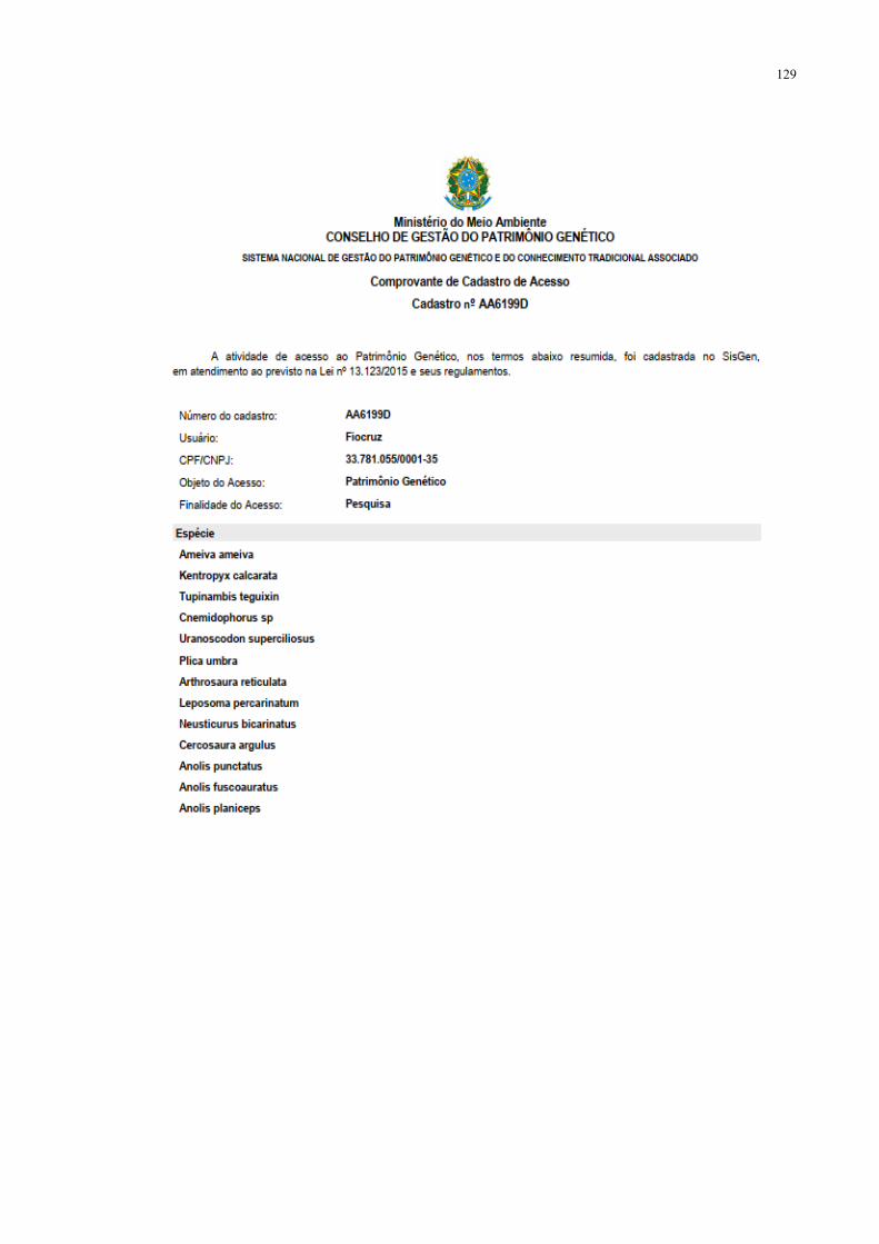
Reacutepteis e Anfiacutebios (RANICMBio) SisGen - Ministeacuterio do Meio Ambiente Comitecirc de Eacutetica
no Uso de Animais da UFAM pela concessatildeo das autorizaccedilotildees relativas agrave pesquisa
Muito obrigada ao Dr Ralph Lainson que infelizmente natildeo pude conhececirc-lo mas sua
vida de protozoologista na Amazocircnia foi fonte de grande inspiraccedilatildeo Sua memoacuteria permaneceraacute
viva em seu trabalho e atraveacutes daqueles que continuarem seu legado
Sem vocecircs esse sonho natildeo seria possiacutevel a todos muito obrigada
ldquoIrsquove always said to young Brazilian students what
a wonderful place theyrsquore in If you turn over a
stone yoursquoll find four new species underneath it
The Amazon region is a veritable mine of
parasitological information yet very very few
people were engaged in parasitological studies in
this regionrdquo
Ralph Lainson
(1927-2015)
viii
Resumo
Os parasitos satildeo reconhecidos pela sua grande capacidade de influenciar a evoluccedilatildeo e
ecologia de seus hospedeiros tanto ao niacutevel de indiviacuteduo quanto de comunidade Entretanto
no Brasil um dos paiacuteses com a maior biodiversidade do planeta estudos sobre parasitismo em
populaccedilotildees silvestres ainda satildeo relativamente escassos Na regiatildeo amazocircnica levantamentos
anteriores constataram uma rica fauna de parasitos de sangue em lagartos e deram indiacutecios sobre
um elevado potencial para descoberta de novas espeacutecies Nesse contexto a presente tese
investigou a ocorrecircncia de hemoparasitos em lagartos da Amazocircnia Central explorando ao
longo de trecircs capiacutetulos aspectos relacionados agrave diversidade taxonomia e suas relaccedilotildees
ecoloacutegico-evolutivas Para tanto foram obtidas amostras sanguiacuteneas de diversas espeacutecies de
lagartos capturadas em aacutereas de floresta de terra-firme localizadas proacuteximas aos muniacutecipios de
Manaus Presidente Figueiredo e Rio Preto da Eva no Estado do Amazonas Brasil No
primeiro capiacutetulo foram reunidos os resultados sobre a prevalecircncia e a riqueza de
hemoparasitos encontrados nessas localidades apresentando tambeacutem uma lista atualizada dos
estudos realizados no Brasil sobre hemoparasitos em lagartos O segundo capiacutetulo traz a
redescriccedilatildeo taxonocircmica usando dados morfoloacutegicos e moleculares de uma espeacutecie de
hemogregarina Hepatozoon ameivae detectada em lagartos Ameiva ameiva Para o terceiro
capiacutetulo foi levantada a hipoacutetese de que a ecologia do lagarto Uranoscodon superciliosus estaacute
moldando a diversidade dos tripanossomas que os parasitam o que pode ser evidenciado pelas
relaccedilotildees filogeneacuteticas de dois novos genoacutetipos de tripanossomas isolados nessa espeacutecie de
hospedeiro Por fim os resultados obtidos nessa tese ampliaram o conhecimento sobre a
diversidade e distribuiccedilatildeo dos hemoparasitos no Brasil aleacutem de terem gerado informaccedilotildees
ineacuteditas sobre o sistema parasito-hospedeiro formado pelos lagartos e seus hemoparasitos na
regiatildeo amazocircnica
Palavras-chave Biodiversidade Filogenia Floresta Amazocircnica Morfologia Parasitos de
Sangue Squamata
ix
Abstract
Parasites are recognized for their great ability to influence the evolution and ecology of
their hosts at the individual and community levels However in Brazil one of the countries
with the greatest biodiversity on the planet studies on parasitism in wild populations are still
relatively scarce In the Brazilian Amazonia previous surveys found a rich fauna of blood
parasites in lizards and gave indications of a high potential for the discovery of new species In
this context the present thesis investigated the occurrence of hemoparasites in lizards from
Central Amazonia exploring over three chapters aspects related to the diversity taxonomy and
their ecological-evolutionary relationships For this purpose blood samples were obtained from
different lizard species captured in areas of upland forests (terra-firme) located near the
municipalities of Manaus Presidente Figueiredo and Rio Preto da Eva in the State of
Amazonas Brazil In the first chapter results on the prevalence and richness of hemoparasites
found in these locations were gathered also presenting an updated list of studies carried out in
Brazil involving hemoparasites in lizards The second chapter presents the taxonomic
redescription using morphological and molecular data of a hemogregarine species
Hepatozoon ameivae detected in Ameiva ameiva lizards In the third chapter we raised the
hypothesis that the ecology of the lizard Uranoscodon superciliosus is shaping the diversity of
the trypanosomes that parasitize it which can be evidenced by the phylogenetic relationships
of the two new trypanosome genotypes isolated from this host species Finally the results
obtained in this thesis expanded the knowledge about the diversity and distribution of
hemoparasites in Brazil in addition to novel information about the host-parasite system formed
by lizards and their hemoparasites in the Amazonian region
Keywords Biodiversity Phylogeny Amazon rainforest Morphology Blood Parasites
Squamata
x
Sumaacuterio
Lista de Tabelas xi
Lista de Figuras xii
Introduccedilatildeo Geral 14
Capiacutetulo 1 Under the light high prevalence of haemoparasites in lizards (Reptilia Squamata)
from Central Amazonia revealed by microscopy 21
Capiacutetulo 2 Redescription of Hepatozoon ameivae (Carini and Rudolph 1912) from the lizard
Ameiva ameiva (Linnaeus 1758) 62
Capiacutetulo 3 Trypanosome phylogenetic relationships from the Amazonian Diving Lizard
indicate host ecology as a driver of parasite diversification 86
Consideraccedilotildees Finais 117
Referecircncias Bibliograacuteficas 120
Anexos 128
xi
Lista de Tabelas
Introduccedilatildeo Geral
Tabela 1 Diversidade de hemoparasitos do Filo Apicomplexa e de tripanosomatiacutedeos descritos
em reacutepteis ao redor do mundo e seus vetores 17
Capiacutetulo 1
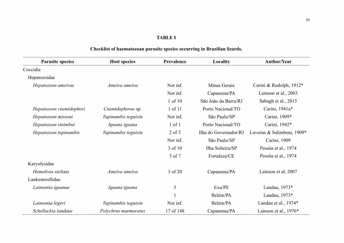
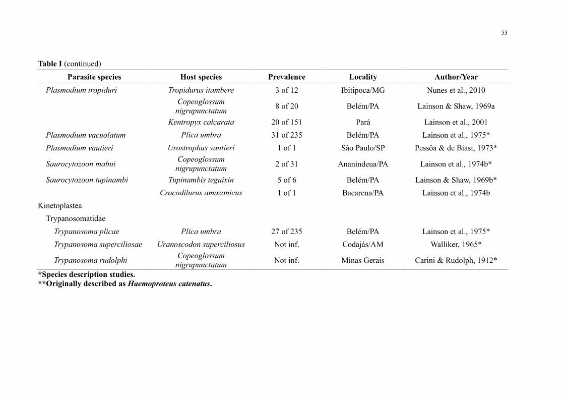
Table I Checklist of haematozoan parasite species occurring in Brazilian lizards 50
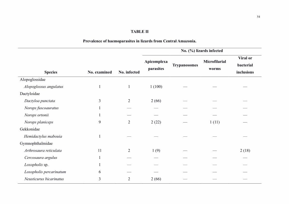
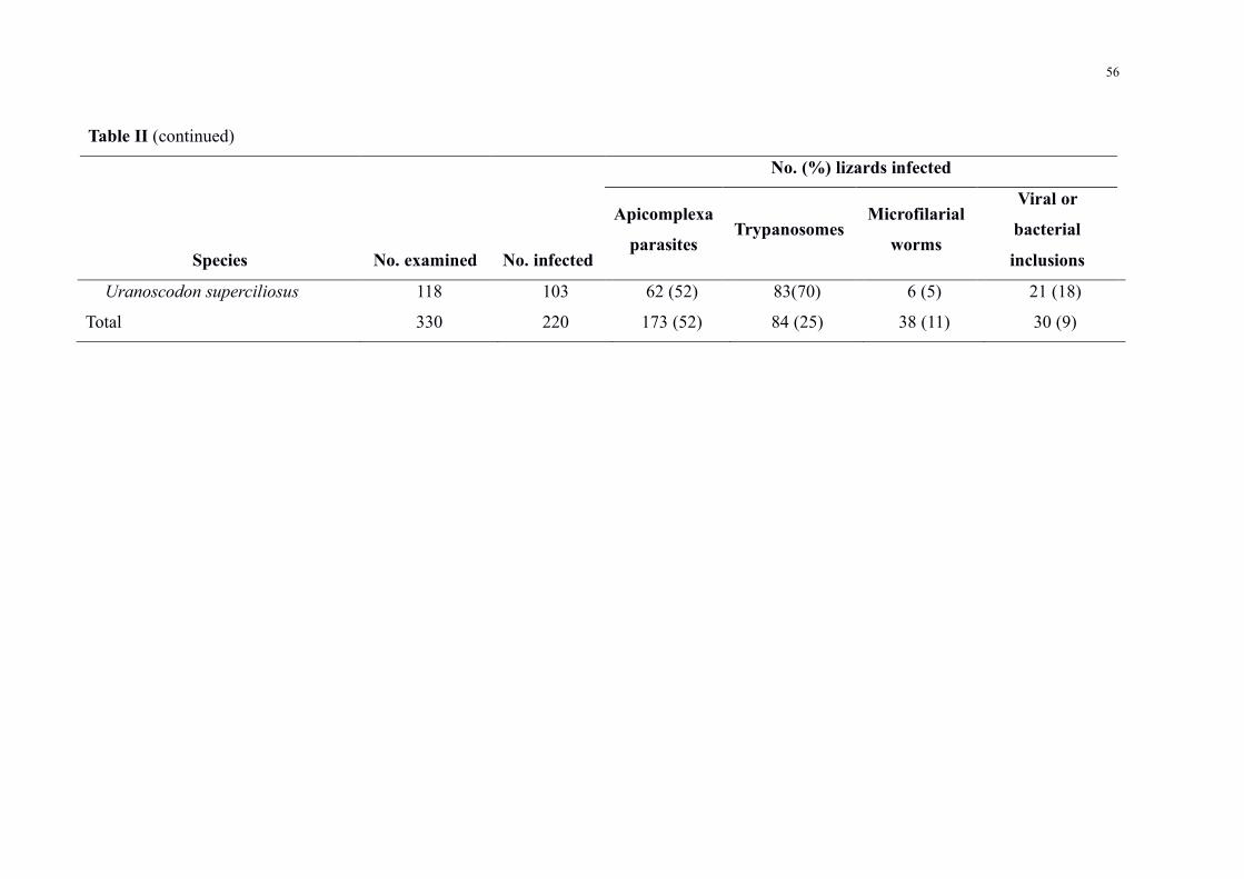
Table II Prevalence of haemoparasites in lizards from Central Amazonia 54
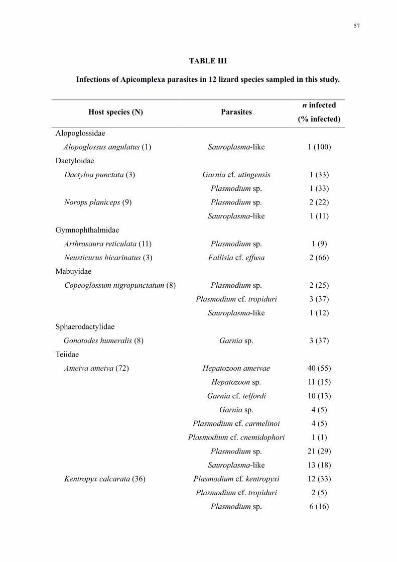
Table III Infections of Apicomplexa parasites in 12 lizard species sampled in this study 57
Capiacutetulo 2
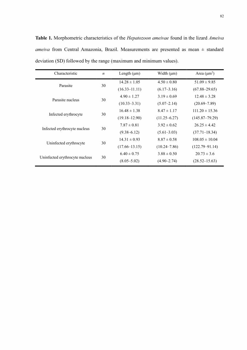
Table 1 Morphometric characteristics of the Hepatozoon ameivae found in the lizard Ameiva
ameiva from Central Amazonia Brazil Measurements are presented as mean plusmn standard
deviation (SD) followed by the range (maximum and minimum values) 82
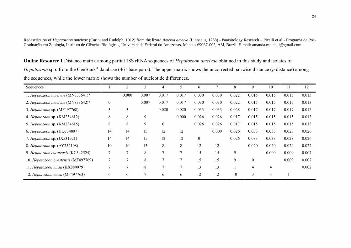
Online Resource 1 Distance matrix among partial 18S rRNA sequences of Hepatozoon
ameivae obtained in this study and isolates of Hepatozoon spp from the GenBankreg database
(461 base pairs) The upper matrix shows the uncorrected pairwise distance (p distance) among
the sequences while the lower matrix shows the number of nucleotide differences 84
Capiacutetulo 3
Table 1 Morphometric characteristics of the trypomastigotes found in the lizard Uranoscodon
superciliosus from Central Amazonia Brazil Measurements are presented as mean plusmn standard
deviation (SD) followed by the range (maximum and minimum values) 112
xii
Lista de Figuras
Capiacutetulo 1
Figure 1 Sampling areas in Central Amazonia (1) Campus of the Federal University of
Amazonas (UFAM) (2) UFAM Experimental Farm (3) Biological Dynamics of Forest
Fragments Project (BDFFP) Reserve (4) Agrovila Rio Pardo59
Figure 2 Parasites and inclusions found in lizards from Central Amazonia Gametocytes of (a)
Hepatozoon ameivae and (b) Hepatozoon sp in Ameiva ameiva (c) Sauroplasma-like infection
in Uranoscodon superciliosus (d) Trophozoite with nuclear division of Plasmodium carmelinoi
from A ameiva (e) Trophozoite and mature (f) gametocyte of Plasmodium sp in Norops
planiceps (g) Macrogametocytes and microgametocyte of Plasmodium kentropyxi in
Kentropyx calcarata (h) Gametocyte of Sarocytozoon tupinambi in a lymphocyte from
Tupinambis teguixin (i) Fallisia simplex in Plica umbra showing single and double
gametocyte infections in the thrombocytes (j) Gametocyte of Garnia uranoscodoni from U
superciliosus Trypanosoma spp infections in (k) U superciliosus and (l) P umbra (m)
Microfilaria in A ameiva and in (n) mixed infection in U superciliosus Vacuole-like inclusions
in erythrocytes from (o) U superciliosus and (p) A ameiva Arrow heads indicate pigment
granules black arrows indicate parasite vacuoles and asterisks indicate inclusions Micrographs
are from Giemsa-stained thin blood films Scale bar is 10 μm 60
Capiacutetulo 2
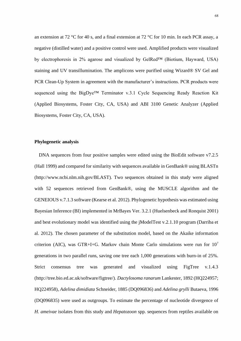
Fig 1 Gametocytes of Hepatozoon ameivae in the blood of Ameiva ameiva from Central
Amazonia Brazil a b and c mdash Intraerythrocytic gamonts d and e mdash Parasites invading
leukocytes (L) fmdash Gamont free in blood (gf) Arrows indicate parasites asterisks indicate
gamont nuclei and (n) indicates host cell nucleus Micrographs are from Giemsa-stained thin
blood films Scale bar is 20 μm 83
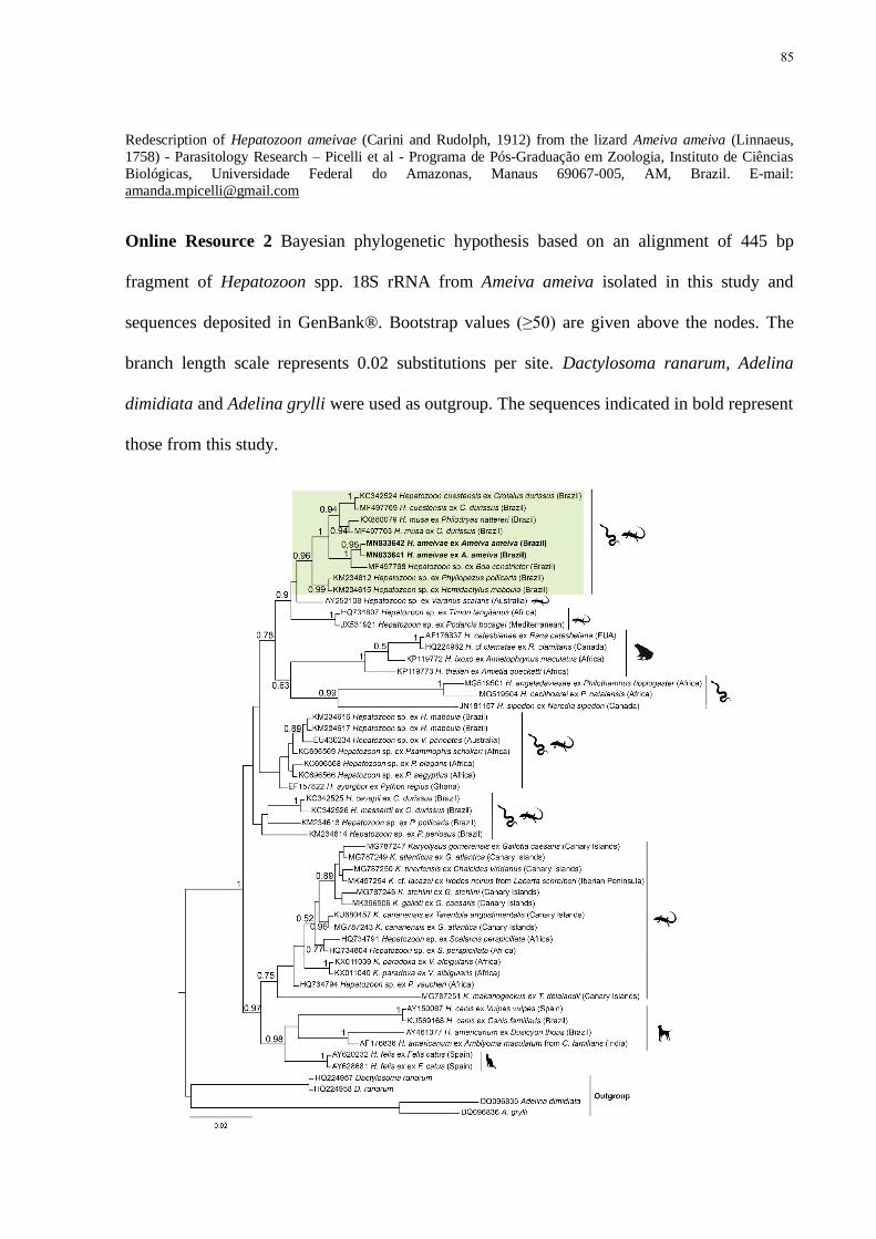
Online Resource 2 Bayesian phylogenetic hypothesis based on an alignment of 445 bp
fragment of Hepatozoon spp 18S rRNA from Ameiva ameiva isolated in this study and
sequences deposited in GenBankreg Bootstrap values (ge50) are given above the nodes The
branch length scale represents 002 substitutions per site Dactylosoma ranarum Adelina
xiii
dimidiata and Adelina grylli were used as outgroup The sequences indicated in bold represent
those from this study 85
Capiacutetulo 3
Fig 1 Geographical origin of trypanosome isolates from Uranoscodon surpercilosus in Central
Amazonia Brazil (1) Experimental Farm of the Federal University of Amazonas ndash FEX-
UFAM and (2-6) Area of Relevant Ecological Interest of the Biological Dynamics of Forest
Fragments Project ndash AREI-BDFFP Tones of gray indicate the trypanosome prevalence and the
sizes of the circles indicate the number of individuals analyzed per site 113
Fig 2 Trypomastigote forms found in the peripheral blood of Uranoscodon superciliosus from
Central Amazonia Brazil (a ndash d) Rounded or elliptical morphologies (e ndash h) leaf-shaped
trypanosomes (i ndash l) elongated forms with cytoplasmic projections Abbreviations n nucleus
k kinetoplast f flagellum cp cytoplasmic projection Micrographs are from Giemsa-stained
thin blood films Scale bar is 10 μm 114
Fig 3 Phylogenetic positioning of trypanosomes of Uranoscodon superciliosus from Central
Amazonia Phylogenetic trees (ML) inferred from V7V8 SSU rDNA (a) and by gGAPDH
(characters 812 Ln = minus8826807868) (b) gene sequences Maximum Likelihood inference
(characters 606 Ln = minus5963950862) supported the Genotype 01 and Genotype 02 in the
Aquatic clade Trypanosomes of the terrestrial lineages and trypanosomatids of other genera
were used as outgroups Bootstrap values are given under the nodes 115
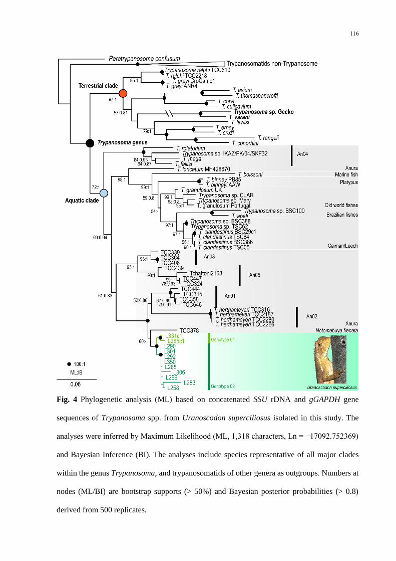
Fig 4 Phylogenetic analysis (ML) based on concatenated SSU rDNA and gGAPDH gene
sequences of Trypanosoma spp from Uranoscodon superciliosus isolated in this study The
analyses were inferred by Maximum Likelihood (ML 1318 characters Ln = minus17092752369)
and Bayesian Inference (BI) The analyses include species representative of all major clades
within the genus Trypanosoma and trypanosomatids of other genera as outgroups Numbers at
nodes (MLBI) are bootstrap supports (gt 50) and Bayesian posterior probabilities (gt 08)
derived from 500 replicates 116
14
Introduccedilatildeo Geral
Com origens e histoacuterias evolutivas independentes o estilo de vida parasitaacuterio eacute complexo
e possivelmente o mais bem-sucedido entre os seres vivos estando presente em praticamente
todos os grandes grupos taxonocircmicos (De Meeucircs e Renaud 2002 ODonoghue 2017) Haacute
estimativas que sugerem que mais da metade das espeacutecies viventes podem ser consideradas em
sentido amplo como parasitos (Dobson et al 2008 Poulin 2014 Morand 2015)
Tradicionalmente os parasitos (gr parasitos - quem come agrave mesa do outro) satildeo definidos como
organismos que passam a maior parte ou toda a vida associados a outros organismos
(hospedeiros) alimentando-se deles e como consequecircncia causando algum tipo de prejuiacutezo a
esses indiviacuteduos (Price 1977 Poulin e Morand 2004) Contudo dada a particularidade de cada
parasito e das caracteriacutesticas que envolvem a relaccedilatildeo com seu hospedeiro eacute possiacutevel aprofundar
esse significado aleacutem de obter nutrientes muitas espeacutecies dependem de seus hospedeiros para
seu desenvolvimento reproduccedilatildeo e dispersatildeo estrateacutegias parasitaacuterias que podem ou natildeo levar
seus hospedeiros agrave morte (Erbet e Herre 1996 Poulin e Morand 2004)
Ecologicamente parasitos satildeo considerados como verdadeiros ldquoengenheirosrdquo da natureza
pois aleacutem de intervirem sobre a coexistecircncia ou a exclusatildeo das espeacutecies podem atuar em
diferentes niacuteveis e processos dentro das comunidades influenciando variaacuteveis que estruturam
os ecossistemas (Poulin 1999 Thomas et al 2000 Hatcher et al 2012 Buck 2019) Satildeo
organismos-chave para a composiccedilatildeo de algumas teias alimentares aumentando a
complexidade e afetando os fluxos de energia e a ciclagem de nutrientes (Lafferty et al 2008
Hatcher et al 2012 Anaya‐Rojas et al 2019) Em seus hospedeiros satildeo capazes de influenciar
vaacuterios processos ecoloacutegicos e evolutivos (eg seleccedilatildeo sexual migraccedilatildeo competiccedilatildeo e predaccedilatildeo
especiaccedilatildeo e extinccedilatildeo) que por sua vez moldam a dinacircmica da populaccedilatildeo hospedeira e levam
a efeitos diretos ou indiretos sobre outras espeacutecies que interagem com seus hospedeiros (eg
15
predadores e competidores) (Schall 1992 Tompkins e Begon 1999 Hatcher et al 2012 Buck
2019) Dessa forma os parasitos satildeo importantes preditores da biodiversidade e da sauacutede dos
ecossistemas (Hudson et al 2006 Dobson et al 2008 Thompson et al 2018)
Por outro lado os parasitos geralmente satildeo reconhecidos pelos diversos efeitos negativos
que produzem sobre seus hospedeiros principalmente quando associados agraves populaccedilotildees
humanas (eg malaacuteria Doenccedila de Chagas filariose entre outras) com severos impactos na
sauacutede puacuteblica e economia mundial (Perkins 2014 Telleria e Tibayrenc 2017 WHO 2019)
Como resultado grande parte das pesquisas estatildeo concentradas em poucos grupos de
hospedeiros animais sobretudo parasitos de mamiacuteferos com interesse econocircmico ou
envolvendo animais silvestres associados a doenccedilas zoonoacuteticas (Valkiunas 2005 Spodareva et
al 2018) Entretanto negligenciar espeacutecimes pode ter implicaccedilotildees sobre o entendimento da
virulecircncia e evoluccedilatildeo dos patoacutegenos de importacircncia meacutedica (Rambaut et al 2001 Galen et al
2018) A acuraacutecia das inferecircncias sobre processos evolutivos depende de uma amostragem
ampla dos taxa (Heath et al 2008) e especialmente da reconstruccedilatildeo das transiccedilotildees entre grupos
de hospedeiros que levaram agrave origem da doenccedila (Liu et al 2010)
Embora sejam hospedeiros subestimados os reacutepteis (Chordata Reptilia) se destacam pela
grande variedade de parasitos sanguiacuteneos (hemoparasitos) que albergam com uma riqueza de
espeacutecies registradas superior agravequelas conhecidas para aves e mamiacuteferos (Davies e Johnston
2000 Telford 2009) Isso provavelmente se deve agrave antiga idade fileacutetica dos reacutepteis [final do
periacuteodo Carboniacutefero (~315 Ma)] e tambeacutem agrave elevada diversidade ecoloacutegica e taxonocircmica
desses hospedeiros (Poinar e Poinar 2004 Vitt e Caldwell 2013) Nesse aspecto os lagartos
(Lepidosauria Squamata) podem ser considerados como hospedeiros potencialmente diversos
em espeacutecies de hemoparasitos uma vez que aleacutem de possuiacuterem 60 das 11 mil espeacutecies
descritas da classe Reptilia Laurenti 1768 apresentam haacutebitos de vida bastante diversificados
16
e satildeo encontrados em uma ampla variedade de ambientes (Vitt et al 2008 Faria et al 2019
Peixoto et al 2020 Uetz et al 2020)
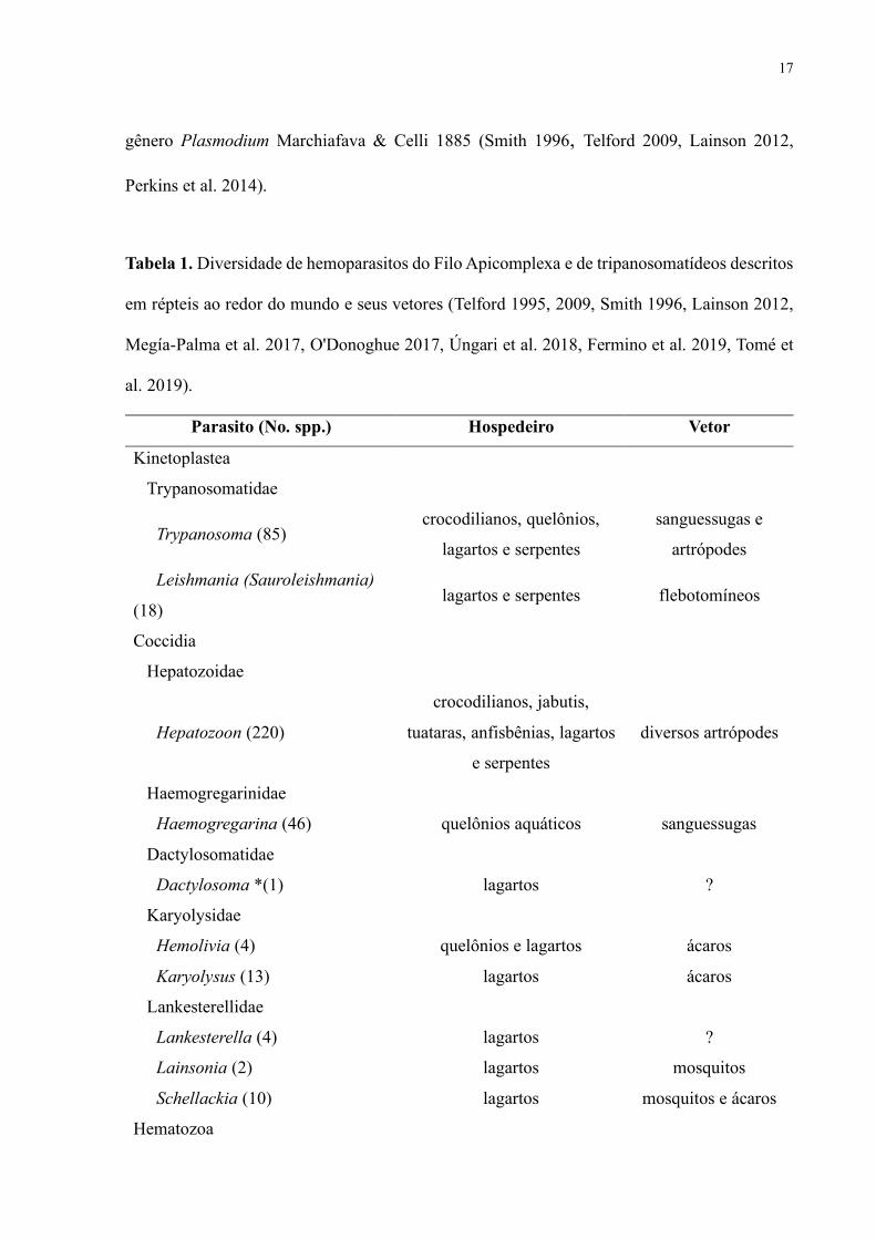
Nos reacutepteis os hemoparasitos encontrados com maior frequecircncia pertencem ao filo
Apicomplexa Levine 1970 e agrave famiacutelia Trypanosomatidae Doflein 1901 (Telford 2009
ODonoghue 2017) Esses dois taxa contabilizam juntos nesses hospedeiros aproximadamente
570 espeacutecies divididas em 18 gecircneros e 10 famiacutelias das quais mais da metade (ca 320) foram
identificadas em lagartos (Tabela 1) Haacute tambeacutem outros organismos menos frequentes como os
estaacutegios larvais (microfilaacuterias) do filo Nematoda Diesing 1861 e tambeacutem inclusotildees virais e
bacterianas (Telford 2009 Halla et al 2014) Aleacutem disso entre os hemoparasitos agrave exceccedilatildeo de
alguns viacuterus e bacteacuterias haacute uma convergecircncia adaptativa ao uso de invertebrados hematoacutefagos
como principais vetores para transmissatildeo entre seus hospedeiros vertebrados (ODonoghue
2017 Tabela 1)
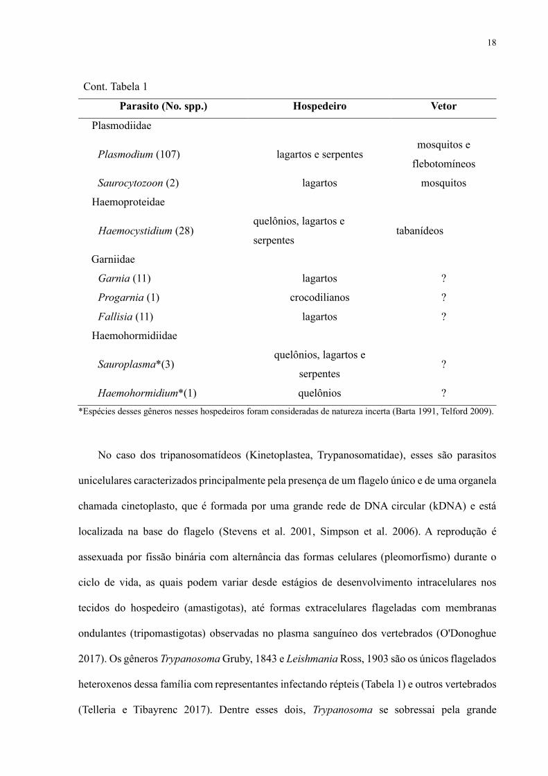
O filo Apicomplexa (Chromista Alveolata) deteacutem mais de 6 mil espeacutecies descritas
(Votyacutepka et al 2017) Todas satildeo endossimbiontes obrigatoacuterias e apresentam um conjunto de
estruturas na extremidade anterior denominado complexo apical que possibilita a invasatildeo e
sobrevivecircncia dentro da ceacutelula hospedeira (Levine et al 1980 Morrissette e Sibley 2002 Baum
et al 2008 Tardieux e Baum 2016) O desenvolvimento dos Apicomplexa eacute uacutenico entre os
eucariotos por apresentar uma ontogenia reprodutiva ciacuteclica que conteacutem duas fases assexuadas
merogonia e esporogonia e uma sexuada chamada de gametogonia (Striepen et al 2007 Baum
et al 2008 Votyacutepka et al 2017) Os membros desse filo com estaacutegios de desenvolvimento
intracelular nas ceacutelulas sanguiacuteneas dos reacutepteis satildeo (Tabela 1) as hemogregarinas (Coccidia
Adeleorina) os hemococciacutedios (Coccidia Eimeriorina) os hemosporiacutedeos (Hematozoa
Haemosporida) e os piroplasmas (Hematozoa Piroplasmida) Destes os mais encontrados em
lagartos satildeo as hemogregarinas do gecircnero Hepatozoon Miller 1908 e os hemosporiacutedeos do
17
gecircnero Plasmodium Marchiafava amp Celli 1885 (Smith 1996 Telford 2009 Lainson 2012
Perkins et al 2014)
Tabela 1 Diversidade de hemoparasitos do Filo Apicomplexa e de tripanosomatiacutedeos descritos
em reacutepteis ao redor do mundo e seus vetores (Telford 1995 2009 Smith 1996 Lainson 2012
Megiacutea-Palma et al 2017 ODonoghue 2017 Uacutengari et al 2018 Fermino et al 2019 Tomeacute et
al 2019)
Parasito (No spp) Hospedeiro Vetor
Kinetoplastea
Trypanosomatidae
Trypanosoma (85) crocodilianos quelocircnios
lagartos e serpentes
sanguessugas e
artroacutepodes
Leishmania (Sauroleishmania)
(18) lagartos e serpentes flebotomiacuteneos
Coccidia
Hepatozoidae
Hepatozoon (220)
crocodilianos jabutis
tuataras anfisbecircnias lagartos
e serpentes
diversos artroacutepodes
Haemogregarinidae
Haemogregarina (46) quelocircnios aquaacuteticos sanguessugas
Dactylosomatidae
Dactylosoma (1) lagartos
Karyolysidae
Hemolivia (4) quelocircnios e lagartos aacutecaros
Karyolysus (13) lagartos aacutecaros
Lankesterellidae
Lankesterella (4) lagartos
Lainsonia (2) lagartos mosquitos
Schellackia (10) lagartos mosquitos e aacutecaros
Hematozoa
18
Cont Tabela 1
Parasito (No spp) Hospedeiro Vetor
Plasmodiidae
Plasmodium (107) lagartos e serpentes mosquitos e
flebotomiacuteneos
Saurocytozoon (2) lagartos mosquitos
Haemoproteidae
Haemocystidium (28) quelocircnios lagartos e
serpentes tabaniacutedeos
Garniidae
Garnia (11) lagartos
Progarnia (1) crocodilianos
Fallisia (11) lagartos
Haemohormidiidae
Sauroplasma(3) quelocircnios lagartos e
serpentes
Haemohormidium(1) quelocircnios
Espeacutecies desses gecircneros nesses hospedeiros foram consideradas de natureza incerta (Barta 1991 Telford 2009)
No caso dos tripanosomatiacutedeos (Kinetoplastea Trypanosomatidae) esses satildeo parasitos
unicelulares caracterizados principalmente pela presenccedila de um flagelo uacutenico e de uma organela
chamada cinetoplasto que eacute formada por uma grande rede de DNA circular (kDNA) e estaacute
localizada na base do flagelo (Stevens et al 2001 Simpson et al 2006) A reproduccedilatildeo eacute
assexuada por fissatildeo binaacuteria com alternacircncia das formas celulares (pleomorfismo) durante o
ciclo de vida as quais podem variar desde estaacutegios de desenvolvimento intracelulares nos
tecidos do hospedeiro (amastigotas) ateacute formas extracelulares flageladas com membranas
ondulantes (tripomastigotas) observadas no plasma sanguiacuteneo dos vertebrados (ODonoghue
2017) Os gecircneros Trypanosoma Gruby 1843 e Leishmania Ross 1903 satildeo os uacutenicos flagelados
heteroxenos dessa famiacutelia com representantes infectando reacutepteis (Tabela 1) e outros vertebrados
(Telleria e Tibayrenc 2017) Dentre esses dois Trypanosoma se sobressai pela grande
19
diversidade geneacutetica e morfoloacutegica de suas espeacutecies em lagartos e serpentes (Viola et al 2008
2009 Telford 2009 Fermino et al 2019)
Com os avanccedilos das accedilotildees antroacutepicas sobre os ambientes naturais e em face a uma eminente
crise climaacutetica haacute uma crescente preocupaccedilatildeo sobre os possiacuteveis impactos da perda em massa
das espeacutecies (Ceballos et al 2017) Mas apesar de serem componentes essenciais da
biodiversidade os parasitos satildeo inconspiacutecuos para a maioria dos esforccedilos conservacionistas
(Thompson et al 2018 Milotic et al 2020) No entanto satildeo extremante vulneraacuteveis agrave extinccedilatildeo
e frequentemente correm maiores riscos de desaparecem do que seus hospedeiros (Dunn et al
2009 Thompson et al 2018 Milotic et al 2020) Isso se deve ao fato de estes poderem ser
extintos em decorrecircncia da extinccedilatildeo dos seus hospedeiros (co-extinccedilatildeo) ou atraveacutes do decliacutenio
da populaccedilatildeo hospedeira (Milotic et al 2020) Aleacutem disso assim como na recente pandemia
causada pelo viacuterus Sars-CoV-2 (novo coronaviacuterus) alguns patoacutegenos podem se beneficiar com
as atividades humanas e seus efeitos uma vez que proporcionam a esses organismos
oportunidades de propagaccedilatildeo e o estabelecimento em novos hospedeiros (Jones et al 2008
Cizauskas et al 2017 Zohdy et al 2019) Dessa forma alteraccedilotildees na dinacircmica da fauna
parasitaacuteria produzem consequecircncias profundas sobre a sauacutede de populaccedilotildees humanas e
silvestres (Dobson et al 2008 Corlett et al 2020 FAO 2020)
Nesse aspecto a Floresta Amazocircnica a qual concentra grande parte da biodiversidade do
planeta tem sido irreversivelmente destruiacuteda e modificada no Brasil sob o amparo de uma
legislaccedilatildeo ambiental e governos que incentivam a exploraccedilatildeo de recursos e o desmatamento
(Novaes e Souza 2013 Magnusson et al 2018) Apenas no ano de 2019 houve um aumento de
quase 34 (10129 kmsup2) em relaccedilatildeo agrave taxa de desmatamento de 2018 na Amazocircnia brasileira
(INPE 2020) Uma condiccedilatildeo bastante dramaacutetica tendo em vista que nesse bioma haacute
constantemente descriccedilotildees de espeacutecies novas de hospedeiros reptilianos como os lagartos
cujos hemoparasitos ainda satildeo pobremente conhecidos (Costa e Beacuternils 2018 Ribeiro-Juacutenior et
20
al 2020) De fato haacute registros de hemoparasitos em somente 10 (n = 16152) das espeacutecies de
lagartos que ocorrem na Amazocircnia brasileira (Lainson 2012 Costa e Beacuternils 2018)
Apesar dessa quantidade relativamente baixa de animais amostrados os estudos pioneiros
conduzidos pelo Dr Ralph Lainson principalmente entre os anos de 1966 e 1992 mostraram a
existecircncia de uma rica fauna de hemoparasitos nesses hospedeiros sugerindo assim que a regiatildeo
amazocircnica no Brasil deteacutem um potencial elevado para novas espeacutecies destes parasitos (Lainson
2012) Entre os hemoparasitos registrados nesses levantamentos foram 27 espeacutecies do filo
Apicomplexa e duas espeacutecies de tripanosomas (para mais detalhes ver Tabela 1 do Capiacutetulo 1)
Poreacutem a maioria dessas foram detectadas em lagartos oriundos da Amazocircnia Oriental e em
sua maioria descritas apenas com base na taxonomia tradicional sem o uso de ferramentas
moleculares Isso pode levar a um desconhecimento dos padrotildees de distribuiccedilatildeo e identidade
das espeacutecies de parasitos que ocorrem no Brasil aleacutem de refletir sobre uma baixa compreensatildeo
da interaccedilatildeo desses organismos com seus hospedeiros lagartos (Heath et al 2008 Morand
2018)
Nesse contexto a presente tese investigou a diversidade de hemoparasitos e caracterizou
atraveacutes de dados morfoloacutegicos e moleculares a composiccedilatildeo desta comunidade em hospedeiros
lagartos da Amazocircnia Central Os resultados alcanccedilados encontram-se organizados em trecircs
capiacutetulos
Capiacutetulo 1 trata da prevalecircncia e diversidade dos hemoparasitos encontrados nos
largartos da regiatildeo da Amazocircnia Central
Capiacutetulo 2 apresenta a redescriccedilatildeo da hemogregarina Hepatozoon ameivae (Carini e
Rudolph 1912) do lagarto Ameiva ameiva
Capiacutetulo 3 aborda as relaccedilotildees filogeneacuteticas dos tripanosomas que parasitam
Uranoscodon superciliosus (Linnaeus 1758)
21
CAPIacuteTULO 1
Amanda M Picelli Adriane C Ramires Gabriel S Masseli Felipe A C
Pessoa Lucio A Viana e Igor L Kaefer Under the light high prevalence
of haemoparasites in lizards (Reptilia Squamata) from Central
Amazonia revealed by microscopy Manuscrito publicado no perioacutedico
Anais da Academia Brasileira de Ciecircncias em 20 de julho de 2020 DOI
1015900001-3765202020200428
22
Under the light high prevalence of haemoparasites in lizards (Reptilia Squamata) from
Central Amazonia revealed by microscopy
AMANDA M PICELLI1 ADRIANE C RAMIRES2 GABRIEL S MASSELI3 FELIPE A C PESSOA4
LUCIO A VIANA5 amp IGOR L KAEFER1 2
1Programa de Poacutes-Graduaccedilatildeo em Zoologia Universidade Federal do Amazonas Av General Rodrigo Octavio
Jordatildeo Ramos 1200 Coroado I 69067-005 Manaus AM Brazil
2Instituto de Ciecircncias Bioloacutegicas Universidade Federal do Amazonas Av General Rodrigo Octavio Jordatildeo
Ramos 1200 Coroado I 69067-005 Manaus AM Brazil
3Programa de Poacutes-Graduaccedilatildeo em Ecologia Instituto Nacional de Pesquisas da Amazocircnia Av Andreacute Arauacutejo 2936
Petroacutepolis 69067-375 Manaus AM Brazil
4Laboratoacuterio de Ecologia de Doenccedilas Transmissiacuteveis na Amazocircnia (EDTA) Instituto Leocircnidas e Maria Deane
Fundaccedilatildeo Oswaldo Cruz Rua Terezina 476 Adrianoacutepolis 69067-005 Manaus AM Brazil
5Laboratoacuterio de Estudos Morfofisioloacutegicos e Parasitaacuterios Departamento de Ciecircncias Bioloacutegicas e da Sauacutede
Universidade Federal do Amapaacute Rod Juscelino Kubitschek sn Jardim Marco Zero 68903-419 Macapaacute AP
Brazil
Key words Biodiversity blood parasites Lacertilia morphology Neotropics
Running title Haemoparasites in lizards from Central Amazonia
Academy Section Biological Sciences
Amanda Maria Picelli httpsorcidorg0000-0001-7543-168X
Adriane Costa Ramires httpsorcidorg0000-0002-2547-614X
Gabriel Sales Masseli httpsorcidorg0000-0002-5762-758X
Felipe Arley Costa Pessoa httpsorcidorg0000-0002-6318-1887
Lucio Andreacute Viana httpsorcidorg0000-0002-0932-0479
Igor Luis Kaefer httpsorcidorg0000-0001-6515-0278
Correspondence to Amanda Maria Picelli
E-mail amandampicelligmailcom
23
Abstract Blood samples from 330 lizards of 19 species were collected to investigate the
occurrence of haemoparasites Samplings were performed in areas of upland (terra-firme)
forest adjacent to Manaus municipality Amazonas Brazil Blood parasites were detected in 220
(66) lizards of 12 species and comprised four major groups Apicomplexa (including
haemogregarines piroplasms and haemosporidians) trypanosomatids microfilarid nematodes
and viral or bacterial organisms Order Haemosporida had the highest prevalence with 118
(35) animals from 11 species For lizard species Uranoscodon superciliosus was the most
parasitised host with 103 (87 n = 118) positive individuals This species also presented the
highest parasite diversity with the occurrence of six taxa Despite the difficulties attributed by
many authors regarding the use of morphological characters for taxonomic resolution of
haemoparasites our low-cost approach using light microscopy recorded a high prevalence and
diversity of blood parasite taxa in a relatively small number of host species This report is the
first survey of haemoparasites in lizards in the study region It revealed a high diversity of lizard
haemoparasites and highlights the need to understand their impacts on hosts
24
INTRODUCTION
The protozoologist Dr Ralph Lainson (1992) two decades ago in his work on neglected
parasites in the Amazonia basin quoted a phrase from PCC Garnham his former advisor
There is a serious danger that malarial parasites become extinct Since that time very few
efforts have been made to contain the threats to the diversity of these parasites and other
organisms (Ferrante amp Fearnside 2019) In fact these threats have been aggravated by increased
habitat destruction in recent years particularly in tropical regions (INPE 2019) Extinction
alteration in the abundance or introduction of parasites can have profound impacts on the health
of a large number of free-living species (Dobson et al 2008) because parasites are ecologically
involved in important mechanisms that regulate wildlife populations and structure communities
(Tompkins amp Begon 1999 Thomas et al 2000) Moreover they may influence their host
biological processes such as sexual selection (Ehman amp Scott 2002 Megiacutea-Palma et al 2018)
predation and competition dynamics (Schall 1992 Garcia-Longoria et al 2015) as well as
speciation and extinction processes (Anderson amp May 1978 Poulin 1999 Prenter et al 2004)
Reptiles are hosts for a wide variety of parasites especially for diverse groups that
parasitise blood cells (Davies amp Johnston 2000 Telford 2009) These blood parasites may be
intra- or extracellular organisms that range from protozoan kinetoplastids (Killick-Kendrick et
al 1986 Telford 1995) and apicomplexan parasites (Levine 1988 ODonoghue 2017) to
microfilarid nematodes (Thoisy et al 2000 Halla et al 2014) as well as viral and bacterial
inclusions (Telford 2009) Except for the last two pathogens whose transmission is not yet
clear the other three parasitic taxa share a common feature by using a range of haematophagous
invertebrates as the main vectors for transmission between vertebrate hosts (Smallridge amp
Paperna 1997 Viana et al 2012 Van As et al 2015 Fermino et al 2019) Furthermore the
haemoprotozoans of Phylum Apicomplexa Levine 1970 are apparently the most studied of all
25
and also represent the taxon with the largest number of species parasitising reptiles (Levine
1988) Only in lizards (Squamata Sauria) approximately 14 genera were recorded
(ODonoghue 2017) haemogregarines and haemosporidians are the most frequently identified
groups (Smith 1996 Perkins 2014)
Although Brazil is a megadiverse country and has the third richest reptilian fauna in the
world (Costa amp Beacuternils 2018) approximately 795 species knowledge about haemoparasite
diversity in these hosts consists of mainly a few concentrated studies in the eastern Amazon
region (Lainson 1992 2012) These studies recorded a rich haematozoan fauna in lizards and
also suggest that the Amazon biome has a great potential for the discovery of new
haemoparasitic species in these vertebrates as 29 (80) of the 36 known protozoan species in
the country occur in this region (Table I) However these records are limited to a total of 20
lizard species (Table I) which represent 7 (n = 276) of the described Brazilian lizard fauna
and 10 (n = 16152) for the Amazon region (Costa amp Beacuternils 2018) This small number is
probably due to the difficulties in collecting these hosts and also the lack of specialists interested
in working with haemoparasites from herpetofauna
Light microscopy is an important tool for diagnosing infections that has crossed centuries
and generations of scientists still being the fastest and most accessible technique for searching
parasites (Halla et al 2014) This is especially true for studies adopting horizontal approaches
that aim to estimate parasitism in poorly known groups In this sense we sought to investigate
using light microscopy the presence and diversity of haemoparasites in lizards from Central
Amazonia
26
MATERIALS AND METHODS
STUDY AREA
The study was conducted in four upland (terra-firme) forest sites in Brazilian Central
Amazonia all located in the State of Amazonas Brazil (Figure 1) The first study area was the
Federal University of Amazonas forest fragment campus (UFAM 3ordm434S 59ordm5730W)
located in the eastern part of the city of Manaus The three remaining study areas were located
respectively 38 km (UFAM Experimental Farm 2deg38576S 60deg311W) 80 km (Biological
Dynamics of Forest Fragments Project [BDFFP] 2deg25S 59deg50W) and 160 km (Agrovila Rio
Pardo 1deg48S 60deg19 W) north of Manaus These sampling regions present a mean annual
temperature of approximately 26degC with relative air humidity over 80 (Araujo et al 2002)
The yearly precipitation is over 2000 mm and mostly concentrated in a rainy season that usually
occurs from December to May (Marques-Filho et al 1981) The vegetation of the sampling
sites is mainly composed of a mosaic of upland Amazonian rainforest which varies from
primary and secondary forests to open areas The average elevation is 40ndash160 m above sea level
(Laurance et al 2011) Some of these landscapes are relatively undisturbed (Deichmann et al
2010 Rojas-Ahumada et al 2012) but most exhibit anthropogenic alterations (Rocha et al
2004 Ramos et al 2014)
LIZARD AND BLOOD SAMPLING
A total of 330 lizards from 19 species distributed in 17 genera and 10 families were
sampled between 2016 and 2019 (Table II) Animals were captured using several methods such
as active search (Doan 2003) and traps ie pitfalls with drift-fences (Jenkins et al 2003)
funnels made out of PVC pipes (Abrahatildeo et al 2019) and live-traps (Vieira et al 2015) Lizards
were identified through specialised literature (Aacutevila-Pires 1995 Vitt et al 2008) and taxonomic
27
nomenclature was adopted following Costa amp Beacuternils (2018) The blood samples were obtained
by tail or cardiac puncture using a sterile insulin syringe (Samour et al 1984) A portion of
collected blood was used to make smears which were fixed with absolute methanol and stained
with 10 Giemsa The other portion was applied to a filter paper for molecular analyses
Lizards were released within 24 h of capture but in the case of cardiac puncture the blood was
collected after euthanasia (via injection of 2 lidocaine) Specimens were preserved in 10
formalin and deposited in the Zoological Collections of the National Institute of Amazonian
Research (INPA) and UFAM in Manaus Brazil
Lizard sampling and access to the genetic data were authorised by the Brazilian Ministry
of the Environment (SISBIO ndeg 53851-4 and SISGEN AA6199D respectively) All procedures
were approved by the ethics committee on animal use from Universidade Federal do Amazonas
(protocol number 0122016)
MICROSCOPIC ANALYSES
Blood smears were examined for up to 20 min under a Leica DM4B microscope (Leica
Microsystems Heerbrugg Switzerland) at times 400 and times 1000 total magnification The slides
with parasites were carefully examined and images were captured with an attached Leica
DMC4500 digital camera and processed with LAS V48 (Leica Microsystems Suiza Limited
2015) Morphometric measurements were taken with this same system However they will not
be presented here in this work as they are part of ongoing taxonomic studies Haematozoan
parasites were taxonomically identified by comparing their morphologies to the descriptions
from the guides of Telford (2009) and Lainson (2012) besides original description articles
Additionally to confirm the identification of some haemosporidian species we compared our
material with that of the collection of Dr Ralph Lainson deposited at the Evandro Chagas
Institute (IEC) in Beleacutem Brazil
28
RESULTS
Haemoparasite infections were detected in 220 (66) out of 330 lizards of 12 species
distributed among seven families (Table II) Mixed infections occurred in 91 positive
specimens For sampling sites BDFFP had 78 (n = 156200) of the infected lizards UFAM
Experimental Farm had 68 (n = 1319) Agrovila Rio Pardo had 47 (n = 50105) and UFAM
urban forest fragment had 16 (n = 16) Parasites were grouped into four major groups (Figure
2) with the following prevalence (i) intracellular apicomplexan parasites at 173 (52)
individuals (ii) trypanosomatids at 84 (25) (iii) microfilarial worms at 38 (11) (iv)
unidentified viral or bacterial inclusions at 30 (9)
Among the positive lizards Tropiduridae and Teiidae were the families that showed the
highest prevalence with 86 (n = 112130) and 66 (n = 90135) positive animals
respectively With regards to lizard species Uranoscodon superciliosus Linnaeus 1758 stood
out for presenting a high prevalence with 87 (n = 103118) of infected individuals and also
because it was the species with the greatest diversity of parasites with the occurrence of six
different taxa Haemohormidiidae Plasmodiidae Garniidae Trypanosomatidae microfilarial
worms and unidentified inclusions
Parasites of phylum Apicomplexa (Table III) were found in all infected lizard species 14
species from five families were identified Two morphotypes of the genus Hepatozoon
(Hepatozoidae) were observed in 40 Ameiva ameiva Linnaeus 1758 (55 n = 72) and one was
identified as Hepatozoon ameivae Carini amp Rudolph 1912 (Figure 2a) H ameivae was
recorded overlapping the nucleus of the parasitised cells whereas the other morphotype caused
lateral displacement of the nucleus to one end of the red blood cell (Figure 2b) Both parasites
were restricted to erythrocytes Sauroplasma-like (Haemohormidiidae) infections (Figure 2c)
appeared in 14 (n = 49330) of individuals from six lizard species (Table III) Notably U
29
superciliosus had the highest number of parasite occurrences with 32 (27 n = 118) positive
specimens
Haemosporidian parasites presented the highest prevalence with 35 (n = 118330) animals
infected and except for Alopoglossus angulatus Linnaeus 1758 all positive host species were
parasitised by malaria Based on blood stage morphology 13 species from two families
Plasmodiidae and Garniidae were identified (Table III Figure 2d-j) It is important to note that
despite some authors (eg Levine 1988 Telford 2009) here we recognise the family Garniidae
as well as the genera Garnia and Fallisia as valid taxa diagnosed by absence of pigment and
ultrastructural characteristics (Lainson et al 1971 Boulard et al 1987)
Plasmodium spp (Figure 2d-g) were detected in 64 (19 n = 330) lizards from nine species
with the highest number of positive specimens seen in A ameiva (36 n = 72) At least 11
morphotypes were visualised and five Plasmodium species could be recognised (Table III)
Gametocytes of Saurocytozoon cf tupinambi Lainson amp Shaw 1969b were observed in
leucocytes (Figure 2h) from five (20 n = 25) Tubinambis teguixin Linnaeus 1758 Non-
pigmented malaria parasites from the genera Fallisia (Figure 2i) and Garnia (Figure 2j) were
found in four (1 n = 330) and 46 (14 n = 330) lizards respectively (Table III) Two Fallisia
species were detected in Plica umbra Linnaeus 1758 Fallisia cf simplex Lainson et al 1975
and Fallisia cf audaciosa Lainson et al 1975 In Neusticurus bicarinatus Linnaeus 1758 we
found Fallisia cf effusa Lainson et al 1974a Parasites of the genus Garnia were mainly
recorded in U superciliosus (22 n = 118) We also detected four unidentified morphotypes
and four species of this genus (Table III)
Extracellular parasites of the family Trypanosomatidae (Table II) were found in 83 U
superciliosus (70 n = 118) and one P umbra (8 n = 12) each tropidurid species had one
Trypanosoma morphotype The trypanosome of U superciliosus had an elongated body and
diffuse nucleus (Figure 2k) while the observed P umbra had a rounded shape and compact
30
nucleus (Figure 2l) Microfilarial worms (Nematoda) occurred in five lizard species (Table II)
with higher prevalence in A ameiva with 37 (n = 2772) positive specimens These blood
parasites exhibited highly variable sizes and shapes (Figure 2m-n) and were very similar to the
genus Piratuba However accurate diagnoses of filarial worms is mainly based on
morphological features of adult worms Thus identification of this group in the present study
remains indeterminate
The last of the four major groups inclusions of uncertain nature (Figure 2o-p) were detected
in erythrocytes of five lizard species and showed little morphological variation They consisted
of a large spherical shape with a rarely darker stained margin These vacuoles resemble
rickettsial parasites recorded for other reptilian hosts although without ultrastructural study it
was not possible to confirm this identification
DISCUSSION
We observed a high prevalence of blood parasites among lizards from Central Amazonia
More than half of the sampled individuals and species were infected We also demonstrated that
lizards are the hosts for a wide variety of haemoparasites Indeed we observed great parasite
richness in a small number of host species and in a limited sampling area This finding
reinforces that the neotropical region holds a rich haemoparasite fauna as shown by studies
conducted in other localities across the Amazon Basin (Renjifo et al 1952 Telford 1970 1973
1980 Ayala et al 1973 Lainson 1992 Thoisy et al 2000 Matta et al 2018) Furthermore it is
important to note that we sampled lizard species with diversified microhabitat use ranging from
terrestrial (eg A ameiva) semi-aquatic (eg Neusticurus bicarinatus) scansorial (eg P
umbra) to arboreal (eg U superciliosus) (Vitt et al 2008) This environmental diversity may
imply determinant characteristics for the composition of the haemoparasite assemblages found
31
in these lizards because different species of vectors including mosquitoes sandflies and ticks
are likely distributed along the gradient occupied by these hosts
Parasite and host checklists are crucial in expanding our knowledge on species distribution
Nonetheless surveys and descriptive studies of haemoparasite species on lizards conducted in
the Amazonian biome have decreased considerably in recent years In Brazil it has been 10
years since a haematozoan species from a lizard has been described (Lainson et al 2010) Most
access to this haemoparasitic diversity in the Neotropics departs from the classical approach by
using light microscopy to investigate prevalence and parasite identity The exclusive use of
morphological attributes for the diagnosis of species has been strongly criticised as unreliable
because molecular tools have advanced in solving taxonomic problems (Pineda-Catalan et al
2013 Perkins 2014) In fact molecular biology constitutes a modern and acurate tool in
parasitology but its use still faces financial and technical limitations mdashie difficulties in
developing protocols and molecular markersmdash especially in megadiverse and developing
countries such as Brazil (Perkins et al 2011 Morand 2018) Nevertheless in comprehensive
multi-species approaches like ours whose main objective is not to solve systematic and
phylogenetic questions observations of blood smears under a microscope still prove to be a
feasible method to access the prevalence and parasite diversity hidden in these hosts despite
some taxonomic limitations
Among the 12 infected lizard species haemoparasites were recorded for the first time in three
of them Arthrosaura reticulata OrsquoShaughnessy 1881 Norops planiceps Troschel 1848 and A
angulatus However we did not find blood parasites in nine lizard taxa (Table II) even though
parasites have already recorded in some of these hosts in other localities (Table I) This
discrepancy may simply reflect unequal sampling efforts The methods we used for capture
were diversified and effective for certain hosts groups such as Teiidae and Tropiduridae but
are limited for many lizard species mainly those that access subterranean microhabitats (Faria
32
et al 2019) Indeed Teiidae Tropiduridae and a lizard species U superciliosus were the hosts
with highest parasite prevalence However with the myriad known problems in obtaining
samples (Perkins et al 2011)mdashfinancial technical and logistical difficulties in accessing remote
areasmdashand the need to move forward on other parasitology research fronts such as vectors and
life cycle landscape and epizootiology studies those abundant taxa may be an interesting
choice to be included in ecological parasitic systems as model organisms Additionally for
many reasons lizards are considered model organisms (Huey et al 1983 Camargo et al 2010)
as they respond very well when testing ecological and evolutionary hypotheses (Schall 1996)
Most of the parasites found in this study belong to phylum Apicomplexa Indeed all host
species had some representative of this group One of them was the genus Hepatozoon
relatively common parasite in reptiles and despite the great diversity of lizards sampled in this
study was found exclusively infecting A ameiva Hepatozoon ameivae was described by Carini
amp Rudolph 1912 in A ameiva in the State of Minas Gerais and later recorded in the municipality
of Satildeo Joatildeo da Barra State of Rio de Janeiro both in southeastern Brazil (Carini amp Rudolph
1912 Sabagh et al 2015) Lainson et al (2003) also probably recorded H ameivae in lizards
from the municipality of Capanema State of Paraacute northern Brazil This parasite has an
outstanding feature its gametocytes are found in the erythrocyte nucleus a relatively
uncommon developmental pattern in the Apicomplexa that can lead to severe distortion and
even lysis of the infected cell nucleus (Telford 2009) It is important to note that H ameivae
found here was morphologically and molecularly characterized and the analysis of its
phylogenetic position clearly showed that this parasite belongs to the genus Hepatozoon (Picelli
et al unpublished data)
Our results showed a relatively low prevalence for Sauroplasma-like and we thought that
positive lizard species were not previously recorded for piroplasms (Table III) Sauroplasma
infections are common in lizards even though there are only three species described for these
33
hosts Sauroplasma thomasi du Toit 1938 Sauroplasma zonurum Pienaar 1962 and
Sauroplasma boreale Svahn 1976 (Telford 2009 Halla et al 2014) In Brazil these parasites
were recently recorded in the freshwater turtle Podocnemis expansa (Picelli et al 2016)
Morphologically they are small (25-4 microm) vacuole-shaped intraerythrocytic parasites with
chromatin granules associated (Halla et al 2014 Picelli et al 2016) These morphological
features mislead many authors to identify Sauroplasma-like inclusions as Chelonoplasma
Nuttalia or Pirhemocyton (Bardi et al 2019) They can also be overlooked as artefacts or
bacterial and viral infections (Telford 2009) Parasitologists always pay attention to this
conflicting taxonomic situation but no molecular data is yet known for this genus
Haemosporidian were the most predominant and richest taxon detected on lizards mainly
from Plasmodiidae parasites It is well known that malaria parasites are widely distributed
geographically ubiquitous for most lizard families and are morphologically diverse with over
100 species reported to infect reptiles (Schall 1996 Telford 2009) In the Eastern Brazilian
Amazonia 21 species of lizard malaria are known and 13 (61) of them were found in our
research For some of these (Garnia cf uranoscodoni Lainson et al 1975 Garnia cf
multiformes Lainson et al 1975 Garnia cf utingensis Lainson et al 1971 Fallisia cf
audaciosa and F cf effusa) this finding is the first occurrence record away from their type
localities Recently Matta et al (2018) reported the presence of Plasmodium kentropyxi
Lainson et al 2001 and Plasmodium carmelinoi Lainson et al 2010 in Teiidae lizards at a low
prevalence in the Colombia Orinoco-Amazon basin The difference between our findings is
that here P cf kentropyxi was found at a relatively high prevalence only in its type host
Kentropyx calcarata Spix 1825 Another interesting species seen in our study is Plasmodium
cf tropiduri Aragatildeo amp Neiva 1909 It was the only haemosporidian species found in two
different host species K calcarata and Copeoglossum nigropunctatum Spix 1825 This
haemoparasite was one of the worldrsquos first reptilian malaria parasites described by Aragatildeo amp
34
Neiva (1909) in the lizard Tropidurus torquatus Wied-Neuwied 1820 Since then it was
observed across many lizard families and can be considered one of the most widespread saurian
malaria species in South America (Telford 2009) In fact most of the haemosporidians present
here were previously reported in other Amazonian locations (Telford 2009 Matta et al 2018)
evidence that malaria species rediscovered here may be widely distributed throughout the
biome Phylogenetic and phylogeographic studies that involve samples from different
Amazonian localities may provide insights regarding the diversification and evolution of this
group
Parasites of the genus Trypanosoma were restricted to two lizard species and at a low
prevalence when compared to protozoans of the phylum Apicomplexa which parasitised more
than half of the captured lizards However these flagellates were found in several U
superciliosus Both P umbra and U superciliosus already had trypanosomatids recorded by
Walliker (1965) and Lainson et al (1975) respectively The first author provided a poor
morphological description of Trypanosoma superciliosae Walliker 1965 without reporting their
prevalence in U superciliosus from the municipality of Codajaacutes Amazonas State Interestingly
Lainson et al (1975) mentioned that they searched in Paraacute state for this parasite in a large
number of U superciliosus individuals but were unsuccessful Nevertheless the same authors
described Trypanosoma plicae Lainson et al 1975 in P umbra Besides these species there is
only one other species described for this genus on Brazilian lizards Trypanosoma rudolphi
recorded just once in C nigropunctatum (Carini amp Rudolph 1912) This low species richness
is probably due to the lack of studies conducted on these parasites in Brazilian lizards Indeed
trypanosome species have been reported worldwide in lizards more than in any other reptilian
group (Fermino et al 2019) Although trypanosomes have a unique stage of their life cycle by
circulating in the blood of reptiles trypomastigote forms exhibit high polymorphism and
35
plasticity (Spodareva et al 2018) Therefore it is not possible to confirm that we found the
same species described for those hosts even with some morphological similarities
Our data revealed a low prevalence of microfilaria which are larval stages from nematodes
of the superfamily Filaroidea These vector-borne parasite larvae are commonly found in the
peripheral blood of vertebrates and here except for U superciliosus all lizard species that we
found positive for these parasites already had records for adult worms from many
Onchocercidae species in other locations (Aacutevila amp Silva 2010 Macedo et al 2017) For U
superciliosus the occurrence of microfilariae has been vaguely reported in eastern Amazonia
and these studies did not provide morphological characterisation of these nematodes (Lainson
et al 1975) In reptiles Oswaldofilariinae a onchocercid subfamily stands out as the main
filarid group that parasitise these hosts Some genera that infected lizards include
Oswaldofilaria Piratuboides and Piratuba (Pereira et al 2010) Adult worms from this taxon
are recognised by the long distance between the head and vulva and a series of other characters
are used for species identification (Pereira et al 2010) Given that there is scarce information
on their larval morphology and we did not collect data related to the adult phase of these
helminths we are unable to advance the identification of this group in this study
One of the most intriguing findings of our work was the intraerythrocytic inclusions of an
uncertain nature These vacuole-like inclusions appeared at a low prevalence and resembled
some bacterial infections caused by Rickettsia and also to the viruses of the Lizard
Erythrocytic Virus (LEV) group such as Pirhemocyton (Telford amp Jacobson 1993 Telford
2009) In fact pirhemocytonosis are commonly found in lizards mainly green iguanas (Iguana
iguana) as white square vacuole-like cytoplasmic inclusions (Harr et al 2001 Halla et al
2014) Viral or bacterial infections have been reported in many amphibians and reptiles across
the world and some of them can cause diseases in these hosts (Davies amp Johnston 2000 Ariel
2011) However these organisms are poorly studied and their diagnosis can be complex because
36
it involves several approaches including electron microscopy serological surveys and
molecular tools (Ariel 2011) Unfortunately our knowledge about these inclusions and its
occurrence throughout the Amazonian biome is very limited and therefore we were unable to
deepen in their identification
Parasites commonly co-occur in the same host (Vaumourin et al 2015 Galen et al 2019)
and we detected a high prevalence of this interaction Indeed we observed the co-occurrence
of very distinct groups of haemoparasites in terms of life cycles evolutionary history and in the
exploitation of their hosts The presence of an infracommunity in a host may be the result of a
random occurrence of these parasites or a consequence modulated by the existence of a previous
infection (Vaumourin et al 2015 Hernandes-Coacuterdoba amp Braga 2019) Meanwhile there are
several challenges to understanding these interactions Most previous studies ignored them and
only recently has the importance of such multiparasitism been recognised (Vaumourin et al
2015) For lizards parasitic ecological systems are frequently based on the one-on-one
interactions and focus mainly on ecology of coccidian or malarial parasitism (Schall 1996 Amo
et al 2005 Hernandes-Coacuterdoba amp Braga 2019 Megiacutea-Palma et al 2020) From our
perspective there is still a long and curious path to explore until we can better understand
haemoparasites and their lizard hosts
This study is the first multi-species haemoparasite survey performed on lizard assemblages
in Central Amazonia We also present the most complete and updated list of haematozoan
species described for these hosts in this region Furthermore our low-cost investigation using
light microscopy demonstrates that Central Amazonia has a high prevalence and significant
diversity with potential for new records of haemoparasites especially malaria species These
findings might support future taxonomic characterisation of the parasites reported here as well
as further studies in parasite ecology and evolution At last our work emphasizes the importance
37
of screening parasites in wildlife animals to allow a better understanding of the biodiversity of
this biome
ACKNOWLEDGEMENTS
We are grateful to Brazilian CAPES (Coordination for the Improvement of Higher Education
Personnel) and FAPEAM (Foundation for Research Support of the State of Amazonas) for the
Doctorate Scholarship to AMP to CNPq (Brazilian National Council for Scientific and
Technological Development) for the productivity fellowship to ILK and FACP to Laboratoacuterio
Temaacutetico de Microscopia Oacuteptica e Eletrocircnica - LTMOCPAAFINPA for allowing the use of
the equipment and imaging system to Dr Fernando Silveira and Dr Thiago Vasconcelos from
IEC for giving us permission and help to work with the material gathered by Dr Lainsons
collections We also thank Giulliana Appel lsquoJurunarsquo Ociacuterio Pereira Rafael P Kautzmann and
to the field team of EDTA for the help in fieldwork This study was financed in part by CAPES
(Finance Code 001) also supported by the CNPq (Universal 4615732014-8 and
4291322016-6) and Excellence Program in Basic and Applied Health Research (PROEP
FIOCRUZ FAPEAM 0012014) We also thank the Biological Dynamics of Forest Fragments
Project (BDFFP) Thomas Lovejoy Research Fellowship Program for fieldwork support This
is publication number 792 in the BDFFP technical series
AUTHOR CONTRIBUTIONS
AMP LAV FAC and ILK conceived and designed the study AMP ACR and GSM
performed the fieldwork AMP and ACR processed the data and performed the microscopic
analysis AMP interpreted the results and worked on the manuscript LAV FAC and ILK
38
contributed to critical reading of the manuscript and supervised the findings of this work All
authors took part on the preparation revised and approved the final version of the manuscript
REFERENCES
ABRAHAtildeO CR RUSSELL JC SILVA JCR FERREIRA F ampDIAS RA 2019 Population
assessment of a novel island invasive tegu (Salvator merianae) of Fernando de
Noronha Island invasives scaling up to meet the challenge 62 317-325
AMO L FARGALLO JA MARTINEZ-PADILLA J MILLAacuteN J LOacutePEZ P amp MARTIN J
2005 Prevalence and intensity of blood and intestinal parasites in a field population of a
Mediterranean lizard Lacerta lepida Parasitol Res 96 413-417
ANDERSON RM amp MAY RM 1978 Regulation and stability of host-parasite population
interactions I Regulatory processes J Anim Ecol 47 219-247
ARAGAtildeO HB amp NEIVA A 1909 Contribuiccedilatildeo para o estudo dos parazitas intraglobulares
dos laceacutertidas Plasmodium diploglossi n sp Pl tropiduri n sp Mem Inst Oswaldo
Cruz 1 44-50
ARAUJO AC NOBRE AD KRUJIT B ELBERS JA DALLAROSA R STEFANI P VON
RANDOW C MANZI AO CULF AD GASH JHC amp VALENTINI R 2002
Comparative measurements of carbon dioxide fluxes from two nearby towers in a central
Amazonian rainforest The Manaus LBA site J Geophys Res 107 1-20
ARIEL E 2011 Viruses in reptiles Vet Res 42 100
AacuteVILA RW amp SILVA RJ 2010 Checklist of helminths from lizards and amphisbaenians
(Reptilia Squamata) of South America J Venom Anim Toxins incl Trop Dis 16 543-
572
39
AacuteVILA-PIRES TC 1995 Lizards of Brazilian Amazonia (Reptilia Squamata) Zool Verh
299 1-706
AYALA SC DALESSANDRO A MACKENZIE R amp ANGEL D 1973 Hemoparasite
infections in 830 wild animals from the eastern Llanos of Colombia J Parasitol 1 52-59
BARDI E NOVIELLO E amp HOFMANNOV L 2019 Protozoa and protozoal infections in
chelonians J Exot Pet Med 31 5-12
BOULARD Y LANDAU I BACCAM D amp PETIT G 1987 Observations ultrastructurales
sur les formes sanguines des Garniideacutes (Garnia gonatodi G uranoscondoni et Fallisia
effusa) parasites de Ieacutezards Sud-Ameacutericains Eur J Protistol 23 66-75
CAMARGO A SINERVO B amp SITES JW Jr 2010 Lizards as model organisms for linking
phylogeographic and speciation studies Mol Ecol 19 3250-3270
CARINI A 1909 Sobre duas hemogregarines do Tupinambis teguixin Rev Soc Scient Satildeo
Paulo 41-3
CARINI A 1941a Sobre uma hemogregarina dos globulos vermelhos do lagarto
Cnemidophorus lemniscatus lemniscatus Arq Biol 25 293-294
CARINI A 1941b Sobre um plasmodio endoglobular e uma eimeria do lagarto
Cnemidophorus lemniscatus lemniscarus Arq Biol 25 205-208
CARINI A 1941c Sobre um Plasmodium endoglobular de um largarto Arq Biol 25 46-47
CARINI A 1942 Sobre uma haemogregarina e um Plasmodium da Iguana iguana Arq Biol
Satildeo Paulo 26 6-7
CARINI A 1945 Consideraccedilotildees sobre o Plasmodium rhadinurum (Thompson and Huff
1944) da Iguana Arq Biol Satildeo Paulo 29 147-149
CARINI A amp RUDOLPH M 1912 Sur quelques heacutematozoaires de leacutezards au Breacutesil Bull Soc
Pathol Exot 5 592-595
40
CORDEIRO NS 1977 Verificaccedilatildeo do parasitismo do Polychrus acutirostris Spix 1821
(Sauria Iguanidae) novo hospedeiro natural do Plasmodium (Carinamoeba) minasense
Carini and Rudolph 1912 Mem Inst Butantan 4041 299-304
COSTA HC amp BEacuteRNILS RS 2018 Reacutepteis do Brasil e suas Unidades Federativas Lista de
espeacutecies Herpetologia Brasileira 7 11-57
DAVIES AJ amp JOHNSTON MRL 2000 The biology of some intraerythrocytic parasites of
fishes amphibia and reptiles Adv Parasit 45 1-107
DEICHMANN JL WILLIAMSON GB LIMA AP ampALLMON WD 2010 A note on
amphibian decline in a central Amazonian lowland forest Biodivers Conserv 19 3619-
3627
DOAN TM 2003 Which methods are most effective for surveying rain forest herpetofauna J
Herpetol 3772-82
DOBSON A LAFFERTY KD KURIS AM HECHINGER RF amp JETZ W 2008 Homage to
Linnaeus how many parasites How many hosts Proc Natl Acad Sci USA 105
11482-11489
EHMAN KD amp SCOTT ME 2002 Female mice mate preferentially with non-parasitized
males Parasitology 125 461-466
FARIA AS MENIN M amp KAEFER IL 2019 Riparian zone as a main determinant of the
structure of lizard assemblages in upland Amazonian forests Austral Ecol 44 850-858
FERMINO BR PAIVA F VIOLA LB RODRIGUES CM GARCIA HA CAMPANER M
TAKATA CS SHEFERAW D KISAKYE JJ KATO A amp JARED CA 2019 Shared
species of crocodilian trypanosomes carried by tabanid flies in Africa and South
America including the description of a new species from caimans Trypanosoma kaiowa
n sp Parasit Vectors 12 225
41
FERRANTE L amp FEARNSIDE PM 2019 Brazilrsquos new president and lsquoruralistsrsquo threaten
Amazoniarsquos environment traditional peoples and the global climate Environ Conserv 1-
3
GALEN SC BORNER J WILLIAMSON JL WITT CC amp PERKINS SL 2019
Metatranscriptomics yields new genomic resources and sensitive detection of infections
for diverse blood parasites Mol Ecol Resour
GARCIA-LONGORIA L MOslashLLER AP BALBONTIacuteN J DE LOPE F amp MARZAL A 2015
Do malaria parasites manipulate the escape behaviour of their avian hosts An
experimental study Parasitol Res 114 4493-4501
HALLA U KORBEL R MUTSCHMANN F amp RINDER M 2014 Blood parasites in reptiles
imported to Germany Parasitol Res 113 4587-4599
HARR KE ALLEMAN AR DENNIS PM MAXWELL LK LOCK BA BENNETT RA amp
JACOBSON ER 2001 Morphologic and cytochemical characteristics of blood cells and
hematologic and plasma biochemical reference ranges in green iguanas J Am Vet Med
Assoc 218 915-921
HERNANDES-COacuteRDOBA OD amp BRAGA EM 2019 Plasmodium tropiduri tropiduri in co-
occurrence with chigger mites and microfilaria in the ground lizard Tropidurus
torquatus Herpetol Conserv Bio 14 402-410
HUEY RB PIANKA ER amp SCHOENER TW 1983 Lizard Ecology Studies of a Model
Organism Cambridge Harvard University Press 501 p
INPE (Instituto Nacional de Pesquisas Espaciais) 2019 Alertas do DETER na Amazocircnia em
junho somam 207203 km2
httpwwwinpebrnoticiasnoticiaphp20Cod_Noticia=5147 Accessed 22 August
2019
42
JENKINS CL MCGARIGAL K amp GAMBLE LR 2003 Comparative effectiveness of two
trapping techniques for surveying the abundance and diversity of reptiles and amphibians
along drift fence arrays Herpetol Rev 34 39-42
KILLICK-KENDRICK R LAINSON R RIOUX JA amp SAFJANOVA VM 1986 The
taxonomy of Leishmania-like parasites of reptiles In RIOUX JA (Ed) Leishmania
Taxonomie et Phylogenegravese Application Eacuteco-epidemiologiques (Colloque International
du CNRSINSERM 1984) IMEE Montpellier p 143-148
LAINSON R 1992 A protozoologist in Amazonia Neglected parasites with particular
reference to member of Coccidia (Protozoa Apicomplexa) Ciecircn Cult 44 81-93
LAINSON R 2012 Atlas de parasitas protozoaacuterios da fauna da Amazocircnia Brasileira
Haemosporida de reacutepteis Ananindeua Instituto Evandro Chagas 78 p
LAINSON R FRANCO CM amp MATTA R 2010 Plasmodium carmelinoi n sp
(Haemosporida Plasmodiidae) of the lizard Ameiva ameiva (Squamata Teiidae) in
Amazonian Brazil Parasite 17 129-132
LAINSON R LANDAU I amp PAPERNA I 2001 Plasmodium kentropyxi n sp
(Apicomplexa Haemosporina Plasmodiidae) and a Plasmodium tropiduri-like parasite
in the lizard Kentropyx calcarata (Lacertilia Teiidae) in north Brazil Parasite 8 107-
113
LAINSON R LANDAU I amp SHAW JJ R 1971 On a new family of non-pigmented parasites
in the blood of reptiles Garniidae fam nov (Coccidiida Haemosporidiidae) Some
species of the new genus Garnia Int J Parasitol 1 241-250
LAINSON R LANDAU I ampSHAW JJ 1974a Further parasites of the family Garniidae
(Coccidiida Haemosporidiidea) in Brazilian lizards Fallisia effusa gen nov sp nov
and Fallisia modesta gen nov sp nov Parasitology 68 117-125
43
LAINSON R LANDAU I ampSHAW JJ 1974b Observations on non-pigmented
haemosporidia of Brazilian lizards including a new species of Saurocytozoon in Mabuya
mabouya (Scincidae) Parasitology 69 215-223
LAINSON R amp NAIFF RD 1999 Garnia karyolytica n sp (Apicomplexa Haemosporina
Garniidae) a blood parasite of the Brazilian lizard Thecodactylus rapicaudus (Squamata
Gekkonidae) Parasite 6 209-215
LAINSON R amp PAPERNA I 1996 Plasmodium neusticuri nsp (Apicomplexa
Plasmodiidae) a parasite of the lizard Neusticurus bicarinatus (Lacertilia Teiidae) in
Amazonian Brazil Ciecircn Cult 48 200-203
LAINSON R amp SHAW JJ 1969a New host records for Plasmodium diploglossi P tropiduri
Aragatildeo and Neiva 1909 and P cnemidophori Carini 1941 Parasitology 59 163-170
LAINSON R amp SHAW JJ 1969b A new haemosporidian of lizards Saurocytozoon
tupinambi gen nov sp nov in Tupinambus nigropunctatus (Teiidae) Parasitology 59
159-162
LAINSON R SHAW JJ amp LANDAU I 1975 Some blood parasites of the Brazilian lizards
Plica umbra and Uranoscodon superciliosa (Iguanidae) Parasitology 70 119-141
LAINSON R SHAW JJ amp WARD RD 1976 Schellackia landauae sp nov (Eimeriorina
Lankesterellidae) in the Brazilian lizard Polychrus marmoratus (Iguanidae)
experimental transmission by Culex pipiens fatigans Parasitology 72 225-243
LAINSON R SOUZA M amp CONSTAcircNCIA MF 2003 Haematozoan parasites of the lizard
Ameiva ameiva (Teiidae) from Amazonian Brazil a preliminary note Mem Inst Oswaldo
Cruz 98 1067-1070
LAINSON R SOUZA M amp FRANCO CM 2007 Natural and experimental infection of the
lizard Ameiva ameiva with Hemolivia stellata (Adeleina Haemogregarinidae) of the toad
Bufo marinus Parasite 14 323-328
44
LANDAU I 1973 Diversiteacute des meacutecanismes assurant la peacuterenniteacute de lrsquoinfection chez les
sporozoaires coccidiomorphes Mem Mus Nat Hist Natur A Zool 77 1-62
LANDAU I LAINSON R BOULARD Y amp SHAW JJ 1973 Developpement chez Culex
pipiens de Saurocytozoon tupinambi (Sporozaire Leucocytozoidae) parasite de leacutezards
breacutesiliens C R Hebd Acad Sci 276 2449-2452
LANDAU I LAINSON R BOULARD Y amp SHAW JJ 1974 Transmission au laboratoire et
description de lrsquoHeacutemogreacutegarine Lainsonia legeri n sp (Lankesterellidae) parasite de
leacutezards breacutesiliens Ann Parasitol Hum Comp 49 253-263
LAURANCE WF et al 2011 The fate of Amazonian forest fragments a 32-year
investigation Biol Conserv 144 56-67
LAVERAN A amp SALIMBENI AT 1909 Sur une heacutemogreacutegarin de Tupinambis teguixin C R
Acad Sci 148 132
LEICA MICROSYSTEMS SUIZA LIMITED 2015 Leica Microsystems Suiza Limited In
Leica Microsyst httpswwwleica-microsystemscomproductsmicroscope-
softwaredetailsproductleica-application-suite Accessed 21 Jul 2019
LEVINE ND 1988 The Protozoan Phylum Apicomplexa vols I and II CRC Press Boca
Raton 665 p
MACEDO LC GARDNER SL MELO FTV GIESE EG amp SANTOS JN 2017 Nematodes
Parasites of Teiid Lizards from the Brazilian Amazon Rainforest J Parasitol 103 176-
182
MARQUES-FILHO AO RIBEIRO MNG SANTOS HM amp SANTOS JM 1981 Estudos
climatologicos da Reserva Florestal Ducke Manaus AM IV ndash Precipitaccedilatildeo Acta Amaz
11 759-768
MATTA NE GONZAacuteLEZ LP PACHECO MA ESCALANTE AA MORENO AM
GONZAacuteLEZA D amp CALDEROacuteN-ESPINOSA ML 2018 Plasmodium parasites in
45
reptiles from the Colombia Orinoco-Amazon basin a re-description of Plasmodium
kentropyxi Lainson R Landau I Paperna I 2001 and Plasmodium carmelinoi Lainson R
Franco CM da Matta R 2010 Parasitol Res 117 1357-1370
MEGIacuteA-PALMA R PARANJPE D REGUERA S MARTIacuteNEZ J COOPER RD
BLAIMONT P MERINO S amp SINERVO B 2018 Multiple color patches and parasites
in Sceloporus occidentalis Differential relationships by sex and infection Curr Zool 64
703-711
MEGIacuteA-PALMA R PARANJPE D BLAIMONT P COOPER R amp SINERVO B 2020 To
cool or not to cool Intestinal coccidians disrupt the behavioral hypothermia of lizards in
response to tick infestation Ticks Tick Borne Dis 11 101275
MORAND S 2018 Advances and challenges in barcoding of microbes parasites and their
vectors and reservoirs Parasitology 145537-542
NUNES JV SOUSA BM ELISEI T amp ELISE C 2010 Tropidurus itambere (Rock Lizard)
Haemoparasites Herpetol Rev 41 230-231
ODONOGHUE P 2017 Haemoprotozoa making biological sense of molecular phylogenies
Int J Parasitol Parasites Wildl 6 241-256
PEREIRA FB SOUZA LIMA S amp BAIN O 2010 Oswaldofilaria chabaudi n sp
(Nematoda Onchocercidae) from a south american tropidurid lizard (Squamata Iguania)
with an update on Oswaldofilariinae Parasite 17 307-318
PERKINS SL 2014 Malarias many mates past present and future of the systematics of the
order Haemosporida J Parasitol 100 11-26
PERKINS SL MARTINSEN ES amp FALK BG 2011 Do molecules matter more than
morphology Promises and pitfalls in parasites Parasitology 138 1664-1674
PESSOcircA SB AND DE BIASI P 1973 Plasmoacutedio de uma lagartixa Urostrophus vautieri D amp
B (Sauria Iguanidae) Mem Inst Butantan 37 309-316
46
PESSOcircA SB DE BIASI P amp SACCHETTAL 1974 Nota socircbre o Hepatozoon tupinambis
(Laveran e Salibeni 1909) (Protozoa Apicomplexa) parasita do tejuacute (Tupinambis
teguixin Lineu 1758) (Sauria Teiidae) Mem Inst Butantan 38 123-130
PESSOcircA SB amp CAVALHEIRO J 1970 Sobre uma nova especie de Haemoproteus parasita
de uma largartixa Enyolius catenatus (Wied 1821) de Satildeo Paulo (Brasil) Rev Bras Biol
30 403-404
PESSOcircA SB amp LOPES JAS 1963 Nota socircbre alguns hematozoarios de lagartixas de
Jacobina (Bahia Brasil) Rev Inst Med Trop 5 133-139
PICELLI AM CARVALHO AV VIANA LA amp MALVASIO A 2016 Parasitization by
Sauroplasma sp (Apicomplexa Haemohormidiidae) in Chelonian Podocnemis expansa
(Testudines Podocnemididae) in the Brazilian Amazon J Parasitol 102 161-164
PINEDA-CATALAN O PERKINS SL PEIRCE MA ENGSTRAND R GARCIA-DAVILA
C PINEDO-VASQUEZ M amp AGUIRRE AA 2013 Revision of hemoproteid genera
and description and redescription of two species of chelonian hemoproteid parasites J
Parasitol 99 1089-1098
POULIN R 1999 The functional importance of parasites in animal communities many roles
at many levels Int J Parasitol 29 903-914
PRENTER J MACNEIL C DICK JT amp DUNN AM 2004 Roles of parasites in animal
invasions Trends Ecol Evol 19 385-390
RAMOS WR MEDEIROS JF JULIAtildeO GR RIacuteOS-VELAacuteSQUEZ CM MARIALVA EF
DESMOULIEacuteRE SJ LUZ SL amp PESSOA FAC 2014 Anthropic effects on sand fly
(Diptera Psychodidae) abundance and diversity in an Amazonian rural settlement
Brazil Acta Trop 139 44-52
RENJIFO S SANMARTIN C amp ZULUETA J 1952 A survey of the blood parasites of
vertebrates in eastern Colombia Acta Trop 9 151-169
47
ROCHA LC LOROSA NE amp FRANCO AM 2004 Feeding preference of the sand flies
Lutzomyia umbratilis and L spathotrichia (Diptera Psychodidae Phlebotominae) in an
urban forest patch in the city of Manaus Amazonas Brazil Mem Inst Oswaldo Cruz 99
571-574
ROJAS‐AHUMADA DP LANDEIRO VL ampMENIN M 2012 Role of environmental and
spatial processes in structuring anuran communities across a tropical rain forest Austral
Ecology 37 865-73
SABAGH LT BORGES-JUacuteNIOR V WINCK G VIANA L amp ROCHA C 2015 Low
prevalence of hemoparasites in a lizard assemblage from a coastal environment in
southeastern Brazil Herpetol Notes 8 413-416
SAMOUR HJ RISLEY D MARCH T SAVAGE B NIEVA O amp JONES DM 1984 Blood
sampling techniques in reptiles Vet Rec 114 472-476
SCHALL JJ 1992 Parasite-mediated competition in Anolis lizards Oecologia 92 58-64
SCHALL JJ 1996 Malarial Parasites of Lizards Diversity and Ecology Adv Parasit 11 37-
255
SILVA EOR amp RODRIGUES DC 1974 Encontro do Plasmodium (S) tropiduri no estado de
Satildeo Paulo Brasil Rev Sauacutede Publ 8 163-170
SMALLRIDGE C amp PAPERNA I 1997 The tick-transmitted haemogregarinid of the
Australian sleepy lizard Tiliqua rugosa belongs to the genus Hemolivia Parasite 4 359-
363
SMITH TG 1996 The genus Hepatozoon (Apicomplexa Adeleina) J Parasitol 82 565-585
SPODAREVA VV GRYBCHUK-IEREMENKO A LOSEV A VOTYacutePKA J LUKEŠ J
YURCHENKO V amp KOSTYGOV AY 2018 Diversity and evolution of anuran
trypanosomes insights from the study of European species Parasit Vectors 11 447
48
TELFORD SR Jr 1970 Saurian malarial parasites in eastern Panama J Protozool 17 566-
574
TELFORD SR Jr 1973 Saurian malarial parasites from Guyana their effect upon the validity
of the family Garniidae and the genus Garnia with descriptions of two new species Int J
Parasitol 3 829-842
TELFORD SR Jr 1979 A taxonomic revision of small Neotropical saurian malarias allied to
Plasmodium minasense Ann Parasitol Hum Comp 54 409-422
TELFORD SR Jr 1980 The saurian malarias of Venezuela Plasmodium species from iguanid
and teiid hosts Int J Parasitol 10 365-374
TELFORD SR Jr 1995 The kinetoplastid hemoflagellates of reptiles In KREIER J (Ed)
Parasitic Protozoa vol 10 2nd ed San Diego Academic Press p 161-223
TELFORD SR Jr 2009 Hemoparasites of the Reptilia Color Atlas and Text Boca Raton
CRC Press 355 p
TELFORD SR Jr amp JACOBSON ER 1993 Lizard erythrocytic virus in East African
chameleons J Wild Dis 29 57-63
THOMAS F GUEacuteGAN JF MICHALAKIS Y amp RENAUD F 2000 Parasites and host life-
history traits implications for community ecology and species co-existence Int J
Parasitol 30 669-674
THOISY B MICHEL JC VOGEL I amp VIE JC 2000 A survey of hemoparasite infections in
free-ranging mammals and reptiles in French Guiana J Parasitol 86 1035-1040
TOMPKINS DM amp BEGON M 1999 Parasites can regulate wildlife populations Parasitol
Today 15 311-313
VAN AS J DAVIES AJ amp SMIT NJ 2015 Life cycle of Hepatozoon affluomaloti sp n
(Apicomplexa Haemogregarinidae) in crag lizards (Sauria Cordylidae) and in culicine
mosquitoes from South Africa Folia Parasit 62 008
49
VAUMOURIN E VOURCrsquoH G GASQUI P amp VAYSSIER-TAUSSAT M 2015 The
importance of multiparasitism examining the consequences of co-infections for human
and animal health Parasit Vectors 8 545
VIANA LA SOARES P SILVA JE PAIVA F amp COUTINHO ME 2012 Anurans as
paratenic hosts in the transmission of Hepatozoon caimani to caimans Caiman yacare
and Caiman latirostris Parasitol Res 110 88-886
VIEIRA RC OLIVEIRA AS FAGUNDES NJR ampVERRASTRO L 2015 Approaches to
capturing the Black and White Tegu Salvator merianae (Squamata Teiidae) Zoologia
(Curitiba) 32 317-320
VITT LJ MAGNUSSON WE AacuteVILA-PIRES TC amp LIMA AP 2008 Guide to the Lizards of
Reserva Adolpho Ducke Central Amazonia Manaus Aacutettema Editorial 176 p
WALLIKER D 1965 Trypanosoma superciliosae sp nov from the lizard Uranoscodon
superciliosa L Parasitology 55 601-606
WALLIKER D 1966 Malaria parasites of some Brazilian lizards Parasitology 56 39-44
50
TABLE I
Checklist of haematozoan parasite species occurring in Brazilian lizards
Parasite species Host species Prevalence Locality AuthorYear
Coccidia
Hepatozoidae
Hepatozoon ameivae Ameiva ameiva Not inf Minas Gerais Carini amp Rudolph 1912
Not inf CapanemaPA Lainson et al 2003
1 of 10 Satildeo Joatildeo da BarraRJ Sabagh et al 2015
Hepatozoon cnemidophori Cnemidophorus sp 1 of 11 Porto NacionalTO Carini 1941a
Hepatozoon missoni Tupinambis teguixin Not inf Satildeo PauloSP Carini 1909
Hepatozoon sinimbui Iguana iguana 1 of 1 Porto NacionalTO Carini 1942
Hepatozoon tupinambis Tupinambis teguixin 2 of 3 Ilha do GovernadorRJ Laveran amp Salimbeni 1909
Not inf Satildeo PauloSP Carini 1909
3 of 10 Ilha SolteiraSP Pessocirca et al 1974
3 of 7 FortalezaCE Pessocirca et al 1974
Karyolysidae
Hemolivia stellata Ameiva ameiva 3 of 20 CapanemaPA Lainson et al 2007
Lankesterellidae
Lainsonia iguanae Iguana iguana 3 ExuPE Landau 1973
1 BeleacutemPA Landau 1973
Lainsonia legeri Tupinambis teguixin Not inf BeleacutemPA Landau et al 1974
Schellackia landaue Polychrus marmoratus 17 of 148 CapanemaPA Lainson et al 1976
51
Table I (continued)
Parasite species Host species Prevalence Locality AuthorYear
Haemospororida
Garniidae
Fallisia audaciosa Plica umbra 4 of 235 BeleacutemPA Lainson et al 1975
Fallisia effusa Neusticurus bicarinatus 26 of 32 CapanemaPA Lainson et al 1974a
Fallisia modesta Tropidurus oreadicus 1 of 1 BeleacutemPA Lainson et al 1974a
Fallisia simplex Plica umbra 20 of 235 BeleacutemPA Lainson et al 1975
Garnia gonatodi Gonatodes humeralis 26 of 52 BeleacutemPA Lainson et al 1971
Garnia karyolytica Thecadactylus rapicauda 1 of 4 Novo RepartimentoPA Lainson amp Naiff 1999
Garnia morula Copeoglossum
nigrupunctatum 18 of 63 BeleacutemPA Lainson et al 1974b
Garnia multiformis Plica umbra 41 of 235 BeleacutemPA Lainson et al 1975
Garnia telfordi Ameiva ameiva 1 of 1 Nova XavantinaMT Lainson et al 1971
Garnia uranoscodoni Uranoscodon superciliosus 46 of 167 BeleacutemPA Lainson et al 1975
Garnia utingensis Dactyloa punctata 2 of 6 BeleacutemPA Lainson et al 1971
Haemoproteidae
Haemocystidium catenatus Enyalius catenatus 1 of 1 JuquitibaSP Pessoa amp Cavalheiro 1970
Plasmodiidae
Plasmodium carmelinoi Ameiva ameiva 12 of 206 Paraacute Lainson et al 2010
Plasmodium cnemidophori Cnemidophorus sp 2 of 4 Porto NacionalTO Carini 1941b
Ameiva ameiva 29 of 66 BeleacutemPA Lainson amp Shaw 1969a
Plasmodium dipoglossi Diploglossus fasciatus 2 of 2 XereacutemRJ Aragatildeo amp Neiva 1909
52
Table I (continued)
Parasite species Host species Prevalence Locality AuthorYear
Plasmodium dipoglossi Copeoglossum
nigrupunctatum 6 of 20 BeleacutemPA Lainson amp Shaw 1969a
Plasmodium kentropyxi Kentropyx calcarata 31 of 39 OuteiroPA Lainson et al 2001
36 of 55 CapanemaPA Lainson et al 2001
35 of 57 BeleacutemPA Lainson et al 2001
Plasmodium minasense Copeoglossum
nigrupunctatum Not inf Minas Gerais Carini amp Rudolph 1912
Polychrus acutirostris 1 of 1 Mateus LemeMG Cordeiro 1977
Iguana iguana Not inf SalvadorBA Telford 1979
Tupinambis teguixin 5 of 6 BeleacutemPA Landau et al 1973
Plasmodium neusticuri Neusticurus bicarinatus 134 of 465 BeleacutemPA Lainson amp Paperna 1996
Plasmodium rhadinurum Iguana iguana Not inf Porto NacionalTO Carini 1945
3 of 3 CodajaacutesAM Walliker 1966
Plasmodium tropiduri Tropidurus torquatus 1 of 1 BicudosMG Aragatildeo amp Neiva 1909
4 of 39 JacobinaBA Pessocirca amp Lopes 1963
10 of 51 DavinolacircndiaSP Silva amp Rodrigues 1974
2 of 31 PinhalSP Silva amp Rodrigues 1974
6 of 49 Aacuteguas da PrataSP Silva amp Rodrigues 1974
1 of 1 Porto NacionalTO Carini 1941c
10 of 89 Belo HorizonteMG Hernandes-Coacuterdoba amp Braga
2019
50 of 87 Ouro PretoMG Hernandes-Coacuterdoba amp Braga
2019
53
Table I (continued)
Parasite species Host species Prevalence Locality AuthorYear
Plasmodium tropiduri Tropidurus itambere 3 of 12 IbitipocaMG Nunes et al 2010
Copeoglossum
nigrupunctatum 8 of 20 BeleacutemPA Lainson amp Shaw 1969a
Kentropyx calcarata 20 of 151 Paraacute Lainson et al 2001
Plasmodium vacuolatum Plica umbra 31 of 235 BeleacutemPA Lainson et al 1975
Plasmodium vautieri Urostrophus vautieri 1 of 1 Satildeo PauloSP Pessocirca amp de Biasi 1973
Saurocytozoon mabui Copeoglossum
nigrupunctatum 2 of 31 AnanindeuaPA Lainson et al 1974b
Saurocytozoon tupinambi Tupinambis teguixin 5 of 6 BeleacutemPA Lainson amp Shaw 1969b
Crocodilurus amazonicus 1 of 1 BacarenaPA Lainson et al 1974b
Kinetoplastea
Trypanosomatidae
Trypanosoma plicae Plica umbra 27 of 235 BeleacutemPA Lainson et al 1975
Trypanosoma superciliosae Uranoscodon superciliosus Not inf CodajaacutesAM Walliker 1965
Trypanosoma rudolphi Copeoglossum
nigrupunctatum Not inf Minas Gerais Carini amp Rudolph 1912
Species description studies
Originally described as Haemoproteus catenatus
54
TABLE II
Prevalence of haemoparasites in lizards from Central Amazonia
Species No examined No infected
No () lizards infected
Apicomplexa
parasites Trypanosomes
Microfilarial
worms
Viral or
bacterial
inclusions
Alopoglossidae
Alopoglossus angulatus 1 1 1 (100) mdash mdash mdash
Dactyloidae
Dactyloa punctata 3 2 2 (66) mdash mdash mdash
Norops fuscoauratus 1 mdash mdash mdash mdash mdash
Norops ortonii 1 mdash mdash mdash mdash mdash
Norops planiceps 9 2 2 (22) mdash 1 (11) mdash
Gekkonidae
Hemidactylus mabouia 1 mdash mdash mdash mdash mdash
Gymnophthalmidae
Arthrosaura reticulata 11 2 1 (9) mdash mdash 2 (18)
Cercosaura argulus 1 mdash mdash mdash mdash mdash
Loxopholis sp 1 mdash mdash mdash mdash mdash
Loxopholis percarinatum 6 mdash mdash mdash mdash mdash
Neusticurus bicarinatus 3 2 2 (66) mdash mdash mdash
55
Table II (continued)
Species No examined No infected
No () lizards infected
Apicomplexa
parasites Trypanosomes
Microfilarial
worms
Viral or
bacterial
inclusions
Iguanidae
Iguana iguana 1 mdash mdash mdash mdash mdash
Mabuyidae
Copeoglossum nigropunctatum 8 6 6 (75) mdash 2 (25) mdash
Phyllodactylidae
Thecadactylus rapicauda 10 mdash mdash mdash mdash mdash
Sphaerodactylidae
Gonatodes humeralis 8 3 3 (37) mdash mdash mdash
Teiidae
Ameiva ameiva 72 60 58 (80) mdash 27 (37) 3 (4)
Cnemidophorus sp 2 mdash mdash mdash mdash mdash
Kentropyx calcarata 36 24 23 (63) mdash 2 (5) mdash
Tupinambis teguixin 25 6 6 (24) mdash mdash 3 (12)
Tropiduridae
Plica umbra 12 9 7 (58) 1 (8) mdash 1 (8)
56
Table II (continued)
Species No examined No infected
No () lizards infected
Apicomplexa
parasites Trypanosomes
Microfilarial
worms
Viral or
bacterial
inclusions
Uranoscodon superciliosus 118 103 62 (52) 83(70) 6 (5) 21 (18)
Total 330 220 173 (52) 84 (25) 38 (11) 30 (9)
57
TABLE III
Infections of Apicomplexa parasites in 12 lizard species sampled in this study
Host species (N) Parasites n infected
( infected)
Alopoglossidae
Alopoglossus angulatus (1) Sauroplasma-like 1 (100)
Dactyloidae
Dactyloa punctata (3) Garnia cf utingensis 1 (33)
Plasmodium sp 1 (33)
Norops planiceps (9) Plasmodium sp 2 (22)
Sauroplasma-like 1 (11)
Gymnophthalmidae
Arthrosaura reticulata (11) Plasmodium sp 1 (9)
Neusticurus bicarinatus (3) Fallisia cf effusa 2 (66)
Mabuyidae
Copeoglossum nigropunctatum (8) Plasmodium sp 2 (25)
Plasmodium cf tropiduri 3 (37)
Sauroplasma-like 1 (12)
Sphaerodactylidae
Gonatodes humeralis (8) Garnia sp 3 (37)
Teiidae
Ameiva ameiva (72) Hepatozoon ameivae 40 (55)
Hepatozoon sp 11 (15)
Garnia cf telfordi 10 (13)
Garnia sp 4 (5)
Plasmodium cf carmelinoi 4 (5)
Plasmodium cf cnemidophori 1 (1)
Plasmodium sp 21 (29)
Sauroplasma-like 13 (18)
Kentropyx calcarata (36) Plasmodium cf kentropyxi 12 (33)
Plasmodium cf tropiduri 2 (5)
Plasmodium sp 6 (16)
58
Table III (continued)
Host species (N) Parasites n infected
( infected)
Sauroplasma sp 13 (36)
Tupinambis teguixin (25) Plasmodium sp 1 (4)
Saurocytozoon cf tupinambi 5 (20)
Tropiduridae
Plica umbra (12) Fallisia cf audaciosa 1 (8)
Fallisia cf simplex 1 (8)
Garnia cf multiformes 2 (16)
Plasmodium cf vacuolatum 4 (33)
Uranoscodon superciliosus (118) Garnia cf uranoscodoni 16 (13)
Garnia sp 10 (8)
Plasmodium sp 2 (1)
Sauroplasma-like 32 (27)
59
Figure 1 Sampling areas in Central Amazonia (1) Campus of the Federal University of
Amazonas (UFAM) (2) UFAM Experimental Farm (3) Biological Dynamics of Forest
Fragments Project (BDFFP) Reserve (4) Agrovila Rio Pardo
60
Figure 2 Parasites and inclusions found in lizards from Central Amazonia Gametocytes of (a)
Hepatozoon ameivae and (b) Hepatozoon sp in Ameiva ameiva (c) Sauroplasma-like infection
in Uranoscodon superciliosus (d) Trophozoite with nuclear division of Plasmodium carmelinoi
61
from A ameiva (e) Trophozoite and mature (f) gametocyte of Plasmodium sp in Norops
planiceps (g) Macrogametocytes and microgametocyte of Plasmodium kentropyxi in
Kentropyx calcarata (h) Gametocyte of Sarocytozoon tupinambi in a lymphocyte from
Tupinambis teguixin (i) Fallisia simplex in Plica umbra showing single and double
gametocyte infections in the thrombocytes (j) Gametocyte of Garnia uranoscodoni from U
superciliosus Trypanosoma spp infections in (k) U superciliosus and (l) P umbra (m)
Microfilaria in A ameiva and in (n) mixed infection in U superciliosus Vacuole-like inclusions
in erythrocytes from (o) U superciliosus and (p) A ameiva Arrow heads indicate pigment
granules black arrows indicate parasite vacuoles and asterisks indicate inclusions Micrographs
are from Giemsa-stained thin blood films Scale bar is 10 μm
62
CAPIacuteTULO 2
Amanda M Picelli Maria R L da Silva Adriane C Ramires Tuacutellio R R
da Silva Felipe A C Pessoa Lucio A Viana e Igor L Kaefer
Redescription of Hepatozoon ameivae (Carini and Rudolph 1912) from
the lizard Ameiva ameiva (Linnaeus 1758) Manuscrito publicado no
perioacutedico Parasitology Research em 12 de junho de 2020 DOI
101007s00436-020-06760-x
63
Redescription of Hepatozoon ameivae (Carini and Rudolph 1912) from the lizard Ameiva
ameiva (Linnaeus 1758)
Amanda Maria Picelli1
middot Maria Regina Lucas da Silva2 middot Adriane Costa Ramires
3 middot Tuacutellio Romatildeo
Ribeiro da Silva45
middot Felipe Arley Costa Pessoa4 middot Lucio Andreacute Viana
2 middot Igor Luis Kaefer
13
1Programa de Poacutes-Graduaccedilatildeo em Zoologia Instituto de Ciecircncias Bioloacutegicas Universidade Federal do
Amazonas Manaus 69067-005 AM Brazil
2Laboratoacuterio de Estudos Morfofisioloacutegicos e Parasitaacuterios Departamento de Ciecircncias Bioloacutegicas e da Sauacutede
Universidade Federal do Amapaacute Macapaacute 68903-419 AP Brazil
3Instituto de Ciecircncias Bioloacutegicas Universidade Federal do Amazonas Manaus 69067-005 AM Brazil
4Laboratoacuterio de Ecologia de Doenccedilas Transmissiacuteveis na Amazocircnia Instituto Leocircnidas e Maria Deane Fundaccedilatildeo
Oswaldo Cruz Manaus 69067-005 AM Brazil
5Programa de Poacutes-Graduaccedilatildeo em Biologia Parasitaacuteria Instituto Oswaldo Cruz Fundaccedilatildeo Oswaldo Cruz Rio de
Janeiro 21040-360 RJ Brazil
Corresponding author Amanda Maria Picelli
e-mail amandampicelligmailcom
ORCid httpsorcidorg0000-0001-7543-168X
64
Abstract
Blood samples from 72 Ameiva ameiva lizards from Central Amazonian upland forests were
collected and thin smears of 40 (555) animals were positive for gamonts of Hepatozoon with
a mean level of intensity of infection of 14 parasites2000 blood erythrocytes (073) The
gametocytes were found attached with host cells nuclei and their dimensions were 1428 plusmn 105
microm in length and 450 plusmn 080 microm in width Phylogenetic analyses of the 18S rRNA gene showed
that the new sequences obtained from A ameiva constitute a monophyletic sister clade to the
Hepatozoon spp from Brazilian snakes Based on morphological features and new molecular
data we redescribe this haemogregarine as Hepatozoon ameivae This study also provides the
first molecular characterization of a Hepatozoon species from a Brazilian lizard
Keywords Adeleorid taxonomy middot Ameiva ameiva middot Hepatozoon ameivae middot Molecular
description middot Reptiles middot 18S ribosomal RNA gene
Introduction
Haemogregarines of the genus Hepatozoon (Miller 1908) are a group of heteroxenous
apicomplexan parasites (Apicomplexa Adeleorina) recognized by the remarkable morphology
of their gametocytes present in the blood cells of a wide variety of vertebrate hosts (Smith
1996) Hepatozoids have been reported worldwide in reptiles (Telford 2009) and are uniquely
diverse among squamates especially in snakes that hold most of the known species (Uacutengari et
al 2018) In addition these parasites can use a wide range of invertebrate definitive hosts (ie
flies mosquitoes phlebotomines ticks and leeches) which implies in a complex variety of life
cycles (Telford 2009) Although there are still few elucidated life cycles involving reptilian
hosts Hepatozoon parasites are generally transmitted among reptiles through ingestion of the
65
vector or by predation of the paratenic vertebrate hosts such as anurans lizards and rodents
(Smith 1996 Paperna and Lainson 2004 Viana et al 2012)
Phylogenetic estimates using 18S rRNA gene sequences indicated that Hepatozoon is a
paraphyletic taxon (Maia et al 2012 Kvičerovaacute et al 2014 Cook et al 2016) however there
is a consensus that this species group should still be considered as belonging this genus (Maia
et al 2016) In Brazil there are records of ca 50 species described as Hepatozoon infecting
reptiles (Uacutengari et al 2018) Nevertheless there are molecular data attributed to only 10 of
them and these are derived from alligators Hepatozoon caimani (Carini 1909) Pessoa de
Biase de Souza 1972 (Soares et al 2017) and snakes Hepatozoon cevapii ODwyer et al 2013
Hepatozoon massardi ODwyer et al 2013 and Hepatozoon cuestensis ODwyer et al 2013
Hepatozoon musa Borges-Nojosa et al 2017 (ODwyer et al 2013 Borges-Nojosa et al 2017)
For species that parasitize lizards the situation is even worse There are no molecular data for
any of the six species described in lizards Hepatozoon tupinambis Laveran and Salimbeni
1909 Hepatozoon missoni Carini 1909 Hepatozoon ameivae Carini and Rudolphi 1912
Hepatozoon goianense Carini 1941 Hepatozoon cnemidophori Carini 1941 and Hepatozoon
sinimbui Carini 1942 Their insufficient morphological descriptions were based almost
exclusively on the characteristics of the intraerythrocytic gamonts Furthermore except for H
sinimbui that was reported in Iguana iguana Linnaeus 1758 (Carini 1942) these Hepatozoon
species were described from lizards of the Teiidae family with Ameiva ameiva (Linnaeus
1758) appointed as host for at least one of them H ameivae
The neotropical species A ameiva is a heliothermic terrestrial lizard widely distributed in
South America (Vitt et al 2008) It has an active foraging habit and is considered an
opportunistic predator consuming a wide variety of food categories (eg anurans arthropods
and even plants) (Vitt and Colli 1994) It lives associated with open areas and can be found
from clearings and edges of primary forests to anthropized environments (Sartorius et al 1999)
66
In Amazonia this lizard is abundant and has been object of previous studies with apicomplexan
parasites (Lainson 2012) However most of them focused mainly on haemosporidians and
intestinal coccidians providing scarce information about hepatozoids in these lizard hosts
Here we aimed to investigate the presence of Hepatozoon parasites in A ameiva from
Central Amazon region In addition we redescribe H ameivae based on morphological and
molecular characteristics
Material and methods
Lizard capture and blood sampling
Specimens of A ameiva (n = 72) were captured using pitfalls with drift-fences (Jenkins et al
2003) live-traps baited with eggs (Vieira et al 2015) and by noose with fishing line (Garciacutea-
Muntildeoz and Sillero 2010) at upland (terra-firme) forest sites nearby the municipalities of
Manaus (2deg20S 60deg6W) Presidente Figueiredo (1deg48S 60deg19W) and Rio Preto da Eva
(2deg25S 59deg50W) all located in the State of Amazonas Brazil Using a sterile insulin syringe
approximately 05 to 1 mL of blood was collected by cardiac or tail venipuncture (Samour et
al 1984) Afterwards a portion of collected blood was used to make thin smears which were
air-dried fixed with absolute methanol and stained with 10 Giemsa The other portion was
applied to a filter paper or stored in 96 ethanol for molecular analyses Lizards were released
within 24 h to their collection sites but in the case of cardiac puncture individuals were
euthanized with 2 lidocaine preserved in 10 formalin and deposited in the Zoological
Collections of the National Institute of Amazonian Research (INPA) and Amazonas Federal
University (UFAM) in Manaus Brazil
67
Microscopic analyses
Blood smears were screened for Hepatozoon gamonts using a Leica DM4B microscope
(Leica Microsystems Heerbrugg Switzerland) at times 400 and times 1000 total magnification for up
to 20 min (Bromwich and Schall 1986) The slides with parasites were carefully examined and
images were captured with an attached Leica DMC4500 digital camera and processed with LAS
V48 (Leica Microsystems Suiza Limited 2015) Measurements of the length width and area
of the gamonts and host cells (infected and uninfected) were performed under this system
Morphometric data are given in micrometres (microm) and for each metric the averages ranges and
standard deviations were also analysed The intensity of infection was estimated by the number
of forms observed in 2000 erythrocytes in 20 replicates of 100 erythrocytes per field examined
(Godfrey et al 1987)
DNA extraction amplification and sequencing
Blood samples that were positive for Hepatozoon infection by microscopic examination had
total DNA extracted by using DNeasy Blood amp Tissue Kit (QIAGEN Valencia CA) following
the manufacturerrsquos instructions Detection of Hepatozoon DNA was performed by nested PCR
using the primers 4558 (5prime-GCT AAT ACA TGA GCA AAA TCT CAA-3prime) and 2733 (5prime-CGG
AAT TAA CCA GAC AAA T-3prime) followed by HepF300 (5prime-GTT TCT GAC CTA TCA GCT
TTC GAC G-3prime) and Hep900 (5prime-CAA ATC TAA GAA TTT CAC CTC TGA C-3prime) which
targeted a fragment of 600 base pairs (bp) of the 18S rRNA gene for sequencing and
phylogenetic analysis (Silva et al 2018) The polymerase chain reaction (PCR) conditions for
the primary PCR (primers 45582733) consisted of a pre-PCR step at 94 degC for 3 min followed
by 45 cycles of 94 degC for 1 min 55 degC for 2 min an extension at 72 degC for 1 min and 10 s and
a final extension at 72 degC for 10 min The secondary PCR (primers HepF300Hep900) consisted
of a pre-PCR step at 94 degC for 3 min followed by 45 cycles of 94 degC for 45 s 56 degC for 1 min
68
an extension at 72 degC for 40 s and a final extension at 72 degC for 10 min In each PCR assay a
negative (distilled water) and a positive control were used Amplified products were visualized
by electrophoresis in 2 agarose and visualized by GelRedtrade (Biotium Hayward USA)
staining and UV transillumination The amplicons were purified using Wizardreg SV Gel and
PCR Clean-Up System in agreement with the manufacturerrsquos instructions PCR products were
sequenced using the BigDyetrade Terminator v31 Cycle Sequencing Ready Reaction Kit
(Applied Biosystems Foster City CA USA) and ABI 3100 Genetic Analyzer (Applied
Biosystems Foster City CA USA)
Phylogenetic analysis
DNA sequences from four positive samples were edited using the BioEdit software v725
(Hall 1999) and compared for similarity with sequences available in GenBankreg using BLASTn
(httpwwwncbinlmnihgovBLAST) Two sequences obtained in this study were aligned
with 52 sequences retrieved from GenBankreg using the MUSCLE algorithm and the
GENEIOUS v713 software (Kearse et al 2012) Phylogenetic hypothesis was estimated using
Bayesian Inference (BI) implemented in MrBayes Ver 321 (Huelsenbeck and Ronquist 2001)
and best evolutionary model was identified using the jModelTest v2110 program (Darriba et
al 2012) The chosen parameter of the substitution model based on the Akaike information
criterion (AIC) was GTR+I+G Markov chain Monte Carlo simulations were run for 107
generations in two parallel runs saving one tree each 1000 generations with burn-in of 25
Strict consensus tree was generated and visualized using FigTree v143
(httptreebioedacuksoftwarefigtree) Dactylosoma ranarum Lankester 1892 (HQ224957
HQ224958) Adelina dimidiata Schneider 1885 (DQ096836) and Adelina grylli Butaeva 1996
(DQ096835) were used as outgroups To estimate the percentage of nucleotide divergence of
H ameivae isolates from this study and Hepatozoon spp sequences from reptiles available on
69
GenBankreg an alignment of 461 bp was used Pairwise p distances were calculated using
MEGA6 software (Tamura et al 2013)
Results
Hepatozoon were detected in the blood of 40 out of 72 (555) A ameiva specimens
examined by light microscopy For the sampled areas the prevalence was as follows Manaus
had 81 (n = 911) of the lizards infected Rio Preto da Eva had 67 (n = 2131) and Presidente
Figueiredo had 33 (n = 1030) The average intensity of infection was 14 parasites2000
blood erythrocytes (073 plusmn 145) ranging from 1 to 55 parasites Gametocytes were
elongated large and wide with an eccentric nucleus and rounded extremities (Fig 1 Table 1)
Parasites were also found frequently overlapping the nuclei of the parasitised erythrocytes (Fig
1) These morphological attributes matched with the characteristics described for H ameivae
at the same host species (Carini and Rudolph 1912)
The four isolated Hepatozoon sequences of 18S rRNA obtained from the blood of A ameiva
were identical among them and a BLAST search showed they were distinct from the
Hepatozoon spp sequences available in GenBankreg The percentage of nucleotide divergence
(p distance) between Hepatozoon sequences from this study and Hepatozoon spp from other
lizard and snake species ranged from 07 to 3 (Online Resource 1) Based on the
morphological characteristics similarity search and genetic divergence we identified the
isolated sequences as belonging to H ameivae The sequences obtained in this study were
deposited in GenBankreg under the following accession numbers MN833639 - MN833642
The phylogenetic tree (Online Resource 2) derived from a Bayesian Inference analysis of the
445-bp fragment of the Hepatozoon 18S rRNA gene demonstrated that H ameivae clustered
in a major monophyletic clade with other Hepatozoon spp sequences from lizards snakes and
70
frogs from different geographical locations These new sequences were placed in a subclade
composed exclusively of haemogregarines from Brazilian squamates hosts and grouped into a
well-supported smaller clade with Hepatozoon sp recently isolated from Boa constrictor
Linnaeus 1758 (Uacutengari et al 2018) This indicated that this subgroup is a sister taxa to the
lineage comprised by H cuestensis and H musa (OrsquoDwyer et al 2013 Borges-Nojosa et al
2017 Uacutengari et al 2018) Furthermore H ameivae was positioned distinct of Hepatozoon spp
sequences from carnivores and Karyolysus spp sequences from lizards
Redescription of Hepatozoon ameivae (Carini and Rudolph 1912)
This parasite was first described infecting the lizard A ameiva in southeast Brazil here it
was found in the peripheral blood of the same host species from Central Amazonia Brazil
Gametocytes were found inside of mature erythrocytes (Fig 1a-c) and in leukocytes (Fig 1d-
e) at higher levels of infection intensity (up to 15) and were also detected free in blood at
low frequency (Fig 1f) In the original description parasites were seen infecting only
erythrocytes and the free forms were detected in the organ smears
Gamonts were elongated large and wide with uniform cytoplasm and dimensions 16ndash11 times
6ndash3 μm (Fig 1 Table 1) Both the extremities were rounded and slightly arched but one of
them was wider than the other Parasite nucleus was densely stained with strongly condensed
chromatin strands rounded or irregular in shape with dimensions 10ndash3 times 5ndash2 μm (Table 1) and
was located at the narrow end of the parasite Gamonts were associated with the erythrocytesrsquo
nuclei being positioned over (Fig 1a) or laterally (Fig 1c) to it Infected erythrocytes exhibited
few visible cytopathological effects when compared to the size of uninfected erythrocytes
(Table 1) but were slightly elongated or more irregularly shaped than these (Fig 1a-c) The
nuclei of infected erythrocytes were somewhat hypertrophied and those that were fully
enveloped by the parasite became longer and thinner (Fig 1a) or may also have been displaced
71
laterally as a result of the position assumed by the parasite in the cell (Fig 1b-c) Overall these
characteristics were similar to the original species description and yet no encapsulated form as
described by Carini and Rudolph (1912) was seen in our study
Taxonomic status
Host type Ameiva ameiva (Linnaeus 1758 - Teiidae) (Fig 1 Table 1)
Other Hosts Unknown
Vector Unknown
Type Locality State of Minas Gerais southeast Brazil (Carini and Rudolph 1912)
Other Localities In northern Brazil Capanema Paraacute State (Lainson et al 2003b) Presidente
Figueiredo (1deg48S 60deg19W) Rio Preto da Eva (2deg25S 59deg50W) and Manaus (2deg20S
60deg6W) municipalities Amazonas State (present study) Satildeo Joatildeo da Barra Rio de Janeiro
State southeast Brazil (Sabagh et al 2015)
Prevalence (present study) Presidente Figueiredo 10 out of 30 (33) Rio Preto da Eva 21
out of 31 (67) and Manaus 9 out of 11 (81)
Infection site Mature erythrocytes and leukocytes at low frequency
Intensity of infection Average intensity of 14 parasites2000 blood erythrocytes (073 plusmn
145) ranging from 1 to 55 parasites
DNA sequences The 18S ribosomal gene sequences were deposited in GenBankreg (accession
numbers MN833639 - MN833642)
Material Four blood slides from Ameiva ameiva were deposited at the National Institute of
Amazonian Research (INPA) Manaus AM Brazil (nordm INPA 18a INPA 18b INPA 18c and
INPA 18d)
72
Remarks Gamonts of H ameivae reported in different locations presented morphological
variation in their dimensions Minas Gerais with 13ndash11 times 4ndash3 μm (Rudolphi and Carini 1912)
Eastern Amazonia 14ndash11 times 3 μm (Lainson et al 2003) and Central Amazonia (this study) with
16ndash11 times 6ndash3 μm As detailed below H ameivae is compared with four species described for
teiid lizards (gamont dimensions) Hepatozoon tupinambis (16 times 6 μm) infected erythrocytes
are always distorted and greatly hypertrophied to about three times and have a thin
dehemoglobinized cytoplasm (Laveran and Salimbeni 1909 Telford 2009) In contrast H
missoni (9 times 3 μm) and H goianense (6 times 4 μm) develop smaller gametocytes (Carini 1909
Carini 1941a) and the last species was recorded only in leukocytes (Carini 1941a) Hepatozoon
cnemidophori (17 times 4 μm) (Carini 1941b) are very similar to H ameivae gamonts but this
exhibit uniform cytoplasm and nuclei in a polar position with condensed chromatin Regarding
the phylogenetic relationships found here we considered relevant to compare it with the close
species (gamont dimensions) Despite the resemblance to size and curved shape of H musa
(172 times 40 μm) H cuestensis (156 times 30 μm) and Hepatozoon sp from B constrictor (133
times 46 μm) to H ameivae the main diagnostic feature is its unique interaction with the host cell
nucleus being positioned over or completely attached to it
Discussion
To our knowledge this is the first molecular and morphological data attributed to a species
of Hepatozoon in Brazilian lizards and based on this we redescribed H ameivae Indeed there
is still paucity of sequences available for this taxon despite the blood parasite richness found
in neotropical lizard species (Picelli et al in press) In South America knowledge on
herpetofaunas Hepatozoon began to be accessed not long ago with the first sequences of these
parasites in snakes (ODwyer et al 2013) Harris et al (2015) were the first to investigate at the
molecular level haemoparasites from Phylum Apicomplexa in saurian hosts from Brazil and
73
showed a great diversity of Hepatozoon haplotypes in a few native and exotic gecko species
Due to the type of material analyzed (tissue fragments) there were no morphological
characteristics associated with these sequences and therefore no species were designated This
approach has been frequently employed in haemogregarine studies (Harris et al 2015 2018
Tomeacute et al 2016) however an exclusively molecular view does not provide all the information
necessary for species-level identification and to understand ecological and life cycle aspects of
the organism
Here we found H ameivae with a relatively high prevalence and a moderate level of
infection in A ameiva from Central Amazonian upland forests This parasite was first observed
in 1912 by Carini and Rudolph in this lizard species from an imprecise location in Minas Gerais
State southeastern Brazil Since then it has been found in coastal sand dune (lsquorestingarsquo) and
forest environments always infecting the same vertebrate host (Lainson et al 2003 Sabagh et
al 2015) Our record increases the known geographic distribution range for H ameivae which
appears to be as widespread as its lizard host In fact this teiid species is a ubiquitous lizard
with very generalist habits (Vitt and Colli 1994) These characteristics may increase the
probability of infection and facilitate the spread of parasites mainly those with low host
specificity such as hepatozoids (Smith 1996 Tomeacute et al 2012) In fact A ameiva was assigned
as a haemogregarine carrier in Caribbean Island of Providencia (Ayala 1975) Eastern Colombia
(Ayala et al 1973) and Panama (Telford 1977) but none of these records properly identified
the parasites
Our morphometric analyses showed that H ameivae gametocytes from this study region are
relatively larger than those observed in other locations where the parasite was registered (see
Remarks) Nevertheless its diagnostic feature the overlap of the nucleus was found in all
reports (Carini and Rudolph 1912 Lainson et al 2003 Sabagh et al 2015) Perhaps this size
variation may be effect of the different environmental pressures faced by the hosts from each
74
landscape (Bower et al 2019) or simply a difference between how the measurements were made
by the different authors (Perkins et al 2011) To resolve this issue it would be necessary to
access the material deposited by those authors or collect new data from those locations to test
such host-induced variation hypothesis
Another interesting finding in our study was that H ameivae gamonts were parasitizing
leukocytes from animals with high levels of infection The infection of leukocytes by
Hepatozoon spp in lizards is rare but common in birds (Valkiūnas et al 2016) and mammals
(Silva et al 2018) Some authors argue that parasites in reptiles have been phagocytized by
leukocytes of older and larger hosts (Laird 1950 Godfrey et al 2011) However as we observed
in this study this type of parasitism in reptiles was associated with high levels of infection and
therefore the presence of parasites in white blood cells would be a consequence of this (Telford
2009)
Overall the phylogenetic relationships based on analysis of 18S rRNA gene reported here
were similar to those observed in previous works (Harris et al 2015 2018 Borges-Nojosa et
al 2017 Uacutengari et al 2018) maintaining a main clade composed by Hepatozoon from reptiles
and amphibians In addition it also reinforced the hypothesis of a possible biographical pattern
proposed by Harris et al (2015) with lineages from squamate hosts from South America within
a Hepatozoon subclade Nevertheless the sequences obtained in this study were clustered in a
lineage considered hitherto exclusive of hepatozoids from snakes (Uacutengari et al 2018) Truly
H ameivae was most closely related to Hepatozoon sp (MF497768) from B constrictor from
southern Brazil (Uacutengari et al 2018) differing by only three nucleotides (07) This is a very
intriguing relationship which may be reflecting prey-predator transmission (Tomeacute et al 2012
Cook et al 2018) or low vertebrate host specificity (Barta et al 2012) It is possible that A
ameiva may be serving for H ameivae as both intermediate (presence of gamonts in blood cells)
and paratenic hosts Carini and Rudolph (1912) reported cystic forms in A ameiva liver and it
75
is well known that this lizard is part of the diet of several ophidian species including B
constrictor (Sanches et al 2018) However the fact that the lizard has cysts in its tissues does
not necessarily make it a paratenic host (Telford 2009) Further cross-infection experiments
such as those performed by Paperna and Lainson (2004) and Lainson et al (2007) will be
needed to elucidate the possibility of H ameivae occurrence in B constrictor On the other
hand as has been shown in other studies (Barta et al 2012 Cook et al 2015 Borges-Nojosa et
al 2017) 18S rRNA gene is a conserved marker and genetic distances close to 1 could be
considerable for species distinction Moreover H ameivae gamonts did not share
morphological and morphometric characteristics with those from B constrictor (Uacutengari et al
2018) Therefore for the reasons given here we do not consider H ameivae to be the same
species found in B constrictor
In sum our study redescribes H ameivae through an integrative taxonomic approach using
morphological morphometric and molecular data of this species We also provided the first
genetic sequence to this haemoparasite and expanded its known geographic distribution
Furthermore these novel sequences contribute with new information on phylogenetic
relationship among Hepatozoon spp from Brazilian squamate hosts
Acknowledgements We are grateful to Brazilian CAPES (Coordination for the Improvement
of Higher Education Personnel) and FAPEAM (Foundation for Research Support of the State
of Amazonas) for the Doctorate Scholarship to AMP to CNPq (Brazilian National Council for
Scientific and Technological Development) for the productivity fellowship to ILK and FACP
to Laboratoacuterio Temaacutetico de Microscopia Oacuteptica e Eletrocircnica - LTMOCPAAFINPA for
allowing the use of the equipment and imaging system We also thank Giulliana Appel Gabriel
S Masseli lsquoJurunarsquo Ociacuterio Pereira Rafael P Kautzmann and to the field team of Laboratoacuterio
de Ecologia de Doenccedilas Transmissiacuteveis na Amazocircnia - EDTA for the help in fieldwork
76
Funding information This work was supported by the CNPq (Universal 4615732014-8 to
ILK and 4291322016-6 to LAV) and Excellence Program in Basic and Applied Health
Research (PROEP FIOCRUZ FAPEAM 0012014 to FACP) We also thank the Biological
Dynamics of Forest Fragments Project (BDFFP) Thomas Lovejoy Research Fellowship
Program for fieldwork support to AMP
Compliance with ethical standards
Conflict of interest On behalf of all authors the corresponding author states that there is no
conflict of interest
Ethical approval All procedures performed in this study involving animals were approved by
the ethics committee on animal use from Universidade Federal do Amazonas (protocol number
0122016) and lizard sampling and access to the genetic data were authorised by the Brazilian
Ministry of the Environment (SISBIO number 53851 and SISGEN AA6199D respectively)
References
Ayala SC (1975) Malaria and hemogregarines from lizards of the Western Caribbean Islands of
San Andreacutes and Providencia Rev Inst Med Trop Satildeo Paulo 17 218ndash224
Ayala SC DrsquoAlessandro A Mackenzie R Angel D (1973) Hemoparasite infections in 830 wild
animals from the eastern llanos of Colombia J Parasitol 5952ndash59
httpsdoiorg1023073278571
Barta JR Ogedengbe JD Martin DS Smith TG (2012) Phylogenetic position of the Adeleorinid
Coccidia (Myzozoa Apicomplexa Coccidia Eucoccidiorida Adeleorina) inferred using
18S rDNA sequences J Eukaryot Microbiol 59171ndash180 httpsdoiorg101111j1550-
7408201100607x
Borges-Nojosa DM Borges-Leite MJ Maia JP Zanchi-Silva D da Rocha Braga R Harris DJ
(2017) A new species of Hepatozoon Miller 1908 (Apicomplexa Adelerina) from the
77
snake Philodryas nattereri Steindachner (Squamata Dipsadidae) in northeastern Brazil
Syst Parasitol 9465ndash72 httpsdoiorg101007s11230-016-9676-2
Bower DS Brannelly LA McDonald CA Webb RJ Greenspan SE Vickers M Gardner MG
Greenlees MJ (2019) A review of the role of parasites in the ecology of reptiles and
amphibians Austral Ecol 44433-348 httpsdoiorg101111aec12695
Bromwich CR Schall JJ (1986) Infection dynamics of Plasmodium mexicanum a malarial
parasite of lizards Ecology 671227ndash1235 httpsdoiorg1023071938678
Carini A (1909) Sobre duas hemogregarines do Tupinambis teguixin Rev Soc Scient Satildeo Paulo
41ndash3
Carini A (1941a) Sobre uma hemogregarina intra-leucocitaria do lagarto Cnemidophorus
lemniscatus lemniscatus Arq Biol 25 294ndash296
Carini A (1941b) Sobre uma hemogregarina dos globulos vermelhos do lagarto Cnemidophorus
lemniscatus lemniscatus Arq Biol 25293ndash294
Carini A (1942) Sobre uma haemogregarina e um Plasmodium da Iguana iguana Arq Biol Satildeo
Paulo 266ndash7
Carini A Rudolph M (1912) Sur quelques heacutematozoaires de leacutezards au Breacutesil Bull Soc Pathol
Exot 5592ndash595
Cook CA Netherlands EC Smit NJ (2015) First Hemolivia from Southern Africa reassigning
chelonian Haemogregarina parvula Dias 1953 (Adeleorina Haemogregarinidae) to
Hemolivia (Adeleorina Karyolysidae) Afr Zool 50165ndash173
httpsdoiorg1010801562702020151044467
Cook CA Netherlands EC Smit NJ (2016) Redescription molecular characterization and
taxonomic re-evaluation of a unique African monitor lizard haemogregarine Karyolysus
paradoxa (Dias 1954) n comb (Karyolysidae) Parasit Vectors 9347
httpsdoiorg101186s13071-016-1600-8
Cook CA Netherlands EC Smit NJ Van As J (2018) Two new species of Hepatozoon
(Apicomplexa Hepatozoidae) parasitising species of Philothamnus (Ophidia Colubridae)
from South Africa Folia Parasit 65004 httpsdoiorg1014411fp2018004
Darriba D Taboada GL Doallo R Posada D (2012) jModelTest 2 more models new heuristics
and parallel computing Nat Methods 9772 httpsdoiorg101038nmeth2109
Garciacutea-Muntildeoz E Sillero N (2010) Two new types of noose for capturing herps Acta Herpetol
5259ndash263 httpdxdoiorg1013128Acta_Herpetol-9033
Godfrey RD Jr Fedynich AM Pence DB (1987) Quantification of haematozoa in blood smears
J Wildlife Dis 23558ndash565 httpsdoiorg1075890090-3558-234558
78
Godfrey SS Nelson NJ Bull CM (2011) Ecology and dynamics of the blood parasite
Hepatozoon tuatarae (Apicomplexa) in tuatara (Sphenodon punctatus) on Stephens
Island New Zealand J Wildl Dis 47126ndash139 httpsdoiorg1075890090-3558-
471126
Hall TA (1999) BioEdit a user-friendly biological sequence alignment editor and analysis
program for Windows 9598NT Nucl Ac Symp Series 4195ndash98
Harris DJ Borges-Nojosa DM Maia JP (2015) Prevalence and diversity of Hepatozoon in
native and exotic geckos from Brazil J Parasitol 10180ndash85 httpdxdoiorg10164514-
5221
Harris DJ Halajian A Santos JL Swanepoel LH Taylor PJ Xavier R (2018) Diversity of
haemoprotozoan parasites infecting the wildlife of South Africa Folia Parasit 651ndash8
httpsdoiorg1014411fp2018015
Huelsenbeck JP Ronquist F (2001) Mr Bayes Bayesian inference of phylogenetic trees
Bioinformatics 17754ndash755
Jenkins CL McGarigal K Gamble LR (2003) Comparative effectiveness of two trapping
techniques for surveying the abundance and diversity of reptiles and amphibians along
drift fence arrays Herpetol Rev 3439ndash42
Kearse M Moir R Wilson A Stones-Havas S Cheung M Sturrock S Buxton S Cooper A
Markowitz S Duran C Thierer T Ashton B Meintjes P Drummond AJ (2012) Geneious
basic an integrated and extendable desktop software platform for the organization and
analysis of sequence data Bioinformatics 281647ndash1649
Kvičerovaacute J Hypša V Dvořaacutekovaacute N Mikuliacuteček P Jandzik D Gardner MG Javanbakht H Tiar
G Širokyacute P (2014) Hemolivia and Hepatozoon haemogregarines with tangled
evolutionary relationships Protist 165688ndash700
httpsdoiorg101016jprotis201406001
Lainson R (2012) Atlas of protozoan parasites of the Amazonian fauna of Brazil Volume 1
Haemosporida of reptiles Instituto Evandro Chagas Ananindeua
Lainson R Souza M Constacircncia MF (2003) Haematozoan parasites of the lizard Ameiva
ameiva (Teiidae) from Amazonian Brazil a preliminary note Mem Inst Oswaldo Cruz 98
1067ndash1070 httpdxdoiorg101590S0074-02762003000800016
Lainson R Souza M Franco CM (2007) Natural and experimental infection of the lizard
Ameiva ameiva with Hemolivia stellata (Adeleina Haemogregarinidae) of the toad Bufo
marinus Parasite 14323ndash328 httpsdoiorg101051parasite2007144323
79
Laird M (1950) Haemogregarina tuatarae spn from the New Zealand Rhynchocephalian
Sphenodon punctatus (Gray) Proc Zool Soc Lond 120529ndash533
httpsdoiorg101111j1096-36421950tb00662x
Laveran A Salimbeni AT (1909) Sur une heacutemogreacutegarin de Tupinambis teguixin C R Acad Sci
148 132
Maia JP Perera A Harris DJ (2012) Molecular survey and microscopic examination of
Hepatozoon Miller 1908 (Apicomplexa Adeleorina) in lacertid lizards from the western
Mediterranean Folia Parasit 59241ndash248 httpsdoiorg1014411fp2012033
Maia JP Carranza S Harris DJ (2016) Comments on the systematic revision of adeleid
haemogregarines are more data needed J Parasitol 102549ndash553
httpsdoiorg10164515-930
OrsquoDwyer LH Moccedilo TC Paduan KS Spenassatto C Silva RJ Ribolla PEM (2013)
Descriptions of three new species of Hepatozoon (Apicomplexa Hepatozoidae) from
rattlesnakes (Crotalus durissus terrificus) based on molecular morphometric and
morphologic characters Exp Parasitol 135200ndash207
httpsdoiorg101016jexppara201306019
Paperna I Lainson R (2004) Hepatozoon cf terzii (Sambon and Seligman 1907) infection in
the snake Boa constrictor constrictor from north Brazil transmission to the mosquito
Culex quinquefasciatus and the lizard Tropidurus torquatus Parasite 11175ndash181
httpsdoiorg101051parasite2004112175
Perkins SL Martinsen ES Falk BG (2011) Do molecules matter more than morphology
Promises and pitfalls in parasites Parasitology 1381664ndash1674
httpsdoiorg101017S0031182011000679
Picelli AM Ramires AC Masseli GS Pessoa FAC Viana LA Kaefer IL (in press) Under the
light high prevalence of haemoparasites in lizards (Reptilia Squamata) from Central
Amazonia revealed by microscopy An Acad Bras Ciecircnc
Sabagh LT Borges-Juacutenior V Winck G Viana L Rocha C (2015) Low prevalence of
hemoparasites in a lizard assemblage from a coastal environment in southeastern Brazil
Herpetol Notes 8413ndash416
Samour HJ Risley D March T Savage B Nieva O Jones DM (1984) Blood sampling
techniques in reptiles Vet Rec 114472ndash476 httpsdoiorg101136vr11419472
Sanches PR Gama CS Santos FP Costa-Campos CE (2018) Predation on Iguana iguana
(Squamata Iguanidae) by Boa constrictor (Squamata Boidae) in a fluvial island in the
80
Amazonas river Brazil including a list of saurophagy events with Boa constrictor as
predator Cuad Herpetol 32129ndash132 httpsdoiorg1031017CdH2018(2018-002)
Sartorius SS Vitt LJ Colli GR (1999) Use of naturally and anthropogenically disturbed habitats
in Amazonian rainforest by the teiid lizard Ameiva ameiva Biol Conserv 9091ndash101
httpsdoiorg101016S0006-3207(99)00019-1
Silva MR Fornazari F Martins TF Hippoacutelito AG Rolim LS Bisca JM Teixeira CR OrsquoDwyer
LH (2018) A survey of hemoparasites and ectoparasites in Nasua nasua Linnaeus 1766
with a redescription of Hepatozoon procyonis Richards 1961 based on morphological and
molecular data Parasitol Res 1172159ndash2169 httpsdoiorg101007s00436-018-5903-
x
Smith TG (1996) The genus Hepatozoon (Apicomplexa Adeleina) J Parasitol 82565ndash585
httpsdoiorg1023073283781
Soares P Borghesan TC Tavares LE Ferreira VL Teixeira MM Paiva F (2017) Hepatozoon
caimani Carini 1909 (Adeleina Hepatozoidae) in wild population of Caiman yacare
Daudin 1801 (Crocodylia Alligatoridae) Pantanal Brazil Parasitol Res 1161907ndash1916
httpsdoiorg101007s00436-017-5467-1
Tamura K Stecher G Peterson D Filipski A Kumar S (2013) MEGA6 molecular evolutionary
genetics analysis version 60 Mol Biol Evol 302725ndash2729
Telford SR Jr (1977) The distribution incidence and general ecology of saurian malaria in
Middle America Int J Parasitol 7299ndash314 httpsdoiorg1010160020-7519(77)90039-
X
Telford SR Jr (2009) Hemoparasites of the Reptilia Color Atlas and Text CRC Press Boca
Raton
Tomeacute B Maia JP Harris DJ (2012) Hepatozoon infection prevalence in four snake genera
influence of diet prey parasitemia levels or parasite type J Parasitol 98913ndash918
httpsdoiorg101645GE-31111
Tomeacute B Rato C Perera A Harris DJ (2016) High diversity of Hepatozoon spp in geckos of
the genus Tarentola J Parasitol 102476ndash480 httpsdoiorg10164515-908
Uacutengari LP Santos AL OrsquoDwyer LH da Silva MR Santos TC da Cunha MJ Pinto RD Cury
MC (2018) Molecular characterization and identification of Hepatozoon species Miller
1908 (Apicomplexa Adeleina Hepatozoidae) in captive snakes from Brazil Parasitol Res
1173857ndash3865 httpsdoiorg101007s00436-018-6092-3
81
Valkiūnas G Mobley K Iezhova TA (2016) Hepatozoon ellisgreineri n sp (Hepatozoidae)
description of the first avian apicomplexan blood parasite inhabiting granulocytes
Parasitol Res 115609ndash613 httpsdoiorg101007s00436-015-4777-4
Viana LA Soares P Silva JE Paiva F Coutinho ME (2012) Anurans as paratenic hosts in the
transmission of Hepatozoon caimani to caimans Caiman yacare and Caiman latirostris
Parasitol Res 110883ndash886 httpsdoiorg101007s00436-011-2570-6
Vieira RC Oliveira AS Fagundes NJR Verrastro l (2015) Approaches to capturing the Black
and White Tegu Salvator merianae (Squamata Teiidae) Zoologia (Curitiba) 32317ndash320
httpdxdoiorg101590S1984-46702015000400007
Vitt LJ Colli GR (1994) Geographical ecology of a Neotropical lizard Ameiva ameiva
(Teiidae) in Brazil Can J Zool 721986ndash2008 httpsdoiorg101139z94-271
Vitt LJ Magnusson WE Aacutevila-Pires TC Lima AP (2008) Guide to the Lizards of Reserva
Adolpho Ducke Central Amazonia Aacutettema Editorial Manaus
82
Table 1 Morphometric characteristics of the Hepatozoon ameivae found in the lizard Ameiva
ameiva from Central Amazonia Brazil Measurements are presented as mean plusmn standard
deviation (SD) followed by the range (maximum and minimum values)
Characteristic n Length (μm) Width (μm) Area (μm2)
Parasite 30 1428 plusmn 105
(1633ndash1111)
450 plusmn 080
(617ndash316)
5109 plusmn 985
(6788ndash2965)
Parasite nucleus 30 490 plusmn 127
(1033ndash331)
319 plusmn 069
(507ndash214)
1248 plusmn 328
(2069ndash789)
Infected erythrocyte 30 1648 plusmn 138
(1918ndash1290)
847 plusmn 117
(1125ndash627)
11120 plusmn 1536
(14587ndash7929)
Infected erythrocyte nucleus 30 787 plusmn 081
(938ndash612)
392 plusmn 062
(561ndash303)
2625 plusmn 442
(3771ndash1834)
Uninfected erythrocyte 30 1431 plusmn 093
(1766ndash1315)
887 plusmn 058
(1024ndash786)
10805 plusmn 1004
(12279ndash9114)
Uninfected erythrocyte nucleus 30 640 plusmn 075
(805ndash502)
388 plusmn 050
(490ndash274)
2073 plusmn 36
(2852ndash1563)
83
Fig 1 Gametocytes of Hepatozoon ameivae in the blood of Ameiva ameiva from Central
Amazonia Brazil a b and c mdash Intraerythrocytic gamonts d and e mdash Parasites invading
leukocytes (L) fmdash Gamont free in blood (gf) Arrows indicate parasites asterisks indicate
gamont nuclei and (n) indicates host cell nucleus Micrographs are from Giemsa-stained thin
blood films Scale bar is 20 μm
84
Redescription of Hepatozoon ameivae (Carini and Rudolph 1912) from the lizard Ameiva ameiva (Linnaeus 1758) - Parasitology Research ndash Picelli et al - Programa de Poacutes-
Graduaccedilatildeo em Zoologia Instituto de Ciecircncias Bioloacutegicas Universidade Federal do Amazonas Manaus 69067-005 AM Brazil E-mail amandampicelligmailcom
Online Resource 1 Distance matrix among partial 18S rRNA sequences of Hepatozoon ameivae obtained in this study and isolates of
Hepatozoon spp from the GenBankreg database (461 base pairs) The upper matrix shows the uncorrected pairwise distance (p distance) among
the sequences while the lower matrix shows the number of nucleotide differences
Sequences 1 2 3 4 5 6 7 8 9 10 11 12
1 Hepatozoon ameivae (MN833641) 0000 0007 0017 0017 0030 0030 0022 0015 0015 0015 0013
2 Hepatozoon ameivae (MN833642) 0 0007 0017 0017 0030 0030 0022 0015 0015 0015 0013
3 Hepatozoon sp (MF497768) 3 3 0020 0020 0033 0033 0028 0017 0017 0017 0015
4 Hepatozoon sp (KM234612) 8 8 9 0000 0026 0026 0017 0015 0015 0015 0013
5 Hepatozoon sp (KM234615) 8 8 9 0 0026 0026 0017 0015 0015 0015 0013
6 Hepatozoon sp (HQ734807) 14 14 15 12 12 0000 0026 0033 0033 0028 0026
7 Hepatozoon sp (JX531921) 14 14 15 12 12 0 0026 0033 0033 0028 0026
8 Hepatozoon sp (AY252108) 10 10 13 8 8 12 12 0020 0020 0024 0022
9 Hepatozoon cuestensis (KC342524) 7 7 8 7 7 15 15 9 0000 0009 0007
10 Hepatozoon cuestensis (MF497769) 7 7 8 7 7 15 15 9 0 0009 0007
11 Hepatozoon musa (KX880079) 7 7 8 7 7 13 13 11 4 4 0002
12 Hepatozoon musa (MF497763) 6 6 7 6 6 12 12 10 3 3 1
85
Redescription of Hepatozoon ameivae (Carini and Rudolph 1912) from the lizard Ameiva ameiva (Linnaeus
1758) - Parasitology Research ndash Picelli et al - Programa de Poacutes-Graduaccedilatildeo em Zoologia Instituto de Ciecircncias
Bioloacutegicas Universidade Federal do Amazonas Manaus 69067-005 AM Brazil E-mail
amandampicelligmailcom
Online Resource 2 Bayesian phylogenetic hypothesis based on an alignment of 445 bp
fragment of Hepatozoon spp 18S rRNA from Ameiva ameiva isolated in this study and
sequences deposited in GenBankreg Bootstrap values (ge50) are given above the nodes The
branch length scale represents 002 substitutions per site Dactylosoma ranarum Adelina
dimidiata and Adelina grylli were used as outgroup The sequences indicated in bold represent
those from this study
86
CAPIacuteTULO 3
Amanda M Picelli Bruno R Fermino Adriane C Ramires Felipe A C
Pessoa Lucio A Viana Igor L Kaefer e Marta M G Teixeira
Trypanosome phylogenetic relationships from the Amazonian Diving
Lizard indicate host ecology as a driver of parasite diversification
Manuscrito em preparaccedilatildeo para revista Parasitology Research
87
Trypanosome phylogenetic relationships from the Amazonian Diving Lizard indicate host
ecology as a driver of parasite diversification
Amanda Maria Picelli1
middot Bruno Rafael Fermino2
middot Adriane Costa Ramires3
middot Felipe Arley Costa
Pessoa4 middot Lucio Andreacute Viana
5 middot Igor Luis Kaefer
13 middot Marta Maria Geraldes Teixeira
26
1Programa de Poacutes-Graduaccedilatildeo em Zoologia Instituto de Ciecircncias Bioloacutegicas Universidade Federal do
Amazonas Manaus 69067-005 AM Brazil
2Departamento de Parasitologia Instituto de Ciecircncias Biomeacutedicas Universidade de Satildeo Paulo Satildeo
Paulo 05508-000 SP Brazil
3Instituto de Ciecircncias Bioloacutegicas Universidade Federal do Amazonas Manaus 69067-005 AM Brazil
4Laboratoacuterio de Ecologia de Doenccedilas Transmissiacuteveis na Amazocircnia Instituto Leocircnidas e Maria Deane
Fundaccedilatildeo Oswaldo Cruz Manaus 69067-005 AM Brazil
5Laboratoacuterio de Estudos Morfofisioloacutegicos e Parasitaacuterios Departamento de Ciecircncias Bioloacutegicas e da
Sauacutede Universidade Federal do Amapaacute Macapaacute 68903-419 AP Brazil
6Instituto Nacional de Epidemiologia na Amazocircnia Ocidental Porto Velho 76812-245 Rondocircnia
Brazil
Corresponding author Amanda Maria Picelli
e-mail amandampicelligmailcom
ORCid httpsorcidorg0000-0001-7543-168X
88
Abstract
Lizard trypanosomes are notable for their species richness and for presenting lineages
associated with both the Aquatic and Terrestrial clades However they remain one of the least
understood groups within the evolutionary history of the genus Trypanosoma In this study we
explored the diversity of trypanosomes in the Amazonian Diving Lizard Uranoscodon
superciliosus through phylogenetic and morphological approaches Blood samples from 116
lizards were collected and smears of 83 (71) individuals were positive for Trypanosoma
Trypomastigotes forms observed showed striking morphological (ie rounded elliptical leaf-
shaped and fusiform bodies) and morphometric (dimensions 32-124 times 133-329 microm) variation
Phylogenies based on Small Subunit rDNA (SSU rDNA) and glycosomal Glyceraldehyde 3-
Phosphate Dehydrogenase (gGAPDH) genes sequences revealed two novel trypanosome
genotypes among eleven isolates obtained here with more than one haplotype each that were
grouped into two different subclades The new genotypes clustered into a monophyletic group
within the Aquatic clade and formed a sister-taxon of the trypanosomes from neotropical frogs
To our knowledge this study provides the first molecular data for lizard trypanosomes from the
Amazonian biome and also suggests that hosts using similar microhabitats could harbor more
closely-related trypanosomes
Keywords gGAPDH gene middot Morphology middot Phylogeny middot Uranoscodon superciliosus middot SSU
rDNA gene middot Trypanosoma
Introduction
Organisms with parasitic lifestyle are most often analyzed from the perspective of their
deleterious effects and how they affect the ecological and evolutionary processes of their hosts
(Tompkins and Begon 1999 Botero et al 2013 Kubacka et al 2019) However as the parasite-
host interaction is a two-way road the evolutionary history of the parasites depends among
89
other factors on the ecological features of their hosts such as foraging habits population
density geographical range and habitat use (Arriero and Moslashller 2008 Bordes et al 2009 Leung
and Koprivnikar 2016) The role and degree of importance of these traits on parasite
diversification varies among systems and is also conditioned by the transmission mode of the
parasites (Poulin and Morand 2004) Properties associated with host microhabitat can act as
selective filters for parasites (Combes and Theacuteron 2000 Peacuterez et al 2019) and in this respect
flagellate parasites of the genus Trypanosoma Gruby 1843 (Kinetoplastea Trypanosomatidae)
stand out for presenting a marked dichotomy in its phylogeny the division between the aquatic
and terrestrial habitats used by their vertebrate hosts and vectors (Stevens et al 2001 Hamilton
et al 2004)
Those two major evolutionary clusters are known as Aquatic and Terrestrial clades and
are strongly supported by phylogenetic reconstructions based on SSU rDNA (small subunit of
ribosomal DNA) and gGAPDH (glycosomal glyceraldehyde 3-phosphate dehydrogenase) gene
sequences (Hamilton et al 2004 Simpson et al 2004) In the Aquatic clade are included
lineages predominantly from aquatic or semi-aquatic hosts such as leeches fishes platypus
anurans chelonians caimans and some lizards (Haag et al 1998 Dvořaacutekovaacute et al 2015
Fermino et al 2015 Lemos et al 2015 Spodareva et al 2018) Differently the Terrestrial clade
contains mostly trypanosomes from mammals birds and also reptilian hosts (Votyacutepka et al
2012 Fermino et al 2019 Peacuterez et al 2019 Rodrigues et al 2019) These phylogenetic
relationships do show that trypanosomes have a very interesting evolutionary history with high
capacity for switching and adaptation to new hosts (Hamilton et al 2007 Telleria and Tibayrenc
2017 Peacuterez et al 2019) Such traits can be evidenced by the current high species richness (ca
500 species) that infect all class of vertebrate hosts around the world (Spodareva et al 2018
Ortiz and Solari 2019) associated with a wide variety of blood-sucking invertebrates (ie
insects ticks and leeches) that act as vectors (Viola et al 2008a Fermino et al 2015) However
90
despite the recent contributions from evolutionary studies with the genus there is still a gap in
sampling non-mammalian hosts which renders poorly resolved relationships among minor
trypanosome clades (Telleria and Tibayrenc 2017)
This sample bias is very pronounced in Neotropical lizards especially for those that occur
in the Amazonian biome (Picelli et al 2020) Despite the great diversity of lizards in the
Neotropics (Ribeiro-Juacutenior and Amaral 2016a) trypanosome species that were recognized in
these hosts represent only a small part of the described species for this genus There are at least
11 trypanosome species and a single molecular sequence available without a nominal species
attributed to neotropical lizards (Telford 2009 Cabalero et al 2015) Moreover these
flagellates as well as most trypanosomatid species were named using classical methods based
on trypomastigote morphology present in the bloodstream of their hosts and also by association
with host species and geographic region (Telford 2009) This probably mislead species
identifications causing under or overestimation of trypanosome diversity because
trypanosomes besides being pleomorphic organisms exhibit a high degree of polymorphism
and low specificity with their hosts (Ferreira et al 2007 Spodareva et al 2018)
From Amazonian lizards belonging to the family Tropiduridae Bell 1843 there are three
species formally described (Telford 2009) Trypanosoma superciliosae Walliker 1965 in
Uranoscodon superciliosus (Linnaeus 1758) Trypanosoma plicae Lainson Shaw and Landau
1975 in Plica umbra ochrocollaris (Linnaeus 1758) and Trypanosoma plicaplicae Telford
1996 in Plica plica (Linnaeus 1758) All of them lack molecular data and only trypomastigotes
were characterized Among these we can highlight T superciliosae for being the largest
trypanosome species ever described infecting lizards with the trypomastigote forms measuring
between 87-124 microm in length (Walliker 1965) Apparently it was found occurring under a
heavy infection in U superciliosus from Central Amazonia (Walliker 1965) This host is an
arboreal species widely distributed throughout the Amazon Basin and lives in close association
91
with water bodies mainly small streams of primary rainforests (Faria et al 2019 Picelli et al
2019) It has an interesting escape behavior jumping into the water and diving at the any sign
of a predator ndash because of this it is called Diving Lizard (Howland et al 1990) Although it is
considered abundant in the microhabitat that it occurs there have been no further studies with
U superciliosus and their trypanosomes except for a recent survey of hemoparasites that we
performed on saurian hosts from Brazilian Amazonia (Picelli et al 2020) In that study we
recorded trypomastigotes in blood smears of this lizard species but without analyzing them in
a phylogenetic context
Herein we aimed to investigate the diversity and phylogenetic relationships of
trypanosomatids that parasitize the Diving Lizard U superciliosus from the Central Amazonian
region Based on the association between the evolutionary history of the parasites and the
ecological features of their hosts we hypothesized that the trypanosomes found in this species
would belong to the Aquatic Clade
Material and methods
Collection sites lizard and blood sampling
From 2016 to 2019 we conducted the study in six areas of primary upland (lsquoterra-firmersquo)
forest distributed across the municipalities of Manaus and Rio Preto da Eva State of Amazonas
Brazil (Fig 1) Five of these localities belong to Area of Relevant Ecological Interest of the
Biological Dynamics of Forest Fragments Project (AREI-BDFFP 2deg25S 59deg50W) and the
other site is the Experimental Farm of the Federal University of Amazonas (FEX-UFAM
2deg38576S 60deg311W) (Laurance et al 2017 Faria et al 2019) Lizards were captured
through nocturnal active search while resting on branches or tree trunks along the banks of
small streams Approximately 05 to 1 mL of blood was collected from these animals by tail
92
venipuncture using a sterile insulin syringe (Samour et al 1984) A portion of these blood
samples was used to make thin smears which were air-dried fixed with absolute methanol and
stained with 10 Giemsa (Picelli et al 2020) The other part was applied to a filter paper or
stored in 96 ethanol for molecular analyses Most lizards were returned to the collection site
at 24 hours after capture while others were euthanized (via injection of 2 lidocaine)
preserved in 10 formalin and deposited as voucher specimens in the Zoological Collection
of the Federal University of Amazonas (UFAM)
Microscopic analyses
The search for trypanosomes in blood smears was performed under a Leica DM4B
microscope (Leica Microsystems Heerbrugg Switzerland) at times 200 times 400 and times 1000 total
magnification Positive slides were examined in their entirety for the capture of images by using
Leica DMC4500 digital camera and processed with LAS V48 (Leica Microsystems Suiza
Limited 2015) Morphometric analysis of sanguineous trypomastigote forms found were taken
with ImageJ software (Schneider et al 2012) Measurements of the body length body width
free flagellum and distance form kinetoplast to posterior end of the body are given in
micrometers (microm)
DNA extraction PCR amplification and sequencing
Total genomic DNA from positive blood samples determined by microscopic
examination were extracted using the ammonium acetate method following a previously
described protocol (Fermino et al 2019) Detection of trypanosomes DNA was performed by
nested-PCRs (polymerase chain reactions) for amplification of non-coding V7V8 SSU rDNA
and gGAPDH gene fragments The PCR amplification of SSU rDNA sequences [~900 base
pairs (bp)] was carried out using the primers 609 F (5rsquo-CAC CCG CGG TAA TTC CAG C-3rsquo)
93
and 706 R (5rsquo-CTG AGA CTG TAA CCT CAA-3rsquo) (Borghesan et al 2013) For nested-PCR
amplification of gGAPDH (~608 bp) sequences a set of primers was employed the first round
of primers were GAPDH SF (5rsquo-GTG GCG GTKGTYGAC ATG AAC A-3rsquo) and GAPDH SR
(5rsquo-TTG GAG TCR TAG ATR GAG CT-3rsquo) followed by GAP 3F (5rsquo-GTG AAG GCG CAG
CGC AAC-3rsquo) and GAP 5R (5rsquo-CCG AGG ATG YCC TTC ATG-3rsquo) (Fermino et al 2015)
For both molecular markers PCR reaction mixtures and conditions were performed as
described previously (Borghesan et al 2013 Fermino et al 2019) Sequences from 5ndash10 clones
of each amplified sample were determined screened for chimera by the RDP4 package and
those that were representatives of the whole genetic diversity will be deposited in GenBankreg
Phylogenetic analyses of SSU rDNA and gGAPDH sequences
The newly generated sequences were aligned with sequences retrieved from GenBankreg
using Clustal W and MUSCLE programs (Thompson et al 1994) Three alignments were
created (i) consisting of the V7V8 region of the SSU rDNA gene (ii) including gGAPDH
sequences and (iii) concatenated V7V8 SSU rDNA and gGAPDH genes sequences We
employed sequences from trypanosomes found in U superciliosus and specimens representing
the main clades plus using non-trypanosome trypanosomatids as an outgroup For the
reconstruction of phylogenetic relationships Maximum Likelihood (ML) and Bayesian
inference (BI) analyses were employed as described previously (Teixeira et al 2011 Fermino
et al 2013 2015 2019) The ML analysis was performed using RAxML v82 (Stamatakis et
al 2014) Tree searches employed GTRGAMMAI model and proportion of invariable sites
model parameters were estimated in RAxML over the duration of tree search and bootstrap
supports were estimated with 500 replicates The BI analysis was conducted using MrBayes
(Ronquist et al 2012) with GTRGAMMAI the first 25 of the tree was discarded as lsquoburn inrsquo
The specimens included in the phylogenetic trees analyses and their respective GenBank
94
accession numbers are shown in the phylogenetic trees Divergence between gGAPDH
sequences were calculated using the p-distance model (Nei and Kumar 2000) and were
conducted in MEGA7 (Kumar et al 2016)
Results
Prevalence and morphology of blood trypanosomes
We detected trypanosomes in blood smears of 83 (71) out of 116 U superciliosus The
prevalence for the two collection sites FEX-UFAM and AREI-BDFFP (Fig 1) were 72 (n
= 811) and 71 (n = 75105) respectively
Trypomastigote forms observed through light microscopy presented morphological
variation including rounded elliptical leaf-shaped and fusiform parasites (Fig 2) It is
important to note that these morphologies occurred together in some blood smears (37 n =
44116) Below we highlight the main morphological (Fig 2) and morphometric (Table 1)
characteristics by comparing shape size kinetoplast position and features of the nucleus and
undulating membrane of these flagellates
Rounded and elliptical trypanosomes (Fig 2a-d Table 1) present broad bodies (mean
dimensions 452 plusmn 9 times 225 plusmn 47 μm) and short free flagellum (122 plusmn 69 μm) The sausage-
shaped nucleus is in a marginal position and situated close to the small kinetoplast Spherical
trypomastigotes (Fig 2a) has conspicuous undulating membranes and the presence of many
cytoplasmic vacuoles Flagellates with ellipsoid bodies (Fig 2b-d) present many-folded
undulating membranes and inside to it the flagellum with a prominent line
Leaf-shaped flagellates (Fig 2e-h Table 1) possess large and wide cells (mean
dimensions 782 plusmn 127 times 216 plusmn 49 μm) with rounded posterior ends The nucleus is long
thin diffuse and located at posterior end of the body with the kinetoplast appended to it (Fig
95
2f) Free flagellum is long (21 plusmn 107 μm) and undulating membrane is well-developed and
many-folded Cytoplasm is dark stained and in some cases with granules or vacuoles (Fig 2g)
Fusiform trypomastigotes (Fig 2i-l Table 1) show large and elongated bodies tapered at
both ends (mean dimensions 989 plusmn 108 times 178 plusmn 34 μm) The nucleus has an irregular
appearance or in some cases sausage-shaped and it is disposed laterally in the middle part of
the body Kinetoplast is small dark stained and placed posterior to the nucleus The undulating
membrane was well-developed and the free flagellum was short (117 plusmn 56 μm) The dark
stained cytoplasm sometimes made the nucleus and kinetoplast hard to discern and in several
parasites contained granules or vacuoles (Fig 2i-j) Both extremities are generally lighter
stained than the rest of body and the posterior end exhibited cytoplasmic projections of
filamentous aspects These morphologies were the most predominant compared to the previous
ones occurring at greater numbers in the blood samples
Barcoding and phylogenetic analyses
The comparison of the alignment of V7V8 SSU rDNA barcode sequences and their
position in the derived phylogenetic tree (ML Fig 3a) revealed the presence of two different
genotypes of the genus Trypanosoma (Genotype 01 and Genotype 02) among 11 isolates
obtained from the blood of U superciliosus The novel isolates clustered in the main
monophyletic Aquatic clade with other trypanosomes lineages from herpetofauna fishes
platypus and leeches These genotypes grouped into a well-supported monophyletic clade
within a major assemblage of Trypanosoma spp from anurans
Inferred phylogenetic relationships (ML and BI analyses) using gGAPDH sequences (Fig
3b) and concatenated SSU rDNA and gGAPDH sequences (Fig 4) supported the position of the
newly generated sequences in the Aquatic clade They also showed a new monophyletic cluster
composed by the two new genotypes plus the lineage obtained from the South American lizard
96
Notomabuya frenata (COPE 1862) (TCC878 Caballero et al 2015) This lizard clade was
closely related to the trypanosome lineages from neotropical anurans comprised by An01 and
An02 clades (Attias et al 2016) Interestingly although there were slight differences on the
position of the subclades of this group the topology recovered in our gGAPDH and
concatenated analysis indicated that the Anuran clade comprised a polyphyletic group with
reasonable support nodes values (Figs 3 and 4) forming four monophyletic clades (i) An04
(ii) Trypanosoma loricatum (Mayer 1843) (MH428670 Spodareva et al 2018) (iii)
An03An05 and (iv) An01An02 Furthermore the sequences from our study were positioned
distinct of trypanosome isolates from other reptilian hosts placed into Terrestrial clade The
divergence of gGAPDH sequences between trypanosomes isolated in this study and
Trypanosoma spp from other lizards and snakes ranged from ~43 (N frenata [TCC878]) to
~24 (Lizard-SnakeRodent-Marsupial clade) The distance separating Genotype 01 and
Genotype 02 was ~33 Intraspecific divergence of gGAPDH sequences was 15 in
Genotype 01 and 025 in Genotype 02
Based on the phylogenetic positioning and molecular data we considered the two
trypanosome genotypes isolated from U superciliosus as two species with at least one of them
being new (see the Discussion Section below) Unfortunately as we could not to associate the
trypomastigote forms with the obtained sequences we are unable to taxonomically describe
them at this moment Lizards with Genotype 01 and Genotype 02 shared trypanosomes with
the same morphological features in their blood smears In addition we noted that in gGAPDH
analyzes some specimens (L285 and L331) had trypanosome sequences from both genotypes
indicating mixed infections
Discussion
97
In this study we provide the first molecular data of trypanosomes from an Amazonian lizard
species and we were able to detected two novels genotypes Lizard trypanosomes despite their
relative high species richness (ca 50 species) still constitute one of the least researched groups
within the genus Trypanosoma with few molecular information assessed thus far (Telford
2009 Hamilton et al 2004) Precisely there are only three sequences associated with described
species (Lukeš et al 1997 Haag et al 1998) Trypanosoma varani Wenyon 1908
Trypanosoma therezieni Brygoo 1963 and Trypanosoma scelopori Ayala 1970 In addition
beyond the new isolates from our research the current knowledge about the distribution and
evolution of reptilian trypanosomes in the Neotropical region lies mostly on species that
parasitize caimans (Viola et al 2008b Fermino et al 2013 2015 2019) with few lineages
derived from squamates (Viola et al 2009 Caballero et al 2015) This scarcity of phylogenetic
data can lead to a superficial understanding of virulence host switches and diversification
among trypanosomatid species (Heath et al 2008) Consequently on a large-scale view such
gap may have implications for the development of more efficient treatments to combat diseases
caused by pathogenic trypanosome species (Telleria and Tibayrenc 2017)
Another interesting finding concerns to the high prevalence of trypanosomes observed
through light microscopy most of the sampled U superciliosus were positive and many of them
had large numbers of parasites This is an intriguing result because usually trypanosomes were
found at low prevalence on lizard hosts (Telford 2009) probably due to low parasitemia in
peripheral blood which can lead to a false negative Furthermore as previously discussed (see
Picelli et al 2020) there are two other studies involving trypanosomes research in this host
species at the Brazilian Amazon (Walliker 1965 Lainson et al 1975) However there are
differences between these two surveys and ours Lainson et al (1975) examined blood slides of
a reasonably larger number of U superciliosus (n = 167) in the Eastern Amazonia than we did
here and yet no parasitized individuals were found which is quite curious given the authors
98
extensive research on hemoparasites carry out in that region (Lainson 1992 Shaw 2019) In the
case of the Wallikers study (1965) in the municipality of Codajaacutes (Amazonas Brazil) despite
the author have found trypanosomes we cannot compare the prevalence data as there was no
mention on the number of lizards examined but it was reported that parasites were abundant in
the blood smears and also in liver tissue sections For now we cannot explain why this peculiar
prevalence of trypanosomes is occurring on U superciliosus in this study region since we have
not tested any hypothesis related to this here However it is possible to speculate that intrinsic
characteristics of the sampling sites may be determining the presence of parasites and as well
as vectors In fact in many circumstances landscape features were considered as a proxy for
hemoparasites prevalence and parasitemia in wild fauna (Ferreira et al 2017 Carbayo et al
2019 Kubacka et al 2019 Peacuterez et al 2019 Werner et al 2020) but to date no ecological
study has used landscape metrics to predict interactions between trypanosomes and lizards
With regard to trypomastigote forms we found a remarkable morphological diversity in U
superciliosus which can be considered as evidence for the occurrence of co-infections as much
as phenotypic plasticity Here the molecular data confirmed the presence of mixed infections
in these hosts which we believe to be the first record in lizards But as trypanosomes species
commonly exhibit polymorphic trypomastigotes (Ferreira et al 2007 Spodareva et al 2018)
we consider that both situations are actually happening in our study Although we are aware of
the problems attributed to trypomastigote morphologies (Gu et al 2007 Thompson et al 2013
Hayes et al 2014) we think it is taxonomically relevant to compare them with trypanosome
species formerly described from tropidurid lizards (T plicaplicae T plicae and T
superciliosae) In T plicaplicae trypomastigotes have undeveloped undulating membrane
lightly stained cytoplasm and compacted oval nucleus (Telford et al 1996) characteristics
markedly different from those observed here We also noted that the leaf-shaped parasites (Fig
1e-h) resembled in some aspects with T plicae such as shape presence of cytoplasmic granules
99
and diffuse elongated nucleus however the dimensions of the trypomastigotes of T plicae (49ndash
64 times10ndash21 μm [Lainson et al 1975]) differed from those of our study (47ndash1026 times 133ndash329
μm) With respect to T superciliosae its morphological description matches the fusiform
flagellates (Fig 1i-l) that we observed here both trypomastigotes had large bodies lengths (this
study 747ndash124 μm and description made by Walliker [1965] 87ndash124 μm) filamentous
cytoplasmic extensions and irregular shaped nuclei (Walliker 1965) Those features could be
considered according to Walliker (1965) as the main keys for the diagnosis of T superciliosae
and based on these attributes we think that we might rediscovered this taxon However as
there is no previous molecular data to T superciliosae and due to the variety of morphologies
observed we are not sure of which the two new isolated genotypes would belong to this species
and whether the fusiform trypomastigotes can be considered as the only representatives of T
superciliosae To solve this puzzle it is necessary to isolate those parasites in culture media
which we unsuccessfully tried to do so far and obtain morphological data from other life cycle
stages as well as reliable molecular information from pure isolates
Overall our phylogenetic analyzes emphasized the polyphyletic origin of lizard
trypanosomes demonstrating that there are at least three different evolutionary adaptations for
these hosts (i) a terrestrial lineage that together with trypanosomes from snakes and mammals
form the Lizard-SnakeRodent-Marsupial clade (Viola et al 2008a Ortiz et al 2018) and two
aquatic lineages that constitute two independent subclades related to anuran trypanosomes ndash a
(ii) subclade consisting of T therezieni plus anuran sequences (clade An04) from North
America Europe and Africa (Haag et al 1998 Spodareva et al 2018) and the (iii) subclade
comprising trypanosomes from lizards U superciliosus (this study) and N frenata (TCC878)
and anurans (clade An01An02) from South America (Attias et al 2016 Cabalero et al 2015)
An interesting point about this last aquatic subclade is that all sequences come from several
Brazilian biomes including other samples from the Amazonian region (Caballero et al 2015
100
Ferreira et al 2008) This could be a possible biogeographic pattern however a larger sampling
of lizard trypanosomes is needed to test such hypothesis Moreover phylogenetic relationships
among lizard trypanosomes are still poorly understood but some studies have proposed that
similar ecological features of host species andor the use of the same vector group may shape
their evolutionary history (Stevens et al 2001 Viola et al 2008a)
Host ecology has a considerable importance in shaping evolution among parasites (Poulin
and Morand 2004) and for lizard trypanosomes it is possible to recognize that there are certain
similarities on natural history of hosts positioned within the same clade as well as differences
between those who are in distant clades For example lizards [Varanus exanthematicus (Bosc
1792) Tarentola annularis (Geoffroy Saint-Hilaire 1827) and Sceloporus jarrovii Cope 1875]
and snakes [Crotalus durissus Linnaeus 1758 and Pseudoboa nigra (Dumeacuteril et al 1854)]
harboring parasites from the Terrestrial clade are hosts species commonly found on the ground
in open areas of savannas or in desertic environments (Gadsden et al 2007 Orofino et al 2010)
Differently U superciliosus N frenata and Calumma brevicorne (Guumlnther 1879) host species
from Aquatic clade are arboreal and semi-arboreal lizards that inhabit riparian areas of dense
or sparse humid forests (Raxworthy and Nussbaum 2006 Ribeiro-Juacutenior and Amaral 2016b
Faria et al 2019) However trypanosomes of lizard in the Aquatic clade did not form like
squamates in Terrestrial clade a monophyletic group and the use of analogous microhabitats
by those hosts may be just the result of an adaptive convergence between them Furthermore
the sequences positioning within one of those major clades does not necessarily reflect the
environment used by the reptilian host as in the case of the South American caimans which
exhibit mostly aquatic habit use and the lineages that infect them are allocated in both
Terrestrial and Aquatic clades (Fermino et al 2015 Fermino et al 2019)
Subclades formed by lizard isolates in both Terrestrial and Aquatic clades do not constitute
exclusive groups of hosts from the order Squamata Actually lizard trypanosomes were more
101
closely related to other classes of vertebrates than with other reptilian flagellates (Haag et al
1998 Dario et al 2017 Ortiz et al 2018) Previous studies demonstrated that in Terrestrial
clade the cluster composed by trypanosomes from lizards snakes bats rodents and marsupials
most likely presented sandflies as vectors (Ayala 1970 Minter-Goedbloed et al 1993 Hamilton
et al 2007 Viola et al 2008a Ortiz et al 2018) For the aquatic lineage comprised by T
therezieni the vector incrimination remains unclear but it is expected to be by an insect given
the microhabitat of its chameleon host (Hamilton et al 2007) Further anuran trypanosomes
from clade An04 that are related to T therezieni do not share a same group of vectors some
species are transmitted by leeches and others by mosquitoes (Desser et al 1973 Martin and
Desser 1991) which hampers any inference for this lizardanuran clade With respect to
trypanosomes from U superciliosus and N frenata ithey are likely transmitted by sandflies
because their sister-group clade An01An02 are anuran trypanosomes transmitted by these
hematophagous insects (Ferreira et al 2008) Therefore clades with lizard trypanosomes may
be showing also a common vector association which may elucidate the proximity between
trypanosomes from phylogenetically distant vertebrate hosts In this sense it is possible that
hosts such as lizards and frogs with similar lifestyles living within the same environmental
gradient could harbor more closely-related trypanosomes because at some point in their
evolutionary stories they may have shared vectors To test this hypothesis it would be necessary
to return to our study areas and collect samples from arboreal frogs and also from potential
vectors to see where they fit phylogenetically
Taken together our analyzes also supported that trypanosomes from hosts such as U
superciliosus whose life history depends mostly on aquatic environments are positioned within
the Aquatic clade The relationship of the two major trypanosome clades with the environments
from which they carry their names was observed in the first molecular phylogenies that
revealed a small monophyletic group composed by isolates of leech-transmitted fish
102
trypanosomes that were named as Aquatic clade (Stevens and Gibson 1999 Stevens et al 2001
Hamilton et al 2004) Despite the continuous debate on the drivers of trypanosome diversity
(Peacuterez et al 2019) recent phylogenetic reconstructions reinforce the strong connection of these
parasites with the microhabitat of their hosts (Fermino et al 2013 Lemos et al 2015 Ortiz et
al 2018 Peacuterez et al 2019) Furthermore the presence of some ldquointrusiverdquo host species whose
predominant habitat does not correspond exactly with the trypanosome clade that it is inserted
it is absolutely plausible considering the vagility of the hosts and the fact that in many natural
systems like the continuous environmental gradient of tropical forests there are no barriers that
constraint aquatic and terrestrial environments (Faria et al 2019 Peacuterez et al 2019)
Interestingly this absence of boundaries is quite strong among the lizards in the Central
Amazonia as demonstrated by Faria et al (2019) where few species are restricted to riparian
zones and most of them can be found occurring along the entire forest gradient
In conclusion this study revealed the presence of an intriguing morphological and molecular
diversity of trypanosomes from the diving lizard U superciliosus in Central Amazonia Thus
we demonstrate that this lizard species harbors more than one genotype of trypanosome at the
same geographic area as well as within same host individuals Finally our study also
contributed with new data on the phylogenetic relationship of Trypanosoma in lizards and other
vertebrates and their association to hosts habitat use emphasizing how the knowledge about
the diversity of Amazonian biodiversity remains neglected for certain group of organisms such
as trypanosomes
Acknowledgements We are grateful to Brazilian CAPES (Coordination for the Improvement
of Higher Education Personnel) and FAPEAM (Foundation for Research Support of the State
of Amazonas) for the Doctorate Scholarship to AMP to CNPq (Brazilian National Council for
Scientific and Technological Development) for productivity fellowships to ILK and FACP to
103
Laboratoacuterio Temaacutetico de Microscopia Oacuteptica e Eletrocircnica - LTMOCPAAFINPA for allowing
the use of the equipment and imaging system We also thank Giulliana Appel Gabriel S
Masseli lsquoJurunarsquo Ociacuterio Pereira and to the field team of Laboratoacuterio de Ecologia de Doenccedilas
Transmissiacuteveis na Amazocircnia - EDTA for the help in fieldwork
Funding information This study was financed in part by CAPES (Finance Code 001) also
supported by the CNPq (Universal 4615732014-8 to ILK and 4291322016-6 to LAV) and
Excellence Program in Basic and Applied Health Research (PROEP FIOCRUZ FAPEAM
0012014 to FACP) We thank the Biological Dynamics of Forest Fragments Project (BDFFP)
Thomas Lovejoy Research Fellowship Program for fieldwork support to AMP
Compliance with ethical standards
Conflict of interest On behalf of all authors the corresponding author states that there is no
conflict of interest
Ethical approval All procedures on lizards were performed according to the recommendations
and approval of the ethics committee on animal use from UFAM (protocol number 0122016)
and were authorized by the Brazilian Ministry of the Environment (SISBIO number 53851 and
SISGEN AA6199D)
References
Arriero E Moslashller AP (2008) Host ecology and life history traits associated with blood parasite
species richness in birds J Evol Biol 211504ndash1513 httpsdoiorg101111j1420-
9101200801613x
Attias M Sato LH Ferreira RC Takata CS Campaner M Camargo EP Teixeira MMG Souza
W (2016) Developmental and ultrastructural characterization and phylogenetic analysis
of Trypanosoma herthameyeri n sp of Brazilian Leptodactilydae frogs J Eukaryot
Microbiol 63610ndash622 httpsdoiorg101111jeu12310
104
Ayala SC (1970) Two new trypanosomes from California toads and lizards J Protozool 17370ndash
373 httpsdoiorg101111j1550-74081970tb04696x
Bordes F Morand S Kelt DA Van Vuren DH (2009) Home range and parasite diversity in
mammals Am Nat 173467ndash474 httpsdoiorg101086597227
Borghesan TC Ferreira RC Takata CS Campaner M Borda CC Paiva F Milder RV Teixeira
MMG Camargo EP (2013) Molecular phylogenetic redefinition of Herpetomonas
(Kinetoplastea Trypanosomatidae) a genus of insect parasites associated with flies
Protist 164129e152 httpsdoiorg101016jprotis201206001
Botero A Thompson CK Peacock CS Clode PL Nicholls PK Wayne AF Lymbery AJ
Thompson RCA (2013) Trypanosomes genetic diversity polyparasitism and the
population decline of the critically endangered Australian marsupial the brush tailed
bettong or woylie (Bettongia penicillata) Int J Parasitol Parasites Wildl 277ndash89
httpsdoiorg101016jijppaw201303001
Caballero ZC Costa-Martins AG Ferreira RC Alves JM Serrano MG Camargo EP Buck
GA Minoprio P Teixeira MMG (2015) Phylogenetic and syntenic data support a single
horizontal transference to a Trypanosoma ancestor of a prokaryotic proline racemase
implicated in parasite evasion from host defences Parasit Vectors 8222
httpsdoiorg101186s13071-015-0829-y
Carbayo J Martiacuten J Civantos E (2019) Habitat type influences parasite load in Algerian
Psammodromus (Psammodromus algirus) lizards Can J Zool 97172ndash180
httpsdoiorg101139cjz-2018-0145
Combes C Theacuteron A (2000) Metazoan parasites and resource heterogeneity constraints and
benefits Int J Parasitol 30299ndash304 httpsdoiorg101016S0020-7519(00)00012-6
Dario MA Rocha RMM Schwabl P Jansen AM Llewellyn MS (2017) Small subunit
ribosomal metabarcoding reveals extraordinary trypanosomatid diversity in Brazilian
bats Plos Neglect Trop D 11e0005790 httpsdoiorg101371journalpntd0005790
Desser SS McIver SB Ryckman A (1973) Culex territans as a potential vector of Trypanosoma
rotatorium I Development of the flagellate in the mosquito J Parasitol 59353ndash358
httpsdoiorg1023073278833
105
Dvořaacutekovaacute N Čepička I Qablan MA Gibson W Blažek R Širokyacute P (2015) Phylogeny and
morphological variability of trypanosomes from African pelomedusid turtles with
redescription of Trypanosoma mocambicum Pienaar 1962 Protist 166599ndash608
httpsdoiorg101016jprotis201510002
Faria AS Menin M Kaefer IL (2019) Riparian zone as a main determinant of the structure of
lizard assemblages in upland Amazonian forests Austral Ecol 44850ndash858
httpsdoiorg101111aec12754
Fermino BR Viola LB Paiva F Garcia HA de Paula CD Botero-Arias R Takata CS
Campaner M Hamilton PB Camargo EP Teixeira MMG (2013) The phylogeography of
trypanosomes from South American alligatorids and African crocodilids is consistent with
the geological history of South American river basins and the transoceanic dispersal of
Crocodylus at the Miocene Parasit Vectors 6313 httpsdoiorg1011861756-3305-6-
313
Fermino BR Paiva F Soares P et al (2015) Field and experimental evidence of a new caiman
trypanosome species closely phylogenetically related to fish trypanosomes and
transmitted by leeches Int J Parasitol Parasites Wildlife 4368ndash378
httpsdoiorg101016jijppaw201510005
Fermino BR Paiva F Viola LB et al (2019) Shared species of crocodilian trypanosomes carried
by tabanid flies in Africa and South America including the description of a new species
from caimans Trypanosoma kaiowa n sp Parasit Vectors 12225
httpsdoiorg101186s13071-019-3463-2
Ferreira RC Campaner M Viola LB Takata CS Takeda GF Teixeira MM (2007)
Morphological and molecular diversity and phylogenetic relationships among anuran
trypanosomes from the Amazonia Atlantic Forest and Pantanal biomes in Brazil
Parasitology1341623ndash38 httpsdoiorg101017S0031182007003058
Ferreira RC De Souza AA Freitas RA Campaner M Takata CS Barrett TV Barrett TV Shaw
JJ Teixeira MMG (2008) A phylogenetic lineage of closely related trypanosomes
(Trypanosomatidae Kinetoplastida) of anurans and sand flies (Psychodidae Diptera)
sharing the same ecotopes in Brazilian Amazonia J Eukaryot Microbiol 55427ndash35
httpsdoiorg101111j1550-7408200800342x
106
Ferreira Jr FCF Rodrigues RA Ellis VA Leite LO Borges MA Braga EM (2017) Habitat
modification and seasonality influence avian haemosporidian parasite distributions in
southeastern Brazil PloS one 12e0178791 httpsdoiorg101371journalpone0178791
Gadsden H Estrada-RodrIacuteguez JL (2007) Ecology of the spiny lizard Sceloporus jarrovii in the
central Chihuahuan Desert Southwest Nat 52600ndash608 httpsdoiorg1018940038-
4909(2007)52[600EOTSLS]20CO2
Gu Z Wang J Li M Zhang J Ke X Gong X (2007) Morphological and genetic differences of
Trypanosoma in some Chinese freshwater fishes difficulties of species identification
Parasitol Res 101723ndash730 httpsdoiorg101007s00436-007-0536-5
Haag J OhUigin C Overath P (1998) The molecular phylogeny of trypanosomes evidence for
an early divergence of the Salivaria Mol Biochem Parasit 9137ndash49
httpsdoiorg101016S0166-6851(97)00185-0
Hamilton PB Stevens JR Gaunt MW Gidley J Gibson WC (2004) Trypanosomes are
monophyletic evidence from genes for glyceraldehyde phosphate dehydrogenase and
small subunit ribosomal RNA Int J Parasitol 341393ndash1404
httpsdoiorg101016jijpara200408011
Hamilton PB Gibson WC Stevens JR (2007) Patterns of co-evolution between trypanosomes
and their hosts deduced from ribosomal RNA and protein-coding gene phylogenies Mol
Phylogenet Evol 4415ndash25 httpsdoiorg101016jympev200703023
Hayes P M Lawton SP Smit NJ Gibson WC Davies AJ (2014) Morphological and
molecular characterization of a marine fish trypanosome from South Africa including its
development in a leech vector Parasit Vectors 750 httpsdoiorg1011861756-3305-
7-50
Heath TA Hedtke SM Hillis DM (2008) Taxon sampling and the accuracy of phylogenetic
analyses J Sys Evol 46239ndash257 httpsdoiorg103724SPJ1002200808016
Howland JM Vitt LJ Lopez PT (1990) Life on the edge the ecology and life history of the
tropidurine iguanid lizard Uranoscodon superciliosum Can J Zool 681366ndash1373
httpsdoiorg101139z90-204
Kubacka J Gerleacutee A Foucher J Korb J Podmokła E (2019) Correlates of blood parasitism in
a threatened marshland passerine infection by kinetoplastids of the genus Trypanosoma
107
is related to landscape metrics of habitat edge Parasitology 1461036ndash1046
httpsdoiorg101017S0031182019000350
Kumar S Stecher G Tamura K (2016) MEGA7 Molecular Evolutionary Genetics Analysis
version 70 for bigger datasets Mol Biol Evol 331870ndash1874
httpsdoiorg101093molbevmsw054
Lainson R (1992) A protozoologist in Amazonia Neglected parasites with particular reference
to member of Coccidia (Protozoa Apicomplexa) Ciecircn Cult 44 81-93
Lainson R Shaw JJ Landau I (1975) Some blood parasites of the Brazilian lizards Plica umbra
and Uranoscodon superciliosa (Iguanidae) Parasitology 70119ndash141
httpsdoiorg101017S0031182000048927
Laurance WF Camargo JL Fearnside PM et al (2017) An Amazonian rainforest and its
fragments as a laboratory of global change Biol Rev 93223ndash247
httpsdoiorg101111brv12343
Lemos M Fermino BR Simas-Rodrigues C Hoffmann L Silva R Camargo EP Teixeira
MMG Souto-Padroacuten T (2015) Phylogenetic and morphological characterization of
trypanosomes from Brazilian armoured catfishes and leeches reveal high species diversity
mixed infections and a new fish trypanosome species Parasit Vectors 8573
httpsdoiorg101186s13071-015-1193-7
Leung TL Koprivnikar J (2016) Nematode parasite diversity in birds the role of host ecology
life history and migration J Anim Ecol 851471ndash1480 httpsdoiorg1011111365-
265612581
Lukeš J Jirků M Doležel D Kralovaacute I Hollar L Maslov DA (1997) Analysis of ribosomal
RNA genes suggests that trypanosomes are monophyletic J Mol Evol 44521ndash527
httpsdoiorg101007PL00006176
Martin DS Desser SS (1991) Development of Trypanosoma fallisi in the leech Desserobdella
picta in toads (Bufo americanus) and in vitro A light and electron microscopic study
Parasitol Res 7718ndash26 httpsdoiorg101007BF00934379
Minter-Goedbloed E Leak CJ Minter DM McNamara J Kimber C Bastien P Evans DA Le
Ray D (1993) Trypanosoma varani and T grayi-like trypanosomes development in vitro
and in insect hosts Parasitol Res 79329ndash333 httpsdoiorg101007BF00932190
108
Nei M Kumar S (2000) Molecular evolution and phylogenetics Oxford University Press
Ortiz PA Garcia HA Lima L et al (2018) Diagnosis and genetic analysis of the worldwide
distributed Rattus-borne Trypanosoma (Herpetosoma) lewisi and its allied species in
blood and fleas of rodents Infect Genet Evol 63380ndash390
httpsdoiorg101016jmeegid201709001
Ortiz S Solari A (2019) Excavata-Kinetoplastea Trypanosomatidae Parasites and the
Interaction with their Hosts Int J Trop Dis 2015 httpsdoiorg1023937IJTD-
20171710015
Orofino PR Pizzatto L Marques OA (2010) Reproductive biology and food habits of
Pseudoboa nigra (Serpentes Dipsadidae) from the Brazilian cerrado Phyllomedusa 953ndash
61 httpsdoiorg1011606issn2316-9079v9i1p53-61
Peacuterez SD Grummer JA Fernandes-Santos RC Joseacute CT Medici EP Marcili A (2019)
Phylogenetics patterns of genetic variation and population dynamics of Trypanosoma
terrestris support both coevolution and ecological host-fitting as processes driving
trypanosome evolution Parasit Vectors 12473 httpsdoiorg101186s13071-019-
3726-y
Picelli AM Masseli GS Kaefer IL (2019) Predation on giant earthworms (Clitellata
Rhinodrilidae) by the Diving Lizard Uranoscodon superciliosus (Linnaeus 1758)
(Squamata Tropiduridae) Herpetol Notes 121203ndash1205
httpswwwbiotaxaorghnarticleview50040
Picelli AM Ramires AC Masseli GS Pessoa FAC Viana LA Kaefer IL (2020) Under the
light high prevalence of haemoparasites in lizards (Reptilia Squamata) from Central
Amazonia revealed by microscopy An Acad Bras Ciecircnc 92e20200428
httpsdoiorg1015900001-3765202020200428
Poulin R Morand S (2004) Parasite biodiversity Smithsonian Institution Washington
Raxworthy CJ Nussbaum RA (2006) Six new species of occipital-lobed Calumma chameleons
(Squamata Chamaeleonidae) from montane regions of Madagascar with a new
description and revision of Calumma brevicorne Copeia 4711ndash734
httpsdoiorg1016430045-8511(2006)6[711SNSOOC]20CO2
109
Ronquist F Teslenko M van der Mark P Ayres DL Darling A Hohna S et al (2012) MrBayes
32 efficient Bayesian phylogenetic inference and model choice across a large model
space Syst Biol 61539ndash542 httpsdoiorg101093sysbiosys029
Rodrigues MS Lima L Xavier SCC Herrera HM Rocha FL Roque ALR Teixeira MMG
Jansen AM (2019) Uncovering Trypanosoma spp diversity of wild mammals by the use
of DNA from blood clots Int J Parasitol Parasites Wildl 8171ndash181
httpsdoiorg101016jijppaw201902004
Ribeiro-Juacutenior MA Amaral S (2016a) Diversity distribution and conservation of lizards
(Reptilia Squamata) in the Brazilian Amazonia Neotrop Biodivers 2195ndash421
httpsdoiorg1010802376680820161236769
Ribeiro-Junior MA Amaral S (2016b) Catalogue of distribution of lizards (Reptilia Squamata)
from the Brazilian Amazonia III Anguidae Scincidae Teiidae Zootaxa 4205401ndash430
httpdoiorg1011646zootaxa420551
Shaw JJ (2019) Opening doors a review of the Protozoa and Insecta taxa published under
Wellcome Trust funding at Instituto Evandro Chagas Beleacutem Paraacute State Brazil Rev Pan
Amaz Saude 10 e201900375 httpsdoiorg105123S2176-6223201900375
Schneider CA Rasband WS Eliceiri KW (2012) NIH image to ImageJ 25 years ofimage
analysis Nat Methods 9671ndash675 httpsdoiorg101038nmeth2089
Simpson AG Roger AJ (2004) Protein phylogenies robustly resolve the deep-level
relationships within Euglenozoa Mol Phylogenet Evol 30201ndash212
httpsdoiorg101016S1055-7903(03)00177-5
Spodareva V Grybchuk-Ieremenko A Losev A Votyacutepka J Yurchenko V Kostygov AY
(2018) Diversity and evolution of anuran trypanosomes insights from the study of
European species Parasit Vectors 11447 httpsdoiorg101186s13071-018-3023-1
Samour HJ Risley D March T Savage B Nieva O Jones DM (1984) Blood sampling
techniques in reptiles Vet Rec 114472ndash476 httpsdoiorg101136vr11419472
Stamatakis A (2014) RAxML version 8 a tool for phylogenetic analysis and post- analysis of
large phylogenies Bioinformatics 301312ndash1313
httpsdoiorg101093bioinformaticsbtu033
110
Stevens JR Gibson W (1999) The molecular evolution of trypanosomes Parasitol Today
15432ndash437 httpsdoiorg101016S0169-4758(99)01532-X
Stevens JR Noyes HA Schofield CJ Gibson W (2001) The molecular evolution of
Trypanosomatidae Adv Parasit 482ndash56 httpsdoiorg101016s0065-308x(01)48003-1
Teixeira MMG Borghesan TC Ferreira RC et al (2011) Phylogenetic validation of the genera
Angomonas and Strigomonas of trypanosomatids harboring bacterial endosymbionts with
the description of new species of trypanosomatids and of Proteobacterial symbionts
Protist 162503ndash524 httpsdoiorg101016jprotis201101001
Telford SR (1996) A review of the trypanosomes from lizards of the family Iguanidae (sensu
lato) including the descriptions of five new species and an evaluation of the effect of host
difference upon taxonomic characters of saurian trypanosomes Syst Parasitol 34215ndash
237 httpsdoiorg101007BF00009388
Telford SR Jr (2009) Hemoparasites of the Reptilia Color Atlas and Text CRC Press Boca
Raton
Telleria J Tibayrenc M (Eds) (2017) American trypanosomiasis Chagas disease one hundred
years of research Academic Press Elsevier httpsdoiorg101016B978-0-12-801029-
700035-6
Tompkins DM Begon M (1999) Parasites can regulate wildlife populations Parasitol Today
15311ndash313 httpsdoiorg101016S0169-4758(99)01484-2
Thompson JD Higgins DG Gibson TJ (1994) CLUSTAL W improving the sensitivity of
progressive multiple sequence alignment through sequence weighting position-specific
gap penalties and weight matrix choice Nucleic Acids Res 224673ndash4680
httpsdoiorg101093nar22224673
Thompson CK Botero A Wayne AF Godfrey SS Lymbery AJ Thompson RA (2013)
Morphological polymorphism of Trypanosoma copemani and description of the
genetically diverse T vegrandis sp nov from the critically endangered Australian
potoroid the brush-tailed bettong (Bettongia penicillata (Gray 1837)) Parasit Vectors
6121 httpsdoiorg1011861756-3305-6-121
Viola LB Campaner M Takata CSA et al (2008a) Phylogeny of snake trypanosomes inferred
by SSU rDNA sequences their possible transmission by phlebotomines and taxonomic
111
appraisal by molecular cross-infection and morphological analysis Parasitology
135595ndash605 httpsdoiorg101017S0031182008004253
Viola LB Almeida R Ferreira RC Campaner M Takata CSA Rodrigues AC Paiva F
Camargo EP Teixeira MMG (2008b) Evolutionary history of trypanosomes from South
American caiman (Caiman yacare) and African crocodiles inferred by phylogenetic
analyses using SSU rDNA and gGAPDH genes Parasitology 13655ndash65
httpsdoiorg101017S003118200800512X
Viola LB Attias M Takata CS Campaner M De Souza W Camargo EP Teixeira MM (2009)
Phylogenetic Analyses Based on Small Subunit rRNA and Glycosomal Glyceraldehyde‐
3‐Phosphate Dehydrogenase Genes and Ultrastructural Characterization of Two Snake
Trypanosomes Trypanosoma serpentis n sp from Pseudoboa nigra and Trypanosoma
cascavelli from Crotalus durissus terrificus J Eukaryot Microbiol 56594ndash602
httpsdoiorg101111j1550-7408200900444x
Votyacutepka J Szabova J Radrova J Ziacutedkovaacute L Svobodova M (2012) Trypanosoma culicavium
sp nov an avian trypanosome transmitted by Culex mosquitoes Int J Syst Evol Micr
62745ndash754 httpsdoiorg101099ijs0032110-0
Walliker D (1965) Trypanosoma superciliosae sp nov from the lizard Uranoscodon
superciliosa L Parasitology 55601ndash606 httpsdoiorg101017S0031182000086194
Werner CS Nunn CL (2020) Effect of urban habitat use on parasitism in mammals a meta-
analysis Proceedings of the Royal Society B 28720200397
httpsdoiorg101098rspb20200397
112
Table 1 Morphometric characteristics of the trypomastigotes found in the lizard Uranoscodon
superciliosus from Central Amazonia Brazil Measurements are presented as mean plusmn standard
deviation (SD) followed by the range (maximum and minimum values)
Morphology n Body length a
(μm)
Body width
(μm)
Free
flagellum
(μm)
Kinetoplast to
posterior end of
the body (μm)
Rounded or
elliptical 15
452 plusmn 9
(321ndash657)
225 plusmn 47
(134ndash318)
122 plusmn 69
(25ndash258)
162 plusmn 45
(59ndash236)
Leaf-shaped 30 782 plusmn 127
(47ndash1026)
216 plusmn 49
(133ndash329)
21 plusmn 107
(51ndash459)
131 plusmn 81
(35ndash388)
Fusiform 30 989 plusmn 108
(747ndash124)
178 plusmn 34
(11ndash24)
117 plusmn 56
(47ndash266)
422 plusmn 74
(299ndash63)
a Measure taken without the free flagellum
113
Fig 1 Geographical origin of isolates of Uranoscodon surpercilosus trypanosomes in Central
Amazonia Brazil (1) Experimental Farm of the Federal University of Amazonas ndash FEX-
UFAM and (2-6) Area of Relevant Ecological Interest of the Biological Dynamics of Forest
Fragments Project ndash AREI-BDFFP Tones of gray indicate the trypanosome prevalence and
the sizes of the circles indicate the number of individuals analyzed per site
114
Fig 2 Trypomastigote forms found in the peripheral blood of Uranoscodon superciliosus from
Central Amazonia Brazil (a ndash d) Rounded or elliptical morphologies (e ndash h) leaf-shaped
trypanosomes (i ndash l) elongated forms with cytoplasmic projections Abbreviations n nucleus
k kinetoplast f flagellum cp cytoplasmic projection Micrographs are from Giemsa-stained
thin blood films Scale bar is 10 μm
115
Fig 3 Phylogenetic positioning of trypanosomes of Uranoscodon superciliosus from Central
Amazonia Phylogenetic trees (ML) inferred from V7V8 SSU rDNA (a) and by gGAPDH
(characters 812 Ln = minus8826807868) (b) gene sequences Maximum Likelihood inference
(characters 606 Ln = minus5963950862) supported the Genotype 01 and Genotype 02 in the
Aquatic clade Trypanosomes of the terrestrial lineages and trypanosomatids of other genera
were used as outgroups Bootstrap values are given under the nodes
116
Fig 4 Phylogenetic analysis (ML) based on concatenated SSU rDNA and gGAPDH gene
sequences of Trypanosoma spp from Uranoscodon superciliosus isolated in this study The
analyses were inferred by Maximum Likelihood (ML 1318 characters Ln = minus17092752369)
and Bayesian Inference (BI) The analyses include species representative of all major clades
within the genus Trypanosoma and trypanosomatids of other genera as outgroups Numbers at
nodes (MLBI) are bootstrap supports (gt 50) and Bayesian posterior probabilities (gt 08)
derived from 500 replicates
117
CONSIDERACcedilOtildeES FINAIS
118
De um modo geral os resultados obtidos nesta tese contribuiacuteram para o conhecimento
sobre os hemoparasitos que infectam lagartos ampliando as informaccedilotildees sobre a diversidade e
distribuiccedilatildeo desses organismos e seus hospedeiros um sistema de estudo ateacute entatildeo
negligenciado por grande parte dos zooacutelogos e parasitoacutelogos no Brasil
No Capiacutetulo 1 realizamos o primeiro levantamento de hemoparasitos em assembleia de
lagartos na Amazocircnia Central demostrando que essa regiatildeo natildeo abriga apenas uma fauna
diversificada desses hospedeiros mas tambeacutem uma elevada riqueza de hemoparasitos com
potencial para novas espeacutecies Tal diversidade pocircde ser acessada mesmo sem o uso das
modernas ferramentas moleculares dada a alta prevalecircncia nesses hospedeiros Neste capiacutetulo
tambeacutem produzimos uma lista com os estudos realizados ateacute o momento no Brasil sobre esses
parasitos e hospedeiros mostrando que haacute uma grande lacuna nessa aacuterea de estudo no paiacutes uma
vez que a maior dos biomas brasileiros e de suas espeacutecies de lagartos ainda natildeo tiveram seus
hemoparasitos investigados
Com relaccedilatildeo ao Capiacutetulo 2 fornecemos pela primeira vez dados moleculares e
morfoloacutegicos de uma hemogragarina de lagarto no Brasil Hepatozoon ameivae o que levou agrave
redescriccedilatildeo dessa espeacutecie Aleacutem disso inferimos a relaccedilatildeo filogeneacutetica desse parasito com
outras hemogregarinas da herpetofauna mostrando uma proximidade com espeacutecies que
infectam serpentes e a existecircncia de um provaacutevel padratildeo biogeograacutefico para esses parasitos
O Capiacutetulo 3 revelou que Uranoscodon supercilisous eacute hospedeiro de pelo menos dois
novos genoacutetipos de tripanosomas sendo que um deles provavelmente pertence a uma espeacutecie
jaacute descrita nesse lagarto Trypanosoma superciliosae Esses tripanosomas aleacutem de serem
geneticamente diversos exibiram grande variabilidade em suas formas e tamanhos Aleacutem disso
os dados sobre a relaccedilatildeo filogeneacutetica desses parasitos demonstraram uma forte associaccedilatildeo com
o uso do habitat do hospedeiro reforccedilando nossa hipoacutetese ecoloacutegica-evolutiva
119
Por fim os resultados encontrados aqui poderatildeo embasar futuros estudos taxonocircmicos
ecoloacutegicos e evolutivos com esses hemoparasitos e seus lagartos hospedeiros Esperamos que
essa tese incentive a outros pesquisadores a se engajarem nesse fascinante tema
120
Referecircncias Bibliograacuteficas
Anaya‐Rojas JM Best RJ Brunner FS Eizaguirre C Leal MC Meliaacuten CJ Seehausen O
Matthews B (2019) An experimental test of how parasites of predators can influence
trophic cascades and ecosystem functioning Ecology 100e02744
httpsdoiorg101002ecy2744
Barta JR (1991) The Dactylosomatidae Adv Parasit 301ndash37 httpsdoiorg101016S0065-
308X(08)60305-X
Baum J Gilberger TW Frischknecht F Meissner M (2008) Host-cell invasion by malaria
parasites insights from Plasmodium and Toxoplasma Trends Parasitol 24557ndash563
httpsdoiorg101016jpt200808006
Buck JC (2019) Indirect effects explain the role of parasites in ecosystems Trends Parasitol
35835ndash847 httpsdoiorg101016jpt201907007
Ceballos G Ehrlich PR Dirzo R (2017) Biological annihilation via the ongoing sixth mass
extinction signaled by vertebrate population losses and declines Proc Natl Acad Sci USA
114E6089ndashE6096 httpsdoiorg101073pnas1704949114
Cizauskas CA Carlson CJ Burgio KR Clements CF Dougherty ER Harris NC Phillips AJ
(2017) Parasite vulnerability to climate change an evidence-based functional trait
approach R Soc open sci 4160535 httpdxdoiorg101098rsos160535
Corlett RT Primack RB Devictor V et al (2020) Impacts of the coronavirus pandemic on
biodiversity conservation Biol Conserv 246108571
httpsdoiorg101016jbiocon2020108571
Costa HC Beacuternils RS (2018) Reacutepteis do Brasil e suas Unidades Federativas Lista de espeacutecies
Herpetologia Brasileira 711ndash57
121
Davies AJ Johnston MRL (2000) The biology of some intraerythrocytic parasites of fishes
amphibia and reptiles Adv Parasit 451ndash107 httpsdoiorg101016S0065-
308X(00)45003-7
De Meeucircs T Renaud F (2002) Parasites within the new phylogeny of eukaryotes Trends
Parasitol 18247ndash251 httpsdoiorg101016S1471-4922(02)02269-9
Dobson A Lafferty KD Kuris AM Hechinger RF Jetz W (2008) Homage to Linnaeus how
many parasites How many hosts Proc Natl Acad Sci U S A 10511482ndash11489
httpsdoiorg101073pnas0803232105
Dunn RR Harris NC Colwell RK Koh LP Sodhi NS (2009) The sixth mass coextinction are
most endangered species parasites and mutualists Proc R Soc B 2763037ndash3045
httpsdoiorg101098rspb20090413
Ebert D Herre EA (1996) The evolution of parasitic diseases Parasitol Today 1296ndash101
httpsdoiorg1010160169-4758(96)80668-5
FAO ndash Food and Agriculture Organization of the United Nations (2020) Global emergence of
infectious diseases links with wild meat consumption ecosystem disruption habitat
degradation and biodiversity loss Disponiacutevel em
lthttpwwwfaoorg3ca9456enCA9456ENpdfgt acesso10 agosto 2020
Faria AS Menin M Kaefer IL (2019) Riparian zone as a main determinant of the structure of
lizard assemblages in upland Amazonian forests Austral Ecol 44850ndash858
httpsdoiorg101111aec12754
Fermino BR Paiva F Viola LB et al (2019) Shared species of crocodilian trypanosomes carried
by tabanid flies in Africa and South America including the description of a new species
from caimans Trypanosoma kaiowa n sp Parasit Vectors 12225
httpsdoiorg101186s13071-019-3463-2
122
Galen SC Borner J Martinsen ES Schaer J Austin CC West CJ Perkins Sl (2018) The
polyphyly of Plasmodium comprehensive phylogenetic analyses of the malaria parasites
(order Haemosporida) reveal widespread taxonomic conflict R Soc open sci 5171780
httpdxdoiorg101098rsos171780
Halla U Korbel R Mutschmann F Rinder M 2014 Blood parasites in reptiles imported to
Germany Parasitol Res 1134587ndash4599 httpdxdoiorg101007s00436-014-4242-9
Hatcher MJ Dick JT Dunn AM (2012) Diverse effects of parasites in ecosystems linking
interdependent processes Front Ecol Environ 10186ndash194 httpsdoiorg101890110016
Heath TA Hedtke SM Hillis DM (2008) Taxon sampling and the accuracy of phylogenetic
analyses J Sys Evol 46239ndash257 httpsdoiorg103724SPJ1002200808016
Hudson PJ Dobson AP Lafferty KD (2006) Is a healthy ecosystem one that is rich in parasites
Trends Ecol Evol 21381ndash385 httpsdoiorg101016jtree200604007
INPE ndash Instituto Nacional de Pesquisas Espaciais (2020) Monitoramento do Desmatamento da
Floresta Amazocircnica Brasileira por Sateacutelite Disponiacutevel em
lthttpwwwobtinpebrOBTassuntosprogramasamazoniaprodesgt acesso em 31
agosto 2020
Jones KE Patel NG Levy MA Storeygard A Balk D Gittleman JL Daszak P (2008) Global
trends in emerging infectious diseases Nature 451990ndash993
httpsdoi101038nature06536
Lafferty KD Allesina S Arim M et al (2008) Parasites in food webs the ultimate missing links
Ecol Lett 11533ndash546 httpsdoi101111j1461-0248200801174x
Lainson R (2012) Atlas de parasitas protozoaacuterios da fauna da Amazocircnia Brasileira
Haemosporida de reacutepteis Instituto Evandro Chagas Ananindeua
Levine ND Corliss JO Cox FEG et al (1980) A Newly Revised Classification of the Protozoa
J Protozool 2737ndash58 httpsdoiorg101111j1550-74081980tb04228x
123
Liu W Li Y Learn GH et al (2010) Origin of the human malaria parasite Plasmodium
falciparum in gorillas Nature 467420ndash425 httpsdoi101038nature09442
Magnusson WE Grelle CE Marques M et al (2018) Effects of Brazils political crisis on the
science needed for biodiversity conservation Front Ecol Evol 6163
httpsdoiorg103389fevo201800163
Megiacutea-Palma R Martiacutenez J Paranjpe D et al (2017) Phylogenetic analyses reveal that
Schellackia parasites (Apicomplexa) detected in American lizards are closely related to
the genus Lankesterella is the range of Schellackia restricted to the Old World Parasit
Vectors 10470 httpsdoiorg101186s13071-017-2405-0
Milotic M Lymbery A Thompson A Doherty JF Godfrey S (2020) Parasites are endangered
by the conservation of their hosts Meta-analyses of the effect of host captivity on the odds
of parasite infection Biol Conserv 248108702
httpsdoiorg101016jbiocon2020108702
Morand S (2015) (macro-) Evolutionary ecology of parasite diversity From determinants of
parasite species richness to host diversification Int J Parasitol Parasites Wildl 480ndash87
httpsdoiorg101016jijppaw201501001
Morand S (2018) Advances and challenges in barcoding of microbes parasites and their
vectors and reservoirs Parasitology 145537ndash542
httpsdoiorg101017S0031182018000884
Morrissette NS Sibley LD (2002) Cytoskeleton of Apicomplexan parasites Microbiol Mol Biol
R 66 21ndash38 httpsdoiorg101128MMBR66121-382002
Novaes RLM Souza RF (2013) Legalizing environmental exploitation in Brazil The retreat of
public policies for biodiversity protection Trop Conserv Sci 6477ndash83
httpsdoiorg101177194008291300600402
124
ODonoghue P (2017) Haemoprotozoa making biological sense of molecular phylogenies Int
J Parasitol Parasites Wildl 6241ndash256 httpsdoiorg101016jijppaw201708007
Peixoto GM Fraga R Arauacutejo MC Kaefer IL Lima AP (2020) Hierarchical effects of historical
and environmental factors on lizard assemblages in the upper Madeira River Brazilian
Amazonia PloS 15e0233881 httpsdoiorg101371journalpone0233881
Perkins SL (2014) Malarias many mates past present and future of the systematics of the
order Haemosporida J Parasitol 10011ndash26 httpsdoiorg10164513-3621
Poinar G Jr Poinar R (2004) Evidence of vector-borne disease of Early Cretaceous reptiles
Vector Borne Zoonotic Dis 4281ndash284 httpsdoiorg101089vbz20044281
Poulin R (1999) The functional importance of parasites in animal communities many roles at
many levels Int J Parasitol 29903ndash914 httpsdoiorg101016S0020-7519(99)00045-4
Poulin R (2014) Parasite biodiversity revisited frontiers and constraints Int J Parasitol 44581ndash
589 httpsdoiorg101016jijpara201402003
Poulin R Morand S (2004) Parasite biodiversity Smithsonian Institution Washington
Price PW (1977) General Concepts on the Evolutionary Biology of Parasites Evolution
31405ndash420 httpsdoiorg1023072407761
Rambaut A Robertson DL Pybus OG Peeters M Holmes EC (2001) Human
immunodeficiency virus phylogeny and the origin of HIV-1 Nature 4101047ndash1048
httpsdoiorg10103835074179
Ribeiro-Juacutenior MA Choueri E Lobos S Venegas P Torres-Carvajal O Werneck F (2020)
Eight in one morphological and molecular analyses reveal cryptic diversity in Amazonian
alopoglossid lizards (Squamata Gymnophthalmoidea) Zool J Linnean Soc 190227ndash270
httpsdoiorg101093zoolinneanzlz155
Schall JJ (1992) Parasite-mediated competition in Anolis lizards Oecologia 9258ndash64
httpsdoiorg101007BF00317262
125
Shaw JJ (2019) Opening doors a review of the Protozoa and Insecta taxa published under
Wellcome Trust funding at Instituto Evandro Chagas Beleacutem Paraacute State Brazil Rev Pan
Amaz Saude 10 e201900375 httpsdoiorg105123S2176-6223201900375
Simpson AGB Stevens JR Lukeš J (2006) The evolution and diversity of kinetoplastid
flagellates Trends Parasitol 22168ndash174 httpsdoiorg101016jpt200602006
Smith TG (1996) The genus Hepatozoon (Apicomplexa Adeleina) J Parasitol 82565ndash585
httpsdoiorg1023073283781
Spodareva V Grybchuk-Ieremenko A Losev A Votyacutepka J Yurchenko V Kostygov AY
(2018) Diversity and evolution of anuran trypanosomes insights from the study of
European species Parasit Vectors 11447 httpsdoiorg101186s13071-018-3023-1
Striepen B Jordan CN Reiff S Van Dooren GG (2007) Building the perfect parasite cell
division in Apicomplexa PLoS Pathog 3e78
httpsdoiorg101371journalppat0030078
Stevens JR Noyes HA Schofield CJ Gibson W (2001) The molecular evolution of
Trypanosomatidae Adv Parasit 482ndash56 httpsdoiorg101016s0065-308x(01)48003-1
Tardieux I Baum J (2016) Reassessing the mechanics of parasite motility and host-cell
invasion J Cell Biol 214507ndash515 httpsdoiorg101083jcb201605100
Telford SR Jr (1995) The kinetoplastid hemoflagellates of reptiles In Kreier J (Ed) Parasitic
Protozoa vol 10 2nd ed Academic Press San Diego
Telford SR Jr (2009) Hemoparasites of the Reptilia Color Atlas and Text CRC Press Boca
Raton
Telleria J Tibayrenc M (Eds) (2017) American trypanosomiasis Chagas disease one hundred
years of research Academic Press Elsevier httpsdoiorg101016B978-0-12-801029-
700035-6
126
Thomas F Gueacutegan JF Michalakis Y Renaud F (2000) Parasites and host life history traits
implications for community ecology and species co-existence Int J Parasitol 30669ndash
674 httpsdoiorg101016S0020-7519(00)00040-0
Thompson RA Lymbery AJ Godfrey SS (2018) Parasites at riskndashInsights from an endangered
marsupial Trends Parasitol 3412ndash22 httpsdoiorg101016jpt201709001
Tomeacute B Pereira A Harris DJ Carretero MA Perera A (2019) A paradise for parasites Seven
new haemogregarine species infecting lizards from the Canary Islands Parasitology 1ndash
12 httpsdoiorg101017S0031182018002160
Tompkins DM Begon M (1999) Parasites can regulate wildlife populations Parasitol Today
15311ndash313 httpsdoiorg101016S0169-4758(99)01484-2
Uetz P Freed P Hošek J (eds) (2020) The Reptile Database Disponiacutevel em
lthttpwwwreptile-databaseorggt acesso em 24 agosto 2020
Uacutengari LP Santos AL OrsquoDwyer LH Silva MR Santos TC Cunha MJ Pinto RD Cury MC
(2018) Molecular characterization and identification of Hepatozoon species Miller 1908
(Apicomplexa Adeleina Hepatozoidae) in captive snakes from Brazil Parasitol Res
1173857ndash3865 httpsdoiorg101007s00436-018-6092-3
Valkiunas G (2005) Avian malaria parasites and other haemosporidia CRC Press Boca Raton
Viola LB Campaner M Takata CSA et al (2008) Phylogeny of snake trypanosomes inferred
by SSU rDNA sequences their possible transmission by phlebotomines and taxonomic
appraisal by molecular cross-infection and morphological analysis Parasitology
135595ndash605 httpsdoiorg101017S0031182008004253
Viola LB Attias M Takata CS Campaner M De Souza W Camargo EP Teixeira MM (2009)
Phylogenetic Analyses Based on Small Subunit rRNA and Glycosomal Glyceraldehyde‐
127
3‐Phosphate Dehydrogenase Genes and Ultrastructural Characterization of Two Snake
Trypanosomes Trypanosoma serpentis n sp from Pseudoboa nigra and Trypanosoma
cascavelli from Crotalus durissus terrificus J Eukaryot Microbiol 56594ndash602
httpsdoiorg101111j1550-7408200900444x
Vitt L Magnusson WE Aacutevila-Pires TC Lima AP (2008) Guia de Lagartos da Reserva Adolpho
Ducke Amazocircnia Central Aacutettema Design Editorial Manaus
Vitt LJ Caldwell JP (2013) Herpetology 4th edn An Introductory Biology of Amphibians and
Reptiles Academic Press New York
Votyacutepka J Modryacute D Oborniacutek M Šlapeta J Lukeš J (2017) Apicomplexa In Archibald J
Simpson A Slamovits C (eds) Handbook of the Protists Springer Cham
httpsdoiorg101007978-3-319-28149-0_20
WHOndashWorld Health Organization (2019) World malaria report 2019 World Health
Organization Geneva
Zohdy S Schwartz TS Oaks JR (2019) The Coevolution Effect as a Driver of Spillover Trends
Parasitol 35399ndash408 httpsdoiorg101016jpt201903010
128
129
130
AMANDA MARIA PICELLI
DIVERSIDADE DE HEMOPARASITOS EM LAGARTOS DA
AMAZOcircNIA CENTRAL
Tese apresentada ao Programa de Poacutes-
Graduaccedilatildeo em Zoologia da Universidade
Federal do AmazonasInstituto Nacional de
Pesquisas da Amazocircnia como parte dos
requisitos para obtenccedilatildeo do tiacutetulo de Doutora
em Zoologia
Orientador Igor Luiacutes Kaefer
Coorientador Felipe Arley Costa Pessoa
Coorientador Luacutecio Andreacute Viana Dias
Manaus Amazonas
Outubro 2020
Sinopse
Esta tese investigou a prevalecircncia e a diversidade de hemoparasitos em
lagartos da Amazocircnia Central Aleacutem disso traz informaccedilotildees sobre
distribuiccedilatildeo taxonomia e relaccedilotildees ecoloacutegico-evolutivas desses parasitos
e seus hospedeiros
Palavras-chave Biodiversidade herpetofauna morfologia parasitos de
sangue relaccedilotildees filogeneacuteticas taxonomia
Dedico esta tese aos meus amados pais
Aparecida e Joseacute Eduardo (In memoriam) e agrave
todas as mulheres cientistas cujas histoacuterias de
vida me inspiraram a seguir nessa jornada
Agradecimentos
Ao meu amado pai que agora mora entres as estrelas Sei que de alguma forma vocecirc
estaacute comigo e feliz pela conclusatildeo da minha tese Agrave minha famiacutelia principalmente minha matildee
pelo carinho apoio e compreensatildeo Tambeacutem devo agradecer ao meu querido Paulinho cuja
serenidade e amor foram essenciais para que eu conseguisse finalizar este trabalho
Aos meus queridos orientadores Prof Dr Igor Kaefer Dr Felipe Pessoa e Prof Dr
Luacutecio Viana que acreditaram no meu potencial como cientista Muito obrigada pela paciecircncia
pelos ensinamentos conselhos e confianccedila Cada um de vocecircs foi essencial agrave sua maneira ao
longo desse processo de aprendizagem
Sou muito grata a todos os amigos que de alguma forma estiveram presentes nessa
caminhada Aos amigos de longa data de Rio Claro Amanda Ronaldo Carol Pri Marina
Rebeca e Annelise Agrave famiacutelia manauara (ldquocasa trouxardquo) Tito Carol Well e Uacuteyra (Emerson)
sem duacutevida o tempo que moramos juntos foi muito importante e sempre vou sentir saudades
Agraves mulheres mais fortes e maravilhosas que o doutorado trouxe Giu Aline Val Sula e Gabi ndash
com vocecircs ao lado foi mais faacutecil e divertido Aos meus amigos e vizinhos (Rubana Camis
Marcatildeo Ju Renan Diogo Julia Pedro Cybelli e Itanna) da Vilinha do Chaves obrigada pelas
conversas cuidados com a Malu banhos de piscina e peixinhos assados
Agrave Universidade Federal do Amazonas (UFAM) que aleacutem de ter oferecido a
infraestrutura para realizaccedilatildeo desta tese foi o local onde me reconheci como cientista e
desenvolvi minhas melhores relaccedilotildees interpessoais ao longo desses quatro anos Tambeacutem quero
agradecer agrave coordenaccedilatildeo e equipe de funcionaacuterios da Fazenda Experimental da UFAM pelo
apoio aos trabalhos de campo
Sou grata muito ao Programa de Poacutes-Graduaccedilatildeo em Zoologia da UFAM e as pessoas
que se dedicam diariamente ao funcionamento do mesmo como nosso querido secretaacuterio Gil
(que facilita muito a vida dos alunos) aos docentes discentes membros do conselho e a
coordenaccedilatildeo (Prof Menin e Prof Fabriacutecio - que sempre tiveram paciecircncia e disponibilidade
para conversar) Nesse contexto preciso tambeacutem agradecer aos meus grandes amigos e
companheiros de poacutes Igor Joventino e Alexandre Almeida pelos cafeacutes cervejas conselhos e
risadas
Aos alunos do KaeferLab obrigada pela amizade por me ensinarem muito sobre
herpetologia e por despertarem em mim o ldquocrushrdquo pela Ecologia Lu obrigada por ser essa fada
sensata e sempre me ajudar durante os momentos de ansiedade Adriane minha primeira filha
acadecircmica natildeo sei o que teria feito sem sua organizaccedilatildeo e talento obrigada por todo seu
empenho nesse projeto
Agradeccedilo ao Instituto Leocircnidas e Maria Deane (ILMD-Fiocruz) pela infraestrutura
tanto laboratorial quanto de campo (base da Fiocruz na Agrovila Rio Pardo) suporte teacutecnico e
logiacutestico para as atividades de pesquisa Agrave Dra Yara (IOC-Fiocruz) que foi fonte de inspiraccedilatildeo
e admiraccedilatildeo nesse uacuteltimo ano de doutorado Agrave equipe do EDTA-ILMD (Laboratoacuterio de
Ecologia de Doenccedilas Transmissiacuteveis da Amazocircnia) Dra Claudia Rios Dra Keillen Dra
Alessandra Nava Eric Jordan Heliana Jeacutessica Maacuterio Rebeca Tuacutellio Emanuelle e demais
membros Obrigada pelas contribuiccedilotildees cientiacuteficas confraternizaccedilotildees e auxiacutelios em campo e no
laboratoacuterio
Ao Instituto Nacional de Pesquisas da Amazocircnia (INPA) pela infraestrutura e apoio a
esta pesquisa Ao Laboratoacuterio Temaacutetico de Microscopia Oacuteptica e Eletrocircnica (LTMO-INPA)
pela autorizaccedilatildeo do uso dos sistemas de imagens e ao querido Lucas gerente do laboratoacuterio
por toda colaboraccedilatildeo e amizade Agraves coordenadoras das coleccedilotildees zooloacutegicas de Herpetologia e
Mastozoologia Dra Fernanda Werneck e Dra Nazareth Silva respectivamente pelo apoio com
material de campo Agrave toda equipe do Projeto Dinacircmica Bioloacutegica dos Fragmentos Florestais
(PDBFF) pela autorizaccedilatildeo e auxiacutelio com as atividades na ARIE-PDBFF
Aos pesquisadores da Universidade de Satildeo Paulo (USP) Satildeo PauloSP Dra Marta
Teixeira Dr Bruno Fermino e Lyslaine Sato o conhecimento e a experiecircncia de vocecircs foram
imprescindiacuteveis para os resultados obtidos no terceiro capiacutetulo desta tese
Aos Dr Fernando Silveira e Dr Thiago Vasconcelos do Instituto Evandro Chagas (IEC)
AnanindeuaPA por permitir o acesso ao material reunido pelo Dr Ralph Lainson Muito
obrigada tambeacutem por toda a ajuda espaccedilo e equipamentos fornecidos para anaacutelise desse rico
material durante minha estadia no IEC
Agradeccedilo agravequeles que me auxiliaram em campo Ayra Joseacute Neto Alexandre Adriane
Mota Sebastiatildeo Eric Moca Rafael Luna Danilo Alan Emanuelle Wellyngton Gabi Giu
Gabriel Karina e Juruna Todos vocecircs foram fundamentais natildeo apenas para a coleta dos dados
mas por fazerem os dias de campo mais alegres
A presente tese foi realizada com o apoio da Coordenaccedilatildeo de Aperfeiccediloamento de
Pessoal de Niacutevel Superior (CAPES) ndash coacutedigo de financiamento 001 Tambeacutem agradeccedilo agrave essa
agecircncia e a Fundaccedilatildeo de Amparo agrave Pesquisa do Estado do Amazonas (FAPEAM) pela
concessatildeo da bolsa de doutorado
Pelo suporte financeiro destinado agrave execuccedilatildeo desta pesquisa agradeccedilo ao Conselho
Nacional de Desenvolvimento Cientiacutefico e Tecnoloacutegico (CNPq Universal 4615732014-8 e
4291322016-6) ao Programa de Excelecircncia em Pesquisa em Sauacutede Baacutesica e Aplicada
(PROEP FIOCRUZ FAPEAM 0012014) e ao Programa PDBFF de Auxiacutelio-Pesquisa Thomas
Lovejoy
Meus agradecimentos tambeacutem ao Centro Nacional de Pesquisa e Conservaccedilatildeo de
Reacutepteis e Anfiacutebios (RANICMBio) SisGen - Ministeacuterio do Meio Ambiente Comitecirc de Eacutetica
no Uso de Animais da UFAM pela concessatildeo das autorizaccedilotildees relativas agrave pesquisa
Muito obrigada ao Dr Ralph Lainson que infelizmente natildeo pude conhececirc-lo mas sua
vida de protozoologista na Amazocircnia foi fonte de grande inspiraccedilatildeo Sua memoacuteria permaneceraacute
viva em seu trabalho e atraveacutes daqueles que continuarem seu legado
Sem vocecircs esse sonho natildeo seria possiacutevel a todos muito obrigada
ldquoIrsquove always said to young Brazilian students what
a wonderful place theyrsquore in If you turn over a
stone yoursquoll find four new species underneath it
The Amazon region is a veritable mine of
parasitological information yet very very few
people were engaged in parasitological studies in
this regionrdquo
Ralph Lainson
(1927-2015)
viii
Resumo
Os parasitos satildeo reconhecidos pela sua grande capacidade de influenciar a evoluccedilatildeo e
ecologia de seus hospedeiros tanto ao niacutevel de indiviacuteduo quanto de comunidade Entretanto
no Brasil um dos paiacuteses com a maior biodiversidade do planeta estudos sobre parasitismo em
populaccedilotildees silvestres ainda satildeo relativamente escassos Na regiatildeo amazocircnica levantamentos
anteriores constataram uma rica fauna de parasitos de sangue em lagartos e deram indiacutecios sobre
um elevado potencial para descoberta de novas espeacutecies Nesse contexto a presente tese
investigou a ocorrecircncia de hemoparasitos em lagartos da Amazocircnia Central explorando ao
longo de trecircs capiacutetulos aspectos relacionados agrave diversidade taxonomia e suas relaccedilotildees
ecoloacutegico-evolutivas Para tanto foram obtidas amostras sanguiacuteneas de diversas espeacutecies de
lagartos capturadas em aacutereas de floresta de terra-firme localizadas proacuteximas aos muniacutecipios de
Manaus Presidente Figueiredo e Rio Preto da Eva no Estado do Amazonas Brasil No
primeiro capiacutetulo foram reunidos os resultados sobre a prevalecircncia e a riqueza de
hemoparasitos encontrados nessas localidades apresentando tambeacutem uma lista atualizada dos
estudos realizados no Brasil sobre hemoparasitos em lagartos O segundo capiacutetulo traz a
redescriccedilatildeo taxonocircmica usando dados morfoloacutegicos e moleculares de uma espeacutecie de
hemogregarina Hepatozoon ameivae detectada em lagartos Ameiva ameiva Para o terceiro
capiacutetulo foi levantada a hipoacutetese de que a ecologia do lagarto Uranoscodon superciliosus estaacute
moldando a diversidade dos tripanossomas que os parasitam o que pode ser evidenciado pelas
relaccedilotildees filogeneacuteticas de dois novos genoacutetipos de tripanossomas isolados nessa espeacutecie de
hospedeiro Por fim os resultados obtidos nessa tese ampliaram o conhecimento sobre a
diversidade e distribuiccedilatildeo dos hemoparasitos no Brasil aleacutem de terem gerado informaccedilotildees
ineacuteditas sobre o sistema parasito-hospedeiro formado pelos lagartos e seus hemoparasitos na
regiatildeo amazocircnica
Palavras-chave Biodiversidade Filogenia Floresta Amazocircnica Morfologia Parasitos de
Sangue Squamata
ix
Abstract
Parasites are recognized for their great ability to influence the evolution and ecology of
their hosts at the individual and community levels However in Brazil one of the countries
with the greatest biodiversity on the planet studies on parasitism in wild populations are still
relatively scarce In the Brazilian Amazonia previous surveys found a rich fauna of blood
parasites in lizards and gave indications of a high potential for the discovery of new species In
this context the present thesis investigated the occurrence of hemoparasites in lizards from
Central Amazonia exploring over three chapters aspects related to the diversity taxonomy and
their ecological-evolutionary relationships For this purpose blood samples were obtained from
different lizard species captured in areas of upland forests (terra-firme) located near the
municipalities of Manaus Presidente Figueiredo and Rio Preto da Eva in the State of
Amazonas Brazil In the first chapter results on the prevalence and richness of hemoparasites
found in these locations were gathered also presenting an updated list of studies carried out in
Brazil involving hemoparasites in lizards The second chapter presents the taxonomic
redescription using morphological and molecular data of a hemogregarine species
Hepatozoon ameivae detected in Ameiva ameiva lizards In the third chapter we raised the
hypothesis that the ecology of the lizard Uranoscodon superciliosus is shaping the diversity of
the trypanosomes that parasitize it which can be evidenced by the phylogenetic relationships
of the two new trypanosome genotypes isolated from this host species Finally the results
obtained in this thesis expanded the knowledge about the diversity and distribution of
hemoparasites in Brazil in addition to novel information about the host-parasite system formed
by lizards and their hemoparasites in the Amazonian region
Keywords Biodiversity Phylogeny Amazon rainforest Morphology Blood Parasites
Squamata
x
Sumaacuterio
Lista de Tabelas xi
Lista de Figuras xii
Introduccedilatildeo Geral 14
Capiacutetulo 1 Under the light high prevalence of haemoparasites in lizards (Reptilia Squamata)
from Central Amazonia revealed by microscopy 21
Capiacutetulo 2 Redescription of Hepatozoon ameivae (Carini and Rudolph 1912) from the lizard
Ameiva ameiva (Linnaeus 1758) 62
Capiacutetulo 3 Trypanosome phylogenetic relationships from the Amazonian Diving Lizard
indicate host ecology as a driver of parasite diversification 86
Consideraccedilotildees Finais 117
Referecircncias Bibliograacuteficas 120
Anexos 128
xi
Lista de Tabelas
Introduccedilatildeo Geral
Tabela 1 Diversidade de hemoparasitos do Filo Apicomplexa e de tripanosomatiacutedeos descritos
em reacutepteis ao redor do mundo e seus vetores 17
Capiacutetulo 1
Table I Checklist of haematozoan parasite species occurring in Brazilian lizards 50
Table II Prevalence of haemoparasites in lizards from Central Amazonia 54
Table III Infections of Apicomplexa parasites in 12 lizard species sampled in this study 57
Capiacutetulo 2
Table 1 Morphometric characteristics of the Hepatozoon ameivae found in the lizard Ameiva
ameiva from Central Amazonia Brazil Measurements are presented as mean plusmn standard
deviation (SD) followed by the range (maximum and minimum values) 82
Online Resource 1 Distance matrix among partial 18S rRNA sequences of Hepatozoon
ameivae obtained in this study and isolates of Hepatozoon spp from the GenBankreg database
(461 base pairs) The upper matrix shows the uncorrected pairwise distance (p distance) among
the sequences while the lower matrix shows the number of nucleotide differences 84
Capiacutetulo 3
Table 1 Morphometric characteristics of the trypomastigotes found in the lizard Uranoscodon
superciliosus from Central Amazonia Brazil Measurements are presented as mean plusmn standard
deviation (SD) followed by the range (maximum and minimum values) 112
xii
Lista de Figuras
Capiacutetulo 1
Figure 1 Sampling areas in Central Amazonia (1) Campus of the Federal University of
Amazonas (UFAM) (2) UFAM Experimental Farm (3) Biological Dynamics of Forest
Fragments Project (BDFFP) Reserve (4) Agrovila Rio Pardo59
Figure 2 Parasites and inclusions found in lizards from Central Amazonia Gametocytes of (a)
Hepatozoon ameivae and (b) Hepatozoon sp in Ameiva ameiva (c) Sauroplasma-like infection
in Uranoscodon superciliosus (d) Trophozoite with nuclear division of Plasmodium carmelinoi
from A ameiva (e) Trophozoite and mature (f) gametocyte of Plasmodium sp in Norops
planiceps (g) Macrogametocytes and microgametocyte of Plasmodium kentropyxi in
Kentropyx calcarata (h) Gametocyte of Sarocytozoon tupinambi in a lymphocyte from
Tupinambis teguixin (i) Fallisia simplex in Plica umbra showing single and double
gametocyte infections in the thrombocytes (j) Gametocyte of Garnia uranoscodoni from U
superciliosus Trypanosoma spp infections in (k) U superciliosus and (l) P umbra (m)
Microfilaria in A ameiva and in (n) mixed infection in U superciliosus Vacuole-like inclusions
in erythrocytes from (o) U superciliosus and (p) A ameiva Arrow heads indicate pigment
granules black arrows indicate parasite vacuoles and asterisks indicate inclusions Micrographs
are from Giemsa-stained thin blood films Scale bar is 10 μm 60
Capiacutetulo 2
Fig 1 Gametocytes of Hepatozoon ameivae in the blood of Ameiva ameiva from Central
Amazonia Brazil a b and c mdash Intraerythrocytic gamonts d and e mdash Parasites invading
leukocytes (L) fmdash Gamont free in blood (gf) Arrows indicate parasites asterisks indicate
gamont nuclei and (n) indicates host cell nucleus Micrographs are from Giemsa-stained thin
blood films Scale bar is 20 μm 83
Online Resource 2 Bayesian phylogenetic hypothesis based on an alignment of 445 bp
fragment of Hepatozoon spp 18S rRNA from Ameiva ameiva isolated in this study and
sequences deposited in GenBankreg Bootstrap values (ge50) are given above the nodes The
branch length scale represents 002 substitutions per site Dactylosoma ranarum Adelina
xiii
dimidiata and Adelina grylli were used as outgroup The sequences indicated in bold represent
those from this study 85
Capiacutetulo 3
Fig 1 Geographical origin of trypanosome isolates from Uranoscodon surpercilosus in Central
Amazonia Brazil (1) Experimental Farm of the Federal University of Amazonas ndash FEX-
UFAM and (2-6) Area of Relevant Ecological Interest of the Biological Dynamics of Forest
Fragments Project ndash AREI-BDFFP Tones of gray indicate the trypanosome prevalence and the
sizes of the circles indicate the number of individuals analyzed per site 113
Fig 2 Trypomastigote forms found in the peripheral blood of Uranoscodon superciliosus from
Central Amazonia Brazil (a ndash d) Rounded or elliptical morphologies (e ndash h) leaf-shaped
trypanosomes (i ndash l) elongated forms with cytoplasmic projections Abbreviations n nucleus
k kinetoplast f flagellum cp cytoplasmic projection Micrographs are from Giemsa-stained
thin blood films Scale bar is 10 μm 114
Fig 3 Phylogenetic positioning of trypanosomes of Uranoscodon superciliosus from Central
Amazonia Phylogenetic trees (ML) inferred from V7V8 SSU rDNA (a) and by gGAPDH
(characters 812 Ln = minus8826807868) (b) gene sequences Maximum Likelihood inference
(characters 606 Ln = minus5963950862) supported the Genotype 01 and Genotype 02 in the
Aquatic clade Trypanosomes of the terrestrial lineages and trypanosomatids of other genera
were used as outgroups Bootstrap values are given under the nodes 115
Fig 4 Phylogenetic analysis (ML) based on concatenated SSU rDNA and gGAPDH gene
sequences of Trypanosoma spp from Uranoscodon superciliosus isolated in this study The
analyses were inferred by Maximum Likelihood (ML 1318 characters Ln = minus17092752369)
and Bayesian Inference (BI) The analyses include species representative of all major clades
within the genus Trypanosoma and trypanosomatids of other genera as outgroups Numbers at
nodes (MLBI) are bootstrap supports (gt 50) and Bayesian posterior probabilities (gt 08)
derived from 500 replicates 116
14
Introduccedilatildeo Geral
Com origens e histoacuterias evolutivas independentes o estilo de vida parasitaacuterio eacute complexo
e possivelmente o mais bem-sucedido entre os seres vivos estando presente em praticamente
todos os grandes grupos taxonocircmicos (De Meeucircs e Renaud 2002 ODonoghue 2017) Haacute
estimativas que sugerem que mais da metade das espeacutecies viventes podem ser consideradas em
sentido amplo como parasitos (Dobson et al 2008 Poulin 2014 Morand 2015)
Tradicionalmente os parasitos (gr parasitos - quem come agrave mesa do outro) satildeo definidos como
organismos que passam a maior parte ou toda a vida associados a outros organismos
(hospedeiros) alimentando-se deles e como consequecircncia causando algum tipo de prejuiacutezo a
esses indiviacuteduos (Price 1977 Poulin e Morand 2004) Contudo dada a particularidade de cada
parasito e das caracteriacutesticas que envolvem a relaccedilatildeo com seu hospedeiro eacute possiacutevel aprofundar
esse significado aleacutem de obter nutrientes muitas espeacutecies dependem de seus hospedeiros para
seu desenvolvimento reproduccedilatildeo e dispersatildeo estrateacutegias parasitaacuterias que podem ou natildeo levar
seus hospedeiros agrave morte (Erbet e Herre 1996 Poulin e Morand 2004)
Ecologicamente parasitos satildeo considerados como verdadeiros ldquoengenheirosrdquo da natureza
pois aleacutem de intervirem sobre a coexistecircncia ou a exclusatildeo das espeacutecies podem atuar em
diferentes niacuteveis e processos dentro das comunidades influenciando variaacuteveis que estruturam
os ecossistemas (Poulin 1999 Thomas et al 2000 Hatcher et al 2012 Buck 2019) Satildeo
organismos-chave para a composiccedilatildeo de algumas teias alimentares aumentando a
complexidade e afetando os fluxos de energia e a ciclagem de nutrientes (Lafferty et al 2008
Hatcher et al 2012 Anaya‐Rojas et al 2019) Em seus hospedeiros satildeo capazes de influenciar
vaacuterios processos ecoloacutegicos e evolutivos (eg seleccedilatildeo sexual migraccedilatildeo competiccedilatildeo e predaccedilatildeo
especiaccedilatildeo e extinccedilatildeo) que por sua vez moldam a dinacircmica da populaccedilatildeo hospedeira e levam
a efeitos diretos ou indiretos sobre outras espeacutecies que interagem com seus hospedeiros (eg
15
predadores e competidores) (Schall 1992 Tompkins e Begon 1999 Hatcher et al 2012 Buck
2019) Dessa forma os parasitos satildeo importantes preditores da biodiversidade e da sauacutede dos
ecossistemas (Hudson et al 2006 Dobson et al 2008 Thompson et al 2018)
Por outro lado os parasitos geralmente satildeo reconhecidos pelos diversos efeitos negativos
que produzem sobre seus hospedeiros principalmente quando associados agraves populaccedilotildees
humanas (eg malaacuteria Doenccedila de Chagas filariose entre outras) com severos impactos na
sauacutede puacuteblica e economia mundial (Perkins 2014 Telleria e Tibayrenc 2017 WHO 2019)
Como resultado grande parte das pesquisas estatildeo concentradas em poucos grupos de
hospedeiros animais sobretudo parasitos de mamiacuteferos com interesse econocircmico ou
envolvendo animais silvestres associados a doenccedilas zoonoacuteticas (Valkiunas 2005 Spodareva et
al 2018) Entretanto negligenciar espeacutecimes pode ter implicaccedilotildees sobre o entendimento da
virulecircncia e evoluccedilatildeo dos patoacutegenos de importacircncia meacutedica (Rambaut et al 2001 Galen et al
2018) A acuraacutecia das inferecircncias sobre processos evolutivos depende de uma amostragem
ampla dos taxa (Heath et al 2008) e especialmente da reconstruccedilatildeo das transiccedilotildees entre grupos
de hospedeiros que levaram agrave origem da doenccedila (Liu et al 2010)
Embora sejam hospedeiros subestimados os reacutepteis (Chordata Reptilia) se destacam pela
grande variedade de parasitos sanguiacuteneos (hemoparasitos) que albergam com uma riqueza de
espeacutecies registradas superior agravequelas conhecidas para aves e mamiacuteferos (Davies e Johnston
2000 Telford 2009) Isso provavelmente se deve agrave antiga idade fileacutetica dos reacutepteis [final do
periacuteodo Carboniacutefero (~315 Ma)] e tambeacutem agrave elevada diversidade ecoloacutegica e taxonocircmica
desses hospedeiros (Poinar e Poinar 2004 Vitt e Caldwell 2013) Nesse aspecto os lagartos
(Lepidosauria Squamata) podem ser considerados como hospedeiros potencialmente diversos
em espeacutecies de hemoparasitos uma vez que aleacutem de possuiacuterem 60 das 11 mil espeacutecies
descritas da classe Reptilia Laurenti 1768 apresentam haacutebitos de vida bastante diversificados
16
e satildeo encontrados em uma ampla variedade de ambientes (Vitt et al 2008 Faria et al 2019
Peixoto et al 2020 Uetz et al 2020)
Nos reacutepteis os hemoparasitos encontrados com maior frequecircncia pertencem ao filo
Apicomplexa Levine 1970 e agrave famiacutelia Trypanosomatidae Doflein 1901 (Telford 2009
ODonoghue 2017) Esses dois taxa contabilizam juntos nesses hospedeiros aproximadamente
570 espeacutecies divididas em 18 gecircneros e 10 famiacutelias das quais mais da metade (ca 320) foram
identificadas em lagartos (Tabela 1) Haacute tambeacutem outros organismos menos frequentes como os
estaacutegios larvais (microfilaacuterias) do filo Nematoda Diesing 1861 e tambeacutem inclusotildees virais e
bacterianas (Telford 2009 Halla et al 2014) Aleacutem disso entre os hemoparasitos agrave exceccedilatildeo de
alguns viacuterus e bacteacuterias haacute uma convergecircncia adaptativa ao uso de invertebrados hematoacutefagos
como principais vetores para transmissatildeo entre seus hospedeiros vertebrados (ODonoghue
2017 Tabela 1)
O filo Apicomplexa (Chromista Alveolata) deteacutem mais de 6 mil espeacutecies descritas
(Votyacutepka et al 2017) Todas satildeo endossimbiontes obrigatoacuterias e apresentam um conjunto de
estruturas na extremidade anterior denominado complexo apical que possibilita a invasatildeo e
sobrevivecircncia dentro da ceacutelula hospedeira (Levine et al 1980 Morrissette e Sibley 2002 Baum
et al 2008 Tardieux e Baum 2016) O desenvolvimento dos Apicomplexa eacute uacutenico entre os
eucariotos por apresentar uma ontogenia reprodutiva ciacuteclica que conteacutem duas fases assexuadas
merogonia e esporogonia e uma sexuada chamada de gametogonia (Striepen et al 2007 Baum
et al 2008 Votyacutepka et al 2017) Os membros desse filo com estaacutegios de desenvolvimento
intracelular nas ceacutelulas sanguiacuteneas dos reacutepteis satildeo (Tabela 1) as hemogregarinas (Coccidia
Adeleorina) os hemococciacutedios (Coccidia Eimeriorina) os hemosporiacutedeos (Hematozoa
Haemosporida) e os piroplasmas (Hematozoa Piroplasmida) Destes os mais encontrados em
lagartos satildeo as hemogregarinas do gecircnero Hepatozoon Miller 1908 e os hemosporiacutedeos do
17
gecircnero Plasmodium Marchiafava amp Celli 1885 (Smith 1996 Telford 2009 Lainson 2012
Perkins et al 2014)
Tabela 1 Diversidade de hemoparasitos do Filo Apicomplexa e de tripanosomatiacutedeos descritos
em reacutepteis ao redor do mundo e seus vetores (Telford 1995 2009 Smith 1996 Lainson 2012
Megiacutea-Palma et al 2017 ODonoghue 2017 Uacutengari et al 2018 Fermino et al 2019 Tomeacute et
al 2019)
Parasito (No spp) Hospedeiro Vetor
Kinetoplastea
Trypanosomatidae
Trypanosoma (85) crocodilianos quelocircnios
lagartos e serpentes
sanguessugas e
artroacutepodes
Leishmania (Sauroleishmania)
(18) lagartos e serpentes flebotomiacuteneos
Coccidia
Hepatozoidae
Hepatozoon (220)
crocodilianos jabutis
tuataras anfisbecircnias lagartos
e serpentes
diversos artroacutepodes
Haemogregarinidae
Haemogregarina (46) quelocircnios aquaacuteticos sanguessugas
Dactylosomatidae
Dactylosoma (1) lagartos
Karyolysidae
Hemolivia (4) quelocircnios e lagartos aacutecaros
Karyolysus (13) lagartos aacutecaros
Lankesterellidae
Lankesterella (4) lagartos
Lainsonia (2) lagartos mosquitos
Schellackia (10) lagartos mosquitos e aacutecaros
Hematozoa
18
Cont Tabela 1
Parasito (No spp) Hospedeiro Vetor
Plasmodiidae
Plasmodium (107) lagartos e serpentes mosquitos e
flebotomiacuteneos
Saurocytozoon (2) lagartos mosquitos
Haemoproteidae
Haemocystidium (28) quelocircnios lagartos e
serpentes tabaniacutedeos
Garniidae
Garnia (11) lagartos
Progarnia (1) crocodilianos
Fallisia (11) lagartos
Haemohormidiidae
Sauroplasma(3) quelocircnios lagartos e
serpentes
Haemohormidium(1) quelocircnios
Espeacutecies desses gecircneros nesses hospedeiros foram consideradas de natureza incerta (Barta 1991 Telford 2009)
No caso dos tripanosomatiacutedeos (Kinetoplastea Trypanosomatidae) esses satildeo parasitos
unicelulares caracterizados principalmente pela presenccedila de um flagelo uacutenico e de uma organela
chamada cinetoplasto que eacute formada por uma grande rede de DNA circular (kDNA) e estaacute
localizada na base do flagelo (Stevens et al 2001 Simpson et al 2006) A reproduccedilatildeo eacute
assexuada por fissatildeo binaacuteria com alternacircncia das formas celulares (pleomorfismo) durante o
ciclo de vida as quais podem variar desde estaacutegios de desenvolvimento intracelulares nos
tecidos do hospedeiro (amastigotas) ateacute formas extracelulares flageladas com membranas
ondulantes (tripomastigotas) observadas no plasma sanguiacuteneo dos vertebrados (ODonoghue
2017) Os gecircneros Trypanosoma Gruby 1843 e Leishmania Ross 1903 satildeo os uacutenicos flagelados
heteroxenos dessa famiacutelia com representantes infectando reacutepteis (Tabela 1) e outros vertebrados
(Telleria e Tibayrenc 2017) Dentre esses dois Trypanosoma se sobressai pela grande
19
diversidade geneacutetica e morfoloacutegica de suas espeacutecies em lagartos e serpentes (Viola et al 2008
2009 Telford 2009 Fermino et al 2019)
Com os avanccedilos das accedilotildees antroacutepicas sobre os ambientes naturais e em face a uma eminente
crise climaacutetica haacute uma crescente preocupaccedilatildeo sobre os possiacuteveis impactos da perda em massa
das espeacutecies (Ceballos et al 2017) Mas apesar de serem componentes essenciais da
biodiversidade os parasitos satildeo inconspiacutecuos para a maioria dos esforccedilos conservacionistas
(Thompson et al 2018 Milotic et al 2020) No entanto satildeo extremante vulneraacuteveis agrave extinccedilatildeo
e frequentemente correm maiores riscos de desaparecem do que seus hospedeiros (Dunn et al
2009 Thompson et al 2018 Milotic et al 2020) Isso se deve ao fato de estes poderem ser
extintos em decorrecircncia da extinccedilatildeo dos seus hospedeiros (co-extinccedilatildeo) ou atraveacutes do decliacutenio
da populaccedilatildeo hospedeira (Milotic et al 2020) Aleacutem disso assim como na recente pandemia
causada pelo viacuterus Sars-CoV-2 (novo coronaviacuterus) alguns patoacutegenos podem se beneficiar com
as atividades humanas e seus efeitos uma vez que proporcionam a esses organismos
oportunidades de propagaccedilatildeo e o estabelecimento em novos hospedeiros (Jones et al 2008
Cizauskas et al 2017 Zohdy et al 2019) Dessa forma alteraccedilotildees na dinacircmica da fauna
parasitaacuteria produzem consequecircncias profundas sobre a sauacutede de populaccedilotildees humanas e
silvestres (Dobson et al 2008 Corlett et al 2020 FAO 2020)
Nesse aspecto a Floresta Amazocircnica a qual concentra grande parte da biodiversidade do
planeta tem sido irreversivelmente destruiacuteda e modificada no Brasil sob o amparo de uma
legislaccedilatildeo ambiental e governos que incentivam a exploraccedilatildeo de recursos e o desmatamento
(Novaes e Souza 2013 Magnusson et al 2018) Apenas no ano de 2019 houve um aumento de
quase 34 (10129 kmsup2) em relaccedilatildeo agrave taxa de desmatamento de 2018 na Amazocircnia brasileira
(INPE 2020) Uma condiccedilatildeo bastante dramaacutetica tendo em vista que nesse bioma haacute
constantemente descriccedilotildees de espeacutecies novas de hospedeiros reptilianos como os lagartos
cujos hemoparasitos ainda satildeo pobremente conhecidos (Costa e Beacuternils 2018 Ribeiro-Juacutenior et
20
al 2020) De fato haacute registros de hemoparasitos em somente 10 (n = 16152) das espeacutecies de
lagartos que ocorrem na Amazocircnia brasileira (Lainson 2012 Costa e Beacuternils 2018)
Apesar dessa quantidade relativamente baixa de animais amostrados os estudos pioneiros
conduzidos pelo Dr Ralph Lainson principalmente entre os anos de 1966 e 1992 mostraram a
existecircncia de uma rica fauna de hemoparasitos nesses hospedeiros sugerindo assim que a regiatildeo
amazocircnica no Brasil deteacutem um potencial elevado para novas espeacutecies destes parasitos (Lainson
2012) Entre os hemoparasitos registrados nesses levantamentos foram 27 espeacutecies do filo
Apicomplexa e duas espeacutecies de tripanosomas (para mais detalhes ver Tabela 1 do Capiacutetulo 1)
Poreacutem a maioria dessas foram detectadas em lagartos oriundos da Amazocircnia Oriental e em
sua maioria descritas apenas com base na taxonomia tradicional sem o uso de ferramentas
moleculares Isso pode levar a um desconhecimento dos padrotildees de distribuiccedilatildeo e identidade
das espeacutecies de parasitos que ocorrem no Brasil aleacutem de refletir sobre uma baixa compreensatildeo
da interaccedilatildeo desses organismos com seus hospedeiros lagartos (Heath et al 2008 Morand
2018)
Nesse contexto a presente tese investigou a diversidade de hemoparasitos e caracterizou
atraveacutes de dados morfoloacutegicos e moleculares a composiccedilatildeo desta comunidade em hospedeiros
lagartos da Amazocircnia Central Os resultados alcanccedilados encontram-se organizados em trecircs
capiacutetulos
Capiacutetulo 1 trata da prevalecircncia e diversidade dos hemoparasitos encontrados nos
largartos da regiatildeo da Amazocircnia Central
Capiacutetulo 2 apresenta a redescriccedilatildeo da hemogregarina Hepatozoon ameivae (Carini e
Rudolph 1912) do lagarto Ameiva ameiva
Capiacutetulo 3 aborda as relaccedilotildees filogeneacuteticas dos tripanosomas que parasitam
Uranoscodon superciliosus (Linnaeus 1758)
21
CAPIacuteTULO 1
Amanda M Picelli Adriane C Ramires Gabriel S Masseli Felipe A C
Pessoa Lucio A Viana e Igor L Kaefer Under the light high prevalence
of haemoparasites in lizards (Reptilia Squamata) from Central
Amazonia revealed by microscopy Manuscrito publicado no perioacutedico
Anais da Academia Brasileira de Ciecircncias em 20 de julho de 2020 DOI
1015900001-3765202020200428
22
Under the light high prevalence of haemoparasites in lizards (Reptilia Squamata) from
Central Amazonia revealed by microscopy
AMANDA M PICELLI1 ADRIANE C RAMIRES2 GABRIEL S MASSELI3 FELIPE A C PESSOA4
LUCIO A VIANA5 amp IGOR L KAEFER1 2
1Programa de Poacutes-Graduaccedilatildeo em Zoologia Universidade Federal do Amazonas Av General Rodrigo Octavio
Jordatildeo Ramos 1200 Coroado I 69067-005 Manaus AM Brazil
2Instituto de Ciecircncias Bioloacutegicas Universidade Federal do Amazonas Av General Rodrigo Octavio Jordatildeo
Ramos 1200 Coroado I 69067-005 Manaus AM Brazil
3Programa de Poacutes-Graduaccedilatildeo em Ecologia Instituto Nacional de Pesquisas da Amazocircnia Av Andreacute Arauacutejo 2936
Petroacutepolis 69067-375 Manaus AM Brazil
4Laboratoacuterio de Ecologia de Doenccedilas Transmissiacuteveis na Amazocircnia (EDTA) Instituto Leocircnidas e Maria Deane
Fundaccedilatildeo Oswaldo Cruz Rua Terezina 476 Adrianoacutepolis 69067-005 Manaus AM Brazil
5Laboratoacuterio de Estudos Morfofisioloacutegicos e Parasitaacuterios Departamento de Ciecircncias Bioloacutegicas e da Sauacutede
Universidade Federal do Amapaacute Rod Juscelino Kubitschek sn Jardim Marco Zero 68903-419 Macapaacute AP
Brazil
Key words Biodiversity blood parasites Lacertilia morphology Neotropics
Running title Haemoparasites in lizards from Central Amazonia
Academy Section Biological Sciences
Amanda Maria Picelli httpsorcidorg0000-0001-7543-168X
Adriane Costa Ramires httpsorcidorg0000-0002-2547-614X
Gabriel Sales Masseli httpsorcidorg0000-0002-5762-758X
Felipe Arley Costa Pessoa httpsorcidorg0000-0002-6318-1887
Lucio Andreacute Viana httpsorcidorg0000-0002-0932-0479
Igor Luis Kaefer httpsorcidorg0000-0001-6515-0278
Correspondence to Amanda Maria Picelli
E-mail amandampicelligmailcom
23
Abstract Blood samples from 330 lizards of 19 species were collected to investigate the
occurrence of haemoparasites Samplings were performed in areas of upland (terra-firme)
forest adjacent to Manaus municipality Amazonas Brazil Blood parasites were detected in 220
(66) lizards of 12 species and comprised four major groups Apicomplexa (including
haemogregarines piroplasms and haemosporidians) trypanosomatids microfilarid nematodes
and viral or bacterial organisms Order Haemosporida had the highest prevalence with 118
(35) animals from 11 species For lizard species Uranoscodon superciliosus was the most
parasitised host with 103 (87 n = 118) positive individuals This species also presented the
highest parasite diversity with the occurrence of six taxa Despite the difficulties attributed by
many authors regarding the use of morphological characters for taxonomic resolution of
haemoparasites our low-cost approach using light microscopy recorded a high prevalence and
diversity of blood parasite taxa in a relatively small number of host species This report is the
first survey of haemoparasites in lizards in the study region It revealed a high diversity of lizard
haemoparasites and highlights the need to understand their impacts on hosts
24
INTRODUCTION
The protozoologist Dr Ralph Lainson (1992) two decades ago in his work on neglected
parasites in the Amazonia basin quoted a phrase from PCC Garnham his former advisor
There is a serious danger that malarial parasites become extinct Since that time very few
efforts have been made to contain the threats to the diversity of these parasites and other
organisms (Ferrante amp Fearnside 2019) In fact these threats have been aggravated by increased
habitat destruction in recent years particularly in tropical regions (INPE 2019) Extinction
alteration in the abundance or introduction of parasites can have profound impacts on the health
of a large number of free-living species (Dobson et al 2008) because parasites are ecologically
involved in important mechanisms that regulate wildlife populations and structure communities
(Tompkins amp Begon 1999 Thomas et al 2000) Moreover they may influence their host
biological processes such as sexual selection (Ehman amp Scott 2002 Megiacutea-Palma et al 2018)
predation and competition dynamics (Schall 1992 Garcia-Longoria et al 2015) as well as
speciation and extinction processes (Anderson amp May 1978 Poulin 1999 Prenter et al 2004)
Reptiles are hosts for a wide variety of parasites especially for diverse groups that
parasitise blood cells (Davies amp Johnston 2000 Telford 2009) These blood parasites may be
intra- or extracellular organisms that range from protozoan kinetoplastids (Killick-Kendrick et
al 1986 Telford 1995) and apicomplexan parasites (Levine 1988 ODonoghue 2017) to
microfilarid nematodes (Thoisy et al 2000 Halla et al 2014) as well as viral and bacterial
inclusions (Telford 2009) Except for the last two pathogens whose transmission is not yet
clear the other three parasitic taxa share a common feature by using a range of haematophagous
invertebrates as the main vectors for transmission between vertebrate hosts (Smallridge amp
Paperna 1997 Viana et al 2012 Van As et al 2015 Fermino et al 2019) Furthermore the
haemoprotozoans of Phylum Apicomplexa Levine 1970 are apparently the most studied of all
25
and also represent the taxon with the largest number of species parasitising reptiles (Levine
1988) Only in lizards (Squamata Sauria) approximately 14 genera were recorded
(ODonoghue 2017) haemogregarines and haemosporidians are the most frequently identified
groups (Smith 1996 Perkins 2014)
Although Brazil is a megadiverse country and has the third richest reptilian fauna in the
world (Costa amp Beacuternils 2018) approximately 795 species knowledge about haemoparasite
diversity in these hosts consists of mainly a few concentrated studies in the eastern Amazon
region (Lainson 1992 2012) These studies recorded a rich haematozoan fauna in lizards and
also suggest that the Amazon biome has a great potential for the discovery of new
haemoparasitic species in these vertebrates as 29 (80) of the 36 known protozoan species in
the country occur in this region (Table I) However these records are limited to a total of 20
lizard species (Table I) which represent 7 (n = 276) of the described Brazilian lizard fauna
and 10 (n = 16152) for the Amazon region (Costa amp Beacuternils 2018) This small number is
probably due to the difficulties in collecting these hosts and also the lack of specialists interested
in working with haemoparasites from herpetofauna
Light microscopy is an important tool for diagnosing infections that has crossed centuries
and generations of scientists still being the fastest and most accessible technique for searching
parasites (Halla et al 2014) This is especially true for studies adopting horizontal approaches
that aim to estimate parasitism in poorly known groups In this sense we sought to investigate
using light microscopy the presence and diversity of haemoparasites in lizards from Central
Amazonia
26
MATERIALS AND METHODS
STUDY AREA
The study was conducted in four upland (terra-firme) forest sites in Brazilian Central
Amazonia all located in the State of Amazonas Brazil (Figure 1) The first study area was the
Federal University of Amazonas forest fragment campus (UFAM 3ordm434S 59ordm5730W)
located in the eastern part of the city of Manaus The three remaining study areas were located
respectively 38 km (UFAM Experimental Farm 2deg38576S 60deg311W) 80 km (Biological
Dynamics of Forest Fragments Project [BDFFP] 2deg25S 59deg50W) and 160 km (Agrovila Rio
Pardo 1deg48S 60deg19 W) north of Manaus These sampling regions present a mean annual
temperature of approximately 26degC with relative air humidity over 80 (Araujo et al 2002)
The yearly precipitation is over 2000 mm and mostly concentrated in a rainy season that usually
occurs from December to May (Marques-Filho et al 1981) The vegetation of the sampling
sites is mainly composed of a mosaic of upland Amazonian rainforest which varies from
primary and secondary forests to open areas The average elevation is 40ndash160 m above sea level
(Laurance et al 2011) Some of these landscapes are relatively undisturbed (Deichmann et al
2010 Rojas-Ahumada et al 2012) but most exhibit anthropogenic alterations (Rocha et al
2004 Ramos et al 2014)
LIZARD AND BLOOD SAMPLING
A total of 330 lizards from 19 species distributed in 17 genera and 10 families were
sampled between 2016 and 2019 (Table II) Animals were captured using several methods such
as active search (Doan 2003) and traps ie pitfalls with drift-fences (Jenkins et al 2003)
funnels made out of PVC pipes (Abrahatildeo et al 2019) and live-traps (Vieira et al 2015) Lizards
were identified through specialised literature (Aacutevila-Pires 1995 Vitt et al 2008) and taxonomic
27
nomenclature was adopted following Costa amp Beacuternils (2018) The blood samples were obtained
by tail or cardiac puncture using a sterile insulin syringe (Samour et al 1984) A portion of
collected blood was used to make smears which were fixed with absolute methanol and stained
with 10 Giemsa The other portion was applied to a filter paper for molecular analyses
Lizards were released within 24 h of capture but in the case of cardiac puncture the blood was
collected after euthanasia (via injection of 2 lidocaine) Specimens were preserved in 10
formalin and deposited in the Zoological Collections of the National Institute of Amazonian
Research (INPA) and UFAM in Manaus Brazil
Lizard sampling and access to the genetic data were authorised by the Brazilian Ministry
of the Environment (SISBIO ndeg 53851-4 and SISGEN AA6199D respectively) All procedures
were approved by the ethics committee on animal use from Universidade Federal do Amazonas
(protocol number 0122016)
MICROSCOPIC ANALYSES
Blood smears were examined for up to 20 min under a Leica DM4B microscope (Leica
Microsystems Heerbrugg Switzerland) at times 400 and times 1000 total magnification The slides
with parasites were carefully examined and images were captured with an attached Leica
DMC4500 digital camera and processed with LAS V48 (Leica Microsystems Suiza Limited
2015) Morphometric measurements were taken with this same system However they will not
be presented here in this work as they are part of ongoing taxonomic studies Haematozoan
parasites were taxonomically identified by comparing their morphologies to the descriptions
from the guides of Telford (2009) and Lainson (2012) besides original description articles
Additionally to confirm the identification of some haemosporidian species we compared our
material with that of the collection of Dr Ralph Lainson deposited at the Evandro Chagas
Institute (IEC) in Beleacutem Brazil
28
RESULTS
Haemoparasite infections were detected in 220 (66) out of 330 lizards of 12 species
distributed among seven families (Table II) Mixed infections occurred in 91 positive
specimens For sampling sites BDFFP had 78 (n = 156200) of the infected lizards UFAM
Experimental Farm had 68 (n = 1319) Agrovila Rio Pardo had 47 (n = 50105) and UFAM
urban forest fragment had 16 (n = 16) Parasites were grouped into four major groups (Figure
2) with the following prevalence (i) intracellular apicomplexan parasites at 173 (52)
individuals (ii) trypanosomatids at 84 (25) (iii) microfilarial worms at 38 (11) (iv)
unidentified viral or bacterial inclusions at 30 (9)
Among the positive lizards Tropiduridae and Teiidae were the families that showed the
highest prevalence with 86 (n = 112130) and 66 (n = 90135) positive animals
respectively With regards to lizard species Uranoscodon superciliosus Linnaeus 1758 stood
out for presenting a high prevalence with 87 (n = 103118) of infected individuals and also
because it was the species with the greatest diversity of parasites with the occurrence of six
different taxa Haemohormidiidae Plasmodiidae Garniidae Trypanosomatidae microfilarial
worms and unidentified inclusions
Parasites of phylum Apicomplexa (Table III) were found in all infected lizard species 14
species from five families were identified Two morphotypes of the genus Hepatozoon
(Hepatozoidae) were observed in 40 Ameiva ameiva Linnaeus 1758 (55 n = 72) and one was
identified as Hepatozoon ameivae Carini amp Rudolph 1912 (Figure 2a) H ameivae was
recorded overlapping the nucleus of the parasitised cells whereas the other morphotype caused
lateral displacement of the nucleus to one end of the red blood cell (Figure 2b) Both parasites
were restricted to erythrocytes Sauroplasma-like (Haemohormidiidae) infections (Figure 2c)
appeared in 14 (n = 49330) of individuals from six lizard species (Table III) Notably U
29
superciliosus had the highest number of parasite occurrences with 32 (27 n = 118) positive
specimens
Haemosporidian parasites presented the highest prevalence with 35 (n = 118330) animals
infected and except for Alopoglossus angulatus Linnaeus 1758 all positive host species were
parasitised by malaria Based on blood stage morphology 13 species from two families
Plasmodiidae and Garniidae were identified (Table III Figure 2d-j) It is important to note that
despite some authors (eg Levine 1988 Telford 2009) here we recognise the family Garniidae
as well as the genera Garnia and Fallisia as valid taxa diagnosed by absence of pigment and
ultrastructural characteristics (Lainson et al 1971 Boulard et al 1987)
Plasmodium spp (Figure 2d-g) were detected in 64 (19 n = 330) lizards from nine species
with the highest number of positive specimens seen in A ameiva (36 n = 72) At least 11
morphotypes were visualised and five Plasmodium species could be recognised (Table III)
Gametocytes of Saurocytozoon cf tupinambi Lainson amp Shaw 1969b were observed in
leucocytes (Figure 2h) from five (20 n = 25) Tubinambis teguixin Linnaeus 1758 Non-
pigmented malaria parasites from the genera Fallisia (Figure 2i) and Garnia (Figure 2j) were
found in four (1 n = 330) and 46 (14 n = 330) lizards respectively (Table III) Two Fallisia
species were detected in Plica umbra Linnaeus 1758 Fallisia cf simplex Lainson et al 1975
and Fallisia cf audaciosa Lainson et al 1975 In Neusticurus bicarinatus Linnaeus 1758 we
found Fallisia cf effusa Lainson et al 1974a Parasites of the genus Garnia were mainly
recorded in U superciliosus (22 n = 118) We also detected four unidentified morphotypes
and four species of this genus (Table III)
Extracellular parasites of the family Trypanosomatidae (Table II) were found in 83 U
superciliosus (70 n = 118) and one P umbra (8 n = 12) each tropidurid species had one
Trypanosoma morphotype The trypanosome of U superciliosus had an elongated body and
diffuse nucleus (Figure 2k) while the observed P umbra had a rounded shape and compact
30
nucleus (Figure 2l) Microfilarial worms (Nematoda) occurred in five lizard species (Table II)
with higher prevalence in A ameiva with 37 (n = 2772) positive specimens These blood
parasites exhibited highly variable sizes and shapes (Figure 2m-n) and were very similar to the
genus Piratuba However accurate diagnoses of filarial worms is mainly based on
morphological features of adult worms Thus identification of this group in the present study
remains indeterminate
The last of the four major groups inclusions of uncertain nature (Figure 2o-p) were detected
in erythrocytes of five lizard species and showed little morphological variation They consisted
of a large spherical shape with a rarely darker stained margin These vacuoles resemble
rickettsial parasites recorded for other reptilian hosts although without ultrastructural study it
was not possible to confirm this identification
DISCUSSION
We observed a high prevalence of blood parasites among lizards from Central Amazonia
More than half of the sampled individuals and species were infected We also demonstrated that
lizards are the hosts for a wide variety of haemoparasites Indeed we observed great parasite
richness in a small number of host species and in a limited sampling area This finding
reinforces that the neotropical region holds a rich haemoparasite fauna as shown by studies
conducted in other localities across the Amazon Basin (Renjifo et al 1952 Telford 1970 1973
1980 Ayala et al 1973 Lainson 1992 Thoisy et al 2000 Matta et al 2018) Furthermore it is
important to note that we sampled lizard species with diversified microhabitat use ranging from
terrestrial (eg A ameiva) semi-aquatic (eg Neusticurus bicarinatus) scansorial (eg P
umbra) to arboreal (eg U superciliosus) (Vitt et al 2008) This environmental diversity may
imply determinant characteristics for the composition of the haemoparasite assemblages found
31
in these lizards because different species of vectors including mosquitoes sandflies and ticks
are likely distributed along the gradient occupied by these hosts
Parasite and host checklists are crucial in expanding our knowledge on species distribution
Nonetheless surveys and descriptive studies of haemoparasite species on lizards conducted in
the Amazonian biome have decreased considerably in recent years In Brazil it has been 10
years since a haematozoan species from a lizard has been described (Lainson et al 2010) Most
access to this haemoparasitic diversity in the Neotropics departs from the classical approach by
using light microscopy to investigate prevalence and parasite identity The exclusive use of
morphological attributes for the diagnosis of species has been strongly criticised as unreliable
because molecular tools have advanced in solving taxonomic problems (Pineda-Catalan et al
2013 Perkins 2014) In fact molecular biology constitutes a modern and acurate tool in
parasitology but its use still faces financial and technical limitations mdashie difficulties in
developing protocols and molecular markersmdash especially in megadiverse and developing
countries such as Brazil (Perkins et al 2011 Morand 2018) Nevertheless in comprehensive
multi-species approaches like ours whose main objective is not to solve systematic and
phylogenetic questions observations of blood smears under a microscope still prove to be a
feasible method to access the prevalence and parasite diversity hidden in these hosts despite
some taxonomic limitations
Among the 12 infected lizard species haemoparasites were recorded for the first time in three
of them Arthrosaura reticulata OrsquoShaughnessy 1881 Norops planiceps Troschel 1848 and A
angulatus However we did not find blood parasites in nine lizard taxa (Table II) even though
parasites have already recorded in some of these hosts in other localities (Table I) This
discrepancy may simply reflect unequal sampling efforts The methods we used for capture
were diversified and effective for certain hosts groups such as Teiidae and Tropiduridae but
are limited for many lizard species mainly those that access subterranean microhabitats (Faria
32
et al 2019) Indeed Teiidae Tropiduridae and a lizard species U superciliosus were the hosts
with highest parasite prevalence However with the myriad known problems in obtaining
samples (Perkins et al 2011)mdashfinancial technical and logistical difficulties in accessing remote
areasmdashand the need to move forward on other parasitology research fronts such as vectors and
life cycle landscape and epizootiology studies those abundant taxa may be an interesting
choice to be included in ecological parasitic systems as model organisms Additionally for
many reasons lizards are considered model organisms (Huey et al 1983 Camargo et al 2010)
as they respond very well when testing ecological and evolutionary hypotheses (Schall 1996)
Most of the parasites found in this study belong to phylum Apicomplexa Indeed all host
species had some representative of this group One of them was the genus Hepatozoon
relatively common parasite in reptiles and despite the great diversity of lizards sampled in this
study was found exclusively infecting A ameiva Hepatozoon ameivae was described by Carini
amp Rudolph 1912 in A ameiva in the State of Minas Gerais and later recorded in the municipality
of Satildeo Joatildeo da Barra State of Rio de Janeiro both in southeastern Brazil (Carini amp Rudolph
1912 Sabagh et al 2015) Lainson et al (2003) also probably recorded H ameivae in lizards
from the municipality of Capanema State of Paraacute northern Brazil This parasite has an
outstanding feature its gametocytes are found in the erythrocyte nucleus a relatively
uncommon developmental pattern in the Apicomplexa that can lead to severe distortion and
even lysis of the infected cell nucleus (Telford 2009) It is important to note that H ameivae
found here was morphologically and molecularly characterized and the analysis of its
phylogenetic position clearly showed that this parasite belongs to the genus Hepatozoon (Picelli
et al unpublished data)
Our results showed a relatively low prevalence for Sauroplasma-like and we thought that
positive lizard species were not previously recorded for piroplasms (Table III) Sauroplasma
infections are common in lizards even though there are only three species described for these
33
hosts Sauroplasma thomasi du Toit 1938 Sauroplasma zonurum Pienaar 1962 and
Sauroplasma boreale Svahn 1976 (Telford 2009 Halla et al 2014) In Brazil these parasites
were recently recorded in the freshwater turtle Podocnemis expansa (Picelli et al 2016)
Morphologically they are small (25-4 microm) vacuole-shaped intraerythrocytic parasites with
chromatin granules associated (Halla et al 2014 Picelli et al 2016) These morphological
features mislead many authors to identify Sauroplasma-like inclusions as Chelonoplasma
Nuttalia or Pirhemocyton (Bardi et al 2019) They can also be overlooked as artefacts or
bacterial and viral infections (Telford 2009) Parasitologists always pay attention to this
conflicting taxonomic situation but no molecular data is yet known for this genus
Haemosporidian were the most predominant and richest taxon detected on lizards mainly
from Plasmodiidae parasites It is well known that malaria parasites are widely distributed
geographically ubiquitous for most lizard families and are morphologically diverse with over
100 species reported to infect reptiles (Schall 1996 Telford 2009) In the Eastern Brazilian
Amazonia 21 species of lizard malaria are known and 13 (61) of them were found in our
research For some of these (Garnia cf uranoscodoni Lainson et al 1975 Garnia cf
multiformes Lainson et al 1975 Garnia cf utingensis Lainson et al 1971 Fallisia cf
audaciosa and F cf effusa) this finding is the first occurrence record away from their type
localities Recently Matta et al (2018) reported the presence of Plasmodium kentropyxi
Lainson et al 2001 and Plasmodium carmelinoi Lainson et al 2010 in Teiidae lizards at a low
prevalence in the Colombia Orinoco-Amazon basin The difference between our findings is
that here P cf kentropyxi was found at a relatively high prevalence only in its type host
Kentropyx calcarata Spix 1825 Another interesting species seen in our study is Plasmodium
cf tropiduri Aragatildeo amp Neiva 1909 It was the only haemosporidian species found in two
different host species K calcarata and Copeoglossum nigropunctatum Spix 1825 This
haemoparasite was one of the worldrsquos first reptilian malaria parasites described by Aragatildeo amp
34
Neiva (1909) in the lizard Tropidurus torquatus Wied-Neuwied 1820 Since then it was
observed across many lizard families and can be considered one of the most widespread saurian
malaria species in South America (Telford 2009) In fact most of the haemosporidians present
here were previously reported in other Amazonian locations (Telford 2009 Matta et al 2018)
evidence that malaria species rediscovered here may be widely distributed throughout the
biome Phylogenetic and phylogeographic studies that involve samples from different
Amazonian localities may provide insights regarding the diversification and evolution of this
group
Parasites of the genus Trypanosoma were restricted to two lizard species and at a low
prevalence when compared to protozoans of the phylum Apicomplexa which parasitised more
than half of the captured lizards However these flagellates were found in several U
superciliosus Both P umbra and U superciliosus already had trypanosomatids recorded by
Walliker (1965) and Lainson et al (1975) respectively The first author provided a poor
morphological description of Trypanosoma superciliosae Walliker 1965 without reporting their
prevalence in U superciliosus from the municipality of Codajaacutes Amazonas State Interestingly
Lainson et al (1975) mentioned that they searched in Paraacute state for this parasite in a large
number of U superciliosus individuals but were unsuccessful Nevertheless the same authors
described Trypanosoma plicae Lainson et al 1975 in P umbra Besides these species there is
only one other species described for this genus on Brazilian lizards Trypanosoma rudolphi
recorded just once in C nigropunctatum (Carini amp Rudolph 1912) This low species richness
is probably due to the lack of studies conducted on these parasites in Brazilian lizards Indeed
trypanosome species have been reported worldwide in lizards more than in any other reptilian
group (Fermino et al 2019) Although trypanosomes have a unique stage of their life cycle by
circulating in the blood of reptiles trypomastigote forms exhibit high polymorphism and
35
plasticity (Spodareva et al 2018) Therefore it is not possible to confirm that we found the
same species described for those hosts even with some morphological similarities
Our data revealed a low prevalence of microfilaria which are larval stages from nematodes
of the superfamily Filaroidea These vector-borne parasite larvae are commonly found in the
peripheral blood of vertebrates and here except for U superciliosus all lizard species that we
found positive for these parasites already had records for adult worms from many
Onchocercidae species in other locations (Aacutevila amp Silva 2010 Macedo et al 2017) For U
superciliosus the occurrence of microfilariae has been vaguely reported in eastern Amazonia
and these studies did not provide morphological characterisation of these nematodes (Lainson
et al 1975) In reptiles Oswaldofilariinae a onchocercid subfamily stands out as the main
filarid group that parasitise these hosts Some genera that infected lizards include
Oswaldofilaria Piratuboides and Piratuba (Pereira et al 2010) Adult worms from this taxon
are recognised by the long distance between the head and vulva and a series of other characters
are used for species identification (Pereira et al 2010) Given that there is scarce information
on their larval morphology and we did not collect data related to the adult phase of these
helminths we are unable to advance the identification of this group in this study
One of the most intriguing findings of our work was the intraerythrocytic inclusions of an
uncertain nature These vacuole-like inclusions appeared at a low prevalence and resembled
some bacterial infections caused by Rickettsia and also to the viruses of the Lizard
Erythrocytic Virus (LEV) group such as Pirhemocyton (Telford amp Jacobson 1993 Telford
2009) In fact pirhemocytonosis are commonly found in lizards mainly green iguanas (Iguana
iguana) as white square vacuole-like cytoplasmic inclusions (Harr et al 2001 Halla et al
2014) Viral or bacterial infections have been reported in many amphibians and reptiles across
the world and some of them can cause diseases in these hosts (Davies amp Johnston 2000 Ariel
2011) However these organisms are poorly studied and their diagnosis can be complex because
36
it involves several approaches including electron microscopy serological surveys and
molecular tools (Ariel 2011) Unfortunately our knowledge about these inclusions and its
occurrence throughout the Amazonian biome is very limited and therefore we were unable to
deepen in their identification
Parasites commonly co-occur in the same host (Vaumourin et al 2015 Galen et al 2019)
and we detected a high prevalence of this interaction Indeed we observed the co-occurrence
of very distinct groups of haemoparasites in terms of life cycles evolutionary history and in the
exploitation of their hosts The presence of an infracommunity in a host may be the result of a
random occurrence of these parasites or a consequence modulated by the existence of a previous
infection (Vaumourin et al 2015 Hernandes-Coacuterdoba amp Braga 2019) Meanwhile there are
several challenges to understanding these interactions Most previous studies ignored them and
only recently has the importance of such multiparasitism been recognised (Vaumourin et al
2015) For lizards parasitic ecological systems are frequently based on the one-on-one
interactions and focus mainly on ecology of coccidian or malarial parasitism (Schall 1996 Amo
et al 2005 Hernandes-Coacuterdoba amp Braga 2019 Megiacutea-Palma et al 2020) From our
perspective there is still a long and curious path to explore until we can better understand
haemoparasites and their lizard hosts
This study is the first multi-species haemoparasite survey performed on lizard assemblages
in Central Amazonia We also present the most complete and updated list of haematozoan
species described for these hosts in this region Furthermore our low-cost investigation using
light microscopy demonstrates that Central Amazonia has a high prevalence and significant
diversity with potential for new records of haemoparasites especially malaria species These
findings might support future taxonomic characterisation of the parasites reported here as well
as further studies in parasite ecology and evolution At last our work emphasizes the importance
37
of screening parasites in wildlife animals to allow a better understanding of the biodiversity of
this biome
ACKNOWLEDGEMENTS
We are grateful to Brazilian CAPES (Coordination for the Improvement of Higher Education
Personnel) and FAPEAM (Foundation for Research Support of the State of Amazonas) for the
Doctorate Scholarship to AMP to CNPq (Brazilian National Council for Scientific and
Technological Development) for the productivity fellowship to ILK and FACP to Laboratoacuterio
Temaacutetico de Microscopia Oacuteptica e Eletrocircnica - LTMOCPAAFINPA for allowing the use of
the equipment and imaging system to Dr Fernando Silveira and Dr Thiago Vasconcelos from
IEC for giving us permission and help to work with the material gathered by Dr Lainsons
collections We also thank Giulliana Appel lsquoJurunarsquo Ociacuterio Pereira Rafael P Kautzmann and
to the field team of EDTA for the help in fieldwork This study was financed in part by CAPES
(Finance Code 001) also supported by the CNPq (Universal 4615732014-8 and
4291322016-6) and Excellence Program in Basic and Applied Health Research (PROEP
FIOCRUZ FAPEAM 0012014) We also thank the Biological Dynamics of Forest Fragments
Project (BDFFP) Thomas Lovejoy Research Fellowship Program for fieldwork support This
is publication number 792 in the BDFFP technical series
AUTHOR CONTRIBUTIONS
AMP LAV FAC and ILK conceived and designed the study AMP ACR and GSM
performed the fieldwork AMP and ACR processed the data and performed the microscopic
analysis AMP interpreted the results and worked on the manuscript LAV FAC and ILK
38
contributed to critical reading of the manuscript and supervised the findings of this work All
authors took part on the preparation revised and approved the final version of the manuscript
REFERENCES
ABRAHAtildeO CR RUSSELL JC SILVA JCR FERREIRA F ampDIAS RA 2019 Population
assessment of a novel island invasive tegu (Salvator merianae) of Fernando de
Noronha Island invasives scaling up to meet the challenge 62 317-325
AMO L FARGALLO JA MARTINEZ-PADILLA J MILLAacuteN J LOacutePEZ P amp MARTIN J
2005 Prevalence and intensity of blood and intestinal parasites in a field population of a
Mediterranean lizard Lacerta lepida Parasitol Res 96 413-417
ANDERSON RM amp MAY RM 1978 Regulation and stability of host-parasite population
interactions I Regulatory processes J Anim Ecol 47 219-247
ARAGAtildeO HB amp NEIVA A 1909 Contribuiccedilatildeo para o estudo dos parazitas intraglobulares
dos laceacutertidas Plasmodium diploglossi n sp Pl tropiduri n sp Mem Inst Oswaldo
Cruz 1 44-50
ARAUJO AC NOBRE AD KRUJIT B ELBERS JA DALLAROSA R STEFANI P VON
RANDOW C MANZI AO CULF AD GASH JHC amp VALENTINI R 2002
Comparative measurements of carbon dioxide fluxes from two nearby towers in a central
Amazonian rainforest The Manaus LBA site J Geophys Res 107 1-20
ARIEL E 2011 Viruses in reptiles Vet Res 42 100
AacuteVILA RW amp SILVA RJ 2010 Checklist of helminths from lizards and amphisbaenians
(Reptilia Squamata) of South America J Venom Anim Toxins incl Trop Dis 16 543-
572
39
AacuteVILA-PIRES TC 1995 Lizards of Brazilian Amazonia (Reptilia Squamata) Zool Verh
299 1-706
AYALA SC DALESSANDRO A MACKENZIE R amp ANGEL D 1973 Hemoparasite
infections in 830 wild animals from the eastern Llanos of Colombia J Parasitol 1 52-59
BARDI E NOVIELLO E amp HOFMANNOV L 2019 Protozoa and protozoal infections in
chelonians J Exot Pet Med 31 5-12
BOULARD Y LANDAU I BACCAM D amp PETIT G 1987 Observations ultrastructurales
sur les formes sanguines des Garniideacutes (Garnia gonatodi G uranoscondoni et Fallisia
effusa) parasites de Ieacutezards Sud-Ameacutericains Eur J Protistol 23 66-75
CAMARGO A SINERVO B amp SITES JW Jr 2010 Lizards as model organisms for linking
phylogeographic and speciation studies Mol Ecol 19 3250-3270
CARINI A 1909 Sobre duas hemogregarines do Tupinambis teguixin Rev Soc Scient Satildeo
Paulo 41-3
CARINI A 1941a Sobre uma hemogregarina dos globulos vermelhos do lagarto
Cnemidophorus lemniscatus lemniscatus Arq Biol 25 293-294
CARINI A 1941b Sobre um plasmodio endoglobular e uma eimeria do lagarto
Cnemidophorus lemniscatus lemniscarus Arq Biol 25 205-208
CARINI A 1941c Sobre um Plasmodium endoglobular de um largarto Arq Biol 25 46-47
CARINI A 1942 Sobre uma haemogregarina e um Plasmodium da Iguana iguana Arq Biol
Satildeo Paulo 26 6-7
CARINI A 1945 Consideraccedilotildees sobre o Plasmodium rhadinurum (Thompson and Huff
1944) da Iguana Arq Biol Satildeo Paulo 29 147-149
CARINI A amp RUDOLPH M 1912 Sur quelques heacutematozoaires de leacutezards au Breacutesil Bull Soc
Pathol Exot 5 592-595
40
CORDEIRO NS 1977 Verificaccedilatildeo do parasitismo do Polychrus acutirostris Spix 1821
(Sauria Iguanidae) novo hospedeiro natural do Plasmodium (Carinamoeba) minasense
Carini and Rudolph 1912 Mem Inst Butantan 4041 299-304
COSTA HC amp BEacuteRNILS RS 2018 Reacutepteis do Brasil e suas Unidades Federativas Lista de
espeacutecies Herpetologia Brasileira 7 11-57
DAVIES AJ amp JOHNSTON MRL 2000 The biology of some intraerythrocytic parasites of
fishes amphibia and reptiles Adv Parasit 45 1-107
DEICHMANN JL WILLIAMSON GB LIMA AP ampALLMON WD 2010 A note on
amphibian decline in a central Amazonian lowland forest Biodivers Conserv 19 3619-
3627
DOAN TM 2003 Which methods are most effective for surveying rain forest herpetofauna J
Herpetol 3772-82
DOBSON A LAFFERTY KD KURIS AM HECHINGER RF amp JETZ W 2008 Homage to
Linnaeus how many parasites How many hosts Proc Natl Acad Sci USA 105
11482-11489
EHMAN KD amp SCOTT ME 2002 Female mice mate preferentially with non-parasitized
males Parasitology 125 461-466
FARIA AS MENIN M amp KAEFER IL 2019 Riparian zone as a main determinant of the
structure of lizard assemblages in upland Amazonian forests Austral Ecol 44 850-858
FERMINO BR PAIVA F VIOLA LB RODRIGUES CM GARCIA HA CAMPANER M
TAKATA CS SHEFERAW D KISAKYE JJ KATO A amp JARED CA 2019 Shared
species of crocodilian trypanosomes carried by tabanid flies in Africa and South
America including the description of a new species from caimans Trypanosoma kaiowa
n sp Parasit Vectors 12 225
41
FERRANTE L amp FEARNSIDE PM 2019 Brazilrsquos new president and lsquoruralistsrsquo threaten
Amazoniarsquos environment traditional peoples and the global climate Environ Conserv 1-
3
GALEN SC BORNER J WILLIAMSON JL WITT CC amp PERKINS SL 2019
Metatranscriptomics yields new genomic resources and sensitive detection of infections
for diverse blood parasites Mol Ecol Resour
GARCIA-LONGORIA L MOslashLLER AP BALBONTIacuteN J DE LOPE F amp MARZAL A 2015
Do malaria parasites manipulate the escape behaviour of their avian hosts An
experimental study Parasitol Res 114 4493-4501
HALLA U KORBEL R MUTSCHMANN F amp RINDER M 2014 Blood parasites in reptiles
imported to Germany Parasitol Res 113 4587-4599
HARR KE ALLEMAN AR DENNIS PM MAXWELL LK LOCK BA BENNETT RA amp
JACOBSON ER 2001 Morphologic and cytochemical characteristics of blood cells and
hematologic and plasma biochemical reference ranges in green iguanas J Am Vet Med
Assoc 218 915-921
HERNANDES-COacuteRDOBA OD amp BRAGA EM 2019 Plasmodium tropiduri tropiduri in co-
occurrence with chigger mites and microfilaria in the ground lizard Tropidurus
torquatus Herpetol Conserv Bio 14 402-410
HUEY RB PIANKA ER amp SCHOENER TW 1983 Lizard Ecology Studies of a Model
Organism Cambridge Harvard University Press 501 p
INPE (Instituto Nacional de Pesquisas Espaciais) 2019 Alertas do DETER na Amazocircnia em
junho somam 207203 km2
httpwwwinpebrnoticiasnoticiaphp20Cod_Noticia=5147 Accessed 22 August
2019
42
JENKINS CL MCGARIGAL K amp GAMBLE LR 2003 Comparative effectiveness of two
trapping techniques for surveying the abundance and diversity of reptiles and amphibians
along drift fence arrays Herpetol Rev 34 39-42
KILLICK-KENDRICK R LAINSON R RIOUX JA amp SAFJANOVA VM 1986 The
taxonomy of Leishmania-like parasites of reptiles In RIOUX JA (Ed) Leishmania
Taxonomie et Phylogenegravese Application Eacuteco-epidemiologiques (Colloque International
du CNRSINSERM 1984) IMEE Montpellier p 143-148
LAINSON R 1992 A protozoologist in Amazonia Neglected parasites with particular
reference to member of Coccidia (Protozoa Apicomplexa) Ciecircn Cult 44 81-93
LAINSON R 2012 Atlas de parasitas protozoaacuterios da fauna da Amazocircnia Brasileira
Haemosporida de reacutepteis Ananindeua Instituto Evandro Chagas 78 p
LAINSON R FRANCO CM amp MATTA R 2010 Plasmodium carmelinoi n sp
(Haemosporida Plasmodiidae) of the lizard Ameiva ameiva (Squamata Teiidae) in
Amazonian Brazil Parasite 17 129-132
LAINSON R LANDAU I amp PAPERNA I 2001 Plasmodium kentropyxi n sp
(Apicomplexa Haemosporina Plasmodiidae) and a Plasmodium tropiduri-like parasite
in the lizard Kentropyx calcarata (Lacertilia Teiidae) in north Brazil Parasite 8 107-
113
LAINSON R LANDAU I amp SHAW JJ R 1971 On a new family of non-pigmented parasites
in the blood of reptiles Garniidae fam nov (Coccidiida Haemosporidiidae) Some
species of the new genus Garnia Int J Parasitol 1 241-250
LAINSON R LANDAU I ampSHAW JJ 1974a Further parasites of the family Garniidae
(Coccidiida Haemosporidiidea) in Brazilian lizards Fallisia effusa gen nov sp nov
and Fallisia modesta gen nov sp nov Parasitology 68 117-125
43
LAINSON R LANDAU I ampSHAW JJ 1974b Observations on non-pigmented
haemosporidia of Brazilian lizards including a new species of Saurocytozoon in Mabuya
mabouya (Scincidae) Parasitology 69 215-223
LAINSON R amp NAIFF RD 1999 Garnia karyolytica n sp (Apicomplexa Haemosporina
Garniidae) a blood parasite of the Brazilian lizard Thecodactylus rapicaudus (Squamata
Gekkonidae) Parasite 6 209-215
LAINSON R amp PAPERNA I 1996 Plasmodium neusticuri nsp (Apicomplexa
Plasmodiidae) a parasite of the lizard Neusticurus bicarinatus (Lacertilia Teiidae) in
Amazonian Brazil Ciecircn Cult 48 200-203
LAINSON R amp SHAW JJ 1969a New host records for Plasmodium diploglossi P tropiduri
Aragatildeo and Neiva 1909 and P cnemidophori Carini 1941 Parasitology 59 163-170
LAINSON R amp SHAW JJ 1969b A new haemosporidian of lizards Saurocytozoon
tupinambi gen nov sp nov in Tupinambus nigropunctatus (Teiidae) Parasitology 59
159-162
LAINSON R SHAW JJ amp LANDAU I 1975 Some blood parasites of the Brazilian lizards
Plica umbra and Uranoscodon superciliosa (Iguanidae) Parasitology 70 119-141
LAINSON R SHAW JJ amp WARD RD 1976 Schellackia landauae sp nov (Eimeriorina
Lankesterellidae) in the Brazilian lizard Polychrus marmoratus (Iguanidae)
experimental transmission by Culex pipiens fatigans Parasitology 72 225-243
LAINSON R SOUZA M amp CONSTAcircNCIA MF 2003 Haematozoan parasites of the lizard
Ameiva ameiva (Teiidae) from Amazonian Brazil a preliminary note Mem Inst Oswaldo
Cruz 98 1067-1070
LAINSON R SOUZA M amp FRANCO CM 2007 Natural and experimental infection of the
lizard Ameiva ameiva with Hemolivia stellata (Adeleina Haemogregarinidae) of the toad
Bufo marinus Parasite 14 323-328
44
LANDAU I 1973 Diversiteacute des meacutecanismes assurant la peacuterenniteacute de lrsquoinfection chez les
sporozoaires coccidiomorphes Mem Mus Nat Hist Natur A Zool 77 1-62
LANDAU I LAINSON R BOULARD Y amp SHAW JJ 1973 Developpement chez Culex
pipiens de Saurocytozoon tupinambi (Sporozaire Leucocytozoidae) parasite de leacutezards
breacutesiliens C R Hebd Acad Sci 276 2449-2452
LANDAU I LAINSON R BOULARD Y amp SHAW JJ 1974 Transmission au laboratoire et
description de lrsquoHeacutemogreacutegarine Lainsonia legeri n sp (Lankesterellidae) parasite de
leacutezards breacutesiliens Ann Parasitol Hum Comp 49 253-263
LAURANCE WF et al 2011 The fate of Amazonian forest fragments a 32-year
investigation Biol Conserv 144 56-67
LAVERAN A amp SALIMBENI AT 1909 Sur une heacutemogreacutegarin de Tupinambis teguixin C R
Acad Sci 148 132
LEICA MICROSYSTEMS SUIZA LIMITED 2015 Leica Microsystems Suiza Limited In
Leica Microsyst httpswwwleica-microsystemscomproductsmicroscope-
softwaredetailsproductleica-application-suite Accessed 21 Jul 2019
LEVINE ND 1988 The Protozoan Phylum Apicomplexa vols I and II CRC Press Boca
Raton 665 p
MACEDO LC GARDNER SL MELO FTV GIESE EG amp SANTOS JN 2017 Nematodes
Parasites of Teiid Lizards from the Brazilian Amazon Rainforest J Parasitol 103 176-
182
MARQUES-FILHO AO RIBEIRO MNG SANTOS HM amp SANTOS JM 1981 Estudos
climatologicos da Reserva Florestal Ducke Manaus AM IV ndash Precipitaccedilatildeo Acta Amaz
11 759-768
MATTA NE GONZAacuteLEZ LP PACHECO MA ESCALANTE AA MORENO AM
GONZAacuteLEZA D amp CALDEROacuteN-ESPINOSA ML 2018 Plasmodium parasites in
45
reptiles from the Colombia Orinoco-Amazon basin a re-description of Plasmodium
kentropyxi Lainson R Landau I Paperna I 2001 and Plasmodium carmelinoi Lainson R
Franco CM da Matta R 2010 Parasitol Res 117 1357-1370
MEGIacuteA-PALMA R PARANJPE D REGUERA S MARTIacuteNEZ J COOPER RD
BLAIMONT P MERINO S amp SINERVO B 2018 Multiple color patches and parasites
in Sceloporus occidentalis Differential relationships by sex and infection Curr Zool 64
703-711
MEGIacuteA-PALMA R PARANJPE D BLAIMONT P COOPER R amp SINERVO B 2020 To
cool or not to cool Intestinal coccidians disrupt the behavioral hypothermia of lizards in
response to tick infestation Ticks Tick Borne Dis 11 101275
MORAND S 2018 Advances and challenges in barcoding of microbes parasites and their
vectors and reservoirs Parasitology 145537-542
NUNES JV SOUSA BM ELISEI T amp ELISE C 2010 Tropidurus itambere (Rock Lizard)
Haemoparasites Herpetol Rev 41 230-231
ODONOGHUE P 2017 Haemoprotozoa making biological sense of molecular phylogenies
Int J Parasitol Parasites Wildl 6 241-256
PEREIRA FB SOUZA LIMA S amp BAIN O 2010 Oswaldofilaria chabaudi n sp
(Nematoda Onchocercidae) from a south american tropidurid lizard (Squamata Iguania)
with an update on Oswaldofilariinae Parasite 17 307-318
PERKINS SL 2014 Malarias many mates past present and future of the systematics of the
order Haemosporida J Parasitol 100 11-26
PERKINS SL MARTINSEN ES amp FALK BG 2011 Do molecules matter more than
morphology Promises and pitfalls in parasites Parasitology 138 1664-1674
PESSOcircA SB AND DE BIASI P 1973 Plasmoacutedio de uma lagartixa Urostrophus vautieri D amp
B (Sauria Iguanidae) Mem Inst Butantan 37 309-316
46
PESSOcircA SB DE BIASI P amp SACCHETTAL 1974 Nota socircbre o Hepatozoon tupinambis
(Laveran e Salibeni 1909) (Protozoa Apicomplexa) parasita do tejuacute (Tupinambis
teguixin Lineu 1758) (Sauria Teiidae) Mem Inst Butantan 38 123-130
PESSOcircA SB amp CAVALHEIRO J 1970 Sobre uma nova especie de Haemoproteus parasita
de uma largartixa Enyolius catenatus (Wied 1821) de Satildeo Paulo (Brasil) Rev Bras Biol
30 403-404
PESSOcircA SB amp LOPES JAS 1963 Nota socircbre alguns hematozoarios de lagartixas de
Jacobina (Bahia Brasil) Rev Inst Med Trop 5 133-139
PICELLI AM CARVALHO AV VIANA LA amp MALVASIO A 2016 Parasitization by
Sauroplasma sp (Apicomplexa Haemohormidiidae) in Chelonian Podocnemis expansa
(Testudines Podocnemididae) in the Brazilian Amazon J Parasitol 102 161-164
PINEDA-CATALAN O PERKINS SL PEIRCE MA ENGSTRAND R GARCIA-DAVILA
C PINEDO-VASQUEZ M amp AGUIRRE AA 2013 Revision of hemoproteid genera
and description and redescription of two species of chelonian hemoproteid parasites J
Parasitol 99 1089-1098
POULIN R 1999 The functional importance of parasites in animal communities many roles
at many levels Int J Parasitol 29 903-914
PRENTER J MACNEIL C DICK JT amp DUNN AM 2004 Roles of parasites in animal
invasions Trends Ecol Evol 19 385-390
RAMOS WR MEDEIROS JF JULIAtildeO GR RIacuteOS-VELAacuteSQUEZ CM MARIALVA EF
DESMOULIEacuteRE SJ LUZ SL amp PESSOA FAC 2014 Anthropic effects on sand fly
(Diptera Psychodidae) abundance and diversity in an Amazonian rural settlement
Brazil Acta Trop 139 44-52
RENJIFO S SANMARTIN C amp ZULUETA J 1952 A survey of the blood parasites of
vertebrates in eastern Colombia Acta Trop 9 151-169
47
ROCHA LC LOROSA NE amp FRANCO AM 2004 Feeding preference of the sand flies
Lutzomyia umbratilis and L spathotrichia (Diptera Psychodidae Phlebotominae) in an
urban forest patch in the city of Manaus Amazonas Brazil Mem Inst Oswaldo Cruz 99
571-574
ROJAS‐AHUMADA DP LANDEIRO VL ampMENIN M 2012 Role of environmental and
spatial processes in structuring anuran communities across a tropical rain forest Austral
Ecology 37 865-73
SABAGH LT BORGES-JUacuteNIOR V WINCK G VIANA L amp ROCHA C 2015 Low
prevalence of hemoparasites in a lizard assemblage from a coastal environment in
southeastern Brazil Herpetol Notes 8 413-416
SAMOUR HJ RISLEY D MARCH T SAVAGE B NIEVA O amp JONES DM 1984 Blood
sampling techniques in reptiles Vet Rec 114 472-476
SCHALL JJ 1992 Parasite-mediated competition in Anolis lizards Oecologia 92 58-64
SCHALL JJ 1996 Malarial Parasites of Lizards Diversity and Ecology Adv Parasit 11 37-
255
SILVA EOR amp RODRIGUES DC 1974 Encontro do Plasmodium (S) tropiduri no estado de
Satildeo Paulo Brasil Rev Sauacutede Publ 8 163-170
SMALLRIDGE C amp PAPERNA I 1997 The tick-transmitted haemogregarinid of the
Australian sleepy lizard Tiliqua rugosa belongs to the genus Hemolivia Parasite 4 359-
363
SMITH TG 1996 The genus Hepatozoon (Apicomplexa Adeleina) J Parasitol 82 565-585
SPODAREVA VV GRYBCHUK-IEREMENKO A LOSEV A VOTYacutePKA J LUKEŠ J
YURCHENKO V amp KOSTYGOV AY 2018 Diversity and evolution of anuran
trypanosomes insights from the study of European species Parasit Vectors 11 447
48
TELFORD SR Jr 1970 Saurian malarial parasites in eastern Panama J Protozool 17 566-
574
TELFORD SR Jr 1973 Saurian malarial parasites from Guyana their effect upon the validity
of the family Garniidae and the genus Garnia with descriptions of two new species Int J
Parasitol 3 829-842
TELFORD SR Jr 1979 A taxonomic revision of small Neotropical saurian malarias allied to
Plasmodium minasense Ann Parasitol Hum Comp 54 409-422
TELFORD SR Jr 1980 The saurian malarias of Venezuela Plasmodium species from iguanid
and teiid hosts Int J Parasitol 10 365-374
TELFORD SR Jr 1995 The kinetoplastid hemoflagellates of reptiles In KREIER J (Ed)
Parasitic Protozoa vol 10 2nd ed San Diego Academic Press p 161-223
TELFORD SR Jr 2009 Hemoparasites of the Reptilia Color Atlas and Text Boca Raton
CRC Press 355 p
TELFORD SR Jr amp JACOBSON ER 1993 Lizard erythrocytic virus in East African
chameleons J Wild Dis 29 57-63
THOMAS F GUEacuteGAN JF MICHALAKIS Y amp RENAUD F 2000 Parasites and host life-
history traits implications for community ecology and species co-existence Int J
Parasitol 30 669-674
THOISY B MICHEL JC VOGEL I amp VIE JC 2000 A survey of hemoparasite infections in
free-ranging mammals and reptiles in French Guiana J Parasitol 86 1035-1040
TOMPKINS DM amp BEGON M 1999 Parasites can regulate wildlife populations Parasitol
Today 15 311-313
VAN AS J DAVIES AJ amp SMIT NJ 2015 Life cycle of Hepatozoon affluomaloti sp n
(Apicomplexa Haemogregarinidae) in crag lizards (Sauria Cordylidae) and in culicine
mosquitoes from South Africa Folia Parasit 62 008
49
VAUMOURIN E VOURCrsquoH G GASQUI P amp VAYSSIER-TAUSSAT M 2015 The
importance of multiparasitism examining the consequences of co-infections for human
and animal health Parasit Vectors 8 545
VIANA LA SOARES P SILVA JE PAIVA F amp COUTINHO ME 2012 Anurans as
paratenic hosts in the transmission of Hepatozoon caimani to caimans Caiman yacare
and Caiman latirostris Parasitol Res 110 88-886
VIEIRA RC OLIVEIRA AS FAGUNDES NJR ampVERRASTRO L 2015 Approaches to
capturing the Black and White Tegu Salvator merianae (Squamata Teiidae) Zoologia
(Curitiba) 32 317-320
VITT LJ MAGNUSSON WE AacuteVILA-PIRES TC amp LIMA AP 2008 Guide to the Lizards of
Reserva Adolpho Ducke Central Amazonia Manaus Aacutettema Editorial 176 p
WALLIKER D 1965 Trypanosoma superciliosae sp nov from the lizard Uranoscodon
superciliosa L Parasitology 55 601-606
WALLIKER D 1966 Malaria parasites of some Brazilian lizards Parasitology 56 39-44
50
TABLE I
Checklist of haematozoan parasite species occurring in Brazilian lizards
Parasite species Host species Prevalence Locality AuthorYear
Coccidia
Hepatozoidae
Hepatozoon ameivae Ameiva ameiva Not inf Minas Gerais Carini amp Rudolph 1912
Not inf CapanemaPA Lainson et al 2003
1 of 10 Satildeo Joatildeo da BarraRJ Sabagh et al 2015
Hepatozoon cnemidophori Cnemidophorus sp 1 of 11 Porto NacionalTO Carini 1941a
Hepatozoon missoni Tupinambis teguixin Not inf Satildeo PauloSP Carini 1909
Hepatozoon sinimbui Iguana iguana 1 of 1 Porto NacionalTO Carini 1942
Hepatozoon tupinambis Tupinambis teguixin 2 of 3 Ilha do GovernadorRJ Laveran amp Salimbeni 1909
Not inf Satildeo PauloSP Carini 1909
3 of 10 Ilha SolteiraSP Pessocirca et al 1974
3 of 7 FortalezaCE Pessocirca et al 1974
Karyolysidae
Hemolivia stellata Ameiva ameiva 3 of 20 CapanemaPA Lainson et al 2007
Lankesterellidae
Lainsonia iguanae Iguana iguana 3 ExuPE Landau 1973
1 BeleacutemPA Landau 1973
Lainsonia legeri Tupinambis teguixin Not inf BeleacutemPA Landau et al 1974
Schellackia landaue Polychrus marmoratus 17 of 148 CapanemaPA Lainson et al 1976
51
Table I (continued)
Parasite species Host species Prevalence Locality AuthorYear
Haemospororida
Garniidae
Fallisia audaciosa Plica umbra 4 of 235 BeleacutemPA Lainson et al 1975
Fallisia effusa Neusticurus bicarinatus 26 of 32 CapanemaPA Lainson et al 1974a
Fallisia modesta Tropidurus oreadicus 1 of 1 BeleacutemPA Lainson et al 1974a
Fallisia simplex Plica umbra 20 of 235 BeleacutemPA Lainson et al 1975
Garnia gonatodi Gonatodes humeralis 26 of 52 BeleacutemPA Lainson et al 1971
Garnia karyolytica Thecadactylus rapicauda 1 of 4 Novo RepartimentoPA Lainson amp Naiff 1999
Garnia morula Copeoglossum
nigrupunctatum 18 of 63 BeleacutemPA Lainson et al 1974b
Garnia multiformis Plica umbra 41 of 235 BeleacutemPA Lainson et al 1975
Garnia telfordi Ameiva ameiva 1 of 1 Nova XavantinaMT Lainson et al 1971
Garnia uranoscodoni Uranoscodon superciliosus 46 of 167 BeleacutemPA Lainson et al 1975
Garnia utingensis Dactyloa punctata 2 of 6 BeleacutemPA Lainson et al 1971
Haemoproteidae
Haemocystidium catenatus Enyalius catenatus 1 of 1 JuquitibaSP Pessoa amp Cavalheiro 1970
Plasmodiidae
Plasmodium carmelinoi Ameiva ameiva 12 of 206 Paraacute Lainson et al 2010
Plasmodium cnemidophori Cnemidophorus sp 2 of 4 Porto NacionalTO Carini 1941b
Ameiva ameiva 29 of 66 BeleacutemPA Lainson amp Shaw 1969a
Plasmodium dipoglossi Diploglossus fasciatus 2 of 2 XereacutemRJ Aragatildeo amp Neiva 1909
52
Table I (continued)
Parasite species Host species Prevalence Locality AuthorYear
Plasmodium dipoglossi Copeoglossum
nigrupunctatum 6 of 20 BeleacutemPA Lainson amp Shaw 1969a
Plasmodium kentropyxi Kentropyx calcarata 31 of 39 OuteiroPA Lainson et al 2001
36 of 55 CapanemaPA Lainson et al 2001
35 of 57 BeleacutemPA Lainson et al 2001
Plasmodium minasense Copeoglossum
nigrupunctatum Not inf Minas Gerais Carini amp Rudolph 1912
Polychrus acutirostris 1 of 1 Mateus LemeMG Cordeiro 1977
Iguana iguana Not inf SalvadorBA Telford 1979
Tupinambis teguixin 5 of 6 BeleacutemPA Landau et al 1973
Plasmodium neusticuri Neusticurus bicarinatus 134 of 465 BeleacutemPA Lainson amp Paperna 1996
Plasmodium rhadinurum Iguana iguana Not inf Porto NacionalTO Carini 1945
3 of 3 CodajaacutesAM Walliker 1966
Plasmodium tropiduri Tropidurus torquatus 1 of 1 BicudosMG Aragatildeo amp Neiva 1909
4 of 39 JacobinaBA Pessocirca amp Lopes 1963
10 of 51 DavinolacircndiaSP Silva amp Rodrigues 1974
2 of 31 PinhalSP Silva amp Rodrigues 1974
6 of 49 Aacuteguas da PrataSP Silva amp Rodrigues 1974
1 of 1 Porto NacionalTO Carini 1941c
10 of 89 Belo HorizonteMG Hernandes-Coacuterdoba amp Braga
2019
50 of 87 Ouro PretoMG Hernandes-Coacuterdoba amp Braga
2019
53
Table I (continued)
Parasite species Host species Prevalence Locality AuthorYear
Plasmodium tropiduri Tropidurus itambere 3 of 12 IbitipocaMG Nunes et al 2010
Copeoglossum
nigrupunctatum 8 of 20 BeleacutemPA Lainson amp Shaw 1969a
Kentropyx calcarata 20 of 151 Paraacute Lainson et al 2001
Plasmodium vacuolatum Plica umbra 31 of 235 BeleacutemPA Lainson et al 1975
Plasmodium vautieri Urostrophus vautieri 1 of 1 Satildeo PauloSP Pessocirca amp de Biasi 1973
Saurocytozoon mabui Copeoglossum
nigrupunctatum 2 of 31 AnanindeuaPA Lainson et al 1974b
Saurocytozoon tupinambi Tupinambis teguixin 5 of 6 BeleacutemPA Lainson amp Shaw 1969b
Crocodilurus amazonicus 1 of 1 BacarenaPA Lainson et al 1974b
Kinetoplastea
Trypanosomatidae
Trypanosoma plicae Plica umbra 27 of 235 BeleacutemPA Lainson et al 1975
Trypanosoma superciliosae Uranoscodon superciliosus Not inf CodajaacutesAM Walliker 1965
Trypanosoma rudolphi Copeoglossum
nigrupunctatum Not inf Minas Gerais Carini amp Rudolph 1912
Species description studies
Originally described as Haemoproteus catenatus
54
TABLE II
Prevalence of haemoparasites in lizards from Central Amazonia
Species No examined No infected
No () lizards infected
Apicomplexa
parasites Trypanosomes
Microfilarial
worms
Viral or
bacterial
inclusions
Alopoglossidae
Alopoglossus angulatus 1 1 1 (100) mdash mdash mdash
Dactyloidae
Dactyloa punctata 3 2 2 (66) mdash mdash mdash
Norops fuscoauratus 1 mdash mdash mdash mdash mdash
Norops ortonii 1 mdash mdash mdash mdash mdash
Norops planiceps 9 2 2 (22) mdash 1 (11) mdash
Gekkonidae
Hemidactylus mabouia 1 mdash mdash mdash mdash mdash
Gymnophthalmidae
Arthrosaura reticulata 11 2 1 (9) mdash mdash 2 (18)
Cercosaura argulus 1 mdash mdash mdash mdash mdash
Loxopholis sp 1 mdash mdash mdash mdash mdash
Loxopholis percarinatum 6 mdash mdash mdash mdash mdash
Neusticurus bicarinatus 3 2 2 (66) mdash mdash mdash
55
Table II (continued)
Species No examined No infected
No () lizards infected
Apicomplexa
parasites Trypanosomes
Microfilarial
worms
Viral or
bacterial
inclusions
Iguanidae
Iguana iguana 1 mdash mdash mdash mdash mdash
Mabuyidae
Copeoglossum nigropunctatum 8 6 6 (75) mdash 2 (25) mdash
Phyllodactylidae
Thecadactylus rapicauda 10 mdash mdash mdash mdash mdash
Sphaerodactylidae
Gonatodes humeralis 8 3 3 (37) mdash mdash mdash
Teiidae
Ameiva ameiva 72 60 58 (80) mdash 27 (37) 3 (4)
Cnemidophorus sp 2 mdash mdash mdash mdash mdash
Kentropyx calcarata 36 24 23 (63) mdash 2 (5) mdash
Tupinambis teguixin 25 6 6 (24) mdash mdash 3 (12)
Tropiduridae
Plica umbra 12 9 7 (58) 1 (8) mdash 1 (8)
56
Table II (continued)
Species No examined No infected
No () lizards infected
Apicomplexa
parasites Trypanosomes
Microfilarial
worms
Viral or
bacterial
inclusions
Uranoscodon superciliosus 118 103 62 (52) 83(70) 6 (5) 21 (18)
Total 330 220 173 (52) 84 (25) 38 (11) 30 (9)
57
TABLE III
Infections of Apicomplexa parasites in 12 lizard species sampled in this study
Host species (N) Parasites n infected
( infected)
Alopoglossidae
Alopoglossus angulatus (1) Sauroplasma-like 1 (100)
Dactyloidae
Dactyloa punctata (3) Garnia cf utingensis 1 (33)
Plasmodium sp 1 (33)
Norops planiceps (9) Plasmodium sp 2 (22)
Sauroplasma-like 1 (11)
Gymnophthalmidae
Arthrosaura reticulata (11) Plasmodium sp 1 (9)
Neusticurus bicarinatus (3) Fallisia cf effusa 2 (66)
Mabuyidae
Copeoglossum nigropunctatum (8) Plasmodium sp 2 (25)
Plasmodium cf tropiduri 3 (37)
Sauroplasma-like 1 (12)
Sphaerodactylidae
Gonatodes humeralis (8) Garnia sp 3 (37)
Teiidae
Ameiva ameiva (72) Hepatozoon ameivae 40 (55)
Hepatozoon sp 11 (15)
Garnia cf telfordi 10 (13)
Garnia sp 4 (5)
Plasmodium cf carmelinoi 4 (5)
Plasmodium cf cnemidophori 1 (1)
Plasmodium sp 21 (29)
Sauroplasma-like 13 (18)
Kentropyx calcarata (36) Plasmodium cf kentropyxi 12 (33)
Plasmodium cf tropiduri 2 (5)
Plasmodium sp 6 (16)
58
Table III (continued)
Host species (N) Parasites n infected
( infected)
Sauroplasma sp 13 (36)
Tupinambis teguixin (25) Plasmodium sp 1 (4)
Saurocytozoon cf tupinambi 5 (20)
Tropiduridae
Plica umbra (12) Fallisia cf audaciosa 1 (8)
Fallisia cf simplex 1 (8)
Garnia cf multiformes 2 (16)
Plasmodium cf vacuolatum 4 (33)
Uranoscodon superciliosus (118) Garnia cf uranoscodoni 16 (13)
Garnia sp 10 (8)
Plasmodium sp 2 (1)
Sauroplasma-like 32 (27)
59
Figure 1 Sampling areas in Central Amazonia (1) Campus of the Federal University of
Amazonas (UFAM) (2) UFAM Experimental Farm (3) Biological Dynamics of Forest
Fragments Project (BDFFP) Reserve (4) Agrovila Rio Pardo
60
Figure 2 Parasites and inclusions found in lizards from Central Amazonia Gametocytes of (a)
Hepatozoon ameivae and (b) Hepatozoon sp in Ameiva ameiva (c) Sauroplasma-like infection
in Uranoscodon superciliosus (d) Trophozoite with nuclear division of Plasmodium carmelinoi
61
from A ameiva (e) Trophozoite and mature (f) gametocyte of Plasmodium sp in Norops
planiceps (g) Macrogametocytes and microgametocyte of Plasmodium kentropyxi in
Kentropyx calcarata (h) Gametocyte of Sarocytozoon tupinambi in a lymphocyte from
Tupinambis teguixin (i) Fallisia simplex in Plica umbra showing single and double
gametocyte infections in the thrombocytes (j) Gametocyte of Garnia uranoscodoni from U
superciliosus Trypanosoma spp infections in (k) U superciliosus and (l) P umbra (m)
Microfilaria in A ameiva and in (n) mixed infection in U superciliosus Vacuole-like inclusions
in erythrocytes from (o) U superciliosus and (p) A ameiva Arrow heads indicate pigment
granules black arrows indicate parasite vacuoles and asterisks indicate inclusions Micrographs
are from Giemsa-stained thin blood films Scale bar is 10 μm
62
CAPIacuteTULO 2
Amanda M Picelli Maria R L da Silva Adriane C Ramires Tuacutellio R R
da Silva Felipe A C Pessoa Lucio A Viana e Igor L Kaefer
Redescription of Hepatozoon ameivae (Carini and Rudolph 1912) from
the lizard Ameiva ameiva (Linnaeus 1758) Manuscrito publicado no
perioacutedico Parasitology Research em 12 de junho de 2020 DOI
101007s00436-020-06760-x
63
Redescription of Hepatozoon ameivae (Carini and Rudolph 1912) from the lizard Ameiva
ameiva (Linnaeus 1758)
Amanda Maria Picelli1
middot Maria Regina Lucas da Silva2 middot Adriane Costa Ramires
3 middot Tuacutellio Romatildeo
Ribeiro da Silva45
middot Felipe Arley Costa Pessoa4 middot Lucio Andreacute Viana
2 middot Igor Luis Kaefer
13
1Programa de Poacutes-Graduaccedilatildeo em Zoologia Instituto de Ciecircncias Bioloacutegicas Universidade Federal do
Amazonas Manaus 69067-005 AM Brazil
2Laboratoacuterio de Estudos Morfofisioloacutegicos e Parasitaacuterios Departamento de Ciecircncias Bioloacutegicas e da Sauacutede
Universidade Federal do Amapaacute Macapaacute 68903-419 AP Brazil
3Instituto de Ciecircncias Bioloacutegicas Universidade Federal do Amazonas Manaus 69067-005 AM Brazil
4Laboratoacuterio de Ecologia de Doenccedilas Transmissiacuteveis na Amazocircnia Instituto Leocircnidas e Maria Deane Fundaccedilatildeo
Oswaldo Cruz Manaus 69067-005 AM Brazil
5Programa de Poacutes-Graduaccedilatildeo em Biologia Parasitaacuteria Instituto Oswaldo Cruz Fundaccedilatildeo Oswaldo Cruz Rio de
Janeiro 21040-360 RJ Brazil
Corresponding author Amanda Maria Picelli
e-mail amandampicelligmailcom
ORCid httpsorcidorg0000-0001-7543-168X
64
Abstract
Blood samples from 72 Ameiva ameiva lizards from Central Amazonian upland forests were
collected and thin smears of 40 (555) animals were positive for gamonts of Hepatozoon with
a mean level of intensity of infection of 14 parasites2000 blood erythrocytes (073) The
gametocytes were found attached with host cells nuclei and their dimensions were 1428 plusmn 105
microm in length and 450 plusmn 080 microm in width Phylogenetic analyses of the 18S rRNA gene showed
that the new sequences obtained from A ameiva constitute a monophyletic sister clade to the
Hepatozoon spp from Brazilian snakes Based on morphological features and new molecular
data we redescribe this haemogregarine as Hepatozoon ameivae This study also provides the
first molecular characterization of a Hepatozoon species from a Brazilian lizard
Keywords Adeleorid taxonomy middot Ameiva ameiva middot Hepatozoon ameivae middot Molecular
description middot Reptiles middot 18S ribosomal RNA gene
Introduction
Haemogregarines of the genus Hepatozoon (Miller 1908) are a group of heteroxenous
apicomplexan parasites (Apicomplexa Adeleorina) recognized by the remarkable morphology
of their gametocytes present in the blood cells of a wide variety of vertebrate hosts (Smith
1996) Hepatozoids have been reported worldwide in reptiles (Telford 2009) and are uniquely
diverse among squamates especially in snakes that hold most of the known species (Uacutengari et
al 2018) In addition these parasites can use a wide range of invertebrate definitive hosts (ie
flies mosquitoes phlebotomines ticks and leeches) which implies in a complex variety of life
cycles (Telford 2009) Although there are still few elucidated life cycles involving reptilian
hosts Hepatozoon parasites are generally transmitted among reptiles through ingestion of the
65
vector or by predation of the paratenic vertebrate hosts such as anurans lizards and rodents
(Smith 1996 Paperna and Lainson 2004 Viana et al 2012)
Phylogenetic estimates using 18S rRNA gene sequences indicated that Hepatozoon is a
paraphyletic taxon (Maia et al 2012 Kvičerovaacute et al 2014 Cook et al 2016) however there
is a consensus that this species group should still be considered as belonging this genus (Maia
et al 2016) In Brazil there are records of ca 50 species described as Hepatozoon infecting
reptiles (Uacutengari et al 2018) Nevertheless there are molecular data attributed to only 10 of
them and these are derived from alligators Hepatozoon caimani (Carini 1909) Pessoa de
Biase de Souza 1972 (Soares et al 2017) and snakes Hepatozoon cevapii ODwyer et al 2013
Hepatozoon massardi ODwyer et al 2013 and Hepatozoon cuestensis ODwyer et al 2013
Hepatozoon musa Borges-Nojosa et al 2017 (ODwyer et al 2013 Borges-Nojosa et al 2017)
For species that parasitize lizards the situation is even worse There are no molecular data for
any of the six species described in lizards Hepatozoon tupinambis Laveran and Salimbeni
1909 Hepatozoon missoni Carini 1909 Hepatozoon ameivae Carini and Rudolphi 1912
Hepatozoon goianense Carini 1941 Hepatozoon cnemidophori Carini 1941 and Hepatozoon
sinimbui Carini 1942 Their insufficient morphological descriptions were based almost
exclusively on the characteristics of the intraerythrocytic gamonts Furthermore except for H
sinimbui that was reported in Iguana iguana Linnaeus 1758 (Carini 1942) these Hepatozoon
species were described from lizards of the Teiidae family with Ameiva ameiva (Linnaeus
1758) appointed as host for at least one of them H ameivae
The neotropical species A ameiva is a heliothermic terrestrial lizard widely distributed in
South America (Vitt et al 2008) It has an active foraging habit and is considered an
opportunistic predator consuming a wide variety of food categories (eg anurans arthropods
and even plants) (Vitt and Colli 1994) It lives associated with open areas and can be found
from clearings and edges of primary forests to anthropized environments (Sartorius et al 1999)
66
In Amazonia this lizard is abundant and has been object of previous studies with apicomplexan
parasites (Lainson 2012) However most of them focused mainly on haemosporidians and
intestinal coccidians providing scarce information about hepatozoids in these lizard hosts
Here we aimed to investigate the presence of Hepatozoon parasites in A ameiva from
Central Amazon region In addition we redescribe H ameivae based on morphological and
molecular characteristics
Material and methods
Lizard capture and blood sampling
Specimens of A ameiva (n = 72) were captured using pitfalls with drift-fences (Jenkins et al
2003) live-traps baited with eggs (Vieira et al 2015) and by noose with fishing line (Garciacutea-
Muntildeoz and Sillero 2010) at upland (terra-firme) forest sites nearby the municipalities of
Manaus (2deg20S 60deg6W) Presidente Figueiredo (1deg48S 60deg19W) and Rio Preto da Eva
(2deg25S 59deg50W) all located in the State of Amazonas Brazil Using a sterile insulin syringe
approximately 05 to 1 mL of blood was collected by cardiac or tail venipuncture (Samour et
al 1984) Afterwards a portion of collected blood was used to make thin smears which were
air-dried fixed with absolute methanol and stained with 10 Giemsa The other portion was
applied to a filter paper or stored in 96 ethanol for molecular analyses Lizards were released
within 24 h to their collection sites but in the case of cardiac puncture individuals were
euthanized with 2 lidocaine preserved in 10 formalin and deposited in the Zoological
Collections of the National Institute of Amazonian Research (INPA) and Amazonas Federal
University (UFAM) in Manaus Brazil
67
Microscopic analyses
Blood smears were screened for Hepatozoon gamonts using a Leica DM4B microscope
(Leica Microsystems Heerbrugg Switzerland) at times 400 and times 1000 total magnification for up
to 20 min (Bromwich and Schall 1986) The slides with parasites were carefully examined and
images were captured with an attached Leica DMC4500 digital camera and processed with LAS
V48 (Leica Microsystems Suiza Limited 2015) Measurements of the length width and area
of the gamonts and host cells (infected and uninfected) were performed under this system
Morphometric data are given in micrometres (microm) and for each metric the averages ranges and
standard deviations were also analysed The intensity of infection was estimated by the number
of forms observed in 2000 erythrocytes in 20 replicates of 100 erythrocytes per field examined
(Godfrey et al 1987)
DNA extraction amplification and sequencing
Blood samples that were positive for Hepatozoon infection by microscopic examination had
total DNA extracted by using DNeasy Blood amp Tissue Kit (QIAGEN Valencia CA) following
the manufacturerrsquos instructions Detection of Hepatozoon DNA was performed by nested PCR
using the primers 4558 (5prime-GCT AAT ACA TGA GCA AAA TCT CAA-3prime) and 2733 (5prime-CGG
AAT TAA CCA GAC AAA T-3prime) followed by HepF300 (5prime-GTT TCT GAC CTA TCA GCT
TTC GAC G-3prime) and Hep900 (5prime-CAA ATC TAA GAA TTT CAC CTC TGA C-3prime) which
targeted a fragment of 600 base pairs (bp) of the 18S rRNA gene for sequencing and
phylogenetic analysis (Silva et al 2018) The polymerase chain reaction (PCR) conditions for
the primary PCR (primers 45582733) consisted of a pre-PCR step at 94 degC for 3 min followed
by 45 cycles of 94 degC for 1 min 55 degC for 2 min an extension at 72 degC for 1 min and 10 s and
a final extension at 72 degC for 10 min The secondary PCR (primers HepF300Hep900) consisted
of a pre-PCR step at 94 degC for 3 min followed by 45 cycles of 94 degC for 45 s 56 degC for 1 min
68
an extension at 72 degC for 40 s and a final extension at 72 degC for 10 min In each PCR assay a
negative (distilled water) and a positive control were used Amplified products were visualized
by electrophoresis in 2 agarose and visualized by GelRedtrade (Biotium Hayward USA)
staining and UV transillumination The amplicons were purified using Wizardreg SV Gel and
PCR Clean-Up System in agreement with the manufacturerrsquos instructions PCR products were
sequenced using the BigDyetrade Terminator v31 Cycle Sequencing Ready Reaction Kit
(Applied Biosystems Foster City CA USA) and ABI 3100 Genetic Analyzer (Applied
Biosystems Foster City CA USA)
Phylogenetic analysis
DNA sequences from four positive samples were edited using the BioEdit software v725
(Hall 1999) and compared for similarity with sequences available in GenBankreg using BLASTn
(httpwwwncbinlmnihgovBLAST) Two sequences obtained in this study were aligned
with 52 sequences retrieved from GenBankreg using the MUSCLE algorithm and the
GENEIOUS v713 software (Kearse et al 2012) Phylogenetic hypothesis was estimated using
Bayesian Inference (BI) implemented in MrBayes Ver 321 (Huelsenbeck and Ronquist 2001)
and best evolutionary model was identified using the jModelTest v2110 program (Darriba et
al 2012) The chosen parameter of the substitution model based on the Akaike information
criterion (AIC) was GTR+I+G Markov chain Monte Carlo simulations were run for 107
generations in two parallel runs saving one tree each 1000 generations with burn-in of 25
Strict consensus tree was generated and visualized using FigTree v143
(httptreebioedacuksoftwarefigtree) Dactylosoma ranarum Lankester 1892 (HQ224957
HQ224958) Adelina dimidiata Schneider 1885 (DQ096836) and Adelina grylli Butaeva 1996
(DQ096835) were used as outgroups To estimate the percentage of nucleotide divergence of
H ameivae isolates from this study and Hepatozoon spp sequences from reptiles available on
69
GenBankreg an alignment of 461 bp was used Pairwise p distances were calculated using
MEGA6 software (Tamura et al 2013)
Results
Hepatozoon were detected in the blood of 40 out of 72 (555) A ameiva specimens
examined by light microscopy For the sampled areas the prevalence was as follows Manaus
had 81 (n = 911) of the lizards infected Rio Preto da Eva had 67 (n = 2131) and Presidente
Figueiredo had 33 (n = 1030) The average intensity of infection was 14 parasites2000
blood erythrocytes (073 plusmn 145) ranging from 1 to 55 parasites Gametocytes were
elongated large and wide with an eccentric nucleus and rounded extremities (Fig 1 Table 1)
Parasites were also found frequently overlapping the nuclei of the parasitised erythrocytes (Fig
1) These morphological attributes matched with the characteristics described for H ameivae
at the same host species (Carini and Rudolph 1912)
The four isolated Hepatozoon sequences of 18S rRNA obtained from the blood of A ameiva
were identical among them and a BLAST search showed they were distinct from the
Hepatozoon spp sequences available in GenBankreg The percentage of nucleotide divergence
(p distance) between Hepatozoon sequences from this study and Hepatozoon spp from other
lizard and snake species ranged from 07 to 3 (Online Resource 1) Based on the
morphological characteristics similarity search and genetic divergence we identified the
isolated sequences as belonging to H ameivae The sequences obtained in this study were
deposited in GenBankreg under the following accession numbers MN833639 - MN833642
The phylogenetic tree (Online Resource 2) derived from a Bayesian Inference analysis of the
445-bp fragment of the Hepatozoon 18S rRNA gene demonstrated that H ameivae clustered
in a major monophyletic clade with other Hepatozoon spp sequences from lizards snakes and
70
frogs from different geographical locations These new sequences were placed in a subclade
composed exclusively of haemogregarines from Brazilian squamates hosts and grouped into a
well-supported smaller clade with Hepatozoon sp recently isolated from Boa constrictor
Linnaeus 1758 (Uacutengari et al 2018) This indicated that this subgroup is a sister taxa to the
lineage comprised by H cuestensis and H musa (OrsquoDwyer et al 2013 Borges-Nojosa et al
2017 Uacutengari et al 2018) Furthermore H ameivae was positioned distinct of Hepatozoon spp
sequences from carnivores and Karyolysus spp sequences from lizards
Redescription of Hepatozoon ameivae (Carini and Rudolph 1912)
This parasite was first described infecting the lizard A ameiva in southeast Brazil here it
was found in the peripheral blood of the same host species from Central Amazonia Brazil
Gametocytes were found inside of mature erythrocytes (Fig 1a-c) and in leukocytes (Fig 1d-
e) at higher levels of infection intensity (up to 15) and were also detected free in blood at
low frequency (Fig 1f) In the original description parasites were seen infecting only
erythrocytes and the free forms were detected in the organ smears
Gamonts were elongated large and wide with uniform cytoplasm and dimensions 16ndash11 times
6ndash3 μm (Fig 1 Table 1) Both the extremities were rounded and slightly arched but one of
them was wider than the other Parasite nucleus was densely stained with strongly condensed
chromatin strands rounded or irregular in shape with dimensions 10ndash3 times 5ndash2 μm (Table 1) and
was located at the narrow end of the parasite Gamonts were associated with the erythrocytesrsquo
nuclei being positioned over (Fig 1a) or laterally (Fig 1c) to it Infected erythrocytes exhibited
few visible cytopathological effects when compared to the size of uninfected erythrocytes
(Table 1) but were slightly elongated or more irregularly shaped than these (Fig 1a-c) The
nuclei of infected erythrocytes were somewhat hypertrophied and those that were fully
enveloped by the parasite became longer and thinner (Fig 1a) or may also have been displaced
71
laterally as a result of the position assumed by the parasite in the cell (Fig 1b-c) Overall these
characteristics were similar to the original species description and yet no encapsulated form as
described by Carini and Rudolph (1912) was seen in our study
Taxonomic status
Host type Ameiva ameiva (Linnaeus 1758 - Teiidae) (Fig 1 Table 1)
Other Hosts Unknown
Vector Unknown
Type Locality State of Minas Gerais southeast Brazil (Carini and Rudolph 1912)
Other Localities In northern Brazil Capanema Paraacute State (Lainson et al 2003b) Presidente
Figueiredo (1deg48S 60deg19W) Rio Preto da Eva (2deg25S 59deg50W) and Manaus (2deg20S
60deg6W) municipalities Amazonas State (present study) Satildeo Joatildeo da Barra Rio de Janeiro
State southeast Brazil (Sabagh et al 2015)
Prevalence (present study) Presidente Figueiredo 10 out of 30 (33) Rio Preto da Eva 21
out of 31 (67) and Manaus 9 out of 11 (81)
Infection site Mature erythrocytes and leukocytes at low frequency
Intensity of infection Average intensity of 14 parasites2000 blood erythrocytes (073 plusmn
145) ranging from 1 to 55 parasites
DNA sequences The 18S ribosomal gene sequences were deposited in GenBankreg (accession
numbers MN833639 - MN833642)
Material Four blood slides from Ameiva ameiva were deposited at the National Institute of
Amazonian Research (INPA) Manaus AM Brazil (nordm INPA 18a INPA 18b INPA 18c and
INPA 18d)
72
Remarks Gamonts of H ameivae reported in different locations presented morphological
variation in their dimensions Minas Gerais with 13ndash11 times 4ndash3 μm (Rudolphi and Carini 1912)
Eastern Amazonia 14ndash11 times 3 μm (Lainson et al 2003) and Central Amazonia (this study) with
16ndash11 times 6ndash3 μm As detailed below H ameivae is compared with four species described for
teiid lizards (gamont dimensions) Hepatozoon tupinambis (16 times 6 μm) infected erythrocytes
are always distorted and greatly hypertrophied to about three times and have a thin
dehemoglobinized cytoplasm (Laveran and Salimbeni 1909 Telford 2009) In contrast H
missoni (9 times 3 μm) and H goianense (6 times 4 μm) develop smaller gametocytes (Carini 1909
Carini 1941a) and the last species was recorded only in leukocytes (Carini 1941a) Hepatozoon
cnemidophori (17 times 4 μm) (Carini 1941b) are very similar to H ameivae gamonts but this
exhibit uniform cytoplasm and nuclei in a polar position with condensed chromatin Regarding
the phylogenetic relationships found here we considered relevant to compare it with the close
species (gamont dimensions) Despite the resemblance to size and curved shape of H musa
(172 times 40 μm) H cuestensis (156 times 30 μm) and Hepatozoon sp from B constrictor (133
times 46 μm) to H ameivae the main diagnostic feature is its unique interaction with the host cell
nucleus being positioned over or completely attached to it
Discussion
To our knowledge this is the first molecular and morphological data attributed to a species
of Hepatozoon in Brazilian lizards and based on this we redescribed H ameivae Indeed there
is still paucity of sequences available for this taxon despite the blood parasite richness found
in neotropical lizard species (Picelli et al in press) In South America knowledge on
herpetofaunas Hepatozoon began to be accessed not long ago with the first sequences of these
parasites in snakes (ODwyer et al 2013) Harris et al (2015) were the first to investigate at the
molecular level haemoparasites from Phylum Apicomplexa in saurian hosts from Brazil and
73
showed a great diversity of Hepatozoon haplotypes in a few native and exotic gecko species
Due to the type of material analyzed (tissue fragments) there were no morphological
characteristics associated with these sequences and therefore no species were designated This
approach has been frequently employed in haemogregarine studies (Harris et al 2015 2018
Tomeacute et al 2016) however an exclusively molecular view does not provide all the information
necessary for species-level identification and to understand ecological and life cycle aspects of
the organism
Here we found H ameivae with a relatively high prevalence and a moderate level of
infection in A ameiva from Central Amazonian upland forests This parasite was first observed
in 1912 by Carini and Rudolph in this lizard species from an imprecise location in Minas Gerais
State southeastern Brazil Since then it has been found in coastal sand dune (lsquorestingarsquo) and
forest environments always infecting the same vertebrate host (Lainson et al 2003 Sabagh et
al 2015) Our record increases the known geographic distribution range for H ameivae which
appears to be as widespread as its lizard host In fact this teiid species is a ubiquitous lizard
with very generalist habits (Vitt and Colli 1994) These characteristics may increase the
probability of infection and facilitate the spread of parasites mainly those with low host
specificity such as hepatozoids (Smith 1996 Tomeacute et al 2012) In fact A ameiva was assigned
as a haemogregarine carrier in Caribbean Island of Providencia (Ayala 1975) Eastern Colombia
(Ayala et al 1973) and Panama (Telford 1977) but none of these records properly identified
the parasites
Our morphometric analyses showed that H ameivae gametocytes from this study region are
relatively larger than those observed in other locations where the parasite was registered (see
Remarks) Nevertheless its diagnostic feature the overlap of the nucleus was found in all
reports (Carini and Rudolph 1912 Lainson et al 2003 Sabagh et al 2015) Perhaps this size
variation may be effect of the different environmental pressures faced by the hosts from each
74
landscape (Bower et al 2019) or simply a difference between how the measurements were made
by the different authors (Perkins et al 2011) To resolve this issue it would be necessary to
access the material deposited by those authors or collect new data from those locations to test
such host-induced variation hypothesis
Another interesting finding in our study was that H ameivae gamonts were parasitizing
leukocytes from animals with high levels of infection The infection of leukocytes by
Hepatozoon spp in lizards is rare but common in birds (Valkiūnas et al 2016) and mammals
(Silva et al 2018) Some authors argue that parasites in reptiles have been phagocytized by
leukocytes of older and larger hosts (Laird 1950 Godfrey et al 2011) However as we observed
in this study this type of parasitism in reptiles was associated with high levels of infection and
therefore the presence of parasites in white blood cells would be a consequence of this (Telford
2009)
Overall the phylogenetic relationships based on analysis of 18S rRNA gene reported here
were similar to those observed in previous works (Harris et al 2015 2018 Borges-Nojosa et
al 2017 Uacutengari et al 2018) maintaining a main clade composed by Hepatozoon from reptiles
and amphibians In addition it also reinforced the hypothesis of a possible biographical pattern
proposed by Harris et al (2015) with lineages from squamate hosts from South America within
a Hepatozoon subclade Nevertheless the sequences obtained in this study were clustered in a
lineage considered hitherto exclusive of hepatozoids from snakes (Uacutengari et al 2018) Truly
H ameivae was most closely related to Hepatozoon sp (MF497768) from B constrictor from
southern Brazil (Uacutengari et al 2018) differing by only three nucleotides (07) This is a very
intriguing relationship which may be reflecting prey-predator transmission (Tomeacute et al 2012
Cook et al 2018) or low vertebrate host specificity (Barta et al 2012) It is possible that A
ameiva may be serving for H ameivae as both intermediate (presence of gamonts in blood cells)
and paratenic hosts Carini and Rudolph (1912) reported cystic forms in A ameiva liver and it
75
is well known that this lizard is part of the diet of several ophidian species including B
constrictor (Sanches et al 2018) However the fact that the lizard has cysts in its tissues does
not necessarily make it a paratenic host (Telford 2009) Further cross-infection experiments
such as those performed by Paperna and Lainson (2004) and Lainson et al (2007) will be
needed to elucidate the possibility of H ameivae occurrence in B constrictor On the other
hand as has been shown in other studies (Barta et al 2012 Cook et al 2015 Borges-Nojosa et
al 2017) 18S rRNA gene is a conserved marker and genetic distances close to 1 could be
considerable for species distinction Moreover H ameivae gamonts did not share
morphological and morphometric characteristics with those from B constrictor (Uacutengari et al
2018) Therefore for the reasons given here we do not consider H ameivae to be the same
species found in B constrictor
In sum our study redescribes H ameivae through an integrative taxonomic approach using
morphological morphometric and molecular data of this species We also provided the first
genetic sequence to this haemoparasite and expanded its known geographic distribution
Furthermore these novel sequences contribute with new information on phylogenetic
relationship among Hepatozoon spp from Brazilian squamate hosts
Acknowledgements We are grateful to Brazilian CAPES (Coordination for the Improvement
of Higher Education Personnel) and FAPEAM (Foundation for Research Support of the State
of Amazonas) for the Doctorate Scholarship to AMP to CNPq (Brazilian National Council for
Scientific and Technological Development) for the productivity fellowship to ILK and FACP
to Laboratoacuterio Temaacutetico de Microscopia Oacuteptica e Eletrocircnica - LTMOCPAAFINPA for
allowing the use of the equipment and imaging system We also thank Giulliana Appel Gabriel
S Masseli lsquoJurunarsquo Ociacuterio Pereira Rafael P Kautzmann and to the field team of Laboratoacuterio
de Ecologia de Doenccedilas Transmissiacuteveis na Amazocircnia - EDTA for the help in fieldwork
76
Funding information This work was supported by the CNPq (Universal 4615732014-8 to
ILK and 4291322016-6 to LAV) and Excellence Program in Basic and Applied Health
Research (PROEP FIOCRUZ FAPEAM 0012014 to FACP) We also thank the Biological
Dynamics of Forest Fragments Project (BDFFP) Thomas Lovejoy Research Fellowship
Program for fieldwork support to AMP
Compliance with ethical standards
Conflict of interest On behalf of all authors the corresponding author states that there is no
conflict of interest
Ethical approval All procedures performed in this study involving animals were approved by
the ethics committee on animal use from Universidade Federal do Amazonas (protocol number
0122016) and lizard sampling and access to the genetic data were authorised by the Brazilian
Ministry of the Environment (SISBIO number 53851 and SISGEN AA6199D respectively)
References
Ayala SC (1975) Malaria and hemogregarines from lizards of the Western Caribbean Islands of
San Andreacutes and Providencia Rev Inst Med Trop Satildeo Paulo 17 218ndash224
Ayala SC DrsquoAlessandro A Mackenzie R Angel D (1973) Hemoparasite infections in 830 wild
animals from the eastern llanos of Colombia J Parasitol 5952ndash59
httpsdoiorg1023073278571
Barta JR Ogedengbe JD Martin DS Smith TG (2012) Phylogenetic position of the Adeleorinid
Coccidia (Myzozoa Apicomplexa Coccidia Eucoccidiorida Adeleorina) inferred using
18S rDNA sequences J Eukaryot Microbiol 59171ndash180 httpsdoiorg101111j1550-
7408201100607x
Borges-Nojosa DM Borges-Leite MJ Maia JP Zanchi-Silva D da Rocha Braga R Harris DJ
(2017) A new species of Hepatozoon Miller 1908 (Apicomplexa Adelerina) from the
77
snake Philodryas nattereri Steindachner (Squamata Dipsadidae) in northeastern Brazil
Syst Parasitol 9465ndash72 httpsdoiorg101007s11230-016-9676-2
Bower DS Brannelly LA McDonald CA Webb RJ Greenspan SE Vickers M Gardner MG
Greenlees MJ (2019) A review of the role of parasites in the ecology of reptiles and
amphibians Austral Ecol 44433-348 httpsdoiorg101111aec12695
Bromwich CR Schall JJ (1986) Infection dynamics of Plasmodium mexicanum a malarial
parasite of lizards Ecology 671227ndash1235 httpsdoiorg1023071938678
Carini A (1909) Sobre duas hemogregarines do Tupinambis teguixin Rev Soc Scient Satildeo Paulo
41ndash3
Carini A (1941a) Sobre uma hemogregarina intra-leucocitaria do lagarto Cnemidophorus
lemniscatus lemniscatus Arq Biol 25 294ndash296
Carini A (1941b) Sobre uma hemogregarina dos globulos vermelhos do lagarto Cnemidophorus
lemniscatus lemniscatus Arq Biol 25293ndash294
Carini A (1942) Sobre uma haemogregarina e um Plasmodium da Iguana iguana Arq Biol Satildeo
Paulo 266ndash7
Carini A Rudolph M (1912) Sur quelques heacutematozoaires de leacutezards au Breacutesil Bull Soc Pathol
Exot 5592ndash595
Cook CA Netherlands EC Smit NJ (2015) First Hemolivia from Southern Africa reassigning
chelonian Haemogregarina parvula Dias 1953 (Adeleorina Haemogregarinidae) to
Hemolivia (Adeleorina Karyolysidae) Afr Zool 50165ndash173
httpsdoiorg1010801562702020151044467
Cook CA Netherlands EC Smit NJ (2016) Redescription molecular characterization and
taxonomic re-evaluation of a unique African monitor lizard haemogregarine Karyolysus
paradoxa (Dias 1954) n comb (Karyolysidae) Parasit Vectors 9347
httpsdoiorg101186s13071-016-1600-8
Cook CA Netherlands EC Smit NJ Van As J (2018) Two new species of Hepatozoon
(Apicomplexa Hepatozoidae) parasitising species of Philothamnus (Ophidia Colubridae)
from South Africa Folia Parasit 65004 httpsdoiorg1014411fp2018004
Darriba D Taboada GL Doallo R Posada D (2012) jModelTest 2 more models new heuristics
and parallel computing Nat Methods 9772 httpsdoiorg101038nmeth2109
Garciacutea-Muntildeoz E Sillero N (2010) Two new types of noose for capturing herps Acta Herpetol
5259ndash263 httpdxdoiorg1013128Acta_Herpetol-9033
Godfrey RD Jr Fedynich AM Pence DB (1987) Quantification of haematozoa in blood smears
J Wildlife Dis 23558ndash565 httpsdoiorg1075890090-3558-234558
78
Godfrey SS Nelson NJ Bull CM (2011) Ecology and dynamics of the blood parasite
Hepatozoon tuatarae (Apicomplexa) in tuatara (Sphenodon punctatus) on Stephens
Island New Zealand J Wildl Dis 47126ndash139 httpsdoiorg1075890090-3558-
471126
Hall TA (1999) BioEdit a user-friendly biological sequence alignment editor and analysis
program for Windows 9598NT Nucl Ac Symp Series 4195ndash98
Harris DJ Borges-Nojosa DM Maia JP (2015) Prevalence and diversity of Hepatozoon in
native and exotic geckos from Brazil J Parasitol 10180ndash85 httpdxdoiorg10164514-
5221
Harris DJ Halajian A Santos JL Swanepoel LH Taylor PJ Xavier R (2018) Diversity of
haemoprotozoan parasites infecting the wildlife of South Africa Folia Parasit 651ndash8
httpsdoiorg1014411fp2018015
Huelsenbeck JP Ronquist F (2001) Mr Bayes Bayesian inference of phylogenetic trees
Bioinformatics 17754ndash755
Jenkins CL McGarigal K Gamble LR (2003) Comparative effectiveness of two trapping
techniques for surveying the abundance and diversity of reptiles and amphibians along
drift fence arrays Herpetol Rev 3439ndash42
Kearse M Moir R Wilson A Stones-Havas S Cheung M Sturrock S Buxton S Cooper A
Markowitz S Duran C Thierer T Ashton B Meintjes P Drummond AJ (2012) Geneious
basic an integrated and extendable desktop software platform for the organization and
analysis of sequence data Bioinformatics 281647ndash1649
Kvičerovaacute J Hypša V Dvořaacutekovaacute N Mikuliacuteček P Jandzik D Gardner MG Javanbakht H Tiar
G Širokyacute P (2014) Hemolivia and Hepatozoon haemogregarines with tangled
evolutionary relationships Protist 165688ndash700
httpsdoiorg101016jprotis201406001
Lainson R (2012) Atlas of protozoan parasites of the Amazonian fauna of Brazil Volume 1
Haemosporida of reptiles Instituto Evandro Chagas Ananindeua
Lainson R Souza M Constacircncia MF (2003) Haematozoan parasites of the lizard Ameiva
ameiva (Teiidae) from Amazonian Brazil a preliminary note Mem Inst Oswaldo Cruz 98
1067ndash1070 httpdxdoiorg101590S0074-02762003000800016
Lainson R Souza M Franco CM (2007) Natural and experimental infection of the lizard
Ameiva ameiva with Hemolivia stellata (Adeleina Haemogregarinidae) of the toad Bufo
marinus Parasite 14323ndash328 httpsdoiorg101051parasite2007144323
79
Laird M (1950) Haemogregarina tuatarae spn from the New Zealand Rhynchocephalian
Sphenodon punctatus (Gray) Proc Zool Soc Lond 120529ndash533
httpsdoiorg101111j1096-36421950tb00662x
Laveran A Salimbeni AT (1909) Sur une heacutemogreacutegarin de Tupinambis teguixin C R Acad Sci
148 132
Maia JP Perera A Harris DJ (2012) Molecular survey and microscopic examination of
Hepatozoon Miller 1908 (Apicomplexa Adeleorina) in lacertid lizards from the western
Mediterranean Folia Parasit 59241ndash248 httpsdoiorg1014411fp2012033
Maia JP Carranza S Harris DJ (2016) Comments on the systematic revision of adeleid
haemogregarines are more data needed J Parasitol 102549ndash553
httpsdoiorg10164515-930
OrsquoDwyer LH Moccedilo TC Paduan KS Spenassatto C Silva RJ Ribolla PEM (2013)
Descriptions of three new species of Hepatozoon (Apicomplexa Hepatozoidae) from
rattlesnakes (Crotalus durissus terrificus) based on molecular morphometric and
morphologic characters Exp Parasitol 135200ndash207
httpsdoiorg101016jexppara201306019
Paperna I Lainson R (2004) Hepatozoon cf terzii (Sambon and Seligman 1907) infection in
the snake Boa constrictor constrictor from north Brazil transmission to the mosquito
Culex quinquefasciatus and the lizard Tropidurus torquatus Parasite 11175ndash181
httpsdoiorg101051parasite2004112175
Perkins SL Martinsen ES Falk BG (2011) Do molecules matter more than morphology
Promises and pitfalls in parasites Parasitology 1381664ndash1674
httpsdoiorg101017S0031182011000679
Picelli AM Ramires AC Masseli GS Pessoa FAC Viana LA Kaefer IL (in press) Under the
light high prevalence of haemoparasites in lizards (Reptilia Squamata) from Central
Amazonia revealed by microscopy An Acad Bras Ciecircnc
Sabagh LT Borges-Juacutenior V Winck G Viana L Rocha C (2015) Low prevalence of
hemoparasites in a lizard assemblage from a coastal environment in southeastern Brazil
Herpetol Notes 8413ndash416
Samour HJ Risley D March T Savage B Nieva O Jones DM (1984) Blood sampling
techniques in reptiles Vet Rec 114472ndash476 httpsdoiorg101136vr11419472
Sanches PR Gama CS Santos FP Costa-Campos CE (2018) Predation on Iguana iguana
(Squamata Iguanidae) by Boa constrictor (Squamata Boidae) in a fluvial island in the
80
Amazonas river Brazil including a list of saurophagy events with Boa constrictor as
predator Cuad Herpetol 32129ndash132 httpsdoiorg1031017CdH2018(2018-002)
Sartorius SS Vitt LJ Colli GR (1999) Use of naturally and anthropogenically disturbed habitats
in Amazonian rainforest by the teiid lizard Ameiva ameiva Biol Conserv 9091ndash101
httpsdoiorg101016S0006-3207(99)00019-1
Silva MR Fornazari F Martins TF Hippoacutelito AG Rolim LS Bisca JM Teixeira CR OrsquoDwyer
LH (2018) A survey of hemoparasites and ectoparasites in Nasua nasua Linnaeus 1766
with a redescription of Hepatozoon procyonis Richards 1961 based on morphological and
molecular data Parasitol Res 1172159ndash2169 httpsdoiorg101007s00436-018-5903-
x
Smith TG (1996) The genus Hepatozoon (Apicomplexa Adeleina) J Parasitol 82565ndash585
httpsdoiorg1023073283781
Soares P Borghesan TC Tavares LE Ferreira VL Teixeira MM Paiva F (2017) Hepatozoon
caimani Carini 1909 (Adeleina Hepatozoidae) in wild population of Caiman yacare
Daudin 1801 (Crocodylia Alligatoridae) Pantanal Brazil Parasitol Res 1161907ndash1916
httpsdoiorg101007s00436-017-5467-1
Tamura K Stecher G Peterson D Filipski A Kumar S (2013) MEGA6 molecular evolutionary
genetics analysis version 60 Mol Biol Evol 302725ndash2729
Telford SR Jr (1977) The distribution incidence and general ecology of saurian malaria in
Middle America Int J Parasitol 7299ndash314 httpsdoiorg1010160020-7519(77)90039-
X
Telford SR Jr (2009) Hemoparasites of the Reptilia Color Atlas and Text CRC Press Boca
Raton
Tomeacute B Maia JP Harris DJ (2012) Hepatozoon infection prevalence in four snake genera
influence of diet prey parasitemia levels or parasite type J Parasitol 98913ndash918
httpsdoiorg101645GE-31111
Tomeacute B Rato C Perera A Harris DJ (2016) High diversity of Hepatozoon spp in geckos of
the genus Tarentola J Parasitol 102476ndash480 httpsdoiorg10164515-908
Uacutengari LP Santos AL OrsquoDwyer LH da Silva MR Santos TC da Cunha MJ Pinto RD Cury
MC (2018) Molecular characterization and identification of Hepatozoon species Miller
1908 (Apicomplexa Adeleina Hepatozoidae) in captive snakes from Brazil Parasitol Res
1173857ndash3865 httpsdoiorg101007s00436-018-6092-3
81
Valkiūnas G Mobley K Iezhova TA (2016) Hepatozoon ellisgreineri n sp (Hepatozoidae)
description of the first avian apicomplexan blood parasite inhabiting granulocytes
Parasitol Res 115609ndash613 httpsdoiorg101007s00436-015-4777-4
Viana LA Soares P Silva JE Paiva F Coutinho ME (2012) Anurans as paratenic hosts in the
transmission of Hepatozoon caimani to caimans Caiman yacare and Caiman latirostris
Parasitol Res 110883ndash886 httpsdoiorg101007s00436-011-2570-6
Vieira RC Oliveira AS Fagundes NJR Verrastro l (2015) Approaches to capturing the Black
and White Tegu Salvator merianae (Squamata Teiidae) Zoologia (Curitiba) 32317ndash320
httpdxdoiorg101590S1984-46702015000400007
Vitt LJ Colli GR (1994) Geographical ecology of a Neotropical lizard Ameiva ameiva
(Teiidae) in Brazil Can J Zool 721986ndash2008 httpsdoiorg101139z94-271
Vitt LJ Magnusson WE Aacutevila-Pires TC Lima AP (2008) Guide to the Lizards of Reserva
Adolpho Ducke Central Amazonia Aacutettema Editorial Manaus
82
Table 1 Morphometric characteristics of the Hepatozoon ameivae found in the lizard Ameiva
ameiva from Central Amazonia Brazil Measurements are presented as mean plusmn standard
deviation (SD) followed by the range (maximum and minimum values)
Characteristic n Length (μm) Width (μm) Area (μm2)
Parasite 30 1428 plusmn 105
(1633ndash1111)
450 plusmn 080
(617ndash316)
5109 plusmn 985
(6788ndash2965)
Parasite nucleus 30 490 plusmn 127
(1033ndash331)
319 plusmn 069
(507ndash214)
1248 plusmn 328
(2069ndash789)
Infected erythrocyte 30 1648 plusmn 138
(1918ndash1290)
847 plusmn 117
(1125ndash627)
11120 plusmn 1536
(14587ndash7929)
Infected erythrocyte nucleus 30 787 plusmn 081
(938ndash612)
392 plusmn 062
(561ndash303)
2625 plusmn 442
(3771ndash1834)
Uninfected erythrocyte 30 1431 plusmn 093
(1766ndash1315)
887 plusmn 058
(1024ndash786)
10805 plusmn 1004
(12279ndash9114)
Uninfected erythrocyte nucleus 30 640 plusmn 075
(805ndash502)
388 plusmn 050
(490ndash274)
2073 plusmn 36
(2852ndash1563)
83
Fig 1 Gametocytes of Hepatozoon ameivae in the blood of Ameiva ameiva from Central
Amazonia Brazil a b and c mdash Intraerythrocytic gamonts d and e mdash Parasites invading
leukocytes (L) fmdash Gamont free in blood (gf) Arrows indicate parasites asterisks indicate
gamont nuclei and (n) indicates host cell nucleus Micrographs are from Giemsa-stained thin
blood films Scale bar is 20 μm
84
Redescription of Hepatozoon ameivae (Carini and Rudolph 1912) from the lizard Ameiva ameiva (Linnaeus 1758) - Parasitology Research ndash Picelli et al - Programa de Poacutes-
Graduaccedilatildeo em Zoologia Instituto de Ciecircncias Bioloacutegicas Universidade Federal do Amazonas Manaus 69067-005 AM Brazil E-mail amandampicelligmailcom
Online Resource 1 Distance matrix among partial 18S rRNA sequences of Hepatozoon ameivae obtained in this study and isolates of
Hepatozoon spp from the GenBankreg database (461 base pairs) The upper matrix shows the uncorrected pairwise distance (p distance) among
the sequences while the lower matrix shows the number of nucleotide differences
Sequences 1 2 3 4 5 6 7 8 9 10 11 12
1 Hepatozoon ameivae (MN833641) 0000 0007 0017 0017 0030 0030 0022 0015 0015 0015 0013
2 Hepatozoon ameivae (MN833642) 0 0007 0017 0017 0030 0030 0022 0015 0015 0015 0013
3 Hepatozoon sp (MF497768) 3 3 0020 0020 0033 0033 0028 0017 0017 0017 0015
4 Hepatozoon sp (KM234612) 8 8 9 0000 0026 0026 0017 0015 0015 0015 0013
5 Hepatozoon sp (KM234615) 8 8 9 0 0026 0026 0017 0015 0015 0015 0013
6 Hepatozoon sp (HQ734807) 14 14 15 12 12 0000 0026 0033 0033 0028 0026
7 Hepatozoon sp (JX531921) 14 14 15 12 12 0 0026 0033 0033 0028 0026
8 Hepatozoon sp (AY252108) 10 10 13 8 8 12 12 0020 0020 0024 0022
9 Hepatozoon cuestensis (KC342524) 7 7 8 7 7 15 15 9 0000 0009 0007
10 Hepatozoon cuestensis (MF497769) 7 7 8 7 7 15 15 9 0 0009 0007
11 Hepatozoon musa (KX880079) 7 7 8 7 7 13 13 11 4 4 0002
12 Hepatozoon musa (MF497763) 6 6 7 6 6 12 12 10 3 3 1
85
Redescription of Hepatozoon ameivae (Carini and Rudolph 1912) from the lizard Ameiva ameiva (Linnaeus
1758) - Parasitology Research ndash Picelli et al - Programa de Poacutes-Graduaccedilatildeo em Zoologia Instituto de Ciecircncias
Bioloacutegicas Universidade Federal do Amazonas Manaus 69067-005 AM Brazil E-mail
amandampicelligmailcom
Online Resource 2 Bayesian phylogenetic hypothesis based on an alignment of 445 bp
fragment of Hepatozoon spp 18S rRNA from Ameiva ameiva isolated in this study and
sequences deposited in GenBankreg Bootstrap values (ge50) are given above the nodes The
branch length scale represents 002 substitutions per site Dactylosoma ranarum Adelina
dimidiata and Adelina grylli were used as outgroup The sequences indicated in bold represent
those from this study
86
CAPIacuteTULO 3
Amanda M Picelli Bruno R Fermino Adriane C Ramires Felipe A C
Pessoa Lucio A Viana Igor L Kaefer e Marta M G Teixeira
Trypanosome phylogenetic relationships from the Amazonian Diving
Lizard indicate host ecology as a driver of parasite diversification
Manuscrito em preparaccedilatildeo para revista Parasitology Research
87
Trypanosome phylogenetic relationships from the Amazonian Diving Lizard indicate host
ecology as a driver of parasite diversification
Amanda Maria Picelli1
middot Bruno Rafael Fermino2
middot Adriane Costa Ramires3
middot Felipe Arley Costa
Pessoa4 middot Lucio Andreacute Viana
5 middot Igor Luis Kaefer
13 middot Marta Maria Geraldes Teixeira
26
1Programa de Poacutes-Graduaccedilatildeo em Zoologia Instituto de Ciecircncias Bioloacutegicas Universidade Federal do
Amazonas Manaus 69067-005 AM Brazil
2Departamento de Parasitologia Instituto de Ciecircncias Biomeacutedicas Universidade de Satildeo Paulo Satildeo
Paulo 05508-000 SP Brazil
3Instituto de Ciecircncias Bioloacutegicas Universidade Federal do Amazonas Manaus 69067-005 AM Brazil
4Laboratoacuterio de Ecologia de Doenccedilas Transmissiacuteveis na Amazocircnia Instituto Leocircnidas e Maria Deane
Fundaccedilatildeo Oswaldo Cruz Manaus 69067-005 AM Brazil
5Laboratoacuterio de Estudos Morfofisioloacutegicos e Parasitaacuterios Departamento de Ciecircncias Bioloacutegicas e da
Sauacutede Universidade Federal do Amapaacute Macapaacute 68903-419 AP Brazil
6Instituto Nacional de Epidemiologia na Amazocircnia Ocidental Porto Velho 76812-245 Rondocircnia
Brazil
Corresponding author Amanda Maria Picelli
e-mail amandampicelligmailcom
ORCid httpsorcidorg0000-0001-7543-168X
88
Abstract
Lizard trypanosomes are notable for their species richness and for presenting lineages
associated with both the Aquatic and Terrestrial clades However they remain one of the least
understood groups within the evolutionary history of the genus Trypanosoma In this study we
explored the diversity of trypanosomes in the Amazonian Diving Lizard Uranoscodon
superciliosus through phylogenetic and morphological approaches Blood samples from 116
lizards were collected and smears of 83 (71) individuals were positive for Trypanosoma
Trypomastigotes forms observed showed striking morphological (ie rounded elliptical leaf-
shaped and fusiform bodies) and morphometric (dimensions 32-124 times 133-329 microm) variation
Phylogenies based on Small Subunit rDNA (SSU rDNA) and glycosomal Glyceraldehyde 3-
Phosphate Dehydrogenase (gGAPDH) genes sequences revealed two novel trypanosome
genotypes among eleven isolates obtained here with more than one haplotype each that were
grouped into two different subclades The new genotypes clustered into a monophyletic group
within the Aquatic clade and formed a sister-taxon of the trypanosomes from neotropical frogs
To our knowledge this study provides the first molecular data for lizard trypanosomes from the
Amazonian biome and also suggests that hosts using similar microhabitats could harbor more
closely-related trypanosomes
Keywords gGAPDH gene middot Morphology middot Phylogeny middot Uranoscodon superciliosus middot SSU
rDNA gene middot Trypanosoma
Introduction
Organisms with parasitic lifestyle are most often analyzed from the perspective of their
deleterious effects and how they affect the ecological and evolutionary processes of their hosts
(Tompkins and Begon 1999 Botero et al 2013 Kubacka et al 2019) However as the parasite-
host interaction is a two-way road the evolutionary history of the parasites depends among
89
other factors on the ecological features of their hosts such as foraging habits population
density geographical range and habitat use (Arriero and Moslashller 2008 Bordes et al 2009 Leung
and Koprivnikar 2016) The role and degree of importance of these traits on parasite
diversification varies among systems and is also conditioned by the transmission mode of the
parasites (Poulin and Morand 2004) Properties associated with host microhabitat can act as
selective filters for parasites (Combes and Theacuteron 2000 Peacuterez et al 2019) and in this respect
flagellate parasites of the genus Trypanosoma Gruby 1843 (Kinetoplastea Trypanosomatidae)
stand out for presenting a marked dichotomy in its phylogeny the division between the aquatic
and terrestrial habitats used by their vertebrate hosts and vectors (Stevens et al 2001 Hamilton
et al 2004)
Those two major evolutionary clusters are known as Aquatic and Terrestrial clades and
are strongly supported by phylogenetic reconstructions based on SSU rDNA (small subunit of
ribosomal DNA) and gGAPDH (glycosomal glyceraldehyde 3-phosphate dehydrogenase) gene
sequences (Hamilton et al 2004 Simpson et al 2004) In the Aquatic clade are included
lineages predominantly from aquatic or semi-aquatic hosts such as leeches fishes platypus
anurans chelonians caimans and some lizards (Haag et al 1998 Dvořaacutekovaacute et al 2015
Fermino et al 2015 Lemos et al 2015 Spodareva et al 2018) Differently the Terrestrial clade
contains mostly trypanosomes from mammals birds and also reptilian hosts (Votyacutepka et al
2012 Fermino et al 2019 Peacuterez et al 2019 Rodrigues et al 2019) These phylogenetic
relationships do show that trypanosomes have a very interesting evolutionary history with high
capacity for switching and adaptation to new hosts (Hamilton et al 2007 Telleria and Tibayrenc
2017 Peacuterez et al 2019) Such traits can be evidenced by the current high species richness (ca
500 species) that infect all class of vertebrate hosts around the world (Spodareva et al 2018
Ortiz and Solari 2019) associated with a wide variety of blood-sucking invertebrates (ie
insects ticks and leeches) that act as vectors (Viola et al 2008a Fermino et al 2015) However
90
despite the recent contributions from evolutionary studies with the genus there is still a gap in
sampling non-mammalian hosts which renders poorly resolved relationships among minor
trypanosome clades (Telleria and Tibayrenc 2017)
This sample bias is very pronounced in Neotropical lizards especially for those that occur
in the Amazonian biome (Picelli et al 2020) Despite the great diversity of lizards in the
Neotropics (Ribeiro-Juacutenior and Amaral 2016a) trypanosome species that were recognized in
these hosts represent only a small part of the described species for this genus There are at least
11 trypanosome species and a single molecular sequence available without a nominal species
attributed to neotropical lizards (Telford 2009 Cabalero et al 2015) Moreover these
flagellates as well as most trypanosomatid species were named using classical methods based
on trypomastigote morphology present in the bloodstream of their hosts and also by association
with host species and geographic region (Telford 2009) This probably mislead species
identifications causing under or overestimation of trypanosome diversity because
trypanosomes besides being pleomorphic organisms exhibit a high degree of polymorphism
and low specificity with their hosts (Ferreira et al 2007 Spodareva et al 2018)
From Amazonian lizards belonging to the family Tropiduridae Bell 1843 there are three
species formally described (Telford 2009) Trypanosoma superciliosae Walliker 1965 in
Uranoscodon superciliosus (Linnaeus 1758) Trypanosoma plicae Lainson Shaw and Landau
1975 in Plica umbra ochrocollaris (Linnaeus 1758) and Trypanosoma plicaplicae Telford
1996 in Plica plica (Linnaeus 1758) All of them lack molecular data and only trypomastigotes
were characterized Among these we can highlight T superciliosae for being the largest
trypanosome species ever described infecting lizards with the trypomastigote forms measuring
between 87-124 microm in length (Walliker 1965) Apparently it was found occurring under a
heavy infection in U superciliosus from Central Amazonia (Walliker 1965) This host is an
arboreal species widely distributed throughout the Amazon Basin and lives in close association
91
with water bodies mainly small streams of primary rainforests (Faria et al 2019 Picelli et al
2019) It has an interesting escape behavior jumping into the water and diving at the any sign
of a predator ndash because of this it is called Diving Lizard (Howland et al 1990) Although it is
considered abundant in the microhabitat that it occurs there have been no further studies with
U superciliosus and their trypanosomes except for a recent survey of hemoparasites that we
performed on saurian hosts from Brazilian Amazonia (Picelli et al 2020) In that study we
recorded trypomastigotes in blood smears of this lizard species but without analyzing them in
a phylogenetic context
Herein we aimed to investigate the diversity and phylogenetic relationships of
trypanosomatids that parasitize the Diving Lizard U superciliosus from the Central Amazonian
region Based on the association between the evolutionary history of the parasites and the
ecological features of their hosts we hypothesized that the trypanosomes found in this species
would belong to the Aquatic Clade
Material and methods
Collection sites lizard and blood sampling
From 2016 to 2019 we conducted the study in six areas of primary upland (lsquoterra-firmersquo)
forest distributed across the municipalities of Manaus and Rio Preto da Eva State of Amazonas
Brazil (Fig 1) Five of these localities belong to Area of Relevant Ecological Interest of the
Biological Dynamics of Forest Fragments Project (AREI-BDFFP 2deg25S 59deg50W) and the
other site is the Experimental Farm of the Federal University of Amazonas (FEX-UFAM
2deg38576S 60deg311W) (Laurance et al 2017 Faria et al 2019) Lizards were captured
through nocturnal active search while resting on branches or tree trunks along the banks of
small streams Approximately 05 to 1 mL of blood was collected from these animals by tail
92
venipuncture using a sterile insulin syringe (Samour et al 1984) A portion of these blood
samples was used to make thin smears which were air-dried fixed with absolute methanol and
stained with 10 Giemsa (Picelli et al 2020) The other part was applied to a filter paper or
stored in 96 ethanol for molecular analyses Most lizards were returned to the collection site
at 24 hours after capture while others were euthanized (via injection of 2 lidocaine)
preserved in 10 formalin and deposited as voucher specimens in the Zoological Collection
of the Federal University of Amazonas (UFAM)
Microscopic analyses
The search for trypanosomes in blood smears was performed under a Leica DM4B
microscope (Leica Microsystems Heerbrugg Switzerland) at times 200 times 400 and times 1000 total
magnification Positive slides were examined in their entirety for the capture of images by using
Leica DMC4500 digital camera and processed with LAS V48 (Leica Microsystems Suiza
Limited 2015) Morphometric analysis of sanguineous trypomastigote forms found were taken
with ImageJ software (Schneider et al 2012) Measurements of the body length body width
free flagellum and distance form kinetoplast to posterior end of the body are given in
micrometers (microm)
DNA extraction PCR amplification and sequencing
Total genomic DNA from positive blood samples determined by microscopic
examination were extracted using the ammonium acetate method following a previously
described protocol (Fermino et al 2019) Detection of trypanosomes DNA was performed by
nested-PCRs (polymerase chain reactions) for amplification of non-coding V7V8 SSU rDNA
and gGAPDH gene fragments The PCR amplification of SSU rDNA sequences [~900 base
pairs (bp)] was carried out using the primers 609 F (5rsquo-CAC CCG CGG TAA TTC CAG C-3rsquo)
93
and 706 R (5rsquo-CTG AGA CTG TAA CCT CAA-3rsquo) (Borghesan et al 2013) For nested-PCR
amplification of gGAPDH (~608 bp) sequences a set of primers was employed the first round
of primers were GAPDH SF (5rsquo-GTG GCG GTKGTYGAC ATG AAC A-3rsquo) and GAPDH SR
(5rsquo-TTG GAG TCR TAG ATR GAG CT-3rsquo) followed by GAP 3F (5rsquo-GTG AAG GCG CAG
CGC AAC-3rsquo) and GAP 5R (5rsquo-CCG AGG ATG YCC TTC ATG-3rsquo) (Fermino et al 2015)
For both molecular markers PCR reaction mixtures and conditions were performed as
described previously (Borghesan et al 2013 Fermino et al 2019) Sequences from 5ndash10 clones
of each amplified sample were determined screened for chimera by the RDP4 package and
those that were representatives of the whole genetic diversity will be deposited in GenBankreg
Phylogenetic analyses of SSU rDNA and gGAPDH sequences
The newly generated sequences were aligned with sequences retrieved from GenBankreg
using Clustal W and MUSCLE programs (Thompson et al 1994) Three alignments were
created (i) consisting of the V7V8 region of the SSU rDNA gene (ii) including gGAPDH
sequences and (iii) concatenated V7V8 SSU rDNA and gGAPDH genes sequences We
employed sequences from trypanosomes found in U superciliosus and specimens representing
the main clades plus using non-trypanosome trypanosomatids as an outgroup For the
reconstruction of phylogenetic relationships Maximum Likelihood (ML) and Bayesian
inference (BI) analyses were employed as described previously (Teixeira et al 2011 Fermino
et al 2013 2015 2019) The ML analysis was performed using RAxML v82 (Stamatakis et
al 2014) Tree searches employed GTRGAMMAI model and proportion of invariable sites
model parameters were estimated in RAxML over the duration of tree search and bootstrap
supports were estimated with 500 replicates The BI analysis was conducted using MrBayes
(Ronquist et al 2012) with GTRGAMMAI the first 25 of the tree was discarded as lsquoburn inrsquo
The specimens included in the phylogenetic trees analyses and their respective GenBank
94
accession numbers are shown in the phylogenetic trees Divergence between gGAPDH
sequences were calculated using the p-distance model (Nei and Kumar 2000) and were
conducted in MEGA7 (Kumar et al 2016)
Results
Prevalence and morphology of blood trypanosomes
We detected trypanosomes in blood smears of 83 (71) out of 116 U superciliosus The
prevalence for the two collection sites FEX-UFAM and AREI-BDFFP (Fig 1) were 72 (n
= 811) and 71 (n = 75105) respectively
Trypomastigote forms observed through light microscopy presented morphological
variation including rounded elliptical leaf-shaped and fusiform parasites (Fig 2) It is
important to note that these morphologies occurred together in some blood smears (37 n =
44116) Below we highlight the main morphological (Fig 2) and morphometric (Table 1)
characteristics by comparing shape size kinetoplast position and features of the nucleus and
undulating membrane of these flagellates
Rounded and elliptical trypanosomes (Fig 2a-d Table 1) present broad bodies (mean
dimensions 452 plusmn 9 times 225 plusmn 47 μm) and short free flagellum (122 plusmn 69 μm) The sausage-
shaped nucleus is in a marginal position and situated close to the small kinetoplast Spherical
trypomastigotes (Fig 2a) has conspicuous undulating membranes and the presence of many
cytoplasmic vacuoles Flagellates with ellipsoid bodies (Fig 2b-d) present many-folded
undulating membranes and inside to it the flagellum with a prominent line
Leaf-shaped flagellates (Fig 2e-h Table 1) possess large and wide cells (mean
dimensions 782 plusmn 127 times 216 plusmn 49 μm) with rounded posterior ends The nucleus is long
thin diffuse and located at posterior end of the body with the kinetoplast appended to it (Fig
95
2f) Free flagellum is long (21 plusmn 107 μm) and undulating membrane is well-developed and
many-folded Cytoplasm is dark stained and in some cases with granules or vacuoles (Fig 2g)
Fusiform trypomastigotes (Fig 2i-l Table 1) show large and elongated bodies tapered at
both ends (mean dimensions 989 plusmn 108 times 178 plusmn 34 μm) The nucleus has an irregular
appearance or in some cases sausage-shaped and it is disposed laterally in the middle part of
the body Kinetoplast is small dark stained and placed posterior to the nucleus The undulating
membrane was well-developed and the free flagellum was short (117 plusmn 56 μm) The dark
stained cytoplasm sometimes made the nucleus and kinetoplast hard to discern and in several
parasites contained granules or vacuoles (Fig 2i-j) Both extremities are generally lighter
stained than the rest of body and the posterior end exhibited cytoplasmic projections of
filamentous aspects These morphologies were the most predominant compared to the previous
ones occurring at greater numbers in the blood samples
Barcoding and phylogenetic analyses
The comparison of the alignment of V7V8 SSU rDNA barcode sequences and their
position in the derived phylogenetic tree (ML Fig 3a) revealed the presence of two different
genotypes of the genus Trypanosoma (Genotype 01 and Genotype 02) among 11 isolates
obtained from the blood of U superciliosus The novel isolates clustered in the main
monophyletic Aquatic clade with other trypanosomes lineages from herpetofauna fishes
platypus and leeches These genotypes grouped into a well-supported monophyletic clade
within a major assemblage of Trypanosoma spp from anurans
Inferred phylogenetic relationships (ML and BI analyses) using gGAPDH sequences (Fig
3b) and concatenated SSU rDNA and gGAPDH sequences (Fig 4) supported the position of the
newly generated sequences in the Aquatic clade They also showed a new monophyletic cluster
composed by the two new genotypes plus the lineage obtained from the South American lizard
96
Notomabuya frenata (COPE 1862) (TCC878 Caballero et al 2015) This lizard clade was
closely related to the trypanosome lineages from neotropical anurans comprised by An01 and
An02 clades (Attias et al 2016) Interestingly although there were slight differences on the
position of the subclades of this group the topology recovered in our gGAPDH and
concatenated analysis indicated that the Anuran clade comprised a polyphyletic group with
reasonable support nodes values (Figs 3 and 4) forming four monophyletic clades (i) An04
(ii) Trypanosoma loricatum (Mayer 1843) (MH428670 Spodareva et al 2018) (iii)
An03An05 and (iv) An01An02 Furthermore the sequences from our study were positioned
distinct of trypanosome isolates from other reptilian hosts placed into Terrestrial clade The
divergence of gGAPDH sequences between trypanosomes isolated in this study and
Trypanosoma spp from other lizards and snakes ranged from ~43 (N frenata [TCC878]) to
~24 (Lizard-SnakeRodent-Marsupial clade) The distance separating Genotype 01 and
Genotype 02 was ~33 Intraspecific divergence of gGAPDH sequences was 15 in
Genotype 01 and 025 in Genotype 02
Based on the phylogenetic positioning and molecular data we considered the two
trypanosome genotypes isolated from U superciliosus as two species with at least one of them
being new (see the Discussion Section below) Unfortunately as we could not to associate the
trypomastigote forms with the obtained sequences we are unable to taxonomically describe
them at this moment Lizards with Genotype 01 and Genotype 02 shared trypanosomes with
the same morphological features in their blood smears In addition we noted that in gGAPDH
analyzes some specimens (L285 and L331) had trypanosome sequences from both genotypes
indicating mixed infections
Discussion
97
In this study we provide the first molecular data of trypanosomes from an Amazonian lizard
species and we were able to detected two novels genotypes Lizard trypanosomes despite their
relative high species richness (ca 50 species) still constitute one of the least researched groups
within the genus Trypanosoma with few molecular information assessed thus far (Telford
2009 Hamilton et al 2004) Precisely there are only three sequences associated with described
species (Lukeš et al 1997 Haag et al 1998) Trypanosoma varani Wenyon 1908
Trypanosoma therezieni Brygoo 1963 and Trypanosoma scelopori Ayala 1970 In addition
beyond the new isolates from our research the current knowledge about the distribution and
evolution of reptilian trypanosomes in the Neotropical region lies mostly on species that
parasitize caimans (Viola et al 2008b Fermino et al 2013 2015 2019) with few lineages
derived from squamates (Viola et al 2009 Caballero et al 2015) This scarcity of phylogenetic
data can lead to a superficial understanding of virulence host switches and diversification
among trypanosomatid species (Heath et al 2008) Consequently on a large-scale view such
gap may have implications for the development of more efficient treatments to combat diseases
caused by pathogenic trypanosome species (Telleria and Tibayrenc 2017)
Another interesting finding concerns to the high prevalence of trypanosomes observed
through light microscopy most of the sampled U superciliosus were positive and many of them
had large numbers of parasites This is an intriguing result because usually trypanosomes were
found at low prevalence on lizard hosts (Telford 2009) probably due to low parasitemia in
peripheral blood which can lead to a false negative Furthermore as previously discussed (see
Picelli et al 2020) there are two other studies involving trypanosomes research in this host
species at the Brazilian Amazon (Walliker 1965 Lainson et al 1975) However there are
differences between these two surveys and ours Lainson et al (1975) examined blood slides of
a reasonably larger number of U superciliosus (n = 167) in the Eastern Amazonia than we did
here and yet no parasitized individuals were found which is quite curious given the authors
98
extensive research on hemoparasites carry out in that region (Lainson 1992 Shaw 2019) In the
case of the Wallikers study (1965) in the municipality of Codajaacutes (Amazonas Brazil) despite
the author have found trypanosomes we cannot compare the prevalence data as there was no
mention on the number of lizards examined but it was reported that parasites were abundant in
the blood smears and also in liver tissue sections For now we cannot explain why this peculiar
prevalence of trypanosomes is occurring on U superciliosus in this study region since we have
not tested any hypothesis related to this here However it is possible to speculate that intrinsic
characteristics of the sampling sites may be determining the presence of parasites and as well
as vectors In fact in many circumstances landscape features were considered as a proxy for
hemoparasites prevalence and parasitemia in wild fauna (Ferreira et al 2017 Carbayo et al
2019 Kubacka et al 2019 Peacuterez et al 2019 Werner et al 2020) but to date no ecological
study has used landscape metrics to predict interactions between trypanosomes and lizards
With regard to trypomastigote forms we found a remarkable morphological diversity in U
superciliosus which can be considered as evidence for the occurrence of co-infections as much
as phenotypic plasticity Here the molecular data confirmed the presence of mixed infections
in these hosts which we believe to be the first record in lizards But as trypanosomes species
commonly exhibit polymorphic trypomastigotes (Ferreira et al 2007 Spodareva et al 2018)
we consider that both situations are actually happening in our study Although we are aware of
the problems attributed to trypomastigote morphologies (Gu et al 2007 Thompson et al 2013
Hayes et al 2014) we think it is taxonomically relevant to compare them with trypanosome
species formerly described from tropidurid lizards (T plicaplicae T plicae and T
superciliosae) In T plicaplicae trypomastigotes have undeveloped undulating membrane
lightly stained cytoplasm and compacted oval nucleus (Telford et al 1996) characteristics
markedly different from those observed here We also noted that the leaf-shaped parasites (Fig
1e-h) resembled in some aspects with T plicae such as shape presence of cytoplasmic granules
99
and diffuse elongated nucleus however the dimensions of the trypomastigotes of T plicae (49ndash
64 times10ndash21 μm [Lainson et al 1975]) differed from those of our study (47ndash1026 times 133ndash329
μm) With respect to T superciliosae its morphological description matches the fusiform
flagellates (Fig 1i-l) that we observed here both trypomastigotes had large bodies lengths (this
study 747ndash124 μm and description made by Walliker [1965] 87ndash124 μm) filamentous
cytoplasmic extensions and irregular shaped nuclei (Walliker 1965) Those features could be
considered according to Walliker (1965) as the main keys for the diagnosis of T superciliosae
and based on these attributes we think that we might rediscovered this taxon However as
there is no previous molecular data to T superciliosae and due to the variety of morphologies
observed we are not sure of which the two new isolated genotypes would belong to this species
and whether the fusiform trypomastigotes can be considered as the only representatives of T
superciliosae To solve this puzzle it is necessary to isolate those parasites in culture media
which we unsuccessfully tried to do so far and obtain morphological data from other life cycle
stages as well as reliable molecular information from pure isolates
Overall our phylogenetic analyzes emphasized the polyphyletic origin of lizard
trypanosomes demonstrating that there are at least three different evolutionary adaptations for
these hosts (i) a terrestrial lineage that together with trypanosomes from snakes and mammals
form the Lizard-SnakeRodent-Marsupial clade (Viola et al 2008a Ortiz et al 2018) and two
aquatic lineages that constitute two independent subclades related to anuran trypanosomes ndash a
(ii) subclade consisting of T therezieni plus anuran sequences (clade An04) from North
America Europe and Africa (Haag et al 1998 Spodareva et al 2018) and the (iii) subclade
comprising trypanosomes from lizards U superciliosus (this study) and N frenata (TCC878)
and anurans (clade An01An02) from South America (Attias et al 2016 Cabalero et al 2015)
An interesting point about this last aquatic subclade is that all sequences come from several
Brazilian biomes including other samples from the Amazonian region (Caballero et al 2015
100
Ferreira et al 2008) This could be a possible biogeographic pattern however a larger sampling
of lizard trypanosomes is needed to test such hypothesis Moreover phylogenetic relationships
among lizard trypanosomes are still poorly understood but some studies have proposed that
similar ecological features of host species andor the use of the same vector group may shape
their evolutionary history (Stevens et al 2001 Viola et al 2008a)
Host ecology has a considerable importance in shaping evolution among parasites (Poulin
and Morand 2004) and for lizard trypanosomes it is possible to recognize that there are certain
similarities on natural history of hosts positioned within the same clade as well as differences
between those who are in distant clades For example lizards [Varanus exanthematicus (Bosc
1792) Tarentola annularis (Geoffroy Saint-Hilaire 1827) and Sceloporus jarrovii Cope 1875]
and snakes [Crotalus durissus Linnaeus 1758 and Pseudoboa nigra (Dumeacuteril et al 1854)]
harboring parasites from the Terrestrial clade are hosts species commonly found on the ground
in open areas of savannas or in desertic environments (Gadsden et al 2007 Orofino et al 2010)
Differently U superciliosus N frenata and Calumma brevicorne (Guumlnther 1879) host species
from Aquatic clade are arboreal and semi-arboreal lizards that inhabit riparian areas of dense
or sparse humid forests (Raxworthy and Nussbaum 2006 Ribeiro-Juacutenior and Amaral 2016b
Faria et al 2019) However trypanosomes of lizard in the Aquatic clade did not form like
squamates in Terrestrial clade a monophyletic group and the use of analogous microhabitats
by those hosts may be just the result of an adaptive convergence between them Furthermore
the sequences positioning within one of those major clades does not necessarily reflect the
environment used by the reptilian host as in the case of the South American caimans which
exhibit mostly aquatic habit use and the lineages that infect them are allocated in both
Terrestrial and Aquatic clades (Fermino et al 2015 Fermino et al 2019)
Subclades formed by lizard isolates in both Terrestrial and Aquatic clades do not constitute
exclusive groups of hosts from the order Squamata Actually lizard trypanosomes were more
101
closely related to other classes of vertebrates than with other reptilian flagellates (Haag et al
1998 Dario et al 2017 Ortiz et al 2018) Previous studies demonstrated that in Terrestrial
clade the cluster composed by trypanosomes from lizards snakes bats rodents and marsupials
most likely presented sandflies as vectors (Ayala 1970 Minter-Goedbloed et al 1993 Hamilton
et al 2007 Viola et al 2008a Ortiz et al 2018) For the aquatic lineage comprised by T
therezieni the vector incrimination remains unclear but it is expected to be by an insect given
the microhabitat of its chameleon host (Hamilton et al 2007) Further anuran trypanosomes
from clade An04 that are related to T therezieni do not share a same group of vectors some
species are transmitted by leeches and others by mosquitoes (Desser et al 1973 Martin and
Desser 1991) which hampers any inference for this lizardanuran clade With respect to
trypanosomes from U superciliosus and N frenata ithey are likely transmitted by sandflies
because their sister-group clade An01An02 are anuran trypanosomes transmitted by these
hematophagous insects (Ferreira et al 2008) Therefore clades with lizard trypanosomes may
be showing also a common vector association which may elucidate the proximity between
trypanosomes from phylogenetically distant vertebrate hosts In this sense it is possible that
hosts such as lizards and frogs with similar lifestyles living within the same environmental
gradient could harbor more closely-related trypanosomes because at some point in their
evolutionary stories they may have shared vectors To test this hypothesis it would be necessary
to return to our study areas and collect samples from arboreal frogs and also from potential
vectors to see where they fit phylogenetically
Taken together our analyzes also supported that trypanosomes from hosts such as U
superciliosus whose life history depends mostly on aquatic environments are positioned within
the Aquatic clade The relationship of the two major trypanosome clades with the environments
from which they carry their names was observed in the first molecular phylogenies that
revealed a small monophyletic group composed by isolates of leech-transmitted fish
102
trypanosomes that were named as Aquatic clade (Stevens and Gibson 1999 Stevens et al 2001
Hamilton et al 2004) Despite the continuous debate on the drivers of trypanosome diversity
(Peacuterez et al 2019) recent phylogenetic reconstructions reinforce the strong connection of these
parasites with the microhabitat of their hosts (Fermino et al 2013 Lemos et al 2015 Ortiz et
al 2018 Peacuterez et al 2019) Furthermore the presence of some ldquointrusiverdquo host species whose
predominant habitat does not correspond exactly with the trypanosome clade that it is inserted
it is absolutely plausible considering the vagility of the hosts and the fact that in many natural
systems like the continuous environmental gradient of tropical forests there are no barriers that
constraint aquatic and terrestrial environments (Faria et al 2019 Peacuterez et al 2019)
Interestingly this absence of boundaries is quite strong among the lizards in the Central
Amazonia as demonstrated by Faria et al (2019) where few species are restricted to riparian
zones and most of them can be found occurring along the entire forest gradient
In conclusion this study revealed the presence of an intriguing morphological and molecular
diversity of trypanosomes from the diving lizard U superciliosus in Central Amazonia Thus
we demonstrate that this lizard species harbors more than one genotype of trypanosome at the
same geographic area as well as within same host individuals Finally our study also
contributed with new data on the phylogenetic relationship of Trypanosoma in lizards and other
vertebrates and their association to hosts habitat use emphasizing how the knowledge about
the diversity of Amazonian biodiversity remains neglected for certain group of organisms such
as trypanosomes
Acknowledgements We are grateful to Brazilian CAPES (Coordination for the Improvement
of Higher Education Personnel) and FAPEAM (Foundation for Research Support of the State
of Amazonas) for the Doctorate Scholarship to AMP to CNPq (Brazilian National Council for
Scientific and Technological Development) for productivity fellowships to ILK and FACP to
103
Laboratoacuterio Temaacutetico de Microscopia Oacuteptica e Eletrocircnica - LTMOCPAAFINPA for allowing
the use of the equipment and imaging system We also thank Giulliana Appel Gabriel S
Masseli lsquoJurunarsquo Ociacuterio Pereira and to the field team of Laboratoacuterio de Ecologia de Doenccedilas
Transmissiacuteveis na Amazocircnia - EDTA for the help in fieldwork
Funding information This study was financed in part by CAPES (Finance Code 001) also
supported by the CNPq (Universal 4615732014-8 to ILK and 4291322016-6 to LAV) and
Excellence Program in Basic and Applied Health Research (PROEP FIOCRUZ FAPEAM
0012014 to FACP) We thank the Biological Dynamics of Forest Fragments Project (BDFFP)
Thomas Lovejoy Research Fellowship Program for fieldwork support to AMP
Compliance with ethical standards
Conflict of interest On behalf of all authors the corresponding author states that there is no
conflict of interest
Ethical approval All procedures on lizards were performed according to the recommendations
and approval of the ethics committee on animal use from UFAM (protocol number 0122016)
and were authorized by the Brazilian Ministry of the Environment (SISBIO number 53851 and
SISGEN AA6199D)
References
Arriero E Moslashller AP (2008) Host ecology and life history traits associated with blood parasite
species richness in birds J Evol Biol 211504ndash1513 httpsdoiorg101111j1420-
9101200801613x
Attias M Sato LH Ferreira RC Takata CS Campaner M Camargo EP Teixeira MMG Souza
W (2016) Developmental and ultrastructural characterization and phylogenetic analysis
of Trypanosoma herthameyeri n sp of Brazilian Leptodactilydae frogs J Eukaryot
Microbiol 63610ndash622 httpsdoiorg101111jeu12310
104
Ayala SC (1970) Two new trypanosomes from California toads and lizards J Protozool 17370ndash
373 httpsdoiorg101111j1550-74081970tb04696x
Bordes F Morand S Kelt DA Van Vuren DH (2009) Home range and parasite diversity in
mammals Am Nat 173467ndash474 httpsdoiorg101086597227
Borghesan TC Ferreira RC Takata CS Campaner M Borda CC Paiva F Milder RV Teixeira
MMG Camargo EP (2013) Molecular phylogenetic redefinition of Herpetomonas
(Kinetoplastea Trypanosomatidae) a genus of insect parasites associated with flies
Protist 164129e152 httpsdoiorg101016jprotis201206001
Botero A Thompson CK Peacock CS Clode PL Nicholls PK Wayne AF Lymbery AJ
Thompson RCA (2013) Trypanosomes genetic diversity polyparasitism and the
population decline of the critically endangered Australian marsupial the brush tailed
bettong or woylie (Bettongia penicillata) Int J Parasitol Parasites Wildl 277ndash89
httpsdoiorg101016jijppaw201303001
Caballero ZC Costa-Martins AG Ferreira RC Alves JM Serrano MG Camargo EP Buck
GA Minoprio P Teixeira MMG (2015) Phylogenetic and syntenic data support a single
horizontal transference to a Trypanosoma ancestor of a prokaryotic proline racemase
implicated in parasite evasion from host defences Parasit Vectors 8222
httpsdoiorg101186s13071-015-0829-y
Carbayo J Martiacuten J Civantos E (2019) Habitat type influences parasite load in Algerian
Psammodromus (Psammodromus algirus) lizards Can J Zool 97172ndash180
httpsdoiorg101139cjz-2018-0145
Combes C Theacuteron A (2000) Metazoan parasites and resource heterogeneity constraints and
benefits Int J Parasitol 30299ndash304 httpsdoiorg101016S0020-7519(00)00012-6
Dario MA Rocha RMM Schwabl P Jansen AM Llewellyn MS (2017) Small subunit
ribosomal metabarcoding reveals extraordinary trypanosomatid diversity in Brazilian
bats Plos Neglect Trop D 11e0005790 httpsdoiorg101371journalpntd0005790
Desser SS McIver SB Ryckman A (1973) Culex territans as a potential vector of Trypanosoma
rotatorium I Development of the flagellate in the mosquito J Parasitol 59353ndash358
httpsdoiorg1023073278833
105
Dvořaacutekovaacute N Čepička I Qablan MA Gibson W Blažek R Širokyacute P (2015) Phylogeny and
morphological variability of trypanosomes from African pelomedusid turtles with
redescription of Trypanosoma mocambicum Pienaar 1962 Protist 166599ndash608
httpsdoiorg101016jprotis201510002
Faria AS Menin M Kaefer IL (2019) Riparian zone as a main determinant of the structure of
lizard assemblages in upland Amazonian forests Austral Ecol 44850ndash858
httpsdoiorg101111aec12754
Fermino BR Viola LB Paiva F Garcia HA de Paula CD Botero-Arias R Takata CS
Campaner M Hamilton PB Camargo EP Teixeira MMG (2013) The phylogeography of
trypanosomes from South American alligatorids and African crocodilids is consistent with
the geological history of South American river basins and the transoceanic dispersal of
Crocodylus at the Miocene Parasit Vectors 6313 httpsdoiorg1011861756-3305-6-
313
Fermino BR Paiva F Soares P et al (2015) Field and experimental evidence of a new caiman
trypanosome species closely phylogenetically related to fish trypanosomes and
transmitted by leeches Int J Parasitol Parasites Wildlife 4368ndash378
httpsdoiorg101016jijppaw201510005
Fermino BR Paiva F Viola LB et al (2019) Shared species of crocodilian trypanosomes carried
by tabanid flies in Africa and South America including the description of a new species
from caimans Trypanosoma kaiowa n sp Parasit Vectors 12225
httpsdoiorg101186s13071-019-3463-2
Ferreira RC Campaner M Viola LB Takata CS Takeda GF Teixeira MM (2007)
Morphological and molecular diversity and phylogenetic relationships among anuran
trypanosomes from the Amazonia Atlantic Forest and Pantanal biomes in Brazil
Parasitology1341623ndash38 httpsdoiorg101017S0031182007003058
Ferreira RC De Souza AA Freitas RA Campaner M Takata CS Barrett TV Barrett TV Shaw
JJ Teixeira MMG (2008) A phylogenetic lineage of closely related trypanosomes
(Trypanosomatidae Kinetoplastida) of anurans and sand flies (Psychodidae Diptera)
sharing the same ecotopes in Brazilian Amazonia J Eukaryot Microbiol 55427ndash35
httpsdoiorg101111j1550-7408200800342x
106
Ferreira Jr FCF Rodrigues RA Ellis VA Leite LO Borges MA Braga EM (2017) Habitat
modification and seasonality influence avian haemosporidian parasite distributions in
southeastern Brazil PloS one 12e0178791 httpsdoiorg101371journalpone0178791
Gadsden H Estrada-RodrIacuteguez JL (2007) Ecology of the spiny lizard Sceloporus jarrovii in the
central Chihuahuan Desert Southwest Nat 52600ndash608 httpsdoiorg1018940038-
4909(2007)52[600EOTSLS]20CO2
Gu Z Wang J Li M Zhang J Ke X Gong X (2007) Morphological and genetic differences of
Trypanosoma in some Chinese freshwater fishes difficulties of species identification
Parasitol Res 101723ndash730 httpsdoiorg101007s00436-007-0536-5
Haag J OhUigin C Overath P (1998) The molecular phylogeny of trypanosomes evidence for
an early divergence of the Salivaria Mol Biochem Parasit 9137ndash49
httpsdoiorg101016S0166-6851(97)00185-0
Hamilton PB Stevens JR Gaunt MW Gidley J Gibson WC (2004) Trypanosomes are
monophyletic evidence from genes for glyceraldehyde phosphate dehydrogenase and
small subunit ribosomal RNA Int J Parasitol 341393ndash1404
httpsdoiorg101016jijpara200408011
Hamilton PB Gibson WC Stevens JR (2007) Patterns of co-evolution between trypanosomes
and their hosts deduced from ribosomal RNA and protein-coding gene phylogenies Mol
Phylogenet Evol 4415ndash25 httpsdoiorg101016jympev200703023
Hayes P M Lawton SP Smit NJ Gibson WC Davies AJ (2014) Morphological and
molecular characterization of a marine fish trypanosome from South Africa including its
development in a leech vector Parasit Vectors 750 httpsdoiorg1011861756-3305-
7-50
Heath TA Hedtke SM Hillis DM (2008) Taxon sampling and the accuracy of phylogenetic
analyses J Sys Evol 46239ndash257 httpsdoiorg103724SPJ1002200808016
Howland JM Vitt LJ Lopez PT (1990) Life on the edge the ecology and life history of the
tropidurine iguanid lizard Uranoscodon superciliosum Can J Zool 681366ndash1373
httpsdoiorg101139z90-204
Kubacka J Gerleacutee A Foucher J Korb J Podmokła E (2019) Correlates of blood parasitism in
a threatened marshland passerine infection by kinetoplastids of the genus Trypanosoma
107
is related to landscape metrics of habitat edge Parasitology 1461036ndash1046
httpsdoiorg101017S0031182019000350
Kumar S Stecher G Tamura K (2016) MEGA7 Molecular Evolutionary Genetics Analysis
version 70 for bigger datasets Mol Biol Evol 331870ndash1874
httpsdoiorg101093molbevmsw054
Lainson R (1992) A protozoologist in Amazonia Neglected parasites with particular reference
to member of Coccidia (Protozoa Apicomplexa) Ciecircn Cult 44 81-93
Lainson R Shaw JJ Landau I (1975) Some blood parasites of the Brazilian lizards Plica umbra
and Uranoscodon superciliosa (Iguanidae) Parasitology 70119ndash141
httpsdoiorg101017S0031182000048927
Laurance WF Camargo JL Fearnside PM et al (2017) An Amazonian rainforest and its
fragments as a laboratory of global change Biol Rev 93223ndash247
httpsdoiorg101111brv12343
Lemos M Fermino BR Simas-Rodrigues C Hoffmann L Silva R Camargo EP Teixeira
MMG Souto-Padroacuten T (2015) Phylogenetic and morphological characterization of
trypanosomes from Brazilian armoured catfishes and leeches reveal high species diversity
mixed infections and a new fish trypanosome species Parasit Vectors 8573
httpsdoiorg101186s13071-015-1193-7
Leung TL Koprivnikar J (2016) Nematode parasite diversity in birds the role of host ecology
life history and migration J Anim Ecol 851471ndash1480 httpsdoiorg1011111365-
265612581
Lukeš J Jirků M Doležel D Kralovaacute I Hollar L Maslov DA (1997) Analysis of ribosomal
RNA genes suggests that trypanosomes are monophyletic J Mol Evol 44521ndash527
httpsdoiorg101007PL00006176
Martin DS Desser SS (1991) Development of Trypanosoma fallisi in the leech Desserobdella
picta in toads (Bufo americanus) and in vitro A light and electron microscopic study
Parasitol Res 7718ndash26 httpsdoiorg101007BF00934379
Minter-Goedbloed E Leak CJ Minter DM McNamara J Kimber C Bastien P Evans DA Le
Ray D (1993) Trypanosoma varani and T grayi-like trypanosomes development in vitro
and in insect hosts Parasitol Res 79329ndash333 httpsdoiorg101007BF00932190
108
Nei M Kumar S (2000) Molecular evolution and phylogenetics Oxford University Press
Ortiz PA Garcia HA Lima L et al (2018) Diagnosis and genetic analysis of the worldwide
distributed Rattus-borne Trypanosoma (Herpetosoma) lewisi and its allied species in
blood and fleas of rodents Infect Genet Evol 63380ndash390
httpsdoiorg101016jmeegid201709001
Ortiz S Solari A (2019) Excavata-Kinetoplastea Trypanosomatidae Parasites and the
Interaction with their Hosts Int J Trop Dis 2015 httpsdoiorg1023937IJTD-
20171710015
Orofino PR Pizzatto L Marques OA (2010) Reproductive biology and food habits of
Pseudoboa nigra (Serpentes Dipsadidae) from the Brazilian cerrado Phyllomedusa 953ndash
61 httpsdoiorg1011606issn2316-9079v9i1p53-61
Peacuterez SD Grummer JA Fernandes-Santos RC Joseacute CT Medici EP Marcili A (2019)
Phylogenetics patterns of genetic variation and population dynamics of Trypanosoma
terrestris support both coevolution and ecological host-fitting as processes driving
trypanosome evolution Parasit Vectors 12473 httpsdoiorg101186s13071-019-
3726-y
Picelli AM Masseli GS Kaefer IL (2019) Predation on giant earthworms (Clitellata
Rhinodrilidae) by the Diving Lizard Uranoscodon superciliosus (Linnaeus 1758)
(Squamata Tropiduridae) Herpetol Notes 121203ndash1205
httpswwwbiotaxaorghnarticleview50040
Picelli AM Ramires AC Masseli GS Pessoa FAC Viana LA Kaefer IL (2020) Under the
light high prevalence of haemoparasites in lizards (Reptilia Squamata) from Central
Amazonia revealed by microscopy An Acad Bras Ciecircnc 92e20200428
httpsdoiorg1015900001-3765202020200428
Poulin R Morand S (2004) Parasite biodiversity Smithsonian Institution Washington
Raxworthy CJ Nussbaum RA (2006) Six new species of occipital-lobed Calumma chameleons
(Squamata Chamaeleonidae) from montane regions of Madagascar with a new
description and revision of Calumma brevicorne Copeia 4711ndash734
httpsdoiorg1016430045-8511(2006)6[711SNSOOC]20CO2
109
Ronquist F Teslenko M van der Mark P Ayres DL Darling A Hohna S et al (2012) MrBayes
32 efficient Bayesian phylogenetic inference and model choice across a large model
space Syst Biol 61539ndash542 httpsdoiorg101093sysbiosys029
Rodrigues MS Lima L Xavier SCC Herrera HM Rocha FL Roque ALR Teixeira MMG
Jansen AM (2019) Uncovering Trypanosoma spp diversity of wild mammals by the use
of DNA from blood clots Int J Parasitol Parasites Wildl 8171ndash181
httpsdoiorg101016jijppaw201902004
Ribeiro-Juacutenior MA Amaral S (2016a) Diversity distribution and conservation of lizards
(Reptilia Squamata) in the Brazilian Amazonia Neotrop Biodivers 2195ndash421
httpsdoiorg1010802376680820161236769
Ribeiro-Junior MA Amaral S (2016b) Catalogue of distribution of lizards (Reptilia Squamata)
from the Brazilian Amazonia III Anguidae Scincidae Teiidae Zootaxa 4205401ndash430
httpdoiorg1011646zootaxa420551
Shaw JJ (2019) Opening doors a review of the Protozoa and Insecta taxa published under
Wellcome Trust funding at Instituto Evandro Chagas Beleacutem Paraacute State Brazil Rev Pan
Amaz Saude 10 e201900375 httpsdoiorg105123S2176-6223201900375
Schneider CA Rasband WS Eliceiri KW (2012) NIH image to ImageJ 25 years ofimage
analysis Nat Methods 9671ndash675 httpsdoiorg101038nmeth2089
Simpson AG Roger AJ (2004) Protein phylogenies robustly resolve the deep-level
relationships within Euglenozoa Mol Phylogenet Evol 30201ndash212
httpsdoiorg101016S1055-7903(03)00177-5
Spodareva V Grybchuk-Ieremenko A Losev A Votyacutepka J Yurchenko V Kostygov AY
(2018) Diversity and evolution of anuran trypanosomes insights from the study of
European species Parasit Vectors 11447 httpsdoiorg101186s13071-018-3023-1
Samour HJ Risley D March T Savage B Nieva O Jones DM (1984) Blood sampling
techniques in reptiles Vet Rec 114472ndash476 httpsdoiorg101136vr11419472
Stamatakis A (2014) RAxML version 8 a tool for phylogenetic analysis and post- analysis of
large phylogenies Bioinformatics 301312ndash1313
httpsdoiorg101093bioinformaticsbtu033
110
Stevens JR Gibson W (1999) The molecular evolution of trypanosomes Parasitol Today
15432ndash437 httpsdoiorg101016S0169-4758(99)01532-X
Stevens JR Noyes HA Schofield CJ Gibson W (2001) The molecular evolution of
Trypanosomatidae Adv Parasit 482ndash56 httpsdoiorg101016s0065-308x(01)48003-1
Teixeira MMG Borghesan TC Ferreira RC et al (2011) Phylogenetic validation of the genera
Angomonas and Strigomonas of trypanosomatids harboring bacterial endosymbionts with
the description of new species of trypanosomatids and of Proteobacterial symbionts
Protist 162503ndash524 httpsdoiorg101016jprotis201101001
Telford SR (1996) A review of the trypanosomes from lizards of the family Iguanidae (sensu
lato) including the descriptions of five new species and an evaluation of the effect of host
difference upon taxonomic characters of saurian trypanosomes Syst Parasitol 34215ndash
237 httpsdoiorg101007BF00009388
Telford SR Jr (2009) Hemoparasites of the Reptilia Color Atlas and Text CRC Press Boca
Raton
Telleria J Tibayrenc M (Eds) (2017) American trypanosomiasis Chagas disease one hundred
years of research Academic Press Elsevier httpsdoiorg101016B978-0-12-801029-
700035-6
Tompkins DM Begon M (1999) Parasites can regulate wildlife populations Parasitol Today
15311ndash313 httpsdoiorg101016S0169-4758(99)01484-2
Thompson JD Higgins DG Gibson TJ (1994) CLUSTAL W improving the sensitivity of
progressive multiple sequence alignment through sequence weighting position-specific
gap penalties and weight matrix choice Nucleic Acids Res 224673ndash4680
httpsdoiorg101093nar22224673
Thompson CK Botero A Wayne AF Godfrey SS Lymbery AJ Thompson RA (2013)
Morphological polymorphism of Trypanosoma copemani and description of the
genetically diverse T vegrandis sp nov from the critically endangered Australian
potoroid the brush-tailed bettong (Bettongia penicillata (Gray 1837)) Parasit Vectors
6121 httpsdoiorg1011861756-3305-6-121
Viola LB Campaner M Takata CSA et al (2008a) Phylogeny of snake trypanosomes inferred
by SSU rDNA sequences their possible transmission by phlebotomines and taxonomic
111
appraisal by molecular cross-infection and morphological analysis Parasitology
135595ndash605 httpsdoiorg101017S0031182008004253
Viola LB Almeida R Ferreira RC Campaner M Takata CSA Rodrigues AC Paiva F
Camargo EP Teixeira MMG (2008b) Evolutionary history of trypanosomes from South
American caiman (Caiman yacare) and African crocodiles inferred by phylogenetic
analyses using SSU rDNA and gGAPDH genes Parasitology 13655ndash65
httpsdoiorg101017S003118200800512X
Viola LB Attias M Takata CS Campaner M De Souza W Camargo EP Teixeira MM (2009)
Phylogenetic Analyses Based on Small Subunit rRNA and Glycosomal Glyceraldehyde‐
3‐Phosphate Dehydrogenase Genes and Ultrastructural Characterization of Two Snake
Trypanosomes Trypanosoma serpentis n sp from Pseudoboa nigra and Trypanosoma
cascavelli from Crotalus durissus terrificus J Eukaryot Microbiol 56594ndash602
httpsdoiorg101111j1550-7408200900444x
Votyacutepka J Szabova J Radrova J Ziacutedkovaacute L Svobodova M (2012) Trypanosoma culicavium
sp nov an avian trypanosome transmitted by Culex mosquitoes Int J Syst Evol Micr
62745ndash754 httpsdoiorg101099ijs0032110-0
Walliker D (1965) Trypanosoma superciliosae sp nov from the lizard Uranoscodon
superciliosa L Parasitology 55601ndash606 httpsdoiorg101017S0031182000086194
Werner CS Nunn CL (2020) Effect of urban habitat use on parasitism in mammals a meta-
analysis Proceedings of the Royal Society B 28720200397
httpsdoiorg101098rspb20200397
112
Table 1 Morphometric characteristics of the trypomastigotes found in the lizard Uranoscodon
superciliosus from Central Amazonia Brazil Measurements are presented as mean plusmn standard
deviation (SD) followed by the range (maximum and minimum values)
Morphology n Body length a
(μm)
Body width
(μm)
Free
flagellum
(μm)
Kinetoplast to
posterior end of
the body (μm)
Rounded or
elliptical 15
452 plusmn 9
(321ndash657)
225 plusmn 47
(134ndash318)
122 plusmn 69
(25ndash258)
162 plusmn 45
(59ndash236)
Leaf-shaped 30 782 plusmn 127
(47ndash1026)
216 plusmn 49
(133ndash329)
21 plusmn 107
(51ndash459)
131 plusmn 81
(35ndash388)
Fusiform 30 989 plusmn 108
(747ndash124)
178 plusmn 34
(11ndash24)
117 plusmn 56
(47ndash266)
422 plusmn 74
(299ndash63)
a Measure taken without the free flagellum
113
Fig 1 Geographical origin of isolates of Uranoscodon surpercilosus trypanosomes in Central
Amazonia Brazil (1) Experimental Farm of the Federal University of Amazonas ndash FEX-
UFAM and (2-6) Area of Relevant Ecological Interest of the Biological Dynamics of Forest
Fragments Project ndash AREI-BDFFP Tones of gray indicate the trypanosome prevalence and
the sizes of the circles indicate the number of individuals analyzed per site
114
Fig 2 Trypomastigote forms found in the peripheral blood of Uranoscodon superciliosus from
Central Amazonia Brazil (a ndash d) Rounded or elliptical morphologies (e ndash h) leaf-shaped
trypanosomes (i ndash l) elongated forms with cytoplasmic projections Abbreviations n nucleus
k kinetoplast f flagellum cp cytoplasmic projection Micrographs are from Giemsa-stained
thin blood films Scale bar is 10 μm
115
Fig 3 Phylogenetic positioning of trypanosomes of Uranoscodon superciliosus from Central
Amazonia Phylogenetic trees (ML) inferred from V7V8 SSU rDNA (a) and by gGAPDH
(characters 812 Ln = minus8826807868) (b) gene sequences Maximum Likelihood inference
(characters 606 Ln = minus5963950862) supported the Genotype 01 and Genotype 02 in the
Aquatic clade Trypanosomes of the terrestrial lineages and trypanosomatids of other genera
were used as outgroups Bootstrap values are given under the nodes
116
Fig 4 Phylogenetic analysis (ML) based on concatenated SSU rDNA and gGAPDH gene
sequences of Trypanosoma spp from Uranoscodon superciliosus isolated in this study The
analyses were inferred by Maximum Likelihood (ML 1318 characters Ln = minus17092752369)
and Bayesian Inference (BI) The analyses include species representative of all major clades
within the genus Trypanosoma and trypanosomatids of other genera as outgroups Numbers at
nodes (MLBI) are bootstrap supports (gt 50) and Bayesian posterior probabilities (gt 08)
derived from 500 replicates
117
CONSIDERACcedilOtildeES FINAIS
118
De um modo geral os resultados obtidos nesta tese contribuiacuteram para o conhecimento
sobre os hemoparasitos que infectam lagartos ampliando as informaccedilotildees sobre a diversidade e
distribuiccedilatildeo desses organismos e seus hospedeiros um sistema de estudo ateacute entatildeo
negligenciado por grande parte dos zooacutelogos e parasitoacutelogos no Brasil
No Capiacutetulo 1 realizamos o primeiro levantamento de hemoparasitos em assembleia de
lagartos na Amazocircnia Central demostrando que essa regiatildeo natildeo abriga apenas uma fauna
diversificada desses hospedeiros mas tambeacutem uma elevada riqueza de hemoparasitos com
potencial para novas espeacutecies Tal diversidade pocircde ser acessada mesmo sem o uso das
modernas ferramentas moleculares dada a alta prevalecircncia nesses hospedeiros Neste capiacutetulo
tambeacutem produzimos uma lista com os estudos realizados ateacute o momento no Brasil sobre esses
parasitos e hospedeiros mostrando que haacute uma grande lacuna nessa aacuterea de estudo no paiacutes uma
vez que a maior dos biomas brasileiros e de suas espeacutecies de lagartos ainda natildeo tiveram seus
hemoparasitos investigados
Com relaccedilatildeo ao Capiacutetulo 2 fornecemos pela primeira vez dados moleculares e
morfoloacutegicos de uma hemogragarina de lagarto no Brasil Hepatozoon ameivae o que levou agrave
redescriccedilatildeo dessa espeacutecie Aleacutem disso inferimos a relaccedilatildeo filogeneacutetica desse parasito com
outras hemogregarinas da herpetofauna mostrando uma proximidade com espeacutecies que
infectam serpentes e a existecircncia de um provaacutevel padratildeo biogeograacutefico para esses parasitos
O Capiacutetulo 3 revelou que Uranoscodon supercilisous eacute hospedeiro de pelo menos dois
novos genoacutetipos de tripanosomas sendo que um deles provavelmente pertence a uma espeacutecie
jaacute descrita nesse lagarto Trypanosoma superciliosae Esses tripanosomas aleacutem de serem
geneticamente diversos exibiram grande variabilidade em suas formas e tamanhos Aleacutem disso
os dados sobre a relaccedilatildeo filogeneacutetica desses parasitos demonstraram uma forte associaccedilatildeo com
o uso do habitat do hospedeiro reforccedilando nossa hipoacutetese ecoloacutegica-evolutiva
119
Por fim os resultados encontrados aqui poderatildeo embasar futuros estudos taxonocircmicos
ecoloacutegicos e evolutivos com esses hemoparasitos e seus lagartos hospedeiros Esperamos que
essa tese incentive a outros pesquisadores a se engajarem nesse fascinante tema
120
Referecircncias Bibliograacuteficas
Anaya‐Rojas JM Best RJ Brunner FS Eizaguirre C Leal MC Meliaacuten CJ Seehausen O
Matthews B (2019) An experimental test of how parasites of predators can influence
trophic cascades and ecosystem functioning Ecology 100e02744
httpsdoiorg101002ecy2744
Barta JR (1991) The Dactylosomatidae Adv Parasit 301ndash37 httpsdoiorg101016S0065-
308X(08)60305-X
Baum J Gilberger TW Frischknecht F Meissner M (2008) Host-cell invasion by malaria
parasites insights from Plasmodium and Toxoplasma Trends Parasitol 24557ndash563
httpsdoiorg101016jpt200808006
Buck JC (2019) Indirect effects explain the role of parasites in ecosystems Trends Parasitol
35835ndash847 httpsdoiorg101016jpt201907007
Ceballos G Ehrlich PR Dirzo R (2017) Biological annihilation via the ongoing sixth mass
extinction signaled by vertebrate population losses and declines Proc Natl Acad Sci USA
114E6089ndashE6096 httpsdoiorg101073pnas1704949114
Cizauskas CA Carlson CJ Burgio KR Clements CF Dougherty ER Harris NC Phillips AJ
(2017) Parasite vulnerability to climate change an evidence-based functional trait
approach R Soc open sci 4160535 httpdxdoiorg101098rsos160535
Corlett RT Primack RB Devictor V et al (2020) Impacts of the coronavirus pandemic on
biodiversity conservation Biol Conserv 246108571
httpsdoiorg101016jbiocon2020108571
Costa HC Beacuternils RS (2018) Reacutepteis do Brasil e suas Unidades Federativas Lista de espeacutecies
Herpetologia Brasileira 711ndash57
121
Davies AJ Johnston MRL (2000) The biology of some intraerythrocytic parasites of fishes
amphibia and reptiles Adv Parasit 451ndash107 httpsdoiorg101016S0065-
308X(00)45003-7
De Meeucircs T Renaud F (2002) Parasites within the new phylogeny of eukaryotes Trends
Parasitol 18247ndash251 httpsdoiorg101016S1471-4922(02)02269-9
Dobson A Lafferty KD Kuris AM Hechinger RF Jetz W (2008) Homage to Linnaeus how
many parasites How many hosts Proc Natl Acad Sci U S A 10511482ndash11489
httpsdoiorg101073pnas0803232105
Dunn RR Harris NC Colwell RK Koh LP Sodhi NS (2009) The sixth mass coextinction are
most endangered species parasites and mutualists Proc R Soc B 2763037ndash3045
httpsdoiorg101098rspb20090413
Ebert D Herre EA (1996) The evolution of parasitic diseases Parasitol Today 1296ndash101
httpsdoiorg1010160169-4758(96)80668-5
FAO ndash Food and Agriculture Organization of the United Nations (2020) Global emergence of
infectious diseases links with wild meat consumption ecosystem disruption habitat
degradation and biodiversity loss Disponiacutevel em
lthttpwwwfaoorg3ca9456enCA9456ENpdfgt acesso10 agosto 2020
Faria AS Menin M Kaefer IL (2019) Riparian zone as a main determinant of the structure of
lizard assemblages in upland Amazonian forests Austral Ecol 44850ndash858
httpsdoiorg101111aec12754
Fermino BR Paiva F Viola LB et al (2019) Shared species of crocodilian trypanosomes carried
by tabanid flies in Africa and South America including the description of a new species
from caimans Trypanosoma kaiowa n sp Parasit Vectors 12225
httpsdoiorg101186s13071-019-3463-2
122
Galen SC Borner J Martinsen ES Schaer J Austin CC West CJ Perkins Sl (2018) The
polyphyly of Plasmodium comprehensive phylogenetic analyses of the malaria parasites
(order Haemosporida) reveal widespread taxonomic conflict R Soc open sci 5171780
httpdxdoiorg101098rsos171780
Halla U Korbel R Mutschmann F Rinder M 2014 Blood parasites in reptiles imported to
Germany Parasitol Res 1134587ndash4599 httpdxdoiorg101007s00436-014-4242-9
Hatcher MJ Dick JT Dunn AM (2012) Diverse effects of parasites in ecosystems linking
interdependent processes Front Ecol Environ 10186ndash194 httpsdoiorg101890110016
Heath TA Hedtke SM Hillis DM (2008) Taxon sampling and the accuracy of phylogenetic
analyses J Sys Evol 46239ndash257 httpsdoiorg103724SPJ1002200808016
Hudson PJ Dobson AP Lafferty KD (2006) Is a healthy ecosystem one that is rich in parasites
Trends Ecol Evol 21381ndash385 httpsdoiorg101016jtree200604007
INPE ndash Instituto Nacional de Pesquisas Espaciais (2020) Monitoramento do Desmatamento da
Floresta Amazocircnica Brasileira por Sateacutelite Disponiacutevel em
lthttpwwwobtinpebrOBTassuntosprogramasamazoniaprodesgt acesso em 31
agosto 2020
Jones KE Patel NG Levy MA Storeygard A Balk D Gittleman JL Daszak P (2008) Global
trends in emerging infectious diseases Nature 451990ndash993
httpsdoi101038nature06536
Lafferty KD Allesina S Arim M et al (2008) Parasites in food webs the ultimate missing links
Ecol Lett 11533ndash546 httpsdoi101111j1461-0248200801174x
Lainson R (2012) Atlas de parasitas protozoaacuterios da fauna da Amazocircnia Brasileira
Haemosporida de reacutepteis Instituto Evandro Chagas Ananindeua
Levine ND Corliss JO Cox FEG et al (1980) A Newly Revised Classification of the Protozoa
J Protozool 2737ndash58 httpsdoiorg101111j1550-74081980tb04228x
123
Liu W Li Y Learn GH et al (2010) Origin of the human malaria parasite Plasmodium
falciparum in gorillas Nature 467420ndash425 httpsdoi101038nature09442
Magnusson WE Grelle CE Marques M et al (2018) Effects of Brazils political crisis on the
science needed for biodiversity conservation Front Ecol Evol 6163
httpsdoiorg103389fevo201800163
Megiacutea-Palma R Martiacutenez J Paranjpe D et al (2017) Phylogenetic analyses reveal that
Schellackia parasites (Apicomplexa) detected in American lizards are closely related to
the genus Lankesterella is the range of Schellackia restricted to the Old World Parasit
Vectors 10470 httpsdoiorg101186s13071-017-2405-0
Milotic M Lymbery A Thompson A Doherty JF Godfrey S (2020) Parasites are endangered
by the conservation of their hosts Meta-analyses of the effect of host captivity on the odds
of parasite infection Biol Conserv 248108702
httpsdoiorg101016jbiocon2020108702
Morand S (2015) (macro-) Evolutionary ecology of parasite diversity From determinants of
parasite species richness to host diversification Int J Parasitol Parasites Wildl 480ndash87
httpsdoiorg101016jijppaw201501001
Morand S (2018) Advances and challenges in barcoding of microbes parasites and their
vectors and reservoirs Parasitology 145537ndash542
httpsdoiorg101017S0031182018000884
Morrissette NS Sibley LD (2002) Cytoskeleton of Apicomplexan parasites Microbiol Mol Biol
R 66 21ndash38 httpsdoiorg101128MMBR66121-382002
Novaes RLM Souza RF (2013) Legalizing environmental exploitation in Brazil The retreat of
public policies for biodiversity protection Trop Conserv Sci 6477ndash83
httpsdoiorg101177194008291300600402
124
ODonoghue P (2017) Haemoprotozoa making biological sense of molecular phylogenies Int
J Parasitol Parasites Wildl 6241ndash256 httpsdoiorg101016jijppaw201708007
Peixoto GM Fraga R Arauacutejo MC Kaefer IL Lima AP (2020) Hierarchical effects of historical
and environmental factors on lizard assemblages in the upper Madeira River Brazilian
Amazonia PloS 15e0233881 httpsdoiorg101371journalpone0233881
Perkins SL (2014) Malarias many mates past present and future of the systematics of the
order Haemosporida J Parasitol 10011ndash26 httpsdoiorg10164513-3621
Poinar G Jr Poinar R (2004) Evidence of vector-borne disease of Early Cretaceous reptiles
Vector Borne Zoonotic Dis 4281ndash284 httpsdoiorg101089vbz20044281
Poulin R (1999) The functional importance of parasites in animal communities many roles at
many levels Int J Parasitol 29903ndash914 httpsdoiorg101016S0020-7519(99)00045-4
Poulin R (2014) Parasite biodiversity revisited frontiers and constraints Int J Parasitol 44581ndash
589 httpsdoiorg101016jijpara201402003
Poulin R Morand S (2004) Parasite biodiversity Smithsonian Institution Washington
Price PW (1977) General Concepts on the Evolutionary Biology of Parasites Evolution
31405ndash420 httpsdoiorg1023072407761
Rambaut A Robertson DL Pybus OG Peeters M Holmes EC (2001) Human
immunodeficiency virus phylogeny and the origin of HIV-1 Nature 4101047ndash1048
httpsdoiorg10103835074179
Ribeiro-Juacutenior MA Choueri E Lobos S Venegas P Torres-Carvajal O Werneck F (2020)
Eight in one morphological and molecular analyses reveal cryptic diversity in Amazonian
alopoglossid lizards (Squamata Gymnophthalmoidea) Zool J Linnean Soc 190227ndash270
httpsdoiorg101093zoolinneanzlz155
Schall JJ (1992) Parasite-mediated competition in Anolis lizards Oecologia 9258ndash64
httpsdoiorg101007BF00317262
125
Shaw JJ (2019) Opening doors a review of the Protozoa and Insecta taxa published under
Wellcome Trust funding at Instituto Evandro Chagas Beleacutem Paraacute State Brazil Rev Pan
Amaz Saude 10 e201900375 httpsdoiorg105123S2176-6223201900375
Simpson AGB Stevens JR Lukeš J (2006) The evolution and diversity of kinetoplastid
flagellates Trends Parasitol 22168ndash174 httpsdoiorg101016jpt200602006
Smith TG (1996) The genus Hepatozoon (Apicomplexa Adeleina) J Parasitol 82565ndash585
httpsdoiorg1023073283781
Spodareva V Grybchuk-Ieremenko A Losev A Votyacutepka J Yurchenko V Kostygov AY
(2018) Diversity and evolution of anuran trypanosomes insights from the study of
European species Parasit Vectors 11447 httpsdoiorg101186s13071-018-3023-1
Striepen B Jordan CN Reiff S Van Dooren GG (2007) Building the perfect parasite cell
division in Apicomplexa PLoS Pathog 3e78
httpsdoiorg101371journalppat0030078
Stevens JR Noyes HA Schofield CJ Gibson W (2001) The molecular evolution of
Trypanosomatidae Adv Parasit 482ndash56 httpsdoiorg101016s0065-308x(01)48003-1
Tardieux I Baum J (2016) Reassessing the mechanics of parasite motility and host-cell
invasion J Cell Biol 214507ndash515 httpsdoiorg101083jcb201605100
Telford SR Jr (1995) The kinetoplastid hemoflagellates of reptiles In Kreier J (Ed) Parasitic
Protozoa vol 10 2nd ed Academic Press San Diego
Telford SR Jr (2009) Hemoparasites of the Reptilia Color Atlas and Text CRC Press Boca
Raton
Telleria J Tibayrenc M (Eds) (2017) American trypanosomiasis Chagas disease one hundred
years of research Academic Press Elsevier httpsdoiorg101016B978-0-12-801029-
700035-6
126
Thomas F Gueacutegan JF Michalakis Y Renaud F (2000) Parasites and host life history traits
implications for community ecology and species co-existence Int J Parasitol 30669ndash
674 httpsdoiorg101016S0020-7519(00)00040-0
Thompson RA Lymbery AJ Godfrey SS (2018) Parasites at riskndashInsights from an endangered
marsupial Trends Parasitol 3412ndash22 httpsdoiorg101016jpt201709001
Tomeacute B Pereira A Harris DJ Carretero MA Perera A (2019) A paradise for parasites Seven
new haemogregarine species infecting lizards from the Canary Islands Parasitology 1ndash
12 httpsdoiorg101017S0031182018002160
Tompkins DM Begon M (1999) Parasites can regulate wildlife populations Parasitol Today
15311ndash313 httpsdoiorg101016S0169-4758(99)01484-2
Uetz P Freed P Hošek J (eds) (2020) The Reptile Database Disponiacutevel em
lthttpwwwreptile-databaseorggt acesso em 24 agosto 2020
Uacutengari LP Santos AL OrsquoDwyer LH Silva MR Santos TC Cunha MJ Pinto RD Cury MC
(2018) Molecular characterization and identification of Hepatozoon species Miller 1908
(Apicomplexa Adeleina Hepatozoidae) in captive snakes from Brazil Parasitol Res
1173857ndash3865 httpsdoiorg101007s00436-018-6092-3
Valkiunas G (2005) Avian malaria parasites and other haemosporidia CRC Press Boca Raton
Viola LB Campaner M Takata CSA et al (2008) Phylogeny of snake trypanosomes inferred
by SSU rDNA sequences their possible transmission by phlebotomines and taxonomic
appraisal by molecular cross-infection and morphological analysis Parasitology
135595ndash605 httpsdoiorg101017S0031182008004253
Viola LB Attias M Takata CS Campaner M De Souza W Camargo EP Teixeira MM (2009)
Phylogenetic Analyses Based on Small Subunit rRNA and Glycosomal Glyceraldehyde‐
127
3‐Phosphate Dehydrogenase Genes and Ultrastructural Characterization of Two Snake
Trypanosomes Trypanosoma serpentis n sp from Pseudoboa nigra and Trypanosoma
cascavelli from Crotalus durissus terrificus J Eukaryot Microbiol 56594ndash602
httpsdoiorg101111j1550-7408200900444x
Vitt L Magnusson WE Aacutevila-Pires TC Lima AP (2008) Guia de Lagartos da Reserva Adolpho
Ducke Amazocircnia Central Aacutettema Design Editorial Manaus
Vitt LJ Caldwell JP (2013) Herpetology 4th edn An Introductory Biology of Amphibians and
Reptiles Academic Press New York
Votyacutepka J Modryacute D Oborniacutek M Šlapeta J Lukeš J (2017) Apicomplexa In Archibald J
Simpson A Slamovits C (eds) Handbook of the Protists Springer Cham
httpsdoiorg101007978-3-319-28149-0_20
WHOndashWorld Health Organization (2019) World malaria report 2019 World Health
Organization Geneva
Zohdy S Schwartz TS Oaks JR (2019) The Coevolution Effect as a Driver of Spillover Trends
Parasitol 35399ndash408 httpsdoiorg101016jpt201903010
128
129
130