Redalyc.Current diagnosis and management of blunt ... · traumática da aorta torácica, tratamento...
11
Jornal Vascular Brasileiro ISSN: 1677-5449 [email protected] Sociedade Brasileira de Angiologia e de Cirurgia Vascular Brasil Bansal, Vishal; Lee, Jeanne; Coimbra, Raul Current diagnosis and management of blunt traumatic rupture of the thoracic aorta Jornal Vascular Brasileiro, vol. 6, núm. 1, 2007, pp. 64-73 Sociedade Brasileira de Angiologia e de Cirurgia Vascular São Paulo, Brasil Available in: http://www.redalyc.org/articulo.oa?id=245016532009 How to cite Complete issue More information about this article Journal's homepage in redalyc.org Scientific Information System Network of Scientific Journals from Latin America, the Caribbean, Spain and Portugal Non-profit academic project, developed under the open access initiative
Transcript of Redalyc.Current diagnosis and management of blunt ... · traumática da aorta torácica, tratamento...
Jornal Vascular Brasileiro
ISSN: 1677-5449
Sociedade Brasileira de Angiologia e de
Cirurgia Vascular
Brasil
Bansal, Vishal; Lee, Jeanne; Coimbra, Raul
Current diagnosis and management of blunt traumatic rupture of the thoracic aorta
Jornal Vascular Brasileiro, vol. 6, núm. 1, 2007, pp. 64-73
Sociedade Brasileira de Angiologia e de Cirurgia Vascular
São Paulo, Brasil
Available in: http://www.redalyc.org/articulo.oa?id=245016532009
How to cite
Complete issue
More information about this article
Journal's homepage in redalyc.org
Scientific Information System
Network of Scientific Journals from Latin America, the Caribbean, Spain and Portugal
Non-profit academic project, developed under the open access initiative
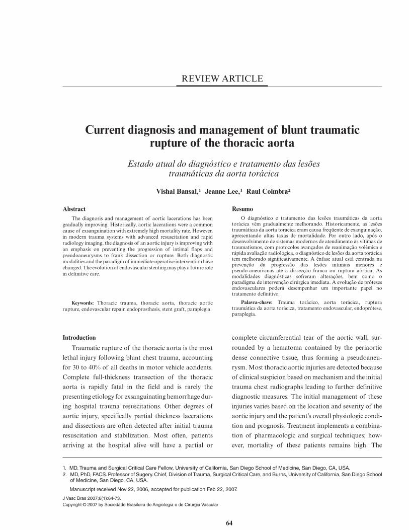
REVIEW ARTICLE
Current diagnosis and management of blunt traumaticrupture of the thoracic aorta
Estado atual do diagnóstico e tratamento das lesõestraumáticas da aorta torácica
Vishal Bansal,1 Jeanne Lee,1 Raul Coimbra2
AbstractThe diagnosis and management of aortic lacerations has been
gradually improving. Historically, aortic lacerations were a commoncause of exsanguination with extremely high mortality rate. However,in modern trauma systems with advanced resuscitation and rapidradiology imaging, the diagnosis of an aortic injury is improving withan emphasis on preventing the progression of intimal flaps andpseudoaneurysms to frank dissection or rupture. Both diagnosticmodalities and the paradigm of immediate operative intervention havechanged. The evolution of endovascular stenting may play a future rolein definitive care.
Keywords: Thoracic trauma, thoracic aorta, thoracic aorticrupture, endovascular repair, endoprosthesis, stent graft, paraplegia.
ResumoO diagnóstico e tratamento das lesões traumáticas da aorta
torácica vêm gradualmente melhorando. Historicamente, as lesõestraumáticas da aorta torácica eram causa freqüente de exanguinação,apresentando altas taxas de mortalidade. Por outro lado, após odesenvolvimento de sistemas modernos de atendimento às vítimas detraumatismos, com protocolos avançados de reanimação volêmica erápida avaliação radiológica, o diagnóstico de lesões da aorta torácicatem melhorado significativamente. A ênfase atual está centrada naprevenção da progressão das lesões intimais menores epseudo-aneurismas até a dissecção franca ou ruptura aórtica. Asmodalidades diagnósticas sofreram alterações, bem como oparadigma de intervenção cirúrgica imediata. A evolução de prótesesendovasculares poderá desempenhar um importante papel notratamento definitivo.
Palavra-chave: Trauma torácico, aorta torácica, rupturatraumática da aorta torácica, tratamento endovascular, endoprótese,paraplegia.
Introduction
Traumatic rupture of the thoracic aorta is the most
lethal injury following blunt chest trauma, accounting
for 30 to 40% of all deaths in motor vehicle accidents.
Complete full-thickness transection of the thoracic
aorta is rapidly fatal in the field and is rarely the
presenting etiology for exsanguinating hemorrhage dur-
ing hospital trauma resuscitations. Other degrees of
aortic injury, specifically partial thickness lacerations
and dissections are often detected after initial trauma
resuscitation and stabilization. Most often, patients
arriving at the hospital alive will have a partial or
complete circumferential tear of the aortic wall, sur-
rounded by a hematoma contained by the periaortic
dense connective tissue, thus forming a pseudoaneu-
rysm. Most thoracic aortic injuries are detected because
of clinical suspicion based on mechanism and the initial
trauma chest radiographs leading to further definitive
diagnostic measures. The initial management of these
injuries varies based on the location and severity of the
aortic injury and the patient’s overall physiologic condi-
tion and prognosis. Treatment implements a combina-
tion of pharmacologic and surgical techniques; how-
ever, mortality of these patients remains high. The
1. MD. Trauma and Surgical Critical Care Fellow, University of California, San Diego School of Medicine, San Diego, CA, USA.2. MD, PhD, FACS. Professor of Sugery. Chief, Division of Trauma, Surgical Critical Care, and Burns, University of California, San Diego School
of Medicine, San Diego, CA, USA.
Manuscript received Nov 22, 2006, accepted for publication Feb 22, 2007.
J Vasc Bras 2007;6(1):64-73.Copyright © 2007 by Sociedade Brasileira de Angiologia e de Cirurgia Vascular
64
emergence of endovascular stenting (EVS) is increas-
ingly playing a role in definitive repair, although long
term efficacy has yet to be established.
Incidence and etiology
Blunt thoracic aortic injury from motor vehicle
accidents (MVA) are the predominant cause in most
Western nations.1 Civilian violence encapsulates most
penetrating aortic trauma, however, given the increased
use of percutaneous cardiac and vascular procedures,
iatrogenic etiology is an increasingly recognized cause of
injury.2
Blunt thoracic aortic injury carries an extremely
high mortality and remains the second leading cause of
death, after head injury, in MVA with more than 80-90%
dying at the scene.3 In the American Association for the
Surgery of Trauma (AAST) multi-center institutional
study, Fabian et al. reported MVA as the causative
factor in greater than 80% of blunt thoracic aortic
injuries.4 In one large retrospective, autopsy study
almost 70% of traumatic thoracic aortic rupture were
due to MVA, 17% from pedestrian versus auto, 8% from
motorcycle accidents, and 4% from falls.5 The incidence
of thoracic aortic transection is rare in the pediatric
population; however, retrospective studies show that
47% of these injuries occur from falls of an average of 6
m high.6
The location of these injuries has given rise to several
proposed mechanisms. The majority of these injuries
involve the proximal descending thoracic aorta, within
millimeters of the ligamentum arteriosum (aortic isth-
mus). The ascending aorta and mid-distal thoracic
aorta represent a less likely location of injury.7 The
descending thoracic aorta remains fixed at the ligamen-
tum arteriosum, and therefore a high velocity impact
causes differential deceleration and a sheering force
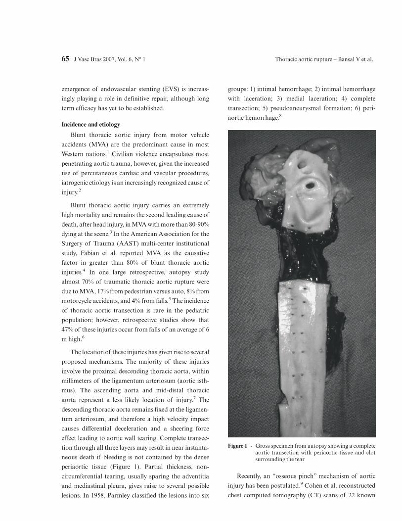
effect leading to aortic wall tearing. Complete transec-
tion through all three layers may result in near instanta-
neous death if bleeding is not contained by the dense
periaortic tissue (Figure 1). Partial thickness, non-
circumferential tearing, usually sparing the adventitia
and mediastinal pleura, gives raise to several possible
lesions. In 1958, Parmley classified the lesions into six
groups: 1) intimal hemorrhage; 2) intimal hemorrhage
with laceration; 3) medial laceration; 4) complete
transection; 5) pseudoaneurysmal formation; 6) peri-
aortic hemorrhage.8
Recently, an “osseous pinch” mechanism of aortic
injury has been postulated.9 Cohen et al. reconstructed
chest computed tomography (CT) scans of 22 known
Figure 1 - Gross specimen from autopsy showing a completeaortic transection with periaortic tissue and clotsurrounding the tear
65 J Vasc Bras 2007, Vol. 6, Nº 1 Thoracic aortic rupture – Bansal V et al.
traumatic thoracic aortic ruptures finding the site of
aortic injury corresponding to sandwiching of the
thoracic aorta between the anterior spine and the
anterior bony complex (sternum, first rib, or clavicle).
Isolated case reports have reported similar findings.10
Regardless of the exact mechanism, patients sustaining
massive chest trauma associated with rapid deceleration
(i.e. MVA or falls) should carry suspicion of thoracic
aortic injury until empiric diagnostic evaluation is
complete.
Diagnosis
The most important factor in preventing a delay in
diagnosis is the arousal of clinical suspicion based on
mechanism of injury.11 Classically, thoracic aortic rup-
ture secondary to blunt trauma has been associated with
head-on MVA.8 However, lateral impact is an often
overlooked mechanism of thoracic aortic injury in
addition to head-on front impact accidents. In a review
of 81 autopsy reports from deaths as a result of thoracic
aortic rupture from MVA, lateral impact from side
MVA were responsible for 49.5% of the injuries.12
Classic signs or symptoms of thoracic aortic injury
may be absent upon presentation. The presence of other
significant chest injuries and the appropriate mecha-
nisms may raise the suspicion of a blunt thoracic aortic
injury (Table 1). Physical examination may reveal differ-
ences in upper extremity pulses or suggest a pseudoco-
arctation syndrome, characterized by upper extremity
hypertension and lower extremity hypotension. Neuro-
logic deficits due to spinal cord ischemia without spinal
fracture rarely occur. Clinical exam is notoriously non-
specific and described findings such as an intracapsular
murmur or pseudocoarctation are unlikely to be appre-
ciated during a trauma resuscitation.13 Sternal and rib
fractures, chest wall ecchymosis, flail segments, hemo/
pneumothorax, solid organ injuries, and pelvic fractures
should all indicate high energy impact and should
heighten awareness of thoracic aortic injury.
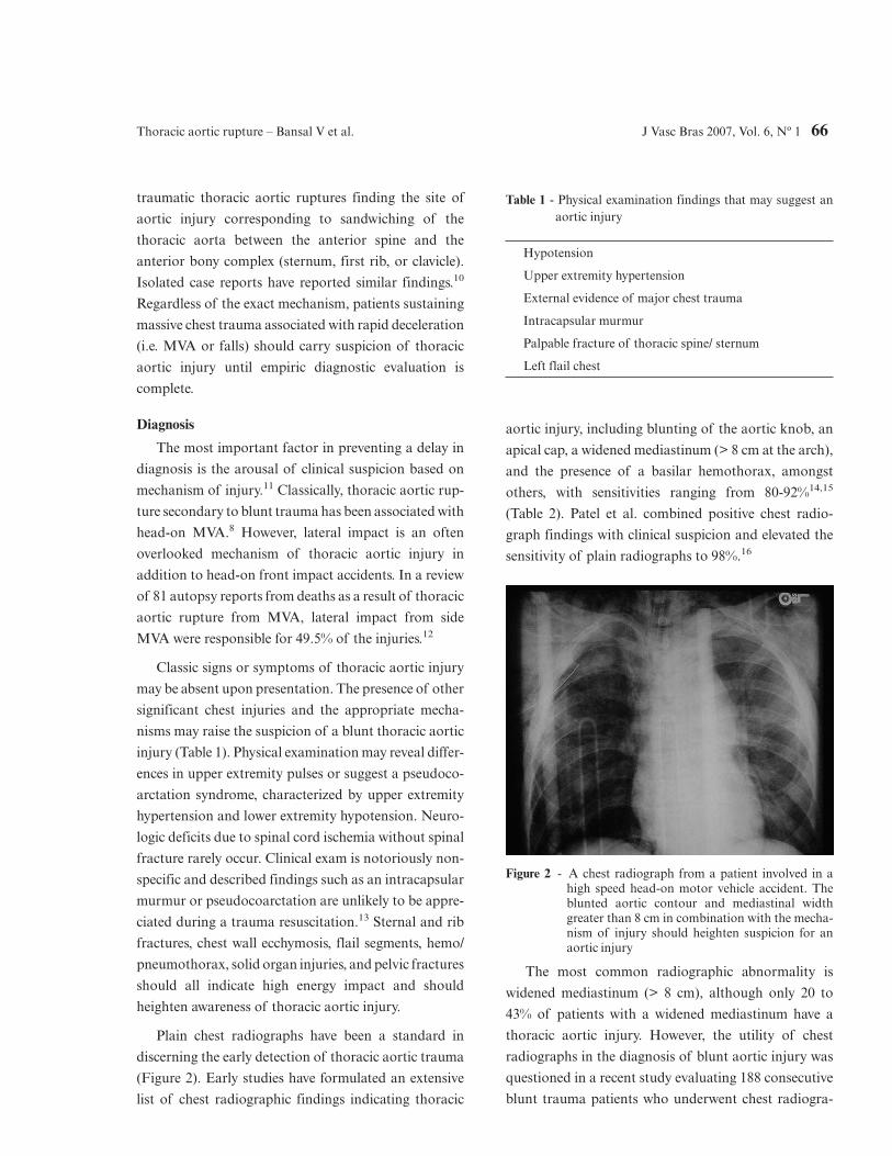
Plain chest radiographs have been a standard in
discerning the early detection of thoracic aortic trauma
(Figure 2). Early studies have formulated an extensive
list of chest radiographic findings indicating thoracic
aortic injury, including blunting of the aortic knob, an
apical cap, a widened mediastinum (> 8 cm at the arch),
and the presence of a basilar hemothorax, amongst
others, with sensitivities ranging from 80-92%14,15
(Table 2). Patel et al. combined positive chest radio-
graph findings with clinical suspicion and elevated the
sensitivity of plain radiographs to 98%.16
The most common radiographic abnormality is
widened mediastinum (> 8 cm), although only 20 to
43% of patients with a widened mediastinum have a
thoracic aortic injury. However, the utility of chest
radiographs in the diagnosis of blunt aortic injury was
questioned in a recent study evaluating 188 consecutive
blunt trauma patients who underwent chest radiogra-
Table 1 - Physical examination findings that may suggest anaortic injury
Hypotension
Upper extremity hypertension
External evidence of major chest trauma
Intracapsular murmur
Palpable fracture of thoracic spine/ sternum
Left flail chest
Figure 2 - A chest radiograph from a patient involved in ahigh speed head-on motor vehicle accident. Theblunted aortic contour and mediastinal widthgreater than 8 cm in combination with the mecha-nism of injury should heighten suspicion for anaortic injury
Thoracic aortic rupture – Bansal V et al. J Vasc Bras 2007, Vol. 6, Nº 1 66
phy and aortography. The study demonstrated that
radiographic signs on plain chest films carry low speci-
ficity and no improvement in overall accuracy.17
Therefore, there are several important factors to
bear in mind when reviewing the literature. Foremost is
the fact that these findings are based on upright chest
radiographs, thus decreasing projection and magnifica-
tion artifact appreciated on supine studies. This is
complicated by the difficulty in obtaining upright plain
films during initial trauma resuscitations secondary to a
patient’s injury status. The finding of a widened medi-
astinum may be an indirect determinant of thoracic
aortic injury being a result more from bleeding small
vessels contained within the mediastinum hemorrhage
than from aortic bleeding or an aortic pseudoaneurysm,
therefore giving low positive predictive values.14 Fur-
thermore, there are several reports documenting tho-
racic aortic rupture with normal initial chest radio-
graphs.18 Therefore, at best, plain chest radiographs can
serve as a marker for further diagnostic evaluation and
should not be used as sole criteria for either excluding or
confirming thoracic aortic injury.
Computed tomography versus angiography versusechocardiography
Given the low positive predictive value of plain chest
radiographs, there has been enduring debate over the
best radiographic technique to establish definitive diag-
nosis. Establishing early diagnosis of thoracic aortic
injuries is essential in order to begin therapeutic inter-
vention.
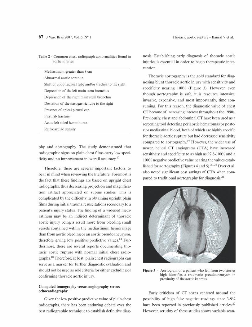
Thoracic aortography is the gold standard for diag-
nosing blunt thoracic aortic injury with sensitivity and
specificity nearing 100% (Figure 3). However, even
though aortography is safe, it is resource intensive,
invasive, expensive, and most importantly, time con-
suming. For this reason, the diagnostic value of chest
CT became of increasing interest throughout the 1990s.
Previously, chest and abdominal CT have been used as a
screening tool detecting periaortic hematomas or poste-
rior mediastinal blood, both of which are highly specific
for thoracic aortic rupture but had decreased sensitivity
compared to aortography.19 However, the wider use of
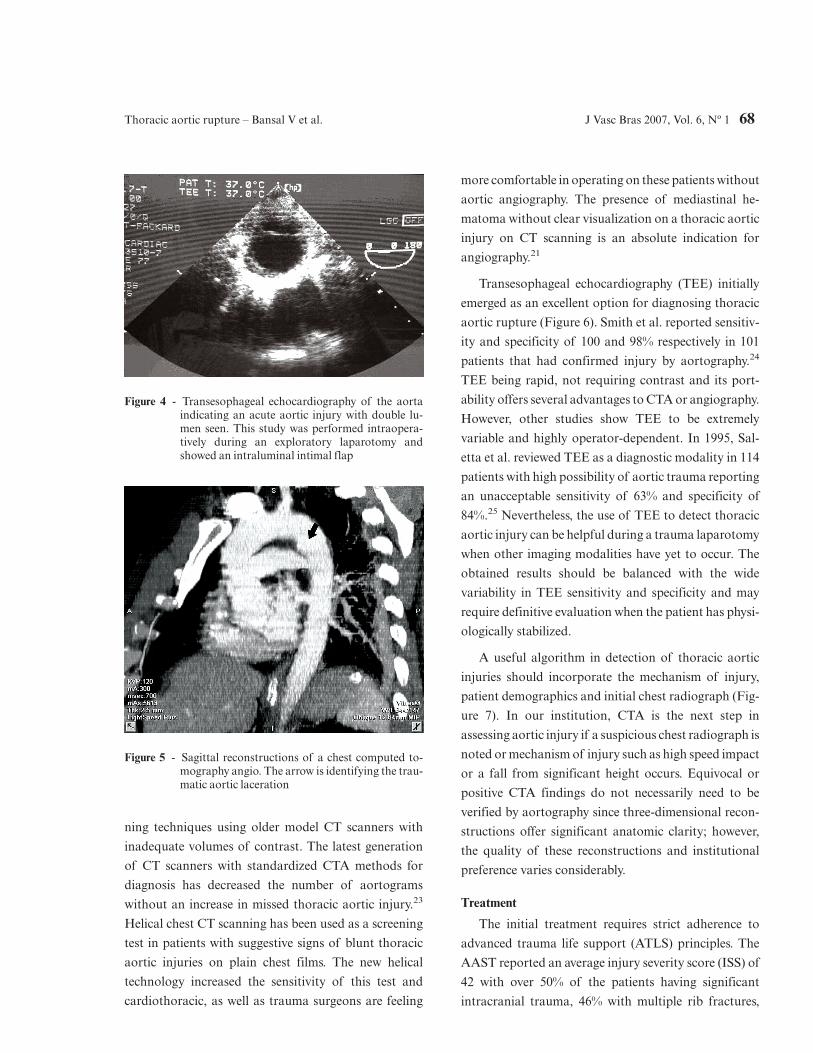
newer, helical CT angiograms (CTA) have increased
sensitivity and specificity to as high as 97.8-100% and a
100% negative predictive value nearing the values estab-
lished for aortography (Figures 4 and 5).20,21 Dyer et al.
also noted significant cost savings of CTA when com-
pared to traditional aortography for diagnosis.21
Early criticism of CT scans centered around the
possibility of high false negative readings since 3-9%
have been reported in previously published articles.22
However, scrutiny of these studies shows variable scan-
Table 2 - Common chest radiograph abnormalities found inaortic injuries
Mediastinum greater than 8 cm
Abnormal aortic contour
Shift of endotracheal tube and/or trachea to the right
Depression of the left main stem bronchus
Depression of the right main stem bronchus
Deviation of the nasogastric tube to the right
Presence of apical pleural cap
First rib fracture
Acute left sided hemothorax
Retrocardiac density
Figure 3 - Aortogram of a patient who fell from two storieshigh identifies a traumatic pseudoaneurysm inproximity of the aortic isthmus
67 J Vasc Bras 2007, Vol. 6, Nº 1 Thoracic aortic rupture – Bansal V et al.
ning techniques using older model CT scanners with
inadequate volumes of contrast. The latest generation
of CT scanners with standardized CTA methods for
diagnosis has decreased the number of aortograms
without an increase in missed thoracic aortic injury.23
Helical chest CT scanning has been used as a screening
test in patients with suggestive signs of blunt thoracic
aortic injuries on plain chest films. The new helical
technology increased the sensitivity of this test and
cardiothoracic, as well as trauma surgeons are feeling
more comfortable in operating on these patients without
aortic angiography. The presence of mediastinal he-
matoma without clear visualization on a thoracic aortic
injury on CT scanning is an absolute indication for
angiography.21
Transesophageal echocardiography (TEE) initially
emerged as an excellent option for diagnosing thoracic
aortic rupture (Figure 6). Smith et al. reported sensitiv-
ity and specificity of 100 and 98% respectively in 101
patients that had confirmed injury by aortography.24
TEE being rapid, not requiring contrast and its port-
ability offers several advantages to CTA or angiography.
However, other studies show TEE to be extremely
variable and highly operator-dependent. In 1995, Sal-
etta et al. reviewed TEE as a diagnostic modality in 114
patients with high possibility of aortic trauma reporting
an unacceptable sensitivity of 63% and specificity of
84%.25 Nevertheless, the use of TEE to detect thoracic
aortic injury can be helpful during a trauma laparotomy
when other imaging modalities have yet to occur. The
obtained results should be balanced with the wide
variability in TEE sensitivity and specificity and may
require definitive evaluation when the patient has physi-
ologically stabilized.
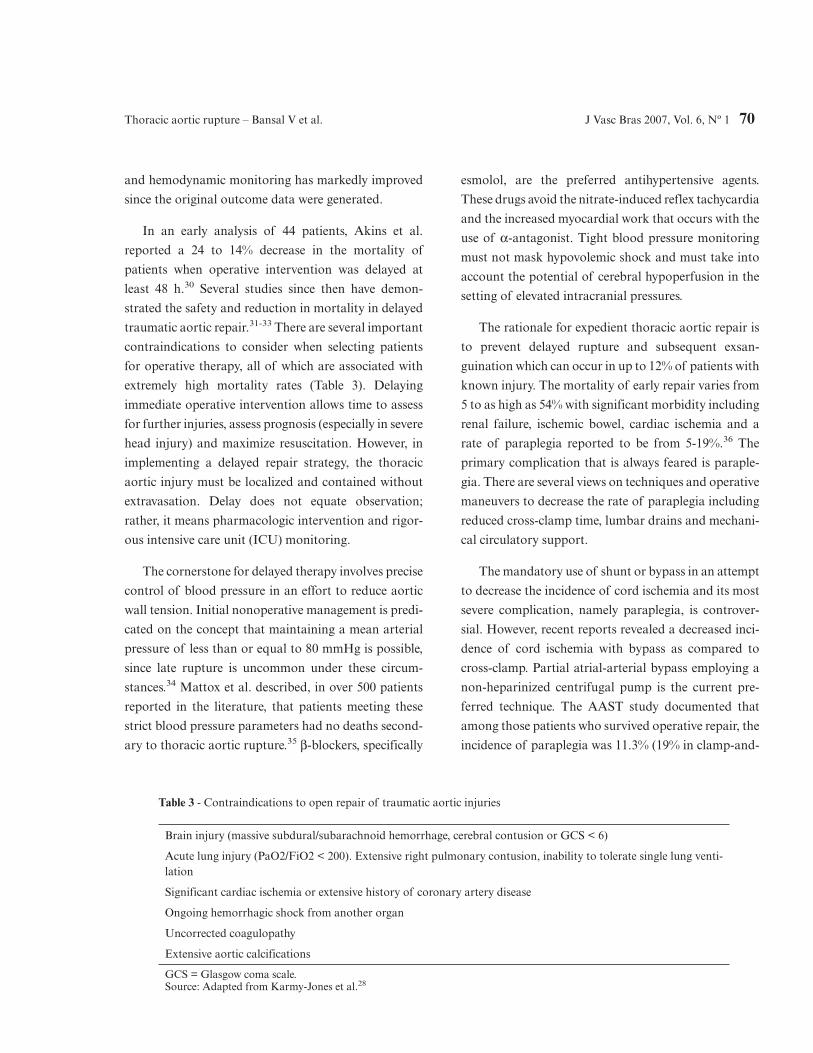
A useful algorithm in detection of thoracic aortic
injuries should incorporate the mechanism of injury,
patient demographics and initial chest radiograph (Fig-
ure 7). In our institution, CTA is the next step in
assessing aortic injury if a suspicious chest radiograph is
noted or mechanism of injury such as high speed impact
or a fall from significant height occurs. Equivocal or
positive CTA findings do not necessarily need to be
verified by aortography since three-dimensional recon-
structions offer significant anatomic clarity; however,
the quality of these reconstructions and institutional
preference varies considerably.
Treatment
The initial treatment requires strict adherence to
advanced trauma life support (ATLS) principles. The
AAST reported an average injury severity score (ISS) of
42 with over 50% of the patients having significant
intracranial trauma, 46% with multiple rib fractures,
Figure 4 - Transesophageal echocardiography of the aortaindicating an acute aortic injury with double lu-men seen. This study was performed intraopera-tively during an exploratory laparotomy andshowed an intraluminal intimal flap
Figure 5 - Sagittal reconstructions of a chest computed to-mography angio. The arrow is identifying the trau-matic aortic laceration
Thoracic aortic rupture – Bansal V et al. J Vasc Bras 2007, Vol. 6, Nº 1 68
and 31% with pelvic fractures (4). Accordingly, the fact
that the majority of hypotension in patients with
thoracic aortic injury is a result of other associated
injuries cannot be overemphasized.26 Although uncon-
trolled hypertension physiologically increases aortic
wall tension and propensity for rupture, overcorrection
and subsequent iatrogenic hypotension increases the
metabolic and physiologic consequences of shock and
can increase the rate of secondary brain injury. Thus,
unless a patient has stabilized, an adequate survey for
concomitant injury has been complete, and the diagno-
sis of thoracic aortic injury has been established, blood
pressure lowering pharmacologic agents should not be
used. Often, pain medication works quickly and suffices
for initial hypertensive control.
Assessment of other injuries will determine the
subsequent management of thoracic aortic injury. Pa-
tients who are in physiologic distress or have significant
solid organ injury often undergo exploratory laparo-
tomy before the diagnosis of thoracic aortic injury has
been assessed. The exact consequence of this on mortal-
ity is unknown. However, Hunt et al. in reviewing 144
patients with diagnosed thoracic aortic trauma did not
find any measurable difference in mortality in patients
undergoing laparotomy before the diagnosis of aortic
injury.27
Once the stability and assessment of a patient have
been achieved and the presence of a thoracic aortic
injury has been established, the subsequent manage-
ment can vary considerably. The degree of aortic
disruption, the safety of controlling blood pressure, and
clinical assessment of the risk of early free rupture
versus the patient’s overall physiologic prognosis deter-
mines whether operative, nonoperative, or endovascular
approaches strategies can be used.28
The traditional approach to traumatic thoracic aor-
tic injury has been urgent and immediate open repair
through a thoracotomy, since in classic teaching delay in
repair is associated with greater than 90% mortality.8
However, the paradigm of timing in the classic ap-
proach must be challenged for a variety of reasons.
Foremost, it is the inherent bias in this patient popula-
tion since aortic injury has a high incidence of associ-
ated injuries, which is the most likely cause of early
demise.29 Second, the advancement of critical care,
injury diagnosis, and the improvement of resuscitation
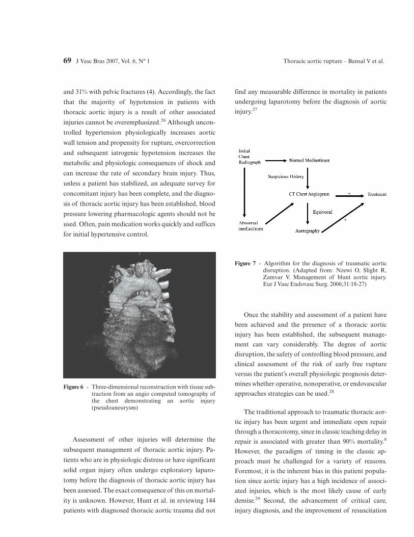
Figure 6 - Three-dimensional reconstruction with tissue sub-traction from an angio computed tomography ofthe chest demonstrating an aortic injury(pseudoaneurysm)
Figure 7 - Algorithm for the diagnosis of traumatic aorticdisruption. (Adapted from: Nzewi O, Slight R,Zamvar V. Management of blunt aortic injury.Eur J Vasc Endovasc Surg. 2006;31:18-27)
69 J Vasc Bras 2007, Vol. 6, Nº 1 Thoracic aortic rupture – Bansal V et al.
and hemodynamic monitoring has markedly improved
since the original outcome data were generated.
In an early analysis of 44 patients, Akins et al.
reported a 24 to 14% decrease in the mortality of
patients when operative intervention was delayed at
least 48 h.30 Several studies since then have demon-
strated the safety and reduction in mortality in delayed
traumatic aortic repair.31-33 There are several important
contraindications to consider when selecting patients
for operative therapy, all of which are associated with
extremely high mortality rates (Table 3). Delaying
immediate operative intervention allows time to assess
for further injuries, assess prognosis (especially in severe
head injury) and maximize resuscitation. However, in
implementing a delayed repair strategy, the thoracic
aortic injury must be localized and contained without
extravasation. Delay does not equate observation;
rather, it means pharmacologic intervention and rigor-
ous intensive care unit (ICU) monitoring.
The cornerstone for delayed therapy involves precise
control of blood pressure in an effort to reduce aortic
wall tension. Initial nonoperative management is predi-
cated on the concept that maintaining a mean arterial
pressure of less than or equal to 80 mmHg is possible,
since late rupture is uncommon under these circum-
stances.34 Mattox et al. described, in over 500 patients
reported in the literature, that patients meeting these
strict blood pressure parameters had no deaths second-
ary to thoracic aortic rupture.35 β-blockers, specifically
esmolol, are the preferred antihypertensive agents.
These drugs avoid the nitrate-induced reflex tachycardia
and the increased myocardial work that occurs with the
use of α-antagonist. Tight blood pressure monitoring
must not mask hypovolemic shock and must take into
account the potential of cerebral hypoperfusion in the
setting of elevated intracranial pressures.
The rationale for expedient thoracic aortic repair is
to prevent delayed rupture and subsequent exsan-
guination which can occur in up to 12% of patients with
known injury. The mortality of early repair varies from
5 to as high as 54% with significant morbidity including
renal failure, ischemic bowel, cardiac ischemia and a
rate of paraplegia reported to be from 5-19%.36 The
primary complication that is always feared is paraple-
gia. There are several views on techniques and operative
maneuvers to decrease the rate of paraplegia including
reduced cross-clamp time, lumbar drains and mechani-
cal circulatory support.
The mandatory use of shunt or bypass in an attempt
to decrease the incidence of cord ischemia and its most
severe complication, namely paraplegia, is controver-
sial. However, recent reports revealed a decreased inci-
dence of cord ischemia with bypass as compared to
cross-clamp. Partial atrial-arterial bypass employing a
non-heparinized centrifugal pump is the current pre-
ferred technique. The AAST study documented that
among those patients who survived operative repair, the
incidence of paraplegia was 11.3% (19% in clamp-and-
Table 3 - Contraindications to open repair of traumatic aortic injuries
Brain injury (massive subdural/subarachnoid hemorrhage, cerebral contusion or GCS < 6)
Acute lung injury (PaO2/FiO2 < 200). Extensive right pulmonary contusion, inability to tolerate single lung venti-lation
Significant cardiac ischemia or extensive history of coronary artery disease
Ongoing hemorrhagic shock from another organ
Uncorrected coagulopathy
Extensive aortic calcifications
GCS = Glasgow coma scale.Source: Adapted from Karmy-Jones et al.28
Thoracic aortic rupture – Bansal V et al. J Vasc Bras 2007, Vol. 6, Nº 1 70
sew and 5.2% when circulatory support was employed).
Many authors believe that mechanical circulatory sup-
port may allow some liberalization of the cross-clamp
time; however, circulatory support may not be feasible
in unstable patients with active bleeding when rapid
repair is essential.
Endovascular approach
The successful use of EVS for aneurysmal disease
has given rise to a new option for treating traumatic
thoracic aortic injury. This method is especially promis-
ing for patients with significant intracerebral hemor-
rhage, pulmonary contusion, or for elderly patients
whose existing comorbidities may preclude an open
repair. Many successful case series have been described
both in the United States and in Europe with minimal
morbidity and mortality.37-39 In a recent publication, 46
blunt thoracic aortic injury patients underwent either
EVS or open thoracotomy. Twenty-two patients under-
went open repair with a 22.7% incidence of paraplegia
and 13.6% 30-day mortality. The stent group showed 0%
paralysis and 8.3% mortality rate.40 In another study, 18
patients underwent repair of blunt thoracic aortic
injuries; six via EVS and 12 through a traditional
thoracotomy. There were no significant differences in
demographics or injury severity between each group.
The open group had a 17% mortality and a 16%
incidence of paraplegia. This is in contrast to a 0% rate
of mortality and paraplegia in the EVS group.41 Peter-
son et al. treated 11 thoracic aortic rupture patients with
commercially available EVS devices with 0% mortality
or paralysis.42
Worldwide, 244 cases of acute traumatic aortic
injuries repaired through endovascular techniques have
been reported to date in MEDLINE. A success rate >
95% and paraplegia rates of 0% have been reported.
Considering all series, the median follow-up has been of
26 months with one death unrelated to the endovascular
approach. The first case series of seven patients treated
by the endovascular approach in Brazil was published
by Mioto Neto et al., in 2005.43 The importance of this
initial publication was emphasized by an editorial in the
J Vasc Bras by von Ristow.44
It is difficult to predict what the future of EVS will
be. To date, there is no long-term Level I evidence to
verify and recommend EVS as the absolute approach
for thoracic aortic injury. However, the short-term
complication rate for EVS is substantially lower when
compared to open repair. Long term complications of
EVS for aortic injury are similar to EVS for aneurysmal
disease and include stent migration, endoleak and stent
collapse. In one of the longest follow-up studies to date,
Hornweg et al. retrospectively analyzed 28 patients in
the Netherlands who underwent EVS repair of blunt
thoracic aortic injury.45 There was no stent-related
mortality on a mean follow-up of 26.5 months with only
one instance of stent collapse that was corrected.
If the location of injury is within 1 cm of the left
subclavian artery, an inadequate neck for EVS will
preclude its usage.46 Other considerations including the
tortuosity of the aorta, iliac or femoral artery injuries
and the length of aortic involvement will not make open
repair obsolete. However, available data and experience
suggest that EVS may herald new practice guidelines
and eventually may replace open repair as the procedure
of choice (Figure 8).
Conclusions
Thoracic aortic rupture remains a highly lethal
injury, with most patients dying rapidly in the field.
Figure 8 - Thoracic aortic stent graft
71 J Vasc Bras 2007, Vol. 6, Nº 1 Thoracic aortic rupture – Bansal V et al.
Suspicion of these injuries should arise from the pa-
tient’s history and plain chest radiograph findings. The
gold standard for diagnosis is aortic angiography;
however, CTA has several advantages with near equiva-
lent sensitivity. If feasible, operative repair should be
delayed until the patient has been adequately resusci-
tated, all concomitant injuries have been identified, and
prognosis has been established. Open repair has a very
high mortality and morbidity yet has established long-
term efficacy. EVS is gaining in popularity and may
surpass traditional open repair as standard of care.
References1. Tambyraja AL, Scollay JM, Beard D, Henry JM, Murie JA,
Chalmers RT. Aortic trauma in Scotland--a population basedstudy. Eur J Vasc Endovasc Surg. 2006;32:686-9. Epub 2006Jun 5.
2. Chen FH, Shim WH, Chang BC, Park SJ, Won JY, Lee DY.False aneurysms at both ends of a descending thoracic aorticstent-graft: complication after endovascular repair of apenetrating atherosclerotic ulcer. J Endovasc Ther.2003;10:249-53.
3. Smith RS, Chang FC. Traumatic rupture of the aorta: still alethal injury. Am J Surg. 1986;152:660-3.
4. Fabian TC, Richardson JD, Croce MA, et al. Prospectivestudy of blunt aortic injury: Multicenter Trial of theAmerican Association for the Surgery of Trauma. J Trauma.1997;42:374-80.
5. Burkhart HM, Gomez GA, Jacobson LE, Pless JE, BroadieTA. Fatal blunt aortic injuries: a review of 242 autopsy cases.J Trauma. 2001;50:113-5.
6. Balci AE, Kazez A, Eren S, Ayan E, Ozalp K, Eren MN. Bluntthoracic trauma in children: review of 137 cases. Eur JCardiothorac Surg. 2004;26:387-92.
7. Feczko JD, Lynch L, Pless JE, Clark MA, McClain J, HawleyDA. An autopsy case review of 142 nonpenetrating (blunt)injuries of the aorta. J Trauma. 1992;33:846-9.
8. Parmley LF, Mattingly TW, Manion WC, Jahnke EJ Jr.Nonpenetrating traumatic injury of the aorta. Circulation.1958;17:1086-101.
9. Cohen AM, Crass JR, Thomas HA, Fisher RG, Jacobs DG.CT evidence for the "osseous pinch" mechanism of traumaticaortic injury. AJR Am J Roentgenol. 1992;159:271-4.
10. Javadpour H, O'Toole JJ, McEniff JN, Luke DA, Young VK.Traumatic aortic transection: evidence for the osseous pinchmechanism. Ann Thorac Surg. 2002;73:951-3.
11. Blackmore CC, Zweibel A, Mann FA. Determining risk oftraumatic aortic injury: how to optimize imaging strategy.AJR Am J Roentgenol. 2000;174:343-7.
12. Katyal D, McLellan BA, Brenneman FD, Boulanger BR,Sharkey PW, Waddell JP. Lateral impact motor vehiclecollisions: significant cause of blunt traumatic rupture of thethoracic aorta. J Trauma. 1997;42:769-72.
13. Cook J, Salerno C, Krishnadasan B, Nicholls S, Meissner M,Karmy-Jones R. The effect of changing presentation andmanagement on the outcome of blunt rupture of the thoracicaorta. J Thorac Cardiovasc Surg. 2006;131:594-600.
14. Fishman JE. Imaging of blunt aortic and great vessel trauma.J Thorac Imaging. 2000;15:97-103.
15. Mirvis SE, Bidwell JK, Buddemeyer EU, et al. Value of chestradiography in excluding traumatic aortic rupture. Radiology.1987;163:487-93.
16. Patel NH, Stephens KE Jr., Mirvis SE, Shanmuganathan K,Mann FA. Imaging of acute thoracic aortic injury due to blunttrauma: a review. Radiology. 1998;209:335-48.
17. Cook AD, Klein JS, Rogers FB, Osler TM, Shackford SR.Chest radiographs of limited utility in the diagnosis of blunttraumatic aortic laceration. J Trauma. 2001;50:843-7.
18. Symbas PJ, Horsley WS, Symbas PN. Rupture of theascending aorta caused by blunt trauma. Ann Thorac Surg.1998;66:113-7.
19. Wong H, Gotway MB, Sasson AD, Jeffrey RB. Periaortichematoma at diaphragmatic crura at helical CT: sign of bluntaortic injury in patients with mediastinal hematoma.Radiology. 2004;231:185-9.
20. Mirvis SE, Shanmuganathan K, Buell J, Rodriguez A. Use ofspiral computed tomography for the assessment of blunttrauma patients with potential aortic injury. J Trauma.1998;45:922-30.
21. Dyer DS, Moore EE, Mestek MF, et al. Can chest CT be usedto exclude aortic injury? Radiology. 1999;213:195-202.
22. Miller FB, Richardson JD, Thomas HA, Cryer HM, WillingSJ. Role of CT in diagnosis of major arterial injury after bluntthoracic trauma. Surgery. 1989;106:596-602; discussion 602-3.
23. Bruckner BA, DiBardino DJ, Cumbie TC, et al. Criticalevaluation of chest computed tomography scans for bluntdescending thoracic aortic injury. Ann Thorac Surg.2006;81:1339-46.
24. Smith MD, Cassidy JM, Souther S, et al. Transesophagealechocardiography in the diagnosis of traumatic rupture of theaorta. N Engl J Med. 1995;332:356-62.
25. Saletta S, Lederman E, Fein S, Singh A, Kuehler DH, FortuneJB. Transesophageal echocardiography for the initialevaluation of the widened mediastinum in trauma patients. JTrauma. 1995;39:137-41; discussion 141-2.
26. Hudson HM 2nd, Woodson J, Hirsch E. The management oftraumatic aortic tear in the multiply-injured patient. Ann VascSurg. 1991;5:445-8.
27. Hunt JP, Baker CC, Lentz CW, et al. Thoracic aorta injuries:management and outcome of 144 patients. J Trauma.1996;40:547-55.
28. Karmy-Jones R, Jurkovich GJ. Blunt chest trauma. CurrProbl Surg. 2004;41:211-380.
29. Karmy-Jones R, Carter YM, Nathens A, et al. Impact ofpresenting physiology and associated injuries on outcomefollowing traumatic rupture of the thoracic aorta. Am Surg.2001;67:61-6.
30. Akins CW, Buckley MJ, Daggett W, McIlduff JB, Austen WG.Acute traumatic disruption of the thoracic aorta: a ten-yearexperience. Ann Thorac Surg. 1981;31:305-9.
Thoracic aortic rupture – Bansal V et al. J Vasc Bras 2007, Vol. 6, Nº 1 72
31. Galli R, Pacini D, Di Bartolomeo R, et al. Surgical indicationsand timing of repair of traumatic ruptures of the thoracicaorta. Ann Thorac Surg. 1998;65:461-4.
32. Pate JW, Fabian TC, Walker W. Traumatic rupture of theaortic isthmus: an emergency? World J Surg. 1995;19:119-25;discussion 125-6.
33. Maggisano R, Nathens A, Alexandrova NA, et al. Traumaticrupture of the thoracic aorta: should one always operateimmediately? Ann Vasc Surg. 1995;9:44-52.
34. Carter YM, Karmy-Jones R, Aldea GS. Delayed surgicalmanagement of a traumatic aortic arch injury. Ann ThoracSurg. 2002;73:294-6.
35. Mattox KL, Wall MJ Jr. Historical review of blunt injury tothe thoracic aorta. Chest Surg Clin N Am. 2000;10:167-82.
36. von Oppell UO, Dunne TT, De Groot MK, Zilla P. Traumaticaortic rupture: twenty-year metaanalysis of mortality and riskof paraplegia. Ann Thorac Surg. 1994;58:585-93.
37. Rousseau H, Soula P, Perreault P, et al. Delayed treatment oftraumatic rupture of the thoracic aorta with endoluminalcovered stent. Circulation. 1999;99:498-504.
38. Lachat ML, Pfammatter T, Witzke HJ, et al. Endovascularrepair with bifurcated stent-grafts under local anaesthesia toimprove outcome of ruptured aortoiliac aneurysms. Eur JVasc Endovasc Surg. 2002;23:528-36.
39. Brandt M, Hussel K, Walluscheck KP, et al. Stent-graft repairversus open surgery for the descending aorta: a case-controlstudy. J Endovasc Ther. 2004;11:535-8.
40. Andrassy J, Weidenhagen R, Meimarakis G, Lauterjung L,Jauch KW, Kopp R. Stent versus open surgery for acute andchronic traumatic injury of the thoracic aorta: a single-centerexperience. J Trauma. 2006;60:765-71.
41. Ott MC, Stewart TC, Lawlor DK, Gray DK, Forbes TL.Management of blunt thoracic aortic injuries: endovascularstents versus open repair. J Trauma. 2004;56:565-70.
42. Peterson BG, Matsumura JS, Morasch MD, West MA,Eskandari MK. Percutaneous endovascular repair of bluntthoracic aortic transection. J Trauma. 2005;59:1062-5.
43. Mioto Neto B, Aun R, Estenssoro AEV, Puech-Leão P.Tratamento das lesões de aorta nos traumatismos torácicosfechados. J Vasc Bras. 2005;4:217-26.
44. Von Ristow A. Tratamento das lesões de aorta nostraumatismos torácicos fechados [editorial]. J Vasc Bras.2005;4:215-6.
45. Hoornweg LL, Dinkelman MK, Goslings JC, et al.Endovascular management of traumatic ruptures of thethoracic aorta: a retrospective multicenter analysis of 28 casesin The Netherlands. J Vasc Surg. 2006;43:1096-102; discussion1102.
46. Borsa JJ, Hoffer EK, Karmy-Jones R, et al. Angiographicdescription of blunt traumatic injuries to the thoracic aortawith specific relevance to endograft repair. J Endovasc Ther.2002;9 Suppl 2:II84-91.
Correspondence:Raul CoimbraUniversity of California San Diego200 W. Arbor Drive, 8896San Diego, CA92103-8896E-mail: [email protected]
73 J Vasc Bras 2007, Vol. 6, Nº 1 Thoracic aortic rupture – Bansal V et al.