Characterization and phylogenetic affiliation of ...
16
Genetics and Molecular Research 16 (3): gmr16039703 Characterization and phylogenetic affiliation of Actinobacteria from tropical soils with potential uses for agro-industrial processes J.C.M. Dornelas 1 , J.E.F. Figueiredo 2 , C.S. de Abreu 3 , U.G.P. Lana 2 , C.A. Oliveira 2 and I.E. Marriel 1,2 1 Programa de Pós-Graduação em Ciências Agrárias, Departamento de Ciências Agrárias, Universidade Federal de São João Del Rei, Sete Lagoas, MG, Brasil 2 Laboratório de Microbiologia e Biologia Molecular, Embrapa Milho e Sorgo, Sete Lagoas, MG, Brasil 3 Programa de Pós-Graduação em Microbiologia, Departamento de Microbiologia, Universidade Federal de Minas Gerais, Belo Horizonte, MG, Brasil Corresponding author: I.E. Marriel E-mail: [email protected] Genet. Mol. Res. 16 (3): gmr16039703 Received April 17, 2017 Accepted July 3, 2017 Published August 31, 2017 DOI http://dx.doi.org/10.4238/gmr16039703 Copyright © 2017 The Authors. This is an open-access article distributed under the terms of the Creative Commons Attribution ShareAlike (CC BY-SA) 4.0 License. ABSTRACT. Secondary metabolites produced by Actinobacteria of tropical soils represent a largely understudied source of novel molecules with relevant application in medicine, pharmaceutical and food industries, agriculture, and environmental bioremediation. The present study aimed to characterize sixty-nine Actinobacteria isolated from compost and tropical soils using morphological, biochemical, and molecular methods. All the isolates showed high variation for morphological traits considering the color of pigments of the aerial and vegetative mycelium and spore chain morphology. The enzymatic activity of amylase, cellulase, and lipase was highly variable. The amylase activity was detected in 53 (76.81%) isolates. Eighteen
Transcript of Characterization and phylogenetic affiliation of ...
Genetics and Molecular Research 16 (3): gmr16039703
Characterization and phylogenetic affiliation of Actinobacteria from tropical soils with potential uses for agro-industrial processes
J.C.M. Dornelas1, J.E.F. Figueiredo2, C.S. de Abreu3, U.G.P. Lana2, C.A. Oliveira2 and I.E. Marriel1,2
1Programa de Pós-Graduação em Ciências Agrárias, Departamento de Ciências Agrárias, Universidade Federal de São João Del Rei, Sete Lagoas, MG, Brasil2Laboratório de Microbiologia e Biologia Molecular, Embrapa Milho e Sorgo, Sete Lagoas, MG, Brasil3Programa de Pós-Graduação em Microbiologia, Departamento de Microbiologia, Universidade Federal de Minas Gerais, Belo Horizonte, MG, Brasil
Corresponding author: I.E. MarrielE-mail: [email protected]
Genet. Mol. Res. 16 (3): gmr16039703Received April 17, 2017Accepted July 3, 2017Published August 31, 2017DOI http://dx.doi.org/10.4238/gmr16039703
Copyright © 2017 The Authors. This is an open-access article distributed under the terms of the Creative Commons Attribution ShareAlike (CC BY-SA) 4.0 License.
ABSTRACT. Secondary metabolites produced by Actinobacteria of tropical soils represent a largely understudied source of novel molecules with relevant application in medicine, pharmaceutical and food industries, agriculture, and environmental bioremediation. The present study aimed to characterize sixty-nine Actinobacteria isolated from compost and tropical soils using morphological, biochemical, and molecular methods. All the isolates showed high variation for morphological traits considering the color of pigments of the aerial and vegetative mycelium and spore chain morphology. The enzymatic activity of amylase, cellulase, and lipase was highly variable. The amylase activity was detected in 53 (76.81%) isolates. Eighteen
2J.C.M. Dornelas et al.
Genetics and Molecular Research 16 (3): gmr16039703
isolates showed enzymatic index (EI) > 4.0, and the isolates ACJ 45 (Streptomyces curacoi) and ACSL 6 (S. hygroscopicus) showed the highest EI values (6.44 and 6.42, respectively). The cellulase activity varied significantly (P ≤ 0.05) among the isolates. Twenty-nine isolates (42.02%) showed high cellulase activity, and the isolates ACJ 48 (S. chiangmaiensis) and ACJ 53 (S. cyslabdanicus) showed the highest EI values (6.56 for both isolates). The lipase activity varied statistically (P ≤ 0.05) with fourteen isolates (20.29%) considered good lipase producers (EI > 2.0). The isolate ACSL 6 (S. hygroscopicus) showed the highest EI value of 2.60. Molecular analysis of partial 16S rRNA gene sequencing revealed the existence of 49 species, being 38 species with only one representative member and 11 species represented by one or more strains. All species belonged to three genera, namely Streptomyces (82.61%), Amycolatopsis (7.25%), and Kitasatospora (10.14%). The present results showed the high biotechnological potential of different Actinobacteria from tropical soils.
Key words: Actinomycetes; Morphological traits; Enzymatic activity; 16S rRNA gene; Molecular identification
INTRODUCTION
The phylum Actinobacteria (Actinomycetes) is an ancient bacterial group branched off from the other prokaryotes very early in the evolutionary process (Battistuzzi et al., 2004; Ventura et al., 2007). Actinobacteria comprise an ecologically diverse group ubiquitously distributed in various natural environments as free-living, pathogens, and endophyte symbionts (Hardoim et al., 2015). They are distinguished as Gram-positive bacteria, normally aerobic, non-acid fast and with a high GC content in their DNA, varying to less than 50% in a few species to more than 70% in some genera (Lewin et al., 2016).
Actinobacteria exhibit a high level of diversity of biochemical features, such as the production of a wide variety of secondary metabolites and extracellular enzymes with relevant applications in different fields (Suneetha et al., 2011; Barka et al., 2015; Li et al., 2016). Although near half of the bioactive molecules with different uses in medicine, industrial processes, agriculture, and environmental bioremediation are produced by Actinobacteria, it represents only a small fraction of the overall metabolites already identified in this bacterial group (Abid et al., 2016). New enzymes selected based on enzymatic index (EI) criterion have been extensively explored commercially by the food, textile, and biofuel industry. Amylase, lipase, cellulase are some examples of enzymes isolated from Actinobacteria and currently used in the global market (Sathya and Ushadevi, 2014). Thus, the understudied Actinobacteria from tropical soils may represent a promising new source of secondary metabolites for many purposes.
The taxonomy of Actinobacteria has been a subject of intense debate. Traditionally, morphological traits as growth pattern and mycelia type are the main characteristics used to define order, genera, and species in this group, and biochemical tests using enzyme activities are used for identifying new bioactive metabolites. Recently, molecular data based on DNA sequencing of the 16S rRNA gene assumed an important role on systematic of the phylum Actinobacteria (Ventura et al., 2007).
3Actinobacteria from tropical soils
Genetics and Molecular Research 16 (3): gmr16039703
In the present study, morphological traits, enzymatic activities, and partial sequencing of the 16S rRNA gene were used for characterizing Actinobacteria isolated from Brazilian tropical soils.
MATERIAL AND METHODS
The study was performed with sixty-nine bacterial isolates deposited at the Embrapa Maize and Sorghum Multifunctional Culture Collection (CCMF-CNPMS) that were previously identified as Actinobacteria based on their growth morphology (Table 1).
Table 1. Collection location (origin and location sites), the year at which the material was collected, and identification of the Actinobacteria isolates.
Origin Location site Year Identification Cerrado soil with high level of phosphate at the Experimental Station of Embrapa Sete Lagoas 2000 ACSL 1A, 2, 8 Cerrado soil from the Experimental Station of Embrapa Sete Lagoas 2001 ACSL 12, 16A Cerrado soil from the Experimental Station of Embrapa Sete Lagoas 2004 ACSL 485, 490, 495, 509, 517 Cerrado soil from the Experimental Station of Embrapa Sete Lagoas 2014 ACSL 1B, 6, 13, 16B, 18B, 22, 23, 27B Maize Rhizospheral soil from the Experimental Station of Embrapa Sete Lagoas 2004 ACSL 7, 18A, 25, 27A, 50, 53, 54, 64B, 67, 77, 82, 91 Organic farming from the Experimental Station of Embrapa Sete Lagoas 2002 ACSL 432, 448, 449, 450, 453, 457, 470 Cerrado soil planted with eucalyptus Sete Lagoas 2001 ACSL 64A, 80, 83, 85, 93, 115 Cerrado soil planted with eucalyptus and pinus woods Sete Lagoas 2003 ACSL 404 Cerrado soil from the Fazenda Santa Rita Experimental Station Prudente de Morais 2006 ACPM 641 Cerrado soil from the Fazenda Santa Rita Experimental Station Prudente de Morais 2007 ACPM 5, 29, 31, 38, 66 Cerrado soil planted with peanuts from the Fazenda Santa Rita Experimental Station Prudente de Morais 2002 ACPM 346, 363, 364 Agricultural Cerrado soil from the Experimental Station of Embrapa Jaíba 2001 ACJ 66, 76 Cerrado degradated soil Jaíba 2001 ACJ 1, 17, 26, 29 Mata seca Jaíba 2001 ACJ 36, 43, 45 Protected area Jaíba 2001 ACJ 48, 49, 51, 52, 53 Compost Papagaios 2013 ACP 35 Compost Capim Branco 2015 ACCB 1
The isolates were grown in the agar glycerol-asparagine (AGA) medium [1 g/L L-asparagine, 10 g/L glycerol, 1 g/L KH2PO4, 15 g/L agar, 1 mL/L micronutrient solution (0.1 g FeSO4×7H2O, 0.1 g MnCl2×4H2O, 0.1 g ZnSO4×7H2O, qsp 100 mL deionized water)] according to Pridham and Lyons (1961) and supplemented with 0.03 g/L cycloheximide. After inoculations, the plates were incubated for 14 days at 28°C.
Morphological characterization
The morphological analysis was performed according to Shirling and Gottlieb (1966). The following growing cultural parameters were evaluated: the color of the vegetative and aerial mycelium, and changes in the color of the medium around the colony.
For the micromorphological analysis, the microculture technique was used according to Holt et al. (1994). After the incubation period, the coverslip was removed and placed on another sterile microscopic slide containing 10 µL Amann lactophenol and the edges were sealed with colorless nail enamel. The spore chain morphology was observed under the optical microscope Olympus BX 60 (Olympus Optical Co. Ltd., Tokyo, Japan) with 1000X magnification, and photographed with a digital camera (Leica DFC 490, Leica Microsystems Inc., Buffalo Grove, IL, USA).
Molecular characterization
The DNA extraction, PCR, and DNA sequencing were made according to Lana et al. (2012). The 16S rRNA gene was amplified using the universal primers 8F (5'-AGA GTT
4J.C.M. Dornelas et al.
Genetics and Molecular Research 16 (3): gmr16039703
TGA TCC TGG CTC AG-3') and 1492R (5'-GGT TAC CTT GTT ACG ACT T-3') (Turner et al., 1999). The sequencing was made with the PCR primers and the internal primers 515F (5'-GTG CCA GCM GCC GCG GTA A-3'; Turner et al., 1999) and 902R (5'-GTC AAT TCI TTT GAG TTT YAR YC-3'; Hodkinson and Lutzoni, 2009). In the primers, the letters Y; M; R; and I represent the nucleotides cytosine or thymine; adenine or cytosine; adenine or guanine; and a modified guanine, respectively.
The DNA sequences were generated in the Applied Biosystems 3500xL Genetic Analyzer (Applied Biosystem, Foster City, CA, USA). The nucleotide sequences were edited using the Sequencher 4.1 program and compared in the GenBank database (http://www.ncbi.nlm.nih.gov/) through the BLAST N program (Altschul et al., 1997) located at the NCBI (National Center for Biotechnology Information).
Phylogenetic analysis
The phylogenetic analysis was performed by the neighbor-joining method using the Molecular Evolutionary Genetics Analysis version 5 (MEGA5) program (Tamura et al., 2011).
Enzymatic activity
To test the enzymatic activity of amylase, cellulase, and lipase, disks of cultures on agar plate were inoculated in triplicate in a completely random design and incubated at 28°C for 10 days (amylase and cellulase) and 72 h (lipase). The EI was estimated by the following equation: EI = ratio of halo diameter/ratio of colony diameter. The isolates were classified as non-producer (EI = 0), low producer (0 > EI ≤ 2), middle producer (2 > EI ≤ 4), and high producer (EI > 4). The data were analyzed for significance (P < 0.05) using the Scott-Knott test.
Amylase
The amylase production was determined as described by Coon et al. (1957) modified by the addition of 6.6% soluble starch. The isolates were inoculated on the starch agar medium (6.6 g/L soluble starch, 0.5 g/L sodium chloride, 3 g/L meat extract, 1 g/L peptone casein, 15 g/L agar with the pH adjusted to 7.0). After culture growth, 10 mL Lugol’s solution (5 g iodine, 10 g potassium iodide in 100 mL distilled water) was diluted to 1:10 and added to the dishes. The amylase production was detected by the formation of a light yellow zone around the colony corresponding to the discoloration of the medium.
Cellulase
The cellulase production was tested in the culture medium supplemented with carboxymethylcellulose (CMC) as the sole carbon source (3 g/L NaNO3, 1 g/L K2HPO4, 0.5 g/L MgSO4, 0.5 g/L KCl, 10 mg/L FeSO4.7H2O, 10 g/L CMC, 15 g/L agar with the pH adjusted to 7.0) according to Lewis (1988). After incubation, 10 mL 0.5% Congo red dye was added to each plate, incubated for 15 min at room temperature and washed with NaCl (5 M). Afterward, the excess of solution was discarded, and 10 mL NaCl solution (1 M) was added to each plate and incubated for 30 min at room temperature. The production of the enzyme was observed by the discoloration of the medium, which forms an orange zone around the colony.
5Actinobacteria from tropical soils
Genetics and Molecular Research 16 (3): gmr16039703
Lipase
Lipase production was tested according to Savitha et al. (2007) using a culture medium with the following composition: 5 g/L peptone, 1 g/L yeast extract, 4 g/L sodium chloride, 15 g/L agar, 31.25 mL/L olive oil, 0.01 g/L Rhodamine B, pH 7.0. Disks of the cultured isolates were inoculated in the medium, and after the incubation period, the plates were exposed to ultraviolet radiation to observe the formation of blue stained halos around the positive colonies, which is the parameter used to indicate the activity of the enzyme (Colen, 2006).
RESULTS
In this study, the analysis of morphological traits, enzyme activities, and molecular sequencing revealed a high level of variability among sixty-nine isolates of Actinobacteria from composts and tropical soils.
Morphological characterization
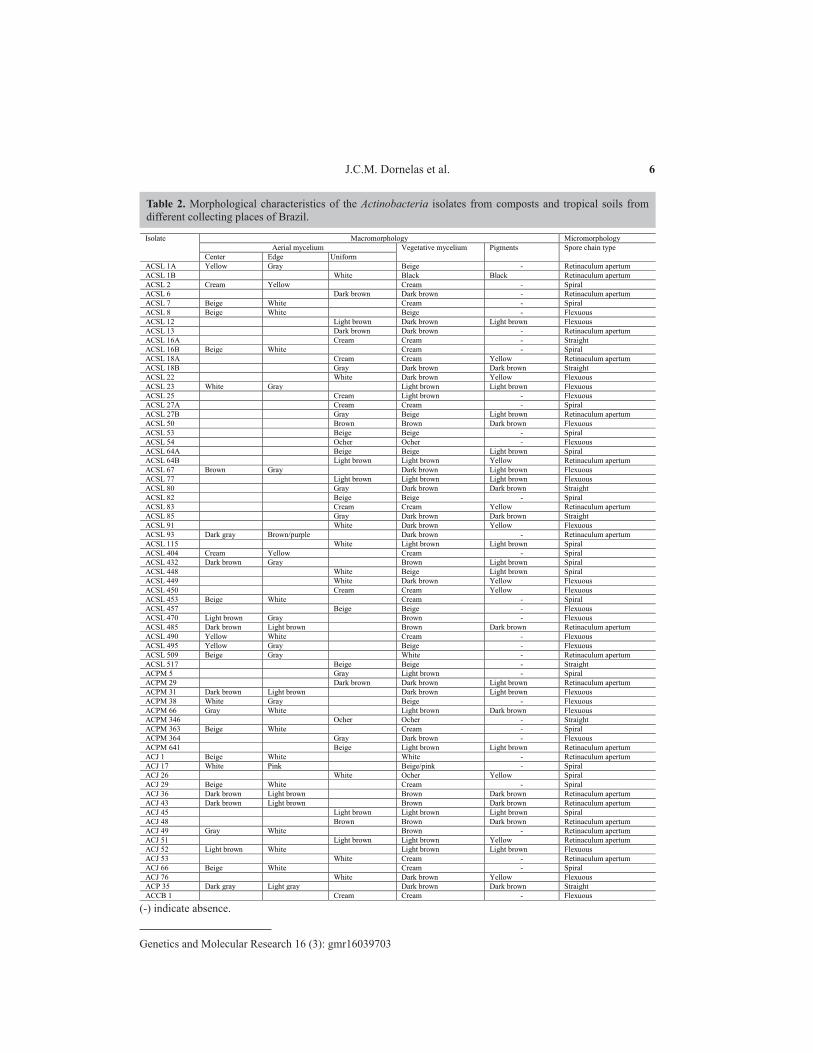
The analyses of the four main morphological traits used for Actinobacteria identification were highly variable (Table 2). Besides these characteristics, the production of classic pulverulent mycelium and radial growth confirmed the identity of the isolates as Actinobacteria. All sixty-nine morphospecies produced vegetative mycelium on the AGA medium with evident variation in the pattern of development, color, and pigment production (Figure 1). The release of soluble pigments on the AGA medium was observed in 50.72% of the isolates and varied from yellow (28.57%), light brown (40%), and dark brown (28.57%) to black (2.86%).
The micromorphological features of the bacterial colonies were assessed with a light microscope to determine the presence, absence, and morphology of the spore chain compared with valid criteria reported in the Bergey’s Manual of Systematic Bacteriology (Holt et al., 1994) (Table 2). The morphological structure of the spore chain varied depending on the isolate, and was classified as straight (10.14%), retinaculum apertum (28.99%), spiral (27.54%), and flexuous (33.33%) (Figure 2). The phylogenetic tree constructed using morphological and biochemical data showed all species grouped together (data not shown).
Molecular characterization and phylogenetic analysis
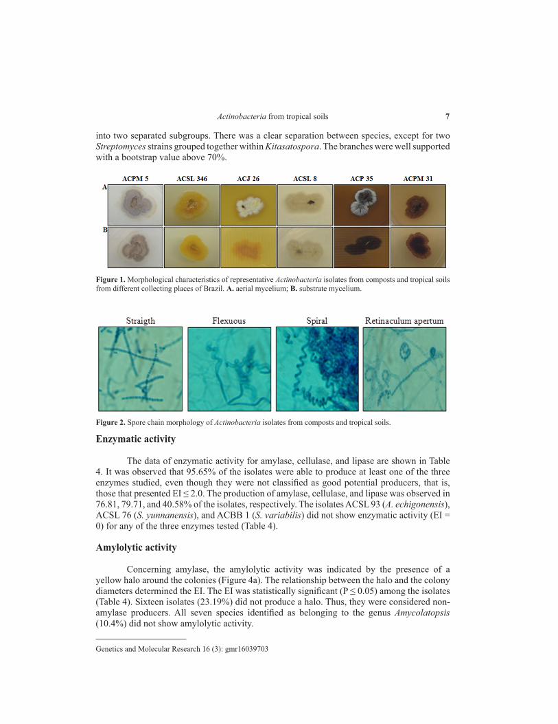
Partial sequences of the 16S rRNA gene, ranging in length from 1210 to 1400 nucleotides, were determined for 69 isolates. The molecular characterization based on nucleotide comparisons of the 16S rRNA gene with nucleotide sequences deposited in the GenBank (accession numbers: KY585931 to KY585999) confirmed the morphological identity of the isolates as Actinobacteria (Table 3). A total of forty-nine taxa were identified distributed among the following three genera: Streptomyces (82.61%), Amycolatopsis (10.14%), and Kitasatospora (7.25%).
The phylogenetic tree constructed by the maximum likelihood, evolutionary distance, and maximum parsimony methods with the MEGA5 program (Tamura et al., 2011) generated two distinct clades with Amycolatopsis separated from Streptomyces and Kitasatospora (Figure 3). The clade formed by the genera Streptomyces and Kitasatospora was divided
6J.C.M. Dornelas et al.
Genetics and Molecular Research 16 (3): gmr16039703
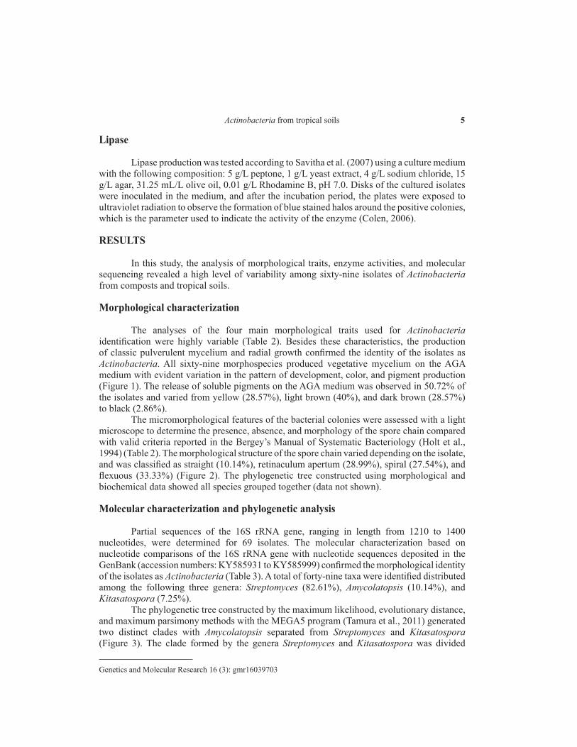
Isolate Macromorphology Micromorphology Aerial mycelium Vegetative mycelium Pigments Spore chain type
Center Edge Uniform ACSL 1A Yellow Gray Beige - Retinaculum apertum ACSL 1B White Black Black Retinaculum apertum ACSL 2 Cream Yellow Cream - Spiral ACSL 6 Dark brown Dark brown - Retinaculum apertum ACSL 7 Beige White Cream - Spiral ACSL 8 Beige White Beige - Flexuous ACSL 12 Light brown Dark brown Light brown Flexuous ACSL 13 Dark brown Dark brown - Retinaculum apertum ACSL 16A Cream Cream - Straight ACSL 16B Beige White Cream - Spiral ACSL 18A Cream Cream Yellow Retinaculum apertum ACSL 18B Gray Dark brown Dark brown Straight ACSL 22 White Dark brown Yellow Flexuous ACSL 23 White Gray Light brown Light brown Flexuous ACSL 25 Cream Light brown - Flexuous ACSL 27A Cream Cream - Spiral ACSL 27B Gray Beige Light brown Retinaculum apertum ACSL 50 Brown Brown Dark brown Flexuous ACSL 53 Beige Beige - Spiral ACSL 54 Ocher Ocher - Flexuous ACSL 64A Beige Beige Light brown Spiral ACSL 64B Light brown Light brown Yellow Retinaculum apertum ACSL 67 Brown Gray Dark brown Light brown Flexuous ACSL 77 Light brown Light brown Light brown Flexuous ACSL 80 Gray Dark brown Dark brown Straight ACSL 82 Beige Beige - Spiral ACSL 83 Cream Cream Yellow Retinaculum apertum ACSL 85 Gray Dark brown Dark brown Straight ACSL 91 White Dark brown Yellow Flexuous ACSL 93 Dark gray Brown/purple Dark brown - Retinaculum apertum ACSL 115 White Light brown Light brown Spiral ACSL 404 Cream Yellow Cream - Spiral ACSL 432 Dark brown Gray Brown Light brown Spiral ACSL 448 White Beige Light brown Spiral ACSL 449 White Dark brown Yellow Flexuous ACSL 450 Cream Cream Yellow Flexuous ACSL 453 Beige White Cream - Spiral ACSL 457 Beige Beige - Flexuous ACSL 470 Light brown Gray Brown - Flexuous ACSL 485 Dark brown Light brown Brown Dark brown Retinaculum apertum ACSL 490 Yellow White Cream - Flexuous ACSL 495 Yellow Gray Beige - Flexuous ACSL 509 Beige Gray White - Retinaculum apertum ACSL 517 Beige Beige - Straight ACPM 5 Gray Light brown - Spiral ACPM 29 Dark brown Dark brown Light brown Retinaculum apertum ACPM 31 Dark brown Light brown Dark brown Light brown Flexuous ACPM 38 White Gray Beige - Flexuous ACPM 66 Gray White Light brown Dark brown Flexuous ACPM 346 Ocher Ocher - Straight ACPM 363 Beige White Cream - Spiral ACPM 364 Gray Dark brown - Flexuous ACPM 641 Beige Light brown Light brown Retinaculum apertum ACJ 1 Beige White White - Retinaculum apertum ACJ 17 White Pink Beige/pink - Spiral ACJ 26 White Ocher Yellow Spiral ACJ 29 Beige White Cream - Spiral ACJ 36 Dark brown Light brown Brown Dark brown Retinaculum apertum ACJ 43 Dark brown Light brown Brown Dark brown Retinaculum apertum ACJ 45 Light brown Light brown Light brown Spiral ACJ 48 Brown Brown Dark brown Retinaculum apertum ACJ 49 Gray White Brown - Retinaculum apertum ACJ 51 Light brown Light brown Yellow Retinaculum apertum ACJ 52 Light brown White Light brown Light brown Flexuous ACJ 53 White Cream - Retinaculum apertum ACJ 66 Beige White Cream - Spiral ACJ 76 White Dark brown Yellow Flexuous ACP 35 Dark gray Light gray Dark brown Dark brown Straight ACCB 1 Cream Cream - Flexuous
(-) indicate absence.
Table 2. Morphological characteristics of the Actinobacteria isolates from composts and tropical soils from different collecting places of Brazil.
7Actinobacteria from tropical soils
Genetics and Molecular Research 16 (3): gmr16039703
into two separated subgroups. There was a clear separation between species, except for two Streptomyces strains grouped together within Kitasatospora. The branches were well supported with a bootstrap value above 70%.
Figure 1. Morphological characteristics of representative Actinobacteria isolates from composts and tropical soils from different collecting places of Brazil. A. aerial mycelium; B. substrate mycelium.
Figure 2. Spore chain morphology of Actinobacteria isolates from composts and tropical soils.
Enzymatic activity
The data of enzymatic activity for amylase, cellulase, and lipase are shown in Table 4. It was observed that 95.65% of the isolates were able to produce at least one of the three enzymes studied, even though they were not classified as good potential producers, that is, those that presented EI ≤ 2.0. The production of amylase, cellulase, and lipase was observed in 76.81, 79.71, and 40.58% of the isolates, respectively. The isolates ACSL 93 (A. echigonensis), ACSL 76 (S. yunnanensis), and ACBB 1 (S. variabilis) did not show enzymatic activity (EI = 0) for any of the three enzymes tested (Table 4).
Amylolytic activity
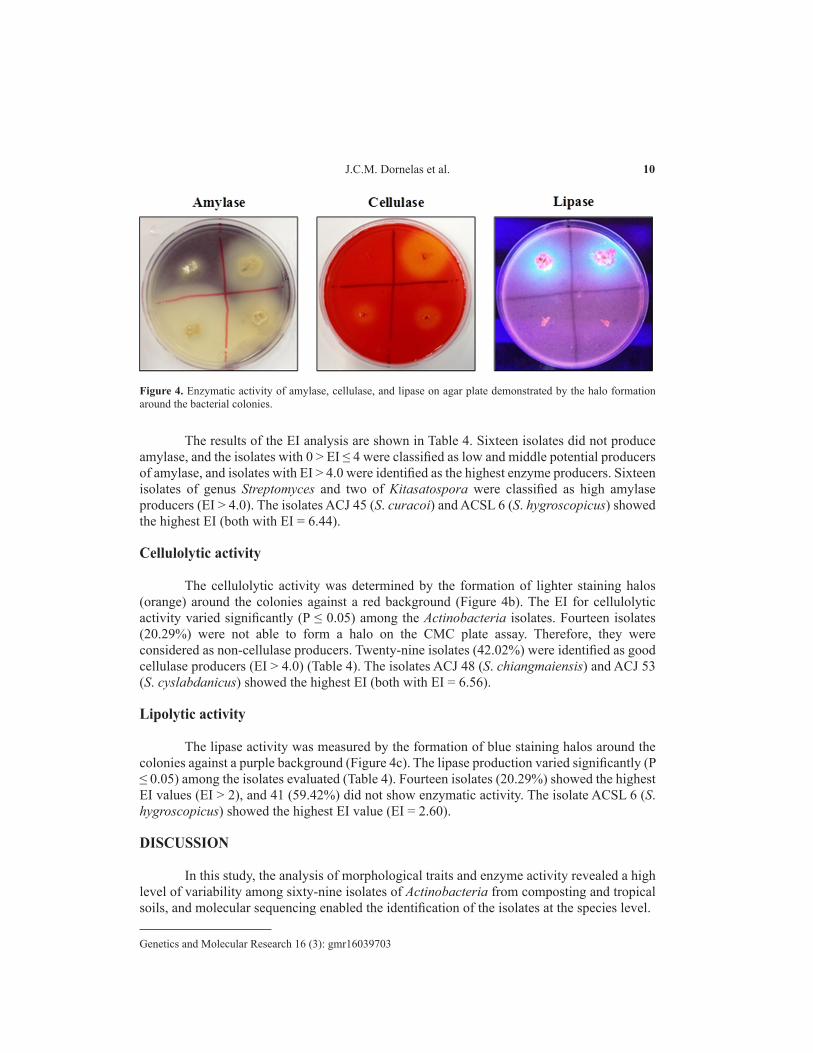
Concerning amylase, the amylolytic activity was indicated by the presence of a yellow halo around the colonies (Figure 4a). The relationship between the halo and the colony diameters determined the EI. The EI was statistically significant (P ≤ 0.05) among the isolates (Table 4). Sixteen isolates (23.19%) did not produce a halo. Thus, they were considered non-amylase producers. All seven species identified as belonging to the genus Amycolatopsis (10.4%) did not show amylolytic activity.
8J.C.M. Dornelas et al.
Genetics and Molecular Research 16 (3): gmr16039703
Isolate
GenBank accession No.
Species Similarity (%) Isolate
GenBank accession No.
Species Similarity (%)
ACSL 1A KY585949 Streptomyces seymenliensis 99 ACSL 450 KY585946 A. bullii 98 ACSL 1B KY585960 S. massasporeus 99 ACSL 453 KY585958 S. galbus 99 ACSL 2 KY585933 S. chartreusis 99 ACSL 457 KY585981 A. pretoriensis 99 ACSL 6 KY585976 S. hygroscopicus 100 ACSL 470 KY585944 S. pseudovenezuelae 100 ACSL 7 KY585992 S. galbus 99 ACSL 485 KY585964 S. psammoticus 100 ACSL 8 KY585974 S. sporocinereus 100 ACSL 490 KY585977 A. kentuckyensis 100 ACSL 12 KY585963 Kitasatospora atroaurantiaca 99 ACSL 495 KY585979 A. lexingtonensis 99 ACSL 13 KY585937 S. hygroscopicus 100 ACSL 509 KY585980 S. deserti 99 ACSL 16A KY585957 S. purpeofuscus 99 ACSL 517 KY585931 S. phaeochromogenes 98 ACSL 16B KY585934 S. galbus 99 ACPM 5 KY585962 S. olivochromogenes 99 ACSL 18A KY585996 S. longwoodensis 99 ACPM 29 KY585969 S. scabiei 99 ACSL 18B KY585951 S. phaeochromogenes 98 ACPM 31 KY585941 S. phaeopurpureus 99 ACSL 22 KY585986 S. yunnanensis 99 ACPM 38 KY585942 S. rishiriensis 99 ACSL 23 KY585972 S. indiaensis 100 ACPM 66 KY585984 S. sioyaensis 99 ACSL 25 KY585988 Amycolatopsis rifamycinica 99 ACPM 346 KY585953 S. endophyticus 99 ACSL 27A KY585995 S. lydicus 99 ACPM 363 KY585940 S. galbus 99 ACSL 27B KY585943 S. corchorusii 99 ACPM 364 KY585935 K. viridis 99 ACSL 50 KY585998 S. sampsonii 99 ACPM 641 KY585959 S. lannensis 100 ACSL 53 KY585990 K. paracochleata 99 ACJ 1 KY585945 S. ossamyceticus 100 ACSL 54 KY585991 S. sasae 99 ACJ 17 KY585947 S. bangladeshensis 99 ACSL 64A KY585999 S. coacervatus 99 ACJ 26 KY585978 S. capoamus 99 ACSL 64B KY585987 S. griseoruber 99 ACJ 29 KY585967 S. galbus 99 ACSL 67 KY585994 S. phaeopurpureus 100 ACJ 36 KY585975 S. psammoticus 100 ACSL 77 KY585997 K. phosalacinea 99 ACJ 43 KY585970 S. psammoticus 99 ACSL 80 KY585938 S. phaeochromogenes 98 ACJ 45 KY585954 S. curacoi 99 ACSL 82 KY585989 K. paracochleata 99 ACJ 48 KY585971 S. chiangmaiensis 100 ACSL 83 KY585936 S. longwoodensis 99 ACJ 49 KY585948 A. rhabdoformis 100 ACSL 85 KY585983 S. phaeochromogenes 99 ACJ 51 KY585968 S. griseoruber 100 ACSL 91 KY585993 S. yunnanensis 99 ACJ 52 KY585973 S. yaanensis 100 ACSL 93 KY585966 A. echigonensis 99 ACJ 53 KY585932 S. cyslabdanicus 99 ACSL 115 KY585939 S. thioluteus 99 ACJ 66 KY585965 S. galbus 99 ACSL 404 KY585950 S. chartreusis 100 ACJ 76 KY585955 S. yunnanensis 99 ACSL 432 KY585961 S. novaecaesareae 99 ACP 35 KY585956 S. laculatispora 99 ACSL 448 KY585952 S. sioyaensis 99 ACCB 1 KY585985 S. variabilis 100 ACSL 449 KY585982 S. yunnanensis 99 - - - -
Table 3. Molecular characterization of the Actinobacteria isolates from composts and tropical soils from different Brazilian collecting places.
Figure 3. Dendrogram based on partial sequences of the 16S rRNA gene of the Actinobacteria isolates from composts and tropical soils.
9Actinobacteria from tropical soils
Genetics and Molecular Research 16 (3): gmr16039703
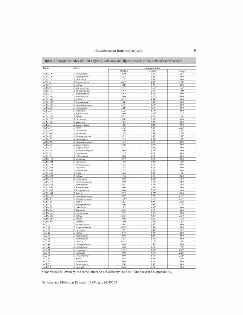
Mean values followed by the same letters do not differ by the Scott-Knott test at 5% probability.
Table 4. Enzymatic index (EI) for amylase, cellulase, and lipase activity of the Actinobacteria isolates.
Isolate Species Enzymatic index Amylase Cellulase Lipase
ACSL 1A S. seymenliensis 2.20i 3.36e 2.17b ACSL 1B S. massasporeus 2.67h 3.42e 0.00 ACSL 2 S. chartreusis 1.52k 5.55b 0.00 ACSL 6 S. hygroscopicus 6.42a 4.00e 2.60a ACSL 7 S. galbus 5.10d 3.78e 0.00 ACSL 8 S. sporocinereus 5.43c 3.67e 1.22e ACSL 12 K. atroaurantiaca 4.81d 2.93f 1.46d ACSL 13 S. hygroscopicus 4.87d 5.33b 0.00 ACSL 16A S. purpeofuscus 0.00 4.17d 0.00 ACSL 16B S. galbus 4.13f 4.58d 0.00 ACSL 18A S. longwoodensis 3.40g 3.83e 0.00 ACSL 18B S. phaeochromogenes 2.93h 4.33d 0.00 ACSL 22 S. yunnanensis 3.15g 0.00 0.00 ACSL 23 S. indiaensis 3.22g 5.13c 0.00 ACSL 25 A. rifamycinica 0.00 4.39d 2.02b ACSL 27A S. lydicus 4.31f 0.00 1.66c ACSL 27B S. corchorusii 5.00d 5.50b 0.00 ACSL 50 S. sampsonii 3.13g 3.93e 0.00 ACSL 53 K. paracochleata 1.65k 3.44e 0.00 ACSL 54 S. sasae 3.42g 0.00 2.23b ACSL 64A S. coacervatus 3.60g 2.86f 2.12b ACSL 64B S. griseoruber 1.13m 2.73f 2.20b ACSL 67 S. phaeopurpureus 3.21g 3.92e 2.16b ACSL 77 K. phosalacinea 2.14i 4.21d 1.79c ACSL 80 S. phaeochromogenes 1.24l 0.00 0.00 ACSL 82 K. paracochleata 6.00b 3.75e 0.00 ACSL 83 S. longwoodensis 2.77h 4.92c 0.00 ACSL 85 S. phaeochromogenes 0.00 4.28d 0.00 ACSL 91 S. yunnanensis 2.71h 0.00 0.00 ACSL 93 A. echigonensis 0.00 0.00 0.00 ACSL 115 S. thioluteus 2.37i 0.00 0.00 ACSL 404 S. chartreusis 1.98j 3.90e 1.52d ACSL 432 S. novaecaesareae 4.93d 3.17f 0.00 ACSL 448 S. sioyaensis 2.61h 0.00 0.00 ACSL 449 S. yunnanensis 3.27g 1.78g 0.00 ACSL 450 A. bullii 0.00 3.98e 1.68c ACSL 453 S. galbus 1.96j 0.00 0.00 ACSL 457 A. pretoriensis 0.00 4.42d 1.47d ACSL 470 S. pseudovenezuelae 4.00f 4.33d 0.00 ACSL 485 S. psammoticus 4.52e 4.53d 1.51d ACSL 490 A. kentuckyensis 0.00 2.89f 0.00 ACSL 495 A. lexingtonensis 0.00 3.20f 0.00 ACSL 509 S. deserti 2.29i 4.27d 0.00 ACSL 517 S. phaeochromogenes 2.55h 5.70b 2.07b ACPM 5 S. olivochromogenes 3.29g 3.58e 2.07b ACPM 29 S. scabiei 5.52c 5.08c 2.10b ACPM 31 S. phaeopurpureus 3.39g 4.07d 2.00b ACPM 38 S. rishiriensis 2.83h 4.47d 0.00 ACPM 66 S. Sioyaensis 4.18f 0.00 0.00 ACPM 346 S. endophyticus 3.07g 3.56e 0.00 ACPM 363 S. galbus 2.62h 5.06c 2.27b ACPM 364 K. viridis 0.00 0.00 1.93b ACPM 641 S. lannensis 1.90j 2.75f 1.37e ACJ 1 S. ossamyceticus 5.14d 4.42d 0.00 ACJ 17 S. bangladeshensis 4.28f 5.08c 0.00 ACJ 26 S. capoamus 3.29g 3.83e 1.75c ACJ 29 S. galbus 2.17i 4.75c 0.00 ACJ 36 S. psammoticus 0.00 5.00c 0.00 ACJ 43 S. psammoticus 4.51e 1.83g 0.00 ACJ 45 S. curacoi 6.44a 4.17d 1.13e ACJ 48 S. chiangmaiensis 0.00 6.56a 0.00 ACJ 49 A. rhabdoformis 0.00 0.00 1.58d ACJ 51 S. griseoruber 3.45g 4.55d 2.23b ACJ 52 S. yaanensis 0.00 4.58d 2.13b ACJ 53 S. cyslabdanicus 0.00 6.56a 2.08b ACJ 66 S. galbus 2.03j 3.55e 0.00 ACJ 76 S. yunnanensis 0.00 0.00 0.00 ACP 35 S. laculatispora 5.70c 3.67e 0.00 ACCB 1 S. variabilis 0.00 0.00 0.00
10J.C.M. Dornelas et al.
Genetics and Molecular Research 16 (3): gmr16039703
The results of the EI analysis are shown in Table 4. Sixteen isolates did not produce amylase, and the isolates with 0 > EI ≤ 4 were classified as low and middle potential producers of amylase, and isolates with EI > 4.0 were identified as the highest enzyme producers. Sixteen isolates of genus Streptomyces and two of Kitasatospora were classified as high amylase producers (EI > 4.0). The isolates ACJ 45 (S. curacoi) and ACSL 6 (S. hygroscopicus) showed the highest EI (both with EI = 6.44).
Cellulolytic activity
The cellulolytic activity was determined by the formation of lighter staining halos (orange) around the colonies against a red background (Figure 4b). The EI for cellulolytic activity varied significantly (P ≤ 0.05) among the Actinobacteria isolates. Fourteen isolates (20.29%) were not able to form a halo on the CMC plate assay. Therefore, they were considered as non-cellulase producers. Twenty-nine isolates (42.02%) were identified as good cellulase producers (EI > 4.0) (Table 4). The isolates ACJ 48 (S. chiangmaiensis) and ACJ 53 (S. cyslabdanicus) showed the highest EI (both with EI = 6.56).
Lipolytic activity
The lipase activity was measured by the formation of blue staining halos around the colonies against a purple background (Figure 4c). The lipase production varied significantly (P ≤ 0.05) among the isolates evaluated (Table 4). Fourteen isolates (20.29%) showed the highest EI values (EI > 2), and 41 (59.42%) did not show enzymatic activity. The isolate ACSL 6 (S. hygroscopicus) showed the highest EI value (EI = 2.60).
DISCUSSION
In this study, the analysis of morphological traits and enzyme activity revealed a high level of variability among sixty-nine isolates of Actinobacteria from composting and tropical soils, and molecular sequencing enabled the identification of the isolates at the species level.
Figure 4. Enzymatic activity of amylase, cellulase, and lipase on agar plate demonstrated by the halo formation around the bacterial colonies.
11Actinobacteria from tropical soils
Genetics and Molecular Research 16 (3): gmr16039703
Morphological characterization
The analysis of morphological characteristics based on valid criteria described in the Bergey’s Manual of Systematic Bacteriology (Holt et al., 1994) confirmed all isolates as Actinobacteria. The high morphological diversity and growth habit, characterized by the formation of highly differentiated hyphae and branched aerial mycelium with formation of spores, have been extensively used as valid criteria for identification of genera and species of the phylum Actinobacteria (Ventura et al., 2007; Suneetha et al., 2011; Barka et al., 2015). The grouping of six morphological characters and three enzymatic parameters showed the existence of fifty-seven different morphospecies. Twelve isolates distributed into five different groups and showing the same morphological pattern were considered to represent reisolates of the same sample. In the present study, only the AGA medium was used for bacterial growth. However, it is necessary to point out that the parameter color of the pigment used as a criterion for discriminating among the Actinobacterial isolates may change considerably depending on various cultural factors such medium composition, pH, and temperature of incubation (Holt et al., 1994).
Enzymatic activity
Enzymes of microbial origin present a great variety of catalytic activities with many applications in various industrial and biotechnological processes, especially in the textile and food industry (Luz et al., 2016). In the present study, the enzymatic activities of amylase, cellulase, and lipase were examined on agar media containing starch, carboxymethylcellulose, and olive oil as substrates, respectively. All but three isolates showed the ability to produce at least one of the three enzymes tested, proving their potential for industrial applications. Members of the three genera identified in the present study are of great importance due to their ability to produce compounds for medical, pharmaceutical, and agricultural purposes.
Amylase activity
The microbial amylase (EC 3.2.1.1) is among the most relevant classes of enzymes due to the wide range application in industrial biotechnological processes such as processed food, fermentation, and pharmaceutical purposes (de Souza and Oliveira Magalhães, 2010; Adrio and Demain, 2014).
The EI has been used a fast tool for selecting and comparing the enzyme production among different bacterial isolates (Carrim et al., 2006; Castro et al., 2014). Fungaro and Maccheroni (2002) suggested that EI greater than 1.0 are a reliable indicator for the presence of enzymes excreted by microorganisms, while EI ≥ 2.0 is considered good indicator for potential enzyme production by a bacterium (Lealem and Gashe, 1994). However, while for lipase the variation in EI is minimal (~2.0), for cellulase and lipase the scale of EI values vary between >2.0 to >6.0. In the current EI scales, differences of only 0.1 in the scale used to measure enzymatic activity represent a great difference in the potential for enzyme production. In our study, we introduced a new grade of variation in the scale for selecting bacterial species candidates for application in biotechnological processes. In our study, isolates with EI values of 2 > EI ≤ 4 were considered middle producer, and only EI values above 4.0 were considered high producers. Thus, the isolates with EI values >4 were selected for further studies aiming at biotechnological applications.
12J.C.M. Dornelas et al.
Genetics and Molecular Research 16 (3): gmr16039703
The production of amylase by microorganisms is very sensitive to incubation temperature. Minotto et al. (2014), performing the enzymatic characterization of endophytic Actinobacteria from tomato plants, observed that the highest EI value recorded for starch degradation was 6.46 when the microorganisms were incubated at 28°C. Also, results of EI values by Karanja et al. (2010) varied between 3.4 and 5.2 for starch degradation by Streptomyces sp isolates from soil in Kenya. In our study, 32 isolates showed similar results for amylase production with EI values varying between 3.0 and 6.44 using a different medium, and the same incubation temperature (28°C).
In our study, none of the seven Amycolatopsis isolates showed amylase activity corroborating the study by Ding et al. (2007). In fact, the absence of amylase activity is one important diagnostic feature of this genus of Actinobacteria.
Cellulase activity
Cellulose, xylan, and lignin (lignocellulose) are the three primary constituents of plant biomass (Yu et al., 2017). These are the most abundant biopolymers in the planet, therefore, the main renewable resources that can be used to produce glucose and proteins, industrial fertilizer, biofuel, and compost (Ramírez and Calzadíaz, 2016). Recently, cellulolytic enzymes of bacterial origin have received special attention of the bioenergy industry due to the higher bacterial growth, sustainability, and environmental impact compared with non-renewable fossil fuel counterparts. However, the industrial-scale breakdown of lignocellulosic plant biomass into simple sugars that can be converted into biofuels is one of the major barriers to the lignocellulosic ethanol production (Lewin et al., 2016).
In Kenya, Karanja et al. (2010) found EI values between 3.4 and 5.2 for starch degradation by Streptomyces sp isolated from soil. In Brazil, Minotto et al. (2014), analyzing Actinobacteria isolated from tomato cultivated in Cerrado soil, and Silva et al. (2015), studying Streptomyces isolated from rhizosphere soil from semi-arid climates, found highest EI values of 4.04 and 6.90, respectively. In our study, 31 isolates of Streptomyces presented EI values between 4.0 and 6.56 indicating their high biotechnological potential uses for large-scale industrial enzyme production and composting.
In our study, two Streptomyces (ACP 35 and ACCB 1) from compost did not produce cellulases. Rodrigues (2006) found that 9% of Streptomyces isolates were not able to degrade cellulose. Considering that the amount of cellulose in the composting process is higher than that available in other soils, the ability to degrade cellulose confers to Streptomyces an important role in the composting processes. Thus, the Streptomyces isolates with the highest EI observed in the present study may be tested for degrading lignocellulose in composting.
Lipase activity
The enzyme lipase (EC 3.1.1.3) is an important catalyst in biotechnology due to its wide versatility. Lipase can be applied in different industrial processes such as food processing, oils and fats, detergent manufacturing, drug synthesis, cosmetics, and many other products. Microbial lipases have also been used in grease trap waste for the treatment of heavy oils and grease (Bornscheuer, 2002).
The diverse characteristics of lipases produced by microorganisms appear to have evolved to guarantee the fast and efficient access of the microorganism to different sources of
13Actinobacteria from tropical soils
Genetics and Molecular Research 16 (3): gmr16039703
organic matter (Roveda et al., 2010). Therefore, this group of enzymes is very attractive for industrial applications and recycling processes of lipid-rich compounds (Bornscheuer, 2002).
Despite the knowledge on lipid metabolism in many species of Streptomyces, studies on lipolytic activity in this group are still incipient. Mohamed et al. (2015), evaluating the lipase activity of streptomycetes from soil samples of Taif (Saudi Arabia), detected lipolytic activity in 91.3% of the isolates. Regarding the EI, Karanja et al. (2010) observed values varying between 3.0 and 4.2 for the lipolytic activity of Streptomyces isolated from soils in Kenya.
In the present study, the highest EI value for lipase was only 2.60, which was lower than the lowest EI value measured by Karanja et al. (2010). These results may be due to the close relationship between the abundance of lipid compounds in the environment and the specific characteristics of the enzymes produced by each microorganism (Gomes et al., 2007). Nowadays, the use of immobilized lipase or whole cell catalysts is one of the most promising methods to produce renewable and environmentally friendly alternative biofuels compared to the non-renewable fossil combustible (Yan et al., 2014).
Molecular characterization
The molecular data analysis based on the partial sequencing of the 16S rRNA gene enabled the identification of a total of 49 species, being 38 represented by only one isolate and 11 molecular species with more than one strain. Four of the twelve reisolates identified by the morphological traits were confirmed by the molecular analysis.
The three genera of Actinobacteria identified comprise two distinct families of the order Actinomycetales. The genus Amycolatopsis belong to the family Pseudonocardiaceae and the genera Streptomyces and Kitasatospora are classified in the family Streptomycetaceae. However, the three genera are very closely related genetically, and many strains have been misidentified by different authors as belonging to any of these three genera (Ward and Bora, 2015).
Cluster analysis of the morphological and biochemical data grouped the three genera together (data not shown). However, considering only the molecular data, two clusters were observed with the genus Amycolatopsis separated from Streptomyces and Kitasatospora. These results using different criteria reinforce the idea that these subgroups of the phylum Actinobacteria may be recognized as representing a complex of intimately related species, thus difficult to be separated into distinct taxa with the current methods used for species and genus identification. Even studies using detailed molecular data analysis of the 16S rRNA gene, a consensus does not exist about the status of various taxa among systematists of the phylum Actinobacteria. In fact, the variations within the 16S rRNA genes of the Actinobacterial group, even in the variable regions, are insufficient to clarify doubts concerning the identification of species and to estimate the species divergence within a genus or the phylogenetic relationships among genera (Kämpfer et al., 2014); this is the case for the genus Kitasatospora proposed by Omura et al. (1982), which the identity as a valid genus has been changed within years (Zhang et al., 1997; Girard et al., 2014). Bacterial strains identified as belonging to Kitasatospora and Streptomyces exhibit similar lifestyle and morphological traits (Ichikawa et al., 2010). In fact, the differentiation between Kitasatospora and Streptomyces strains is only based on the composition of peptidoglycan in the cell wall. Therefore, the presence of LL-isomer of diaminopimelic acid (DAP) in the aerial mycelia and meso-DAP in the vegetative mycelia of Kitasatospora is the main criterion used to discriminate strains between Kitasatospora and Streptomyces, while the latter display LL-DAP in both the aerial and vegetative mycelia
14J.C.M. Dornelas et al.
Genetics and Molecular Research 16 (3): gmr16039703
(Zhang et al., 1997; Takahashi, 2017). Likewise, representatives of the genus Amycolatopsis and other six genera in the family Pseudonocardiaceae, which was proposed based on the 16S rRNA sequence analysis, vary greatly in their morphology and other phenotypic characteristics (Embley et al., 1988). However, the current members of Pseudonocardiaceae contain meso-DAP in their cell wall, and the presence of arabinose and galactose sugars in whole-cell hydrolysates is used as accepted criterion for the diagnosis and species identification in this group (Embley et al., 1988).
Comprehensive comparative studies including protein-coding gene sequences with higher phylogenetic resolution and genome-based studies are needed to clarify the relationships and species delineation within the Streptomycetaceae (Kämpfer et al., 2014).
CONCLUSION
The ability to degrade several substrates reveals that isolates from the genus Streptomyces, Kitasatospora, and Amycolatopsis of the phylum Actinobacteria have high biotechnological potential uses and may be used in future studies intended to new sources for enzyme production.
Conflicts of interest
The authors declare no conflict of interest.
ACKNOWLEDGMENTS
Research supported by Universidade Federal de São João del-Rei (UFSJ), Rede Mineira de Endofíticos and Embrapa Milho e Sorgo (CNPMS). We thank the anonymous reviewers for reviewing this manuscript.
REFERENCES
Abid I, Mujamammi R and Alkahtani MDF (2016). Antimicrobial activity and molecular identification of Streptomyces strains isolated from Saudi Arabia. J. Environ. Biol. 37: 1225-1230. https://doi.org/10.22438/jeb/37/6/prn.97
Adrio JL and Demain AL (2014). Microbial enzymes: tools for biotechnological processes. Biomolecules 4: 117-139. https://doi.org/10.3390/biom4010117
Altschul SF, Madden TL, Schäffer AA, Zhang J, et al. (1997). Gapped BLAST and PSI-BLAST: a new generation of protein database search programs. Nucleic Acids Res. 25: 3389-3402. https://doi.org/10.1093/nar/25.17.3389
Barka EA, Vatsa P, Sanchez L, Gaveau-Vaillant N, et al. (2015). Taxonomy, physiology, and natural products of Actinobacteria. Microbiol. Mol. Biol. Rev. 80: 1-43. https://doi.org/10.1128/MMBR.00019-15
Battistuzzi FU, Feijao A and Hedges SB (2004). A genomic timescale of prokaryote evolution: insights into the origin of methanogenesis, phototrophy, and the colonization of land. BMC Evol. Biol. 4: 44. https://doi.org/10.1186/1471-2148-4-44
Bornscheuer UT (2002). Microbial carboxyl esterases: classification, properties and application in biocatalysis. FEMS Microbiol. Rev. 26: 73-81. https://doi.org/10.1111/j.1574-6976.2002.tb00599.x
Carrim AJI, Barbosa EC and Vieira JDG (2006). Enzymatic activity of endophytic bacterial isolates of Jacaranda decurrens Cham. (Carobinha-do-campo). Braz. Arch. Biol. Technol. 49: 353-359. https://doi.org/10.1590/S1516-89132006000400001
Castro RA, Quecine MC, Lacava PT, Batista BD, et al. (2014). Isolation and enzyme bioprospection of endophytic bacteria associated with plants of Brazilian mangrove ecosystem. Springerplus 3: 382. https://doi.org/10.1186/2193-1801-3-382
Colen G (2006). Isolamento e seleção de fungos filamentosos produtores de lipases. Tese de doutorado, Universidade Federal de Minas Gerais, Belo Horizonte.
15Actinobacteria from tropical soils
Genetics and Molecular Research 16 (3): gmr16039703
Coon HJ, Jennison MW and Week OB (1957). Routine tests for the identification of bacteria. In: Manual of Microbiological Methods (Society of American Bacteriologists, ed.). McGraw-Hal, New York, 239-262.
de Souza PM and de Oliveira Magalhães P (2010). Application of microbial α-amylase in industry - A review. Braz. J. Microbiol. 41: 850-861. https://doi.org/10.1590/S1517-83822010000400004
Ding L, Hirose T and Yokota A (2007). Amycolatopsis echigonensis sp. nov. and Amycolatopsis niigatensis sp. nov., novel actinomycetes isolated from a filtration substrate. Int. J. Syst. Evol. Microbiol. 57: 1747-1751. https://doi.org/10.1099/ijs.0.64791-0
Embley TM, Smida J and Stackebrandt E (1988). The phylogeny of mycolateless wall chemotype IV actinomycetes and description of Pseudonocardiaceae fam. nov. Syst. Appl. Microbiol. 11: 44-52. https://doi.org/10.1016/S0723-2020(88)80047-X
Fungaro MHP and Maccheroni W (2002). Melhoramento genético para produção de enzimas aplicadas a Indústria de Alimentos. In: Recursos Genéticos e Melhoramento-Microrganismo (Melo IS, Inglis MCV, Nass LL and Valois ACC, eds.). Embrapa Meio Ambiente, Jaguariúna, 426-453.
Girard G, Willemse J, Zhu H, Claessen D, et al. (2014). Analysis of novel kitasatosporae reveals significant evolutionary changes in conserved developmental genes between Kitasatospora and Streptomyces. Antonie van Leeuwenhoek 106: 365-380. https://doi.org/10.1007/s10482-014-0209-1
Gomes E, Guez MAU, Martin N and Silva R (2007). Enzimas termoestáveis: fontes, produção e aplicação industrial. Quim. Nova 30: 136-145. https://doi.org/10.1590/S0100-40422007000100025
Hardoim PR, van Overbeek LS, Berg G, Pirttilä AM, et al. (2015). The hidden world within plants: ecological and evolutionary considerations for defining functioning of microbial endophytes. Microbiol. Mol. Biol. Rev. 79: 293-320. https://doi.org/10.1128/MMBR.00050-14
Hodkinson BP and Lutzoni FA (2009). Microbiotic survey of lichen-associated bacteria reveals a new lineage from the Rhizobiales. Symbiosis 49: 163-180. https://doi.org/10.1007/s13199-009-0049-3
Holt JG, Krieg NR, Sneath PHA, Staley JT, et al. (1994). Bergey’s Manual of Determinative Bacteriology. 9th edn. Williams & Wilkins, Baltimore.
Ichikawa N, Oguchi A, Ikeda H, Ishikawa J, et al. (2010). Genome sequence of Kitasatospora setae NBRC 14216T: an evolutionary snapshot of the family Streptomycetaceae. DNA Res. 17: 393-406. https://doi.org/10.1093/dnares/dsq026
Kämpfer P, Glaeser SP, Parkes L, van Keulen G, et al. (2014). The Family Streptomycetaceae. In: The Prokaryotes (DeLong EF, Lory S, Stackebrandt E and Thompson F, eds.). Springer, Berlin, Heidelberg.
Karanja EN, Boga HI, Muigai AW, Wamunyokoli F, et al. (2010). Optimization of growth conditions and characterization of enzymatic activity of selected novel Streptomyces species from Kenyan soils. Scient. Conf. Proc. 17-30.
Lana UGP, Gomes EA, Silva DD, Costa RV, et al. (2012). Detection and molecular diversity of Pantoea ananatis associated with white spot disease in maize, sorghum and crabgrass in Brazil. J. Phytopathol. 160: 441-448. https://doi.org/10.1111/j.1439-0434.2012.01924.x
Lealem F and Gashe BA (1994). Amylase production by a Gram-positive bacterium isolated from fermenting tef (Eragrostis tef). J. Appl. Bacteriol. 77: 348-352. https://doi.org/10.1111/j.1365-2672.1994.tb03084.x
Lewin GR, Carlos C, Chevrette MG, Horn HA, et al. (2016). Evolution and ecology of Actinobacteria and their bioenergy applications. Annu. Rev. Microbiol. 70: 235-254. https://doi.org/10.1146/annurev-micro-102215-095748
Lewis KJ (1988). Biological control mechanism of the mycoparasitae Phytum oligandum Dreschler. PhD thesis, University of Sheffield, Sheffield, Lima.
Li Q, Chen X, Jiang Y and Jiang C (2016). Morphological identification of Actinobacteria. In: Actinobacteria - Basics and Biotechnological Applications, (Dhanasekaran D and Jiang Y, eds.). Intech Publisher, Rijeka.
Luz BDS, Bicas JL, Sarrouh B and Lofrano RCZ (2016). Bioprospecção de microrganismos produtores de enzimas de interesse industrial realizada no Parque Estadual Serra do Ouro Branco, Brasil. Interbio 10: 13-24.
Minotto E, Milagre LP, Oliveira MT and Van Der Sand ST (2014). Enzyme characterization of endophytic Actinobacteria isolated from tomato plants. J. Adv. Scient. Res. 5: 16-23.
Mohamed HS, Altalhi AD, El-Zahrani GSB and Sadik AS (2015). Enzymatic activities of some streptomycetes isolated from soil at Taif region. Int. J. Curr. Microbiol. Appl. Sci. 4: 19-32.
Omura S, Takahashi Y, Iwai Y and Tanaka H (1982). Kitasatosporia, a new genus of the order Actinomycetales. J. Antibiot. 35: 1013-1019. https://doi.org/10.7164/antibiotics.35.1013
Pridham TG and Lyons Jr AJ (1961). Streptomyces albus (Rossi-Doria) Waksman et Henrici: taxonomic study of strains labeled Streptomyces albus. J. Bacteriol. 81: 431-441.
Ramírez MV and Calzadíaz L (2016). Industrial enzymes and metabolites from Actinobacteria in food and medicine industry. In: Actinobacteria - Basics and Biotechnological Applications (Dhanasekaran D and Jiang Y, eds.). Intech Publisher, Rijeka.
16J.C.M. Dornelas et al.
Genetics and Molecular Research 16 (3): gmr16039703
Rodrigues K (2006). Identificação, produção de antimicrobianos e complexos enzimáticos de isolados de actinomicetos. Dissertação (Mestrado). Universidade Federal do Rio Grande do Sul, Porto Alegre.
Roveda M, Hemkemeier M and Colla LM (2010). Avaliação da produção de lipases por diferentes cepas de microrganismos isolados em efluentes de laticínios por fermentação submersa. Ciênc. Tecnol. Alim. 30: 126-131. https://doi.org/10.1590/S0101-20612010000100019
Sathya R and Ushadevi T (2014). Industrially important enzymes producing streptomyces species from mangrove sediments. Int J. Pharm. Pharm. Sci. 6: 233-237.
Savitha J, Srividya S, Jagat R, Payal P, et al. (2007). Identification of potential fungal strains for the production of inducible, extracellular and alkalophilic lipase. Afr. J. Biotechnol. 6: 564-568.
Shirling EB and Gottlieb D (1966). Methods for charactrerization of Streptomyces species. Int. J. Syst. Bacteriol. 16: 313-340. https://doi.org/10.1099/00207713-16-3-313
Silva VMA, Brito FAE, Ramos KA, Silva RM, et al. (2015). Atividade enzimática de actinobactérias do semiárido. Rev. Bras. Geofís. 8: 560-572.
Suneetha V, Raj K and Prathusha K (2011). Isolation and identification of Streptomyces ST1 and ST2 strains from Tsunami affected soils: Morphological and biochemical studies. J. Oceanography Marine Sci. 2: 96-101.
Takahashi Y (2017). Genus Kitasatospora, taxonomic features and diversity of secondary metabolites. J. Antibiot. 70: 506-513. https://doi.org/10.1038/ja.2017.8
Tamura K, Peterson D, Peterson N, Stecher G, et al. (2011). MEGA5: molecular evolutionary genetics analysis using maximum likelihood, evolutionary distance, and maximum parsimony methods. Mol. Biol. Evol. 28: 2731-2739. https://doi.org/10.1093/molbev/msr121
Turner S, Pryer KM, Miao VPW and Palmer JD (1999). Investigating deep phylogenetic relationships among cyanobacteria and plastids by small subunit rRNA sequence analysis. J. Eukaryot. Microbiol. 46: 327-338. https://doi.org/10.1111/j.1550-7408.1999.tb04612.x
Ventura M, Canchaya C, Tauch A, Chandra G, et al. (2007). Genomics of Actinobacteria: tracing the evolutionary history of an ancient phylum. Microbiol. Mol. Biol. Rev. 71: 495-548. https://doi.org/10.1128/MMBR.00005-07
Ward AC and Bora N (2015). The Actinobacteria. In: Practical Handbook of Microbiology (Goldman E and Green LH, eds.). CRC Press, Taylor & Francis Group, Boca Raton.
Yan J, Zheng X, Du L and Li S (2014). Integrated lipase production and in situ biodiesel synthesis in a recombinant Pichia pastoris yeast: an efficient dual biocatalytic system composed of cell free enzymes and whole cell catalysts. Biotechnol. Biofuels 7: 55. https://doi.org/10.1186/1754-6834-7-55
Yu J, Paterson N, Blamey J and Millan M (2017). Cellulose, xylan and lignin interactions during pyrolysis of lignocellulosic biomass. Fuel 191: 140-149. https://doi.org/10.1016/j.fuel.2016.11.057
Zhang Z, Wang Y and Ruan J (1997). A proposal to revive the genus Kitasatospora (Omura, Takahashi, Iwai, and Tanaka 1982). Int. J. Syst. Bacteriol. 47: 1048-1054. https://doi.org/10.1099/00207713-47-4-1048