Redalyc.Eficiência antimicrobiana e antiparasitária de desinfetantes ...
Upload
duongduongCategory
view
212download
0
PONTIFÍCIA UNIVERSIDADE CATÓLICA DO RIO GRANDE DO SUL
FACULDADE DE ODONTOLOGIA
PROGRAMA DE PÓS-GRADUAÇÃO EM ODONTOLOGIA
DOUTORADO EM ODONTOLOGIA
ÁREA DE CONCENTRAÇÃO EM ENDODONTIA
ANÁLISE DA AÇÃO ANTIMICROBIANA E DA SUBSTANTIVIDADE DE
DIFERENTES FORMULAÇÕES DE CLOREXIDINA COMUMENTE
UTILIZADAS DURANTE O TRATAMENTO ENDODÔNTICO
MATHEUS ALBINO SOUZA
PORTO ALEGRE / 2013

PONTIFÍCIA UNIVERSIDADE CATÓLICA DO RIO GRANDE DO SUL
FACULDADE DE ODONTOLOGIA
PROGRAMA DE PÓS-GRADUAÇÃO EM ODONTOLOGIA
DOUTORADO EM ODONTOLOGIA
ÁREA DE CONCENTRAÇÃO EM ENDODONTIA
MATHEUS ALBINO SOUZA
ANÁLISE DA AÇÃO ANTIMICROBIANA E DA SUBSTANTIVIDADE DE
DIFERENTES FORMULAÇÕES DE CLOREXIDINA COMUMENTE
UTILIZADAS DURANTE O TRATAMENTO ENDODÔNTICO
Tese apresentada como parte dos
requisitos obrigatórios para a obtenção
do título de Doutor em Odontologia,
área de concentração em Endodontia.
ORIENTADOR: PROF. DR. JOSÉ ANTÔNIO POLI DE FIGUEIREDO
PORTO ALEGRE / 2013

“Acredite que você pode.
Assim você já estará na
metade do caminho”.
(Theodore Roosevelt)

i
AGRADECIMENTOS
Aos meus pais, Antônio e Sandra, exemplos de vida, que me deram educação,
ensinaram valores, abdicaram de seus sonhos para que eu tivesse a oportunidade de
realizar os meus, que não mediram esforços em mostrar o caminho correto e que me
incentivaram fazendo seguir adiante fossem quais fossem os obstáculos – amor eterno.
Aos meus irmãos, Diogo e Tiago, pela amizade, companheirismo, incentivo,
cumplicidade e torcida incondicional durante todo esse período.
Ao meu orientador, Prof. Dr. José Antônio Poli de Figueiredo, cuja sabedoria,
trabalho, conhecimento e dedicação à ciência são admiráveis. Agradeço pelas inúmeras
oportunidades propiciadas durante toda a pós-graduação. Seus ensinamentos e,
principalmente, a sua amizade jamais serão esquecidos. Obrigado pelo convívio e pela
honra de desenvolver esse trabalho em conjunto, estimado Mestre.
Às professoras, Dra. Maristela Borba e Dra. Fabiana Pelisser, pela amizade,
auxílio e significante contribuição no desenvolvimento da presente tese.
Às professoras, Dra. Silvia Dias de Oliveira e Dra. Maria Martha Campos, e seus
respectivos orientados, Jéssica Nazário de Oliveira e Carlos Leite, pela brilhante
cooperação na realização da parte experimental da presente tese.
Ao professor e amigo, Dr. João Vicente Barbizam, exemplo de determinação,
organização e coerência atrás de um objetivo e na realização de um trabalho. Presente
desde o início da jornada, mostrando quais eram os caminhos corretos a seguir, estando
sempre disposto a ajudar quando tudo significava incerteza, dúvida e dificuldade,
apresentando em todas as ocasiões o apoio e o incentivo.
Aos amigos e colegas de Endodontia, Prof. Dr. Doglas Cecchin e Prof. Dr.
Francisco Montagner, pelas conversas, troca de conhecimentos, pelos momentos de
descontração, pelo auxílio e atenção de sempre, e, acima de tudo, pela enorme amizade
constituída e fortalecida ao longo desses anos. É um prazer e uma honra ter pessoas
como vocês do meu lado. Devo muito a vocês, pessoas especiais que eternamente serei
grato por tudo que me acrescentaram.
Aos funcionários da Secretaria do Programa de Pós-Graduação em Odontologia, Ana
Prestes, Davenir Brusch, Marcos Corrêa e Paulo Silva, pela simpatia, competência e
disponibilidade no trabalho realizado.
E, concluindo, agradeço a todos que, de alguma forma, contribuíram para a
realização deste trabalho.

ii
RESUMO GERAL
O propósito da presente Tese foi analisar, in vitro, a ação antimicrobiana e a
substantividade de diferentes formulações de clorexidina comumente utilizadas durante
o tratamento endodôntico.
Para o presente estudo foram utilizados 85 dentes bovinos e 45 dentes humanos
extraídos para o experimento de ação antimicrobiana e substantividade respectivamente.
Os 85 dentes bovinos foram inoculados com Enterococcus faecalis, permanecendo em
cultura por 30 dias para a formação do biofilme. Os dentes foram divididos em dez
grupos de acordo com a presença de penetração desinfetante, medicação intracanal
utilizada e o local de colocação desta medicação. . Teste microbiológico (contagem de
UFCs) e microscopia eletrônica de varredura (MEV) foram realizados para avaliar e
ilustrar respectivamente a eficácia dos tratamentos propostos. Os 45 dentes humanos
foram divididos em três grupos de acordo com a substância química auxiliar utilizada
no preparo do canal radicular. Os canais radiculares foram preparados apicalmente até o
instrumento #45. Sulcos longitudinais foram confeccionados na superfície das raízes,
proporcionando duas metades de cada raíz e resultando em 30 amostras por grupo. As
amostras de cada grupo foram aleatoriamente divididas em três subgrupos e a
substantividade foi avaliada após 24 horas, 30 dias e 90 dias de incubação. A
quantidade de clorexidina (em µM) foi mensurada através de cromatografia de fase-
reversa de alta performance. Análise estatística foi realizada através de ANOVA,
seguido pelo post-hoc de Tukey (α = 0.05) em ambos experimentos.
Diante da limitação dos estudos da presente tese, foi possível concluir que as
formulações de clorexidina, líquida e gel, são efetivas medicações intracanais no que diz
respeito ao combate ao Enterococcus faecalis, quando associadas à prévia penetração
desinfetante com hipoclorito de sódio 2%. Além disso, foi possível concluir que as
formulações de clorexidina, líquida e gel, permanecem retidas no interior do canal
radicular por até 90 dias após a realização do preparo químico-mecânico com estas
substâncias.
Palavras-chave: endodontia, clorexidina, medicação intracanal, substantividade.

iii
GENERAL ABSTRACT
The purpose of this thesis was to evaluate, in vitro, antimicrobial activity and
substantivity of chlorhexidine formulations which are used during root canal therapy.
Eighty five bovine teeth and forty five human teeth were extracted for antimicrobial
and substantivity experiments respectively. The eighty five bovine teeth were inoculated
with Enterococcus faecalis in order to provide biofilm formation. The teeth were
divided into ten groups according to disinfectant penetration, intracanal dressing and
medication placement site. Microbiological test (UFCs counting) and scanning
electronic microscopy (SEM) were performed to evaluate and illustrate the efficacy of
proposed treatments respectively. The forty five human teeth were divided into three
groups according to chemical auxiliary substance used during root canal therapy. The
root canals were prepared up to #45 file. Longitudinal grooves were made in the root
surface, providing two halves of each root and resulting in thirty samples per group. The
samples of each group were randomly divided into three subgroups and substantivity
was evaluated after 24 hours, 30 days and 90 days of incubation. The amount of CHX
(in µM) was measured through reverse-phase high-performance liquid chromatography.
Statistical analysis was performed by analysis of variance and the Tukey test for post
hoc comparisons (α = 0.05) in both experiments.
According to limitation of experiments from this thesis, it was possible to conclude
that chlorhexidine formulations, liquid and gel, can be considered effectives as
intracanal dressing against Enterococcus faecalis, when associated to previous
disinfectant penetration with 2% sodium hypochlorite. Furthermore, it was possible to
conclude that chlorhexidine formulations, liquid and gel, remained into the root canal
up to 90 days after chemo-mechanical preparation.
Key words: endodontics, chlorhexidine, intracanal dressing, substantivity.

iv
ARTIGO 1
RESUMO
Objetivo: o propósito deste estudo foi avaliar, in vitro, a eficácia de diferentes
protocolos de medicação intracanal em canais radiculares infectados com Enterococcus
faecalis. Metodologia: oitenta e oito incisivos bovinos foram inoculados com
Enterococcus faecalis, permanecendo em cultura por 30 dias para a formação do
biofilme. Os dentes foram divididos em dez grupos de acordo com a presença de
penetração desinfetante, medicação intracanal utilizada e o local de colocação desta
medicação: G1(CHX gel) – clorexidina gel 2% (terço cervical), G2(CHX liq) –
clorexidina líquida 2% (terço cervical), G3(TC) – tricresol formalina (entrada do canal);
nestes grupos (n=10) não foi realizada penetração desinfetante com hipoclorito de sódio
2%. Seguindo, G4(DP+CHX gel) – clorexidina gel 2% (todos os terços), G5(DP+CHX
liq) – clorexidina líquida 2% (todos os terços), G6(DP+TC) – tricresol formalina
(entrada do canal), G7(DP+Ca (OH)2) – pasta de hidróxido de cálcio (todos os terços);
nestes grupos (n=10) foi realizada penetração desinfetante com hipoclorito de sódio 2%.
Seguindo, G8(DP NaOCl) – penetração desinfetante com hipoclorito de sódio 2%,
G9(DP H2O) – penetração desinfetante com água destilada, G10(sem tratamento); estes
grupos (n=6) foram considerados controles. Teste microbiológico (contagem de UFCs)
e microscopia eletrônica de varredura (MEV) foram realizados para avaliar e ilustrar
respectivamente a eficácia dos tratamentos propostos. Análise estatística foi realizada
através de ANOVA, seguido pelo post-hoc de Tukey (α = 0.05). Resultados: o teste
microbiológico demonstrou que os grupos G4(DP+CHX gel), G5(DP+CHX liq),
G6(DP+TC) e G7(DP+Ca(OH)2) não apresentaram crescimento bacteriano, sendo
estatisticamente diferentes dos demais grupos (p<0,05). Conclusão: clorexidina gel 2%,
clorexidina líquida 2% e pasta de hidróxido de cálcio em todos os terços do canal
radicular, bem como tricresol formalina na entrada do canal radicular, podem ser
consideradas efetivas medicações intracanais contra Enterococcus faecalis, quando
associadas à penetração desinfetante prévia com hipoclorito de sódio 2%.
Palavras-chave: biofilme, Enterococcus faecalis, dentes bovinos, medicação intracanal

v
ARTIGO 2
RESUMO
Objetivo: o propósito deste estudo foi avaliar a substantividade das formulações
líquida e gel de clorexidina no sistema de canais radiculares nos períodos de 24 horas,
30 dias e 90 dias. Metodologia: quarenta e cinco dentes humanos anteriores extraídos
foram usados no presente estudo. As amostras foram divididas em três grupos de acordo
com a substância química auxiliar utilizada no preparo do canal radicular: G1 –
clorexidina líquida 2%, G2 – clorexidina gel 2%, G3 – água destilada (controle). O
comprimento de trabalho foi determinado através da inserção de uma lima tipo-K #10
no interior do canal até o momento da sua ponta ser vista no forâmen apical, reduzindo
em 1 mm esta medida. Os canais radiculares foram preparados apicalmente até o
instrumento #45. Sulcos longitudinais foram confeccionados na superfície das raízes,
proporcionando duas metades de cada raíz e resultando em 30 amostras por grupo. As
amostras de cada grupo foram aleatoriamente divididas em três subgrupos (n=10) e a
substantividade foi avaliada após 24 horas, 30 dias e 90 dias de incubação. A
quantidade de clorexidina (em µM) foi mensurada através de cromatografia de fase-
reversa de alta performance. Análise estatística foi realizada através de ANOVA,
seguido pelo post-hoc de Tukey (α = 0.05). Resultados: o grupo controle não
apresentou substantividade. Quantidade significante de clorexidina líquida e gel
permaneceram na superfície dentinária, independentemente do tempo de incubação
(p<0,05). A clorexidina líquida apresentou maior substantividade que a clorexidina gel,
com exceção dos grupos incubados por 90 dias. A quantidade decrescente de
clorexidina retida no interior do canal foi: para a clorexidina líquida: 24h > 30 dias > 90
dias; para a clorexidina gel: 24h > 30 dias ≥ 90 dias. Conclusão: os resultados deste
estudo indicam que as formulações líquida e gel de clorexidina permanecem retidas na
dentina radicular por até 90 dias.
Palavras-chave: dentina, clorexidina, substantividade.

vi
LISTA DE TABELAS DO ARTIGO 1
Tabela 1 – distribuição dos grupos de acordo com a presença de penetração
desinfetante, medicação intracanal utilizada e local de colocação da medicação.........17
LISTA DE FIGURAS DO ARTIGO 1
Figura 1 – gráfico demonstrando os resultados do teste microbiológico, descrevendo
a mediana de UFC/ml observada em todos os grupos...................................................18
Figura 2 – imagens de MEV ilustrando o padrão de colonização de Enterococcus
faecalis e a eficácia dos tratamentos propostos.............................................................19
LISTA DE TABELAS DO ARTIGO 2
Tabela 1 – média e desvio padrão da quantidade de clorexidina líquida e gel
remanescente (em µM) ao longo dos períodos de observação................................................34
vii
SUMÁRIO
1 INTRODUÇÃO GERAL……………………….........................................................1
2 ARTIGO 1.....................................................................................................................4
2.1 Abstract...................................................................................................................6
2.2 Introduction.............................................................................................................7
2.3 Material and methods..............................................................................................7
2.4 Results...................................................................................................................10
2.5 Discussion.............................................................................................................11
2.6 References.............................................................................................................13
2.7 Table 1..................................................................................................................17
2.8 Figure 1.................................................................................................................18
2.9 Figure 2.................................................................................................................19
3 ARTIGO 2...................................................................................................................20
3.1 Abstract.................................................................................................................22
3.2 Introduction...........................................................................................................23
3.3 Material and methods............................................................................................24
3.4 Results...................................................................................................................26
3.5 Discussion.............................................................................................................26
3.6 References.............................................................................................................30
3.7 Table 1..................................................................................................................34
4 DISCUSSÃO GERAL................................................................................................35
5 REFERÊNCIAS..........................................................................................................39
6 ANEXOS......................................................................................................................46
7 APENDICES...............................................................................................................48

1
1 INTRODUÇÃO GERAL
A grande maioria das alterações patológicas que acometem a polpa e os tecidos
perirradiculares é de natureza inflamatória e de etiologia microbiana. Bactérias e seus
produtos exercem um papel significativo na indução e, principalmente, na perpetuação
de tais doenças (1,2).
Mais de cem espécies bacterianas diferentes, muitas potencialmente patogênicas, têm
sido isoladas de canais radiculares infectados, com grande prevalência de bactérias
anaeróbias estritas (3). As espécies aeróbias ou anaeróbias facultativas também têm sido
encontradas na microbiota endodôntica, associadas às infecções persistentes ou
secundárias. Estas espécies podem comprometer o sucesso da terapia endodôntica,
destacando-se entre elas o Enterococcus faecalis (4,5).
O Enterococcus faecalis é um microorganismo anaeróbio facultativo (6), altamente
resistente e desempenha um papel importante na etiologia de lesões perirradiculares
persistentes após o tratamento de canais radiculares, sendo freqüentemente encontrado
nos casos de insucesso endodôntico (7). Sua prevalência é maior em infecções
persistentes que em infecções primárias (8). Isto pode ser explicado pela sua capacidade
de suportar prolongados períodos com limitação de nutrientes, permitindo que ele
persista como um patógeno no interior do canal radicular (9). Alguns fatores de
virulência do Enterococcus. faecalis são de extrema importância para a sua
patogenicidade, incluindo substâncias de agregação, feromônios, ácido lipoteicóico,
produção de superóxido extracelular, enzimas líticas e citolisinas (10). Além disso,
possui a capacidade de facilmente invadir os túbulos dentinários (11) e formar biofilme
microbiano (12,13).
A maioria das bactérias encontradas na microbiota dos canais radiculares pode ser
removida, simplesmente, através da ação mecânica dos instrumentos endodônticos. No
entanto, devido à complexidade anatômica do sistema de canais radiculares, bactérias e
resíduos orgânicos localizados profundamente nos túbulos dentinários, bem como em
regiões de istmos e reentrâncias, podem não ser alcançados (14).
Nesse sentido, diferentes substâncias químicas auxiliares têm sido utilizadas durante
o preparo dos canais radiculares no intuito de ajudar na neutralização dos
microorganismos que não puderam ser eliminados pela instrumentação mecânica (15).

2
Além disso, a utilização de medicações intracanais pode contribuir para a redução da
microbiota endodôntica (16).
O hipoclorito de sódio é a substância química auxiliar mais freqüentemente utilizada
na endodontia atualmente (17). Este composto apresenta uma série de propriedades e
vantagens, entre as quais se inclui a capacidade de dissolver matéria orgânica (18,19) e
o amplo espectro antibacteriano que possibilita a eliminação efetiva de
microorganismos do canal radicular (20) e do interior dos túbulos dentinários (21). A
ação antimicrobiana ocorre a partir da formação de compostos contendo cloro ativo,
como o ácido hipocloroso e o íon hipoclorito, os quais induzem, através de diferentes
mecanismos, injúria aos componentes microbianos (22). Entre as suas desvantagens, é
instável ao armazenamento (23), extremamente citotóxico quando extravasado no
interior dos tecidos perirradiculares (24), diminui a resistência à fratura dos dentes e a
resistência de união dos materiais restauradores à dentina (25).
Conforme mencionado anteriormente, a utilização de uma medicação intracanal
também pode contribuir para o sucesso da terapia endodôntica. A medicação intracanal
mais usualmente utilizada na endodontia é a pasta de hidróxido de cálcio, sendo aceita
em razão de apresentar ação antimicrobiana, neutralizar toxinas bacterianas, ser
biocompatível com os tecidos periapicais e estimular o processo de mineralização (26-
28). A ação antimicrobiana ocorre a partir da liberação de íons hidroxila que se
difundem no interior dos túbulos dentinários, atingindo níveis de pH alcalino suficientes
para destruição das bactérias. Além disso, funciona como barreira física inibindo o fluxo
de nutrientes e a recolonização bacteriana (29). Por outro lado, como desvantagem,
apresenta limitada efetividade contra microorganismos como Enterococcus faecalis (7)
e Candida albicans (30), e diminui a resistência à fratura do elemento dentário (31).
O tricresol formalina também tem sido utilizado entre as sessões do tratamento
endodôntico, sendo considerado um importante agente antimicrobiano (32) e atuando
tanto por contato quanto à distância através da liberação de vapores (33). A atividade
antimicrobiana ocorre a partir da ação alquilante do formaldeído sobre proteínas e
ácidos nucléicos microbianos, ocorrendo a penetração e, conseqüentemente, injúria da
célula bacteriana (34). Por outro lado, pode induzir efeitos mutagênicos (35), sendo
considerado potencialmente carcinogênico (36).
Devido a uma série de propriedades, o uso de clorexidina na terapia endodôntica tem
sido preconizado como medicação intracanal ou como substância química auxiliar no
3
preparo químico-mecânico de canais radiculares (37). A clorexidina é usada
principalmente na forma de apresentação líquida, mas tem sido preconizado o seu uso
na formulação gel dentro dos protocolos de tratamento do sistema de canais radiculares
(14,38).
O amplo espectro antimicrobiano é uma das propriedades desejáveis presentes nas
formulações de clorexidina (39,40). A sua eficácia contra um grande número de
microorganismos se deve ao fato da interação entre a carga positiva de sua molécula
com a parede celular bacteriana carregada negativamente, alterando por esse mecanismo
o equilíbrio celular osmótico (41). Com isso aumenta a permeabilidade da parede
celular, o que permite a penetração das moléculas de clorexidina no interior das
bactérias e, conseqüentemente, a eliminação destes microorganismos (42).
Alem da ação antimicrobiana de amplo espectro, a clorexidina apresenta
substantividade (43), isto é, se liga por adsorção à hidroxiapatita do esmalte ou dentina
e a grupos aniônicos ácidos de glicoproteínas, sendo lentamente liberada à medida que a
sua concentração no meio decresce, permitindo, desse modo, um tempo de atuação
prolongado (44,45).
Em conjunto com estas propriedades, a clorexidina retarda a recontaminação do
sistema de canais radiculares via coronária quando utilizada como medicação intracanal
(42), não interfere na estabilidade de união entre o material restaurador e a dentina (46),
apresenta ausência de citotoxicidade (47) e promove efetiva ação lubrificante (14). No
entanto, como desvantagem, não possui a capacidade de dissolver tecido orgânico (19).
Levando em consideração a necessidade de inativação dos microorganismos para que
ocorra o sucesso da terapia endodôntica, julga-se necessário o uso de uma substância
química auxiliar que promova adequada descontaminação e possua um adequado tempo
de atuação no interior do sistema de canais radiculares. Nesse sentido, torna-se
justificável a realização deste estudo no intuito de avaliar a ação antimicrobiana e a
substantividade de diferentes formulações de clorexidina comumente utilizadas durante
o tratamento endodôntico.
A presente tese compreende dois estudos apresentados sob a forma de artigo
científico, atendendo aos requisitos necessários do Programa de Pós-Graduação em
Odontologia da Pontifícia Universidade Católica do Rio Grande do Sul, para a obtenção
do título de Doutor em Odontologia – Área de Concentração em Endodontia.

4
2 ARTIGO 1
O artigo a seguir intitula-se “Effectiveness of intracanal dressing protocols on
Enterococcus faecalis biofilm in a bovine tooth model – an in vitro study” e foi
formatado de acordo com as normas para publicação do periódico Australian
Endodontic Journal.

5
Effectiveness of intracanal dressing protocols on Enterococcus faecalis biofilm in a
bovine tooth model – an in vitro study
Maristela Gutierrez de Borba1, Matheus Albino Souza
1, Fabiana Vieira Vier-Pelisser
1,
Alexandre Correa Ghisi1,
Jéssica Stephanie Rodrigues Nasário2, Silvia Dias de
Oliveira2, José Antônio Poli de Figueiredo
1
1 School of Dentistry, Pontificial Catholic University of Rio Grande do Sul, Porto
Alegre, RS, Brazil.
2 School of Biosciences, Pontificial Catholic University of Rio Grande do Soul, Porto
Alegre, RS, Brazil.
Address to correspondence: Matheus Albino Souza. Post-Graduate Program in Dentistry. Pontifical
Catholic University of Rio Grande do Sul – PUCRS. 6681 Ipiranga Av., Building 6, suite 507. Zip code:
90619-900. Porto Alegre-RS-Brazil. Telephone: +55 51 3320 3638 E-mail: [email protected]

6
ABSTRACT
Aim: the purpose of study was to evaluate, in vitro, the effectiveness of intracanal
dressing protocols in root canals infected with E.faecalis. Methodology: eighty eight
bovine incisors were inoculated with E.faecalis, remaining in culture for 30 days for
biofilm formation. The teeth were divided into ten groups according to presence of
disinfectant penetration (DP), intracanal dressing and medication placement site: G1
(CHX gel) – 2% chlorhexidine (CHX) gel (cervical third), G2 (CHX liq) – 2% CHX
liquid (cervical third), G3 (TC) – tricresol formalin (canal entrance). In these groups
(n=10), DP was not performed. G4 (DP+CHX gel) – 2% CHX gel (all thirds), G5
(DP+CHX liq) – 2% CHX liquid (all thirds), G6 (DP+TC) – tricresol formalin (canal
entrance), G7 (DP+Ca(OH)2) – calcium hydroxide paste (all thirds). In these groups
(n=10) DP with 2% NaOCl was performed. Groups G8 (DP NaOCl) – DP with 2%
NaOCl, G9 (DP H2O) – DP with distilled water, and G10 – (no treatment) were
considered controls (n=6). Microbiological test (CFUs counting) and scanning electron
microscopy (SEM) were performed to evaluate and illustrate respectively the
effectiveness of proposed treatments. Results: microbiological test demonstrated that
groups G4 (DP+CHX gel), G5 (DP+CHX liq), G6 (DP+TC) and G7 (DP+Ca(OH)2)
showed no bacterial growth, being statistically different from all other groups (p <
0.05). Conclusion: 2% chlorhexidine gel, 2% chlorhexidine liquid and calcium
hydroxide paste in all root canal thirds, as well as tricresol formalin on root canal
entrance, are effective intracanal dressings against E.faecalis, when associated to
previous DP with 2% NaOCl.
Key-words: biofilm, Enterococcus faecalis, bovine teeth, intracanal dressing.

7
INTRODUCTION
Most pathological changes affecting pulp and periradicular tissues have microbial
etiology. Bacteria and their products play essential role in the pathogenesis and
progression of such conditions (1,2)
Studies have reported that bacteria found in endodontic microbiota can be removed
using sodium hypochlorite in chemomechanical preparation (3,4). However, some
bacteria such as Enterococcus faecalis possess resistance to endodontic treatment and
remain viable into dentinal tubules even after root canal preparation (5).
On this way, intracanal dressing is advocated to prevent multiplication of
microorganisms remaining even after careful cleaning and shaping of root canal (6).
Calcium hydroxide has been recommended as intracanal dressing due to some
properties such as antibacterial activity, endotoxin neutralization and inducement of
hard tissue formation (7,8). However, microorganisms such as Enterococcus faecalis
may persist (9) and its antimicrobial activity may vary depending on the location into
root canal (10).
Tricresol formalin has been used as an alternative to intracanal dressing, especially in
situations which the root canal is not enlarged enough to allow calcium hydroxide
placement. It is considered a strong disinfectant and effective bactericide (11).
Chlorhexidine gluconate has been used because of its broad antimicrobial spectrum
(12-14) and substantivity (15). However, the actual impact of these substances on teeth
with a biofilm along the canal space and dentinal tubules needs further investigations.
The purpose of this study was to evaluate, in vitro, the effectiveness of intracanal
dressing protocols in root canals of bovine teeth infected with Enterococcus faecalis.
For that, the influence of previous disinfectant penetration with 2% sodium hypochlorite
and the site of medication placement were assessed.
MATERIALS AND METHODS
This study was submitted to the Science and Ethics Commission of the School of
Dental Medicine of Pontifical Catholic University of Rio Grande do Sul – PUCRS.

8
Sample obtaining and preparation
Eighty eight bovine incisors were extracted from animals killed for commercial
reasons. The dental crowns were sectioned so that all the roots remained with 18 mm in
length. The pulp tissue was removed by irrigation with 2% sodium hypochlorite
(NaOCl) (Virex Plus – JohnsonDiversey, São Paulo, Brazil) and instrumentation with
#60 k-file (Dentsply-Maillefer, Ballaigues, Switzerland) calibrated in 17 mm. Then, a
final rinse with 17% EDTA (Iodontosul, Porto Alegre, Brazil) was performed for smear
layer removal.
Each root was fixed in a plastic micro-tube (GenuineAxygenQuality, CA, USA), so
that it remained upright with the cervical portion facing upward. A hole was opened in
the side of micro-tube for culture medium exchange. The samples were randomly
divided into seven experimental groups (n=10) and three control groups (n=6). The
samples were sterilized in autoclave (Dabi Atlante – Ribeirão Preto, SP, Brazil) for a
period of 30 minutes.
Culture and inoculum preparation
The culture and inoculum preparation were performed according to previous study
(16). The reference strain used was Enterococcus faecalis (ATCC 19433). The bacteria
were cultivated in BHI (Brain Heart Infusion) broth for 18 to 24 hours, at 37°C, in
bacteriological incubator.
100 µL of Enterococcus faecalis culture were inoculated inside the root canal of the
88 samples previously sterilized. Following, the sterile BHI was added into the micro-
tube so that it was completely filled with the culture medium. The culture of
Enterococcus faecalis was maintained for 30 days in order to obtain the biofilm
formation, with the renewal of one third of the BHI every 2 days. Once a week, an
aliquot of BHI from the teeth was submitted to Gram staining and cultured on blood
agar followed by catalase and esculin tests to verify the absence of contamination with
other microorganisms.
Classification of the groups
The roots were mounted on utility wax basis (Wilson, Cotia, Brazil) to avoid
substance extravasation. The group distribution is demonstrated in Table 1, according to
presence of disinfectant penetration, intracanal dressing and medication placement site.

9
Firstly, the root canal was filled with 2% NaOCl in all groups, except 9 (DP H2O)
and 10 (no treatment). A size 2 LA Axxess (Sybron-Endo,Orange,USA) was used to
prepare the cervical third, followed by irrigation with 2 ml of 2% NaOCl and
concomitant aspiration. The same procedure was performed in group 9 (DP H2O),
replacing the chemical substance with distilled water.
The disinfectant penetration (DP) was simulated through root canal filling with 2%
NaOCl or distilled water according to Table 1, and agitation of the solution with a K
#25 file (Dentsply-Maillefer,Ballaigues,Switzerland) for 60 seconds.
Irrigation with 2 ml of distilled water followed by aspiration was performed before
the intracanal dressing placement, in order to neutralize de NaOCl.
Both formulations of 2% chlorhexidine (CHX) (Essencial Pharma, Itapetininga,
Brazil) were introduced into root canal using a disposable sterile syringe (Descarpack,
São Paulo, Brazil) and Ultradent needle (Ultradent, Indaiatuba, Brazil). Tricresol
formalin (TC) (Essencial Pharma, Itapetininga, Brazil) was impregnated in a sterilized
cotton pellet which was positioned in the root canal entrance. The calcium hydroxide
paste (Ca(OH)2) (Calen – SS White, Rio de Janeiro, Brazil) was introduced into root
canal with ML endodontic syringe (SS White, Rio de Janeiro, Brazil) attached to a
Septojet XL needle (Septodont, Barueri, Brazil) The roots were sealed with sterilized
cotton pellet and Cavit (3M, Sumaré, Brazil). In the group 10, no procedure was
performed.
The samples were stored in bacteriological incubator at 37oC for 7 days.
Microbiological analysis
After storage period, the intracanal dressing was removed by irrigation with 5 ml of
distilled water. Then, 5 teeth in the experimental groups and 3 teeth in the control
groups were immediately immersed in the fixation solution and were used for analysis
in scanning electron microscopy (SEM). The remaining teeth in each group were used
for microbiological test. Following treatment, the canal was immediately filled with
sterile saline solution, which was stirred with a file number 60 (Dentsply, Maillefer -
Ballaigues, Switzerland) for 15 seconds. An aliquot of 50 µL of the solution was
removed from the canal and transferred to a tube containing 450 µL of sterile saline
solution at 0.85%. The material was homogenized and diluted to 10-3
. Aliquots of 100
µL of the solution and the dilutions were cultivated on the surface of the blood agar, in

10
duplicate, with the aid of a Drigalsky handle, being incubated for 18 to 24 hours at
37°C. After the incubation period, the counting of number of colony-forming units of
the plates was performed.
SEM preparation and analysis
The roots were fixed for 7 days in 2% glutaraldehyde and washed three times for
30 minutes in a 1:1 ratio of 0.2M phosphate buffer and distilled water. After
dehydration, the roots were longitudinally sectioned, providing two halves of each
sample. The samples were coated with gold and the image acquisition was made under
SEM (Philips XL 30, Eindhoven, Netherlands), using the backscattering resource
(BSE). The image records were made at 5000x in the canal wall, into all thirds of root
canal, in order to illustrate the effectiveness of proposed treatments.
Data analysis
One-way ANOVA was applied in the microbiologic evaluation, followed by Tukey´s
post hoc procedure, at 5% of significance level. Descriptive analysis was performed
over SEM illustrations.
RESULTS
The results are expressed in Figure 1. Groups 4 (DP+CHX gel), 5 (DP+CHX liq), 6
(DP+TC) and 7 (DP+Ca(OH)2) showed no bacterial growth, being statistically different
from all other groups (p<0.05). Group 2 (CHX liq) showed a lower median of CFU/ml
than groups 1 (CHX gel), 3 (TC), 8 (DP NaOCl), 9 (DP H2O) and 10 (no treatment),
being statistically different from them (p<0.05). Groups 1 (CHX gel), 3 (TC), 8 (DP
NaOCl) showed a lower median of CFU/ml than groups 9 (DP H2O) and 10 (no
treatment), being statistically different (p<0.05). However, there were no significant
differences between groups 1 (CHX gel), 3 (TC) and 8 (DP NaOCl).
SEM revealed that root canal walls of samples from group 10 (no treatment) were
densely colonized by Enterococcus faecalis (Fig 2 – A and B). In several areas, cells
were organized in biofilm and were seen penetrating the dentinal tubules. At the same
time, root canal walls of samples from groups 4 (DP+CHX gel), 5 (DP+CHX liq), 6
(DP+TC) and 7 (DP+Ca(OH)2) (Fig 2 – C,D,E,F,G,H,I and J) showed absence of
bacteria.

11
DISCUSSION
One of the most important factors of endodontic success is an effective
decontamination of root canal system. Chemical substances and intracanal dressings are
available to perform this role concurrently with mechanical action of endodontic
instruments.
The model of biofilm formation used in this study simulates the clinical conditions
which are found in infected root canals. Enterococcus faecalis was chosen because of its
ability to successfully colonize the root canal system in the biofilm form (5,17).
However, there is no consensus in literature about time of biofilm formation, varying
from 24 hours (18) to 21 (19) and 50 days (16). In the present study, 30 days of biofilm
formation was adopted, believing that the biofilm would be better structured and
mimicking the clinical situation. Then, decontamination protocols were effectively
tested.
Bovine teeth were used to perform the model of biofilm formation in the present
study, as in previous studies (11,20). These teeth are used because of the anatomical and
physical similarities with human teeth, plus the ease of obtaining (21).
The counting of colony forming units (CFUs) was used to evaluate the effect of the
proposed treatments in the present study. This method was chosen based in previous
studies (11,20) and because it allows bacteria quantification per milligram of dentin
(22).
The groups where DP with 2% NaOCl was performed, previously to intracanal
dressing placement in the descripted regimens, showed better results in the
Enterococcus faecalis elimination when compared to groups where intracanal dressing
was used alone. These findings testify that previous neutralization of microbial content
is necessary to promote an appropriate cleaning of root canal system, as well as the
antimicrobial activity of NaOCl showed in previous studies (23,24).
According to present study, 2% CHX liquid, when used only in the cervical third,
showed a lower median in the counting of CFUs when compared to gel formulation in
the same regimen and concentration. It can be explained by the lower superficial tension
of liquid formulation, which provides a higher diffusion into root canal system and
dentinal tubules.
Studies have suggested that CHX gluconate is an effective intracanal medication due
its antimicrobial activity (11-13), which is in agreement of the findings of the present

12
study. The present results showed that both CHX formulations, when placed in all
extension of root canal, after previous DP with NaOCl, promoted complete elimination
of Enterococcus faecalis. CHX efficacy is explained by interaction between positive
charge of the molecule and negatively charged phosphate groups on microbial cell
walls, altering the cells’ osmotic equilibrium. This increases the permeability of cell
wall, which allows the CHX molecule penetration, resulting in bacteria cell death (11).
The procedures performed in groups 6 (DP+TC) and 7 (DP+Ca(OH)2) promoted
complete elimination of Enterococcus faecalis. Both regimens were associated to
previous DP with 2% NaOCl. These findings are in accordance with previous studies
which showed the antimicrobial activity of tricresol formalin (15) and calcium
hydroxide (25,26). The tricresol formalin antimicrobial activity occurs from
formaldehyde action over microorganism components, making penetration and inducing
injury in bacterial cell (27). In other hand, the mechanism of action of calcium
hydroxide is dependent on dissociation of the calcium and hydroxyl ions, followed by
its diffusion through the dentinal tubules and ramifications of the root canal (8).
Furthermore, it promotes the inhibition of bacterial LPS (28).
Groups 4 (DP+CHX gel), 5 (DP+CHX liq), 6 (DP+TC) and 7 (DP+Ca(OH)2) have
not showed statistical difference between them. These findings suggest that 2% CHX
gel, 2% CHX liquid, tricresol formalin and calcium hydroxide paste can be used as
intracanal dressing, helping with the elimination of Enterococcus faecalis, when
associated with previous DP with 2% NaOCl. However, components of tricresol
formalin, especially formaldehyde, may cause mutagenic effects (29), being considered
potentially carcinogenic (30).
The present study suggests the use of CHX gluconate as an alternative of intracanal
dressing, from the moment that is effective against Enterococcus faecalis and doesn’t
have the disadvantages of the other tested substances. In addition, chlorhexidine
promotes substantivity, ensuring its activity for long period of time in dentin (14),
absence of citotoxicity (31) and beneficial effects in the bond strength between
restorative material and dentin (32,33).
Under the limitation of this study, it can be concluded that 2% CHX gel, 2% CHX
liquid and calcium hydroxide paste in all root canal thirds, as well as tricresol formalin
on root canal entrance, are effective intracanal dressings against Enterococcus faecalis,
when associated to previous DP with 2% NaOCl.
13
REFERENCES
1. Sundqvist G. Bacteriological studies of necrotic dental pulps [dissertation]. Umea
(Sweden): University of Umea; 1976.
2. Kakehashi S, Stanley HR, Fitzgerald RJ. The effects of surgical exposures of
dental pulps in germ-free and conventional laboratory rats. Oral Surg Oral Med Oral
Pathol 1965;20:340-349.
3. Berber VB, Gomes BP, Sena NT, Vianna ME, Ferraz CC, Zaia AA et al. Efficacy
of various concentrations of NaOCl and instrumentation techniques in reducing
Enterococcus faecalis within root canals and dentinal tubules. Int Endod J
2006;39:10-17.
4. Giardino L, Ambu E, Savoldi E, Rimondini R, Cassanelli C, Debbia EA.
Comparative evaluation of antimicrobial efficacy of sodium hypochlorite, MTAD,
and Tetraclean against Enterococcus faecalis biofilm. J Endod 2007;33:852-855.
5. George S, Kishen A, Song KP. The role of environmental changes on monospecies
biofilm formation on root canal wall by Enterococcus faecalis. J Endod 2005;
31:867–872.
6. Sjögren U, Figdor D, Persson S, Sundqvist G. Influence of infection at the time of
root filling on the outcome of endodontic treatment of teeth with apical periodontitis.
Int Endod J 1997;30:297-306.
7. Silva LAB, Nelson-Filho P, Leonardo MR, Rossi MA, Pansani CA. Effect of a
calcium hydroxide on bacterial endotoxin in vitro. J Endod 2002;28:94-98.
8. Leonardo MR, Hernandez ME, Silva LA, Tanomaru-Filho M. Effect of a calcium
hydroxide-based root canal dressing on periapical repair in dogs: a histological study.
Oral Surg Oral Med Oral Pathol Oral Radiol Endod 2006;102:680-685.
9. Evans M, Davies JK, Sundqvist G, Figdor D. Mechanisms involved in the
resistance of Enterococcus faecalis to calcium hydroxide. Int Endod J 2002;35:221-
228.
10. Siqueira JF Jr, Lopes HP. Mechanisms of antimicrobial activity of calcium
hydroxide: a critical review. Int Endod J 1999;32:361-369.

14
11. Menezes MM, Valera MC, Jorge AOC, Koga-Ito CY, Camargo CHR, Mancini
MNG. In vitro evaluation of the effectiveness of irrigants and intracanal
medicaments on microorganisms within root canals. Int Endod J 2004;37:311-319.
12. Gomes BPFA. Souza SFC, Ferraz CCR, Teixeira FB, Zaia AA, Valdrighi L,
Souza-Filho FJ. Effectiveness of 2% chlorhexidine gel and calcium hydroxide
against Enterococcus faecalis in bovine root dentine in vitro. Int Endod J
2003;36:267-275.
13. Gomes BP, Vianna ME, Sena NT, Zaia AA, Ferraz CC, Souza Filho FJ. In vitro
evaluation of the antimicrobial activity of calcium hydroxide combined with
chlorhexidine gel used as intracanal medicament. Oral Surg Oral Med Oral Pathol
Oral Radiol Endod 2006;102:544-550.
14. Krithikadatta J, Indira R, Dorothykalyani AL. Disinfection of dentinal tubules
with 2% chlorhexidine, 2% metronidazole, bioactive glass when compared with
calcium hydroxide as intracanal medicaments. J Endod 2007;33:1473-1476.
15. Carrilho MR, Carvalho RM, Sousa EN, Nicolau J, Breschi L, Mazzoni A, et al.
Substantivity of chlorhexidine to human dentin. Dent Mater 2010;26:779–785.
16. Gründling GL, Zechin JG, Jardim WM, Oliveira SD, Figueiredo JAP. Effect of
ultrasonics on Enterococcus faecalis biofilm in a bovine tooth model. J Endod
2011;37:1128-1133.
17. Sedgley CM, Lennan SL, Appelbe OK. Survival of Enterococcus faecalis in root
canals ex vivo. Int Endod J 2005;38:735–742.
18. Chávez de Paz LE, Bergenholtz G, Svensäter G. The Effects of Antimicrobials
on Endodontic Biofilm Bacteria. J Endod 2010;36:70-77.
19. Soares JA, Carvalho MAR, Santos SMC, Mendonça RMC, Ribeiro-Sobrinho
AP, Brito-Júnior M, Magalhães PP, Santos MH, Farias LM. Effectiveness of
Chemomechanical Preparation with Alternating Use of Sodium Hypochlorite and
EDTA in Eliminating Intracanal Enterococcus faecalis Biofilm. J Endod
2010;36:894-898.
20. Evans MD, Baumgartner JC, Khemaleelakul S, Xia T. Efficacy of calcium
hydroxide: chlorhexidine paste as an intracanal medication in bovine dentin. J Endod
2003;29:338-339.

15
21. Orstavik D, Haapasalo M. Disinfection by endodontic irrigants and dressings of
experimentally infected dentinal tubules. Endod Dent Traumatol 1990; 6:142-149.
22. Peters LB, Wesselink PR, Moorer WR. The fate and the role of bacteria left in
root dentinal tubules. Int Endod J 1995;28:95–99.
23. Krause TA, Liewehr FR, Hahn CL. The antimicrobial effect of MTAD, sodium
hypochlorite, doxycycline, and citric acid on Enterococcus faecalis. J Endod
2007;33:28-30.
24. Oliveira DP, Barbizam JV, Trope M, Teixeira FB. In vitro antibacterial efficacy
of endodontic irrigants against Enterococcus faecalis. Oral Surg Oral Med Oral
Pathol Oral Radiol Endod 2007;103:702-706.
25. Delgado RJ, Gasparoto TH, Sipert CR, Pinheiro CR, Moraes IG, Garcia RB, et
al. Antimicrobial effects of calcium hydroxide and chlorhexidine on Enterococcus
faecalis. J Endod. 2010;36:1389-1393.
26. Lima RKP, Guerreiro-Tanomaru JM, Faria-Júnior NB, Tanomaru-Filho M.
Effectiveness of calcium hydroxide-based intracanal medicaments against
Enterococcus faecalis. Int Endod J. 2011[doi: 10.1111/j.1365-2591.2011.01976.x.]
27. Hill SD, Berry CW, Seale S, Kaga M. Comparison of antimicrobial and cytotoxic
effects of glutaraldehyde and formocresol. Oral Surg Oral Med Oral Pathol
1991;71:89-95.
28. Tanomaru JM, Leonardo MR, Tanomaru Filho M, Bonetti Filho I, Silva LA.
Effect of different irrigation solutions and calcium hydroxide on bacterial LPS. Int
Endod J 2003;36:733-739.
29. Ramos ME, Cavalcanti BC, Lotufo LV, Moraes MO, Cerqueira M, Pessoa C.
Evaluation of mutagenic effects of formocresol: detection of DNA-protein cross-
links and micronucleus in mouse bone marrow. Oral Surg Oral Med Oral Pathol Oral
Radiol Endod 2008;105:398-404.
30. IARC, Formaldehyde, in: IARC Monographs on the evaluation of carcinogenic
risk of chemicals to humans. Int Agency for Research on Cancer. 1982;29:345–389.

16
31. Ribeiro DA, Scolastici C, de Almeida PLA, Marques PLA, Marques MEA,
Savadori MF. Genotoxicity of antimicrobial compounds by single cell gel assay in
Chinese hamster ovary cells. Oral Sug Oral Med Oral Pathol Oral Radiol and Endod.
2005;99:637-640.
32. Carrilho MR, Carvalho RM, de Goes MF, di Hipólito V, Geraldeli S, Pashley
DH et al. In vivo preservation of the hybrid layer by chlorhexidine. J Dent Res.
2007;86:529-533.
33. Cecchin D, Almeida JF, Gomes BP, Zaia AA, Ferraz CCR. Effect of
chlorhexidine and ethanol on the durability of the adhesion of the fiber post relined
with resin. J Endod 2011;37:678–83.

17
Table 1: group distribution according to presence of disinfectant penetration, intracanal dressing and
medication placement site.
Group N Disinfectant penetration
(substance)
Intracanal Dressing Medication
placement site
1.CHX gel 10 No 2% Chlorhexidine gel Cervical third
2.CHX liq 10 No 2% Chlorhexidine liquid Cervical third
3.TC 10 No Tricresol formalin Canal entrance
4.DP+CHX gel 10 Yes (2% NaOCl) 2% Chlorhexidine gel All thirds
5.DP+CHX liq 10 Yes (2% NaOCl) 2% Chlorhexidine liquid All thirds
6.DP+TC 10 Yes (2% NaOCl) Tricresol formalin Canal entrance
7.DP+Ca(OH)2 10 Yes (2% NaOCl) Calcium hydroxide paste All thirds
8. DP NaOCl 6 Yes (2% NaOCl) No -
9. DP H2O 6 Yes (H2O) No -
10. no treatment. 6 No No -
** CHX gel: chlorhexidine gel; CHX liq: chlorhexidine liquid; TC: tricresol
formalin; DP: disinfectant penetration; Ca(OH)2: calcium hydroxide; NaOCl: sodium
hypochlorite; H2O: distilled water.

18
______________________________________________________________________
Figure 1: Graph of microbiological test results, depicting the median of CFU/ml
observed for all groups. DP+CHX gel: disinfectant penetration with 2% NaOCl + 2%
chlorhexidine gel (all thirds); DP+CHX liq: disinfectant penetration with 2% NaOCl +
2% chlorhexidine liquid (all thirds); DP+TC: disinfectant penetration with 2% NaOCl +
tricresol formalin (canal entrance); DP+Ca(OH)2: disinfectant penetration with 2%
NaOCl + calcium hydroxide paste (all thirds); DP H2O: disinfectant penetration with
distilled water; DP NaOCl: disinfectant penetration with 2% NaOCl; CHX liq: 2%
chlorhexidine liquid (cervical third); CHX gel: 2% chlorhexidine gel (cervical third);
TC: tricresol formalin (canal entrance); No treatment: no treatment was performed.
Different letters indicate a statistically significant difference at the 5% level.

19
Figure 2: SEM illustrating the pattern of colonization by Enterococcus faecalis and the
effectiveness of proposed treatments - A and B: G10(no treatment) in the canal wall and
exposed tubule area respectively; C and D: G4(DP+CHX gel) in the canal wall and
exposed tubule area respectively; E and F: G5(DP+CHX liq) in the canal wall and
exposed tubule area respectively; G and H: G6(DP+TC) in the canal wall and exposed
tubule area respectively; I and J: G7(DP+Ca(OH)2) in the canal wall and exposed tubule
area respectively.

20
3 ARTIGO 2
O artigo a seguir intitula-se “Evaluation of chlorhexidine substantivity on human
dentin – a chemical analysis” e foi formatado de acordo com as normas para
publicação do periódico Journal of Endodontics.

21
Evaluation of chlorhexidine substantivity on human dentin – a chemical analysis
Matheus Albino Souza1, Charles da Cunha Pereira
1, Carlos Eduardo Leite
2, Fernanda
Fernandes Cruz2, Doglas Cecchin
3, Ana Paula Farina
3, Caio Cesar Randi Ferraz
4, José
Antônio Poli de Figueiredo1
1 School of Dentistry, Pontificial Catholic University of Rio Grande do Sul, Porto
Alegre, RS, Brazil.
2 School of Biosciences, Pontificial Catholic University of Rio Grande do Sul, Porto
Alegre, RS, Brazil.
3 School of Dentistry, University of Passo Fundo, Passo Fundo, RS, Brazil.
4 School of Dentistry of Piracicana, State University of Campinas, Piracicaba, SP,
Brazil.
Address to correspondence: Matheus Albino Souza. Post-Graduate Program in Dentistry. Pontifical
Catholic University of Rio Grande do Sul – PUCRS. 6681 Ipiranga Av., Building 6, suite 507. Zip code:
90619-900. Porto Alegre-RS-Brazil. Telephone: +55 51 3320 3638 E-mail: [email protected]

22
ABSTRACT
Aim: to evaluate the substantivity of solution and gel chlorhexidine (CHX) within a
root canal system for 24 hours, 30 days and 90 days. Methodology: forty-five extracted
human anterior teeth were used for the present study. The samples were divided into
three groups according to the chemical auxiliary substance used to perform the root
canal preparation: G1, 2% liquid CHX; G2, 2% gel CHX; and G3, distilled water
(control). The working length (WL) was determined by inserting a #10 K-file into the
canal up to the moment its tip was seen in the apex foramen, then withdrawing it 1 mm.
The roots were prepared up to the instrument #45. Longitudinal grooves were carved on
the free surfaces of the roots providing two halves of each root and resulting in 30
samples per group. Each group was randomly divided into three subgroups (n=10) and
substantivity was evaluated after 24 h, 30 days and 90 days of incubation. The amount
of CHX (in µM) was measured through reverse-phase high-performance liquid
chromatography. Statistical analysis was performed by analysis of variance and the
Tukey test for post hoc comparisons (α = 0.05). Results: the control group showed no
substantivity. Significant amounts of solution and gel CHX remained retained in dentin
substrates, independent of the time of incubation (P < .05). The solution CHX showed a
higher substantivity than gel CHX, with the exception of groups incubated for 90 days.
The decreasing amounts of retained CHX inside the canal were: for the solution CHX:
24h > 30 days > 90 days; for the gel CHX: 24h > 30 days ≥ 90 days. Conclusion: the
results of this study indicate that solution and gel CHX are retained in root canal dentin
for up to 90 days.
Keywords: dentin, chlorhexidine, substantivity

23
INTRODUCTION
A major goal of endodontic therapy is to eliminate bacteria from the root canal
system to create an environment that is most favorable for healing (1). This is achieved
through mechanical cleaning and shaping, as well as irrigation with antibacterial agents
(2,3). Sodium hypochlorite (NaOCl) in a concentration range from 0.5% to 5.25% has
traditionally been used for irrigation during root canal treatment because of its
antimicrobial activity and the ability to dissolve organic matter (2). Its antimicrobial
property is proportional to the chemical concentration. In low concentrations, it is
ineffective against specific microorganisms (4,5), and severe irritations have been
reported when such concentrated solutions were forced into the periapical tissues (6,7).
Chlorhexidine digluconate (CHX) has been suggested as an auxiliary irrigant
substance in endodontic treatment because of its antimicrobial activity (8, 9) and
substantivity (10-13). Studies comparing the antimicrobial effectiveness of NaOCl and
CHX have reported that CHX is more effective (11, 14), and others observed no
significant difference between them (15-17). Furthermore, CHX does not affect the
bond strength of resin composite restorations to the coronal dentin (18) or the root canal
sealer to dentin (19). According to Moreira et al. (20), CHX is an auxiliary chemical
substance that does not interfere with collagen present in the organic matrix of root
dentin. In this way, it maintains the quality of the dentin substrate for posterior filling or
restoration of the tooth with resin-based materials. Recent studies showed that CHX
improves the longevity of composite adhesive bonding to dentin by inhibiting hybrid
layer collagen-degrading enzymes called of matrix metalloproteinases (MMPs) (21–23),
thereby offering a valuable alternative to clinicians who seek to delay the degradation
process of adhesive restorations.
Unlike sodium hypochlorite, the chlorhexidine is capable of remaining onto dentin.
This remaining imparts long-lasting effects on dentin, termed substantivity (10-13).
Dametto et al. (11) showed that the 2% CHX (gel and liquid) keeping low colony-
forming units (CFU) of E. faecalis for 7 days after the biomechanical preparation. In an
in vivo study, Leonardo et al. (10) evaluated the antimicrobial substantivity of 2% CHX
in teeth with pulp necrosis and radiographically visible chronic periapical lesions. They
showed that CHX prevented microbial activity with residual effects in the root canal
system for up to 48 h after application.

24
Thus, the aim of this study was to investigate the substantivity of gel and solution
CHX within a root canal system for 24 hours, 30 days and 90 days, by chemical
analysis. The tested null hypotheses were that: (1) chlorhexidine gel and solution have
the substantivity; (2) the substantivity is time-dependent.
MATERIAL AND METHODS
Specimen Preparation
This study was submitted to the Science and Ethics Commission. Teeth were stored
in 0.02% thymol solution, prepared within 1 month of extraction and autoclaved before
use. Forty-five freshly human extracted anterior teeth with similar root segments and
fully developed apices were selected. The root surfaces were examined for the absence
of fracture lines or anatomical irregularities and were discarded if any of these features
were found. Each tooth was decoronated below the cementoenamel junction
perpendicular to the longitudinal axis using a slow-speed, water-cooled diamond disc
(Isomet 2000; Buehler Ltd., Lake Bluff, IL). The roots were cut to a uniform length of
15 mm from the apical end. Following the procedures, the root canals were irrigated
with distilled water and the pulp tissue was removed with a #15 K-file (Maillefer,
Ballaigues, Switzerland). The working length (WL) was determined by inserting a #10
K-file (Maillefer) into the canal up to the moment its tip was seen in the apex foramen,
then withdrawing it 1 mm. None they had the initial endodontic treatment and the apical
foramen was sealed with composite resin (B0.5, Z250, 3M ESPE St Paul, MN).
Chemo-mechanical preparation
All teeth were instrumented with the crown down technique using rotary nickel-
titanium K3 instruments (SybronEndo, Glendora, CA, USA) at a constant speed of 350
rpm up to a #45.02 file to the WL. The apical stop was established using files up to a
size 45, followed by a step-back instrumentation, which ended after the use of 3 files
larger than the last file used for the apical preparation. Stepping-back ended after the
use of three files larger (K-file 60) than the file that prepared the apical stop. This
technique was described previously by Berber et al. (24).
The following regimen was used: Group 1: prior to a new instrument, the canal was
filled with 2% solution CHX (Natufarma, Passo Fundo, RS, Brazil). The root canal was

25
filled with CHX using 3 mL syringe with 19-ga needle. The needles were centered
within the canal, 3 mm short of the working length. Each instrument was used for 3
minutes about in the root canal. After the use of each instrument, 5.0 mL of distilled
water was used as irrigating solution with 5 mL syringe and 30-ga needle 3 mm short of
the working length. In Group 2 the same protocol was used as in G1, however, 2% gel
CHX (Natufarma) was used as auxiliary chemical substance. In Group 3 (control), the
same protocol as in G1 was used, however, distilled water was used as an auxiliary
chemical substance. Final irrigation with 2 ml of 17% EDTA for 3 min followed by
irrigation with 5 ml of distilled water was performed in order to remove the smear layer
(25). After that, all canals were dried with sterile paper points to conclude the protocol.
The CHX, in the group 1 and 2, was the chemical auxiliary used with the endodontic
instrument for root canal preparation. Distilled water was irrigating solutions used to
remove the CHX and material originated from instrumentation of the root canal.
Longitudinal grooves were carved on the free surfaces of the roots with a diamond
disk, taking care not to invade the inner part of the root canal. The complete fracture
was made with chisel and hammer, providing two halves of each root and resulting in
30 samples per group. The samples were stored at 37ºC, under 100% of relative
humidity. Each group was randomly divided into three subgroups (n=10) and
substantivity was evaluated after 24 h, 30 days and 90 days of incubation.
Quantification of chlorhexidine
The method for chlorhexidine determination was adapted from Rasimick et al. (13).
The samples were place in tubes of 5 mL and 1 mL of extraction solution (acetonitrile:
formic acid 1%, 20:80) was added. The tubes were heated in water bath at 80°C for 20
minutes and sonicated for 10 minutes. Subsequently, the liquid contents were
transferred to 1.5 mL tubes and centrifuged at 6000 rpm for 15 minutes. The
supernatants were diluted 10 times and 20 uL were injected into the HPLC system.
Chlorhexidine was assayed by using an isocratic separation with methanol:water
(63:37, v/v). Triethylamine (0,4%) was added to mobile phase and the pH was adjusted
to 3.7 with chloridric acid. A 1.0 mL/min flow rate was maintained with the DAD set at
260 nm, producing a total run time of 8 min. Twenty microliters of samples were
injected in a high-performance liquid chromatograph equipped with an isocratic pump,

26
DAD detector, degasser, and manual injection system (all HPLC components and
es Inc., Santa Clara, CA, USA).
Chromatographic separations were performed using a reverse-phase column (250
-18). The column was protected by a guard
col -18) and was maintained at a
temperature of 22±2°C.
The means and standard deviations of substantivity of solution and gel CHX were
calculated in µM, and the data were analyzed using two-way ANOVA and Tukey’s test
for post-hoc comparisons (α = 0.05).
RESULTS
The means and standard deviations are presented in Table 1. The control group
showed no substantivity. Significant amounts of solution and gel CHX remained
retained in dentin substrates, independent of the time of incubation (P < .05). The
solution CHX showed a higher substantivity than gel CHX, with the exception of
groups incubated for 90 days. The decreasing amounts of retained CHX inside the canal
were, for the solution CHX, 24h > 30 days > 90 days and for the gel CHX 24h > 30
days ≥ 90 days.
DISCUSSION
Although chemo-mechanical preparation reduces the bacterial load, complete
disinfection is almost impossible to achieve as a result of the complex anatomy of the
root canal system (26, 27). In this regard, Peters et al. (28) have reported that
mechanical instrumentation alone left more than 35% of the root canal surface
untouched. Sodium hypochlorite, owing to its powerful germicidal and bactericidal
properties, is still the most frequently used root canal irrigant (2). However, NaOCl acts
only during the instrumentation procedures, but it does not exert any residual
antimicrobial activity (11) so that the recolonization of persistent microorganisms
would not be prevented. On initial exposure to chlorhexidine, Dametto et al. (11)
showed that the antimicrobial activity of CHX is at least as effective as NaOCl. In
addition, as revealed in this study, its substantive antimicrobial activity offers potential
protection of the canal tissues for as many as 7 days after instrumentation. Although
NaOCl may be equally effective on initial exposure, it is not a substantive antimicrobial

27
agent. As antimicrobial effectiveness is surely the most important property required for
an irrigant solution to be used during treatment of teeth with apical periodontitis (17),
some investigators have suggested the use of CHX as an auxiliary antimicrobial agent
during the biomechanical procedures (9,11,14).
CHX is characterized by being a strong base with cationic properties (29). Its
efficacy is because of the interaction of the positive charge of the molecule and the
negatively charged phosphate groups on microbial cell walls, thereby altering the cells’
osmotic equilibrium. This increases the permeability of the cell wall, which allows
CHX molecule to penetrate into the bacteria (30). At low concentration (0.2%), low
molecular weight substances, specifically potassium and phosphorous, will leak out of
the cell. On the other hand, at higher concentration (2%), CHX is bactericidal as
precipitation of the cytoplasmic contents occurs, which results in cell death (30).
Furthermore, chlorhexidine adsorbs to surfaces covered with acidic proteins, such as
hydroxyapatite, and is gradually released in the form of an active cation (substantivity),
justifying its clinical use (10-13). This substantivity was confirmed in this study.
The results of this study indicate that CHX (solution or gel) is retained in root canal
dentin amounts for at least 90 days. Therefore, the first hypothesis tested in this study
was confirmed. Previous studies that have investigated the substantive properties of
CHX have tested for its presence for up to 48 hours (10), 7 days (11), and 8 weeks (12).
In addition, according to Rasimick et al. (13), groups monitoring decomposition of
chlorhexidine in water had half-lives of 40 weeks. The half-life of the antimicrobials on
dentin is suspected to be largely due to diffusion of the antimicrobials. These previous
studies only analyzed the substantivity of solution CHX. In the current study, the
substantivity of solution or gel CHX were measured through reverse-phase high-
performance liquid chromatography used to estimate the amount of CHX that is
retained in the dentin of the root canal wall. We observed that solution CHX has greater
substantivity than gel CHX. This is possibly due to the higher capacity of the solution to
penetrate the dentinal tubules than gel.
Moreover, in our study we observed that CHX substantivity is time-dependent. Thus,
over time, the amount that remains in the chlorhexidine on dentin reduces. These results
are consistent with other studies (12, 13) and confirm the (2) hypothesis in the study.
There are several factors that might limit the substantivity of chlorhexidine. In addition
to dentin, other molecules present in the root canal can alter the efficacy of irrigants.

28
Proteins such as serum albumin and collagen as well as killed microbes tend to reduce
efficacy. Bacteria undergoing rapid growth tend to be sensitive to irrigants, whereas
stressed microbes are usually resistant. These mitigating factors might shorten the life
span of CHX (13).
Chemical substances used during biomechanical preparation of root canals may alter
the structure of dentin, mainly collagen. This may interfere with the penetration of
monomers to within the demineralized dentin structure, consequently putting the quality
and durability of direct restorations and fiber post-cementation at risk (31, 32). Details
of this information are important due of the necessity of sealing endodontically treated
teeth using resin-based materials or when using a resin sealer for root canal obturation.
Moreira et al. (20) showed that when bovine root dentin was exposed to 5.25% NaOCl
for 30 minutes, whether it was associated or not to 17% EDTA, a morphologic
disorganization and structure loss of the dentin organic matrix was observed closer to
the root canal. Sodium hypochlorite causes dentin degeneration because of the
dissolution of collagen by breaking down bonds between carbon atoms and
disorganizing the proteic primary structure (32). On the other hand, the 2% CHX,
whether associated or not associated with 17% EDTA, did not promote morphologic
structure alterations of the dentin organic matrix. Hence, these results indicate that 2%
CHX is an auxiliary chemical substance that does not interfere with the collagen present
in the organic matrix of root dentin; thus, it maintains the quality of the dentin substrate
for posterior obturation or restoration of the tooth with resin-based materials.
Furthermore, CHX also has potent anti-MMP-2, -8 and -9 activity, resulting in
beneficial effects on the preservation of resin–dentin bonds (21). MMP-2, -8, and -9
have been detected in human crown dentin (33, 34) and radicular dentin (35) and their
release and activation contribute to the organic matrix degradation along resin-dentin–
bonded interfaces (36, 37), compromising the durability of adhesive restorations over
time. Cecchin et al. (22) and Cecchin et al. (23) showed that the pretreatment of root
dentin with CHX kept the adhesive longevity for 12 months because the bond strength
of the anatomic post to the root dentin remained high and unchanged in relation to the
immediate control groups. Carrilho et al. (36) and Ricci et al. (37) showed in vivo that
the protection of CHX application against the degradation of the coronal adhesive
interface lasted for up to 14 and 12 months, respectively, after the establishment of
resin-dentin bonds. According to Carrilho et al. (21), the long-term action of CHX can
29
be explained by its confinement to the adhesive interface because it is possible that the
removal by the dentinal fluid outflow is minimized by the formation of resin tags that
obliterate the tubules. In addition, the adhesive monomers that envelop the collagen
fibrils treated with CHX as well as the presence of an adhesive layer on the hybrid layer
can contribute to the preservation of CHX at the interface and prolong its inhibitory
action.
CHX has been suggested as an endodontic intracanal irrigant by a number of authors
due to its cleansing ability (8-11), antimicrobial activity (11, 14-17) and substantivity
(10-13). Furthermore, Tanomaru Filho (38) evaluated the apical and periapical repair
after endodontic treatment of teeth with pulp necrosis and a chronic periapical lesion in
dogs using 5.25% NaOCl or 2% CHX as the irrigating solution. These authors observed
that the irrigation with chlorhexidine solution resulted in better repair than sodium
hypochlorite. Moreover, the recent research indicates that the substantivity of CHX to
dentin may play a paramount role in the inhibition of collagen-bound proteases and,
consequently, in the stability of CHX-treated resin-bonded interfaces (22, 23, 36, 37).
While these substantive and antimicrobial properties of CHX found here are promising,
it does not have the tissue-dissolving properties of NaOCl (39). Furthermore, the
association between substances should be better investigated. Although the association
between NaOCl and CHX is not indicated by the possibility of formation of a
precipitate and color change of dental structure (40), Baca et al. (42) suggested an
association between CHX and Cetrimide. These authors showed that the combination of
CHX and cetrimide would be an effective alternative final irrigation regimen given its
antimicrobial action over time. Therefore, the impact of the use of CHX as an
endodontic irrigant associated with mechanical instrumentation should be evaluated by
clinical trials.
Under the limitation of this study, it can be concluded that solution and gel CHX are
retained in root canal dentin for up to 90 days.

30
REFERENCES
1. Siqueira JF Jr, Roças IN. Clinical implications and microbiology of bacterial
persistence after treatment procedures. J Endod 2008;34:1291–301.
2. Zehnder M. Root canal irrigants. J Endod 2006;32:389–98.
3. Gu LS, Kim JR, Ling J, et al. Irrigant agitation techniques and devices. J Endod
2009; 35:791–804.
4. Gomes BPFA, Ferraz CCR, Vianna ME, et al. In vitro antimicrobial activity of
several concentrations of sodium hypoclorite and chlorexidine gluconate in the
elimination of Enterococcus faecalis. Int Endod J 2001;34:424–8.
5. Vianna ME, Gomes BPFA, Berber VB, et al. In vitro evaluation of the
antimicrobial activity of chlorhexidine and sodium hypochlorite. Oral Surg Oral Med
Oral Pathol Oral Radiol Endod 2004;97:79–84.
6. Pashley EL, Birdsong NL, Bowman K, Pashley DH. Cytotoxic effects of NaOCl
on vital tissue. J Endod 1985;11:525–8.
7. Hulsmann M, Hahn W. Complications during root canal irrigation—literature
review and case reports. Int Endod J 2000;33:186 –93.
8. Siqueira JF Jr, Batista MM, Fraga RC, et al. Antibacterial effects of endodontic
irrigants on black-pigmented gram-negative anaerobes and facultative bacteria. J
Endod 1998;24:414–6.
9. Ferraz CC, Gomes BP, Zaia AA, et al. In vitro ssessment of the antimicrobial
action and the mechanical ability of chlorhexidine gel as an endodontic irrigant. J
Endod 2001;27:452–5.
10. Leonardo MR, Tanomaru Filho M, Silva LAB, et al. In vivo antimicrobial
activity of 2% chlorhexidine used as a root canal irrigating solution. J Endod
1999;25:167–71.
11. Dametto FR, Ferraz CC, de Almeida Gomes BP, et al. In vitro assessment of the
immediate and prolonged antimicrobial action of chlorhexidine gel as an endodontic
irrigant against Enterococcus faecalis. Oral Surg Oral Med Oral Pathol Oral Radiol
Endod 2005;99:768–72.

31
12. Carrilho MR, Carvalho RM, Sousa EN, et al. Substantivity of chlorhexidine to
human dentin. Dent Mater 2010;26:779–85.
13. Rasimick BJ, Wan J, Musikant BL, et al. Stability of doxycycline and
chlorhexidine absorbed on root canal dentin. J Endod 2010;36:489–92.
14. Menezes MM, Valera MC, Jorge AO, et al. In vitro evaluation of the
effectiveness of irrigants and intracanal medicaments on microorganisms within root
canals. Int Endod J 2004;37:311–9.
15. Jeansonne MJ, White RR. A comparison of 2.0% chlorhexidine gluconate and
5.25% sodium hypochlorite as antimicrobial endodontic irrigants. J Endod 1994;20:
276–8.
16. Ercan E, Ozekinci T, Atakul F, et al. Antibacterial activity of 2% chlorhexidine
gluconate and 5.25% sodium hypochlorite in infected root canal: in vivo study. J
Endod 2004;30:84–7.
17. Rôças IN, Siqueira JF Jr. Comparison of the in vivo antimicrobial effectiveness
of sodium hypochlorite and chlorhexidine used as root canal irrigants: a molecular
microbiology study. J Endod 2011;37:143–50.
18. Santos JN, Carrilho MR, De Goes MF, et al. Effect of chemical irrigants on the
bond strength of a self-etching adhesive to pulp chamber dentin. J Endod 2006;32:
1088–90.
19. Nassar M, Awawdeh L, Jamleh A, et al. Adhesion of Epiphany self-etch sealer to
dentin treated with intracanal irrigating solutions. J Endod 2011;37:228–30.
20. Moreira DM, Almeida JF, Ferraz CC, et al. Structural analysis of bovine root
dentin after use of different endodontics auxiliary chemical substances. J Endod
2009;35:1023–7.
21. Gendron R, Greiner D, Sorsa T, et al. Inhibition of the activities of matrix
metalloproteinases 2, 8, and 9 by chlorhexidine. Clin Diagn Lab Immunol
1999;6:437–9.

32
22. Cecchin D, Almeida JF, Gomes BP, et al. Effect of chlorhexidine and ethanol on
the durability of the adhesion of the fiber post relined with resin. J Endod
2011;37:678–83.
23. Cecchin D, de Almeida JF, Gomes BP, et al. Influence of chlorhexidine and
ethanol on the bond strength and durability of the adhesion of the fiber posts to root
dentin using a total etching adhesive system. J Endod 2011;37:1310-5.
24. Berber VB, Gomes BP, Sena NT, et al. Efficacy of various concentrations of
NaOCl and instrumentation techniques in reducing Enterococcus faecalis within root
canals and dentinal tubules. Int Endod J 2006;39:10–7.
25. Signoretti FG, Gomes BP, Montagner F, et al. Influence of 2% chlorhexidine gel
on calcium hydroxide ionic dissociation and its ability of reducing endotoxin. Oral
Surg Oral Med Oral Pathol Oral Radiol Endod 2011;111:653–8.
26. Siqueira JF, Rocas IN, Faviera A, Lima KC. Chemomechanical reduction of the
bacterial population in the root canal after instrumentation and irrigation with 1.0, 2.5
and 5.25% sodium hypochlorite. J Endod 2000;26:331–4.
27. Siqueira JF, Lima KC, Magalhaes FAC, Lopes HP, de Uzeda M. Mechanical
reduction of the bacterial population in the root canal by three instrumentation
technique. J Endod 1999;25:332–5.
28. Peters OA, Laib A, Gohring TN, Barbakow F. Changes in root canal geometry
after preparation assessed by high-resolution computed tomography. J Endod
2001;27: 1–6.
29. Nerurkar MJ, Zentner GM, Howard Rytting J. Effect of chloride in the releasing
of chlorhexidine salts from methyl methacrylate: 2-hydroxyethyl methacrylate
copolymer reservoir devices. J Control Rel 1995;33:357–63.
30. Gomes BPFA, Souza SFC, Ferraz CCR et al. Effectiveness of 2% chlorhexidine
gel and calcium hydroxide against Enterococcus faecalis in bovine root dentine in
vitro. Int Endod J 2003;36:267–75.
31. Morris MD, Lee KW, Agee KA, et al. Effects of sodium hypochlorite and RC-
Prep on bond strengths of resin cement to endodontic surfaces. J Endod 2001;27:
753–7.

33
32. Ishizuka T, Kataoka H, Yoshioka T, et al. Effect of NaOCl treatment on bonding
to root canal dentin using a new evaluation method. Dent Mater J 2001;20:24–33.
33. Mazzoni A, Mannello F, Tay FR, et al. Zymographic analysis and
characterization of MMP-2 and -9 forms in human sound dentin. J Dent Res
2007;86:436–40.
34. Sulkala M, Tervahartiala T, Sorsa T, et al. Matrix metalloproteinase-8 (MMP-8)
is the major collagenase in human dentin. Arch Oral Biol 2007;52:121–7.
35. Santos J, Carrilho M, Tervahartiala T, et al. Determination of matrix
metalloproteinases in human radicular dentin. J Endod 2009;35:686–9.35
36. Carrilho MR, Geraldeli S, Tay F, et al. In vivo preservation of the hybrid layer by
chlorhexidine. J Dent Res 2007;86:529–33.
37. Ricci HA, Sanabe ME, de Souza Costa CA, et al. Chlorhexidine increases the
longevity of in vivo resin–dentin bonds. Eur J Oral Sci 2010;118:411–6.
38. Tanomaru Filho M, Leonardo MR, da Silva LA. Effect of irrigating solution and
calcium hydroxide root canal dressing on the repair of apical and periapical tissues of
teeth with periapical lesion. J Endod 2002;28:295–9.
39. Okino LA, Siqueira EL, Santos M, et al. Dissolution of pulp tissue by aqueous
solution of chlorhexidine digluconate and chlorhexidine digluconate gel. Int Endod J
2004;37:38–41.
40. Rossi-Fedele G, Doğramaci EJ, Guastalli AR, et al. Antagonistic interactions
between sodium hypochlorite, chlorhexidine, EDTA, and citric acid. J Endod
2012;38:426–31.
41. Baca P, Mendoza-Llamas ML, Arias-Moliz MT, et al. Residual effectiveness of
final irrigation regimens on Enteroccus faecalis-infected root canals. J Endod
2011;37:1121–3.

34
Table 1 The amount of CHX (in µM) remaining (i.e. substantivity) over time as a function of
the liquid and gel.
24 hours 30 days 90 days
2% CHX Liquid 48.97 ± 7.85a 13.95 ± 4.42
c 3.38 ± 1.59
d
2% CHX gel 22.22 ± 5.19b 5.40 ± 1.03
d 2.03 ± 1.14
d
Distilled water (control) 0.00 ± 0.00e 0.00 ± 0.00
e 0.00 ± 0.00
e
Different letters indicate a statistically significant difference at the 5% level.

35
4 DISCUSSÃO GERAL
Um dos mais importantes fatores para alcançar o sucesso do tratamento endodôntico
é uma efetiva descontaminação do sistema de canais radiculares. Apesar da significativa
evolução das técnias e instrumentos endodônticos nos últimos anos, existe uma grande
dificuldade de alcançar uma completa desinfecção devido às complexidades anatômicas
do sistema de canais radiculares (48,49). Peters et al. (50) relataram que a
instrumentação mecânica isoladamente deixou mais de 35% da superfície do canal
radicular intacta, o que contribuiria significantemente para a proliferação de
microorganismos no interior do canal radicular e progressão em direção aos tecidos
periapicais. Nesse sentido, substâncias químicas auxiliares e medicações intracanais
estão disponíveis para realizar o processo de descontaminação concomitantemente à
ação mecânica dos instrumentos endodônticos.
Estudos têm sugerido que as formulações de clorexidina podem ser utilizadas na
terapia endodôntica, devido sua ação antimicrobiana de amplo espectro (42,51,52), o
que está em acordância com os achados do primeiro estudo da presente tese. Os
resultados deste primeiro estudo demonstraram que ambas as formulações de
clorexidina, quando colocadas em toda a extensão do canal radicular, após prévia
penetração desinfetante com hipoclorito de sódio, promoveram completa eliminação de
Enterococcus faecalis. A eficácia antimicrobiana desta substância é explicada pela
interação entre cargas positivas da molécula de clorexidina e a parede celular bacteriana
carregada negativamente, alterando o equilíbrio osmótico das células microbianas. Esta
alteração aumenta a permeabilidade da parede celular bacteriana, permitindo a
penetração das moléculas de clorexidina no seu interior e resultando na morte celular
bacteriana (42).
Os procedimentos realizados nos grupos 6(DP+TC) e 7(DP+Ca(OH)2) também
promoveram completa eliminação de Enterococcus faecalis, sem diferença
estatisticamente significante quando comparados aos grupos onde as formulações de
clorexidina foram utilizadas como medicação intracanal. Ambos os regimes foram
associados com penetração desinfetante prévia com hipoclorito de sódio 2%. A ação
antimicrobiana do tricresol formalina ocorre a partir da ação do formaldeído sobre
componentes microbianos, ocorrendo penetração e injúria da célula bacteriana (34). Por
outro lado, o mecanismo de ação do hidróxido de cálcio é dependente da dissociação

36
dos íons cálcio e hidroxila, seguido pela sua difusão através dos túbulos dentinários e
ramificações do canal radicular (28). Além disso, promove a inibição do LPS bacteriano
(53). No entanto, componentes do tricresol formalina, principalmente o formaldeído,
podem causar efeitos mutagênicos (35), sendo considerados potencialmente
carcinogênicos (36). Ao mesmo tempo, o uso prolongado do hidróxido de cálcio pode
diminuir a resistência à fratura dos dentes (31).
Nesse sentido, o primeiro estudo da presente tese sugere o uso das formulações de
clorexidina como uma alternativa de medicação intracanal, a partir do momento que se
mostra efetiva no combate ao Enterococcus faecalis e não apresenta as desvantagens
das demais substâncias testadas.
A clorexidina é caracterizada por ser uma base forte com propriedades catiônicas
(54). Além da ação antimicrobiana de amplo espectro, apresenta substantividade, ou
seja, por adsorção, esta substância se liga à superfícies cobertas com proteínas ácidas,
como hidroxiapatita, e é gradualmente liberada na forma de um cátion ativo,
propiciando um tempo de atuação prolongado (40,55,56,57), o que também pode
justificar o seu uso clínico durante a realização da terapia endodôntica.
Estudos prévios investigaram a propriedade de substantividade da clorexidina em
períodos de observação de 48 horas (56), 7 dias (40) e 56 dias (55). Estes estudos
analisaram somente a substantividade da clorexidina líquida. No segundo estudo da
presente tese, a substantividade das formulações gel e líquida de clorexidina foi
avaliada. Esta avaliação ocorreu através de cromatografia líquida de fase-reversa de
alta performance, a qual estimou a quantidade de clorexidina que permaneceu retida na
superfície dentinária do canal radicular. Os resultados deste segundo estudo indicam que
a clorexidina, gel e líquida, permanece retida na dentina do canal radicular pelo período
de até 90 dias. Ao mesmo tempo, os resultados demonstraram que a clorexidina líquida
apresentou maior substantividade que a clorexidina gel. Isso pode ser explicado,
supostamente, pela maior capacidade da formulação líquida de penetrar os túbulos
dentinários, comparada à formulação gel. A partir destes resultados, é possível
hipotetizar que a clorexidina, quando utilizada na terapia endodôntica, assegura um
tempo de atuação prolongado no interior do canal radicular, auxiliando
significativamente no processo de descontaminação.
Além disso, neste segundo estudo, foi possível observar que a substantividade da
clorexidina é tempo-dependente. Ou seja, a quantidade de clorexidina que permanece

37
retida na superfície dentinária reduz ao longo do tempo. Estes resultados estão em
acordância com estudos prévios (55,57), onde também foi possível observar a relação de
tempo-dependência no que diz respeito à retenção de clorexidina na superfície
dentinária radicular. Existem diversos fatores que podem limitar a substantividade da
clorexidina. Além da estrutura dentinária, proteínas presentes no canal radicular, como,
por exemplo, serum albumina e colágeno, e bactérias submetidas a algum tipo de stress
podem encurtar a vida útil da clorexidina (57).
Além da efetiva ação antimicrobiana e substantividade demonstradas nos estudos da
presente tese, a literatura apresenta outras propriedades em relação as formulações de
clorexidina, que tornam esta substância uma alternativa considerável dentro da terapia
endodôntica.
As substâncias químicas utilizadas durante o tratamento dos canais radiculares
podem alterar componentes da estrutura dentinária, especialmente o colágeno. Isso pode
interferir na penetração de monômeros dentro da estrutura dentinária desmineralizada,
colocando em risco a qualidade e durabilidade de restaurações diretas, bem como a
cimentação de pinos de fibra (58,59)
Detalhes desta informação são importantes devido à necessidade de selamento dos
dentes tratados endodonticamente utilizando materiais resinosos ou diante da utilização
de cimento endodôntico resinoso para a obturação do canal radicular. Moreira et al.
(60) constaram que a dentina radicular bovina, quando exposta ao hipoclorito de sódio
5,25% por 30 minutos, associado ou não ao EDTA 17%, demonstrou uma
desorganização morfológica e uma perda estrutural da matriz orgânica dentinária
próxima ao canal radicular. O hipoclorito de sódio promove degradação dentinária
através da dissolução de colágeno, ocorrendo a quebra das ligações entre os átomos de
carbono e a desorganização da estrutura protéica primária (59). Por outro lado, a
clorexidina 2%, associada ou não ao EDTA 17%, não promoveu alteração morfológica
e estrutural da matriz orgânica dentinária, mantendo a qualidade do substrato dentinário
para posterior obturação do canal radicular ou restauração do dente com materiais
resinosos.
A clorexidina também apresenta efetiva atividade anti-MMP-2, MMP-8 e MMP-9,
resultando em efeitos benéficos na preservação da adesão entre material restaurador e
dentina (61). MMP-2, MMP-8 e MMP-9 têm sido detectadas na dentina humana
coronária (62,63) e radicular (64) e sua liberação e ativação contribuem para a

38
degradação da matriz orgânica ao longo da interface dentina/material restaurador
(46,65), comprometendo a durabilidade de restaurações adesivas ao longo do tempo.
Cecchin et al. (66,67) demonstraram que o pré-tratamento da dentina radicular com
clorexidina manteve a longevidade da restauração adesiva por 12 meses, devido ao fato
de que a força de adesão do pino anatômico à dentina radicular permaneceu inalterada
neste período, comparado aos grupos controle onde alterações foram constatadas.
Em conjunto com estas propriedades, a clorexidina retarda a recontaminação do
sistema de canais radiculares via coronária quando utilizada como medicação intracanal
(42), apresenta ausência de citotoxicidade (47) e promove efetiva ação lubrificante (14).
Além disso, promove a indução de reparo periapical quando utilizada na terapia
endodôntica. Tanomaru Filho et al. (68) avaliaram o reparo periapical após o tratamento
endodôntico de dentes com necrose pulpar e lesão periapical crônica em cães utilizando
hipoclorito de sódio 5,25% ou clorexidina líquida 2% como substância química auxiliar.
Estes autores constataram que a irrigação com clorexidina líquida 2% resultou em
melhor reparo comparado aos casos onde foi realizada irrigação com hipoclorito de
sódio 5,25%.
Diante da limitação dos estudos da presente tese, foi possível concluir que as
formulações de clorexidina, líquida e gel, são efetivas medicações intracanais no que diz
respeito ao combate ao Enterococcus faecalis, quando associadas à prévia penetração
desinfetante com hipoclorito de sódio 2%. Além disso, foi possível concluir que as
formulações de clorexidina, líquida e gel, permanecem retidas no interior do canal
radicular por até 90 dias após a realização do preparo químico-mecânico com estas
substâncias.
Frente aos resultados encontrados nos presentes estudos e a partir das propriedades
descritas na literatura, a presente tese sugere a utilização das formulações de clorexidina
na terapia endodôntica, como medicação intracanal ou substância química auxiliar na
realização do preparo químico-mecânico, a partir do momento em que elenca uma série
de características desejáveis ao processo de descontaminação do sistema de canais
radiculares.

39
5 REFERÊNCIAS
1. Kakehashi S, Stanley HR, Fitzgerald RJ. The effects of surgical exposures of
dental pulps in germ-free and conventional laboratory rats. Oral Surg Oral Med Oral
Pathol 1965;20(3):340-349.
2. Sundqvist G. Bacteriological studies of necrotic dental pulps [dissertação].
Umea (Suécia): University of Umea; 1976.
3. Haapasalo M. Bacteroides spp in dental root canal infections. Endod Dent
Traumatol 1989;5:1-10.
4. Molander A, Reit C, Dahlén G, Kvist T. Microbiological status of root-filled teeth
with periapical periodontitis. Int Endod J 1998;31:1-7.
5. Sundqvist G, Figdor D, Persson S, Sjögren U. Microbiological analysis of teeth
with failed endodontic treatment and the outcome of conservative retreatment. Oral
Surg Oral Med Oral Pathol Oral Radiol Endod 1998;85:86-93.
6. Rôças IN, Siqueira JF, Santos KRN. Association of Enterococcus faecalis with
different forms of periradicular diseases. J Endod 2004;30:315–320.
7. Evans M, Davies JK, Sundqvist G, Figdor D. Mechanisms involved in the
resistance of Enterococcus faecalis to calcium hydroxide. Int Endod J 2002;35:221–
228.
8. Stuart CH, Schwartz SA, Beeson TJ, Owatz CB. Enterococcus faecalis: Its Role
in Root Canal Treatment Failure and Current Concepts in Retreatment. J Endod.
2006;32(2):93-98.
9. Figdor D, Davies JK, Sundqvist G. Starvation survival, growth and recovery of
Enterococcus faecalis in human serum. Oral Microbiol Immunol. 2003;18(4):234-
239.
10. Kayaoglu G, Ørstavik D. Virulence Factors of Enterococcus faecalis:
Relationship to Endodontic Disease. Crit Rev Oral Biol Med. 2004;15(5):308-320.

40
11. Chivatxaranukul P, Dashper SG, Messer HH. Dentinal Tubule invasion and
adherence by Enterococcus Faecalis. Int Endod J. 2008;41: 873-882.
12. Distel JW, Hatton JF, Gillespie MJ. Biofilm formation in medicated root canals.
J Endod. 2002;28(10):689-693.
13. Estrela C, Sydney GB, Figueiredo JAP, Estrela CRdeA. A Model System to
Study Antimicrobial Strategies in Endodontic Biofilms. J Appl Oral Sci.
2009;17(2):87-91.
14. Ferraz CCR, Gomes BPFA, Zaia AA, Teixeira FB, Souza-Filho FJ. In vitro
assessment of the antimicrobial action and the mechanical ability of chlorhexidine
gel as an endodontic irrigant. J Endod 2001;27:452-455.
15. Safavi KE, Spangberg LS, Langeland K. Root canal dentinal tubule disinfection.
J Endod 1990;16:207-210.
16. Estrela C, Estrela CRA., Decurcio DA, Hollanda ACB, Silva, JA. Antimicrobial
efficacy of ozonated water, gaseous ozone, sodium hypochlorite and chlorhexidine in
infected human root canals. Int Endod J 2007;40:85-93.
17. Izu KH, Thomas SJ, Zhang P, Izu AE, Michalek S. Effectiveness of sodium
hypochlorite in preventing inoculation of periapical tissues with contaminated
patency files. J Endod 2004;30:92-94.
18. Zehnder. Root canal irrigants. J Endod 2006;32:389-398.
19. Okino LA, Siqueira EL, Santos M, Bombana AC, Figueiredo JA. Dissolution of
pulp tissue by aqueous solution of chlorhexidine digluconate and chlorhexidine
digluconate gel. Int Endod J 2004;37(1):38-41.
20. Jeansonne MJ, White RR. A comparison of 2.0% chlorhexidine gluconate and
5.25% sodium hypochlorite as antimicrobial endodontic irrigants. J Endod
1994;20(6):276-278.
21. Buck RA, Eleazer PD, Staat RH, Scheetz JP. Effectiveness of three endodontic
irrigants at various tubular depths in human dentin. J Endod 2001;27: 206–208.

41
22. Baumgartner JC, Ibay AC. The chemical reactions of irrigants used for root canal
debridement. J Endod 1987;13:47-51.
23. Souza MA, Pereira CDC, Müller R, Menin MLF, Cecchin D, Farina AP.
Analysis of concentration and method of storage of solutions of sodium hypochlorite
in dental offices. Dent Press Endod 2011;1:40-44.
24. Chang YC, Huang FM, Tai KW, Chou MY. The effect of sodium hypochlorite
and chlorhexidine on cultured human periodontal ligament cells. Oral Surg Oral Med
Oral Pathol Oral Radiol Endod 2001;92:446–50.
25. Cecchin D, Farina AP, Barbizam JVB, Paranhos MPG, Carlini Júnior B. Effects
of endodontic irrigants solutions on the adhesive bond strength to dentin. Braz Dent J
No prelo 2009.
26. Leonardo MR, Almeida WA, Ito IY, Silva LAB. Radiographic and microbiologic
evaluation of posttreatment apical and periapical repair of root canals of dogs teeth
with experimentally induced chronic lesion. Oral Surg Oral Med Oral Pathol Oral
Radiol Endod 1994;78:232-238.
27. Silva LAB, Nelson-Filho P, Leonardo MR, Rossi MA, Pansani CA. Effect of a
calcium hydroxide on bacterial endotoxin in vitro. J Endod 2002;28:94-98.
28. Leonardo MR, Hernandez ME, Silva LA, Tanomaru-Filho M. Effect of a calcium
hydroxide-based root canal dressing on periapical repair in dogs: a histological study.
Oral Surg Oral Med Oral Pathol Oral Radiol Endod 2006;102:680-685.
29. Siqueira-Júnior JF, Lopes HP. Mechanisms of antimicrobial activity of calcium
hydroxide: a critical review. Int Endod J 1999;32:361-369.
30. Waltimo T, Ørstavik D, Sire´n E, Haapasalo M. In vitro susceptability of
Candida albicans to four disinfectants and their combnation. Int Endod J 1999;32:
421–429.
31. Andreasen JO, Farik B, Munksgaard EC. Long-term calcium hydroxide as a root
canal dressing may increase risk of root fracture. Dent Traumatol 2002;18:134-137.

42
32. Menezes MM, Valera MC, Jorge AOC, Koga-Ito CY, Camargo CHR, Mancini
MNG. In vitro evaluation of the effectiveness of irrigants and intracanal
medicaments on microorganisms within root canals. Int Endod J 2004;37:311-319.
33. Vander Wall GL, Dowson J, Charles-Shipman J. Antibacterial efficacy and
cytotoxicity of Three Endodontic Drugs. Oral Surg 1972;33:230-241.
34. Hill SD, Berry CW, Seale S, Kaga M. Comparison of antimicrobial and cytotoxic
effects of glutaraldehyde and formocresol. Oral Surg Oral Med Oral Pathol
1991;71:89-95.
35. Ramos ME, Cavalcanti BC, Lotufo LV, Moraes MO, Cerqueira M, Pessoa C.
Evaluation of mutagenic effects of formocresol: detection of DNA-protein cross-
links and micronucleus in mouse bone marrow. Oral Surg Oral Med Oral Pathol Oral
Radiol Endod 2008;105:398-404.
36. IARC, Formaldehyde, in: IARC Monographs on the evaluation of carcinogenic
risk of chemicals to humans. Int Agency for Research on Cancer. 1982;29:345–389..
37. Siqueira Jr. JF, Uzeda M. Intracanal medicaments: evaluation of the antibacterial
effects of chlorhexidine, metronidazole, and calcium hydroxide associated with three
vehicles. J Endod. 1997;23(3):167-169.
38. Vivacqua-Gomes N, Ferraz CC, Gomes BP, Zaia AA, Teixeira FB, Souza-Filho
FJ. Influence of irrigants on the coronal microleakage of laterally condensed gutta-
percha root fillings. Int Endod J. 2002;35:791-795.
39. Gomes BPFA, Ferraz CC, Vianna ME, Berber VB, Teixeira FB, Souza-Filho FJ.
In vitro antimicrobial activity of several concentrations of sodium hypochlorite and
chlorhexidine gluconate in the elimination of Enterococcus faecalis. Int Endod J.
2001;34(6):424-428.
40. Dametto FR, Ferraz CC, Gomes BPFA, Zaia AA, Teixeira FB, Souza-Filho FJ.
In vitro assessment of the immediate and prolonged antimicrobial action of
chlorhexidine gel as an endodontic irrigant against Enterococcus faecalis. Oral Surg
Oral Med Oral Pathol Oral Radiol Endod. 2005;99(6):768-772.
41. Gomes BPFA, Souza SF, Ferraz CC, Teixeira FB, Zaia AA, Valdrighi L, et al.
Effectiveness of 2% chlorhexidine gel and calcium hydroxide against Enterococcus
faecalis in bovine root dentine in vitro. Int Endod J. 2003;36:267-275.

43
42. Gomes BPFA, Sato E, Ferraz CC, Teixeira FB, Zaia AA, Souza-Filho FJ.
Evaluation of time required for recontamination of coronally sealed canals medicated
with calcium hydroxide and chlorhexidine. Int Endod J. 2003;36:604-609.
43. Khademi AA, Mohammadi Z, Havaee A. Evaluation of the antibacterial
substantivity of several intracanal agents. Aust Endod J 2006;32:112-115.
44. Rolla G, Melsen B. On the mechanism of the plaque inhibition by chlorhexidine.
J Dent Res 1975;54(1):357-362.
45. Denton GW. Chlorhexidine. In: Block SS: Disinfection sterilization and
preservation. 4a ed. Philadelphia: Lea & Febiger; 1991:276-277.
46. Carrilho MR, Carvalho RM, de Goes MF, di Hipólito V, Geraldeli S, Pashley DH
et al. Chlorhexidine preserves dentine Bond in vitro. J Dent Res. 2007;86:529-533.
47. Ribeiro DA, Scolastici C, de Almeida PLA, Marques PLA, Marques MEA,
Savadori MF. Genotoxicity of antimicrobial compounds by single cell gel assay in
Chinese hamster ovary cells. Oral Sug Oral Med Oral Pathol Oral Radiol and Endod.
2005;99:637-640.
48. Siqueira JF, Rocas IN, Faviera A, Lima KC. Chemomechanical reduction of the
bacterial population in the root canal after instrumentation and irrigation with 1.0, 2.5
and 5.25% sodium hypochlorite. J Endod 2000;26:331–334.
49. Siqueira JF, Lima KC, Magalhaes FAC, Lopes HP, de Uzeda M. Mechanical
reduction of the bacterial population in the root canal by three instrumentation
technique. J Endod 1999;25:332–335.
50. Peters OA, Laib A, Gohring TN, Barbakow F. Changes in root canal geometry
after preparation assessed by high-resolution computed tomography. J Endod
2001;27: 1–6.
51. Gomes BP, Vianna ME, Sena NT, Zaia AA, Ferraz CC, Souza Filho FJ. In vitro
evaluation of the antimicrobial activity of calcium hydroxide combined with
chlorhexidine gel used as intracanal medicament. Oral Surg Oral Med Oral Pathol
Oral Radiol Endod 2006;102:544-550.
52. Krithikadatta J, Indira R, Dorothykalyani AL. Disinfection of dentinal tubules
with 2% chlorhexidine, 2% metronidazole, bioactive glass when compared with
calcium hydroxide as intracanal medicaments. J Endod 2007;33:1473-1476.
44
53. Tanomaru JM, Leonardo MR, Tanomaru Filho M, Bonetti Filho I, Silva LA.
Effect of different irrigation solutions and calcium hydroxide on bacterial LPS. Int
Endod J 2003;36:733-739.
54. Nerurkar MJ, Zentner GM, Howard Rytting J. Effect of chloride in the releasing
of chlorhexidine salts from methyl methacrylate: 2-hydroxyethyl methacrylate
copolymer reservoir devices. J Control Rel 1995;33:357–363.
55. Carrilho MR, Carvalho RM, Sousa EN, Nicolau J, Breschi L, Mazzoni A, et al.
Substantivity of chlorhexidine to human dentin. Dent Mater 2010;26:779–785.
56. Leonardo MR, Tanomaru Filho M, Silva LAB, et al. In vivo antimicrobial
activity of 2% chlorhexidine used as a root canal irrigating solution. J Endod
1999;25:167–171.
57. Rasimick BJ, Wan J, Musikant BL, et al. Stability of doxycycline and
chlorhexidine absorbed on root canal dentin. J Endod 2010;36:489–492.
58. Morris MD, Lee KW, Agee KA, et al. Effects of sodium hypochlorite and RC-
Prep on bond strengths of resin cement to endodontic surfaces. J Endod 2001;27:
753–757.
59. Ishizuka T, Kataoka H, Yoshioka T, et al. Effect of NaOCl treatment on bonding
to root canal dentin using a new evaluation method. Dent Mater J 2001;20:24–33.
60. Moreira DM, Almeida JF, Ferraz CC, et al. Structural analysis of bovine root
dentin after use of different endodontics auxiliary chemical substances. J Endod
2009;35:1023–1027.
61. Gendron R, Greiner D, Sorsa T, et al. Inhibition of the activities of matrix
metalloproteinases 2, 8, and 9 by chlorhexidine. Clin Diagn Lab Immunol
1999;6:437–439.
62. Mazzoni A, Mannello F, Tay FR, et al. Zymographic analysis and
characterization of MMP-2 and -9 forms in human sound dentin. J Dent Res
2007;86:436–440.
63. Sulkala M, Tervahartiala T, Sorsa T, et al. Matrix metalloproteinase-8 (MMP-8)
is the major collagenase in human dentin. Arch Oral Biol 2007;52:121–127.

45
64. Santos J, Carrilho M, Tervahartiala T, et al. Determination of matrix
metalloproteinases in human radicular dentin. J Endod 2009;35:686–689.
65. Ricci HA, Sanabe ME, de Souza Costa CA, et al. Chlorhexidine increases the
longevity of in vivo resin–dentin bonds. Eur J Oral Sci 2010;118:411–416.
66. Cecchin D, Almeida JF, Gomes BP, et al. Effect of chlorhexidine and ethanol on
the durability of the adhesion of the fiber post relined with resin. J Endod
2011;37:678–683.
67. Cecchin D, de Almeida JF, Gomes BP, et al. Influence of chlorhexidine and
ethanol on the bond strength and durability of the adhesion of the fiber posts to root
dentin using a total etching adhesive system. J Endod 2011;37:1310-1315.
68. Tanomaru Filho M, Leonardo MR, da Silva LA. Effect of irrigating solution and
calcium hydroxide root canal dressing on the repair of apical and periapical tissues of
teeth with periapical lesion. J Endod 2002;28:295–299.

46
6 ANEXOS
6.1 Anexo A – Carta de submissão ao Australian Endodontic Journal
Australian Endodontic Journal - Manuscript ID AEJ-2012-254
De: "[email protected]" <[email protected]>
Para: [email protected]
10-Dec-2012
Dear Dr. Souza:
Your manuscript entitled "Effectiveness of intracanal dressings on Enterococcus
faecalis biofilm in a bovine tooth model – an in vitro study" has been successfully
submitted online and is presently being given full consideration for publication in the
Australian Endodontic Journal.
Your manuscript ID is AEJ-2012-254.
Please fill-out and return the attached Exclusive License form via post or fax or by
emailing a scanned copy. Returning the form promptly assists in the speed of the
production process. Should your manuscript not be accepted for publication, all forms
will be destroyed and you retain full copyright of your manuscript.
Please mention the above manuscript ID in all future correspondence or when calling
the office for questions. If there are any changes in your street address or e-mail
address, please log in to ScholarOne Manuscripts at http://mc.manuscriptcentral.com/aej
and edit your user information as appropriate.
You can also view the status of your manuscript at any time by checking your Author
Centre after logging in to http://mc.manuscriptcentral.com/aej .
Thank you for submitting your manuscript to the Australian Endodontic Journal.
Yours sincerely,
Editor-in-Chief
Australian Endodontic Journal
Wiley Blackwell
John Wiley & Sons
155 Cremorne Street, Richmond, Vic, 3121, Australia.
Ph: +61 3 9274 3133
Fax:+61 3 9274 3390

47
6.2 Anexo B – Carta de aceitação do Journal of Endodontics
----- Mensagem encaminhada -----
De: The Journal of Endodontics <[email protected]>
Para: [email protected]
Enviadas: Segunda-feira, 4 de Junho de 2012 15:41
Assunto: Acceptance of JOE Manuscript
Ref.: Ms. No. JOE 12-198R1
Evaluation of chlorhexidine substantivity on human dentin - a chemical analysis
Dear Dr. Souza,
I am pleased to inform you that your manuscript has now been accepted for publication
in Journal of Endodontics.
You will soon be contacted by our publisher to review the galley proofs.
Thank you for submitting this manuscript. I look forward to seeing it published soon.
With kind regards,
Ken Hargreaves
Editor
Journal of Endodontics

48
7 APÊNDICES
7.1 Imagens MEV – grupo 10 (nenhum procedimento)
Figura 1 – magnificação de 5.000x – luz do canal.
Figura 2 – magnificação de 10.000x – interface dentinária.
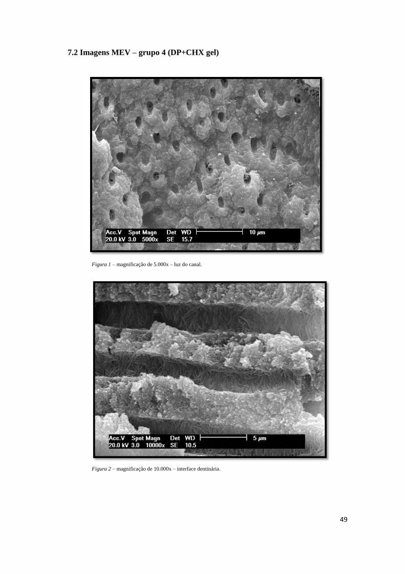
49
7.2 Imagens MEV – grupo 4 (DP+CHX gel)
Figura 1 – magnificação de 5.000x – luz do canal.
Figura 2 – magnificação de 10.000x – interface dentinária.

50
7.3 Imagens MEV – grupo 5 (DP+CHX liq)
Figura 1 – magnificação de 5.000x – luz do canal.
Figura 2 – magnificação de 10.000x – interface dentinária.

51
7.4 Imagens MEV – grupo 6 (DP+TC)
Figura 1 – magnificação de 5.000x – luz do canal.
Figura 2 – magnificação de 10.000x – interface dentinária.

52
7.5 Imagens MEV – grupo 7 (DP+Ca(OH)2)
Figura 1 – magnificação de 5.000x – luz do canal.
Figura 2 – magnificação de 10.000x – interface dentinária.